Quantification of HLA class II-specific memory B cells in HLA-sensitized individuals
8
Quantification of HLA class II-specific memory B cells in HLA-sensitized individuals Gonca E. Karahan a , Yvonne J.H. de Vaal a , Dave L. Roelen a , Rico Buchli b , Frans H.J. Claas a , Sebastiaan Heidt a,⇑ a Department of Immunohaematology and Blood Transfusion, Leiden University Medical Center, Leiden, The Netherlands b R&D, Pure Protein LLC, Oklahoma City, OK, USA article info Article history: Received 12 September 2014 Accepted 14 January 2015 Available online 27 January 2015 Keywords: HLA antibodies Luminex Pregnancy-immunized women Kidney transplantation Chronic rejection abstract For the quantification of HLA-specific memory B cells from peripheral blood of sensitized individuals, a limited number of methods are available. However, none of these are capable of detecting memory B cells directed at HLA class II molecules. Since the majority of antibodies that occur after transplantation appear to be specific for HLA class II, our aim was to develop an assay to detect and quantify HLA class II-specific memory B cells from peripheral blood. By using biotinylated soluble HLA class II molecules as detection agent, we were able to develop an HLA class II-specific memory B cell ELISPOT assay. The assay was validated using B cell-derived hybridomas that produce human monoclonal antibodies directed at specific HLA class II molecules. In pregnancy- immunized females, we found memory B cell frequencies ranging from 25 to 756 spots per 10 6 B cells specific for the immunizing paternal HLA class II molecules, whereas in non-immunized males no signif- icant spot formation was detected. Here, we present a novel ELISPOT assay for quantifying HLA class II-specific memory B cells from peripheral blood. This technique provides a unique tool for monitoring the HLA class II-specific memory B cell pool in sensitized transplant recipients. Ó 2015 American Society for Histocompatibility and Immunogenetics. Published by Elsevier Inc. All rights reserved. 1. Introduction Pre-existing as well as de novo produced HLA antibodies repre- sent an important risk factor for adverse transplantation outcome [1]. In the last decade, major advances in HLA antibody detection have enabled a clear definition of antibody specificities in the serum of transplant recipients, and thereby improved the predic- tion of immunologically low and high-risk patients [2–4]. How- ever, none of the currently available methods to detect serum HLA antibodies provide information on the presence or absence of HLA-specific memory B cells. Since serum antibodies are mainly produced by bone marrow residing plasma cells, serum antibody levels may not be represen- tative for the size of the peripheral memory B cell pool [5,6]. Upon a re-encounter with antigen, memory B cells can rapidly differen- tiate into antibody secreting cells to drive anamnestic responses [7]. Therefore, while serum antibody levels may be undetectable prior to transplant, accelerated antibody mediated rejection can occur in case memory B cells directed at donor HLA antigens are present [8]. Furthermore, apart from antibody production, memory B cells are potent cytokine producing cells [9] and can also function as antigen presenting cells [10], potentially driving the rejection process by activating alloantigen-specific T cells. Determining the level of HLA-specific memory B cells pre-transplant would benefit the risk assessment for early humoral rejection, whereas monitor- ing these cells post-transplant may provide a better understanding on how B cells may affect transplant outcomes in other ways than by antibody production [11]. As a result of the technical challenge of detecting and enumer- ating the relatively low numbers of HLA-specific memory B cells in peripheral blood, only a few studies exist aiming at quantification of HLA-specific memory B cells in sensitized individuals [12–18]. http://dx.doi.org/10.1016/j.humimm.2015.01.014 0198-8859/Ó 2015 American Society for Histocompatibility and Immunogenetics. Published by Elsevier Inc. All rights reserved. Abbreviations: ELISPOT, enzyme-linked immunosorbent spot; B-LCL, B-lympho- blastoid cell line; DSA, donor-specific antibody; FCS, fetal calf serum; HLA, human leukocyte antigen; IMDM, Iscove’s modified Dulbecco’s medium; ITS, insulin- transferrin-sodium selenite; mAb, monoclonal antibody; MFI, mean fluorescence intensity; ODN, oligodeoxynucleotides; PBMC, peripheral blood mononuclear cell; PBS, phosphate-buffered saline; SFU, spot forming unit; TLR, toll-like receptor. ⇑ Corresponding author at: Leiden University Medical Center, Department of Immunohaematology and Blood Transfusion, Albinusdreef 2, 2300 RC Leiden, The Netherlands. Fax: +31 71 5265267. E-mail address: [email protected] (S. Heidt). Human Immunology 76 (2015) 129–136 Contents lists available at ScienceDirect www.ashi-hla.org journal homepage: www.elsevier.com/locate/humimm
-
Upload
independent -
Category
Documents
-
view
4 -
download
0
Transcript of Quantification of HLA class II-specific memory B cells in HLA-sensitized individuals

Human Immunology 76 (2015) 129–136
Contents lists available at ScienceDirect
www.ashi-hla.org
journal homepage: www.elsevier .com/locate /humimm
Quantification of HLA class II-specific memory B cells in HLA-sensitizedindividuals
http://dx.doi.org/10.1016/j.humimm.2015.01.0140198-8859/� 2015 American Society for Histocompatibility and Immunogenetics. Published by Elsevier Inc. All rights reserved.
Abbreviations: ELISPOT, enzyme-linked immunosorbent spot; B-LCL, B-lympho-blastoid cell line; DSA, donor-specific antibody; FCS, fetal calf serum; HLA, humanleukocyte antigen; IMDM, Iscove’s modified Dulbecco’s medium; ITS, insulin-transferrin-sodium selenite; mAb, monoclonal antibody; MFI, mean fluorescenceintensity; ODN, oligodeoxynucleotides; PBMC, peripheral blood mononuclear cell;PBS, phosphate-buffered saline; SFU, spot forming unit; TLR, toll-like receptor.⇑ Corresponding author at: Leiden University Medical Center, Department of
Immunohaematology and Blood Transfusion, Albinusdreef 2, 2300 RC Leiden, TheNetherlands. Fax: +31 71 5265267.
E-mail address: [email protected] (S. Heidt).
Gonca E. Karahan a, Yvonne J.H. de Vaal a, Dave L. Roelen a, Rico Buchli b, Frans H.J. Claas a,Sebastiaan Heidt a,⇑a Department of Immunohaematology and Blood Transfusion, Leiden University Medical Center, Leiden, The Netherlandsb R&D, Pure Protein LLC, Oklahoma City, OK, USA
a r t i c l e i n f o
Article history:Received 12 September 2014Accepted 14 January 2015Available online 27 January 2015
Keywords:HLA antibodiesLuminexPregnancy-immunized womenKidney transplantationChronic rejection
a b s t r a c t
For the quantification of HLA-specific memory B cells from peripheral blood of sensitized individuals, alimited number of methods are available. However, none of these are capable of detecting memory B cellsdirected at HLA class II molecules. Since the majority of antibodies that occur after transplantation appearto be specific for HLA class II, our aim was to develop an assay to detect and quantify HLA class II-specificmemory B cells from peripheral blood.
By using biotinylated soluble HLA class II molecules as detection agent, we were able to develop an HLAclass II-specific memory B cell ELISPOT assay. The assay was validated using B cell-derived hybridomasthat produce human monoclonal antibodies directed at specific HLA class II molecules. In pregnancy-immunized females, we found memory B cell frequencies ranging from 25 to 756 spots per 106 B cellsspecific for the immunizing paternal HLA class II molecules, whereas in non-immunized males no signif-icant spot formation was detected.
Here, we present a novel ELISPOT assay for quantifying HLA class II-specific memory B cells fromperipheral blood. This technique provides a unique tool for monitoring the HLA class II-specific memoryB cell pool in sensitized transplant recipients.� 2015 American Society for Histocompatibility and Immunogenetics. Published by Elsevier Inc. All rights
reserved.
1. Introduction Since serum antibodies are mainly produced by bone marrow
Pre-existing as well as de novo produced HLA antibodies repre-sent an important risk factor for adverse transplantation outcome[1]. In the last decade, major advances in HLA antibody detectionhave enabled a clear definition of antibody specificities in theserum of transplant recipients, and thereby improved the predic-tion of immunologically low and high-risk patients [2–4]. How-ever, none of the currently available methods to detect serumHLA antibodies provide information on the presence or absenceof HLA-specific memory B cells.
residing plasma cells, serum antibody levels may not be represen-tative for the size of the peripheral memory B cell pool [5,6]. Upona re-encounter with antigen, memory B cells can rapidly differen-tiate into antibody secreting cells to drive anamnestic responses[7]. Therefore, while serum antibody levels may be undetectableprior to transplant, accelerated antibody mediated rejection canoccur in case memory B cells directed at donor HLA antigens arepresent [8]. Furthermore, apart from antibody production, memoryB cells are potent cytokine producing cells [9] and can also functionas antigen presenting cells [10], potentially driving the rejectionprocess by activating alloantigen-specific T cells. Determining thelevel of HLA-specific memory B cells pre-transplant would benefitthe risk assessment for early humoral rejection, whereas monitor-ing these cells post-transplant may provide a better understandingon how B cells may affect transplant outcomes in other ways thanby antibody production [11].
As a result of the technical challenge of detecting and enumer-ating the relatively low numbers of HLA-specific memory B cells inperipheral blood, only a few studies exist aiming at quantificationof HLA-specific memory B cells in sensitized individuals [12–18].

Table 1Biotinylated HLA class I and class II molecules.
HLA Coupled HLA allele Clone designation Peptide sequence
DRB1*01:01 DRA1*01:01 C5B6 –DRB1*04:05 DRA1*01:01 #3.14 –DRB1*07:01 DRA1*01:01 D3C2 –DRB1*09:01 DRA1*01:01 C2G5 –DRB1*11:01 DRA1*01:01 H9/III –DRB1*13:03 DRA1*01:01 #4.8 –DRB1*15:02 DRA1*01:01 B12 –DQB1*02:01 DQA1*02:01 E12G8 –DQB1*03:01 DQA1*02:01 C6E3 –DQB1*03:02 DQA1*02:01 C9A8 –DQB1*03:03 DQA1*02:01 E2C9 –DQB1*06:02 DQA1*01:01 C3F10 –A*01:01 – – EVDPIGHHLYA*02:01 – – YLEPGPVTA
130 G.E. Karahan et al. / Human Immunology 76 (2015) 129–136
Assays determining HLA antibody production capacity by in vitropolyclonal B cell activation [12,19], as well as HLA class I tetramerstaining of CD19+ B cells [13–15] have previously been describedby us and others. Though useful, these assays are limited to thedetection of HLA class I-specific memory responses in sensitizedindividuals. Importantly, recent evidence suggests that the pre-dominance of HLA antibodies developing post kidney transplanta-tion is directed at HLA class II [2,20,21]. Therefore, we aimed atdeveloping an assay capable of detecting and quantifying HLA classII-specific memory B cells from peripheral blood. Previously, wehave developed an HLA class I-specific B cell ELISPOT assay bycombining the ability of polyclonally activated B cells to produceHLA antibodies in vitro with the potential of these antibodies tobind HLA tetramers [17]. In the current study, we adapted and val-idated this technique for the detection of HLA class II-specificmemory B cells. We show that this newly developed assay allowsfor the quantification of HLA class II-specific memory B cells in sen-sitized individuals.
2. Materials and methods
2.1. Subjects
The study population consisted of pregnancy immunizedwomen and non-immunized males. Pregnancy immunized individ-uals (n = 6) were assessed for the presence of peripheral HLA classII-specific memory B cells against a total of 18 immunizing and/orself HLA class II molecules. For the same purpose, B cells isolatedfrom peripheral blood mononuclear cells (PBMC) of non-immu-nized healthy males (n = 3) were tested against a total of 6 self aswell as non-self HLA class II molecules.
2.2. Cells
Peripheral blood was obtained with informed consent underguidelines issued by the Medical Ethics Committee of the LeidenUniversity Medical Center (Leiden, the Netherlands). PBMCs wereisolated by Ficoll Hypaque (pharmacy LUMC, Leiden, the Nether-lands) density gradient centrifugation and frozen in liquid nitrogenuntil further use. After thawing, B cells were isolated by negativeselection using EasySep Human B cell enrichment kit (Stem CellTechnologies, Grenoble, France) according to the manufacturer‘sinstructions. The purity of B cells was found to be >98% as assessedby CD19 positivity measured by flow cytometry.
In order to optimize and validate the HLA class II-specific mem-ory B cell ELISPOT assay, previously described human B cell hybrid-omas producing class I and class II anti-HLA monoclonal antibodies(mAbs) were used [22]: GV5D1 (IgG, anti-HLA-A1/9), SN607D8(IgG, anti-HLA-A2/28), RTLK10E12 (IgG, anti-HLA-DR11) andRTLK1E2 (IgG, anti-HLA-DR3/8/11/12/13/14).
2.3. Biotinylated soluble HLA class I and class II molecules
Synthetic biotinylated HLA class I monomers were constructedaround various peptide sequences (Table 1) as described previ-ously [17,23]. For HLA class II, constructs were made by deletionof the transmembrane domains of the alpha and beta chains ofHLA-DR and HLA-DQ molecules and replacing a 7 amino acid linkerfollowed by leucine zipper ACIDp1 for alpha chains and leucinezipper BASEp1 for beta chains. These constructs were cloned intothe mammalian expression vector pcDNA3.1(�) and expressed inB-lymphoblastoid cell lines (B-LCL) by transfection, as describedelsewhere [24]. Secreted HLA class II molecules were used in bio-tinylated form. Table 1 shows the list of biotinylated HLA class Iand class II molecules used.
2.4. Cell cultures
Purified B cells were activated for 6 days at 2.5 � 105 cells/wellin 24-well flat bottom plates (Corning Incorporated, Corning, NY).Cell cultures were carried out in Iscove’s modified Dulbecco’s med-ium (IMDM) (Gibco Invitrogen, Paisley, UK) containing 10% fetalcalf serum (FCS) (Gibco Invitrogen), supplemented with 50 lM 2-mercaptoethanol (Sigma–Aldrich, Zwijndrecht, the Netherlands),2 mM L-glutamine (Gibco Invitrogen), ITS (5 lg/ml insulin,5 lg/ml transferrin, sodium selenite 5 ng/ml, (Sigma–Aldrich)and 100 U/ml penicillin with 100 lg/ml streptomycin (GibcoInvitrogen). Cells were activated with 500 ng/ml a-CD40 mAb(R&D systems, Minneapolis, MN, USA), 2.5 lg/ml toll-likereceptor-9 (TLR-9) ligand oligodeoxynucleotides (ODN)-2006 CpG(Hycult Biotechnology, Uden, the Netherlands), 600 IU/ml IL-2(Proleukin, Amsterdam, the Netherlands), 25 ng/ml IL-10 (R&Dsystems, Minneapolis, MN, USA) and 100 ng/ml IL-21 (Gibco Invit-rogen), as described previously [25], and cultured at 37 �C in a 5%CO2 humidified incubator. At day 6, supernatants were collectedand 10-fold concentrated using 0.5 ml centrifugal filter units(Amicon Ultra, Merck Millipore Ltd.), and frozen at �20 �C untilfurther use. Cells were harvested and thoroughly washed.
2.5. ELISPOT assays
We performed total IgG ELISPOT assays as previously described[26]. For the HLA class II-specific ELISPOT assays, we coated 96-well ELISPOT plates (Millipore, Billerica, MA, USA) with 5 lg/mlgoat anti-human IgG (Jackson Immunoresearch Laboratories Inc.,Baltimore, PA, USA) in phosphate buffered saline (PBS) and incu-bated overnight at 4 �C. Following blocking for at least 1 h with5% FCS/IMDM at 37 �C, thoroughly washed activated B cells(2.5 � 105 cells/well) or hybridoma cells (2500 cells/well) wereadded to each plate. After overnight incubation at 37 �C, plateswere washed and incubated for 4 h with biotinylated HLA class IImolecules (1000 ng/ml) at room temperature on a platform shaker.Following washing, plates were incubated with streptavidin-conjugated alkaline phosphatase (Sigma–Aldrich) for 1 h at roomtemperature. BCIP/NBT substrate (Mabtech, Nacka Strand, Sweden)was added to visualize the spots. The reaction was stopped by coldtap water and after drying, analyzed by an automated ELISPOTreader (Bio-Sys GmbH, Germany).
To correct for non-specific background spots, we included wellswithout adding the HLA molecules at the detection step. Spots, ifany, were subtracted from the spot numbers counted in the wellswith HLA molecules added. The ratio of HLA class II-specific B cellsper total input B cells was calculated as described previously[17].
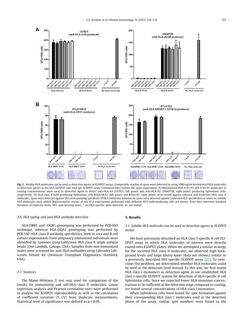
Fig. 1. Soluble HLA molecules can be used as detection agents in ELISPOT assays. Comparable number of spots were detected by using 1000 ng/ml biotinylated HLA moleculesas detection agents in the HLA-ELISPOT and total IgG ELISPOT assay (checkered bars) within the same experiment (A) Biotinylated HLA-A*01:01 and A*02:01 molecules atvarying concentrations were used as detection agent to detect anti-HLA-A1 (GV5D1, left panel) and anti-HLA-A2 (SN607D8, right panel) producing hybridoma cells,respectively. (B) HLA class II mAb-producing hybridoma cells RTLK10E12 (left panel) and RTLK1E2 (right panel) were tested against relevant and irrelevant HLA class IImolecules. Spots were detected against the corresponding specificity of HLA molecules whereas no spots were detected against irrelevant HLA specificities or when no solubleHLA molecules were added. Representative results of one of 2 experiments performed with different HLA mAb-producing cells are shown. Error bars represent standarddeviation of triplicate wells. SFU, spot forming units; *, no HLA-specific spots detected; nt, not tested.
G.E. Karahan et al. / Human Immunology 76 (2015) 129–136 131
2.6. HLA typing and anti-HLA antibody detection
HLA-DRB1 and -DQB1 genotyping was performed by PCR-SSOtechnique, whereas HLA-DQA1 genotyping was performed byPCR-SSP. HLA class II antibody specificities, both in sera and B cellculture supernatants from pregnancy immunized individuals wereidentified by Luminex using LabScreen HLA class II single antigenbeads (One Lambda, Canoga, USA). Samples from non-immunizedmales were screened for anti-HLA antibodies using Lifecodes Life-screen Deluxe kit (Immucor Transplant Diagnostics, Stamford,USA).
2.7. Statistics
The Mann–Whitney U test was used for comparison of theresults for immunizing and self-HLA class II molecules. Linearregression analysis and Pearson correlation tests were performedto analyze the ELISPOT reproducibility as well as the calculationof coefficient variation (% CV) from duplicate measurements.Statistical level of significance was defined as p < 0.05.
3. Results
3.1. Soluble HLA molecules can be used as detection agents in ELISPOTassays
We have previously described an HLA class I-specific B cell ELI-SPOT assay in which HLA molecules of interest were directlycoated onto ELISPOT plates. When we attempted a similar strategyfor the secreted HLA class II molecules, we observed high back-ground levels and large, blurry spots (data not shown), similar toa previously described HIV-specific ELISPOT assay [27]. To over-come this problem, we determined whether HLA molecules couldbe used at the detection level instead. To this aim, we first testedHLA class I monomers as detection agent in our established HLAclass I-specific ELISPOT system for detection of HLA-specific B cellhybridoma cells. Since we expected lower HLA monomer concen-trations to be sufficient at the detection stage compared to coating,we tested several concentrations of HLA class I monomers.
When hybridoma cells were tested for spot formation againsttheir corresponding HLA class I molecules used in the detectionphase of the assay, similar spot numbers were found in the
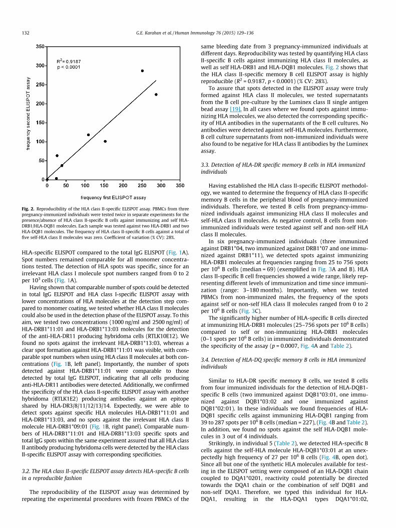
Fig. 2. Reproducibility of the HLA class II-specific ELISPOT assay. PBMCs from threepregnancy-immunized individuals were tested twice in separate experiments for thepresence/absence of HLA class II-specific B cells against immunizing and self HLA-DRB1/HLA-DQB1 molecules. Each sample was tested against two HLA-DRB1 and twoHLA-DQB1 molecules. The frequency of HLA class II-specific B cells against a total offive self-HLA class II molecules was zero. Coefficient of variation (% CV): 28%.
132 G.E. Karahan et al. / Human Immunology 76 (2015) 129–136
HLA-specific ELISPOT compared to the total IgG ELISPOT (Fig. 1A).Spot numbers remained comparable for all monomer concentra-tions tested. The detection of HLA spots was specific, since for anirrelevant HLA class I molecule spot numbers ranged from 0 to 2per 103 cells (Fig. 1A).
Having shown that comparable number of spots could be detectedin total IgG ELISPOT and HLA class I-specific ELISPOT assay withlower concentrations of HLA molecules at the detection step com-pared to monomer coating, we tested whether HLA class II moleculescould also be used in the detection phase of the ELISPOT assay. To thisaim, we tested two concentrations (1000 ng/ml and 2500 ng/ml) ofHLA-DRB1*11:01 and HLA-DRB1*13:03 molecules for the detectionof the anti-HLA-DR11 producing hybridoma cells (RTLK10E12). Wefound no spots against the irrelevant HLA-DRB1*13:03, whereas aclear spot formation against HLA-DRB1*11:01 was visible, with com-parable spot numbers when using HLA class II molecules at both con-centrations (Fig. 1B, left panel). Importantly, the number of spotsdetected against HLA-DRB1*11:01 were comparable to thosedetected by total IgG ELISPOT, indicating that all cells producinganti-HLA-DR11 antibodies were detected. Additionally, we confirmedthe specificity of the HLA class II-specific ELISPOT assay with anotherhybridoma (RTLK1E2) producing antibodies against an epitopeshared by HLA-DR3/8/11/12/13/14. Expectedly, we were able todetect spots against specific HLA molecules HLA-DRB1*11:01 andHLA-DRB1*13:03, and no spots against the irrelevant HLA class IImolecule HLA-DRB1*09:01 (Fig. 1B, right panel). Comparable num-bers of HLA-DRB1*11:01 and HLA-DRB1*13:03 specific spots andtotal IgG spots within the same experiment assured that all HLA classII antibody producing hybridoma cells were detected by the HLA classII-specific ELISPOT assay with corresponding specificities.
3.2. The HLA class II-specific ELISPOT assay detects HLA-specific B cellsin a reproducible fashion
The reproducibility of the ELISPOT assay was determined byrepeating the experimental procedures with frozen PBMCs of the
same bleeding date from 3 pregnancy-immunized individuals atdifferent days. Reproducibility was tested by quantifying HLA classII-specific B cells against immunizing HLA class II molecules, aswell as self HLA-DRB1 and HLA-DQB1 molecules. Fig. 2 shows thatthe HLA class II-specific memory B cell ELISPOT assay is highlyreproducible (R2 = 0.9187, p < 0.0001) (% CV: 28%).
To assure that spots detected in the ELISPOT assay were trulyformed against HLA class II molecules, we tested supernatantsfrom the B cell pre-culture by the Luminex class II single antigenbead assay [19]. In all cases where we found spots against immu-nizing HLA molecules, we also detected the corresponding specific-ity of HLA antibodies in the supernatants of the B cell cultures. Noantibodies were detected against self-HLA molecules. Furthermore,B cell culture supernatants from non-immunized individuals werealso found to be negative for HLA class II antibodies by the Luminexassay.
3.3. Detection of HLA-DR specific memory B cells in HLA immunizedindividuals
Having established the HLA class II-specific ELISPOT methodol-ogy, we wanted to determine the frequency of HLA class II-specificmemory B cells in the peripheral blood of pregnancy-immunizedindividuals. Therefore, we tested B cells from pregnancy-immu-nized individuals against immunizing HLA class II molecules andself-HLA class II molecules. As negative control, B cells from non-immunized individuals were tested against self and non-self HLAclass II molecules.
In six pregnancy-immunized individuals (three immunizedagainst DRB1*04, two immunized against DRB1*07 and one immu-nized against DRB1*11), we detected spots against immunizingHLA-DRB1 molecules at frequencies ranging from 25 to 756 spotsper 106 B cells (median = 69) (exemplified in Fig. 3A and B). HLAclass II-specific B cell frequencies showed a wide range, likely rep-resenting different levels of immunization and time since immuni-zation (range: 3–180 months). Importantly, when we testedPBMCs from non-immunized males, the frequency of the spotsagainst self or non-self HLA class II molecules ranged from 0 to 2per 106 B cells (Fig. 3C).
The significantly higher number of HLA-specific B cells directedat immunizing HLA-DRB1 molecules (25–756 spots per 106 B cells)compared to self or non-immunizing HLA-DRB1 molecules(0–1 spots per 106 B cells) in immunized individuals demonstratedthe specificity of the assay (p = 0.0007, Fig. 4A and Table 2).
3.4. Detection of HLA-DQ specific memory B cells in HLA immunizedindividuals
Similar to HLA-DR specific memory B cells, we tested B cellsfrom four immunized individuals for the detection of HLA-DQB1-specific B cells (two immunized against DQB1*03:01, one immu-nized against DQB1*03:02 and one immunized againstDQB1*02:01). In these individuals we found frequencies of HLA-DQB1 specific cells against immunizing HLA-DQB1 ranging from39 to 287 spots per 106 B cells (median = 227), (Fig. 4B and Table 2).In addition, we found no spots against the self HLA-DQB1 mole-cules in 3 out of 4 individuals.
Strikingly, in individual 5 (Table 2), we detected HLA-specific Bcells against the self-HLA molecule HLA-DQB1*03:01 at an unex-pectedly high frequency of 27 per 106 B cells (Fig. 4B, open dot).Since all but one of the synthetic HLA molecules available for test-ing in the ELISPOT setting were composed of an HLA-DQB1 chaincoupled to DQA1*0201, reactivity could potentially be directedtowards the DQA1 chain or the combination of self DQB1 andnon-self DQA1. Therefore, we typed this individual for HLA-DQA1, resulting in the HLA-DQA1 types DQA1*01:02,
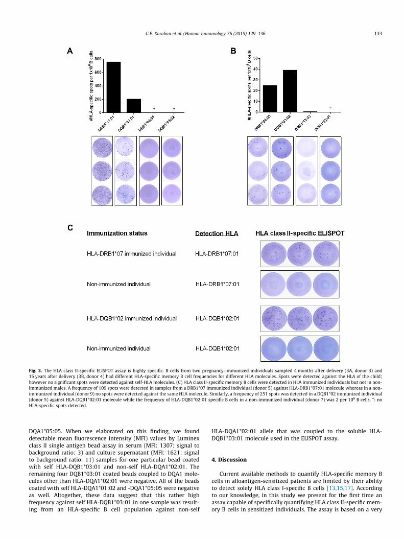
Fig. 3. The HLA class II-specific ELISPOT assay is highly specific. B cells from two pregnancy-immunized individuals sampled 4 months after delivery (3A, donor 3) and15 years after delivery (3B, donor 4) had different HLA-specific memory B cell frequencies for different HLA molecules. Spots were detected against the HLA of the child;however no significant spots were detected against self-HLA molecules. (C) HLA class II-specific memory B cells were detected in HLA immunized individuals but not in non-immunized males. A frequency of 109 spots were detected in samples from a DRB1*07 immunized individual (donor 5) against HLA-DRB1*07:01 molecule whereas in a non-immunized individual (donor 9) no spots were detected against the same HLA molecule. Similarly, a frequency of 251 spots was detected in a DQB1*02 immunized individual(donor 5) against HLA-DQB1*02:01 molecule while the frequency of HLA-DQB1*02:01 specific B cells in a non-immunized individual (donor 7) was 2 per 106 B cells. *: noHLA-specific spots detected.
G.E. Karahan et al. / Human Immunology 76 (2015) 129–136 133
DQA1*05:05. When we elaborated on this finding, we founddetectable mean fluorescence intensity (MFI) values by Luminexclass II single antigen bead assay in serum (MFI: 1307; signal tobackground ratio: 3) and culture supernatant (MFI: 1621; signalto background ratio: 11) samples for one particular bead coatedwith self HLA-DQB1*03:01 and non-self HLA-DQA1*02:01. Theremaining four DQB1*03:01 coated beads coupled to DQA1 mole-cules other than HLA-DQA1*02:01 were negative. All of the beadscoated with self HLA-DQA1*01:02 and -DQA1*05:05 were negativeas well. Altogether, these data suggest that this rather highfrequency against self HLA-DQB1*03:01 in one sample was result-ing from an HLA-specific B cell population against non-self
HLA-DQA1*02:01 allele that was coupled to the soluble HLA-DQB1*03:01 molecule used in the ELISPOT assay.
4. Discussion
Current available methods to quantify HLA-specific memory Bcells in alloantigen-sensitized patients are limited by their abilityto detect solely HLA class I-specific B cells [13,15,17]. Accordingto our knowledge, in this study we present for the first time anassay capable of specifically quantifying HLA class II-specific mem-ory B cells in sensitized individuals. The assay is based on a very

Fig. 4. Frequencies of HLA class II-specific memory B cells detected by ELISPOT. The number of spots directed at immunizing (A) HLA-DRB1 and (B) HLA-DQB1 molecules wassignificantly higher than the number of spots against self or antibody negative HLA molecules. The open dot in (B) is the unexpectedly high frequency of 27 spots per 106 Bcells for a self HLA-DQB1 molecule. Bars indicate the median in each group. ab, antibody.
Table 2Characteristics of the study population.
Donor Immunizationstatus
Sampling timeafter lastdelivery
Self/child HLA Seruma
abDetection HLAmolecule
Frequencyb MFIsupernatantc
1 Immunized 12 years Unknown Yes DRB1*04:05 25 18482 Immunized 10 years Self: DR9, DR15 Yes DRB1*07:01 30 71393 Immunized 4 months Self: DRB1*04, DQB1*03:02, DQB1*04:02 Yes DRB1*11:01 756 16,615
(Child: DRB1*04, DRB1*11, DQB1*03:01, DQB1*04:02) Yes DQB1*03:01 203 15,967No DRB1*04:05 0 0No DQB1*03:02 0 0
4 Immunized 15 years Self: DRB1*03:01, DRB1*13:01, DQB1*02, DQB1*06:03 Yes DRB1*04:05 25 3415(Child:DRB1*04, DRB1*13:01, DQB1*03:02, DQB1*06:03) Yes DQB1*03:02 39 6250
No DRB1*13:03 1 34No DQB1*02:01 0 0
5 Immunized 2 years Self: DRB1*11:01, DRB1*15:02, DQB1*03:01, DQB1*06:02 Yes DRB1*07:01 109 11,991DQA1*01:02, DQA1*05:05 Yes DQB1*02:01 251 12,923
No DRB1*15:02 0 0No DQB1*03:01 27 1621*
6 Immunized 3 months Self: DRB1*13:01, DRB1*15, DQB1*06:02, DQB1*06:03 Yes DRB1*04:05 153 9346(Child:DRB1*04:02, DRB1*13:01, DQB1*03:01, DQB1*06:03) Yes DQB1*03:01 287 19,337
No DRB1*15:02 0 0No DQB1*06:02 0 0
7 Non-immunized – DQB1*02:01, DQB1*06:04 ntd DQB1*02:01 2 0nt DQB1*03:03 0 0
8 Non-immunized – DRB1*11:01, DRB1*15:01 nt DRB1*11:01 0 0nt DRB1*09:01 1 0
9 Non-immunized – DRB1*07:01, DRB1*13:02 nt DRB1*07:01 0 0nt DRB1*01:01 0 0
a HLA antibodies in the serum belonging to the same date of cell collection for ELISPOT experiments, determined by Luminex single antigen bead assay (except for donor 5,serum data is 6 months after cell collection).
b Total number of HLA-specific spots per 106 B cells.c B cell culture supernatants were tested by Luminex for the presence of HLA antibodies using HLA class II single antigen beads in samples of pregnancy immunized
individuals.d Sera from non-immunized males were not available for testing (not tested).* In contrast to all other supernatants, in this particular supernatant only one out five HLA-DQB1*03:01 beads showed reactivity. Positive reaction was only found for HLA-
DQB1*03:01 bead that is coupled to HLA-DQA1*02:01. All the remaining HLA-DQB1*03:01 beads coupled to other HLA-DQA1 molecules were negative.
134 G.E. Karahan et al. / Human Immunology 76 (2015) 129–136
sensitive and practical ELISPOT platform, allowing for detection oflow level of HLA class II-specific memory B cells from peripheralblood.
Previously, we have utilized an HLA class I-specific memory Bcell assay in which PBMCs depleted of CD2+ T cells from sensitizedindividuals were polyclonally activated in vitro and assayed in HLAclass I monomer-coated ELISPOT plates [17]. Recently, Lynch et al.showed the presence of donor-specific memory B cells directedtowards donor HLA in the absence of donor-specific serum IgGantibodies in kidney transplant recipients by ELISPOT usingdonor-derived fibroblasts as the HLA target [18]. Although this isan elegant method to track donor specific B cell responses, thesource of donor material and long culture time of fibroblasts(30 days) pose a potential barrier for its application in routine
clinical monitoring. Furthermore, considering that fibroblastsexpress only HLA class I molecules, such an assay does not allowfor the estimation of HLA class II-specific B cell frequencies.
Despite recent advances in transplantation, the long-term out-come of transplanted organs remains affected by chronic rejection.Using highly sensitive and specific Luminex single antigen beadassays, several groups reported high incidences of post-transplan-tation de novo donor-specific antibody (DSA), particularly directedat mismatched HLA class II molecules [2,20]. These class II antibod-ies were shown to be a risk factor for late kidney allograft failure[21,28]. In a re-transplant cohort of 112 adult recipients,Worthington et al. showed a detrimental role for DSA directed atHLA class II, resulting in late graft damage, while class I DSAseemed to be associated more with early graft failure [29]. Since

G.E. Karahan et al. / Human Immunology 76 (2015) 129–136 135
there may be a difference in the development of humoral immu-nity towards HLA class I and class II in time after transplantation,assays to analyze both the HLA class I- and class II-specific memoryB cell compartments are of particular interest.
In the current HLA class II-specific ELISPOT assay, we usedhighly purified B cells from PBMCs for the detection of HLA classII-specific memory B cells. We have recently shown that the cur-rent polyclonal B cell activation protocol could reliably be usedfor estimating the percentage of pre-existing antigen-specific Bcells, since no antigen-specific IgG spots were detected in naïve Bcells following a 6-day activation [25]. The finding that no spotswere detected in non-immunized individuals against self or non-self HLA class II molecules in the present study further assured thatthe detected spots were formed by memory B cells. Regarding theELISPOT phase of the assay, we chose to use HLA class II moleculesas the detection matrix instead of the coating agent. Besides thefact that clear spot formation was only observed when this strategywas used, it has the additional benefit that a lower concentration ofHLA molecules can be used [27].
By using this method, we found up to 756 HLA class II-specific Bcells (median: 130) per 106 B cells in HLA immunized individuals.Previously, we have found a median frequency of 43 (range: 0–182) HLA class I-specific B per 106 B cells in pregnancy immunizedindividuals by HLA class I-specific ELISPOT [17]. Whether HLA classII immunization leads to more profound memory formation com-pared to HLA class I needs to be further elucidated. In a follow-up study, we plan to determine the level of immunization for bothHLA class I and class II within patients immunized by both preg-nancy and transplantation.
Similar to solid phase antibody assays, we observed thatHLA-DQ molecules may give rise to unexpected results due tothe polymorphic nature of the HLA-DQA1 chain in addition tothe HLA-DQB1 chain. Tambur et al. [30] have shown the presenceof DQ antibodies directed at Luminex beads coated with self HLA-DQB1 combined with a non-self HLA-DQA1 and pointed out theunderestimation of the role of HLA-DQA1 in the detection of anti-bodies in Luminex assays. Similarly, in the present study, we foundspots against a self HLA-DQB1*03:01 molecule in one sample froma pregnancy-immunized individual, which are likely due to anti-body producing memory B cells directed at the non-selfDQA1*02:01 allele coupled to a self HLA-DQB1*03:01 molecule.Whether this individual produced antibodies directed against anepitope solely on the DQA1*02:01 chain or an epitope formed bythe combination of non-self DQA1*02:01 and self HLA-DQB1*03:01 is beyond the scope of this paper [31]. Regardless,our results indicate that in future studies, soluble HLA-DQ mole-cules comprising of several combinations of HLA-DQB and HLA-DQA chains should be tested in order to overcome this limitation.
The high reproducibility rate of the assay and availability of bio-tinylated soluble HLA class II molecules covering a wide range ofHLA class II specificities support the utilization of the assay inthe clinical setting. A volume of 10–15 ml peripheral blood samplefrom the patients which is routinely collected for bio-banking inmany transplant centers is sufficient to perform the assay. Itshould be noted that even if all combinations of the available sol-uble HLA molecules are used, the donor specific HLA-repertoiremay not be completely represented. In conclusion, we have devel-oped a highly specific, sensitive and reproducible ELISPOT assayenabling the quantification of the HLA class II-specific memory Bcells from peripheral blood, which may become a useful tool forpre-transplant risk assessment in sensitized transplant recipients.
Acknowledgments
G.E.K. designed and performed experiments, collected and ana-lysed data and wrote the paper; Y.J.H.V. performed experiments
and analysed data; D.L.R. participated in study design: R.B. pro-vided the HLA class II molecules; F.H.J.C. participated in studydesign and wrote the paper; S.H. designed the study, analysed dataand wrote the paper.
The authors thank Sophia Stein, Simone Brand-Schaaf and JosDrabbels for the technical assistance in antibody testing and HLAtyping, Geert W. Haasnoot in statistical analysis and Dr. ArendMulder for hybridomas. This work was supported by the NationalReference Center for Histocompatibility Testing. S.H. is supportedby a grant from the Dutch Kidney Foundation, the Netherlands(VR.12.03).
References
[1] Terasaki PI, Ozawa M. Predicting kidney graft failure by HLA antibodies: aprospective trial. Am J Transplant 2004;4:438–43.
[2] Everly MJ, Rebellato LM, Haisch CE, Ozawa M, Parker K, Briley KP, et al.Incidence and impact of de novo donor-specific alloantibody in primary renalallografts. Transplantation 2013;95:410–7.
[3] Wiebe C, Nickerson P. Posttransplant monitoring of de novo human leukocyteantigen donor-specific antibodies in kidney transplantation. Curr Opin OrganTransplant 2013;18:470–7.
[4] Tait BD, Hudson F, Cantwell L, Brewin G, Holdsworth R, Bennett G, et al. Reviewarticle: Luminex technology for HLA antibody detection in organtransplantation. Nephrology (Carlton) 2009;14:247–54.
[5] Crotty S, Kersh EN, Cannons J, Schwartzberg PL, Ahmed R. SAP is required forgenerating long-term humoral immunity. Nature 2003;421:282–7.
[6] Manz RA, Arce S, Cassese G, Hauser AE, Hiepe F, Radbruch A. Humoralimmunity and long-lived plasma cells. Curr Opin Immunol 2002;14:517–21.
[7] Berek C, Berger A, Apel M. Maturation of the immune response in germinalcenters. Cell 1991;67:1121–9.
[8] Zachary AA, Lucas DP, Montgomery RA, Leffell MS. Rituximab prevents ananamnestic response in patients with cryptic sensitization to HLA.Transplantation 2013;95:701–4.
[9] Lund FE. Cytokine-producing B lymphocytes-key regulators of immunity. CurrOpin Immunol 2008;20:332–8.
[10] Noorchashm H, Reed AJ, Rostami SY, Mozaffari R, Zekavat G, Koeberlein B, et al.B cell-mediated antigen presentation is required for the pathogenesis of acutecardiac allograft rejection. J Immunol 2006;177:7715–22.
[11] Sarwal M, Chua MS, Kambham N, Hsieh SC, Satterwhite T, Masek M, et al.Molecular heterogeneity in acute renal allograft rejection identified by DNAmicroarray profiling. N Engl J Med 2003;349:125–38.
[12] Mulder A, Kardol MJ, Kamp J, Uit Het Broek C, Schreuder GM, Doxiadis II, et al.Determination of the frequency of HLA antibody secreting B-lymphocytes inalloantigen sensitized individuals. Clin Exp Immunol 2001;124:9–15.
[13] Mulder A, Eijsink C, Kardol MJ, Franke-van Dijk ME, van der Burg SH, Kester M,et al. Identification, isolation, and culture of HLA-A2-specific B lymphocytesusing MHC class I tetramers. J Immunol 2003;171:6599–603.
[14] Zachary AA, Kopchaliiska D, Montgomery RA, Leffell MS. HLA-specific B cells: I.A method for their detection, quantification, and isolation using HLAtetramers. Transplantation 2007;83:982–8.
[15] Zachary AA, Kopchaliiska D, Montgomery RA, Melancon JK, Leffell MS. HLA-specific B cells: II. Application to transplantation. Transplantation2007;83:989–94.
[16] Wadia PP, Herrera ND, Abecassis MM, Tambur AR. Mycophenolic acid inhibitsmaturation and function of human dendritic cells and B cells. Hum Immunol2009;70:692–700.
[17] Heidt S, Roelen DL, de Vaal YJ, Kester MG, Eijsink C, Thomas S, et al. A novelELISPOT assay to quantify HLA-specific B cells in HLA-immunized individuals.Am J Transplant 2012;12:1469–78.
[18] Lynch RJ, Silva IA, Chen BJ, Punch JD, Cascalho M, Platt JL. Cryptic B cellresponse to renal transplantation. Am J Transplant 2013;13:1713–23.
[19] Han M, Rogers J, Lavingia B, Stastny P. Peripheral blood B cells producingdonor-specific HLA antibodies in vitro. Hum Immunol 2009;70:29–34.
[20] Wiebe C, Gibson IW, Blydt-Hansen TD, Karpinski M, Ho J, Storsley LJ, et al.Evolution and clinical pathologic correlations of de novo donor-specific HLAantibody post kidney transplant. Am J Transplant 2012;12:1157–67.
[21] Campos EF, Tedesco-Silva H, Machado PG, Franco M, Medina-Pestana JO,Gerbase-DeLima M. Post-transplant anti-HLA class II antibodies as risk factorfor late kidney allograft failure. Am J Transplant 2006;6:2316–20.
[22] Mulder A, Kardol M, Regan J, Buelow R, Claas F. Reactivity of twenty-twocytotoxic human monoclonal HLA antibodies towards soluble HLA class I in anenzyme-linked immunosorbent assay (PRA-STAT). Hum Immunol1997;56:106–13.
[23] Altman JD, Moss PA, Goulder PJ, Barouch DH, McHeyzer-Williams MG, Bell JI, et al.Phenotypic analysis of antigen-specific T lymphocytes. Science 1996;274:94–6.
[24] McMurtrey C, Lowe D, Buchli R, Daga S, Royer D, Humphrey A, et al. Profilingantibodies to class II HLA in transplant patient sera. Hum Immunol2014;75:261–70.
[25] Karahan GE, Eikmans M, Anholts JD, Claas FH, Heidt S. Polyclonal B cellactivation for accurate analysis of pre-existing antigen-specific memory Bcells. Clin Exp Immunol 2014;177:333–40.

136 G.E. Karahan et al. / Human Immunology 76 (2015) 129–136
[26] Heidt S, Roelen DL, Eijsink C, van Kooten C, Claas FH, Mulder A. Effects ofimmunosuppressive drugs on purified human B cells: evidence supporting theuse of MMF and rapamycin. Transplantation 2008;86:1292–300.
[27] Dosenovic P, Chakrabarti B, Soldemo M, Douagi I, Forsell MN, Li Y, et al.Selective expansion of HIV-1 envelope glycoprotein-specific B cell subsetsrecognizing distinct structural elements following immunization. J Immunol2009;183:3373–82.
[28] Freitas MC, Rebellato LM, Ozawa M, Nguyen A, Sasaki N, Everly M, et al. Therole of immunoglobulin-G subclasses and C1q in de novo HLA-DQdonor-specific antibody kidney transplantation outcomes. Transplantation2013;95:1113–9.
[29] Worthington JE, Martin S, Al-Husseini DM, Dyer PA, Johnson RW.Posttransplantation production of donor HLA-specific antibodies as apredictor of renal transplant outcome. Transplantation 2003;75:1034–40.
[30] Tambur AR, Leventhal JR, Friedewald JJ, Ramon DS. The complexity of humanleukocyte antigen (HLA)-DQ antibodies and its effect on virtual crossmatching.Transplantation 2010;90:1117–24.
[31] Tambur AR, Rosati J, Roitberg S, Glotz D, Friedewald JJ, Leventhal JR. Epitopeanalysis of HLA-DQ antigens: what does the antibody see? Transplantation2014;98:157–66.