QL536. B43 bite C.1 - UGSpace
154
QL536. B43 bite C.1 THE BALME LIBRARY University of Ghana http://ugspace.ug.edu.gh
-
Upload
khangminh22 -
Category
Documents
-
view
3 -
download
0
Transcript of QL536. B43 bite C.1 - UGSpace

QL536. B43 bite C.1 THE BALME LIBRARY
University of Ghana http://ugspace.ug.edu.gh

VECTOR COMPETENCE OF THE ANOPHELES
(DIPTERA: CULICIDAE) POPULATIONS FOR
WUCHERERIA BANCROFTI (SPIRURIDA: FILARIIDAE),
AFTER MASS DRUG ADMINISTRATION IN THE
GOMOA DISTRICT OF GHANA
BY
BETHEL KWANSA-BENTUM (BSc Hons)
University of Ghana http://ugspace.ug.edu.gh

VECTOR COMPETENCE OF THE ANOPHELES
(DIPTERA: CULICIDAE) POPULATIONS FOR
WUCHERERIA BANCROFTI (SPIRURIDA: FILARIIDAE)
AFTER MASS DRUG ADMINISTRATION IN THE
GOMOA DISTRICT OF GHANA
BY
BETHEL KWANSA-BENTUM (BSc Hons)(10174126)
THIS THESIS IS SUBMITTED TO THE SCHOOL OF RESEARCH AND
GRADUATE STUDIES, UNIVERSITY OF GHANA - LEGON, IN PARTIAL
FULFILMENT FOR THE REQUIREMENT FOR THE AWARD OF MASTER OF
PHILOSOPHY DEGREE IN ZOOLOGY (APPLIED PARASITOLOGY)
MAY 2005
University of Ghana http://ugspace.ug.edu.gh

I hereby declare that except for references to other people’s work, which have duly been
acknowledged, this exercise is a result of my own research and this thesis neither in whole
nor in part, had been presented for another degree elsewhere.
DECLARATION
(CANDIDATE)
PROFESSOR MICHAEL DAVID WILSON (SUPERVISOR)
(SUPERVISOR)
University of Ghana http://ugspace.ug.edu.gh

DEDICATION
THIS WORK IS DEDICATED TO THE ANAA-BENTUM FAMILY
FOR THEIR LOVE, SUPPORT, AND ENCOURAGEMENTS THAT HAVE
BROUGHT MY EDUCATION THIS FAR
University of Ghana http://ugspace.ug.edu.gh

ACKNOWLEDGEMENTS
It is my first duty to acknowledge with pleasure, my indebtedness to all the individuals,
organisations and institutions that contributed in various ways to the formulation, execution
and submission of the work described in the thesis. I am very much thankful to Professor
David Ofori-Adjei, the Director of the Noguchi Memorial Institute for Medical Research
(NMIMR), University of Ghana, Legon for permitting me to use the facilities of the Institute.
I wish to express my heartfelt gratitude and deep appreciation to my supervisors Dr. Daniel
Adjei Boakye and Professor Michael David Wilson, both of Parasitology Department of
NMIMR. Their expert guidance made a great impact in achieving this goal. I am also
indebted to Mr. Maxwell Appawu, Dr. Kwabena Mante Bosompem, Dr. Charles Brown and
Professor Dominic Edoh for the invaluable contribution to this work. Their patience and
constructive criticisms during the conduct of the study made this dream a reality.
I also acknowledge the timely contributions by the following colleagues; Ms. Yvonne
Aryeetey, Fred Aboagye-Antwi, Evans D. Glah, Sampson Otoo, Philip Doku, Joseph
Otchere, Haruna Abdul, Charles Quaye and Yaw Gaisah. My appreciation also goes to the
volunteers who availed themselves as sources of blood meal for the mosquitoes to feed on,
without them the work would not have been done.
I thank the entire staff of the Parasitology Department (NMIMR); Ms. Helena Baidoo, Ms.
Naiki Puplampu, Ms. Irene Larbi, Mrs. Beverly Egyir, Mrs. Mercy Mintah Afari, Dziedzom
de Souza, Jonas Asigbee, Daniel Boamah, Tony Tetteh, Osei Agyeman Duah and Joseph
University of Ghana http://ugspace.ug.edu.gh

Quartey for their support throughout the study. I also thank all lecturers of Zoology and
Biochemistry Departments of the University of Ghana, for their invaluable support.
My appreciation also goes to Mrs. Benedicta Kuivi, Mrs. Anastasia Aikins, Ms. Afua Okobea
Anti, Ms. Abena Amoah, Ms. Gloria Ivy Mensah, Ms. Melody Ocloo, Ms. Rita Amegadzie,
Ms. Evelyn Stacy Adjei, Ms. Angela Parry-Hanson, Daniel Amoako-Sakyi, Selorme
Adukpo, Ernest Afful, Emmanuel Tender, Tony Osei Agyeman, Thomas Oguah, Enyam
Lumor, Dr. Frank Osei and all my friends who encouraged me to get the work reported in
this thesis. I am above all grateful to the Almighty God for taking me through this course of
study and the gift of life.
This work was supported by WHO/ TDR Research Grant to Dr. Daniel Adjei Boakye
(identification number: A00638) and was undertaken as part of a five year WHO/ TDR L FII
project entitled “Trend levels in the transmission of lymphatic filariasis before and after mass
drug administration of ivermectin and albendazole’1.
University of Ghana http://ugspace.ug.edu.gh

TABLE OF CONTENTS
DECLARATION ................................................................................................................... •*
DEDICATION............................................................................................................................. “
ACKNOWLEDGEMENTS ............................................................................................ u»
TABLE OF CONTENTS............................................................................................................ v
LIST OF ILLUSTRATIONS.....................................................................................................
LIST OF TABLES..................................................................................................................... xii
LIST OF PLATES.................................................................................................................... xiii
LIST OF APPENDICES..........................................................................................................xiv
LIST OF ABBREVIATIONS...................................................................................................xv
ABSTRACT................. .xvu
CHAPTER ONE.............................. 1
GENERAL INTRODUCTION................................................................................................... 1
1.1 Introduction...................................................................................................................... 1
1.2 Rationale of Study............................................................................................................3
1.2.1 Aim of study..........................................................................................................4
1.2.2 Specific objectives.................................................................................................4
CHAPTER TWO......................................................................................................................... ..
LITERATURE REVIEW........................................................................................................... 5
2.1 The Disease.................................................................................................................... ..
v
University of Ghana http://ugspace.ug.edu.gh

2.1.1 Global distribution of lymphatic filariasis............................................................... '
2.1.2 Pathogenesis and pathology..................................................................................... 9
2.1.3 Clinical features of the disease.............................................................................. 13
2.1.4 Asymptomatic presentations of the disease.........................................................15
2.1.5 Clinical diagnosis of Wuchereria bancrofti infection in humans........................15
2.1.5.1 Immunological detection of microfilaria....................................................... 16
2.1.5.2 Morphological detection of microfilaria................................ 17
2.1.5.3 Molecular detection of microfilaria................................................................18
2.1.5.4 Detection of lymphatic filarial infection by X -ray.......................................20
2.1.6 Prevention of the disease....................................................................................... 20
2.1.7 Treatment of the disease........................................................................................ 22
2.1.8 Management of the disease....................................................................................23
2.2 Biology and Life Cycle of Wuchereria bancrofti......................................................24
2.3 Biology and Life Cycle of the Vectors.......................................................................27
2.3.1 Biology of the vectors............................................................................................ 27
2.3.2 Anopheles gambiae complex and An. funestus group.......................................... 31
2.3.3 Life cycle of the vectors......................................................................................... 32
2.4 Molecular Characterisation of Anopheles Population........................................... 36
2.4.1 PCR-RFLP identification o f Anopheles funestus and An. gambiae species
complex............................................................................................................................ 3 7
2.5 Detection of Wuchereria bancrofti in Mosquito Vectors........................................ 39
2.5.1 Microscopy.............................................................................................................3 9
2.5.2 Polymerase Chain Reaction (PCR)........................................................................40
2 .6 Factors Affecting the Transmission of Wuchereria bancrofti...............................40
University of Ghana http://ugspace.ug.edu.gh

2.6.1 Infection in the human population ...............................................................- 4U
2.6.2 Vectorial capacity .......................... 41
2.6.2.1 Vector density relative to man and human feeding habit of vector.............41
2.6 .2.2 Vector survival and time from infection to infectivity.................................42
2.6.3 Vectorial competence...................................................... 42
2.6.3.1 Parasite uptake.................... 43
2.6.3.2 Parasite development to infective larvae (L3) ............................................... 44
2.6.3.3 Density dependent processes in the vector....................................................44
2.6.3.4 Parasite induced vector mortality................................................................... 45
2.6.4 Climatic factors............... 46
2.6.4.1 Temperature ..................................................................... 47
2.6.4.2 Sunshine...........................................................................................................47
2 6.4.3 Rainfall ................................................................................................48
2.6.4.4 Relative humidity....................... 48
CHAPTER THREE................................................................................................................... 49
MATERIALS AND METHODS............................................................................................ 49
3.1 Chemicals, Reagents and Equipments......................................... 49
3.2 Study Site...................................................................................................................... 49
3.3 Ethical Considerations................................................................................................52
3.4 Mass Screening for Microfilaria and Feeding Experiment................................... 52
3.5 Laboratory Studies......................................................................................................55
3.5.1 Morphological identification, maintenance and dissection of mosquitoes 5 5
3.5.2 Molecular Studies............................... 5 8
University of Ghana http://ugspace.ug.edu.gh

3.5.2.1 Chemicals, reagents and solutions.......................................................... -><>
3.5.2.2 PCR-RFLP species identification of molecular forms of the Anopheles
gambiae Giles complex................................................................................................58
3.5.2.2.1 Genomic DNA extraction........................................................................58
3.5.2.2.2 PCR amplification.................................................................................... 59
3.5.2.2.3 Molecular forms of Anopheles gambiae s.s. identification................... 60
3.5.2.3 Identification of members of the Anopheles funestus group........................ 60
3.5.2.3.1 Genomic DNA extraction........................................................................60
3.5.2.3.2 PCR amplification.................................................................................... 61
3.5.2.4 Species identification of Wuchereria bancrofti............................................ 61
3.5.2.4.1 Genomic DNA extraction......................................................................61
3.5.2A2 PCR identification of Wuchereria bancrofti.......................................... 62
3.5.2.5 Observation and analyses of amplified PCR-RFLP products...................... 63
3.6 Sampling Method and Data Analysis......................................................................64
CHAPTER FOUR...................................................................................................................... 65
RESULTS................................................................................................................................ 65
4.1 Microfilaraemia Load in the Study Area..................................................................65
4.2 Collection and Dissection of Mosquitoes...................................................................68
4.3 Molecular Identification of Anopheles funestus and An. gambiae Species
Complexes............................................................ 75
4.4 PCR Identification of Wuchereria bancrofti............................................................ 75
CHAPTER FIVE...................................................................................................................... ..
DISCUSSION............................................................................................................................
University of Ghana http://ugspace.ug.edu.gh

CONCLUSION AND RECOMMENDATIONS..................................... 85
REFERENCES______________________________________________________________86
APPENDICES 107
University of Ghana http://ugspace.ug.edu.gh

Figure 1: Global distribution of lymphatic filariasis.......................................................
Figure 2: Clinical presentations of bancroftian filariasis in adult populations living in
LIST OF ILLUSTRATIONS
filariasis endemic areas.........................................................................................................1 2
Figure 3: Schematic diagram of the life cycle of Wuchereria bancrofti.................................26
Figure 4: Eggs of mosquitoes.....................................................................................................28
Figure 5: Mosquito egg rafts and clusters attached to underside of a floating leaf................28
Figure 7: Map of Ghana showing the study site, Gomoa District........................................... 51
Figure 8: Generic composition of of the mosquitoes caught during the feeding experiment 69
Figure 9: Variation of Anopheles species and the geometric mean intensity (geomean) of
microfilaraemia among the twelve consented volunteers observed during the collection
of mosquitoes.......................................................................................................... 70
Figure 10: Photograph of an example of ethidium bromide-stained agarose gel (2%)
electrophoregram of amplified PCR products for the identification of Anopheles
gambiae s.l. species..............................................................................................................76
Figure 11: Photograph of an example ethidium bromide-stained agarose gel (2%)
electrophoregram of PCR amplified products for the identification of Anopheles
funestus s.l. species.............................................................................................................. 7 7
Figure 12: Photograph of ethidium bromide-stained agarose gel (2 %) electrophoregram of
Hha I restriction enzyme digest for the identification of molecular forms o f Anopheles
gambiae s.s............................................................................................
University of Ghana http://ugspace.ug.edu.gh

Figure 13: Photograph of an example of ethidium bromide-stained agarose gel (2%)
electrophoregram analysis of a PCR diagnostic test for detection of Wuchereria
bancrofti.....................................................................................................................
University of Ghana http://ugspace.ug.edu.gh

Table 1: The numbers of individuals examined and number of cases in the nine study sites 6 6
Table 2: The geometric mean intensities of Wuchereria bancrofti infections in the nine study
sites**...................................................................................................................................67
Table 4: Distribution of infected mosquitoes and number of m f ingested by mosquitoes .... 71
Table 5: Distribution of infected and infective Anopheles mosquitoes, and number of m f
ingested by female Anopheles mosquitoes caught during the study.................................72
Table 6 : Distribution of infection with Wuchereria bancrofti in the various mosquito species
caught after 13 days of maintenance...................................................................................7 3
Table 7: Distribution of infection with Wuchereria bancrofti in the Anopheles mosquitoes
caught after 13 days of maintenance...................................................................................74
LIST OF TABLES
University of Ghana http://ugspace.ug.edu.gh

Plate 1: Finger prick blood being taken from a volunteer sleeping under mosquito net. 54
Plate 2: Boxes with netted paper-cups containing the collected mosquitoes at the insectary
.........................................................................................................................................58
LIST OF PLATES
xiii
University of Ghana http://ugspace.ug.edu.gh

Appendix I: Preparation of standard solutions........................................................................ 107
Appendix II: Sequence of the synthetic oligonucleotide primers used in the molecular
studies................................................................................................................................. 1 1 0
Appendix III: Constituents of a 2 0 jllL PCR reaction mix used in the molecular studies.... 113
Appendix IV:Information and consent form ...........................................................................116
Appendix V: The hourly examinations of blood for Wuchereria bancrofti among the twelve
consented volunteers during the mosquito collection...................................................... 1 2 1
Appendix VI: The number of subjects examined, number of positive and mfJ ml intensity of
blood among the age groupings during mass screening for Wuchereria bancrofti in the
individual communities...................................................................................................... 1 2 2
Appendix Vila: Entomological survey sheet.......................................................................... 131
Appendix Vllb: Mosquito dissection entry sheet...................................................................132
LIST OF APPENDICES
xiv
University of Ghana http://ugspace.ug.edu.gh

ADLA
LIST OF ABBREVIATIONS
Acute Dermatolymphangioadenitis
AFL Acute Filarial Lymphangitis
ANOVA One-way analysis of variance
bp base pair
CFA Circulating Filarial Antigen
CIOMS Council for Intemationaal Organisations of Medical Science
DEC Diethylcarbamazine
DNA Deoxyribonucleic acid
DNTPs Deoxyribonucleotide triphosphates
EDTA Ethylene Diamine Tetra Acetic Acid
ELISA Enzyme Linked Immunosorbent Assay
GMI Geometric Mean Intensity
GPELF Global Programme to Eliminate Lymphatic Filariasis
IgE, IgG4 Immunoglobins E, G4
IGS Intergenic Spacer
IL-4, IL-5, IL-10 Interleukins-4, 5, 10
ITS Internal Transcribed Spacer
Li First stage larvae of m f
L2 Second stage larvae of mf
u Human infective third-stage larvae
u Fourth stage larvae of mf
mf microfilariae
University of Ghana http://ugspace.ug.edu.gh

OCP Onchocerciasis Control Programme
PCR Polymerase Chain Reaction
RAPD Random Amplified Polymorphic DNA
rDNA Ribosomal DNA
RFLP Restriction Fragment Length Polymorphisms
RNAse Ribonuclease
rpm Revolution per minute
s.l. sensu latu
s.s. sensu stricto
SDS Sodium deodecylsulphate
SSCP Single-Stranded Conformation Polymorphism
TAE Tris-acetate EDTA
Tm Melting temperature
TPE Tropical Pulmonary Eosinophilia
Tris 2-amino- 1 -hydroxyl-1,3-propanediol
VNTR Variable Number of Tandem Repeats
WHO World Health Organisation
xvi
University of Ghana http://ugspace.ug.edu.gh

ABSTRACT
Ability of mosquitoes to ingest microfilariae (mf), promote their maturation to the infective
stage, and their survival rate to parasite maturation for transmission to humans seems to
differ according to geographic mosquito strains. The proportion of ingested m f that develops
successfully into L3 may decrease (limitation) or increase (facilitation) with higher mf
uptake. Transmission intensity depends on a number of factors such as the level of infection
in the human population, vectorial capacity, vectorial competence, and climatic factors.
Results obtained from a preliminary study after three years of mass drug administration
(MDA) showed a decrease in annual transmission potential (ATP) o f Anopheles funestus but
no change in An. gambiae s.s. This study was conducted to determine the vector competence
of these two Anopheles species in the transmission of Wuchereria bancrofti at low m f levels.
Mass screening for mf was done using IOOjllI finger-prick blood and consented positive
individuals who volunteered and slept under mosquito nets that had one side opened. Wild
mosquitoes that fed on them were collected hourly from 21:00 to 06:00 hours GMT.
Approximately half of the mosquitoes were killed immediately and dissected to count the
number of mf ingested. The remaining mosquitoes were maintained for 13 days to observe
parasite maturation after which they were dissected. Along side the mosquito collection,
lOOjutl finger-prick blood was collected hourly to observe m f level in peripheral blood. The
overall prevalence of mf in the study community (N = 1083) was 1.6%. The levels of mf
varied from 0 to 59 mf7 100jLtl blood, with a geometric mean intensity of 1.1 mf7 ml. Some
variation in intensity with age-group was observed, however neither the intensities in age
group (P = 0.40) nor the intensities in the male and female subjects (P = 0.91) were
significant. Out of the 564 mosquitoes collected, 62.1% were Anopheles species, 32.3%
Mansonia species, 5% Aedes species and 0.7% Culex species. Anopheles funestus and An.
University of Ghana http://ugspace.ug.edu.gh

gambiae formed 8 8 .6 % and 9.1% of Anopheles caught respectively. Both m f level in
peripheral blood and biting rates of the Anopheles mosquitoes peaked between 00:00 and
03:00 hours. Six mosquitoes each o f Anopheles (1.7%) and Mansonia (3.3%) were found
infected but none was infective after day 13 of maintenance. Molecular studies showed all
Anopheles gambiae s.l. to be An. gambiae s.s. out of which 70% were M form. All infected
Anopheles gambiae were M forms. A total of 8 6 % of the An. funestus were identified as An.
funestus s.s. with 6 % being An. leesoni. Although these Anopheles species were not
competent in promoting the maturation of the parasites when mf is low, a repeat of this study
targeting larger mosquito numbers is required to ascertain the role played especially by M
forms of An. gambiae in the transmission of lymphatic filariasis when parasite levels in the
community are low. Considering the fact that the study was conducted in the natural setting,
this finding will help as to whether the combination therapy with ivermectin and albendazole
is enough to eliminate the disease or vector management has to be integrated for the success
of the GPELF in areas like Ghana where Anopheles gambiae and An. funestus are the main
vectors.
University of Ghana http://ugspace.ug.edu.gh

CHAPTER ONE
GENERAL INTRODUCTION
1.1 Introduction
The relationship between ingestion of microfilariae (mf), production of infective larvae (L3)
and mf density in human blood has been suggested as an important determinant in the
transmission dynamics of lymphatic filariasis (Albuquerque et al., 1999). Understanding
vector-parasite interactions is thus, essential for assessing the prospects of elimination and
rational development of control strategies. This is particularly important, considering that
vectorial competence (the ability of mosquitoes to ingest m f and to promote their maturation
until the infective stage), and the rate of mosquito survival until parasite maturation (Failloux
et al., 1995; Bryan et al., 1990), seems to differ according to geographic mosquito strains
(Crans, 1973; McGreevy et al., 1982; Wharton, 1960). Variation in density of m f in blood
and parasite behaviour also influences vector-parasite relationships (Failloux et al., 1995;
Southgate and Bryan, 1992; Tabachnick et a l , 1985).
Studies on the factors that affect transmission of Wuchereria bancrofti by anopheline
mosquitoes include the uptake of m f by mosquitoes, which depend on the density and
distribution of mf in the human host (Bryan and Southgate, 1988; Samarawickrema et al.,
1985). The ratio of numbers of ingested m f to numbers of infective larvae, which
subsequently develop is another important effect on the transmission dynamics of W.
bancrofti. Vector competence involves three processes; the uptake of mf from the human
1
University of Ghana http://ugspace.ug.edu.gh

host, the development of mf to the infective-stage larvae (L3) and the transmission of L3 to
human (Subramanian et al., 1997).
Changes in climatic factors such as temperature, rainfall and sunshine affect human health
and disease vector populations in various parts of the globe in very different ways, often
through complex changes in ecological systems. It is the interaction among these factors in
combination with other non-climatic factors that will determine the timing of infectious
disease outbreaks. The female mosquito becomes infected with Wuchereria bancrofti if it
sucks blood from an infected person, and may then infect the next person it bites. The spread
of the disease is thus limited by conditions that favour the vector and the parasite growth.
For the Global Programme to Eliminate Lymphatic Filariasis (GPELF) to be successful, the
ability of various mosquito vectors to pick the mf (especially when the community m f load is
low), support the development of the ingested m f into L3, and to transmit those L3 to humans
has to be understood. Some species of mosquito (those exhibiting the phenomenon of
facilitation) are unable to transmit parasites from humans with low levels of microfilaraemia
whereas other species (exhibiting limitation) can effectively transmit the parasites even when
the mf in their blood meal source is at a very low level.
The quantitative relations of transmission intensity and mf reservoir such as the proportion
(40 60%) of ingested mfs which are damaged by the pharyngeal foregut armature of
Anopheles mosquitoes, percentage of mosquitoes ingesting m f and host m f density, also the
percentage of mosquitoes infected or mf density per mosquito and numbers of mf per
2
University of Ghana http://ugspace.ug.edu.gh

millilitre of host blood have been found to vary among members of the Anopheles gambiae
complex and. An. funestus (Bryan and Southgate, 1988; McGreevy et al.9 1982; Bryan et al.,
1990).
1.2 Rationale of Study
Several sympatric Anopheles species that are vectors of lymphatic filariasis in Ghana might
differ in vectorial role and capacity to transmit low-density mf. Dzodzomenyo et a l (1999)
identified An. gambiae and An. funestus as the most important vectors of the disease along
the coast of Ghana. The parent project of which this work is a sub-component seeks to
investigate the trends in levels of transmission and infection with W. bancrofti during mass
treatment with ivermectin and albendazole in some communities of Gomoa District of the
Central Region of Ghana. The preliminary results seem to indicate that although transmission
potential of An. funestus has decreased significantly after mass chemotherapy with
ivermectin and albendazole, there appears to be no change in that for An. gambiae s.s. in the
area. A similar study in the Bongo District of Northern Ghana showed a probable
relationship of limitation between W. bancrofti and An. gambiae s.l., An. funestus or both
taxa (Boakye et al., 2004). This work therefore sets out to determine the roles of the two
different Anopheles species in transmission of W. bancrofti at low m f levels since this
information is crucial to the success of the Global Lymphatic Filariasis Elimination
Programme.
3
University of Ghana http://ugspace.ug.edu.gh

1-2.1 Aim of study
The main aim of the study is to look at the competence of Anopheles gambiae s.s. and An.
funestus in the transmission of lymphatic filariasis after mass drug treatment with ivermectin
and albendazole in an endemic area of the Gomoa District of Ghana.
1.2.2 Specific objectives
The specific objectives of this study are:
1. To determine the importance of low density microfilaraemia in the transmission of
Wuchereria bancrofti.
2. To ascertain the intensity of lymphatic filariasis transmission in the study area.
3. To evaluate the vector competence of W. bancrofti in Anopheles gambiae and An.
funestus after feeding on humans with varying densities of mf.
4. To identify by polymerase chain reaction (PCR) the sibling species of An. gambiae
s.s. and An. funestus collected.
5. To identify and determine the distribution of the M and S forms of An. gambiae s.s.
by restriction fragment length polymorphism (RFLP).
6 . To confirm the W. bancrofti in the human population and the mosquitoes using PCR.
4
University of Ghana http://ugspace.ug.edu.gh

CHAPTER TWO
LITERATURE REVIEW
2.1 The Disease
Lymphatic filariasis (LF) commonly known as elephantiasis is caused by the mosquito-borne
parasitic nematodes Wuchereria bancrofti, Brugia malayi and B. timori that live almost
exclusively in humans of which W. bancrofti makes up about 90% of the cases (Michael et
al., 1996). These worms lodge in the human lymphatic system, the network of nodes and
vessels that maintain the delicate fluid balance between the tissues and blood, which is an
essential component for the body's immune defence system. The adult filarial worms live for
4-6 years in the vessels of the lymphatic system causing the vessels to dilate leading to
dysfunction due to the slow movement of lymph fluid (WHO, 2000) due to decrease in
pressure of lymph flow. Large numbers of bacteria build up that are not filtered away in
acute stage leading to blockade of the vessels. The adult worms produce millions of
immature mf (minute larvae) that circulate in the blood, which may be picked up by
mosquitoes, therefore spreading the infection to others. About 20-50 % of men and up to
10% of women in endemic communities can be affected (WHO, 2000).
Lymphatic filariasis causes enlargement of the entire leg or arm, the genitals (vulva, scrotum)
and breast in its most obvious manifestations. The adult worms cause internal damage to the
kidneys, lymphatic system and the disabilities caused by the disease have considerable
economic impact on affected communities (WHO, 2000). It is a major cause of poverty and
people afflicted do not live normal working and social life. The psychological and social
5
University of Ghana http://ugspace.ug.edu.gh

stigmata associated with these aspects of the disease are immense, including reduced marital
prospects (Ramaiah et aL, 1997; Ahorlu et al 2001).
The disease was generally thought to occur once a while in children although recognized as
an infection of adults (Witt and Ottesen, 2001). New highly sensitive diagnostic tests such as
antigen detection and ultrasound examination, now reveal the disease is primarily acquired in
childhood, often before age 5 (Witt and Ottesen, 2001). Individual case reports and numerous
community-based epidemiological studies attest both to the existence of lymphatic filariasis
infection in children and the occurrence of clinically evident disease. Microfilaraemia
prevalence in children from different populations is related significantly to the level of
endemicity seen in their respective adult populations (WHO, 2000). Initial damage to the
lymphatic system by the parasites generally remains hidden for years or gives rise to
presentations of adenitis (adenopathy), but especially after puberty lymphoedema
(elephantiasis) and hydrocoele which are the characteristic clinical features begin to develop.
Recognizing lymphatic filariasis as a disease of childhood has immediate practical
implications both for management and prevention in individual patients and for broader
public health efforts to overcome the crippling, debilitating diseases of childhood. Protecting
children from lymphatic filariasis infection and disease should therefore be a primary goal of
national elimination programmes. Recognizing this means children will not only be the
principal benefactors of lymphatic filariasis elimination but also a population particularly
important to target in order to achieve the Global Programme to Eliminate Lymphatic
Filariasis twin goals of interrupting transmission and preventing disease (WHO, 2000).
6
University of Ghana http://ugspace.ug.edu.gh

2.1.1 Global distribution of lymphatic filariasis
The disease affects 120 million people in more than 83 countries worldwide with 1.2 billion
(20% of the world’s population) people at risk of acquiring infection (WHO, 2000). One third
of these infected persons live in India, one-third in Africa (with prevalence rates exceeding
10% in 17 of 34 endemic countries). Most of the remainder occurs in Asia, the Pacific and
the Americas. Wuchereria bancrofti cause ninety percent of these infections and most of the
remainder by Brugia malayi (WHO, 2000). Even though certain strains of B. malayi can also
infect some feline and monkey species, humans are the exclusive host for W. bancrofti. The
life cycles in humans and in these animals remain epidemiologically distinct that overlap
little (WHO, 2000). In most urban and semi-urban areas, the major vectors for W. bancrofti
are Culex mosquitoes. Anopheles species is the major vector in rural areas of Africa and
elsewhere, while Aedes mosquitoes in many of the endemic Pacific Islands. For the Brugian
parasites, Mansonia species serve as the major vector, but in some areas anopheline
mosquitoes are responsible for transmitting the infection. Brugian parasites are confined to
areas of east and south Asia, especially India, Malaysia, Indonesia, the Philippines, and
China. In Ghana, prevalence of lymphatic filariasis is between 9.2-25.4 % along the coast
(Dunyo et ah, 1996) and 20-40 % in the northern regions (Gyapong et ai, 1996).
7
University of Ghana http://ugspace.ug.edu.gh

University of Ghana http://ugspace.ug.edu.gh

2.1.2 Pathogenesis and pathology
The pathology associated with lymphatic filariasis results from a complex interplay of the
pathogenic potential of the parasite, the immune response of the host, and external bacterial
and fungal infections (WHO, 2000). In the absence of such overt inflammatory responses
some changes that can lead to both lymphoedema and hydrocoele formation occurs. Genital
damage particularly hydrocoele (collection of serous fluid in the cavity of the tunica
vaginalis caused by lymphatic dysfunction) and chylocoele (collection of white fat-rich
lymph fluid in the cavity of the tunica vaginalis caused by a ruptured dilated lymphatic
vessel) occur. Others include chyluria (milky fluid caused by the presence of white lymph
fluid that is rich in fat, resulting from a ruptured dilated lymphatic vessel in the excretory
urinary tract) and lymph scrotum (superficial dilated lymphatic vessels of the scrotal skin
with intermittent discharge of white or straw-coloured lymph fluid). The rest are disfiguring
clinical presentation of lymphoedema (with hypertrophy and fibrosis of the skin and
subcutaneous tissues as a result of long-term lymphoedema after recurrent skin bacterial
episodes) known as acute dermatolymphangioadenitis (ADLA) are the most recognizable
clinical entities associated with lymphatic filarial infections (WHO, 2000).
There are much earlier stages of lymphatic pathology and dysfunction whose recognition has
only recently been made possible through ultrasonographic and lymphoscintigraphic
techniques (WHO, 2000). For example, ultrasonography has identified massive lymphatic
dilation around and for several centimetres beyond adult filarial worms, which though in
continuous vigorous motion, remain ’fixed’ at characteristic sites within lymphatic vessels.
Dilation and proliferation of lymphatic endothelium can be identified histologically, and the
9
University of Ghana http://ugspace.ug.edu.gh

abnormal lymphatic function associated with these changes can be readily documented by
lymphoscintigraphy (WHO, 2000).
The immune system keeps itself 'down-regulated' through the production of contra-
inflammatory immune molecules during the development of 'non-inflammatory pathology’.
These are the characteristic mediators of Th2-type T-cell responses (IL-4, IL-5, IL-10) and
antibodies of the IgG4 (non-complement-fixing) subclass that serve as "blocking antibodies"
(WHO, 2000). Such adaptations serve to promote the biological principle of parasitism in
which a satisfactory balance between parasite 'aggressiveness' and host responsiveness must
evolve to maintain this special relationship. This response can be initiated by immune
reactivity (clinically expressed as the characteristic adenitis and retrograde lymphangitis
earlier described as 'filarial fevers') or by bacterial and fungal super infections of tissues with
compromised lymphatic function originating from filarial infection (WHO, 2000).
Recognition of the importance of these secondary infections in causing much of the
progression and physical destruction associated with elephantiasis has had a major impact on
improving the care, management and prospects for affected patients.
Immune-mediated pathology in lymphatic filariasis most commonly derives from the
lymphatic obstructive consequences of the responses to dead or dying worms in the
lymphatics. However, tropical pulmonary eosinophilia (TPE) syndrome pathogenesis is
distinctly different. Indeed, it is this syndrome that demonstrates most dramatically what
happens when the immune system's response to the parasite goes unchecked (i.e., escapes the
down-regulating mechanisms usually seen during patent infection). In TPE, there is
10
University of Ghana http://ugspace.ug.edu.gh

enormous immunologic hyper-responsiveness especially of IgE and other pro-inflammatory
molecules directed against mf. This results in massive hyper-eosinophilia, allergic and other
immunologic responses to those mf stage parasites causing them to be rapidly opsonized and
cleared from the blood immediately as they pass through the lungs. The consequence of these
unchecked, un-modulated responses and consequent inflammation is severe pulmonary
functional compromise and tissue destruction that leads to crippling and permanent lung
disease. Other clinical presentations include lymphangiectasia and acute filarial lymphangitis
(AFL) and are shown in Figure 2.
11
University of Ghana http://ugspace.ug.edu.gh

Figure 2: Clinical presentations of bancroftian filariasis in adult populations living in
filariasis endemic areas.
(a): Lymphangiectasia: dilation of lymphatic vessels (*) not caused by obstruction but by ‘toxins’ released by living filarial adult worms (arrows) in this context. No inflammatory reaction is found in the lymphatic vessel wall (**). Hematocyclin and eosin stained.
(c): Acute skin bacterial episode:a reticular lymphangitis (*) caused by bacterial infection, currently named acute dermatolymphangioadenitis (ADLA)
(b): Acute filarial lymphangitis (AFL)(arrows): caused by the death of adult worms
(d): Lymphoedema: swelling of the skin, as a result of accumulation of interstitial fluid after recurrent bacterial infections, predisposed in its turn by lymphatic dysfunction
12
University of Ghana http://ugspace.ug.edu.gh

2.1.3 Clinical features of the disease
There are chronic, acute and 'asymptomatic' presentations of lymphatic filarial disease, as
well as a number of syndromes associated with these infections that may or not be caused by
the parasites. Hydrocoele is found only with W. bancrofti infections (i.e. not Brugia
infections) yet it is the most common clinical manifestation of lymphatic filariasis (WHO,
2000). The disease is not common in childhood but seen more frequently post-puberty and
with a progressive increase in prevalence with age (Witt and Ottesen, 2001). In many
endemic communities 40-60% of all adult males have hydrocoele (WHO, 2000). It often
develops in the absence of overt inflammatory reactions, and many patients with hydrocoele
also have microfilariae circulating in the blood.
The mechanism of fluid accumulation in the tunica vaginalis is still unknown, but direct
ultrasonographic evidence indicates that in bancroftian filariasis the scrotal lymphatic are the
preferred site for localization of the adult worms, and their presence may stimulate not only
the proliferation of lymphatic endothelium but also a transudation of Tiydrocoele fluid' whose
chemical constituents are similar to those of serum (WHO, 2000). The localization of adult
worms in the lymphatic of the spermatic cord leads to a thickening of the cord that is
palpable on physical examination of most patients. The hydrocoele can become massive but
still occur without lymphoedema or elephantiasis developing in the penis and scrotum, since
the lymphatic drainage of these tissues is separate and more superficial.
13
University of Ghana http://ugspace.ug.edu.gh

Recently filarial syndrome has been described as one of clinical and immOffologic hyper
responsiveness found in expatriate visitors to regions endemic for loasis (WHO, 2000). This
has also been described in patients with onchocerciasis, lymphatic filariasis, and other filarial
infections (WHO, 2000). Persons who grow up outside endemic regions and then move to
these regions and acquire filarial infection manifest prominent signs and symptoms of
inflammatory (including allergic) reactions to the mature or maturing parasites instead of
developing the commonly described chronic clinical manifestations. In loasis, manifestations
primarily include Calabar swellings, hives, rashes and occasionally asthma whereas in
bancroftian filariasis (migrants to endemic areas who acquire the infection), lymphangitis,
lymphadenitis, genital pain (from inflammation of the associated lymphatic), along with
hives, rashes and other 'allergic-like' manifestations, including blood eosinophilia are the
symptoms. The different immunoregulatory responses to filarial antigens between those with
long (including prenatal) exposure to these antigens and those meeting them for the first time
leads to these different clinical presentations
Other syndromes co-existing with filariasis are found in filarial endemic regions, and because
they show some evidence of therapeutic response to diethylcarbamazine (DEC), they have
been suggested as possible manifestations of lymphatic filariasis (WHO, 2000). These
include arthritis (typically monoarticular), endomyocardial fibrosis, tenosynovitis,
thrombophlebitis, glomerulonephritis, lateral popliteal nerve palsy, and others. While future
studies may strengthen the relationships, such syndromes at present cannot confidently be
attributed to filarial infection (WHO, 2000).
14
University of Ghana http://ugspace.ug.edu.gh

2.1.4 Asymptomatic presentations of the disease
Though patients of lymphatic filariasis have mf circulating in their blood and essentially all
have hidden damage to their lymphatic and/ or renal systems (microscopic heamaturia and/
or proteinuria), at least half appear clinically asymptomatic (WHO, 2000). This state of
asymptomatic microfilaraemia is associated with high down-regulated immune system, but it
is unclear how, when or whether these persons will progress to develop one of the more overt
clinical manifestations of filarial disease (WHO, 2000). Another asymptomatic ’presentation'
previously termed ‘endemic normals' also exists. Their infections are not defined by
microfilaraemia instead by the presence of parasite antigen in the blood (which will
disappear after appropriate treatment) (WHO, 2000). This group of patients were recognised
recently and both their clinical features and consequence remain to be defined (WHO, 2000).
2.1.5 Clinical diagnosis of Wuchereria bancrofti infection in humans
Diagnosis of filarial infection until recently depended on the direct demonstration of the
parasite in blood or skin specimens. Alternative methods based on detection of antibodies by
immunodiagnostic tests (WHO, 2000) did not prove satisfactory since they both failed to
distinguish between active and past infections and had problems with specificity owing to
their cross-reactivity with common gastrointestinal parasites and other organisms. Circulating
filarial antigen (CFA) detection is now the standard for diagnosing W. bancrofti infections
(WHO, 2 0 0 0 ).
The specificity of CFA assays is near complete, and the sensitivity is greater than that
achievable by the earlier parasite-detection assays. All individuals with microfilaraemia have
15
University of Ghana http://ugspace.ug.edu.gh

detectable circulating antigen, as well as do a proportion of those individuals with clinical
manifestations of filariasis (e.g. lymphoedema or elephantiasis) but with no circulating mf. In
addition, some individuals who appear normal also have detectable circulating antigen that
disappears after effective treatment with DEC for these cryptic infections (WHO, 2000). Two
methods, one based on ELISA yielding semi-quantitative results, and the other based on a
simple card (immunochromatographic) test, giving only qualitative (positive/ negative)
answers are available (WHO, 2000).
2.1.5.1 Immunological detection of microfilaria
Many lymphatic filariasis patients are amicrofilaraemic, and because no serologic test other
than detecting CFA is specific, in the absence of antigen testing the diagnoses of these
infections must be made clinically with support from antibody or other laboratory assays.
The tropical eosinophilia syndrome is the most secure of these clinical diagnoses. In addition
to its distinctive clinical presentation such patients have extraordinarily high levels of total
serum IgG and IgE depending on the specific tests used. For other amicrofilaraemic
syndromes serologic findings based on detecting IgG4 antibodies have proven helpful, since
this subclass has greater diagnostic specificity and is stimulated by the presence of active
infection (WHO, 2000). Such antibody analyses are also especially helpful in diagnosing the
expatriate syndrome' where 'background (i.e. pre-exposure) levels' of IgG and especially
IgG4 antibodies to filarial antigens will be very low, so that elevated levels have significant
diagnostic implications in association with the clinical presentations. Eosinophilia is a
frequent concomitant of all filarial syndromes, but they are diagnostically helpful only when
the levels are extremely high.
16
University of Ghana http://ugspace.ug.edu.gh

2.1.5.2 Morphological detection of microfilaria
Prior to the development of the CFA assay, detection of m f in blood was the standard
approach to diagnosing lymphatic filarial infection. It is still the one required today for
situations where antigen detection test is not available for bancroftian filariasis. The simplest
technique for examining blood or other fluids (including hydrocoele fluid, articular effusions
and urine) is to spread 20jnl evenly over a clean slide, dried and then stained with Giemsa or
a similar stain (Mak, 1989). A wet smear may also be made by diluting 20-40 \il of anti
coagulated blood with water or 2% saponin, which lyses the red blood cells but allow the mf
to remain motile and thus more readily identifiable (Mak, 1989). One must take into account
the parasites’ possible nocturnal periodicity in selecting the optimal blood drawing time
(2 2 0 0 - 0 2 0 0 hrs) for such assessments.
The larger the blood volumes examined, the likelihood of detecting low parasitaemia will be
greater. Knott’s concentration technique has been used to examine 1ml volumes of anti
coagulated blood by mixing the blood with 1 0 ml of 2 % formalin, centrifuging the
preparation and examining the sediment either unstained or fixed and stained (Mak, 1989).
The mfs are non-motile, generally straight and can be easily missed if the viscous sediment is
not searched diligently. Membrane filtration has been advanced as the most sensitive
technique for detecting and quantifying m f in blood, urine or other body fluids (Mak, 1989).
Polycarbonate (Nuclepore) filters with a 3 \xm pore size has proved most satisfactory. A
known volume of anti-coagulated blood or other fluid is passed through a Swinnex holder
containing the filter, followed by a large volume (about 3 5 ml) of pre-filtered water that lyses
the red blood cells. A volume of air then follows the water, and the filter is removed, placed
17
University of Ghana http://ugspace.ug.edu.gh

on a slide and stained. Morphology of the parasite is much more difficult to assess than when
specimens are prepared initially on slides, but detection and quantification are very
straightforward.
2.1.5.3 Molecular detection of microfilaria
The advent of new molecular biological techniques such as DNA probes, PCR have provided
the opportunity for improved diagnosis of lymphatic filarial parasites. The PCR assay is very
sensitive even in cases of low-level infections because amplification process is exponential.
It may also be possible with PCR to detect circulating parasite DNA liberated from host-
destroyed mf or from adult worms.
Zhong et al. (1996) reported that the Sspl PCR assay for W. bancrofti DNA detection was
developed and was first tested on blood samples collected in French Polynesia (Williams et
al., 1996), India (McCarthy et al., 1996) and Egypt (Ramzy et al., 1997). This was after the
first PCR-based assay designed to detect DNA from a human filarial parasite (B. malayi) was
developed (Lizotte et al., 1994). This Sspl PCR assay was adopted, improved and field-tested
on pools of field-collected mosquitoes (Ramzy et al., 1997). Other laboratories have been
successful in adapting the PCR-based assay for mosquitoes, in a number of different field
situations since then (Nicolas and Plichart, 1997; Fischer et a l 1999; Bockarie et al., 2000;
Farid et a l 2001; Hoti et al., 2001; Kamal et al., 2001). The specific protocols used by these
investigators somewhat differed and standardization was clearly needed to move this
technique from the realm of research into a routine monitoring tool for LF control efforts.
The feasibility of this goal was supported by the highly successful screening of blackflies in
18
University of Ghana http://ugspace.ug.edu.gh

the countries covered by the Onchocerciasis Control Programme (OCP) in West Africa,
where the PCR-based detection of onchocercal larvae is now a routinely used and has been
found to be a very reliable monitoring tool (Yameogo et al., 1999). The detection of W.
bancrofti in mosquitoes with this PCR-based assay has two principal roles; the
xenomonitoring of microfilaraemia during LF-elimination programme and determining the
absence of infection in a defined region or country (particularly for certifying that a country
had successfully eliminated LF) (WHO, 2000).
With respect to xenomonitoring, the PCR-based approach to identifying infection in a
community has the particular advantage of a ‘real-time’ assessment of the transmission of
infection. Antigen and antibody tests only give a positive result many months post-infection
and therefore the results of such tests reflect the state of filarial transmission at a much earlier
time point (Helmy, et al., 2004). Compared with mosquito dissection, the potential of the
PCR-based assays to screen pools up to 40 mosquitoes/ tube and 30 tubes/ run (i.e. up to
1 2 0 0 mosquitoes/ run) will prove particularly valuable when the prevalence of infection in
the mosquitoes falls to levels below 1%. The ability to screen such large numbers of
mosquitoes rapidly is also clearly advantageous in determining the reductions to levels below
1% and the absence of infection in a defined region or country, following the completion of
an LF-elimination programme. Dissection is an inexpensive method but requires dedicated
personnel trained in the identification of larvae in dissected mosquitoes, and becomes
increasingly inefficient as prevalence of infection in the vector population decreases. Both
dissection and PCR-based methods are employed as surveillance tools at the beginning of
19
University of Ghana http://ugspace.ug.edu.gh

eradication programme, with PCR taking over as the primary screening tool as transmission
level declines (Helmy et a l, 2004).
2.1.5.4 Detection of lymphatic filarial infection by X-ray
Conventional X-rays are rarely helpful in diagnosing lymphatic filarial infection, except in
the case of tropical eosinophilia where the picture can vary but characteristically includes
interstitial thickening and diffuse nodular mottling in the lung fields (Fox et a l, 2005).
Ultrasound examination of the lymphatic (especially scrotal lymphatic in men, and the breast
and retro-peritoneal lymphatic in women) can reveal rapidly moving adult worms, and
though not diagnostic of filarial infection lymphoscintigraphy can identify lymphatic
functional and gross anatomical abnormalities (Fox et al., 2005).
2.1.6 Prevention of the disease
Filarial infection is acquired only from vector-borne infective larvae. Prevention of infection
can therefore be achieved either by decreasing contact between humans and vectors or by
decreasing the amount of infection the vector can acquire, by treating the human host.
Individually, contact with infected mosquitoes can be decreased through the use of personal
repellents, bednets or insecticide-impregnated materials. Alternatively, suggestive evidence
from animal models and some limited experience in human populations indicate that a
prophylactic regimen of DEC ( 6 mg/ kg per day x 2 days each month) could be effective in
preventing the acquisition of infection (Shenoy et a l , 1998; Ramaiah et a l , 2003).
20
University of Ghana http://ugspace.ug.edu.gh

Efforts at filariasis control through reducing the numbers of mosquito vectors have been
difficult as mosquitoes have high fecundity rate and large range of breeding sites. Even when
good mosquito control are put in place, the long life-span of the parasite (4-8 years) means
that the infection remains in the community for a long period of time, generally longer than
intensive vector control efforts can be sustained. More recently, with the advent of extremely
effective single-dose, once-yearly, 2 -drug treatment regimens (selecting among albendazole
and either ivermectin or DEC). An initiative has been launched through the World Health
Organization to utilize a strategy of yearly mass treatment to all population at risk by
decreasing m f load in endemic communities thereby interrupting transmission and preventing
infection permanently, particularly if the vectors are anopheline mosquitoes to eliminate
lymphatic filariasis as a public health problem (Webber, 1991; Southgate and Bryan, 1992;
Bockarie et al., 1998).
This strategy is based on the assumption that if mf reservoir in the human host can be
reduced to below a certain threshold, the transmission of W. bancrofti by anopheline vectors
will be interrupted. Southgate and Bryan (1992) reported that Anopheles appears to produce
infection and disease much more effectively than Culex and Aedes transmitting yet observed
that Mansonia, Culex and Aedes species vectors ingest and develop low-density m f readily as
against Anopheles species because they exhibit limitation or proportionality. Facilitation has
been advocated as being responsible for the possible elimination of anopheline-transmitted
filariasis but Southgate and Bryan (1992) observed facilitation in An. gambiae s.s and An.
arabiensis and not in An. melas in Gambia or An. merus in Tanzania.
21
University of Ghana http://ugspace.ug.edu.gh

2.1.7 Treatment of the disease
Advances in treating lymphatic filariasis have been achieved, but most of these have
focussed not on the individual but rather on the community with infection. Thus, the goal has
been to reduce microfilaraemia in a community to levels below which successful
transmission of infection will not occur. Few clinical trials, however, have focussed on
optimizing treatment of the individual patient, so there is little new data arguing for or
against a change from the earlier recommended treatment regimens of DEC ( 6 mg/ kg per
day) for 12 days in bancroftian filariasis and for six days in brugian filariasis. These regimens
repeated at 1-6 monthly intervals if necessary, or even the administration of DEC (6 - 8 mg/kg
per day) for 2 days each month for over a period of about 5-6 years is appropriate for treating
lymphatic filariasis (Shenoy et al., 1998; Ramaiah et al., 2003).
Although very effective in decreasing microfilaraemia, ivermectin appears not to kill adult
worms (i.e. not macrofilaricidal) and so does not completely cure infection (Dreyer et al.,
1995; Plaisier et al., 1999). Albendazole on the other hand can be macrofilaricidal for W.
bancrofti if given daily for 2-3 weeks, but optimisation of its usage has not been attempted
(Simonsen et a l , 2004), Thus, for treating infection in individual patients single or repeated
courses of DEC are still recommended. Since the use of DEC in patients with either
onchocerciasis or loasis can be unsafe, it is however important that patients with bancroftian
filariasis who live in areas endemic for these other infections be examined for co-infection
with these parasites before being treated with DEC.
22
University of Ghana http://ugspace.ug.edu.gh

2.1.8 Management of the disease
While it is important to cure the infection itself, management of the infection (particularly the
lymphoedema, elephantiasis and hydrocoele) is what is often of greatest concern to the
patient. It has now been shown repeatedly that treatment of hydrocoele in communities with
either intermittent (monthly, 6 -monthly, yearly) drug administration or steady use of DEC-
fortified table/ cooking salt, leads to clinical improvement with decreases in both hydrocoele
size and prevalence (Nanda and Ramaiah, 2003). It is also common to find early
lymphoedema regressing completely after treatment of an affected patient with DEC (Nanda
and Ramaiah, 2003). Larger hydrocoele that do not regress spontaneously in more chronic
states or after treatment must be subjected to surgical procedures to drain the fluid and render
the tunica vaginalis incapable of trapping and retaining it again. The sclerosing effects of
lymphangiography or, often time alone can lead to the cessation of the lymphatic leakage
into the renal pelvis collecting system and the urine.
Management regimens include twice-daily washing of the affected parts with soap and water,
raising the affected limb at night, regularly exercising the affected limb to promote lymph
flow, keeping the nails clean, wearing shoes, use of antiseptic or antibiotic creams to treat
small wounds or abrasions. These same intensive hygiene efforts and antibiotic ointments
can also decrease the frequency of recurrent infection episodes in patients with elephantiasis
of the penis or scrotum, but principles of management have not yet been developed for
successfully reversing the anatomic distortions caused by the infection (WHO, 2000). Non-
invasive management of chyluria relies on nutritional support, especially substitution of fat-
23
University of Ghana http://ugspace.ug.edu.gh

rich foods by high protein, high fluid diets supplemented where possible with medium chain
triglycerides.
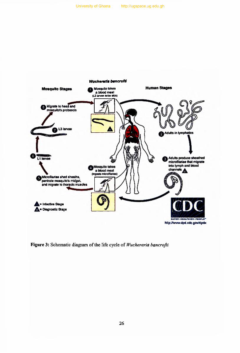
2.2 Biology and Life Cycle of Wuchereria bancrofti
The life cycle of W bancrofti is shown in Figure 3. Wuchereria bancrofti belongs to the class
Nematoda, subclass Secementea, superfamily Filarioidea and family Onchocercidae
(Anderson, 1992). When an infective mosquito takes a blood meal, some or all of the L3
enter human host through the surface of the labella on to the skin surface. The L3 enter the
human host through the puncture made by the mosquito, as they are unable to penetrate intact
skin, and those left on the skin surface in a drop of haemolymph have to enter before it dries
out (McGreevy et al., 1974). Only a few L3 manage to enter the skin after a blood meal
(Denham and McGreevy, 1977). The L3 migrate to the lymphatic system in human and
transform into L4 between 9-14 days after entry. Within 6-12 months they grow into mature
adults, which can live in the human host for 4 to 8 years. Female worms measure 80 to 100
mm in length and 0.24 to 0.30 mm in diameter, while the males measure about 40 by 1mm.
Adults reside in lymphatic vessels, mate and the viviparous females produce thousands of
sheathed mf (Li) measuring 244 to 296 fim by 7.5 to 10 /zm, into lymph circulation. The m f
migrates from lymphatic system to circulatory system and has nocturnal periodicity, except
in the South Pacific there is absence of marked periodicity (Lardeux and Cheffort, 2001;
Hawking et al., 1966; Denham and McGreevy, 1977). Microfilaria numbers fluctuate in the
peripheral blood over 24 hours. Nocturnal, periodic worms peak in the peripheral blood from
24
University of Ghana http://ugspace.ug.edu.gh

2200 to 0200 hours, corresponding with peak mosquito biting. Microfilariae are concentrated
in the micro-vessels of deep tissues, mainly the lungs during the day and one of the many
theories put forward to account for this interesting behavioural pattern is that oxygen tension
plays a role (Edeson et a l , 1957; Hawking et al., 1966; Hawking and Gammage, 1968;
Denham and McGreevy, 1977; Mossinger, 1991). If a person is given extra pure oxygen
during the night, the microfilariae stay in deep tissues other than accumulating in peripheral
circulation. Another suggestion has been that mfs peak in the peripheral blood when humans
are inactive (Hawking et al., 1966; Denham and McGreevy, 1977).
A mosquito picks mf, during a blood meal and ingested Li stage of m f move to the stomach
of the mosquito, lose their sheaths and some of them work their way through the wall of the
proventriculus and cardiac portion of the mosquito's midgut and reach the thoracic flight
muscles (Christensen et al., 1984). Within 6-10 days of entering the mosquito, Li m f develop
into the L2 “sausage” stage in the thoracic muscles. The L2 larvae develop between 11-13
days and moult into third-stage infective larvae (L3) and reach a length of 1.2-1.6 mm. The
L3 larvae migrate through the haemocoel to the mosquito's proboscis and when blood meal is
taken from another human, the infective L3 emerge and enters the skin via the bite wound to
continue the cycle. Unlike malaria parasites that are injected with saliva into the host,
infective stage filarial worms actively break out of the proboscis within a drop of
haemolymph and must find and enter the puncture wound made by the mosquito, hair
follicles, or other abrasions. The transmission of filarial worms is highly inefficient and
filarial worm development in the mosquito is not a benign process and may even disable or
kill their mosquito host.
25
University of Ghana http://ugspace.ug.edu.gh
Mosquito Stages
A Migrate eo head and w mosquito's proboscis
A Mosquito lakes ” a Wood meal
<L3 tarvae enter skin)
Wuchar&ria bancroftl
AMScfofiiaria* shad shaashs, ” pentrate mosquito's midgiit,
and migrate to thoracic muscles
A = Infective Slage
A° Olagnostic Slage
Human Stages
hltp /Avtw.dpd ode gov Wpdx
Figure 3: Schematic diagram of the life cycle of Wuchereria bancrofti
26
University of Ghana http://ugspace.ug.edu.gh

2.3 Biology and Life Cycle of the Vectors
Depending on the geographical location, different species of the following genera of
mosquitoes are vectors of bancroftian filariasis. Among them are: Anopheles (An. arabiensis,
An. bancroftii, An. farauti, An. funestus, An. gambiae, An. koliensis, An. melas, merus,
An. punctulatus and An. wellcomei); Culex (Cx. annulirostris, Cx. bitaeniorhynchus, Cx
quinquefasciatus, and Cx. pipiens); Aedes (A. aegypti, A aquasalis, A. bellator; A cooki, A.
darlingi, A. kochi, A. polynesiensis, A. pseudoscutellaris, A. rotumae, A. scapularis, and A
vigilax); Mansonia (M. pseudotitillans, M. uniformis); Coquillettidia (C. juxtamansonia)
These different species, however, have a similar life cycle that is completed in four distinct
developmental stages (complete metamorphosis): egg, larva, pupa and adult.
2.3.1 Biology of the vectors
The female mosquitoes lay about 50 to 500 eggs at a time that are deposited on water or site
that is liable to flood. The eggs of most mosquitoes are elongate, ovoid, or spindle-shaped
with a few being spherical or rhomboid. Each egg is protected by an eggshell or chorion,
which in many species is highly sculptured. The chorion of Anopheles species have unique,
transparent, air-filled compartments flanking the egg that serve as floats. Whereas the eggs of
certain mosquito species are laid individually (e.g. Anopheles, Aedes, Ochlerotatus,
Psorophora, Toxorhynchites, Wyeomyia and Haemagogus species), others attach their eggs
together in a single clump forming a floating egg raft (Culex and Culiseta) or a submerged
cluster (Coquillettidia and Mansonia). Viable eggs take 2-3 days after oviposition to hatch
into larvae. Adult male and female mosquitoes feed on plant nectar but only the latter needs
blood to nourish and mature the eggs. Mating could be once in the life time of a female
27
University of Ghana http://ugspace.ug.edu.gh

mosquito. This is as a result of a sac-like compartment called spermatheaca, which stores
sperm during mating and it is stimulated to release sperm at every oviposition.
Figure 4: Eggs of mosquitoes: A-Anopheles\ B-Culex; C-Aedes aegypti\ D-Toxorhynchites
Figure 5: Mosquito egg rafts and clusters. A-Culex\ B-Mansonia attached to underside of a
floating leaf
28
University of Ghana http://ugspace.ug.edu.gh

Mosquito larvae, commonly known as wrigglers pass through four instars which closely
resemble one another except for their size. When it hatches from the egg, the young mosquito
larva is fully adapted for living in water. It uses atmospheric oxygen for respiration and
water-borne particles as food. The air-breathing habit requires mosquito larvae either to live
more or less permanently at the air/ water interface, or to make frequent visits to the water
surface. The pair of spiracles at the end of the abdomen or at the end of a tube called siphon,
is the only respiratory openings from which air-filled trachea extends to all parts of the body.
The position of the spiracles determines the larval position in relation to the water surface.
Larvae of two genera are able to force saw-like tips of their respiratory siphons into the air-
filled stems or roots of certain aquatic plants, and remain permanently submerged. Within 6 -
9 days the larvae moult and reach pupa stage.
The pupae also commonly known as tumblers are comma-shaped with the head and thorax
fused to form a cephalothorax. The abdomen is curled beneath with two broad paddles at the
end, which propel the pupa through the water when it is disturbed. An air bubble, which is
enclosed between the appendages, provides buoyancy that allows the pupa to float with the
top of its thorax in contact with the water surface. Located at the top of the cephalothorax are
air trumpets, which are used to obtain oxygen. Certain larval organs are destroyed during the
pupal stage while others are carried over to the adult stage. When the adult is fully formed
the pupa starts to swallow air and the consequent increase in internal pressure forces a split
along the midline of the pupal thoracic cuticle, with the adult slowly expanding out of the
pupal cuticle and steps on to the water surface. The pupa which does not feed reaches adult
stage within 2-3 days.
29
University of Ghana http://ugspace.ug.edu.gh

Adult mosquitoes are slender, have thin legs and narrow, elongate wings. The body surface is
covered with scales, setae, and fine pile, creating the characteristic markings and colors of
each species. Like other Diptera, mosquitoes are fluid feeders with mouthparts, which have
evolved into an elongate composite proboscis, half as long as the body, suitable for probing
for nectars and for piercing skin, and imbibing blood from peripheral blood vessels. The
saliva that is injected as the mouthparts penetrate the skin contains a substance that prevents
blood coagulation. The saliva is also the source of pathogens; such is mf of W. bancrofti and
immunogens that are responsible for the characteristic skin reactions to mosquito bites.
Within a few minutes, engorging mosquitoes can imbibe blood up to four times their own
weight. This inflicts upon the mosquito a water load, and potentially toxic amounts of sodium
and potassium. The mosquito excretory system is capable of rapid elimination of water and
salts, and diuresis commences while the female is still feeding. An adult mosquito can live
for about 3 weeks.
The resting position of adult Anopheles is usually at an angle of 45° to the surface with the
proboscis and abdomen in a straight line. Some species such as An. culicifacies rests at
angles much smaller than right angles, while others are at almost that angle to the surface
(Kettle, 1992). A distinctive morphological feature of Anopheles mosquitoes are the dark and
pale scales on their wing veins arranged in blocks to form a spotted pattern. The spots vary
between species. Female Anopheles mosquitoes have non-plumose antennae while males
have plumose antennae. Adult anopheline females have palps, which are almost as long as
the proboscis and usually lie closely alongside it and may be marked, especially the apical
half with broad and narrow rings of pale scales. The palps of the males may also have apical
rings of pale scales besides being distinctly swollen at the ends.
30
University of Ghana http://ugspace.ug.edu.gh

Adult mosquitoes are slender, have thin legs and narrow, elongate wings. The body surface is
covered with scales, setae, and fine pile, creating the characteristic markings and colors of
each species. Like other Diptera, mosquitoes are fluid feeders with mouthparts, which have
evolved into an elongate composite proboscis, half as long as the body, suitable for probing
for nectars and for piercing skin, and imbibing blood from peripheral blood vessels. The
saliva that is injected as the mouthparts penetrate the skin contains a substance that prevents
blood coagulation. The saliva is also the source of pathogens; such is m f of W. bancrofti and
immunogens that are responsible for the characteristic skin reactions to mosquito bites.
Within a few minutes, engorging mosquitoes can imbibe blood up to four times their own
weight. This inflicts upon the mosquito a water load, and potentially toxic amounts of sodium
and potassium. The mosquito excretory system is capable of rapid elimination of water and
salts, and diuresis commences while the female is still feeding. An adult mosquito can live
for about 3 weeks.
The resting position of adult Anopheles is usually at an angle of 45° to the surface with the
proboscis and abdomen in a straight line. Some species such as An. culicifacies rests at
angles much smaller than right angles, while others are at almost that angle to the surface
(Kettle, 1992). A distinctive morphological feature of Anopheles mosquitoes are the dark and
pale scales on their wing veins arranged in blocks to form a spotted pattern. The spots vary
between species. Female Anopheles mosquitoes have non-plumose antennae while males
have plumose antennae. Adult anopheline females have palps, which are almost as long as
the proboscis and usually lie closely alongside it and may be marked, especially the apical
half with broad and narrow rings of pale scales. The palps of the males may also have apical
rings of pale scales besides being distinctly swollen at the ends.
30
University of Ghana http://ugspace.ug.edu.gh

2.3.2 Anopheles gambiae complex and An.funestus group
Several techniques are employed in identifying the members of Anopheles gambiae s.l. with
varying degrees of specificity (Collins et al., 2000). These are largely based on universal
characters that cut across geographical zones for particular sibling species members and are
applicable in field situations. Gillies and De Meillon (1968) as well as Gillies and Coetzee
(1987) provide established identification techniques, by using morphological characters, for
the anopheline mosquitoes. An. gambiae adult females are identified by their smooth palps
with 3 pale bands on the 3rd, 4th and 5th segments; the wing field is pale with yellowish or
creamy markings and has pale fairly long costal spots. The femora, tibia and 1st tarsal
segment speckled to a variable degree. The abdomen is pale brown and hairy with scales on
the 8 th tergite and scales on the cerci.
Adult female An. funestus are identified by three pale bands on the 2nd, 3rd and 5th segments
of the palps. The dark wings also bears characteristic pale scales, with the costa having four
pale spots usually shorter than the intervening dark areas. The abdomen is dark brown and
lack scale including the cerci, while the legs are usually dark with a small apical white spot
on the tibia.
Anopheles gambiae s.l. lives in sympatry with An. funestus and adapts to changing
environments in many parts of Africa (Coluzzi, 1984). During rainy seasons, An. gambiae s.l.
populations dominate while populations of the An. funestus become more abundant in the dry
seasons (Coluzzi, 1984). In African urban centres where proportions of anopheline
mosquitoes are largely made of An. gambiae s.l., An. funestus is very rare. The former
colonizes well-lit, clean temporary pools of rainwater, shallow slow moving streams, bore
31
University of Ghana http://ugspace.ug.edu.gh

holes and burrow pits, roadside puddles formed by tire tracks, irrigation ditches and other
artificial bodies of water (Gillies and De Meillon, 1968; Gimnig et al., 2001).
Anopheles funestus s.l. on the other hand preferentially breeds in semi-permanent water pools
and are often associated with rice fields (Gillies and De Meillon, 1968). These two species
have narrow host ranges and exhibit preferentially anthropophagic and endophilic characters
that make humans their main source of blood meal (Burkot, 1988). The processes involved
in host selection are nonetheless well coordinated and the choice of an individual from a pool
of those available is influenced mainly by the degree of defensive behaviour exhibited by the
selected host (Burkot, 1988). Parasitized and morbid potential hosts exhibit less defensive
activity and are therefore more prone to being prime sources of vector blood meal (Burkot,
1988), perhaps an adaptive modification by the parasites for their optimal and uninterrupted
uptake during mosquito blood meal.
2.3.3 Life cycle of the vectors
The life cycle of Anopheles mosquitoes (Figure 8) takes about 12-14 days. There is a wide
range of habitats for breeding, varying from mostly permanent and large collections of water
such as fresh water swamps, marshes, rice fields and borrow pits to smaller collections of
temporary water such as small pools, puddles, water filled car tracks, ditches, drains, gullies,
hoof prints, etc. The most preferred breeding sites are the shallow open sun lit pools (Service,
1993). Wells and manmade container habitats such as clay pots, motor vehicle tyres, water
storage jars and tin cans may also be ideal for breeding (Chinery, 1984). Anopheles gambiae
s.s however, prefers small and undisturbed temporary pools of water exposed to sunshine as
such their predominance in irrigated and forested areas (Muirhead-Thomson, 1945).
32
University of Ghana http://ugspace.ug.edu.gh

Heavy mortality among larvae leading to the drastic reduction in number of eggs that develop
into adults is a result of predation, disease, drought, flood and desiccation. Notonectidae and
other larvivorous fishes such as Gambusia affinis and Lebister reticulatus which prey on the
larvae o f Anopheles spp, and Poecilia reticulata and Oreochromis mossambicus on Culexspp
are being used as biological control tools (Cech and Linden, 1987; Blaustein, 1989;
Salitemik, 1977; Mahmoud, 1985). It may be noted that the vagility often displayed by An.
gambiae larvae, in contrast to species like An. funestus would tend to increase their
vulnerability to attack by predators (Service, 1980). In some instances, Culex tigripes
colonizes the same pools as An. gambiae, causing a dramatic reduction in larval density due
to competition pressure (Haddow, 1942).
Adult females lay in singles 50-200 small (1mm long and 2-5mm wide) brown or blackish
boat shaped eggs with lateral floats scattered on the water surface. Viable eggs hatch 2-3
days after oviposition, but may take 4-7 days or longer to hatch in the temperate regions
(Service, 1980). The larva is a filter feeder and forages for microscopic organisms and plants
even as it is lying parallel to the water surface. The larvae reach pupa stage after four moults,
within 6-9 days depending on the availability of nutrients, optimum temperature (Gimnig et
al., 2002) and other competing organisms within the habitat. Under the same conditions the
pupa, which does not feed, remains in the water for 2-3 days. The pupal case splits dorsally
and the adult emerges. The duration from an egg to the adult stage is thus in a range of 7-14
days and after emerging the adult inflates its wings, separates and grooms its head
appendages before flying away (Kettle, 1992).
33
University of Ghana http://ugspace.ug.edu.gh

Gimnig et a l (2002) reported that in most cases prolonged larval developmental stages might
result in emergence of small females due to high population densities of developing An.
gambiae s.l larva. The males in any egg batch emerge first as adults and become ready for
mating within 24 hours before the females emerge. The females on the other hand, require
vertebrate blood meal for ovarian development, followed by the maturation and oviposition
of a batch of eggs (Gillies, 1955). The feeding of the females on a vertebrate is however
stimulated by a combination of carbon dioxide, temperature, moisture, smell, colour and host
movement (Service, 1980). Adult males feed on plant nectar and do not bite, however most
of the male mosquitoes usually die after a single mating.
Anopheles are mostly nocturnal in their activities, thus emergence from the pupae, mating,
blood feeding and oviposition normally occur in the evenings, at night or early in the
morning around sunrise. Some species may bite man mainly outdoors (exophagic) from
about sunset to 2 1 0 0 hours whereas others bite mainly after 2 1 0 0 hours and mostly indoors
(endophagic). Whereas some species rest outside (exophilic) in a variety of natural shelters
like vegetation, rodent burrows, cracks and crevices in trees, under bridges, termite mounds,
and other cracks in the ground in between feeding, others rest in houses (endophilic).
Mixtures of these extremes of behaviour are exhibited by most Anopheles species that are
exophagic and endophagic, exophilic and endophilic. Few of this species exclusively feed on
humans though they are predominantly zoophilic. Most feed on both human and animals but
the degree of anthropophilism and zoophilism varies according to species and host
availability.
34
University of Ghana http://ugspace.ug.edu.gh

Adult
Pupa
Figure 6 : A typical life cycle of Anopheles mosquito
Larva
&
35
University of Ghana http://ugspace.ug.edu.gh

2.4 Molecular Characterisation of Anopheles Population
The theory and practise of classifying organisms, called Taxonomy dates back over two
centuries and draws from extensive accumulated information on morphology, biogeography,
and habitat distributions of a large proportion of extant species. Species are identified on the
basis of one or several consistently distinguishing morphological characters. As knowledge
of species however became more detailed, more examples appeared where morphologically
identical organisms differed greatly in behaviour, physiology and genetics suggesting that
these identical organisms might belong to different species. For these reasons morphological
species concept came to be replaced by the more stringent biological species concept in
which species were only considered valid when they exist as reproductively isolated gene
pools (Mayr and Ashlock, 1991). With the biological species concept, morphologically
identical organisms, sympatric (overlapping in geographical range) are classified as separate
species when they can be shown to be reproductively isolated. Conversely, morphologically
distinct organisms can be a single species, especially when allopatric (distributed in different
geographic ranges), or when the morphological features constitute intraspecies genetic
polymorphisms.
Many closely related vector species are difficult or impossible to identify using
morphological characters. In these cases, researchers have turned to the molecular
phylogenetic and concordant species concept. Molecular taxonomy is the classification and
identification of organisms based on either protein or nucleic acid characters rather than
morphological characters, and the vector species composition in a geographical region is
often well known and correct identifications are obtained without the need of highly trained
taxonomists. Once a correct identification has been made, control programs can interrupt
36
University of Ghana http://ugspace.ug.edu.gh

disease transmission by accessing critical information on adult and larval habitats, host
preference, ecology, vector competence, histories of earlier epidemics and ultimately, apply
the control strategy that are appropriate for that species. Accurate species identification is
critical to medical entomologists during control of disease outbreaks and also in associated
epidemiological and ecological studies for monitoring the vectors population. The
polymerase chain reaction (PCR) is a molecular technique, which currently widely employed
for species identification. The technique can amplify a DNA sequence from a minute amount
of template DNA, which can then be analysed by various methods to determine species
identity. The technique also permits the investigator to retain most of the specimen that can
then be examined for pathogens, parasites, biological control agents, gut contents, age or
other epidemiological informative characters.
2.4.1 PCR-RFLP identification of Anopheles funestus and An. gambiae species complex
There are presently nine recognised members of the Anopheles funestus group; these are An.
funestus s.s., An. vaneedeni Gilles and Coetzee, An. leesoni Evans, An. rivulorum Leeson,
An. parensis Gilles, An. fuseivenosus Leeson, An. aruni Subii, An. brucei Service and An.
confuses Evans and Leeson. The PCR-based method for identification of Anopheles funestus
group is based on sequence differences of a variable 3D domain in the 28S gene (Koekemoer
et a l} 1999). Primers are designed from conserve regions of the 18S, 5.8S or 28S that
contains amplify regions of the internal transcribed spacer (ITS) 1 and 2 . This PCR method
then uses cocktail primers that are species-specific for the identification of these species. See
Appendix II for sequence details of the primers.
37
University of Ghana http://ugspace.ug.edu.gh

There are seven recognised sibling species of the Anopheles gambiae complex which are
morphologically indistinguishable. Anopheles gambiae s.s., An. arabiensis, An.
quadriannulatus A and B, An. merus, An. melas, An. bwambae (White, 1985; Scott et al.,
1993). There is a further subdivision of the Anopheles gambiae s.s. based on gene
arrangements of the 2R chromosomal arm into five chromosomal forms; Mopti, Bamako,
Forest, Savanna and Bissau (Coluzzi, et al, 2002; Toure, et al., 1994; Powell et al.9 1999).
Molecular analysis of these forms has shown the existence of genetic discontinuity in this
species. Two molecular forms; M and S have been identified based on diagnostic difference
in the intergenic sequence (IGS) and internal transcribed sequence (ITS) regions of rDNA on
the X chromosome (della Torre et al., 2002; Favia et al., 1997; Favia et al., 2001). Even
though breeding between M and S forms yields fertile progenyin the laboratory, M-S hybrids
are rarely observed in nature (della Torre et al., 2002; Favia et al., 2001).
The complexity of Anopheles gambiae Giles s.s., a sibling species of the An. gambiae
complex has been shown by the extent of chromosomal inversion polymorphisms and more
recently, by divergence at the molecular level (Masendu et al., 2004). Within the amplified
rDNA, there are two nucleotide substitutions; AT for M but GC for S population forms. The
molecular M and S forms of An. gambiae have been found by Yawson et al. (2004) to occur
in sympatry in southern Ghana. The S form is predominant throughout its distribution in the
coastal savannah except at one location in the strand and mangrove zone where there is rice
cultivation. The M form was the only form collected in northern Ghana. For the identification
of the M and S molecular forms, the PCR products obtained using the method of Scott et a l
(1993) is restricted further, using Hha I or Tru I which cuts the S form at one site but not the
M form.
38
University of Ghana http://ugspace.ug.edu.gh

2.5 Detection of Wuckereria bancrofti in Mosquito Vectors
The limition in mass screening of human population for microfilaria is that not every body
will be willing to be screened. This means that one is likely to miss some positive
individuals. Observations of the various stages of the parasite in mosquito vectors are used as
a xenomonitory procedure to add up to the parasitological monitoring.
2.5.1 Microscopy
The dissection of mosquitoes for presence of the parasites larval stages and identification of
the larvae based on morphology continues to be the most reliable detection method. For this
the head, thorax and abdomen of each mosquito are separated from each other and placed
into separate drops of saline on a microscope slide. The labium is separated from the other
parts of the proboscis and if infective filarial worms are present they emerge from the labium
into the saline. The remaining part of the head, thorax, and abdomen are carefully macerated
under a dissecting microscope and examined for both immature and infective filarial worms.
Careful examination of the slides for the parasite at lOOx magnification is carried out using a
compound microscope.
Microfilariae in mosquitoes can be morphologically identified by their characteristic smooth
silvery outline, rounded at the anterior end and pointed at the posterior and their size, and 250
to 300fim long x 7.5 to lOjum wide (Sasa, 1976). The L3S of W. bancrofti have the
characteristic three equal caudal papilla or protuberances, which are bubble-like. The
infective larvae which measure 1 1 0 0 fim in length can be separated from the second stage
larvae (L2) that are 500 - 600/mi long and 25 - 30fim wide, and the lack of an anal plug
(protrusion), which is present in the 3rd stage larvae.
39
University of Ghana http://ugspace.ug.edu.gh

2.5.2 Polymerase Chain Reaction (PCR)
Polymerase chain reaction (PCR) assay for W. bancrofti infection in mosquitoes has been
developed and tested in the field (Farid et a l , 2001). This detects a repeated sequence of W.
bancrofti DNA and is very sensitive although expensive and requires highly trained technical
staff.
2.6 Factors Affecting the Transmission of Wuchereria bancrofti
Transmission of the parasite involves three processes; the uptake of mf from the human host,
the development of mf to the infective-stage larvae (L3) and the transmission of L3 to human
(Subramanian et al., 1997). Transmission intensity depends on a number of factors such as
the level of infection in the human population, vectorial capacity, vectorial competence, and
climatic factors.
2.6.1 Infection in the human population
It has been demonstrated experimentally that the uptake of mf by mosquitoes depends on the
density and distribution of mf in the human host (Bryan and Southgate, 1988). Microfilaria
density in human population differs from one endemic community to the other. This may be
due to variation in human exposure to mosquitoes, susceptibility in tha human population to
infection and their response to treatment. Such heterogeneneities are often overlooked
leading to overestimation of the effectiveness of population-based control measures and the
probability of elimination (Duerr et al., 2005). Individual mosquitoes on the average pick up
more in a human population that has high mf prevalence and mean intensity compared to low
prevalence and mean intensity (Southgate and Bryan, 1992). The overall load of mf picked
40
University of Ghana http://ugspace.ug.edu.gh

up by the vectors however depends on vector density and human-vector contact in a given
locality.
Some vectors ingest mf at high human microfilaraemia density and not all may develop to
the L3 stage. Others ingest mf at low-density human microfilaraemia much more readily than
others and almost all develop to the infective L3 stage. Microfilariae may also be ingested not
at high-density human microfilaraemia as in facilitation and not at very low-density human
microfilaraemia as in limitation, which is proportionality.
2.6.2 Vectorial capacity
The capacity of vector concept in the transmission of parasites was introduced by Garret-
Jones (1964) based on work done by MacDonald (1952) as an attempt to quantify the
capacity of a given mosquito population to transmit malaria under natural conditions, which
is also very useful in the transmission of lymphatic filariasis. Vectorial capacity (C) is
defined by the formula:
C = (ima2p n)/ -In p
Where C is the estimated number of new infections that arise from a single infection per day;
fcm’ is vector density relative to man, ‘a’ is human feeding habit of the vector, cp ’ is the daily
vector survivor and ‘n’ is the time from infection to infectivity.
2.6.2.1 Vector density relative to man and human feeding habit of vector
The estimated human biting rate (ma) is the product of the vector density relative to man (m)
and the human feeding habit of the vector (a), which is expressed in bites per man per night.
Vector density relative to man or vector abundance depends largely on the availability of
41
University of Ghana http://ugspace.ug.edu.gh

suitable breeding sites (Gillies, 1988). White (1969) has shown that inhabitants living close
to abundant mosquito breeding sites were more infected compared to those far away. This
index is the main component of the entomological indices used in the estimation of exposure
to filariasis infection [i.e. Annual Biting Rate (ABR) and Annual Transmission Potential
(ATP)].
The human feeding habit of the vector is determined by two factors; the frequency at which a
female mosquito takes blood meal and the proportion of the meals that is taken on humans. A
mosquito species that takes 90% of its blood meal on humans has 81% probability of taking
human blood meal twice, i.e. at the time of ingesting mf and time of releasing infective
larvae (Sasa, 1976). Other species on the contrary that have 5% rate of feeding on human
have 0.25% probability. This means the former species is 324 times more efficient as a
vector than the latter.
2.6.2.2 Vector survival and time from infection to infectivity
The time it takes from when a mosquito picks m f in a blood meal until the parasites develop
to infective larvae and migrate to the head of the mosquito takes 1 0 to 1 2 days depending on
temperature and other tropical conditions. This means only adult mosquitoes that live longer
than this are able to transmit. The daily vector survivor that is linked to vector longevity is
an important condition for a vector.
2.6.3 Vectorial competence
This takes into account the physiological ability of a vector to acquire and maintain the
parasite development to the infective stage and transmit it to the definitive host (Reisen,
42
University of Ghana http://ugspace.ug.edu.gh

1989). He therefore agued that vector competence (v) should be included in the above
equation to give the one below.
C = (ma2vpn)/ -In p
The factors that influence the proportion of m f development to infective larvae include
parasite uptake, parasite development, density dependent processes in the vector and parasite
induced vector mortality.
2.6.3.1 Parasite uptake
Bryan and Southgate (1976) found that wild caught A. polynesiensis feeding on a carrier with
low mf density of 6 parasites/ ml took up about 1 2 times the expected number of m f when
compared to the concentration in the venous blood of the carrier. Comparison between m f
concentration in mosquito blood meal and human blood reveals higher concentration in the
former. This trend has been attributed partly to the excretion of blood especially by
Anopheles mosquitoes during feeding (Brengues, 1975; Reid, 1982), as well as variation in
mf concentration in the vascular system of the host whereby mosquitoes ingest more m f than
expected simply because they draw blood from superficial vessels where mf are
concentrated.
Barriers in the oesophagus of mosquitoes are also known to affect mf uptake by vectors. The
mf during ingestion has to pass the cibarial and bucco-pharyngeal armatures, which are
sclerotized teeth protruding into the oesophagus lumen. The armatures, which are well
developed in An. gambiae, An. arabiensis, An. melas, and An. funestus but poorly in Cx.
quinquefasciatus can inflict lethal damage on the mf (Bryan and Southgate, 1988b).
43
University of Ghana http://ugspace.ug.edu.gh

2.63.2 Parasite development to infective larvae (L3)
When mosquito takes a blood meal, the stomach wall dilates and this initiates the formation
of a chitinous structure called peritrophic membrane (Pedersen, 1977). Ingested blood starts
to coagulate when it reaches the mid gut at a rate, which varies with mosquito species and mf
can get trapped in the clot or by the peritrophic membrane (Deham and McGreevy, 1977).
Although the effect of the peritrophic membrane as a mid gut barrier is not well known
(Townson and Chaitong, 1991), coagulation has been reported to have a substantial effect on
mf migration in Cx. quinquefasciatus by retaining 97% of ingested m f in blood clot (Ewert,
1971). Townson and Chaitong (1991) found that addition of anticoagulants such as heparin to
the blood increases the migration of B. pahangi in refractory mosquitoes to virtually the same
level as that found in susceptible strains.
The mf that passes across the stomach wall can be trapped in the haemocoele and be
melanized (Townson and Chaitong, 1991). In Cx. pipiens fatigans the defence mechanism
against W. bancrofti is evident after the parasite has entered the thoracic muscle and reached
the sausage stage (Omar and Zielke, 1978). Larvae that are not trapped and melanized in the
haemocoele develop to L3, which then escape from the muscles and migrate to the
haemocoele of the head and mouthparts (Lindsay and Denham, 1986).
2.6.3.3 Density dependent processes in the vector
Three types of parasite-vector relationships have been described for W. bancrofti and its
vectors. These are limitation, facilitation and proportionality. Limitation is when the parasite
success rate or yield in a mosquito decreases as the mf intake increases. The converse is
facilitation, when the parasite yield increases as the mf intake increases and proportionality
44
University of Ghana http://ugspace.ug.edu.gh

occurs when the parasite yield is a constant ratio, neither increasing nor decreasing as mf
intake increases. In all three types of host-parasite relationship, the value of the parasite yield
can never exceed unity, as there is no multiplication of filarial larvae in mosquitoes, i.e. one
mf can never yield more than one infective larva (Bain, 1971; Bain and Brengues, 1972).
Facilitation has been observed in An. gambiae (Southgate and Bryan, 1992). Vectors
exhibiting such a vector-parasite relationship appear to produce infection and disease much
more effectively than those exhibiting limitation and proportionality. With limitation as in
Cx. quinquefasciatus (Southgate and Bryan, 1992), the vector ingests m f at low-density
human microfilaraemia and almost all develop to the infective L3 stage. In proportionality,
mf may be ingested not at high-density human microfilaraemia as in facilitation and not at
very low-density human microfilaraemia as in limitation. The argument is that, in the case of
limitation, it would be difficult to totally interrupt transmission even when control
programmes reduce mf prevalence and intensity to very low levels. On the other hand, it
would be relatively easier to block transmission and eradicate the parasite from the human
population in the case of facilitation (Subramanian et al., 1997).
2.6.3A Parasite induced vector mortality
Bahr (1912) was the first to find out that large numbers of ingested mfs result in mortality of
vectors during parasite maturation. High filarial intensities have been shown to kill a
considerable number of mosquitoes in laboratory experiments (Pedersen, 1977; Lindsay and
Denham, 1986). Vector mortality begins within a few days after ingestion of the infected
blood meal and is associated with the migration of mf from the gut to the thorax
(Knshnamoorthy et al., 2004) and when the L3S start to migrate from the thorax to the
45
University of Ghana http://ugspace.ug.edu.gh

labium. It is also thought that the rate of mortality in a population of infected mosquitoes at
this time is dependent on the intrinsic activity of the L3, which varies from one species to
another. A third period occurs after another blood meal, possibly due to the damage or by the
emergence of the L3, loss of haemolymph from the mosquito or increased chance of bacterial
infection through the wound caused by the escaping larvae.
This means the use of mass drug administrations (MDA) to reduce the prevalence and
intensity of microfilaraemia may increase the mean lifespan of some of the local Anopheles
species (Pichon 2002). If these same species also act as vectors of malarial parasites,
effective drug-based control of W bancrofti may worsen the problem posed by malaria.
Therefore, wherever malaria and bancroftian filariasis are co-endemic and caused by
parasites transmitted by the same species of mosquito, MDA should be augmented by vector
management such use of bednets or house-spraying against adult Anopheles mosquitoes
(Pichon 2002).
2.6.4 Climatic factors
Changes in climatic factors such as temperature, rainfall and sunshine affect human health in
very different ways, often through complex changes in ecological systems. It is the
interaction among these factors, in combination with other non-climatic factors that
determine the timing of infectious disease outbreaks. The female mosquito becomes infected
with W. bancrofti if it sucks blood from an infected person, and may then infect the next
person it bites. The spread of the disease is thus limited by conditions that favour the vector
and the parasite growth.
46
University of Ghana http://ugspace.ug.edu.gh

2.6.4.1 Temperature
Temperature is known to affect mosquito larval development, thereby influencing the density
of mosquitoes. The range of temperatures suitable for the development of aquatic stages and
maturation of the gonads in adults differ between vector species but the optimal range is 2 0 -
30 °C (Molineaux, 1988). An increasing temperature within this range tends to increase the
growth rate of vector populations by shortening the intervals between successive oviposition.
Gillies and DeMeillon (1968) found that An. gambiae s.l., which has a generation time of two
to three weeks at lower temperatures shortened to 10-11 days at high temperatures. The rate
of aquatic development at a given temperature varies between species and the high rates
attained by certain vectors (e.g. An. gambiae and An. arabiensis) allow them to breed in
temporary pools and to get ahead of vegetation, pollution, competitors and predators.
High temperatures on the other hand can reduce the longevity of adult mosquitoes and thus
reduce population growth and density. The optimal temperatures for the vectors ranges
between 25 and 28°C (rarely exceeding 30°C) also favour the growth of filarial larvae inside
the vector. Although a few, An. gambiae and An. funestus are found in areas with
temperatures that exceed 30°C.
2.6.4.2 Sunshine
Duration of sunshine has a direct influence on green plants, and the effects of changing
intensities and temperature on photosynthesis are enormous. Increasing intensity of light
accelerates photosynthesis but increasing temperature does not (Zaltsman and Feder, 2005;
Rozema et al., 2002). At high light intensities, however, an increase in temperature greatly
accelerates photosynthesis. Plants provide nectar that are fed on by mosquitoes, and also
47
University of Ghana http://ugspace.ug.edu.gh

serve as hiding places of mosquitoes especially during the day. During summer, people spend
more time outdoors and together with warmth, rain and sunshine favourable for mosquito
production, thereby increasing human-vector contact.
2.6.4.3 Rainfall
Rainfall is important for creating breeding sites, thereby influencing vector abundance.
Excessive rainfall (floods) may cause destruction of breeding sites and a temporary reduction
in numbers of vectors, but never eliminates the vector (Craig et al., 1999). A number of
studies carried out in different areas of Africa showed negative or positive relationship
between rainfall and the peak breeding periods of mosquitoes (Dzodzomenyo et al., 1999).
Subra (1973) recorded in urban areas of Burkina Faso that mosquito densities peak in the
rainy season and at the beginning of the dry season.
2.6.4.4 Relative humidity
High relative humidity is favourable for mosquitoes by increasing their life span. Mosquito
vectors normally prefer a relative humidity above 60% (Molineaux, 1988). High humidity
has been reported to increase longevity for adult anopheline mosquitoes and hence their
vectorial capacity (Muir, 1988). The infective larvae in a drop of haemolymph left on the
skin surface of man (from an infective mosquito bite) have to enter the body before the drop
dries out and therefore high humidity also favours a successful transmission (Lindsay and
Denham, 1986).
48
University of Ghana http://ugspace.ug.edu.gh

CHAPTER THREE
MATERIALS AND METHODS
3.1 Chemicals, Reagents and Equipments
The sources and manufacturers of reagents, buffers, solutions and equipments used in the
study are shown in Appendix I.
3.2 Study Site
The study was part of an onging larger programme (GPELF) aimed at eliminating the
disease. For the past 4 years MDA with ivermectin and albendazole are administered to the
population at risk. This study was used as a monitoring programme to determine the effect of
the combination treatment on ransmission of bancroftian filariasis by Anopheles vectors in
Ghana. The study was conducted in Gomoa District of Central Region of Ghana (Figure 9).
Nine communities were selected based on available data on population, endemicity of
lymphatic filariasis and logistic considerations. These areas have been earmarked for
treatment and are Gomoa Mampong, Gomoa Kyeren, Gomoa Fawomanyo, Gomoa Amanful
and Gomoa Obiri. The rest are Gomoa Hwida, Gomoa Dago, Gomoa Ayesuano and Gomoa
Okyereko. After mass screening for microfilareamia, four (Gomoa Hwida, Gomoa Dago,
Gomoa Ayesuano and Gomoa Okyereko) had positive cases. The study was however
concentrated in Gomoa Okyereko due to vector species distribution; thus only Culex and
Aedes species were collected from the other three communities after the first round of
mosquito collection, whereas Anopheles, Mansonia, Culex and Aedes species were caught in
Gomoa Okyereko.
49
University of Ghana http://ugspace.ug.edu.gh

Gomoa Okyereko is located about 50 km west of Accra and 10 km northeast of Winneba on
the western extension of the coastal savannah zone of Ghana. It has an average annual
rainfall range between 760 and 1 0 0 0 mm, a mean temperature range of 26 to 30 °C, and has a
natural vegetation of strand and mangrove swamp with patchy growth tolerant of salt water.
Its geographical coordinate is between Latitudes 5° 24’ to 35’N and Longitudes 0° 25’ to
36’W.
50
University of Ghana http://ugspace.ug.edu.gh

Burkina Faso
Cote \ d’Ivoirec'
///
Central
Togo
G om o a District |1
Gulf of Guinea70 140 Miles
Figure 7: Map of Ghana showing the study site, Gomoa District
51
University of Ghana http://ugspace.ug.edu.gh

3.3 Ethical Considerations
Mass screening of the population for mfs was carried out and those found positive were
informed on the importance, purpose and procedures of the study. Individual consent was,
however, obtained at the time of subject recruitment. This study was conducted in
accordance with good clinical practice with the latest edition of the Helsinki declaration on
the use of human subjects in biomedical research, and with local laws and customs (CIOMS,
2002). Prior to the screening, permission was sought from the divisional chiefs and opinion
leaders of the study area. Durbars were held at which the study was thoroughly explained to
the people. A copy of the information and consent form used in the study can be found in
Appendix IV.
3.4 Mass Screening for Microfilaria and Feeding Experiment
For the mass screening, IOOjliI of blood from a finger prick was collected from each
individual using heparinized 100^1 capillary tube. The blood was then transferred into 900pl
3% acetic acid, mixed thoroughly and poured into a Sedgwick-Rafter counting chamber and
the number of mfs observed under microscope countered (McMahon et a l , 1979). The
geometric mean intensity (GMI) of microfilaraemia in the community was calculated as
Antilog [Elog (x+l)/n], where x is number of mf/ ml blood in the mf individuals, and n is
number of people examined.
From these, 12 consented volunteers with different levels of microfilaraemia were then made
to sleep under a mosquito net with one side partly open (Plate 1) for mosquito entry.
Mosquitoes that fed on each subject during the night were collected hourly from 2100 hours
52
University of Ghana http://ugspace.ug.edu.gh

to 0600 hours the next day (Subramanian et a l 1998). At the end of each hour, the opened
part of the net was let down briefly as spotlight was used to locate the host-seeking mosquito
and any mosquitoes trapped were collected using an aspirator. The trapped mosquitoes were
released into paper cups covered with mosquito netting and labelled to reflect the hour,
subject and site of collection. The mosquitoes were fed on 10% sucrose solution soaked in
cotton wool that was placed on the netting cover. This was repeated the following night for
more mosquitoes but no blood sample was taken this time.
At the mid-point of each collection hour, a finger prick sample of blood was collected from
each of the 12 volunteers using lOOjil heparinized capillary tube. The blood was mixed with
900^1 3% acetic acid and number of mf determined by microscopy. The blood count of m f at
each hour was paired with the results of the corresponding mosquito dissection.
53
University of Ghana http://ugspace.ug.edu.gh

Plate 1: Finger prick blood being taken from a volunteer sleeping under a mosquito net with
one side partly open for mosquito entry.
54
University of Ghana http://ugspace.ug.edu.gh

3.5 Laboratory Studies
Mosquito samples were transported to the laboratory of the Parasitology Department of the
Noguchi Memorial Institute for Medical Research for identification, maintenance and
dissection.
3.5.1 Morphological identification, maintenance and dissection of mosquitoes
About half of the mosquitoes collected each hour from each subject were killed immediately
after feeding by putting them into the fridge and stored for less than 18 hours at 4°C until
ready to use. They were then sorted out according to species, kept in labelled petri dishes on
damp filter paper at -20° C until ready for dissection. They were dissected and the number of
mf in the blood meal of each mosquito counted.
The remaining 50% of mosquitoes were maintained in cups and kept in boxes and
transported under cool condition to the insectary at Noguchi (Plate 2). They were maintained
at approximately 26 - 28°C and 70 - 80% relative humidity and were fed on 20% sucrose
solution until day 1 2 post collection when they were killed, dissected and examined for L3
(Janousek and Lowrie, 1989). Any mosquito that died before day 1 2 was also dissected and
any larval stages of W. bancrofti if harboured recorded.
Each specimen was placed in a drop of normal saline (0.9 NaCl solution) on a labelled slide,
under a dissecting microscope (OLYMPUS SZ 1 1 , Japan) at 20X magnification. The legs
and wings were removed and the head, thorax and abdomen were then separated and each
part transferred into separate drops of saline on the same slide. The labium was separated
from other parts of the proboscis. If L3S were present they emerged from the labium into the
55
University of Ghana http://ugspace.ug.edu.gh

saline. The remaining part of the head, thorax, and abdomen were teased apart under the high
power of the dissecting microscope and examined for mfs. Careful examination of the slides
at 100X magnification was done under a compound microscope (OLYMPUS B50, Japan) to
ensure no parasite was missed. The mosquitoes positive for filarial infection were recorded
and positive slides as well as negatives were placed in racks and kept at - 4 C until ready for
molecular identification of the mosquitoes and W. bancrofti.
56
University of Ghana http://ugspace.ug.edu.gh

Plate 2: Boxes with netted paper-cups containing the collected mosquitoes at the insectary.
Each cup has 20% of sucrose-soaked cotton wool placed on it to feed them.
57
University of Ghana http://ugspace.ug.edu.gh

3.5.2 Molecular Studies
The molecular identifications of Anopheles gambiae, An. funestus and Wuchereria bancrofti
were done as described below.
3.5.2.1 Chemicals, reagents and solutions
Preparations of standard solutions for the molecular studies are described in Appendix I.
3.5.2.2 PCR-RFLP species identification of molecular forms of the Anopheles gambiae
Giles complex
The genomic DNA extraction and DNA amplification for identification were carried out as
described below.
3.5.2.2.1 Genomic DNA extraction
The carcass of dissected An. gambiae s.l. mosquito was placed in a 1.5 ml eppendorf tube
and homogenized with sterile Konte’s plastic pestles in lOOjal bender buffer. The
homogenate was then incubated at 65°C for 30 minutes followed by the addition of 125p,l of
phenol. It was vortexed and span at 14,000rpm for 10 minutes. The supernatant was
transferred into a fresh tube and 250jnl of pre-chilled absolute ethanol and lOjal of 8 M
potassium acetate were added. It was then incubated at -40°C for one hour, span at
10,000rpm for 10 minutes and supernatant poured off. The pellet was then rinsed with 200jal
of 70% ethanol, span at 10,000rpm for 5 minutes and supernatant poured off. The pellet was
dried and redissolved in 50jal TE + RNAse and then kept at 4 % until required for PCR. One
micro-litre was used for PCR.
58
University of Ghana http://ugspace.ug.edu.gh

3.5.2.2.2 PCR amplification
Five sets of oligonucleotide primers designed from the DNA sequences of the intergenic
spacer region of An. gambiae complex ribosomal DNA (rDNA) were used in PCR for the
identifications (Scott et a l 1993). The sequence details of these primers abbreviated UN,
GA, ME, AR and QD (expected sizes of the PCR products are given in Appendix 2). The UN
primer anneals to the same position on the rDNA sequences of all five species and GA
anneals specifically to An. gambiae s.s., ME to both An. merus and melas, AR to An.
arabiensis, and QD to An. quadriannulatus.
The PCR reaction mix of 25jxl contained IX PCR buffer supplied by the manufacturer
(Sigma, USA), 200fiM of each of the four deoxynucleotide triphosphates (dNTPs), IO j iM of
each oligonucleotide primers and 0.125 units of Taq Polymerase enzyme (Sigma, USA). One
micro litre of the genomic DNA was used as template for the amplification reaction and
sterile double distilled water added to make up the volume. The reaction mix was centrifuged
briefly and overlaid with mineral oil to avoid evaporation and refluxing during thermo
cycling. The amplification was carried out using a PTC 100 thermal cycler (MJ Research
Inc., USA). The cycling parameters for the reactions were as follows: 93° C for 3min (initial
denaturation), followed by 35 cycles of 93°C for 30 sec, 50°C for 30 sec, 72°C for lmin
(annealing) and ended with a cycle of 93° C for 30 sec, 50° C for 30 sec and 72°C for 10 min.
For each reaction a positive control containing PCR product of Anopheles of the same primer
set, as a template and a negative control that contained no DNA template were included.
59
University of Ghana http://ugspace.ug.edu.gh

3.5.2.2.3 Molecular forms of Anopheles gambiae s.s. identification
Members of the Anopheles gambiae complex were identified using PCR and An. gambiae s.s.
specimens were further analyzed by amplification of 1.3kb rDNA followed by Restriction
Fragment Length Polymorphism (RFLP) with restriction enzyme Hha I to determine the M
and S population forms.
The Hha I enzyme digestions were carried out using the recommended protocol of the
manufacturers (Sigma-Aldrich, USA). The final reaction volume of 2 0 p l contained 10(Jl of
amplified product, 0 .3 |J l of 1U Hha I, 2 |J l of lx Buffer C (Promega, USA), made up to the
final volume by adding sterile double distilled water and then digested at 37°C for three
hours. Electrophoresis was done on the digested fragments through 2 % agarose gel stained
with ethidium bromide and photographed under UV light illumination as described in Section
3.5 .2 .5 .
3.5.2.3 Identification of members of the Anopheles funestus group
The genomic DNA extraction and amplification were done as shown below for An. funestus
mosquitoes.
3.5.2.3.1 Genomic DNA extraction
The carcass of dissected An. funestus s.l. mosquito was placed in a 1 .5 ml eppendorf tube and
homogenized with sterile Konte’s plastic pestles in lOOjul bender buffer. The homogenate
was then incubated at 65°C for 30 minutes followed by the addition of 125|nl of phenol. It
was vortexed and span at 14,000rpm for 10 minutes. The supernatant was transferred into
fresh tube and 250jil of pre-chilled absolute ethanol and 1 O j l x I of 8 M potassium acetate were
60
University of Ghana http://ugspace.ug.edu.gh
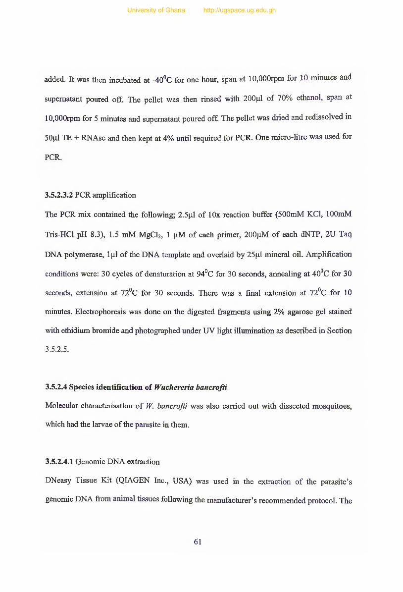
added. It was then incubated at -40°C for one hour, span at 1 0 ,0 0 0 rpm for 10 minutes and
supernatant poured off. The pellet was then rinsed with 200jul of 70% ethanol, span at
10,000rpm for 5 minutes and supernatant poured off. The pellet was dried and redissolved in
50|il TE + RNAse and then kept at 4% until required for PCR. One micro-litre was used for
PCR.
3.5.2.3.2 PCR amplification
The PCR mix contained the following; 2.5jnl of lOx reaction buffer (500mM KC1, lOOmM
Tris-HCl pH 8.3), 1.5 mM MgCh, 1 |iM of each primer, 200p,M of each dNTP, 2U Taq
DNA polymerase, ljxl of the DNA template and overlaid by 25^1 mineral oil. Amplification
conditions were: 30 cycles of denaturation at 94°C for 30 seconds, annealing at 40°C for 30
seconds, extension at 72°C for 30 seconds. There was a final extension at 72°C for 10
minutes. Electrophoresis was done on the digested fragments using 2% agarose gel stained
with ethidium bromide and photographed under UV light illumination as described in Section
3.5.2.5.
3.5.2.4 Species identification of Wuchereria bancrofti
Molecular characterisation of W. bancrofti was also carried out with dissected mosquitoes,
which had the larvae of the parasite in them.
3.5.2.4.1 Genomic DNA extraction
DNeasy Tissue Kit (QIAGEN Inc., USA) was used in the extraction of the parasite’s
genomic DNA from animal tissues following the manufacturer’s recommended protocol. The
61
University of Ghana http://ugspace.ug.edu.gh

dried W bancrofti larvae on slides were scrapped into 1.5ml eppendorf tube, and were
homogenised in 90jal buffer ATL using sterile pestles.
Then 10p,l proteinase K was added to each tube, vortexed and incubated at 55°C in a water
bath for 3 hours with intermittent vortexing. The homogenates were vortexed finally for 15
seconds and then lOOjul of buffer ATL added. The tubes were then incubated at 70°C for 10
minutes and 100]ul ethanol (96 to 100%) added and vortex to mix thoroughly. Each mixture
was transferred into a DNeasy mini column and centrifuged at 8000 rpm for 1 minute after
which the collection tubes with contents were discarded. The DNeasy mini column was then
placed in a fresh 2 ml collection tube and 500^il buffer AW1 added and centrifuged at 8000
rpm for 1 minute to wash the extracts. The flow trough and collection tubes were again
discarded. The samples were washed again by placing the DNeasy mini columns in fresh 2
ml collection tubes and 500p,l buffer AW2 added, and centrifuged at 8000 rpm for 3 minutes.
The DNeasy mini columns were placed in new 1.5 ml microcentrifuge tubes and 200 ml
buffer AE added directly onto the membrane. Samples were incubated at room temperature
for 1 minute and then centrifuged for 1 minute at 8000 rpm to elute the DNA. Aliquots of 5jal
of the filarial DNA extract from the mosquitoes were used as templates for the amplification
reaction.
3.5.2.4.2 PCR identification of Wuchereria bancrofti
The PCR assay was performed using two published specific oligonucleotide primers, NV-1
and NV-2 (Ramzy et al., 1997). The contents of the reaction mix and their concentrations are
given in Appendix III. The 25\x\ reaction mix was centrifuged briefly and overlaid with
mineral oil to prevent evaporation and refluxing.
62
University of Ghana http://ugspace.ug.edu.gh

The thermal cycling profile was as follows; an initial denaturation at 94°C for 3 minutes, 35
cycles of denaturation at 94°C for 1 minute, annealing at 55°C for 1 minute and extension at
72°C for 2 minutes, and ended with a final cycle of 94°C for 1 minute, annealing at 55°C for
1 minute and extension at 72°C for 10 minutes. For each reaction a positive control
containing PCR product of W. bancrofti of the same primer set, as a template and a negative
control that contained no DNA template were included. Electrophoresis was done on the
digested fragments through 2 % agarose gel stained with ethidium bromide and photographed
under UV light illumination as described in Section 3.5.2.5.
3.S.2.5 Observation and analyses of amplified PCR-RFLP products
The amplified products of An. gambiae, An. funestus and W. bancrofti were electrophoresed
separately on 2% agarose gel stained with 0.5jng/ml Ethidium Bromide. Eight microlitres of
each sample was added to 1 jllI of orange G (5X) gel loading dye for the electrophoresis. The
gel was prepared and electrophoresed in IX TAE buffer using a mini gel system (BIORAD
USA) at 100 volts for one hour and the gel photographed over a UV trans-illuminator (UPC,
USA) at short wavelength using a Polaroid camera and film type 667 (Polaroid, USA). The
sizes of the PCR products were estimated by comparison with the mobility of a 100 base pair
molecular marker (Sigma p-1473, see Appendix I) and the expected diagnostic sizes of the
PCR amplified fragments of the different species are in Appendix II.
63
University of Ghana http://ugspace.ug.edu.gh

3.6 Sampling Method and Data Analysis
This work was part of an ongoing larger longitudinal intervention study; a community based
rather than individuals randomised to receive intervention. Therefore cluster sampling
method was used for participants and the mosquitoes as well. The data was analysed to
determine the vector competency of Anopheles in transmitting W bancrofti and to quantify
the relationship between the level of microfilaraemia in the blood meal source and the m f
uptake by the mosquitoes. One-way analysis of variance (ANOVA) was used to test for the
significance of the age- and gender-specific variations found in the initial screening of the
total number of people screened. The geometric mean intensity of microfilaria in the
population was used to estimate the intensity of the parasite in the study communities.
64
University of Ghana http://ugspace.ug.edu.gh

CHAPTER FOUR
RESULTS
4.1 Microfilaraemia Load in the Study Area
Four out of the nine communities (Gomoa Okyereko, Gomoa Hwida, Gomoa Dago and
Gomoa Ayesuano) recorded positive cases. Among these communities Gomoa Okyereko
recorded the highest number of cases with a geometric mean intensity of 1 .2 mf! ml, whilst
Gomoa Dago recorded the lowest of 1.0 mf/ ml. Among these positive cases, Gomoa
Okyereko recorded 155.6 mf7 ml whilst Gomoa Dago recorded 15.3 mf/ ml.
The overall prevalence of microfilaraemia in the study community (N = 1083) was 1.6%. The
prevalence among males and females (2.05% and 1.10% respectively) was not significant (P
= 0.39). The youngest individual found microfilaraemic was a girl of 10 years. Prevalence
was observed to generally increase with age (Table 2). The levels of microfilaraemia in the
1083 individuals varied from 0 to 59 mf7 100/xl blood, with a geometric mean intensity of 1.1
mf/ ml. Although some variation in intensity with age-group was observed, no significant
differences were observed between age groups (P = 0.40) nor the intensities between sexes
(P = 0.91).
65
University of Ghana http://ugspace.ug.edu.gh

Tabl
e 1:
The
num
bers
of
indi
vidu
als
exam
ined
and
nu
mbe
r of
case
s in
the
nine
study
si
tes
N®
cd
"d
■ V-4
>a
COOAstwOs
1£
ro<L>acd
<D
cd
■§
Hi■ H
O<31;§
IH
CO
'cd
coCD
IPh
I
co
Id
8i£
is
!OCDbOC
t—i onO ON ONCO
m in
rt*vqcovo
PIT)
On
CO
U->VOdMO CO
in©N .
cooo
CN
ooCO(N
O<NCOCO vo
3:n- in<NCM mvo
CO00o
VOVO
<Nuo<N CO "vt- oCO
CO VOCOtrj
<N' 1-CN in (N <NON TfWi
<Ni-< wo
CO
incn
5i
inco
+incl'M©H
University of Ghana http://ugspace.ug.edu.gh

Tabl
e 2:
The
geom
etric
me
an
inte
nsiti
es o
f W
uche
reria
ba
ncro
fti i
nfec
tions
in
the
nine
CO
>VSCOO
inooKCN
Ovdoo m
vovqcom
if)rr•ONr-
T3OFJ
**
1
ICO CO
1
§<Ds(Dcd1532 o
c3-4- >
H
CO
*<3S
CO<D*0}I
mo
ino
ino
ino voo
00
VO o
cnCN
in©
aJId1)
1 -CDP-0CD
1CD
£CgM133T3
T 3_ g
ET 3OO
a1OcdI3S<d
CD
tC .5
- 1-*cs£oo>%
T 3apo■g>
CD
£o
Ise d3
TT 3
;g*■3s
VO
COcdCD
?I(D££><
CN1
c ni ii
ini
inl
mrH cn m
+in03oH
+W )oMU J<500
1 < *
'■3-X
*6gfX
CDCD
00**
University of Ghana http://ugspace.ug.edu.gh

4.2 Collection and Dissection of Mosquitoes
The 564 mosquitoes collected were mostly Anopheles species (62.1%) followed by Mansonia
species (32.3%), a few Aedes species (5%) and Culex species (0.7%) (Figure 8 ). The
Anopheles caught comprised of 8 8 .6 % An. funestus, 9.1% An. gambiae and 2.3% An.
pharoensis (Figure 8 ). The 12 volunteers who participated in the feeding experiments carried
between 1 and 59 mf/ 100fil blood, with a geometric mean intensity of 83.3 mf7 ml blood. It
was observed that the peak period of the parasites in peripheral blood coincided with the peak
biting rates of the Anopheles mosquitoes around 00:00 and 03:00 hours GMT (Figure 9). In
all, 32% of Anopheles, 50% Culex, 7% Aedes and 41.8% Mansonia were engorged with blood
(Tables 4 and 5). Six mosquitoes each of Anopheles and Mansonia dissected immediately
were found infected.
There were a total of 47 Li out of which 35 (74.5%) and 12 (25.5%) were found in Anopheles
and Mansonia respectively. All the infected mosquitoes were among those dissected within
five days after feeding, and as such no L3 was found. Among the Anopheles infected four
(1.3%) were An. funestus and two (6.3%) were An. gambiae, while no infection was found in
An. pharoensis with 31.6%, 37.5% and 25% engorged with blood respectively. Entomological
parameters (Table 6 ) such as infection rates were found to be 0.02 and 0.03 for Anopheles and
Mansonia respectively. Biting rates were 70, 0.8, 5.6 and 36.4 for Anopheles, Culex, Aedes
and Mansonia respectively. Survival rates were 0.48, 5.00 and 0.13 for Anopheles, Aedes and
Mansonia respectively. Among the Anopheles, infection rates were 0.06 and 0 .0 1 for An.
gambiae and An. funestus respectively (Table 7). Biting rates were 6.4, 62 and 1.6 while
survival rates were 0.5, 0.47 and 1 respectively for An. gambiae, An. funestus and An.
pharoensis.
68
University of Ghana http://ugspace.ug.edu.gh

No:
of
mos
quito
es c
augh
t
60
Mid-point of mosquito collection hour
Figure 8 : Generic composition of of the mosquitoes caught during the feeding experiment
69
University of Ghana http://ugspace.ug.edu.gh

Geom
ean
of mf
7 ml
blo
od
120
100 -
80 -
60 -
40 -
20 -
t 1 4
- 12
- 10
- 8
- 6
- 4
- 2
&cz>oa
-St§■R
C+HO<DIdbO£
• T*CPQ
+ +21:30 22:30 23:30 0:30 1:30 2:30 3:30 4:30
Mid-point of mosquito-collection hour5:30
Figure 9: Variation of Anopheles species and the geometric mean intensity (geomean) of
microfilaraemia among the twelve consented volunteers observed during the
collection of mosquitoes
70
University of Ghana http://ugspace.ug.edu.gh

Tabl
e 4:
Dist
ribut
ion
of in
fecte
d m
osqu
itoes
and
nu
mbe
r of
mf i
nges
ted
by m
osqu
itoes
J 3 1
3O -COO
S1<D
CD£
I*8. 1
I<D
S3
&TJm
Iq -5
T3
W)ICD
,<D
bJQa • *d<D£
50a*d<u
Icd
3crco
IO»-ca>
i£
*dQJ+Jo 0) CO CO • *-« -73
dCD-+-»o
,<Da
■ r H
0CD
1 I
"dcd+-»o-1
*dCDbQJ-cSo[§
ndCD
-*_*OCDa><C« • Q
13)§a
&o<co
3crCOO
&COO
1oCDCOCO
*8
g-00o
g*COO
ErcoO
'd0)+-*oCDCOCO
T3<D+-»OajCOCO
VO VO <s
tom CN
VO VO
CN VOr-* s
omco
om
&CO
J8
&«
oo
00CN
OhCO
a&
<NOO
&CO
RO
I
Tf\oIfl
sin
University of Ghana http://ugspace.ug.edu.gh

Tabl
e 5:
Dist
ribut
ion
of in
fecte
d and
in
fecti
ve
Anop
hele
s m
osqu
itoes
, and
nu
mbe
r of
mf
inge
sted
by fe
male
An
ophe
les
&3co
CD■S.s?IfOw<d
•ICOO
3crCOo
ErcoO
SCD
i
<D£scS*8a• H
IS
3crCOOSO<u
1
cd”3cn
I
£O
tsCD
(DaGj6
W>P3*3CD
.CD<+-<
I(D
,<D<4-H
W)a*3CDcSs4=5c3
o(DcoCO
<D
I.aChO8
T3(DO
<8
T3<D2?Ipq
«(DCOCO
■ T—I
p
fu
R*PhCO
pa*COO
§*COQ
a8coCO
§COO
CM VO
§>MI
iCOO
oCDCOCO
’TJa8COCO
CS com
CS VO CSr-
ooOn CSfS
CS or-HmT-H OO
CSmorHm
CO
oV)r>
3P. caoH 3A
noph
eles
gam
biae
,bAn
ophe
les f
unes
tus,
eAno
phele
s ph
aroe
nsis
University of Ghana http://ugspace.ug.edu.gh

Tabl
e 6:
Dist
ribut
ion
of in
fecti
on
with
Wuc
here
ria
banc
rofti
in
the
vario
us m
osqu
ito
spec
ies c
augh
t aft
er 13
days
of
mai
nten
ance o
8o
ma>»-*38>
Q1 T—I
>oo©
m COt-h
©
ro
o
wCO<L>O
H
3CO
CD
IboPj
PQ<4hO
CO
g
O 00©
vqin vd
CO
ao■ wo£S3
COr*-<D1
• ^ > ’■£» O4-1§
*T3
CO<D3GOa
CNO COO
CO<D• rHo<DP hCO
3crCO
js"S
&
Isa
&
&•S
I
5 T31 ^ 1 * *V m
University of Ghana http://ugspace.ug.edu.gh

Tabl
e 7:
Dist
ribu
tion
of in
fect
ion
with
W
uche
reri
a ba
ncro
fti
in th
e A
noph
eles
m
osqu
itoes
ca
ught
af
ter
13 da
ys
of m
aint
enan
ce 1 ■ f—io
<DU
SO0s-
2
> O 'sJ-©
o\CO<DO
3CO
I•¥-4P Q
<+-io-I?
CO
§
2
§■■g. CLi <4-1
CNVO VO •S3 “
$2£I<2
S3
§'S<8
CO
2VOo o
dJ r
CO<D• *—i8a,
CO
§•COO
.a
ij3
&K
I
i ,s
■s&RS
■S3
§g
*§*5«
'3 ? *9
University of Ghana http://ugspace.ug.edu.gh

4.3 Molecular Identification of Anopheles funestus and An. gambiae Species Complexes
All the anophelines captured in the study were identified at the molecular level with the
exception of Anopheles pharoensis. Out of the 32 An. gambiae s.l. collected, 30 (93.8%)
were identified by PCR to be An. gambiae s.s. (Figure 10) and of the 310 An. funestus s.l.
identified, 286 were successfully identified by PCR for which 267 (8 6 %) were An. funestus
s.s. and 19 (6 %) were An. leesoni (Figure 11). Twenty-four (7.7%) were not successfully
amplified by PCR. PCR-RFLP also identified 21 (70%) M and 9 (30%) S forms of the An.
gambiae s.s. (Figure 12). All infected Anopheles gambiae were M forms.
4.4 PCR Identification of Wuchereria bancrofti
Thirty-five parasites were identified in 6 Anopheles gambiae. All 6 mosquitoes were used for
PCR identification of the parasites and 4 were successfully identified to harbour Wuchereria
bancrofti (Figure 13). Ten positive blood samples of volunteers were also used for PCR
identification of parasites. All were successfully identified as W. bancrofti.
75
University of Ghana http://ugspace.ug.edu.gh

1 2 3 4 5 6
600 bp ►+------ 390 bp
1 0 0 bp ------►
Figure 10: Photograph of an example of ethidium bromide-stained agarose gel (2%)
electrophoregram of amplified PCR products for the identification of Anopheles
gambiae s.l. species. Lane 1 is negative control; Lane 3 is molecular weight
marker (100-bp ladder); Lane 4 is An. gambiae s.s. positive control; Lanes 5 and
6 are An. gambiae s.s. Also indicated is the diagnostic band of 390bp for An.
gambiae s.s.
76
University of Ghana http://ugspace.ug.edu.gh
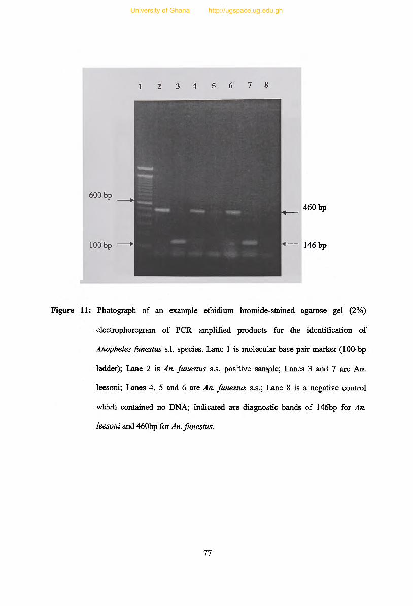
1 2 3 4 5 6 7 8
600 bp ___^
1 0 0 bp
^___ 460 bp
146 bp
Figure 11: Photograph of an example ethidium bromide-stained agarose gel (2%)
eleetrophoregram of PCR amplified products for the identification of
Anopheles funestus s.l. species. Lane 1 is molecular base pair marker (100-bp
ladder); Lane 2 is An. funestus s.s. positive sample; Lanes 3 and 7 are An.
leesoni; Lanes 4, 5 and 6 are An. funestus s.s.; Lane 8 is a negative control
which contained no DNA; Indicated are diagnostic bands of 146bp for An.
leesoni and 460bp fox An. funestus.
11
University of Ghana http://ugspace.ug.edu.gh

1 2 3 4 5 6 7 8 9 10 11 12
600 bp
1 0 0 bp
(HBMfrmm.
m <®mm m *m m m m m m m m
<---- 390 bp
m m <---- 280 bp
■r& *— 1 1 0 bp
Figure 12: Photograph of ethidium bromide-stained agarose gel (2 %) electrophoregram of
Hha I restriction enzyme digest for the identification of molecular forms of
Anopheles gambiae s.s. Lane 1 is molecular weight marker (100-bp ladder);
Lanes 2-4 and 6-10 are M forms; Lanes 5 and 11 are S forms; Lane 12 is
undigested An. gambiae s.s. PCR product; also indicated are the sizes of the
restricted products.
78
University of Ghana http://ugspace.ug.edu.gh

1 2 3 4 5 6 7 8
600 bp
188 bp
Figure 13: Photograph of an example of ethidium bromide-stained agarose gel (2%)
electrophoregram analysis of a PCR diagnostic test for detection of Wuchereria
bancrofti. Lane 1 is molecular base pair marker (100-bp ladder); Lane 2 is W
bancrofti positive control; Lanes 3-5 and 8 are W. bancrofti; Lane 6 is a negative
control. Also indicated is the diagnostic band size of 188bp for W. bancrofti.
79
University of Ghana http://ugspace.ug.edu.gh

CHAPTER FIVE
DISCUSSION
Low density microfilaraemia has been defined by Southgate (1992) as the density of
circulating m f in a specified blood compartment that cannot be detected in a significant
number of instances when commonly used blood sampling techniques are applied in
epidemiological studies. This, however, depends on a number of variables such as volume of
blood examined, source of blood sampled (usually venous or capillary) and method of m f
detection adopted. The author gave the limit of a value of 4mf per 2 0 jil (200mf per ml) and
less of capillary blood to denote low microfilaraemia. Southgate (1974) has also reported that
increases in blood volume will reduce the proportion of mf zeros reported and the theoretical
analyses of Grenfell et at. (1990) indicate increase in sampling volume has greater effect than
increase in m f detection efficiency. In this study, IOOjllI of finger prick blood was used and
read in counting chamber, which is an appreciable amount of blood compared to the popular
technique for m f detection in routine public health practice of 2 0 jil finger-prick blood films
(Southgate, 1992).
With this background it can be concluded that the mf mean intensity of 1.07 and 7 9 .4 5 mf
per ml of capillary blood among the entire district and microfilaraemic individuals
respectively that the densities were low in the studied site. This may be due to the fact that
the area has been receiving mass chemotherapy with ivermectin and albendazole for the past
4 years aimed at elimination of the disease. This is supported by the fact that Gomoa
80
University of Ghana http://ugspace.ug.edu.gh

Okyereko, which recorded the highest intensity of 155.6 mf/ ml of capillary blood among the
microfilaraemic individuals in this study, was recording 819 mf/ ml of blood in 1999
(Dzodzomenyo et al, 1999). In their study, Dzodzomenyo et al. (1999) observed no
significant difference in prevalence among males and females as well as variation in
intentisities between age-groups. It was no surprise a similar result was observed in this study
although Boakye et al. (2004) observed prevalence to be significantly higher in males than
females. Dunyo et al. (1996) also observed that more females than males are affected by
elephantiasis in Ghana unlike the coastal part of east Africa. These inconsistent observations
may be attributed to the occupational activities of the inhabitants of the study areas and the
biting pattern of the anopheline vectors.
Anopheles are mostly nocturnal in their activities, thus emergence from the pupae, mating,
blood feeding and oviposition normally occur in the evenings, at night or early in the
morning around sunrise. Some species may bite man mainly outdoors (exophagic) from
about sunset to 2 1 :0 0 hours whereas others bite mainly after 2 1 : 0 0 hours and mostly indoors
(endophagic). Whereas some species rest outside (exophilic) in a variety of natural shelters
like vegetation, rodent burrows, cracks and crevices in trees, under bridges, termite mounds,
and other cracks in the ground in between feeding, others rest in houses (endophilic).
Mixtures of these extremes of behaviour are exhibited by most Anopheles species that are
exophagic and endophagic, exophilic and endophilic. Few of this species exclusively feed on
humans though they are predominantly zoophilic. Most feed on both human and animals but
the degree of anthropophilism and zoophilism varies according to species and host
availability.
81
University of Ghana http://ugspace.ug.edu.gh

The inhabitants of Gomoa Okyereko are mainly peasant farmers and although they will not
be on the farm in the night, they may be either indoors or outside in the early hours of the
night. The disease manifests itself in adulthood, eventhough it is contracted in childhood as
long as one is exposed to infective bites of the mosquitoes (WHO, 2000).
Muirhead-Thompson (1954) in a small survey near Accra in Ghana demonstrated that m f of
W. bancrofti were present in highest concentration in the peripheral blood during night time.
Apart from Dzodzomenyo et al. (1999) no detailed study had described the pattern of
circadian mf periodicity in Ghana. Based on hourly examination of twelve volunteers for
nine hours, it was found that mf concentration in peripheral blood followed a wave-like
concentration peaking around 01:00 hours. The observed periodicity then is similar to those
reported from other parts of Africa (Tanaka, 1981; Gatika et al., 1994) probably because
night biting species of mosquitoes serve as vectors throughout the continent. Many other
explanations have been given to account for this interesting behavioural pattern of mf. One
suggestion is that oxygen tension plays a part and that if a person is given oxygen during the
night, the mfs will stay in deep tissues other than accumulating in peripheral circulation
(Hawking et al., 1966; Denham and McGreevy, 1977). Another suggestion is that periodicity
is related to human activity because if a person reverses his/ her working hours (resting
during the day and working at night) after a few days, there will be reversal in periodicity
(Hawking et a l , 1966; Denham and McGreevy, 1977).
Dzodzomenyo M. et al. (1999) in a study carried out at the same site observed An.junestus to
be the most abundant species of mosquito in early dry season while An. gambiae was
82
University of Ghana http://ugspace.ug.edu.gh

predominant in the wet season with a few culicines. The authors also observed a remarkable
increase in abundance of An. gambiae and reduction in abundance o f An. funestus during the
wet season. This pattern was similarly observed in this work. March is in the peak of the dry
month in Ghana and this may contribute to the low number of An. gambiae captured. Even
though 1.7% Anopheles and 3 .3 % Mansonia were found infected with the Li stage of W
bancrofti, there was no recovery of L3 from any of the mosquitoes after 1 2 days of
maintenance. Each of the infected An. gambiae had one Li stage of the parasite whiles on the
average the infected Mansonia species had two each of the parasite. On the other hand,
however, each of the infected An. funestus had 8 L1 stage of the parasite. Survival rate was 0.5
for both An. gambiae and An. funestus but 0.1 for Mansonia species. Mansonia transmits W.
bancrofti in parts of Asia but has not been identified as a vector in Africa (Sasa, 1976;
Service, 1990). It was therefore not surprising to find no infective stage larvae of the parasite
in the Mansonia species.
In their work, Southgate and Bryan (1992) reported that although many of the m f ingested by
Anopheles vectors is damaged by the mosquito’s foregut armature, the proportion of mf
destroyed is independent on the number of m f ingested and varies between members of the
An. gambiae complex and An. funestus. Studies on the relationship between the level of
microfilaraemia in the blood meal source and the percentage of feeding mosquitoes that
ingest mf or the number of mf ingested per mosquito have not provided consistent results for
Anopheles species (McGreevy et al., 1982; Bryan et al., 1990; Bryan and Southgate, 1988).
83
University of Ghana http://ugspace.ug.edu.gh

Some species of Anopheles have been reported by Southgate and Bryan (1992) to exhibit
facilitation in their relationship with W. bancrofti unlike An. melas in Gambia and An. merus
in Tanzania, which exhibit limitation. In view of the observed variation with mosquito
species and geographical region, it will be unreasonable to extrapolate the results of one
study on one Anopheles species in a region to the same species in another region or to a
different species in the same or different geographical area. A similar study conducted in the
Bongo District of Ghana found limitation to be more probable mechanism for W. bancrofti
and An. gambiae s.l. and An.funestus or both of these taxa (Boakye et al., 2004).
The significance of distinctly different host-parasite relationships lies in the importance of
low-density mf in sustaining transmission in various endemic areas with different genera of
mosquito vectors (Southgate, 1992). In theory, low level proportionality and limitation will
allow transmission to occur and build-up when most infected human hosts have low m f
densities, whereas situations of well marked proportionality or facilitation will give rise to
transmission thresholds below which the ultimate transmission will cease and the parasites
eliminated from the human population. However predictions of parasite extinction or parasite
resurgence can be made with confidence when characteristics of the local vector/ filarial
relationship are known (Webber, 1991).
Yawson et al. (2004) found the M and S forms of Anopheles gambiae s.s. to occur in
sympatry in southern Ghana. Studies in this coastal community have shown the abundance of
the M form. It was therefore not surprising to find most of the Anopheles gambiae s.s. being
M which has a remarkable ecological flexibility and is known to prevail in inundated areas
84
University of Ghana http://ugspace.ug.edu.gh

where dry season breeding opportunities exist (Appawu et al., 1994). A reduction in the m f
level of the human population following drug distribution is likely to lead to a decrease in
transmission of W. bancrofti by anophelines because they show facilitation pattern in
infections with filaria (Southgate & Bryan, 1992). This seems to be true for the M form of
Anopheles gambiae s.s.
CONCLUSION AND RECOMMENDATIONS
The main vector species encountered in this study were the M form of Anopheles gambiae
s.s. and An. funestus s.s. Considering the fact that the study was conducted in the natural
setting, this finding shows that the yearly combination therapy with ivermectin and
albendazole for 5 to 6 years will help eliminate the disease in areas like Ghana where
Anopheles gambiae and An. funestus are the main vectors. Although these Anopheles species
were not competent in promoting the maturation of the parasites when m f is low, a repeat of
this study targeting larger mosquito numbers is required to ascertain the role played
especially by M forms of An. gambiae in the transmission of lymphatic filariasis when
parasite levels in the community are low.
85
University of Ghana http://ugspace.ug.edu.gh

REFERENCES
Ahorlu, CK., Dunyo, SK., Asamoah, G. and Simonsen PE. (2001). Cosequences of
hydrocoel and the benefits of hydroceletomy: a qualitative study in lymphatic
filariasis endemic communities on the coast of Ghana. Acta Tropica 80:215-221.
Albuquerque, CM., Cavalcanti, VM., Melo, MA., Vercosa, P., Regis, LN. and Hurd, H.
(1999). Blood meal microfilariae density and the uptake and establishment of
Wuchereria bancrofti infections in Culex quinquefasciatus and Aedes aegypti.
Memorial Institute o f Oswaldo Cruz. 94(5): 591-596.
Anderson, RC. (1992). Nematode Parasites of Vertebrates. Their development and
transmission. Wallingford, UK: CAB International.
Anyanwu, GI., Molyneux, DH. and Phillips, A. (2000). Variation in cuticular
hydrocarbons among strains of the Anopheles gambiae sensu stricto by analysis of
cuticular hydrocarbons using gas liquid chromatography of larvae. Memorial
Institute o f Oswaldo Cruz, Rio de Janeiro, 95(3): 295-300.
Appawu, MA., Baffoe-Wilmot, A., Afari, EA., Nkrumah, FK. and Petrarca, V. (1994).
Species composition and inversion polymorphism of the Anopheles gambiae
complex in some sites of Ghana, West Africa. Acta Tropica. 56:15-23.
Bahr, PH. (1912). Filariasis and Elephantiasis in Fuji. Research Memoirs o f the London
School o f Tropical Medicine. 1:1-192.
86
University of Ghana http://ugspace.ug.edu.gh

Bain, O. (1971). Transmission des filarioses. Limitation des passages des microfilaires
ingerees vers l’hemocele du vecteur; interpretation. Annales de Parasitologie
humaine et comparee, 46:613-631.
Bain, O. and Brengues, J. (1972). Transmisssion de la wuchereriose et de la setariose
bovine: etude histologique de la traversee de la paroi stomacale &’Anopheles
gambiae A et d yAed.es aegypti par les microfilaires. Annales de Parasitologie
humaine et comparee, 47:399-412.
Blaustein L. (1989). Spatial distributions of Anopheles freebomi, Gambusia affinis and
Lepomis cyanellus in experimental rice plots. Journal o f the American Mosquito
Control Association. 5(2):254-7.
Boakye, DA., Wilson MD., Appawu MA. and Gyapong, J. (2004). Vector competence for
Wuchereria bancrofti of the Anopheles populations in the Bongo District of Ghana.
Annals o f Tropical Medicine and Parasitology. 98(5): 501-508.
Bockarie, MJ., Alexander, NDE., Hyun, P., Dimber, Z., Bockarie, F., Ibam, E., Alpers,
MP. and Kazura, JW. (1998). Randomised community-based trial of annual
single-dose diethylcarbamazine with or without ivermectin against Wuchereria
bancrofti infection inhuman beings and mosquitoes. Lancet. 351:162-168.
Bockarie, MJ., Fischer, P., Williams, SA., Zimmerman, PA., Griffin, L., Alpers, MP.
and Kazura, JW. (2000). Application of a polymerase chain reaction-ELISA to
detect Wuchereria bancrofti in pools of wild-caught Anopheles punctulatus in a
filariasis control area in Papua New Guinea. American Journal o f Tropical Medicine
and Hygiene, 62:363-367.
87
University of Ghana http://ugspace.ug.edu.gh

Brengues, J. (1975). La filariose de Bancroft en Afrique del’Ouest. Memoires
d ’O.R.S.TO.M. 79:1-299.
Brengues, J. and Bain, O. (1972). Passage des microfilaires de Pestomac vers l’hemocele
du vecteur, dans les couples Wuchereria bancrofti-Anopheles gambiae A, W.
bancrofti-Aedes aegypti et Setaria labiatopapillosa-A. aegypti. Cahiers ORSTOM,
Serie Entomologie medicale et Parasitologie, 10:235-249.
Brito, AC., Williams, P., Fontes, G., Rocha, EMM. (1997). A comparison of two Brazilian
populations of Culex quinquefasciatus (Say, 1823) from endemic and non-endemic
areas to infection with Wuchereria bancrofti (Cobbold, 1877). Memorial Institute o f
Oswaldo Cruz 92:33-36.
Bryan, JH. and Southgate, BA. (1976) Some observations on the filariasis in Western
Samoa after mass administration of diethylcarbamazine. Transactions o f the Royal
Society o f Tropical Medicine and Hygiene. 70:39-48.
Bryan, JH. and Southgate, BA. (1988a). Factors affecting transmission of Wuchereria
bancrofti by anopheles mosquitoes. 1. Uptake of microfilariae. Transactions o f the
Royal Society o f Tropical Medicine and Hygiene 82:128-137.
Bryan, JH. and Southgate, BA. (1988b). Factors affecting transmission of Wuchereria
bancrofti by anopheles mosquitoes. 2. Damage to ingested microfilariae by
mosquito foregut armatures and development of filarial larvae in mosquitoes.
Transactions o f the Royal Society o f Tropical Medicine and Hygiene 82:138-145.
Bryan, JH. and Southgate, BA. (1992). Factors affecting transmission of Wuchereria
bancrofti by anopheles mosquitoes. 4. Facilitation, limitation, proportionality and
88
University of Ghana http://ugspace.ug.edu.gh

their epidemiological significance. Transactions o f the Royal Society o f Tropical
Medicine and Hygiene 86:523-530.
Bryan, JH., McMahon, P. and Barnes, A. (1990). Factors affecting transmission of
Wuchereria bancrofti by anopheline mosquitoes. 3. Uptake and damage to ingested
microfilariae by An. gambiae, An. arabiensis, An. merus and An. funestus in East
Africa. Transactions o f the Royal Society o f Tropical Medicine and Hygiene 84:265-
268.
Burkot, TR. (1988). Non-random host selection by anopheline mosquitoes. Parasitology
Today, 4(6): 156-161.
Carlson, DA. and Service, MW. (1979). Differentiation between the species of the
Anopheles gambiae complex (Diptera: Culicidae) by analysis of cuticular
hydrocarbons. Annals o f Tropical Medicine and Parasitology 3:589-592.
Carlson, DA. and Service, MW. (1980). Identification of mosquitoes o f Anopheles gambiae
species complex ‘a’ and ‘b ’ by analysis of cuticular components. Science 207:1089-
1109.
Carlson, DA. and Walsh JP. (1981). Identification of two West African blackflies (Diptera:
Simuliidae) of the Simulium damnosum species complex by analysis of cuticular
paraffins. Acta Tropica 38:235-239.
Cech JJ. Jr. and Linden AL. (1987). Comparative larvivorous performances of
mosquitofish, Gambusia affinis, and juvenile Sacramento blackfish, Orthodon
microlepidotus, in experimental paddies. Journal o f the American Mosquito Control
Association. 3(1):35-41.
89
University of Ghana http://ugspace.ug.edu.gh

Chanteau, S., Luquiaud, P., Failloux, AB. and Williams, SA. (1994). Detection of
Wuchereria bancrofti larvae in pools of mosquitoes by the polymerase chain
reaction. Transactions o f the Royal Society o f Tropical Medicine and Hygiene,
88:665-666.
Chinery, WA. (1984) Effects of ecological changes on the malaria vector Anopheles
funestus and the Anopheles gambiae complex of mosquitoes in Accra, Ghana.
Journal o f Tropical Medicine and Hygiene, 87:75-81.
Christensen, BM., Sutherland, DR. and Gleasson, LN. (1984). Defence reactions of
mosquitoes to filarial worms: comparative studies on the response of three different
mosquitoes to inoculated Brugia pahangi and Dirofilaria immitis microfilariae.
Journal o f Invertebrate Pathology. 44:267-274.
CIOMS and WHO. (2002). International Ethical Guidelines for Biomedical Research
Involving Human Subjects, ppl-60.
Collins, FH., Mendez, MA., Rasmussen, MO., Mehaffey, PC., Besansky, NJ. and
Finnerty, V. (1987). A ribosomal RNA gene probe differentiates member species of
the Anopheles gambiae complex. American Journal o f Tropical Medicine and
Hygiene. 37:37-41.
Coluzzi, M. (1984). Heterogeneities of the malaria vectorial system in tropical Africa and
their significance in malaria prevention and control. Bulletin o f the World Health
Organization 62: (Supplement): 107-113.
Coluzzi, M. (1993). Advances in the study of Afro tropical malaria vectors Parassitologia
35(Supplement): 23-29.
90
University of Ghana http://ugspace.ug.edu.gh

Coluzzi, M. and Sabatini, A. (1967). Cytogenetic observations on species A and B of the
Anopheles gambiae complex. Parasitologia 9:73-88.
Coluzzi, M., Sabatini, A., della Torre, A., Di Deco, MA. and Petrarca, V. (2002). The
polythene chromosome map of Anopheles gambiae: cytotaxonomic findings,
patterns of rearrangements, evolutionary and malariological implications. Science.
298:1415-1418.
Coluzzi, M., Sabatini, A., Petrarca, V. and Di Deco, MA. (1979). Chromosomal
differentiation and adaptation to human environments in the Anopheles gambiae
complex. Transactions o f the Royal Society o f Tropical Medicine and Hygiene
73:483-497.
Craig, MH., Snow, RW. and leSueur, D. (1999). A climate-based distribution model of
malaria transmission in sub-Saharan Africa. Parasitology Today. 15:105-111.
Crans, WJ. (1973). Experimental infection of Anopheles gambiae and Culex
quinquefasciatus pipiens fatigans with Wuchereria bancrofti in coastal East Africa.
Journal o f Medical Entomology 10:189-193.
della Torre, A., Costantini, C., Besansky, NJ., Caccone, A., Petrarca, V., Powell, JR.
and Colluzzi, M. (2002). Incipient speciation within Anopheles gambiae: the glass
is half full. Science. 298:115-117.
Denham, DA. and McGreevy, PB. (1977). Brugian filariasis: Epidemiological and
experimental studies. Advances in Parasitology. 15:243-309.
Detinova, TS. (1962). Age grouping methods in Diptera of medical importance with special
references to some vectors of malaria. Geneva: World Health Organisation,
monograph Series, no. 49:1-216.
91
University of Ghana http://ugspace.ug.edu.gh

Dreyer, G., Noroes, J., Amaral., Nen, A., Medeiros, Z., Coutinho, A. and Addis, D.
(1995). Direct assessment of the adulticidal efficacy of a single dose of ivermectin in
bancroftian filariasis. Transactions o f the Royal Society o f Tropical Medicine and
Hygeine 89:441-443.
Duerr, HP., Dietz, K. and Eichner, M. (2005). Determinants of the eradicability of filarial
infections: a conceptual approach. Trends in Parasitology. 21(2):88-96.
Dunyo, SK., Appawu, M., Nkrumah, FK-, Baffoe-Wilmot, A., Pedersen, EM. and
Simonsen, PE. (1996). Lymphatic filariasis on the coast of Ghana. Transactions o f
the Royal Society o f Tropical Medicine and Hygiene 90: 634 -638.
Dzodzomenyo, M., Dunyo, SK., Ahorlu, CK., Coker, WZ., Appawu, MA., Pedersen,
EM. and Simonsen PE. (1999). Bancroftian filariasis in an irrigated project
community in southern Ghana. Tropical Medicine and International Health. 4 (1)
13-18.
Edeson, JFB., Hawking, F. and Symes, CB. (1957). The periodicity of microfilariae. 6 . The
response of microfilariae of Wuchereria malayi and Wuchereria bancrofti, Pacific
type, to various stimuli. Transactions o f the Royal Society o f Tropical Medicine and
Hygiene. 51:359-365.
Ewert, A. (1971). Distribution of developing amd mature Brugia malayi in cats at various
times after single inoculation. The Journal o f Parasitology. 57:1039-1042.
Failloux, AB., Raymond, M., Ung, A., Glaziou, P., M artin, PMV. and Pasteur, N.
(1995). Variation in the vector competence of Aedes aegypti polynesiensis for
Wuchereria bancrofti. Parasitology 111:19-29.
92
University of Ghana http://ugspace.ug.edu.gh

Farid, HA., Hamraad, RE., Hassan, MM., Morsy, ZS., Karaal, IH., Weil, GJ. and
Ramzy, RM. (2001). Detection of Wuchereria bancrofti in mosquitoes by the
polymerase chain reaction: a potentially useful tool for large-scale control programs.
Transactions o f the Royal Society o f Tropical Medicine and Hygiene, 95:29-32.
Favia, G., Delle Torre, A., Bagayoko, M., Lanfrancotti, A., Sagnon, NF. Toure, YT. and
Colluzzi, M. (1997). Molecular identification of sympatric chromosomal forms of
anopheles gambiae and further evidence of their reproductive isolation. Insect
Molecular Biology. 6:377-383.
Favia, G., Lanfrancotti, A., Spanos, L., Siden-Kiamos, I. and Lois, C-, (2001). Molecular
characterization of ribosomal DNA polymorphisms discriminating among
chromosomal forms of Anopheles gambiae s.s. Insect Molecular Biology. 10:19-23.
Fischer, P., Liu, X., Lizotte-Waniewski, M., Kamal, IH., Ramzy, RM. and Williams, SA.
(1999). Development of a quantitative, competitive polymerase chain reaction-
enzyme-linked immuno-sorbent assay for the detection of Wuchereria bancrofti
DNA. Parasitology Research, 85:176-183.
Fox, LM., Furness, BW., Haser, JK., Brissau, JM., Louis-Charles, J., Wilson, SF.,
Addiss, DG., Lammie, PJ. and Beach, MJ. (2005). Ultrasonographic examination
of Haitian children with lymphatic filariasis: a longitudinal assessment in the context
of antifilarial drug treatment. The American Journal o f Tropical Medicine and
Hygiene. 72(5):642-648.
Garret-Jones, C. (1964). The human blood index of malaria vectors in relation to
epidemiological assessment. Bulletin o f the World Health Organisation. 30:241-261.
93
University of Ghana http://ugspace.ug.edu.gh

Gatika, SM., Fugimaki, Y., Njuguna, MN., Gachihi, GS. and Mbugua, JM. (1994). The
microfilarial periodicity pattern of Wuchereria bancrofti in Kenya. Journal o f
Tropical Medicine and Hygiene. 97:60-64.
Gilles MT. and Coetzee, M. (1987). A supplement to the Anophelinae of Africa South of
the Sahara. Johannesburg: South African Institute o f Medical Research. No.55
Gilles MT. and De Meillon, B. (1968). The anophelinae of Africa south of Sahara
(Ethiopian Zoogeographical Region). Publication o f the South African Institute fo r
Medical Research 54:343pp.
Gilles, MT. (1955). The pre-gravid phase of ovarian development in Anopheles funestus.
Annals o f Tropical Medicine and Parasitology 49:320-325.
Gilles, MT. (1988). Anopheline mosquitoes: vector behaviour and bionomics in malaria.
Principle and Practice o f Malariology Wemsdorfer, W. and McGregor, I. (Eds.).
Edinburgh, London, Melbourne and new York: Churchill Livingstone
Gilles, MT. and Coetzee, M. (1987). A supplement to the Anophelenae of Africa South of
the Sahara. Johannesburg: South African Institute fo r Medical Research 55:105-126.
Gilles, MT. and De Meillon, B. (1968). The Anophelenae of Africa South of the Sahara
(Ethiopian Zoographical Region). Johannesburg: Publication o f South African
Institute fo r Medical Research 54:343.
Gilles, MT. and Wilkes, TJ. (1965). A study of the age composition of populations of
Anopheles gambiae Giles and An. funestus Giles in North-Eastern Tanzania. Bulletin
o f Entomological Research. 56:237-262.
94
University of Ghana http://ugspace.ug.edu.gh

Gimnig, JE., Ombok, M., Kamau, L. and Hawley, WA. (2001). Characteristics of larval
anopheline (Diptera: Culicidae) habitats in western Kenya. Journal o f Medical
Entomology 38:282-288.
Gimnig, JE., Ombok, M., Otieno, S., Kaufman, MG., Vulule, John, M. and Walker,
ED. (2002). Density-Dependent Development of Anopheles gambiae (Diptera:
Culicidae) Larvae in Artificial Habitats Journal o f Medical Entomology 39(1): 162-
172.
Grenfell, BT., Das, PK., Rajagopalan, PK. and Bundy, DAP. (1990). Frequency
distribution of lymphatic filariasis microfilariae in human populations: population
processes and statistical estimation. Parasitology. 101:417-427.
Gyapong, M., Gyapong, JO., Adjei, S. and Carol Vlassoff (1996). Filariasis in Northern
Ghana: Some Cultural Beliefs and Practices and their Implications for Disease
Control. Social Science and Medicine 43(2): 235-242.
Haddow, AJ. (1942). The mosquito fauna and climatic of native huts at Kisumu, Kenya.
Bulletin o f Entomological Research 33: 91.
Hawking, F. and Gammage, K. (1968). The periodic migration of microfilariae of Brugia
malayi and its response to various stimuli. American Journal o f Tropical Medicine
and Hygiene. 17:724-729.
Hawking, F., Pattanayak, S. and Sharma, HL. (1966). The periodicity of microfilariae XI.
The effect of body temperature and other stimuli upon the cycles of Wuchereria
bancrofti, Brugia malayi, B. ceylonensis and Dirofilaria repens. Transactions o f the
Royal Society o f Tropical Medicine and Hygiene. 60:496-513.
95
University of Ghana http://ugspace.ug.edu.gh

Helmy, H., Fischer, P., Farid, HA., Bradley, MH. and Ramzy, RM. (2004). Test strip
detection of Wuchereria bancrofti amplified DNA in wild-caught Culexpipiens and
estimation of infection rate by a Pool-screen algorithm. Tropical Medicine and
International Health. 9(1): 158-63.
Hoti, SL., Vasuki, V., Patra, KP., Hariths, VR., Ravi, G. and Sushma, S. (2001).
Laboratory evaluation of Ssp I PCR assay for the detection of Wuchereria bancrofti
infection in Culex quinquefasciatus. Indian Journal o f Medical Research, 114:59—
64.
Janousek, TE. and Lowrie Jr, RC. (1989). Vector competency of Culex quinquefasciatus
(Haitian strain) following infection with Wuchereria bancrofti. Transactions o f the
Royal Society o f Tropical and Hygiene. 83:679-680.
Kamal, IH., Fischer, P., Adly, M., Sayed, AS., Morsy, ZS. and Ramzy, RM. (2001).
Evaluation of PCR-ELISA to detect Wuchereria bancrofti in Culex pipiens from an
Egyptian village with a low prevalence of filariasis. Annals o f Tropical Medicine
and Parasitology. 95:833-841.
Kartman, L. (1953). Factors influencing infection of the mosquito with Dirofilaria immitis
(Leidy, 1956). Experimental Parasitology 2:27-78.
Kartman, L. (1954). Suggestions concerning an index of experimental filarial infection in
mosquitoes. American Journal o f Tropical Medicine and Hygiene 3:329-337.
Kettle, DS. (1992). Medical and Veterinary Entomology. Centre for Agriculture and
Biosciences International. Wallingford, UK. pp 99-136.
96
University of Ghana http://ugspace.ug.edu.gh
Koekemoer, LL., Kamau, L., Hunt, RH. and Coetzee, M. (2002). A Cocktail Polymerase
Chain Reaction assay to identify numbers of Anopheles funestus (Diptera.
Culicidae) group. American Society o f Tropical Medicine and Hygiene 66:806-811.
Koekemoer, LL., Lochouarn, L., Hunt, RH. and Coetzee, M. (1999). Single-Stranded
Conformation Polymorphism Analysis for identification of four members of the
Anopheles funestus (Diptera: Culicidae) group. Journal o f Medical Entomology
36:125-130.
Krishnamoorthy, K., Subramanian, S., Van Oortmarssen, GJ., Habbema, JD. and Das,
PK. (2004) Vector survival and parasite infection: the effect of Wuchereria
bancrofti on its vector Culex quinquefasciatus. Parasitology. 129(l):43-50.
Lardeux, F. and Cheffort, J. (2001) Ambient temperature effects on the extrinsic
incubation period of Wuchereria bancrofti in Aedes polynesiensis: implications for
filariasis transmission dynamics and distribution in French Polynesia. Medical and
Veterinary Entomology. 15(2): 167-76.
Lindsay, SW. and Denham, DA. (1986). The ability of Aedes aegypti mosquitoes to survive
and transmit infective larvae of Brugia pahangi over successive blood meals.
Journal o f Helminthology. 60:159-168.
Lizotte, MR., Supali, T., Partono, F. and Williams, SA. (1994). A polymerase chain
reaction assay for the detection of Brugia malayi in blood. American Journal o f
Tropical Medicine and Hygiene. 51:314-321.
MacDonald, G. (1952). The analysis of sporozoite rate. Tropical Diseases Bulletin. 49:227-
229.
97
University of Ghana http://ugspace.ug.edu.gh

Mahmoud AA. (1985). Mosquitofish Gambusia affinis holbrooki as a malaria vector control
agent in Gezira irrigation canals of the Sudan. Journal o f the American Mosquito
Control Association. 1(4):524-6.
Mak, JW . (1989). Recent advances in the laboratory diagnosis of filariasis. The Malasian
Journaal o f Pathology. 1 1 :1-5 .
Masendu, HT., Hunt, RH., Govere, J., Brooke, BD., Awolola, TS. and Coetzee, M.
(2004). The sympatric occurrence of two molecular forms of the malaria vector
Anopheles gambiae Giles sensu stricto in Kanyemba, in the Zambezi Valley,
Zimbabwe. Transactions o f the Royal Society o f Tropical Medicine and Hygiene
98(7):393-6.
Mayr, E. and Ashlock, PD. (1991). Principles o f Systematic Zoology, 2nd Ed. McGraw-Hill
Inc., New York.
McGreevy, PB., Kolstrup, N-, Tao, J., McGreevy, MM. and Marshall, TF. de C (1982).
Ingestion and development of Wuchereria bancrofti in Culex quinquefasciatus,
Anopheles gambiae and Aedes aegypti after feeding on humans with varying
densities of microfilariae in Tanzaniae. Transactions o f the Royal Society o f
Tropical Medicine and Hygiene 76:288-296.
McGreevy, PB., Theis, JH., Lavoipierre, M J. and Clark, J. (1974). Studies on filariasis.
III. Dilofilaria immits: emergence of infective larvae from the mouthparts of Aedes
aegypti. Journal o f Helminthology. 48:221-228.
McMahon, JE., Marshall, TF de C., Vaughan, JP . and Abaru, DE. (1979). Bancroftian
filariasis: a comparison of microfilariae counting techniques using counting
98
University of Ghana http://ugspace.ug.edu.gh

chamber, standard slide and membrane (Nucleopore) filtration. Annals o f Tropical
Medicine and Parasitology. 73:457-464.
Michael, E., Bundy, DA. and Grenfell, BT. (1996). Re-assessing the global prevalence and
distribution of lymphatic filariasis. Parasitology 112:409-428.
Micklos, DA. and Freyer, GA. (1990). DNA Science: A First Course in Recombinant DNA
Technology. Carolina Biological Supply Company, Burlington, NC and Cold Spring
Harbor Laboratory Press, Cold Spring Harbour, NY.
Molineaux, L. (1988). The epidemiology of human malaria as an explanation of its
distribution, including some implications for its control. In: Malaria. Principles and
Practice o f Malariology. Pp 913-998. Wemsdorfer, W. and McGregor, I. (Eds.).
Edinburgh. London, Melbourne and New York: Churchill Livingstone.
Mossinger, J. (1991). In vitro cultivation of adult Litomosoides carinii: evaluation of basic
culture media, gas phases and supplements. Parasitology. 103 (l):85-.95.
Muir, DA. (1988). Anopheline mosquitoes: vector production, life-cycle and biotope. In:
Malaria. Priciples and practice o f malariology. Wemsdorfer, W. and McGregor, I.
(Eds.) pp. 431-451. Edinburgh, London, Melbourne and New York: Churchill
Livingstone.
Muirhead-Thomson, RC. (1945). Studies on the breeding places and control of Anopheles
gambiae var melas in coastal districts of Sierra Leone. Bulletin o f Entomological
Research 36:185.
Muirhead-Thomson, RC. (1954). Factors determining the true reservoir of infection of
Plasmodium falciparum and Wuchereria bancrofti in a West African village.
Transactions o f the Royal Society o f Tropicaal Medicine and Hygiene. 48:208-225.
99
University of Ghana http://ugspace.ug.edu.gh

Mullis, KB. (1990). The unusual origin of the polymerase chain reaction. Scientific
American 262:56-65.
Nanda, B. and Ramaiah, KD. (2003). Lymphoedema-management measures practised by
cases of chronic lymphatic filariasis. Annals o f Tropical Medicine and Parasitology.
97(4):427-31.
Nicolas, L. and Plichart, C. (1997). A universally applicable internal standard for PCR
detection of Wuchereria bancrofti in biological samples. Parasitology 4:253-257.
Omar, MS. and Zielke, E. (1978). Abortive development of Wuchereria bancrofti in a West
African stain of Culex pipiens fatigans. Tropenmedizin und Parasitologie, 29:364-
370.
Paabo, S., Higuchi, RG. and Wilson, AC. (1989). Ancient DNA and the polymerase chain
reaction. Journal o f Biological Chemistry. 264:9709-9712.
Pedersen, EM. (1977). Studies on the influence of the peritrophic membrane on the
migration of Brugia pahangi in Aedes aegypti. Dissertation, London School of
Hygiene and Tropical Medicine, June 1977.
Philippon, B. and Bain, O. (1972). Transmission de l’onchocercose humaine en zone de
savane d’Afrique occidentale: passage des microfilaires d 'Onchocerca volvulus
Leuck. dans l ’hemocele de la femelle de Simulium damnosum Th. Cahiers
ORSTOM, Serie Entomologie medicate et Parasitologie, 10:251-261.
Pichon G, and Treuil JP. (2004). Genetic determinism of parasitic circadian periodicity and
subperiodicity in human lymphatic filariasis. 327(12): 1087-94.
100
University of Ghana http://ugspace.ug.edu.gh

Pichon, G. (2002). Limitation and facilitation in the vectors and other aspects of the
dynamics of filarial transmission: the need for vector control against Anopheles-
transmitted filariasis. Annals o f Tropical Medicine and Parasitology. 96
Supplementary 2 :S 143-52.
Plaisier, AP., Cao, WC., van Oortmarssen GJ. and Habbema JD. (1999). Efficacy of
ivermectin in the treatment of Wuchereria bancrofti infection: a model-based
analysis of trial results. Parasitology 119:385-394.
Powell, JR., Petrarca, V., della Torre, A., Caccone, A. and Colluzi, M. (1999). Population
structure, speciation and introgression in the Anopheles gambiae complex.
Parasitologia 41:101-114.
Ramachandran, CP. (1970). A guide to methods and techniques in Filariasis Investigations,
Filar Res O ff Inst Med Res, Kuala Lumpur, 39pp.
Ramaiah, KD, Das, PK, Anamail, P. and Pani, SP. (2003). The impact of six rounds of
single-dose mass administration of diethylcarbamazine or ivermectin on the
transmission of Wuchereria bancrofti by Culex quinquefasciatus and its implications
for lymphatic filariasis elimination programmes. Tropical Medicine and
International Health. 8(12): 1082-92.
Ramaiah, KD., Vanamail, P., Pani, SP. and Das, PK. (1997). Functional impairment
caused by lymphatic filariasis in rural areas of south India. Tropical Medicine and
International Health 2:832-838
Ramzy, RM., Farid, HA., Kamal, IH., Ghada, HI., Zakariah, SM., Rifky, F., Weil, GJ.,
Williams, SA. and Gad, AM. (1997). A polymerase chain reaction -based assay for
101
University of Ghana http://ugspace.ug.edu.gh

detection of Wuchereria bancrofti in human blood and Culex pipiens. Transactions
o f the Royal Society o f Tropical Medicine and Hygiene 91:156-160.
Reid, JA. (1982). Concentration of microfilariae in mosquitoes during feeding. Transactions
o f the Royal Society o f Tropical Medicine and Hygiene. 76:850-851.
Reisen, WK. (1989). Estimation of vectorial capacity: Introduction. Bulletin of the Society
of vector Ecology. 14:39-40.
Rozema, J., Bjorn, LO., Bornman, JF., Gaberscik, A., Hader, DP., Trost, T-, Germ, M.,
Klisch, M., Groniger, A., Sinha, RP., Lebert, M., He, YY., Buffoni-Hall, R., de
Bakker, NV., van de Staaij, J. and Meijkamp, BB. (2002). The role of UV-B
radiation in aquatic and terrestrial ecosystems-an experimental and functional
analysis of the evolution of UV-absorbing compounds. Journal o f Photochemistry
and Photobiology, B. Biology. 66(1):2-12.
Saliternik Z. (1977). Specific vector control methods for prevention and eradication of
malaria in Israel. Tropical and Geographical Medicine.;29(l):l-7.
Samarawickrema, WA., Spears, GFS., Sone, PF., Ichimori, K. and Cummings, RF.
(1985). Filariasis transmission in Samoa. 1. Relation between density of
microfilariae and larval density in laboratory-bred and wild-caught Aedes
(iStegomyia) polynesiensis (Marks) and wild-caught Aedes {Finlaya) samoanus
(Gruenburg). Annals o f Tropical Medicine and Parasitology 79:89-100.
Sasa, M. (1976). Human filariasis. A global survey of epidemiology and control. University
Park Press, Baltimore, 819.
102
University of Ghana http://ugspace.ug.edu.gh

Scott, JA., Brogdon, WG. and Collins, FH. (1993). Identification of single specimens of
the Anopheles gambiae complex by the polymerase chain reaction. American
Journal o f Tropical Medicine and Hygiene 49(4): 520-529.
Service, MW. (1980). A Guide to Medical Entomology. Macmillan Press, London: 24-52.
Service, MW. (1990). Handbook to the Afrotropical toxorhynchitine and culicine
mosquitoes, excepting Aedes and Culex. London: British Museum (Natural History).
Service, MW. (1993) Mosquitoes (Culicidae) in: Medical Insects and Arachnids (Lane R.P
and Grosskey R.W.) eds., Chapman & Hall, London: 120-240.
Shenoy, RK., Suma, TK., Raj an, K., and Kumaraswami, V. (1998) Prevention of acute
adenolymphangitis in brugian filariasis: comparison of the efficacy of ivermectin
and diethylcarbamazine, each combined with local treatment of the affected limb.
Annals o f Tropical Medicine and Parasitology. 92(5):587-94.
ShibuyaT., Tanaka, H. and Cabrera, DB. (1981). Low density microfilaraemia of
Wuchereria bancrofti infections in pre- and post-treatment phases in the Philippines.
Japanese Journal o f Experimental Medicine. 51, 133-135.
Simonsen, PE, Magesa, SM, Dunyo, SK, Malecela-Lazaro, MN, Michael, E. (2004). The
effect of single dose ivermectin alone or in combination with albendazole on
Wuchereria bancrofti infection in primary school children in Tanzania Transactions
o f the Royal Society o f Tropical Medicine and Hygiene. 98(8):462-72.
Southgate, BA. (1974). A quantitative approach to parasitological techniques in bancroftian
filariasis and its effect on epidemiological understanding. Transactions o f the Royal
Society o f Tropical Medicine and Hygiene. 68:177-186.
103
University of Ghana http://ugspace.ug.edu.gh

Southgate, BA. (1992). The significance of low density microfilareamia in the transmission
of lymphatic filarial parasites. Journal o f Tropical Medicine and Hygiene. 95.79-86.
Southgate, BA. and Bryan, JH. (1992). Factors affecting transmission of Wuchereria
bancrofti by anopheline mosquitoes. 4. Facilitation, limitation, proportionality and
their epidemiological significance. Transactions o f the Royal Society o f Tropical
Medicine and Hygiene 86:523-530.
Subra, R. (1973). Etudes ecologiques sur Culex pipiens fatigans Wiedemann, 1828,
(Diptera, Culicidae) dans une zone urbaine de savanne soudanienne oust-afiicaine.
Dynamique des populations imaginales. Cahiers ORSTOM, Series Entomological
Medical Parasitology, 11:79-100.
Subramanian, S., Krishnamoorthy, K., Ramaiah, KD., Habbema, JDF., Das, PK. and
Plaiser, AP. (1997). The relationship between microfilarial load in the human host
and uptake and development of Wuchereria bancrofti microfilariae by Culex
quinquefasciatus: a study under natural conditions. Parasitology 116:243-255.
Tabachnick, WJ., Wallis, GP., Aitken, THG., Miller, BR., Amato, GD., Lorenz, L.,
Powell, JR. and Beaty, BJ. (1985). Oral infection of Aedes aegypti with yellow
fever virus: geographical variation and genetic considerations. American Journal o f
Tropical Medicine and Hygiene 34:1219-1224.
Tanaka, H. (1981). Periodicity of microfilariae of human filariasis analysed by a
trigonometric method (Aikat and Das). Japanese Journal o f Experimental Medicine.
51:97-103.
Toure, YT., Petrarca, V., Traore, SF., Coulibaly, A., Maiga, HM., Sankare, O., Sow, M.,
Di Deco, MA. and Coluzzi, M. (1994). Ecological genetic studies in the
104
University of Ghana http://ugspace.ug.edu.gh

chromosomal form Mopti o f Anopheles gambiae s.s. in Mali, West Africa. Genetica.
94:213-223.
rownson, H. and Chaitong, U. (1991). Mosquito host influences on development of
fllariae. Annals o f Tropical Medicine and Parasitology. 85:149-163.
Webber, RH. (1991). Can anopheline-transmitted filariasis be eradicated? Journal o f
Tropical Medicine and Hygiene. 94:241-244.
Webber, RH. (1991). Eradication of Wuchereria bancrofti through vector control.
Transactions o f the Royal Society o f Tropical Medicine and Hygiene. 73:722-724.
Wharton, RH. (1960). Studies on filariasis in Malaya: field and laboratory investigations of
the vectors of a rural strain of Wuchereria bancrofti. Annals o f Tropical Medical and
Parasitology 54:78-91.
White, GB. (1969). Factors influencing densities of mosquitoes resting indoors. Annual
Report o f the East African Institute o f Malaria and Vector-borne Diseases. 37-43.
White, GB. (1985). Anopheles bwambae sp. n., a malaria vector in the Semliki Valley,
Uganda, and its relationship with other sibling species of the An. gambiae complex
(Diptera: Culicidae). Systematic Entomology. 10:501-522.
Witt, C. and Ottesen, EA. (2001). Lymphatic filariasis: an infection of childhood. Tropical
Medicine and International Health. 6(8): 582-606.
World Health Organisation (2000). The Global Alliance to Eliminate Lymphatic Filariasis.
Yameogo, L., Toe, L., Hougard, JM., Boatin, BA. and Unnasch, TR. (1999). Pool screen
polymerase chain reaction for estimating the prevalence of Onchocerca volvulus
infection in Simulium damnosum sensu lato: results of a field trial in an area subject
105
University of Ghana http://ugspace.ug.edu.gh

to successful vector control. American Journal o f Tropical Medicine and Hygiene.
60:124-128.
Yawson, AE., McCall, PJ., Wilson, MD. and Donnelly, MJ. (2004). Species abundance
and insecticide resistance of Anopheles gambiae in selected areas of Ghana and
Burkina Faso. Medical Veterinary Entomology. 18(4):372-7.
Zaltsman, A. and Feder, A. (2005). Developmental and light effects on the accumulation of
FtsH protease in Arabidopsis chloroplasts - implications for thylakoid formation and
photosystem II maintenance.Adam Z. The Plant Journal fo r Cell and Molecular
Biology. 42(5):609-17.
Zhong, M., McCarthy, J., Bierwert, L., Lizottea-Waniewski, M., Chanteau, S., Nutman,
TB., Ottesen, EA. and Williams, SA. (1996). A polymerase chain reaction assay
for detection of the parasite Wuchereria bancrofti in human blood samples.
American Journal o f Tropical Medicine and Hygiene. 54:357—363.
106
University of Ghana http://ugspace.ug.edu.gh

APPENDICES
APPENDIX I
PREPARATION OF STANDARD SOLUTIONS
With standard sterile double distilled water (sdd H20), the following standard solutions were
prepared and autoclaved at 121 lb/ sq for 15 minutes in an Eyela autoclave (Rikikkaki,
Tokyo).
0.5 M EDTA (pH 8.0)
186 g of EDTA, dissolved in 800 ml double distilled water, pH adjusted with
NaOH pellets, the volume made to 1000 ml with ssd H2O and stored at room
temperature
10ml of TE (pH 8.0)
20(4.1 of 0.5 M EDTA, 50jul of 2.0M Tris HC1, 9.93ml of sddH20 and stored at
room temperature.
TE-RNAse
995ju.l TE in 5|j.l RNAse
107
University of Ghana http://ugspace.ug.edu.gh

Primers for PCR
Primers used in PCR reaction were reconstituted and diluted according to the
manufacturers recommendations (Oswell Laboratories).
Bender Buffer
0.1M NaCl, 0.2M Sucrose, 0.1M Tris HC1 (pH 7.5), 0.05M EDTA (pH 9.1),
0.5% SDS.
Buffer C for PCR mixture
300mM Tris HC1, 75mM (NEUfcSO* 2.5mM MgCl2
10XTAE buffer
242g Tris base, 57.1 ml glacial acetic acid, 100 ml 0.5 M EDTA, pH adjusted
to 7.7 (with glacial acetic acid) and the volume made to 1000 ml with double
distilled water.
5X Orange G (Gel loading buffer)
20% (w/ v) Ficoll, 25 mM EDTA, 2.5% (w/ v) orange G and stored at room
temperature.
108
University of Ghana http://ugspace.ug.edu.gh

DNA molecular weight size markers
lOObp DNA molecular weight size marker obtained from the manufacturers
(Sigma) was diluted according to recommendations and used; IOjjI of DNA
ladder, 30jnl of loading dye (orange G), 60|il of sddl^O. For the lOObp ladder,
the first band size is lOObp, subsequent ones are read 200, 300, 400,
..................................lOOObp.
2% Agarose gel
First l.Og of agarose powder was put into flask and TAE was added to make a
volume of 50ml. It was heated in microwave oven (230V, 50Hz, 2660W,
12.0A) for 1 minute to dissolve solute. The mixture was cooled under running
water and 2jj,1 of Ethidium Bromide was added. The gel was cast to set in a
chamber with comb to make the wells.
109
University of Ghana http://ugspace.ug.edu.gh

SEQ
UEN
CE
OF
THE
SYN
THET
IC
OLI
GO
NU
CLE
OTI
DE
PRIM
ERS
USED
IN
THE
MO
LECU
LAR
STU
DIE
S O
F:
vaaIcl"O£o<uAW
DCdtav
a>GOC«<uNas
IQ
u3s-ai
S3a
Ua
H
QOO)'3QJQiW)GO
•K»
II1
fO
a>sg-0)czi
as*ca«4HoaacS£
$‘SCuce
00 OVO OsCO
COoo
o<iouUU
OUUU£
§O
bia
i
1
COcK
aa<uoauHCDoH£ago
<o
COCO
Q■-Cse§)
Iso*s§-si
vo
<Nm
<a
oooHUcSuo
uo
sH
W
s>5
§<R
mi—ICO
r-
.£3i
i
R
co
CN
?S %u< huu
HaH Po HH£
<wO 5fc uP uH <O
§
O<y
i•sS3§II
.1
1R
University of Ghana http://ugspace.ug.edu.gh

Anop
hele
s fu
nestu
s s.l
. sp
ecie
s
University of Ghana http://ugspace.ug.edu.gh

Expe
cted
am
plifi
ed
DN
A
University of Ghana http://ugspace.ug.edu.gh

APPENDIX III
CONSTITUENTS OF A 20jil PCR REACTION M IX USED IN THE MOLECULAR
STUDIES OF:
Anopheles gambiae DNA
Reagent Volume Final concentration
Sterile distilled water 14.3 jul -
10 x PCR buffer 2 .0 fxl lx
20juM of each dNTP (dATP, dCTP, dGTP, dTTP)
0 .2 jul 20jiM
2 0 \iM of each primer (UN, GA, ME, AR, QD)
1.5|il 20p.M
5U Taq polymerase 0 .1 |xl 0.625 jlM
DNA template 1 ng-lp.g
113
University of Ghana http://ugspace.ug.edu.gh

Anopheles funestus DNA
Reagent Volume Final concentration
Sterile distilled water 14.9fil -
10 x PCR buffer (plus 25mM MgCl) 2 .0 jxl lx
20jj,M of each dNTP (dATP, dCTP, dGTP, dTTP)
0 .2 ^ 1 2 0 0 iM each
20 jxM of each primer (UN, FUN, VAN, RIV, PAR, LEES)
1 .8 |xl 20j-iM
5U Taq polymerase 0 .1 |xl 0.625U/ fil
DNA template l.Ofxl 1 ng-lug
114
University of Ghana http://ugspace.ug.edu.gh

Wuchereria bancrofti DNA
Reagent Volume Final concentration
Sterile distilled water 11.7|*1
10 xPCR buffer 2 .0 jxl lx
20|iM of each dNTP (dATP, dGTP, dCTP, dTTP)
0 .2 p.l 2 0 0 |xM each
2 0 uM of each primer (NV-l.NV-2)
l.Ojil 200nM
5U Taq polymerase 0 .1^1 0.025U/ |il
DNA template 5.010.1 1 ng-lug
115
University of Ghana http://ugspace.ug.edu.gh

APPENDIX IV
INFORMATION AND CONSENT FORM
Invitation to participate in the study
We invite you to take part in a research study by the Noguchi Memorial Institute for Medical
Research, University of Ghana, Legon. The research study is called: Vector competence of
the Anopheles (Diptera: Culicidae) populations for Wuchereria bancrofti (Spirurida:
Filariidae) in the Gomoa District of Ghana after mass drug administration. The primary
investigator for the study is Dr. Daniel Adjei Boakye, NMIMR.
Introduction
This consent form informs you about the background, aims and the method of this study. In
addition it explains the anticipated benefits, potential risk of the study and the discomfort it
may entail. Finally it informs you of entrance criteria and your rights regarding participating
in this study.
Background and purpose of this study
The relationship between ingestion of microfilariae (mf), production of infective larvae (L3)
and mf density in human blood has been suggested as an important determinant in the
transmission dynamics of lymphatic filariasis. Thus understanding vector-parasite
interactions is essential for rational development of filariasis control strategies. This is
particularly important, considering that vectorial competence (the ability of mosquitoes to
ingest mf and to promote their maturation until the infective stage), and the rate of mosquito
116
University of Ghana http://ugspace.ug.edu.gh

survival until parasite maturation, seems to differ according to geographic mosquito strains.
Variation in the density of mf in the blood and parasite behaviour also influences vector-
parasite relationships.
For the Global Programme to Eliminate Lymphatic Filariasis (GPELF) to be successful, the
competence of various mosquito vectors to pick the mf (especially when the community m f
load is low), support the development of the ingested m f into the human-infective third-stage
larvae (L3), and to transmit those L3 to humans has to be understood. Some species of
mosquito (those exhibiting the phenomenon of facilitation) are unable to transmit parasites
from humans with low levels of microfilaraemia whereas other species (exhibiting limitation)
can effectively transmit the parasites even when the mf in their blood meal source is at a very
low level. The quantitative relations of transmission intensity and mf reservoir such as the
proportion of microfilaria (mf) ingested by Anopheles mosquitoes, which are damaged by
their foregut armature, percentage of mosquitoes ingesting mf, host mf density, the
percentage of mosquitoes infected or mf density per mosquito and numbers of mf per
millilitre of host blood have been found to vary among members of the Anopheles gambiae
complex and An. funestus.
In Ghana several sympatric Anopheles species are vectors of lymphatic filariasis that might
differ in vectorial role and capacity to transmit low-density microfilaraemia. An. gambiae and
An. funestus have been identified as the most important vectors of the disease in earlier
studies conducted along the coast of Ghana. Results obtained from a preliminary study after
three years of MDA showed decrease in ATP o f Anopheles funestus but no change in An.
117
University of Ghana http://ugspace.ug.edu.gh

gambiae s.s. in eight communities of the Gomoa District. This study was conducted to
determine the vector competence of these Anopheles species in transmission of Wuchereria
bancrofti at low mf levels.
Procedure
There will be mass screening for microfilaraemia in the population, using lOOjxl blood/
subject and standard techniques. From these 20 adults with different levels of
microfilaraemia (all of whom will provide their informed consent) will be selected as blood
meal sources. The volunteers will sleep under a mosquito net with one side partly open for
mosquito entry. Hourly collection of mosquitoes feeding on each subject during the night
will be done from 2100 to 0600 hours the next. At the mid-point of each collection hour, a
finger prick sample of blood will be collected from each subject. Part of each sample will be
screened for mf as a blood smear, while IOOjllI will be mixed with 900^1 3% acetic acid so
that the number of mf could be determined by microscopy.
About 50% of the mosquitoes from each hour's collection from each subject will be killed
immediately after feeding, stored for less than 18 hours at 4°C, and then dissected so that the
number of mf in the blood meal of each mosquito could be counted. The remaining
mosquitoes will be maintained at 26-28°C and 70%-80% relative humidity, with access to
1 0 % sucrose solution until day 12 post collection, when they will be killed, dissected and
checked for L3. Any mosquito that will die before day 12 will also be dissected so that larval
118
University of Ghana http://ugspace.ug.edu.gh

stages of W. bancrofti they harboured could be recorded. Molecular identification of W.
bancrofti, An. gambiae s.l. and An. funestus will be done. The data will be analysed to
determine vector competency of Anopheles in transmitting W. bancrofti and to quantify the
relationship between the level of microfilaraemia in the blood meal source and the m f uptake
by the mosquitoes.
Benefit
There will be no direct benefit for subjects’ participation in this study as such but they will
receive drugs for the ailment. The main benefit of your participation is indirect as you will
help us understand the vector competence in the transmission of the parasite.
No major risks will be seen from your participation in this study apart from the bite one may
get from the mosquitoes and a small discomfort as a result of pain from the drawing of blood.
Confidentiality
Your records will be kept in a secure location at NMIMR. A study ID number will be
assigned to secure confidentiality. It is likely that data obtained from tests done on you may
be published in medical journals, however, your identity will not be disclosed.
119
University of Ghana http://ugspace.ug.edu.gh

Your Right
Your participation is solely voluntary and you may change your mind at any time. Refusal to
participate in or withdraw from this study will not have any penalties or loss of benefits that
you may be entitled to.
Contact information
For any further information you may contact the primary investigator, Dr. Daniel Adjei
Boakye, Noguchi Memorial Institute for Medical Research, University of Ghana, Box
LG581, Legon. Tel: 021 500374.
Your Statement as a volunteer
I have read this consent form. I have received satisfactory answers to my questions. I
understand that my participation is voluntary. I know about the purpose, methods, risks and
possible benefits of the research study to judge that I want to participate, and I know that I
can call my study assistant if I have any questions or concerns. I understand that I will be
given a copy of the consent form so that the above information is available to me.
Informed consent signature
Name_______________________________ Signature or thumb print______________
Witness Name _ _ Witness signature____________
Primary Investigator Signature__________________________________________
Date
(Study ID number assigned to this participation)
120
University of Ghana http://ugspace.ug.edu.gh

The
hour
ly
exam
inat
ions
of
bloo
d for
W
uche
reria
ba
ncro
fti a
mon
g the
tw
elve
co
nsen
ted
volu
ntee
rs
durin
g th
e m
osqu
ito
0©tCo0>ou
oCOin
oCO
ao■ T—4
a og CO(tf COHCD
oCO o3 COO CN
TdclS oCD CO
H+-»ctf
3B o
COd
CO.22■ r* oCOn CO<D CO
(N• rH
s OCOCNCN
OCO1—HCN
CD
8C/2
<D
O>
o
o"5J-
oVO
omCO
<N
OVO
o
oon
oON
om
oVO<N
ooo<N
ooCN
o
o00
oco
ocs
om<N o co ^ in ^
o o 1— < ^
oco
o o O o O o oCN o vo ON VO O in oCO r*4 t—H T—H CN CN r-H
oON*n© oin
oCO
oi-HCM
o S o<N £ <N
oVO
O O o O o o oin ro in CO o CN ON r-Hr—H •st* T—H CO t-H CN
o o O O o O oCN oo CO ON OO 00 rsi inCN ( *—H CO *—< vN CO
O o o o o o O oON i-H ON m o o l> AO COr-H T—4 t-H r--4 t-H CN CN vN CN
oomoinCN
oooo<N
OmCM
o /- s O o o o o O oCN <n in o CN o r - o o
r-H CN i—< v—H CN CN I CN
O CO VOr—i T—I
S
CN CO
o oin
o
o o’-H T t
o oy— i CO
ovo
o o
oCO
in © i-H mV/~l r—t VO
o1*
om
-a<Uc1go§•<D
oJ-r0
1
-a§w13'V,.c
S-S'Ooo
s<+-!ot-H01 c<u
X
0)f
tboosbO01 < *
CN
University of Ghana http://ugspace.ug.edu.gh

(A)
The
num
ber
of su
bjec
ts ex
amin
ed,
num
ber
of po
sitiv
e an
d mf
/ ml
int
ensit
y of
bloo
d am
ong
the
age
grou
ping
s du
ring
mas
s
University of Ghana http://ugspace.ug.edu.gh

(B)
Table
sh
owin
g the
nu
mbe
r of
subj
ects
exam
ined
, nu
mbe
r of
posit
ive
and
mf/
ml i
nten
sity
of blo
od
amon
g the
ag
e gr
oupi
ngs
durin
g
University of Ghana http://ugspace.ug.edu.gh

University of Ghana http://ugspace.ug.edu.gh

University of Ghana http://ugspace.ug.edu.gh

(E)
Table
sh
owin
g the
num
ber
of su
bjec
ts ex
amin
ed,
num
ber
of po
sitiv
e and
mf
7 ml
int
ensit
y of
blood
am
ong
the
age
grou
ping
s du
ring
University of Ghana http://ugspace.ug.edu.gh

(F) T
able
show
ing
the n
umbe
r of
subj
ects
exam
ined
, nu
mbe
r of
posit
ive
and
mf7
ml i
nten
sity
of blo
od
amon
g the
age
gr
oupi
ngs
durin
g
University of Ghana http://ugspace.ug.edu.gh

University of Ghana http://ugspace.ug.edu.gh

University of Ghana http://ugspace.ug.edu.gh

(I) T
able
show
ing
the
num
ber
of su
bjec
ts ex
amin
ed,
num
ber
of po
sitiv
e and
mf
7 ml
int
ensit
y of
blood
am
ong
the
age
grou
ping
s du
ring
University of Ghana http://ugspace.ug.edu.gh

Oth
ers
<L> Ae.
sp-M&oGOCD
■ 1—4O Man
s
CDPhw2 • 1-H3cr* Cu
. sp
COo3
An.
fu.
An.
ga.
*JL>O0s3crCO
1oZ
iool-HC+5s
•g1.■sCO
cdP h
Time
(G
MT)
2100
-220
0
2200
-230
0
2300
-000
0 OOrHoioooo
oos1ooo 02
00-0
300
0300
-040
0
0400
-050
0
0500
-060
0
CO
University of Ghana http://ugspace.ug.edu.gh

(B)
MO
SQU
ITO
D
ISSE
CTIO
N
ENTR
Y SH
EE
T
Subj
ect
code
Num
ber
of da
ys
old
after
capt
ure
Stag
e of
mf f
ound
in ab
dom
en L3L2
1—H
d
Stag
e of
mf f
ound
in th
orax
L3L2
iJ
Stag
e of
mf
foun
d in
head L3
12Sp
p.Lo
c.Ti
me
No:
<N cn 10 VO oo ONO
1 T— 4
University of Ghana http://ugspace.ug.edu.gh