Bite performance in clariid fishes with hypertrophied jaw adductors as deduced by bite modeling

Bite Force Production Capability and Efficiency inNeandertals and Modern HumansCarol F. O’Connor,1 Robert G. Franciscus,2,3* and Nathan E. Holton2
1Department of Research and Development, Renton Technical College, Renton, Washington 980562Department of Anthropology, University of Iowa, Iowa City, Iowa 522423Neuroscience Graduate Program, University of Iowa, Iowa City, Iowa 52242
KEY WORDS masticatory biomechanics; anterior dental loading; facial form; hominidpaleontology
ABSTRACT Although there is consensus that Nean-dertal craniofacial morphology is unique in the genusHomo, debate continues regarding the precise anatomicalbasis for this uniqueness and the evolutionary mechanismthat produced it. In recent years, biomechanical explana-tions have received the most attention. Some proponentsof the “anterior dental loading hypothesis” (ADLH) main-tain that Neandertal facial anatomy was an adaptive re-sponse to high-magnitude forces resulting from both mas-ticatory and paramasticatory activity. However, whilemany have argued that Neandertal facial structure waswell-adapted to dissipate heavy occlusal loads, few haveconsidered, much less demonstrated, the ability of theNeandertal masticatory system to generate these presum-ably heavy loads. In fact, the Neandertal masticatory con-figuration has often been simultaneously interpreted asbeing disadvantageous for producing large bite forces.With rare exception, analyses that attempted to resolvethis conflict were qualitative rather than quantitative.Using a three-dimensional digitizer, we recorded a se-quence of points on the cranium and associated mandibleof the Amud 1, La Chapelle-aux-Saints, and La Ferrassie1 Neandertals, and a sample of early and recent modernhumans (n ! 29), including a subsample with heavy den-tal wear and documented paramasticatory behavior. Fromthese points, we calculated measures of force-production
capability (i.e., magnitudes of muscle force, bite force, andcondylar reaction force), measures of force production ef-ficiency (i.e., ratios of force magnitudes and muscle me-chanical advantages), and a measure of overall size (i.e.,the geometric mean of all linear craniofacial measure-ments taken). In contrast to the expectations set forth bythe ADLH, the primary dichotomy in force-production ca-pability was not between Neandertal and modern speci-mens, but rather between large (robust) and small (grac-ile) specimens overall. Our results further suggest thatthe masticatory system in the genus Homo scales suchthat a certain level of force-production efficiency is main-tained across a considerable range of size and robusticity.Natural selection was probably not acting on Neandertalfacial architecture in terms of peak bite force dissipation,but rather on large tooth size to better resist wear andabrasion from submaximal (but more frequent) biting andgrinding forces. We conclude that masticatory biome-chanical adaptation does not underlie variation in thefacial skeleton of later Pleistocene Homo in general, andthat continued exploration of alternative explanations forNeandertal facial architecture (e.g., climatic, respiratory,developmental, and/or stochastic mechanisms) seems war-ranted. Am J Phys Anthropol 127:129–151, 2005.© 2004 Wiley-Liss, Inc.
Facial features exhibited by Neandertals havelong been considered unique within the genusHomo. These include: elongated vertical facial di-mensions; midsagittal upper facial projection; broadsquared anterior palates; relatively wide andsquare-shaped piriform apertures; broad, projectingnasal bridges; depressed internal nasal floors;swept-back zygomatic arches; inflated infraorbitalareas; and long mandibles with high coronoid and/orlow condylar processes, retromolar spaces, and rel-atively large anterior dentition (Smith, 1983;Stringer et. al, 1984; Trinkaus, 1987; Smith andPaquette, 1989; Franciscus, 1999, 2003; Rak andHylander, 2003). While individually these featuresmay not be apomorphies in the strict sense (Fran-ciscus, 1995, 1999, 2003; Franciscus and Trinkaus,1995; Yaroch, 1996), the pattern they produce col-
lectively in the Neandertal face is universally con-sidered unique.
No consensus exists, however, regarding the po-tential adaptive bases for this uniqueness and the
We dedicate this manuscript to the memory of Dr. John M. Kallfelz.
Grant sponsor: NSF; Grant number: SBR-9312567; Grant sponsor:L.S.B. Leakey Foundation.
*Correspondence to: Robert G. Franciscus, Department of Anthro-pology, 114 Macbride Hall, University of Iowa, Iowa City, IA 52242.E-mail: [email protected]
Received 9 June 2003; accepted 16 December 2003.
DOI 10.1002/ajpa.20025Published online 19 November 2004 in Wiley InterScience (www.
interscience.wiley.com).
AMERICAN JOURNAL OF PHYSICAL ANTHROPOLOGY 127:129–151 (2005)
© 2004 WILEY-LISS, INC.

evolutionary mechanisms that produced it, althoughworkers have developed and/or tested various hy-potheses. These include: cold adaptation (Coon,1962), possibly in balance with random genetic drift(Howell, 1951, 1952; Hublin, 1990, 1998, 2000) andaltered growth patterns (Brothwell, 1975; Smith,1991; Ponce de Leon and Zollikofer, 2001); respira-tory moisture retention in cold and/or arid climates(Sergi, 1962; Franciscus and Trinkaus, 1988); andmasticatory biomechanics (Heim, 1976; Smith,1983; Rak, 1986; Demes, 1987; Trinkaus, 1987;Smith and Paquette, 1989; Anton, 1990, 1994a; Cou-ture, 1993; Spencer and Demes, 1993). In recentyears, the last has received by far the most attentionin the paleoanthropological literature.
Large, heavily worn anterior dentition, differen-tial dental wear patterns, and degenerative remod-eling of the temporomandibular joint have beencited as evidence for excessive or unusual para-masticatory activity in Neandertals (Stewart, 1959;Coon, 1962; Brace, 1962, 1963; Brace et al., 1981;Smith, 1983; Trinkaus, 1983; Smith and Paquette,1989). According to the “anterior dental loading” or“teeth-as-tools hypothesis” (ADLH), Neandertal fa-cial anatomy was an adaptive response to the heavyanterior dental loads that resulted from such activ-ity. Characteristic features, particularly midfacialprognathism and elongated vertical dimensions(Smith, 1983; Rak, 1986), and changes in the in-fraorbital areas (Smith, 1983; Rak, 1986; Demes,1987), are interpreted as adaptations for effectivelydissipating these loads. Further, the “corollary” ofthis hypothesis is used by some to explain the emer-gence of modern craniofacial form: technological im-provements in the later Pleistocene (e.g., cooking,blade technology, composite tools) reduced the fre-quency and extent of dental loading, thereby relax-ing the need for large teeth and large faces (Brace,1963, 1995; Smith, 1983, 1985; Wolpoff, 1996).
A critical assumption made by supporters of theADLH is that the forces Neandertals generated dur-ing paramastication were unusually heavy: “mas-sive anterior dental loads” (Smith, 1983); “heavyocclusal loads” (Rak, 1986); “presumably highforces” (Demes, 1987); and “extensive, vertical-occlu-sally derived forces” (Smith and Paquette, 1989).Although this assumption appears sound, given thedental evidence, it has not been convincingly dem-onstrated. Before the argument that Neandertal fa-cial structure was well-adapted to dissipate heavyocclusal loads can be made, the ability of their mas-ticatory system to generate these presumably heavyloads must be established.
Paradoxically, the masticatory configuration inNeandertals has been simultaneously perceived asdisadvantageous for producing large bite forces(Smith, 1983; Rak, 1986; Trinkaus, 1987). Trinkaus(1987) pointed to their relatively posteriorly posi-tioned zygomatico-ramal region (and associatedmusculature) and relatively anteriorly positioneddentition as evidence of their reduced ability to pro-
duce heavy occlusal loads, perhaps even in relationto their ancestors. Even proponents of the ADLHrecognize that this typical combination of progna-thism and posteriorly positioned musculature wouldcompromise bite force potential in Neandertals(Smith, 1983; Rak, 1986; Smith and Paquette, 1989).As noted by Spencer and Demes (1993), it is some-what contradictory that Neandertal faces would be-come more adept at dissipating high occlusal loadsand simultaneously less adept at generating them.With rare exceptions (Anton, 1990, 1994a; Couture,1993; Spencer and Demes, 1993), analyses that at-tempted to resolve this conflict were largely qualita-tive rather than quantitative.
At issue are both the amount of bite force Nean-dertals could generate (i.e., their force productioncapability) and the relative ease with which they didso (i.e., their force production efficiency). Because itrequires estimates of muscle force magnitudes, theformer is more difficult to quantify than the latter;only Anton (1990, 1994a) attempted to do so forNeandertals. She found that despite much largermuscle force estimates, bite force estimates wereabsolutely smaller in Amud 1 than in recent hu-mans. She further found that even moderate levelsof occlusal loading produced substantial condylarreaction forces in the Neandertal. Anton (1990,1994a) concluded that Neandertals were both lesscapable of and less efficient at producing incisal biteforce than recent humans. She rejected the ADLH,attributing their anterior dental attrition to repeti-tive rather than high-magnitude loading.
Couture (1993), studying maxillary projection inNeandertals, examined the impact of anatomicalchanges in the Neandertal face, relative to pre-Ne-andertals and recent humans (Eskimos and Aborig-inal Australians) with regard to mastication andparamastication. Examining mechanical advantageas a ratio of the moment arm of the masseter and themoment arm of bite force, it was determined that thesamples exhibited similar masseter moment armlengths. Bite force moment arm lengths in the Ne-andertal sample were distinct from the modern sam-ples due to the increased distance between the tem-poromandibular joint (TMJ) and bite point (bothmolar and incisal). Given these differences, Couture(1993) found that the Neandertal sample still main-tained masticatory efficiency similar to her modernhuman samples in cases of mastication and para-mastication. As a result of these similarities, cou-pled with the distinct facial morphology of Neander-tals, Couture (1993) concluded that the Neandertalface is difficult to explain in terms of masticatoryfunction.
Spencer and Demes (1993) formulated several ex-pectations for increased anterior tooth use, based onthe biomechanical model of Greaves (1978). Usingestimates of bite and muscle position, they com-pared the bite force production efficiency of closelyrelated populations, one known (or assumed, in thecase of the Neandertals) to be specialized for ante-
130 C.F. O’CONNOR ET AL.

rior tooth use and the other not specialized. That is,rather than comparing Neandertals directly to mod-ern humans, they made separate pairwise compari-sons of Neandertals with Middle Pleistocene speci-mens from Europe and Africa, and of Inuits withNative Americans. The authors found a number ofsimilarities in the adaptations exhibited by the spe-cialized populations (i.e., Neandertals and Inuits)compared to their nonspecialized counterparts (i.e.,Middle Pleistocene specimens and Native Ameri-cans). They attributed these similarities to height-ened use of the anterior dentition. Spencer andDemes (1993) concluded that Neandertals werewell-designed for incisal use compared with MiddlePleistocene specimens, and did not reject the ADLH.If a comparison of the Neandertals with the Inuitsand the Native Americans is made, however, theirresults (like those of Anton, 1990, 1994a) suggestthat Neandertals were less efficient producers ofincisal bite force than both specialized and nonspe-cialized recent humans.
Although the above quantitative studies contrib-uted greatly to our understanding of Neandertalmasticatory biomechanics, all have certain short-comings that we argue warrant further examinationof this problem. We believe the primary weakness inAnton (1990, 1994a) is her extremely small sample:she used only one Neandertal and one unspecifiedrecent human in her analysis. Second to this areseveral assumptions and simplifications that Anton(1990, 1994a) made in her biomechanical analysisthat may have biased her results (see Discussion fordetails). Since most Pleistocene crania lack an asso-ciated mandible, Spencer and Demes (1993) consid-ered only cranial morphology in their study. Al-though this approach increased the availablespecimens and arguably allowed for a more suitablecomparative framework, it did not provide a com-plete representation of the masticatory system.Muscle moment arms could not be estimated, sincethey depend on both origin and insertion; instead,the authors used the anteroposterior position of theorigin to approximate muscle leverage. Further,they measured only a single point on the cranium toassess muscle position. Finally, because they did notestimate muscle-force magnitudes, their results per-tain only to the question of how efficient Neander-tals were at producing bite force, but not to howlarge those forces may have been. Similarly, Cou-ture (1993) did not consider force-magnitude produc-tion, evaluating only mechanical advantage. More-over, she quantified the mechanical advantage ofmasseter only, leaving out consideration of the tem-poralis and medial pterygoid muscles.
The goal of this study was to test the ADLH byincorporating the strengths of previous studieswhile addressing some of their weaknesses. Sincehigh-magnitude loads are clearly implicated in theADLH, we considered it important to assess bothforce-production capability and force-production ef-ficiency. We believe that an analysis based on the
entire masticatory system, rather than isolated ele-ments, is likely to produce the most accurate assess-ment of these parameters.
Although an assessment of the adaptive basis ofNeandertal craniofacial anatomy is enhanced bycomparing Neandertals to their Middle Pleistocenepredecessors, much can also be gained by a directcomparison of Neandertal bite force to that of mod-ern humans. Actual values of bite force have beenobtained for living humans, and the relationshipsbetween bite force, chewing muscle mass, andcraniofacial variation have been studied extensivelyin a clinical context (e.g., Hannam and Wood, 1989;Waltimo et al., 1994; Raadsheer et al., 1999; Tuxenet al., 1999). Therefore, using modern humans as acomparison to Neandertals provides the opportunityto compare Neandertal estimates to real values ob-tained for a species of the genus Homo in addition toestimates derived from other fossil taxa.
Moreover, in addition to providing a direct com-parison with other published results (e.g., Anton,1994a), our comparison of modern humans to Nean-dertals also affords the opportunity to assess varia-tions in bite force over a wide range of craniofacialsize and robusticity variation and documentedrather than inferred levels of paramasticatory be-havior.
The following specific questions guided our study:
1. Force-production capability: Were Neandertalscapable of generating significantly larger ante-rior dental loads compared with early modernand recent modern humans?
2. Force-production efficiency: Were Neandertalssignificantly more (or less) efficient at generatinganterior dental loads compared with early mod-ern and recent modern humans?
To gain a more complete understanding of Nean-dertal masticatory biomechanics in general, and theADLH in particular, we also asked:
3. Differential condylar loading patterns: Werethere significant differences in the condylar load-ing patterns of Neandertals during anterior bit-ing compared with early modern and recent mod-ern humans?
4. Craniofacial size and robusticity: How do force-production capability and force-production effi-ciency vary as a function of overall craniofacialsize and robusticity across all specimens?
MATERIALS
The Neandertal specimens chosen for this study(Amud 1, La Ferrassie 1, and La Chapelle-aux-Saints) date to the earlier part of the Last Glaciation(i.e., between 70–40 kya; Grun and Stringer, 1991),and were selected based on their retention of rea-sonably complete and associated crania and man-dibulae, as well as their prominence in previousstudies of masticatory and facial biomechanics of
NEANDERTAL BITE FORCE PRODUCTION AND EFFICIENCY 131

Neandertals (Heim, 1976; Rak, 1986; Trinkaus,1987; Couture, 1993; Spencer and Demes, 1993; An-ton, 1994a). We used the McGregor restoration todigitize the points on La Chapelle-aux-Saints. Theprecise locations for the digitized points on the re-construction were guided by the use of a more recentand unrestored high-resolution cast (by M. Chech).
Our modern human sample is geographically,morphologically, and temporally diverse. Skhul 5 isthe best-preserved specimen of the robust “earlyanatomically modern” sample from the Levantinesite of Skhul (120–90 kya; Stringer et al., 1989;Mercier et al., 1993). It is widely considered to be themost modern of the Skhul sample in overall cranialshape, and despite substantial restoration of itsmidfacial region, it remains the best-preserved spec-imen from the site (Stringer, 1996), and indeed oneof the best referents for early moderns in general(Lahr, 1996). It also provides the only possible com-parison of the earliest modern humans to Neander-tals currently available in terms of biomechanicalproperties estimated from directly associated, suffi-ciently intact crania and mandibulae. The inclusionof the relatively robust Levantine Ohalo 2 specimen(20 kya, Nadel et al., 1995) and the gracile SouthAfrican Fish Hoek specimen (ca. 13 kya; R.G. Klein,personal communication) are included here primar-ily to augment Skhul 5 by extending the range ofwell-preserved Pleistocene modern specimensagainst which the Neandertals can be compared interms of overall size and robusticity. Detailed obser-vations from previous study of the original Pleisto-cene fossil specimens by one of us (R.G.F.), andcomparison of standard measurements on our caststo published values on originals, were employed tovalidate the accuracy of all casts.
Our primary comparative sample consists of 20adult (males ! 10; females ! 10) central California(Ohlone) Amerindians from the Ryan Mound (Ala-329), which dates from 500 AD to just before Euro-pean contact (Coberly, 1973). The aggregate sampleis characterized by extreme dental wear, among themost severe for any modern population yet described(Jurmain, 1990). Moreover, heavy wear is evidenteven in the deciduous dentitions of very young chil-dren.
The marked dental wear characterizing speci-mens from this and other contemporaneous centralCalifornian sites probably occurred in large part dueto the introduction of a large amount of grit in theirdiet by the processing of acorn, a process that in-cluded leaching and grinding (Moratto, 1984). Inaddition, the heavy wear on their anterior teeth waslikely further promoted by paramasticatory behav-ior associated with such activities as processing fi-bers for baskets and cordage for twine (Molnar,1972; Schulz, 1977; Larsen, 1985). In order to in-crease the geographic range and overall size range ofour recent comparative sample, we also included 2unprovenienced Australian Aboriginals (1 male, 1female) with moderate dental wear, and 4 unprove-
nienced male specimens from the Indian subconti-nent with moderate to light dental wear (StanfordUniversity Teaching Collection). Comparisons of in-dividual Neandertal and Pleistocene modern speci-mens therefore were made to our recent combinedsample (n ! 26), as well as to three recent sub-samples: all recent females (n ! 11; 10 Ohlone and 1Australian Aboriginal); robust recent males I (n !11; 10 Ohlone and 1 Australian Aboriginal); andgracile recent males II (n ! 4; Indian subcontinent).
METHODS
Data collection
A sequence of 37 points was marked on each cra-nium and associated mandible, and was subse-quently recorded using a three-dimensional digitizer(Faroarm, Faro Technologies, Inc.). These points(detailed below) included bite locations, referencepoints to establish the Frankfurt horizontal and oc-clusal planes, and specific locations that demarcatedthe extent of muscle attachment and muscle cross-sectional areas. Both sides of La Chapelle-aux-Saints, Skhul 5, Ohalo 2, and the recent modernspecimens were measured, and the results averagedfor each individual. Only the left sides of Amud 1, LaFerrassie 1, and Fish Hoek were considered intactenough to yield reliable measurements. The infra-temporal fossa of Amud 1 is sufficiently damagedthat it was necessary to substitute average valuesfor this region, derived from digitizing recent castsof the La Chapelle and Grotte Guatarri 1 Neander-tals. Also, given the damage to the pterygoid platesof Amud 1, it was necessary to estimate the originpoint for the medial pterygoid based on the loca-tional information from peripheral features, usingthe remainder of the Neandertal sample as a refer-ent.
Estimation of model inputsMuscle force vectors. The muscles modeled inthis analysis were restricted to those that elevatethe jaw and that are active during the power (i.e.,crushing) stroke of the chewing cycle: the masseter,medial pterygoid, and temporalis. Muscles that actmainly to stabilize or depress the mandible, such asthe lateral pterygoid and digastric (Pruim et al.,1980; Osborn and Baragar, 1985), were not included.
Macroscopically, the masseter, medial pterygoid,and temporalis are pennate in fiber arrangementand consist of one or more functional units (i.e.,portions that contract independently and/or havedifferent lines-of-action). Microscopically, they arecomposed mostly of slow-twitch fibers, although theprecise composition and distribution of fibers varywith each muscle (Anton, 1994b). These factors(pennation, number of functional units, and fibertype) and others such as muscle length during con-traction, velocity of contraction, and type of contrac-tion all strongly influence muscle force production(Pitman and Peterson, 1989). Because few of these
132 C.F. O’CONNOR ET AL.

factors are reflected in skeletal anatomy, muscle-force prediction in extinct hominids is problematic.By making several assumptions and simplifications,however, reasonable estimates of muscle force vec-tors can be derived from bony morphology. The as-sumptions and simplifications we made in thepresent analysis are detailed below.
First, we assumed that each muscle consisted of asingle functional unit that could be replaced with aconcentrated force vector. We estimated the direc-tion, point of application, and magnitude of eachmuscle force vector from cranial and mandibularmeasurements (Fig. 1).
Direction was determined using the “straight-lineapproach:” the centroid of the insertion area on themandible was joined to the centroid of the originarea on the cranium (Hiiemae, 1971; Jensen andDavy, 1975; Pruim et al., 1980; Osborn and Baragar,1985, 1992; Baragar and Osborn, 1987; Koolstra etal., 1988; Koolstra and van Eijden, 1995; Trainor etal., 1995; Osborn, 1996). The point of application ofeach muscle force vector was taken as the centroid ofits insertion area (Fig. 1a).
Given the minimal displacement of the mandibleduring the power stroke of mastication, we nextassumed isometric muscle contractions at optimalmuscle length. The maximum force a muscle cangenerate during an isometric contraction is equal toits physiological cross-sectional area (PCSA) multi-plied by the intrinsic strength of skeletal muscle("M; Weijs and Hillen, 1985b; Koolstra et al., 1988).PCSA is defined as the total cross-sectional area ofall muscle fibers at a specified muscle length, mostappropriately the length of contraction. In order toestimate muscle-force magnitudes, therefore, wehad to first estimate muscle PCSA. We assumedthat the following measurements of “raw PCSA”taken on dry skulls provided reasonable proxies of“actual PCSA” (modified from Anton, 1990, 1994a,b):
1. Masseter: The product of masseteric “length” and“width;” “length” was defined as the length of themuscle origin on the zygomatic arch; “width” wasdefined as the mediolateral distance, projectedonto the Frankfurt horizontal plane, between thelateral edge of the zygomatic arch and the cen-troid of the muscle insertion area on the mandib-ular ramus.
2. Medial pterygoid: The area of the triangle formedby the following three points on the interior as-pect of the mandible: the gonion, the anteroinfe-rior point of muscle insertion, and the superopos-terior point of insertion on the ramus.
3. Temporalis: The area enclosed by the temporalfossa, projected onto the Frankfurt horizontalplane (Fig. 1b).
Although representative of muscle size, the rawPCSAs were likely to either overestimate or under-estimate the actual PCSAs. To determine theamount of correction necessary for each muscle, we
compared the mean raw PCSAs calculated for therecent combined sample (n ! 26) with publishedmean values of actual masticatory muscle PCSAs.Although the precise relationship between the boneproxies and actual PCSA is unclear (see Discussion),significant linear correlations were found betweenother facial dimensions and muscle cross-sectional
Fig. 1. Estimation of muscle force vectors (shown for tempo-ralis). a: To determine direction of muscle force vector associatedwith temporalis (Ftemp), centroid of insertion area on mandible (I)was connected to centroid of origin area on cranium (O); point ofapplication was taken as centroid of insertion area on mandible(I); given direction and point of application of force vector, leverarm with respect to condyle (C) was determined (dtemp). b: Todetermine magnitude of muscle force vector associated with tem-poralis, area enclosed by temporal fossa was first measured (RawPCSAtemp); this value was then scaled using correction factor fortemporalis (CFTEMP) and multiplied by intrinsic strength of skel-etal muscle ("M).
NEANDERTAL BITE FORCE PRODUCTION AND EFFICIENCY 133

area (Weijs and Hillen, 1986; Hannam and Wood,1989). Thus, assuming a linear relationship betweenthe raw and the actual muscle PCSA, we generateda set of correction factors (CFMASS, i, CFMPT, i,CF
TEMP, i) for the i-th specimen (i ! 1, 2, . . ., 26)
according to:
CFMASS, i !raw PCSAMASS, i
actual PCSAMASS
CFMPT, i !raw PCSAMPT, i
actual PCSAMPT
CFTEMP, i !raw PCSATEMP, i
actual PCSATEMP
where the “actual PCSAs” were taken from com-puted tomography studies of the human jaw muscles(Weijs and Hillen, 1985a, 1986). The 26 correctionfactors computed for each muscle were averaged toobtain the correction factors shown in Table 1. Theraw PCSAs for both Pleistocene and recent modernspecimens were then divided by the average correc-tion factor for each muscle to obtain reasonable es-timates of the actual muscle PCSAs. Implicit in thisstep is our assumption that the same set of correc-tion factors applies to Neandertals as to Pleistoceneand recent modern human samples. We base this onthe more general assumption of structural similar-ity between Neandertal and modern human masti-catory muscles (e.g., Rak, 1986; Demes, 1987; Anton,1990, 1994a; Spencer and Demes, 1993), a hypothe-sis that to date has not been rejected (Anton, 1996a).
The final step in estimating muscle-force magni-tudes was to multiply the corrected values of PCSAby the intrinsic strength of skeletal muscle, "M. Al-though experimentally determined values of "Mrange from as low as 9 N/cm2 (Maughan et al., 1983)to as high as 140 N/cm2 (Pruim et al., 1980), themajority of researchers found "M to be between30–40 N/cm2 for a variety of vertebrate skeletalmuscle (Haxton, 1944; Hettinger, 1961; Weis-Foghand Alexander, 1977; Nygaard et al., 1983; Weijsand Hillen, 1985b; van Spronsen et al., 1989). Fol-lowing the recommendation of Zajac (1989), a valueof 35 N/cm2 was chosen for this analysis.
Bite force direction and lever arms. The biteforce was assumed to act at the center of the bitingtooth (M1 for molar biting and I2 for incisal biting),and its direction was taken perpendicular to theocclusal plane. Although some studies showed that
maximum bite force does not necessarily occur per-pendicular to the occlusal plane, especially in theincisal region (Hylander, 1978; Baragar and Osborn,1987; Koolstra et al., 1988), this assumption wasmade to render the system statically determinate(see below). Once their direction and point of appli-cation were established, the lever arms of the biteforce and the muscle forces relative to the condylewere calculated by taking the perpendicular dis-tance between the line of action of a given force andthe condylar axis (Figs. 1a, 2).
Biomechanical model description
The mandible was modeled as a three-dimen-sional rigid body rotating about an axis passingthrough the condyles. The following orthogonal co-ordinate system was used: the origin was fixed in themidsagittal plane along the line connecting the cen-ters of the left and right condyles; the positive x-axiswas directed along the same line to the right; thepositive y-axis was directed anteriorly parallel to theFrankfurt horizontal; and the positive z-axis wasdirected superiorly, perpendicular to the Frankfurthorizontal. External forces acting on the mandiblewere assumed to consist only of bite force, muscleforces, and condylar reaction forces; given its rela-tively small mass, the force due to body weight of themandible was neglected. As previously mentioned,the mandible was assumed to be in static equilib-rium.
With muscle force vectors defined from the bonymorphology as described above, and without making
TABLE 1. Physiological cross-sectional areas (PCSA) andcorrection factors (CF) of masticatory muscles in recent humans
Muscle Raw PCSA (cm2)1 Actual PCSA (cm2)2 CF1
Masseter 8.98 9.10 0.99M. pterygoid 1.72 6.60 0.26Temporalis 7.01 10.90 0.64
1 Average of all recent modern specimen (n ! 26).2 Taken from Weijs and Hillen (1985a, 1986).
Fig. 2. Bilateral biting analysis in sagittal projection. Esti-mated muscle force vectors (Fmass, Fm.pt, Ftemp) and lever arms(dB, dmass, dm.pt, dtemp) were used to calculate magnitude of max-imum bite force vector (FB) and condylar reaction force vector (FC)in sagittal plane.
134 C.F. O’CONNOR ET AL.

any assumptions about bite force direction, themodel contained nine unknowns: the three compo-nents of the bite force, the left condylar reactionforce, and the right condylar reaction force. Thereare, however, only six equations of static equilib-rium which these force components must satisfy.Systems in which the unknown forces outnumberthe equations of equilibrium, such as this one, arestatically indeterminate. Given the complexity ofjoint systems, muscle recruitment patterns, and ar-ticular contact regions, static indeterminacy is com-monly encountered in biomechanical analyses. Sev-eral different approaches were developed toovercome this problem. In the “optimization”method, objective or constraint functions are formu-lated and optimized, thereby increasing the numberof equations to the number of unknowns (Osbornand Baragar, 1985; Koolstra et al., 1988; Trainor etal., 1995). In the “reduction” method, reasonableassumptions and simplifications are made to reducethe number of unknowns to the number of equations(Throckmorton et al., 1980; Throckmorton, 1985;Throckmorton and Throckmorton, 1985). The latterapproach was taken in this analysis.
To render the system statically determinate, wefurther assumed bilateral symmetry (i.e., both themorphology of the left and right halves of the man-dible as well as the muscle forces acting on each halfwere symmetrical with respect to the midsagittalplane) and, as stated above, that bite force actedperpendicular to the occlusal plane.
Under these two assumptions, the three-dimen-sional analysis was essentially reduced to a two-dimensional one operating in the sagittal plane (Fig.2). That is, the number of useful equations wasreduced from six to three, and the number of vari-ables reduced from nine to the following three:1magnitude of total bite force, anteroposterior com-ponent of resulting total condylar reaction force, andsuperoinferior component of resulting total condylarreaction force. “Total” simply refers to the sum of theleft and right sides (since these are now collapsedinto one plane), or for those specimens with dam-aged right sides, to twice the value calculated for theleft side.
Force production capability
A bilateral symmetric biting analysis in a sagittalprojection was done, using the model describedabove. This yielded the maximum bite force a spec-imen was capable of generating and the associatedcondylar reaction force under optimal muscular con-ditions (i.e., all muscles undergoing maximal iso-metric contractions simultaneously). We character-
ized the force production capability of themasticatory system by: the magnitude of total biteforce (Fb); the magnitude of total condylar reactionforce (Fc); and the total sum of muscle-force magni-tudes (Fm).2
Force production efficiency
Generally speaking, the efficiency of a system canbe expressed as the ratio of a designated output tothe required input. Taking Fm as a measure of en-ergy expended in producing Fb and the associatedFc, we defined force-production efficiency of the mas-ticatory system as the “outputs” Fb and Fc scaled bythe required “input” Fm. The expectation is that anefficient system maximizes bite force and minimizescondylar reaction force for a given muscle force, andshould therefore have a relatively high bite to mus-cle force ratio (Fb/Fm), but a low condylar reaction tomuscle force ratio (Fc/Fm). Although dimensionless,these ratios are a function of both muscle size andrelative position. More importantly, they reflect theperformance of the entire masticatory system, i.e.,how they function together, rather than the perfor-mance of individual components.
To compare our results with those of Spencer andDemes (1993), we also assessed force-production ef-ficiency by mechanical advantage, defined as themuscle force lever arm divided by the bite force leverarm (Throckmorton et al., 1980). A relatively largemechanical advantage value indicates that a muscleis well-positioned for producing a given bite force.Unlike force ratios, mechanical advantage does notaccount for muscle size, and measures the perfor-mance of each muscle separately.
Differential loading of the condyles
Bilateral symmetric biting requires that the con-dyles are loaded equally. During nonsymmetric bit-ing, the reaction force will not be distributed evenly,however, and the extent of differential loading de-pends primarily on the mediolateral dimensions ofthe mandible. To explore condylar loading patternsand their implications for the ADLH, we performeda unilateral biting analysis in a frontal projection.By applying the principle of superposition to theresults from symmetric biting analysis, we calcu-lated the magnitude of the working-, or chewing-,side condylar reaction force (Fc,WORK), and the mag-nitude of the balancing-side condylar reaction force(Fc,BAL). The ratio of the working-side to the balanc-ing-side condylar reaction force (Fc,WORK/Fc,BAL)was used to further illustrate the extent of differen-tial loading (Fig. 3; see Appendix for details).
1The mediolateral component of the condylar reaction force remainsindeterminate under the assumption of bilateral symmetry. Althoughfrom a mathematical viewpoint it can assume any value and stillsatisfy the equations of equilibrium, from a physiological viewpoint itmost likely assumes one close to the minimum solution (i.e., zero).
2Since it is used to characterize the required input or physiological“cost” to the masticatory system, Fm is defined as the sum of themagnitudes of the masseter, medial pterygoid, and temporalis, ratherthan as the magnitude of their vector sum. This is a more appropriatecharacterization of the energy required to produce bite force, since itmeasures both useful and nonuseful muscle force; the latter measuresonly the useful muscle force generated.
NEANDERTAL BITE FORCE PRODUCTION AND EFFICIENCY 135

Both the bilateral symmetric and unilateral non-symmetric analyses were performed for incisal (I2)biting to address the questions posed in the intro-duction. We also analyzed molar (M1) biting to de-termine if any individual exhibited significant dif-ferences between anterior and posterior biting. Allcalculations, including the estimation of model in-puts, were done using a custom-written FORTRANprogram by the first author.
Model validation
Ideally, the theoretical value of each model vari-able should be compared with an experimentallymeasured value to verify its accuracy. While biteforce can be measured directly with a force trans-ducer, for technical and ethical reasons, muscle forceand condylar reaction force can only be measuredindirectly. We were therefore limited to comparingour calculated bite-force magnitudes (Fb) with pub-lished clinical values, since no such comparativedata exist for Fc and Fm.
To further test the performance of our modelacross a considerable size and shape range, we dig-itized two adult Pongo pygmaeus specimens (onefemale cast and one male cast with no apparentdental or cranial pathologies). We compared our cal-culated bite-force magnitudes with an experimen-tally obtained maximum bite-force estimate for thisspecies (Lucas et al., 1994).
Size and robusticity
The estimates of muscle physiological cross-sec-tional area and muscle force magnitudes providedobvious measures of individual masticatory size.Also of interest, however, were the degree and man-ner in which these masticatory variables and theirderivatives correlated with bony measures of cranio-mandibular size across all specimens. Therefore, the
logged geometric mean of the linear distances fromthe porion to all 37 digitized points was used as ameasurement of overall size.
It is clear that size and robusticity can be verydifferent parameters in skeletal morphometrics; thisis especially evident in the long bones of the post-cranial skeleton. Cranial robusticity, in contrast, isoften equated with overall size implicitly and moreexplicitly as the size and development of varioussuperstructures such as tori, ridges, and tubercles.Moreover, Lahr and Wright (1996) showed that thedevelopment of such discrete robusticity variables ishighly positively correlated with overall cranial sizein Homo sapiens. Cortical thickness in some parts ofthe skull, especially in the mandibular corpus, maywell constitute a more explicit measure of robustic-ity in some cases. However, with this caveat inmind, we will refer to the geometric mean describedhere as a measure of each individual’s overall sizeand general robusticity.
Statistical comparisons
Each of the six Pleistocene specimens was evalu-ated individually against the combined recent sam-ple and the three recent subsamples for each mas-ticatory variable (i.e., muscle PCSAs, lever arms,force magnitudes, and force ratios), using a modifi-cation of the standard two-tailed t-test given bySokal and Rohlf (1981, p. 230). This test is designedfor the comparison of a single observation with themean of a sample:
ts !Y1 " Y! 2
s2!n2 # 1n2
where Y1 ! Pleistocene specimen value; Y2 ! recentsample mean; s2 ! the recent sample standard de-
Fig. 3. Unilateral biting analysis in frontal projection, using principle of superposition. Results from symmetric case (i.e., bilateralbiting) were added to results from antisymmetric case in which no muscle forces were acting; this yielded nonsymmetric case (i.e.,unilateral biting) shown at right. For details, refer to Appendix.
136 C.F. O’CONNOR ET AL.
viation; n2 ! the recent sample size; and the degreesof freedom for small sample size (#30) ! n2 $ 1.With the small recent sample sizes used here, thepower to detect a true difference (1 $ %) betweeneach individual specimen and the comparative sam-ple is low; however, rejection of the null hypothesisof equality should be viewed as a robust result. Inthis sense, the model provides a conservative mea-sure of the biomechanical differences between thePleistocene specimens and recent humans.
RESULTSModel validation
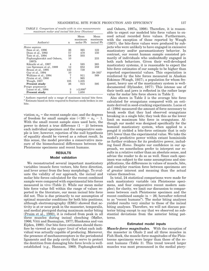
We reconstructed several important masticatoryvariables (muscle force vectors, bite force direction,and lever arms) from the bony morphology. To eval-uate the validity of our approach, the incisal andmolar bite forces calculated for the recent combinedsample were compared with experimental bite forcesmeasured in vivo (Table 2). While our mean molarbite force value fell within the range of values re-ported in the literature, our mean incisal bite forcedid not. This is due primarily to our assumption ofoptimal muscular conditions for both bite positions;although electromyography (EMG) showed that ac-tivity is at or near peak in the masseter, temporalis,and medial pterygoid during maximum molar biting(Pruim et al., 1980), it is reduced from peak in allthree muscles during incisal clenching (Møller,1966; Vitti and Basmajian, 1977; Blanksma and vanEijden, 1990). Our bite force estimates should there-fore be viewed as the upper limit of what each indi-vidual was actually capable of producing. Moreover,the presence of mechanoreceptors in the periodontalligaments and the pulp cavity that serve to protectthe dentition from damaging bite force levels is well-established (e.g., Hannam, 1969; Paphangkorakit
and Osborn, 1997a, 1998). Therefore, it is reason-able to expect our modeled bite force values to ex-ceed actual recorded force values. Furthermore,with the exception of those reported by Waugh(1937), the bite-force values were produced by sub-jects who were unlikely to have engaged in excessivemasticatory and/or paramasticatory behavior. Incontrast, our recent human sample consisted pri-marily of individuals who undoubtedly engaged inboth such behaviors. Given their well-developedmasticatory systems, it is reasonable to expect thebite-force estimates of our sample to be higher thanreported experimental values. This expectation isreinforced by the bite forces measured in AlaskanEskimos (Waugh, 1937), a population for whom fre-quent, heavy use of the masticatory system is well-documented (Hylander, 1977). This intense use oftheir teeth and jaws is reflected in the rather largevalue for molar bite force shown in Table 2.
Also shown in Table 2 is the average bite forcecalculated for orangutans compared with an esti-mate derived in seed-cracking experiments. Lucas etal. (1994) measured the amount of force necessary tobreak seeds that they had observed orangutansbreaking in a single bite; they took this as the lowerlimit on maximum bite force in orangutans. Al-though our model was designed specifically for thehominid masticatory system, when applied to apongid it yielded a bite-force estimate that is only18% lower than the experimental value. We take themodel’s predictive power within extant hominioidsas further evidence for its predictive power, includ-ing fossil Homo. Despite our confidence in our ap-proach, we nonetheless prefer to interpret our re-sults in a relative rather than an absolute sense, andadvise the reader to do so as well. Since each spec-imen was subject to the same assumptions and sim-plifications, the differences in values of muscle, bite,and condylar reaction force between specimens areof greater interest and meaning than the actualvalues themselves.
In total, 24 statistical comparisons were made foreach masticatory variable (six Pleistocene speci-mens, and four comparative recent modern sam-ples); for clarity, we limit our discussion to compar-isons between each Pleistocene specimen and therecent combined sample (n ! 26; hereafter referredto as “recent humans”). The molar biting analysesyielded results very similar to those of the incisalbiting analyses. Therefore, we will not discuss pos-terior biting except to say that we observed no sub-stantial deviations from the anterior biting pat-terns.
Estimated model inputsMuscle-force magnitudes. With the exception ofthe masseter in Ohalo 2 and all three muscles inFish Hoek, the muscle-force magnitudes of the Pleis-tocene specimens were larger than those of the re-cent humans (Table 3). This trend toward largermuscles was most pronounced in the medial ptery-
TABLE 2. Comparison of results with in vivo measurements:maximum molar and incisal bite force (Newtons)
Author(s) n
Meanmaximummolar Fb
Meanmaximumincisal Fb
Homo sapiensSinn et al., 1996 26 365 122Dean et al., 1992 57 478 150Tate et al., 1994 58 503Paphangkorakit and Osborn,
1997b10 233
Kikuchi et al., 1997 4 585 265van Spronsen et al., 1989 12 652 359Braun et al., 1995 142 738Gay et al., 1994 10 39–3921
Waltimo et al., 1994 7 911 569Pruim et al., 1980 7 965Waugh, 1937 105 1,235Present study 26 815 595
Pongo pygmaeusLucas et al., 1994 3 $2,0002
Present study 2 1,650
1 Authors reported only a range of maximum incisal bite force.2 Estimate based on force required to fracture seeds broken in onebite.
NEANDERTAL BITE FORCE PRODUCTION AND EFFICIENCY 137
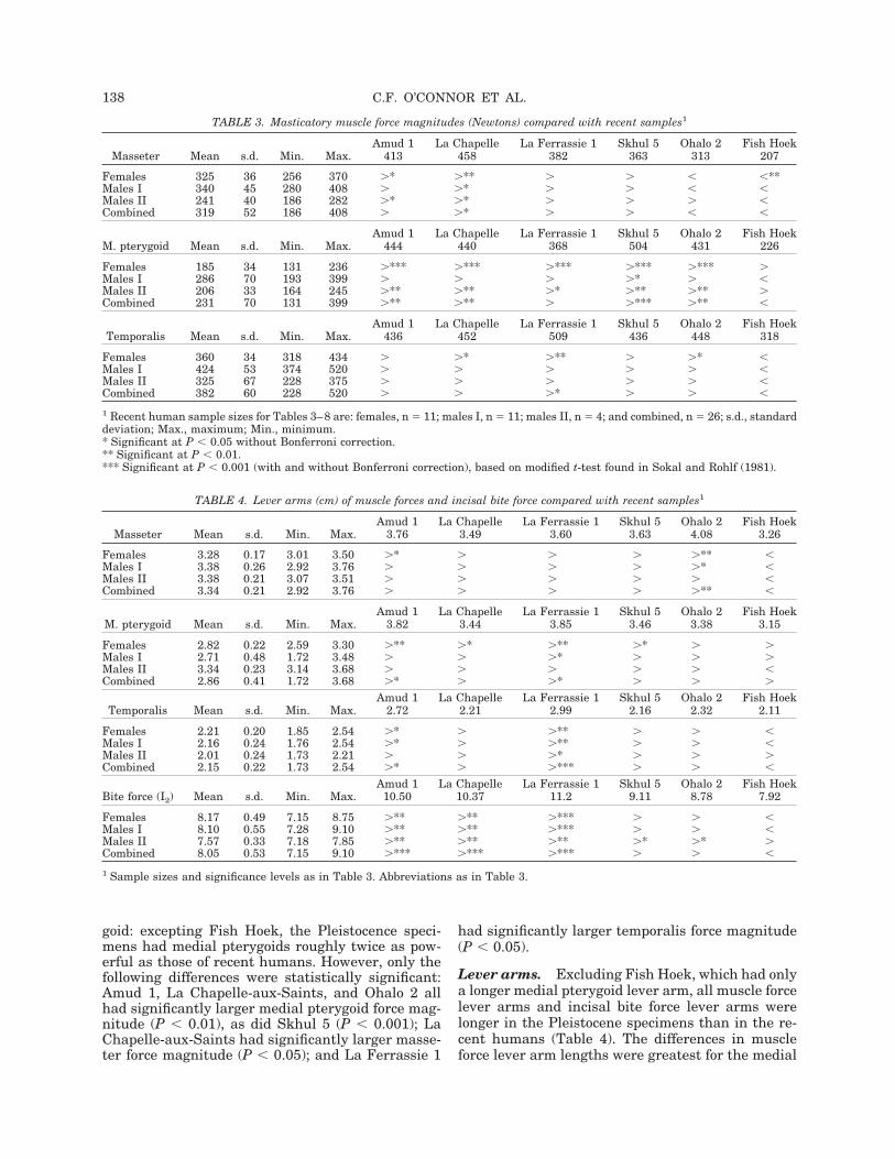
goid: excepting Fish Hoek, the Pleistocence speci-mens had medial pterygoids roughly twice as pow-erful as those of recent humans. However, only thefollowing differences were statistically significant:Amud 1, La Chapelle-aux-Saints, and Ohalo 2 allhad significantly larger medial pterygoid force mag-nitude (P # 0.01), as did Skhul 5 (P # 0.001); LaChapelle-aux-Saints had significantly larger masse-ter force magnitude (P # 0.05); and La Ferrassie 1
had significantly larger temporalis force magnitude(P # 0.05).
Lever arms. Excluding Fish Hoek, which had onlya longer medial pterygoid lever arm, all muscle forcelever arms and incisal bite force lever arms werelonger in the Pleistocene specimens than in the re-cent humans (Table 4). The differences in muscleforce lever arm lengths were greatest for the medial
TABLE 3. Masticatory muscle force magnitudes (Newtons) compared with recent samples1
Masseter Mean s.d. Min. Max.Amud 1
413La Chapelle
458La Ferrassie 1
382Skhul 5
363Ohalo 2
313Fish Hoek
207
Females 325 36 256 370 &* &** & & # #**Males I 340 45 280 408 & &* & & # #Males II 241 40 186 282 &* &* & & & #Combined 319 52 186 408 & &* & & # #
M. pterygoid Mean s.d. Min. Max.Amud 1
444La Chapelle
440La Ferrassie 1
368Skhul 5
504Ohalo 2
431Fish Hoek
226
Females 185 34 131 236 &*** &*** &*** &*** &*** &Males I 286 70 193 399 & & & &* & #Males II 206 33 164 245 &** &** &* &** &** &Combined 231 70 131 399 &** &** & &*** &** #
Temporalis Mean s.d. Min. Max.Amud 1
436La Chapelle
452La Ferrassie 1
509Skhul 5
436Ohalo 2
448Fish Hoek
318
Females 360 34 318 434 & &* &** & &* #Males I 424 53 374 520 & & & & & #Males II 325 67 228 375 & & & & & #Combined 382 60 228 520 & & &* & & #
1 Recent human sample sizes for Tables 3–8 are: females, n ! 11; males I, n ! 11; males II, n ! 4; and combined, n ! 26; s.d., standarddeviation; Max., maximum; Min., minimum.* Significant at P # 0.05 without Bonferroni correction.** Significant at P # 0.01.*** Significant at P # 0.001 (with and without Bonferroni correction), based on modified t-test found in Sokal and Rohlf (1981).
TABLE 4. Lever arms (cm) of muscle forces and incisal bite force compared with recent samples1
Masseter Mean s.d. Min. Max.Amud 1
3.76La Chapelle
3.49La Ferrassie 1
3.60Skhul 5
3.63Ohalo 2
4.08Fish Hoek
3.26
Females 3.28 0.17 3.01 3.50 &* & & & &** #Males I 3.38 0.26 2.92 3.76 & & & & &* #Males II 3.38 0.21 3.07 3.51 & & & & & #Combined 3.34 0.21 2.92 3.76 & & & & &** #
M. pterygoid Mean s.d. Min. Max.Amud 1
3.82La Chapelle
3.44La Ferrassie 1
3.85Skhul 5
3.46Ohalo 2
3.38Fish Hoek
3.15
Females 2.82 0.22 2.59 3.30 &** &* &** &* & &Males I 2.71 0.48 1.72 3.48 & & &* & & &Males II 3.34 0.23 3.14 3.68 & & & & & #Combined 2.86 0.41 1.72 3.68 &* & &* & & &
Temporalis Mean s.d. Min. Max.Amud 1
2.72La Chapelle
2.21La Ferrassie 1
2.99Skhul 5
2.16Ohalo 2
2.32Fish Hoek
2.11
Females 2.21 0.20 1.85 2.54 &* & &** & & #Males I 2.16 0.24 1.76 2.54 &* & &** & & #Males II 2.01 0.24 1.73 2.21 & & &* & & &Combined 2.15 0.22 1.73 2.54 &* & &*** & & #
Bite force (I2) Mean s.d. Min. Max.Amud 1
10.50La Chapelle
10.37La Ferrassie 1
11.2Skhul 5
9.11Ohalo 2
8.78Fish Hoek
7.92
Females 8.17 0.49 7.15 8.75 &** &** &*** & & #Males I 8.10 0.55 7.28 9.10 &** &** &*** & & #Males II 7.57 0.33 7.18 7.85 &** &** &** &* &* &Combined 8.05 0.53 7.15 9.10 &*** &*** &*** & & #
1 Sample sizes and significance levels as in Table 3. Abbreviations as in Table 3.
138 C.F. O’CONNOR ET AL.

pterygoid, although significant only for Amud 1 andLa Ferrassie 1 (P # 0.05). Other significant differ-ences included Ohalo 2’s longer masseter lever arm(P # 0.01), and the longer temporalis lever arm ofAmud 1 (P # 0.05) and La Ferrassie 1 (P # 0.001).Overall, the differences in incisal bite force leverarm lengths relative to recent humans were morepronounced, especially for the Neandertals (P #0.001 for all three). No other differences were sta-tistically significant.
Force production capability
The magnitudes of total muscle force (Fm), totalbite force (Fb), and total condylar reaction force (Fc)for bilateral incisal biting were substantially largercompared to the recent humans with the exceptionof Fish Hoek, which had uniformly smaller values(Table 5). However, while Amud 1, La Ferrassie 1,Skhul 5 (P # 0.05), and La Chapelle-aux-Saints (P #0.01) all had significantly higher muscle-force mag-nitudes than the recent humans, only Skhul 5 wascapable of generating a significantly higher biteforce (P # 0.05). Further, only the Neandertals had
significantly larger condylar reaction forces (P #0.05).
Force-production efficiencyForce ratios. Despite substantial differences inforce magnitudes between the Pleistocene speci-mens and recent humans, only minor differences inforce ratios were observed (Table 6). Collectively, theNeandertal sample exhibited a pattern of lower biteto muscle force ratios (Fb/Fm) and higher condylarreaction to muscle force ratios (Fc/Fm). They there-fore appeared to be slightly less efficient at produc-ing incisal bite force than the recent humans. Com-pared to Neandertals, Ohalo 2 and Fish Hoek hadslightly higher bite to muscle force ratios (Fb/Fm)and lower condylar reaction to muscle force ratios(Fc/Fm) than the recent humans. This indicates thatfor an equal amount of muscle force, Ohalo 2 andFish Hoek were able to generate slightly higher biteforces and slightly lower condylar reaction forcesthan recent humans, making them somewhat moreefficient incisal biters overall. Finally, Skhul 5 ex-hibited a mixed pattern: both a lower bite to muscle
TABLE 5. Force production capability: force magnitudes (Newtons) compared with recent samples (left/right side summed)1
Fm Mean s.d. Min. Max.Amud 1
2587La Chapelle
2700La Ferrassie 1
2516Skhul 5
2606Ohalo 2
2382Fish Hoek
1500
Females 1,742 162 1,467 2,024 &*** &*** &*** &*** &*** #**Males I 2,098 252 1,714 2,528 & &* & & & #*Males II 1,545 154 1,319 1,652 &** &** &* &** &* #Combined 1,862 293 1,319 2,528 &* &** &* &* & #
Fb Mean s.d. Min. Max.Amud 1
790La Chapelle
752La Ferrassie 1
723Skhul 5
826Ohalo 2
791Fish Hoek
481
Females 552 68 440 678 &** &* &* &** &** #Males I 662 84 543 780 & & & & & #Males II 533 81 439 632 & & & &* & #Combined 595 94 439 780 & & & &* & #
Fc Mean s.d. Min. 1,053Amud 1
1385La Chapelle
1479La Ferrassie 1
1463Skhul 5
1353Ohalo 2
1039Fish Hoek
692
Females 945 89 756 1,053 &*** &*** &*** &** & #*Males I 1,139 138 916 1,380 & &* &* & & #*Males II 738 89 645 857 &** &** &** &** & #Combined 995 180 645 1,380 &* &* &* & & #
1 Sample sizes and significance levels as in Table 3. Abbreviations as in Table 3.
TABLE 6. Force production efficiency: force ratios (%) in fossils compared with recent samples1
Fb/Fm ' 100% Mean s.d. Min. Max.Amud 1
30.5La Chapelle
27.9La Ferrassie 1
28.7Skhul 5
31.7Ohalo 2
33.2Fish Hoek
32.1
Females 31.6 2.0 29.3 34.7 # # # & & &Males I 31.6 1.9 28.9 34.6 # # # & & &Males II 34.5 3.0 31.2 38.3 # # # # # #Combined 32.0 2.3 28.9 38.3 # # # # & &
Fc/Fm ' 100% Mean s.d. Min. Max.Amud 1
53.5La Chapelle
54.8La Ferrassie 1
58.1Skhul 5
51.9Ohalo 2
43.6Fish Hoek
46.1
Females 54.3 2.8 50.5 58.5 # & & # #** #*Males I 54.4 3.1 48.8 59.0 # & & # #** #*Males II 47.8 4.0 43.0 52.5 & & & & # #Combined 53.3 3.8 43.0 59.0 & & & # #* #
1Sample sizes and significance levels as in Table 3. Abbreviations as in Table 3.
NEANDERTAL BITE FORCE PRODUCTION AND EFFICIENCY 139

force ratio (Fb/Fm), like the Neandertals, and alower condylar reaction to muscle force ratio (Fc/Fm), like Ohalo 2 and Fish Hoek. We should empha-size that all differences with recent humans wereminor, and only one reached statistical significance(Fc/Fm for Ohalo 2; P # 0.05).
Mechanical advantage. As with the force ratios,few differences in muscle mechanical advantageswere statistically significant. Although lever arms inthe Pleistocene specimens were generally longerthan in the recent humans, in several instances theincisal bite force lever arm was disproportionatelylonger than a given muscle force lever arm. Themechanical advantage of such a muscle was there-fore found to be smaller than that of the same mus-cle in recent humans (Table 7). This was the case forthe masseter in Amud 1, La Chapelle-aux-Saints(P # 0.01), La Ferrassie 1 (P # 0.01), and Skhul 5;for the medial pterygoid in La Chapelle-aux-Saintsand La Ferrassie 1; and for the temporalis in allPleistocene specimens, although significant only forLa Chapelle-aux-Saints (P # 0.05). The smaller val-ues of mechanical advantage indicate that thesemuscles were less well-positioned for efficient incisalbite force production than in the recent humans. Thelarger values of mechanical advantage in the re-maining cases (masseter in Ohalo 2 and Fish Hoek,and medial pterygoid in all specimens except LaChapelle-aux-Saints and La Ferrassie 1) indicatethat these muscles were better-positioned than inthe recent humans (not significant).
We can conclude that those individuals with uni-formly smaller mechanical advantages, such as LaChapelle-aux-Saints and La Ferrassie 1, were lessefficient producers of incisal bite force. The mechan-ical advantages of the remaining Pleistocene speci-mens are neither uniformly smaller nor uniformlylarger. Therefore, we cannot make any definitive
conclusions concerning their efficiency based solelyon mechanical advantage.
Differential loading of the condyles
For unilateral incisal biting, the magnitudes ofthe working-side condylar reaction force (Fc,WORK)and the balancing-side condylar reaction force(Fc,BAL) were consistently larger in the Pleistocenespecimens (again with the exception of Fish Hoek)than in the recent humans (Table 8). The differenceswere most pronounced in the Neandertals andSkhul 5: Amud 1, La Chapelle-aux-Saints, and LaFerrassie 1 had significantly larger working-sidecondylar reaction forces (P # 0.05), while Amud 1,La Chapelle-aux-Saints, and Skhul 5 had signifi-cantly larger balancing-side condylar reaction force(P # 0.05). In all specimens, both Pleistocene andrecent, the balancing-side condylar reaction forcewas greater than the working-side condylar reactionforce.
None of the differences in the ratio of working-sideto balancing-side condylar reaction force (Fc,WORK/Fc,BAL) were statistically significant. The Neander-tals and Fish Hoek did have slightly higher ratiosthan the recent humans (i.e., closer to unity), indi-cating that they were able to more evenly distributethe joint reaction force between the two condylesduring nonsymmetric incisal biting. In contrast, thebalancing-side condyle of Skhul 5 and Ohalo 2 sus-tained a larger portion of the total reaction force, asevidenced by their lower Fc,WORK/Fc,BAL ratio.
Relationship of masticatory variables to overallsize and robusticity
The distribution of overall size measured by thegeometric mean for all specimens is shown in Figure4. Not surprisingly, the Neandertals were amongthe largest, although the geometric mean of two
TABLE 7. Force production efficiency: muscle mechanical advantage (MA) compared with recent samples1
MAMASS Mean s.d. Min. Max.Amud 1
0.358La Chapelle
0.336La Ferrassie 1
0.321Skhul 5
0.398Ohalo 2
0.465Fish Hoek
0.411
Females 0.403 0.024 0.359 0.425 # #* #** # &* &Males I 0.418 0.025 0.378 0.470 #* #* #** # & #Males II 0.447 0.029 0.414 0.484 # #* #* # & #Combined 0.416 0.028 0.359 0.484 # #** #** # & #
MAMPT Mean s.d. Min. Max.Amud 1
0.364La Chapelle
0.332La Ferrassie 1
0.344Skhul 5
0.380Ohalo 2
0.384Fish Hoek
0.398
Females 0.347 0.036 0.305 0.412 & # # & & &Males I 0.335 0.054 0.219 0.416 & & & & & &Males II 0.442 0.026 0.415 0.469 # #* #* # # #Combined 0.356 0.056 0.219 0.469 & # # & & &
MATEMP Mean s.d. Min. Max.Amud 1
0.259La Chapelle
0.213La Ferrassie 1
0.267Skhul 5
0.237Ohalo 2
0.264Fish Hoek
0.267
Females 0.271 0.025 0.241 0.310 # # # # # #Males I 0.266 0.022 0.224 0.289 # #* & # # #Males II 0.266 0.037 0.220 0.307 # # & # # #Combined 0.268 0.024 0.220 0.310 # #* # # # #
1 Sample sizes and significance levels as in Table 3. Abbreviations as in Table 3.
140 C.F. O’CONNOR ET AL.

recent specimens (one Ohlone male and the Austra-lian Aboriginal male) approached that of LaChapelle-aux-Saints. As the largest specimen, LaFerrassie 1 was 44% larger in raw (i.e., unlogged)metric units than the smallest recent human (anIndian subcontinent male) and 31% larger than thesmallest Pleistocene specimen (Fish Hoek). Theearly modern Skhul 5 specimen was approximately10% smaller than the Neandertals and was virtuallyidentical to the mean of the Recent Male I sample.The Ohlone females were comparatively quite large:Ohlone males averaged only 2% larger than thefemales, and Ohalo 2, considered to be a relativelylarge, robust male by Nadel and Hershkovitz (1991),was actually smaller than most Ohlone females. The
diminutive Fish Hoek specimen was smaller than allbut one of the recent modern specimens (a verysmall and gracile Indian subcontinent male).
Correlations between each masticatory variableand overall size across our total sample (Pleistoceneand recent hominids combined) are shown in Table9. All force magnitudes were significantly correlatedwith overall size, as were muscle force magnitudesand all force lever arms except that of the medialpterygoid. In contrast, all force ratios and musclemechanical advantages were statistically uncorre-
TABLE 8. Differential loading patterns of condyles in fossils compared with four recent samples1
FC,WORK Mean s.d. Min. Max.Amud 1
634La Chapelle
663La Ferrassie 1
621Skhul 5
586Ohalo 2
454Fish Hoek
307
Females 420 39 345 460 &*** &*** &*** &** & #*Males I 509 62 399 620 & &* & & # #*Males II 322 46 281 385 &** &** &** &* & #Combined 443 83 281 620 &* &* &* & & #
FC,BAL Mean s.d. Min. Max.Amud 1
742La Chapelle
782La Ferrassie 1
732Skhul 5
744Ohalo 2
581Fish Hoek
376
Females 516 51 411 593 &** &*** &** &** & #*Males I 617 74 491 734 & & & & # #*Males II 412 40 364 462 &** &** &** &** &* #Combined 543 94 364 734 &* &* & &* & #
FC,WORK/FC,BAL' 100% Mean s.d. Min. Max.
Amud 185.4
La Chapelle84.8
La Ferrassie 184.8
Skhul 578.8
Ohalo 278.1
Fish Hoek81.6
Females 81.3 2.6 75.8 84.7 & & & # # &Males I 82.6 2.3 79.8 87.6 & & & # # #Males II 78.0 4.2 73.0 83.3 & & & & & #Combined 81.3 3.1 73.0 87.6 & & & # # &
1 Sample sizes and significance levels as in Table 3. Abbreviations as in Table 3.
Fig. 4. Distribution of overall cranio-mandibular size as mea-sured by geometric mean for all Pleistocene and recent humanspecimens. Geometric mean was calculated for each individual,using linear distances from porion to all 37 digitized points. Fossilhominid abbreviations as in Figure 5.
TABLE 9. Correlations between masticatory variables andgeometric mean1
rs
Muscle-force magnitudesMasseter 0.815**M. pterygoid 0.385*Temporalis 0.656**
Lever armsMasseter 0.398*M. pterygoid 0.244Temporalis 0.503**Bite force (I2) 0.575**
Force-productioncapabilityFm 0.702**Fb 0.672**Fc 0.725**
Force-production efficiencyFb/Fm $0.164Fc/Fm 0.235
MAMASS $0.318MAMPT $0.191MATEMP 0.039Differential loading
Fc,WORK 0.688**Fc,BAL 0.717**Fc,WORK/Fc,BAL 0.219
1 Calculated from total sample (fossils and recent humans com-bined, n ! 32), using Spearman’s rank correlation.* Significant at P # 0.05.** Significant at P # 0.01.
NEANDERTAL BITE FORCE PRODUCTION AND EFFICIENCY 141

lated with overall size. In other words, force-produc-tion capability of the masticatory system scaled withincreasing cranio-mandibular size, but force-produc-tion efficiency was maintained throughout the sizerange (Figs. 5, 6).
While measures of efficiency were statistically un-correlated with overall size and robusticity acrossour sample, the signs of the correlations (i.e., regres-sion slopes) in most cases suggest that efficiency andsize/robusticity were inversely related (Fig. 6). Thatis, larger, more robust individuals appeared to beslightly less efficient than smaller, gracile hominids.Larger samples sizes might well result in statisticalsignificance for this patterning; however, the rela-tively tighter coupling of force magnitudes with size/robusticity compared with efficiency measures is un-likely to change.
DISCUSSION
Model validity
In our model, we assumed that all three jaw ele-vators were maximally active during both bilateraland unilateral isometric incisal biting, that therewere no regional differences of activation withineach muscle, and that there was no antagonisticmuscle activity. Electromyographic studies of themasticatory muscles show that this is generally notthe case. During incisal clenching, activity in themasseter, medial pterygoid, and especially tempora-lis is reduced from peak (Møller, 1966; Vitti andBasmajian, 1977; Blanksma and van Eijden, 1990;Spencer, 1998); the lateral pterygoid is active (Hy-lander, 1992); and at least two functional portionsare distinguishable in the temporalis and masseter(Blanksma and van Eijden, 1990). Furthermore, Hy-lander and Johnson (1985) and Ross and Hylander(2000) showed that differential muscle activity dur-ing incision occurs, with the superficial masseterexhibiting relatively higher activity than that foundin the anterior temporalis. In addition, during uni-lateral biting, working-side muscle activity is typi-cally greater than balancing-side (van Eijden et al.,1993). In the context of the present study, however,little would have been gained by reducing muscleforces to coincide with EMG patterns, since: therelationship between EMG activity and muscle forceis tenuous; only substantial differences in muscleactivation between the Pleistocene and recent com-parative specimens might have changed our results,and there is no way to determine what such differ-ences might be; and our goal was to estimate theupper bounds of potential bite force.
We also assumed that the muscle force vectorscould be adequately estimated from cranial andmandibular measurements. The “straight-line” ap-proach used here to determine muscle force direc-tion and point of application from bony attachmentareas is frequently used in biomechanical analyses(for masticatory biomechanics, see Pruim et al.,1980; Osborn and Baragar, 1985, 1992; Koolstra et
Fig. 5. Force magnitudes vs. overall size (geometric mean) forbilateral incisal biting. Magnitudes of bite force (Fb), condylarreaction force (Fc), and resultant muscle force (Fm) are plotted vs.geometric mean for each Pleistocene and recent modern speci-men. Regression lines were obtained using least-squares. Allforce magnitudes were highly significantly correlated with overallsize (P # 0.0001). Solid triangles, Neandertals; solid squares,Pleistocene modern; open circles, recent females; open diamonds,recent males. Sk5, Skhul 5; Oh2, Ohalo 2; FH, Fish Hoek; Am1,Amud 1; LCh, La Chapelle; LF1, La Ferassie 1.
142 C.F. O’CONNOR ET AL.

al., 1988; Koolstra and van Eijden, 1995; Trainor etal., 1995; Osborn, 1996; for lower extremity biome-chanics, see Brand et al., 1982; Herzog and Read,1993; Glitsch and Baumann, 1997; for lumbar spinebiomechanics, see McGill and Norman, 1986). Al-though functional units and other complexities areignored by this approach, it provides a reliable first-order approximation of muscle-force direction andapplication point. Predicting muscle-force magni-tude from bony morphology has proven more diffi-cult (and controversial). Although the linear corre-lation between muscle-force magnitude and musclePCSA during isometric contraction is well-estab-
lished (Weijs and Hillen, 1985b; Koolstra et al.,1988), the relationship between muscle PCSA andbony morphology is not. It is generally accepted thatlarge, robust muscle scars reflect large muscles, butthe precise nature of this relationship is not well-understood. This is especially true for multipennatemuscles.
Anton (1994b, 1996b) investigated the extent towhich bone proxies can predict masticatory musclePCSA in modern humans and in several species ofmacaques. She found that bony measures of thetemporalis, masseter, and medial pterygoid wereonly weakly correlated with their respective PCSAs.Anton (1994b, 1996b) therefore concluded that boneproxies are better suited for testing hypotheses re-lated to relative rather than absolute force produc-tion, and that they should be used with care, partic-ularly when applied to fossil remains. Given thatour “raw PCSAs” were quite similar, although notidentical, to those used in her studies, her caution-ary remarks apply to the present study.
Because a reliable method for its measurementdoes not yet exist, deriving realistic values for mus-cle-force magnitude is a problem common to mostbiomechanical analyses. There are clearly addi-tional difficulties when attempting to do so for fossilhominids. Cadaver studies can determine whichskeletal measures, if any, predict muscle PCSA inrecent humans, but will these same measures holdfor fossils? Assuming they do, is the value of intrin-sic muscle strength the same? And finally, are theredifferences in internal muscle architecture that mayaffect force production? Given the relatively closephylogenetic relationship between the archaic andmodern humans used in the present study, the an-swer to the first two questions is most likely yes. Inan attempt to answer the third question, Anton(1996a) examined the tendon-associated features ofmasticatory muscles in Neandertal and recent mod-ern human crania. She found bony evidence that theanterior portion of the temporalis and the deep por-tion of the masseter were larger and more defined inNeandertals than in modern humans. Consideringfiber orientation, which is predominantly vertical inthe anterior temporalis and lateral in the deep mas-seter, our results can be reconciled with her find-ings: the vertical force component of temporalis andthe lateral force component of masseter were pro-portionately larger in the Neandertals than in therecent humans.
Together with the generally good agreement ofour results with published values of bite force (Table2), we take this as evidence that our method cap-tured the most important aspects of masticatoryforce production. We attribute this to: the use ofmultiple points, recorded with a three-dimensionaldigitizer, to establish each model parameter; theconsideration of both the cranium and mandible inour analysis; and carrying out calculations in threedimensions. While these steps also served to mini-mize measurement and calculation error, we recog-
Fig. 6. Force ratios vs. overall size (geometric mean) for bi-lateral incisal biting. Ratio of bite-force magnitude to resultantmuscle-force magnitude (Fb/Fm), and condylar reaction forcemagnitude to resultant muscle force magnitude (Fc/Fm), are plot-ted vs. geometric mean for each Pleistocene and recent modernspecimen. Regression lines were obtained using least-squares.Neither force magnitude ratio was significantly correlated withoverall size (P ! 0.3379 and 0.2049, respectively), although weaktrends toward inefficiency in larger specimen are indicated. Notethat an efficient system maximizes bite force (i.e., high Fb/Fm)and minimizes condylar reaction force (i.e., low Fc/Fm). Symboldesignations as in Figure 5. Fossil hominid abbreviations as inFigure 5.
NEANDERTAL BITE FORCE PRODUCTION AND EFFICIENCY 143

nize that the potential error associated with ourestimation of muscle-force magnitude remains. Thiserror can be mitigated, however, if the results con-cerning force-production capability are viewed in arelative framework. That is, since each specimenwas subject to the same assumptions and simplifi-cations, differences between specimens are primar-ily a reflection of true differences in force magni-tudes rather than of methodology. Alternatively,this error can be completely eliminated if only mus-cle mechanical advantages are considered, althoughwe feel valuable information is eliminated by doingso.
Discussion of results and comparison withprevious studies
In the introduction, we posed four questions re-lated to certain aspects of the ADLH. We will discussthe results of our study in the context of those ques-tions and, when possible, compare our results withprevious work.
Force-production capability
In contrast to the expectation set forth by theADLH, our results indicate that Neandertals werenot capable of generating significantly larger ante-rior dental loads compared with early modern andrecent modern humans. In fact, the primary dichot-omy in all measures of force-production capabilitywas not between Neandertal and modern specimens,but rather between large, robust vs. small, gracilespecimens. Estimates of incisal bite force, muscleforce, and condylar reaction force magnitudes werevery similar among the largest Pleistocene speci-mens, Skhul 5 and the Neandertals (Table 5; Figs. 5,7). Further, several recent humans with comparablylarge geometric means had equally large force esti-mates. Most pertinent to the ADLH, the incisal biteforce magnitude of all three Neandertals was ex-ceeded by Skhul 5 and Ohalo 2 and, in the case of LaChapelle-aux-Saints and La Ferrassie 1, by severalOhlone males. Therefore, the assumption that Ne-andertals were generating exceptionally heavy an-terior dental loads compared with both early modernand recent modern humans appears unwarranted.We note that Bonferroni corrections to the pairwisecomparisons noted at the bottom of Tables 3–8 onlyserve to further minimize force magnitude and otherdifferences between the Neandertals and the com-parative sample, and that our discussion of uncor-rected values (above) is thus actually conservativewith respect to this question.
Our results for force-production capability do notagree completely with those of Anton (1990, 1994b).While our muscle force and condylar force estimateswere similar to hers, our bite force estimate was 44%higher (Table 10). This difference results from twofactors. First, Anton (1990, 1994b) combined theforce vectors of the masseter and medial pterygoid,assuming the same lever arm for both. Second, she
moved the combined vector closer to the condylewithout preserving its angular effect on the system.Both of these simplifications reduced the momentgenerated by the masseter and medial pterygoidabout the condyle, resulting in artificially low valuesof bite force (and higher values of condylar reactionforce). The net effect is disproportionately greater inNeandertals than in recent humans due to the mag-nitude of lever arms in these muscles. This studydoes not support the low Fb found for Amud 1 byAnton (1990, 1994b); however, it should be notedthat this fossil is still modeled here as producing lessbite force per muscle force (Table 6) than our modernsample and a relatively large amount of condylarreaction force. Thus, her conclusion that Neandertalfacial architecture may not be a result of high-mag-nitude occlusal loads is supported here by our en-hanced dataset, although the increased comparativesample size suggests there is less difference in effi-ciency than she suggested (see below).
Fig. 7. Bar graph illustrating force magnitudes (top) andforce ratios (bottom) for bilateral incisal biting for each Pleisto-cene individual and recent combined sample. Large values formuscle force resultant (Fm) and condylar reaction (Fc) forces areevident for Neandertals and Skhul 5. In contrast, Neandertal biteforce values (Fb) are not remarkable. Bottom graph illustratesslightly lower values of force production efficiency (i.e., forceratios) for Neandertals.
144 C.F. O’CONNOR ET AL.

Force-production efficiency
We found that Neandertals were neither consid-erably more nor less efficient at generating anteriordental loads compared with early modern and recentmodern humans. In fact, our results suggest that themasticatory system in the genus Homo scales suchthat force-production efficiency is maintained acrossa considerable range of size and robusticity. Whilemuscle, bite, and condylar reaction force magnitudesincreased predictably with the geometric mean, theratios of these force magnitudes remained relativelyconstant (Table 9; Figs. 6, 7). Similarly, muscle andbite force lever arms were positively correlated withthe geometric mean, but the ratios of the two, ormuscle mechanical advantages, were not (Table 9).
There was a trend, although not statistically sig-nificant, toward decreasing efficiency with increas-ing size (Table 9; Fig. 6). Interestingly, Hylander(1985) observed that body-size increase in macaquesis associated with a decreased ability to generatemasticatory muscle force and an increased level ofrecruitment of balancing-side muscle force. Subse-quent studies (Hylander et al., 1998, 2000), how-ever, did not find this size relationship. Nonetheless,given the rather prognathic lower faces and conse-quently long bite force lever arms of the larger Pleis-tocene individuals in our study, this trend for de-creased efficiency with larger overall facial size isnot unexpected. We propose that their substantialmedial pterygoids compensated for their increasedprognathism, thereby allowing them to operate witha similar efficiency as the less prognathic specimens.This interpretation is supported by the work of
Weijs and Hillen (1986), who found that the cross-sectional area of medial pterygoids in recent hu-mans was most strongly correlated with measures ofmandibular length. This also underscores the impor-tance of considering muscle size as well as muscleleverage; an increase in the former can compensatefor a decrease in the latter.
Considering the differences in force magnitudes,our force ratios for Amud 1 differed predictably fromthose of Anton (1990, 1994b) (Table 10). Because ourbite force estimate was considerably higher thanhers, our bite to muscle force ratio (Fb/Fm) was alsohigher. Similarly, because our condylar reactionforce estimate was lower, so too was our condylarreaction to muscle force ratio (Fc/Fm). Amud 1 ap-peared considerably more efficient at incisal bitingin the present analysis than in the previous, for thesame reasons given above.
A comparison of our estimates of muscle mechan-ical advantage (MA) with those of Spencer andDemes (1993) reveals two important differences (Ta-ble 10). First, our values are considerably smaller.Second, while Spencer and Demes (1993) found themasseter to have a considerably larger MA than thetemporalis and medial pterygoid, we instead foundthe medial pterygoid and masseter to have a com-parable MA (both larger than the temporalis MA).These differences do not result from differences inthe biomechanical models used. To borrow terminol-ogy from Spencer (1998), we employed an “uncon-strained” lever model of mastication, whereas Spen-cer and Demes (1993) employed a “constrained”lever model. Briefly, the former places no restric-tions on muscle activity, whereas the latter assumesthat there is a zone in which balancing-side muscleactivity must decrease as the bite point moves pos-teriorly (for a detailed description of this model, seeSpencer and Demes, 1993; Spencer, 1998). However,as this zone corresponds roughly to the molar region,there is essentially no difference between the twomodels for incisal biting; neither predicts the exper-imentally observed reduction in muscle activity foranterior biting. We feel that the differences in MAvalues are instead due to the use by Spencer andDemes (1993) of anteroposterior muscle positionrather than muscle lever arm to calculate MA; ge-ometry informs us that the former will always begreater than the latter.3 Further, they used only onepoint, the anteriormost cranial attachment point, toassess muscle position; in essence, they character-ized the leverage of each muscle by the cranial po-sition of its anteriormost fiber. In contrast, we usedthe line-of-action of a centroidal fiber to characterizemuscle leverage, an approach that accounts for mus-cle origin, insertion, and distribution.
3The anteroposterior muscle position, muscle lever arm, and muscleline-of-action form a right triangle, with anteroposterior muscle posi-tion as the hypotenuse (Fig. 2).
TABLE 10. Comparison of results for Neandertals withprevious work
Force-production capability(Newtons)
Fm Fc Fb
Anton (1990, 1994b)1 (n ! 1) 2,240 1,500 550Present study2 (n ! 1) 2,587 1,385 790% difference (15% $8% (44%
Force-productionefficiency
Fb/Fm Fc/Fm
Anton (1990, 1994b)1 (n ! 1) 0.245 0.670Present study2 (n ! 1) 0.305 0.535% difference (6% $14%
MAMASS MAMPT MATEMP
Spencer and Demes (1993)3
(n ! 8)0.626 0.456 0.461
Present study4 (n ! 3) 0.338 0.347 0.246% difference $29% $11% $22%
1 Amud 1 only; Fm was recalculated from results of Anton (1990,1994b) to correspond with our definition (i.e., sum of muscle forcemagnitudes rather than magnitude of their vector sum).2 Amud 1 only.3 Average of Amud 1, La Chapelle 2, La Ferrassie 1, Monte Circeo1, Saccopastore 1, Gibraltar 1, Tabun C1, and Shanidar 1.4 Average of Amud 1, La Chapelle 2, and La Ferrassie 1.
NEANDERTAL BITE FORCE PRODUCTION AND EFFICIENCY 145

Despite the differences in absolute values of MA,we find several points of agreement with Spencerand Demes (1993). Their results, like ours, showedthat the temporalis and masseter operated less effi-ciently in Neandertals than in recent humans dur-ing incisal biting, while the medial pterygoid oper-ated with nearly the same efficiency. Measures ofoverall system efficiency (i.e., unweighted andweighted averages of muscle MA) were also lower inNeandertals than in recent humans. Compared withearly Homo, however, they found Neandertals tohave slightly larger values of MA for all three mus-cles. Because Spencer and Demes (1993) did notevaluate overall size, we can only speculate that thisslight increase in efficiency may be related to a mod-est decrease in overall craniofacial size from earlyHomo to Neandertals (Trinkaus, 1987, 2003; Fran-ciscus, 1995; Franciscus and Trinkaus, 1995). Ourdiffering conclusions regarding the ADLH result pri-marily from the difference in our comparative sam-ples.
Differential loading patterns of the condyles
As expected, the balancing-side condyle was moreheavily loaded than the working-side in all speci-mens during nonsymmetric biting. As evidenced bythe ratio of the two reaction forces (Table 8), thisdifferential loading was the least pronounced in Ne-andertals. That is, the total condylar reaction forcewas more evenly distributed between the two con-dyles in Neandertals than in early modern or recentmodern humans, due to their relatively wider inter-condylar distance. The fact that intercondylar dis-tance affects the load distribution on the condyles(with relatively wide intercondylar distance beingadvantageous) is consistent with observations byHylander (1975, 1977) in humans, and Smith (1978)across anthropoids. Osborn (1996), using a linearprogramming model to study how changes in themandible affect unilateral maximum bite forces,also found that increased intercondylar distance re-sulted in more evenly distributed loads between thetwo condyles. Because the ratio of the distance be-tween the incisors to distance between the condylesis relatively small in Neandertals, the nonsymmet-ric biting case approaches the symmetric biting case,in which the left- and right-side condylar reactionforces must be equal. Because condylar reactionforces were typically high in Neandertals (Table 5),this may have prevented the balancing-side condylefrom being overloaded during unilateral biting.
It should be noted that the large intercondylardistance in Neandertals is functionally and develop-mentally related to their wide cranial bases, thelatter a symplesiomorphy shared with earlier Homo.In light of the above, it is possible that Neandertalcranial base and intercondylar widths, in particular,may have been mechanically constrained to agreater degree than other aspects of their more de-rived craniofacial morphology.
Craniofacial size and implications for the ADLH
It is not difficult to accept that some Neandertalfeatures are, at least in part, related to masticatorybiomechanics. The difficulty comes in describingprecisely which specific features these are, and whatspecific aspect(s) of masticatory biomechanics theymay be related to. Perhaps to avoid this difficulty,the ADLH has historically been set forth in ratherbroad terms: force generation, force dissipation,high-magnitude loading, and repetitive loading haveall been implicated (often simultaneously) in Nean-dertal facial architecture. We feel more precise lan-guage is required, since each of these aspects ofmasticatory biomechanics requires a different bio-mechanical model and/or approach in order to beproperly examined, and each may have resulted indifferent adaptive responses. Force generation canbe treated with rigid body mechanics (e.g., thepresent study; Anton, 1990, 1994b; Spencer andDemes, 1993), while force dissipation must betreated with deformable body mechanics. This canbe done computationally using finite element anal-ysis or experimentally using in vivo and in vitrostrain gages (Hylander et al., 1991; Hylander andJohnson, 1997). High-magnitude loading can be ex-amined with either rigid or deformable body me-chanics, depending on whether the ability to gener-ate loads or the ability to dissipate loads is at issue.Repetitive loading requires a more complicated ap-proach; finite-element models that incorporate bone-remodeling algorithms were able to clarify the tissueresponse to cyclical loads (Beaupre et al., 1990a,b),but this represents adaptations in an individualthrough developmental plasticity rather than evolu-tionary adaptations in a lineage. Given the multipleinterpretations of the ADLH, we do not (indeed can-not) fully reject it on the basis of this study.
We do, however, challenge the direct associationof high-magnitude anterior dental loads and Nean-dertal facial architecture. The amount of incisal biteforce the Neandertals were able to generate underoptimal muscular conditions was not significantlygreater than that generated by recent humans, andwas approximately equal to that generated by Skhul5 and Ohalo 2. Nor did the Neandertals appearconsiderably more (or less) efficient at generatinganterior dental loads; because their muscle positionswere not particularly mechanically advantageous,their increased muscle size appeared only to main-tain, rather than improve, force-production effi-ciency. Studies that investigated the relationshipbetween bite force and craniofacial morphology inrecent humans (Waltimo et al., 1994; Tuxen et al.,1999; Raadsheer et al., 1999) demonstrated thatfacial features typically associated with high biteforces (e.g., reduced facial height) are, in fact, notthose that characterize Neandertal facial morphol-ogy. Further, several studies showed that overallcraniofacial size has a greater influence on bite forcethan individual features (Raadsheer et al., 1996,
146 C.F. O’CONNOR ET AL.

1999), and the fact that large bite forces are pro-duced by large (not necessarily high) faces is consis-tent with our results.
Given the relationship between overall size andbite force production documented here, and theslight decrease in Neandertal facial size relative toMiddle Pleistocene hominids (Franciscus, 1995;Franciscus and Trinkaus, 1995; Trinkaus, 2003), weargue that Neandertals likely also underwent amodest decrease in force production relative to theirMiddle Pleistocene precursors. If this same relation-ship between masticatory forces and overall sizeexists for Middle Pleistocene hominids as it doesacross the wide range of variation exhibited by Ne-andertals, early modern humans, and recent hu-mans, an evolutionary trend toward a decrease ingenerated masticatory forces should be expectedfrom the Middle to Late Pleistocene.
Although we did not specifically examine the issueof force dissipation, we are of the opinion that ifNeandertals were not generating exceptionallyheavy anterior dental loads, then it is unlikely thatcertain of their features were adaptations to dissi-pate them. We instead feel that at least some of theirfeatures (e.g., lateral condylar tubercles) may repre-sent adaptations to dissipate muscle and condylarreaction forces, which were exceptionally heavy.
The absence of significantly higher levels of ante-rior bite force among Neandertals indicates thatincreased dental attrition on the anterior teeth ismore likely a result of repetitive use. This couldoccur either as the result of repetitive vertical occlu-sal forces at low loads, which can have a large effecton bone remodeling (Lanyon and Rubin, 1984), ormore obliquely oriented wear that induces incisorbeveling (Ungar et al., 1997). Selection for largeranterior tooth dimensions to resist such low andmore frequent forces, and the associated attrition,may have also been an important factor in Neander-tal facial morphology, rather than dissipation ofhigh anterior tooth loads. Larger incisor crowns aresignificantly correlated with larger roots, periodon-tal ligaments, and vertical alveolar bone dimensionsin Neandertals and recent humans (Smith and Pa-quette, 1989). Thus larger faces may simply resultfrom the need to maintain large anterior teeth. It isnoteworthy in this regard that while robust austra-lopithecines appeared to have been capable of pro-ducing exceptionally high molar bite forces relativeto other Plio-Pleistocene hominids and extant homi-noids, bite force per unit molar crown area isroughly isometric across all extant and extinct homi-noids (Demes and Creel, 1988). The higher biteforces estimated for robust australopithecines werenecessary for force equivalency across much largermolar crown areas, the latter likely related to resist-ing high enamel attrition and processing a higher-volume, low-quality diet. In both of these cases, se-lection on crown size seems primary, with scaledbite-force production following as a secondary ac-commodation.
The results of this analysis may help explain theunique muscle-scarring patterns found among Ne-andertals. Muscle-scarring patterns observed byAnton (1996a) reveal that Neandertals exhibit ahigher frequency of postorbital ridging as opposedto tubercles that mark modern humans. Her re-sults, coupled with the nonsignificantly differentvalues for temporalis forces found in this study,may indicate functional differences for the ante-rior region of the Neandertal temporalis. Further-more, the relatively high forces generated by themedial pterygoid muscle documented here may, inpart, explain the unique morphology of the medialaspect of the Neandertal mandibular ramus. Al-though there is considerable debate with regard tothe medial pterygoid tubercle as a Neandertal au-tapomorphy (e.g., Rak et al., 1994; Creed-Miles etal., 1996; Richards et al., 2003), the force gener-ated by the Neandertal medial pterygoid may re-sult in the unique complex of features includinggonial inversion and truncation, and a higher fre-quency of the medial pterygoid tubercle. Westress, however, that such alterations are bestseen as accommodative responses to maintainfunctional levels of force and efficiency, and not asindicators of enhanced biomechanical capacity.
If biomechanics do not underlie variation in thefacial skeleton in later Pleistocene Homo to the de-gree previously argued, then this lessens one impor-tant aspect of environmental influence on facial ar-chitecture. This, in turn, lends increased credibilityto using facial traits for phylogenetic analysis.Whether this argument can be extended to otherperiods of hominid evolution remains to be seen. Inearly Plio-Pleistocene hominids, craniofacial ho-moplasies may indeed be due to the commonality ofpostcanine megadontia and masticatory functionalconvergence (McHenry, 1996). We suggest, however,that the methods employed in the present analysismight fruitfully be extended to specimens from thistime period to more fully test this idea. With respectto later Pleistocene Homo, we conclude that masti-catory biomechanical adaptation is not the primaryfactor underlying the evolution of Neandertal facialarchitecture, and that continued exploration of al-ternative factors (i.e., climatic, respiratory, and es-pecially developmental and/or stochastic mecha-nisms) seems warranted.
ACKNOWLEDGMENTS
We are grateful to Marc Levoy and James Davis(Computer Graphics Laboratory, Computer ScienceDepartment, Stanford University) for providing ac-cess to their digitizing laboratory. We are also in-debted to Robert Jurmain (Department of Anthro-pology, San Jose State University) for providingaccess to, and information on, the Ala-329 collection.Fred Smith provided casts of La Chapelle-aux-Saints, and Erik Trinkaus provided a cast of LaFerrassie 1. We appreciate the advice and guidanceC.R. Steele and his research group provided con-
NEANDERTAL BITE FORCE PRODUCTION AND EFFICIENCY 147

cerning the mechanical analysis. Thanks also go tothe curators and institutions providing access tooriginal Pleistocene specimens: G. Avery (South Af-rican Museum); M.A. Fugazzola (Museo “L. Pig-orini”); A. Langaney (Musee de l’Homme); D. Pil-beam (Peabody Museum); I. Hershkovitz (Universityof Tel Aviv); and J. Zias (Rockefeller Museum).Thanks also go to S. Anton, B. Demes, R.G. Klein,F.H. Smith, M. Spencer, and E. Trinkaus for helpfuldiscussions. This research was supported by an NSFGraduate Research Fellowship to C.F.O., and bygrants from NSF (SBR-9312567) and the L.S.B.Leakey Foundation to R.G.F.
LITERATURE CITED
Anton SC. 1990. Neandertals and the anterior dental loadinghypothesis: a biomechanical evaluation of bite force production.Kroeber Anthropol Soc Pap 71–72:67–76.
Anton SC. 1994a. Mechanical and other perspectives on Nean-dertal craniofacial morphology. In: Corruccini RS, Ciochon RL,editors. Integrative paths to the past. Englewood Cliffs: Pren-tice Hall. p 677–695.
Anton SC. 1994b. Masticatory muscle architecture and bone mor-phology in primates. Ph.D. thesis, University of California(Berkeley).
Anton SC. 1996a. Tendon-associated bone features of the masti-catory system in Neandertals. J Hum Evol 31:391–408.
Anton SC. 1996b. Cranial adaptation to high attrition diet inJapanese macaques. Int J Primatol 17:401–427.
Baragar FA, Osborn JW. 1987. Efficiency as a predictor of humanjaw design in the sagittal plane. J Biomech 20:447–457.
Beaupre GS, Orr TE, Carter DR. 1990a. An approach for time-dependent bone modeling and remodeling—theoretical devel-opment. J Orthop Res 8:651–661.
Beaupre GS, Orr TE, Carter DR. 1990b. An approach for time-dependent bone modeling and remodeling-application: a pre-liminary remodeling simulation. J Orthop Res 8:662–670.
Blanksma NG, van Eijden TMGJ. 1990. Electromyographic het-erogeneity in the human temporalis muscle. J Dent Res 69:1686–1690.
Brace CL. 1962. Cultural factors in the evolution of the humandentition. In: Ashley Montagu MF, editor. Culture and theevolution of man. New York: Oxford. p 343–354.
Brace CL. 1963. Structural reduction in human evolution. AmNat 97:39–49.
Brace CL. 1995. Biocultural interaction and the mechanism ofmosaic evolution in the emergence of “modern” morphology. AmAnthropol 97:711–721.
Brace CL, Ryan AS, Smith BH. 1981. Comment on: “Tooth wearin La Ferrassie man.” Curr Anthropol 22:426–430.
Brand RA, Crowninshield RD, Wittstock CE, Pedersen DR, ClarkCR, van Krieken FM. 1982. A model of lower extremity mus-cular anatomy. J Biomech Eng 104:304–310.
Braun S, Bantleon HP, Hnat WP, Freudenthaler JW, MarcotteMR, Johnson BE. 1995. A study of bite force, part 1: relation-ship to various physical characteristics. Angle Orthod 65:397–372.
Brothwell DR. 1975. Adaptive growth rate changes as a possibleexplanation for the distinctiveness of the Neanderthalers. JArchaeol Sci 2:161–163.
Coberly MB. 1973. The archaeology of the Ryan Mound, siteAla-329, a central California village site. University of North-ern Colorado, Museum of Anthropology occasional publicationsin anthropology, archaeology series 4. Greenley: University ofNorthern Colorado.
Coon CS. 1962. The origin of races. New York: Knopf.Couture C. 1993. Changements de position du massif facial et de
l’articulation temporomandibulaire dans la lignee neandertali-enne. Organisation crAnio-maxillo-faciale des neandertali-ennes. C R Acad Sci [II] 316:1627–1633.
Creed-Miles M, Rosas A, Kruszynski R. 1996. Issues in the iden-tification of Neandertal derivative traits at early post-natalstages. J Hum Evol 30:147–153.
Dean JS, Throckmorton GS, Ellis E, Sinn DP. 1992. A prelimi-nary study of maximum voluntary bite force in pre-orthog-nathic surgery patients. J Oral Maxillofac Surg 50:2395–1288.
Demes B. 1987. Another look at an old face: biomechanics of theNeandertal facial skeleton reconsidered. J Hum Evol 16:297–303.
Demes B, Creel N. 1988. Bite force, diet, and cranial morphologyof fossil hominids. J Hum Evol 17:657–670.
Franciscus RG. 1995. Later Pleistocene nasofacial variation inwestern Eurasia and Africa and modern human origins. Ph.D.thesis, University of New Mexico.
Franciscus RG. 1999. Neandertal nasal structures and upperrespiratory tract “specialization.” Proc Natl Acad Sci USA 96:1805–1809.
Franciscus RG. 2003. Internal nasal floor configuration in Homowith special reference to the evolution of Neandertal facialform. J Hum Evol 44:701–729.
Franciscus RG, Trinkaus E. 1988. The Neandertal nose. Am JPhys Anthropol 75:209–210 [abstract].
Franciscus RG, Trinkaus E. 1995. Determinants of retromolarspace presence in Pleistocene Homo mandibles. J Hum Evol28:577–595.
Gay T, Rendell J, Majoureau A, Maloney FT. 1994 Estimatinghuman incisal bite forces from the electromyogram/bite-forcefunction. Arch Oral Biol 39:111–115.
Glitsch U, Baumann W. 1997. The three-dimensional determina-tion of internal loads in the lower extremity. J Biomech 30:1123–1131.
Greaves WS. 1978. The jaw lever system in ungulates: a newmodel. J Zool (Lond) 184:271–285.
Grun R, Stringer CB. 1991. Electron spin resonance dating andthe evolution of modern humans. Achaeom 33:153–199.
Hannam AG. 1969. The response of periodontal mechanorecep-tors in the dog to controlled loading of the teeth. Arch Oral Biol14:781–791.
Hannam AG, Wood WW. 1989. Relationships between the sizeand spatial morphology of human masseter and medial ptery-goid muscles, the craniofacial skeleton, and jaw biomechanics.Am J Phys Anthropol 80:429–445.
Haxton HA. 1944. Absolute muscle force in the ankle flexors ofman. J Physiol 103:267–273.
Heim J-L. 1976. Les hommes fossiles de La Ferrassie I: le gise-ment. Les squelettes adultes (crane et squellete du tronc). ArchInst Paleontol Hum [Lond] 35:1–331.
Herzog W, Read LJ. 1993. Lines of action and moment arms of themajor force-carrying structures crossing the human knee joint.J Anat 182:213–230.
Hettinger T. 1961. Physiology of strength. Springfield, IL:Charles C. Thomas.
Hiiemae K. 1971. The structure and function of the aw muscles inthe rat (Rattus norvegicus L.) III. The mechanics of the mus-cles. Zool J Linn Soc 50:111–132.
Howell FC. 1951. The place of Neanderthal man in human evo-lution. Am J Phys Anthropol 9:379–416.
Howell FC. 1952. Pleistocene glacial ecology and the evolution of“classic Neandertal” man. Southwest J Anthropol 8:377–410.
Hublin J-J. 1990. Le peuplements paleolithiques de l’Europe: unpoint de vue paleobiogeographique. Mem Mus Prehist France3:29–37.
Hublin J-J. 1998. Climatic changes, paleogeography, and theevolution of the Neandertals. In: Akazawa T, Aoki K, Bar-YosefO, editors. Neandertals and modern humans in western Asia.New York: Plenum Press. p 295–310.
Hublin J-J. 2000. Modern-nonmodern hominid interactions: aMediterranean perspective. In: Bar-Yosef O, Pilbeam D, edi-tors. The geography of Neandertals and modern humans inEurope and the greater Mediterranean. Peabody Museum bul-letin 8. Cambridge, MA: Harvard University Press. p 157–182.
Hylander WL. 1975. The human mandible: lever or link? Am JPhys Anthropol 43:227–242.
148 C.F. O’CONNOR ET AL.

Hylander WL. 1977. The adaptive significance of Eskimo cranio-facial morphology. In: Dahlberg AA, Graber TM, editors. Oral-facial growth and development. The Hague: Mouton. p 129–170.
Hylander WL. 1978. Incisal bite force direction in humans andthe functional significance of mammalian mandibular transla-tion. Am J Phys Anthropol 48:1–7.
Hylander WL. 1985. Mandibular function and biomechanicalstress and scaling. Am Zool 25:315–330.
Hylander WL. 1992. Functional anatomy. In: Sarnat BG, LaskinDM, editors. The temporomandibular joint: a biological basisfor clinical practice, 4th ed. Philadelphia: W.B. Saunders. p60–89.
Hylander WL, Johnson KR. 1985. Temporalis and masseter func-tion in humans and macaques during incision. Int J Primatol6:286–322.
Hylander WL, Johnson KR. 1997. In vivo bone strain patterns inthe zygomatic arch of macaques and the significance of thesepatterns for functional interpretations of craniofacial form.Am J Phys Anthropol 102:203–232.
Hylander WL, Picq PG, Johnson KR. 1991. Masticatory-stresshypotheses and the supraorbital region of primates. Am J PhysAnthropol 86:1–36.
Hylander WL, Ravosa MJ, Ross CF, Johnson KR. 1998. Mandib-ular corpus strain in primates: Further evidence for a func-tional link between symphyseal fusion and jaw-adductor mus-cle force. Am J Phys Anthrpol 107:257–271.
Hylander WL, Ravosa MJ, Ross CF, Wall CE, Johnson KR. 2000.Symphseal fusion and jaw-adductor muscle force: An EMGstudy. Am J Phys Anthropol 112:469–492.
Jensen RH, Davy DT. 1975. An investigation of muscle lines ofaction about the hip: a centroid line approach vs. the straightline approach. J Biomech 8:103–110.
Jurmain RD. 1990. Palelepidemiology of a central California pre-historic population from CA-Ala-329: dental disease. Am JPhys Anthropol 81:333–342.
Kikuchi M, Korioth TWP, Hannam AG. 1997. The associationamong occlusal contacts, clenching effort, and bite force distri-bution in man. J Dent Res 76:1316–1325.
Koolstra JH, van Eijden TM. 1995. Biomechanical analysis ofjaw-closing movements. J Dent Res 74:1564–1570.
Koolstra JH, van Eijden TM, Weijs WA, Naeije M. 1988. A three-dimensional mathematical model of the human masticatorysystem predicting maximum possible bite forces. J Biomech21:563–576.
Lahr MM. 1996. The evolution of modern human diversity: astudy of cranial variation. Cambridge: Cambridge UniversityPress.
Lahr MM, Wright RVS. 1996. The question of robusticity and therelationship between cranial size and shape in Homo sapiens. JHum Evol 31:157–191.
Lanyon LE, Rubin CT. 1984. Static vs. dynamic loads as aninfluence on bone remodelling. J Biomech 17:897–905.
Larsen CS. 1985. Dental modification and tool use in the westernGreat Basin. Am J Phys Anthropol 67:393–402.
Lucas PW, Peters CR, Arrandale SR. 1994. Seed-breaking forcesexerted by orang-utans with their teeth in captivity and a newtechnique for estimating forces produced in the wild. Am J PhysAnthropol 94:365–378.
Maughan RJ, Watson JS, Weir J. 1983. Strength and cross-sectional area of human skeletal muscle. J Physiol [Lond] 338:37–49.
McGill SM, Norman RW. 1986. Partitioning of the L4–L5 dy-namic moment into disc, ligamentous, and muscular compo-nents during lifting. Spine 11:666–678.
McHenry HM. 1996. Homoplasy, clades, and hominid phylogeny.In: Meikle WE, Howell FC, Jablonski NG, editors. Contempo-rary issues in human evolution. San Francisco: CaliforniaAcademy of Sciences. p 77–92
Mercier N, Valladas H, Bar-Yosef O, Vandermeersch B, StringerCB, Joron JL. 1993. Thermoluminescence date for the Moust-erian burial site of es-Skhul, Mt. Carmel. J Archaeol Sci 20:169–174.
Møller E. 1966. The chewing apparatus. An electromyographicstudy of the action of the muscles of mastication and its corre-lation to facial morphology. Acta Physiol Scand 69 [Suppl 280]:1–229.
Molnar S. 1972. Tooth wear and culture: a survey of tooth func-tion among some prehistoric populations. Curr Anthropol 13:511–526.
Moratto MJ. 1984. California archaeology. Orlando: AcademicPress.
Nadel D, Hershkovitz I. 1991. New subsistence data and humanremains from the earliest Levantine Epipaleolithic. Curr An-thropol 32:631–635.
Nadel D, Carmi I, Segal D. 1995. Radiocarbon dating of Ohalo II:archaeological and methodological implications. J Archaeol Sci22:811–822.
Nygaard E, Houston M, Suzuki Y, Jorgensen K, Saltin B. 1983.Morphology of the brachial biceps muscle and elbow flexion inman. Acta Physiol Scand 117:287–292.
Osborn JW. 1996. Features of human jaw design which maximizethe bite force. J Biomech 29:589–595.
Osborn JW, Baragar FA. 1985. Predicted pattern of human mus-cle activity during clenching derived from a computer assistedmodel: symmetric vertical bite forces. J Biomech 18:599–612.
Osborn JW, Baragar FA. 1992. Predicted and observed shapes ofhuman mandibular condyles. J Biomech 25:967–974.
Paphangkorakit J, Osborn JW. 1997a. The effect of pressure on amaximal incisal bite force in man. Arch Oral Biol 42:11–17.
Paphangkorakit J, Osborn JW. 1997b. Effect of jaw opening onthe direction and the magnitude of human bite force. J DentRes 76:561–567.
Paphangkorakit J, Osborn JW. 1998. Effects of human maximumbite force of biting on a softer or harder object. Arch Oral Biol43:833–839.
Pitman MI, Peterson L. 1989. Biomechanics of skeletal muscle.In: Nordin M, Frankel VH, editors. Basic biomechanics of themusculoskeletal system. Philadelphia: Lea & Febiger. p 89–111.
Ponce de Leon MS, Zollikofer CPE. 2001. Neanderthal cranialontogeny and its implications for late hominid diversity. Na-ture 412:534–538.
Pruim GJ, de Jongh HJ, ten Bosch JJ. 1980. Forces acting on themandible during bilateral static bite at different bite forcelevels. J Biomech 13:755–763.
Raadsheer MC, Kiliaridis S, Van Eijden TM, Van Ginkel FC,Prahl-Anderson B. 1996. Masseter muscle thickness in growingindividuals and its relation to facial morphology. Arch Oral Biol41:323–332.
Raadsheer MC, Van Eijden TM, Van Ginkel FC, Prahl-AndersonB. 1999. Contributions of jaw muscle size and craniofacialmorphology to human bite force magnitude. J Dent Res 78:31–42.
Rak Y. 1986. The Neanderthal: a new look at an old face. J HumEvol 15:151–164.
Rak Y, Hylander WL. 2003. Neandertal facial morphology andincreased jaw gape. Am J Phys Anthropol [Suppl] 36:174.
Rak Y, Kimble WH, Hovers E. 1994. A Neandertal infant fromAmud Cave, Israel. J Hum Evol 26:313–324.
Richards GD, Jabbour RS, Anderson JY. 2003. Medial mandibu-lar ramus. Ontogenetic, idiosyncratic, and geographic variationin recent Homo, great apes, and fossil hominids. BAR Int Ser1138:1–113.
Ross CF, Hylander WL. 2000. Electromyography of the anteriortemporalis and masseter muscles of owl monkeys (Aotus trivir-gatus) and the function of the postorbital septum. Am J PhysAnthropol 112:455–468.
Schulz P. 1977. Task activity and anterior grooving in prehistoricCalifornia Indians. Am J Phys Anthropol 46:87–92.
Sergi S. 1962. The Neanderthal Palaeanthropi in Italy. In: HowellsWW, editor. Ideas on human evolution: selected essays, 1949–1961. Cambridge, MA: Harvard University Press. p 500–506.
Sinn DP, De Assis E, Throckmorton GS. 1996. Mandibular excur-sions and maximum bite forces in patients with temporoman-dibular joint disorders. J Oral Maxillofac Surg 54:671–680.
NEANDERTAL BITE FORCE PRODUCTION AND EFFICIENCY 149

Smith RJ. 1978. Mandibular biomechanics and tempormandibularjoint function in primates. Am J Phys Anthropol 49:341–350.
Smith FH. 1983. Behavioral interpretations of changes in cranio-facial morphology across the archaic/modern Homo sapienstransition. BAR Int Ser 164:137–209.
Smith FH. 1985. Continuity and change in the origin of modernHomo sapiens. Z Morphol Anthropol 75:197–222.
Smith FH. 1991. The Neandertals: evolutionary dead ends orancestors of modern people? J Anthropol Res 47:219–238.
Smith FH, Paquette SP. 1989. The adaptive basis of Neandertalfacial form, with some thoughts on the nature of modern hu-man origins. In: Trinkaus E, editor. The emergence of modernhumans: biocultural adaptations in the Later Pleistocene.Cambridge: Cambridge University Press. p 181–210.
Sokal RR, Rohlf FJ. 1981. Biometry, 2nd ed. New York: W.H.Freeman & Co.
Spencer MA. 1998. Force production in the primate masticatorysystem: electromyographic tests of biomechanical hypotheses. JHum Evol 34:25–54.
Spencer MA, Demes B. 1993. Biomechanical analysis of mastica-tory system configuration in Neandertals and Inuits. Am. JPhys Anthropol 91:1–20.
Stewart TD. 1959. Restoration and study of the Shanidar 1 Ne-anderthal skeleton in Baghdad. Yrbk Am Philos Soc 1958. p274–278.
Stringer CB. 1996. Current issues in modern human origins. In:Meikle MH, Howell FC, Jabolonski N, editors. Contemporaryissues in human evolution. San Francisco: California Academyof Sciences. p 115–133.
Stringer CB, Hublin JJ, Vandermeersch B. 1984. The origin ofanatomically modern humans in Western Europe. In: SmithFH, Spencer F, editors. The origins of moderns humans: aworld survey of the fossil evidence. New York: Alan R. Liss. p51–135
Stringer CB, Grun R, Schwarcz HP, Goldberg P. 1989. ESR datesfor the hominid burial site of Es Skhul in Israel. Nature 338:756–758.
Tate GS, Throckmorton GS, Ellis E, Sinn DP. 1994. Masticatoryperformance, muscle activity and occlusal force in pre-orthog-nathic surgery patients. J Oral Maxillofac Surg 52:476–481.
Throckmorton GS. 1985. Quantitative calculations of temporo-mandibular joint reaction forces—II. The importance of thedirection of the jaw muscle forces. J Biomech 18:453–461.
Throckmorton GS, Throckmorton LS. 1985. Quantitative calcu-lations of temporomandibular joint reaction forces—I. The im-portance of the magnitude of the jaw muscle forces. J Biomech18:445–452.
Throckmorton GS, Finn RA, Bell WH. 1980. Biomechanics ofdifferences in lower facial height. Am J Orthod 77:410–420.
Trainor PG, McLachlan KR, McCall WD. 1995. Modeling of forcesin the human masticatory system with optimization of theangulations of the joint loads. J Biomech 28:829–843.
Trinkaus E. 1983. The Shanidar Neandertals. New York: Aca-demic Press.
Trinkaus E. 1987. The Neandertal face: evolutionary and func-tional perspectives on a recent hominid face. J Hum Evol 16:429–443.
Trinkaus E. 2003. Neandertal faces were not long; modern hu-man faces are short. Proc Natl Acad Sci USA 100:8142–8145.
Tuxen A, Bakke M, Pinholt EM. 1999. Comparative data fromyoung men and women on masseter muscle fibres, function andfacial morphology. Arch Oral Biol 44:509–518.
Ungar PS, Fennell KJ, Gordon K, Trinkaus E. 1997. Neandertalincisor beveling. J Hum Evol 32:407–421.
van Eijden TM, Blanksma NG, Brugman P. 1993. Amplitude andtiming of EMG activity in the human masseter muscle duringselected motor tasks. J Dent Res 72:599–606.
van Spronsen PH, Weijs WA, Valk J, Prahl-Andersen B, VanGinkel FC. 1989. Comparison of jaw muscle bite-force cross-sections obtained by means of magnetic resonance imag-ing and high resolution CT scanning. J Dent Res 68:1765–1770.
Vitti M, Basmajian JV. 1977. Integrated actions of masticatorymuscles: simultaneous EMG from eight intramuscular elec-trodes. Anat Rec 187:173–189.
Waltimo A, Nystrom M, Kononen M. 1994. Bite force and dento-facial morphology in men with severe dental attrition. Scand JDent Res 102:92–96.
Waugh LM. 1937. Influence of diet on the jaw and face of theAmerican Eskimo. J Am Dent Assoc 24:1640–1647.
Weijs WA, Hillen B. 1985a. Cross-sectional areas and estimatedintrinsic strength of the human jaw muscles. Acta MorpholNeerl Scand 23:267–274.
Weijs WA, Hillen B. 1985b. Physiological cross-section of thehuman jaw muscles. Acta Anat (Basel) 121:31–35.
Weijs WA, Hillen B. 1986. Correlations between the cross-sec-tional area of the jaw muscles and craniofacial size and shape.Am J Phys Anthropol.70:423–431.
Weis-Fogh T, Alexander RM. 1977. The sustained power outputfrom striated muscle. In: Pedry TJ, editor. Scale effects inanimal locomotion. London: Academic Press. p 511–525.
Wolpoff MH. 1996. Human evolution. New York: McGraw-Hill.Yaroch LA. 1996. Shape analysis using the thin-plate spline:
Neanderthal cranial shape as an example. Yrbk Phys An-thropol 39:43–89.
Zajac FE. 1989. Muscle and tendon: properties, models, scaling,and application to biomechanics and motor control. Crit RevBiomed Eng 17:359–411.
APPENDIXBilateral biting analysis
The magnitude of the total bite force and the two-dimensional (2D) components of the total condylarreaction force can be solved for in a sagittal plane,using the 2D equations of static equilibrium. Withthe appropriate substitutions, these equations takethe following form:
FB ! [FMASS dMASS ( FMPT dMPT ( FTEMP dTEMP]/dB
FC,Y ! FB,Y ( FMASS,Y ( FMPT,Y ( FTEMP,Y
FC,Z ! FB,Z ( FMASS,Z ( FMPT,Z ( FTEMP,Z
where FMASS, FMPT, and FTEMP are the magnitudesof masseter, medial pterygoid, and temporalis, re-spectively; dMASS, dMPT, dTEMP, and dB are the leverarms of masseter, medial pterygoid, temporalis, andthe bite force, respectively; Fi,Y is the y-component offorce Fi; and Fi,Z is the z-component of force Fi.
Unilateral biting analysis
By applying the principle of superposition, thesolution to the symmetric biting analysis can beused in a frontal projection to solve for the working-side condylar reaction force (FC,WORK) and the bal-ancing-side condylar reaction force (FC,BAL) duringnonsymmetric biting. The principle of superpositionstates that the summed input of given variable isequal to their summed output. First, we solve for thecondylar reaction forces during “antisymmetric” bit-ing. This case is a purely mathematical construct(note that no muscle forces are acting) that servesonly as a link between the two physiological bitingcases. Letting wC be the bicondylar width and wB bethe distance between incisors (or molars); summingmoments about any point, we find:
FCZ,ANTI ! 0.5 FB (wB/wC).
150 C.F. O’CONNOR ET AL.

Referring to Figure 3, we see that the antisymmetriccase superimposed on the symmetric case yields thenonsymmetric. Adding the solutions:
FC,WORK ! 0.5 FC,Z $ FCZ,ANTI
! 0.5 FC,Z $ 0.5 FB (wB/wC)
FC,BAL ! 0.5 FC,Z ( FCZ,ANTI
! 0.5 FC,Z ( 0.5 FB (wB/wC).
It is apparent from these equations that the work-ing-side, or chewing-side, condylar reaction forceis less than the balancing-side condylar reac-tion force during unilateral chewing; we also notethat when wB ! 0 (i.e., when the bite force is inthe midsagittal plane), the unilateral nonsym-metric case reduces to the bilateral symmetriccase, in which the two condylar reaction forces areequal.
NEANDERTAL BITE FORCE PRODUCTION AND EFFICIENCY 151
Copyright © 2022 FDOKUMEN