Mechanistic investigation of the initial ... - Research Collection
Upload
independentCategory
view
0download
0
A
e1cwCo©
K
1
ihhgalowaipohtd
0d
Enzyme and Microbial Technology 40 (2007) 1739–1747
Purification and mechanistic characterisation of twopolygalacturonases from Sclerotium rolfsii
W. Schnitzhofer a, H.-J. Weber b, M. Vrsanska c, P. Biely c, A. Cavaco-Paulo d, G.M. Guebitz a,∗a Department of Environmental Biotechnology, Graz University of Technology, Petersgasse 12, 8010 Graz, Austria
b Department of Organic Chemistry, Graz University of Technology, Stremayrgasse 16, 8010 Graz, Austriac Institute of Chemistry, Slovak Acadamy of Sciences, 84238 Bratislava, Slovak Republic
d Department of Textile Engineering, University of Minho, 4800 Guimaraes, Portugal
Received 4 August 2006; received in revised form 6 November 2006; accepted 6 November 2006
bstract
Sclerotium rolfsii (strain CBS 350.80) was found to produce extraordinary high amounts of polygalacturonases (PGs). Two of these extracellularnzymes were purified by a recently introduced preparative electrophoretic device (isoelectric focusing mode of free flow electrophoresis). PG(39.5 kDa, pI 6.5) and PG 2 (38 kDa, pI 5.4) exhibited quite similar properties, they were found to be both endo-acting enzymes. Both PGs
leaved penta- and trigalacturonic acid while tetragalacturonic acid was only cleaved when trigalacturonic acid was present. The latter substrate
as hydrolysed much faster by PG 2. Both enzymes were active on pectins with different degrees of esterification, they were sensitive towardsa-cations and not glycosylated. The kinetic properties were measured by viscosimetry with polygalacturonic acid as a substrate. NMR experimentsn a model substrate revealed an inverting mechanism of carbohydrate hydrolysis for both enzymes.2006 Elsevier Inc. All rights reserved.
gus
tacficibasiCov
t
eywords: Polygalacturonase; FFE; Purification; Pectinase; Plant pathogen fun
. Introduction
Polygalacturonases belong to the complex of pectin degrad-ng enzymes and within those to the depolymerising group actingydrolytically. Their substrates are pectic substances, whichave a common frame-polymer composed from �-1,4-linkedalacturonic acid units, more or less esterified [1]. They occurs a structural material in the primary wall and in the middleamellae of higher plants, showing a high diversity dependingn the natural source [2]. PGs are found to be produced by aide variety of organisms, like bacteria, yeasts, fungi, insects
nd even plants. The physiological functions of PGs are still innvestigation, but the involvement of fungal PGs in the infectionrocesses of plants seem to be evident, one common aspect isften the plant pathogenity of organisms, whereby PGs seem to
ave an important role within the infection process [3]. Indus-rially these enzymes are interesting for a growing number ofifferent applications, fruit clarification as the classical applica-∗ Corresponding author.E-mail address: [email protected] (G.M. Guebitz).
irerose
141-0229/$ – see front matter © 2006 Elsevier Inc. All rights reserved.oi:10.1016/j.enzmictec.2006.11.005
ion, a new field came up, when the potential of pectic substancess dietary fibres was recognised, the preparation of oligosac-harides from pectic polymers and the latest application in theeld of cotton processing, PGs were successfully applied in so-alled bioscouring sequences [4]. As a suitable organism forndustrial production of PG the fungus A. niger is used, not onlyecause of the extraordinary amounts of enzyme produced, butlso because concomitant synthesis of several substances is pos-ible [5]. Another well investigated fungus known to produce PGs the plant pathogen basidiomycete Sclerotium rolfsii. The strainBS 350.80 was found to produce extraordinary high amountsf these enzymes, anyway one of the highest reported activityalues and therefore explored in detail.
Most polygalacturonases have been purified from the fermen-ation broth by means of chromatographic methods, especiallyonexchange and affinity chromatography were applied, as waseviewed by Ref. [6]. The technique of the so-called free flowlectrophoresis (FFE) has been developed for continuous sepa-
ation of molecular and particular substances, e.g. whole cells,rganelles, polymers. Particles are processed in a free fluidystem, where no matrices forming networks influence theirlectrophoretic mobility. Successful separation of proteins by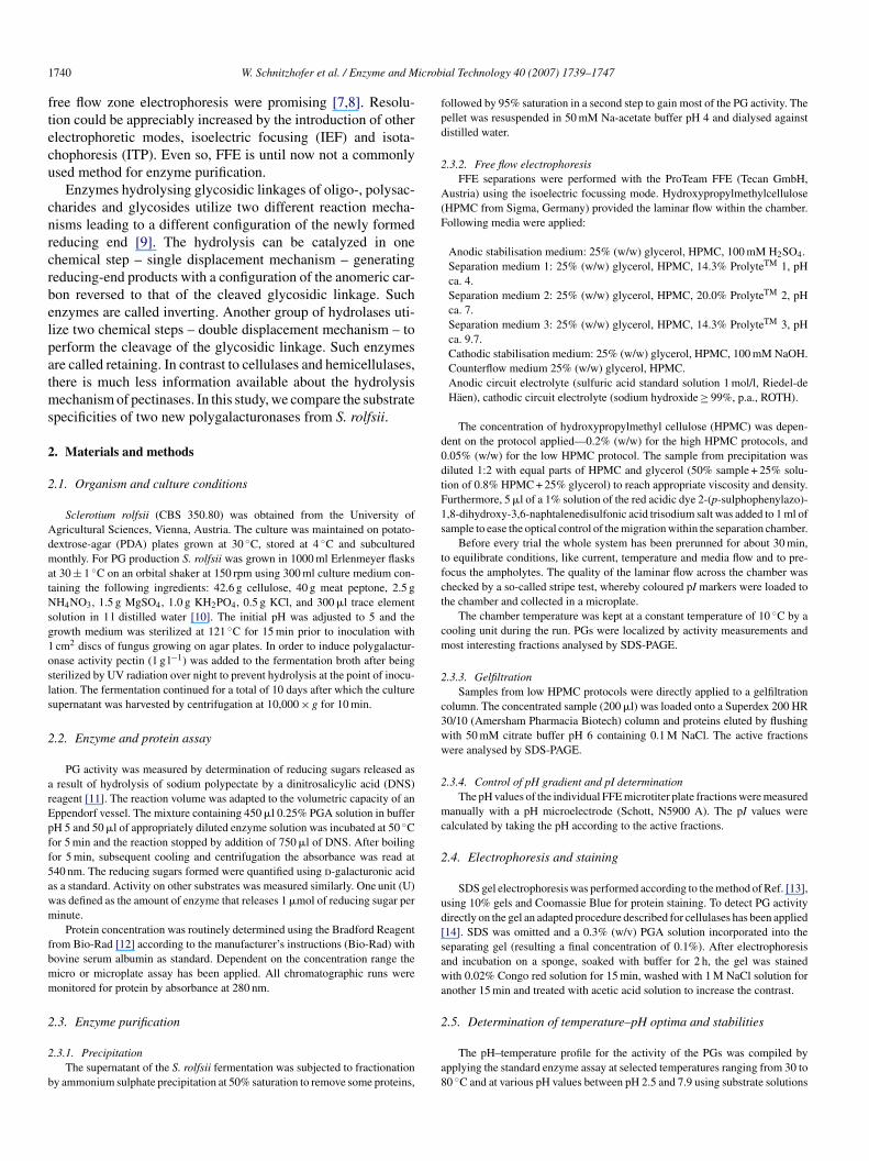
1 icrob
ftecu
cnrcrbelpatms
2
2
AdmatNsg1osls
2
arEpff5awm
fbmm
2
2
b
fpd
2
A(F
d0dtF1s
tfct
cm
2
c3ww
2
mc
2
ud[sawa
740 W. Schnitzhofer et al. / Enzyme and M
ree flow zone electrophoresis were promising [7,8]. Resolu-ion could be appreciably increased by the introduction of otherlectrophoretic modes, isoelectric focusing (IEF) and isota-hophoresis (ITP). Even so, FFE is until now not a commonlysed method for enzyme purification.
Enzymes hydrolysing glycosidic linkages of oligo-, polysac-harides and glycosides utilize two different reaction mecha-isms leading to a different configuration of the newly formededucing end [9]. The hydrolysis can be catalyzed in onehemical step – single displacement mechanism – generatingeducing-end products with a configuration of the anomeric car-on reversed to that of the cleaved glycosidic linkage. Suchnzymes are called inverting. Another group of hydrolases uti-ize two chemical steps – double displacement mechanism – toerform the cleavage of the glycosidic linkage. Such enzymesre called retaining. In contrast to cellulases and hemicellulases,here is much less information available about the hydrolysis
echanism of pectinases. In this study, we compare the substratepecificities of two new polygalacturonases from S. rolfsii.
. Materials and methods
.1. Organism and culture conditions
Sclerotium rolfsii (CBS 350.80) was obtained from the University ofgricultural Sciences, Vienna, Austria. The culture was maintained on potato-extrose-agar (PDA) plates grown at 30 ◦C, stored at 4 ◦C and subculturedonthly. For PG production S. rolfsii was grown in 1000 ml Erlenmeyer flasks
t 30 ± 1 ◦C on an orbital shaker at 150 rpm using 300 ml culture medium con-aining the following ingredients: 42.6 g cellulose, 40 g meat peptone, 2.5 gH4NO3, 1.5 g MgSO4, 1.0 g KH2PO4, 0.5 g KCl, and 300 �l trace element
olution in 1 l distilled water [10]. The initial pH was adjusted to 5 and therowth medium was sterilized at 121 ◦C for 15 min prior to inoculation withcm2 discs of fungus growing on agar plates. In order to induce polygalactur-nase activity pectin (1 g l−1) was added to the fermentation broth after beingterilized by UV radiation over night to prevent hydrolysis at the point of inocu-ation. The fermentation continued for a total of 10 days after which the cultureupernatant was harvested by centrifugation at 10,000 × g for 10 min.
.2. Enzyme and protein assay
PG activity was measured by determination of reducing sugars released asresult of hydrolysis of sodium polypectate by a dinitrosalicylic acid (DNS)
eagent [11]. The reaction volume was adapted to the volumetric capacity of anppendorf vessel. The mixture containing 450 �l 0.25% PGA solution in bufferH 5 and 50 �l of appropriately diluted enzyme solution was incubated at 50 ◦Cor 5 min and the reaction stopped by addition of 750 �l of DNS. After boilingor 5 min, subsequent cooling and centrifugation the absorbance was read at40 nm. The reducing sugars formed were quantified using d-galacturonic acids a standard. Activity on other substrates was measured similarly. One unit (U)as defined as the amount of enzyme that releases 1 �mol of reducing sugar perinute.
Protein concentration was routinely determined using the Bradford Reagentrom Bio-Rad [12] according to the manufacturer’s instructions (Bio-Rad) withovine serum albumin as standard. Dependent on the concentration range theicro or microplate assay has been applied. All chromatographic runs wereonitored for protein by absorbance at 280 nm.
.3. Enzyme purification
.3.1. PrecipitationThe supernatant of the S. rolfsii fermentation was subjected to fractionation
y ammonium sulphate precipitation at 50% saturation to remove some proteins,
2
a8
ial Technology 40 (2007) 1739–1747
ollowed by 95% saturation in a second step to gain most of the PG activity. Theellet was resuspended in 50 mM Na-acetate buffer pH 4 and dialysed againstistilled water.
.3.2. Free flow electrophoresisFFE separations were performed with the ProTeam FFE (Tecan GmbH,
ustria) using the isoelectric focussing mode. HydroxypropylmethylcelluloseHPMC from Sigma, Germany) provided the laminar flow within the chamber.ollowing media were applied:
Anodic stabilisation medium: 25% (w/w) glycerol, HPMC, 100 mM H2SO4.Separation medium 1: 25% (w/w) glycerol, HPMC, 14.3% ProlyteTM 1, pHca. 4.Separation medium 2: 25% (w/w) glycerol, HPMC, 20.0% ProlyteTM 2, pHca. 7.Separation medium 3: 25% (w/w) glycerol, HPMC, 14.3% ProlyteTM 3, pHca. 9.7.Cathodic stabilisation medium: 25% (w/w) glycerol, HPMC, 100 mM NaOH.Counterflow medium 25% (w/w) glycerol, HPMC.Anodic circuit electrolyte (sulfuric acid standard solution 1 mol/l, Riedel-deHaen), cathodic circuit electrolyte (sodium hydroxide ≥ 99%, p.a., ROTH).
The concentration of hydroxypropylmethyl cellulose (HPMC) was depen-ent on the protocol applied—0.2% (w/w) for the high HPMC protocols, and.05% (w/w) for the low HPMC protocol. The sample from precipitation wasiluted 1:2 with equal parts of HPMC and glycerol (50% sample + 25% solu-ion of 0.8% HPMC + 25% glycerol) to reach appropriate viscosity and density.urthermore, 5 �l of a 1% solution of the red acidic dye 2-(p-sulphophenylazo)-,8-dihydroxy-3,6-naphtalenedisulfonic acid trisodium salt was added to 1 ml ofample to ease the optical control of the migration within the separation chamber.
Before every trial the whole system has been prerunned for about 30 min,o equilibrate conditions, like current, temperature and media flow and to pre-ocus the ampholytes. The quality of the laminar flow across the chamber washecked by a so-called stripe test, whereby coloured pI markers were loaded tohe chamber and collected in a microplate.
The chamber temperature was kept at a constant temperature of 10 ◦C by aooling unit during the run. PGs were localized by activity measurements andost interesting fractions analysed by SDS-PAGE.
.3.3. GelfiltrationSamples from low HPMC protocols were directly applied to a gelfiltration
olumn. The concentrated sample (200 �l) was loaded onto a Superdex 200 HR0/10 (Amersham Pharmacia Biotech) column and proteins eluted by flushingith 50 mM citrate buffer pH 6 containing 0.1 M NaCl. The active fractionsere analysed by SDS-PAGE.
.3.4. Control of pH gradient and pI determinationThe pH values of the individual FFE microtiter plate fractions were measured
anually with a pH microelectrode (Schott, N5900 A). The pI values werealculated by taking the pH according to the active fractions.
.4. Electrophoresis and staining
SDS gel electrophoresis was performed according to the method of Ref. [13],sing 10% gels and Coomassie Blue for protein staining. To detect PG activityirectly on the gel an adapted procedure described for cellulases has been applied14]. SDS was omitted and a 0.3% (w/v) PGA solution incorporated into theeparating gel (resulting a final concentration of 0.1%). After electrophoresisnd incubation on a sponge, soaked with buffer for 2 h, the gel was stainedith 0.02% Congo red solution for 15 min, washed with 1 M NaCl solution for
nother 15 min and treated with acetic acid solution to increase the contrast.
.5. Determination of temperature–pH optima and stabilities
The pH–temperature profile for the activity of the PGs was compiled bypplying the standard enzyme assay at selected temperatures ranging from 30 to0 ◦C and at various pH values between pH 2.5 and 7.9 using substrate solutions
icrob
iw
e(Ad
2
HT
dfhfi
2
eI
baia
gttNa
p
2
gpGdAegmpwos
2
cad0pto
2
eoiow3rpasasflw
2
aaab
apa
2
2
tsd
2
a
2
psdmm3[
3
gos
W. Schnitzhofer et al. / Enzyme and M
n constant ionic strength citrate–phosphate buffer. In each case the substrateas preincubated at the required temperature.
To evaluate the thermal stability aliquot amounts of desalted, concentratednzyme samples were diluted with 50 mM citrate buffer of various pH valuespH 3.5, 5, 6.5 and 8) and incubated for fixed time periods at 30, 50 and 70 ◦C.t time intervals samples were withdrawn, cooled on ice before assaying toetermine the residual enzyme activity, using the normal assay procedure.
.6. Effect of cations and surfactants
Out of a big number of potential inhibitors several have been chosen: Fe(3),g(2), Ca, Mg (as chloride salts each), K2CrO4, SDS, EDTA, and the surfactantsriton X-100, Cotemoll.
An appropriate amount of purified enzyme solution has been incubated withifferent concentrations of inhibitors (by dilutions of a 100 mM stock solution)or 10 min at room temperature and activity measured. The starting conditionsave been checked out within preexperiments, the concentration range variedrom 0.05 to 50 mM, dependent on the inhibitor, the surfactants have been appliedn a concentration of 0.05%.
.7. Degree of glycosylation
Possible glycosylation residues were removed enzymatically by treating purenzyme samples with a glycosidase and an amidase (both New England Biolabsnc.).
Deglycosylation with Endoglycosidase H (EndoH): 2 �l of 10× denaturinguffer (0.5% SDS, 1% �-mercaptoethanol) were added to 18 �l of protein samplend heated to 100 ◦C for 10 min, after cooling down the mixture was incubatedn G5 Buffer (2.25 �l of 10× buffer, 50 mM Na-citrate) and 2.25 �l of EndoHt 37 ◦C for 24 h.
Deglycosylation with peptide:N-glycosidase F (PNGase F): 2 �l of 10×lycoprotein denaturing buffer (0.5% SDS, 1% �-mercaptoethanol) were addedo 18 �l of protein sample and heated to 100 ◦C for 10 min, after cooling downhe mixture was incubated in G7 reaction buffer (2.5 �l of 10× buffer, 50 mMa-phosphate), supplemented with 2.5 �l 10% NP-40, and 2.5 �l of PNGaseF
t 37 ◦C for 24 h.The deglycosylation was controlled by applying treated and untreated sam-
les to a SDS-PAGE.
.8. Change in specific viscosity
Enzyme action on PGA was followed by measuring the increase in reducingroups and reduction in viscosity. The viscosity was determined at a fixed tem-erature of 30 ◦C with an AMVn automated mircoviscosimeter (Anton Paar®
mbH, Graz, Austria). For the calculation, it was necessary to determine theensity, performed with the DMA 38 density meter (Anton Paar® GmbH, Graz,ustria) by filling the sample loop with the sample liquid and after a certain
quilibration time reading the density. A 975–997.5 �l of a 0.5% (w/v) poly-alacturonic acid in 50 mM acetate buffer (pH 5) was preincubated at 30 ◦C andixed with 2.5–25 �l of the purified enzymes. The mixture was carefully (to
revent air bubbles) filled into the capillary with 1.6 mm diameter. The capillaryas put into the AMVn and the decrease of viscosity was monitored at an anglef 70◦ till no substantial change was observed. The released reducing sugars ofimilar reaction mixtures were measured with the DNS method.
.9. Determination of kinetic parameters
Kinetic properties of the PGs were determined by the measurement of vis-osity changes of homogalacturonan solutions in 50 mM acetate buffer pH 5nd 30 ◦C, incubated with a certain amount of enzyme similar to the above
escribed measurements. Tested substrate concentrations were in the range of.5–10 g/l. Michaelis–Menten parameters were calculated from Eadie–Hofstedlots V versus V/[S] plots and Hanes–Woolf plots [S]/V versus [S], where [S] ishe substrate concentration and V is the initial rate of hydrolysis. By combinationf the two methods, mean values were calculated.3
e
ial Technology 40 (2007) 1739–1747 1741
.10. Release of oligosaccharides
Oligogalacturonic acids of degree of polymerisation 2–5 were prepared bynzymatic hydrolysis of citrus pectin followed by chromatographic purificationf the fragments. The reaction of PGs with oligogalacturonic acid was performedn Eppendorfs, in a minimized scale, a total volume of 20 �l, containing 20 mMf oligomer in 10 mM acetate buffer pH 5 and a standardized amount of enzyme,hich was evaluated in preexperiments. The reaction temperature was set to0 ◦C and the reaction controlled by TLC. Samples were withdrawn out of theeaction mixture in certain time intervals and two times 0.5 �l spotted on a TLClate (20 cm × 20 cm, aluminium sheets, silica gel 60 from Merck), dried withhair-drier after every spot to stop the enzyme reaction and put in a chamber
aturated with the running solvent 1-butanol–formic acid–water = 2:3:1 (v/v),fter the run was complete, the plate was taken out, dried and the oligosaccharidepots were developed with orcinol solution (containing 1 g orcine monohydraterom Fluka in 5 ml H2SO4 conc. and 95 ml ethanol) by pouring a homogenousayer over the plate, drying the same and heating up to 100 ◦C in a dry chamber,here the spots became dark coloured.
.11. Substrate specificities and side activities
Polygalacturonases were tested for their specificities on following substrates:pple pectin (70–75%) and citrus pectin (63–66%), both Fluka and for their sidectivities on xylan from birchwood (Roth), mannan from S. cerevisiae (Sigma)nd CMC (Merck). The experiments were carried out in principle as describedefore.
The reaction mixtures contained 0.5% of the different substrates in 10 mMcetate buffer pH 5 and always the same amount of enzyme. The reaction tem-erature was set to 30 ◦C. Evaluation was done by reducing sugar determinationnd TLC.
.12. Elucidation of the reaction mechanism
.12.1. Sample preparationThe purified enzyme samples of polygalacturonases 1 and 2 were concen-
rated (Vivaspin 6), simultaneously desalted by washing with D2O (Sigma)everal times and two times lyophilized for being redissolved in D2O imme-iately before use in 1H NMR experiments.
.12.2. Substrate preparationPentaGalU-ol was prepared by NaBH4 reduction of the corresponding
ldouronic acid and lyophilized three times from D2O.
.12.3. Reaction mixtures for 1H NMR spectroscopyA 20 mM of pentaGalU-ol was dissolved in 0.05 M deuterized acetate buffer
H 5 and 0.1 ml of a suitable concentrated enzyme solution to 0.4 ml of thisubstrate added. The amount of enzyme was determined by TLC experiments asescribed before, and should guarantee the hydrolysis rate to be much higher thanutarotation of the newly formed reducing end. 1H NMR spectra of the reactionixture were recorded versus time on a varian unity inova 500 spectrometer at
0 ◦C. The assignment of important resonances was based on published data15].
. Results and discussion
Sclerotium rolfsii was found to produce high amounts of poly-alacturonase activity. The substrate specificities and propertiesf the enzymes responsible for this activity are compared in thistudy.
.1. Production of polygalacturonases by S. rolfsii
The cellulose containing basal medium used during thesexperiments was selected because it assured excellent growth of
1742 W. Schnitzhofer et al. / Enzyme and Microbial Technology 40 (2007) 1739–1747
Fap
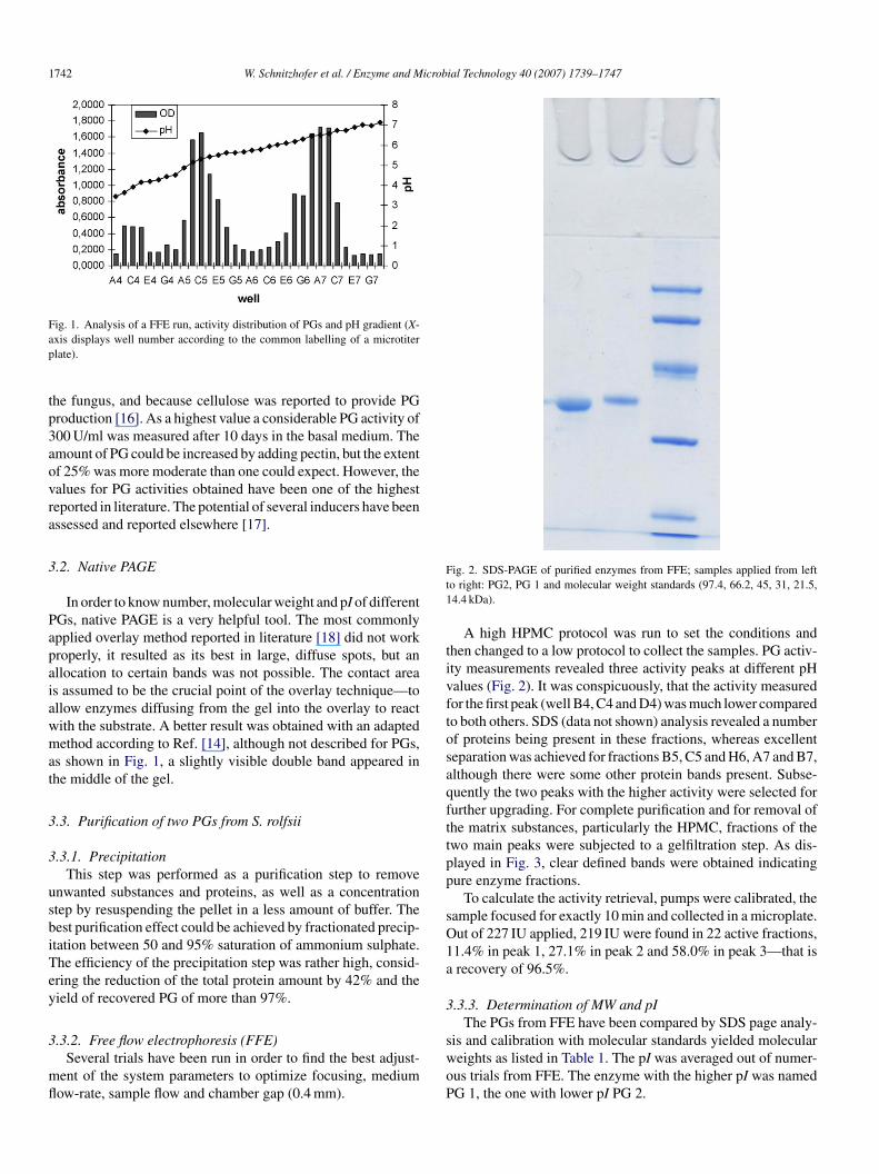
tp3aovra
3
Papaiawmat
3
3
usbiTey
3
mfl
Ft1
tivftosaqfttpp
sO1a
3
ig. 1. Analysis of a FFE run, activity distribution of PGs and pH gradient (X-xis displays well number according to the common labelling of a microtiterlate).
he fungus, and because cellulose was reported to provide PGroduction [16]. As a highest value a considerable PG activity of00 U/ml was measured after 10 days in the basal medium. Themount of PG could be increased by adding pectin, but the extentf 25% was more moderate than one could expect. However, thealues for PG activities obtained have been one of the highesteported in literature. The potential of several inducers have beenssessed and reported elsewhere [17].
.2. Native PAGE
In order to know number, molecular weight and pI of differentGs, native PAGE is a very helpful tool. The most commonlypplied overlay method reported in literature [18] did not workroperly, it resulted as its best in large, diffuse spots, but anllocation to certain bands was not possible. The contact areas assumed to be the crucial point of the overlay technique—tollow enzymes diffusing from the gel into the overlay to reactith the substrate. A better result was obtained with an adaptedethod according to Ref. [14], although not described for PGs,
s shown in Fig. 1, a slightly visible double band appeared inhe middle of the gel.
.3. Purification of two PGs from S. rolfsii
.3.1. PrecipitationThis step was performed as a purification step to remove
nwanted substances and proteins, as well as a concentrationtep by resuspending the pellet in a less amount of buffer. Theest purification effect could be achieved by fractionated precip-tation between 50 and 95% saturation of ammonium sulphate.he efficiency of the precipitation step was rather high, consid-ring the reduction of the total protein amount by 42% and theield of recovered PG of more than 97%.
.3.2. Free flow electrophoresis (FFE)Several trials have been run in order to find the best adjust-
ent of the system parameters to optimize focusing, mediumow-rate, sample flow and chamber gap (0.4 mm).
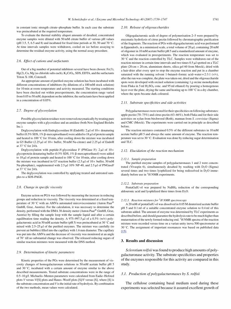
swoP
ig. 2. SDS-PAGE of purified enzymes from FFE; samples applied from lefto right: PG2, PG 1 and molecular weight standards (97.4, 66.2, 45, 31, 21.5,4.4 kDa).
A high HPMC protocol was run to set the conditions andhen changed to a low protocol to collect the samples. PG activ-ty measurements revealed three activity peaks at different pHalues (Fig. 2). It was conspicuously, that the activity measuredor the first peak (well B4, C4 and D4) was much lower comparedo both others. SDS (data not shown) analysis revealed a numberf proteins being present in these fractions, whereas excellenteparation was achieved for fractions B5, C5 and H6, A7 and B7,lthough there were some other protein bands present. Subse-uently the two peaks with the higher activity were selected forurther upgrading. For complete purification and for removal ofhe matrix substances, particularly the HPMC, fractions of thewo main peaks were subjected to a gelfiltration step. As dis-layed in Fig. 3, clear defined bands were obtained indicatingure enzyme fractions.
To calculate the activity retrieval, pumps were calibrated, theample focused for exactly 10 min and collected in a microplate.ut of 227 IU applied, 219 IU were found in 22 active fractions,1.4% in peak 1, 27.1% in peak 2 and 58.0% in peak 3—that isrecovery of 96.5%.
.3.3. Determination of MW and pIThe PGs from FFE have been compared by SDS page analy-
is and calibration with molecular standards yielded moleculareights as listed in Table 1. The pI was averaged out of numer-us trials from FFE. The enzyme with the higher pI was namedG 1, the one with lower pI PG 2.
W. Schnitzhofer et al. / Enzyme and Microb
F(
swoFbePpmGysc
TMr
PP
7aeiond
4
4
iiveattw
4
trations of 50 mM. Fe(II) could not be examined, since it wasimmediately oxidised by the DNS reagent giving a positiveresponse even without enzyme and was therefore replaced byFe(III). I50 values are shown in Table 4. PG 1 and PG 2 were
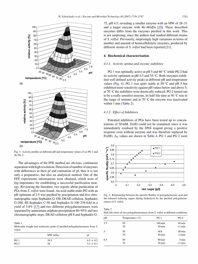
Fig. 4. Relationship between the specific fluidity of polygalacturonic acid andthe released reducing sugars during hydrolysis by the purified polygalactur-onases of S. rolfsii.
ig. 3. Activity profiles at different pH and temperature values of (a) PG 1 andb) PG 2.
The advantages of the FFE method are obvious, continuouseparation with high resolution. Detection of number of enzymesith differences in their pI and estimation of pI, thus it is notnly a preparative, but also an analytical method. Out of theFE experiments informations were obtained, which were ofig importance for establishing a successful purification strat-gy. Reviewing the literature, two reports about purification ofGs from S. rolfsii were found. An acid-stable endo-PG with anH optimum of 2.5 was purified by precipitation and five chro-atographic steps Sephadex G-100, DEAE cellulose, Sephadex
-200, SE-Sephadex C-50 and Sephadex G-100 239-fold in aield of 3.8% [17] and two different polygalacturonases wereeparated by ammonium sulphate precipitation 80–95% and twohromatographic steps, DEAE-cellulose pH 8 and Sephadex G-able 1olecular weight and isoelectric point of purified polygalacturonases from S.
olfsii
MW (kDa) pI
G 1 39.5 6.5 ± 0.2G 2 38 5.3 ± 0.1
TH
p
3
5
6
ial Technology 40 (2007) 1739–1747 1743
5, pH 4.5, revealing a smaller enzyme with an MW of 28–31nd a larger enzyme with 46–48 kDa [19]. These describednzymes differ from the enzymes purified in this work. Thiss not surprising, since the authors had studied different strainsf S. rolfsii. Previously, surprisingly high variations in terms ofumber and amount of hemicellulolytic enzymes, produced byifferent strains of S. rolfsii had been reported [21].
. Biochemical characterisation
.1.1. Activity optima and enzyme stabilities
PG 1 was optimally active at pH 5 and 60 ◦C while PG 2 hadts activity optimum at pH 4.5 and 55 ◦C. Both enzymes exhib-ted well defined activity peaks at different pH and temperaturealues (Fig. 4). PG 1 was quite stable at 50 ◦C and pH 5 butxhibited more sensitivity against pH values below and above 5;t 70 ◦C the stabilities were drastically reduced. PG 2 turned outo be a really sensitive enzyme, its half-life time at 50 ◦C was inhe range of minutes and at 70 ◦C the enzyme was inactivatedithin 1 min (Table 2).
.1.2. Effect of Inhibitors
Potential inhibitors of PGs have been tested up to concen-
able 2alf-life times of two polygalacturonases from S. rolfsii at different conditions
H Temperature (◦C) PG 1 PG 2
.5 50 160 min 45 min70 30 min <1 min
50 48 h 40 min70 50 min <1min
.5 50 80 min 5 min70 20 min <1 min
1 icrobial Technology 40 (2007) 1739–1747
saPdwwgniPbE
4
wHmctht
1b
n[artAsSgs[
4
4
tbead(6h
aoiad
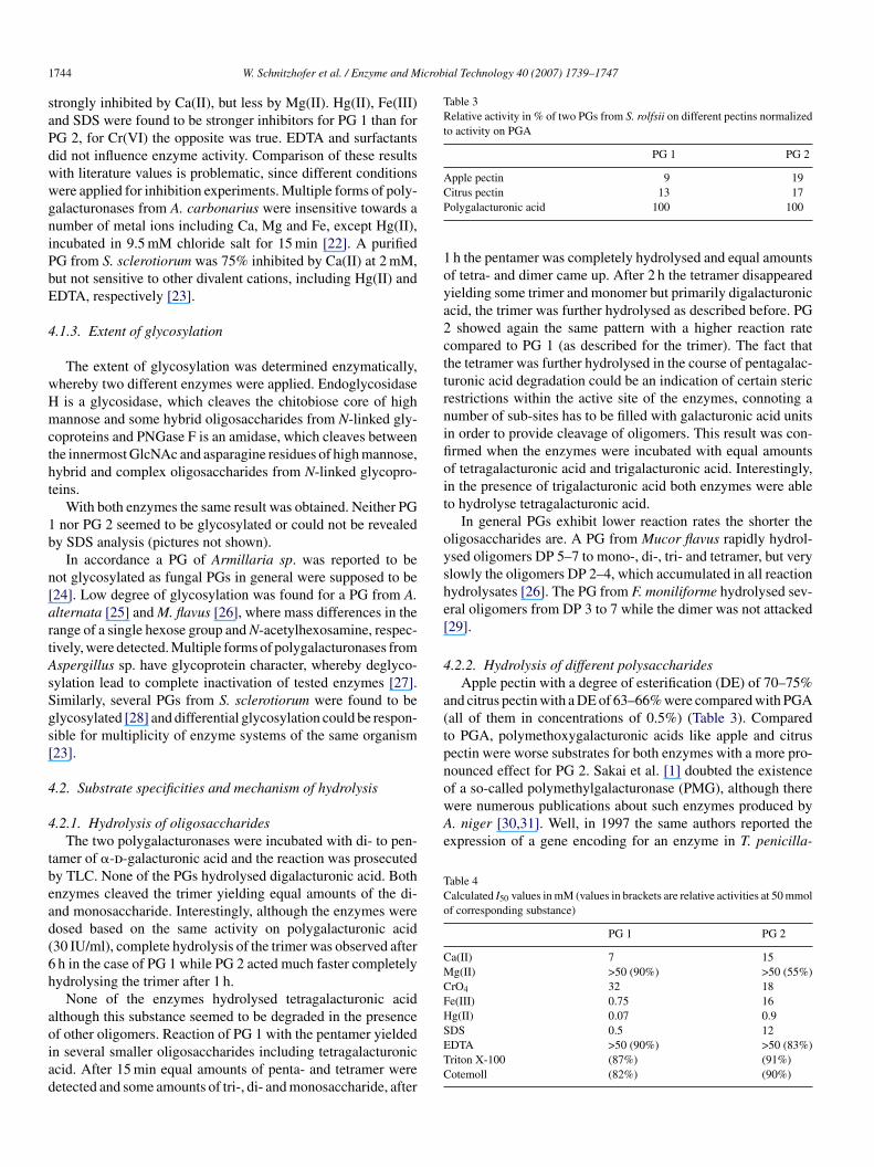
Table 3Relative activity in % of two PGs from S. rolfsii on different pectins normalizedto activity on PGA
PG 1 PG 2
Apple pectin 9 19CP
1oya2cttrnifioit
oyshe[
4
a(tpnof a so-called polymethylgalacturonase (PMG), although therewere numerous publications about such enzymes produced byA. niger [30,31]. Well, in 1997 the same authors reported theexpression of a gene encoding for an enzyme in T. penicilla-
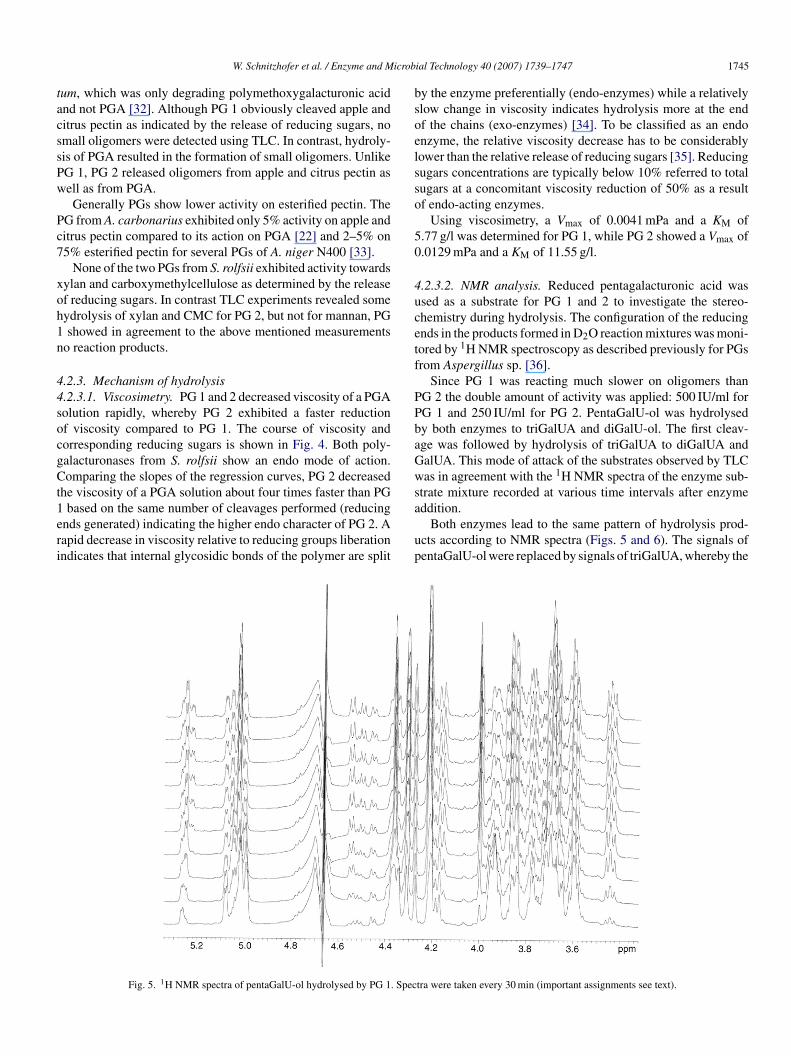
Table 4Calculated I50 values in mM (values in brackets are relative activities at 50 mmolof corresponding substance)
PG 1 PG 2
Ca(II) 7 15Mg(II) >50 (90%) >50 (55%)CrO4 32 18Fe(III) 0.75 16Hg(II) 0.07 0.9
744 W. Schnitzhofer et al. / Enzyme and M
trongly inhibited by Ca(II), but less by Mg(II). Hg(II), Fe(III)nd SDS were found to be stronger inhibitors for PG 1 than forG 2, for Cr(VI) the opposite was true. EDTA and surfactantsid not influence enzyme activity. Comparison of these resultsith literature values is problematic, since different conditionsere applied for inhibition experiments. Multiple forms of poly-alacturonases from A. carbonarius were insensitive towards aumber of metal ions including Ca, Mg and Fe, except Hg(II),ncubated in 9.5 mM chloride salt for 15 min [22]. A purifiedG from S. sclerotiorum was 75% inhibited by Ca(II) at 2 mM,ut not sensitive to other divalent cations, including Hg(II) andDTA, respectively [23].
.1.3. Extent of glycosylation
The extent of glycosylation was determined enzymatically,hereby two different enzymes were applied. Endoglycosidaseis a glycosidase, which cleaves the chitobiose core of high
annose and some hybrid oligosaccharides from N-linked gly-oproteins and PNGase F is an amidase, which cleaves betweenhe innermost GlcNAc and asparagine residues of high mannose,ybrid and complex oligosaccharides from N-linked glycopro-eins.
With both enzymes the same result was obtained. Neither PGnor PG 2 seemed to be glycosylated or could not be revealed
y SDS analysis (pictures not shown).In accordance a PG of Armillaria sp. was reported to be
ot glycosylated as fungal PGs in general were supposed to be24]. Low degree of glycosylation was found for a PG from A.lternata [25] and M. flavus [26], where mass differences in theange of a single hexose group and N-acetylhexosamine, respec-ively, were detected. Multiple forms of polygalacturonases fromspergillus sp. have glycoprotein character, whereby deglyco-ylation lead to complete inactivation of tested enzymes [27].imilarly, several PGs from S. sclerotiorum were found to belycosylated [28] and differential glycosylation could be respon-ible for multiplicity of enzyme systems of the same organism23].
.2. Substrate specificities and mechanism of hydrolysis
.2.1. Hydrolysis of oligosaccharidesThe two polygalacturonases were incubated with di- to pen-
amer of �-d-galacturonic acid and the reaction was prosecutedy TLC. None of the PGs hydrolysed digalacturonic acid. Bothnzymes cleaved the trimer yielding equal amounts of the di-nd monosaccharide. Interestingly, although the enzymes wereosed based on the same activity on polygalacturonic acid30 IU/ml), complete hydrolysis of the trimer was observed afterh in the case of PG 1 while PG 2 acted much faster completelyydrolysing the trimer after 1 h.
None of the enzymes hydrolysed tetragalacturonic acidlthough this substance seemed to be degraded in the presence
f other oligomers. Reaction of PG 1 with the pentamer yieldedn several smaller oligosaccharides including tetragalacturoniccid. After 15 min equal amounts of penta- and tetramer wereetected and some amounts of tri-, di- and monosaccharide, afterSETC
itrus pectin 13 17olygalacturonic acid 100 100
h the pentamer was completely hydrolysed and equal amountsf tetra- and dimer came up. After 2 h the tetramer disappearedielding some trimer and monomer but primarily digalacturoniccid, the trimer was further hydrolysed as described before. PGshowed again the same pattern with a higher reaction rate
ompared to PG 1 (as described for the trimer). The fact thathe tetramer was further hydrolysed in the course of pentagalac-uronic acid degradation could be an indication of certain stericestrictions within the active site of the enzymes, connoting aumber of sub-sites has to be filled with galacturonic acid unitsn order to provide cleavage of oligomers. This result was con-rmed when the enzymes were incubated with equal amountsf tetragalacturonic acid and trigalacturonic acid. Interestingly,n the presence of trigalacturonic acid both enzymes were ableo hydrolyse tetragalacturonic acid.
In general PGs exhibit lower reaction rates the shorter theligosaccharides are. A PG from Mucor flavus rapidly hydrol-sed oligomers DP 5–7 to mono-, di-, tri- and tetramer, but verylowly the oligomers DP 2–4, which accumulated in all reactionydrolysates [26]. The PG from F. moniliforme hydrolysed sev-ral oligomers from DP 3 to 7 while the dimer was not attacked29].
.2.2. Hydrolysis of different polysaccharidesApple pectin with a degree of esterification (DE) of 70–75%
nd citrus pectin with a DE of 63–66% were compared with PGAall of them in concentrations of 0.5%) (Table 3). Comparedo PGA, polymethoxygalacturonic acids like apple and citrusectin were worse substrates for both enzymes with a more pro-ounced effect for PG 2. Sakai et al. [1] doubted the existence
DS 0.5 12DTA >50 (90%) >50 (83%)riton X-100 (87%) (91%)otemoll (82%) (90%)
icrob
tacssPw
Pc7
xoh1n
44socgCt1eri
bsoelsso
50
4ucetf
PPbaGws
W. Schnitzhofer et al. / Enzyme and M
um, which was only degrading polymethoxygalacturonic acidnd not PGA [32]. Although PG 1 obviously cleaved apple anditrus pectin as indicated by the release of reducing sugars, nomall oligomers were detected using TLC. In contrast, hydroly-is of PGA resulted in the formation of small oligomers. UnlikeG 1, PG 2 released oligomers from apple and citrus pectin asell as from PGA.Generally PGs show lower activity on esterified pectin. The
G from A. carbonarius exhibited only 5% activity on apple anditrus pectin compared to its action on PGA [22] and 2–5% on5% esterified pectin for several PGs of A. niger N400 [33].
None of the two PGs from S. rolfsii exhibited activity towardsylan and carboxymethylcellulose as determined by the releasef reducing sugars. In contrast TLC experiments revealed someydrolysis of xylan and CMC for PG 2, but not for mannan, PGshowed in agreement to the above mentioned measurements
o reaction products.
.2.3. Mechanism of hydrolysis
.2.3.1. Viscosimetry. PG 1 and 2 decreased viscosity of a PGAolution rapidly, whereby PG 2 exhibited a faster reductionf viscosity compared to PG 1. The course of viscosity andorresponding reducing sugars is shown in Fig. 4. Both poly-alacturonases from S. rolfsii show an endo mode of action.omparing the slopes of the regression curves, PG 2 decreased
he viscosity of a PGA solution about four times faster than PG
based on the same number of cleavages performed (reducingnds generated) indicating the higher endo character of PG 2. Aapid decrease in viscosity relative to reducing groups liberationndicates that internal glycosidic bonds of the polymer are split
a
up
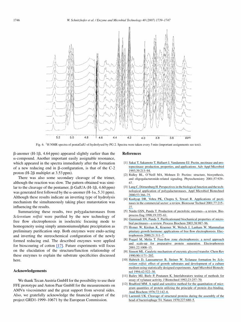
Fig. 5. 1H NMR spectra of pentaGalU-ol hydrolysed by PG 1. Spec
ial Technology 40 (2007) 1739–1747 1745
y the enzyme preferentially (endo-enzymes) while a relativelylow change in viscosity indicates hydrolysis more at the endf the chains (exo-enzymes) [34]. To be classified as an endonzyme, the relative viscosity decrease has to be considerablyower than the relative release of reducing sugars [35]. Reducingugars concentrations are typically below 10% referred to totalugars at a concomitant viscosity reduction of 50% as a resultf endo-acting enzymes.
Using viscosimetry, a Vmax of 0.0041 mPa and a KM of.77 g/l was determined for PG 1, while PG 2 showed a Vmax of.0129 mPa and a KM of 11.55 g/l.
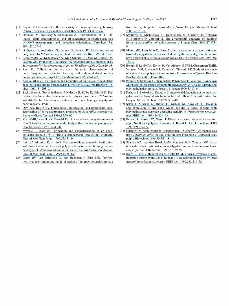
.2.3.2. NMR analysis. Reduced pentagalacturonic acid wassed as a substrate for PG 1 and 2 to investigate the stereo-hemistry during hydrolysis. The configuration of the reducingnds in the products formed in D2O reaction mixtures was moni-ored by 1H NMR spectroscopy as described previously for PGsrom Aspergillus sp. [36].
Since PG 1 was reacting much slower on oligomers thanG 2 the double amount of activity was applied: 500 IU/ml forG 1 and 250 IU/ml for PG 2. PentaGalU-ol was hydrolysedy both enzymes to triGalUA and diGalU-ol. The first cleav-ge was followed by hydrolysis of triGalUA to diGalUA andalUA. This mode of attack of the substrates observed by TLCas in agreement with the 1H NMR spectra of the enzyme sub-
trate mixture recorded at various time intervals after enzyme
ddition.Both enzymes lead to the same pattern of hydrolysis prod-cts according to NMR spectra (Figs. 5 and 6). The signals ofentaGalU-ol were replaced by signals of triGalUA, whereby the
tra were taken every 30 min (important assignments see text).
1746 W. Schnitzhofer et al. / Enzyme and Microbial Technology 40 (2007) 1739–1747
. Spe
��wop
alwAmi
Sfhpaffoth
A
FAAp
R
[
[
Fig. 6. 1H NMR spectra of pentaGalU-ol hydrolysed by PG 2
-anomer (H-1�, 4.64 ppm) appeared slightly earlier than the-compound. Another important easily assignable resonance,hich appeared in the spectra immediately after the formationf a new reducing end in �-configuration, is that of the C-2roton (H-2� multiplet at 3.53 ppm).
There was also some secondary cleavage of the trimer,lthough the reaction was slow. The pattern obtained was simi-ar to the cleavage of the pentamer, �-GalUA (H-1�, 4.60 ppm)as generated first followed by the �-anomer (H-1�, 5.31 ppm).lthough these results indicate an inverting type of hydrolysisechanism the simultaneously taking place mutarotation was
nfluencing the results.Summarizing these results, two polygalacturonases from
clerotium rolfsii were purified by the new technology ofree flow electrophoresis in isoelectric focusing mode toomogeneity using simply ammoniumsulphate precipitation asreliminary purification step. Both enzymes were endo-actingnd inverting the stereochemical configuration of the newlyormed reducing end. The described enzymes were appliedor bioscouring of cotton [17]. Future experiments will focusn the elucidation of the structure/function relationship ofhese enzymes to explain the substrate specificities discussedere.
cknowledgements
We thank Tecan Austria GmbH for the possibility to use their
FE prototype and Anton Paar GmbH for the measurements onMVn viscosimeter and the great support from several sides.lso, we gratefully acknowledge the financial support of theroject GRD1-1999–10671 by the European Commission.[
[
ctra were taken every 5 min (important assignments see text).
eferences
[1] Sakai T, Sakamoto T, Hallaert J, Vandamme EJ. Pectin, pectinase and pro-topectinase: production, properties, and applications. Adv Appl Microbiol1993;39:213–94.
[2] Ridley BL, O’Neill MA, Mohnen D. Pectins: structure, biosynthesis,and oligogalacturonide-related signaling. Phytochemistry 2001;57:929–67.
[3] Lang C, Dornenburg H. Perspectives in the biological function and the tech-nological application of polygalacturonases. Appl Microbiol Biotechnol2000;53:366–75.
[4] Kashyap DR, Vohra PK, Chopra S, Tewari R. Applications of pecti-nases in the commercial sector: a review. Bioresour Technol 2001;77:215–27.
[5] Naidu GSN, Panda T. Production of pectolytic enzymes—a review. Bio-process Eng 1998;19:355–61.
[6] Gummadi SN, Panda T. Purificationand biochamical properties of micro-bial pectinases—a review. Process Biochem 2003;38:987–96.
[7] Hymer W, Kirshan K, Kraemer W, Welsch J, Lanham W. Mammalianpituitary growth hormone: applications of free flow electrophoresis. Elec-trophoresis 2000;21:311–7.
[8] Poggel M, Melin T. Free-flow zone electrophoresis: a novel approachand scale-up for preparative protein separation. Electrophoresis2001;22:1008–15.
[9] Sinnott ML. Catalytic mechanism of enzymic glycosyl transfer. Chem Rev1990;90:1171–202.
10] Haltrich D, Laussameyer B, Steiner W. Xylanase formation by Scle-rotium rolfsii: effect of growth substrates and development of a culturemedium using statistically designed experiments. Appl Microbiol Biotech-nol 1994;42:522–30.
11] Bailey MJ, Biely P, Poutanen K. Interlaboratory testing of methods forassay of xylanase activity. J Biotechnol 1992;23:257–70.
12] Bradford MM. A rapid and sensitive method for the quantitation of micr-gram quantities of protein utilizing the principle of protein dye-binding.Anal Biochem 1976;72:142–6.
13] Laemmli UK. Cleavage of structural proteins during the assembly of thehead of bacteriophage T4. Nature 1970;227:680–5.
icrob
[
[
[
[
[
[
[
[
[
[
[
[
[
[
[
[
[
[
[
[
[
W. Schnitzhofer et al. / Enzyme and M
14] Beguin P. Detection of cellulase activity in polyacrylamide gels usingCongo Red-stained agar replicas. Anal Biochem 1983;131:333–6.
15] Hricovini M, Bystricky S, Malovikova A. Conformations of (1 → 4)-linked [alpha]-galacturono-di- and -tri-saccharides in solution analysedby NMR measurements and theoretical calculations. Carbohydr Res1991;220:23–31.
16] Deshpade BS, Ambedkar SS, Channe PS, Shewale JG. Production of car-bohydrases by Sclerotium rolfsii. Hindustan Antibiot Bull 1992;34:95–9.
17] Schnitzhofer W, Kandelbauer A, Klug-Santner B, Onos M, Calafell M,Guebitz GM. Production of cellulase-free polygalacturonase preparation bySclerotium rolfsii for bioscouring of cotton. J Nat Fibers 2006;3(2/3):19–38.
18] Ried JL, Collmer A. Activity stain for rapid characterization ofpectic enzymes in isoelectric focusing and sodium dodecyl sulfate-polyacrylamide gels. Appl Environ Microbiol 1985;50:615–22.
19] Kaji A, Okada T. Purification and properties of an unusually acid-stableendo-polygalacturonase produced by Corticium rolfsii. Arch Biochem Bio-phys 1969;131:203–9.
21] Sachslehner A, Grosswindhager N, Nidetzky B, Kulbe K, Haltrich D. For-mation of endo-b-1,4-d-mannanase activity by various strains of Sclerotiumand Athelia. In: International conference on biotechnology in pulp andpaper industry. 1998.
22] Devi NA, Rao AGA. Fractionation, purification, and preliminary char-acterization of polygalacturonases produced by Aspergillus carbonarius.Enzyme Microb Technol 1996;18:59–65.
23] Martel MB, Letoublon R, Fevre M. Purification of endo polygalacturonasesfrom Sclerotinia sclerotiorum: multiplicity of the complex enzyme system.Curr Microbiol 1996;33:243–8.
24] Mwenje E, Ride JP. Purification and characterization of an endo-polygalacturonase (PG 1) from a Zimbabwean species of Armillaria.Physiol Mol Plant Pathol 1999;55:131–9.
25] Isshiki A, Akimitsu K, Nishio K, Tsukamoto M, Yamamoto H. Purification
and characterization of an endopolygalacturonase from the rough lemonpathotype of Alternaria alternata, the cause of citrus brown spot disease.Physiol Mol Plant Pathol 1997;51:155–67.26] Gadre RV, Van Driessche G, Van Beeumen J, Bhat MK. Purifica-tion, characterisation and mode of action of an endo-polygalacturonase
[
ial Technology 40 (2007) 1739–1747 1747
from the psychrophilic fungus Mucor flavus. Enzyme Microb Technol2003;32:321–30.
27] Stratilova E, Mislovicova D, Kacurakova M, Machova E, KolarovaN, Markovic O, Jornvall H. The glycoprotein character of multipleforms of Aspergillus polygalacturonase. J Protein Chem 1998;17:173–9.
28] Martel MB, Letoublon R, Fevre M. Purification and characterization oftwo endopolygalacturonases secreted during the early stages of the sapro-phytic growth of Sclerotinia sclerotiorum. FEMS Microbiol Lett 1998;158:133–8.
29] Bonnin E, Le Goff A, Korner R, Van Alebeek G-JWM, Christensen TMIE,Voragen AGJ, Roepstorff P, Caprari C, Thibault J-F. Study of the modeof action of endopolygalacturonase from Fusarium moniliforme. BiochimBiophys Acta 2001;1526:301–9.
30] Pashova S, Slokoska L, Sheremetska P, Krumova E, Vasileva L, AngelovaM. Physiological aspects of immobilised Aspergillus niger cells producingpolymethylgalacturonase. Process Biochem 1999;35:15–9.
31] Pashova S, Slokoska L, Krumova E, Angelova M. Induction of polymethyl-galacturonase biosynthesis by immobilized cells of Aspergillus niger 26.Enzyme Microb Technol 1999;24:535–40.
32] Sakai T, Sirasaka N, Hirano H, Kishida M, Kawasaki H. Isolationand expression of the gene which encodes a novel enzyme withpolymethoxygalacturonate-degrading activity in Trichosporon penicilla-tum. FEBS Lett 1997;414:439–43.
33] Benen JA, Kester HC, Visser J. Kinetic characterization of Aspergillusniger N400 endopolygalacturonases I, II and C. Eur J Biochem/FEBS1999;259:577–85.
34] Guebitz GM, Schnitzhofer W, Balakrishnan H, Steiner W. Two mannanasesfrom Sclerotium rolfsii in total chlorine-free bleaching of softwood kraftpulp. J Biotechnol 1996;50(2/3):181–8.
35] Shanley NA, van den Broek LAM, Voragen AGJ, Couglan MP. Isola-
tion and characterisation of an endopolygalacturonase from Phanerochaetechrysosporium. J Biotechnol 1993;28:179–97.36] Biely P, Benen J, Heinrichova K, Kester HCM, Visser J. Inversion of con-figuration during hydrolysis of [alpha]-1,4-galacturonidic linkage by threeAspergillus polygalacturonases. FEBS Lett 1996;382:249–55.
Copyright © 2022 FDOKUMEN