Mechanistic Insights into Biological Activities of Polyphenolic ...
28
Foods 2022, 11, 67. https://doi.org/10.3390/foods11010067 www.mdpi.com/journal/foods Article Mechanistic Insights into Biological Activities of Polyphenolic Compounds from Rosemary Obtained by Inverse Molecular Docking Samo Lešnik 1 and Urban Bren 1,2, * 1 Laboratory of Physical Chemistry and Chemical Thermodynamics, Faculty of Chemistry and Chemical Engineering, University of Maribor, Smetanova 17, SI-2000 Maribor, Slovenia; [email protected] 2 Faculty of Mathematics, Natural Sciences and Information Technologies, University of Primorska, Glagoljaška 8, SI-6000 Koper, Slovenia * Correspondence: [email protected]; Tel.: +386-2-2294-421 Abstract: Rosemary (Rosmarinus officinalis L.) represents a medicinal plant known for its various health-promoting properties. Its extracts and essential oils exhibit antioxidative, anti-inflammatory, anticarcinogenic, and antimicrobial activities. The main compounds responsible for these effects are the diterpenes carnosic acid, carnosol, and rosmanol, as well as the phenolic acid ester rosmarinic acid. However, surprisingly little is known about the molecular mechanisms responsible for the pharmacological activities of rosemary and its compounds. To discern these mechanisms, we per- formed a large-scale inverse molecular docking study to identify their potential protein targets. Listed compounds were separately docked into predicted binding sites of all non-redundant holo proteins from the Protein Data Bank and those with the top scores were further examined. We fo- cused on proteins directly related to human health, including human and mammalian proteins as well as proteins from pathogenic bacteria, viruses, and parasites. The observed interactions of rose- mary compounds indeed confirm the beforementioned activities, whereas we also identified their potential for anticoagulant and antiparasitic actions. The obtained results were carefully checked against the existing experimental findings from the scientific literature as well as further validated using both redocking procedures and retrospective metrics. Keywords: rosemary; inverse molecular docking; carnosol; carnosic acid; rosmanol; rosmarinic acid 1. Introduction Rosemary (Rosmarinus officinalis L.), which belongs to the Lamiaceae family, repre- sents an evergreen, perennial, branched shrub that can grow up to three feet tall. It grows fragrant, needle-like, dark green leaves with curved margins and tiny white, pink, purple, or blue flowers [1,2]. The plant is native to the Mediterranean region and its leaves are used extensively in Mediterranean cuisine, mainly as a spice. Rosemary has been found to possess several bioactive compounds that exert various pharmacological activities, particularly antioxidative [3], anti-inflammatory [4], antidia- betic [5], and antibacterial [6], effects. Moreover, rosemary extracts exhibit promising an- ticarcinogenic activities in several in vitro [7–9] as well as in vivo studies [10,11]. Carnosic acid (Figure 1a), carnosol (Figure 1b), rosmanol (Figure 1c), and rosmarinic acid (Figure 1d) are most frequently cited in relation to the beneficial pharmacological activities of compounds found in rosemary [12]. Carnosol, carnosic acid, and rosmanol represent polyphenolic diterpenes with similar structures. They consist of the main abi- etane scaffold, a fused six-membered tricyclic ring system, with one of these rings being aromatic. Carnosic acid represents the major constituent of rosemary and constitutes up to 4% of the dried leaves [13]. However, it is not very stable and, once isolated, undergoes Citation: Lešnik, S.; Bren, U. Mechanistic Insights into Biological Activities of Polyphenolic Compounds from Rosemary Obtained by Inverse Molecular Docking. Foods 2022, 11, 67. https://doi.org/10.3390/foods11010067 Academic Editor: Guowen Zhang Received: 3 December 2021 Accepted: 24 December 2021 Published: 28 December 2021 Publisher’s Note: MDPI stays neu- tral with regard to jurisdictional claims in published maps and institu- tional affiliations. Copyright: © 2021 by the authors. Li- censee MDPI, Basel, Switzerland. This article is an open access article distributed under the terms and con- ditions of the Creative Commons At- tribution (CC BY) license (https://cre- ativecommons.org/licenses/by/4.0/).
-
Upload
khangminh22 -
Category
Documents
-
view
4 -
download
0
Transcript of Mechanistic Insights into Biological Activities of Polyphenolic ...
Foods 2022, 11, 67. https://doi.org/10.3390/foods11010067 www.mdpi.com/journal/foods
Article
Mechanistic Insights into Biological Activities of Polyphenolic
Compounds from Rosemary Obtained by Inverse Molecular
Docking
Samo Lešnik 1 and Urban Bren 1,2,*
1 Laboratory of Physical Chemistry and Chemical Thermodynamics, Faculty of Chemistry and Chemical
Engineering, University of Maribor, Smetanova 17, SI-2000 Maribor, Slovenia; [email protected] 2 Faculty of Mathematics, Natural Sciences and Information Technologies, University of Primorska,
Glagoljaška 8, SI-6000 Koper, Slovenia
* Correspondence: [email protected]; Tel.: +386-2-2294-421
Abstract: Rosemary (Rosmarinus officinalis L.) represents a medicinal plant known for its various
health-promoting properties. Its extracts and essential oils exhibit antioxidative, anti-inflammatory,
anticarcinogenic, and antimicrobial activities. The main compounds responsible for these effects are
the diterpenes carnosic acid, carnosol, and rosmanol, as well as the phenolic acid ester rosmarinic
acid. However, surprisingly little is known about the molecular mechanisms responsible for the
pharmacological activities of rosemary and its compounds. To discern these mechanisms, we per-
formed a large-scale inverse molecular docking study to identify their potential protein targets.
Listed compounds were separately docked into predicted binding sites of all non-redundant holo
proteins from the Protein Data Bank and those with the top scores were further examined. We fo-
cused on proteins directly related to human health, including human and mammalian proteins as
well as proteins from pathogenic bacteria, viruses, and parasites. The observed interactions of rose-
mary compounds indeed confirm the beforementioned activities, whereas we also identified their
potential for anticoagulant and antiparasitic actions. The obtained results were carefully checked
against the existing experimental findings from the scientific literature as well as further validated
using both redocking procedures and retrospective metrics.
Keywords: rosemary; inverse molecular docking; carnosol; carnosic acid; rosmanol; rosmarinic acid
1. Introduction
Rosemary (Rosmarinus officinalis L.), which belongs to the Lamiaceae family, repre-
sents an evergreen, perennial, branched shrub that can grow up to three feet tall. It grows
fragrant, needle-like, dark green leaves with curved margins and tiny white, pink, purple,
or blue flowers [1,2]. The plant is native to the Mediterranean region and its leaves are
used extensively in Mediterranean cuisine, mainly as a spice.
Rosemary has been found to possess several bioactive compounds that exert various
pharmacological activities, particularly antioxidative [3], anti-inflammatory [4], antidia-
betic [5], and antibacterial [6], effects. Moreover, rosemary extracts exhibit promising an-
ticarcinogenic activities in several in vitro [7–9] as well as in vivo studies [10,11].
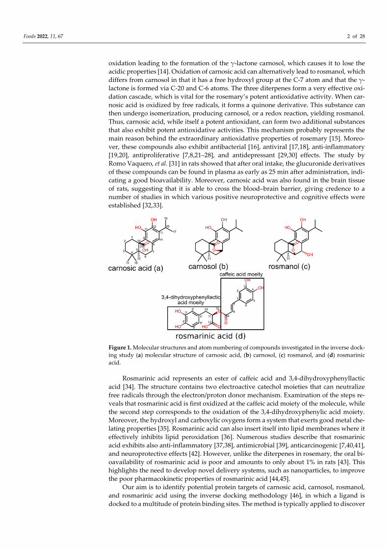
Carnosic acid (Figure 1a), carnosol (Figure 1b), rosmanol (Figure 1c), and rosmarinic
acid (Figure 1d) are most frequently cited in relation to the beneficial pharmacological
activities of compounds found in rosemary [12]. Carnosol, carnosic acid, and rosmanol
represent polyphenolic diterpenes with similar structures. They consist of the main abi-
etane scaffold, a fused six-membered tricyclic ring system, with one of these rings being
aromatic. Carnosic acid represents the major constituent of rosemary and constitutes up
to 4% of the dried leaves [13]. However, it is not very stable and, once isolated, undergoes
Citation: Lešnik, S.; Bren, U.
Mechanistic Insights into Biological
Activities of Polyphenolic
Compounds from Rosemary
Obtained by Inverse Molecular
Docking. Foods 2022, 11, 67.
https://doi.org/10.3390/foods11010067
Academic Editor: Guowen Zhang
Received: 3 December 2021
Accepted: 24 December 2021
Published: 28 December 2021
Publisher’s Note: MDPI stays neu-
tral with regard to jurisdictional
claims in published maps and institu-
tional affiliations.
Copyright: © 2021 by the authors. Li-
censee MDPI, Basel, Switzerland.
This article is an open access article
distributed under the terms and con-
ditions of the Creative Commons At-
tribution (CC BY) license (https://cre-
ativecommons.org/licenses/by/4.0/).
Foods 2022, 11, 67 2 of 28
oxidation leading to the formation of the γ-lactone carnosol, which causes it to lose the
acidic properties [14]. Oxidation of carnosic acid can alternatively lead to rosmanol, which
differs from carnosol in that it has a free hydroxyl group at the C-7 atom and that the γ-
lactone is formed via C-20 and C-6 atoms. The three diterpenes form a very effective oxi-
dation cascade, which is vital for the rosemary’s potent antioxidative activity. When car-
nosic acid is oxidized by free radicals, it forms a quinone derivative. This substance can
then undergo isomerization, producing carnosol, or a redox reaction, yielding rosmanol.
Thus, carnosic acid, while itself a potent antioxidant, can form two additional substances
that also exhibit potent antioxidative activities. This mechanism probably represents the
main reason behind the extraordinary antioxidative properties of rosemary [15]. Moreo-
ver, these compounds also exhibit antibacterial [16], antiviral [17,18], anti-inflammatory
[19,20], antiproliferative [7,8,21–28], and antidepressant [29,30] effects. The study by
Romo Vaquero, et al. [31] in rats showed that after oral intake, the glucuronide derivatives
of these compounds can be found in plasma as early as 25 min after administration, indi-
cating a good bioavailability. Moreover, carnosic acid was also found in the brain tissue
of rats, suggesting that it is able to cross the blood–brain barrier, giving credence to a
number of studies in which various positive neuroprotective and cognitive effects were
established [32,33].
Figure 1. Molecular structures and atom numbering of compounds investigated in the inverse dock-
ing study (a) molecular structure of carnosic acid, (b) carnosol, (c) rosmanol, and (d) rosmarinic
acid.
Rosmarinic acid represents an ester of caffeic acid and 3,4-dihydroxyphenyllactic
acid [34]. The structure contains two electroactive catechol moieties that can neutralize
free radicals through the electron/proton donor mechanism. Examination of the steps re-
veals that rosmarinic acid is first oxidized at the caffeic acid moiety of the molecule, while
the second step corresponds to the oxidation of the 3,4-dihydroxyphenylic acid moiety.
Moreover, the hydroxyl and carboxylic oxygens form a system that exerts good metal che-
lating properties [35]. Rosmarinic acid can also insert itself into lipid membranes where it
effectively inhibits lipid peroxidation [36]. Numerous studies describe that rosmarinic
acid exhibits also anti-inflammatory [37,38], antimicrobial [39], anticarcinogenic [7,40,41],
and neuroprotective effects [42]. However, unlike the diterpenes in rosemary, the oral bi-
oavailability of rosmarinic acid is poor and amounts to only about 1% in rats [43]. This
highlights the need to develop novel delivery systems, such as nanoparticles, to improve
the poor pharmacokinetic properties of rosmarinic acid [44,45].
Our aim is to identify potential protein targets of carnosic acid, carnosol, rosmanol,
and rosmarinic acid using the inverse docking methodology [46], in which a ligand is
docked to a multitude of protein binding sites. The method is typically applied to discover
Foods 2022, 11, 67 3 of 28
new potential protein targets for small molecule drugs [47] or natural products [48–50]
and to explain their mechanisms of action in various diseases. To the best of our
knowledge such an investigation has never been performed for the major rosemary com-
pounds.
2. Materials and Methods
2.1. Starting Coordinates of Rosemary Compounds
The initial coordinates of carnosic acid, carnosol, rosmanol, and rosmarinic acid were
obtained from the ZINC15 database [51], using ZINC IDs ZINC000003984016,
ZINC000003871891, ZINC000031157853, and ZINC0000899870, respectively. Prior to per-
forming inverse molecular docking, all molecules were subjected to a quantum mechani-
cal geometry optimization procedure using the MP2/6-31G* level of theory/basis set com-
bination. This optimization was performed in Gaussian 16 [52].
2.2. In Silico Determination of ADME Properties
In silico determined ADME/Tox profiles provide a useful tool for predicting the phar-
macological and toxicological properties of investigated molecules [53]. To provide a more
detailed prediction of the pharmacokinetic properties of carnosic acid, carnosol, rosmanol,
and rosmarinic acid, which would complement the known experimental data, we imple-
mented the SwissADME web server [54]. SwissADME represents a freely available tool
that enables robust predictions of absorption, distribution, metabolism, and extraction,
based on the two-dimensional data of the molecule. In addition, it yields predictions on
drug-likeness based on well-established metrics.
All compounds were inputted on the SwissADME webpage (http://www.swis-
sadme.ch/ date accessed: 20 December 2021) using the Simplified Molecular-Input Line-
Entry System (SMILES) strings.
2.3. Inverse Molecular Docking
Our goal was to gain mechanistic insight into the potential mechanism of pharmaco-
logical actions of the investigated rosemary compounds using CANDOCK (Chemical
Atomic Network based Docking) [55] inverse molecular docking on more than 65,000 pro-
tein structures potentially associated with human pathologies. Protein binding sites for
small molecules were obtained from the ProBiS-Dock Database [56]. The main advantage
of defining binding sites in this way is that multiple spherical centroids are defined in
advance to describe a very accurate 3D shape that can be used in conjunction with the
CANDOCK algorithm. Moreover, binding sites at the interface of multiple protein chains
are also considered for docking.
For docking, the CANDOCK algorithm applies a hierarchical approach to reconstruct
small molecules from the atomic lattice using graph theory, while applying a generalized
statistical potential function for scoring. The docking scores represent approximations of
the relative binding free energies and are expressed in arbitrary units. Specifically, CAN-
DOCK finds the best-docked poses of small-molecule fragments and applies a fast-maxi-
mum-clique algorithm [57] to link them together. In the molecular reconstruction, the al-
gorithm uses iterative dynamics for better placement of the ligand in the binding pocket.
After the initial docking and reconnection procedure is completed, a minimization proce-
dure based on the Chemistry at Harvard Macromolecular Mechanics (CHARMM) force
field [58] is performed to model the induced fit of the ligand binding to the protein binding
site.

Foods 2022, 11, 67 4 of 28
2.4. Method Validation
To retrospectively validate our inverse molecular docking procedure, we applied re-
ceiver operating characteristic curves (ROC) [59], enrichment curves [60], and predictive-
ness curves (PC) [61]. Briefly, the ROC metric plot shows a correlation between the true-
positive fraction (TPF) on the y-axis and the false-positive fraction (FPF) on the x-axis. In
our case, the TPF represents experimentally confirmed protein targets of rosmarinic acid
from the ChEMBL database [62] with the corresponding PDB entries, while the FPF rep-
resents all other protein targets from the ProBiS-Dock database. We did not perform an
analogous validation for diterpenes as only a small number of confirmed targets is avail-
able for them. The area under the ROC curve (ROC AUC) represents a simple measure to
evaluate the overall performance of the inverse molecular docking method. The larger the
ROC AUC, the more effective is the method at discriminating true from false targets. The
enrichment curve represents the early quantification of target proteins from the TPF.
Moreover, PC also provides the early detection quantification of target proteins from the
TPF, but in addition, it can be used to define the threshold for potential targets from the
inverse molecular docking to be tested experimentally. Contrary to ROC, PC can describe
the dispersion of the inverse docking scores well. To quantify the early detection, we ap-
plied the enrichment factor of 1% of the compounds tested (EF) [63], the Boltzmann-en-
hanced discrimination of ROC (BEDROC) [59], and the robust initial enrichment (RIE) [63]
measures as well. Using PC, the standardized total gain (TG) [61] was also determined,
which summarizes the contribution of the inverse molecular docking scores in explaining
the probability of targets over the entire protein dataset. To calculate all of the listed
measures, the Screening Explorer web server [64] was implemented.
3. Results
3.1. Inverse Molecular Docking of Diterpenes
Because of their similar structure and good agreement, the docking results for carno-
sic acid, carnosol, and rosmanol were combined and analyzed together: the diterpene lig-
and with the best score for the individual protein was considered. The 0.05% (3.5σ) top
scoring proteins from the entire docked database were selected (Figure 2) and among
them, those with implications for human health were chosen. Human and mammalian
proteins as well as proteins from pathogenic bacteria, viruses, and parasites were consid-
ered. Moreover, mammalian proteins were considered in order to increase the protein
space available for docking, where we assumed that within the class of mammals, analo-
gous proteins and their binding sites are similar enough so that our findings from non-
human mammals are transferable to human proteins.
Figure 2. Normal distribution fit of the inverse docking scores. (a) Combined distribution of docking
scores obtained by inversely docking the rosemary diterpenes carnosic acid, carnosol, and rosmanol
to the whole ProBiS-Dock Database. (b) Distribution of docking scores for rosmarinic acid. A cut-
off criterion of 3.5 σ was used to select the most promising protein–ligand complexes to be further
investigated in more detail.
Foods 2022, 11, 67 5 of 28
In Table 1, we present the highest-scoring protein–ligand complexes based on the
cut-off criterion of 3.5 σ. Moreover, where data were available, we redocked ligands/drugs
that are known to bind to the presented targets using an analogous procedure as the one
applied for inverse docking. These results, presented in Table S1, show that in all cases
except for K-Ras G12C and enhanced intracellular survival protein, the docking scores of
the known ligand/drugs are worse than the ones of the rosemary diterpenes. This indi-
cates an already strong binding affinity of the rosemary compounds, although they have
not yet been rationally optimized for these specific protein targets.
Table 1. Best scoring human, mammalian, and pathogen protein targets of rosemary diterpenes
carnosic acid, carnosol, and rosmanol. Docking scores independent of the organism or type of pro-
tein are collected in Supplementary Materials in Table S2.
Rank
PDB ID
with
Chain
Ligand
Predicted Lig-
and Docking
Score (arb.
Units)
Protein Name Organism Protein Function and
Disease Correlation
Reported Experi-
mental Correla-
tion of Protein
and Ligand
1 4lucB Carnosic
acid −69.9 K-Ras G12C Homo sapiens
Controls cell prolifera-
tion and differentiation.
Its gene is a proto-onco-
gene.
[65]
2 3oojA Carnosic
acid −68.2
Glucosamine- fruc-
tose-6-phosphate
aminotransferase
Escherichia coli
Catalyzes the first step in
hexosamine metabolism
and is needed for E. coli
growth and infection
spread.
[66–68]
3 3srdD Carnosic
acid −68.1 Pyruvate kinase M2 Homo sapiens
Catalyzes the last step in
the glycolysis. Important
in providing ATP to can-
cer cells.
4 1kenA Carnosic
acid −66.9 Hemagglutinin HA1 Influenza A virus
Enables viral entry into
cells causing the flu.
5 2hpeA Carnosic
acid −65.0 HIV-2 protease
Human immunode-
ficiency virus 2
Hydrolyzes peptide
bonds leading to func-
tional proteins essential
for HIV infectivity.
[69]
6 4jd6C Carnosic
acid −64.8
Enhanced intracellu-
lar survival protein
Mycobacterium tu-
berculosis
Acetylates amine groups
in aminoglycoside drugs,
thus preventing the bind-
ing to the ribosome, lead-
ing to M. tuberculosis re-
sistance.
7 5u46A Carnosic
acid −64.7
Peroxisome prolifera-
tor activated receptor
delta
Homo sapiens
Regulates lipid catabo-
lism and its transport
and storage and is also
associated with insulin
secretion and resistance.
It is implicated in meta-
bolic disorders and can-
cer.
γ isoform [70]
8 3mt7A Carnosic
acid −64.5
Glycogen phosphory-
lase
Oryctolagus cunic-
ulus
Breaks the non-reducing
ends in the chain of gly-
cogen that enables glu-
cose production. Its inhi-
bition can manage type II
diabetes.
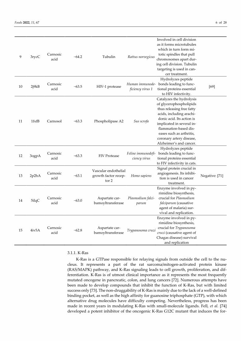
Foods 2022, 11, 67 6 of 28
9 3rycC Carnosic
acid −64.2 Tubulin Rattus norvegicus
Involved in cell division
as it forms microtubules
which in turn form mi-
totic spindles that pull
chromosomes apart dur-
ing cell division. Tubulin
targeting is used in can-
cer treatment.
10 2j9kB Carnosic
acid −63.5 HIV-1 protease
Human immunode-
ficiency virus 1
Hydrolyzes peptide
bonds leading to func-
tional proteins essential
to HIV infectivity.
[69]
11 1fxfB Carnosol −63.3 Phospholipase A2 Sus scrofa
Catalyzes the hydrolysis
of glycerophospholipids
thus releasing free fatty
acids, including arachi-
donic acid. Its action is
implicated in several in-
flammation-based dis-
eases such as arthritis,
coronary artery disease,
Alzheimer’s and cancer.
12 3ogpA Carnosic
acid −63.3 FIV Protease
Feline immunodefi-
ciency virus
Hydrolyzes peptide
bonds leading to func-
tional proteins essential
to FIV infectivity in cats.
13 2p2hA Carnosic
acid −63.1
Vascular endothelial
growth factor recep-
tor 2
Homo sapiens
Signal protein crucial in
angiogenesis. Its inhibi-
tion is used in cancer
treatment.
Negative: [71]
14 5ilqC Carnosic
acid −63.0
Aspartate car-
bamoyltransferase
Plasmodium falci-
parum
Enzyme involved in py-
rimidine biosynthesis,
crucial for Plasmodium
falciparum (causative
agent of malaria) sur-
vival and replication.
15 4iv5A Carnosic
acid −62.8
Aspartate car-
bamoyltransferase Trypanosoma cruzi
Enzyme involved in py-
rimidine biosynthesis,
crucial for Trypanosoma
cruzi (causative agent of
Chagas disease) survival
and replication
3.1.1. K-Ras
K-Ras is a GTPase responsible for relaying signals from outside the cell to the nu-
cleus. It represents a part of the rat sarcoma/mitogen-activated protein kinase
(RAS/MAPK) pathway, and K-Ras signaling leads to cell growth, proliferation, and dif-
ferentiation. K-Ras is of utmost clinical importance as it represents the most frequently
mutated oncogene in pancreatic, colon, and lung cancers [72]. Numerous attempts have
been made to develop compounds that inhibit the function of K-Ras, but with limited
success only [73]. The non-druggability of K-Ras is mainly due to the lack of a well-defined
binding pocket, as well as the high affinity for guanosine triphosphate (GTP), with which
alternative drug molecules have difficulty competing. Nevertheless, progress has been
made in recent years in modulating K-Ras with small-molecule ligands. Fell, et al. [74]
developed a potent inhibitor of the oncogenic K-Ras G12C mutant that induces the for-
Foods 2022, 11, 67 7 of 28
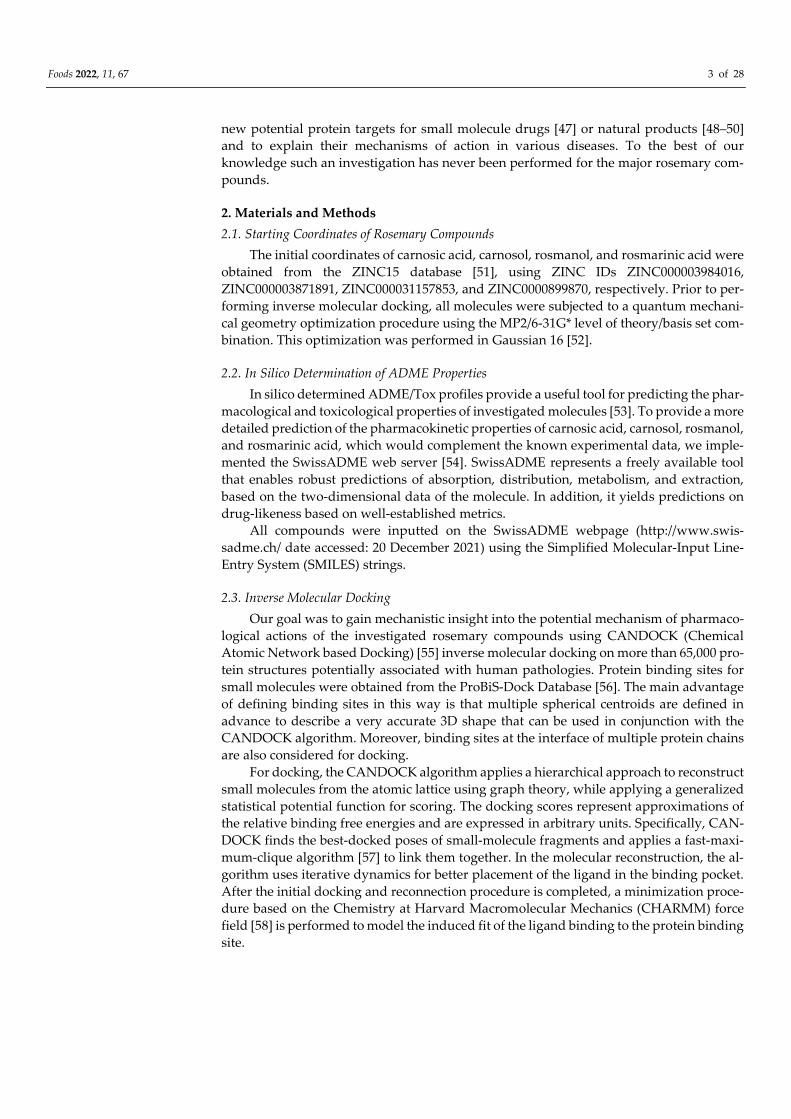
mation of a new binding pocket near the nucleotide (GTP) binding site (Figure 3a). Bind-
ing to this new pocket results in signal inhibition by arresting the enzyme in its inactive
state. Interestingly, this induced binding pocket was ranked most favorable of all the pro-
tein binding sites tested by our method for carnosic acid (Table 1). Carnosic acid docks at
this induced binding site where it forms two hydrogen bonds with Thr58 side chain and
two hydrogen bonds to the backbone atoms of Ala59 and Gly60 (Figure 3b, Table S4). A
strong salt bridge with a distance of 4.1 Å is additionally created between the carboxylate
of carnosic acid and Arg68. Finally, the relatively large hydrophobic ring system of car-
nosic acid forms hydrophobic interactions with Glu62, Tyr96, and Gln99. Although none
of the diterpenes have been previously reported to bind directly to K-Ras, rosemary ex-
tracts have indeed been shown to lead to the down-regulation of K-Ras expression in co-
lon cancer cells [75]. This suggests an interesting potential of carnosic acid for a two-
pronged attack on the protein by down-regulating its expression and by inhibiting it di-
rectly.
Figure 3. (a) K-Ras protein structure highlighting the GTP- and induced-binding site. (b) The in-
duced binding site of the K-Ras protein (blue) with docked carnosic acid (carbon atoms depicted in
grey). Orange dotted lines represent salt–bridge interactions, and blue dotted lines H-bonding in-
teractions. Amino acid residues forming hydrophobic interactions are denoted with yellow sticks.
3.1.2. Glucosamine/Fructose-6-Phosphate Aminotransferase
In humans, infection with pathogenic strains of Escherichia coli leads to various dis-
eases such as gastroenteritis, septic shock, and urinary tract infections. In addition, some
strains have been linked to colon cancer because they can synthesize substances that dam-
age DNA [76]. While most Escherichia coli infections can be treated with existing antibiot-
ics, such as fluoroquinolones, the proliferation of multidrug-resistant strains produces the
need to identify new compounds with antimicrobial activity. Although specific binding
of rosemary diterpenes to glucosamine/fructose-6-phosphate aminotransferase (GlmS) is
not reported in the scientific literature, a number of studies shows that rosemary com-
pounds indeed exhibit activity against Escherichia coli [66–68]. Since no mechanism of this
inhibition has yet been reported, we speculate that carnosic acid may bind to GlmS, which
catalyzes the first step in hexosamine metabolism by converting fructose-6P to glucosa-
mine-6P using glutamine as a nitrogen source [77], yielding N-acetylglucosamine an es-
sential building block of bacterial cell walls. Therefore, targeting this enzyme could lead
to the inhibition of bacterial growth [78]. Predicted interactions between carnosic acid and
GlmS are presented in Table S5.
Foods 2022, 11, 67 8 of 28
3.1.3. Pyruvate Kinase 2–Muscle Isoform
Cancer cells often rely on glycolysis to meet their high energy demands, whereas
normal cells derive most of their energy from oxidative phosphorylation [79]. This differ-
ence in cell metabolism can be, therefore, exploited to target cancer cells. The muscle iso-
form of pyruvate kinase 2 (PKM2) is universally expressed in cancer cells and catalyzes
the final step of glycolysis by transferring a phosphate group from phosphoenolpyruvate
(PEP) to adenosine diphosphate (ADP), resulting in one molecule of pyruvate and one
molecule of adenosine triphosphate (ATP). On the other hand, the remaining isozymes of
pyruvate kinase are expressed in most normal tissues, so targeting PKM2 represents a
viable way to selectively inhibit glucose metabolism in cancer cells [80]. Carnosic acid
binds at the site where variations in two amino acid residues are present compared to
PKM1, namely Ile389Met and Gln393Lys (Table S6). These variations result in a significant
decrease in docking score as the best PKM1 isoform scores −65.1 A.U compared to −68.1
for the M2 isoform (Table 1), which may indicate that carnosic acid is indeed selective
towards PKM2.
3.1.4. Hemagglutinin HA1
Influenza virus hemagglutinin (HA) represents a surface glycoprotein that is critical
for viral infectivity. It has multifunctional activity, allowing entry of the virus by binding
to sialic acid at the surface of host cells, while also being responsible for the fusion of the
viral envelope to the endosomal membrane [81]. Due to its importance, this protein forms
a key target for neutralizing antibodies [82]. However, it is also possible to target it with
small molecules such as arbidol [83]. Carnosic acid docks to a cavity in the HA trimer stem
at the interface between the three protomers. This binding site is separate from the con-
served epitope targeted by the neutralizing antibodies. The drug arbidol is known to sta-
bilize the conformation of HA, thereby preventing the large conformational changes re-
quired for membrane fusion. This could potentially also be the case with carnosic acid, as
it forms three hydrogen bonds, one with each protomer, and could thus act as a so-called
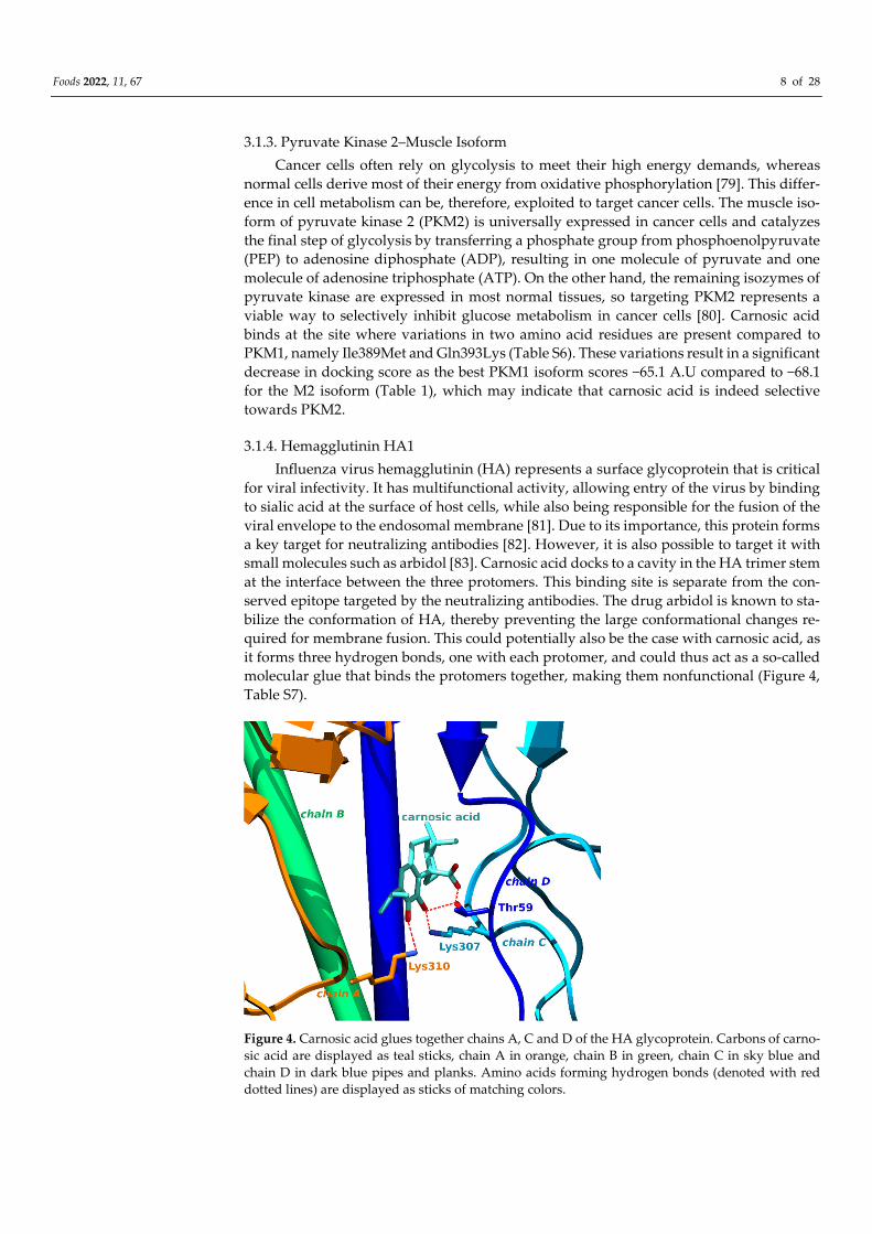
molecular glue that binds the protomers together, making them nonfunctional (Figure 4,
Table S7).
Figure 4. Carnosic acid glues together chains A, C and D of the HA glycoprotein. Carbons of carno-
sic acid are displayed as teal sticks, chain A in orange, chain B in green, chain C in sky blue and
chain D in dark blue pipes and planks. Amino acids forming hydrogen bonds (denoted with red
dotted lines) are displayed as sticks of matching colors.
Foods 2022, 11, 67 9 of 28
3.1.5. HIV-1 and HIV-2 Protease
Human immunodeficiency viruses (HIV) protease is a retroviral aspartyl protease
involved in the hydrolysis of several peptide bonds, which is essential for the life cycle
and replication of HIV [84]. Small molecule inhibitors of HIV protease play a critical role
in the effective treatment of acquired immunodeficiency syndrome AIDS, as they repre-
sent part of the highly active antiretroviral therapy (HAART). While HIV-1, carrier of the
HIV-1 protease isoform, forms the most common subtype worldwide, HIV-2 remains
mainly confined to West Africa and is also spreading in India [85,86]. However, the treat-
ment of HIV-2 is more difficult than that of HIV-1, as most antiviral drugs have been de-
veloped for the HIV-1 isoform. HIV-2 proteases have also been found more resistant to
small-molecule inhibition [87]. Moreover, dual infection with both isoforms is possible as
well [88]. Consequently, novel inhibitors for both HIV proteases would be of great benefit.
It has been shown that carnosic acid exhibits potent inhibition of the HIV-1 protease iso-
enzyme with an IC90 = 0.08 μg/mL [69]. Inhibition has not yet been experimentally demon-
strated for the HIV-2 isoform; however, our studies suggest that carnosic acid is also ca-
pable of inhibiting this isoform. This finding can also be corroborated by the fact that the
binding sites of both isoforms are very similar, with a sequence identity close to 70% and
the ProBiS Z-score of 3.76 [58]. ProBiS Z-score measures the statistical and structural sig-
nificance of local binding site similarity. Binding site alignments leading to ProBiS Z-
Scores higher than 2 are considered to be very similar. In addition, all the equivalent bind-
ing site amino acid residues are of the same charge and polarity type. Overall, carnosic
acid could prove to be a valuable starting point for the development of antivirals that
would be effective against both strains of HIV.
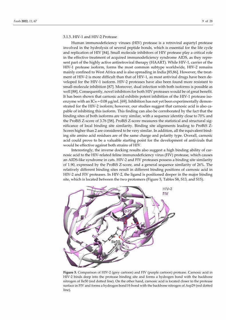
Interestingly, the inverse docking results also suggest a high binding ability of car-
nosic acid to the HIV-related feline immunodeficiency virus (FIV) protease, which causes
an AIDS-like syndrome in cats. HIV-2 and FIV proteases possess a binding site similarity
of 1.90, expressed by the ProBiS Z-score, and a general sequence similarity of 26%. The
relatively different binding sites result in different binding positions of carnosic acid in
HIV-2 and FIV proteases. In HIV-2, the ligand is positioned deeper in the major binding
site, which is located between the two protomers (Figure 5, Tables S8, S13, and S15).
Figure 5. Comparison of HIV-2 (grey cartoon) and FIV (purple cartoon) protease. Carnosic acid in
HIV-2 binds deep into the protease binding site and forms a hydrogen bond with the backbone
nitrogen of Ile50 (red dotted line). On the other hand, carnosic acid is located closer to the protease
surface in FIV and forms a hydrogen bond H-bond with the backbone nitrogen of Asp29 (red dotted
line).
Foods 2022, 11, 67 10 of 28
3.1.6. Enhanced Intra-Cellular Survival Protein
Tuberculosis represents the leading cause of infectious death worldwide, primarily
due to the emergence of multidrug-resistant tuberculosis and due to extensively drug-
resistant strains of Mycobacterium tuberculosis [89]. Up-regulation of the enhanced intra-
cellular survival (Eis) protein was found to be the sole cause of resistance to the amino-
glycoside of last resort-kanamycin in approximately one-third of Mycobacterium tuberculo-
sis isolates. Specifically, Eis represents an acetyltransferase responsible for Mycobacterium
tuberculosis resistance to multiple aminoglycoside drugs. A distinctive property of Eis is
that it acetylates the aminoglycoside drugs at multiple amine functional groups, prevent-
ing them from binding to their target, the ribosome. The simultaneous use of Eis inhibitors
with anti-tuberculosis drugs may therefore provide a way to combat this resistance by
restoring aminoglycoside drug activity [90]. Carnosic acid docks to the aminoglycoside
binding pocket formed by the N-terminal domain to which also tobramycin binds, thereby
suggesting the possibility of competitive inhibition of Eis by carnosic acid (Table S9) [91].
3.1.7. Peroxisome Proliferator-Activated Receptor δ
The peroxisome proliferator-activated receptor (PPARδ) functions as a sensor for di-
etary and endogenous fats [92]. It regulates the transcription of genes associated with lipid
and glucose metabolism. Specifically, it controls lipid degradation, transport, and storage,
while also being associated with insulin secretion and resistance. PPARδ agonists have
been shown beneficial in models of metabolic disorders in primates and may thus possess
therapeutic potential in hyperlipidemia, atherosclerosis, obesity, and diabetes [93,94].
PPARδ is also associated with cancer by promoting chronic inflammation through in-
creasing cyclooxygenase-2 (COX-2) expression and prostaglandin E2 production, leading
to an increase in proinflammatory cytokine concentrations. Moreover, the ability of
PPARδ to promote the use of fatty acids as the energy source may enhance cell survival
and proliferation under harsh metabolic conditions often found in tumors. Therefore,
PPARδ agonists may be useful in treating metabolic disorders, while antagonists may re-
duce inflammation-related disorders and slow down cancer progression.
Whereas there are no experimental data that carnosic acid, carnosol, or rosmanol
bind to PPARδ, it is known that both carnosol and carnosic acid represents agonists of the
PPARγ isoform with half maximal effective concentration (EC50)values of 41 and 20 μM,
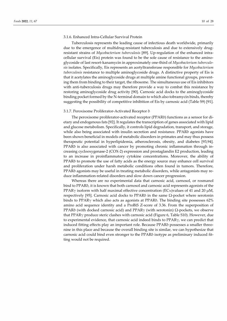
respectively [95]. Carnosic acid docks to PPARδ in the same Ω-pocket where serotonin
binds to PPARγ which also acts as agonists at PPARδ. The binding site possesses 62%
amino acid sequence identity and a ProBiS Z-score of 3.36. From the superposition of
PPARδ (with docked carnosic acid) and PPARγ (with serotonin) Ω-pockets, we observe
that PPARγ produce steric clashes with carnosic acid (Figure 6, Table S10). However, due
to experimental evidence, that carnosic acid indeed binds to PPARγ, we can predict that
induced fitting effects play an important role. Because PPARδ possesses a smaller threo-
nine in this place and because the overall binding site is similar, we can hypothesize that
carnosic acid could bind even stronger to the PPARδ isotype as preliminary induced fit-
ting would not be required.
Foods 2022, 11, 67 11 of 28
Figure 6. The Ω-pocket superimposition between PPARδ (orange cartoon and sticks) and PPARγ
(blue cartoon and sticks). The first amino acid residue numbering corresponds to PPARδ, and the
second to PPARγ. Serotonin is displayed using blue balls and sticks and the docked carnosic acid
using orange balls-and-sticks. We emphasize the difference in amino acid residues Thr252 versus
Arg288. Compared to Thr252 in PPARδ, the large Arg288 in PPARγ would lead to stearic clashes
with carnosic acid.
3.1.8. Glycogen Phosphorylase
Glycogen phosphorylase (GP) is an enzyme that cleaves the non-reducing ends in the
chain of glycogen to produce glucose-1-phosphate monomers which can be further con-
verted to free glucose [96]. Because glycogen is an important source of blood glucose, GP
represents a promising target for the treatment of type II diabetes, and its inhibitors have
been shown effective in controlling blood glucose concentrations in animal studies [97].
GP can exist in two different forms that bind different regulatory molecules: the active
phosphorylated (on Ser14) GPa and the non-phosphorylated GPb form. In addition, GP
has been reported to bind compounds at four different binding sites, identified as: (a) the
catalytic, (b) the allosteric (indole), (c) the novel allosteric, and (d) the inhibitory site (caf-
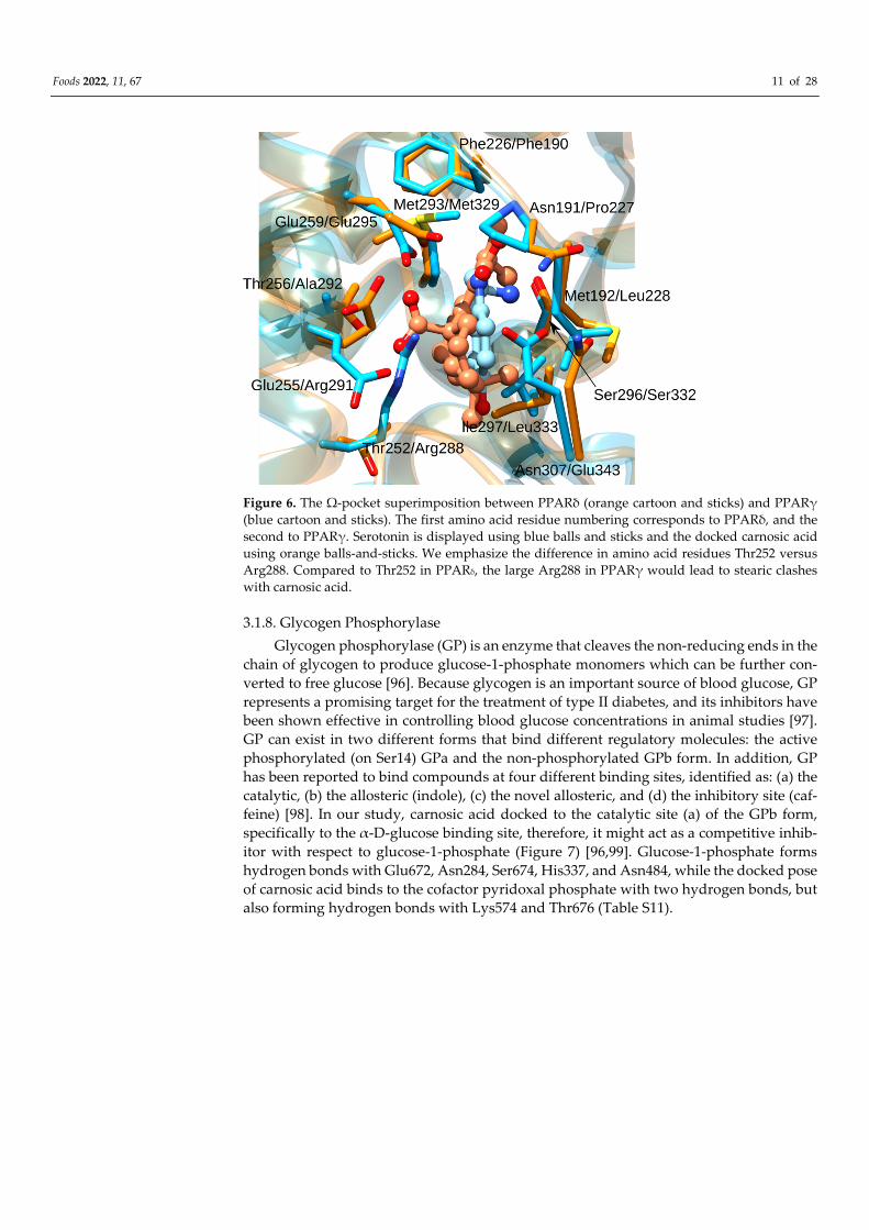
feine) [98]. In our study, carnosic acid docked to the catalytic site (a) of the GPb form,
specifically to the α-D-glucose binding site, therefore, it might act as a competitive inhib-
itor with respect to glucose-1-phosphate (Figure 7) [96,99]. Glucose-1-phosphate forms
hydrogen bonds with Glu672, Asn284, Ser674, His337, and Asn484, while the docked pose
of carnosic acid binds to the cofactor pyridoxal phosphate with two hydrogen bonds, but
also forming hydrogen bonds with Lys574 and Thr676 (Table S11).
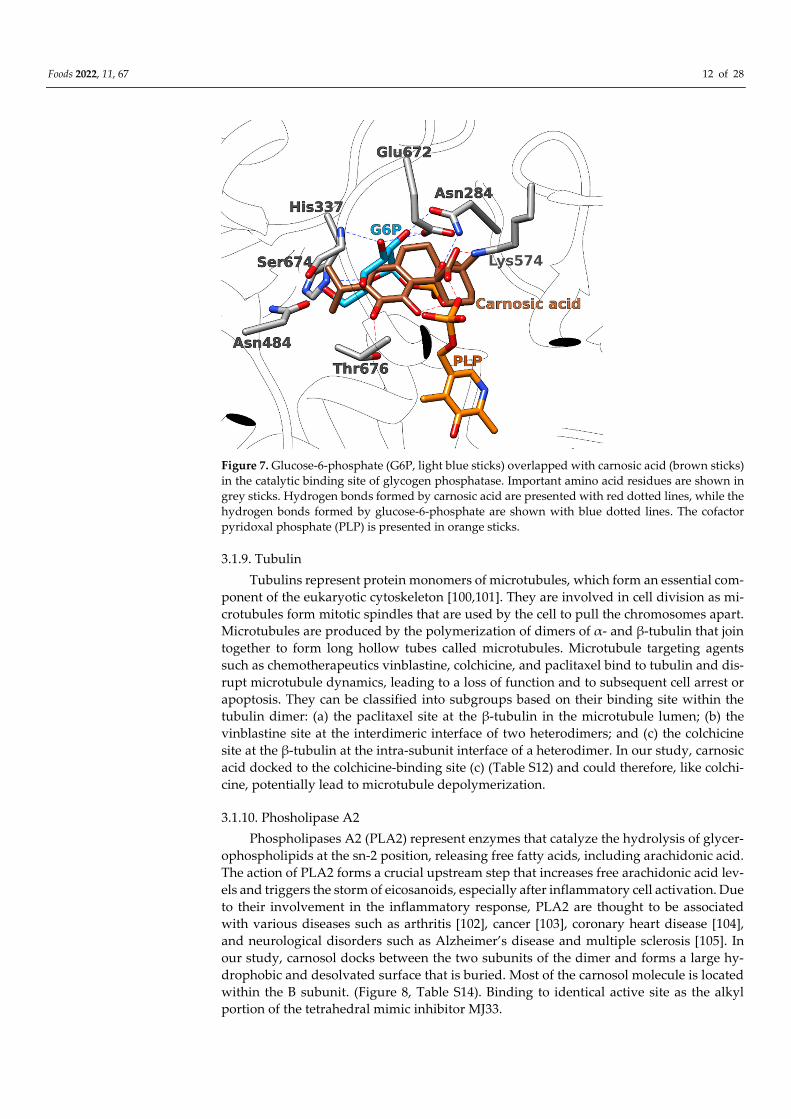
Foods 2022, 11, 67 12 of 28
Figure 7. Glucose-6-phosphate (G6P, light blue sticks) overlapped with carnosic acid (brown sticks)
in the catalytic binding site of glycogen phosphatase. Important amino acid residues are shown in
grey sticks. Hydrogen bonds formed by carnosic acid are presented with red dotted lines, while the
hydrogen bonds formed by glucose-6-phosphate are shown with blue dotted lines. The cofactor
pyridoxal phosphate (PLP) is presented in orange sticks.
3.1.9. Tubulin
Tubulins represent protein monomers of microtubules, which form an essential com-
ponent of the eukaryotic cytoskeleton [100,101]. They are involved in cell division as mi-
crotubules form mitotic spindles that are used by the cell to pull the chromosomes apart.
Microtubules are produced by the polymerization of dimers of α- and β-tubulin that join
together to form long hollow tubes called microtubules. Microtubule targeting agents
such as chemotherapeutics vinblastine, colchicine, and paclitaxel bind to tubulin and dis-
rupt microtubule dynamics, leading to a loss of function and to subsequent cell arrest or
apoptosis. They can be classified into subgroups based on their binding site within the
tubulin dimer: (a) the paclitaxel site at the β-tubulin in the microtubule lumen; (b) the
vinblastine site at the interdimeric interface of two heterodimers; and (c) the colchicine
site at the β-tubulin at the intra-subunit interface of a heterodimer. In our study, carnosic
acid docked to the colchicine-binding site (c) (Table S12) and could therefore, like colchi-
cine, potentially lead to microtubule depolymerization.
3.1.10. Phosholipase A2
Phospholipases A2 (PLA2) represent enzymes that catalyze the hydrolysis of glycer-
ophospholipids at the sn-2 position, releasing free fatty acids, including arachidonic acid.
The action of PLA2 forms a crucial upstream step that increases free arachidonic acid lev-
els and triggers the storm of eicosanoids, especially after inflammatory cell activation. Due
to their involvement in the inflammatory response, PLA2 are thought to be associated
with various diseases such as arthritis [102], cancer [103], coronary heart disease [104],
and neurological disorders such as Alzheimer’s disease and multiple sclerosis [105]. In
our study, carnosol docks between the two subunits of the dimer and forms a large hy-
drophobic and desolvated surface that is buried. Most of the carnosol molecule is located
within the B subunit. (Figure 8, Table S14). Binding to identical active site as the alkyl
portion of the tetrahedral mimic inhibitor MJ33.
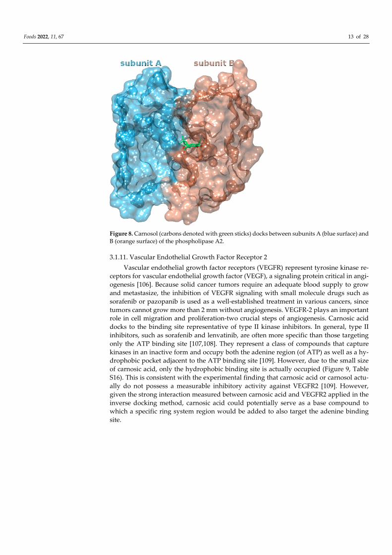
Foods 2022, 11, 67 13 of 28
Figure 8. Carnosol (carbons denoted with green sticks) docks between subunits A (blue surface) and
B (orange surface) of the phospholipase A2.
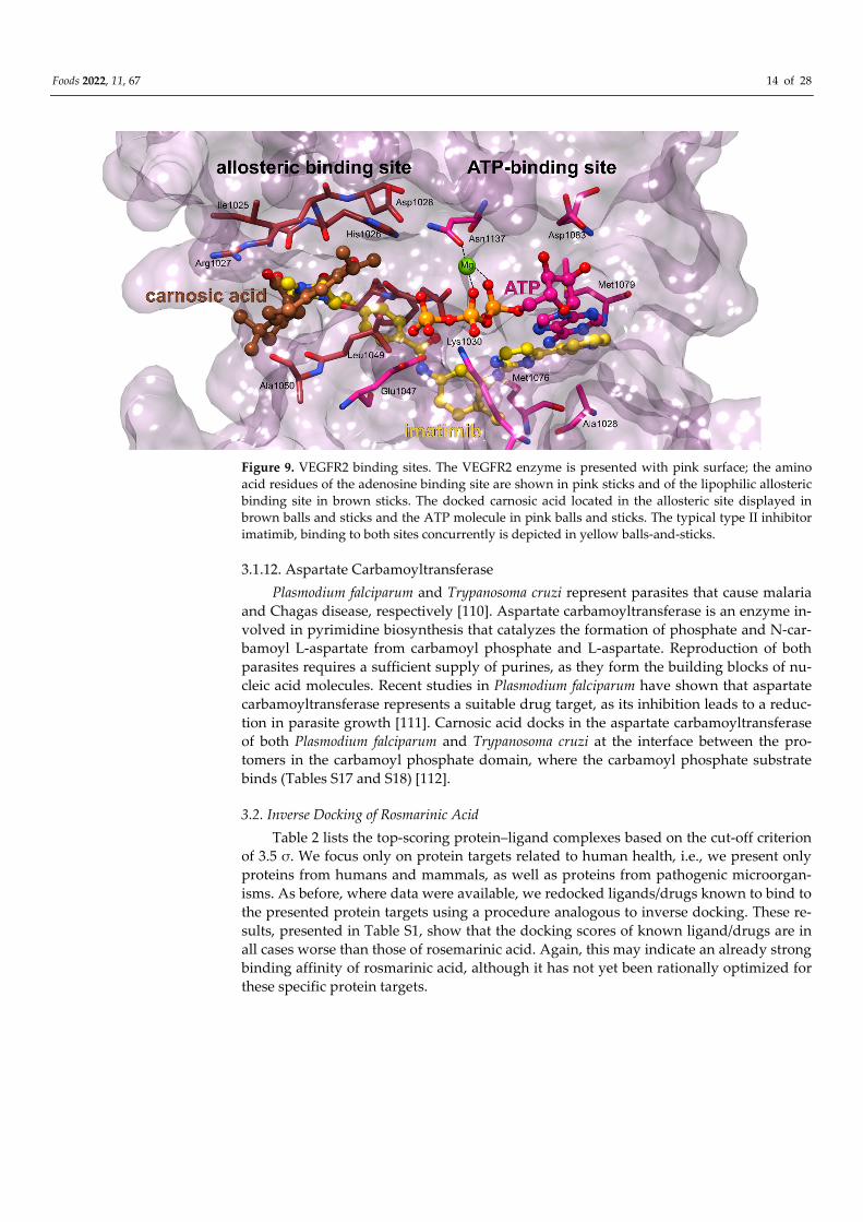
3.1.11. Vascular Endothelial Growth Factor Receptor 2
Vascular endothelial growth factor receptors (VEGFR) represent tyrosine kinase re-
ceptors for vascular endothelial growth factor (VEGF), a signaling protein critical in angi-
ogenesis [106]. Because solid cancer tumors require an adequate blood supply to grow
and metastasize, the inhibition of VEGFR signaling with small molecule drugs such as
sorafenib or pazopanib is used as a well-established treatment in various cancers, since
tumors cannot grow more than 2 mm without angiogenesis. VEGFR-2 plays an important
role in cell migration and proliferation-two crucial steps of angiogenesis. Carnosic acid
docks to the binding site representative of type II kinase inhibitors. In general, type II
inhibitors, such as sorafenib and lenvatinib, are often more specific than those targeting
only the ATP binding site [107,108]. They represent a class of compounds that capture
kinases in an inactive form and occupy both the adenine region (of ATP) as well as a hy-
drophobic pocket adjacent to the ATP binding site [109]. However, due to the small size
of carnosic acid, only the hydrophobic binding site is actually occupied (Figure 9, Table
S16). This is consistent with the experimental finding that carnosic acid or carnosol actu-
ally do not possess a measurable inhibitory activity against VEGFR2 [109]. However,
given the strong interaction measured between carnosic acid and VEGFR2 applied in the
inverse docking method, carnosic acid could potentially serve as a base compound to
which a specific ring system region would be added to also target the adenine binding
site.
Foods 2022, 11, 67 14 of 28
Figure 9. VEGFR2 binding sites. The VEGFR2 enzyme is presented with pink surface; the amino
acid residues of the adenosine binding site are shown in pink sticks and of the lipophilic allosteric
binding site in brown sticks. The docked carnosic acid located in the allosteric site displayed in
brown balls and sticks and the ATP molecule in pink balls and sticks. The typical type II inhibitor
imatimib, binding to both sites concurrently is depicted in yellow balls-and-sticks.
3.1.12. Aspartate Carbamoyltransferase
Plasmodium falciparum and Trypanosoma cruzi represent parasites that cause malaria
and Chagas disease, respectively [110]. Aspartate carbamoyltransferase is an enzyme in-
volved in pyrimidine biosynthesis that catalyzes the formation of phosphate and N-car-
bamoyl L-aspartate from carbamoyl phosphate and L-aspartate. Reproduction of both
parasites requires a sufficient supply of purines, as they form the building blocks of nu-
cleic acid molecules. Recent studies in Plasmodium falciparum have shown that aspartate
carbamoyltransferase represents a suitable drug target, as its inhibition leads to a reduc-
tion in parasite growth [111]. Carnosic acid docks in the aspartate carbamoyltransferase
of both Plasmodium falciparum and Trypanosoma cruzi at the interface between the pro-
tomers in the carbamoyl phosphate domain, where the carbamoyl phosphate substrate
binds (Tables S17 and S18) [112].
3.2. Inverse Docking of Rosmarinic Acid
Table 2 lists the top-scoring protein–ligand complexes based on the cut-off criterion
of 3.5 σ. We focus only on protein targets related to human health, i.e., we present only
proteins from humans and mammals, as well as proteins from pathogenic microorgan-
isms. As before, where data were available, we redocked ligands/drugs known to bind to
the presented protein targets using a procedure analogous to inverse docking. These re-
sults, presented in Table S1, show that the docking scores of known ligand/drugs are in
all cases worse than those of rosemarinic acid. Again, this may indicate an already strong
binding affinity of rosmarinic acid, although it has not yet been rationally optimized for
these specific protein targets.
Foods 2022, 11, 67 15 of 28
Table 2. Best scoring mammalian, human, and pathogen protein targets of rosmarinic acid. Docking
scores independent of the organism or type of protein are collected in Supplementary Materials in
Table S3.
Rank PDB ID
with Chain
Predicted Ligand
Docking Score
(arb. Units)
Protein Name Organism Protein Function and Dis-
ease Correlation
Reported Experi-
mental Correlation
of Protein and Lig-
and
1 2d1jA −86.1 Coagulation factor X Homo sapiens
Serine endopeptidase is in-
volved in the coagulation
cascade. Its deficiency leads
to a bleeding disorder. Its
inhibitors are popular anti-
coagulants.
2 1fxfB −84.8 Phospholipase A2 Sus scrofa
Catalyzes the hydrolysis of
glycerophospholipids thus
releasing free fatty acids, in-
cluding arachidonic acid. Its
action is implicated in sev-
eral inflammation-based
disease such as arthritis,
coronary artery disease,
Alzheimer’s and cancer.
[113]
3 2jt5A −84.5 Matrix metalloproteinase-
3 Homo sapiens
Zinc-dependent endopepti-
dase which is involved in
the remodeling of the extra-
cellular matrix. Involved
in arthritis, multiple sclero-
sis, aneurysms, and the
spread of metastatic cancer.
After traumatic brain in-
jury, matrix metalloprotein-
ase-3 (MMP-3) concentra-
tions increase and lead to
additional damage to the
blood–brain barrier.
4 4jzbA −83.2 Farnesyl pyrophosphate
synthase Leishmania major
Farnesyl pyrophosphate
synthase (FPPS) is an essen-
tial enzyme involved in the
biosynthesis of ergosterol in
leishmania parasites, the
causative agents of leish-
maniasis.
[114]
5 3qmuB −80.2 Glutamate dehydrogen-
ase 1 Bos Taurus
Part of the glutaminolysis
pathway, playing a crucial
role in nitrogen and carbon
metabolism. Inhibition
leads to in vivo and in vitro
reduced viability of cancer
cells.
6 5fi6A −77.6 Glutaminase Homo sapiens
3.2.1. Coagulation Factor X
Factor X represents an enzyme involved in the coagulation cascade that, when acti-
vated by the hydrolysis of factor Xa, claves prothrombin to the active thrombin, which in
turn converts soluble fibrinogen to insoluble fibrin strands [115]. The role of factor X is
particularly important because it is the first enzyme where the intrinsic and extrinsic co-
agulation pathways converge. Drug manipulation of the coagulation cascade is extremely
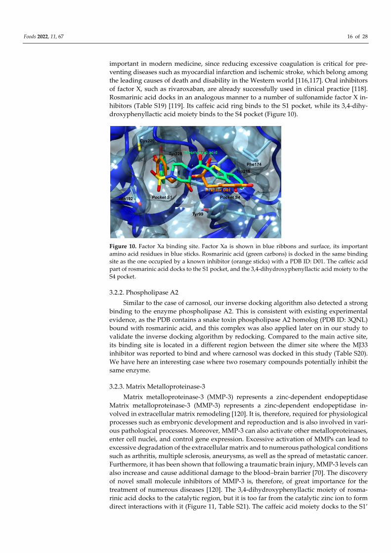
Foods 2022, 11, 67 16 of 28
important in modern medicine, since reducing excessive coagulation is critical for pre-
venting diseases such as myocardial infarction and ischemic stroke, which belong among
the leading causes of death and disability in the Western world [116,117]. Oral inhibitors
of factor X, such as rivaroxaban, are already successfully used in clinical practice [118].
Rosmarinic acid docks in an analogous manner to a number of sulfonamide factor X in-
hibitors (Table S19) [119]. Its caffeic acid ring binds to the S1 pocket, while its 3,4-dihy-
droxyphenyllactic acid moiety binds to the S4 pocket (Figure 10).
Figure 10. Factor Xa binding site. Factor Xa is shown in blue ribbons and surface, its important
amino acid residues in blue sticks. Rosmarinic acid (green carbons) is docked in the same binding
site as the one occupied by a known inhibitor (orange sticks) with a PDB ID: D01. The caffeic acid
part of rosmarinic acid docks to the S1 pocket, and the 3,4-dihydroxyphenyllactic acid moiety to the
S4 pocket.
3.2.2. Phospholipase A2
Similar to the case of carnosol, our inverse docking algorithm also detected a strong
binding to the enzyme phospholipase A2. This is consistent with existing experimental
evidence, as the PDB contains a snake toxin phospholipase A2 homolog (PDB ID: 3QNL)
bound with rosmarinic acid, and this complex was also applied later on in our study to
validate the inverse docking algorithm by redocking. Compared to the main active site,
its binding site is located in a different region between the dimer site where the MJ33
inhibitor was reported to bind and where carnosol was docked in this study (Table S20).
We have here an interesting case where two rosemary compounds potentially inhibit the
same enzyme.
3.2.3. Matrix Metalloproteinase-3
Matrix metalloproteinase-3 (MMP-3) represents a zinc-dependent endopeptidase
Matrix metalloproteinase-3 (MMP-3) represents a zinc-dependent endopeptidase in-
volved in extracellular matrix remodeling [120]. It is, therefore, required for physiological
processes such as embryonic development and reproduction and is also involved in vari-
ous pathological processes. Moreover, MMP-3 can also activate other metalloproteinases,
enter cell nuclei, and control gene expression. Excessive activation of MMPs can lead to
excessive degradation of the extracellular matrix and to numerous pathological conditions
such as arthritis, multiple sclerosis, aneurysms, as well as the spread of metastatic cancer.
Furthermore, it has been shown that following a traumatic brain injury, MMP-3 levels can
also increase and cause additional damage to the blood–brain barrier [70]. The discovery
of novel small molecule inhibitors of MMP-3 is, therefore, of great importance for the
treatment of numerous diseases [120]. The 3,4-dihydroxyphenyllactic moiety of rosma-
rinic acid docks to the catalytic region, but it is too far from the catalytic zinc ion to form
direct interactions with it (Figure 11, Table S21). The caffeic acid moiety docks to the S1’
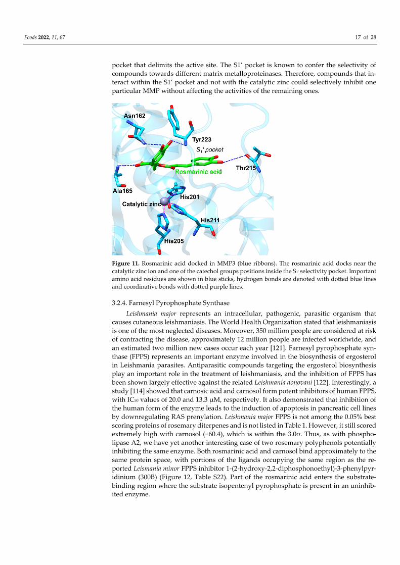
Foods 2022, 11, 67 17 of 28
pocket that delimits the active site. The S1’ pocket is known to confer the selectivity of
compounds towards different matrix metalloproteinases. Therefore, compounds that in-
teract within the S1’ pocket and not with the catalytic zinc could selectively inhibit one
particular MMP without affecting the activities of the remaining ones.
Figure 11. Rosmarinic acid docked in MMP3 (blue ribbons). The rosmarinic acid docks near the
catalytic zinc ion and one of the catechol groups positions inside the S1′ selectivity pocket. Important
amino acid residues are shown in blue sticks, hydrogen bonds are denoted with dotted blue lines
and coordinative bonds with dotted purple lines.
3.2.4. Farnesyl Pyrophosphate Synthase
Leishmania major represents an intracellular, pathogenic, parasitic organism that
causes cutaneous leishmaniasis. The World Health Organization stated that leishmaniasis
is one of the most neglected diseases. Moreover, 350 million people are considered at risk
of contracting the disease, approximately 12 million people are infected worldwide, and
an estimated two million new cases occur each year [121]. Farnesyl pyrophosphate syn-
thase (FPPS) represents an important enzyme involved in the biosynthesis of ergosterol
in Leishmania parasites. Antiparasitic compounds targeting the ergosterol biosynthesis
play an important role in the treatment of leishmaniasis, and the inhibition of FPPS has
been shown largely effective against the related Leishmania donovani [122]. Interestingly, a
study [114] showed that carnosic acid and carnosol form potent inhibitors of human FPPS,
with IC50 values of 20.0 and 13.3 μM, respectively. It also demonstrated that inhibition of
the human form of the enzyme leads to the induction of apoptosis in pancreatic cell lines
by downregulating RAS prenylation. Leishmania major FPPS is not among the 0.05% best
scoring proteins of rosemary diterpenes and is not listed in Table 1. However, it still scored
extremely high with carnosol (−60.4), which is within the 3.0σ. Thus, as with phospho-
lipase A2, we have yet another interesting case of two rosemary polyphenols potentially
inhibiting the same enzyme. Both rosmarinic acid and carnosol bind approximately to the
same protein space, with portions of the ligands occupying the same region as the re-
ported Leismania minor FPPS inhibitor 1-(2-hydroxy-2,2-diphosphonoethyl)-3-phenylpyr-
idinium (300B) (Figure 12, Table S22). Part of the rosmarinic acid enters the substrate-
binding region where the substrate isopentenyl pyrophosphate is present in an uninhib-
ited enzyme.
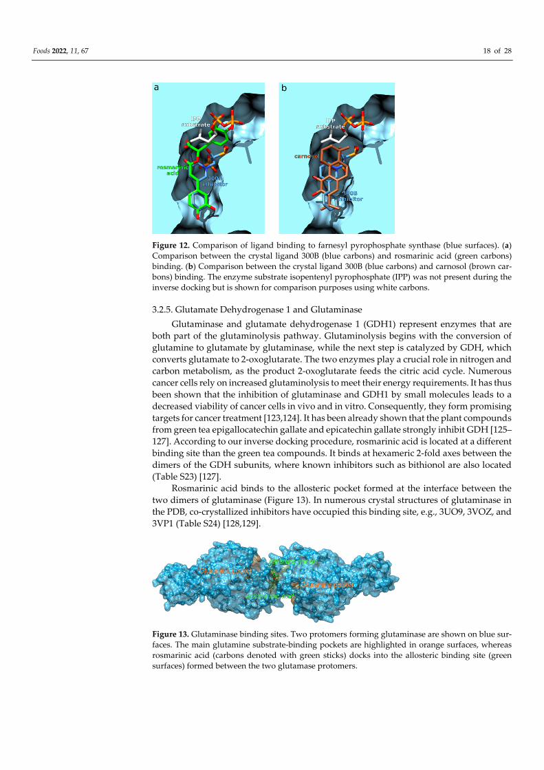
Foods 2022, 11, 67 18 of 28
Figure 12. Comparison of ligand binding to farnesyl pyrophosphate synthase (blue surfaces). (a)
Comparison between the crystal ligand 300B (blue carbons) and rosmarinic acid (green carbons)
binding. (b) Comparison between the crystal ligand 300B (blue carbons) and carnosol (brown car-
bons) binding. The enzyme substrate isopentenyl pyrophosphate (IPP) was not present during the
inverse docking but is shown for comparison purposes using white carbons.
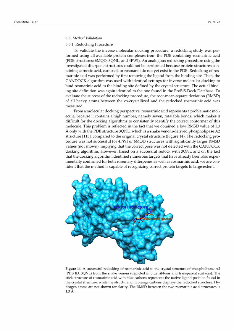
3.2.5. Glutamate Dehydrogenase 1 and Glutaminase
Glutaminase and glutamate dehydrogenase 1 (GDH1) represent enzymes that are
both part of the glutaminolysis pathway. Glutaminolysis begins with the conversion of
glutamine to glutamate by glutaminase, while the next step is catalyzed by GDH, which
converts glutamate to 2-oxoglutarate. The two enzymes play a crucial role in nitrogen and
carbon metabolism, as the product 2-oxoglutarate feeds the citric acid cycle. Numerous
cancer cells rely on increased glutaminolysis to meet their energy requirements. It has thus
been shown that the inhibition of glutaminase and GDH1 by small molecules leads to a
decreased viability of cancer cells in vivo and in vitro. Consequently, they form promising
targets for cancer treatment [123,124]. It has been already shown that the plant compounds
from green tea epigallocatechin gallate and epicatechin gallate strongly inhibit GDH [125–
127]. According to our inverse docking procedure, rosmarinic acid is located at a different
binding site than the green tea compounds. It binds at hexameric 2-fold axes between the
dimers of the GDH subunits, where known inhibitors such as bithionol are also located
(Table S23) [127].
Rosmarinic acid binds to the allosteric pocket formed at the interface between the
two dimers of glutaminase (Figure 13). In numerous crystal structures of glutaminase in
the PDB, co-crystallized inhibitors have occupied this binding site, e.g., 3UO9, 3VOZ, and
3VP1 (Table S24) [128,129].
Figure 13. Glutaminase binding sites. Two protomers forming glutaminase are shown on blue sur-
faces. The main glutamine substrate-binding pockets are highlighted in orange surfaces, whereas
rosmarinic acid (carbons denoted with green sticks) docks into the allosteric binding site (green
surfaces) formed between the two glutamase protomers.
Foods 2022, 11, 67 19 of 28
3.3. Method Validation
3.3.1. Redocking Procedure
To validate the inverse molecular docking procedure, a redocking study was per-
formed using all available protein complexes from the PDB containing rosmarinic acid
(PDB structures: 6MQD, 3QNL, and 4PWI). An analogous redocking procedure using the
investigated diterpene structures could not be performed because protein structures con-
taining carnosic acid, carnosol, or rosmanol do not yet exist in the PDB. Redocking of ros-
marinic acid was performed by first removing the ligand from the binding site. Then, the
CANDOCK algorithm was used with identical settings for inverse molecular docking to
bind rosmarinic acid to the binding site defined by the crystal structure. The actual bind-
ing site definition was again identical to the one found in the ProBiS-Dock Database. To
evaluate the success of the redocking procedure, the root-mean-square deviation (RMSD)
of all heavy atoms between the co-crystallized and the redocked rosmarinic acid was
measured.
From a molecular docking perspective, rosmarinic acid represents a problematic mol-
ecule, because it contains a high number, namely seven, rotatable bonds, which makes it
difficult for the docking algorithms to consistently identify the correct conformer of this
molecule. This problem is reflected in the fact that we obtained a low RMSD value of 1.3
Å only with the PDB structure 3QNL, which is a snake venom-derived phospholipase A2
structure [113], compared to the original crystal structure (Figure 14). The redocking pro-
cedure was not successful for 4PWI or 6MQD structures with significantly larger RMSD
values (not shown), implying that the correct pose was not detected with the CANDOCK
docking algorithm. However, based on a successful redock with 3QNL and on the fact
that the docking algorithm identified numerous targets that have already been also exper-
imentally confirmed for both rosemary diterpenes as well as rosmarinic acid, we are con-
fident that the method is capable of recognizing correct protein targets to large extent.
Figure 14. A successful redocking of rosmarinic acid to the crystal structure of phospholipase A2
(PDB ID: 3QNL) from the snake venom (depicted in blue ribbons and transparent surfaces). The
stick structure of rosmarinic acid with blue carbons represents the native ligand position found in
the crystal structure, while the structure with orange carbons displays the redocked structure. Hy-
drogen atoms are not shown for clarity. The RMSD between the two rosmarinic acid structures is
1.3 Å.
Foods 2022, 11, 67 20 of 28
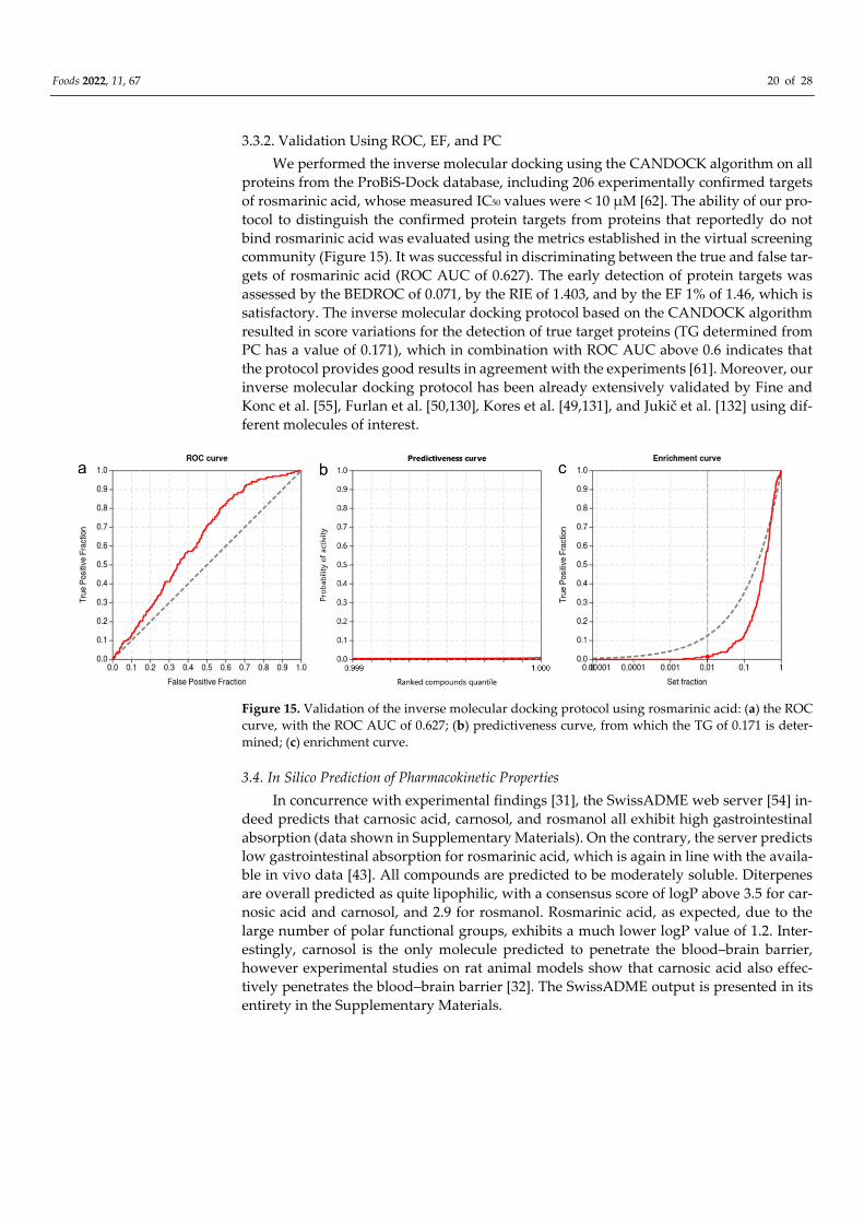
3.3.2. Validation Using ROC, EF, and PC
We performed the inverse molecular docking using the CANDOCK algorithm on all
proteins from the ProBiS-Dock database, including 206 experimentally confirmed targets
of rosmarinic acid, whose measured IC50 values were < 10 μM [62]. The ability of our pro-
tocol to distinguish the confirmed protein targets from proteins that reportedly do not
bind rosmarinic acid was evaluated using the metrics established in the virtual screening
community (Figure 15). It was successful in discriminating between the true and false tar-
gets of rosmarinic acid (ROC AUC of 0.627). The early detection of protein targets was
assessed by the BEDROC of 0.071, by the RIE of 1.403, and by the EF 1% of 1.46, which is
satisfactory. The inverse molecular docking protocol based on the CANDOCK algorithm
resulted in score variations for the detection of true target proteins (TG determined from
PC has a value of 0.171), which in combination with ROC AUC above 0.6 indicates that
the protocol provides good results in agreement with the experiments [61]. Moreover, our
inverse molecular docking protocol has been already extensively validated by Fine and
Konc et al. [55], Furlan et al. [50,130], Kores et al. [49,131], and Jukič et al. [132] using dif-
ferent molecules of interest.
Figure 15. Validation of the inverse molecular docking protocol using rosmarinic acid: (a) the ROC
curve, with the ROC AUC of 0.627; (b) predictiveness curve, from which the TG of 0.171 is deter-
mined; (c) enrichment curve.
3.4. In Silico Prediction of Pharmacokinetic Properties
In concurrence with experimental findings [31], the SwissADME web server [54] in-
deed predicts that carnosic acid, carnosol, and rosmanol all exhibit high gastrointestinal
absorption (data shown in Supplementary Materials). On the contrary, the server predicts
low gastrointestinal absorption for rosmarinic acid, which is again in line with the availa-
ble in vivo data [43]. All compounds are predicted to be moderately soluble. Diterpenes
are overall predicted as quite lipophilic, with a consensus score of logP above 3.5 for car-
nosic acid and carnosol, and 2.9 for rosmanol. Rosmarinic acid, as expected, due to the
large number of polar functional groups, exhibits a much lower logP value of 1.2. Inter-
estingly, carnosol is the only molecule predicted to penetrate the blood–brain barrier,
however experimental studies on rat animal models show that carnosic acid also effec-
tively penetrates the blood–brain barrier [32]. The SwissADME output is presented in its
entirety in the Supplementary Materials.
Foods 2022, 11, 67 21 of 28
4. Discussion
Natural plant-based compounds play an important role in the development of novel
drugs as they may possess several advantages over conventional synthetic compounds,
namely, fewer side effects, lower long-term toxicity, and versatile biological effects [130].
We report the potential targets of the major compounds from Rosmarinus officinalis, in-
cluding the diterpenes carnosic acid, carnosol, and rosmanol, as well as the polyphenolic
ester rosmarinic acid. Their targets were identified in silico using an inverse molecular
docking approach. All four compounds were individually docked to all non-redundant
holo-proteins available in the PDB. To identify the binding sites of each protein in ad-
vance, we applied the recently developed ProBiS-Dock Database—a freely available re-
pository of binding sites between small ligands and proteins. Thereby, the docking pro-
cedure was limited to binding sites already known to bind at least one drug-like small-
molecule ligand or to binding sites exhibiting a high similarity with the already known
binding sites. Moreover, we used the novel CANDOCK algorithm, which employs a frag-
ment-based docking approach with maximum clique and a knowledge-based scoring
function.
Due to the similar molecular structure and docking/scoring values, we combined the
results of all three investigated diterpenes into a single set (Table 1). We identified numer-
ous human/mammalian proteins that could explain the observed anticarcinogenic activi-
ties of rosemary diterpenes. The best docking score was obtained for the complex between
carnosic acid and the proto-oncogene K-Ras G12C. Moreover, the anticarcinogenic activi-
ties can also be explained by the potential binding of rosemary diterpenes to pyruvate
kinase, PPARδ, tubulin, VEGFR2, and phospholipase A2. In general, phospholipase A2
has also been strongly implicated in inflammation-related disorders, so its inhibition may
be likewise beneficial in arthritis, coronary artery disease, or dementia. Due to the identi-
fication of potential binding of the investigated diterpenes to glycogen phosphorylase,
which facilitates glucose production, these compounds may be also useful in the treatment
of type II diabetes. Furthermore, rosemary diterpenes exhibit antiviral activities.
From previous experimental studies, it is known that carnosol strongly inhibits HIV-
1 protease. However, we also found out that rosemary diterpenes may bind strongly to
the HIV-2 enzyme isotype. These compounds therefore likely represent a good starting
point for the development of drugs against AIDS that could treat concurrent infections
with HIV-1 and HIV-2. Interestingly, all diterpenes also yield good docking scores when
bound to the feline immunodeficiency virus (FIV) protease, which is strongly related to
HIV proteases, suggesting their potential utility in veterinary medicine. Finally, we have
also found out that these compounds can bind to HA1 of the influenza A virus, potentially
reducing its infectivity.
The antibacterial activity of investigated diterpenes can be explained by our discov-
ery that they can bind to the enzyme glucosamine-fructose-6-phosphate aminotransferase
in Escherichia coli, which is critical for the first step of hexosamine metabolism responsible
for bacterial growth. Encouragingly, we have also found out that they can bind to the Eis
protein of Mycobacterium tuberculosis, which confers resistance to aminoglycoside drugs,
rendering them inactive. Therefore, the inhibition of this enzyme in conjunction with tu-
berculosis treatment could be beneficial in reducing the bacterial resistance to these drugs.
The investigated diterpenes also displayed binding to aspartate carbamoyltransfer-
ase of two different pathogenic parasites-P. falciparum and T. cruzi. P. falciparum represents
the causative agent of malaria, while T. cruzi causes Chagas disease. Inhibition of this en-
zyme results in the inability of the two parasites to produce pyrimidines, limiting their
biosynthesis of new nucleic acids.
Like diterpenes, rosmarinic acid also shows binding to proteins involved in carcino-
genesis, namely matrix metalloproteinase-3 and phospholipase A2. Interestingly, all four
compounds display very favorable binding scores for the enzyme phospholipase A2,
which could provide a possible explanation for the strong anti-inflammatory effects of
Foods 2022, 11, 67 22 of 28
rosemary. According to our results, rosmarinic acid may also interfere with the glutami-
nolysis pathway, as it forms top-scoring complexes with two related enzymes—glutami-
nase and glutamate dehydrogenase. Inhibition of this pathway by small-molecule drugs
has been indeed shown to reduce cancer cell viability. Moreover, the complex between
rosmarinic acid and coagulation factor X yielded the best scoring result. Regulating blood
clotting with drugs is of utmost importance, as reducing excessive blood clotting is crucial
in preventing diseases such as heart attacks and ischemic strokes, which belong among
the leading causes of death and disability in the Western world. Furthermore, rosmarinic
acid might also possess antiparasitic activity as its binding to farnesyl pyrophosphate syn-
thase (FPPS) of Leishmania major obtained a favorable docking score. This parasite causes
zoonotic cutaneous leishmaniasis, and inhibition of the FPPS prevents the biosynthesis of
ergosterol.
The results of this study will facilitate future molecular dynamics studies. Therein,
we plan to investigate the dynamic binding patterns of prior parameterized rosemary
compounds to the notable protein targets identified here. The molecular dynamics obser-
vations will be extended with the linear interaction energy as well as linear response ap-
proximation calculations to obtain the binding free energy values, which will then be com-
pared with drug ligands already known to bind to the protein targets described here.
5. Conclusions
Using an in silico inverse molecular docking procedure, we identified protein targets
that could explain the observed pharmacological activities of rosemary or its major poly-
phenolic constituents. By identifying protein structures to which carnosic acid, carnosol,
rosmanol, and rosmarinic acid can bind, we provide possible explanations for the ob-
served anticarcinogenic, anti-inflammatory, antidiabetic, antiviral, and antibacterial activ-
ities of rosemary. In addition, using this methodology we were able to predict new effects
of these compounds that have not yet been reported, namely their anticoagulant and an-
tiparasitic activities. Lastly, we believe that our research can form the basis for the devel-
opment of novel drugs, where the rosemary compounds studied here could serve as a
starting point for efficient drug design.
Supplementary Materials: The following are available online at www.mdpi.com/arti-
cle/10.3390/foods11010067/s1. Table S1: Best docking scores of ligands/drugs known to bind to a
specific target. Table S2: Best docking scores for carnosol, carnosic acid and rosmanol. Table S3: Best
docking scores for rosmarinic acid. Table S4–S24: Interaction of compounds with respective targets
presented in Table 1; Table 2. The SwissADME output file can be found in the file swissadme_out-
put.xlsx supplied as part of the Supplementary Materials.
Author Contributions: Conceptualization, S.L. and U.B.; methodology, S.L. and U.B.; validation,
S.L.; formal analysis, S.L.; investigation, S.L.; resources, U.B.; Writing—Original draft preparation,
S.L.; Writing—review and editing, S.L. and U.B.; visualization, S.L.; supervision, U.B. All authors
have read and agreed to the published version of the manuscript.
Funding: Financial support through Slovenian Research Agency (ARRS) grants project and pro-
gramme J1-2471 and P2-0046 as well as through Slovenian Ministry of Education, Science and Sports
project grants C3330-19-952021 and AB FREE is gratefully acknowledged.
Institutional Review Board Statement: Not applicable.
Informed Consent Statement: Not applicable.
Data Availability Statement: All data generated or analyzed during this study are included in the
published article.
Conflicts of Interest: The authors declare no conflict of interest.
Foods 2022, 11, 67 23 of 28
Abbreviations
SMILES Simplified Molecular-Input Line-Entry System
CANDOCK Chemical Atomic Network based Docking
CHARMM Chemistry at Harvard Macromolecular Mechanics
ROC Receiver operating characteristics curve
PC Predictiveness curve
TPF True positive fraction
FPF False positive fraction
PDB Protein Data Bank
ROC AUC Area under the receiver operating characteristics curve
EF Enrichment factor
BEDROC Boltzmann-enhanced discrimination of ROC
RIE Robust initial enhancement
TG Total gain
Eis Enhanced intracellular survival
HIV Human immunodeficiency virus
FIV Feline immunodeficiency virus
RAS/MAPK Rat sarcoma/mitogen-activated protein kinase
GTP Guanosine triphosphate
GlmS Glucosamine/fructose-6-phosphate aminotransferase
PKM Pyruvate kinase M
PEP Phosphoenolpyruvate
ADP Adenosine diphosphate
ATP Adenosine triphosphate
HA Hemagglutinin
AIDS Acquired immunodeficiency syndrome
HAART Highly active antiretroviral therapy
PPAR Peroxisome proliferator-activated receptor
COX-2 Cyclooxygenase-2
EC50 Half maximal effective concentration
GP Glycogen phosphorylase
PLA2 Phospholipase A2
VEGFR Vascular endothelial growth factor receptor
VEGF Vascular endothelial growth factor
MMP Matrix metalloproteinase
FPPS Farnesyl pyrophosphate synthase
IPP Isopentenyl pyrophosphate
GDH1 Glutamate dehydrogenase 1
RMSD Root-mean-square deviation
References
1. Begum, A.; Sandhya, S.; Ali, S.S.; Vinod, K.R.; Reddy, S.; Banji, D. An in-depth review on the medicinal flora Rosmarinus officinalis
(Lamiaceae). Acta Sci. Pol. Technol. Aliment. 2013, 12, 61–74.
2. Al-Sereiti, M.R.; Abu-Amer, K.M.; Sena, P. Pharmacology of rosemary (Rosmarinus officinalis Linn.) and its therapeutic
potentials. IJEB 1999, 37, 124–130.
3. Perez-Fons, L.; Garzon, M.T.; Micol, V. Relationship between the antioxidant capacity and effect of rosemary (Rosmarinus
officinalis L.) polyphenols on membrane phospholipid order. J. Agric. Food Chem. 2009, 58, 161–171.
4. Yu, M.-H.; Choi, J.-H.; Chae, I.-G.; Im, H.-G.; Yang, S.-A.; More, K.; Lee, I.-S.; Lee, J. Suppression of LPS-induced inflammatory
activities by Rosmarinus officinalis L. Food Chem. 2013, 136, 1047–1054.
5. Bakırel, T.; Bakırel, U.; Keleş, O.Ü.; Ülgen, S.G.; Yardibi, H. In vivo assessment of antidiabetic and antioxidant activities of
rosemary (Rosmarinus officinalis) in alloxan-diabetic rabbits. J. Ethnopharmacol. 2008, 116, 64–73.
https://doi.org/10.1016/j.jep.2007.10.039.
6. Bozin, B.; Mimica-Dukic, N.; Samojlik, I.; Jovin, E. Antimicrobial and antioxidant properties of rosemary and sage (Rosmarinus
officinalis L. and Salvia officinalis L., Lamiaceae) essential oils. J. Agric. Food Chem. 2007, 55, 7879–7885.
7. Tai, J.; Cheung, S.; Wu, M.; Hasman, D. Antiproliferation effect of Rosemary (Rosmarinus officinalis) on human ovarian cancer
cells in vitro. Phytomedicine 2012, 19, 436–443. https://doi.org/10.1016/j.phymed.2011.12.012.
Foods 2022, 11, 67 24 of 28
8. Valdés, A.; García-Cañas, V.; Rocamora-Reverte, L.; Gómez-Martínez, Á.; Ferragut, J.A.; Cifuentes, A. Effect of rosemary
polyphenols on human colon cancer cells: Transcriptomic profiling and functional enrichment analysis. Genes Nutr. 2013, 8, 43–
60. https://doi.org/10.1007/s12263-012-0311-9.
9. Yesil-Celiktas, O.; Sevimli, C.; Bedir, E.; Vardar-Sukan, F. Inhibitory effects of rosemary extracts, carnosic acid and rosmarinic
acid on the growth of various human cancer cell lines. Plant Foods Hum. Nutr. 2010, 65, 158–163.
10. Singletary, K.; MacDonald, C.; Wallig, M. Inhibition by rosemary and carnosol of 7,12-dimethylbenz[a]anthracene (DMBA)-
induced rat mammary tumorigenesis and in vivo DMBA-DNA adduct formation. Cancer Lett. 1996, 104, 43–48.
https://doi.org/10.1016/0304-3835(96)04227-9.
11. Huang, M.-T.; Ho, C.-T.; Wang, Z.Y.; Ferraro, T.; Lou, Y.-R.; Stauber, K.; Ma, W.; Georgiadis, C.; Laskin, J.D.; Conney, A.H.
Inhibition of Skin Tumorigenesis by Rosemary and Its Constituents Carnosol and Ursolic Acid. Cancer Res. 1994, 54, 701–708.
12. Lešnik, S.; Furlan, V.; Bren, U. Rosemary (Rosmarinus officinalis L.): Extraction techniques, analytical methods and health-
promoting biological effects. Phytochem. Rev. 2021, 20, 1273–1328.
13. Okamura, N.; Fujimoto, Y.; Kuwabara, S.; Yagi, A, high-performance liquid chromatographic determination of carnosic acid
and carnosol in Rosmarinus officinalis and Salvia officinalis. J. Chromatogr. A 1994, 679, 381–386. https://doi.org/10.1016/0021-
9673(94)80582-2.
14. Johnson, J.J. Carnosol: A promising anti-cancer and anti-inflammatory agent. Cancer Lett. 2011, 305, 1–7.
15. Masuda, T.; Inaba, Y.; Maekawa, T.; Takeda, Y.; Tamura, H.; Yamaguchi, H. Recovery Mechanism of the Antioxidant Activity
from Carnosic Acid Quinone, an Oxidized Sage and Rosemary Antioxidant. J. Agric. Food Chem. 2002, 50, 5863–5869.
https://doi.org/10.1021/jf025605o.
16. Collins, M.A.; Charles, H.P. Antimicrobial activity of Carnosol and Ursolic acid: Two anti-oxidant constituents of Rosmarinus
officinalis L. Food Microbiol. 1987, 4, 311–315. https://doi.org/10.1016/S0740-0020(87)80005-9.
17. Shin, H.-B.; Choi, M.-S.; Ryu, B.; Lee, N.-R.; Kim, H.-I.; Choi, H.-E.; Chang, J.; Lee, K.-T.; Jang, D.S.; Inn, K.-S. Antiviral activity
of carnosic acid against respiratory syncytial virus. Virol J. 2013, 10, 303. https://doi.org/10.1186/1743-422X-10-303.
18. Pukl, M.; Umek, A.; Pariš, A.; Štrukelf, B.; Renko, M.; Korant, B.D.; Turk, V. Inhibitory effect of carnosolic acid on HIV-1
protease. Planta Med. 1992, 58, 632–632.
19. Bai, N.; He, K.; Roller, M.; Lai, C.-S.; Shao, X.; Pan, M.-H.; Ho, C.-T. Flavonoids and Phenolic Compounds from Rosmarinus
officinalis. J. Agric. Food Chem. 2010, 58, 5363–5367. https://doi.org/10.1021/jf100332w.
20. Kuhlmann, A.; Röhl, C. Phenolic Antioxidant Compounds Produced by in Vitro. Cultures of Rosemary (Rosmarinus officinalis)
and Their Anti-inflammatory Effect on Lipopolysaccharide-Activated Microglia. Pharm. Biol. 2006, 44, 401–410.
https://doi.org/10.1080/13880200600794063.
21. Visanji, J.M.; Thompson, D.G.; Padfield, P.J. Induction of G2/M phase cell cycle arrest by carnosol and carnosic acid is associated
with alteration of cyclin A and cyclin B1 levels. Cancer Lett. 2006, 237, 130–136. https://doi.org/10.1016/j.canlet.2005.05.045.
22. González-Vallinas, M.; Molina, S.; Vicente, G.; Zarza, V.; Martín-Hernández, R.; García-Risco, M.R.; Fornari, T.; Reglero, G.; De
Molina, A.R. Expression of MicroRNA-15b and the Glycosyltransferase GCNT3 Correlates with Antitumor Efficacy of
Rosemary Diterpenes in Colon and Pancreatic Cancer. PLoS ONE 2014, 9, e98556. https://doi.org/10.1371/journal.pone.0098556.
23. Xiang, Q.; Ma, Y.; Dong, J.; Shen, R. Carnosic acid induces apoptosis associated with mitochondrial dysfunction and Akt
inactivation in HepG2 cells. Int. J. Food Sci. Nutr. 2015, 66, 76–84. https://doi.org/10.3109/09637486.2014.953452.
24. Kar, S.; Palit, S.; Ball, W.B.; Das, P.K. Carnosic acid modulates Akt/IKK/NF-κB signaling by PP2A and induces intrinsic and
extrinsic pathway mediated apoptosis in human prostate carcinoma PC-3 cells. Apoptosis 2012, 17, 735–747.
https://doi.org/10.1007/s10495-012-0715-4.
25. Barni, M.V.; Carlini, M.J.; Cafferata, E.G.; Puricelli, L.; Moreno, S. Carnosic acid inhibits the proliferation and migration capacity
of human colorectal cancer cells. Oncol. Rep. 2012, 27, 1041–1048. https://doi.org/10.3892/or.2012.1630.
26. Aliebrahimi, S.; Kouhsari, S.M.; Arab, S.S.; Shadboorestan, A.; Ostad, S.N. Phytochemicals, withaferin A and carnosol, overcome
pancreatic cancer stem cells as c-Met inhibitors. Biomed. Pharmacother. 2018, 106, 1527–1536.
27. Lo, A.-H.; Liang, Y.-C.; Lin-Shiau, S.-Y.; Ho, C.-T.; Lin, J.-K. Carnosol, an antioxidant in rosemary, suppresses inducible nitric
oxide synthase through down-regulating nuclear factor-κB in mouse macrophages. Carcinogenesis 2002, 23, 983–991.
https://doi.org/10.1093/carcin/23.6.983.
28. Cheng, A.-C.; Lee, M.-F.; Tsai, M.-L.; Lai, C.-S.; Lee, J.H.; Ho, C.-T.; Pan, M.-H. Rosmanol potently induces apoptosis through
both the mitochondrial apoptotic pathway and death receptor pathway in human colon adenocarcinoma COLO 205 cells. Food
Chem. Toxicol. 2011, 49, 485–493.
29. Machado, D.G.; Cunha, M.P.; Neis, V.B.; Balen, G.O.; Colla, A.; Bettio, L.E.B.; Oliveira, Á.; Pazini, F.L.; Dalmarco, J.B.;
Simionatto, E.L.; et al. Antidepressant-like effects of fractions, essential oil, carnosol and betulinic acid isolated from Rosmarinus
officinalis L. Food Chem. 2013, 136, 999–1005. https://doi.org/10.1016/j.foodchem.2012.09.028.
30. Sasaki, K.; El Omri, A.; Kondo, S.; Han, J.; Isoda, H. Rosmarinus officinalis polyphenols produce anti-depressant like effect
through monoaminergic and cholinergic functions modulation. Behav. Brain Res. 2013, 238, 86–94.
https://doi.org/10.1016/j.bbr.2012.10.010.
31. Romo Vaquero, M.; Garcia Villalba, R.; Larrosa, M.; Yáñez-Gascón, M.J.; Fromentin, E.; Flanagan, J.; Roller, M.; Tomás-Barberán,
F.A.; Espín, J.C.; García-Conesa, M.-T. Bioavailability of the major bioactive diterpenoids in a rosemary extract: Metabolic profile
in the intestine, liver, plasma, and brain of Zucker rats. Mol. Nutr. Food Res. 2013, 57, 1834–1846.
Foods 2022, 11, 67 25 of 28
32. de Oliveira, M.R. The dietary components carnosic acid and carnosol as neuroprotective agents: A mechanistic view. Mol.
Neurobiol. 2016, 53, 6155–6168.
33. Rasoolijazi, H.; Azad, N.; Joghataei, M.; Kerdari, M.; Nikbakht, F.; Soleimani, M. The protective role of carnosic acid against
beta-amyloid toxicity in rats. Sci. World J. 2013, 2013, 1–5.
34. Petersen, M. Rosmarinic acid: New aspects. Phytochem. Rev. 2013, 12, 207–227.
35. de Souza Gil, E.; Adrian Enache, T.; Maria Oliveira-Brett, A. Redox behaviour of verbascoside and rosmarinic acid. Comb. Chem,
high Throughput Screen. 2013, 16, 92–97.
36. Fadel, O.; El Kirat, K.; Morandat, S. The natural antioxidant rosmarinic acid spontaneously penetrates membranes to inhibit
lipid peroxidation in situ. Biochim. Biophys. Acta 2011, 1808, 2973–2980.
37. Kimura, Y.; Okuda, H.; Okuda, T.; Hatano, T.; Arichi, S. Studies on the activities of tannins and related compounds, X. Effects
of caffeetannins and related compounds on arachidonate metabolism in human polymorphonuclear leukocytes. J. Nat. Prod.
1987, 50, 392–399. https://doi.org/10.1021/np50051a009.
38. Lucarini, R.; Bernardes, W.A.; Ferreira, D.S.; Tozatti, M.G.; Furtado, R.; Bastos, J.K.; Pauletti, P.M.; Januário, A.H.; Silva, M.L.A.;
Cunha, W.R. In vivo analgesic and anti-inflammatory activities of Rosmarinus officinalis aqueous extracts, rosmarinic acid and
its acetyl ester derivative. Pharm. Biol. 2013, 51, 1087–1090. https://doi.org/10.3109/13880209.2013.776613.
39. Amaral, G.P.; Mizdal, C.R.; Stefanello, S.T.; Mendez, A.S.L.; Puntel, R.L.; de Campos, M.M.A.; Soares, F.A.A.; Fachinetto, R.
Antibacterial and antioxidant effects of Rosmarinus officinalis L. extract and its fractions. J. Tradit. Complement. Med. 2019, 9, 383–
392.
40. Radziejewska, I.; Supruniuk, K.; Nazaruk, J.; Karna, E.; Poplawska, B.; Bielawska, A.; Galicka, A. Rosmarinic acid influences
collagen, MMPs, TIMPs, glycosylation and MUC1 in CRL-1739 gastric cancer cell line. Biomed. Pharmacother. 2018, 107, 397–407.
41. Ma, Z.-J.; Yan, H.; Wang, Y.-J.; Yang, Y.; Li, X.-B.; Shi, A.-C.; Jing-Wen, X.; Yu-Bao, L.; Li, L.; Wang, X.-X. Proteomics analysis
demonstrating rosmarinic acid suppresses cell growth by blocking the glycolytic pathway in human HepG2 cells. Biomed.
Pharmacother. 2018, 105, 334–349.
42. Cui, H.-Y.; Zhang, X.-J.; Yang, Y.; Zhang, C.; Zhu, C.-H.; Miao, J.-Y.; Chen, R. Rosmarinic acid elicits neuroprotection in ischemic
stroke via Nrf2 and heme oxygenase 1 signaling. Neural Regen. Res. 2018, 13, 2119–2128.
43. Wang, J.; Li, G.; Rui, T.; Kang, A.; Li, G.; Fu, T.; Li, J.; Di, L.; Cai, B. Pharmacokinetics of rosmarinic acid in rats by LC-MS/MS:
Absolute bioavailability and dose proportionality. RSC Adv. 2017, 7, 9057–9063.
44. da Silva, S.B.; Amorim, M.; Fonte, P.; Madureira, R.; Ferreira, D.; Pintado, M.; Sarmento, B. Natural extracts into chitosan
nanocarriers for rosmarinic acid drug delivery. Pharm. Biol. 2015, 53, 642–652.
45. Madureira, A.R.; Campos, D.A.; Fonte, P.; Nunes, S.; Reis, F.; Gomes, A.M.; Sarmento, B.; Pintado, M.M. Characterization of
solid lipid nanoparticles produced with carnauba wax for rosmarinic acid oral delivery. RSC Adv. 2015, 5, 22665–22673.
46. Xu, X.; Huang, M.; Zou, X. Docking-based inverse virtual screening: Methods, applications, and challenges. Biochem. Biophys.
Rep. 2018, 4, 1–16.
47. Warrier, S.B.; Kharkar, P.S. Inverse Virtual Screening in Drug Repositioning: Detailed Investigation and Case Studies. In
Crystallizing Ideas–The Role of Chemistry; Springer: Berlin/Heidelberg, Germany, 2016; pp. 71–83.
48. Konc, J. Identification of neurological disease targets of natural products by computational screening. Neural Regen. Res. 2019,
14, 2075.
49. Kores, K.; Lešnik, S.; Bren, U.; Janežič, D.; Konc, J. Discovery of novel potential human targets of resveratrol by inverse molecular
docking. J. Chem. Inf. Model. 2019, 59, 2467–2478.
50. Furlan, V.; Konc, J.; Bren, U. Inverse molecular docking as a novel approach to study anticarcinogenic and anti-
neuroinflammatory effects of curcumin. Molecules 2018, 23, 3351.
51. Sterling, T.; Irwin, J.J. ZINC 15–ligand discovery for everyone. J. Chem. Inf. Model. 2015, 55, 2324–2337.
52. Frisch, M.; Trucks, G.; Schlegel, H.; Scuseria, G.; Robb, M.; Cheeseman, J.; Scalmani, G.; Barone, V.; Petersson, G.; Nakatsuji, H.
Gaussian 16. Gaussian, Inc.: 2016.
53. Durán-Iturbide, N.A.; Díaz-Eufracio, B.r.I.; Medina-Franco, J.L. In silico ADME/Tox profiling of natural products: A focus on
BIOFACQUIM. ACS Omega 2020, 5, 16076–16084.
54. Daina, A.; Michielin, O.; Zoete, V. SwissADME: A free web tool to evaluate pharmacokinetics, drug-likeness and medicinal
chemistry friendliness of small molecules. Sci. Rep. 2017, 7, 1–13.
55. Fine, J.; Konc, J.; Samudrala, R.; Chopra, G. Candock: Chemical atomic network-based hierarchical flexible docking algorithm
using generalized statistical potentials. J. Chem. Inf. Model. 2020, 60, 1509–1527.
56. Konc, J.; Lešnik, S.; Škrlj, B.; Janežič, D. ProBiS-Dock Database: A Web Server and Interactive Web Repository of Small Ligand–
Protein Binding Sites for Drug Design. J. Chem. Inf. Model. 2021, 61, 4097–4107.
57. Konc, J.; Janežič, D. An improved branch and bound algorithm for the maximum clique problem. MATCH Commun. Math.
Comput. Chem. 2007, 4, 569–590.
58. Konc, J.; Miller, B.T.; Štular, T.; Lešnik, S.; Woodcock, H.L.; Brooks, B.R.; Janežič, D. ProBiS-CHARMMing: Web interface for
prediction and optimization of ligands in protein binding sites. J. Chem. Inf. Model. 2015, 55, 2308–2314.
59. Triballeau, N.; Acher, F.; Brabet, I.; Pin, J.-P.; Bertrand, H.-O. Virtual screening workflow development guided by the “receiver
operating characteristic” curve approach. Application to high-throughput docking on metabotropic glutamate receptor subtype
4. J. Med. Chem. 2005, 48, 2534–2547.
Foods 2022, 11, 67 26 of 28
60. Truchon, J.-F.; Bayly, C.I. Evaluating virtual screening methods: Good and bad metrics for the “early recognition” problem. J.
Chem. Inf. Model. 2007, 47, 488–508.
61. Empereur-Mot, C.; Guillemain, H.; Latouche, A.; Zagury, J.-F.; Viallon, V.; Montes, M. Predictiveness curves in virtual
screening. J. Cheminform. 2015, 7, 1–17.
62. Gaulton, A.; Bellis, L.J.; Bento, A.P.; Chambers, J.; Davies, M.; Hersey, A.; Light, Y.; McGlinchey, S.; Michalovich, D.; Al-Lazikani,
B. ChEMBL: A large-scale bioactivity database for drug discovery. Nucleic Acids Res. 2012, 40, D1100–D1107.
63. Sheridan, R.P.; Singh, S.B.; Fluder, E.M.; Kearsley, S.K. Protocols for bridging the peptide to nonpeptide gap in topological
similarity searches. J. Chem. Inf. Comput. Sci. 2001, 41, 1395–1406.
64. Empereur-Mot, C.; Zagury, J.-F.; Montes, M. Screening explorer–An interactive tool for the analysis of screening results. J. Chem.
Inf. Model. 2016, 56, 2281–2286.
65. Ahmad, H.H.; Hamza, A.H.; Hassan, A.Z.; Sayed, A.H. Promising therapeutic role of Rosmarinus officinalis successive
methanolic fraction against colorectal cancer. Int. J. Pharm. Pharm. Sci 2013, 5, 164–170.
66. Sacco, C.; Bellumori, M.; Santomauro, F.; Donato, R.; Capei, R.; Innocenti, M.; Mulinacci, N. An in vitro evaluation of the
antibacterial activity of the non-volatile phenolic fraction from rosemary leaves. Nat. Prod. Res. 2015, 29, 1537–1544.
67. Moreno, S.; Scheyer, T.; Romano, C.S.; Vojnov, A.A. Antioxidant and antimicrobial activities of rosemary extracts linked to their
polyphenol composition. Free Radic. Res. 2006, 40, 223–231. https://doi.org/10.1080/10715760500473834.
68. Pavić, V.; Jakovljević, M.; Molnar, M.; Jokić, S. Extraction of carnosic acid and carnosol from sage (Salvia officinalis L.) leaves by
supercritical fluid extraction and their antioxidant and antibacterial activity. Plants 2019, 8, 16.
69. Pariš, A.; Štrukelj, B.; Renko, M.; Turk, V.; Pukl, M.; Umek, A.; Korant, B.D. Inhibitory Effect of Carnosolic Acid on HIV-1
Protease in Cell-Free Assays. J. Nat. Prod. 1993, 56, 1426–1430. https://doi.org/10.1021/np50098a031.
70. Falo, M.; Fillmore, H.; Reeves, T.; Phillips, L. Matrix metalloproteinase-3 expression profile differentiates adaptive and
maladaptive synaptic plasticity induced by traumatic brain injury. J. Neurosci. Res. 2006, 84, 768–781.
71. López-Jiménez, A.; García-Caballero, M.; Medina, M.Á.; Quesada, A.R. Anti-angiogenic properties of carnosol and carnosic
acid, two major dietary compounds from rosemary. Eur. J. Nutr. 2013, 52, 85–95.
72. Kranenburg, O. The KRAS oncogene: Past, present, and future. Biochim. Biophys. Acta 2005, 1756, 81–82.
73. Matikas, A.; Mistriotis, D.; Georgoulias, V.; Kotsakis, A. Targeting KRAS mutated non-small cell lung cancer: A history of
failures and a future of hope for a diverse entity. Crit. Rev. Oncol. Hematol. 2017, 110, 1–12.
74. Fell, J.B.; Fischer, J.P.; Baer, B.R.; Blake, J.F.; Bouhana, K.; Briere, D.M.; Brown, K.D.; Burgess, L.E.; Burns, A.C.; Burkard, M.R.;
et al. Identification of the clinical development candidate MRTX849, a covalent KRASG12C inhibitor for the treatment of cancer.
J. Med. Chem. 2020, 63, 6679–6693.
75. Ahmed, Z.; Abdeslam-Hassan, M.; Ouassila, L.; Danielle, B. Extraction and modeling of Algerian rosemary essential oil using
supercritical CO2: Effect of pressure and temperature. Energy Procedia 2012, 18, 1038–1046.
76. Balskus, E.P. Colibactin: Understanding an elusive gut bacterial genotoxin. Nat. Prod. Rep. 2015, 32, 1534–1540.
77. Teplyakov, A.; Obmolova, G.; Badet-Denisot, M.-A.; Badet, B.; Polikarpov, I. Involvement of the C terminus in intramolecular
nitrogen channeling in glucosamine 6-phosphate synthase: Evidence from a 1.6\AA crystal structure of the isomerase domain.
Structure 1998, 6, 1047–1055.
78. Bearne, S.L.; Blouin, C. Inhibition of Escherichia coli Glucosamine-6-phosphate Synthase by Reactive Intermediate Analogues:
The Role of the 2-amino function in Catalysis. J. Biol. Chem. 2000, 275, 135–140.
79. Fadaka, A.; Ajiboye, B.; Ojo, O.; Adewale, O.; Olayide, I.; Emuowhochere, R. Biology of glucose metabolization in cancer cells.
J. Oncol. Sci. 2017, 3, 45–51.
80. Vander Heiden, M.G.; Christofk, H.R.; Schuman, E.; Subtelny, A.O.; Sharfi, H.; Harlow, E.E.; Xian, J.; Cantley, L.C. Identification
of small molecule inhibitors of pyruvate kinase M2. Biochem. Pharmacol. 2010, 79, 1118–1124.
81. Skehel, J.J.; Wiley, D.C. Receptor binding and membrane fusion in virus entry: The influenza hemagglutinin. Annu. Rev. Biochem
2000, 69, 531–569.
82. Wong, S.-S.; Webby, R.J. Traditional and new influenza vaccines. Clin. Microbiol. Rev. 2013, 26, 476–492.
83. Kadam, R.U.; Wilson, I.A. Structural basis of influenza virus fusion inhibition by the antiviral drug Arbidol. Proc. Natl. Acad.
Sci. USA 2017, 114, 206–214.
84. Lv, Z.; Chu, Y.; Wang, Y. hIV protease inhibitors: A review of molecular selectivity and toxicity, hIV AIDS 2015, 7, 95.
85. Malhotra, S.; Dhundial, R.; Bhatia, N.; Duggal, N. hIV-2 Infections from a Tertiary Care Hospital in India-A Case Report. J. Hum.
Virol. Retrovirol. 2018, 5, 2–6.
86. de Silva, T.; Weiss, R.A. hIV-2 goes global: An unaddressed issue in Indian anti-retroviral programmes. Indian J. Med. Res. 2010,
132, 660.
87. Visseaux, B.; Damond, F.; Matheron, S.; Descamps, D.; Charpentier, C. hiv-2 molecular epidemiology. Infect. Genet. Evol. 2016,
46, 233–240.
88. De Silva, T.I.; van Tienen, C.; Rowland-Jones, S.L.; Cotten, M. Dual infection with HIV-1 and HIV-2: Double trouble or
destructive interference? HIV Ther. 2010, 4, 305–323.
89. Annabel, B.; Anna, D.; Hannah, M. Global Tuberculosis Report 2019. World Health Organization: Geneva, Switzerland, 2019.
90. Garzan, A.; Willby, M.J.; Green, K.D.; Tsodikov, O.V.; Posey, J.E.; Garneau-Tsodikova, S. Discovery and optimization of two Eis
inhibitor families as kanamycin adjuvants against drug-resistant M. tuberculosis. ACS Med. Chem. Lett. 2016, 7, 1219–1221.
Foods 2022, 11, 67 27 of 28
91. Houghton, J.L.; Biswas, T.; Chen, W.; Tsodikov, O.V.; Garneau-Tsodikova, S. Chemical and structural insights into the
regioversatility of the aminoglycoside acetyltransferase Eis. Chembiochem 2013, 14, 2127.
92. Wu, C.-C.; Baiga, T.J.; Downes, M.; La Clair, J.J.; Atkins, A.R.; Richard, S.B.; Fan, W.; Stockley-Noel, T.A.; Bowman, M.E.; Noel,
J.P.; et al. Structural basis for specific ligation of the peroxisome proliferator-activated receptor δ. Proc. Natl. Acad. Sci. USA 2017,
114, E2563–E2570.
93. Dressel, U.; Allen, T.L.; Pippal, J.B.; Rohde, P.R.; Lau, P.; Muscat, G.E. The peroxisome proliferator-activated receptor β/δ
agonist, GW501516, regulates the expression of genes involved in lipid catabolism and energy uncoupling in skeletal muscle
cells. Mol. Endocrinol. 2003, 17, 2477–2493.
94. Liu, Y.; Colby, J.K.; Zuo, X.; Jaoude, J.; Wei, D.; Shureiqi, I. The role of PPAR-δ in metabolism, inflammation, and cancer: Many
characters of a critical transcription factor. Int. J. Mol. Sci. 2018, 19, 3339.
95. Waku, T.; Shiraki, T.; Oyama, T.; Maebara, K.; Nakamori, R.; Morikawa, K. The nuclear receptor PPARγ individually responds
to serotonin-and fatty acid-metabolites. EMBO J. 2010, 29, 3395–3407.
96. Alexacou, K.-M.; Tenchiu, A.-C.; Chrysina, E.D.; Charavgi, M.-D.; Kostas, I.D.; Zographos, S.E.; Oikonomakos, N.G.; Leonidas,
D.D. The binding of β-d-glucopyranosyl-thiosemicarbazone derivatives to glycogen phosphorylase: A new class of inhibitors.
Biorg. Med. Chem. 2010, 18, 7911–7922.
97. Treadway, J.L.; Mendys, P.; Hoover, D.J. Glycogen phosphorylase inhibitors for treatment of type 2 diabetes mellitus. Expert
Opin. Investig. Drugs 2001, 10, 439–454.
98. Spasov, A.; Chepljaeva, N.; Vorob’ev, E. Glycogen phosphorylase inhibitors in the regulation of carbohydrate metabolism in
type 2 diabetes. Russ. J. Bioorganic Chem. 2016, 42, 133–142.
99. Martin, J.L.; Johnson, L.N.; Withers, S.G. Comparison of the binding of glucose and glucose 1-phosphate derivatives to T-state
glycogen phosphorylase b. Biochemistry 1990, 29, 10745–10757.
100. Barbier, P.; Tsvetkov, P.O.; Breuzard, G.; Devred, F. Deciphering the molecular mechanisms of anti-tubulin plant derived drugs.
Phytochem. Rev. 2014, 13, 157–169.
101. Nawrotek, A.; Knossow, M.; Gigant, B. The determinants that govern microtubule assembly from the atomic structure of GTP-
tubulin. J. Mol. Biol. 2011, 412, 35–42.
102. Bomalaski, J.S.; Clark, M.A. Phospholipase A2 and arthritis. Arthritis Rheum. 1993, 36, 190–198.
103. Cummings, B.S. Phospholipase A2 as targets for anti-cancer drugs. Biochem. Pharmacol. 2007, 74, 949–959.
104. Mallat, Z.; Lambeau, G.; Tedgui, A. Lipoprotein-associated and secreted phospholipases A2 in cardiovascular disease: Roles as
biological effectors and biomarkers. Circulation 2010, 122, 2183–2200.
105. Ong, W.-Y.; Farooqui, T.; Kokotos, G.; Farooqui, A.A. Synthetic and natural inhibitors of phospholipases A2: Their importance
for understanding and treatment of neurological disorders. ACS Chem. Neurosci. 2015, 6, 814–831.
106. Ivy, S.P.; Wick, J.Y.; Kaufman, B.M. An overview of small-molecule inhibitors of VEGFR signaling. Nat. Rev. Clin. Oncol. 2009,
6, 569–579.
107. Kufareva, I.; Abagyan, R. Type-II kinase inhibitor docking, screening, and profiling using modified structures of active kinase
states. J. Med. Chem. 2008, 51, 7921–7932.
108. Vásquez, A.F.; Reyes Munoz, A.; Duitama, J.; González Barrios, A. Discovery of new potential CDK2/VEGFR2 type II inhibitors
by fragmentation and virtual screening of natural products. J. Biomol. Struct. Dyn. 2021, 39, 3285–3299.
109. Rathi, E.; Kumar, A.; Kini, S.G. Molecular dynamics guided insight, binding free energy calculations and pharmacophore-based
virtual screening for the identification of potential VEGFR2 inhibitors. J. Recept. Signal Transduct. 2019, 39, 415–433.
110. Banerjee, A.K.; Arora, N.; Murty, U.S.N. Aspartate carbamoyltransferase of Plasmodium falciparum as a potential drug target for
designing anti-malarial chemotherapeutic agents. Med. Chem. Res. 2012, 21, 2480–2493.
111. Bosch, S.S.; Lunev, S.; Batista, F.A.; Linzke, M.; Kronenberger, T.; Dömling, A.S.; Groves, M.R.; Wrenger, C. Molecular Target
Validation of Aspartate Transcarbamoylase from Plasmodium falciparum by Torin 2. ACS Infect. Dis. 2020, 6, 986–999.
112. Wang, J.; Stieglitz, K.A.; Cardia, J.P.; Kantrowitz, E.R. Structural basis for ordered substrate binding and cooperativity in
aspartate transcarbamoylase. Proc. Natl. Acad. Sci. USA 2005, 102, 8881–8886.
113. Dos Santos, J.I.; Cardoso, F.F.; Soares, A.M.; dal Pai Silva, M.; Gallacci, M.; Fontes, M.R. Structural and functional studies of a
bothropic myotoxin complexed to rosmarinic acid: New insights into Lys49-PLA2 inhibition. PLoS ONE 2011, 6, e28521.
114. Han, S.; Li, X.; Xia, Y.; Yu, Z.; Cai, N.; Malwal, S.R.; Han, X.; Oldfield, E.; Zhang, Y. Farnesyl Pyrophosphate Synthase as a Target
for Drug Development: Discovery of Natural-Product-Derived Inhibitors and Their Activity in Pancreatic Cancer Cells. J. Med.
Chem. 2019, 62, 10867–10896.
115. Hoffman, M.; Monroe, D.; Oliver, J.; Roberts, H. Factors IXa and Xa play distinct roles in tissue factor-dependent initiation of
coagulation. Blood 1995, 86, 1794–1801. https://doi.org/10.1182/blood.V86.5.1794.bloodjournal8651794.
116. Chen, X.; Zhou, L.; Zhang, Y.; Yi, D.; Liu, L.; Rao, W.; Wu, Y.; Ma, D.; Liu, X.; Zhou, X.-H.A.; et al. Risk factors of stroke in
Western and Asian countries: A systematic review and meta-analysis of prospective cohort studies. BMC Public Health 2014, 14,
776.
117. Li, S.; Peng, Y.; Wang, X.; Qian, Y.; Xiang, P.; Wade, S.W.; Guo, H.; Lopez, J.A.G.; Herzog, C.A.; Handelsman, Y. Cardiovascular
events and death after myocardial infarction or ischemic stroke in an older Medicare population. Clin. Cardiol. 2019, 42, 391–
399.
118. Perzborn, E.; Roehrig, S.; Straub, A.; Kubitza, D.; Mueck, W.; Laux, V. Rivaroxaban: A new oral factor Xa inhibitor. Atertio.
Thromb. Vasc. Biol. 2010, 30, 376–381.
Foods 2022, 11, 67 28 of 28
119. Komoriya, S.; Kobayashi, S.; Osanai, K.; Yoshino, T.; Nagata, T.; Haginoya, N.; Nakamoto, Y.; Mochizuki, A.; Nagahara, T.;
Suzuki, M. Design, synthesis, and biological activity of novel factor Xa inhibitors: Improving metabolic stability by S1 and S4
ligand modification. Biorg. Med. Chem. 2006, 14, 1309–1330.
120. Alcaraz, L.A.; Banci, L.; Bertini, I.; Cantini, F.; Donaire, A.; Gonnelli, L. Matrix metalloproteinase–inhibitor interaction: The
solution structure of the catalytic domain of human matrix metalloproteinase-3 with different inhibitors. J. Biol. Inorg. Chem.
2007, 12, 1197–1206.
121. World Helath Organization. Control of the Leishmaniases WHO Technical Report Series 949; World Health Organization: Geneva,
Switzerland, 2010.
122. Martin, M.B.; Grimley, J.S.; Lewis, J.C.; Heath, H.T.; Bailey, B.N.; Kendrick, H.; Yardley, V.; Caldera, A.; Lira, R.; Urbina, J.A.; et
al. Bisphosphonates Inhibit the Growth of Trypanosoma b rucei, Trypanosoma c ruzi, Leishmania d onovani, Toxoplasma g
ondii, and Plasmodium f alciparum: A Potential Route to Chemotherapy. J. Med. Chem. 2001, 44, 909–916.
123. Jin, L.; Li, D.; Alesi, G.N.; Fan, J.; Kang, H.-B.; Lu, Z.; Boggon, T.J.; Jin, P.; Yi, H.; Wright, E.R.; et al. Glutamate dehydrogenase 1
signals through antioxidant glutathione peroxidase 1 to regulate redox homeostasis and tumor growth. Cancer Cell 2015, 27,
257–270.
124. McDermott, L.A.; Iyer, P.; Vernetti, L.; Rimer, S.; Sun, J.; Boby, M.; Yang, T.; Fioravanti, M.; O’Neill, J.; Wang, L.; et al. Design
and evaluation of novel glutaminase inhibitors. Biorg. Med. Chem. 2016, 24, 1819–1839.
125. Li, C.; Allen, A.; Kwagh, J.; Doliba, N.M.; Qin, W.; Najafi, H.; Collins, H.W.; Matschinsky, F.M.; Stanley, C.A.; Smith, T.J. Green
tea polyphenols modulate insulin secretion by inhibiting glutamate dehydrogenase. J. Biol. Chem. 2006, 281, 10214–10221.
126. Li, C.; Li, M.; Chen, P.; Narayan, S.; Matschinsky, F.M.; Bennett, M.J.; Stanley, C.A.; Smith, T.J. Green tea polyphenols control
dysregulated glutamate dehydrogenase in transgenic mice by hijacking the ADP activation site. J. Biol. Chem. 2011, 286, 34164–
34174.
127. Li, M.; Smith, C.J.; Walker, M.T.; Smith, T.J. Novel Inhibitors Complexed with Glutamate Dehydrogenase Allosteric Regulation
by Control of Protein Dynamics. J. Biol. Chem. 2009, 284, 22988–23000.
128. DeLaBarre, B.; Gross, S.; Fang, C.; Gao, Y.; Jha, A.; Jiang, F.; Song, J.J.; Wei, W.; Hurov, J.B. Full-length human glutaminase in
complex with an allosteric inhibitor. Biochemistry 2011, 50, 10764–10770.
129. Thangavelu, K.; Pan, C.Q.; Karlberg, T.; Balaji, G.; Uttamchandani, M.; Suresh, V.; Schüler, H.; Low, B.C.; Sivaraman, J. Structural
basis for the allosteric inhibitory mechanism of human kidney-type glutaminase (KGA) and its regulation by Raf-Mek-Erk
signaling in cancer cell metabolism. Proc. Natl. Acad. Sci. USA 2012, 109, 7705–7710.
130. Furlan, V.; Bren, U. Insight into Inhibitory Mechanism of PDE4D by Dietary Polyphenols Using Molecular Dynamics
Simulations and Free Energy Calculations. Biomolecules 2021, 11, 479.
131. Kores, K.; Konc, J.; Bren, U. Mechanistic insights into side effects of troglitazone and rosiglitazone using a novel inverse
molecular docking protocol. Pharmaceutics 2021, 13, 315.
132. Jukič, M.; Kores, K.; Janežič, D.; Bren, U. Repurposing of Drugs for SARS-CoV-2 Using Inverse Docking Fingerprints. Front.
Chem. 2021, 9, 757826. https://doi.org/10.3389/fchem.2021.757826.