Mechanistic Insights into Axenfeld–Rieger Syndrome ... - MDPI
12
International Journal of Molecular Sciences Review Mechanistic Insights into Axenfeld–Rieger Syndrome from Zebrafish foxc1 and pitx2 Mutants Curtis R. French Citation: French, C.R. Mechanistic Insights into Axenfeld–Rieger Syndrome from Zebrafish foxc1 and pitx2 Mutants. Int. J. Mol. Sci. 2021, 22, 10001. https://doi.org/10.3390/ ijms221810001 Academic Editors: Seong-Kyu Choe and Cheol-Hee Kim Received: 27 July 2021 Accepted: 5 September 2021 Published: 16 September 2021 Publisher’s Note: MDPI stays neutral with regard to jurisdictional claims in published maps and institutional affil- iations. Copyright: © 2021 by the author. Licensee MDPI, Basel, Switzerland. This article is an open access article distributed under the terms and conditions of the Creative Commons Attribution (CC BY) license (https:// creativecommons.org/licenses/by/ 4.0/). Division of Biomedical Sciences, Faculty of Medicine, Memorial University of Newfoundland and Labrador, St. John’s, NL A1B 3V6, Canada; [email protected]; Tel.: +1-709-864-6503 Abstract: Axenfeld–Rieger syndrome (ARS) encompasses a group of developmental disorders that affect the anterior segment of the eye, as well as systemic developmental defects in some patients. Malformation of the ocular anterior segment often leads to secondary glaucoma, while some patients also present with cardiovascular malformations, craniofacial and dental abnormalities and additional periumbilical skin. Genes that encode two transcription factors, FOXC1 and PITX2, account for almost half of known cases, while the genetic lesions in the remaining cases remain unresolved. Given the genetic similarity between zebrafish and humans, as well as robust antisense inhibition and gene editing technologies available for use in these animals, loss of function zebrafish models for ARS have been created and shed light on the mechanism(s) whereby mutations in these two transcription factors cause such a wide array of developmental phenotypes. This review summarizes the published phenotypes in zebrafish foxc1 and pitx2 loss of function models and discusses possible mechanisms that may be used to target pharmaceutical development and therapeutic interventions. Keywords: Axenfeld–Rieger syndrome; foxc1; pitx2; anterior segment; vasculature; heart 1. Axenfeld–Rieger Syndrome Axenfeld–Rieger syndrome (ARS) is a clinically heterogeneous disorder characterized by ocular anomalies with systemic multi-organ system involvement in some patients. This relatively rare disorder, with a prevalence of 1 in 50,000–100,000 live births [1], presents with structural defects in the ocular anterior segment leading to an early onset glaucoma in about 50% of patients. A subset of patients with ARS have developmental anomalies in other tissues and organs that include craniofacial and dental abnormalities [2,3], cerebral vasculature defects that increase in stroke risk [4], hydrocephalus and Dandy-Walker mal- formation [5,6], and cardiac developmental defects including aberrant formation of valves and the outflow tract [7,8]. Defects in the pituitary gland with secondary endocrinological conditions can also result [9], as can deficits in the auditory system leading to sensorineural hearing loss [10–12] and redundant periumbilical skin is also observed in some cases [13,14]. Clinically, ARS is defined by three subtypes assigned by the presence or absence of systemic anomalies in addition to ocular developmental defects, and correlate with mutations in known ARS causing genes. For an in-depth review of phenotypes associated with ARS, please refer to Seifi et al. [1]. ARS is inherited as an autosomal dominant disorder, with the genetic lesion defined in approximately 40% of cases. Mutations or copy number variation (CNVs) in two genes identified through a variety of family-based studies account for ARS with fully penetrant ocular manifestations; FORKHEAD BOX C1 (FOXC1) and PAIRED-LIKE HOMOEDOMAIN (PITX2). DNA lesions involving PITX2 result in ARS type I, in which patients have fully penetrant ocular phenotypes often observed with craniofacial and dental anomalies. Mutations in FOXC1 result in ARS type III, defined by fully penetrant ocular phenotypes often observed with cardiovascular defects and sensorineural hearing loss. While linkage analysis supports an additional gene causing ARS type II on chromosome 13q14 [15], no causative gene has yet been identified. FOXC1 and PITX2 are transcription factors from Int. J. Mol. Sci. 2021, 22, 10001. https://doi.org/10.3390/ijms221810001 https://www.mdpi.com/journal/ijms
-
Upload
khangminh22 -
Category
Documents
-
view
3 -
download
0
Transcript of Mechanistic Insights into Axenfeld–Rieger Syndrome ... - MDPI
International Journal of
Molecular Sciences
Review
Mechanistic Insights into Axenfeld–Rieger Syndrome fromZebrafish foxc1 and pitx2 Mutants
Curtis R. French
�����������������
Citation: French, C.R. Mechanistic
Insights into Axenfeld–Rieger
Syndrome from Zebrafish foxc1 and
pitx2 Mutants. Int. J. Mol. Sci. 2021,
22, 10001. https://doi.org/10.3390/
ijms221810001
Academic Editors: Seong-Kyu Choe
and Cheol-Hee Kim
Received: 27 July 2021
Accepted: 5 September 2021
Published: 16 September 2021
Publisher’s Note: MDPI stays neutral
with regard to jurisdictional claims in
published maps and institutional affil-
iations.
Copyright: © 2021 by the author.
Licensee MDPI, Basel, Switzerland.
This article is an open access article
distributed under the terms and
conditions of the Creative Commons
Attribution (CC BY) license (https://
creativecommons.org/licenses/by/
4.0/).
Division of Biomedical Sciences, Faculty of Medicine, Memorial University of Newfoundland and Labrador,St. John’s, NL A1B 3V6, Canada; [email protected]; Tel.: +1-709-864-6503
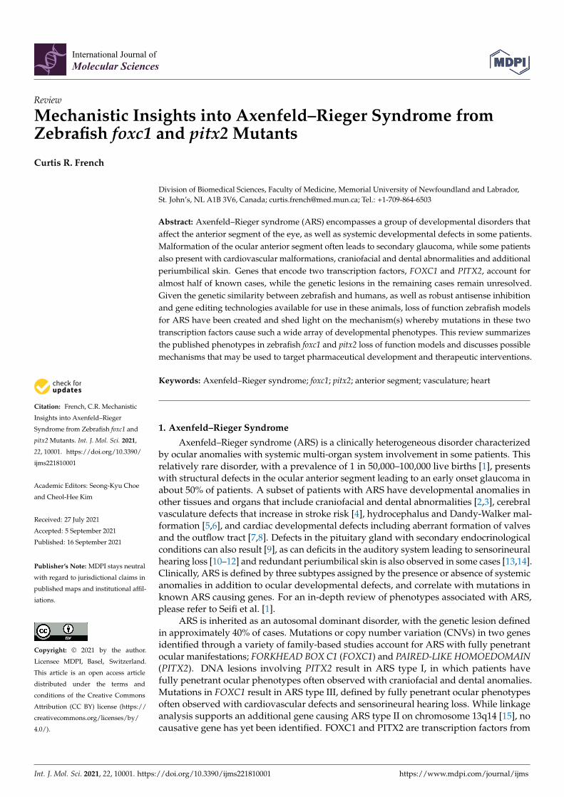
Abstract: Axenfeld–Rieger syndrome (ARS) encompasses a group of developmental disorders thataffect the anterior segment of the eye, as well as systemic developmental defects in some patients.Malformation of the ocular anterior segment often leads to secondary glaucoma, while some patientsalso present with cardiovascular malformations, craniofacial and dental abnormalities and additionalperiumbilical skin. Genes that encode two transcription factors, FOXC1 and PITX2, account foralmost half of known cases, while the genetic lesions in the remaining cases remain unresolved.Given the genetic similarity between zebrafish and humans, as well as robust antisense inhibitionand gene editing technologies available for use in these animals, loss of function zebrafish modelsfor ARS have been created and shed light on the mechanism(s) whereby mutations in these twotranscription factors cause such a wide array of developmental phenotypes. This review summarizesthe published phenotypes in zebrafish foxc1 and pitx2 loss of function models and discusses possiblemechanisms that may be used to target pharmaceutical development and therapeutic interventions.
Keywords: Axenfeld–Rieger syndrome; foxc1; pitx2; anterior segment; vasculature; heart
1. Axenfeld–Rieger Syndrome
Axenfeld–Rieger syndrome (ARS) is a clinically heterogeneous disorder characterizedby ocular anomalies with systemic multi-organ system involvement in some patients. Thisrelatively rare disorder, with a prevalence of 1 in 50,000–100,000 live births [1], presentswith structural defects in the ocular anterior segment leading to an early onset glaucoma inabout 50% of patients. A subset of patients with ARS have developmental anomalies inother tissues and organs that include craniofacial and dental abnormalities [2,3], cerebralvasculature defects that increase in stroke risk [4], hydrocephalus and Dandy-Walker mal-formation [5,6], and cardiac developmental defects including aberrant formation of valvesand the outflow tract [7,8]. Defects in the pituitary gland with secondary endocrinologicalconditions can also result [9], as can deficits in the auditory system leading to sensorineuralhearing loss [10–12] and redundant periumbilical skin is also observed in some cases [13,14].Clinically, ARS is defined by three subtypes assigned by the presence or absence of systemicanomalies in addition to ocular developmental defects, and correlate with mutations inknown ARS causing genes. For an in-depth review of phenotypes associated with ARS,please refer to Seifi et al. [1].
ARS is inherited as an autosomal dominant disorder, with the genetic lesion definedin approximately 40% of cases. Mutations or copy number variation (CNVs) in two genesidentified through a variety of family-based studies account for ARS with fully penetrantocular manifestations; FORKHEAD BOX C1 (FOXC1) and PAIRED-LIKE HOMOEDOMAIN(PITX2). DNA lesions involving PITX2 result in ARS type I, in which patients havefully penetrant ocular phenotypes often observed with craniofacial and dental anomalies.Mutations in FOXC1 result in ARS type III, defined by fully penetrant ocular phenotypesoften observed with cardiovascular defects and sensorineural hearing loss. While linkageanalysis supports an additional gene causing ARS type II on chromosome 13q14 [15], nocausative gene has yet been identified. FOXC1 and PITX2 are transcription factors from
Int. J. Mol. Sci. 2021, 22, 10001. https://doi.org/10.3390/ijms221810001 https://www.mdpi.com/journal/ijms

Int. J. Mol. Sci. 2021, 22, 10001 2 of 12
the forkhead and homeodomain families, respectively, and can regulate gene transcriptionindependently or physically interact on DNA to co-regulate gene transcription [16]. Thiscomplex relationship likely accounts for the phenotypic differences and ARS classificationsin patients with mutations in one of the two causative genes. Mutations in the CYP1B1gene have been found in a single family with ARS [17], and while a relatively commoncause of congenital glaucoma is some populations [18,19], mutations in CYP1B1 appear tobe an extremely rare cause of ARS.
The zebrafish (Danio rerio) has provided valuable mechanistic insights into ARS diseaseetiology. The zebrafish genome contains two homologues of the FOXC1 gene denoted foxc1aand foxc1b, arising from an ancient duplication in the teleost lineage, and one homologue ofPITX2 (pitx2). Gene expression studies using in situ hybridization have highlighted specificcell types that require the expression of foxc1 and pitx2 for normal ocular development.The use of morpholino based antisense inhibition and genome editing have producedzebrafish strains that mimic ARS phenotypes and have provided novel mechanistic datahighlighting downstream genes and signaling pathways that are required for ocular andsystemic manifestations of the syndrome. This review will focus on the role of foxc1 andpitx2 in the regulation of genes and signaling pathways that regulate formation of the eye,cardiovascular system, and craniofacial skeleton, as defined by loss of function zebrafishmutant strains or antisense inhibition data.
2. Expression of ARS Genes in Zebrafish
At early stages of development, zebrafish express foxc1a and foxc1b in overlappingdomains in neural crest cells [4,20] (Figure 1G, (foxc1a)) as well as the lateral plate mesoderm(LPM) [21]. Neural crest cells contribute to the periocular mesenchyme (POM)—a set ofcells that are required for closure of the optic fissure and development of the ocular anteriorsegment [22]. Neural crest cells also populate the central nervous system, craniofacialskeleton, smooth muscle cells of the cerebral vasculature, as well as the cardiac outflowtract and heart valves [23–25]. This expression pattern highlights the important role ofneural crest cells in development and helps to explain the broad range of phenotypesobserved in patients with ARS. The LPM also contributes to the development the heartand cardiovascular system and thus defects in this tissue arising from FOXC1 dysfunctionmay contribute to cardiovascular anomalies observed in ARS patients [21,26,27]. Althoughtypically known as a developmental disease, foxc1a continues to be expressed in the adultzebrafish eye, including the anterior segment and retinal ganglion cell layer [28], possiblycontributing to maintenance of adult ocular tissues.
The zebrafish genome contains a single pitx2 gene that encodes two isoforms viaalternative splicing that correspond to human PITX2A and PITX2C. The expression ofpitx2a is found in partially overlapping domains with foxc1a and foxc1b in developingzebrafish embryos. As early as 24 h post fertilization (hpf), pitx2 expression is observed inthe periocular mesenchyme [22] (Figure 2A), providing an opportunity for co-regulationof gene expression with Foxc1a and Foxc1b. Like foxc1a and foxc1b, pitx2 is expressed inthe neural crest derived tissues of the pharyngeal arches that contribute to the craniofacialskeleton [30]. Additional expression in the dental epithelium and tooth placodes [31,32]likely accounts for dental phenotypes in ARS type 1 patients. Asymmetric expression of thepitx2c isoform in the lateral plate mesoderm [33] (Figure 2B) and expression of pitx2a in theneural crest [34], may contribute to cardiac outflow tract defects and valve incompetenceobserved in a subset of these ARS patients.
Int. J. Mol. Sci. 2021, 22, 10001 3 of 12
Int. J. Mol. Sci. 2021, 22, x FOR PEER REVIEW 3 of 12
crest derived tissues of the pharyngeal arches that contribute to the craniofacial skeleton [30]. Additional expression in the dental epithelium and tooth placodes [31,32] likely ac-counts for dental phenotypes in ARS type 1 patients. Asymmetric expression of the pitx2c isoform in the lateral plate mesoderm [33] (Figure 2B) and expression of pitx2a in the neu-ral crest [34], may contribute to cardiac outflow tract defects and valve incompetence ob-served in a subset of these ARS patients.
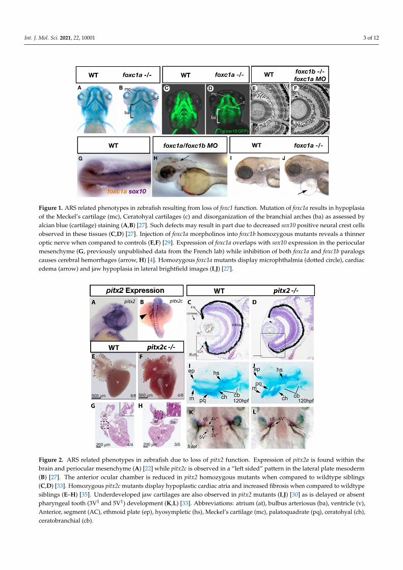
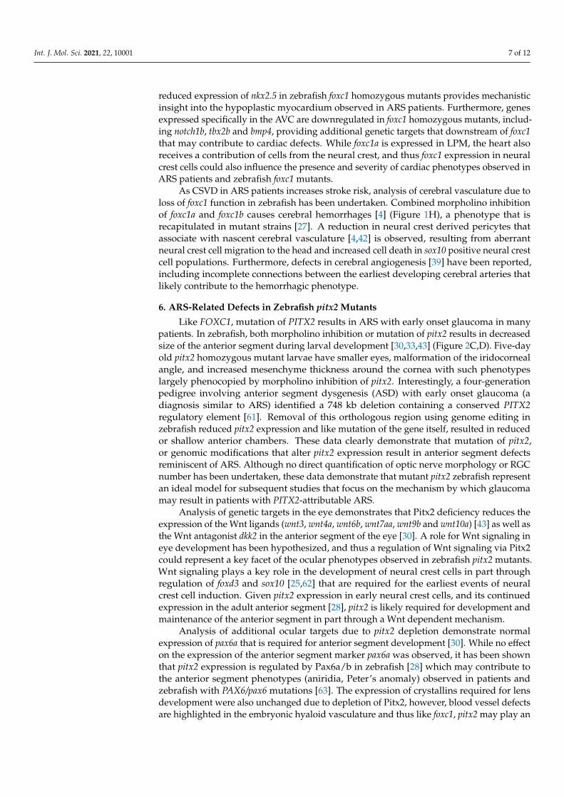
Figure 1. ARS related phenotypes in zebrafish resulting from loss of foxc1 function. Mutation of foxc1a results in hypoplasia of the Meckel’s cartilage (mc), Ceratohyal cartilages (c) and disorganization of the branchial arches (ba) as assessed by alcian blue (cartilage) staining (A,B) [27]. Such defects may result in part due to decreased sox10 positive neural crest cells observed in these tissues (C,D) [27]. Injection of foxc1a morpholinos into foxc1b homozygous mutants reveals a thinner optic nerve when compared to controls (E,F) [29]. Expression of foxc1a overlaps with sox10 expression in the periocular mesenchyme (G, previously unpublished data from the French lab) while inhibition of both foxc1a and foxc1b paralogs causes cerebral hemorrhages (arrow, H) [4]. Homozygous foxc1a mutants display microphthalmia (dotted circle), cardiac edema (arrow) and jaw hypoplasia in lateral brightfield images (I,J) [27].
Figure 1. ARS related phenotypes in zebrafish resulting from loss of foxc1 function. Mutation of foxc1a results in hypoplasiaof the Meckel’s cartilage (mc), Ceratohyal cartilages (c) and disorganization of the branchial arches (ba) as assessed byalcian blue (cartilage) staining (A,B) [27]. Such defects may result in part due to decreased sox10 positive neural crest cellsobserved in these tissues (C,D) [27]. Injection of foxc1a morpholinos into foxc1b homozygous mutants reveals a thinneroptic nerve when compared to controls (E,F) [29]. Expression of foxc1a overlaps with sox10 expression in the periocularmesenchyme (G, previously unpublished data from the French lab) while inhibition of both foxc1a and foxc1b paralogscauses cerebral hemorrhages (arrow, H) [4]. Homozygous foxc1a mutants display microphthalmia (dotted circle), cardiacedema (arrow) and jaw hypoplasia in lateral brightfield images (I,J) [27].
Int. J. Mol. Sci. 2021, 22, x FOR PEER REVIEW 4 of 12
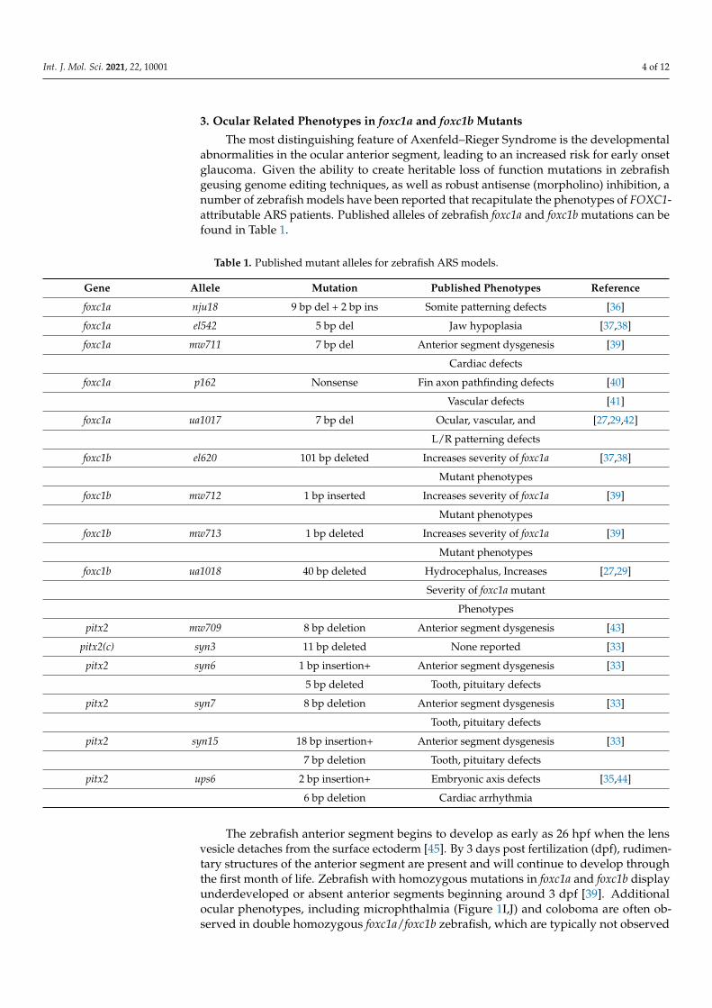
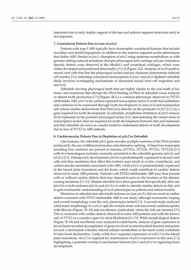
Figure 2. ARS related phenotypes in zebrafish due to loss of pitx2 function. Expression of pitx2a is found within the brain and periocular mesenchyme (A) [22] while pitx2c is observed in a “left sided” pattern in the lateral plate mesoderm (B) [27]. The anterior ocular chamber is reduced in pitx2 homozygous mutants when compared to wildtype siblings (C,D) [33]. Homozygous pitx2c mutants display hypoplastic cardiac atria and increased fibrosis when compared to wildtype siblings (G,H) [35]. Underdeveloped jaw cartilages are also observed in pitx2 mutants (I,J) [30] as is delayed or absent pharyngeal tooth (3V1 and 5V1) development (K,L) [33]. Abbreviations: atrium (at), bulbus arteriosus (ba), ventricle (v), Anterior, segment (AC), ethmoid plate (ep), hyosympletic (hs), Meckel’s cartilage (mc), palatoquadrate (pq), ceratohyal (ch), ceratobranchial (cb).
3. Ocular Related Phenotypes in foxc1a and foxc1b Mutants The most distinguishing feature of Axenfeld–Rieger Syndrome is the developmental
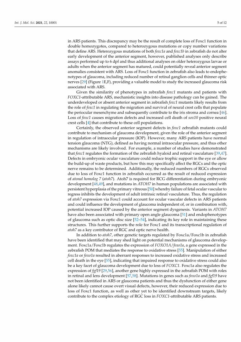
abnormalities in the ocular anterior segment, leading to an increased risk for early onset glaucoma. Given the ability to create heritable loss of function mutations in zebrafish geusing genome editing techniques, as well as robust antisense (morpholino) inhibition, a number of zebrafish models have been reported that recapitulate the phenotypes of FOXC1-attributable ARS patients. Published alleles of zebrafish foxc1a and foxc1b muta-tions can be found in Table 1.
Table 1. Published mutant alleles for zebrafish ARS models.
Gene Allele Mutation Published Phenotypes Reference foxc1a nju18 9 bp del + 2 bp ins Somite patterning defects [36] foxc1a el542 5 bp del Jaw hypoplasia [37,38] foxc1a mw711 7 bp del Anterior segment dysgenesis [39]
Cardiac defects foxc1a p162 Nonsense Fin axon pathfinding defects [40]
Vascular defects [41] foxc1a ua1017 7 bp del Ocular, vascular, and [27,29,42]
L/R patterning defects
Figure 2. ARS related phenotypes in zebrafish due to loss of pitx2 function. Expression of pitx2a is found within thebrain and periocular mesenchyme (A) [22] while pitx2c is observed in a “left sided” pattern in the lateral plate mesoderm(B) [27]. The anterior ocular chamber is reduced in pitx2 homozygous mutants when compared to wildtype siblings(C,D) [33]. Homozygous pitx2c mutants display hypoplastic cardiac atria and increased fibrosis when compared to wildtypesiblings (E–H) [35]. Underdeveloped jaw cartilages are also observed in pitx2 mutants (I,J) [30] as is delayed or absentpharyngeal tooth (3V1 and 5V1) development (K,L) [33]. Abbreviations: atrium (at), bulbus arteriosus (ba), ventricle (v),Anterior, segment (AC), ethmoid plate (ep), hyosympletic (hs), Meckel’s cartilage (mc), palatoquadrate (pq), ceratohyal (ch),ceratobranchial (cb).
Int. J. Mol. Sci. 2021, 22, 10001 4 of 12
3. Ocular Related Phenotypes in foxc1a and foxc1b Mutants
The most distinguishing feature of Axenfeld–Rieger Syndrome is the developmentalabnormalities in the ocular anterior segment, leading to an increased risk for early onsetglaucoma. Given the ability to create heritable loss of function mutations in zebrafishgeusing genome editing techniques, as well as robust antisense (morpholino) inhibition, anumber of zebrafish models have been reported that recapitulate the phenotypes of FOXC1-attributable ARS patients. Published alleles of zebrafish foxc1a and foxc1b mutations can befound in Table 1.
Table 1. Published mutant alleles for zebrafish ARS models.
Gene Allele Mutation Published Phenotypes Reference
foxc1a nju18 9 bp del + 2 bp ins Somite patterning defects [36]
foxc1a el542 5 bp del Jaw hypoplasia [37,38]
foxc1a mw711 7 bp del Anterior segment dysgenesis [39]
Cardiac defects
foxc1a p162 Nonsense Fin axon pathfinding defects [40]
Vascular defects [41]
foxc1a ua1017 7 bp del Ocular, vascular, and [27,29,42]
L/R patterning defects
foxc1b el620 101 bp deleted Increases severity of foxc1a [37,38]
Mutant phenotypes
foxc1b mw712 1 bp inserted Increases severity of foxc1a [39]
Mutant phenotypes
foxc1b mw713 1 bp deleted Increases severity of foxc1a [39]
Mutant phenotypes
foxc1b ua1018 40 bp deleted Hydrocephalus, Increases [27,29]
Severity of foxc1a mutant
Phenotypes
pitx2 mw709 8 bp deletion Anterior segment dysgenesis [43]
pitx2(c) syn3 11 bp deleted None reported [33]
pitx2 syn6 1 bp insertion+ Anterior segment dysgenesis [33]
5 bp deleted Tooth, pituitary defects
pitx2 syn7 8 bp deletion Anterior segment dysgenesis [33]
Tooth, pituitary defects
pitx2 syn15 18 bp insertion+ Anterior segment dysgenesis [33]
7 bp deletion Tooth, pituitary defects
pitx2 ups6 2 bp insertion+ Embryonic axis defects [35,44]
6 bp deletion Cardiac arrhythmia
The zebrafish anterior segment begins to develop as early as 26 hpf when the lensvesicle detaches from the surface ectoderm [45]. By 3 days post fertilization (dpf), rudimen-tary structures of the anterior segment are present and will continue to develop throughthe first month of life. Zebrafish with homozygous mutations in foxc1a and foxc1b displayunderdeveloped or absent anterior segments beginning around 3 dpf [39]. Additionalocular phenotypes, including microphthalmia (Figure 1I,J) and coloboma are often ob-served in double homozygous foxc1a/foxc1b zebrafish, which are typically not observed
Int. J. Mol. Sci. 2021, 22, 10001 5 of 12
in ARS patients. This discrepancy may be the result of complete loss of Foxc1 function indouble homozygotes, compared to heterozygous mutations or copy number variationsthat define ARS. Heterozygous mutations of both foxc1a and foxc1b in zebrafish do not alterearly development of the anterior segment, however, published analyses only describeassays performed up to 6 dpf and thus additional analyses on older heterozygous larvae oradults when the anterior segment has matured, could potentially reveal anterior segmentanomalies consistent with ARS. Loss of Foxc1 function in zebrafish also leads to endophe-notypes of glaucoma, including reduced number of retinal ganglion cells and thinner opticnerves [29] (Figure 1E,F), providing a valuable model to study the increased glaucoma riskassociated with ARS.
Given the similarity of phenotypes in zebrafish foxc1 mutants and patients withFOXC1-attributable ARS, mechanistic insights into disease pathology can be gained. Theunderdeveloped or absent anterior segment in zebrafish foxc1 mutants likely results fromthe role of foxc1 in regulating the migration and survival of neural crest cells that populatethe periocular mesenchyme and subsequently contribute to the iris stroma and cornea [46].Loss of foxc1 causes migration defects and increased cell death of sox10 positive neuralcrest cells [4] that contribute to these cell populations.
Certainly, the observed anterior segment defects in foxc1 zebrafish mutants couldcontribute to mechanism of glaucoma development, given the role of the anterior segmentin regulation of intraocular pressure (IOP). However, many ARS patients have normaltension glaucoma (NTG), defined as having normal intraocular pressure, and thus othermechanisms are likely involved. For example, a number of studies have demonstratedthat foxc1 regulates the formation of the zebrafish hyaloid and retinal vasculature [39,47].Defects in embryonic ocular vasculature could reduce trophic support in the eye or allowthe build-up of waste products, but how this may specifically affect the RGCs and the opticnerve remains to be determined. Additionally, the reduced numbers of RGCs observeddue to loss of Foxc1 function in zebrafish occurred as the result of reduced expressionof atonal homolog 7 (atoh7). Atoh7 is required for RCG differentiation during embryonicdevelopment [48,49], and mutations in ATOH7 in human populations are associated withpersistent hyperplasia of the primary vitreous [50] whereby failure of fetal ocular vascular toregress inhibits the development of adult intrinsic retinal vasculature. Thus, the regulationof atoh7 expression via Foxc1 could account for ocular vascular defects in ARS patientsand could influence the development of glaucoma independent of, or in combination with,potential increased IOP caused by the anterior segment dysgenesis. Variants in ATOH7have also been associated with primary open angle glaucoma [51] and endophenotypesof glaucoma such as optic disc size [52–54], indicating its key role in maintaining thesestructures. This further supports the role for Foxc1 and its transcriptional regulation ofatoh7 as a key contributor of RGC and optic nerve health.
In addition to atoh7, other genetic targets regulated by Foxc1a/Foxc1b in zebrafishhave been identified that may shed light on potential mechanisms of glaucoma develop-ment. Foxc1a/Foxc1b regulates the expression of FOXO1A/foxo1a, a gene expressed in thezebrafish POM that mediates the response to oxidative stress [55]. Manipulation of eitherfoxc1a or foxo1a resulted in aberrant responses to increased oxidative stress and increasedcell death in the eye [55], indicating that impaired response to oxidative stress could alsobe a key facet of glaucoma development due to loss of FOXC1. Foxc1a also regulates theexpression of fgf19 [29,56], another gene highly expressed in the zebrafish POM with rolesin retinal and lens development [57,58]. Mutations in genes such as foxo1a and fgf19 havenot been identified in ARS or glaucoma patients and thus the dysfunction of either genealone likely cannot cause overt visual defects, however, their reduced expression due toloss of Foxc1 function, as well as other yet to be identified downstream targets, likelycontribute to the complex etiology of RGC loss in FOXC1-attributable ARS patients.
Int. J. Mol. Sci. 2021, 22, 10001 6 of 12
4. Craniofacial Defects in Zebrafish foxc1a and foxc1b Mutants
Zebrafish foxc1a mutants, as well as foxc1a/foxc1b double mutants, have craniofacialdefects consistent with abnormalities in the facial structure of ARS patients. In thesepatients, hypertelorism (increased space between the eyes) and a prominent forehead areoften described. In zebrafish, foxc1a and foxc1b are expressed in the neural crest cells thatpopulate the first and second pharyngeal arches that give rise to anterior jaw structures.Mutation of foxc1a alone results in craniofacial dysmorphism [27,37] including underde-veloped symplectic cartilage and like many ocular phenotypes observed in these mutants,severity increased in double foxc1a/foxc1b mutants. Phenotypes in double homozygousmutants included under-developed palatoquadrate and hyomandibula cartilages [37],demonstrating that foxc1a and foxc1b play critical roles in the development of anterior facialcartilages (Figure 1A–D, foxc1a). Analysis of genetic targets downstream of foxc1 revealedboth foxc1a and foxc1b regulate Sox9-dependent expression of cartilage specification genes,accounting for reduced jaw structures in these animals. While Foxc1 does not directlyregulate sox9 expression, loss of foxc1 paralogs in zebrafish causes a decrease in chromatinaccessibility for transcription factors such as Sox9 in chondrocytes [38], and thus supportsthe hypothesis that Foxc1 drives cartilage development through a chromatin remodelingmechanism.
Analysis of heterozygous foxc1a mutants in conjunction with homozygous loss offoxc1b (which survive to adulthood) demonstrates craniofacial abnormalities in adults thatinclude a misshapen head that closely mimics that of ARS patients, as well as mandibularretrognathia and dorsally positioned eyes [39]. Combined with larval-based studies, thesedata demonstrate that Foxc1 regulates chromatin accessibility in chondrocytes to shapethe jaw and head in zebrafish, with such mechanisms likely contributing the craniofacialdysmorphia observed in ARS patients with mutations or CNVs involving FOXC1.
5. Cardiovascular Anomalies in Zebrafish foxc1 Mutants
Cardiac anomalies have been described in ARS patients with FOXC1 mutations orCNVs that include hypoplastic ventricular outflow tract morphology, dysplastic arcademitral valve, and atrial septal defect [7,59]. Within the brain, cerebral small vessel disease(CSVD) has been described, which includes increased perivascular spaces, subclinicalinfarcts and white matter hyperintensities [4]—all of which increase stroke risk [60]. Theseclinical phenotypes clearly indicate that FOXC1 plays an important role in early heart andcerebral vascular development, with such phenotypes recapitulated in zebrafish loss offoxc1 function models.
In zebrafish, mutation of foxc1a alone or in combination with foxc1b leads to cardiacphenotypes that include hypoplastic myocardium and ventricular outflow tract, as wellas defects in cardiac valve formation that are similar to that observed in patients withFOXC1-attributible ARS [21,26]. While zebrafish studies generally support a role or foxc1 inheart development, reports differ in their analysis of heart morphology, assay different timepoints, and study different combinations of mutations. For example, Yue et al. [21] demon-strate a hypoplastic myocardium, shorter outflow tract, defective primitive valve leaflets,and cardiac edema at during early larval development (4-5 dpf) of foxc1a−/− embryos(cardiac edema in foxc1−/− displayed in Figure 1J). Ferre-Fernandez et al. [39] assayedcardiac trabecular zone thickness, outflow tract and AV valve morphology in foxc1a+/−;foxc1b−/− embryos at 6 dpf and found no difference. Compact zone thickness, however,was significantly larger in these fish when compared to wildtype siblings. While thesestudies support a role for foxc1 in heart development, they indicate that homozygous lossof foxc1a is required to induce heart defects similar to ARS patients, who typically haveheterozygous mutations or CNVs involving FOXC1.
Utilizing foxc1 homozygous loss of function embryos, a role for this gene in regulat-ing cardiac progenitor specification and atrioventricular canal (AVC) formation has beenproposed. Such studies show that Foxc1a directly binds to the promoter of nkx2.5 [26], agene required for cardiac progenitor specification in the lateral plate mesoderm (LPM). The
Int. J. Mol. Sci. 2021, 22, 10001 7 of 12
reduced expression of nkx2.5 in zebrafish foxc1 homozygous mutants provides mechanisticinsight into the hypoplastic myocardium observed in ARS patients. Furthermore, genesexpressed specifically in the AVC are downregulated in foxc1 homozygous mutants, includ-ing notch1b, tbx2b and bmp4, providing additional genetic targets that downstream of foxc1that may contribute to cardiac defects. While foxc1a is expressed in LPM, the heart alsoreceives a contribution of cells from the neural crest, and thus foxc1 expression in neuralcrest cells could also influence the presence and severity of cardiac phenotypes observed inARS patients and zebrafish foxc1 mutants.
As CSVD in ARS patients increases stroke risk, analysis of cerebral vasculature due toloss of foxc1 function in zebrafish has been undertaken. Combined morpholino inhibitionof foxc1a and foxc1b causes cerebral hemorrhages [4] (Figure 1H), a phenotype that isrecapitulated in mutant strains [27]. A reduction in neural crest derived pericytes thatassociate with nascent cerebral vasculature [4,42] is observed, resulting from aberrantneural crest cell migration to the head and increased cell death in sox10 positive neural crestcell populations. Furthermore, defects in cerebral angiogenesis [39] have been reported,including incomplete connections between the earliest developing cerebral arteries thatlikely contribute to the hemorrhagic phenotype.
6. ARS-Related Defects in Zebrafish pitx2 Mutants
Like FOXC1, mutation of PITX2 results in ARS with early onset glaucoma in manypatients. In zebrafish, both morpholino inhibition or mutation of pitx2 results in decreasedsize of the anterior segment during larval development [30,33,43] (Figure 2C,D). Five-dayold pitx2 homozygous mutant larvae have smaller eyes, malformation of the iridocornealangle, and increased mesenchyme thickness around the cornea with such phenotypeslargely phenocopied by morpholino inhibition of pitx2. Interestingly, a four-generationpedigree involving anterior segment dysgenesis (ASD) with early onset glaucoma (adiagnosis similar to ARS) identified a 748 kb deletion containing a conserved PITX2regulatory element [61]. Removal of this orthologous region using genome editing inzebrafish reduced pitx2 expression and like mutation of the gene itself, resulted in reducedor shallow anterior chambers. These data clearly demonstrate that mutation of pitx2,or genomic modifications that alter pitx2 expression result in anterior segment defectsreminiscent of ARS. Although no direct quantification of optic nerve morphology or RGCnumber has been undertaken, these data demonstrate that mutant pitx2 zebrafish representan ideal model for subsequent studies that focus on the mechanism by which glaucomamay result in patients with PITX2-attributable ARS.
Analysis of genetic targets in the eye demonstrates that Pitx2 deficiency reduces theexpression of the Wnt ligands (wnt3, wnt4a, wnt6b, wnt7aa, wnt9b and wnt10a) [43] as well asthe Wnt antagonist dkk2 in the anterior segment of the eye [30]. A role for Wnt signaling ineye development has been hypothesized, and thus a regulation of Wnt signaling via Pitx2could represent a key facet of the ocular phenotypes observed in zebrafish pitx2 mutants.Wnt signaling plays a key role in the development of neural crest cells in part throughregulation of foxd3 and sox10 [25,62] that are required for the earliest events of neuralcrest cell induction. Given pitx2 expression in early neural crest cells, and its continuedexpression in the adult anterior segment [28], pitx2 is likely required for development andmaintenance of the anterior segment in part through a Wnt dependent mechanism.
Analysis of additional ocular targets due to pitx2 depletion demonstrate normalexpression of pax6a that is required for anterior segment development [30]. While no effecton the expression of the anterior segment marker pax6a was observed, it has been shownthat pitx2 expression is regulated by Pax6a/b in zebrafish [28] which may contribute tothe anterior segment phenotypes (aniridia, Peter’s anomaly) observed in patients andzebrafish with PAX6/pax6 mutations [63]. The expression of crystallins required for lensdevelopment were also unchanged due to depletion of Pitx2, however, blood vessel defectsare highlighted in the embryonic hyaloid vasculature and thus like foxc1, pitx2 may play an
Int. J. Mol. Sci. 2021, 22, 10001 8 of 12
important role in early trophic support of the lens and anterior segment structures early indevelopment.
7. Craniofacial Defects Due to Loss of pitx2
Patients with type I ARS typically have dysmorphic craniofacial features that includemaxillary and dental hypoplasia, in addition to the anterior segment ocular phenotypesthat define ARS. Similar to foxc1, disruption of pitx2 using antisense morpholinos or withgenome editing induced mutations disrupts pharyngeal arch cartilage and jaw formation.Specific defects were observed in the Meckel’s and ceratohyal cartilages, which wereunder-developed and positioned abnormally [30,43] (Figure 2I,J). Analysis of sox10 positiveneural crest cells that line the pharyngeal arches and jaw elements demonstrate reducedcell number [36], indicating craniofacial dysmorphism in foxc1 and pitx2 depleted zebrafishlikely involves overlapping mechanisms of decreased neural crest cell migration andsurvival.
Zebrafish develop pharyngeal teeth that are highly similar to the oral teeth of hu-mans, and mutations that disrupt the DNA binding of Pitx2 in zebrafish cause reducedor absent tooth production [33] (Figure 2K,L)-a common phenotype observed in PITX2-attributable ARS. pitx2 is the earliest expressed transcription factor in tooth bud epitheliumand continues to be expressed through tooth development in mice [64] and mammaliancell culture studies demonstrate that Pitx2 acts directly on the promoter of dlx2 [65,66], agene required for tooth development. In zebrafish, morpholino inhibition of pitx2 reducesdlx2a expression in the posterior pharyngeal arches [30], demonstrating the conservation oftranscription factors that are required for tooth development between fish and mammalsand that zebrafish can serve as a useful model to understand defects in tooth developmentdue to loss of PITX2 in ARS patients.
8. Cardiovascular Defects Due to Depletion of pitx2 in Zebrafish
Like humans, the zebrafish pitx2 gene encodes multiple isoforms of the Pitx2 proteinproduced by the use of different promoters and alternative splicing. At least four transcriptsencoding four isoforms are present in humans (PITX2a, PITX2b, PITX2c, PITX2d) [67]with two homologous isoforms currently annotated in the zebrafish genome (pitx2a andpitx2c) [33]. During early development, pitx2a is predominantly expressed in neural crestcells and thus mutations that affect this isoform may result in ocular, craniofacial, andcardiovascular anomalies associated with ARS, while pitx2c is predominately expressedin the lateral plate mesoderm and the heart, which could contribute to cardiac defectsobserved in some ARS patients. Patients with PITX2-attributable ARS may thus presentwith or without cardiac defects that may depend in part on the location of the disease-causing mutations [67,68]. Mutant zebrafish have been generated that specifically alter one(pitx2a) or both isoforms (pitx2a and pitx2c) in order to identify similar defects in fish, andto gain mechanistic understanding of such phenotypes in patients and animal models.
Mutations in zebrafish that affect both isoforms (or just pitx2c) did not result in cardiacdefects consistent with PITX2-attributable ARS in one study, although asymmetric loopingand overall morphology were the only phenotypes tested [33]. A second study analyzedadult heart morphology in a pitx2c specific mutant strain and uncovered cardiomyopathywith fibrosis (Figure 2E–H) and arrythmias, particularly when the fish are stressed [35].This is consistent with cardiac defects observed in some ARS patients and with the knownrole of PITX2 as a causative gene for atrial fibrillation [69,70]. While morphological defects(Figure 2E–H) and arrythmias were analyzed in adult hearts, analysis of gene expression inlarval hearts revealed dysregulation of genes involved in mitochondrial function and pointstoward a mechanism whereby altered cellular metabolism in the heart could contributeto later heart dysfunction. Lastly, while foxc1 regulates expression of nxk2.5 in the lateralplate mesoderm, nkx2.5 is required for maintenance of pitx2 expression in this area [71],highlighting a potential overlap in mechanism between foxc1 and pitx2 in regulating heartdevelopment.

Int. J. Mol. Sci. 2021, 22, 10001 9 of 12
9. Summary
Analysis of zebrafish foxc1 and pitx2 loss of function models provides understandingof the mechanisms that lead to most ARS related phenotypes. Both genes when mutatedin zebrafish result in defects consistent with mammalian ARS models and patient pheno-types. Zebrafish ARS mutants have ocular anterior chamber defects, and foxc1 mutantsadditionally display endophenotypes of glaucoma. Disruption of foxc1 or pitx2 in zebrafishdisplay craniofacial anomalies consistent with ARS, as well as cardiovascular defects thatare often observed in patients. pitx2 mutants additionally display tooth hypoplasia thatis often observed in Type 1 ARS. Mechanisms involving downstream gene regulation arebeginning to be uncovered using homozygous embryos and larvae, however, analysisof adult phenotypes in heterozygous mutants is somewhat lacking in the literature. Thecontinued analysis of such mutants will reveal novel insights into disease mechanisms, andgiven the utility of zebrafish for translational research, pharmaceutical approaches usinghigh-throughput drug screening for phenotypic rescue provides a path toward testingtherapeutic interventions.
Funding: This work was funded in part by a grant from the Glaucoma Research Society of Canada,and Memorial University of Newfoundland and Labrador.
Institutional Review Board Statement: Work contained in this article that was performed in theFrench Lab was approved by the Canadian Council of Animal Care and Memorial University’sanimal welfare review committee.
Informed Consent Statement: Not applicable.
Data Availability Statement: Not applicable.
Acknowledgments: I would like to thank Danielle French for critical comments on this manuscript.The French lab is funded in part by the Glaucoma Research Society of Canada which contributed tothe open access cost of publishing this review. Permission for data included in Figures 1 and 2 weregrated from Elsevier, The Natural Academy of Sciences, or through the Creative Commons openaccess agreement.
Conflicts of Interest: The author declares no conflict of interest.
References1. Seifi, M.; Walter, M.A. Axenfeld-Rieger syndrome. Clin. Genet. 2018, 93, 1123–1130. [CrossRef]2. Waldron, J.M.; Mcnamara, C.; Hewson, A.R.; Mcnamara, C.M. Axenfeld-Rieger syndrome (ARS): A review and case report. Spec.
Care Dent. 2010, 30, 218–222. [CrossRef]3. Dressler, S.; Meyer-Marcotty, P.; Weisschuh, N.; Jablonski-Momeni, A.; Pieper, K.; Gramer, G.; Gramer, E. Dental and Craniofacial
Anomalies Associated with Axenfeld-Rieger Syndrome with PITX2 Mutation. Case Rep. Med. 2010, 2010, 621984. [CrossRef]4. French, C.R.; Seshadri, S.; Destefano, A.L.; Fornage, M.; Arnold, C.R.; Gage, P.J.; Skarie, J.M.; Dobyns, W.B.; Millen, K.J.; Liu, T.;
et al. Mutation of FOXC1 and PITX2 induces cerebral small-vessel disease. J. Clin. Investig. 2014, 124, 4877–4881. [CrossRef]5. Aldinger, K.A.; Lehmann, O.J.; Hudgins, L.; Chizhikov, V.V.; Bassuk, A.G.; Ades, L.C.; Krantz, I.D.; Dobyns, W.B.; Millen,
K.J. FOXC1 is required for normal cerebellar development and is a major contributor to chromosome 6p25.3 Dandy-Walkermalformation. Nat. Genet. 2009, 41, 1037–1042. [CrossRef] [PubMed]
6. Maclean, K.; Smith, J.; St Heaps, L.; Chia, N.; Williams, R.; Peters, G.B.; Onikul, E.; McCrossin, T.; Lehmann, O.J.; Adès, L.C.Axenfeld-Rieger malformation and distinctive facial features: Clues to a recognizable 6p25 microdeletion syndrome. Am. J. Med.Genet. A 2005, 132, 381–385. [CrossRef]
7. Gripp, K.W.; Hopkins, E.; Jenny, K.; Thacker, D.; Salvin, J. Cardiac anomalies in Axenfeld-Rieger syndrome due to a novel FOXC1mutation. Am. J. Med. Genet. A 2013, 161, 114–119. [CrossRef] [PubMed]
8. Mammi, I.; De Giorgio, P.; Clementi, M.; Tenconi, R. Cardiovascular anomaly in Rieger Syndrome: Heterogeneity or contiguity?Acta Ophthalmol. Scand. 1998, 76, 509–512. [CrossRef] [PubMed]
9. Idrees, F.; Bloch-Zupan, A.; Free, S.L.; Vaideanu, D.; Thompson, P.J.; Ashley, P.; Brice, G.; Rutland, P.; Bitner-Glindzicz, M.; Khaw,P.T.; et al. A novel homeobox mutation in the PITX2 gene in a family with Axenfeld-Rieger syndrome associated with brain,ocular, and dental phenotypes. Am. J. Med. Genet. B Neuropsychiatr. Genet. 2006, 141, 184–191. [CrossRef] [PubMed]
10. Yamazaki, H.; Nakamura, T.; Hosono, K.; Yamaguchi, T.; Hiratsuka, Y.; Hotta, Y.; Takahashi, M. Sensorineural hearing loss andhypoplastic cochlea in Axenfeld-Rieger syndrome with FOXC1 mutation. Auris Nasus Larynx 2021, 48, 1204–1208. [CrossRef][PubMed]
Int. J. Mol. Sci. 2021, 22, 10001 10 of 12
11. Gauthier, A.C.; Wiggs, J.L. Childhood glaucoma genes and phenotypes: Focus on FOXC1 mutations causing anterior segmentdysgenesis and hearing loss. Exp. Eye Res. 2020, 190, 107893. [CrossRef] [PubMed]
12. Cunningham, E.T., Jr.; Eliott, D.; Miller, N.R.; Maumenee, I.H.; Green, W.R. Familial Axenfeld-Rieger anomaly, atrial septal defect,and sensorineural hearing loss: A possible new genetic syndrome. Arch. Ophthalmol. 1998, 116, 78–82. [CrossRef]
13. Law, S.K.; Sami, M.; Piri, N.; Coleman, A.L.; Caprioli, J. Asymmetric phenotype of Axenfeld-Rieger anomaly and aniridiaassociated with a novel PITX2 mutation. Mol. Vis. 2011, 17, 1231–1238.
14. Zhang, L.; Peng, Y.; Ouyang, P.; Liang, Y.; Zeng, H.; Wang, N.; Duan, X.; Shi, J. A novel frameshift mutation in the PITX2 gene in afamily with Axenfeld-Rieger syndrome using targeted exome sequencing. BMC Med. Genet. 2019, 20, 105. [CrossRef]
15. Phillips, J.C.; del Bono, E.A.; Haines, J.L.; Pralea, A.M.; Cohen, J.S.; Greff, L.J.; Wiggs, J.L. A second locus for Rieger syndromemaps to chromosome 13q14. Am. J. Hum. Genet. 1996, 59, 613–619. [PubMed]
16. Berry, F.B.; Lines, M.A.; Oas, J.M.; Footz, T.; Underhill, D.A.; Gage, P.J.; Walter, M.A. Functional interactions between FOXC1 andPITX2 underlie the sensitivity to FOXC1 gene dose in Axenfeld-Rieger syndrome and anterior segment dysgenesis. Hum. Mol.Genet. 2006, 15, 905–919. [CrossRef]
17. Tanwar, M.; Dada, T.; Dada, R. Axenfeld-Rieger Syndrome Associated with Congenital Glaucoma and Cytochrome P4501B1 GeneMutations. Case Rep. Med. 2010, 2010, 212656. [CrossRef]
18. Jubair, S.; N Al-Rubae’i, S.H.; M Al-Sharifi, A.N.; Jabbar Suleiman, A.A. Investigation of CYP1B1 Gene Involvement in PrimaryCongenital Glaucoma in Iraqi Children. Middle East Afr. J. Ophthalmol. 2019, 26, 203–209. [CrossRef] [PubMed]
19. Lim, S.H.; Tran-Viet, K.N.; Yanovitch, T.L.; Freedman, S.F.; Klemm, T.; Call, W.; Powell, C.; Ravichandran, A.; Metlapally, R.;Nading, E.B. CYP1B1, MYOC, and LTBP2 mutations in primary congenital glaucoma patients in the United States. Am. J.Ophthalmol. 2013, 155, 508–517.e5. [CrossRef]
20. Topczewska, J.M.; Topczewski, J.; Solnica-Krezel, L.; Hogan, B.L. Sequence and expression of zebrafish foxc1a and foxc1b,encoding conserved forkhead/winged helix transcription factors. Mech. Dev. 2001, 100, 343–347. [CrossRef]
21. Yue, Y.; Jiang, M.; He, L.; Zhang, Z.; Zhang, Q.; Gu, C.; Liu, M.; Li, N.; Zhao, Q. The transcription factor Foxc1a in zebrafishdirectly regulates expression of nkx2.5, encoding a transcriptional regulator of cardiac progenitor cells. J. Biol. Chem. 2018, 293,638–650. [CrossRef]
22. Van Der Meulen, K.L.; Vöcking, O.; Weaver, M.L.; Meshram, N.N.; Famulski, J.K. Spatiotemporal Characterization of AnteriorSegment Mesenchyme Heterogeneity during Zebrafish Ocular Anterior Segment Development. Front. Cell Dev. Biol. 2020, 8, 379.[CrossRef]
23. Girolamo, F.; de Trizio, I.; Errede, M.; Longo, G.; d’Amati, A.; Virgintino, D. Neural crest cell-derived pericytes act as pro-angiogenic cells in human neocortex development and gliomas. Fluids Barriers CNS 2021, 18, 14. [CrossRef] [PubMed]
24. George, R.M.; Maldonado-Velez, G.; Firulli, A.B. The heart of the neural crest: Cardiac neural crest cells in development andregeneration. Development 2020, 147, dev188706. [CrossRef]
25. Rocha, M.; Singh, N.; Ahsan, K.; Beiriger, A.; Prince, V.E. Neural crest development: Insights from the zebrafish. Dev. Dyn. 2020,249, 88–111. [CrossRef] [PubMed]
26. Zhang, Q.; Liang, D.; Yue, Y.; He, L.; Li, N.; Jiang, D.; Hu, P.; Zhao, Q. Axenfeld-Rieger syndrome-associated mutants of thetranscription factor FOXC1 abnormally regulate NKX2-5 in model zebrafish embryos. J. Biol. Chem. 2020, 295, 11902–11913.[CrossRef]
27. Chrystal, P.W.; French, C.R.; Jean, F.; Havrylov, S.; van Baarle, S.; Peturson, A.M.; Xu, P.; Crump, J.G.; Pilgrim, D.B.; Lehmann, O.J.;et al. The Axenfeld-Rieger Syndrome Gene FOXC1 Contributes to Left-Right Patterning. Genes 2021, 12, 170. [CrossRef]
28. Takamiya, M.; Weger, B.D.; Schindler, S.; Beil, T.; Yang, L.; Armant, O.; Ferg, M.; Schlunck, G.; Reinhard, T.; Dickmeis, T.; et al.Molecular description of eye defects in the zebrafish Pax6b mutant, sunrise, reveals a Pax6b-dependent genetic network in thedeveloping anterior chamber. PLoS ONE 2015, 10, e0117645.
29. Umali, J.; Hawkey-Noble, A.; French, C.R. Loss of foxc1 in zebrafish reduces optic nerve size and cell number in the retinalganglion cell layer. Vis. Res. 2019, 156, 66–72. [CrossRef] [PubMed]
30. Liu, Y.; Semina, E.V. pitx2 Deficiency results in abnormal ocular and craniofacial development in zebrafish. PLoS ONE 2012, 7,e30896. [CrossRef]
31. Aigler, S.R.; Jandzik, D.; Hatta, K.; Uesugi, K.; Stock, D.W. Selection and constraint underlie irreversibility of tooth loss incypriniform fishes. Proc. Natl. Acad. Sci. USA 2014, 111, 7707–7712. [CrossRef] [PubMed]
32. Jackman, W.R.; Yoo, J.J.; Stock, D.W. Hedgehog signaling is required at multiple stages of zebrafish tooth development. BMC Dev.Biol. 2010, 10, 119. [CrossRef]
33. Ji, Y.; Buel, S.M.; Amack, J.D. Mutations in zebrafish pitx2 model congenital malformations in Axenfeld-Rieger syndrome but donot disrupt left-right placement of visceral organs. Dev. Biol. 2016, 416, 69–81. [CrossRef]
34. Ai, D.; Liu, W.; Ma, L.; Dong, F.; Lu, M.F.; Wang, D.; Verzi, M.P.; Cai, C.; Gage, P.J.; Evans, S.; et al. Pitx2 regulates cardiac left-rightasymmetry by patterning second cardiac lineage-derived myocardium. Dev. Biol. 2006, 296, 437–449. [CrossRef]
35. Collins, M.M.; Ahlberg, G.; Hansen, C.V.; Guenther, S.; Marín-Juez, R.; Sokol, A.M.; El-Sammak, H.; Piesker, J.; Hellsten, Y.;Olesen, M.S.; et al. Early sarcomere and metabolic defects in a zebrafish pitx2c cardiac arrhythmia model. Proc. Natl. Acad. Sci.USA 2019, 116, 24115–24121. [CrossRef] [PubMed]
36. Li, J.; Yue, Y.; Dong, X.; Jia, W.; Li, K.; Liang, D.; Dong, Z.; Wang, X.; Nan, X.; Zhang, Q.; et al. Zebrafish foxc1a plays a crucial rolein early somitogenesis by restricting the expression of aldh1a2 directly. J. Biol. Chem. 2015, 290, 10216–10228. [CrossRef]

Int. J. Mol. Sci. 2021, 22, 10001 11 of 12
37. Xu, P.; Balczerski, B.; Ciozda, A.; Louie, K.; Oralova, V.; Huysseune, A.; Crump, J.G. Fox proteins are modular competency factorsfor facial cartilage and tooth specification. Development 2018, 145, dev165498. [CrossRef]
38. Xu, P.; Yu, H.V.; Tseng, K.C.; Flath, M.; Fabian, P.; Segil, N.; Crump, J.G. Foxc1 establishes enhancer accessibility for craniofacialcartilage differentiation. eLife 2021, 10, e63595. [CrossRef] [PubMed]
39. Ferre-Fernandez, J.J.; Sorokina, E.A.; Thompson, S.; Collery, R.F.; Nordquist, E.; Lincoln, J.; Semina, E.V. Disruption of foxc1genes in zebrafish results in dosage-dependent phenotypes overlapping Axenfeld-Rieger syndrome. Hum. Mol. Genet. 2020, 29,2723–2735. [CrossRef] [PubMed]
40. Banerjee, S.; Hayer, K.; Hogenesch, J.B.; Granato, M. Zebrafish foxc1a drives appendage-specific neural circuit development.Development 2015, 142, 753–762. [CrossRef]
41. Shin, M.; Nozaki, T.; Idrizi, F.; Isogai, S.; Ogasawara, K.; Ishida, K.; Yuge, S.; Roscoe, B.; Wolfe, S.A.; Fukuhara, S.; et al. Valves Area Conserved Feature of the Zebrafish Lymphatic System. Dev. Cell 2019, 51, 374–386.e5. [CrossRef]
42. Whitesell, T.R.; Chrystal, P.W.; Ryu, J.R.; Munsie, N.; Grosse, A.; French, C.R.; Workentine, M.L.; Li, R.; Zhu, L.J.; Waskiewicz,A.; et al. foxc1 is required for embryonic head vascular smooth muscle differentiation in zebrafish. Dev. Biol. 2019, 453, 34–47.[CrossRef] [PubMed]
43. Hendee, K.E.; Sorokina, E.A.; Muheisen, S.S.; Reis, L.M.; Tyler, R.C.; Markovic, V.; Cuturilo, G.; Link, B.A.; Semina, E.V. PITX2deficiency and associated human disease: Insights from the zebrafish model. Hum. Mol. Genet. 2018, 27, 1675–1695. [CrossRef]
44. Collins, M.M.; Maischein, H.M.; Dufourcq, P.; Charpentier, M.; Blader, P.; Stainier, D.Y. Pitx2c orchestrates embryonic axisextension via mesendodermal cell migration. eLife 2018, 7, e34880. [CrossRef] [PubMed]
45. Soules, K.A.; Link, B.A. Morphogenesis of the anterior segment in the zebrafish eye. BMC Dev. Biol. 2005, 5, 12. [CrossRef]46. Akula, M.; Park, J.W.; West-Mays, J.A. Relationship between neural crest cell specification and rare ocular diseases. J. Neurosci.
Res. 2019, 97, 7–15. [CrossRef]47. Skarie, J.M.; Link, B.A. FoxC1 is essential for vascular basement membrane integrity and hyaloid vessel morphogenesis. Investig.
Ophthalmol. Vis. Sci. 2009, 50, 5026–5034. [CrossRef] [PubMed]48. Kay, J.N.; Finger-Baier, K.C.; Roeser, T.; Staub, W.; Baier, H. Retinal ganglion cell genesis requires lakritz, a Zebrafish atonal
Homolog. Neuron 2001, 30, 725–736. [CrossRef]49. Kay, J.N.; Link, B.A.; Baier, H. Staggered cell-intrinsic timing of ath5 expression underlies the wave of ganglion cell neurogenesis
in the zebrafish retina. Development 2005, 132, 2573–2585. [CrossRef] [PubMed]50. Prasov, L.; Masud, T.; Khaliq, S.; Mehdi, S.Q.; Abid, A.; Oliver, E.R.; Silva, E.D.; Lewanda, A.; Brodsky, M.C.; Borchert, M.;
et al. ATOH7 mutations cause autosomal recessive persistent hyperplasia of the primary vitreous. Hum. Mol. Genet. 2012, 21,3681–3694. [CrossRef]
51. Chen, J.H.; Wang, D.; Huang, C.; Zheng, Y.; Chen, H.; Pang, C.P.; Zhang, M. Interactive effects of ATOH7 and RFTN1 in associationwith adult-onset primary open-angle glaucoma. Investig. Ophthalmol. Vis. Sci. 2012, 53, 779–785. [CrossRef]
52. Venturini, C.; Nag, A.; Hysi, P.G.; Wang, J.J.; Wong, T.Y.; Healey, P.R.; Mitchell, P.; Hammond, C.J.; Viswanathan, A.C.; WellcomeTrust Case Control Consortium 2; et al. Clarifying the role of ATOH7 in glaucoma endophenotypes. Br. J. Ophthalmol. 2014, 98,562–566. [CrossRef]
53. Macgregor, S.; Hewitt, A.W.; Hysi, P.G.; Ruddle, J.B.; Medland, S.E.; Henders, A.K.; Gordon, S.D.; Andrew, T.; McEvoy, B.;Sanfilippo, P.G.; et al. Genome-wide association identifies ATOH7 as a major gene determining human optic disc size. Hum. Mol.Genet. 2010, 19, 2716–2724. [CrossRef]
54. Ramdas, W.D.; van Koolwijk, L.M.; Ikram, M.K.; Jansonius, N.M.; de Jong, P.T.; Bergen, A.A.; Isaacs, A.; Amin, N.; Aulchenko,Y.S.; Wolfs, R.C.; et al. A genome-wide association study of optic disc parameters. PLoS Genet. 2010, 6, e1000978. [CrossRef][PubMed]
55. Berry, F.B.; Skarie, J.M.; Mirzayans, F.; Fortin, Y.; Hudson, T.J.; Raymond, V.; Link, B.A.; Walter, M.A. FOXC1 is required for cellviability and resistance to oxidative stress in the eye through the transcriptional regulation of FOXO1A. Hum. Mol. Genet. 2008,17, 490–505. [CrossRef] [PubMed]
56. Tamimi, Y.; Skarie, J.M.; Footz, T.; Berry, F.B.; Link, B.A.; Walter, M.A. FGF19 is a target for FOXC1 regulation in ciliarybody-derived cells. Hum. Mol. Genet. 2006, 15, 3229–3240. [CrossRef]
57. Nakayama, Y.; Miyake, A.; Nakagawa, Y.; Mido, T.; Yoshikawa, M.; Konishi, M.; Itoh, N. Fgf19 is required for zebrafish lens andretina development. Dev. Biol. 2008, 313, 752–766. [CrossRef] [PubMed]
58. Vinothkumar, S.; Rastegar, S.; Takamiya, M.; Ertzer, R.; Strähle, U. Sequential and cooperative action of Fgfs and Shh in thezebrafish retina. Dev. Biol. 2008, 314, 200–214. [CrossRef]
59. Du, R.F.; Huang, H.; Fan, L.L.; Li, X.P.; Xia, K.; Xiang, R. A Novel Mutation of FOXC1 (R127L) in an Axenfeld-Rieger SyndromeFamily with Glaucoma and Multiple Congenital Heart Diseases. Ophthalmic Genet. 2016, 37, 111–115. [PubMed]
60. Cannistraro, R.J.; Badi, M.; Eidelman, B.H.; Dickson, D.W.; Middlebrooks, E.H.; Meschia, J.F. CNS small vessel disease: A clinicalreview. Neurology 2019, 92, 1146–1156. [CrossRef]
61. Volkmann, B.A.; Zinkevich, N.S.; Mustonen, A.; Schilter, K.F.; Bosenko, D.V.; Reis, L.M.; Broeckel, U.; Link, B.A.; Semina, E.V.Potential novel mechanism for Axenfeld-Rieger syndrome: Deletion of a distant region containing regulatory elements of PITX2.Investig. Ophthalmol. Vis. Sci. 2011, 52, 1450–1459. [CrossRef]
62. Lewis, J.L.; Bonner, J.; Modrell, M.; Ragland, J.W.; Moon, R.T.; Dorsky, R.I.; Raible, D.W. Reiterated Wnt signaling during zebrafishneural crest development. Development 2004, 131, 1299–1308. [CrossRef]

Int. J. Mol. Sci. 2021, 22, 10001 12 of 12
63. Hanson, I.M.; Fletcher, J.M.; Jordan, T.; Brown, A.; Taylor, D.; Adams, R.J.; Punnett, H.H.; van Heyningen, V. Mutations at thePAX6 locus are found in heterogeneous anterior segment malformations including Peters’ anomaly. Nat. Genet. 1994, 6, 168–173.[CrossRef] [PubMed]
64. Hjalt, T.A.; Semina, E.V.; Amendt, B.A.; Murray, J.C. The Pitx2 protein in mouse development. Dev. Dyn. 2000, 218, 195–200.[CrossRef]
65. Espinoza, H.M.; Cox, C.J.; Semina, E.V.; Amendt, B.A. A molecular basis for differential developmental anomalies in Axenfeld-Rieger syndrome. Hum. Mol. Genet. 2002, 11, 743–753. [CrossRef] [PubMed]
66. Green, P.D.; Hjalt, T.A.; Kirk, D.E.; Sutherland, L.B.; Thomas, B.L.; Sharpe, P.T.; Snead, M.L.; Murray, J.C.; Russo, A.F.; Amendt,B.A. Antagonistic regulation of Dlx2 expression by PITX2 and Msx2: Implications for tooth development. Gene Expr. 2001, 9,265–281. [CrossRef]
67. Zhao, C.M.; Peng, L.Y.; Li, L.; Liu, X.Y.; Wang, J.; Zhang, X.L.; Yuan, F.; Li, R.G.; Qiu, X.B.; Yang, Y.Q. PITX2 Loss-of-FunctionMutation Contributes to Congenital Endocardial Cushion Defect and Axenfeld-Rieger Syndrome. PLoS ONE 2015, 10, e0124409.[CrossRef] [PubMed]
68. Strungaru, M.H.; Dinu, I.; Walter, M.A. Genotype-phenotype correlations in Axenfeld-Rieger malformation and glaucomapatients with FOXC1 and PITX2 mutations. Investig. Ophthalmol. Vis. Sci. 2007, 48, 228–237. [CrossRef]
69. Zhou, Y.M.; Zheng, P.X.; Yang, Y.Q.; Ge, Z.M.; Kang, W.Q. A novel PITX2c lossoffunction mutation underlies lone atrial fibrillation.Int. J. Mol. Med. 2013, 32, 827–834. [CrossRef]
70. Yang, Y.Q.; Xu, Y.J.; Li, R.G.; Qu, X.K.; Fang, W.Y.; Liu, X. Prevalence and spectrum of PITX2c mutations associated with familialatrial fibrillation. Int. J. Cardiol. 2013, 168, 2873–2876. [CrossRef]
71. Shiratori, H.; Sakuma, R.; Watanabe, M.; Hashiguchi, H.; Mochida, K.; Sakai, Y.; Nishino, J.; Saijoh, Y.; Whitman, M.; Hamada, H.Two-step regulation of left-right asymmetric expression of Pitx2: Initiation by nodal signaling and maintenance by Nkx2. Mol.Cell 2001, 7, 137–149. [CrossRef]