android jelly bean - ready-to-go enterprise applications for ...
Upload
independentCategory
view
1download
0
Develop. Growth & Differ., 32 (5), 473-487 (1990) Development Growth &-Differentiation
Purification and Characterization of the Egg Jelly Macromolecules, Sialoglycoprotein and Fucose Sulfate Glycoconjugate, of the Sea Urchin Hemicentrotus Pulcherrimus (sialoglycoprotein/fucose sulfate glycoconjugate/acrosome reactton/egg jelly/sea urchin spermatozoa)
Takeshi Shimizu, Hiroaki Kinoh, Masaaki Yamaguchi and Norio Suzuki' Noto Marine Laboratory, Kanazawa University, Ogi, Uchiura, lshikawa 927-05, Japan
A sialoglycoprotein and a fucose sulfate glycoconjugate (FSG) were purified from egg jelly of the sea urchin Hemicentrotus pulcherrimus. Sialoglycoprotein which consisted of sialic acid (go%,, w/w) and protein (lo%, w/w) did not cause induction of the acrosome reaction and sperm isoagglutination. FSG which contained one mol sulfate/mol fucose possessed 2.0 times protein to fucose by weight. The proteins in intact FSG were separated to two major (258 kDa and 237 kDa) and one minor (120 kDa) proteins by SDS-polyacrylamide gel electrophoresis (SDS-PAGE) in the presence of 2-mercaptoethanol (2-ME) while the proteins could not be separated by HPLC in the presence of 0.1% SDS or SDS-PAGE without 2-ME. However, after carboxymethylation of FSG, two major (260 kDa and 240 kDa) proteins and two minor (140 kDa and 135 kDa) proteins were separated from the fucose sulfate moiety by HPLC in the presence of 0.1% SDS or SDS-PAGE without 2-ME. When FSG was first carboxymethylated with non-radioactive iodoacetic acid and then reduced with 2-ME and finally carboxymethylated with ''C-iodoacetic acid, the most of radioactivity was detected in 140 kDa and 135 kDa proteins. Carboxymethylted-FSG was less potent than intact FSG in induction of the acrosome reaction. Fucoidan, a fucose sulfate polymer, did not induce the acrosome reaction.
Introduction
The sea urchin egg is surrounded by a trans- parent extracellular matrix which is mainly com- posed of two large acidic glycoconjugates and oligopeptides (6, 14, 15, 18, 19, 21, 48, 52, 54). The jelly coat has been shown to activate sperm metabolism (1, 11, 36) and induce the acrosome reaction in spermatozoa (3, 4, 5, 6, 8, 23, 24, 29, 48, 50, 51, 56). The acrosome reaction of sea urchin spermatozoa is accompanied with net Na+ and Ca2+ influx, and Hf and K+ efflux (7, 13, 17, 25, 27, 42, 43, 44, 45, 60). Activation of sperm metabolism is also accompanied with these ionic changes which cause an increase in intracellular pH and trigger plasma membrane depolarization (30, 36, 46, 51, 60). In the last nine years subst- ances responsible for activation of sperm metabol- ism were intensively studied and were found to be oligopeptides. These oligopeptides are called as
' To whom all correspondence should be addressed
sperm-activating peptides. Sperm-activating pep- tide I (SAP-I, Gly-Phe-Asp-Leu-Asn-Gly-Gly-Gly- Val-Gly) is the first oligopeptide isolated from the egg jelly coats of Hemicentrotus pulcherrimus and Strongylocentrotus purpuratus (1 4, 52, 54).
Fucose sulfate-rich glycoconjugate, a large acidic glycoconjugate in the jelly coat, is reported to trigger the acrosome reaction (8, 15, 23, 24, 25, 48, 49), increase CAMP concentration (13, 15, 25), activate a cyclic AMP-dependent protein kinase (1 6, 39, 40) and stimulate Ca*+-accumulation by sperm cells (25, 48, 49). However, it induces the acrosome reaction fully only in Ca2+-enriched sea water. SeGall and Lennarz reported that fucose sulfate glycoconjugates purified from the egg jelly of four species of sea urchins contained less than 30% protein (48, 49). They implied that the acro- some reaction in spermatozoa of these sea urchins is not dependent on the protein moiety of the fucose sulfate glycoconjugates. In connection with this, it should be noted that starfish egg jelly contains a sulfated glycoprotein called acrosome
473
474 T. Shimizu et a/.
reaction-inducing substance (ARIS), steroidal saponins (Co-ARIS) and oligopeptides, and that pronase-digested ARE (P-ARIS) is fully effective for induction of the acrosome reaction with Co- ARIS, although P-ARIS as well as intact ARIS is not effective individually (30, 31, 34, 35, 63). This suggests that in starfish, the protein moiety of A R E is not critical for induction of the acrosome reac- tion. We previously reported that SAP-I-free mac- romolecular fraction or SAP-I-free fucose-rich gly- coconjugate (FRG) of the jelly coat of H. pulcherri- mus eggs was less active than the crude jelly in induction of the acrosome reaction and that addi- tion of synthetic SAP-I increased the rates of the acrosome reaction in H. pulcherrimus spermato- zoa to those seen with the crude jelly (65, 66). FRG contained a large amount of protein, com- pared with fucose sulfate glycoconjugate from the egg jelly of sea urchins, Arbacia punctulata and S. purpuratus. Pronase digestion of FRG resulted in a 50% decrease in induction of the acrosome reaction and also in the elevation of CAMP concen- tration in H. pulcherrimus spermtozoa (66). Ishi- hara and Dan (20) reported that treatment of sea urchin egg jelly with trypsin and pronase resulted in a reduction of acrosome reaction-inducing abil- ity to about half of that with untreated jelly. These results suggest that the protein moiety of a major acrosome reaction-inducing substance in sea urchin egg jelly, which is considered to be FRG in the case of H. pulcherrimus, plays an important role in induction of the acrosome reaction. The present study was performed to characterize FRG and proteins associated with FRG as well as an another macromolecule, a sialic acid-containing material of H. pulcherrimus egg jelly.
Materials and Methods
Preparation of egg jelly H. pulcherrimus were collected near the Noto
Marine Laboratory. Artificial sea water (ASW) was prepared as described previously (65). Eggs were collected in ASW without buffers by intra- coelomic injection of 0.5 M KCI. The egg suspen- sion was adjusted to pH 5.0 with 0.1 N HCI and centrifuged at 600xg for 10 min at room tempera- ture. The supernatant was centrifuged at 10,OOOxg for 30 min at 4"C, and the resultant supernatant was used as a jelly solution.
Purification of sialoglycoprotein A jelly solution was frozen at -70°C for over
night. After thawing, the solution was centrifuged
at 10,OOOxg for 30 min at 4°C. The resultant supernatant was dialyzed against 0.1 M NaCl and then applied to a Sepharose 26 column (5x88 cm) equilibrated with 0.1 M NaCI, followed by elution with 0.1 M NaCl at a flow rate of 50 ml/hr at 4°C. Fractions which contained sialic acid were pooled and dialyzed against deionized and distilled water (DDW) containing 0.1 M NaCI. Then, 1 M sodium acetate (pH 5.0) was added to the dialysate to give afinal concentration of 10 mM and the solution was subjected to chromatography on a DEAE-Sepha- dex A-25 column (2.5x20cm) with a linear gra- dient of NaCl from 0.1 M to 1.2 M in 10 mM sodium acetate (pH 5.0). Fractions containing sialic acid were pooled and dialyzed against DDW. Solid guanidine hydrochloride, CsCi and 1 M sodium acetate (pH 5.0) were added to the dialysate at final concentrations of 4 M, 0.47 g/ml and 10 mM, respectively. The solution was centrifuged at 100,OOOxg for 48 hr in a Hitachi RPS 50 rotor at 4°C. After centrifugation, fractions of 0.5 ml were collected and monitored for the density. Sialic acid, protein and fucose concentrations of the fractions, after being dialyzed against DDW, were colorimetrically determined.
Purification of fucose sulfate glycoconjugate Fucose sulfate glycoconjugate was purified
from a jelly solution by sequential chromatography on Sepharose 4B and Sepharose 2B columns according to the method of Yamaguchi et a/ (66) with a slight modification. A jelly solution was applied to a Sepharose 4B column (5x84 cm) equilibrated with ASW containing 10 mM sodium acetate buffer (pH 5.0) and eluted with the same solution at a flow rate of 60 ml/hr at 4°C. Fractions containing fucose and sialic acid were pooled (macromolecular fraction) and dialyzed against 0.1 M NaCI. The macromolecular fraction was ap- plied to a Sepharose 28 column (5x84 cm) equili- brated with 0.1 M NaCl and eluted with 0.1 M NaCl at a flow rate of 50 ml/hr at 4°C. Fractions which contained most of fucose were pooled and solid NaCl was added to the pooled fraction at a final concentration of 1 M. The mixture was kept in a freezer at -70°C for over night and then thawed. The solution was centrifuged at 10,OOOxg for 30 min at 4°C. The resultant precipitate was dis- solved in DDW and followed by addition of an equal volume of 0.2 M NaCI. The mixture was applied to a Sepharose 28 column (5x84 cm) equilibrated with 0.1 M NaCl and eluted with 0.1 M NaCl at a flow rate of 50 mlihr at 4°C. Fractions which contained fucose were pooled. The pooled
Sialoglycoprotein and Fucose Sulfate Glycoconjugate 475
fraction contained fucose, sulfate and protein and is referred to as fucose sulfate glycoconjugate (FSG) in the present study. When FSG was sub- jected to chromatography on a DEAE Bio-Gel A column (25x25 cm) and eluted with a linear gra- dient of NaCl from 0.1 M to 1.2 M in N-2- hydroxyethylpiperazine-N'-2-ethanesulfonic acid (HEPES) (pH 7.8), about 30% of applied FSG was eluted with 1 .O-1.2 M NaCl and about 70% of FSG did not come out of the column even with 4 M NaCl in 10 mM HEPES (pH 7.8). FSG was also purified from the precipitate fraction of frozen-thawed jelly solution by a similar procedure as described above.
Treatment of FSG with NaOH and NaBH, FSG containing 160 p g fucose was incubated
in 15 ml of 0.1 N NaOH and 1 mM NaBH, at 37°C for 48 hr. After the incubation, pH of the solution was adjusted to 7.0 with 1 N acetic acid. Solid urea (biochemical grade, Nakarai Chemicals, Japan) and HEPES were added to the solution at final concentrations of 7 M and 10 mM, respective- ly. The mixture was adjusted to pH 7.0 with 1 N NaOH, then applied to a Sepharose CL-2B column (2.6x61.5 cm) equilibrated with 7 M urea and 10 mM HEPES (pH 7.0) and eluted with the equilibrat- ing solution at a flow rate of 15 ml/hr at 4°C.
Carboxymethylation of sialoglycoprotein and FSG FSG (1.2 mg protein) or sialoglycoprotein (91
p g protein) in 45 ml of 8 M urea was reduced with 2-mercaptoethanol and then carboxyrnethylated with iodoacetic acid according to the method of Crestfield eta/ (2). When free sulfhydryl groups in FSG were carboxymethylated, FSG was not treated with 2-mercaptoethanol before reacting with iodoacetic acid. After the reaction, the solution (30 ml) was applied to a Sepharose CL-2B column (2.6x61.5 cm) equilibrated with 7 M urea and 10 mM HEPES (pH 7.0) and eluted with the urea solution at a flow rate of 15 mllhr at 4°C. The rest of the solution (15 ml) was dialyzed against 0.2 M acetic acid, then against DDW and lyophilized. The sample was analyzed by sodium dodecyl sulfate-polyacrylamide gel electrophoresis (SDS- PAGE) .
[ 1 -'4C]-iodoacetic acid-labeling of FSG FSG solution (20 p g fucose/ml) was concen-
trated to 110pg fucose/ml with a Amicon YM-10 membrane. To 0.5 ml of the concentrated solu- tion, 0.5 ml of 1.73 M Tris-HCI solution (pH 8.6) containing 3 mg EDTA-2Na and 0.72 g urea were
added. The mixture was flushed with N2 gas for 20 min and followed by addition of 0.5 mg iodoace- tic acid in 20pl of 1 N NaOH in order to carboxy- methylate free sulfhydryl groups in FSG. After incubation for 30 min at room temperature, 2 pI of 2-mercaptoethanol and then 0.2 ml of 1 N NaOH which contained 4.86 mg of iodoacetic acid con- taining 50 pCi of [1-'4C]iodoacetic acid (17.9 mCi/ mmol, New England Nuclear) were added to the solution. The mixture was incubated for 30 min at room temperature, dialyzed against 0.2 M acetic acid, then against DDW at 4"C, and lyophilized.
High performance liquid chromatography High performance liquid chromatography
(HPLC) was carried out using a Hitachi HPLC system L-6000-L-6200 equipped with a L-4200 UV-VIS Detector and a D-2500 Chromato- Integrator. A sample in 0.1 M sodium phosphate (pH 6.8) containing 0.1% SDS was applied onto a TSK G-6000 PW column (7.5 x300 mm, Toyo Soda K.K.) or a TSK G-6000 PW column connected to a TSK G-4000 SW column, and eluted with 0.1 M sodium phosphate (pH 6.8) containing 0.1 % SDS at a flow rate of 0.5 ml/min (for a single column system) or 0.25 ml/min (for double column system) at room temperature. The column effluent was monitored for an absorbance at 280nm and pro- tein and fucose concentrations.
SDS-PAGE SDS-PAGE was carried out according to the
method of Laemmli (26). Gels were stained by the method of Morrissey (32). For fluorography gel was stained according to the method of Fair- banks et a/. (12), soaked in 100 ml of 1 M sodium salicylate (pH 6.0) for 30 min, dried with an aspir- ator at 75°C and exposed to a Kodak X-omat AR film at -70°C for a desired period.
Cellulose acetate electrophoresis FSG (0 .5pg) in DDW was applied to a cellu-
lose acetate strip (Separax, Joko Sangyo, Japan) and electrophoresed in 0.47 M formic acid in 0.1 M pyridine (pH 3.0) for 20min or in 0.1 M barium acetate for 90 min at a constant current of 1 mA/ cm. The strip was stained with 0.1% alcian blue in 50% ethanol, 50 mM MgCI2 and 1% acetic acid (pH 3.0) for detection of sulfated fucan, and with 0.01 % nigrosine in 6% trichloroacetic acid (TCA) for detection of protein. Standards (hyaluronic acid, chondroitin sulfate C and dermatan sulfate) for electrophoresis were obtained from Seikagaku Kogyo, Japan. Heparin was a product of Fluka
476 T. Shimizu et a/.
AG (Switzerland). For fluorography, the strip stained with alcian blue or nigrosine was dipped rapidly into 1 M sodium salicylate (pH 6.0), dried in air and exposed to a Kodak X-omat AR film at -70°C for a desired period.
Acrosome reaction Dry sperm (3.5 mg wet weight/5 pl) were sus-
pended and incubated in 0.5 ml of ASW (pH 8.2) containing various agents at 20°C for 30 sec. The spermatozoa were fixed with 2.5% glutaraldehyde in ASW (pH 8.2) and stained with 1% erythrosin in 50% ASW (pH 8.2) as reported previously (65). Acrosomal vesicles were observed as stained spots by the oil immersion method (64). Sperma- tozoa without a stained spot were regarded as having reacted. Counts were made of at least 100 spermatozoa in each assay.
Cyclic nucleotide determination Dry sperm (10 mg wet weight) were sus-
pended and incubated in 0.5 ml of ASW (pH 8.2) containing various agents at 20°C for 5 sec. The reaction was stopped by addition of 0.5 ml of 20% (w/v) TCA. Zero time cyclic nucleotide concentra- tions were determined in the samples prepared by adding spermatozoa to the incubation mixture con- taining 10% TCA. The samples were then centri- fuged at 700xg for 20 min, and the supernatant was extracted four times with an equal volume of diethyl ether to remove TCA. The resultant aqueous layer was lyophilized and the residue was kept at -20°C until use. Cyclic AMP and cGMP were determined by radioimmunoassay using CAMP and cGMP assay kits (Yamasa Shoyu K.K., Chiba, Japan) as described previously (66).
immunological methods Purified FSG was dialyzed against DDW. The
dialysate (20 p g protein/ml) was emulsified in com- plete Fruend’s adjuvant (1 /1, v/v). The emulsion containing 2 p g of protein was injected intracu- taneously in the back neck of a Balb/c AnNCrj mouse (age, ten weeks) at six different sites. Subsequently the antigen (1 p g of protein) in com- plete adjuvant was administered at one week and three weeks after the first injection. Three weeks after the third injection, the mouse was bled from eyes with capillary, and the serum was stored at - 70°C until use. lmmunoelectrophoretic blotting using a nitrocellulose filter and immunodiffusion experiments with a cellulose acetate sheet were carried out by the method described by Tjian etal (57) and Towbin et a1 (59). Horseradish perox-
idase-conjugated goat IgG anti-mouse IgG (Orga- non Teknika Corp, West Chester, PA, USA) was reconstituted before use according to the manu- factu rer’s instruction.
Other methods Amino acids were analyzed on a Hitachi L-
8500 amino acid analyzer after hydrolysis in vacuo in constant-boiling HCI at 110°C for 24 hr. For amino acid analysis of a 260kDa or 240kDa protein, reduction-carboxymethylated FSG (CmFSG) was subjected to SDS-PAGE and then blotted to a polyvinylidene difluoride (PVDF) sheet (Millipore, 0.45 pm). The sheet having the corres- ponding protein was cut and put into a hydrolysis tube. Protein was determined by the method de- scribed by Schacterle and Pollack using bovine serum albumin as a standard (47). Fucose was estimated by the method of Dishe and Shettles using a-L-fucose as a standard (9). Sulfate was determined by the method of Terho and Hartiala with sodium sulfate as a standard (55). Sialic acid was determined by the method of Jourdian et a/ with N-acetylneuraminic acid as a standard (22). Sperm-isoagglutination was examined under a light microscope as described by lsaka etal (19).
Results
Sialoglycoprotein A jelly solution which contained 35.5 pg/ml
sialic acid and 22.5 pglrnl fucose was frozen and thawed, and then centrifuged. The supernatant containing 27.0 pg/ml sialic acid and 1.8 pglml fucose was applied to a Sepharose 2B column (Fig. 1). By this chromatography, sialic acid- containing material was separated from low molecular weight substances. Fractions which contained sialic acid were pooled and purified by chromatography on a DEAE-Sephadex A-25 col- umn. In the chromatography, sialic acid- containing material was eluted with 0.5 M NaCl in 10 mM sodium acetate (pH 5.0), while fucose- containing material remained in the column. Sialic acid-containing material obtained from the ion-exchange chromatography was further purified by density gradient centrifugation (Fig. 2). The clear, bottom fraction with a density of 1.47 g/ml was dialyzed exhaustively against DDW and then lyophilized. The residue was placed in a desicca- tor over solid NaOH and phosphorus pentoxide (P205) and weighed (470 pg), and was dissolved in 0.5 ml of DDW. The solution contained 417.5 pg/0.5 ml of sialic acid and 41.9 pg/0.5 ml of pro-
Sialoglycoprotein and Fucose Sulfate Glycoconjugate 477
.03
.02 0
2 .01
.oo
0
0
- SlALlC ACID a * FUCOSE
A280
Vt
20 30 40 50 60 70 80 90 100 110
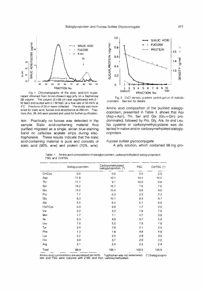
FRACTION No. Fig. 1 Chromatography of the sialic acid-rich super-
natant obtained from frozen-thawed egg jelly on a Sepharose 26 column. The column (5x88 cm) was equilibrated with 0.1 M NaCl and eluted with 0.1 M NaCl at a flow rate of 50 rnl/hr at 4°C. Fractions of 20 ml were collected. The eluate was moni- tored for sialic acid, fucose and absorbance at 280 nm. Frac- tions (No. 39-64) were pooled and used for further purification.
tein. Practically no fucose was detected in the sample. Sialic acid-containing material thus purified migrated as a single, alcian blue-staining band on cellulose acetate strips during elec- trophoresis. These results indicate that the sialic acid-containing material is pure and consists of sialic acid (go%, w/w) and protein (lo%, w/w).
1 - SlALlC ACID
::I 0 0.2
4 FUCOSE
-? PROTEIN 1.5
- 1.4 5
0
> 1.3 t
CJY z W n
-
1 2 3 4 5 6 7 8 9 10 top bottom FRACTION No.
Fig. 2 CsCl density gradient centrifugation of sialogly- coprotein. See text for details.
Amino acid composition of the purified sialogly- coprotein, presented in Table 1, shows that Asx (Asp+Asn), Thr, Ser and Glx (Glu+Gln) pre- dominated, followed by Pro, Gly, Ala, Ile and Leu. No cysteine or carboxymethylcysteine was de- tected in native and/or carboxymethylated sialogly- coprotein.
Fucose sulfate glycoconjugate A jelly solution, which contained 68 mg pro-
Table 1 . Amino acid compositions of sialoglycoprotein, carboxyrnethylated-sialoglycoprotein, FSG and CmFSG
Sialoglycoprotein Carboxyrnethylated- sialoglycoprotein (*) FSG CmFSG (*)
CmCys 0.0 0.0 0.0 2.0
ASP 11.8 10.1 14.4 14.2 Thr 11.1 9.1 10.0 9.8 Ser 15.2 16.7 7.8 7.5 Glu 14.2 15.4 9.9 9.6 Pro 7.7 6.3 2.3 2.3
GlY 6.3 10.1 8.3 8.7 Ala 5.4 6.4 6.7 6.5 Half-Cys 0.0 0.0 1.2 0.0 Val 3.2 3.2 7.9 7.9 Met 1.7 1.1 4.2 3.9 Ile 5.3 4.0 5.7 5.8 Leu 7.0 5.5 7.9 7.9
TYr 2.4 2.0 2.1 2.5 Phe 1.3 1.6 4.8 4.9
LYS 2.2 2.5 2.9 3.0 His 3.0 3.7 2.0 2.0
Arg 2.1 2.4 2.4 2.4
Total 99.9 100.1 100.5 100.9
Amino acid compositions are expressed as mol%. Tryptophan was not determined. (*) Sialoglycopro- tein and FSG were reduced with 2-ME and then carboxymethylated.

478 T. Shimizu et a/.
tein, 27 mg fucose and 23 mg sialic acid, was applied to a Sepharose 46 column equilibrated with ASW containing 10 mM sodium acetate buffer (pH 5.0). Fractions containing fucose, sialic acid and protein were pooled, dialyzed against 0.1 M NaCl and applied to a Sepharose 26 column equilibrated with 0.1 M NaCI. Fucose-containing material was eluted earlier than sialic acid- containing material as reported previously (65). Fractions which contained most of fucose were pooled (fucose-rich fraction). Solid NaCl was added to the fraction at a final concentration of 1 M, and the mixture was frozen and then thawed. The freezing-thawing made fucose-containing material insoluble in 1 M NaCl and about 80% of fucose in the original sample was recovered in the precipi- tate fraction by centrifugation at 10,000 x g for 30 min. The precipitate thus obtained was easily dissolved in DDW but not in ASW or even in 4 M guanidine hydrochloride solution. The precipitate was dissolved in DDW, followed by addition of an equal volume of 0.2 M NaCI, and the mixture was submitted to gel filtration on a Sepharose 28 col- umn. Fucose was co-eluted with materials having an absorbance at 280 nm. Fractions which con- tained fucose and protein were pooled (FSG) and used for further experiments. FSG possessed 2.0 times protein to fucose by weight when FSG was purified from a freshly prepared jelly solution, but it contained a little less amount of protein when it was purified from long time-stored jelly soltuion even if the jelly solution was kept at -40°C. However, FSG purified from either material contained one mol sulfate/mol fucose and practically no sialic acid. When FSG was analyzed by SDS-PAGE in the presence of 2-mercaptoethanol, two major pro- tein bands (258 kDa and 237 kDa) and one broad protein band (120 kDa) were observed, and they were also major protein components of crude jelly. Among them a 120 kDa protein was silver-stained to be red. These three components, however, could not be detected in an electrophoretogram of SDS-PAGE of FSG without 2-mercaptoethanol (Fig. 3). An electrophoretogram of the fucose-contain- ing fraction obtained from chromatography on a DEAE Bio-Gel A column was the same as that of FSG purified by chromatography on Sepharose CL-2B column. However, FSG which was purified from a jelly solution stored in a refrigerator or a freezer for long time without protease inhibitors showed a 210 kDa protein as a major band in addition to 258 kDa, 237 kDa and 120 kDa protein bands in SDS-PAGE, suggesting that some pro- teases which might come from eggs degrade
Fig. 3 SDS polyacrylamide gel electrophoresis of crude jelly and purified FSG. Approximately 2pg of protein was loaded to each lene (lane 1: crude jelly with 2-mercaptoethanol; lane 2: FSG without 2mercaptoethanol; lane 3: FSG with 2-rnercaptoethanol). The gel was silver-stained according to the method of Morrissey (32).
proteins in FSG. However, purified FSG could be stored in 0.1 M NaCl at -70°C without any change in protein bands in SDS-PAGE for at least six months. In some cases, FSG, stored particularly in high concentration, formed precipitate but this precipitate could be completely dissolved in 0.1 M NaCl at any time.
For testing the specificity of anti-serum against FSG, crude jelly was electrophoresed on a polyac- rylamide gel containing SDS and 2-mercapto- ehtanol and then electrophoretically blotted to a nitrocellulose filter. The blots were reacted with mouse anti-serum against FSG, followed by treat- ment with horseradish peroxidase-conjugated goat IgG anti-mouse IgG and the color reaction. As shown in Fig. 4, two bands, which correspond to 258 kDa and 237 Da proteins, respectively, were detected on the filter. As expected, two colored bands, corresponding to 258 kDa and 237 kDa proteins, respectively, were detected on the filter when purified FSG was subjected to immunoelec-
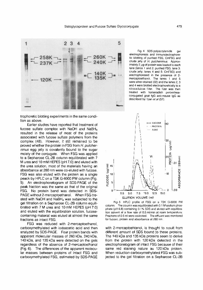
Sialoglycoprotein and Fucose Sulfate Glycoconjugate 479
trophoretic blotting experiments in the same condi- tion as above.
Earlier studies have reported that treatment of fucose sulfate complex with NaOH and NaBH, resulted in the release of most of the proteins associated with fucose sulfate polymers from the complex (48). However, it still remained to be proved whether the protein in FSG from H. pukher- rimus egg jelly is covalently bound to the sugar moiety of the conjugate. When FSG was applied to a Sepharose CL-2B column equilibrated with 7 M urea and 10 mM HEPES (pH 7.0) and eluted with the urea solution, most of the materials having an absorbance at 280 nm were co-eluted with fucose. FSG was also eluted with the protein as a single peack by HPLC on a TSK G-6000 PW column (Fig. 5). An electrophoretogram of SDS-PAGE of the peak fraction was the same as that of the original FSG. No protein band was detected in SDS- PAGE without 2-mercaptoethanol. When FSG tre- ated with NaOH and NaBH, was subjected to the gel filtration on a Sepharose CL-2B column equili- brated with 7 M urea and 10 mM HEPES (pH 7.0) and eluted with the equilibration solution, fucose- containing material was eluted at almost the same fractions as intact FSG.
FSG was reduced with 2-mercaptoethanol, carboxymethylated with iodoacetic acid and then analyzed by SDS-PAGE. Four protein bands with apparent molecular masses of 260 kDa, 240 kDa, 140 kDa, and 135 kDa were detected on the gels regardless of the absence of 2-mercaptoethanol (Fig. 6). The differences of the apparent molecu- lar masses between proteins of intact FSG and carboxymethylated FSG, estimated by SDS-PAGE
Fig. 4 SDS-polyacrylamide gel electrophoresis and immunolectrophore- tic blotting of purified FSG, CmFSG and crude jelly of H. pulcherrimus. Approx- imately 2 p g of protein was loaded to each lane (lanes 1 and 2: purified FSG; lane 3: crude jelly; lanes 4 and 5: CmFSG) and electrophoresed in the presence of 2- mercaptoethanol. The lanes 1 and 5 were silver-stained (32) and the lanes 2, 3 and 4 were blotted electrophoretically to a nitrocellulose filter. The filter was then treated with horseradish peroxidase- conjugated goat IgG anti-mouse IgG as described by Tjian eta/ (57).
- FUCOSE - PROTEIN
- A 2 8 0 1*0 -30,
E
a . 0, -
w
m a 0 -.oo
2.5 5.0 7.5 10.0 12.5 15.0 ELUTION VOLUME (ml)
Fig. 5 HPLC profile of FSG on a TSK G-6000 PW column. The column was equilibrated with 0.1 M sodium phos- phate (pH 6.8) containing 0.1% SDS and eluted with equilibra- tion solvent at a flow rate of 0.5 ml/min at room temperature. Fractions of 0.5 ml were collected. The effluent was monitored for fucose, protein and absorbance at 280 nm.
with 2-mercaptoehtanol, is thought to result from different amount of SDS bound to these proteins. The 140 kDa and 135 kDa proteins seem to derive from the protein with 120 kDa detected in the electrophoretogram of intact FSG because of their same red staining nature as 120 kDa protein. When reduction-carboxymethylated FSG was sub- jected to the gel filtration on a Sepharose CL-2B

480 T. Shirnizu et a/.
Fig. 6 SDS polyacrylarnide gel electrophoresis of CmFSG. Approximately 2 ,ug of proteins was loaded to each lane (lane 1: without 2-rnercaptoethanol; lane 2: with 2- rnercaptoethanol). The gel was silver-stained by the method of Morrissey (32).
column equilibrated with 7 M urea and 10mM HEPES (pH 7.0) and eluted with equilibration solu- tion, fucose-containing material and most of pro- tein originally associated with FSG were eluted together in almost the same fractions as in those obtained with intact FSG. However, when the CmFSG was subjected to HPLC, fucose-containing material, which possessed about 30% of protein originally associated with FSG, was separated from the rest of proteins (Fig. 7). The proteins sepa- rated from fucose-containing material were analy- zed by SDS-PAGE. Four protein bands (260 kDa, 240 kDA, 140 kDa and 135 kDa) were deceted. These results suggest that 258 kDa, 237 kDa and 120 kDa protein components associated each other in intact FSG by disulfide bonds and reduc- tion-alkylation released these components from FSG .
In order to confirm the presence of disulfide
- FUCOSE - PROTIEN
- A 2 8 0
i? 0
30
I - E
!O- . 0 2
2 W I-
-
a 10
0
.02
E
.01 2
z
0
N
W
m .....
a m
m a
U
.oo 2.5 5.0 7.5 10.0 12.5 15.0
ELUTION VOLUME (ml) Fig. 7 HPLC profile of CrnFSG. HPLC was carried out
in the same condition as described in the legend for Fig. 5.
bonds in FSG, free sulfhydryl groups in FSG was carboxymethylated with 'cold' iodoacetic acid and then reduced with 2-mercaptoethanol, The sam- ple was carboxymethylated with [ 1 -'4C]iodoacetic acid and analyzed by SDS-PAGE and cellulose acetate electrophoresis. In SDS-PAGE, a protein band which corresponds to (140 kDa+135 kDa) proteins showed strong radioactivity and weaker radioactivities were detected in 260 kDa, 240 kDa, 110 kDa and 87 kDa protein bands although only two coomassie blue-stained protein bands (260 kDa and 240 kDa) were detected on the gel (Fig. 8). The electrophoretogram obtained by cellulose acetate electrophoresis of the labeled sample showed that alcian blue- and nigrosine-stained materials stayed at the origin and the radioactivity was also detected at the origin (Fig. 9). However, when the labeled sample as well as intact FSG was heated at 100°C for 5 min before the analysis, two alcian blue-stained materials were detected in the electrophoretogram, while the radioactivity and a nigrosine-stained material stayed at the origin (Fig. 9). Both an alcian blue-stained material and a nigrosine-stained material were reacted with mouse anti-serum agianst FSG.
Proteins associated with FSG were purified from CmFSG using HPLC on a TSK G-6000 PW column connected to a TSK G-4000 SW column system and/or SDS-PAGE (Fig. 10). Amino acid compositions of FSG, CmFSG, sugar-associated protein moiety of CmFSG, 260 kDa protein, 240 kDa protein, and 140 kDa plus 135 kDa proteins are shown in Tables 1 and 2. Purified FSG con- tained 1.2 mol% of cysteine of which value is not reliable because of breakdown during acid hyd-

Sialoglycoprotein and Fucose Sulfate Glycoconjugate 481
Fig. 8 SDS polyacrylamide elec- trophoretograrn and the fluorogram of 14C- carboxymethylated FSG. SDS-PAGE was per- formed according to the method of Fairbanks et a/ (12). The gel was stained by coomassie blue (A) and then exposed to a Kodak X-omat AR film (B). Lane 1: without 2-mercaptoetha- nol; lane 2: with 2-mercaptoethanol.
Fig. 9 Cellulose acetate electrophoretogram and the fluorogram of ''C-carboxymethylated FSG. (A), Alcian blue-staining; (B), Fluorogram of the alcian blue-stained strip; (C), Nigrosine-staining; (D), Fluorogram of the nigrosine-stained strip. Lanes 1 and 8: standards, C: chondroitin sulfate C, D: dermatan sulfate, Ha: hyaluronic acid, Hp: heparin; lane 2: crude jelly; lane 3: sialoglycoprotein; lane 4: FSG; lane 5: FSG heated at 100°C for 5 min; lane 6: ''C-carboxymethylated FSG; lane 7: ''C-carboxymethylated FSG heated at 100°C for 5 rnin.
rolysis. When FSG was carboxymethylated with- CmCys. However, none of major protein compo- out 2-mercaptoethanol, resultant carboxymethy- nents (protein with fucose sulfate, 260 kDa and 240 lated FSG contained 0.9 mol% of carboxymethy- kDa proteins) isolated from CmFSG of which cys- lated cysteine (CmCys), while reduction- teine residues have been completely carboxy- carboxymethylated FSG possesed 2.0 mol% of methylated on the basis of total amino acid analy-

482 T. Shimizu et a/,
sis after carboxymethylation of FSG contained more than 1.6mol% CmCys although 140 kDa+ 135 kDa protein fraction contained the largest mot% of CmCys (3.6 mol%) and some of protein components (protein with fucose sulfate, 260 kDa protein and 140 kDa+135 kDa protein fraction) analyzed contained non-derivatized cys- teine residues.
Acrosome reaction The acrosome reaction of H. pulcherrimus
spermatozoa occurred in crude jelly within 30 sec at 20°C and the number of the reacted spermato- zoa did not increase significantly for further incuba- tion. In some cases, prolonged incubation gave us difficulty to count the reacted spermatozoa because of their sticking nature. Therefore, we usually stopped the reaction at 30 sec unless men- tioned. As shown in Table3, purified sialogly- coprotein was not capable for induction of the acrosome reaction, Addition of SAP-I did not in-
Fig. 10 HPLC profile of CmFSG on a TSK G6000 PW G4000 SW column system (A) and SDS-PAGE pattern of fractions from the column system (B). An aliquot (50~1) of each fraction (500 PI) designated by numbers was subjected to SDS-PAGE. The gel was silver-stained (32).
Table 2. Amino acid compositions Of protein Components obtained from CmFSG
Protein with 260 kDa 240 kDa 140 kDa+ 135 kDa fucose sulfate protein protein protein
CmCys 0.6 1.6 1.4 3.6
Thr 4.8 9.1 7.8 6.5
Glu 12.8 10.6 11.8 12.3 Pro 3.8 3.2 3.2 4.6
GlY 18.3 11.8 14.5 15.5 Ala 6.5 7.7 6.9 6.6 Half-Cys 0.3 0.2 0.0 0.6 Val 3.3 7.7 6.3 5.1 Met 1.6 2.2 1.4 1.5
Leu 4.9 7.7 6.9 6.2
Phe 3.2 4.6 4.1 3.2 LYS 3.2 2.2 3.0 2.4
ASP 9.9 11.8 11.0 10.2
Ser 14.9 7.7 9.1 8.7
Ile 2.4 5.1 4.3 3.9
TYr 2.6 2.0 1.7 2.0
His 4.1 4.0 4.6 4.9 Arg 2.8 1.8 2.1 2.0
Total 100.0 101.0 100.1 99.8
Amino acid compositions are expressed as mOl%. Tryptophan was not determined.
Sialoglycoprotein and Fucose Sulfate Glycoconjugate 483
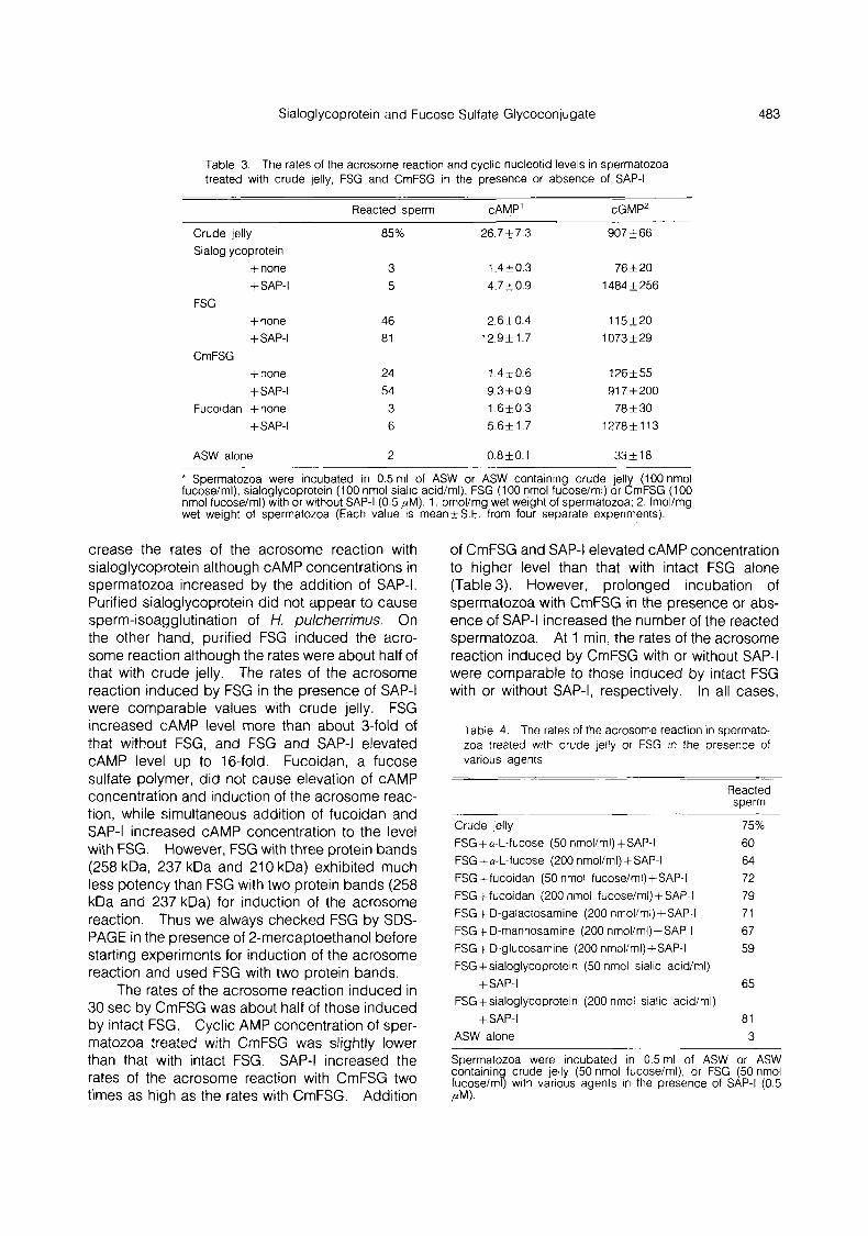
Table 3. The rates of the acrosome reaction and cyclic nucleotid levels in spermatozoa treated with crude jelly, FSG and CmFSG in the presence or absence of SAP-I
Reacted sperm CAMP’ cGMP2
Crude jelly Sialoglycoprotein
+none +SAP-I
FSG +none +SAP-I
CmFSG +none
Fucoidan +none +SAP-I
+SAP-I
ASW alone
85%
3 5
46 81
24 54 3 6
2
26.7k7.3
1.4+0.3 4.7k0.9
2.6k0.4 12.9k 1.7
1.4k0.6 9.3f0.9 1 .6k0 .3 5.6k1.7
0.8f0.1
907 k 66
76 k 20 1484 k 256
115k20 1073 t 29
1 2 6 t 5 5 917k200
78 f 30 1278f 11 3
33+18
* Spermatozoa were incubated in 0.5ml, of ASW or ASW containing crude iell (100 nmol fucose/ml), sialoglycoprotein (100 nmol sialic acid/ml), FSG (100 nmol fucose/ml) or ZmFSG (100 nmol fucose/ml) with or without SAP-I (0.5 pM). 1 . pmol/mg wet weight of spermatozoa; 2. fmol/mg wet weight of spermatozoa (Each value is mean+S.E. from four separate experiments).
crease the rates of the acrosome reaction with sialoglycoprotein although cAMP concentrations in spermatozoa increased by the addition of SAP-I. Purified sialoglycoprotein did not appear to cause sperm-isoagglutination of H. pulcherrimus. On the other hand, purified FSG induced the acro- some reaction although the rates were about half of that with crude jelly. The rates of the acrosome reaction induced by FSG in the presence of SAP-I were comparable values with crude jelly. FSG increased cAMP level more than about 3-fold of that without FSG, and FSG and SAP-I elevated cAMP level up to 16-fold. Fucoidan, a fucose sulfate polymer, did not cause elevation of cAMP concentration and induction of the acrosome reac- tion, while simultaneous addition of fucoidan and SAP-I increased cAMP concentration to the level with FSG. However, FSG with three protein bands (258 kDa, 237 kDa and 210 kDa) exhibited much less potency than FSG with two protein bands (258 kDa and 237 kDa) for induction of the acrosome reaction. Thus we always checked FSG by SDS- PAGE in the presence of 2-mercaptoethanol before starting experiments for induction of the acrosome reaction and used FSG with two protein bands.
The rates of the acrosome reaction induced in 30 sec by CmFSG was about half of those induced by intact FSG. Cyclic AMP concentration of sper- matozoa treated with CmFSG was slightly lower than that with intact FSG. SAP-I increased the rates of the acrosome reaction with CmFSG two times as high as the rates with CmFSG. Addition
of CmFSG and SAP-I elevated cAMP concentration to higher level than that with intact FSG alone (Table 3). However, prolonged incubation of spermatozoa with CmFSG in the presence or abs- ence of SAP-I increased the number of the reacted spermatozoa. At 1 min, the rates of the acrosome reaction induced by CmFSG with or without SAP-I were comparable to those induced by intact FSG with or without SAP-I, respectively. In all cases,
Table 4. The rates of the acrosome reaction in spermato- zoa treated with crude jelly or FSG in the presence of various agents
Reacted sperm
Crude jelly FSG +a-L-fucose (50 nmol/ml)+SAP-I FSG +a-L-fucose (200 nmol/ml)+SAP-I FSGffucoidan (50 nmol fucose/ml)+SAP-I FSG +fucoidan (200 nmol fucoseiml) +SAP-I FSG + D-galactosamine (200 nmol/ml)+SAP-I FSG +D-mannosamine (200 nmol/ml)+SAP-I FSG + D-glucosamine (200 nmol/ml)+SAP-I FSG f sialoglycoprotein (50 nmol sialic acrdiml)
FSG +sialoglycoprotein (200 nmol sialic acid/rnl)
ASW alone
+SAP-I
+SAP-I
75% 60 64 72 79 71 67 59
65
81 3
Spermatozoa were incubated in 0.5 ml of ASW or ASW containing crude jelly (50 nrnol fucose/ml), or FSG (50 nmol fucose/ml) with various agents in the presence of SAP-I (0.5 PM).
484 T. Shimizu et a/.
SAP-I elevated sperm cGMP concentrations. As shown in Table 4, a-L-fucose, fucoidan, amino sugars such as D-galactosamine, D-mannosamine and D-glucosamine and sialoglycoprotein, which were not capable for induction of the acrosome reaction by themselves, did not inhibit the ability of FSG to induce the acrosome reaction in the pre- sence of SAP-I.
Discussion
We purified and characterized two major egg jelly macromolecules, a sialoglycoprotein and a fucose sulfate glycoconjugate, of H. pulcherrimus in the present study. Sialoglycoprotein seems to consist only of sialic acid and protein since dry weight of purified sialoglycoprotein was almost equal to weight of sialic acid plus protein, which were determined colorimetrically, and it did not contain measurable amount of fucose. It did not appear to cause sperm-isoagglutination of H. pul- cherrimus. These results are not compatible with results of earlier studies (18, 19). We think that these incompatiblities might come from impurity of sialoglycoprotein used in earlier studies or use of different species of sea urchins. Sialoglycopro- tein purified in the present study did not induce the acrosome reaction of H. pulcherrimus spermato- zoa. This agrees with results of earlier studies.
Fucose sulfate glycoconjugate isolated from H. pulcherrimus contained a large amount of pro- tein. The proteins associated with fucose sulfate was not removed even when FSG was dissolved in 7 M urea or 0.1 Yo SDS and chromatographed on a Sepharose CL-2B or a TSK G-6000 PW column. Furthermore, no protein band was detected in the electrophoretogram of SDS-PAGE of intact FSG without 2-mercaptoethanol. These results sug- gest that the proteins may be covalently bound to the polysaccharide portion of FSG. When FSG was dissolved in SDS solution containing 2- mercaptoethanol and subjected to SDS-PAGE in the presence of 2-mercaptoethanol, two major (258 kDa and 237 kDa) and one minor (1 20 kDa) protein bands were appeared. Reduced and carboxy- methylated FSG showed two major (260 kDa and 240 kDa) and two minor (140 kDa and 135 kDa) protein bands in the electrophoretogram of SDS- PAGE despite absence of 2-mercaptoethanol. These results indicate that FSG consists of fucose sulfate polymers and several protein components which associate each other with disulfide bonds. In this regard, it should be noted that when FSG was carboxymethylated with 'cold' iodoacetic acid
and then reduced and carboxymethylated with ''C-iodoacetic acid, most of radioactivity was de- tected in 140 kDa plus 135 kDa protein bands. lodoacetic acid treatment of FSG without 2- mercaptoethanol resulted in carboxymethylation of about half of cysteine residues, indicating that about half of cysteine residues in FSG involved in formation of disulfide bonds. Furthermore, a pro- tein with fucose sulfate, 260 kDa and 240 kDa proteins isolated from reduced-carboxymethylated FSG contained much less mol% of carboxymethy- lated cysteine than that of 140 kDa plus 135 kDa proteins. These results suggest that 140 kDa and 135 kDa proteins, which are probably the same protein(s) as detected in a 120 kDa protein band dected in SDS-PAGE of intact FSG with 2- mercaptoethanol have more sulfhydryl groups than 258 kDa and 237 kDa proteins available for dis- ulfide bonds and may serve to make bridges between the polysaccharide-containing protein and other protein components.
As we reported previously (66), purified FSG induced the acrosome reaction, but the rates were about half of that with crude jelly. SAP-I promoted the rates of the acrosome reaction with FSG to values comparable with those induced by crude jelly. In the paper, we have shown that pronase- digestion of fucose-rich glycoconjugate which should be the same molecule as FSG in the pre- sent study resulted in a large reduction of the acrosome reaction-inducing capacity (66). Com- plete carboxymethylation of cysteine residues in FSG resulted in release of the major constituent proteins from FSG and an about 50% decrease in acrosome reaction-inducing capacity of FSG. This does not agree with the results of SeGall and Lennarz demonstrating that the acrosome reac- tion-inducing capacity of the egg jelly of four species of sea urchins resides solely in the fucose sulfate polysaccharide (48, 49), and suggests that at least in FSG of H. pulcherrimus the association of these proteins with fucose sulfate polymers is important for induction of the acrosome reaction. However, pronase-digested ARIS, a major acro- some reaction-inducing substance in starfish, is reported to be fully effective for induction of the acrosome reaction with Co-ARIS (31, 34, 35). At the present time, it is not clear whether or not the differences in the role of proteins associated with a major acrosome reaction-inducing substance are due to species variations.
lshihara et a/ reported that the fucan sulfate isolated from H. pulcherrimus and Pseudocentro- tus depressus egg jelly contained L-fucose-4-
Sialoglycoprotein and Fucose Sulfate Glycoconjugate 485
sulfate (21). Fucoidan fucose-4-sulfate did not acrosome reaction of H. zoa despite increase in
which possesses L- appreciably induce the pulcherrimus spermato- cAMP concentration in-
duced by SAP-I. L-Fucose and amino sugars showed almost no effect on induction of the acro- some reaction by FSG with SAP-I. These results suggest that the fucose sulfate and amino sugars in FSG may not play an important role in induction of the acrosome reaction and that cAMP is not involved in the acrosome reaction.
Sperm-activating peptides stimulate net H+ efflux and enhance sperm cAMP and cGMP con- centrations (14, 28, %, 46, 54). A fucose sulfate- rich glycoconjugate causes Ca2+-accumulation, elevates cAMP and induces the acrosome reaction (13, 25). In connection with this, it should be noted that monoclonal antibodies to a 210 kDa glycoprotein of S. purpuratus sperm plasma mem- brane cause an increase in intracellular Ca2+ of S. purpuratus spermatozoa and induce the acrosome reaction when the intracellular pH is increased with NH&I (29, 37, 38, 60, 61, 62). The antibodies induce the CAMP-dependent phosphorylation of sperm histone H1 as occurs upon treatment of spermatozoa with egg jelly (1 6, 39, 40). However, ionophore A23187 which is known to induce the acrosome reaction and acts as a co-factor of pronase-digested FSG for induction of the acro- some reaction neither induces H1 phosphorylation nor increases cAMP concentration in sea urchin spermatozoa (8, 39, 66). These support the idea suggesting that cAMP is not directly involved in the acrosome reaction. Recently, Domino and Gar- bers reported that FSG induces the elevation of inositol trisphosphate (10). The elevation is de- pendent on external Ca2+. The Ca2+ channel blockers, verapamil and nifedipine, inhibited in- creases in both inositol trisphosphate and CAMP. The products generated from stimulated PI turnov- er in sea urchin spermatozoa may be potential second messengers in induction of the acrosome reaction.
Acknowledgments: We wish to thank Mr. M. Matada for collecting and culturing sea urchins. Authors are grateful to the staff of the Radioisotope Center, Kanaza- wa University, for facilitating use of equipments for determing cyclic nucleotide concentrations. This work was supported by a Grant-in-Aid for Scientific Research (B) to N.S. (No. 63480021) from the Ministry of Educa- tion, Science and Culture of Japan.
References 1. Christen, R., R. W. Schackmann and B. M. Shapiro,
1983. Interaction between sperm and sea urchin egg jelly. Develop. Bjol., 98, 1-14.
2. Crestifield, A. M., S. Moore and W. H. Stein, 1963. The preparation and enzymatic hydrolysis of reduced and 35S- carboxymethylated proteins. J. Biol. Chem., 238, 622-627.
3. Collins, F. and 0. Epel, 1977. The role of calcium ions in the acrosome reaction of sea urchin sperm. Exp. Cell Res., 106, 21 1-222.
4. Dan, J. C., 1952. Studies of the acrosome. I. Reaction to egg-water and other stimuli. Biol. Bull., 103, 54-66.
5. Dan, J. C., 1954. Studies of the acrosome. 1 1 1 . Effects of calcium deficiency. Biol. Bull.. 107, 335-349.
6. Dan, J. C., 1956. The acrosome reaction. Int. Rev. Cytol., 5, 365-393.
7. Darszon, A., J. Garcia-Soto, A. Lievano, J. A. Sanchez and A. D. Islas-Trejo, 1986. Ionic channels in the plasma membrane of sea urchin sperm. In Ionic Channels in Cells and Model Systems (Ed. R. Latorre), Plenum Publishing Corp., New York, pp. 291 -305.
8. Decker, G. L., D. 8. Joseph and W. J. Lennarz, 1976. A study of factors involved in induction of the acrosome reaction in sperm of the sea urchin, Arbacia punctulafa. Develop. Biol., 53, 115-125.
9. Dische, Z. and L. B. Shettles, 1951. A new spectropho- tometric test for the detection of methylpentose. J. Biol. Chem., 192, 579-582.
10. Domino, S. E. and D. L. Garbers, 1988. The fucose- sulfate glycoconjugate that induces an acrosome reaction in spermatozoa stimulates inositol 1,4,5-triphosphate accumula- tion. J. Biol. Chem., 263, 690-695.
11. Epel, D., 1978. Mechanisms of activation of sperm and egg during fertilization of sea urchin gametes. Curr. Top. Develop. Biol., 12, 185-246.
12. Fairbanks, G., T. L. Steck and D. F. H. Wallach, 1971. Electrophoretic analysis of the major polypeptides of the human eryhtrocyte membrane. Biochemistry, 10, 2606-261 6.
13. Garbers. D. L. and G. S. Kopf, 1980. The regulation of spermatozoa by calcium and cyclic nucleotides. Adv. Cyclic Nucleotide Res., 13, 251-306.
14. Garbers, D. L., H. D. Watkins, J. R. Hansbrough and K. S. Misono, 1982. The amino acid sequence and chemical synthesis of speract and of speract analogues. J. Biol. Chem., 257, 2734-2737.
15. Garbers, D. L., G. S. Kopf, D. J. Tubb and G. Olson, 1983. Elevation of sperm adenosine 3': 5'-monophosphate concentrations by a fucose-sulfate-rich complex associated with eggs. I. Structural characterization. Biol. Reprod., 29, 121 1-1 220.
16. Green, G. R. and D. L. Poccia, 1985. Phosphorylation of sea urchin sperm H1 and H2B histones precedes chromatin decondensation and H1 exchange during pronuclear formation. Develop. Biol., 108, 235-245.
17. Guerrero, A. and A. Darszon, 1989. Egg jelly triggers a calcium influx which inactivates and is inhibited by calmodulin antagonists in the sea urchin sperm. Biochem. Biophys. Acta, 980, 109-1 16.
18. Hotta, K., H. Hamazaki, M. Kurokawa and S. Isaka, 1970. Isolation and properties of a new type of sialopolysac- charideprotein complex from the jelly coat of sea urchin eggs. J. Biol. Chem., 245, 5434-5440.
19. Isaka, S., K. Hotta and M. Kurokawa. 1970. Jelly coat

486 T. Shimizu et a/
substances of sea uchin eggs. Exp. Cell Res., 59, 37-42. 20. Ishihara, K. and J. C. Dan, 1970. Effects of chemical
disruption on the biological activities of sea urchin egg jelly. Develop. Growth & Differ., 12, 179-188.
21. Ishihara, K., K. Oguri and H. Taniguchi, 1973. Isolation and characterization of fucose sulfate from jelly coat glycopro- tein of sea urchin egg. Biochem. Biophys. Acta, 320, 628-634.
22. Jourdian, G. W., L. Dean and S. Roseman, 1972. The sialic acids. XI. A periodate-resorcinol method for the quantita- tive estimation of free sialic acids and their glycosides. J. Biol. Chem., 246, 430-435.
23. Kinsey, W. H., G. K. SeGall and W. J. Lennarz, 1979. The effect of the acrosome reaction on the respiratory activity and fertilizing capacity of echinoid sperm. Develop. Biol., 71, 49-59.
24. Kinsey, W. H., J. A. Rubin and W. J. Lennarz, 1980. Studies on the specificity of sperm binding in echinoderm fertilization. Develop. Biol.. 74, 245-250.
25. Kopf, G. S. and D. L. Garbers, 1980. Calcium and fucose-sulfate-rich polymer regulate sperm cyclic nucleotide metabolism and the acrosome reaction. Biol. Reprod., 22,
26. Laemmli, U. K. 1970. Cleavage of structural proteins during the assembly of the head of bacteriophage T4. Nature, 257, 680-685.
27. Lee, H. C., C. Johnson and D. Epel. 1983. Changes in internal pH associated with initiation of motility and acrosome reaction of sea urchin sperm. Develop. Biol,, 72, 126-137.
28. Lee, H. C. and D. L. Garbers, 1986. Modulation of the voltage-sensitive Na+/H+ exchage in sea urchin spermatozoa through membrane potential changes induced by the egg peptide speract. J. Biol. Chem., 261, 16026-16032.
29. Lopo, A. C. and V. D. Vacquier, 1980. Antibody to sperm surface glycoprotein inhibits the egg jelly-induced acro- some reaction of sea urchin sperm. Develop. Biol., 79, 325- 333.
30. Matsui, T., I. Nishiyama, A. Hino and M. Hoshi, 1986. Induction of the acrosome reaction in starfish. Develop. Growth & Differ., 28, 339-348.
31. Matsui, T., I. Nishiyama, A. Hino and M. Hoshi, 1986. Acrosome reaction-inducing substance purified from the egg Ielly inhibits the jelly-induced acrosome reaction in starfish: An apparent contradiction. Develop. Growth & Differ., 28, 349- 357.
32. Morrissey, J. H. 1981. Silver stain for proteins in polyacrylamide gels: A modified procedure with enhanced uniform sensitivity. Analyt. Biochem., 117, 307-310.
33. Nishioka, D. and N. Cross, 1978. The role of external Na+ in sea urchin fertilization. In Cell Reproduction (Eds. E. R. Dirksen, D. Prescott and C. F. Fox), Academic Press, New York, pp. 403-41 3.
34. Nishiyama, I., T. Matsui and M. Hoshi, 1987. Purifica- tion of Co-ARIC, a cofactor for acrosome reaction-inducing substance, from the egg jelly of starfish. Develop. Growth & Differ., 29, 161-169.
35. Nishiyama, I., T. Matsui, Y. Fujimoto, N. lkekawa and M. Hoshi, 1987. Correlation between the molecular structure and the biological activity of Co-ARIS, a cofactor for acrosome- reaction-inducing substance. Develop. Growth & Differ., 29, 171 -1 76.
36. Ohtake. H., 1976. Respiratory behaviour of sea-urchin spermatozoa. I. Effect of pH and egg water on the respiratory rate. J. Exp. Zool., 198, 303-312.
37. Podell, S . 8. and V. D. Vacquier, 1984. Inhibition of sea urchin sperm acrosome reaction by antibodies directed against
11 18-1 126.
two sperm membrane proteins. Exp. Cell Res., 155, 467-476. 38. Podell. S. 6. and V. D. Vacquier, 1985. Purification of
the Mr 80,000 and Mr 210,000 proteins of sea urchin sperm plasma membrane. J. Biol. Chem., 260, 2715-2718.
39. Porter, D. C. and V. D Vacquier, 1986. Phosphoryla- tion of sperm histone H1 is induced by the egg jelly layer in the sea urchin Stronglylocentrotus purpuratus. Develop. Biol., 116, 203-212.
40. Porter, D. C., G. W. Moy and V. D. Vacquier, 1988. CAMP dependent protein kinase of sea urchin sperm phsphory- lates histone H1 on a single site. J. Biol. Chem., 263, 2750- 2755.
41. Repaske, D. R. and D. L. Garbers, 1983. A hydrogen ion flux mediates stimulation of respiratory activity by speract in sea urchin spermatozoa. J. Biol. Chem., 258, 6025-6029.
42. Schackmann, R. W., E. M. Eddy and 6. M. Shapiro, 1978. The acrosome reaction of Sfrongyfocentfofotus purpur- atus sperm. Ion requirements and movements. Develop. Biol., 65. 483-495.
43. Schackmann, R. W. and B. M. Shapiro, 1981. A partial sequence of ionic changes associated with the acrosome reaction of Sfrongylocenfrotus purpurafus. Develop. Biol., 81, 145-154.
44. Schackmann, R. W., R. Christen and 6. M. Shapiro, 1981. Membrane potential depolarization and increased in- tracellular pH accompanying the acrosome reaction of sea urchin sperm. Proc. Natl. Acad. Sci. USA, 78, 6066-6070.
45. Schackmann, R. W., R. Christen and 6. M. Shapiro, 1984. Measurement of plasma membrane and mitochondria1 potentials in sea urchin sperm. Changes upon activation and induction of the acrosome reaction. J. 6iol. Chem., 259, 1391 4- 1 3922.
46. Schackmann, R. W. and P. 6. Chock, 1986. Alteration of intracellular [Ca’+] jn sea urchin sperm by the egg peptide speract. Evidence that increased intracellular Ca2+ is cou- pled to Na+ entry and increased intracellular pH. J. Biol. Chem., 261, 8719-8728.
47. Schacterle, G. R. and R. L. Pollack, 1973. A simplified method for the quantitative assay of small amount of protein in biologic material. Anal. Biochem., 51, 654-655.
48. SeGall, G. K. and W. J. Lennarz, 1979. Chemical characterization of the component of the egg jelly coat from sea urchin eggs responsible for induction of the acrosome reaction. Develop. Biol., 71, 33-48.
49. SeGall, G. K. and W. J. Lennarz, 1981. Jelly coat induction of the acrosome reaction in echinoid sperm. De- velop. Biol., 86, 87-93.
50. Summers, R. G. and B. L. Hylander, 1975. Species- specificity of acrosomal reaction and primary gamete binding in echinoids. Exp. Cell Res., 93, 63-98.
51. Summers, R. G., 8. L. Hylander, L. H. Colwin and A. L. Colwin, 1975. The functional anatomy of the echinoderm sper- matozoon and its interaction with the egg at fertilization. Am. Zool.. 15, 523-551.
52. Suzuki. N., K. Nomura, H. Ohtake and S. Isaka, 1981, Purification and the primary structure of sperm-activating pep- tides from the jelly coat of sea urchin eggs. Biochem. Biophys. Res. Commun., 99, 1238-1244.
53. Suzuki, N., Y. Ohizumi, I. Yasumasu and S. Isaka, 1984. Respiration of sea urchin spermatozoa in the presence Of a synthetic jelly coat peptide and ionophores. Develop. Growth & Differ., 26, 17-24.
54. Suzuki, N., 1989. Sperm activating peptides from sea urchin egg jelly, In Bioorganic Marine Chemistry (Ed. P. J. Scheuer), vol. 3, pp. 47-70, Springer-Verlag, Berlin.
Sialoglycoprotein and Fucose Sulfate Glycoconjugate 487
55. Terho, T. T. and K. Hartiala, 1971. Method for deter- mination of the sulfate content of glycosaminoglycans. Analyt. Biochem., 41, 471 -476.
56. Tilney, L. G., S. Hatano, H. lshikawa and M. S. Moosek- er, 1973. The polymerization of actin: Its role in the generation of the acrosomal process of certain echinoderm sperm. J. Cell Biol., 59, 109-126.
57. Tjian, R., D. Stinchomb and R. Losick, 1974. Antibody directed against Bacillus subtilis factor purified by sodium dodecyl sulfate slab gel electrophoresis. Effect on transcrip- tion by RNA polymerase in crude extracts of vegetative and sporulating Cells. J. Biol. Chem., 250, 8824-8828.
58. Toowicharanont, P. and 13. M. Shapiro, 1988. Regina1 differentiation of the sea urchin sperm plasma membrane. J. Biol. Chem., 263, 6877-6883.
59. Towbin, H., T. Staehelin and J. Gordon, 1979. Elec- trophoretic transfer of proteins from polyacrylarnide gels to nitrocellulose sheets: Procedure and some applications. Proc. Natl. Acad. Sci. USA, 76, 4350-4354.
60. Trimmer, J. S., I. S. Trowbridge and V. D. Vacquier, 1985. Monoclonal antibody to a membrane glycoprotein in- hibits the acrosome reaction and associated Ca2+ and H+ fluxes of sea urchin sperm. Cell, 40, 697-703.
61. Trimmer, J. S., R. W. Schackmann and V. D. Vacqier,
1986. Monoclonal antibodies increase intracellular Ca2+ in sea urchin spermatozoa. Proc. Natl. Acad. Sci. USA, 83, 9055-9059.
62. Trimmer, J. S., Y. Ebina, R. W. Schackmann, C-G. Meinhof and V. D. Vacquier, 1987. Characterization of a monoclonal antibody that induces the acrosome reaction of sea urchin sperm. J. Cell Biol., 105, 1121-1 128.
63. Uno, Y. and M. Hoshi, 1978. Separation of the sperm agglutinin and the acrosorne reaction-inducing substance in egg jelly of starfish. Science, 200, 58-59.
64. Vacquier. V. D., 1986. Handling, labeling, and frac- tionating sea uchin spermatozoa. Methods Cell Biol., 27, 15- 40.
65. Yarnaguchi, M., T. Niwa. M. Kurita and N. Suzuki. 1988. The participation of speract in the acrosome reaction of Hemicentrotus pulcherrimus. Develop. Growth & Differ., 30, 159-1 67.
66. Yamaguchi, M., M. Kurita and N. Suzuki, 1989. Induc- tion of the acrosorne reaction of Hemicentrotus pulcherrimus spermatozoa by the egg jelly molecules, fucose-rich glyco- conjugate and sperm-activating peptide l Develop. Growth & Differ., 31, 233-239.
(Received November 9, 1989; accepted February 5, 1990)
Copyright © 2022 FDOKUMEN