Purification and characterization of a nitrilase from Aspergillus niger K10
6
Process Biochemistry 45 (2010) 1115–1120 Contents lists available at ScienceDirect Process Biochemistry journal homepage: www.elsevier.com/locate/procbio Purification and characterization of nitrilase from Fusarium solani IMI196840 Vojtˇ ech Vejvoda a , David Kubᡠc a , Alˇ zbˇ eta Davidová a , Ondˇ rej Kaplan a , Miroslav ˇ Sulc a,b , Ondˇ rej ˇ Sveda a , Radka Chaloupková c , Ludmila Martínková a,∗ a Institute of Microbiology, Centre of Biocatalysis and Biotransformation, Academy of Sciences of the Czech Republic, Víde ˇ nská 1083, CZ-142 20 Prague, Czech Republic b Department of Biochemistry, Faculty of Science, Charles University Prague, Hlavova 8, CZ-128 40 Prague, Czech Republic c Loschmidt Laboratories, Department of Experimental Biology and Centre of Biocatalysis and Biotransformation, Faculty of Science, Masaryk University, Kamenice 5/A4, CZ-625 00 Brno, Czech Republic article info Article history: Received 11 November 2009 Received in revised form 26 March 2010 Accepted 30 March 2010 Keywords: Fusarium solani Nitrilase Purification Characterization Nitriles abstract Nitrilase activity in Fusarium solani IMI196840 (approx. 1500 U l −1 of culture broth) was induced by 2- cyanopyridine. The enzyme was purified by a factor of 20.3 at a yield of 26.9%. According to gel filtration, the holoenzyme was an approx. 550-kDa homooligomer consisting of subunits with a molecular weight of approximately 40 kDa, as determined by SDS-PAGE. Mass spectrometry analysis of the tryptic frag- ments suggested a high similarity of this enzyme to the hypothetical CN hydrolases from Aspergillus oryzae, Gibberella zeae, Gibberella moniliformis and Nectria haematococca. Circular dichroism and fluo- rescence spectra indicated that secondary structure content and overall tertiary structure, respectively, were almost identical in nitrilases from F. solani IMI196840 and F. solani O1. The melting temperatures of the enzymes were 49.3 ◦ C and 47.8 ◦ C, respectively. The best substrates for the purified nitrilase from F. solani IMI196840 were benzonitrile and 4-cyanopyridine, which were hydrolyzed at the rates of 144 and 312 U mg −1 protein, respectively, under the optimum conditions of pH 8 and 45 ◦ C. The enzyme was highly chemoselective, producing ≤2% amides as by-products. © 2010 Elsevier Ltd. All rights reserved. 1. Introduction Nitrilases (EC 3.5.5.1; 3.5.5.2; 3.5.5.4–3.5.5.7) have been recog- nized as valuable biocatalysts for the synthesis of high added-value carboxylic acids from cheap and readily available nitriles. Interest in their use is rising as illustrated by the increasing number of articles and patents on this topic [1]. This is because the enzymes enable nitrile hydrolysis to be performed at mild pH and temperature and are often also enantioselective or regioselective. Furthermore, among nitrilases there is broad choice of enzymes with differing substrate specificities (e.g., [1–5]). A number of companies (DuPont, Lonza, Dow, Diversa, BASF, DSM) have investigated the use of enzymatic nitrile hydrolysis in their processes. For instance, (R)- mandelic and (R)-chloromandelic acid have already been produced commercially by Mitsubishi Rayon Co. [5]. Nitrilases were discovered in the 1960s, when enzymes from barley [6] and Pseudomonas [7] were purified. Since then, a large number of various nitrilases have been purified and characterized in bacteria, but only a few in eukaryotic organisms (plants and filamentous fungi; for a review see [3]). Nevertheless, the broad occurrence of nitrilases in filamentous fungi has been indicated by ∗ Corresponding author. Tel.: +420 296 442 569; fax: +420 296 442 509. E-mail address: [email protected] (L. Martínková). screening collection strains [8] and by gene and protein database searches [9]. We have recently been focusing our research on filamentous fungi as a source of new nitrilases with potentially different cat- alytic properties from those of the bacterial enzymes, and purified and characterized the nitrilases from Aspergillus niger K10 [10] and Fusarium solani O1 [11]. The latter enzyme exhibited similar spe- cific activities to the nitrilase from Fusarium oxysporum f. sp. melonis [12]. On the other hand, the nitrilase from the F. solani IMI196840 strain described in one of the pioneering works on nitrilases in 1977 [13] exhibited a specific activity that was 87-times lower. This enzyme also differed from typical nitrilases in its subunit molecular mass (76 kDa [13] vs. 37–40 kDa in other fungal enzymes [10–12] and 38–46 kDa in most bacterial enzymes [3,4]). The nitrilase in F. solani IMI196840 was induced by benzonitrile as the sole source of C and N but the activity yield was very low [13,14]. Previously, we identified 2-cyanopyridine as a powerful nitrilase inducer, which was efficient in all the nitrilase-producing fungal strains examined (Aspergillus, Fusarium, Penicillium) [15]. In this work, we used this compound to induce a high nitrilase activity in the F. solani IMI196840 strain. The nitrilase purified and char- acterized by us from this strain is substantially different from that described in the same strain previously [13] but similar to the other two nitrilases described in Fusarium genus [11,12], though some differences between these enzymes could be observed in terms of 1359-5113/$ – see front matter © 2010 Elsevier Ltd. All rights reserved. doi:10.1016/j.procbio.2010.03.033
-
Upload
independent -
Category
Documents
-
view
1 -
download
0
Transcript of Purification and characterization of a nitrilase from Aspergillus niger K10
P
VOa
b
c
C
a
ARRA
KFNPCN
1
nctanaasLemc
bnifio
1d
Process Biochemistry 45 (2010) 1115–1120
Contents lists available at ScienceDirect
Process Biochemistry
journa l homepage: www.e lsev ier .com/ locate /procbio
urification and characterization of nitrilase from Fusarium solani IMI196840
ojtech Vejvodaa, David Kubáca, Alzbeta Davidováa, Ondrej Kaplana, Miroslav Sulca,b,ndrej Svedaa, Radka Chaloupkovác, Ludmila Martínkováa,∗
Institute of Microbiology, Centre of Biocatalysis and Biotransformation, Academy of Sciences of the Czech Republic, Vídenská 1083, CZ-142 20 Prague, Czech RepublicDepartment of Biochemistry, Faculty of Science, Charles University Prague, Hlavova 8, CZ-128 40 Prague, Czech RepublicLoschmidt Laboratories, Department of Experimental Biology and Centre of Biocatalysis and Biotransformation, Faculty of Science, Masaryk University, Kamenice 5/A4,Z-625 00 Brno, Czech Republic
r t i c l e i n f o
rticle history:eceived 11 November 2009eceived in revised form 26 March 2010ccepted 30 March 2010
eywords:
a b s t r a c t
Nitrilase activity in Fusarium solani IMI196840 (approx. 1500 U l−1 of culture broth) was induced by 2-cyanopyridine. The enzyme was purified by a factor of 20.3 at a yield of 26.9%. According to gel filtration,the holoenzyme was an approx. 550-kDa homooligomer consisting of subunits with a molecular weightof approximately 40 kDa, as determined by SDS-PAGE. Mass spectrometry analysis of the tryptic frag-ments suggested a high similarity of this enzyme to the hypothetical CN hydrolases from Aspergillus
usarium solaniitrilaseurificationharacterizationitriles
oryzae, Gibberella zeae, Gibberella moniliformis and Nectria haematococca. Circular dichroism and fluo-rescence spectra indicated that secondary structure content and overall tertiary structure, respectively,were almost identical in nitrilases from F. solani IMI196840 and F. solani O1. The melting temperaturesof the enzymes were 49.3 ◦C and 47.8 ◦C, respectively. The best substrates for the purified nitrilase fromF. solani IMI196840 were benzonitrile and 4-cyanopyridine, which were hydrolyzed at the rates of 144and 312 U mg−1 protein, respectively, under the optimum conditions of pH 8 and 45 ◦C. The enzyme was
roduc
highly chemoselective, p. Introduction
Nitrilases (EC 3.5.5.1; 3.5.5.2; 3.5.5.4–3.5.5.7) have been recog-ized as valuable biocatalysts for the synthesis of high added-valuearboxylic acids from cheap and readily available nitriles. Interest inheir use is rising as illustrated by the increasing number of articlesnd patents on this topic [1]. This is because the enzymes enableitrile hydrolysis to be performed at mild pH and temperaturend are often also enantioselective or regioselective. Furthermore,mong nitrilases there is broad choice of enzymes with differingubstrate specificities (e.g., [1–5]). A number of companies (DuPont,onza, Dow, Diversa, BASF, DSM) have investigated the use ofnzymatic nitrile hydrolysis in their processes. For instance, (R)-andelic and (R)-chloromandelic acid have already been produced
ommercially by Mitsubishi Rayon Co. [5].Nitrilases were discovered in the 1960s, when enzymes from
arley [6] and Pseudomonas [7] were purified. Since then, a large
umber of various nitrilases have been purified and characterizedn bacteria, but only a few in eukaryotic organisms (plants andlamentous fungi; for a review see [3]). Nevertheless, the broadccurrence of nitrilases in filamentous fungi has been indicated by
∗ Corresponding author. Tel.: +420 296 442 569; fax: +420 296 442 509.E-mail address: [email protected] (L. Martínková).
359-5113/$ – see front matter © 2010 Elsevier Ltd. All rights reserved.oi:10.1016/j.procbio.2010.03.033
ing ≤2% amides as by-products.© 2010 Elsevier Ltd. All rights reserved.
screening collection strains [8] and by gene and protein databasesearches [9].
We have recently been focusing our research on filamentousfungi as a source of new nitrilases with potentially different cat-alytic properties from those of the bacterial enzymes, and purifiedand characterized the nitrilases from Aspergillus niger K10 [10] andFusarium solani O1 [11]. The latter enzyme exhibited similar spe-cific activities to the nitrilase from Fusarium oxysporum f. sp. melonis[12]. On the other hand, the nitrilase from the F. solani IMI196840strain described in one of the pioneering works on nitrilases in1977 [13] exhibited a specific activity that was 87-times lower. Thisenzyme also differed from typical nitrilases in its subunit molecularmass (76 kDa [13] vs. 37–40 kDa in other fungal enzymes [10–12]and 38–46 kDa in most bacterial enzymes [3,4]).
The nitrilase in F. solani IMI196840 was induced by benzonitrileas the sole source of C and N but the activity yield was very low[13,14]. Previously, we identified 2-cyanopyridine as a powerfulnitrilase inducer, which was efficient in all the nitrilase-producingfungal strains examined (Aspergillus, Fusarium, Penicillium) [15]. Inthis work, we used this compound to induce a high nitrilase activity
in the F. solani IMI196840 strain. The nitrilase purified and char-acterized by us from this strain is substantially different from thatdescribed in the same strain previously [13] but similar to the othertwo nitrilases described in Fusarium genus [11,12], though somedifferences between these enzymes could be observed in terms of
1 ochem
ti
2
2
ff
2
RtwetgZiscta
vp
2
irsic
2
tHt4lc
15bt
w(4wae
2
tNgw
2
[t
2
s
116 V. Vejvoda et al. / Process Bi
heir substrate specificity, pH and temperature profiles and stabil-ty.
. Materials and methods
.1. Chemicals
Substrates and authentic standards of the reaction products were purchasedrom Alfa Aesar (Germany), Sigma–Aldrich (USA) or Merck (Germany). Chemicalsor protein sequencing were purchased from Applied BioSystems Inc. (USA).
.2. Microorganisms and cultivation
Fusarium solani IMI196840 was purchased from the CABI BIOSCIENCE Geneticesource Collection (Egham, Surrey, UK). The strain was grown in a two-stage cul-ure. In the first stage, the fungus was grown in shaken 500-ml Erlenmeyer flasksith 200 ml of a medium consisting of (g l−1) sucrose 30, malt extract 5, yeast
xtract 5, NaNO3 2 (pH 7.3 before sterilization). After a 48-h cultivation at 28 ◦Che mycelium was harvested, washed with a modified Czapek-Dox medium (inl−1: K2HPO4 1.0, MgSO4·7H2O 0.5, KCl 0.5, FeSO4·7H2O 0.01, CoCl2·6H2O 0.001,nSO4·7H2O 0.0067, yeast extract 0.1, pH 7.3 before sterilization) and resuspendedn the same medium (in 200 ml/500-ml Erlenmeyer flasks). After incubating theuspension in shaken flasks for 1 h at 28 ◦C, 2-cyanopyridine was added to a finaloncentration of 3 g l−1, and the incubation continued for a further 24 h. Afterwardshe mycelium was harvested, washed with Tris/HCl buffer (50 mM, pH 8.0), frozent −80 ◦C, lyophilized overnight and stored at −20 ◦C.
Fusarium solani O1 deposited in the Culture Collection of Fungi, Charles Uni-ersity, Prague (under accession number CCF 3635), was cultivated as describedreviously [11].
.3. Preparation of cell-free extract
The enzyme was purified from a cell-free extract, which was obtained by grind-ng the lyophilized mycelium with a pestle and mortar. The homogenate wasesuspended in 50 mM Tris/HCl buffer (pH 8.0) containing 0.8 M (NH4)2SO4 andonicated in an ultrasonic bath (2 × 5 min, 35 kHz, ELMA, Germany). After each son-cation, the suspension was stirred at 4 ◦C for 10 min. Cell debris was removed byentrifugation (10 000 × g, 4 ◦C, 30 min).
.4. Enzyme purification
The cell-free extract was diluted twice with 50 mM Tris/HCl buffer (pH 8.0) con-aining 0.8 M (NH4)2SO4, centrifuged (15 000 × g, 4 ◦C, 30 min) and injected into ai-Prep 16/10 Phenyl FF column (low sub) (GE Healthcare, UK) pre-equilibrated with
he same buffer. Proteins were eluted with a linear gradient of (NH4)2SO4 (0.8–0 M,0 ml) in Tris/HCl buffer (50 mM, pH 8.0) at a flow rate of 2 ml min−1. The nitri-
ase was eluted at 580–200 mM (NH4)2SO4. The active fractions were pooled andoncentrated using an Amicon Ultra-4 unit (cut-off 10 kDa; Millipore, USA).
The pooled concentrated fractions from the previous step were injected into a6/60 Hi-Prep Sephacryl S-200 column (GE Healthcare, UK) pre-equilibrated with0 mM Tris/HCl buffer (pH 8.0) containing 150 mM NaCl and eluted with the sameuffer at a flow rate of 0.6 ml min−1. The active fractions were pooled and concen-rated as described above.
The pooled concentrated fractions from the previous step were diluted fivefoldith 50 mM Tris/HCl buffer, pH 8.0, and injected into a Hi-Prep 16/10 Q FF column
GE Healthcare, UK). Proteins were eluted with a linear gradient of NaCl (0–1 M,0 ml) in 50 mM Tris/HCl buffer (pH 8.0) at a flow rate of 2 ml min−1. The nitrilaseas eluted at 280–450 mM NaCl. The active fractions were pooled, concentrated
s described above and sucrose was added to a concentration of 2%. The purifiednzyme was stored at −80 ◦C.
.5. Analytical size exclusion chromatography
A TSK G3000SW column (Tosoh Bioscience, Germany) was used to determinehe molecular mass of the native protein. The column was equilibrated with 50 mMa/K phosphate buffer, pH 7.0, 150 mM NaCl and the flow rate was 1.5 ml min−1. Ael filtration HMW calibration kit (GE Healthcare, UK) for the range 158–669 kDaas used for calibration.
.6. Electrophoresis
SDS polyacrylamide gel electrophoresis was performed according to Laemmli16] in 12% polyacrylamide slab gels with protein molecular weight standards in
he range of 14.4–97 kDa (GE Healthcare, UK)..7. Protein assay
The amount of protein was determined according to Bradford [17] using bovineerum albumin as the standard.
istry 45 (2010) 1115–1120
2.8. Mass spectrometry analysis
The protein band of the purified enzyme was manually excised from the SDSpolyacrylamide gel. After destaining and washing (acetonitrile and water), thedigestion with trypsin (50 �g ml−1) was performed in 50 mM 4-ethylmorpholineacetate buffer (pH 8.1) overnight at 37 ◦C. The tryptic peptides were extracted andanalyzed after desalting with a UltraFLEX III MALDI-TOF/TOF mass spectrometer(Bruker-Daltonics) and identified based on peptides LIFT spectra using the MS/MSion search of Mascot program (Matrix Science) or de novo sequencing. Homolo-gous proteins were searched for in the NCBI database using the BLAST program(http://www.ncbi.nlm.nih.gov/BLAST/).
2.9. CD and fluorescence spectroscopy
Circular dichroism spectra were recorded at room temperature (22 ◦C) usinga Jasco J-810 spectropolarimeter (Jasco, Japan). Data were collected from 185 to260 nm, at 100 nm min−1, 1 s response time and 2 nm bandwidth using a 0.1 cmquartz cuvette containing the enzyme in 50 mM potassium phosphate buffer (pH7.5). Each spectrum was the average of ten individual scans and was corrected forabsorbance caused by the buffer. Collected CD data were expressed in terms of themean residue ellipticity (�MRE) using the equation:
�MRE = �obsMw100ncl
where �obs is the observed ellipticity in degrees, Mw is the protein molecular weight,n is number of residues, l is the cell path length, c is the protein concentration (0.22and 0.25 mg ml−1 for nitrilases from F. solani O1 and F. solani IMI196840, respec-tively) and the factor 100 originates from the conversion of the molecular weightto mg dmol−1. Secondary structure content was evaluated from the spectra usingCD Spectra Deconvolution program [18] and Self Consistent method [19] imple-mented in the program DICROPROT [20,21]. Thermal unfolding of the enzymeswas followed by monitoring the ellipticity at 221 nm over the temperature rangeof 20–80 ◦C, with a resolution of 0.1 ◦C, at a heating rate of 1 ◦C min−1. Recordedthermal denaturation curves were roughly normalized to represent signal changesbetween approximately 1 and 0 and fitted to sigmoidal curves using software Origin6.1 (OriginLab, Massachusetts, USA). The melting temperatures (Tm) were evaluatedas the midpoint of the normalized thermal transition.
Fluorescence spectra were acquired by using spectrofluorimeter FluoroMax-4P(HORIBA Jobin Yvon, USA) at room temperature (22 ◦C) from 285 to 450 nm. Fluo-rescence emission spectra were measured with an excitation bandwidth of 1 nm, anemission bandwidth of 1 nm; and scan speed of 50 nm min−1 using a 0.5 cm quartzcuvette. The excitation wavelength was 280 nm. The sample concentrations weresame as used for CD measurements. Each spectrum was corrected for fluorescencecaused by the buffer.
2.10. Nitrilase assays
2.10.1. Spectrometric methodA rapid, semiquantitative detection of the nitrilase activity in fractions obtained
by purification steps was performed by monitoring the absorption of benzoic acid at238 nm (ε = 3300 M−1 cm−1) with a Saphire2 plate reader (Tecan, USA). The reactionproceeded on UV-transparent 96-well plates (Nunc, USA) containing 15 �l of 10 mMbenzonitrile in methanol, 250 �l Tris/HCl (50 mM, pH 8) and 5 �l of the enzymesolution at 45 ◦C. The reaction was quenched after 5 min by the addition of 30 �l of1 M HCl. A reaction mixture with the same amount of HCl added at zero reactiontime was used as the blank.
2.10.2. HPLC methodThe nitrilase activity was assayed with 25 mM benzonitrile (from 500 mM stock
solution in methanol) in 50 mM Tris/HCl (pH 8.0) at 45 ◦C. The reaction was startedby the addition of substrate after 5-min preincubation at 45 ◦C and quenched after10 min with 1 M HCl (0.01 ml/0.1 ml of the reaction mixture). The activity of theenzyme in the presence of inhibitors and cosolvents was also assayed under theseconditions in reaction mixtures containing various metal ions, cosolvents or otheradditives.
The substrate specificity was assayed with a 25 mM concentration of variousnitriles under the same conditions except for bromoxynil and ioxynil, which wereexamined at 0.5 mM concentrations (due to their low solubility) and prolonged reac-tion times (30 min), after which methanol (0.1 ml/0.1 ml of the reaction mixture)was added instead of HCl to stop the reaction, while avoiding substrate precipita-tion. Optimum pH was determined using 50 mM Britton–Robinson (acetic acid/boricacid/phosphoric acid/NaOH) buffers (pH 4–12) at 45 ◦C. Optimum temperature wasdetermined for reactions performed at pH 8.0 (50 mM Tris/HCl buffer) and various
temperatures (25–60 ◦C). The concentrations of benzonitrile, its analogues and thecorresponding reaction products (acids, amides) were analyzed using a ChromolithFlash RP-18 (Merck; 25 mm × 4.6 mm) in a mobile phase consisting of acetonitrile(10–25%, v/v) in water and 0.1% (v/v) H3PO4 at a flow rate of 2 ml min−1 and 35 ◦C.Heterocyclic nitriles and their products were analyzed as described previously [22].Results of enzymatic assays were calculated from four independent measurements.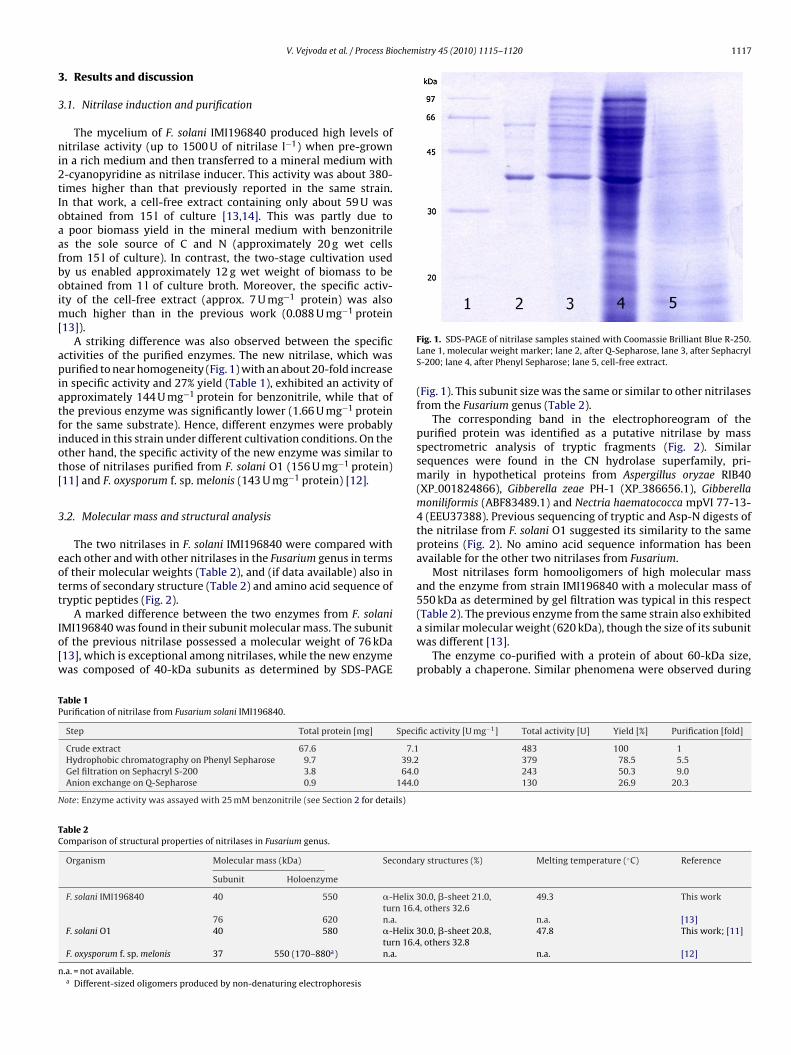
ochemistry 45 (2010) 1115–1120 1117
3
3
ni2tIoaafboim[
apiatfiot[
3
eott
Io[w
TP
N
TC
n
V. Vejvoda et al. / Process Bi
. Results and discussion
.1. Nitrilase induction and purification
The mycelium of F. solani IMI196840 produced high levels ofitrilase activity (up to 1500 U of nitrilase l−1) when pre-grown
n a rich medium and then transferred to a mineral medium with-cyanopyridine as nitrilase inducer. This activity was about 380-imes higher than that previously reported in the same strain.n that work, a cell-free extract containing only about 59 U wasbtained from 15 l of culture [13,14]. This was partly due topoor biomass yield in the mineral medium with benzonitrile
s the sole source of C and N (approximately 20 g wet cellsrom 15 l of culture). In contrast, the two-stage cultivation usedy us enabled approximately 12 g wet weight of biomass to bebtained from 1 l of culture broth. Moreover, the specific activ-ty of the cell-free extract (approx. 7 U mg−1 protein) was also
uch higher than in the previous work (0.088 U mg−1 protein13]).
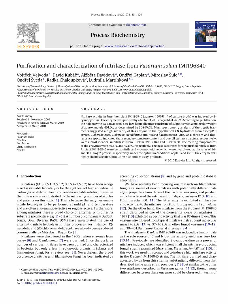
A striking difference was also observed between the specificctivities of the purified enzymes. The new nitrilase, which wasurified to near homogeneity (Fig. 1) with an about 20-fold increase
n specific activity and 27% yield (Table 1), exhibited an activity ofpproximately 144 U mg−1 protein for benzonitrile, while that ofhe previous enzyme was significantly lower (1.66 U mg−1 proteinor the same substrate). Hence, different enzymes were probablynduced in this strain under different cultivation conditions. On thether hand, the specific activity of the new enzyme was similar tohose of nitrilases purified from F. solani O1 (156 U mg−1 protein)11] and F. oxysporum f. sp. melonis (143 U mg−1 protein) [12].
.2. Molecular mass and structural analysis
The two nitrilases in F. solani IMI196840 were compared withach other and with other nitrilases in the Fusarium genus in termsf their molecular weights (Table 2), and (if data available) also inerms of secondary structure (Table 2) and amino acid sequence ofryptic peptides (Fig. 2).
A marked difference between the two enzymes from F. solaniMI196840 was found in their subunit molecular mass. The subunitf the previous nitrilase possessed a molecular weight of 76 kDa13], which is exceptional among nitrilases, while the new enzymeas composed of 40-kDa subunits as determined by SDS-PAGE
able 1urification of nitrilase from Fusarium solani IMI196840.
Step Total protein [mg] Speci
Crude extract 67.6 7.1Hydrophobic chromatography on Phenyl Sepharose 9.7 39.2Gel filtration on Sephacryl S-200 3.8 64.0Anion exchange on Q-Sepharose 0.9 144.0
ote: Enzyme activity was assayed with 25 mM benzonitrile (see Section 2 for details)
able 2omparison of structural properties of nitrilases in Fusarium genus.
Organism Molecular mass (kDa) Seconda
Subunit Holoenzyme
F. solani IMI196840 40 550 �-Helix 3turn 16.4
76 620 n.a.F. solani O1 40 580 �-Helix 3
turn 16.4F. oxysporum f. sp. melonis 37 550 (170–880a) n.a.
.a. = not available.a Different-sized oligomers produced by non-denaturing electrophoresis
Fig. 1. SDS-PAGE of nitrilase samples stained with Coomassie Brilliant Blue R-250.Lane 1, molecular weight marker; lane 2, after Q-Sepharose, lane 3, after SephacrylS-200; lane 4, after Phenyl Sepharose; lane 5, cell-free extract.
(Fig. 1). This subunit size was the same or similar to other nitrilasesfrom the Fusarium genus (Table 2).
The corresponding band in the electrophoreogram of thepurified protein was identified as a putative nitrilase by massspectrometric analysis of tryptic fragments (Fig. 2). Similarsequences were found in the CN hydrolase superfamily, pri-marily in hypothetical proteins from Aspergillus oryzae RIB40(XP 001824866), Gibberella zeae PH-1 (XP 386656.1), Gibberellamoniliformis (ABF83489.1) and Nectria haematococca mpVI 77-13-4 (EEU37388). Previous sequencing of tryptic and Asp-N digests ofthe nitrilase from F. solani O1 suggested its similarity to the sameproteins (Fig. 2). No amino acid sequence information has beenavailable for the other two nitrilases from Fusarium.
Most nitrilases form homooligomers of high molecular massand the enzyme from strain IMI196840 with a molecular mass of550 kDa as determined by gel filtration was typical in this respect
(Table 2). The previous enzyme from the same strain also exhibiteda similar molecular weight (620 kDa), though the size of its subunitwas different [13].The enzyme co-purified with a protein of about 60-kDa size,probably a chaperone. Similar phenomena were observed during
fic activity [U mg−1] Total activity [U] Yield [%] Purification [fold]
483 100 1379 78.5 5.5243 50.3 9.0130 26.9 20.3
ry structures (%) Melting temperature (◦C) Reference
0.0, �-sheet 21.0,, others 32.6
49.3 This work
n.a. [13]0.0, �-sheet 20.8,, others 32.8
47.8 This work; [11]
n.a. [12]
1118 V. Vejvoda et al. / Process Biochemistry 45 (2010) 1115–1120
F i O1f 66), Gih .
tn
IthCssimot
Fc
ig. 2. Fragments of nitrilases from Fusarium solani IMI196840 and Fusarium solanrom genes for hypothetical CN hydrolases in Aspergillus oryzae RIB40 (XP 0018248aematococca mpVI 77-13-4 (EEU37388). Mismatching amino acids are highlighted
he purification of the nitrilase from A. niger K10 [10] and bacterialitrilases [23,24].
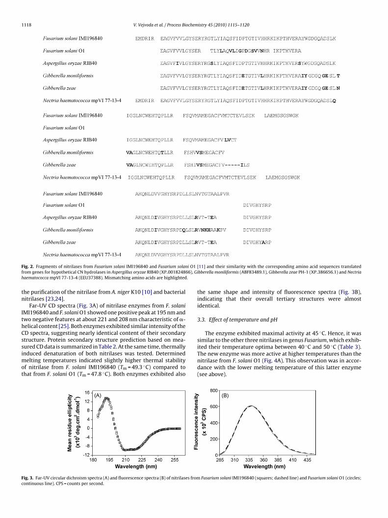
Far-UV CD spectra (Fig. 3A) of nitrilase enzymes from F. solaniMI196840 and F. solani O1 showed one positive peak at 195 nm andwo negative features at about 221 and 208 nm characteristic of �-elical content [25]. Both enzymes exhibited similar intensity of theD spectra, suggesting nearly identical content of their secondarytructure. Protein secondary structure prediction based on mea-
ured CD data is summarized in Table 2. At the same time, thermallynduced denaturation of both nitrilases was tested. Determinedelting temperatures indicated slightly higher thermal stabilityf nitrilase from F. solani IMI196840 (Tm = 49.3 ◦C) compared tohat from F. solani O1 (Tm = 47.8 ◦C). Both enzymes exhibited also
ig. 3. Far-UV circular dichroism spectra (A) and fluorescence spectra (B) of nitrilases fromontinuous line). CPS = counts per second.
[11] and their similarity with the corresponding amino acid sequences translatedbberella moniliformis (ABF83489.1), Gibberella zeae PH-1 (XP 386656.1) and Nectria
the same shape and intensity of fluorescence spectra (Fig. 3B),indicating that their overall tertiary structures were almostidentical.
3.3. Effect of temperature and pH
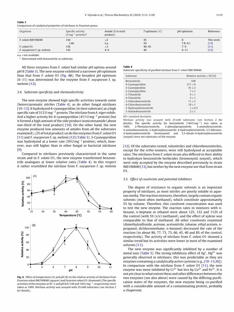
The enzyme exhibited maximal activity at 45 ◦C. Hence, it wassimilar to the other three nitrilases in genus Fusarium, which exhib-
ited their temperature optima between 40 ◦C and 50 ◦C (Table 3).The new enzyme was more active at higher temperatures than thenitrilase from F. solani O1 (Fig. 4A). This observation was in accor-dance with the lower melting temperature of this latter enzyme(see above).Fusarium solani IMI196840 (squares; dashed line) and Fusarium solani O1 (circles;
V. Vejvoda et al. / Process Biochemistry 45 (2010) 1115–1120 1119
Table 3Comparison of catalytical properties of nitrilases in Fusarium genus.
Organism Specific activity(U mg−1 protein)a
Amide (% in totalproduct)
T optimum (◦C) pH optimum Reference
F. solani IMI196840 144 ≤2 45 8 This work1.66 n.a. 50 7.8–9.1 [13]
F. solani O1 156 ≤3 40–45 7–9 [11]40 6–11 [12]
n
pt(m
3
([siioee[we[
sti
FFatf
Table 4Substrate specificity of purified nitrilase from F. solani IMI196840.
Substrate Relative activity ± SD [%]
Benzonitrile 1004-Cyanopyridine 217 ± 93-Cyanopyridine 41 ± 22-Cyanopyridine 7 ± 13-Tolunitrile 9 ± 14-Tolunitrile 5 ± 13-Chlorobenzonitrile 11 ± 24-Chlorobenzonitrile 19 ± 13-Hydroxybenzonitrile 1 ± 0.33-Aminobenzonitrile 2 ± 1
SD = standard deviationNitrilase activity was assayed with 25 mM substrates (see Section 2 fordetails). The specific activity for benzonitrile (144 U mg−1) was taken as
F. oxysporum f. sp. melonis 143 4–6
.a. = not available.a Determined with benzonitrile as substrate.
All three enzymes from F. solani had similar pH optima aroundH 8 (Table 3). The new enzyme exhibited a narrower pH optimumhan that from F. solani O1 (Fig. 4B). The broadest pH optimum6–11) was determined for the enzyme from F. oxysporum f. sp.
elonis [12].
.4. Substrate specificity and chemoselectivity
The new enzyme showed high specific activities towards somehetero)aromatic nitriles (Table 4), as do other fungal nitrilases10–13]. It hydrolyzed 4-cyanopyridine (its best substrate) at a highpecific rate of 312 U mg−1 protein. The nitrilase from A. niger exhib-ted a higher activity for 4-cyanopyridine (411 U mg−1 protein) butt formed a high amount of the side product isonicotinamide (aboutne-third of the total product) [10]. On the other hand, the newnzyme produced low amounts of amides from all the substratesxamined (≤2% of total product) as do the enzymes from F. solani O111] and F. oxysporum f. sp. melonis [12] (Table 3). 3-Cyanopyridineas hydrolyzed at a lower rate (59 U mg−1 protein), which, how-
ver, was still higher than in other fungal or bacterial nitrilases
9].Compared to nitrilases previously characterized in the sametrain and in F. solani O1, the new enzyme transformed benzoni-rile analogues at lower relative rates (Table 4). In this respectt rather resembled the nitrilase from F. oxysporum f. sp. melonis
ig. 4. Effect of temperature (A) and pH (B) on the relative activity of nitrilases fromusarium solani IMI196840 (squares) and Fusarium solani O1 (diamonds).The specificctivities of the enzymes at 45 ◦C and pH 8.0 (144 and 156 U mg−1, respectively) wereaken as 100%. Nitrilase activity was assayed with 25 mM substrates (see Section 2or details).
100%. 2-Phenylacetonitrile, 2-phenylpropionitrile, 2-aminobenzonitrile,4-aminobenzonitrile, 2-hydroxybenzonitrile, 4-hydroxybenzonitrile, 3,5-dibromo-4-hydroxybenzonitrile (bromoxynil) and 3,5-diiodo-4-hydroxybenzonitrile(ioxynil) were not substrates of the enzyme.
[12]. Of the substrates tested, tolunitriles and chlorobenzonitriles,except for the ortho-isomers, were still hydrolyzed at acceptablerates. The nitrilases from F. solani strain also differed in their abilityto hydrolyze benzonitrile herbicides (bromoxynil, ioxynil), whichwere only accepted by the enzyme described previously in strainIMI196840 [13], but neither by the new enzyme nor that from strainO1.
3.5. Effect of cosolvents and potential inhibitors
The degree of resistance to organic solvents is an importantproperty of nitrilases, as most nitriles are poorly soluble in aque-ous media. The reaction mixtures, therefore, largely contain organicsolvents (most often methanol), which constitute approximately5% by volume. Therefore, this cosolvent concentration was usedto test the new enzyme. The reaction rates in mixtures with n-hexane, n-heptane or ethanol were about 125, 122 and 112% ofthe control (with 5% (v/v) methanol), and the effect of xylene wascomparable to that of methanol. All other cosolvents examined(dimethylsulfoxide, acetone, acetonitrile, toluene, ethyl acetate, n-propanol, dichloromethane, n-butanol) decreased the rate of thereaction (to about 86, 77, 73, 73, 66, 45, 40 and 8% of the control,respectively). The activity of nitrilase from F. solani O1 showed asimilar trend but its activities were lower in most of the examinedsolvents [11].
The new enzyme was significantly inhibited by a number ofmetal ions (Table 5). The strong inhibitory effect of Ag+, Hg2+ wasgenerally observed in nitrilases; this was predictable as they areenzymes containing a catalytically active cysteine (e.g., [10–13,26]).In comparison with the nitrilase from F. solani O1 [11], the newenzyme was more inhibited by Cr3+ but less by Cu2+ and Fe3+. It is
not yet clear to what extent these and other differences between thetwo enzymes (see also above) were caused by the differing purifi-cation states of the enzymes, the new enzyme being co-purifiedwith a considerable amount of a contaminating protein, probablya chaperone.
1120 V. Vejvoda et al. / Process Biochem
Table 5Effect of various ions and other reagents on nitrilase activity.
Additive Activity [%] at final concentrationof additive ± SD
1 mM 5 mM
None 100AgNO3 1.7 ± 0.2 2.0 ± 0.2Al2(SO4)3 0.5 ± 0.2CoCl2 75.8 ± 4CuSO4 88.3 ± 2Cr2(SO4)3 27.8 ± 8FeCl3 43.7 ± 4HgCl2 0.1 ± 0.1MgSO4 85.3 ± 2MnSO4 79.3 ± 2NiSO4 62.0 ± 3(CH3CO2)2Pb 13.9 ± 2ZnSO4 8.2 ± 1 4.2 ± 1EDTA 84.1 ± 6Dithiothreitol 79.0 ± 2 80.0 ± 1Mercaptoethanol 90.2 ± 2
SNTt
[atc
4
ant1fsnttt(nuinl(aFlsifat
A
ALl
[
[
[
[
[
[
[
[
[
[
[
[[
[
[
[
[
[
[
D = standard deviation.itrilase activity was assayed with 25 mM benzonitrile (see Section 2 for details).he specific activity for benzonitrile in the absence of any additive (144 U mg−1) wasaken as 100%.
Dithiothreitol has been sometimes used as a nitrilase stabilizer27–29]. However, its adverse effect on nitrilases examined herend previously [10,11,26] suggested that its suitability as an addi-ive to buffers used, e.g., during nitrilase purification, needs to behecked for each of these enzymes.
. Conclusion
Using F. solani IMI196840, a nitrilase-producing isolate frombromoxynil-treated field in Northern Ireland, we increased theitrilase activity in this strain 380-fold with 2-cyanopyridine ashe inducer. In comparison to the previous results reported in977 [13], the fungus produced a different nitrilase with an 87-old higher specific activity for benzonitrile and with a differentubunit molecular weight (40 vs. 76 kDa). A mycelium with highitrilase activity could be easily prepared in appreciable quanti-ies. Hence, the enzyme has much more potential as a biocatalysthan that reported previously in the same strain. Comparison of thewo nitrilases in terms of their structural and catalytic propertiesfirst of all the smaller subunit size and higher specific activity of theew one) strongly suggested that different enzymes were inducedsing the different cultivation protocols. The highest specific activ-
ty of the new enzyme was found for an industrially importantitrile, 4-cyanopyridine, which can serve as a precursor of tubercu-
ostatics. Appreciable activity was also found for 3-cyanopyridineprecursor of nicotinic acid – vitamin B3). In terms of their specificctivities towards these substrates, the fungal nitrilases (from theusarium and Aspergillus genera) seem to surpass most of the nitri-ases from bacteria. The enzymes from Fusarium genus have beenhown to be superior, as far as their thermostability and selectiv-ty (product purity) are concerned. Compared to the other nitrilaserom the same species (F. solani O1), the new enzyme showed, inccordance with its higher melting temperature, an even higherhermostability and higher resistance to organic solvents.
cknowledgements
Financial support through projects IAA500200708 (Grantgency of the Academy of Sciences of the Czech Republic),C06010, OC09046 (Ministry of Education of the Czech Repub-ic), 305/09/H008 (Czech Science Foundation), FT-TA/043 (Ministry
[
istry 45 (2010) 1115–1120
of Industry and Trade of the Czech Republic) and the institu-tional research concept AV0Z50200510 (Institute of Microbiology)is gratefully acknowledged. The authors also wish to thank Prof. J.Damborsky (Loschmidt Laboratories, Masaryk Univ. Brno, CZ) forproviding the facilities for CD and fluorescence spectra measure-ment.
References
[1] Martínková L, Vejvoda V, Kren V. Selection and screening for enzymes of nitrilemetabolism. J Biotechnol 2008;133:318–26.
[2] Martínková L, Mylerová V. Synthetic applications of nitrile-convertingenzymes. Curr Org Chem 2003;7:1279–95.
[3] O’Reilly C, Turner PD. The nitrilase family of CN hydrolysing enzymes – a com-parative study. J Appl Microbiol 2003;95:1161–74.
[4] Banerjee A, Sharma R, Banerjee UC. The nitrile-degrading enzymes: currentstatus and future prospects. Appl Microbiol Biotechnol 2002;60:33–44.
[5] Brady D, Beeton A, Zeevaart J, Kgaje C, van Rantwijk F, Sheldon RA. Character-isation of nitrilase and nitrile hydratase biocatalytic systems. Appl MicrobiolBiotechnol 2004;64:76–85.
[6] Thimann KV, Mahadevan S, Nitrilase I. Occurrence, preparation and generalproperties of the enzyme. Arch Biochem Biophys 1964;105:133–41.
[7] Hook RH, Robinson WG. Ricinine nitrilase II. Purification and properties. J BiolChem 1964;239:4263–7.
[8] Kato Y, Ooi R, Asano Y. Distribution of aldoxime dehydratase in microorganisms.Appl Environ Microbiol 2000;66:2290–6.
[9] Martínková L, Vejvoda V, Kaplan O, Kubác D, Malandra A, Cantarella M, etal. Fungal nitrilases as biocatalysts: recent developments. Biotechnol Adv2009;27:661–70.
10] Kaplan O, Vejvoda V, Plíhal O, Pompach P, Kavan D, Bojarová P, et al. Purificationand characterization of a nitrilase from Aspergillus niger K10. Appl MicrobiolBiotechnol 2006;73:567–75.
11] Vejvoda V, Kaplan O, Bezouska K, Pompach P, Sulc M, Cantarella M, et al. Purifi-cation and characterization of a nitrilase from Fusarium solani O1. J Mol CatalB-Enzym 2008;50:99–106.
12] Goldlust A, Bohak Z. Induction, purification, and characterization of thenitrilase of Fusarium oxysporum f. sp. melonis. Biotechnol Appl Biochem1989;11:581–601.
13] Harper DB. Fungal degradation of aromatic nitriles. Enzymology of C–N cleav-age by Fusarium solani. Biochem J 1977;167:685–92.
14] Please note that the total activity of nitrilase in strain F. solani IMI196840 wascited incorrectly in our previous article (Ref. [9]). The correct value is 3.9 U L−1.
15] Kaplan O, Vejvoda V, Charvátová-Pisvejcová A, Martínková L. Hyperinductionof nitrilases in filamentous fungi. J Ind Microbiol Biotechnol 2006;33:891–6.
16] Laemmli UK. Cleavage of structural proteins during the assembly of the headof bacteriophage T4. Nature 1970;227:680–5.
17] Bradford MM. A rapid and sensitive method for the quantitation of micro-gram quantities of protein utilizing the principle of protein-dye binding. AnalBiochem 1976;72:248–54.
18] Böhm G, Muhr R, Jaenicke R. Quantitative analysis of protein far UV circulardichroism spectra by neural networks. Protein Eng 1992;5:191–5.
19] Sreerama N, Woody RW. A self-consistent method for the analysis of proteinsecondary structure from circular dichroism. Anal Biochem 1993;209:32–44.
20] Deléage G, Geourjon C. An interactive graphic program for calculating the sec-ondary structure content of proteins from circular dichroism spectrum. ComputAppl Biosci 1993;9:197–9.
21] http://dicroprot-pbil.ibcp.fr.22] Malandra A, Cantarella M, Kaplan O, Vejvoda V, Uhnáková B, Stepánková B, et
al. Continuous hydrolysis of 4-cyanopyridine by nitrilases from Fusarium solaniand Aspergillus niger K10. Appl Microbiol Biotechnol 2009;85:277–84.
23] Almatawah QA, Cramp R, Cowan DA. Characterization of an inducible nitrilasefrom a thermophilic bacillus. Extremophiles 1999;3:283–91.
24] Layh N, Parratt J, Willetts A. Characterization and partial purification of an enan-tioselective arylacetonitrilase from Pseudomonas fluorescens DSM 7155. J MolCatal B: Enzym 1998;5:467–74.
25] Fasman GD. Circular dichroism and the conformational analysis ofbiomolecules. New York: Plenum Press; 1996.
26] Kiziak C, Conradt D, Stolz A, Mattes R, Klein J. Nitrilase from Pseudomonasfluorescens EBC191: cloning and heterologous expression of the genesand biochemical characterization of the recombinant enzyme. Microbiology2005;151:3639–48.
27] Nagasawa T, Mauger J, Yamada H. A novel nitrilase, arylacetonitrilase, ofAlcaligenes faecalis JM3. Purification and characterization. Eur J Biochem1990;194:765–72.
28] Kobayashi M, Komeda H, Yanaka N, Nagasawa T, Yamada H. Nitrilase from
Rhodococcus rhodochrous J1. Sequencing and overexpression of the gene andidentification of an essential cysteine residue. J Biol Chem 1992;267:20746–51.29] Bhalla TC, Miura A, Wakamoto A, Ohba Y, Furuhashi K. Asymmetric hydrolysis ofalpha aminonitriles to optically active amino acids by a nitrilase of Rhodococcusrhodochrous PA-34. Appl Microbiol Biotechnol 1992;37:184–90.