Energy conversion efficiency of the pumping kite wind generator
Upload
independentCategory
view
0download
0
25 MERRILL, A. H., Jr (1994) 1. Biol. Chem. 269, 3475-3481 NORRED, W. P., PLAl-TNER, R. D., VESONDER, R. F., BACON, C. W. and VOX, K. A. (1992) food. Chem. Toxicol. 30, 233-237
26
27
28
29 30
37
32
CAWOOD, M. E., GELDERBLOM, W. C., ALBERTS, J. F. and SNYMAN, S. D. (1994) food Chem. Toxicol. 32, 627-632 CELDERBLOM, W. C., SNYMAN, S. D., VAN DER WESTHUIZEN, L. and MARASAS, W. F. 0. (1995) Carcinogenesis 16, 625-631 HORVATH, A., SUTTERLIN, C., MANNING-KRIEC, U., MOWA, N. R. and RIEZMAN, H. (1994) EMBO]. 13, 3687-3695 MERRILL, A. H., Jr et al. (1989) Biochemistry28, 3138-3145 ZHANC, H., DESAI, N. N., MURPHEY, J. M. and SPIEGEL, S. (1990) 1. Biol. Chem. 265,21309-21316 GOODEMOTE, K. A., MATTIE, M. E., BERGER, A. and SPIEGEL, 5. (1995) 1, Biol. Chem. 270,10272-l 0277 JOHNSON, 1. A. and CLARK, R. B. (1990) 1. Viol. Chem. 265, 9333-9339
33 34
35
36
PUSHKAREVA, M. et al. (1995) Biochemistry 34, 1885-l 892 STEVENS, V. L., NIMKAR, S., JAMISON, W. C., LIOl-TA, D. C. and MERRILL, A. H., Jr (1990) Biocbim. Biophys. Acta 1051, 37-45 YOO, H-S., NORRED, W. P., SHOWKER, J. and RILEY, R. E. Toxicol. Appl. Pharmacol. (in press) OHTA, H., SWEENEY, E. A., MASAMUNE, A., YATOMI, Y., HAKOMORI, S-l. and IGARASHI, Y. (1995) Cancer Rex 55, 691-697
37 TOLLESON, W. H. et a/. (1996) Carcinogenesis 17, 239-249 38 HANNUN, Y. A. and OBEID, L. M. (1995) Trends
39
40
41
42 43
44
45 46 47 48
49
50
51
52
Biocbem. Sci. 20, 73-77 NAKAMURA, S., KOZUTSUMI, Y., SUN, Y., MIYAKE, Y., FUJITA, Y. and KAWASAKI, T. (1996) 1. Biol. Chem. 271, 1255-l 257 ZHANG, H., DESAI, N. N., OLIVERA, A., SEKI, T., BROOKER, G. and SPIEGEL, S. (1991)/. CellBiol. 114, 155-167 RILEY, R. T., WANG, E. and MERRILL, A. H., Jr (1993) 1. AOAC Int. 77,533~540 ABBAS, H. K. et al. (1994) P/ant Physiol. 106, 1085-l 093 WANG, E., ROSS, P. F., WILSON, T. M., RILEY, R. T. and MERRILL, A. H., Jr (1992) 1. Nutr. 122, 1706-l 716 MERRILL, A. H., Jr, LINGRELL, S., WANG, E., NIKOLOVA-KARAKASHIAN, M., VALES, T. R. and VANCE, D. E. (1995) 1. No/. Gem. 270, 13834-I 3841 FUKUDA, H. et al. Biochem. Biophys. Res. Commun. (in press) MANDALA, S. M. et a/. (1995) 1. Antibiot. 48, 349-356 MANDALA, S. M. et al. (1994) /. Antibiot. 47, 376-379 ZWEERINK, M. M., EDISON, A. M., WELLS, G. B., PINTO, W. and LESTER, R. L. (1992) /, Biol. Chem. 267,25032-25038 MIYAKE, Y., KOZUTSUMI, Y., NAKAMURA, S., FUJITA, T. and KAWASAKI, T. (1995) Biochem. Biophys. Res. Commun. 211, 396-403 RIBONI, L., PRINETTI, A., BASSI, R., CAMINITI, A. and TETTAMANTI, G. (1995) 1. Biol. Chem. 270,26868-26875 BOSE, R., VERHEIJ, M., HAIMOVITZ-FRIEDMAN, A., SCOTTO, K., FUKS, Z. and KOLESNICK, R. (1995) Cell 82,405414 STRUM, J. C., SWENSON, K. I., TURNER, J. E. and BELL, R. M. (1995) 1. Biol. Chem. 270,13541-l 3547
The existence of a cell is dependent intimately on the presence of iron - too much or too little results in cell death. Cells from all tissues have an essential re- quirement for iron in order to proceed from Gl to S phase of the cell cycle. Iron-containing enzymes catalyse many reactions involving energy metab- olism, respiration and DNA synthesis’. In general, iron metabolism is highly regulated, and iron is found complexed mainly with either specific trans- port proteins or storage proteins. Iron is rarely found in the blood plasma in the free state since it is highly toxic to sensitive tissues such as the liver, pancreas, heart and pituitary2. Owing to its toxicity, mecha- nisms have evolved to control closely the absorption and distribution of iron within the body. Intracellu- lar iron is stored predominantly in the form of ferri- tin within the liver and is supplied to other tissues through the circulation of the serum iron-transport protein, transferrin (Tf). The main role of transferrin is to mop up free iron and to shuttle iron in a soluble non-toxic form among the organs and cells of the body. Although there is extensive literature concern- ing cellular iron uptake and regulation, the picture is still far from complete and there is considerable evi- dence that the process is more complex than thought previously, involving a number of defined and un- defined transporters, receptor-mediated endocytosis (RME) and other uptake mechanisms.
Pumping iron in the ’90s
The role of iron in cell division, cell death and human disease has
recently gained increased attention. The best studied process for
iron uptake into mammalian cells involves transferrin and its
receptor. This review discusses evidence supporting the existence of
other routes by which iron can enter mammalian cells. Specifically,
iron uptake by the cell-surface GPI-linked transferrin homologue,
melanotransfenin orp97, is described and possible functions of
this trans fen-m-independent pa thway are proposed.
Transferrin and the uptake of iron The predominant pathway by which cells acquire
iron from Tf is by RME (Fig. 1) of diferric-Tf complexed
to the transferrin receptor (TR)3. After Tf has bound free iron, it is able to bind to the TR at the cell sur- face. The complex is internalized into endosomes where acidification releases the iron. Tf remains bound to the TR and recycles back to the cell surface.
trends in CELL BIOLOGY (Vol. 6) June 1996 0 1996 Elsevier Science Ltd 223
Acknowledgements
We are grateful to the graduate students, postdoctoral fellows and collaborators who have been instrumental to these studies. Our research on fumonisins has been supported by funds from the NIH, the US Dept of Agriculture and the American Institute for Cancer Research.
PII: SO962-8924(96)10019-2

Extracellular space . . l
(PH 7.4)
(4) ;: ;;;,
. l
.
(pH falls to 5.5) In : .
/’
Key
Apotransferrin ? Transport of iron Transferrin
(5) receptor
1 . Free iron
: I, / Clathrin-
: coated pit
(3) (I, i, _:
1
(6) Ferritin Unidentified carrier protein
FIGURE 1
Receptor-mediated endocytosis of diferric-transferrin. (1) Apotransferrin binds two atoms of iron and then (2) two molecules of diferric-transferrin bind to the transferrin receptor.
(3) The complex is endocytosed via clathrin-coated pits and internalized into endosomes. (4) In the endosome, the pH falls to 5.5 and the iron is released. (5) The iron is then transported
across the endosome membrane and (6) stored complexed as ferritin. The endosome membrane and cytosolic transporters have not been identified yet. (7) The apotransferrin still bound to the
transferrin receptor is sorted to avoid degradation. The sorting process is not understood clearly but may possibly be a default mechanism described as an iterative fractionation processso.
(8) The apotransferrin is then recycled back to the cell surface and, under the increased pH of the blood plasma, the apotransferrin is released and is again able to bind iron and repeat the cycle.
The authors are at the Biotechnology
Laboratory and the Departments of
Medical Genetics, Microbiology and Immunology and
Zoology, University of British Columbia, Vancouver, British
Columbia, Canada V6T 123.
224 trends in CELL BIOLOGY (Vol. 6) June 1996
Although RME of iron is well established it is still not clear how iron is released in the lumen of the endo- some or how it is transported across the endosomal membrane. A problem with the RME model is that iron release in vitro from diferric-Tf at the pH of the endosome takes hours, whereas the entire cycle in vivo is completed within minutes. Thus, the highly efficient release of iron from Tf in the endosome can- not be due solely to the drop in pH. There is now evi- dence that the binding of Tf to the TR alters the pH dependence of iron release and that the TR acceler- ates the release of iron from Tf within the endo- some4J. Another suggestion is that there is an iron- binding protein in the membrane of the endosome that facilitates the release and transport of iron into the cytoso16. This is in agreement with the obser- vation that the process is temperature sensitive and saturable7-9. The identity of this membrane-bound protein remains to be determined, although ATP has been suggested as a possible carrier moleculelo.
In general, the requirement for iron is low in cells that are not dividing. In cells with an increased need for iron, such as haemoglobin-producing cells, pla- cental tissue and cells in the proliferating state, in par- ticular in cells of rapidly growing turnours?, there is an increase in expression of the TR on the cell mem- brane. Normal levels of serum Tf are high and -99% of iron in the plasma is bound to Tf (Ref. 7). The rate of iron uptake is believed to be regulated by the levels
of the TR and ferritin expression12. Gen- erally, any free iron circulates complexed to low-molecular-weight molecules such as ascorbate, citrate and certain amino acids or in association with other serum proteins such as albumin7. In some iron- overload disorders such as haemochro- matosis, thalassaemia and atransferrin- anaemiai3,14, the iron concentration in the plasma can exceed the binding capacity of plasma Tf. This results in excessive iron uptake in the skin, liver, pancreas and heart, leading to skin colour changes, chronic cirrhosis, diabetes and heart failure15. The excessive iron uptake into cells from free iron appears to take place independent of RME and plasma Tf, so what are the possible mechanisms for this and what other iron-transport- ing molecules are involved?
Iron uptake independent of the RME pathway
It has been demonstrated that not all cells use the RME pathway to obtain iron from Tf. In some cases such as rabbiP and mouse17 reticulocytes, it appears that iron is released from the Tf-TR com- plex prior to transport across the plasma membrane. Isolated rat hepatocytes1sJ9 and human fibroblastszO have also been shown to acquire iron from Tf by a non- RME pathway at the plasma membrane. Many of these cells, including mela- noma cells?, appear to use both RME
and non-RME pathways for the uptake of iron1*J9. A redox model” of iron uptake has been proposed to explain these findings. In this model, diferric-Tf binds to the TR in close proximity to the plasma membrane enzyme NADH:ferricyanide oxidoreduc- tase. This enzyme is able to function as a diferric-Tf reductase and, through cooperative proton and elec- tron fluxes, iron is released at the cell surface19. Unfortunately, the model does not identify how the released iron is transported into the cell, and it is pro- posed that the released iron binds to an unknown membrane protein that transports the iron across the plasma membrane to unidentified cytosolic iron acceptors3. Later studies have concluded that this model remains uncertain and controversial, and the work on the existence of a plasma membrane reduc- tase may be misleadin@22. Furthermore, discrepan- cies between the relatively low level of the TR on hep- atocytes and high iron uptake via Tf has led some investigators to conclude that, under certain condi- tions, adsorptive endocytosis or pinocytosisz2 may be the main mechanism of hepatocyte uptake of iron3Js.
Transferrin-independent iron uptake Based on studies where cells are grown in serum-
free, hence Tf-free, media and in cases of iron- overload disorders, it is evident that some cells are able to obtain iron independent of Tf. For example, Raji cells or malignant B lymphocytesz3, hepatocytes7,

HeLa cells*3~24, reticulocytes8j9 and L1210 leukaemic cellsz5 all express transport systems capable of accumulating iron from low-molecular-weight iron chelates. Furthermore, mutant Chinese hamster ovary (CHO) cellsz6 that are unable to release iron from Tf, as well as CHO cells without the TR (Ref. 27), are both able to take up iron from iron salts and grow as well as normal CHO cells. Iron uptake by this Tf- independent pathway has been less well character- ized compared with the Tf-RME pathway.
Unlike the Tf-RME pathway, the accumulation of iron by the Tf-independent pathway does not appear to be affected by the presence of tissue iron or the cell-growth statez4. Thus, the two pathways may be regulated differently’. It has been suggested that, since free iron rarely exists in vivo, the uptake of free iron by cells in vitro may just be an adaptation. In the normal situation, the Tf-RME system probably dominates and the Tf-independent system may be involved predominantly in the transport of other metals into the ce1123~24~28~29~52. The Tf-independent pathway may transport iron only when plasma iron concentrations exceed the iron-binding capacity of the plasma Tf and may, therefore, function to remove toxic levels of iron especially in cases of iron- overload diseases. Again, the identity of the iron transporters involved in this pathway are unknown, although they may be similar to those involved in the transport of iron across the endosomal membrane.
Intestinal-cell iron uptake It has been proposed that cytoplasmic mobilferrin,
a S6-kDa water-soluble metal-binding protein that is a homologue of calreticulinz8, is involved in iron up- take by the Tf-independent route. Evidence for this comes from studies on intestinal cells that do not have the TR on their luminal absorptive surfaceszx. It was proposed that, in some unknown way, integrins on the cell surface facilitate the transfer of iron through the cell membrane prior to iron binding to mobilferrin within the cell. This was based on the observation that mobilferrin coprecipitated with the p3 integrin. In a more recent paper, human-derived erythroleukaemia cells (K.562) were also shown to have a Tf-independent pathway for iron uptake that may involve mobilferrinzg. Unfortunately, this model contributes little to the understanding of Tf-independent cellular iron uptake and does not explain how mobilferrin is involved in the transport of iron across the cell membrane. Mobilferrin may, therefore, only be considered as a possible intra- cellular iron-transport protein. Recently, non-classi- cal MHC class I molecules have been proposed to play a role in intestinal iron transpor@l. However, this role has yet to be clearly demonstrated.
Melanotransferrin as a membrane-bound iron-binding protein and transporter
Another candidate for an iron-binding protein in- volved in the Tf-independent iron-uptake pathway is melanotransferrin (MTf ). MTf, also known as the human melanoma tumour-associated antigen p97, was one of the first cell-surface markers associated
trends in CELL BIOLOGY (Vol. 6) June 1996
with human skin cancer30. In addition to being de- tected at various levels in other tumours such as lymphomas3i, it has been found subsequently on a wide range of cultured normal cell types including liver cells, intestinal epithelial cells, foetal intestinal cells, umbilical chord, placenta and sweat-gland ducts31-33. Most recently, it has been shown that MTf is expressed on the endothelium of the human brain and that its distribution and cellular location are consistent with it having a role in iron transport into or out of the human brain34.
The MTf antigen belongs to an important group of iron-binding proteins that includes human serum transferrin, human lactoferrin and ovotransferrin from avian egg whites35. Furthermore, MTf, Tf and the TR are all sialoglycoproteins that are encoded on chromosome 3 in humans36. However, in contrast to other molecules in the transferrin family, MTf is the only one that is connected to the cell membrane by a glycosylphosphatidylinositol (GPI) anchor33,37. Further examination of the MTf sequence in relation to the three-dimensional structures of human lacto- ferrin and rabbit serum Tf suggests that MTf has an intact transferrin-type iron-binding site in its N-terminal half, but has a defective ‘site in its C-terminal half35 owing to differences in specific amino acids. This conclusion is supported by a study38 that showed that MTf has only one func- tional iron-binding site. The C-terminal half is also involved with GPI-anchor attachment, and this might also affect iron binding3*.
What is the evidence that MTf can act as an iron transporter? In a study on iron uptake by the human malignant melanoma cell SK-MEL 28, a membrane- bound, temperature-dependent, iron-binding com- ponent was identified that was consistent with MTf (Ref. 21). Subsequent studies appeared to indicate that this iron-binding component did not donate its iron to the ce113g. However, in these studies it was not possible to define a specific role for MTf in iron up- take owing to the lack of an appropriate negative control and the presence of multiple iron-uptake pathways in these cells21,40.
Internalization of GPI-anchored proteins If we assume that MTf plays a role in cellular up-
take of iron, how is iron bound to MTf internalized? GPI-anchored proteins have been shown to be in- ternalized and recycled back to the cell surface41. For ex- ample, the folate receptor or folate-binding protein, which internalizes folate42, is an example of a GPI- anchored protein involved in RME. Although most GPI-anchored proteins such as decay-accelerating factor (DAF) and Thy-l are expressed diffusely over the cell surface41,43., the folate receptor appears to cluster in non-clathrin-coated invaginations called ‘caveo1ae’42. These caveolae are believed to close off transiently and, perhaps, detach from the plasma membrane in a process called ‘potocytosis’. Folate is released, presumably as a result of lumen acidification, and may be translocated to endosomes. The caveo- lae then unseal and the receptor is recycled back to the cell surface. Although this is an attractive model, the association between caveolae and GPI-anchored
225
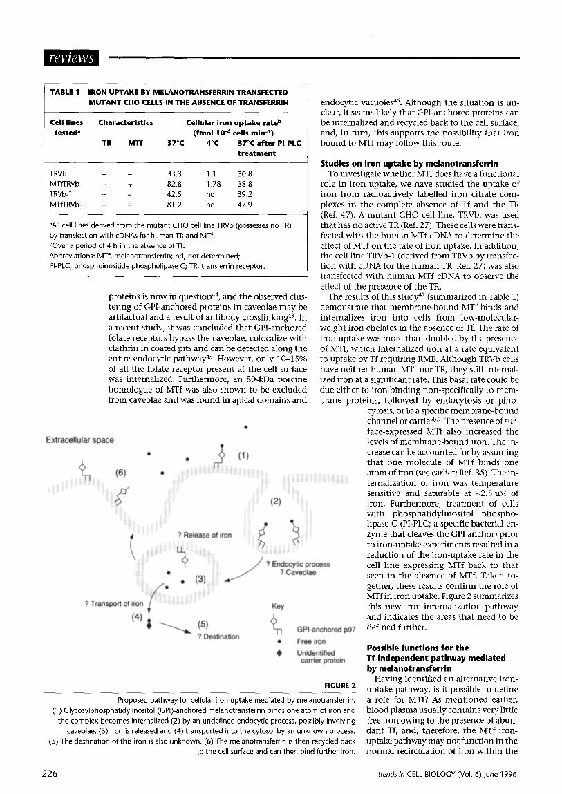
TABLE 1 - IRON UPTAKE BY MELANOTRANSFERRIN-TRANSFECTED MUTANT CHO CELLS IN THE ABSENCE OF TRANSFERRIN
Cell lines tested’
Characteristics
TR MTf
Cellular iron uptake rateb (fmol lo4 cells mln-l)
37°C 4°C 37°C after PI-PLC treatment
TRVb - -
MTfTRVb - f TRVb-1 + - MTfTRVb-1 + f
33.3 1.1 30.8 82.8 1.78 38.8 42.5 nd 39.2 81.2 nd 47.9
aAll cell lines derived from the mutant CHO cell line TRVb (possesses no TR) by transfection with cDNAs for human TR and MTf. bOver a period of 4 h in the absence of Tf. Abbreviations: MTf, melanotransferrin; nd, not determined; PI-PLC, phosphoinositide phospholipase C; TR, transferrin receptor.
proteins is now in question44, and the observed clus- tering of GPI-anchored proteins in caveolae may be artifactual and a result of antibody crosslinking13. In a recent study, it was concluded that GPI-anchored folate receptors bypass the caveolae, colocalize with clathrin in coated pits and can be detected along the entire endocytic pathways. However, only 10-X% of all the folate receptor present at the cell surface was internalized. Furthermore, an SO-kDa porcine homologue of MTf was also shown to be excluded from caveolae and was found in apical domains and
.
Extracellular space
1
R (6) l
II
f (2)
? Release of iron
4
endocytic vacuoles46. Although the situation is un- clear, it seems likely that GPI-anchored proteins can be internalized and recycled back to the cell surface, and, in turn, this supports the possibility that iron bound to MTf may follow this route.
Studies on iron uptake by melanotransferrin To investigate whether MTf does have a functional
role in iron uptake, we have studied the uptake of iron from radioactively labelled iron citrate com- plexes in the complete absence of Tf and the TR (Ref. 47). A mutant CHO cell line, TRVb, was used that has no active TR (Ref. 27). These cells were trans- fected with the human MTf cDNA to determine the effect of MTf on the rate of iron uptake. In addition, the cell line TRVb-1 (derived from TRVb by transfec- tion with cDNA for the human TR; Ref. 27) was also transfected with human MTf cDNA to observe the effect of the presence of the TR.
The results of this study’ (summarized in Table 1) demonstrate that membrane-bound MTf binds and internalizes iron into cells from low-molecular- weight iron chelates in the absence of Tf. The rate of iron uptake was more than doubled by the presence of MTf, which internalized iron at a rate equivalent to uptake by Tf requiring RME. Although TRVb cells have neither human MTf nor TR, they still internal- ized iron at a significant rate. This basal rate could be due either to iron binding non-specifically to mem- brane proteins, followed by endocytosis or pino-
\ 9 ? Endocytic process . ? Caveolae
. (3) .
? Transport of iron Key
\ (5) ? Destination
R GPI-anchored p97
. Free iron
Unidentified carrier protein
FIGURE 2
Proposed pathway for cellular iron uptake mediated by melanotransferrin. (1) Clycosylphosphatidylinositol &PI)-anchored melanotransferrin binds one atom of iron and
the complex becomes internalized (2) by an undefined endocytic process, possibly involving caveolae. (3) Iron is released and (4) transported into the cytosol by an unknown process.
(5) The destination of this iron is also unknown. (6) The melanotransferrin is then recycled back to the cell surface and can then bind further iron.
cytosis, or to a specific membrane-bound channel or carrier81g. The presence of sur- face-expressed MTf also increased the levels of membrane-bound iron. The in- crease can be accounted for by assuming that one molecule of MTf binds one atom of iron (see earlier; Ref. 35). The in- ternalization of iron was temperature sensitive and saturable at -2.5 FM of iron. Furthermore, treatment of cells with phosphatidylinositol phospho- lipase C (PI-PLC; a specific bacterial en- zyme that cleaves the GPI anchor) prior to iron-uptake experiments resulted in a reduction of the iron-uptake rate in the cell line expressing MTf back to that seen in the absence of MTf. Taken to- gether, these results confirm the role of MTf in iron uptake. Figure 2 summarizes this new iron-internalization pathway and indicates the areas that need to be defined further.
Possible functions for the Tf-independent pathway mediated by melanotransferrin
Having identified an alternative iron- uptake pathway, is it possible to define a role for MTf? As mentioned earlier, blood plasma usually contains very little free iron owing to the presence of abun- dant Tf, and, therefore, the MTf iron- uptake pathway may not function in the normal recirculation of iron within the
226 trends in CELL BIOLOGY (Vol. 6) June 1996

body. However, under certain conditions, when iron levels become elevated, for example in iron-overload disorders or during cell death, MTf may act as an iron scavenger to prevent the accumulation of toxic lev- els of iron. The MTf pathway does not appear to be regulated tightly as, in iron-overload disorders, it is the cells expressing the highest levels of MTf that are most likely to be damaged owing to excessive iron uptake15,32. Since MTf is expressed at high levels on rapidly proliferating cells, especially melanoma cells, it may be that these cells are using MTf as an ad- ditional pathway to increase their iron uptake. How- ever, under physiological circumstances where almost all iron is bound to serum Tf, it is not clear how these cells would be able to use MTf to increase their iron uptake. One idea is that metastasizing tumour cells secrete various enzymes specifically to lyse neighbour- ing normal cells and that MTf is then used to scav- enge the released iron and support tumour growth.
MTf may be involved in the intestinal uptake of iron. Recently, an SO-kDa homologue of MTf was identified on porcine foetal intestinal cells4’j, which raises the possibility that MTf is involved in the neonate’s uptake of iron from the mother’s milk. Furthermore, although the expression of MTf on adult tissue is low, MTf may provide a more satisfac- tory explanation for the uptake of dietary iron in intestinal cells than the previously proposed theory involving integrinsz8J9.
Another area where MTf may play an active role is in the transcytosis of iron across the blood-brain bar- rier. In a recent study where the distributions of MTf, Tf and TR were analysed immunohistochemically in human brain tissues, it was noted that the distribu- tions of MTf and the TR were remarkably similar but quite different from that of Tf. In all brain tissues ex- amined, MTf and TR were highly localized to capillary endothelial cells, whereas Tf was localized mainly to glial cells34. This raises the question as to whether Tf is responsible exclusively for mediating iron trans- port into and within the brain. The transcytosis of iron by MTf may explain the observation that high levels of iron can be transported into the brains of hypotransferrinaemic mice48 in the absence of Tf.
A final possible role for the Tf-independent or MTf- mediated iron-uptake pathway is that it provides an alternate pathway for other metals to enter the cell non-competitively with iron. It has been noted that the Tf-independent iron-uptake pathway is inhibited by a number of transition metals7,9J3J2. Furthermore, based on a molecular model of MTf, it appears that MTf has zinc-binding properties49. In this scenario, iron would enter the cell via the RME pathway and only when the level of free iron is relatively high would MTf begin to bind and internalize iron.
In conclusion, it is evident that MTf can provide an alternative pathway to the RME of Tf and the TR for cellular iron uptake. The conditions under which this pathway functions need to be clarified and may vary considerably depending on the cell environ- ment and function. Further research should reveal whether MTf plays a unique role in the growth of rapidly proliferating cells, in the uptake of dietary iron and in the transcytosis of iron into the brain.
trends in CELL BIOLOGY (Vol. 6) June 1996
References 1 KUHN, L. C., SCHULMAN, H. M. and PONKA, P. (1990) in iron
Transport and Storage (Ponka, P., Schulman, H. M. and Woodward, R. C., eds), pp. 149-I 91, Boca Ranton, CRC Press
2 CRAVEN, C. M., ALEXANDER, J., ELDRIDGE, M., KUSHNER, J. P., BERNSTEIN, S. and KAPLAN, J. (1987) Proc. Nat/ Acad. Sci. USA 84, 3457-3461
3 THORSTENSEN, K. and ROMSLO, I. (1990) Biochem. 1,271, l-10 4 SIPE, D. M. and MURPHY, R. F. (1991)). Biol. Chem. 266,
8002-8007 5 AISEN, P. (1992) Ann. Neural. 32, S62-568 6 NUNEZ, M. T., PINTO, I. and GLASS, J. (1989) 1. Membr. Biol.
107,129-l 35 7 WRIGHT, T. L., BRISSOT, P., MA, W. L. and WEISIGER, R. A.
(1986) 1. Biol. Chem. 261,10909-l 0914 8 EGYED, A. (1988) Br. 1, Haematol. 68, 483-486 9 MORGAN, E. H. (1988) Biochim. Biophys. Acta 934, 428-439
10 WEAVER, J. and POLLACK, S. (1989) Biochem. /. 261, 787-792 11 TAETLE, R., RHYMER, K., CASTAGNOIA, J., TO, D. and
MENDELSOHN, j. (1985) /. C/in. invest. 75, 1061-l 065 12 KUHN, C. L. and HENTZE, M. H. (1992) 1. Inorg. Biochem.
47, 183-I 95 13 KAPLAN, J., JORDAN, I. and STURROCK, A. (1991)j. Biol. Chem.
266,2997-3004 14 SMITH, L. (1990) West. /. Med. 153, 296-308 15 GRACE, N. D. and POWELL, L. W. (1974) Gastroenterology
67, 1257-l 259 16 LOH, T. T., YEUNC, Y. G. and YEUNG, D. (1977) Biochim.
Biophys. Acta 471, 118-l 24 17 NUNEZ, M. J., COLE, E. 5. and GLASS, J. (1983) 1. Biol. Chem.
258,1146-1151 18 PAGE, M., BAKER, E. and MORGAN, E. H. (1986) Am. 1. Physiol.
246,626-633 19 THORSTENSEN, K. and ROMSLO, I. (1988) /. Biol. Chem. 263,
8844-8850 20 OSHIRO, S., NAKAMURA, Y., ISHIGE, R., HORI, M., NAKAJIMA, H.
and GAHL, W. A. (1994) 1. Biol. Chem. 115, 849-852 21 RICHARDSON, D. R. and BAKER, E. (1990) Biochim. Biophys.
Acta 1053,1-12 22 THORSTENSEN, K., TRINDER, D., ZAK, 0. and AISEN, P. (1995)
Eur. 1. Biochem. 232, 129-I 33 23 SELIGMAN, P. A., KOVAR, J., SCHLEICHER, R. B. and
GELFAND, E. W. (1991) Blood 78, 1526-l 531 24 STURROCK, A., ALEXANDER, J., LAMB, J., CRAVEN, C. M. and
KAPLAN, J. (1990) 1. Biol. Chem. 265, 3139-3145 25 BASSET, P., QUESNEAU, Y. and ZWILLER, J. (1986) Cancer Rex.
46, 1644-l 647 26 KI-AUSNER, R. D., VAN RENSWOUDE, J., KEMPF, C., RAO, K.,
BATEMAN, J. L. and BRIDGES, K. R. (1984) /. Cell Biol. 98, 1098-I 101
27 MCGRAW, T. E., GREENFIELD, L. and MAXFIELD, F. R. (1987) 1. Cell Biol. 105, 207-214
28 CONRAD, M. E. and UMBREIT, J, N. (1993) Am. 1, Haematol. 42, 67-73
29 CONRAD, M. E., UMBREIT, J. N., MOORE, E. C., UZEL, C. and BERRY, M. R. (1994) 1, Biol. Chem. 269, 7169-7173
30 BROWN, J. P., NISHIYAMA, K., HELLSTReM, I. and HELLSTROM, K. E. (1981) /. Immunol. 127, 539-546
31 BROWN, 1. P., WOODBURY, R. G., HART, C. E., HELLSTRBM, I. and HELLSTRbM, K. E. (1981) froc. Nat/ Acad. Sci. USA 78,539-543
32 SCIOT, R., DE VOS, R., VAN EYKEN, P., VAN DER STEEN, K., MOERMAN, P. and DESMET, V. J. (1989) liver 9, 11 O-l 19
33 ALEMANY, R., ROSAVIti, M., FRANCI, C., EGEA, G., REAL, F. X.
227

Acknowledgements
Our work was supported by
grants from Synapse
Technologies Inc., the Medical
Research Council of Canada, and NCE
of Canada. We thank our
collaborators who have assisted in
various aspects of the p97 project.
and THOMPSON, J. M. (1993) 1. Cell Sci. 104,1155-l 162 34 ROTHENBERCER, S. et al. (1996) Brain Rex 712, 117-121 35 BAKER, E. N., RUMBALL, S. V. and ANDERSON, B. F. (1987)
Trends Biochem. Sci. 12, 350-353 36 PLOWMAN, G. D. et al. (1983) Nature 303, 70-72 37 FOOD, M. R., ROTHENBERCER, S., GABATHULER, R., HAIDL, I. D.,
REID, C. and JEFFERIES, W. A. (1994) I. Biol. Chem. 269, 3034-3040
38 BAKER, E. N. et ol. (1992) FEBS lett. 298, 215-218 39 RICHARDSON, D. R. and BAKER, E. (1991) Biochim. Biophys.
Acta 1091, 294-302 40 RICHARDSON, D. R. and BAKER, E. (1994) I. Cell. Physiol.
161, 160-168 41 KELLER, C. A., SIEGEL, M. W. and CARAS, I. W. (1992) FMBO].
11,863-874 42 ROTHBERG, K. G., YING, Y., KOLHOUSE, 1. F., KAMEN, B.A.
and ANDERSON, R. G. W. (1990) 1. Cell Biol. 110, 637-649 43 MAYOR, S., ROTHBERG, K. G. and MAXFIELD, R. (1994) Science
264,1948-l 951 44 SCHNITZER, J. E., MCINTOSH, D. P., DVORAK, A. M., LIU, 1.
and OH, P. (1995) Science 269, 1435-I 438 45 RIJNBOUTT, S., JANSEN, G., POSTHUMA, G., HYNES, J. B.,
SCHORNAGEL, ). H. and STROUSS, G. 1. (1996) /. Cell Biol. 132, 35-47
46 DANIELSON, E. M. and VAN DEURS, B. (1995) 1. Cell Biol. 131, 939-950
47 KENNARD, M. L., RICHARDSON, D. R., GABATHULER, R., PONKA, P. and IEFFERIES, W. A. (1995) FIvfMBO]. 14,4178-4186
48 UEDA, F., RAJA, K. B., SIMPSON, R. I., TROWBRIDGE, I. 5. and BRADBURY, W. B. (1993) 1. Neurochem. 60, 106-I 13
49 GARRETT, R. C. and JHOTI, H. (1992) FEBS lett. 30555-61 50 DUNN, K. W., MCGRAW, T. E. and MAXFIELD, F. R. (1989)
1. CellBiol. 109, 3303-3314
References added in proof 51 ROTHENBURG, B. E. and VORLAND, 1. R. (1996) hoc. Nat/
Acad. Sci. USA 93,1529-l 534 52 STEARMAN, R., YUAN, D. S., YAMAGUCHI-IWAI, Y.,
KLAUSNER, R. D. and DANCIS, A. (1996) Science 271, 1552-l 557
The spindle- assembly
checkpoint: aiming for a perfect
mitosis, every time
Checkpoints reduce the frequency of errors in cell division by
delaying the progress of the cell cycle until certain processes are
complete. The spindle-assembly checkpointprevents the onset of
anaphase until a bipolar spindle is present and all chromosomes
are attached to the spindle. Evidence from yeast and mammalian
cells suggests that kinetochores are at least one source of the signal
that stops the cell cycle. Recent studies in budding yeast have
begun to define the signal-transduction pathway involved in the
spindle-assembly checkpoint, but details of the endpoint of the
pathway, where these signals interact with the cell-cycle
machinery, remain to be characterized. The author is at
the Dept of Biochemistry and
Biophysics, University of The eukaryotic cell cycle is driven by the regular
California, San activation and inactivation of cyclin-dependent Francisco, CA kinases (CDKs). These oscillations can be modified
94143-0448, USA. by the requirement that certain events be completed
228 0 1996 Elsevier Science Ltd PII: SOSfiZ-8924(96)10018-O
before others are started; the systems responsible for this monitoring of the progress of the cell cycle are called checkpoints. They contribute to the fidelity of the cell cycle by allowing time for DNA replication and repair before the cell enters mitosis and for align- ment of chromosomes on the spindle before the cell initiates anaphase. When these periodic events are incomplete, the checkpoint systems block further transitions in the cell cycle (see Ref. 1 and recent re- views on the cell biology2 and genetics3 of various checkpoints). If the checkpoints fail to function, genetic errors arise much more frequently. Mutation of the genes encoding proteins involved in check- points may be an important event in the genesis of cancer cells, allowing genetic changes to accumulate more readily, although the stages at which check- point controls are lost may differ for different tumour types4. In this review, I survey what is known about the spindle-assembly checkpoint. This is the mecha- nism or group of mechanisms that ensures that a bipolar spindle is present and that all the chromo- somes are attached to the spindle before the cell initiates anaphase and progresses into the next cell cycle.
In common with other systems in the cell that re- spond to internal or external conditions, a check- point should consist of three functional units: a sensor, a signal-transduction cascade and an effector (Fig. 1). In the case of the spindle-assembly check- point, the sensor monitors events such as attachment of chromosomes to the mitotic spindle and, if there is a defect or the process is not complete, generates a signal. The initial signal is then amplified in a signal- transduction cascade and eventually modifies the cell-cycle machinery such that the cell,cycle is halted until the defect is rectified. Studies in various organ- isms have led to a partial understanding of the sen- sor and signal-transduction cascade, but the nature of the endpoint of the pathway remains unknown.
trends in CELL BIOLOGY (Vol. 6) June 1996
Copyright © 2022 FDOKUMEN