Psilostachyin C: a natural compound with trypanocidal activity
8
International Journal of Antimicrobial Agents 37 (2011) 536–543 Contents lists available at ScienceDirect International Journal of Antimicrobial Agents journal homepage: http://www.elsevier.com/locate/ijantimicag Psilostachyin C: a natural compound with trypanocidal activity Valeria P. Sülsen a,1 , Fernanda M. Frank b,1 , Silvia I. Cazorla b , Patricia Barrera c , Blanca Freixa d , Roser Vila d , Miguel A. Sosa c , Emilio L. Malchiodi b,∗ , Liliana V. Muschietti a,∗∗ , Virginia S. Martino a a Cátedra de Farmacognosia, IQUIMEFA (UBA-CONICET), Facultad de Farmacia y Bioquímica, Universidad de Buenos Aires, Junín 956 (1113), Buenos Aires, Argentina b Cátedra de Inmunología, IDEHU (UBA-CONICET), Facultad de Farmacia y Bioquímica and Departamento de Microbiología, Facultad de Medicina, Universidad de Buenos Aires, Junín 956 (1113), Buenos Aires, Argentina c Universidad Nacional de Cuyo-CONICET, Instituto de Histología y Embriología ‘Dr Mario H. Burgos’, Facultad de Ciencias Médicas, CC 56 (5500), Mendoza, Argentina d Unitat de Farmacologia i Farmacognòsia, Facultat de Farmacia, Universitat de Barcelona, Avinguda de Joan XXIII, 08028 Barcelona, Spain article info Article history: Received 30 September 2010 Accepted 2 February 2011 Keywords: Ambrosia scabra Sesquiterpene lactones Psilostachyin C Trypanocidal activity Leishmanicidal activity abstract In this study, the antiprotozoal activity of the sesquiterpene lactone psilostachyin C was investigated. This natural compound was isolated from Ambrosia scabra by bioassay-guided fractionation and was identified by spectroscopic techniques. Psilostachyin C exerted in vitro trypanocidal activity against Trypanosoma cruzi epimastigotes, trypomastigotes and amastigotes, with 50% inhibitory concentration (IC 50 ) values of 0.6, 3.5 and 0.9 g/mL, respectively, and displayed less cytotoxicity against mammalian cells, with a 50% cytotoxic concentration (CC 50 ) of 87.5 g/mL. Interestingly, this compound induced ultrastructural alterations, as seen by transmission electron microscopy, in which vacuolisation and a structural appear- ance resembling multivesicular bodies were observed even at a concentration as low as 0.2 g/mL. In an in vivo assay, a significant reduction in the number of circulating parasites was found in T. cruzi-infected mice treated with psilostachyin C for 5 days compared with untreated mice (7.4 ± 1.2 × 10 5 parasites/mL vs. 12.8 ± 2.0 × 10 5 parasites/mL) at the peak of parasitaemia. According to these results, psilostachyin C may be considered a promising template for the design of novel trypanocidal agents. In addition, psilostachyin C inhibited the growth of Leishmania mexicana and Leishmania amazonensis promastigotes (IC 50 = 1.2 g/mL and 1.5 g/mL, respectively). © 2011 Elsevier B.V. and the International Society of Chemotherapy. All rights reserved. 1. Introduction Chagas disease, caused by the protozoan parasite Trypanosoma cruzi, is endemic in Latin America. Approximately 16–18 million people are infected in the Americas and ca. 100 million people are at risk of contracting the disease [1]. Prophylactic and thera- peutic vaccines have been pursued but sterilising immunity has not yet been achieved [2,3]. Leishmaniasis is a group of infections caused by Leishmania spp. Annually, 1.5–2 million people around the world are infected by the parasites and 350 million are at risk of contracting the disease [4]. Chemotherapy for the treatment of these parasitoses, which are frequently found to co-infect patients in endemic areas [5–7], has limited efficacy and is not innocuous, mainly due to resistance phenomena and adverse effects. Conse- quently, new drugs are needed. ∗ Corresponding author. Tel.: +54 11 4964 8259; fax: +54 11 4964 0024. ∗∗ Corresponding author. Tel.: +54 11 4508 3642; fax: +54 11 4508 3642. E-mail addresses: [email protected] (E.L. Malchiodi), [email protected] (L.V. Muschietti). 1 These two authors contributed equally to this paper. In previous work, we reported the in vitro trypanocidal activity of several Argentine medicinal plant species [8]. We have isolated two bioactive sesquiterpene lactones (STLs) from Ambrosia tenuifo- lia presenting in vitro activity against T. cruzi epimastigotes and Leishmania spp. promastigotes, one of which exerted a significant in vivo trypanocidal effect [9]. Ambrosia scabra Hook. & Arn. (Asteraceae) is a closely related species popularly known as ‘ajenjo del campo’ and traditionally used against intermittent fevers and worm infections [8,10]. Here we report the trypanocidal and leishmanicidal activities of the STL psilostachyin C isolated from A. scabra by bioassay-guided fraction- ation. In addition, the ultrastructural changes that this compound produced in T. cruzi epimastigotes were evaluated. STLs are C-15 terpenoid compounds and represent an important and biogenetically homogeneous group of secondary metabolites present in higher plants [11]. They display great diversity and an enormously broad spectrum of biological activities, including antiprotozoal activity [12–15]. The discovery of artemisinin (an antimalarial STL isolated from the Chinese herb Artemisia annua) has been a major breakthrough in the field of parasitic diseases and has prompted the investigation of these kinds of compounds. In particular, psilostachyin C is a dilactone of the ambrosanolide type that was first isolated from Ambrosia psilostachya [16] and 0924-8579/$ – see front matter © 2011 Elsevier B.V. and the International Society of Chemotherapy. All rights reserved. doi:10.1016/j.ijantimicag.2011.02.003
Transcript of Psilostachyin C: a natural compound with trypanocidal activity

P
VRa
b
Bc
d
a
ARA
KASPTL
1
cpapnctotimq
l
0d
International Journal of Antimicrobial Agents 37 (2011) 536–543
Contents lists available at ScienceDirect
International Journal of Antimicrobial Agents
journa l homepage: ht tp : / /www.e lsev ier .com/ locate / i jant imicag
silostachyin C: a natural compound with trypanocidal activity
aleria P. Sülsena,1, Fernanda M. Frankb,1, Silvia I. Cazorlab, Patricia Barrerac, Blanca Freixad,oser Vilad, Miguel A. Sosac, Emilio L. Malchiodib,∗, Liliana V. Muschietti a,∗∗, Virginia S. Martinoa
Cátedra de Farmacognosia, IQUIMEFA (UBA-CONICET), Facultad de Farmacia y Bioquímica, Universidad de Buenos Aires, Junín 956 (1113), Buenos Aires, ArgentinaCátedra de Inmunología, IDEHU (UBA-CONICET), Facultad de Farmacia y Bioquímica and Departamento de Microbiología, Facultad de Medicina, Universidad deuenos Aires, Junín 956 (1113), Buenos Aires, ArgentinaUniversidad Nacional de Cuyo-CONICET, Instituto de Histología y Embriología ‘Dr Mario H. Burgos’, Facultad de Ciencias Médicas, CC 56 (5500), Mendoza, ArgentinaUnitat de Farmacologia i Farmacognòsia, Facultat de Farmacia, Universitat de Barcelona, Avinguda de Joan XXIII, 08028 Barcelona, Spain
r t i c l e i n f o
rticle history:eceived 30 September 2010ccepted 2 February 2011
eywords:mbrosia scabraesquiterpene lactonessilostachyin C
a b s t r a c t
In this study, the antiprotozoal activity of the sesquiterpene lactone psilostachyin C was investigated. Thisnatural compound was isolated from Ambrosia scabra by bioassay-guided fractionation and was identifiedby spectroscopic techniques. Psilostachyin C exerted in vitro trypanocidal activity against Trypanosomacruzi epimastigotes, trypomastigotes and amastigotes, with 50% inhibitory concentration (IC50) valuesof 0.6, 3.5 and 0.9 �g/mL, respectively, and displayed less cytotoxicity against mammalian cells, with a50% cytotoxic concentration (CC50) of 87.5 �g/mL. Interestingly, this compound induced ultrastructuralalterations, as seen by transmission electron microscopy, in which vacuolisation and a structural appear-
rypanocidal activityeishmanicidal activity
ance resembling multivesicular bodies were observed even at a concentration as low as 0.2 �g/mL. In anin vivo assay, a significant reduction in the number of circulating parasites was found in T. cruzi-infectedmice treated with psilostachyin C for 5 days compared with untreated mice (7.4 ± 1.2 × 10⁵ parasites/mLvs. 12.8 ± 2.0 × 10⁵ parasites/mL) at the peak of parasitaemia. According to these results, psilostachyinC may be considered a promising template for the design of novel trypanocidal agents. In addition,
the g�g/mlsevie
psilostachyin C inhibited(IC50 = 1.2 �g/mL and 1.5
© 2011 E
. Introduction
Chagas disease, caused by the protozoan parasite Trypanosomaruzi, is endemic in Latin America. Approximately 16–18 millioneople are infected in the Americas and ca. 100 million peoplere at risk of contracting the disease [1]. Prophylactic and thera-eutic vaccines have been pursued but sterilising immunity hasot yet been achieved [2,3]. Leishmaniasis is a group of infectionsaused by Leishmania spp. Annually, 1.5–2 million people aroundhe world are infected by the parasites and 350 million are at riskf contracting the disease [4]. Chemotherapy for the treatment ofhese parasitoses, which are frequently found to co-infect patients
n endemic areas [5–7], has limited efficacy and is not innocuous,ainly due to resistance phenomena and adverse effects. Conse-uently, new drugs are needed.
∗ Corresponding author. Tel.: +54 11 4964 8259; fax: +54 11 4964 0024.∗∗ Corresponding author. Tel.: +54 11 4508 3642; fax: +54 11 4508 3642.
E-mail addresses: [email protected] (E.L. Malchiodi),[email protected] (L.V. Muschietti).
1 These two authors contributed equally to this paper.
924-8579/$ – see front matter © 2011 Elsevier B.V. and the International Society of Chemoi:10.1016/j.ijantimicag.2011.02.003
rowth of Leishmania mexicana and Leishmania amazonensis promastigotesL, respectively).r B.V. and the International Society of Chemotherapy. All rights reserved.
In previous work, we reported the in vitro trypanocidal activityof several Argentine medicinal plant species [8]. We have isolatedtwo bioactive sesquiterpene lactones (STLs) from Ambrosia tenuifo-lia presenting in vitro activity against T. cruzi epimastigotes andLeishmania spp. promastigotes, one of which exerted a significantin vivo trypanocidal effect [9].
Ambrosia scabra Hook. & Arn. (Asteraceae) is a closely relatedspecies popularly known as ‘ajenjo del campo’ and traditionallyused against intermittent fevers and worm infections [8,10]. Herewe report the trypanocidal and leishmanicidal activities of the STLpsilostachyin C isolated from A. scabra by bioassay-guided fraction-ation. In addition, the ultrastructural changes that this compoundproduced in T. cruzi epimastigotes were evaluated.
STLs are C-15 terpenoid compounds and represent an importantand biogenetically homogeneous group of secondary metabolitespresent in higher plants [11]. They display great diversity andan enormously broad spectrum of biological activities, includingantiprotozoal activity [12–15]. The discovery of artemisinin (an
antimalarial STL isolated from the Chinese herb Artemisia annua)has been a major breakthrough in the field of parasitic diseasesand has prompted the investigation of these kinds of compounds.In particular, psilostachyin C is a dilactone of the ambrosanolidetype that was first isolated from Ambrosia psilostachya [16] andotherapy. All rights reserved.

l of An
ssotr
2
2
2cB6c
2
btcwapbttwafee
2
nscbis
2
i(o(wr
fsm
2
DdMt
V.P. Sülsen et al. / International Journa
ubsequently from other Ambrosia spp. [17]. It has been demon-trated to have molluscicidal activity [18] and inhibitory activityn the G2 DNA damage checkpoint [19]. However, this is the firstime that this compound has been found in A. scabra and the firsteport of its trypanocidal and leishmanicidal activities.
. Methods
.1. Plant material
Ambrosia scabra was collected in Buenos Aires, Argentina, in007 and was identified by Dr G. Giberti (Museo de Farma-obotánica, Facultad de Farmacia y Bioquímica, Universidad deuenos Aires, Buenos Aires, Argentina). A voucher specimen (BAF50) was deposited at the Herbarium of the Museo de Farma-obotánica.
.2. Bioassay-guided fractionation
Extraction of the aerial parts of A. scabra (500 g) was doney maceration with dichloromethane:methanol (1:1) at roomemperature. The organic extract was subjected to open columnhromatography over silica gel 60 and was eluted successivelyith cyclohexane, cyclohexane:ethyl acetate (1:1), ethyl acetate
nd methanol to give 23 fractions of 500 mL each. According to theirrofile in thin-layer chromatography, these fractions were com-ined into five final fractions (F1AS–F5AS) and were subsequentlyested for trypanocidal activity against T. cruzi epimastigotes. Frac-ion F5AS was chromatographed on a silica gel column elutedith a cyclohexane:CH2Cl2 gradient (100:0 to 0:100), CH2Cl2:ethyl
cetate gradient (100:0 to 0:100) and 100% methanol to obtain 150ractions (F5AS 1–150) of 10 mL each. Of these fractions, F5AS (75–77)ssentially contained one pure compound that crystallised fromthyl acetate.
.3. Spectrometric analyses
The isolated compound was identified by proton nuclear mag-etic resonance (1H NMR) and carbon NMR (13C NMR) (Inova NMRpectrometer; Varian, Palo Alto, CA) (500 MHz in CDCl3), heteronu-lear single quantum correlation (HSQC), heteronuclear multipleond correlation (HMBC), correlated spectroscopy (COSY), electron
mpact-mass spectrometry (EI-MS) (Agilent 5973) and infraredpectroscopy (Bruker FT-IR IFS25).
.4. Cell cultures
Trypanosoma cruzi epimastigotes (RA strain) were grownn biphasic medium. Leishmania mexicana promastigotesMNYC/BZ/62/M strain) and Leishmania amazonensis promastig-tes (IFLA/BR67/PH8 strain) were grown in liver infusion tryptoseLIT) medium. Trypanosoma cruzi and Leishmania spp. culturesere routinely maintained by weekly passage at 28 ◦C and 26 ◦C,
espectively.Trypanosoma cruzi bloodstream trypomastigotes were obtained
rom infected CF1 mice by cardiac puncture at the peak of para-itaemia on Day 15 post infection. Trypomastigotes were routinelyaintained by infecting 21-day-old CF1 mice.
.5. Animals
Inbred male CF1 and female C3H/HeN mice were nursed at theepartamento de Microbiología, Facultad de Medicina (Universi-ad de Buenos Aires). Mice were housed in groups of five per cage.ice were kept in a conventional room at 24 ± 1 ◦C with free access
o a standard commercial diet and water ad libitum under a 12 h
timicrobial Agents 37 (2011) 536–543 537
light/12 h dark cycle. All procedures were approved by the EthicsReview Board of the Instituto de Estudios de la Inmunidad Humoral(IDEHU-CONICET) and were conducted in accordance with theguidelines established by the National Research Council [20].
2.6. In vitro evaluation of antiprotozoal activity
Growth inhibition of T. cruzi epimastigotes as well as L. mex-icana and L. amazonensis promastigotes was evaluated by a [3H]thymidine uptake assay according to Sülsen et al. [21]. FractionsF1AS–F5AS were tested at 10 �g/mL and 100 �g/mL, and the purecompound and fraction F5AS were tested at concentrations rang-ing from 0.3 �g/mL to 100 �g/mL. Cell density was adjusted to1.5 × 106 parasites/mL and cells were cultivated in the presence ofeach fraction or the pure compound for 72 h or 120 h. Benznidazole(1.3–20.8 �g/mL) (Roche, Rio de Janeiro, Brazil) and amphotericinB (0.025–0.8 �g/mL) (ICN, Costa Mesa, CA) were used as controlsfor T. cruzi and Leishmania spp. growth inhibition, respectively.Percentage inhibition was calculated as 100–{[(cpm of treated par-asites)/(cpm of untreated parasites)] × 100}, and 50% inhibitoryconcentration (IC50) values were estimated by the Alexandermethod [22].
To determine whether the parasites could recover aftertreatment, T. cruzi epimastigotes were incubated with the iso-lated compound (0.2–2.5 �g/mL) for 24 h. Parasites were thencentrifuged at 6000 rpm for 10 min, washed once with phosphate-buffered saline (PBS) (NaCl 0.15 M, NaH2PO4 0.02 M, NaOH 0.017 M,pH 7.2) and were incubated in fresh medium for 6 days.
The pure compound was also tested on bloodstream trypo-mastigotes as previously described [9]. Parasite concentration wasadjusted to 1.5 × 10⁶ parasites/mL by diluting mouse blood con-taining trypomastigotes in complete LIT medium. Parasites wereseeded (150 �L/well) in duplicate in a 96-well microplate and 2 �Lof the compound (0.1–100 �g/mL) or control drug (benznidazole)(0.4–900 �g/mL) was added per well. Plates were incubated for 24 hand the remaining live parasites were counted in a haemocytome-ter. Percentage inhibition was calculated as 100–{[(live parasitesin wells after compound treatment)/(live parasites in untreatedwells)] × 100}.
To evaluate the effect of the isolated compound on intracellularforms of T. cruzi, 96-well plates were seeded with murine peri-toneal macrophages at 5 × 10³ per well in 100 �L of culture mediumand were incubated for 2 h at 37 ◦C in a 5% CO2 atmosphere. Cellswere washed and infected with transfected blood trypomastig-otes expressing �-galactosidase [23] at a parasite:cell ratio of 10:1.After 2 h of co-culture, plates were washed twice with PBS toremove unbound parasites and the pure compound was added at0.01–10 �g/mL per well in 150 �L of fresh complete RPMI mediumwithout phenol red (Gibco, Rockville, MD). Controls includedinfected non-treated cells (100% infection control) and uninfectedcells (0% infection control). The assay was developed by addi-tion of chlorophenolred-�-d-galactopyranoside (CPRG) (100 �M)and 1% Nonidet P40, 48 h later. Plates were incubated for 4–6 hat 37 ◦C. Wells with galactosidase activity turned the media fromyellow to red and this reaction was quantified at 570 nm in amicroplate reader (Bio-Rad Laboratories, Hercules, CA). Percentageinhibition was calculated as 100–{[(absorbance of treated infectedcells)/(absorbance of untreated infected cells)] × 100} and the IC50value was estimated.
2.7. Cytotoxicity assay
Murine peritoneal macrophages were assayed for determina-tion of cell viability by the MTT method. Cells (5 × 10⁵) weresettled at a final volume of 150 �L in a flat-bottom 96-wellmicrotitre plate and were cultured at 37 ◦C in a 5% CO2 atmosphere
5 l of Antimicrobial Agents 37 (2011) 536–543
ip2cT1aotcf
2
tewGc
2
2mdotlut
dpbadbs
2
ofito1es
2
savP
3
3
A
Table 1Effect of fractions F1AS–F5AS from Ambrosia scabra on the growth of Trypanosomacruzi epimastigotes.
Fraction % Growth inhibition (mean ± S.E.M.)
72 h 120 h
100 �g/mL 10 �g/mL 100 �g/Ml 10 �g/mL
F1AS 13.5 ± 0.6 0.3 ± 0.8 33.5 ± 2.4 17.2 ± 8.7F2AS 5.9 ± 1.8 4.0 ± 5.6 7.2 ± 4.9 0.5 ± 3.7F3AS 59.3 ± 1.4 9.4 ± 3.9 63.8 ± 0.1 41.9 ± 4.0F4AS 69.6 ± 1.9 3.1 ± 1.5 68.9 ± 3.0 13.8 ± 1.4F5AS 97.8 ± 0.4 94.4 ± 0.4 96.6 ± 0.2 51.4 ± 0.9
S.E.M., standard error of the mean.
38 V.P. Sülsen et al. / International Journa
n the absence or presence of increasing concentrations of theure compound (1–100 �g/mL). After 48 h, 3-(4,5-dimethylthiazol-yl)-2,5-diphenyltetrazolium bromide (MTT) was added at a finaloncentration of 1.5 mg/mL. Plates were incubated for 2 h at 37 ◦C.he purple formazan crystals were completely dissolved by adding50 �L of ethanol and the absorbance was detected at 570 nm inmicroplate reader. Results were calculated as the ratio betweenptical density in the presence and absence of the compound mul-iplied by 100. The selectivity index (SI) was calculated as the 50%ytotoxic concentration (CC50) divided by the IC50 of the compoundor T. cruzi trypomastigotes and amastigotes.
.8. Effect of the compound in the presence of glutathione
Trypanosoma cruzi epimastigotes (2 × 10⁶ parasites/mL) werereated with 1 �g/mL of the pure compound alone or in the pres-nce of 2 mM of the reducing agent glutathione (GSH). Controlsere performed with LIT medium alone or with the addition ofSH. Parasite concentration was determined at 24, 48 and 72 h byounting the cells in a Neubauer haemocytometer.
.9. In vivo assays
Groups of five female C3H/HeN mice (6–8 weeks old; weight3.8 ± 2.6 g) were infected with 5 × 10³ bloodstream T. cruzi trypo-astigotes by intraperitoneal injection [24–27]. Mice were treated
aily with either 1 mg/kg body weight/day of the pure compoundr benznidazole for five consecutive days (Days 5–10 post infec-ion). Parasitaemia was individually monitored following red cellysis by direct microscopic counting of parasites in 5 �L of bloodsing a haemocytometer. Mice mortality was recorded daily andhe results were expressed as percentage of surviving animals [9].
In addition, three groups of uninfected mice were treated asescribed above in order to evaluate possible toxicity of the com-ound. On Day 13 post treatment, serum samples were collectedy bleeding mice from the tail vein. Serum activities of alanineminotransferase (ALT) and lactate dehydrogenase (LDH) wereetermined as markers of hepatic damage. Assays were carried outy ultraviolet spectrophotometry following the kit manufacturer’specifications (Wiener Lab, Buenos Aires, Argentina).
.10. Transmission electron microscopy
Trypanosoma cruzi epimastigotes were treated with 0.2, 1.0r 2.5 �g/mL of the purified compound for 24 h. Parasites werexed with 3% glutaraldehyde and were subsequently washed threeimes with PBS and post-fixed with 2% osmium tetroxide (OsO4)vernight. After washing twice in PBS, cells were stained with% uranyl acetate [27]. Samples were dehydrated sequentially inthanol and acetone and were embedded in Epon 812. Ultrathinections were examined in a Siemens Elmiskop I microscope.
.11. Statistical analysis
Statistical analysis was carried out using GraphPad Prism 3.0oftware (GraphPad Software Inc., San Diego, CA) using one-waynalysis of variance (ANOVA). The log-rank test was used for sur-ival curves. All comparisons were referred to the control group.-values of <0.05 were considered significant.
. Results
.1. Bioassay-guided fractionation and structure elucidation
Bioassay-guided fractionation of the active organic extract of. scabra yielded five fractions (F1AS–F5AS) that were evaluated
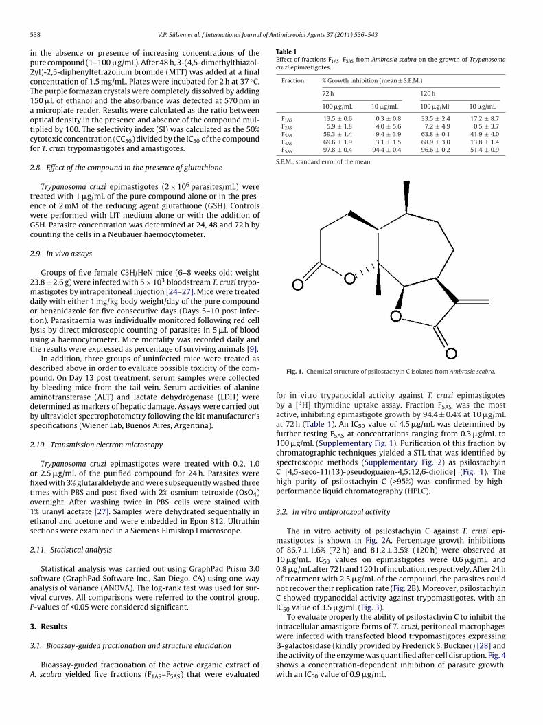
Fig. 1. Chemical structure of psilostachyin C isolated from Ambrosia scabra.
for in vitro trypanocidal activity against T. cruzi epimastigotesby a [3H] thymidine uptake assay. Fraction F5AS was the mostactive, inhibiting epimastigote growth by 94.4 ± 0.4% at 10 �g/mLat 72 h (Table 1). An IC50 value of 4.5 �g/mL was determined byfurther testing F5AS at concentrations ranging from 0.3 �g/mL to100 �g/mL (Supplementary Fig. 1). Purification of this fraction bychromatographic techniques yielded a STL that was identified byspectroscopic methods (Supplementary Fig. 2) as psilostachyinC [4,5-seco-11(13)-pseudoguaien-4,5:12,6-diolide] (Fig. 1). Thehigh purity of psilostachyin C (>95%) was confirmed by high-performance liquid chromatography (HPLC).
3.2. In vitro antiprotozoal activity
The in vitro activity of psilostachyin C against T. cruzi epi-mastigotes is shown in Fig. 2A. Percentage growth inhibitionsof 86.7 ± 1.6% (72 h) and 81.2 ± 3.5% (120 h) were observed at10 �g/mL. IC50 values on epimastigotes were 0.6 �g/mL and0.8 �g/mL after 72 h and 120 h of incubation, respectively. After 24 hof treatment with 2.5 �g/mL of the compound, the parasites couldnot recover their replication rate (Fig. 2B). Moreover, psilostachyinC showed trypanocidal activity against trypomastigotes, with anIC50 value of 3.5 �g/mL (Fig. 3).
To evaluate properly the ability of psilostachyin C to inhibit theintracellular amastigote forms of T. cruzi, peritoneal macrophages
were infected with transfected blood trypomastigotes expressing�-galactosidase (kindly provided by Frederick S. Buckner) [28] andthe activity of the enzyme was quantified after cell disruption. Fig. 4shows a concentration-dependent inhibition of parasite growth,with an IC50 value of 0.9 �g/mL.
V.P. Sülsen et al. / International Journal of Antimicrobial Agents 37 (2011) 536–543 539
Fig. 2. Effect of psilostachyin C on the growth of Trypanosoma cruzi epimastigotes. (A) Inhibition of parasite growth by psilostachyin C. Parasites were incubated in triplicatein the presence of 0.3–100 �g/mL of the compound (solid line) or with 1.3–20.8 �g/mL benznidazole (dotted line). Parasites were cultured for 72 and 120 h, with the additionof [3H] thymidine for the last 16 h. (B) Residual effect of psilostachyin C (P-C) on the growth of T. cruzi epimastigotes. Parasites were incubated in the absence or presence of0.2–2.5 �g/mL psilostachyin C for 24 h. The medium was replaced with fresh medium without the compound and the parasites were allowed to grow for 1, 2, 3 or 6 days.Symbols represent the mean ± standard error of the mean from three independent experiments.
Fig. 3. Effect of psilostachyin C on Trypanosoma cruzi trypomastigotes. Bloodstreamtcect
n7m
FmtAtPtm
Fig. 5. Effect of psilostachyin C on the growth of Leishmania mexicana and Leishmania
rypomastigotes were cultured in duplicate in the presence of 0.1–100 �g/mL of theompound or 0.4–900 �g/mL benznidazole. Cultures were done in 96-well platesmploying 1.5 × 10⁶ parasites/mL over 24 h and the remaining live parasites wereounted in a Neubauer chamber. Symbols represent the mean ± standard error ofhe mean.When psilostachyin C was tested against two species of Leishma-ia promastigotes, similar inhibitory effects were observed. After2 h treatment, IC50 values were 1.2 �g/mL and 1.5 �g/mL for L.exicana and for L. amazonensis, respectively (Fig. 5).
ig. 4. Effect of psilostachyin C on Trypanosoma cruzi amastigotes. Peritonealacrophages (5 × 10³ per well in 100 �L of culture medium) were infected with
ransfected trypomastigotes expressing �-galactosidase (10:1 parasite:cell ratio).fter washing the unbound parasites, psilostachyin C was added at concentra-
ions ranging from 0.01 �g/mL to 10 �g/mL. Two days post infection, Nonidet40 and chlorophenolred-�-d-galactopyranoside (CPRG) were added and galac-osidase activity was measured at an absorbance of 570 nm. Values represent the
ean ± standard error of the mean.
amazonensis promastigotes. Parasites were cultured in triplicate in the presenceof 0.3–100 �g/mL of the compound or 0.025–0.8 �g/mL amphotericin B. Parasiteswere cultured for 72 h with the addition of [3H] thymidine for the last 16 h. Valuesrepresent the mean ± standard error of the mean.
3.3. Effect of psilostachyin C on Trypanosoma cruzi epimastigotesin the presence of glutathione
An important increase in the number of epimastigoteswas observed when parasites were incubated simultaneouslywith GSH and psilostachyin C compared with those treatedwith psilostachyin C alone (6.8 ± 0.3 × 10⁶ parasites/mL vs.2.5 ± 0.4 × 10⁶ parasites/mL) (Fig. 6). However, the number ofparasites was significantly lower than that observed in controls(9.5 ± 2.0 × 10⁶ parasites/mL), indicating that the trypanocidalactivity of psilostachyin C was attenuated by the reducing agentGSH.
3.4. Cytotoxicity assay
The in vitro cytotoxic effect of psilostachyin C on peritonealmacrophages was evaluated by the MTT method and was expressedas cell viability percentage. The results are shown in Fig. 7. Whencells were treated with psilostachyin C, the CC50 was 87.5 �g/mL,indicating that the selectivity of psilostachyin C for T. cruzi trypo-mastigotes (SI = 25.0) and amastigotes (SI = 97.2) is greater than thatfor mammalian cells.
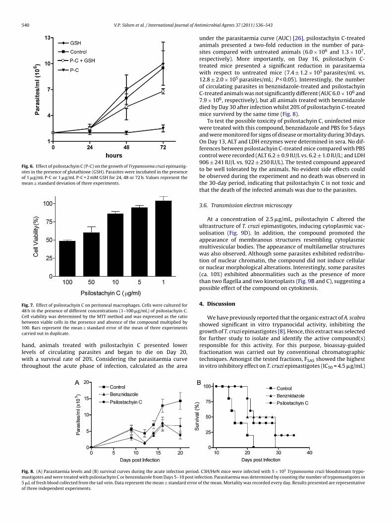
3.5. In vivo assays
Untreated mice infected with T. cruzi trypomastigotes dis-played high levels of parasitaemia (Fig. 8A) and presented 100%mortality on Day 22 post infection (Fig. 8B). On the other
540 V.P. Sülsen et al. / International Journal of An
Fig. 6. Effect of psilostachyin C (P-C) on the growth of Trypanosoma cruzi epimastig-otes in the presence of glutathione (GSH). Parasites were incubated in the presenceof 1 �g/mL P-C or 1 �g/mL P-C + 2 mM GSH for 24, 48 or 72 h. Values represent themean ± standard deviation of three experiments.
Fig. 7. Effect of psilostachyin C on peritoneal macrophages. Cells were cultured for4Cb1c
hlwt
Fm5o
8 h in the presence of different concentrations (1–100 �g/mL) of psilostachyin C.ell viability was determined by the MTT method and was expressed as the ratioetween viable cells in the presence and absence of the compound multiplied by00. Bars represent the mean ± standard error of the mean of three experimentsarried out in duplicate.
and, animals treated with psilostachyin C presented lowerevels of circulating parasites and began to die on Day 20,
ith a survival rate of 20%. Considering the parasitaemia curvehroughout the acute phase of infection, calculated as the area
ig. 8. (A) Parasitaemia levels and (B) survival curves during the acute infection period.astigotes and were treated with psilostachyin C or benznidazole from Days 5–10 post inf�L of fresh blood collected from the tail vein. Data represent the mean ± standard error of three independent experiments.
timicrobial Agents 37 (2011) 536–543
under the parasitaemia curve (AUC) [26], psilostachyin C-treatedanimals presented a two-fold reduction in the number of para-sites compared with untreated animals (6.0 × 10⁶ and 1.3 × 10⁷,respectively). More importantly, on Day 16, psilostachyin C-treated mice presented a significant reduction in parasitaemiawith respect to untreated mice (7.4 ± 1.2 × 10⁵ parasites/mL vs.12.8 ± 2.0 × 10⁵ parasites/mL; P < 0.05). Interestingly, the numberof circulating parasites in benznidazole-treated and psilostachyinC-treated animals was not significantly different (AUC 6.0 × 10⁶ and7.9 × 106, respectively), but all animals treated with benznidazoledied by Day 30 after infection whilst 20% of psilostachyin C-treatedmice survived by the same time (Fig. 8).
To test the possible toxicity of psilostachyin C, uninfected micewere treated with this compound, benznidazole and PBS for 5 daysand were monitored for signs of disease or mortality during 30 days.On Day 13, ALT and LDH enzymes were determined in sera. No dif-ferences between psilostachyin C-treated mice compared with PBScontrol were recorded (ALT 6.2 ± 0.9 IU/L vs. 6.2 ± 1.0 IU/L; and LDH906 ± 241 IU/L vs. 922 ± 250 IU/L). The tested compound appearedto be well tolerated by the animals. No evident side effects couldbe observed during the experiment and no death was observed inthe 30-day period, indicating that psilostachyin C is not toxic andthat the death of the infected animals was due to the parasites.
3.6. Transmission electron microscopy
At a concentration of 2.5 �g/mL, psilostachyin C altered theultrastructure of T. cruzi epimastigotes, inducing cytoplasmic vac-uolisation (Fig. 9D). In addition, the compound promoted theappearance of membranous structures resembling cytoplasmicmultivesicular bodies. The appearance of multilamellar structureswas also observed. Although some parasites exhibited redistribu-tion of nuclear chromatin, the compound did not induce cellularor nuclear morphological alterations. Interestingly, some parasites(ca. 10%) exhibited abnormalities such as the presence of morethan two flagella and two kinetoplasts (Fig. 9B and C), suggesting apossible effect of the compound on cytokinesis.
4. Discussion
We have previously reported that the organic extract of A. scabrashowed significant in vitro trypanocidal activity, inhibiting thegrowth of T. cruzi epimastigotes [8]. Hence, this extract was selected
for further study to isolate and identify the active compound(s)responsible for this activity. For this purpose, bioassay-guidedfractionation was carried out by conventional chromatographictechniques. Amongst the tested fractions, F5AS showed the highestin vitro inhibitory effect on T. cruzi epimastigotes (IC50 = 4.5 �g/mL)C3H/HeN mice were infected with 5 × 10³ Trypanosoma cruzi bloodstream trypo-ection. Parasitaemia was determined by counting the number of trypomastigotes inf the mean. Mortality was recorded every day. Results presented are representative

V.P. Sülsen et al. / International Journal of Antimicrobial Agents 37 (2011) 536–543 541
F tes. Pp ucleus
aa
t(aroastIPL1
m[tAu�o
ip
ig. 9. Ultrastructural effects of psilostachyin C on Trypanosoma cruzi epimastigosilostachyin C, (C) 1.0 �g/mL psilostachyin C or (D) 2.5 �g/mL psilostachyin C. N, ntructures; mv, multivesicular bodies. Magnification ×2500.
nd was selected for further purification, leading to the isolationnd identification of the STL psilostachyin C (Fig. 1).
When psilostachyin C was tested against T. cruzi epimastigotes,he compound exerted marked in vitro activity (IC50 = 0.6 �g/mL)Fig. 2A), showing that a progressive increase in trypanocidalctivity was gained during the purification process. Moreover, noecovery of the parasite replication rate was observed after removalf the pure compound (2.5 �g/mL) from the medium (Fig. 2B). Inddition, when psilostachyin C was assayed on the mammaliantages of T. cruzi, it efficiently inhibited both its infective form (therypomastigote) and the replicative intracellular amastigotes, withC50 values of 3.5 �g/mL and 0.9 �g/mL, respectively (Figs. 3 and 4).silostachyin C also exerted in vitro leishmanicidal activity against. mexicana and L. amazonensis promastigotes, with IC50 values of.2 �g/mL and 1.5 �g/mL, respectively (Fig. 5).
Most STLs contain a common functional structure of �-ethylene-�-lactone, which is highly reactive with thiol groups
29]. We have therefore investigated whether this functional struc-ure is responsible for the trypanocidal activity of psilostachyin C.s shown in Fig. 6, the presence of GSH in the culture media atten-ated the trypanocidal effect of the compound, indicating that the
-methylene-�-lactone moiety is not uniquely responsible for thebserved activity [11].To determine the specificity of the trypanocidal activity, ann vitro cytotoxicity test on mammalian cells was carried out. Wheneritoneal macrophages were treated with psilostachyin C, the
arasites were incubated with (A) Diamond medium alone or with (B) 0.5 �g/mLs; K, kinetoplast; F, flagellum; Vac, vacuoles; mi, mitochondria; ms, multilamellar
CC50 value was >100 �g/mL (3 h; data not shown). At 48 h, the CC50value was 87.5 �g/mL.
The SI was calculated to compare the toxicity to mammaliancells and the activity against T. cruzi trypomastigotes and amastig-otes. The values obtained were 25.0 and 97.2, respectively,indicating that the selectivity of this compound for parasites isgreater than for mammalian cells.
In view of these results, psilostachyin C was also evaluatedin vivo in a murine model. In this assay, the compound induceda significant decrease in parasitaemia compared with untreatedmice (6 × 10⁶ vs. 13 × 106) and proved to be as effective as ben-znidazole. Although treatments were administered for only 5 days,the reduction in parasitaemia was observed throughout the evalu-ation period and was reflected in the significant increase in survivaltime of the animals.
Recently, many STLs have shown interesting in vitro activityagainst different protozoa, including T. cruzi. Nevertheless, to thebest of our knowledge, no in vivo studies have been carried outwith these compounds, with the exception of the one previouslyreported by our group [9].
Electron microscopy has proven to be a reliable and useful tool
to study morphological alterations and target organelles in theinvestigation of new drugs for Chagas disease. In this study, it wasobserved that psilostachyin C causes ultrastructural alterations inT. cruzi epimastigotes, such as cytoplasm vacuolisation, the appear-ance of multilamellar structures, and abnormalities such as the
5 l of An
ptok
dafltflmbtpbnioioi
toc
tcitcpQao
A
LoF
NacBItI
tHw‘
A
t
R
[
[
[
[
[
[
[
[
[
[
[
[
[
[
[
[
[
[
42 V.P. Sülsen et al. / International Journa
resence of multiple kinetoplasts and flagella. However, these mul-iple structures were not accompanied by an increase in the numberf nuclei, indicating that the compound could act in a stage betweeninetoplast segregation and nuclear division.
Recently, it has been shown that the flagellum begins to replicateuring the G2 stage, and later the nucleus and kinetoplast segregatelmost simultaneously during the T. cruzi cell cycle [30]. The newagellum emerges from the flagellar pocket and remains short untilhe kinetoplast segregates and mitosis occurs. However, the newagellum reaches its final size during cytokinesis. The presence ofultiple flagella and kinetoplasts induced by the compound could
e related to the inability of the parasite to synchronise replica-ion of these structures with nuclear division. Synthesis of parasiteroteins or factors that synchronise these stages of the cycle mighte affected by the compound. Detailed molecular studies will beeeded to explain this phenomenon better. The compound stud-
ed herein did not cause mitochondrial swelling as observed withther compounds that block parasite metabolism [31–33], indicat-ng that it may act by other mechanisms. Moreover, the appearancef multilamellar structures could be due to an autophagic processnduced by the isolated compound [34].
According to Lee and Schneider [35], STLs of the ambrosanolideype are one of the promising scaffolds for the discovery or designf new drugs, since these kinds of compounds are not present inurrent trade drugs.
In conclusion, psilostachyin C showed both in vitro and in vivorypanocidal activity. Although the mechanism of action of thisompound remains to be determined, it could be suggested thatt might act as a cytokinesis inhibitor, as an oxidative stress induc-or, consequently or independently producing some ultrastructuralhanges in the parasite. These results make psilostachyin C aromising template for the design of novel trypanocidal agents.uantitative structure–trypanocidal activity relationship studiesmongst members of the STLs are currently being undertaken inur laboratories.
cknowledgments
The authors wish to thank Dr Berta Franke de Cazzulo and Estelaammel for providing Leishmania parasites and T. cruzi epimastig-tes, respectively, as well as Mrs Cristina Aguilera and Mrs Teresaogal for their valuable technical assistance.
Funding: This work was supported by grants from Agenciaacional de Promoción Científica y Tecnológica (ANPCyT) (PICT 608nd 1701), Consejo Nacional de Investigaciones Científicas y Técni-as (CONICET) (PIP 1540) and Universidad de Buenos Aires (B027,030 and B607), Argentina. ELM is also supported by the Fogarty
nternational Center (TW007972), USA, and the International Cen-re for Genetic Engineering and Biotechnology (CRP/ARG09-02),taly.
Competing interests: None declared.Ethical approval: All procedures with animals were approved by
he Ethics Review Board of the Instituto de Estudios de la Inmunidadumoral (IDEHU-CONICET) and were conducted in accordanceith the guidelines established by the National Research Council
Guide for the Care and Use of Laboratory Animals’.
ppendix A. Supplementary data
Supplementary data associated with this article can be found, inhe online version, at doi:10.1016/j.ijantimicag.2011.02.003.
eferences
[1] World Health Organization. Control of Chagas disease. Second report of theWHO Expert Committee. Geneva, Switzerland: WHO; 2002. Technical ReportSeries No. 905.
[
timicrobial Agents 37 (2011) 536–543
[2] Cazorla SI, Frank FM, Malchiodi EL. Vaccination approaches against Try-panosoma cruzi infection. Expert Rev Vaccines 2009;8:921–35.
[3] Cazorla SI, Frank FM, Becker PD, Arnaiz M, Mirkin GA, Corral RS, et al. Redirec-tion of the immune response to the functional catalytic domain of the cysteinproteinase cruzipain improves protective immunity against Trypanosoma cruziinfection. J Infect Dis 2010;202:136–44.
[4] World Health Organization. Neglected tropical diseases: hidden suc-cesses, emerging opportunities. Geneva, Switzerland: WHO; 2009.WHO/HTM/NTD/2009.2.
[5] Chiaramonte MG, Zwirner NW, Caropresi SL, Taranto NJ, Malchiodi EL. Try-panosoma cruzi and Leishmania spp. human mixed infection. Am J Trop MedHyg 1996;54:271–3.
[6] Chiaramonte MG, Frank FM, Furer GM, Taranto NJ, Margni RA, Malchiodi EL.Polymerase chain reaction reveals Trypanosoma cruzi infection suspected byserology in cutaneous and mucocutaneous leishmaniasis patients. Acta Trop1999;72:295–308.
[7] Frank FM, Fernández MM, Taranto NJ, Cajal SP, Margni RA, Castro E, et al.Characterization of human infection by Leishmania spp. in the Northwest ofArgentina: immune response, double infection with Trypanosoma cruzi andspecies of Leishmania involved. Parasitology 2003;169:31–9.
[8] Sülsen VP, Güida C, Coussio J, Paveto C, Muschietti L, Martino V. In vitro evalu-ation of trypanocidal activity in plants used in Argentine traditional medicine.Parasitol Res 2006;98:370–4.
[9] Sülsen VP, Frank FM, Cazorla SI, Anesini C, Malchiodi EL, Freixa B, et al.Trypanocidal and leishmanicidal activities of sesquiterpene lactones fromAmbrosia tenuifolia Sprengel (Asteraceae). Antimicrob Agents Chemother2008;52:2415–9.
10] Muschietti LV, Sülsen VP, Martino VS. Trypanocidal and leishmanicidal activi-ties of South American medicinal plants. In: Martino VS, Muschietti LV, editors.South American medicinal plants as a potential source of bioactive compounds.Kerala, India: Transworld Research; 2008. p. 149–80.
11] Schmidt TJ. Structure–activity relationships of sesquiterpene lactones. In:Atta-ur-Rahman, editor. Studies in natural products chemistry. Amsterdam,The Netherlands: Elsevier; 2006. p. 309–92.
12] Picman AK. Biological activities of sesquiterpene lactones. Biochem Syst Ecol1986;14:255–81.
13] Luna-Herrera J, Costa MC, González HG, Rodrigues AI, Castillo PC. Synergis-tic antimycobacterial activities of sesquiterpene lactones from Laurus spp. JAntimicrob Chemother 2007;59:548–52.
14] Karioti A, Skaltsa H, Linden A, Perozzo R, Brun R, Tasdemir D. Anthecularin:a novel sesquiterpene lactone from Anthemis auriculata with antiprotozoalactivity. J Org Chem 2007;72:8103–6.
15] Pillay P, Vleggaar R, Maharaj VJ, Smith PJ, Lategan CA. Isolation and identifi-cation of antiplasmodial sesquiterpene lactones from Oncosiphon piluliferum. JEthnopharmacol 2007;112:71–6.
16] Kagan HB, Miller HE, Renold W, Lakshmikantham MV, Tether LR, Herz W. Thestructure of psilostachyin C, a new sesquiterpene dilactone from Ambrosiapsilostachya DC. J Org Chem 1966;3:1629–32.
17] Oberti J, Silva G, Sosa V, Kulanthaivel P, Herz W. Ambrosanolides and secoam-brosanolides from Ambrosia tenuifolia. Phytochemistry 1986;25:1355–8.
18] Wang PH, Xu J, Wu MY. Chemical constituents of ragweed (Ambrosia artemisi-ifolia L.). Zhongguo Zhong Yao Za Zhi 1993;18:164–6, 191 [in Chinese].
19] Sturgeon CM, Craig K, Brown C, Rundle NT, Andersen RJ, Roberge M. Modula-tion of the G2 cell cycle checkpoint by sesquiterpene lactones psilostachyins Aand C isolated from the common ragweed Ambrosia artemisiifolia. Planta Med2005;71:938–43.
20] National Research Council. Guide for the care and use of laboratory animals.Washington, DC: National Academy Press; 1996.
21] Sülsen VP, Cazorla SI, Frank FM, Redko FC, Anesini CA, Coussio JD, et al. Try-panocidal and leishmanicidal activities of flavonoids from Argentine medicinalplants. Am J Trop Med Hyg 2007;77:654–9.
22] Alexander B, Browse DJ, Reading SJ, Benjamin IS. A simple and accurate math-ematical method for calculation of the EC50. J Pharmacol Toxicol Methods1999;41:55–8.
23] Buckner FS, Verlinde CL, La Flamme AC, Van Voorhis WC. Efficient technique forscreening drugs for activity against Trypanosoma cruzi using parasites express-ing �-galactosidase. Antimicrob Agents Chemother 1996;40:2592–7.
24] Frank FM, Petray PB, Cazorla SI, Munoz MC, Corral RS, Malchiodi EL. Useof a purified Trypanosoma cruzi antigen and CpG oligodeoxynucleotides forimmunoprotection against a lethal challenge with trypomastigotes. Vaccine2003;22:77–86.
25] Cazorla SI, Becker PD, Frank FM, Ebensen T, Sartori MJ, Corral RS, et al.Oral vaccination with Salmonella enterica as a cruzipain-DNA delivery sys-tem confers protective immunity against Trypanosoma cruzi. Infect Immun2008;76:324–33.
26] Cazorla SI, Frank FM, Becker P, Corral RS, Guzman CA, Malchiodi EL. Prime-boostimmunization with cruzipain co-administered with MALP-2 triggers a protec-tive immune response able to decrease parasite burden and tissue injury in anexperimental Trypanosoma cruzi infection model. Vaccine 2008;26:1999–2009.
27] Brengio SD, Belmonte SA, Guerreiro E, Giordano OS, Pietrobon EO, Sosa MA. The
sesquiterpene lactone dehydroleucodine (DhL) affects the growth of culturedepimastigotes of Trypanosoma cruzi. J Parasitol 2000;86:407–12.28] Kraus JM, Verlinde CL, Karimi M, Lepesheva GI, Gelb MH, Buckner FS. Rationalmodification of a candidate cancer drug for use against Chagas disease. J MedChem 2009;52:1639–47 [Erratum in: J Med Chem 2009;52:4549; J Med Chem2009;52:4979].

l of An
[
[
[
[
[
V.P. Sülsen et al. / International Journa
29] Schmidt TJ, Nour AM, Khalid SA, Kaiser M, Brun R. Quantitativestructure–antiprotozoal activity relationships of sesquiterpene lactones.Molecules 2009;14:2062–76.
30] Elias MC, da Cunha JPC, de Faria FP, Mortara RA, Freymüller E, SchenkmanS. Morphological events during the Trypanosoma cruzi cell cycle. Protist2007;158:147–57.
31] Lazardi K, Urbina JA, de Souza W. Ultrastructural alterations induced bytwo ergosterol biosynthesis inhibitors, ketoconazole and terbinafine, on epi-mastigotes and amastigotes of Trypanosoma (Schizotrypanum) cruzi. AntimicrobAgents Chemother 1990;34:2097–105.
32] Izumi E, Morello LG, Ueda-Nakamura T, Yamada-Ogatta SF, Filho BP,Cortez DA, et al. Trypanosoma cruzi: antiprotozoal activity of parthenolide
[
[
timicrobial Agents 37 (2011) 536–543 543
obtained from Tanacetum parthenium (L.) Schultz Bip. (Asteraceae, Composi-tae) against epimastigote and amastigote forms. Exp Parasitol 2008;118:324–30.
33] Sülsen V, Barrera P, Muschietti L, Martino V, Sosa M. Antiproliferative effectand ultrastructural alterations induced by psilostachyin on Trypanosoma cruzi.Molecules 2010;15:545–53.
34] Menna-Barreto RFS, Salomão K, Dantas AP, Santa-Rita RM, Soares MJ, BarbosaHS, et al. Different cell death pathways induced by drugs in Trypanosoma cruzi:an ultrastructural study. Micron 2009;40:157–68.
35] Lee ML, Schneider G. Scaffold architecture and pharmacophoric properties ofnatural products and trade drugs: application in the design of natural product-based combinatorial libraries. J Comb Chem 2001;3:284–9.