Proteomic profile regulated by the anticancer peptide CIGB-300 in Non-Small Cell Lung Cancer (NSCLC)...
11
Proteomic Profile Regulated by the Anticancer Peptide CIGB-300 in Non-Small Cell Lung Cancer (NSCLC) Cells Arielis Rodrı ´guez-Ulloa,* ,† Yassel Ramos, ‡ Jeovanis Gil, ‡ Yasser Perera, § Lila Castellanos-Serra, ‡ Yairet Garcı ´a, ‡ La ´zaro Betancourt, ‡ Vladimir Besada, ‡ Luis J. Gonza ´lez, ‡ Jorge Ferna ´ndez-de-Cossio, † Aniel Sanchez, ‡ Joem M. Serrano, § Herna ´n Farina, | Daniel F. Alonso, | Boris E. Acevedo, § Gabriel Padro ´n, ‡ Alexis Musacchio, † and Silvio E. Perea § Department of Bioinformatics, Deparment of Proteomics, and Laboratory of Molecular Oncology, Center for Genetic Engineering and Biotechnology, Havana, Cuba, and Laboratory of Molecular Oncology, National University of Quilmes, Buenos Aires, Argentina Received July 14, 2010 CIGB-300 is a proapoptotic peptide-based drug that abrogates the CK2-mediated phosphorylation. This peptide has antineoplastic effect on lung cancer cells in vitro and in vivo. To understand the mechanisms involved on such anticancer activity, the NCI-H125 cell line proteomic profile after short-term incubation (45 min) with CIGB-300 was investigated. As determined by 2-DE or 2D-LC-MS/MS, 137 proteins changed their abundances more than 2-fold in response to the CIGB-300 treatment. The expression levels of proteins related to ribosome biogenesis, metastasis, cell survival and proliferation, apoptosis, and drug resistance were significantly modulated by the presence of CIGB-300. The protein translation process was the most affected (23% of the identified proteins). From the proteome analysis of the NCI-H125 cell line, novel potentialities for CIGB-300 as anticancer agent were evidenced. Keywords: Comparative proteomic analysis • CIGB-300 • ribosome biogenesis • apoptosis • casein kinase 2 • nucleophosmin • lung cancer Introduction Casein Kinase 2 (CK2) is a serine/threonine protein kinase overexpressed in a variety of solid and hematopoietic tumors. 1-3 In particular, lung cancer has been shown to exhibit high levels of CK2 activity and mRNA expression. 4,5 This kinase phospho- rylates a wide range of substrates related to gene expression, cell survival, and apoptotic pathways; its increased expression in tumor cells promotes cellular proliferation and suppresses apoptosis. 6 Previous findings have suggested the potential of CK2 as an attractive anticancer target. 7 Therefore, to inhibit the CK2 activity, different emergent approaches have been assayed so far: (a) down-regulation of CK2 gene expression by antisense oligonucleotide, 8 (b) allosteric inhibition of the enzyme, 9 and (c) blocking of ATP binding site by small compounds. 10 CIGB-300 (formerly P15-Tat) is a novel hypothesis- driven peptide that abrogates the CK2-mediated phosphory- lation by its ability to bind the substrate’s phosphoaceptor domain instead of the enzyme per se. 11 The antineoplastic effect of CIGB-300 has been well docu- mented both in vitro and in vivo by using different cancer cell lines and mouse models. 11-13 In particular, lung cancer cells have been shown to be sensitive to CIGB-300 in vitro and in vivo. For instance, the cell viability on human non-small cell lung cancer (NSCLC) NCI-H125 and on murine lung trans- formed epithelial TC-1 cells were similarly reduced (IC 50 ) 70 µM), 11,12 and the caspase activation was detected after 30 min of CIGB-300 addition. 11 Likewise, CIGB-300 induced massive apoptosis after 10 min upon addition of 100 µM CIGB-300 to small cell lung cancer (SCLC) NCI-H82 cells. 13 In vivo, CIGB- 300 was able to elicit significant antitumor activity both in murine TC-1 and human NCI-H125 implanted tumors. 11,13 CIGB-300 has been already studied in the clinical ground, demonstrating safety, tolerability, and efficacy signs in women with cervical malignancies. 14 Significant advances have been made in proving the CIGB- 300 mechanism of action in lung cancer cells. Recently, the nucleolar chaperone nucleophosmin (B23/NPM) has been described as the major CK2-substrate targeted by CIGB-300. 15 Such interaction inhibited the CK2-mediated phosphorylation on B23/NPM in a dose-dependent manner and led to nucleolar disassemble with subsequent apoptosis. 15 Accumulated evi- dence suggests that CIGB-300 could ultimately induce apop- tosis by impairing the ribosome biogenesis. 15 A detailed description of differential protein expression of cells treated with CIGB-300 is needed to support the proposed mechanism of action for such peptide. * To whom correspondence should be addressed. Department of Bioin- formatics, Center for Genetic Engineering and Biotechnology, P.O. Box 6162, Havana CP10600, Cuba. Phone: 53-7271-6221. Fax: 53-7271-8070. E-mail: [email protected]. † Department of Bioinformatics, Center for Genetic Engineering and Biotechnology. ‡ Department of Proteomics, Center for Genetic Engineering and Biotechnology. § Laboratory of Molecular Oncology, Center for Genetic Engineering and Biotechnology. | National University of Quilmes. 10.1021/pr100728v 2010 American Chemical Society Journal of Proteome Research 2010, 9, 5473–5483 5473 Published on Web 08/30/2010
Transcript of Proteomic profile regulated by the anticancer peptide CIGB-300 in Non-Small Cell Lung Cancer (NSCLC)...
Proteomic Profile Regulated by the Anticancer Peptide CIGB-300 in
Non-Small Cell Lung Cancer (NSCLC) Cells
Arielis Rodrıguez-Ulloa,*,† Yassel Ramos,‡ Jeovanis Gil,‡ Yasser Perera,§ Lila Castellanos-Serra,‡
Yairet Garcıa,‡ Lazaro Betancourt,‡ Vladimir Besada,‡ Luis J. Gonzalez,‡
Jorge Fernandez-de-Cossio,† Aniel Sanchez,‡ Joem M. Serrano,§ Hernan Farina,|
Daniel F. Alonso,| Boris E. Acevedo,§ Gabriel Padron,‡ Alexis Musacchio,† and Silvio E. Perea§
Department of Bioinformatics, Deparment of Proteomics, and Laboratory of Molecular Oncology, Center forGenetic Engineering and Biotechnology, Havana, Cuba, and Laboratory of Molecular Oncology, National
University of Quilmes, Buenos Aires, Argentina
Received July 14, 2010
CIGB-300 is a proapoptotic peptide-based drug that abrogates the CK2-mediated phosphorylation. Thispeptide has antineoplastic effect on lung cancer cells in vitro and in vivo. To understand the mechanismsinvolved on such anticancer activity, the NCI-H125 cell line proteomic profile after short-term incubation(45 min) with CIGB-300 was investigated. As determined by 2-DE or 2D-LC-MS/MS, 137 proteinschanged their abundances more than 2-fold in response to the CIGB-300 treatment. The expressionlevels of proteins related to ribosome biogenesis, metastasis, cell survival and proliferation, apoptosis,and drug resistance were significantly modulated by the presence of CIGB-300. The protein translationprocess was the most affected (23% of the identified proteins). From the proteome analysis of theNCI-H125 cell line, novel potentialities for CIGB-300 as anticancer agent were evidenced.
Keywords: Comparative proteomic analysis • CIGB-300 • ribosome biogenesis • apoptosis • casein kinase2 • nucleophosmin • lung cancer
Introduction
Casein Kinase 2 (CK2) is a serine/threonine protein kinaseoverexpressed in a variety of solid and hematopoietic tumors.1-3
In particular, lung cancer has been shown to exhibit high levelsof CK2 activity and mRNA expression.4,5 This kinase phospho-rylates a wide range of substrates related to gene expression,cell survival, and apoptotic pathways; its increased expressionin tumor cells promotes cellular proliferation and suppressesapoptosis.6 Previous findings have suggested the potential ofCK2 as an attractive anticancer target.7 Therefore, to inhibitthe CK2 activity, different emergent approaches have beenassayed so far: (a) down-regulation of CK2 gene expression byantisense oligonucleotide,8 (b) allosteric inhibition of theenzyme,9 and (c) blocking of ATP binding site by smallcompounds.10 CIGB-300 (formerly P15-Tat) is a novel hypothesis-driven peptide that abrogates the CK2-mediated phosphory-lation by its ability to bind the substrate’s phosphoaceptordomain instead of the enzyme per se.11
The antineoplastic effect of CIGB-300 has been well docu-mented both in vitro and in vivo by using different cancer celllines and mouse models.11-13 In particular, lung cancer cellshave been shown to be sensitive to CIGB-300 in vitro and invivo. For instance, the cell viability on human non-small celllung cancer (NSCLC) NCI-H125 and on murine lung trans-formed epithelial TC-1 cells were similarly reduced (IC50 ) 70µM),11,12 and the caspase activation was detected after 30 minof CIGB-300 addition.11 Likewise, CIGB-300 induced massiveapoptosis after 10 min upon addition of 100 µM CIGB-300 tosmall cell lung cancer (SCLC) NCI-H82 cells.13 In vivo, CIGB-300 was able to elicit significant antitumor activity both inmurine TC-1 and human NCI-H125 implanted tumors.11,13
CIGB-300 has been already studied in the clinical ground,demonstrating safety, tolerability, and efficacy signs in womenwith cervical malignancies.14
Significant advances have been made in proving the CIGB-300 mechanism of action in lung cancer cells. Recently, thenucleolar chaperone nucleophosmin (B23/NPM) has beendescribed as the major CK2-substrate targeted by CIGB-300.15
Such interaction inhibited the CK2-mediated phosphorylationon B23/NPM in a dose-dependent manner and led to nucleolardisassemble with subsequent apoptosis.15 Accumulated evi-dence suggests that CIGB-300 could ultimately induce apop-tosis by impairing the ribosome biogenesis.15 A detaileddescription of differential protein expression of cells treatedwith CIGB-300 is needed to support the proposed mechanismof action for such peptide.
* To whom correspondence should be addressed. Department of Bioin-formatics, Center for Genetic Engineering and Biotechnology, P.O. Box 6162,Havana CP10600, Cuba. Phone: 53-7271-6221. Fax: 53-7271-8070. E-mail:[email protected].
† Department of Bioinformatics, Center for Genetic Engineering andBiotechnology.
‡ Department of Proteomics, Center for Genetic Engineering andBiotechnology.
§ Laboratory of Molecular Oncology, Center for Genetic Engineering andBiotechnology.| National University of Quilmes.
10.1021/pr100728v 2010 American Chemical Society Journal of Proteome Research 2010, 9, 5473–5483 5473Published on Web 08/30/2010

In this work, the proteome of the nuclear fraction from NCI-H125 cells modulated by CIGB-300 was described. Two comple-mentary proteomic techniques, two-dimensional electrophore-sis (2-DE) and two-dimensional liquid chromatography coupledto tandem mass spectrometry (2D-LC-MS/MS), were used. Theanalysis of differentially expressed proteins suggested thatCIGB-300 impair the ribosome biogenesis on these cells. Dataprovided here not only indicate that this peptide regulatesdifferent relevant biological processes in cancer but also setthe molecular basis for a synergistic interaction with anticancerdrugs.
Materials and Methods
Peptide. CIGB-300 is a cyclic peptide (CWMSPRHLGTC)fused to the cell penetrating peptide Tat (48GRKKRRQR-RRPPQ68).11 The CIGB-300 peptide was synthesized on solidphase and purified by reverse phase high performance liquidchromatography (RP-HPLC) to >95% purity on an acetronitile/H2O-trifluoroacetic acid gradient16 and confirmed by electro-spray mass spectrometry (Micromass, U.K.).
Cell Culture and Treatments. The non-small cell lung cancer(NSCLC) cell line NCI-H125, kindly provided by Dr. EduardoSuarez (Center for Molecular Immunology, Havana, Cuba), wasoriginally obtained from ATCC (Rockville, MD). The NCI-H125cells were cultured in RPMI 1640 (Life Technologies, USA)supplemented with 10% fetal bovine serum (FBS; PAA, Canada)and 100 µg/mL gentamicin (Sigma, USA), at 37 °C in ahumidified atmosphere containing 5% CO2.
For proteomic analysis, 20 × 106 tumor cells were seeded inappropriate vessels and incubated for 24 h. The cells wereincubated with 200 µmol/L of CIGB-300 during 45 min, takinginto account that CIGB-300 induces cell death features after60 min of incubation. For Western blot analysis, cell cultureswere treated with 40, 80, or 200 µmol/L of CIGB-300 for 45 min.
Isolation of Nuclear Proteins. NCI-H125 cells were collectedby trypsinization. After washing with phosphate buffered saline(PBS), the cells were suspended in 500 µL of the isotonic buffercontaining 10 mM HEPES-NaOH adjusted to pH 7.5, 0.25 Msucrose, 1 mM EGTA, and protease inhibitors.17 For plasmamembrane solubilization Triton X-100 at final concentrationof 0.25% was added. After 15 min at 4 °C, the cell lysate wascentrifuged for 15 min at 12,000 × g and 4 °C. Nuclear pelletwas washed with 500 µL of a solution containing 10 mMHEPES-NaOH, pH 7.5, 0.34 M sucrose, 0.75 mM spermidine,0.15 mM spermine, 1 mM EDTA, 0.1% Triton X-100, andprotease inhibitors, centrifuged at 1000 × g for 5 min at 4 °C,and decanted. Nuclear proteins were then solubilized in a lysissolution containing 7 M urea, 2 M thiourea, 2% CHAPS, 0.5%ASB-14, 15% glycerol, 2% DTT, and protease inhibitors. Thesuspension was sonicated (2 × 1 min) and incubated 1 h atroom temperature. Free cysteines were blocked with 4%acrylamide during 1 h at room temperature. Anfolytes range3-10 were added at 2% final concentration and DNA wasremoved by centrifugation at 60,000 × g during 3 h. Thesupernatant containing solubilized nuclear proteins was con-served at -70 °C for 2-DE and 2D-LC-MS/MS analysis.
Two-Dimensional Gel Electrophoresis (2-DE). For bothnuclear fractions, treated and nontreated with CIGB-300, threereplicate analytical gels and two preparative gels were run. 2-DEwas performed by combining IEF in IPG for the first dimensionand SDS-PAGE for the second dimension. IPG strips (pI range4-7, 18 cm) (Amersham Pharmacia, Sweden) were rehydratedovernight in 350 µL of lysis solution containing 2% of anfolytes
range 4-7 and approximately 400 µg of nuclear proteins foranalytical gels or 700 µg for preparative gels. Electrofocusingwas done at 20 °C in a Multiphor II equipment (AmershamPharmacia, Sweden) at 50 kV-h under silicone oil. For thesecond dimension, IPG strips were incubated for 15 min in 3mL of equilibration solution (6 M urea, 30% glycerol, 2% SDS,50 mM Tris-HCl, pH 8.8). The focalized proteins were trans-ferred to 16.8% polyacrylamide-SDS gels (prepared 24 h beforeuse) and were run at 16 mA for 30 min and then at 30 mA for6 h. For analytical gels, silver staining was used for detection,while preparative gels were silver stained with a MS compatibleprocedure.18,19
2-D Image Analysis. Images from 2-D gels were processedand analyzed using the Melanie 5 software (GeneBio, Switzer-land). Spots were automatically detected without previouscontrast enhancement, followed by manual editing whennecessary. For each class, gel reproducibility was evaluated byusing the available tools (pair reports and scatter plot analysis).Heuristic clustering was used for automatic classification of theset of gels. The detection of changes in protein maps betweenthe two classes was first done by automatic analysis andconfirmed by visual inspection of the series of gels. Histogramsfor groups based on percent volume of the spots were recorded,using the central tendency as the mean value and the meansquare deviation as the dispersion value. The spots displayinga change in their expression between the classes greater thanthe 2-fold factor (determined from the mean) were consideredfor protein identification by MS. For molecular mass and pIestimation according to the protein migration in 2D gel, severalintense and well-resolved spots corresponding to different gelzones were selected as landmarks. Theoretical mass and pIwere calculated from the sequence of the identified proteinsusing the Compute pI/Mw ExPASy tool (http://ca.expasy.org/tools/pi_tool.html).
In-Gel Trypsin Digestion and Protein Identification.In-Gel Digestion. Replicate spots from two preparative gelswere pooled and silver staining was removed with Farmer’sreagent.20 Spots were cut in 1-3 mm3 pieces, dried withacetonitrile, rehydrated in a minimum volume of ammoniumbicarbonate (25 mM) containing trypsin (12.5 ng/µL, Promega,USA), and digested at 37 °C for 18 h.
MS Analysis. Tryptic peptides from selected spots wereobtained and processed as described by Gonzalez et al.21
ESI-MS spectra were acquired using a QTof-2 hybrid typeorthogonal tandem mass spectrometer (Micromass, U.K.) fittedwith a Z-spray nanoflow electrospray ion source operated at80 °C with a drying gas flow at 50 L/h. Peptides eluted with60% ACN in 1% formic acid from ZipTips (Millipore, USA) wereloaded into the borosilicate nanoflow tips. Capillary and conevoltages of 900 and 35 V were applied, respectively. To acquirethe ESI-MS/MS spectra, the first quadrupole was used to selectthe precursor ion within a window of approximately 2 Th.Argon was used in the collision chamber at 3 × 10⁻² Pa pressureand collision energies between 25 and 50 eV were set tofragment precursor ions. Data acquisition and processing wereperformed using MassLynx v3.5 (Micromass, U.K.).
Protein Identification. Queries were done to sequencedatabases (Swiss-Prot and NCBI) by MASCOT search enginesoftware.22 Monoisotopic masses of tryptic peptides were usedto identify the proteins by peptide mass fingerprinting (PMF),and additionally the most intense signals observed in everyspectrum were sequenced by collision induced dissociation andESI-MS/MS. The MS/MS ion search option of the MASCOT
research articles Rodrıguez-Ulloa et al.
5474 Journal of Proteome Research • Vol. 9, No. 10, 2010

program was used for protein identification; no taxonomicrestrictions were made. Cysteine propionamide was consideredas fixed modification. Variable modifications including dea-midation of glutamine and asparagine, and methionine sul-foxide were taken into account. Other restrictions to MASCOTwere precursor and fragment ions m/z tolerance of 0.2 Da,enzyme digestion with trypsin, and up to one missed cleavage.Additionally, MS/MS spectra of proteins identified on the basisof sequencing of one or two peptides were manually inter-preted, looking for four or more intense consecutive C-terminalyn′′ fragment ions and the complementary b fragment series.
Proteomic Analysis by Two-Dimensional Liquid Chromatog-raphy Coupled to Tandem Mass Spectrometry (2D-LC-MS/MS). Trypsin Digestion. The nuclear protein extracts, previouslyreduced and alkylated as described above, of control (400 µg)and CIGB-300-treated NCI-H125 cells (400 µg) were precipi-tated with acetone/TCA and independently dissolved in 300µL of buffer containing 50 mM ammonium bicarbonate, 2 Murea, pH 8.0. In the case of the treated sample, the buffer wasprepared in 18O-labeled water. Proteins were digested by twoadditions of trypsin at an enzyme-to-substrate mass ratio of1:100, for 16 h for each one, at 37 °C. To inhibit trypsin activityand to stop the proteolytic digestion, samples were reducedand alkylated using the previously described protocol. Bothdigests were mixed and desalted before cation exchangechromatography.
2D-LC-MS/MS Analysis. A combination of strong cationexchange and reversed phase chromatography steps were usedto separate the complex mixture of peptides prior to tandemmass spectrometry analysis (MS/MS). Desalted peptides wereseparated into 14 fractions using a 0.8 mm × 50 mm SCX IIcolumn (Agilent, USA) and NaCl gradient (from 0 to 500 mMin 100 min). Collected fractions were lyophilized and furtherdissolved in 130 µL of 1% formic acid. For each fraction threereplicate aliquots of 40 µL were separated in independentLC-MS/MS experiments by reverse phase chromatography.Data dependent acquisition MS/MS spectra of the elutedpeptides were acquired in three m/z ranges (400-600, 590-900,or 890-2000). Peptides were separated and identified using anAgilent 1100 series nano LC system (Agilent, USA) coupledonline to a QTof-2 hybrid mass spectrometer (Micromass, U.K.).The capillary and cone voltages of the electrospray ionizationsource were operated with 2.4 kV and 45 V, respectively.Samples were applied at 20 µL/min to an LC-Packings PepMapC18 Precolumn Cartridge (5 mm × 300 µm i.d.) for 10 min andthen switched onto a C18 capillary column (15 cm × 75 µmi.d., packed with 5 µm, Zorbax 300 SB) using a mobile phasecontaining 0.1% formic acid, 5-45% acetonitrile gradient over90 min at 300 nL/min flow rate. For protein identification, 1 ssurvey scans were acquired over the selected mass range anda maximum of 4 concurrent MS/MS acquisitions were triggeredfor 2+, 3+ charged precursors ions detected at an intensityabove a 15 counts/s threshold. Each MS/MS acquisition wascompleted and switched back to MS mode when the total ioncurrent fell below a 2 counts/s threshold or after a maximumof 6 s of continuous acquisition.
Data Analysis. Raw files were processed using MASCOTDistiller 2.3 software. Peptides were assigned to MS/MS spectrausing MASCOT search engine against the IPI human database(version 3.61). The following search parameters were selected:2 Da precursor mass tolerance, 0.8 Da MS/MS ions masstolerance, tryptic search with up to one missed cleavage,oxidized methionine as variable modification, and cysteine
propionamide as fixed modification. To accept a peptide hitas positive we considered a false discovery rate (FDR) of 3%based on the target-decoy strategy.23
Quantification was achieved using ISOTOPICA software.24,25
The software enables the relative peptide quantification basedon the detailed analysis of the observed isotopic ion distribu-tion. The software calculates the best ratio of the nonlabeled(16O2, control condition) and labeled (18O1 + 18O2, treated withCIGB-300) to obtain an in silico isotopic ion distribution thatbest match with the observed experimentally. To evaluate thequality of this adjustment, the software calculates the areadifference between both isotopic ion distributions, and it isexpressed as a coefficient named GOF (goodness of fitting). Therelative quantifications of peptides with GOF below 0.6 werenot considered for further analysis. All relative quantificationswere additionally manually inspected.
Western Blot Analysis. Treated cells were collected bystandard trypsinization and subsequently lysed using RIPAbuffer (BDH, Germany) according the supplier’s recommenda-tions. Then, the cellular lysate was cleared by centrifugationat 12,000 rpm during 15 min at 4 °C and quantified by theBradford assay (Bio-Rad, USA). Protein samples were loadedinto individual SDS-PAGE wells (5 µg of total protein/well),electrophoretically resolved, and transferred to a nitrocellulosemembrane. The membrane was blocked 4% skimmed milk inPBS, for 18-20 h at 4 °C. Then, 1 µg/mL of either antinu-cleophosmin (B23/NPM) monoclonal antibody (Zymed, USA)or antiprohibitin (PHB) rabbit polyclonal antibody (Sigma, USA)was added and incubated for 1 h at 37 °C. After washing withPBS, the membrane was incubated with antimouse IgG (1:1000)or antirabbit IgG polyclonal (1:1000) secondary antibodiesconjugated to horseradish peroxidase (Sigma, USA). Finally, theantigen-antibody reaction was developed using enhancedchemiluminescence (Amersham Life Science, USA), accordingto the instructions of the manufacturer.
Bioinformatics Analysis. Functional classification of identi-fied proteins was based on the information annotated in theGene Ontology (GO) database (http://www.geneontology.org).The analysis was performed using the DAVID functionalannotation tool (http://david.abcc.ncifcrf.gov/).26,27 Biologicalpathways associated with differentially expressed proteins wereidentified by using the functional enrichment tool ToppFun.28
During this analysis, ToppFun retrieves information fromdatabases such as BioCyc (http://biocyc.org/), Panther (http://www.pantherdb.org/), Reactome (http://www.reactome.org/cgi-bin/frontpage?DB)gk_current), Kyoto Encyclopedia of Genesand Genome (KEGG, http://www.genome.jp/kegg/pathway.html) and MSigDB (http://www.broadinstitute.org/gsea/msigdb/). The software computes Bonferroni correction toidentified significant enriched pathways (p-values lower than0.05), as statistical analysis. Proteins associated with theenriched pathways were visualized in a network context usingthe Cytoscape software (version 2.6.0).29
Results and Discussion
CIGB-300 is a peptide-based drug that has just started to beexplored at the clinical level. The first data concerning safetyand efficacy signs in women with cervical malignancies havebeen already reported.14 As lung cancer cells have shown tobe highly sensitive toward CIGB-300 anticancer effect eitherin vitro11,12,15 or in vivo,13 these tumors become an attractivetarget for clinical testing of CIGB-300. The nuclear proteomeregulated in the presence of 200 µM of CIGB-300 was then
Proteomic Profile Modulated by CIGB-300 research articles
Journal of Proteome Research • Vol. 9, No. 10, 2010 5475
studied using the NCI-H125 lung cancer cell line as target cells.Studies on the permeability of CIGB-300 have indicated thatsuch peptide dose represents the inhibitory concentration 90(IC90) for CIGB-300 in NCI-H125 cells, which guarantees thefull biological activity and better intracellular uptake in morethan 90% of the cells (unpublished observations).
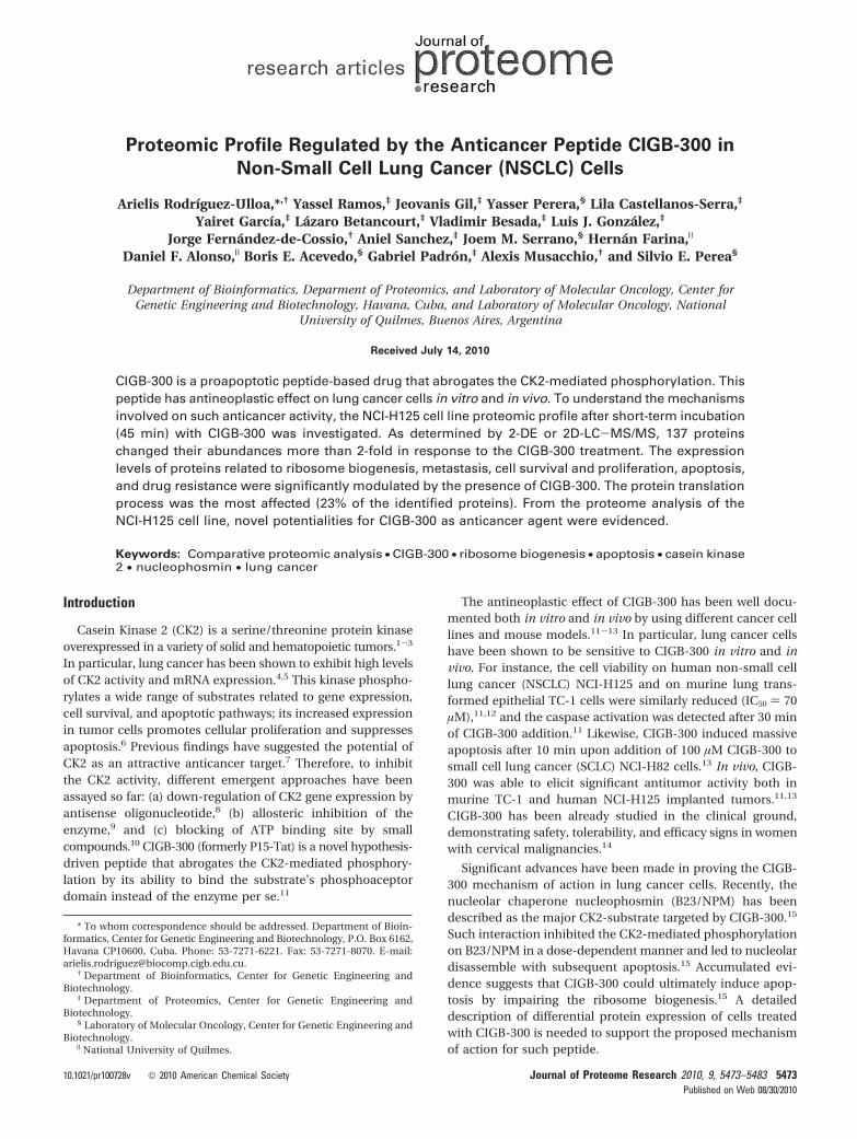
In a first approach, 2-DE in conjunction with quantitativeimage analysis and sequencing by mass spectrometry was usedto investigate the CIGB-300 modulated protein profile. TheFigure 1 shows the 2-D pattern for nuclear proteins of NCI-H125 cells cultured with CIGB-300 or medium alone during45 min. Between 1600 and 2000 spots were detected by imageanalysis in the three replicate analytical gels of each condition.A total of 26 differentially expressed spots were subjected totrypsin digestion and mass spectrometry analysis. The 22identified proteins that were up- or down-regulated more than2-fold after treatment with CIGB-300 are shown in Table 1. Theapparent relative molecular weight (Mr) and the isoelectric
point (pI) determined from 2-D calibrated gels for the identifiedproteins were compared with their corresponding theoreticalvalues. With the exception of LIM and SH3 domain protein 1(LASP1) and B23/NPM protein, all of the identified proteinsrevealed good correlation between both values (Table 1).
In the case of LASP-1 protein, the Mr of 36.4 kDa and pI of6.2 were detected in 2-D gels, similar to 34.5 kDa and pI 6.3values, previously reported.30 Two spots (T8 and T7), with Mr
of 16.0 and 17.7 kDa, corresponding to B23/NPM fragmentswere identified on CIGB-300-treated cells (Figure 1). Thisfinding suggested that CIGB-300 could promote partial B23/NPM degradation in the experimental condition, a relevantobservation, as such a protein was reported to be a major targetfor CIGB-300.15 Given the fact that B23/NPM stability isincreased by its CK2-mediated phosphorylation31 along withthe ability of CIGB-300 to abrogate such a process,15 B23/NPMdegradation is expected to occur, and it represents a subrogatedmarker of CIGB-300 functionality. However, a decreased ex-
Figure 1. Representative bidimensional electrophoresis gels derived from control and CIGB-300 treated cells. Proteins were separatedby combining IEF in IPG (pI range from 4 to 7) and SDS-PAGE (mass range from 12 to 120 kDa). Arrows indicate 26 spots significantlymodulated (fold change >2) by CIGB-300 treatment. The magnified regions (a-c, control condition; d-f, treated cells) show the effectof CIGB-300 on the spot coded as T7 corresponding to nucleophosmin (B23/NPM) fragment. The histogram of three replicates for eachconditions (a-f) is shown at the left of the magnified regions.
research articles Rodrıguez-Ulloa et al.
5476 Journal of Proteome Research • Vol. 9, No. 10, 2010

pression of intact B23/NPM protein (32.6 kDa) could not bedetermined by 2D gel image analysis; probably this protein spotwas oversaturated, avoiding the detection of differential expres-sion levels.
To increase the coverage of the proteome modulated byCIGB-300 in the nuclear fraction of NCI-H125 cells, a secondcomparative proteomic study using 2D-LC-MS/MS was per-formed. A total of 509 protein hits were identified (SupportingInformation), of which 118 hits, corresponding to 127 proteins,were differentially expressed in CIGB-300-treated cells (Table2). Interestingly, through this proteomic approach 12 of the 22proteins identified by 2-DE analysis were also identified; theexpression levels of these proteins were similarly modulatedby CIGB-300 in both experiments (Table 2). Thus, such afinding serves as an internal cross-validation between bothexperimental approaches.
Changes in two differentially expressed proteins were vali-dated through Western blot analysis of total cell extracts fromNCI-H125 cells. CIGB-300 down-regulated the B23/NPM ex-
pression in a dose-dependent manner with the highest rate at200 µM, the selected dose for proteomic studies (Figure 2).Although fragments of lowers apparent molecular weight fromB23/NPM protein were not detected, the decreased expressionof B23/NPM protein supported the detrimental effect of CIGB-300 on its stability. The increased expression of prohibitin(PHB) was also validated using the same experimental condi-tions. Similar to proteomic data, CIGB-300 up-regulated thePHB protein at 200 µM of CIGB-300; no significant effect wasobserved at lower peptide concentrations (Figure 2).
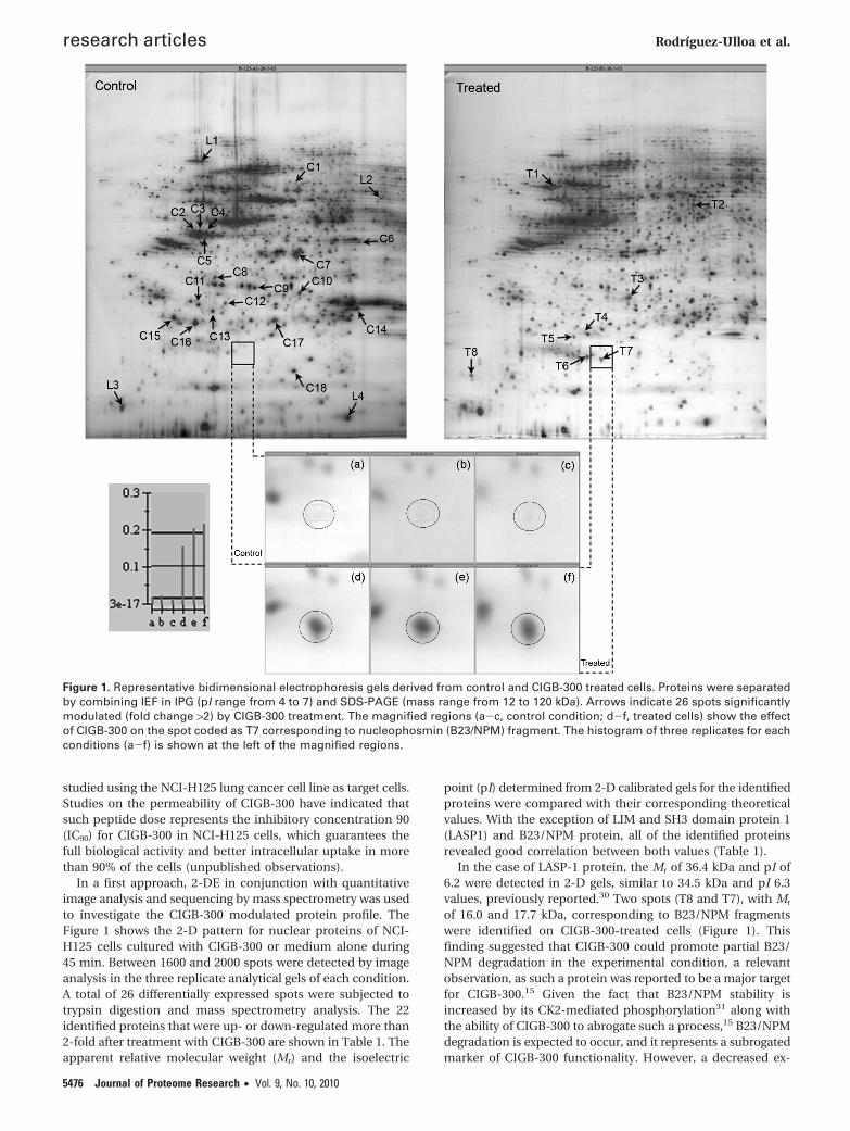
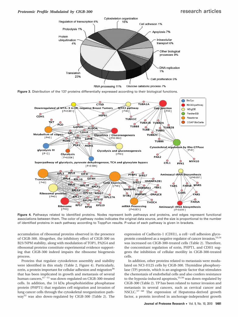
For better understanding of putative cellular processesaffected by CIGB-300 on this lung cancer cell line, the differ-entially expressed proteins were classified in terms of theirbiological functions using the information from the GeneOntology database. A considerable number of the identifiedproteins were involved in translation (23%), cytoskeletonorganization (18%), RNA processing (11%), and apoptosis (7%)(Figure 3). Pathway analysis demonstrated the enrichment ofmetabolic, cytoskeletal signaling, translation, and gene expres-
Table 1. Differentially Expressed Proteins in NCI-H125 Cells Treated with CIGB-300, Identified Using 2-DE and Mass SpectrometryApproach
spotno. UniProt_ACCa description
genesymbolb
% coverage(matchingpeptides)c Mr (kDa)d pId FCe
ApoptosisT3 P35232 Prohibitin PHB 51 (13) 29.8-26.1 5.6-5.5 2.5T7 P06748 Nucleophosmin (fragment) NPM1 36 (6) 32.6-17.7 4.6-5.3 11.0T8 P06748 Nucleophosmin (fragment) NPM1 23 (6) 32.6-16.0 4.6-4.2 8.0C11 P52565 Rho GDP-dissociation inhibitor 1 ARHGDIA 30 (3) 23.1-24.6 5.0-4.9 -5.3C15 P13693 Translationally controlled tumor protein TCTP 65 (6) 19.6-22.6 4.8-4.7 -5.2C16 Q04760 Lactoylglutathione lyase GLO1 84 (17) 20.6-21.9 5.1-4.9 -4.5C17 P09211 Glutathione S-transferase P GSTP1 66 (14) 23.2-22.3 5.4-5.5 -3.1C18 P00441 Superoxide dismutase SOD1 44 (5) 15.8-16.2 5.7-5.7 -6.8
Cell proliferationT2 O60884 DnaJ homologue subfamily A member 2 DNAJA2 50 (12) 45.7-44.5 6.1-6.0 3.0
Glucose catabolic processC7 P07195 L-lactate dehydrogenase B chain LDHB 35 (10) 36.5-33.7 5.7-5.7 -3.8C14 P60174 Triosephosphate isomerase TPI1 54 (11) 26.5-23.9 6.5-6.2 -2.9
RNA processingC1 P61978 Heterogeneous nuclear ribonucleoprotein K HNRNPK 20 (6) 51.0-53.0 5.4-5.7 -3.2C2 P07910 Heterogeneous nuclear ribonucleoproteins C1/C2 HNRNPC 23 (7) 33.5-39.2 4.9-4.8 -16.9C3 P07910 Heterogeneous nuclear ribonucleoproteins C1/C2 HNRNPC 39 (10) 33.5-39.2 4.9-4.9 -13.9C4 P07910 Heterogeneous nuclear ribonucleoproteins C1/C2 HNRNPC 18 (5) 33.5-39.0 4.9-5.0 -11.7C5 P07910 Heterogeneous nuclear ribonucleoproteins C1/C2 HNRNPC 32 (12) 33.5-37.8 4.9-5.0 -3.0
Regulation of transcriptionT4 P45973 Chromobox protein homologue 5 CBX5 24 (4) 22.2-20.7 5.7-5.1 5.4T6 Q13185 Chromobox protein homologue 3 CBX3 44 (5) 20.8-18.2 5.2-5.1 3.5
Protein ubiquitinationC9 Q9UL46 Proteasome activator complex subunit 2 PSME2 27.2-27.3 5.4-5.4 -3.2C10 Q06323 Proteasome activator complex subunit 1 PSME1 38 (9) 28.7-26.4 5.8-5.8 -2.3
Intracellular transportT5 Q9NP72 Ras-related protein Rab-18 RAB18 61 (10) 22.7-20.3 5.1-5.0 4.4C6 Q14847 LIM and SH3 domain protein 1 LASP1 29.7-36.4 6.6-6.2 -3.0
Cytoskeleton organizationT1 Q13509 Tubulin beta-3 chain TUBB3 34 (10) 50.4-50.9 4.8-4.9 4.2C8 Q15691 Microtubule-associated protein RP/EB family member 1 MAPRE1 37 (6) 29.9-29.2 5.0-5.0 -5.6C12 P43487 Ran-specific GTPase-activating protein RANBP1 11 (2) 23.2-24.8 5.2-5.1 -3.6C13 P52566 Rho GDP-dissociation inhibitor 2 ARHGDIB 26 (3) 22.8-23.6 5.1-5.0 -5.1
a UniProtKB/Swiss-Prot entry accession number (http://au.expaxy.org). b Recommended gene name (official gene symbol) as provided byUniProtKB/Swiss-Prot. c Protein sequence coverage and number of signals matching the theoretical mass of tryptic peptides (C6 and C9 spot wereidentified only by MS/MS sequencing). d Theoretical values were calculated using Expasy Compute pI/Mr tool (http://ca.expasy.org/tools/pi_tool.html)(signal peptides were not considered for Mr and pI calculations). Bold-faced numbers indicates Mr and pI experimental values. e Fold change (FC) ofdifferentially regulated proteins [(-) down-regulated].
Proteomic Profile Modulated by CIGB-300 research articles
Journal of Proteome Research • Vol. 9, No. 10, 2010 5477

Table 2. Differentially Expressed Proteins in NCI-H125 Cells Treated with CIGB-300, Identified Using 2D-LC-MS/MS
proteinhit IPI_ACCa UniProt_ACCa description
genesymbolb
% coverage(peptides)c FCd
Translation8 IPI00008529 P05387 60S acidic ribosomal protein P2 RPLP2 63.48 (5) 211 IPI00181728 Q8TDN6 Brix domain-containing protein 2 BXDC2 21.81 (3) 2.712 IPI00554723 P27635 60S ribosomal protein L10 RPL10 13.55 (2) 2.714 IPI00550021 P39023 60S ribosomal protein L3 RPL3 10.42 (3) 7.215 IPI00008530 P05388 60S acidic ribosomal protein P0 RPLP0 17.35 (2) 2.717 IPI00012772 P62917 60S ribosomal protein L8 RPL8 12.06 (2) 9.619 IPI00413324 P18621 60S ribosomal protein L17 RPL17 15.76 (3) 2.921 IPI00003918 P36578 60S ribosomal protein L4 RPL4 15.93 (4) 2.622 IPI00012795 Q13347 Eukaryotic translation initiation factor 3 subunit I EIF3I 24 (3) 225 IPI00745266 Q9Y262 Eukaryotic translation initiation factor 3 subunit L EIF3L 9.93 (3) 2.229 IPI00216587 P62241 40S ribosomal protein S8 RPS8 6.25 (1) 12.130 IPI00456758 P46776 60S ribosomal protein L27a RPL27A 8.78 (1) 3.131 IPI00555744 P50914 60S ribosomal protein L14 RPL14 5.45 (1) 3.734 IPI00329389 Q02878 60S ribosomal protein L6 RPLl6 5.21 (1) 9.243 IPI00215719 Q07020 60S ribosomal protein L18 RPL18 6.91 (1) 2.945 IPI00221093 P08708 40S ribosomal protein S17 RPS17 7.41 (1) 2.446 IPI00008708 O76021 Ribosomal L1 domain-containing protein 1 RSL1D1 4.08 (1) 54 IPI00025447 Q6IQ15 Elongation factor 1-alpha EEF1A1 33.33 (9) -26 IPI00014424 Q05639 Elongation factor 1-alpha 2 EEF1A2 34.34 (8) -215 IPI00008438 P46783 40S ribosomal protein S10 RPS10 32.73 (5) -2.525 IPI00013415 P62081 40S ribosomal protein S7 RPS7 27.32 (3) -2.536 IPI00783097 P41250 Glycyl-tRNA synthetase GARS 8.12 (3) -3.338 IPI00007074 P54577 Tyrosyl-tRNA synthetase, cytoplasmic YARS 10.61 (3) -2.5
41 IPI00816063 P30050 Isoform 2 of 60S ribosomal protein L12 RPL12 14.39 (1) OFFIPI00024933 Isoform 1 of 60S ribosomal protein L12 11.52 (1)
46 IPI00295400 P23381 Tryptophanyl-tRNA synthetase, cytoplasmic WARS 4.03 (1) -5IPI00412737 Tryptophanyl-tRNA synthetase isoform b 4.42 (1)50 IPI00329633 P26639 Threonyl-tRNA synthetase, cytoplasmic TARS 8.85 (3) -3.352 IPI00300074 Q9NSD9 Phenylalanyl-tRNA synthetase beta chain FARSB 5.43 (2) -253 IPI00031820 Q9Y285 Phenylalanyl-tRNA synthetase alpha chain FARSA 4.72 (1) -255 IPI00215780 P39019 40S ribosomal protein S19 RPS19 10.34 (2) -3.3
69 IPI00032635 Q9BX40 Isoform 1 of Protein LSM14 homologue B LSM14B 6.75 (1) -3.3IPI00471933 Isoform 3 of Protein LSM14 homologue B 10.88 (1)
71 IPI00375127 Q15056 Similar to mKIAA0038 protein EIF4H 7.82 (2) -3.3IPI00014263 Isoform Long of Eukaryotic translation initiation factor 4H 9.27 (2)
Apoptosis35 IPI00000816 P62258 14-3-3 protein epsilon YWHAE 16.08 (2) 2.617 IPI00025512 P04792 Heat shock protein beta-1 HSPB1 33.66 (3) -3.321 IPI00218733 P00441 Superoxide dismutase [Cu-Zn] SOD1 44.81 (2) OFF26 IPI00003815 P52565 Rho GDP-dissociation inhibitor 1 ARHGDIA 24.02 (2) -1037 IPI00550900 P13693 Translationally controlled tumor protein TCTP 15.12 (2) -3.340 IPI00220766 Q04760 Lactoylglutathione lyase GLO1 20.65 (2) -564 IPI00026260 P22392 Isoform 1 of Nucleoside diphosphate kinase B NME2 9.87 (1) -2.5
IPI00795292 Isoform 3 of Nucleoside diphosphate kinase B 5.62 (1)72 IPI00219757 P09211 Glutathione S-transferase P GSTP1 9.52 (2) -10
Cell proliferation16 IPI00441473 O14744 Protein arginine N-methyltransferase 5 PRMT5 15.70 (6) 232 IPI00032406 O60884 DnaJ homologue subfamily A member 2 DNAJA2 7.04 (1) 2.833 IPI00395865 Q16576 Histone-binding protein RBBP7 RBBP7 12 (2) 2.344 IPI00013953 Q9NNW5 WD repeat-containing protein 6 WDR6 1.25 (1) 4.220 IPI00020956 P51858 Hepatoma-derived growth factor HDGF 18.33 (2) -2
22 IPI00014197 Q9UKY7 Isoform 1 of Protein CDV3 homologue CDV3 31.40 (2) OFFIPI00798278 Isoform 2 of Protein CDV3 homologue 38.03 (2)30 IPI00307226 P01588 Erythropoietin EPO 12.44 (2) OFF
Glucose catabolic process1 IPI00219018 P04406 Glyceraldehyde-3-phosphate dehydrogenase GAPDH 71.94 (16) -10
2 IPI00479186 P14618 Isoform M2 of Pyruvate kinase isozymes M1/M2 PKM2 54.24 (17) -10IPI00220644 Isoform M1 of Pyruvate kinase isozymes M1/M2 51.04 (15)3 IPI00465439 P04075 Fructose-bisphosphate aldolase A ALDOA 43.96 (8) -105 IPI00169383 P00558 Phosphoglycerate kinase 1 PGK1 35.49 (9) -57 IPI00027497 P06744 Glucose-6-phosphate isomerase GPI 29.03 (8) -10
8 IPI00451401 P60174 Isoform 2 of Triosephosphate isomerase TPI1 35.74 (6) -10IPI00797270 Isoform 1 of Triosephosphate isomerase 47.39 (7)
9 IPI00607708 P00338 Isoform 2 of L-lactate dehydrogenase A chain LDHA 33.43 (6) -10IPI00217966 L-lactate dehydrogenase 30.75 (6)16 IPI00219217 P07195 L-lactate dehydrogenase B chain LDHB 14.37 (4) -544 IPI00916111 P40925 Malate dehydrogenase MDH1 9.38 (2) -2.565 IPI00219525 P52209 6-phosphogluconate dehydrogenase, decarboxylating PGD 3.93 (1) -5
RNA processing23 IPI00026089 O75533 Splicing factor 3B subunit 1 SF3B1 3.53 (3) 224 IPI00217686 Q8IY81 Putative rRNA methyltransferase 3 FTSJ3 5.43 (2) 6.8
27IPI00218592
Q07955Isoform ASF-3 of Splicing factor, arginine/serine-rich 1
SFRS121.39 (2)
2IPI00215884 Isoform ASF-1 of Splicing factor, arginine/serine-rich 1 17.34 (2)IPI00218591 Isoform ASF-2 of Splicing factor, arginine/serine-rich 1 14.73 (2)
research articles Rodrıguez-Ulloa et al.
5478 Journal of Proteome Research • Vol. 9, No. 10, 2010

Table 2. Continued
proteinhit IPI_ACCa UniProt_ACCa description
genesymbolb
% coverage(peptides)c FCd
36 IPI00003377 Q16629 Isoform 1 of Splicing factor, arginine/serine-rich 7 SFRS7 8.82 (1) 3.1
40IPI00790699
O15042Isoform 3 of U2-associated protein SR140
SR1402.91 (2)
2.3IPI00143753 Isoform 1 of U2-associated protein SR140 2.82 (2)IPI00829908 Isoform 2 of U2-associated protein SR140 2.82 (2)
10
IPI00215965P09651
Isoform A1-B of Heterogeneous nuclear ribonucleoprotein A1HNRNPA1
24.73 (7)
-3.3IPI00797148 Isoform 2 of Heterogeneous nuclear ribonucleoprotein A1 34.46 (7)IPI00465365 Isoform A1-A of Heterogeneous nuclear ribonucleoprotein A1 28.75 (7)IPI00644968 P0C7M2 Putative heterogeneous nuclear ribonucleoprotein A1-like protein 3 HNRPA1L3 28.75 (7)
11IPI00414696
P22626Isoform A2 of Heterogeneous nuclear ribonucleoproteins A2/B1
HNRNPA2B133.14 (7) -2
IPI00396378 Isoform B1 of Heterogeneous nuclear ribonucleoproteins A2/B1 32.01 (7)
24IPI00179964
P26599Isoform 1 of Polypyrimidine tract-binding protein 1
PTBP115.82 (4) -2.5
IPI00334175 Isoform 2 of Polypyrimidine tract-binding protein 1 15.27 (4)
27
IPI00412714
Q8NC51
Isoform 4 of Plasminogen activator inhibitor 1 RNA-binding protein
SERBP1
11.89 (2)
-5IPI00470497 Isoform 2 of Plasminogen activator inhibitor 1 RNA-binding protein 3.98 (1)IPI00470498 Isoform 3 of Plasminogen activator inhibitor 1 RNA-binding protein 11.70 (2)IPI00410693 Isoform 1 of Plasminogen activator inhibitor 1 RNA-binding protein 3.92 (1)
54 IPI00328840 Q86V81 THO complex 4 THOC4 9.85 (1) -3.358 IPI00003438 O75937 DnaJ homologue subfamily C member 8 DNAJC8 11.46 (1) -559 IPI00299000 Q9UQ80 Proliferation-associated protein 2G4 PA2G4 8.12 (2) -2
Regulation of transcription
20IPI00513773
Q6NZI2Isoform 2 of Polymerase I and transcript release factor
PTRF14.33 (2)
2.4IPI00176903 Isoform 1 of Polymerase I and transcript release factor 11.03 (2)
31 IPI00872855 Q01844 RNA-binding protein EWS EWSR1 6.40 (2) -2.5
34IPI00853059
Q96AE4Isoform 2 of Far upstream element-binding protein 1
FUBP19.17 (3) -5
IPI00375441 Isoform 1 of Far upstream element-binding protein 1 6.06 (2)42 IPI00234252 Q92922 SWI/SNF complex subunit SMARCC1 SMARCC1 3.17 (2) -1045 IPI00031812 P67809 Nuclease-sensitive element-binding protein 1 YBX1 9.26 (1) -3.367 IPI00171798 O94776 Metastasis-associated protein MTA2 MTA2 4.64 (1) OFF
DNA replication60 IPI00413611 P11387 DNA topoisomerase 1 TOP1 3.66 (2) -270 IPI00292858 P19971 Thymidine phosphorylase TYMP 3.11 (1) -3.3
Protein ubiquitination39 IPI00384051 Q9UL46 Proteasome activator complex subunit 2 PSME2 12.85 (2) -2.548 IPI00645078 P22314 Ubiquitin-like modifier-activating enzyme 1 UBA1 2.36 (2) -2.551 IPI00395627 Q9HB71 Isoform 1 of Calcyclin-binding protein CACYBP 9.21 (1) -562 IPI00479722 Q06323 Proteasome activator complex subunit 1 PSME1 9.24 (1) -3.363 IPI00029623 P60900 Proteasome subunit alpha type-6 PSMA6 7.32 (1) -568 IPI00219913 P54578 Ubiquitin carboxyl-terminal hydrolase 14 USP14 5.87 (1) -3.3
Proteolysis32 IPI00011229 P07339 Cathepsin D CTSD 10.19 (3) -3.361 IPI00026216 P55786 Puromycin-sensitive aminopeptidase NPEPPS 3.16 (2) -2
Intracellular transport26 IPI00001639 Q14974 Importin subunit beta-1 KPNB1 5.37 (3) 239 IPI00017376 Q15437 Protein transport protein Sec23B SEC23B 3 (1) 2.2
12IPI00010154 P31150 Rab GDP dissociation inhibitor alpha GDI1 4.47 (2) -5IPI00645255 Q5SX87 GDP dissociation inhibitor 2 GDI2 25.10 (5)
23IPI00303882
O60664Isoform B of Mannose-6-phosphate receptor-binding protein 1
M6PRBP120.74 (4) -5
IPI00106668 Isoform A of Mannose-6-phosphate receptor-binding protein 1 29.88 (3) -1033 IPI00643041 P62826 GTP-binding nuclear protein Ran RAN 14.81 (2) -3.3
Cytoskeleton organization1 IPI00556012 Q5BKV1 Myosin, heavy chain 9, nonmuscle MYH9 43.58 (9) 22 IPI00007752 P68371 Tubulin beta-2C chain TUBB2C 55.06 (16) 2.43 IPI00909140 P07437 Tubulin beta chain TUBB 54.96 (10) 2.8
4IPI00013475 Q13885 Tubulin beta-2A chain TUBB2A
36.18 (11) 2.5IPI00031370 Q9BVA1 Tubulin beta-2B chain TUBB2B
5 IPI00013683 Q13509 Tubulin beta-3 chain TUBB3 44.89 (12) 2.7
6IPI00180675 Q71U36 Tubulin alpha-1A chain TUBA1A 39.25 (13)
2.3IPI00218343 Q9BQE3 Tubulin alpha-1C chain TUBA1C 33.41 (12)IPI00930688 P68363 Tubulin alpha-1B chain TUBA1B 42.35 (14)
7 IPI00646779 Q9BUF5 Tubulin beta-6 chain TUBB6 19.69 (6) 2.89 IPI00292496 Q3ZCM7 Tubulin beta-8 chain TUBB8 10.59 (3) 2.3
10IPI00397526
P35580Isoform 1 of Myosin-10
MYH106.02 (8)
2.1IPI00790503 Isoform 3 of Myosin-10 5.96 (8)IPI00479307 Isoform 2 of Myosin-10 5.97 (8)
18 IPI00017454 Q9H853 Putative tubulin-like protein alpha-4B TUBA4B 10.79 (2) 2
28IPI00220573 P19105 Myosin regulatory light chain 12A MYL12A 10.53 (1)
2IPI00033494 O14950 Myosin regulatory light chain 12B MYL12B 10.47 (1)IPI00220278 P24844 Myosin regulatory light polypeptide 9 MYL9 10.47 (1)
41 IPI00335168 P60660 Myosin light polypeptide 6 MYL6 10.60 (1) 2.318 IPI00216691 P07737 Profilin-1 PFN1 55.71 (4) -3.328 IPI00843975 P15311 Ezrin EZR 7.17 (3) -5
Proteomic Profile Modulated by CIGB-300 research articles
Journal of Proteome Research • Vol. 9, No. 10, 2010 5479

sion processes in the identified data set (Figure 4). Pathwaysare referred to according to annotations derived from differentdatabases (Figure 4). Particularly, metabolic pathways arerelated to glycolysis and gluconeogenesis, whereas cytoskeletalproteins are mainly related to Gap junction and Rho GTPasesignaling pathways.
Differentially expressed proteins related to the apoptosisprocess support the pro-apoptotic function of CIGB-300.Among these proteins, PHB is a negative regulator of cellproliferation that represses the E2F-mediated transcriptionpathway and activates the p53 transcriptional activity.32,33
Thereby PHB has been described as a potential tumor sup-pressor protein. In this study, PHB expression was increasedby CIGB-300 treatment (Table 1), such up-regulation wasvalidated using Western blot analysis as referred above (Figure2). Furthermore, a decrease of the translationally controlled
tumor protein (TCTP) level was observed on CIGB-300 treatedcells as confirmed through the two proteomic approaches usedin this study (Table 1 and Table 2). TCTP is an antiapoptoticprotein;34 previous studies have demonstrated that down-regulation of TCTP is related with suppression of tumormalignant phenotype.35
From a mechanistic view, the down-regulation of B23/NPMprotein induced by CIGB-300 is of great relevance (Figure 2).This nucleolar protein promotes cell survival through theinhibition of distinct pro-apoptotic pathways including theinactivation of the tumors suppressors IRF1 and p53.36,37
Besides, B23/NPM regulates the ribosome biogenesis pathwayby several mechanisms including the processing of pre-rRNAmolecule in the internal transcribed spacer (ITS2) sequenceto form the 5.8S and 28S rRNA.38 In addition, B23/NPM elicitschaperone activity preventing protein aggregation at the nu-cleolus during ribosome assembly,39 and mediating the nucleo-cytoplasmic transport of both ribosomal subunits.40
Besides B23/NPM, two other proteins related to ribosomebiogenesis were down-regulated 2-fold in the presence of CIGB-300: DNA topoisomerase I (TOP1) and proliferation-associatedprotein 2G4 (PA2G4) (Table 2). TOP1 regulates rRNA transcrip-tion.41 This protein catalyzes the transient breaking and rejoin-ing of a single strand of DNA; such enzymatic function of TOP1has been demonstrated to be activated by CK2 phosphoryla-tion.42 PA2G4 regulates the intermediate and late steps of rRNAprocessing and also contributes to maturation of the 60Sribosomal subunit.43 Furthermore, PA2G4 represses the inhibi-tory phosphorylation of eukaryotic initiation factor 2 R (eIF2R),exerting as a consequence a positive regulation on proteintranslation.44 In line with these findings, ribosomal proteinsas well as different aminoacyl-tRNA synthetases were modu-lated by CIGB-300 treatment (Table 2, Figure 4). Ribosomalproteins failing to assemble into ribosome subunits undergoproteosomal degradation.45 However, a set of proteins relatedto ubiquitin dependent degradation was down-regulated onCIGB-300-treated cells (Table 2). Therefore, the expecteddecrease of proteasome activity would explain the nuclear
Table 2. Continued
proteinhit IPI_ACCa UniProt_ACCa description
genesymbolb
% coverage(peptides)c FCd
35 IPI00419979 Q13177 Serine/threonine-protein kinase PAK 2 PAK2 5.90 (1) -1043 IPI00012011 P23528 Cofilin-1 CFL1 25.30 (2) -1049 IPI00028091 P61158 Actin-related protein 3 ACTR3 12.20 (2) -3.357 IPI00003817 P52566 Rho GDP-dissociation inhibitor 2 ARHGDIB 12.44 (1) OFF
Cell adhesion42 IPI00025861 P12830 Cadherin-1 CDH1 2.27 (1) 2.1
Others
13 IPI00215637 O00571 ATP-dependent RNA helicase DDX3X DDX3X 13.60 (5) 2.0IPI00293616 O15523 ATP-dependent RNA helicase DDX3Y DDX3Y 10.30 (4)37 IPI00376379 Q7Z794 Keratin, type II cytoskeletal 1b KRT77 4.67 (2) 2.438 IPI00021304 P35908 Keratin, type II cytoskeletal 2 epidermal KRT2 4.19 (2) 2.613 IPI00003865 P11142 Isoform 1 of Heat shock cognate 71 kDa protein HSPA8 14.86 (8) -2
14 IPI00792641 B3KSI4 Transketolase isoform 2 TKT 18.70 (5) -5IPI00643920 B4DE31 cDNA FLJ54957, highly similar to Transketolase 16.01 (5)19 IPI00746165 O75083 Isoform 1 of WD repeat-containing protein 1 WDR1 16.17 (4) -529 IPI00289499 P31939 Bifunctional purine biosynthesis protein PURH ATIC 10.30 (3) -2.547 IPI00465431 P17931 Galectin-3 LGALS3 11.60 (2) -2.556 IPI00299977 Q9NRX4 14 kDa phosphohistidine phosphatase PHPT1 16.80 (1) -2.0
66 IPI00644196 P36952 Isoform 2 of Serpin B5 SERPINB5 7.36 (1) -2.5IPI00783625 Isoform 1 of Serpin B5 4.53 (1)
a Accession numbers in IPI (International Protein Index) (http://www.ebi.ac.uk/IPI) and UniProtKB/Swiss-Prot databases (http://au.expaxy.org) TheUniProt accession numbers highlighted in bold indicate those proteins (12) that were also identified by 2-DE analysis. b Recommended gene name (officialgene symbol) as provided by UniProtKB/Swiss-Prot. c Protein sequence coverage and number of identified peptides. d Fold change (FC) of differentiallyregulated proteins [(-) down-regulated].
Figure 2. Validation of nucleophosmin (B23/NPM) and prohibitin(PHB) expression levels by Western blot analysis. NCI-H125 cellswere treated with different concentrations of CIGB-300 during40 min. Total cell lysates were separated on a SDS-PAGE gel andimmunoblotting was performed with anti-B23/NPM and anti-PHBantibodies. �-Actin expression was used as control.
research articles Rodrıguez-Ulloa et al.
5480 Journal of Proteome Research • Vol. 9, No. 10, 2010
accumulation of ribosomal proteins observed in the presenceof CIGB-300. Altogether, the inhibitory effect of CIGB-300 onB23/NPM stability, along with modulation of TOP1, PA2G4 andribosomal proteins constitute experimental evidence support-ing that CIGB-300 indeed impairs the ribosome biogenesisprocess.
Proteins that regulate cytoskeleton assembly and stabilitywere identified in this study (Table 2, Figure 4). Particularly,ezrin, a protein important for cellular adhesion and migration46
that has been implicated in growth and metastasis of severalhuman cancers,47-51 was down-regulated on CIGB-300-treatedcells. In addition, the 14 kDa phosphohistidine phosphataseprotein (PHPT1) that regulates cell migration and invasion oflung cancer cells through the cytoskeletal reorganization path-way52 was also down-regulated by CIGB-300 (Table 2). The
expression of Cadherin-1 (CDH1), a cell-cell adhesion glyco-protein considered as a negative regulator of cancer invasion,53,54
was increased on CIGB-300-treated cells (Table 2). Therefore,the concomitant regulation of ezrin, PHPT1, and CDH1 sug-gests the inhibition of cellular motility in CIGB-300-treatedcells.
In addition, other proteins related to metastasis were modu-lated on NCI-H125 cells by CIGB-300. Thymidine phosphory-lase (TP) protein, which is an angiogenic factor that stimulatesthe chemotaxis of endothelial cells and also confers resistanceto the hypoxia-induced apoptosis,55,56 was down-regulated byCIGB-300 (Table 2). TP has been related to tumor invasion andmetastasis in several cancers, such as cervical cancer andNSCLC.57-60 The expression of Hepatoma-derived growthfactor, a protein involved in anchorage-independent growth
Figure 3. Distribution of the 137 proteins differentially expressed according to their biological functions.
Figure 4. Pathways related to identified proteins. Nodes represent both pathways and proteins, and edges represent functionalassociations between them. The color of pathway nodes indicates the original data source, and the size is proportional to the numberof identified proteins in each pathway according to ToppFun results. P-value of each pathway is given in brackets.
Proteomic Profile Modulated by CIGB-300 research articles
Journal of Proteome Research • Vol. 9, No. 10, 2010 5481

and cell invasion of NSCLC,61 was also decreased on CIGB-300-treated cells (Table 2). Furthermore, Glucose-6-phosphateisomerase, which functions as an extracellular cytokine stimu-lating cell motility,62 was down-regulated by CIGB-300 (Table2), while the Chromobox protein homologue 5, implicated insilencing metastasis-related genes,63 was up-regulated onCIGB-300-treated cells (Table 1). Finally, the expression ofCathepsin D was decreased by CIGB-300 (Table 2). This proteinregulates cell proliferation, invasion and metastasis.64,65 Thus,the modulation of these proteins supports a potential antime-tastasic effect of CIGB-300. Experiments in our lab are evaluat-ing this hypothesis.
Cancer cells use glycolysis, even in aerobic conditions, toproduce ATP and intermediaries metabolites for anabolicreactions.66 Eight glycolytic proteins were down-regulated byCIGB-300 treatment in this study (Tables 1 and 2, Figure 4).Besides, the enzyme 6-phosphogluconate dehydrogenase, whichcatalyzes the third step of the pentose phosphate pathwayoxidative branch, was down-regulated 5-fold by CIGB-300. Theantioxidant defense system is essential for cancer cell survival.67,68
The expression of the enzyme superoxide dismutase, whichcatalyzes the reduction of superoxide to hydrogen peroxide,was decreased on CIGB-300-treated cells (Tables 1 and 2).These results support the inhibitory effect of CIGB-300 on theproliferation of tumor cells. As the exacerbation of the glycolyticpathway has been considered an alternative way of tumor cellsto resist chemotherapy,69 our findings suggest that CIGB-300could add a benefit to the standard anticancer therapies. Inline with this hypothesis, Glutathione S-transferase P andLactoylglutathione lyase, two glutathione conjugation proteinsrelated to chemotherapy resistance,70,71 were down-regulatedon CIGB-300-treated NCI-H125 cells (Table 2). Additionally, thedown-regulation of the Nuclease-sensitive element-bindingprotein 1 (YB-1) on CIGB-300-treated cells (Table 2) is of greatimportance as this factor activates the expression of theP-glycoprotein 1 coding gene related to the multidrug-resistantphenotype.72,73 Furthermore, YB-1 participates in the nucle-otide excision repair pathway and plays a role in the cisplatin-DNA adducts reparation.74 This protein is overexpressed incisplatin-resistant cells.75 Besides the role of TP protein inmetastasis, this protein, superoxide dismutase, and heat shockprotein �-1 have been also related to cisplatin resistantphenotype on tumor cells.76-79 These three proteins weredown-regulated on CIGB-300-treated cells (Table 2). The find-ings observed in this study suggest that combination of CIGB-300 with some anticancer drugs may produce a synergisticinteraction with increased overall antitumor effect.
Conclusions
This study is the first proteomic analysis on cancer cellstreated with the anticancer peptide CIGB-300. A total of 137differentially expressed proteins were identified by two comple-mentary proteomics approaches. CIGB-300 seems to promotethe B23/NPM instability on NCI-H125 cells and the impairmentof the ribosomal biogenesis and translation process. Such dataare in line with previous findings demonstrating that CIGB-300 leads to apoptosis by blocking the CK2-mediated phos-phorylation on B23/NPM with subsequent interference withthe nucleolar assembly. Furthermore, other proteins implicatedin apoptosis and cell proliferation were found modulated likelyas a consequence from the above-mentioned effect of CIGB-300. These proteins could represent subrogate biomarkers ofthe CIGB-300 effect on tumor cells. Proteins related to glyco-
lysis, antioxidant defense, angiogenesis, and metastasis weremodulated on CIGB-300-treated cells, giving important cluesabout potential anticancer effects of CIGB-300 other than thepro-apoptotic action. CIGB-300 decreases the expression levelof proteins implicated in chemotherapy resistance. Thus, suchfindings could support the combination of CIGB-300 withstandard chemotherapeutic drugs to improve the antitumoralresponse. Overall, the dissection of the CIGB-300 modulatedproteome not only documented the pathways supporting thedemonstrated anticancer effect of CIGB-300 in lung cancer cellsbut also uncovered other potentialities of this drug for cancertherapy.
Acknowledgment. This work was supported byHeberBiotec SA and Biorec. The Proteomic and BioinformaticsDepartments at the Center for Genetic Engineering andBiotechnology would like to thank to INSPUR Company(China) for their kind donation of a computer cluster used fordata storage and processing.
Supporting Information Available: A complete tableof identified proteins. This material is available free of chargevia the Internet at http://pubs.acs.org.
References(1) Laramas, M.; Pasquier, D.; Filhol, O.; Ringeisen, F.; Descotes, J. L.;
Cochet, C. Eur. J. Cancer 2007, 43 (5), 928–934.(2) Landesman-Bollag, E.; Romieu-Mourez, R.; Song, D. H.; Sonen-
shein, G. E.; Cardiff, R. D.; Seldin, D. C. Oncogene 2001, 20 (25),3247–3257.
(3) Kim, J. S.; Eom, J. I.; Cheong, J. W.; Choi, A. J.; Lee, J. K.; Yang,W. I.; Min, Y. H. Clin. Cancer Res. 2007, 13 (3), 1019–1028.
(4) Daya-Makin, M.; Sanghera, J. S.; Mogentale, T. L.; Lipp, M.;Parchomchuk, J.; Hogg, J. C.; Pelech, S. L. Cancer Res. 1994, 54(8), 2262–2268.
(5) O-charoenrat, P.; Rusch, V.; Talbot, S. G.; Sarkaria, I.; Viale, A.;Socci, N.; Ngai, I.; Rao, P.; Singh, B. Clin. Cancer Res. 2004, 10(17), 5792–5803.
(6) Trembley, J. H.; Wang, G.; Unger, G.; Slaton, J.; Ahmed, K. Cell.Mol. Life Sci. 2009, 66 (11-12), 1858–1867.
(7) Unger, G. M.; Davis, A. T.; Slaton, J. W.; Ahmed, K. Curr. CancerDrug Targets 2004, 4 (1), 77–84.
(8) Wang, G.; Unger, G.; Ahmad, K. A.; Slaton, J. W.; Ahmed, K. Mol.Cell. Biochem. 2005, 274 (1-2), 77–84.
(9) Hung, M. S.; Xu, Z.; Lin, Y. C.; Mao, J. H.; Yang, C. T.; Chang, P. J.;Jablons, D. M.; You, L. BMC Cancer 2009, 9, 135.
(10) Sarno, S.; Reddy, H.; Meggio, F.; Ruzzene, M.; Davies, S. P.; Donella-Deana, A.; Shugar, D.; Pinna, L. A. FEBS Lett. 2001, 496 (1), 44–48.
(11) Perea, S. E.; Reyes, O.; Puchades, Y.; Mendoza, O.; Vispo, N. S.;Torrens, I.; Santos, A.; Silva, R.; Acevedo, B.; Lopez, E.; Falcon, V.;Alonso, D. F. Cancer Res. 2004, 64 (19), 7127–7129.
(12) Perea, S. E.; Reyes, O.; Baladron, I.; Perera, Y.; Farina, H.; Gil, J.;Rodriguez, A.; Bacardi, D.; Marcelo, J. L.; Cosme, K.; Cruz, M.;Valenzuela, C.; Lopez-Saura, P. A.; Puchades, Y.; Serrano, J. M.;Mendoza, O.; Castellanos, L.; Sanchez, A.; Betancourt, L.; Besada,V.; Silva, R.; Lopez, E.; Falcon, V.; Hernandez, I.; Solares, M.;Santana, A.; Diaz, A.; Ramos, T.; Lopez, C.; Ariosa, J.; Gonzalez,L. J.; Garay, H.; Gomez, D.; Gomez, R.; Alonso, D. F.; Sigman, H.;Herrera, L.; Acevedo, B. Mol. Cell. Biochem. 2008, 316 (1-2), 163–167.
(13) Perera, Y.; Farina, H. G.; Hernandez, I.; Mendoza, O.; Serrano, J. M.;Reyes, O.; Gomez, D. E.; Gomez, R. E.; Acevedo, B. E.; Alonso, D. F.;Perea, S. E. Int. J. Cancer 2008, 122 (1), 57–62.
(14) Solares, A. M.; Santana, A.; Baladron, I.; Valenzuela, C.; Gonzalez,C. A.; Diaz, A.; Castillo, D.; Ramos, T.; Gomez, R.; Alonso, D. F.;Herrera, L.; Sigman, H.; Perea, S. E.; Acevedo, B. E.; Lopez-Saura,P. BMC Cancer 2009, 9, 146.
(15) Perera, Y.; Farina, H. G.; Gil, J.; Rodriguez, A.; Benavent, F.;Castellanos, L.; Gomez, R. E.; Acevedo, B. E.; Alonso, D. F.; Perea,S. E. Mol. Cancer Ther. 2009, Epub ahead of print. DOI:, 10.1158/1535-7163.MCT-08-1056.
(16) Fields, G. B.; Noble, R. L. Int. J. Pept. Protein Res. 1990, 35 (3),161–214.
(17) Klose, J.; Kobalz, U. Electrophoresis 1995, 16 (6), 1034–1059.
research articles Rodrıguez-Ulloa et al.
5482 Journal of Proteome Research • Vol. 9, No. 10, 2010

(18) Heukeshoven, J.; Dernick, R. J. Chromatogr. 1985, 326, 91–101.(19) Jensen, O. N.; Wilm, M.; Shevchenko, A.; Mann, M. Sample
preparation methods for mass spectrometric peptide mappingdirectly from 2-DE gels. In 2-D Proteome Analysis Protocols; Link,A. J., Ed.; Human Press: Totowa, NJ, 1999; pp 513-530.
(20) Gharahdaghi, F.; Weinberg, C. R.; Meagher, D. A.; Imai, B. S.;Mische, S. M. Electrophoresis 1999, 20 (3), 601–605.
(21) Gonzalez, L. J.; Castellanos-Serra, L.; Badock, V.; Diaz, M.; Moro,A.; Perea, S.; Santos, A.; Paz-Lago, D.; Otto, A.; Muller, E. C.; Kostka,S.; Wittmann-Liebold, B.; Padron, G. Electrophoresis 2003, 24 (1-2), 237–252.
(22) Perkins, D. N.; Pappin, D. J.; Creasy, D. M.; Cottrell, J. S. Electro-phoresis 1999, 20 (18), 3551–3567.
(23) Elias, J. E.; Gygi, S. P. Nat. Methods 2007, 4 (3), 207–214.(24) Fernandez-de-Cossio, J.; Gonzalez, L. J.; Satomi, Y.; Betancourt,
L.; Ramos, Y.; Huerta, V.; Amaro, A.; Besada, V.; Padron, G.;Minamino, N.; Takao, T. Nucleic Acids Res. 2004, 32 (Web Serverissue), W674–W678.
(25) Fernandez-de-Cossio, J.; Gonzalez, L. J.; Satomi, Y.; Betancourt,L.; Ramos, Y.; Huerta, V.; Besada, V.; Padron, G.; Minamino, N.;Takao, T. Rapid Commun. Mass Spectrom. 2004, 18 (20), 2465–2472.
(26) Dennis, G., Jr.; Sherman, B. T.; Hosack, D. A.; Yang, J.; Gao, W.;Lane, H. C.; Lempicki, R. A. Genome Biol 2003, 4 (5), 3.
(27) Huang, d. W.; Sherman, B. T.; Lempicki, R. A. Nat. Protoc. 2009, 4(1), 44–57.
(28) Chen, J.; Bardes, E. E.; Aronow, B. J.; Jegga, A. G. Nucleic AcidsRes. 2009, 37 (Web Server issue), W305–W311.
(29) Shannon, P.; Markiel, A.; Ozier, O.; Baliga, N. S.; Wang, J. T.;Ramage, D.; Amin, N.; Schwikowski, B.; Ideker, T. Genome Res.2003, 13 (11), 2498–2504.
(30) Demalte-Annessi, I.; Sanchez, J.-C.; Hoogland, C.; Rouge, V.; Binz,P.-A.; Appel, R. D.; Hochstrasser, D. F. SWISS-2DPAGE databaseentry. http://www.expasy.org/swiss-2dpage/protein/ac)Q14847(accessed Apr, 2010).
(31) Tawfic, S.; Olson, M. O.; Ahmed, K. J. Biol. Chem. 1995, 270 (36),21009–21015.
(32) Wang, S.; Zhang, B.; Faller, D. V. EMBO J. 2002, 21 (12), 3019–3028.
(33) Fusaro, G.; Dasgupta, P.; Rastogi, S.; Joshi, B.; Chellappan, S. J. Biol.Chem. 2003, 278 (48), 47853–47861.
(34) Liu, H.; Peng, H. W.; Cheng, Y. S.; Yuan, H. S.; Yang-Yen, H. F.Mol. Cell. Biol. 2005, 25 (8), 3117–3126.
(35) Tuynder, M.; Susini, L.; Prieur, S.; Besse, S.; Fiucci, G.; Amson, R.;Telerman, A. Proc. Natl. Acad. Sci. U.S.A. 2002, 99 (23), 14976–14981.
(36) Kondo, T.; Minamino, N.; Nagamura-Inoue, T.; Matsumoto, M.;Taniguchi, T.; Tanaka, N. Oncogene 1997, 15 (11), 1275–1281.
(37) Li, J.; Zhang, X.; Sejas, D. P.; Bagby, G. C.; Pang, Q. J. Biol. Chem.2004, 279 (40), 41275–41279.
(38) Savkur, R. S.; Olson, M. O. Nucleic Acids Res. 1998, 26 (19), 4508–4515.
(39) Szebeni, A.; Hingorani, K.; Negi, S.; Olson, M. O. J. Biol. Chem.2003, 278 (11), 9107–9115.
(40) Ahlemann, M.; Zeidler, R.; Lang, S.; Mack, B.; Munz, M.; Gires, O.Mol. Carcinog. 2006, 45 (12), 957–967.
(41) Christensen, M. O.; Barthelmes, H. U.; Boege, F.; Mielke, C. J. Biol.Chem. 2002, 277 (39), 35932–35938.
(42) Durban, E.; Goodenough, M.; Mills, J.; Busch, H. EMBO J. 1985, 4(11), 2921–2926.
(43) Squatrito, M.; Mancino, M.; Donzelli, M.; Areces, L. B.; Draetta,G. F. Oncogene 2004, 23 (25), 4454–4465.
(44) Squatrito, M.; Mancino, M.; Sala, L.; Draetta, G. F. Biochem.Biophys. Res. Commun. 2006, 344 (3), 859–868.
(45) Lam, Y. W.; Lamond, A. I.; Mann, M.; Andersen, J. S. Curr. Biol.2007, 17 (9), 749–760.
(46) Pujuguet, P.; Del Maestro, L.; Gautreau, A.; Louvard, D.; Arpin, M.Mol. Biol. Cell 2003, 14 (5), 2181–2191.
(47) Hunter, K. W. Trends Mol. Med. 2004, 10 (5), 201–204.(48) Osawa, H.; Smith, C. A.; Ra, Y. S.; Kongkham, P.; Rutka, J. T. Neuro-
Oncol. 2009, 11 (4), 381–393.(49) Wang, H. J.; Zhu, J. S.; Zhang, Q.; Sun, Q.; Guo, H. World J.
Gastroenterol. 2009, 15 (16), 2016–2019.
(50) Li, Q.; Wu, M.; Wang, H.; Xu, G.; Zhu, T.; Zhang, Y.; Liu, P.; Song,A.; Gang, C.; Han, Z.; Zhou, J.; Meng, L.; Lu, Y.; Wang, S.; Ma, D.Cancer Lett. 2008, 261 (1), 55–63.
(51) Xie, J. J.; Xu, L. Y.; Xie, Y. M.; Zhang, H. H.; Cai, W. J.; Zhou, F.;Shen, Z. Y.; Li, E. M. Int. J. Cancer 2009, 124 (11), 2549–2558.
(52) Xu, A.; Hao, J.; Zhang, Z.; Tian, T.; Jiang, S.; Hao, J.; Liu, C.; Huang,L.; Xiao, X.; He, D. Lung Cancer 2010, 67 (1), 48–56.
(53) Shibanuma, H.; Hirano, T.; Tsuji, K.; Wu, Q.; Shrestha, B.; Konaka,C.; Ebihara, Y.; Kato, H. Lung Cancer 1998, 22 (2), 85–95.
(54) Kase, S.; Sugio, K.; Yamazaki, K.; Okamoto, T.; Yano, T.; Sugimachi,K. Clin. Cancer Res. 2000, 6 (12), 4789–4796.
(55) Akiyama, S.; Furukawa, T.; Sumizawa, T.; Takebayashi, Y.; Naka-jima, Y.; Shimaoka, S.; Haraguchi, M. Cancer Sci. 2004, 95 (11),851–857.
(56) Ikeda, R.; Tajitsu, Y.; Iwashita, K.; Che, X. F.; Yoshida, K.; Ushiyama,M.; Furukawa, T.; Komatsu, M.; Yamaguchi, T.; Shibayama, Y.;Yamamoto, M.; Zhao, H. Y.; Arima, J.; Takeda, Y.; Akiyama, S.;Yamada, K. Biochem. Biophys. Res. Commun. 2008, 370 (2), 220–224.
(57) Takao, S.; Akiyama, S. I.; Nakajo, A.; Yoh, H.; Kitazono, M.;Natsugoe, S.; Miyadera, K.; Fukushima, M.; Yamada, Y.; Aikou, T.Cancer Res. 2000, 60 (19), 5345–5348.
(58) Yu, E. J.; Lee, Y.; Rha, S. Y.; Kim, T. S.; Chung, H. C.; Oh, B. K.;Yang, W. I.; Noh, S. H.; Jeung, H. C. Mol. Cancer Res. 2008, 6 (10),1554–1566.
(59) Ueda, M.; Terai, Y.; Kumagai, K.; Ueki, K.; Kanemura, M.; Ueki,M. Int. J. Cancer 2001, 91 (6), 778–782.
(60) Sato, J.; Sata, M.; Nakamura, H.; Inoue, S.; Wada, T.; Takabatake,N.; Otake, K.; Tomoike, H.; Kubota, I. Int. J. Cancer 2003, 106 (6),863–870.
(61) Zhang, J.; Ren, H.; Yuan, P.; Lang, W.; Zhang, L.; Mao, L. CancerRes. 2006, 66 (1), 18–23.
(62) Funasaka, T.; Haga, A.; Raz, A.; Nagase, H. Int. J. Cancer 2002, 101(3), 217–223.
(63) Kirschmann, D. A.; Lininger, R. A.; Gardner, L. M.; Seftor, E. A.;Odero, V. A.; Ainsztein, A. M.; Earnshaw, W. C.; Wallrath, L. L.;Hendrix, M. J. Cancer Res. 2000, 60 (13), 3359–3363.
(64) Liaudet-Coopman, E.; Beaujouin, M.; Derocq, D.; Garcia, M.;Glondu-Lassis, M.; Laurent-Matha, V.; Prebois, C.; Rochefort, H.;Vignon, F. Cancer Lett. 2006, 237 (2), 167–179.
(65) Vashishta, A.; Ohri, S. S.; Proctor, M.; Fusek, M.; Vetvicka, V.Anticancer Res. 2006, 26 (6B), 4163–4170.
(66) Kroemer, G.; Pouyssegur, J. Cancer Cell 2008, 13 (6), 472–482.(67) Pelicano, H.; Carney, D.; Huang, P. Drug Resist. Updates 2004, 7
(2), 97–110.(68) Hileman, E. O.; Liu, J.; Albitar, M.; Keating, M. J.; Huang, P. Cancer
Chemother. Pharmacol. 2004, 53 (3), 209–219.(69) Xu, R. H.; Pelicano, H.; Zhou, Y.; Carew, J. S.; Feng, L.; Bhalla, K. N.;
Keating, M. J.; Huang, P. Cancer Res. 2005, 65 (2), 613–621.(70) Su, F.; Hu, X.; Jia, W.; Gong, C.; Song, E.; Hamar, P. J. Surg. Res.
2003, 113 (1), 102–108.(71) Antognelli, C.; Baldracchini, F.; Talesa, V. N.; Costantini, E.; Zucchi,
A.; Mearini, E. Cancer J. 2006, 12 (3), 222–228.(72) Ohga, T.; Uchiumi, T.; Makino, Y.; Koike, K.; Wada, M.; Kuwano,
M.; Kohno, K. J. Biol. Chem. 1998, 273 (11), 5997–6000.(73) Saji, H.; Toi, M.; Saji, S.; Koike, M.; Kohno, K.; Kuwano, M. Cancer
Lett. 2003, 190 (2), 191–197.(74) Gaudreault, I.; Guay, D.; Lebel, M. Nucleic Acids Res. 2004, 32 (1),
316–327.(75) Yahata, H.; Kobayashi, H.; Kamura, T.; Amada, S.; Hirakawa, T.;
Kohno, K.; Kuwano, M.; Nakano, H. J. Cancer Res. Clin. Oncol.2002, 128 (11), 621–626.
(76) Ikeda, R.; Furukawa, T.; Mitsuo, R.; Noguchi, T.; Kitazono, M.;Okumura, H.; Sumizawa, T.; Haraguchi, M.; Che, X. F.; Uchimiya,H.; Nakajima, Y.; Ren, X. Q.; Oiso, S.; Inoue, I.; Yamada, K.;Akiyama, S. Biochem. Biophys. Res. Commun. 2003, 301 (2), 358–363.
(77) Hisano, T.; Ono, M.; Nakayama, M.; Naito, S.; Kuwano, M.; Wada,M. FEBS Lett. 1996, 397 (1), 101–107.
(78) Hour, T. C.; Lai, Y. L.; Kuan, C. I.; Chou, C. K.; Wang, J. M.; Tu,H. Y.; Hu, H. T.; Lin, C. S.; Wu, W. J.; Pu, Y. S.; Sterneck, E.; Huang,A. M. Biochem. Pharmacol. 2010, 80 (3), 325–34.
(79) Lee, J. H.; Sun, D.; Cho, K. J.; Kim, M. S.; Hong, M. H.; Kim, I. K.;Lee, J. S.; Lee, J. H. J. Cancer Res. Clin. Oncol. 2007, 133 (1), 37–46.
PR100728V
Proteomic Profile Modulated by CIGB-300 research articles
Journal of Proteome Research • Vol. 9, No. 10, 2010 5483