Promegantereon ogygia (Felidae, Machairodontinae, Smilodontini) from the Vallesian (late Miocene, MN...
13
This article was downloaded by: [Museo Nal Ciencias Naturales] On: 04 March 2014, At: 07:45 Publisher: Taylor & Francis Informa Ltd Registered in England and Wales Registered Number: 1072954 Registered office: Mortimer House, 37-41 Mortimer Street, London W1T 3JH, UK Journal of Vertebrate Paleontology Publication details, including instructions for authors and subscription information: http://www.tandfonline.com/loi/ujvp20 Promegantereon ogygia (Felidae, Machairodontinae, Smilodontini) from the Vallesian (late Miocene, MN 10) of Spain: morphological and functional differences in two noncontemporary populations Gema Siliceo a , Manuel J. Salesa a , Mauricio Antón a , Marcos F. G. Monescillo a & Jorge Morales a a Departamento de Paleobiología , Museo Nacional de Ciencias Naturales–CSIC C/José Gutiérrez Abascal , 2.28006 , Madrid , Spain Published online: 04 Mar 2014. To cite this article: Gema Siliceo , Manuel J. Salesa , Mauricio Antón , Marcos F. G. Monescillo & Jorge Morales (2014) Promegantereon ogygia (Felidae, Machairodontinae, Smilodontini) from the Vallesian (late Miocene, MN 10) of Spain: morphological and functional differences in two noncontemporary populations, Journal of Vertebrate Paleontology, 34:2, 407-418 To link to this article: http://dx.doi.org/10.1080/02724634.2013.812099 PLEASE SCROLL DOWN FOR ARTICLE Taylor & Francis makes every effort to ensure the accuracy of all the information (the “Content”) contained in the publications on our platform. However, Taylor & Francis, our agents, and our licensors make no representations or warranties whatsoever as to the accuracy, completeness, or suitability for any purpose of the Content. Any opinions and views expressed in this publication are the opinions and views of the authors, and are not the views of or endorsed by Taylor & Francis. The accuracy of the Content should not be relied upon and should be independently verified with primary sources of information. Taylor and Francis shall not be liable for any losses, actions, claims, proceedings, demands, costs, expenses, damages, and other liabilities whatsoever or howsoever caused arising directly or indirectly in connection with, in relation to or arising out of the use of the Content. This article may be used for research, teaching, and private study purposes. Any substantial or systematic reproduction, redistribution, reselling, loan, sub-licensing, systematic supply, or distribution in any form to anyone is expressly forbidden. Terms & Conditions of access and use can be found at http:// www.tandfonline.com/page/terms-and-conditions
-
Upload
biochange-lab -
Category
Documents
-
view
5 -
download
0
Transcript of Promegantereon ogygia (Felidae, Machairodontinae, Smilodontini) from the Vallesian (late Miocene, MN...
This article was downloaded by: [Museo Nal Ciencias Naturales]On: 04 March 2014, At: 07:45Publisher: Taylor & FrancisInforma Ltd Registered in England and Wales Registered Number: 1072954 Registered office: Mortimer House,37-41 Mortimer Street, London W1T 3JH, UK
Journal of Vertebrate PaleontologyPublication details, including instructions for authors and subscription information:http://www.tandfonline.com/loi/ujvp20
Promegantereon ogygia (Felidae, Machairodontinae,Smilodontini) from the Vallesian (late Miocene, MN 10)of Spain: morphological and functional differences intwo noncontemporary populationsGema Siliceo a , Manuel J. Salesa a , Mauricio Antón a , Marcos F. G. Monescillo a & JorgeMorales aa Departamento de Paleobiología , Museo Nacional de Ciencias Naturales–CSIC C/JoséGutiérrez Abascal , 2.28006 , Madrid , SpainPublished online: 04 Mar 2014.
To cite this article: Gema Siliceo , Manuel J. Salesa , Mauricio Antón , Marcos F. G. Monescillo & Jorge Morales (2014)Promegantereon ogygia (Felidae, Machairodontinae, Smilodontini) from the Vallesian (late Miocene, MN 10) of Spain:morphological and functional differences in two noncontemporary populations, Journal of Vertebrate Paleontology, 34:2,407-418
To link to this article: http://dx.doi.org/10.1080/02724634.2013.812099
PLEASE SCROLL DOWN FOR ARTICLE
Taylor & Francis makes every effort to ensure the accuracy of all the information (the “Content”) containedin the publications on our platform. However, Taylor & Francis, our agents, and our licensors make norepresentations or warranties whatsoever as to the accuracy, completeness, or suitability for any purpose of theContent. Any opinions and views expressed in this publication are the opinions and views of the authors, andare not the views of or endorsed by Taylor & Francis. The accuracy of the Content should not be relied upon andshould be independently verified with primary sources of information. Taylor and Francis shall not be liable forany losses, actions, claims, proceedings, demands, costs, expenses, damages, and other liabilities whatsoeveror howsoever caused arising directly or indirectly in connection with, in relation to or arising out of the use ofthe Content.
This article may be used for research, teaching, and private study purposes. Any substantial or systematicreproduction, redistribution, reselling, loan, sub-licensing, systematic supply, or distribution in anyform to anyone is expressly forbidden. Terms & Conditions of access and use can be found at http://www.tandfonline.com/page/terms-and-conditions
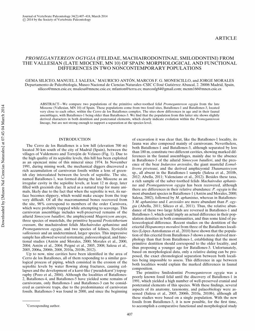
Journal of Vertebrate Paleontology 34(2):407–418, March 2014© 2014 by the Society of Vertebrate Paleontology
ARTICLE
PROMEGANTEREON OGYGIA (FELIDAE, MACHAIRODONTINAE, SMILODONTINI) FROMTHE VALLESIAN (LATE MIOCENE, MN 10) OF SPAIN: MORPHOLOGICAL AND FUNCTIONAL
DIFFERENCES IN TWO NONCONTEMPORARY POPULATIONS
GEMA SILICEO, MANUEL J. SALESA,* MAURICIO ANTON, MARCOS F. G. MONESCILLO, and JORGE MORALESDepartamento de Paleobiologıa, Museo Nacional de Ciencias Naturales–CSIC C/Jose Gutierrez Abascal, 2. 28006 Madrid, Spain,
[email protected]; [email protected]; [email protected]; [email protected]; [email protected]
ABSTRACT—We compare two populations of the primitive saber-toothed felid Promegantereon ogygia from the lateMiocene (Vallesian, MN 10) of Spain. These populations come from two fossil sites, Batallones-1 and Batallones-3, locatedvery close to each other, within the Cerro de los Batallones complex. The sites show differences in age and in their faunalassemblages, with Batallones-1 being older than Batallones-3. We find that the population from this latter site shows slightlyderived characters in both dentition and postcranial elements, which clearly indicate evolution within the Promegantereonlineage, but are not strong enough to support a separation at the species level.
INTRODUCTION
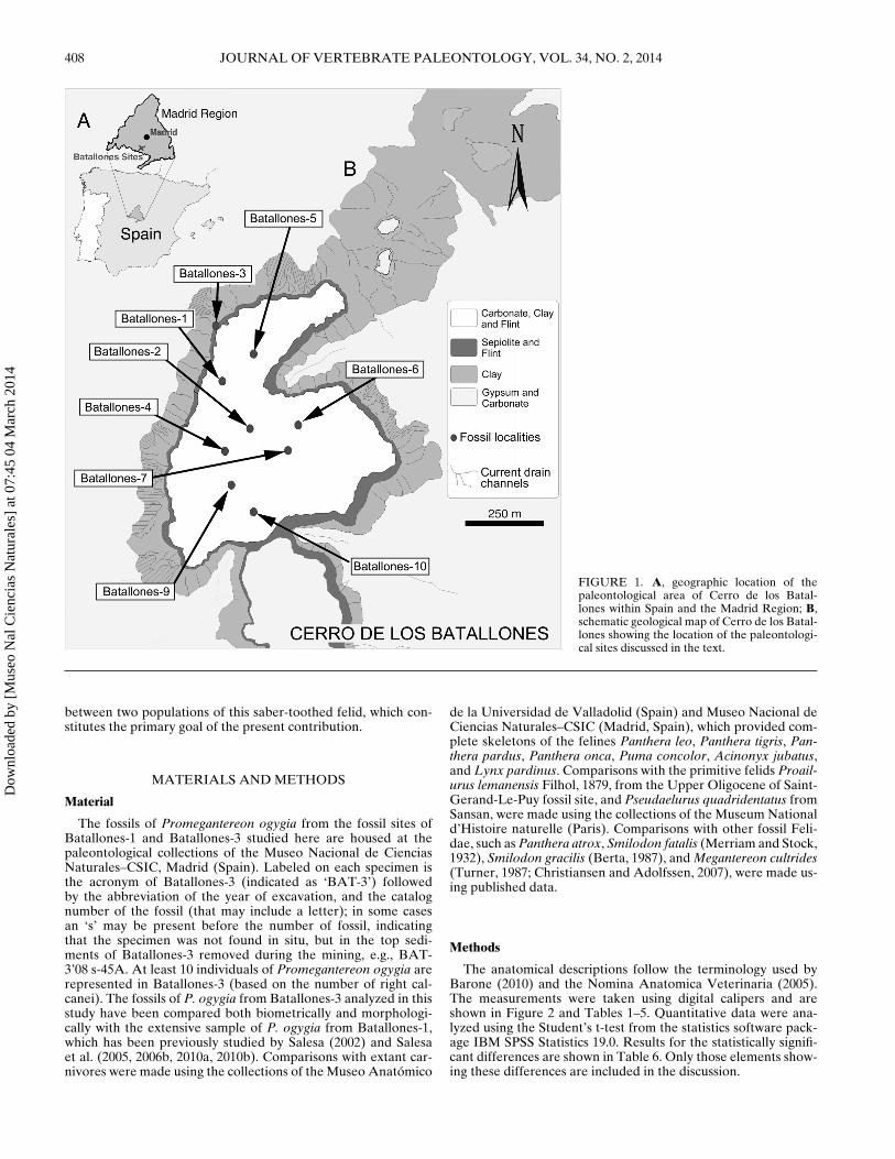
The Cerro de los Batallones is a low hill (elevation 700 m)located 30 km south of the city of Madrid (Spain), between thevillages of Valdemoro and Torrejon de Velasco (Fig. 1). Due tothe high quality of its sepiolite levels, this hill has been exploitedas an opencast mine of this mineral since 1974. In November1991, during mining work, the mechanical diggers discovered arich accumulation of carnivoran fossils within a lens of green-ish clay intercalated between the levels of sepiolite. The site,named Batallones-1, was formed during the late Miocene as anirregular cavity in the sepiolite levels, at least 12 m deep, laterfilled with greenish clay. It acted as a natural trap for many ani-mals, likely due to the fact that when the sepiolite is wet, its sur-face becomes slippery, which would make escape from the trapvery difficult. Of all the macromammal bones recovered fromthe site, 98% correspond to members of the order Carnivora,which were probably trapped while attempting to scavenge. Thiscarnivoran assemblage includes well-preserved remains of theailurid Simocyon batalleri, the amphicyonid Magericyon anceps,three species of mustelids, the primitive hyaenid Protictitheriumcrassum, the machairodont felids Machairodus aphanistus andPromegantereon ogygia, and two species of felines, Styriofelisvallesiensis and an undetermined, larger species. This impressivesample has allowed several systematic, paleoecological, and func-tional studies (Anton and Morales, 2000; Morales et al., 2000,2004; Anton et al., 2004; Peigne et al., 2005, 2008; Salesa et al.,2005, 2006a, 2006b, 2008, 2010a, 2010b, 2012).
Up to now, nine cavities have been identified in the area ofCerro de los Batallones, all of them responding to a similar geo-logical process of piping, which consisted in the erosion of thesepiolite levels by water flowing along fractures, causing col-lapses and the development of a karst-like (‘pseudokarst’) topog-raphy (Pozo et al., 2004). Although the localities of Batallones-2, Batallones-4, and Batallones-5 have yielded some remains ofcarnivorans, only Batallones-1 and Batallones-3 can be consid-ered as carnivore traps, due to the predominance of carnivoranfossils. Batallones-3 was found in 2000, and since the beginning
*Corresponding author.
of excavation it was clear that, like the Batallones-1 locality, itsfauna was also composed mainly of carnivorans. Nevertheless,both Batallones-1 and Batallones-3, although separated by lessthan 180 m, constitute two different cavities, showing marked dif-ferences in the faunal assemblages, mainly due to the absencein Batallones-3 of the ailurid Simocyon batalleri, and the pres-ence of the bear Indarctos arctoides, the giant mustelid Eomel-livora piveteaui, and the derived amphicyonid Thaumastocyonsp., all absent in the Batallones-1 sample (Salesa et al., 2010b,2012; Abella, 2011; Valenciano et al., 2012). Besides these taxa,a rich sample of the saber-toothed felids Machairodus aphanis-tus and Promegantereon ogygia has been recovered, althoughthere are differences in their relative abundance: P. ogygia is themost abundant species in Batallones-1 (Anton and Morales, 2000;Salesa, 2002), followed by M. aphanistus, whereas in Batallones-3 M. aphanistus and I. arctoides are more abundant than P. ogy-gia (Abella, 2011; Siliceo et al., 2011). Thus, the relative abun-dance of these two large felids are reversed in Batallones-1 andBatallones-3, which could imply an actual difference in their pop-ulation densities in both communities, and thus some kind of pa-leoecological difference. Recent studies on the dentition of thecricetid Hispanomys moralesi from three of the Batallones locali-ties (Lopez-Antonanzas et al., 2010) have shown that the popula-tion of this cricetid from Batallones-3 shows a more derived mor-phology than that from Batallones-1, establishing that the mostprimitive dentition should correspond to the older locality, andthus proposing a younger age for Batallones-3. Unfortunately,based on morphological data, only a relative dating can be pro-posed, the exact chronological separation between both locali-ties being impossible to assess. This difference in age betweenboth localities would explain the marked differences in faunalcomposition.
The primitive Smilodontini Promegantereon ogygia was apoorly known fossil felid until the discovery of Batallones-1 in1991, which yielded a high number of well-preserved cranial andpostcranial elements of this species. With these findings, severalaspects of its anatomy, taxonomy, and palaeobiology were as-sessed (Salesa et al., 2005, 2006b, 2010a, 2010b), although allthese studies were based on a single population. With the newfossils from Batallones-3, it is now possible, for the first time,to accomplish a comparative functional and morphological study
407
Dow
nloa
ded
by [
Mus
eo N
al C
ienc
ias
Nat
ural
es]
at 0
7:45
04
Mar
ch 2
014
408 JOURNAL OF VERTEBRATE PALEONTOLOGY, VOL. 34, NO. 2, 2014
FIGURE 1. A, geographic location of thepaleontological area of Cerro de los Batal-lones within Spain and the Madrid Region; B,schematic geological map of Cerro de los Batal-lones showing the location of the paleontologi-cal sites discussed in the text.
between two populations of this saber-toothed felid, which con-stitutes the primary goal of the present contribution.
MATERIALS AND METHODS
Material
The fossils of Promegantereon ogygia from the fossil sites ofBatallones-1 and Batallones-3 studied here are housed at thepaleontological collections of the Museo Nacional de CienciasNaturales–CSIC, Madrid (Spain). Labeled on each specimen isthe acronym of Batallones-3 (indicated as ‘BAT-3’) followedby the abbreviation of the year of excavation, and the catalognumber of the fossil (that may include a letter); in some casesan ‘s’ may be present before the number of fossil, indicatingthat the specimen was not found in situ, but in the top sedi-ments of Batallones-3 removed during the mining, e.g., BAT-3’08 s-45A. At least 10 individuals of Promegantereon ogygia arerepresented in Batallones-3 (based on the number of right cal-canei). The fossils of P. ogygia from Batallones-3 analyzed in thisstudy have been compared both biometrically and morphologi-cally with the extensive sample of P. ogygia from Batallones-1,which has been previously studied by Salesa (2002) and Salesaet al. (2005, 2006b, 2010a, 2010b). Comparisons with extant car-nivores were made using the collections of the Museo Anatomico
de la Universidad de Valladolid (Spain) and Museo Nacional deCiencias Naturales–CSIC (Madrid, Spain), which provided com-plete skeletons of the felines Panthera leo, Panthera tigris, Pan-thera pardus, Panthera onca, Puma concolor, Acinonyx jubatus,and Lynx pardinus. Comparisons with the primitive felids Proail-urus lemanensis Filhol, 1879, from the Upper Oligocene of Saint-Gerand-Le-Puy fossil site, and Pseudaelurus quadridentatus fromSansan, were made using the collections of the Museum Nationald’Histoire naturelle (Paris). Comparisons with other fossil Feli-dae, such as Panthera atrox, Smilodon fatalis (Merriam and Stock,1932), Smilodon gracilis (Berta, 1987), and Megantereon cultrides(Turner, 1987; Christiansen and Adolfssen, 2007), were made us-ing published data.
Methods
The anatomical descriptions follow the terminology used byBarone (2010) and the Nomina Anatomica Veterinaria (2005).The measurements were taken using digital calipers and areshown in Figure 2 and Tables 1–5. Quantitative data were ana-lyzed using the Student’s t-test from the statistics software pack-age IBM SPSS Statistics 19.0. Results for the statistically signifi-cant differences are shown in Table 6. Only those elements show-ing these differences are included in the discussion.
Dow
nloa
ded
by [
Mus
eo N
al C
ienc
ias
Nat
ural
es]
at 0
7:45
04
Mar
ch 2
014
SILICEO ET AL.—PROMEGANTEREON OGYGIA FROM BATALLONES SITES 409
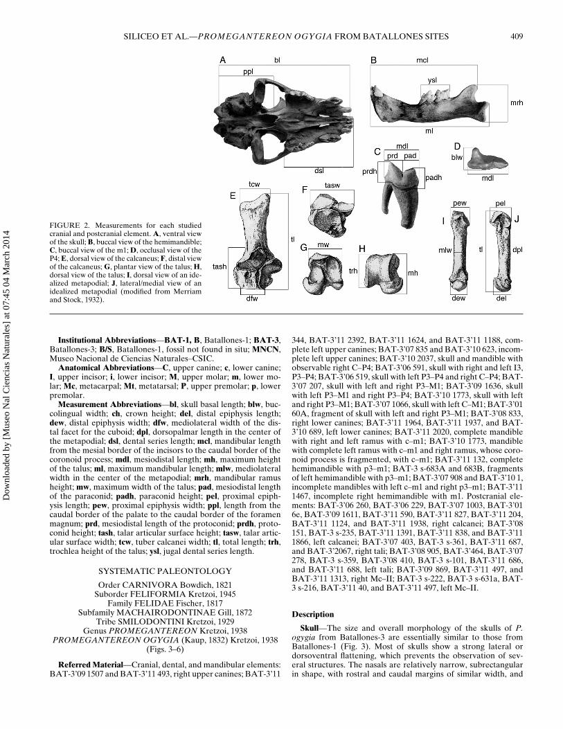
FIGURE 2. Measurements for each studiedcranial and postcranial element. A, ventral viewof the skull; B, buccal view of the hemimandible;C, buccal view of the m1; D, occlusal view of theP4; E, dorsal view of the calcaneus; F, distal viewof the calcaneus; G, plantar view of the talus; H,dorsal view of the talus; I, dorsal view of an ide-alized metapodial; J, lateral/medial view of anidealized metapodial (modified from Merriamand Stock, 1932).
Institutional Abbreviations—BAT-1, B, Batallones-1; BAT-3,Batallones-3; B/S, Batallones-1, fossil not found in situ; MNCN,Museo Nacional de Ciencias Naturales–CSIC.
Anatomical Abbreviations—C, upper canine; c, lower canine;I, upper incisor; i, lower incisor; M, upper molar; m, lower mo-lar; Mc, metacarpal; Mt, metatarsal; P, upper premolar; p, lowerpremolar.
Measurement Abbreviations—bl, skull basal length; blw, buc-colingual width; ch, crown height; del, distal epiphysis length;dew, distal epiphysis width; dfw, mediolateral width of the dis-tal facet for the cuboid; dpl, dorsopalmar length in the center ofthe metapodial; dsl, dental series length; mcl, mandibular lengthfrom the mesial border of the incisors to the caudal border of thecoronoid process; mdl, mesiodistal length; mh, maximum heightof the talus; ml, maximum mandibular length; mlw, mediolateralwidth in the center of the metapodial; mrh, mandibular ramusheight; mw, maximum width of the talus; pad, mesiodistal lengthof the paraconid; padh, paraconid height; pel, proximal epiph-ysis length; pew, proximal epiphysis width; ppl, length from thecaudal border of the palate to the caudal border of the foramenmagnum; prd, mesiodistal length of the protoconid; prdh, proto-conid height; tash, talar articular surface height; tasw, talar artic-ular surface width; tcw, tuber calcanei width; tl, total length; trh,trochlea height of the talus; ysl, jugal dental series length.
SYSTEMATIC PALEONTOLOGY
Order CARNIVORA Bowdich, 1821Suborder FELIFORMIA Kretzoi, 1945
Family FELIDAE Fischer, 1817Subfamily MACHAIRODONTINAE Gill, 1872
Tribe SMILODONTINI Kretzoi, 1929Genus PROMEGANTEREON Kretzoi, 1938
PROMEGANTEREON OGYGIA (Kaup, 1832) Kretzoi, 1938(Figs. 3–6)
Referred Material—Cranial, dental, and mandibular elements:BAT-3’09 1507 and BAT-3’11 493, right upper canines; BAT-3’11
344, BAT-3’11 2392, BAT-3’11 1624, and BAT-3’11 1188, com-plete left upper canines; BAT-3’07 835 and BAT-3’10 623, incom-plete left upper canines; BAT-3’10 2037, skull and mandible withobservable right C–P4; BAT-3’06 591, skull with right and left I3,P3–P4; BAT-3’06 519, skull with left P3–P4 and right C–P4; BAT-3’07 207, skull with left and right P3–M1; BAT-3’09 1636, skullwith left P3–M1 and right P3–P4; BAT-3’10 1773, skull with leftand right P3–M1; BAT-3’07 1066, skull with left C–M1; BAT-3’0160A, fragment of skull with left and right P3–M1; BAT-3’08 833,right lower canines; BAT-3’11 1964, BAT-3’11 1937, and BAT-3’10 689, left lower canines; BAT-3’11 2020, complete mandiblewith right and left ramus with c–m1; BAT-3’10 1773, mandiblewith complete left ramus with c–m1 and right ramus, whose coro-noid process is fragmented, with c–m1; BAT-3’11 132, completehemimandible with p3–m1; BAT-3 s-683A and 683B, fragmentsof left hemimandible with p3–m1; BAT-3’07 908 and BAT-3’10 1,incomplete mandibles with left c–m1 and right p3–m1; BAT-3’111467, incomplete right hemimandible with m1. Postcranial ele-ments: BAT-3’06 260, BAT-3’06 229, BAT-3’07 1003, BAT-3’016e, BAT-3’09 1611, BAT-3’11 590, BAT-3’11 827, BAT-3’11 204,BAT-3’11 1124, and BAT-3’11 1938, right calcanei; BAT-3’08151, BAT-3 s-235, BAT-3’11 1391, BAT-3’11 838, and BAT-3’111866, left calcanei; BAT-3’07 403, BAT-3 s-361, BAT-3’11 687,and BAT-3’2067, right tali; BAT-3’08 905, BAT-3’464, BAT-3’07278, BAT-3 s-359, BAT-3’08 410, BAT-3 s-101, BAT-3’11 686,and BAT-3’11 688, left tali; BAT-3’09 869, BAT-3’11 497, andBAT-3’11 1313, right Mc–II; BAT-3 s-222, BAT-3 s-631a, BAT-3 s-216, BAT-3’11 40, and BAT-3’11 497, left Mc–II.
Description
Skull—The size and overall morphology of the skulls of P.ogygia from Batallones-3 are essentially similar to those fromBatallones-1 (Fig. 3). Most of skulls show a strong lateral ordorsoventral flattening, which prevents the observation of sev-eral structures. The nasals are relatively narrow, subrectangularin shape, with rostral and caudal margins of similar width, and
Dow
nloa
ded
by [
Mus
eo N
al C
ienc
ias
Nat
ural
es]
at 0
7:45
04
Mar
ch 2
014

410 JOURNAL OF VERTEBRATE PALEONTOLOGY, VOL. 34, NO. 2, 2014
FIGURE 3. Skulls of Promegantereon ogygia from Batallones-3. A, B, BAT-3’06 519 in A, lateral view; B, ventral view; C, D, BAT-3’06 591 in C,dorsal view; D, ventral view.
slightly curved caudally; the caudal margin lacks the narrowingobserved in the nasals of large extant felids. The left and righttemporal lines meet dorsally at the level of the glenoid fossa, giv-ing way to a strong sagittal crest that ends in a robust nuchal crest.The rostral border of the premaxilla is curved rostrally, produc-ing a ‘U’-shaped arcade. There is a short diastema between I3and the upper canine. The zygomatic process of the frontal issharp, rough, and slightly laterally projected; it is the origin ofa marked temporal line. The infraorbital foramen is large andplaced at the level of P3. The lacrimal foramen is placed on theinner face of the rostral border of the orbit. The sphenopalatineforamen is relatively smaller than that of Panthera; it is placedon the orbital wall of the palatine bone, close to the maxillopala-tine suture. The posterior palatine foramen is smaller than thesphenopalatine foramen, and it is located slightly rostrally to thelatter. The oval foramen opens on the basisphenoid bone, be-tween the pterygoid process and the postglenoid process. The zy-gomatic arch is much more robust than that of a similarly sizedextant felid, and dorsoventrally higher. The frontal process of thezygomatic is very small, although it is present in all the specimens;it is developed as a small bony bulge, ventrocaudally oriented,with an irregular ventral end. The tympanic bullae are rounded,less inflated than those of Panthera, and extend from the rostralmargin of the jugular process to the caudal margin of the post-glenoid process without contacting with the latter. The caudal en-totympanic penetrates rostrally beside the medial margin of theectotympanic. The external auditory meatus is a single, rostrolat-erally oriented opening. The stylomastoid foramen and tympa-nohyal depression are very close to each other. Both are placedin a more rostral position than in Panthera, that is, at the levelof the central part of the bulla. The jugular process is reduced insize when compared with that of extant large felids, being dorsallydisplaced in comparison with its position in the latter, and medio-laterally oriented; it is developed as a bony sheet with a small dis-
tal protuberance, extending from the caudal margin of the tym-panic bulla. The mastoid process is robust and relatively well de-veloped; it is located on the lateral wall of the tympanic bulla,rostrally to the jugular process and slightly surpassing its ventrallevel. The palate is triangular, with a nasopharyngeal fossa delim-itated by the walls of the pterygoid bone. The palatine fissurae areplaced close to the rostral border of the palate.
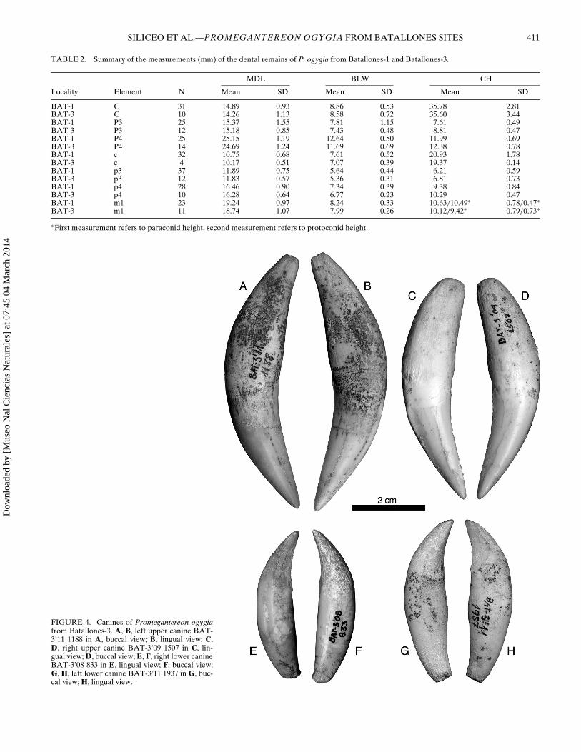
Upper Dentition—The fossil sample of P. ogygia fromBatallones-3 does not include either I1 or I2, although from thealveoli it can be observed that they were much smaller than I3(Fig. 3B). The I3 crown is sharp and caniniform; its mesial surfaceis convex and smooth, whereas the distal one is concave and bearsa slight cingulum along its lingual border; on the buccal surfacethere is a marked ridge from the base to the tip of the crown. Theupper canines show the same morphology as those of P. ogygiafrom Batallones-1 (Fig. 4A–D), with a laterally flattened crownthat curves backwards, with no crenulations on its mesial ordistal margin; both buccal and lingual surfaces are smooth, al-though the mesiolingual border bears a marked crest developedfrom the base to the middle of the crown. One skull (BAT-3’071010) shows a very small tooth in both left and right maxillae,mesial to P3, clearly separated from it; its crown is round and verylow, and it could represent a retained D2, rather than a P2. Notrace of this tooth was found in the 11 studied skulls of P. ogygia
TABLE 1. Summary of the ventral measurements (mm) of the skulls ofP. ogygia from Batallones-1 and Batallones-3.
bl ppl dsl
Locality N Mean SD Mean SD Mean SD
BAT-1 9 166.07 8.58 85.17 3.62 80.18 4.03BAT-3 8 159.41 7.67 81.07 5.15 74.88 3.99
Dow
nloa
ded
by [
Mus
eo N
al C
ienc
ias
Nat
ural
es]
at 0
7:45
04
Mar
ch 2
014
SILICEO ET AL.—PROMEGANTEREON OGYGIA FROM BATALLONES SITES 411
TABLE 2. Summary of the measurements (mm) of the dental remains of P. ogygia from Batallones-1 and Batallones-3.
MDL BLW CH
Locality Element N Mean SD Mean SD Mean SD
BAT-1 C 31 14.89 0.93 8.86 0.53 35.78 2.81BAT-3 C 10 14.26 1.13 8.58 0.72 35.60 3.44BAT-1 P3 25 15.37 1.55 7.81 1.15 7.61 0.49BAT-3 P3 12 15.18 0.85 7.43 0.48 8.81 0.47BAT-1 P4 25 25.15 1.19 12.64 0.50 11.99 0.69BAT-3 P4 14 24.69 1.24 11.69 0.69 12.38 0.78BAT-1 c 32 10.75 0.68 7.61 0.52 20.93 1.78BAT-3 c 4 10.17 0.51 7.07 0.39 19.37 0.14BAT-1 p3 37 11.89 0.75 5.64 0.44 6.21 0.59BAT-3 p3 12 11.83 0.57 5.36 0.31 6.81 0.73BAT-1 p4 28 16.46 0.90 7.34 0.39 9.38 0.84BAT-3 p4 10 16.28 0.64 6.77 0.23 10.29 0.47BAT-1 m1 23 19.24 0.97 8.24 0.33 10.63/10.49∗ 0.78/0.47∗BAT-3 m1 11 18.74 1.07 7.99 0.26 10.12/9.42∗ 0.79/0.73∗
∗First measurement refers to paraconid height, second measurement refers to protoconid height.
FIGURE 4. Canines of Promegantereon ogygiafrom Batallones-3. A, B, left upper canine BAT-3’11 1188 in A, buccal view; B, lingual view; C,D, right upper canine BAT-3’09 1507 in C, lin-gual view; D, buccal view; E, F, right lower canineBAT-3’08 833 in E, lingual view; F, buccal view;G, H, left lower canine BAT-3’11 1937 in G, buc-cal view; H, lingual view.
Dow
nloa
ded
by [
Mus
eo N
al C
ienc
ias
Nat
ural
es]
at 0
7:45
04
Mar
ch 2
014
412 JOURNAL OF VERTEBRATE PALEONTOLOGY, VOL. 34, NO. 2, 2014
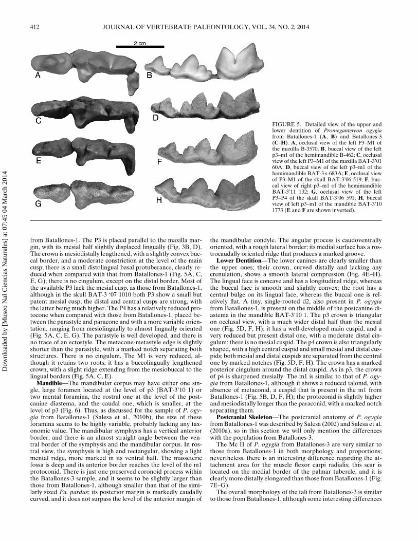
FIGURE 5. Detailed view of the upper andlower dentition of Promegantereon ogygiafrom Batallones-1 (A, B) and Batallones-3(C–H). A, occlusal view of the left P3–M1 ofthe maxilla B-3570; B, buccal view of the leftp3–m1 of the hemimandible B-462; C, occlusalview of the left P3–M1 of the maxilla BAT-3’0160A; D, buccal view of the left p3–m1 of thehemimandible BAT-3 s-683A; E, occlusal viewof P3–M1 of the skull BAT-3’06 519; F, buc-cal view of right p3–m1 of the hemimandibleBAT-3’11 132; G, occlusal view of the leftP3–P4 of the skull BAT-3’06 591; H, buccalview of left p3–m1 of the mandible BAT-3’101773 (E and F are shown inverted).
from Batallones-1. The P3 is placed parallel to the maxilla mar-gin, with its mesial half slightly displaced lingually (Fig. 3B, D).The crown is mesiodistally lengthened, with a slightly convex buc-cal border, and a moderate constriction at the level of the maincusp; there is a small distolingual basal protuberance, clearly re-duced when compared with that from Batallones-1 (Fig. 5A, C,E, G); there is no cingulum, except on the distal border. Most ofthe available P3 lack the mesial cusp, as those from Batallones-1,although in the skull BAT-3 ‘07 1010 both P3 show a small butpatent mesial cusp; the distal and central cusps are strong, withthe latter being much higher. The P4 has a relatively reduced pro-tocone when compared with those from Batallones-1, placed be-tween the parastyle and paracone and with a more variable orien-tation, ranging from mesiolingually to almost lingually oriented(Fig. 5A, C, E, G). The parastyle is well developed, and there isno trace of an ectostyle. The metacone-metastyle edge is slightlyshorter than the parastyle, with a marked notch separating bothstructures. There is no cingulum. The M1 is very reduced, al-though it retains two roots; it has a buccolingually lengthenedcrown, with a slight ridge extending from the mesiobuccal to thelingual borders (Fig. 5A, C, E).
Mandible—The mandibular corpus may have either one sin-gle, large foramen located at the level of p3 (BAT-3’10 1) ortwo mental foramina, the rostral one at the level of the post-canine diastema, and the caudal one, which is smaller, at thelevel of p3 (Fig. 6). Thus, as discussed for the sample of P. ogy-gia from Batallones-1 (Salesa et al., 2010b), the size of theseforamina seems to be highly variable, probably lacking any tax-onomic value. The mandibular symphysis has a vertical anteriorborder, and there is an almost straight angle between the ven-tral border of the symphysis and the mandibular corpus. In ros-tral view, the symphysis is high and rectangular, showing a lightmental ridge, more marked in its ventral half. The massetericfossa is deep and its anterior border reaches the level of the m1protoconid. There is just one preserved coronoid process withinthe Batallones-3 sample, and it seems to be slightly larger thanthose from Batallones-1, although smaller than that of the simi-larly sized Pa. pardus; its posterior margin is markedly caudallycurved, and it does not surpass the level of the anterior margin of
the mandibular condyle. The angular process is caudoventrallyoriented, with a rough lateral border; its medial surface has a ros-trocaudally oriented ridge that produces a marked groove.
Lower Dentition—The lower canines are clearly smaller thanthe upper ones; their crown, curved distally and lacking anycrenulation, shows a smooth lateral compression (Fig. 4E–H).The lingual face is concave and has a longitudinal ridge, whereasthe buccal face is smooth and slightly convex; the root has acentral bulge on its lingual face, whereas the buccal one is rel-atively flat. A tiny, single-rooted d2, also present in P. ogygiafrom Batallones-1, is present on the middle of the postcanine di-astema in the mandible BAT-3’10 1. The p3 crown is triangularon occlusal view, with a much wider distal half than the mesialone (Fig. 5D, F, H); it has a well-developed main cuspid, and avery reduced but present distal one, with a moderate distal cin-gulum; there is no mesial cuspid. The p4 crown is also triangularlyshaped, with a high central cuspid and small mesial and distal cus-pids; both mesial and distal cuspids are separated from the centralone by marked notches (Fig. 5D, F, H). The crown has a markedposterior cingulum around the distal cuspid. As in p3, the crownof p4 is sharpened mesially. The m1 is similar to that of P. ogy-gia from Batallones-1, although it shows a reduced talonid, withabsence of metaconid, a cuspid that is present in the m1 fromBatallones-1 (Fig. 5B, D, F, H); the protoconid is slightly higherand mesiodistally longer than the paraconid, with a marked notchseparating them.
Postcranial Skeleton—The postcranial anatomy of P. ogygiafrom Batallones-1 was described by Salesa (2002) and Salesa et al.(2010a), so in this section we will only mention the differenceswith the population from Batallones-3.
The Mc II of P. ogygia from Batallones-3 are very similar tothose from Batallones-1 in both morphology and proportions;nevertheless, there is an interesting difference regarding the at-tachment area for the muscle flexor carpi radialis; this scar islocated on the medial border of the palmar tubercle, and it isclearly more distally elongated than those from Batallones-1 (Fig.7E–G).
The overall morphology of the tali from Batallones-3 is similarto those from Batallones-1, although some interesting differences
Dow
nloa
ded
by [
Mus
eo N
al C
ienc
ias
Nat
ural
es]
at 0
7:45
04
Mar
ch 2
014
SILICEO ET AL.—PROMEGANTEREON OGYGIA FROM BATALLONES SITES 413
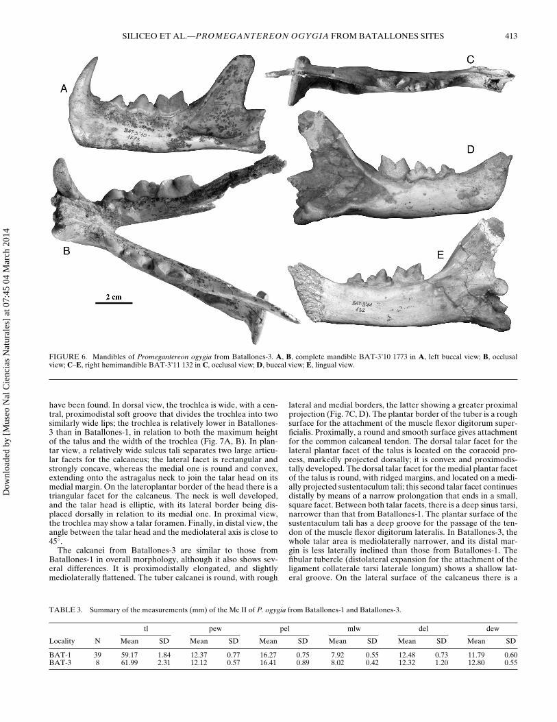
FIGURE 6. Mandibles of Promegantereon ogygia from Batallones-3. A, B, complete mandible BAT-3’10 1773 in A, left buccal view; B, occlusalview; C–E, right hemimandible BAT-3’11 132 in C, occlusal view; D, buccal view; E, lingual view.
have been found. In dorsal view, the trochlea is wide, with a cen-tral, proximodistal soft groove that divides the trochlea into twosimilarly wide lips; the trochlea is relatively lower in Batallones-3 than in Batallones-1, in relation to both the maximum heightof the talus and the width of the trochlea (Fig. 7A, B). In plan-tar view, a relatively wide sulcus tali separates two large articu-lar facets for the calcaneus; the lateral facet is rectangular andstrongly concave, whereas the medial one is round and convex,extending onto the astragalus neck to join the talar head on itsmedial margin. On the lateroplantar border of the head there is atriangular facet for the calcaneus. The neck is well developed,and the talar head is elliptic, with its lateral border being dis-placed dorsally in relation to its medial one. In proximal view,the trochlea may show a talar foramen. Finally, in distal view, theangle between the talar head and the mediolateral axis is close to45◦.
The calcanei from Batallones-3 are similar to those fromBatallones-1 in overall morphology, although it also shows sev-eral differences. It is proximodistally elongated, and slightlymediolaterally flattened. The tuber calcanei is round, with rough
lateral and medial borders, the latter showing a greater proximalprojection (Fig. 7C, D). The plantar border of the tuber is a roughsurface for the attachment of the muscle flexor digitorum super-ficialis. Proximally, a round and smooth surface gives attachmentfor the common calcaneal tendon. The dorsal talar facet for thelateral plantar facet of the talus is located on the coracoid pro-cess, markedly projected dorsally; it is convex and proximodis-tally developed. The dorsal talar facet for the medial plantar facetof the talus is round, with ridged margins, and located on a medi-ally projected sustentaculum tali; this second talar facet continuesdistally by means of a narrow prolongation that ends in a small,square facet. Between both talar facets, there is a deep sinus tarsi,narrower than that from Batallones-1. The plantar surface of thesustentaculum tali has a deep groove for the passage of the ten-don of the muscle flexor digitorum lateralis. In Batallones-3, thewhole talar area is mediolaterally narrower, and its distal mar-gin is less laterally inclined than those from Batallones-1. Thefibular tubercle (distolateral expansion for the attachment of theligament collaterale tarsi laterale longum) shows a shallow lat-eral groove. On the lateral surface of the calcaneus there is a
TABLE 3. Summary of the measurements (mm) of the Mc II of P. ogygia from Batallones-1 and Batallones-3.
tl pew pel mlw del dew
Locality N Mean SD Mean SD Mean SD Mean SD Mean SD Mean SD
BAT-1 39 59.17 1.84 12.37 0.77 16.27 0.75 7.92 0.55 12.48 0.73 11.79 0.60BAT-3 8 61.99 2.31 12.12 0.57 16.41 0.89 8.02 0.42 12.32 1.20 12.80 0.55
Dow
nloa
ded
by [
Mus
eo N
al C
ienc
ias
Nat
ural
es]
at 0
7:45
04
Mar
ch 2
014

414 JOURNAL OF VERTEBRATE PALEONTOLOGY, VOL. 34, NO. 2, 2014
FIGURE 7. Postcranial elements of Promegantereon ogygia fromBatallones-3 that showed statistically significant differences with thosefrom Batallones-1. A, B, left talus BAT-3’08 905 in A, dorsal view; B,plantar view; C, D, right calcaneus BAT-3’06 260 in C, lateral view; D,dorsal view; E–G, left Mc II BAT-3 s-216 in E, dorsal view, F, medialview; G, lateral view.
TABLE 4. Summary of the measurements (mm) of the talus of P. ogy-gia from Batallones-1 and Batallones-3.
th mw mh
Locality N Mean SD Mean SD Mean SD
BAT-1 41 24.62 1.41 26.08 1.34 35.01 2.43BAT-3 13 21.62 1.49 25.29 2.29 34.02 1.85
gentle depression, proximodistally elongated for the attachmentof the muscle quadratus plantae; in both populations of P. ogygiait is developed from the distal border to the middle of the dor-sal border of the calcaneus. Between the proximal border of theattachment surface of the muscle quadratus plantae and the dor-solateral border of the calcaneus, there is a marked round fossafor the ligament short collateral lateral. The distal articular sur-face for the cuboid is semicircular, gently concave, and its plane islaterally much more inclined in Batallones-1 than in Batallones-3. Close to the medioplantar border of this facet there is a roughtubercle for the long plantar ligament.
Statistical Results
The results of the different Student’s t-tests calculated for thequantitative data are shown in Table 6. The most interesting re-sults were as follows:
1. The P3 shows significant differences in the ratio of theheight (H) and the mesiodistal length (MDL) of the crown,as shown by a Student’s t-test for this index. For similarcrown lengths, the population of Batallones-3 shows P3 withproportionally higher crowns than those from Batallones-1.Also, significant differences were found in the buccolingualwidth (BLW) of P3, with those specimens from Batallones-3being narrower than those from Batallones-1.
2. The P4 shows significant differences in Student’s t-testfor an index between the buccolingual width (BLW) andthe mesiodistal length (MDL) of the crown. The P4 fromBatallones-3 are buccolingually narrower in proportion totheir length.
3. The mesiodistal length of the P4 (MDL) shows significant dif-ferences in relation to the basal length of the skull (BL), thepopulation from Batallones-3 having a relatively longer P4than those from Batallones-1.
4. In the p3, a Student’s t-test shows significant differences inthe index between the height (H) and mesiodistal length(MDL) of the crown. The p3s from Batallones-3 showhigher crowns in relation to their length than those fromBatallones-1.
5. The height of the p4 shows significant differences betweenboth populations. This is demonstrated by a Student’s t-testfor an index between the height (H) and length (MDL) ofthe crown, with the p4s from Batallones-3 showing relativelyhigher crowns than those from Batallones-1.
6. Concerning the calcaneus, the articular surface for the talusshows significant differences in its proportions, as shown bya Student’s t-test for an index between the width (TASW)and the height (TASH) of the articular surface for the talus,with Batallones-3 showing a relatively narrower talar articu-lar surface.
7. Also in the calcaneus, the index between the width of thedistal facet for the cuboid (DFW) and the width of the ta-lar articular surface (TASW) shows significant differences,with the calcaneus from Batallones-3 showing a relativelynarrower talar articular surface than those from Batallones-1.
Dow
nloa
ded
by [
Mus
eo N
al C
ienc
ias
Nat
ural
es]
at 0
7:45
04
Mar
ch 2
014
SILICEO ET AL.—PROMEGANTEREON OGYGIA FROM BATALLONES SITES 415
TABLE 5. Summary of the measurements (mm) of the calcaneus of P. ogygia from Batallones-1 and Batallones-3.
ti tasw dfw tash tuw
Locality N Mean SD Mean SD Mean SD Mean SD Mean SD
BAT-1 38 62.96 2.56 26.25 1.73 17.26 1.15 25.25 1.67 17.00 1.09BAT-3 15 62.81 2.97 24.51 1.70 18.10 1.78 24.58 1.87 16.94 1.19
8. Also the talar articular surface of the calcaneus (TASW) issignificantly narrower in relation to the calcaneus tuberclewidth (TW) in Batallones-3, as indicated by a Student’s t-testfor an index between both measurements (TW/TASW).
9. A Student’s t-test for the talus showed significant differencesin the index between the height of the trochlea (TH) andthe maximum width of the talus (MW), with those tali fromBatallones-3 showing a relatively lower TH than those fromBatallones-1.
10. Finally, a Student’s t-tests for an index between the heightof the trochlea (TH) and the maximum height of the talus(MH) showed significant differences between both popula-tions, with that of Batallones-3 having relatively lower THthan that of Batallones-1 when compared with MH, that is, arelatively longer talus neck.
It should be noted that although both populations show sizedifferences in some of the elements, such as the total length of thecalcaneus, these differences are not significant; thus, we shouldconsider the specimens from Batallones-1 and Batallones-3 ashaving a very similar body size.
Discussion
The identification of the small teeth mesial to P3/p3 as D2/d2is based on their extremely reduced size and simple morphology,which precludes their identification as permanent premolars. Al-though Salesa et al. (2010b) cite the presence of a very smallp2 in P. ogygia from Batallones-1, such reduced premolars, ob-served in several fossil felids, have been more recently consideredas retained decidual teeth (Salesa et al., 2012), and that is morelikely the case of the small premolars observed in P. ogygia. Itis remarkable that whereas none of the skulls of P. ogygia fromBatallones-1 show any trait of D2, this tooth appears in at least
TABLE 6. Summarized results of the Student’s t-tests showing the vari-ables that showed significant differences.
Element Variable P Comments
P3 MDL/H 0.000 BAT-3 shows relatively higherP3.
P3 BLW 0.001 BAT-3 shows narrower P3.P4 MDL/BLW 0.010 BAT-3 shows relatively
narrower P4.P4 MDL/BL 0.013 BAT-3 shows relatively longer
P4.p3 H/MDL 0.001 BAT-3 shows relatively higher
p3.p4 H/MDL 0.000 BAT-3 shows relatively higher
p4.Calcaneus TASW/TASH 0.002 BAT-3 shows relatively
narrower TASW.Calcaneus DFW/TASW 0.000 BAT-3 shows relatively
narrower TASW.Calcaneus TW/TASW 0.000 BAT-3 shows relatively
narrower TASW.Talus TH/MW 0.000 BAT-3 shows relatively lower
TH.Talus TH/MH 0.000 BAT-3 shows relatively lower
TH.
one of the eight available skulls from Batallones-3. This couldbe considered as a plesiomorphic character, because it is presentin the more primitive machairodontine Pseudaelurus quadriden-tatus from the Middle Miocene of Europe. On the contrary, thepresence in Batallones-3 of a relatively narrow P3, with a reducedbasal expansion of the crown, implies a morphological separa-tion of this taxon from Ps. quadridentatus, which shares with P.ogygia from Batallones-1 the presence of a marked distolingualbasal expansion in the P3 (Fig. 5A, C, E, G). Other Smilodon-tini such as the Turolian (MN 11–12) Paramachaerodus orientalis,the Pliocene Megantereon, and the Pleistocene Smilodon show arelatively small P3, without distolingual expansion (Salesa et al.,2010b). The difference between the P3 from Batallones-1 andBatallones-3 probably indicates the beginning of the strong re-duction of the P3 observed in more derived taxa.
The relatively reduced protocone in the P4 of P. ogygia fromBatallones-3 (which is relatively buccolingually narrower thanthat from Batallones-1) in comparison with the morphology ofthose from Batallones-1 is a derived feature (Fig. 5A, C, E, G),also observable in the more advanced Paramachaerodus orien-talis, although this species also shows a marked ectostyle, absentin P. ogygia. More derived machairodontines such as Smilodonand Megantereon show markedly reduced protocones in P4.The primitive condition for this character can be seen in theearly Miocene felid Proailurus lemanensis, which shows a well-developed P4 protocone, clearly mesiolingually oriented, similarto the condition observed in members of the family Viverridae.Once again, we observe in the Batallones-3 material a slight mod-ification from the dental morphology seen in the older populationfrom Batallones-1. The reduced protocone creates a more tren-chant P4, a trait not only observed in more-derived smilodon-tins, but also in derived non-felid saber-toothed taxa, such asbarbourofelids and nimravids (Bryant, 1991, 1996; Morales et al.,2001; Morlo et al., 2004). This feature, often associated with alengthening of the P4 crown, is clearly related to more efficientmeat consumption, because it enlarges the cutting blade of theupper carnassial. Also, P3, p3, P4, and p4 show relatively highercrowns in Batallones-3 than in Batallones-1, a difference forwhich we found no clear functional implication. These higher andthus more pointed crowns could also increase the shear capacityof both teeth, mostly considering that this difference is found inall the premolars.
The sample of m1 from Batallones-3 shows a reduced talonid,a derived feature associated with a more trenchant lower carnas-sial (Fig. 5B, D, F, H). The talonid of P. ogygia from Batallones-1shows a basal expansion retaining a tiny metaconid (Salesa et al.,2010b), whereas in Batallones-3 the m1 shows a smaller talonidcomposed only of a basal expansion, without any trace of a meta-conid. The primitive felids Proailurus lemanensis and Pseudaelu-rus quadridentatus show a well-developed talonid with a markedmetaconid, whereas in derived Smilodontini such as Megantereonand Smilodon the talonid is absent. Another primitive smilodon-tin, the Eurasiatic Pa. orientalis, shows a very reduced talonid,whereas in the closely related Pa. maximiliani it is completely ab-sent (Salesa et al., 2010b). In this reduction in the m1 talonid, thepopulation of P. ogygia from Batallones-3 again shows a derivedmorphology in relation to that from Batallones-1.
Concerning the postcranial skeleton, some of the ob-served differences between the populations of Batallones-1 and
Dow
nloa
ded
by [
Mus
eo N
al C
ienc
ias
Nat
ural
es]
at 0
7:45
04
Mar
ch 2
014
416 JOURNAL OF VERTEBRATE PALEONTOLOGY, VOL. 34, NO. 2, 2014
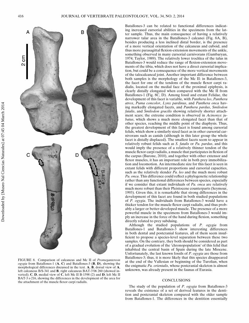
FIGURE 8. Comparison of calcaneus and Mc II of Promegantereonogygia from Batallones-1 (A, C) and Batallones-3 (B, D), showing themorphological differences discussed in the text. A, B, dorsal view of A,left calcaneus B/S-341 and B, right calcaneus BAT-3’06 260 (showed in-verted); C, D, medial view of C, left Mc II B-1598 (2) and D, left Mc IIBAT-3 s-216, showing the differences in the development of the area forthe attachment of the muscle flexor carpi radialis.
Batallones-3 can be related to functional differences indicat-ing increased cursorial abilities in the specimens from the lat-ter sample. Thus, the main consequence of having a relativelynarrower talar area in the Batallones-3 calcanei (Fig. 8A, B),besides producing a less inclined distal border, is the presenceof a more vertical orientation of the calcaneus and cuboid, andthus more parasagittal flexion-extension movements of the ankle,something observed in many cursorial carnivorans (Gambaryan,1974; Taylor, 1989). The relatively lower trochlea of the talus inBatallones-3 would reduce the range of flexion-extension move-ments of the tibia, which does not have a direct cursorial implica-tion, but could be a consequence of the more vertical movementsof the talocalcaneal joint. Another important difference betweenboth samples is the morphology of the Mc II: in Batallones-3,the facet for one of the tendons of the muscle flexor carpi ra-dialis, located on the medial face of the proximal epiphysis, isclearly distally elongated when compared with the Mc II fromBatallones-1 (Fig. 8C, D). Among fossil and extant Felidae, thedevelopment of this facet is variable, with Panthera leo, Pantheraatrox, Puma concolor, Lynx pardinus, and Panthera onca hav-ing markedly elongated facets, and Panthera pardus, Smilodonfatalis, and Smilodon gracilis showing relatively shorter attach-ment scars; the extreme condition is observed in Acinonyx ju-batus, which shows a much more elongated facet than that ofother species, reaching the middle point of the diaphysis. Thus,the greatest development of this facet is found among cursorialfelids, which show a similarly sized facet as in other cursorial car-nivorans such as canids (although in this later group the wholefacet is distally displaced). The smallest facets seem to appear inrelatively robust felids such as S. fatalis or Pa. pardus, and thiswould imply the presence of a relatively thinner tendon of themuscle flexor carpi radialis, a muscle that participates in flexion ofthe carpus (Barone, 2010), and together with other extensor andflexor muscles, it has an important role in both prey immobiliza-tion and locomotion. An intermediate size for this facet is seen inextant felids with different proportions and cursorial capacities,such as the relatively slender Pa. leo and the much more robustPa. onca. This difference could reflect a phylogenetic relationshiprather than any functional differences between species, especiallyif we consider that extant individuals of Pa. onca are relativelymuch more robust than their Pleistocene counterparts (Seymour,1993). Given this, it is remarkable that strong differences in thedevelopment of this facet are found in both studied populationsof P. ogygia. The individuals from Batallones-3 would have athicker tendon for the muscle flexor carpi radialis, and thus prob-ably a larger or better-developed muscle. The presence of a morepowerful muscle in the specimens from Batallones-3 would im-ply an increase in the force of the hand during flexion, somethingdirectly related to prey subduing.
Although the studied populations of P. ogygia fromBatallones-1 and Batallones-3 show interesting differencesin both dental and postcranial features, all of them seem insuf-ficient to propose a species-level separation between these twosamples. On the contrary, they both should be considered as partof a gradual evolution of the ‘chronopopulation’ of this felid thatinhabited the central basin of Spain during the late Miocene.Unfortunately, the last known fossils of P. ogygia are those fromBatallones-3; thus, it is more likely that this species disappearedat the end of the Vallesian or beginning of the Turolian, whenthe enigmatic Pa. orientalis, whose postcranial skeleton is almostunknown, was already present in the faunas of Eurasia.
CONCLUSIONS
The study of the population of P. ogygia from Batallones-3reveals the existence of a set of derived features in the denti-tion and postcranial skeleton compared with the older samplefrom Batallones-1. The differences in the dentition essentially
Dow
nloa
ded
by [
Mus
eo N
al C
ienc
ias
Nat
ural
es]
at 0
7:45
04
Mar
ch 2
014
SILICEO ET AL.—PROMEGANTEREON OGYGIA FROM BATALLONES SITES 417
imply the presence in Batallones-3 of more trenchant premolarsand molars, whereas those observed in the appendicular skeletoncan be related to a reduction in the weight of the hind limb inBatallones-3, which points towards an increase in cursorial abili-ties, and with the need for prey immobilization during the hunt.All these differences, although indicating some degree of mor-phological segregation between both populations, do not seemstrong enough to support the existence of a different species ofPromegantereon in Batallones-3.
ACKNOWLEDGMENTS
This study is part of the research projects CGL2008-00034,CGL2008-05813-C02-01, and CGL2011-25754 (Direccion Gen-eral de Investigacion, Ministerios de Ciencia e Innovacion yEconomıa y Competitividad, Spain). G.S. is a contracted predoc-toral researcher (FPI) of the project CGL2008-00034 (Ministe-rio de Ciencia e Innovacion, reference BES2009-013437). M.J.S.and J.M. belong to the research group UCM-BSCH-910607. Wethank the Government of the Comunidad Autonoma de Madrid(Direccion General de Patrimonio Historico) for its continuousfunding support and research permissions, and J. F. Pastor (Uni-versity of Valladolid, Spain) for specimen loan.
LITERATURE CITED
Abella, J. 2011. Indarctos arctoides Deperet, 1895 (Carnivora, Mammalia)del yacimiento vallesiense de Batallones 3 (cuenca de Madrid).Ph.D. dissertation, Facultad de Ciencias, Universidad Autonoma deMadrid, Madrid, Spain, 403 pp.
Anton, M., and J. Morales. 2000. Inferencias paleoecologicas de laasociacion de carnıvoros del yacimiento de Cerro Batallones; pp.109–201 in J. Morales, M. Nieto, L. Amezua, S. Fraile, E. Gomez,E. Herraez, P. Pelaez-Campomanes, M. J. Salesa, I. M. Sanchez,and D. Soria (eds.), Patrimonio Paleontologico de la Comu-nidad de Madrid. Serie “Arqueologıa, Paleontologıa y Etnografıa,”Volume 6. Comunidad de Madrid, Madrid.
Anton, M., M. J. Salesa, J. Morales, and A. Turner. 2004. First knowncomplete skulls of the scimitar-toothed cat Machairodus aphanis-tus (Felidae, Carnivora) from the Spanish late Miocene site ofBatallones-1. Journal of Vertebrate Paleontology 24:957–969.
Barone, R. 2010. Anatomie comparee des Mammiferes domestiques,tome 1, Osteologie. Fifth edition. Editions Vigot, Paris, 761 pp.
Berta, A. 1987. The sabercat Smilodon gracilis from Florida and a discus-sion of its relationships (Mammalia, Felidae, Smilodontini). Bulletinof the Florida State Museum, Biological Sciences 31:1–63.
Bowdich, T. E. 1821. An Analysis of the Natural Classifications of Mam-malia for use of Students and Travellers. J. Smith, Paris, 115 pp.
Bryant, H. N. 1991. Phylogenetic relationships and systematics of theNimravidae (Carnivora). Journal of Mammalogy 72:56–78.
Bryant, H. N. 1996. Nimravidae; pp. 453–475 in D. R. Prothero and R. J.Emry (eds.), The Terrestrial Eocene-Oligocene Transition in NorthAmerica. Cambridge University Press, Cambridge, U.K.
Christiansen, P., and J. S. Adolfssen. 2007. Osteology and ecology ofMegantereon cultridens SE311 (Mammalia; Felidae; Machairodon-tinae), a sabrecat from the Late Pliocene–Early Pleistoceneof Seneze, France. Zoological Journal of the Linnean Society151:833–884.
Fischer, G. 1817. Adversaria zoologica. Memoires de la Societe Imperialedes Naturalistes de Moscou 5:357–472.
Gambaryan, P. P. 1974. How Mammals Run. Anatomical Adaptations.Wiley, New York, 367 pp.
Gill, T. 1872. Arrangement of the families of mammals with analyticaltables. Smithsonian Miscellaneous Collections 11:1–98.
International Committee on Veterinary Gross Anatomical Nomencla-ture. 2005. Nomina Anatomica Veterinaria. Editorial Committee ofthe ICVGAN, Hannover, Columbia, Gent, Sapporo, 190 pp.
Kaup, J. J. 1832. Vier neue Arten urweltlicher Raubthiere welche im zool-ogischen Museum zu Darmstadt aufbewart werden. Archiv fur Min-eralogie 5:150–158.
Kretzoi, N. 1929. Materialien zur phylogenetischen Klassifikation derAeluroıdeen. X Congres International de Zoologie 2:1293–1355.
Kretzoi, N. 1938. Die Raubtiere von Gombaszog nebst einer Ubersichtder Gesamtfauna. Annales Historico-Naturales Musei NationalisHungarici 31:88–157.
Kretzoi, N. 1945. Bemerkungen uber das Raubtiersystem. AnnalesHistorico-Naturales Musei Nationalis Hungarici 38:59–83.
Lopez-Antonanzas, R., P. Pelaez-Campomanes, M. A. Alvarez-Sierra,and I. Garcıa-Paredes. 2010. New species of Hispanomys (Rodentia,Cricetodontinae) from the Upper Miocene of Batallones (Madrid,Spain). Zoological Journal of the Linnean Society 160:725–747.
Merriam, J. C., and C. Stock. 1932. The Felidae of Rancho La Brea.Carnegie Institution of Washington, Washington, D.C., 231 pp.
Morales, J., M. J. Salesa, M. Pickford, and D. Soria. 2001. A new tribe,new genus and two new species of Barbourofelinae (Felidae, Car-nivora, Mammalia) from the Early Miocene of East Africa andSpain. Transactions of the Royal Society of Edinburgh: Earth Sci-ences 92:97–102.
Morales, J., L. Alcala, L. Amezua, M. Anton, S. Fraile, E. Gomez, P.Montoya, M. Nieto, B. Perez, M. J. Salesa, and I. M. Sanchez.2000. El yacimiento de el Cerro de los Batallones; pp. 179–190 inJ. Morales, M. Nieto, L. Amezua, S. Fraile, E. Gomez, E. Herraez,P. Pelaez-Campomanes, M. J. Salesa, I. M. Sanchez, and D. So-ria (eds.), Patrimonio Paleontologico de la Comunidad de Madrid.Comunidad de Madrid, Serie “Arqueologıa, Paleontologıa y Etno-grafıa,” Volume 6. Comunidad de Madrid, Madrid.
Morales, J., L. Alcala, M. A. Alvarez-Sierra, M. Anton, B. Azanza, J.P. Calvo, P. Carrasco, S. Fraile, I. Garcıa-Paredes, E. Gomez, M.Hernandez Fernandez, L. Merino, A. van der Meulen, C. MartınEscorza, P. Montoya, M. Nieto, S. Peigne, B. Perez, P. Pelaez-Campomanes, M. Pozo, V. Quiralte, M. J. Salesa, I. M. Sanchez, A.Sanchez-Marco, P. G. Silva, M. D. Soria, and A. Turner. 2004. Pa-leontologıa del sistema de yacimientos de mamıferos miocenos delCerro de los Batallones, Cuenca de Madrid. Geogaceta 35:139–142.
Morlo, M., S. Peigne, and D. Nagel. 2004. A new species of Prosansanos-milus: implications for the systematic relationships of the family Bar-bourofelidae new rank (Carnivora, Mammalia). Zoological Journalof the Linnean Society 140:43–61.
Peigne, S., M. J. Salesa, M. Anton, and J. Morales. 2005. Ailurid carnivo-ran mammal Simocyon from the Late Miocene of Spain and the sys-tematics of the genus. Acta Paleontologica Polonica 50:219–238.
Peigne, S., M. J. Salesa, M. Anton, and J. Morales. 2008. A new amph-icyonine (Carnivora: Amphicyonidae) from the Upper Miocene ofBatallones-1, Madrid, Spain. Palaeontology 51:943–965.
Pozo, M., J. P. Calvo, P. Silva, J. Morales, P. Pelaez-Campomanes, andM. Nieto. 2004. Geologıa del sistema de yacimientos de mamıferosmiocenos del Cerro de los Batallones, Cuenca de Madrid. Geogac-eta 35:143–146.
Salesa, M. J. 2002. Estudio Anatomico, Biomecanico, Paleoecologico yFilogenetico de Paramachairodus ogygia (Kaup, 1832) Pilgrim, 1913(Felidae, Machairodontinae) del yacimiento vallesiense (Miocenosuperior) de Batallones-1 (Torrejon de Velasco, Madrid). Ph.D.dissertation, Facultad de Ciencias Biologicas, Universidad Com-plutense de Madrid, Madrid, Spain, 371 pp.
Salesa, M. J., M. Anton, S. Peigne, and J. Morales. 2006a. Evidence ofa false thumb in a fossil carnivore clarifies the evolution of pan-das. Proceedings of the National Academy of Sciences of the UnitedStates of America 103:379–382.
Salesa, M. J., M. Anton, J. Morales, and S. Peigne. 2012. Systematics andphylogeny of the small felines (Carnivora, Felidae) from the LateMiocene of Europe: a new species of Felinae from the Vallesian ofBatallones (MN 10, Madrid, Spain). Journal of Systematic Palaeon-tology 10:87–102.
Salesa, M. J., M. Anton, S. Peigne, and J. Morales. 2008. Functionalanatomy and biomechanics of the postcranial skeleton of Simocyonbatalleri (Viret, 1929) (Carnivora, Ailuridae) from the Late Mioceneof Spain. Zoological Journal of the Linnean Society 152:593–621.
Salesa, M. J., M. Anton, A. Turner, and J. Morales. 2005. Aspects of thefunctional morphology in the cranial and cervical skeleton of thesabre-toothed cat Paramachairodus ogygia (Kaup, 1832) (Felidae,Machairodontina) from the Late Miocene of Spain: implications forthe origins of the machairodont killing bite. Zoological Journal ofthe Linnean Society 144:363–377.
Salesa, M. J., M. Anton, A. Turner, and J. Morales. 2006b. Inferredbehaviour and ecology of the primitive sabre-toothed cat Para-machairodus ogygia (Felidae, Machairodontinae) from the LateMiocene of Spain. Journal of Zoology 268:243–254.
Dow
nloa
ded
by [
Mus
eo N
al C
ienc
ias
Nat
ural
es]
at 0
7:45
04
Mar
ch 2
014
418 JOURNAL OF VERTEBRATE PALEONTOLOGY, VOL. 34, NO. 2, 2014
Salesa, M. J., M. Anton, A. Turner, and J. Morales. 2010a. Func-tional anatomy of the forelimb in Promegantereon ogygia (Feli-dae, Machairodontinae, Smilodontini) from the Late Miocene ofSpain and the origins of the sabre-toothed felid model. Journal ofAnatomy 216:381–396.
Salesa, M. J., M. Anton, A. Turner, L. Alcala, P. Montoya, and J. Morales.2010b. Systematic revision of the Late Miocene sabretoothed felidParamachaerodus in Spain. Palaeontology 53:1369–1391.
Seymour, K. 1993. Size change in North American Quaternary jaguars;pp. 343–372 in R. A. Martin and A. D. Barnosky (eds.), Morpholog-ical Change in Quaternary Mammals of North America. CambridgeUniversity Press, Cambridge, U.K.
Siliceo, G., M. J. Salesa, M. Anton, and J. Morales. 2011. Compara-tive study of two populations of the sabertoothed felid Promegan-tereon ogygia (Felidae, Machairodontinae) from Batallones-1 andBatallones-3 sites (Late Miocene, MN 10, Torrejon de Velasco,Madrid, Spain). Journal of Vertebrate Paleontology 31(6, Supple-ment):195.
Taylor, M. E. 1989. Locomotor adaptations by carnivores; pp. 382–409in J. Gittleman (ed.), Carnivore Behavior, Ecology and Evolution.Comstock Publishing Associates, Cornell University Press, Ithaca,New York.
Valenciano, A., J. Abella, O. Sanisidro, M. A. Alvarez-Sierra, and J.Morales. 2012. A complete skull and mandible of Eomellivorapiveteaui Ozansoy, 1965 (Carnivora, Mammalia) from Batallones-3(MN 10), Upper Miocene (Madrid, Spain); pp. 267–270 in R. Royo-Torres, F. Gasco, and L. Alcala (coordinators), 10th Annual Meet-ing of the European Association of Vertebrate Palaeontologists,Teruel, 20–23 June 2012. !Fundamental! 20.
Submitted January 2, 2013; revisions received May 22, 2013; acceptedJune 2, 2013.Handling editor: Blaire Van Valkenburgh.
Dow
nloa
ded
by [
Mus
eo N
al C
ienc
ias
Nat
ural
es]
at 0
7:45
04
Mar
ch 2
014