programme and abstracts - 35th IADR
137
-
Upload
khangminh22 -
Category
Documents
-
view
0 -
download
0
Transcript of programme and abstracts - 35th IADR


Welcome messages

Dear colleagues and students, On behalf of the International Association of Dental Research Southeast Asian Division (IADR-SEA), it is my great pleasure to welcome you all to the 35th Annual scientific meeting of the IADR-SEA on the virtual platform from 8-9 December 2021 collaboratively hosted by the Faculty of Dentistry, University of Hong Kong. Due to the ongoing COVID-19 pandemic, it is with great regret that the IADR-SEA Council and the LOC cannot host a physical meeting in Hong Kong this year. But, by now we are all “Master of the Virtual” and well prepared for another successful annual meeting. We really do hope that we will be able to physically hold our annual meeting in the Southeast Asian region in just a few years from now. This year, the IADR-SEA meeting theme of “Multidisciplinary Synergy for Innovation in Oral Care” will cover different fields of Dentistry. Our keynote speakers include Prof. Seiji Nakamura, Prof. Edward Lo and Prof. Maurizio Tonetti. Three symposia on “Microbiomics”, “Material-biology interfaces” and “Oral health and successful ageing” will be held at this event. I am extremely excited to have young talented scientists from the SEA region present their research at the Young Scientists’ Forum. Additionally, the always popular pre-conference workshop on 7 December will be chaired by Prof. May Wong. All participants, especially dental undergraduate or postgraduate students or junior faculty members, are encouraged to present their abstracts either in oral or poster presentations. We believe that presenting your work offers a great opportunity for you to develop collaborative relationships among your peers, and it will introduce you and your interests to the more senior members of the SEA society. We thank you for your continued support and look forward to welcoming you to our virtual meeting. Risa Chaisuparat President of the IADR-SEA Council (2019-2021)

Welcome to the joint scientific meeting of the Southeast Asian Division of the International Association for Dental Research and The University of Hong Kong Faculty of Dentistry! The conference provides a forum for students, clinicians, academics, and industry to showcase their research and development and share their thoughts on how to enhance our understanding of and create solutions for oral diseases and conditions. It is a great opportunity to strengthen our scientific community in the region, stimulate critical thinking, foster innovation, and reach out to everyone who is passionate about oral sciences. I would like to thank Prof. May Wong and her team for organizing the conference. Under the theme of “Multidisciplinary Synergy for Innovation in Oral Care” they have put together an exciting programme with renowned experts in clinical trials, ageing, microbiome, and material sciences and young up-and-coming scientists from around South East Asia. I hope you take advantage of the opportunity to make new friends, exchange ideas, and discuss potential collaborations to further oral health in the region and beyond. I look forward to seeing you at the conference. Thomas F. Flemmig M.B.A., Dr. med. dent., Dr. med. dent. habil. Kingboard Professor in Advanced Dentistry Dean, Faculty of Dentistry The University of Hong Kong

Dear colleagues and friends, On behalf of the Organizing Committee, we welcome all of you to the 35th Annual Scientific Meeting of the International Association of Dental Research Southeast Asian Division (IADR-SEA) and the 9th Annual Scientific Meeting of the Faculty of Dentistry, the University of Hong Kong on 8-9 December 2021. At the outset, let me thank you all for your active participation in the meeting. In line with the global research trends and the increasing need for collaborative research, “Multidisciplinary Synergy for Innovation in Oral Care” is aptly our meeting theme. We are very honoured to have reputable keynote speakers, Prof. Seiji Nakamura, Prof. Maurizio Tonetti and Prof. Edward Lo, who will share their internationally recognized and impactful research with us. In addition, we have organized three symposia that cover cutting-edge topics on “Microbiomics”, “Material-biology interfaces”, and “Oral health and successful ageing”, and a group of young stars who will share their remarkable research at the Young Scientist’s Forum. Furthermore, a full-day pre-conference workshop on research methods and academic career development will be held on 7 December. Altogether, we synergize the expertise of basic scientists and clinician-scientists to stimulate innovation in oral care through multidisciplinary research. Besides the invited presentations, we are delighted to have 140 scientific oral and poster presentations where students and faculty members will present their exciting research. This year, the IADR-SEA Annual Scientific Meeting will be conjoint with the Annual Scientific Meeting of the Faculty of Dentistry, HKU. This is a befitting synergy, kicking off our celebration of the 40th anniversary of the faculty, which is the only higher education dental institute in Hong Kong. We are very happy to have support from your enthusiastic sponsors, thanks to whom, 11 awards (2 IADR-SEA Unilevel Hatton Awards, 5 IADR-SEA Research Category Awards, and 4 Faculty of Dentistry Annual Scientific Meeting Awards) will be given to our presenters in recognizing their excellent research and presentations. Albeit that we are hosting the meeting online, we have curated a well-designed web platform that will allow our delegates to view the presentations and interact online regardless of location. Thanks to all for your great support and for making this meeting a successful one. We look forward to meeting you in person in future IADR-SEA meetings. May CM Wong Chairperson, Organizing Committee 35th Annual Scientific Meeting, IADR-SEA & 9th Annual Scientific Meeting, Faculty of Dentistry

The Team

Organizing Committee
Chairperson
Prof. May Chun Mei Wong
Secretary
Dr. Prasanna Neelakantan
Scientific Committee Chairperson
Dr. Waruna Dissanayaka
Scientific Committee
Prof. Edward Chin Man Lo
Prof. Chengfei Zhang
Prof. Lijian Jin
Treasurer
Dr. Ollie Yiru Yu
External Relation External Relation Dr. James Kit Hon Tsoi Dr. Mike Yiu Yan Leung
Webmaster & Web Designer
Jason Chi Chun Mak
Conference Assistant
Elaine Ling Yee Lai

IADR-SEA Division Council
Assoc. Prof. Risa Chaisuparat
Thailand President
Prof. May CM Wong
Hong Kong President-Elect
Prof. Chun-Pin Lin
Taiwan Immediate Past President
Dr. Nareudee Limpuangthip
Thailand Secretary
Assoc. Prof.
Chaminda Jayampath Seneviratne Singapore Treasurer
Dr. Pearly Lim
Philippines Councillor
Prof. Li Deh Lin
Taiwan Councillor
Dr. Hoang Trong
Hung Vietnam Councillor
Dr. Armelia Sari
Widyarman Indonesia Councillor

Schedule of events
(All indicated times are Hong Kong time; GMT+8)

PROGRAMME
35th Annual Scientific Meeting, IADR-SEA Division
Annual Scientific Meeting, Faculty of Dentistry, The University of Hong Kong
8-9 December 2021 | Virtual Meeting at Hong Kong
07 December 2021
Time Programme
09:00 - 17:30
09:00 - 10:30
10:30 - 10:45
10:45 - 12:30
12:30 - 13:30
13:30 - 15:00
15:00 - 15:15
15:15 - 16:00
16:00 - 16:45
16:45 - 17:30
12:00 - 13:30 Unilever Hatton Awards Competition (Senior and Junior category) (Closed Session)
08 December 2021
Time Programme
08:30 - 09:00
09:00 - 10:00
10:00 - 10:15
10:15 - 11:45
Opening Ceremony
Oral Sessions Hall 1 - Keynote Lecture
Professor Seiji Nakamura
A novel disease entity, IgG4-related disease, includes so-called Mikulicz’s disease and Küttner’s
tumor: its clinical, pathological, and immunological aspects
Break
Oral Session Hall 1 - Symposium
Microbiomics: from bench to chair-side
10:15 - 10:40 Professor Kimberly Kline
Pathogenesis of polymicrobial biofilm-associated infection
10:40 - 11:05 Professor Nor Adinar
Relationship between obesity and periodontal disease – could it be the microbiota?
11:05 - 11:30 Professor Yi-Wen Chen
The Associations between Alzheimer’s Disease and Periodontitis from the Aspects of Serology and Oral-microbiota
11:30 - 11:45 Panel discussion
Oral Session Hall 2 - Scientific Oral Presentations
Dental Materials and Biomaterials I (Please see page 4 for schedule)
Oral Session Hall 3 - Scientific Oral Presentations
Regenerative Dentistry and Craniofacial Biology I (Please see page 4 for schedule)
Oral Session Hall 4 - Scientific Oral Presentations
Oral Health Research I (Please see page 4 for schedule)
Pre-conference workshop
Clinical study designs - Professor May Chun Mei Wong
Break
Statistical analysis part 1 - Professor May Chun Mei Wong
Break
Statistical analysis part 2 - Professor May Chun Mei Wong
Break
Grant application and scientific publications (clinical research) - Dr. Gloria Hai Ming Wong
Grant application and scientific publications (basic research) - Dr. Rory Munro Watt
Academic career development - Professor Lijian Jin and Dr. Chaminda Jayampath Seneviratne

15:00 - 16:30 Oral Session Hall 2 - Symposium
Young Scientists' Forum II
17:00 - 18:30 Annual General Meeting of IADR - SEA
15:15 - 15:40 Dr. Pranai Nakaparksin
Title: Good to Great Implant Treatment
15:40 - 16:05 Dr. Nareudee Limpuangthip
Title: Prosthodontic rehabilitation of oral function: Evaluation of patient-based outcome
16:05 - 16:30 Dr. Farinawati Yazid
Title: Chemometric Analysis and Spectroscopy in Caries Detections
15:00 - 16:30 Oral Session Hall 1 - Symposium
Young Scientists' Forum I
15:15 - 15:40 Dr. Sriram Gopu
Title: Multidisciplinary Tools for Biofabrication of Gingival Tissue Constructs
15:40 - 16:05 Dr. Waruna Dissanayaka
Title: Modulating vasculogenic microenvironment for tissue regeneration
16:05 - 16:30 Dr. Huynh Cong Nhat Nam
Title: Single-cell whole-transcriptome analysis in oral cancer
16:30 - 16:55 Dr. Citra Fragrantia Theodorea
Title: “A Game Changer” of Oral Microbiome related to Non Communicable Diseases in Indonesia
Oral Session Hall 3 - Scientific Oral Presentations
Oral Microbiome, Microbiology and Immunology I (Please see page 5 for schedule)
Oral Session Hall 4 - Scientific Oral Presentations
Dental Materials and Biomaterials III (Please see page 5 for schedule)
11:45 - 12:00 Break
12:00 - 13:00 Poster Session Hall 1 - Scientific Poster Presentations
Oral & Maxillofacial Surgery Research (Please see page 6 for schedule)
13:00 - 14:00
14:00 - 15:00
Break
Oral Sessions Hall 1 - Keynote Lecture
Professor Maurizio S. Tonetti
Clinical research and evidence based medicine to inform guidelines and
policy for better oral health
Poster Session Hall 2 - Scientific Poster Presentations
Oral Health Research II (Please see page 6 for schedule)
Poster Session Hall 3 - Scientific Poster Presentations
Oral Health Research III (Please see page 7 for schedule)
Poster Session Hall 4 - Scientific Poster Presentations
Dental Materials and Biomaterials II (Please see page 7 for schedule)

10:15 - 11:45 Oral Session Hall 1 - Symposium
Innovations in Material - Biology Interfaces in Dentistry
10:15 - 10:40 Associate Professor Vinicius Rosa
Title: Graphene nanocoating to improve osteogenesis and decrease biofilms on titanium
10:40 - 11:05 Professor Melanie Djamil
Title: Pandanus conoideus Lam. extract on HONE-1 cell line apoptosis induction
11:05 - 11:30 Professor Po-chun Chang
Title: Development of biomimetic interfaces for alveolar ridge regeneration
11:30 - 11:45 Panel discussion
11:45 - 12:00 Break
12:00 - 13:00
13:00 - 14:00
14:00 - 15:30
Break
Oral Session Hall 1 - Symposium
Oral Health And Successful Ageing: Implications For Research & Practice
16:30-17:30 Awards Ceremony and Closing Remarks
14:00 - 14:25 Professor Murray Thomson
Title: Research into ageing and oral health: some findings from New Zealand
14:25 - 14:50 Professor Colman McGrath
Title: Oral Health & Active Ageing
14:50 - 15:15 Professor Waranuch Ptitiphat
Title: Integrating oral health into national healthcare policy for aging society in Thailand
15:15 - 15:30 Panel discussion
Oral Session Hall 2 - Scientific Oral Presentations
Regenerative Dentistry and Craniofacial Biology II (Please see page 8 for schedule)
Oral Session Hall 3 - Scientific Oral Presentations
Behavioral, Epidemiology and Health Service Research I (Please see page 8 for schedule)
Oral Session Hall 2 - Scientific Oral Presentations
Cariology and Operative Dentistry (Please see page 9 for schedule)
Oral Session Hall 3 - Scientific Oral Presentations
AI in Dentistry and Diagnostic Science (Please see page 9 for schedule)
Oral Session Hall 4 - Scientific Oral Presentations
Oral Health Research IV (Please see page 9 for schedule)
Poster Session Hall 1 - Scientific Poster Presentations
Oral Microbiome, Microbiology and Immunology II (Please see page 10 for schedule)
Poster Session Hall 2 - Scientific Poster Presentations
Behavioral, Epidemiologic and Health Services Research II (Please see page 10 for schedule)
Poster Session Hall 3 - Scientific Poster Presentations
Regenerative Dentistry and Craniofacial Biology III (Please see page 11 for schedule)
09 December 2021
Time Programme
09:00 - 10:00
10:00 - 10:15
Oral Sessions Hall 1 - Keynote Lecture
Professor Edward CM Lo
Clinical studies on dental caries in the Southeast Asian Region
Break

Pre-conference Workshop 07 December 2021 09:00 – 17:30

Clinical study designs and statistical analysis Professor May Chun Mei Wong University of Hong Kong, Hong Kong Prof. May CM Wong is Professor in Dental Public Health and the Director of Clinical Research Centre in the Faculty of Dentistry at the University of Hong Kong. She teaches biostatistics and research methods at both the postgraduate and undergraduate levels. She is the President-elect of the IADR Southeast Asian Division Council and an Editor of the Cochrane Oral Health Group, Cochrane Collaboration. She served as President of the IADR Evidence-based Dentistry Network (2014-16). Her research interest focuses on biostatistics, clinical trials, oral epidemiology, and quality of life research. She has published over 120 scientific articles and has been invited to give lectures and hold workshops at international and regional scientific meetings. She is the 2021 recipient of the IADR Distinguished Scientist H. Trendley Dean Memorial Award, which commends investigators with meritorious research in epidemiology and public health and is one of the highest honours bestowed by IADR.

Academic career development Professor Lijian Jin University of Hong Kong, Hong Kong Prof. Jin is the Modern Dental Laboratory Professor in Clinical Dental Science and Professor of Periodontology at the HKU Faculty of Dentistry. He received the Fellowship in Dental Surgery (ad hominem) from the Royal College of Surgeons of Edinburgh (2007). He is the Chair of FDI Global Periodontal Health Project Task Team (2016-2021) and Board Member of International Academy of Periodontology (2012-). He previously served as a Member of IADR Board of Directors (2018-2021), Presidents of IADR-Asia Pacific Region (2019.12-2020)/IADR-SEA (2013-2015) and Chair of IADR-Task Group in Periodontal Disease-GOHIRA (2009-2012); FDI Councillor (2015-2021.9) and Chair of FDI Science Committee (2012-2015); and President of Asian Pacific Society of Periodontology (2011-2013). He is the editorial/advisory board member of J Clin Periodontol (2011-), J Periodontol (2006-) and J Periodont Res (2007-), and sat on the editorial board of J Dent Res (2010-2015). He obtained 10 GRF grants from the Hong Kong Research Grants Council, and his team received multiple international prizes. Prof. Jin has delivered 226 invited lectures nationally/internationally, and published over 230 journal papers/book chapters. Associate Professor Chaminda Jayampath Seneviratne SingHealth Duke NUS, Singapore Associate Professor Chaminda Jayampath Seneviratne is a Clinician Scientist in Singapore affiliated to SingHealth Healthcare Cluster and Duke National University of Singapore. He is an internationally recognized dental researcher in medical microbiology, with expertise in oral biofilms and oral microbiome-systemic links. His track record includes over 90 publications in reputed international journals, 13 book chapters and an edited book “Microbial Biofilms: OMICS Biology, Antimicrobials and Clinical Implications” Taylor & Francis CRC Press, 2017. He has successfully secured over S$ 6 million from competitive grants for his research and development work. A/Prof. Seneviratne is a founding leader of Singapore Oral Microbiomics Initiative, National Dental Research Institute Singapore (NDRIS). He holds prestigious international positions as Secretary of the Asia Pacific Region (APR), International Association for Dental Research (IADR) and Treasurer, Southeast Asian Division (SEA), IADR. A/Prof. Seneviratne is also an editorial board member of the Journal of Dental Research and Critical Reviews in Microbiology. He has been an outstanding educator and course lead who has won accolades for his innovative teaching. A/Prof. Seneviratne has supervised both undergraduate and postgraduate students to achieve excellence in their research work and his research mentees has received many prestigious awards from IADR.

Grant application and scientific publications (clinical research) Dr. Gloria Hai Ming Wong University of Hong Kong, Hong Kong Dr Gloria Hai Ming Wong is a Clinical Associate Professor in Paediatric Dentistry at The University of Hong Kong (HKU) and the Clinic Manager for Paediatric Dentistry and Orthodontics at the Prince Philip Dental Hospital. She has also served as Assistant Dean (Research and Innovation) and Discipline Coordinator of Paediatric Dentistry at the Faculty of Dentistry, HKU. Dr Wong obtained her professional qualifications during her training in Taiwan [DDS], UK [MDSc (Paediatric Dentistry), M Paed Dent RCSEd, FDS RCSEd, FDT RCSEd], Australia [MRACDS (Paed)], and Hong Kong [PhD, dvDipPaediatrDent, FCDSHK (Paed Dent), FHKAM (Dental Surgery)]. As the Principal Investigator, Dr Wong has procured 11 external peer-reviewed competitive research grants (HKD 8,700,000 in total). She has supervised 4 Postdoctoral fellows and 21 PhD students for their research projects. These accomplishments have resulted in 150 publications. She assists the Australian Dental Research Foundation and the Ministry of Science & Technology of China in their grant review process. She also serves as an Associate Editor of BMC Pediatrics and as an Editorial Board Member of Scientific Reports and European Archives of Paediatric Dentistry.

Grant application and scientific publications (basic research) Dr. Rory Munro Watt University of Hong Kong, Hong Kong Dr. Rory M. Watt is Associate Professor in Oral Biosciences, in the Faculty of Dentistry at the University of Hong Kong (HKU). He has been actively researching the oral microbiome and oral molecular microbiology for more than 12 years. His major research foci include periodontal microbiology, the genomics and biology of oral treponeme bacteria, and the stringent response and other stress response pathways in bacteria. He has supervised more than 20 PhD students, with many going on to successful academic and clinical careers. He has published over 60 scientific papers and is on the editorial board of Clinical Oral Implants Research.

Keynote lectures

Prof. Seiji Nakamura Kyushu University, Japan Prof. Seiji Nakamura is the Dean of Faculty of Dental Science and Graduate School of Dental Science at the School of Dentistry Kyushu University Japan. He is also the Professor & Chairman of the Division of Maxillofacial Diagnostic and Surgical Sciences at the same institution. Currently, Prof. Nakamura is the President of IADR Japanese division as well as the Asia Pacific region. Furthermore, he is the President of Japanese Stomatological Society (a member society of Japanese Association of Medical Sciences) and Japanese Society of Oral Medicine. Prof. Nakamura is a well-known expert in the field of oral cancer and oral mucosal lesions with over 200 publications. He has also been working on molecular mechanisms of the development of salivary gland disease including Sjogren’s syndrome and IgG4-related disease. Recently he also established Dry Mouth Society in Japan, and also acts as a significant contributor for Japanese Association for Dry mouth and Kyushu Association for Sjogren’s syndrome.
A novel disease entity, IgG4-related disease, includes so-called Mikulicz’s disease and Küttner’s tumor: its clinical, pathological, and immunological aspects 08 December 2021, 09:00 – 10:00 IgG4-related disease (IgG4-RD) has recently been proposed from Japan, as a novel systemic disease entity characterized by elevated levels of serum IgG4 concentration and tumefaction infiltrated with IgG4-positive plasma cells, and includes autoimmune pancreatitis, sclerosing cholangitis, Riedel’s thyroiditis, and so on. So-called Mikuliçz’s disease and Küttner’s tumor have also been recognized as IgG4-RD. At this presentation, I will show you the clinical, pathological, and immunological features, in comparison with Sjögren’s syndrome showing some similar features.

Prof. Maurizio S. Tonetti Shanghai Jiao Tong University, China Maurizio S. Tonetti is the Chair Professor of Shanghai Jiao Tong University School of Medicine, Chairman of the Academic Committee and Honorary Director of the National Clinical Research Center, Director of the Shanghai Perio-Implant Innovation Center and the Chief scientist of the Clinical Research Center of the Ninth People Hospital, Shanghai Jiao Tong University School of Medicine. He is also the Executive Director of European Research Group on Periodontology network of clinical research excellence. Prof. Tonetti is recognized as the leading clinical researcher in Periodontology. His work has been widely cited and featured in the international press and television (H-index=100, >37,000 citations), which also brought him key international prizes and honors. His original research work and vision have contributed to the development of modern evidence-based Periodontology and Implant Dentistry, consensus development conferences, novel disease classification systems and clinical guidelines that have been adopted as official policies in many countries around the world. He has advised governments and international organizations in Europe, Asia and America on oral health policies.
Clinical research and evidence based medicine to inform guidelines and policy for better oral health 08 December 2021, 14:00 – 15:00 Relevant clinical research that can be trusted and is applicable to the specific socio-economic and cultural environment represents the basis to inform decision making for both clinicians and health authorities. Oral health research needs to step up to the formidable challenge provided by the recent United Nations commitments towards oral health as a part of universal health care. Different regions of the world have reached different levels of development in the generation of relevant evidence and translation into clinical guidelines and their adolopment into national policies. This keynote address will offer a view of what has recently been achieved in Europe from one of the key promoters of the process and hopes to start a discussion to contribute to the development of oral health in Asia. It will provide a blueprint for evidence informed decision making to effectively prepare for oral health as part of universal health care.

Professor Edward Chin Man Lo University of Hong Kong, Hong Kong Edward C.M. Lo is currently Chair Professor of Dental Public Health in the Faculty of Dentistry in the University of Hong Kong. He is a past President of the IADR Southeast Asian Division and that of the Asia-Pacific Region, and a past IADR Treasurer. His research interest is in oral epidemiology and preventive dentistry. With over 300 papers in international scientific journals, Prof. Lo has published extensively on clinical trials using various topical fluoride agents in the management of dental caries in children and in older adults. With his outstanding contributions to dental research and promotion of oral health, Prof. Lo was awarded the IADR Distinguished Scientist Award in Geriatric Oral Research in 2016 and also the IADR E.W. Memorial Award in 2021.
Clinical studies on dental caries in the Southeast Asian Region 09 December 2021, 09:00 – 10:00 Dental caries has been a prevalent dental disease in various population groups in Southeast Asia, especially among young children. This talk will cover the recent clinical studies on dental caries conducted in the Southeast Asian Region, highlighting the findings and achievements. Suggestions for further developments and collaborative work in the management of dental caries will be presented in the talk.

Symposia

Microbiomics: from bench to chairside 08 December 2021, 10:15 – 11:45 Professor Kimberly Kline Nanyang Technological University, Singapore Kimberly Kline is Associate Dean for Faculty Affairs in the College of Science, Professor of Microbiology in the School of Biological Sciences, and a Principal Investigator at the Singapore Centre for Environmental Life Sciences Engineering (SCELSE), a research institute dedicated to the study of microbial communities and microbiomes. Prior to coming to Singapore in 2011, Kimberly received an MPH in Biostatistics and Epidemiology and a PhD in Microbiology and Immunology from Northwestern University in Chicago. Kimberly completed postdoctoral training at Washington University in St. Louis and at the Karolinska Institute in Stockholm Sweden. Kimberly has received multiple awards for her contributions to the field of microbiology, including a NIH K99 Career Development Award in 2011, the Singapore National Research Foundation Fellowship in 2011, the ICAAC Young Investigator Award from the American Society of Microbiology in 2014, and the Nanyang Education Award in 2017. In addition, Kimberly is an active advocate for increasing diversity in STEM. In 2018, she co- founded Women@NTU, an initiative to bring together members of the NTU community interested in increasing diversity and excellence by providing a platform for exchanging ideas, to advance and promote the roles of women at the university and beyond. Pathogenesis of polymicrobial biofilm-associated infection Enterococcus faecalis is a commensal of the healthy human gut microbiome, as well as an opportunistic pathogen of the urinary tract, wounds, and GI tract of susceptible individuals. These infections are biofilm-associated and often polymicrobial in nature, together promoting antibiotic tolerance and rendering the infections difficult to treat. In this talk, I will describe mechanistic interactions between E. faecalis and common co-infecting organisms that promote virulence and adverse infection outcomes.

Professor Nor Adinar Baharuddin University of Malaya, Malaysia Prof. Nor Adinar Baharuddin is currently an Associate Professor in the field of Periodontics in the Faculty of Dentistry, University of Malaya, Malaysia. She has 17 years of experience in research, teaching and management roles. She has been a principal investigator in various research projects pertaining to obesity and periodontal disease, periodontal-systemic relationship, regeneration, and animal models. Her research output has been published in various peer-reviewed international and local publications. She has also been actively involved as a panel member for the accreditation of the professional postgraduate programmes for various local universities. She believes in nurturing the aspiring researchers to embrace themselves in meaningful research could push the boundaries of knowledge further.
Relationship between obesity and periodontal disease – could it be the microbiota? Obesity is a chronic low grade systemic inflammation. Periodontal disease is a host-mediated inflammatory lesion that leads to periodontal attachment loss. Positive association between the two conditions was consistent but the mechanism remains elusive. I shall explore their associations from the context of oral microbiota.

Dr. Yi-Wen Chen National Taiwan University, Taiwan Dr Yi-Wen Chen is currently an Associate Professor of Graduate Institute of Clinical Dentistry, National Taiwan University, and the Attending Physician, Department of Dentistry, National Taiwan University Hospital. She completed her PhD degree at the Division of Periodontology, Tokyo Medical and Dental University under Prof. Ishikawa and Professor Izumi’s supervision in 2008. She has been conducted numerous clinical and translational research projects, particularly on the microbiological and immunological aspects of the associations between periodontitis and systemic diseases. Recently, Dr. Chen has identified the specific oral microbiota in patients with Alzheimer’s disease (AD) with next-generation sequencing. In order to understand molecular pathogenesis of AD, she also investigates the effect of P. gingivalis on neuron and microglia cells in vitro. Dr. Chen has been awarded the first prize in research competition at the 2017 ITI World Symposium, and she has been nominated as ITI fellow, director of ITI NTUH study club and chair of ITI NTUH ITI scholar center.
The associations between Alzheimer’s disease and periodontitis from the aspects of serology and oral microbiota Alzheimer disease (AD) is a neurodegenerative disease associating with central and peripheral inflammatory responses. Periodontal disease is a bacteria-induced chronic inflammatory disease. Epidemiological studies have reported that patients with periodontal disease have a higher risk of developing AD, but the mechanism was not yet been fully understood. We explored their associations from the aspects of serology and oral microbiota.
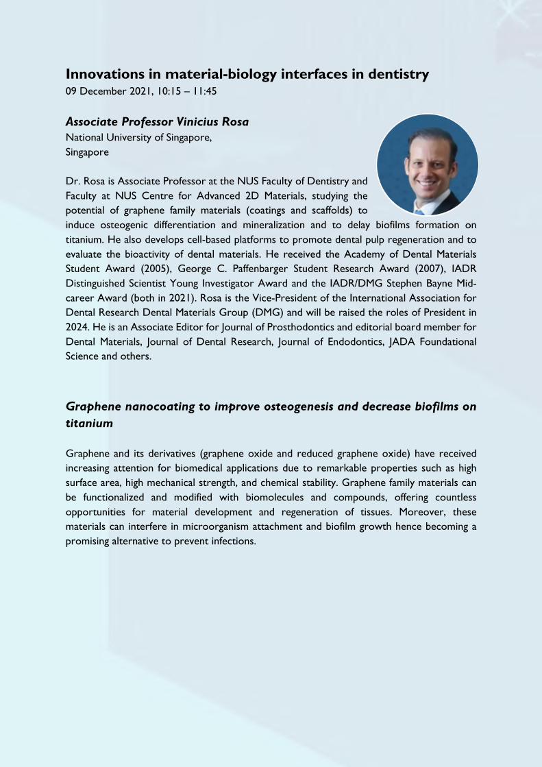
Innovations in material-biology interfaces in dentistry 09 December 2021, 10:15 – 11:45 Associate Professor Vinicius Rosa National University of Singapore, Singapore Dr. Rosa is Associate Professor at the NUS Faculty of Dentistry and Faculty at NUS Centre for Advanced 2D Materials, studying the potential of graphene family materials (coatings and scaffolds) to induce osteogenic differentiation and mineralization and to delay biofilms formation on titanium. He also develops cell-based platforms to promote dental pulp regeneration and to evaluate the bioactivity of dental materials. He received the Academy of Dental Materials Student Award (2005), George C. Paffenbarger Student Research Award (2007), IADR Distinguished Scientist Young Investigator Award and the IADR/DMG Stephen Bayne Mid-career Award (both in 2021). Rosa is the Vice-President of the International Association for Dental Research Dental Materials Group (DMG) and will be raised the roles of President in 2024. He is an Associate Editor for Journal of Prosthodontics and editorial board member for Dental Materials, Journal of Dental Research, Journal of Endodontics, JADA Foundational Science and others.
Graphene nanocoating to improve osteogenesis and decrease biofilms on titanium Graphene and its derivatives (graphene oxide and reduced graphene oxide) have received increasing attention for biomedical applications due to remarkable properties such as high surface area, high mechanical strength, and chemical stability. Graphene family materials can be functionalized and modified with biomolecules and compounds, offering countless opportunities for material development and regeneration of tissues. Moreover, these materials can interfere in microorganism attachment and biofilm growth hence becoming a promising alternative to prevent infections.

Professor Po-Chun Chang National Taiwan University, Taiwan Professor Po-Chun Chang is currently the Director of the Graduate Institute of Clinical Dentistry, School of Dentistry, National Taiwan University, Taipei, Taiwan. He obtained his PhD at University of Michigan, Ann Arbor, MI, USA and specialized in periodontology at National Taiwan University, Taipei, Taiwan. Prof. Chang is a well-known researcher in the fields of tissue engineering, regenerative medicine, periodontology and implant dentistry with publications in reputable journals. He is the editor-in-chief of the Journal of Taiwan Academy of Periodontology and the Journal of Periodontics and Implant Dentistry. Moreover, he is the Chair of the Publication Committee, Taiwan Academy of Periodontology. He also contributes to the ITI section in Taiwan as a study club coordinator. Development of biomimetic interfaces for alveolar ridge regeneration A biomimetic material-tissue interface plays an important role for the success of alveolar ridge regeneration. The research team worked on two aspects including the establishment of a nanofibrous interface with molecular cues on the barrier membrane and fabricating rapidly prototyped hydroxyapatite-based scaffolds with degradable alginate-based matrix. Both aspects aimed at mimicking the extracellular matrix and providing a microenvironment favorable for osteogenesis and had shown promising outcomes in vivo.

Professor Melanie Sadono Djamil Trisakti University, Indonesia Melanie Sadono Djamil is a Professor in Biochemistry Biomolecular and Oral Biology at Faculty of Dentistry, Trisakti University, Indonesia. She is the Head of Indonesia Dental Council – Indonesia Medical Council with a main responsibility in improving continuous dental education, registration of dentists in Indonesia and overall development of the dental profession. She is the past President of IADR-SEA Division from 2015 to 2017. She has been conducting research mainly on utilizing natural agents in dentistry. Prof. Djamil has received an honorary PhD in Oral Health Science, Faculty of Dentistry, Thammasat University, Bangkok, Thailand. Pandanus conoideus Lam. extract on HONE-1 cell line apoptosis induction Malignancy in head and neck areas considering high and become global issues. Pandanus conoideus Lam., as a chemo preventive compound that can enhance the immune response. Apoptosis intrinsic pathway, involves non-receptor induction from mitochondria. Cyt C with several cofactors activates caspase 3. Western blot analysis was used to test Pandanus conoideus Lam, which was immersed in antibody until a specific protein was formed. Based on the western blot analysis of Pandanus conoideus Lam on cell line HONE-1 activation of the target protein was proven at a concentration of 40 g/mL. Conclusion Pandanus conoideus Lam is able to induce apoptosis dependent on concentration and time of exposure, via activation of caspase-3 in cells line HONE-1.

Young Scientists’ Forum 08 December 2021, 15:00 – 16:30 Dr. Sriram Gopu National University of Singapore, Singapore Dr Gopu Sriram is an Assistant Professor on tenure-track at the Faculty of Dentistry, National University of Singapore. He is also a Thrust Co-lead for additive manufacturing for craniofacial and oral applications at the NUS Centre for Additive Manufacturing (AM.NUS). His current research is centered on oral and craniofacial tissue regeneration through the amalgamation of complementary and multidisciplinary tissue engineering technologies such as stem cells, organoids, microfluidics, 3D printing and non-invasive imaging. Through the design and biofabrication of biomimetic tissue constructs, his team focusses towards understanding cellular microenvironments, host-microbiome interactions, oral mucosal biology, ageing, potential therapeutic compounds, and regenerative strategies. His scientific contributions have been published in multidisciplinary journals such as Biofabrication, Materials Today, Lab-on-Chip, Biotechnology Bioengineering and Bio-Design Manufacturing. Multidisciplinary tools for biofabrication of gingival tissue constructs Over the last decade, technologies and tools to fabricate three-dimensional (3D) tissue constructs or grafts have progressed rapidly. Convergence of multidisciplinary tools such as 3D tissue cultures, bioprinting and microfluidics provides unique opportunities to biofabricate complex tissues with precision, spatial control, architecture, and functionality relevant to their in vivo counterparts. Further, imaging tools such as 2-photon and confocal reflectance microscopy provides opportunities for label-free and non-invasive visualization or optical biopsy of the tissues. In this symposium, we will discuss the recent efforts in the application of 3D culture, bioprinting and microfluidics combined with label-free, non-invasive imaging tools to construct biomimetic and functional gingival and oral mucosal tissues.

Dr. Waruna Dissanayaka The University of Hong Kong, Hong Kong Dr. Waruna Dissanayaka is an Assistant Professor at the Faculty of Dentistry, The University of Hong Kong. His research is currently focused on enhancing post-implantation stem cell survival and vascularization during tissue regeneration, understanding the molecular mechanisms behind the dental stem cells; intercellular cross-talk and lineage specific differentiation of dental stem cells into odonto/osteoblasts and endothelial cells. His doctoral research work of scaffold-free microtissue spheroids in dental pulp regeneration was awarded IADR William J Gies Award for Biomaterials and Bioengineering Research (2016) and Journal of Endodontics award for the Best Paper in Basic Science: Biology (2016). In addition, Dr. Dissanayaka received IADR-SEA Award for Tissue Engineering and Craniofacial Biology (2018), HKU-Dentistry Research Output Award (2016), IADR/Colgate Research in Prevention Travel Award (2012), and ISSCR Travel Award (2012). In 2020, he received the IADR Centennial Emerging Leaders Award for his achievements and potential in the advancement of dental, oral and craniofacial research. He has published over 40 scientific papers and is an editorial board member for Clinical Oral Investigations, BMC Oral Health and Journal of Endodontics.
Modulating vasculogenic microenvironment for tissue regeneration Reestablishment of an elaborated vascular network is one of the main focuses in the research in regeneration of craniofacial tissues. A microenvironment of ischemia and hypoxia in the biomaterial core drives propagation, differentiation and reorganization of progenitor/stem cells to assemble into a primitive microvascular framework. Cell-cell crosstalk as well as cell-cytokine interactions are key biomechanical factors controlling blood vessel functionality. Prevascularization strategies and delivery of vasculogenic molecules are important approaches in enhancing endothelial differentiation and stimulating the host regenerative response to promote vessel sprouting.

Dr. Farinawati Yazid Universiti Kebangsaan Malaysia, Malaysia Dr Farinawati Yazid is a paediatric dentistry lecturer and course coordinator for paediatric dentistry undergraduate program in the Faculty of Dentistry, University of Kebangsaan, Malaysia. She completed her dental education from University of Kebangsaan, Malaysia in 2006 and pursued her post graduate qualification from University of Malaya and graduated in 2014. Her research interests are regenerative dentistry, advanced biomaterial, oral fluid disease detection, and innovation for oral care. She has been able to secure multiple local and international research grants to continue her research projects. She has also published her research findings in reputable journals and has been invited as a speaker in multiple conferences.
Chemometric analysis and spectroscopy in caries detection
Ultraviolet (UV) spectroscopy is a technique that measures light absorption of an analyte. It can be applied in dental materials investigations and caries detection. UV spectroscopy coupled with chemometric analysis can be used for caries detection in saliva samples. Difference in caries severity can be measured with the absorption spectrum and analysed with chemometric models.

Dr. Nareudee Limpuangthip Chulalongkorn University, Thailand Dr. Nareudee Limpuangthip is an academic staff member of the Department of Prosthodontics, Faculty of Dentistry, Chulalongkorn University. She is also the Secretary and committee of the International Association for Dental Research – South East Asia division (IADR-SEA). She obtained her PhD from Chulalongkorn University in 2017. She has specialised in prosthodontics and her research interests are prosthodontic rehabilitation, geriatric dentistry, materials in prosthodontics, and quality of life-related to prosthodontics. Dr. Limpuangthip has published numerous publications in local as well as international journals.
Prosthodontic rehabilitation of oral function: Evaluation of patient-based outcome Oral function can be divided into 4 stages: healthy, oral frailty, oral hypofunction, and oral dysfunction stages. Appropriate prosthodontic treatment can maintain and prevent oral function declination. To assess oral function, three types of clinical evaluation are suggested: professional evaluation of dental and prosthesis status, objective evaluation by patients’ performance, and subjective evaluation by patient-reported outcomes. Clinical application and limitations are discussed based on available clinical evidences.

Dr. Pranai Nakaparksin Mahidol University, Thailand Dr. Pranai Nakaparksin graduated from Mahidol University Faculty of Dentistry, Thailand in 2012. After working in Mahidol University and private practice for one year he was accepted for implant/prosthodontic residency training and Master’s degree in Dental Materials from Indiana University, USA. He completed Implant fellowship at University of Florida, USA in 2017 and Loma Linda University, USA in 2018. Dr. Nakaparksin is a Fellow of American Collage of Prosthodontics, Fellow of Royal Collage of Dentistry of Canada, Associate Fellow of American Academy of Implant Dentistry, as well as fellow of International Team of Implantology. He is a co-author of Unanswered Questions in Implant Dentistry. Dr. Nakaparksin is full-time faculty at Mahidol University in Implant Center and Advanced General Dentistry Department. He also maintained his private practice in Bangkok, Thailand focusing on implant dentistry and prosthodontics.
Good to great implant treatment Implant restorations have increasingly become a popular treatment for edentulous esthetic zones due to their high functional success rate. However, research on their esthetic evaluation remains scarce. This research presentation will focus on the patient's perspective on single-tooth implant restorations and how dentists deliver exceptional quality treatments.

Dr. Huynh Cong Nhat Nam University of Medicine and Pharmacy at Ho Chi Minh City, Vietnam Dr. Huynh Cong Nhat Nam is a Lecturer and Researcher in the Laboratory of Oral-Maxillofacial Biology at the Faculty of Odonto-Stomatology, University of Medicine and Pharmacy at Ho Chi Minh City. He is a recipient of the Japanese JSPS postdoctoral fellowships (2018-2020) and carried out his postdoctoral research at Department of Immunology, Graduate school of Medicine and Faculty of Medicine, The University of Tokyo. His research interests include single-cell, osteoimmunology, big data in dentistry, bone regeneration, oral Biology, etc. He has a very strong publication record, including Journal of cellular biochemistry, PloS One, Bone, Archives of Oral Biology, Odontology, Journal of Nanomaterials, Cell Reports, Nature Metabolism, and Journal of Clinical Investigations.
Single-cell whole-transcriptome analysis in oral cancer A perspective on changes in oral tumor ecosystem has not been investigated thoroughly. This leads to difficulties in prognosis, also develop risk-stratified cancer screening, precision screening and treatment strategies. We deeply analyze and integrate published and our own oral cancer scRNAseq data, verify the resemblance of human and mouse data. We also identify the cell-cell interaction network, new biomarkers for diagnosis and prognosis and drug prediction.

Dr. Citra Fragrantia Theodorea Universitas Indonesia, Indonesia Dr. Citra Fragrantia Theodorea obtained her PhD degree from the Health Sciences University of Hokkaido, Japan in 2018. She is currently working as a Lecturer at Department of Oral Biology, Faculty of Dentistry, Universitas Indonesia. Her long-term research interest is oral microbiology which focuses on microbiome study and oral biomarker study in Non-Communicable diseases (Diabetes and Stunting). She has been working with Dr. Izumi Mashima from Ohu University, Japan under the supervisor of Prof. Futoshi Nakazawa to establish new species of the genus Veillonella such as Veillonella infantium and Veillonella Nakazawae which also have been published. “A Game Changer” of oral microbiome related to non-communicable diseases in Indonesia Nowadays, Indonesia is struggling to deal with non-communicable diseases (NCDs). The leading causes of mortality and morbidity in Indonesia have been dominated by the NCDs, such as cerebral stroke, heart diseases, diabetes mellitus and impaired development. NCDs are chronic, often asymptomatic and progressive, thus patients usually unaware of the disease until the sign and symptoms of its complications occur. Some inter-population biological studies reported that oral hygiene status might linked to NCDs based on comprehensive observations of gender-, geography-, ethnicity-, lifestyle-specific variations. Recently, the role of the oral microbiome in health and disease have provided insights into the various ecological events that act as drivers to shift the oral microbiota from homeostasis to fatal dysbiosis. However,determination of oral microbiome profile in Indonesia is challenging due to inter-population biology variation. Although Indonesia has high prevalence of NCDs, the oral microbiome data are not investigated yet. Here we show results of oral microbiome study related to NCDs in Indonesia which may be helpful to understand oral microbial configurations of NCDs in certain area of Indonesia.

Oral health and successful ageing: implications for research and practice 09 December 2021, 14:00 – 15:30
Professor W. Murray Thomson University of Otago, New Zealand Professor W. Murray Thomson is a dental researcher and specialist in dental public health, based at the University of Otago in Dunedin, New Zealand. His work in the renowned Dunedin Study has enhanced understanding of oral health through life. He is an expert on dry mouth and has developed widely used measures for use in clinical practice and research. He has also made important contributions to understanding of the oral health of the ageing population, and of the effectiveness of dental care in improving the lives of children and their families. To date, he has published 416 research papers and 5 book chapters, and his Scopus h index is 56. Now Editor-in-Chief of Gerodontology, he was the Community Dentistry and Oral Epidemiology Editor-in-Chief from 2015 to 2021. He is also Associate Editor for the European Journal of Oral Sciences. He was Editor of the New Zealand Dental Journal from 2007 to 2014. Research into ageing and oral health: some findings from New Zealand Research into the oral health of older people has burgeoned over the last couple of decades, spurred by increases in both tooth retention and the older population. Any consideration of oral health and ageing should acknowledge the fact that the chronic conditions of old age do not suddenly manifest themselves; rather, they have involved decades of subclinical organ system decline associated with biological ageing. Thus, the pace at which someone ages is a critical determinant of their health in old age. In this presentation, I will share some findings from NZ on the pace of ageing and on the oral health of older people, underlining the need for caution in interpreting associations observed in old age between aspects of oral and general health (most notably cognitive decline).

Professor Colman McGrath University of Hong Kong, Hong Kong Colman McGrath is a Clinical Professor in Dental Public Health at the Faculty of Dentistry, The University of Hong Kong. Colman is a past President of the Behavioural Epidemiology & Health Services Research Group of the International Association of Dental Research. Colman recently served on the steering committee for the 2020 Behavioral and Social Oral Health Sciences Summit and was the lead of the Summit’s Reactor Panel in Southeast Asia. Colman’s research interest is on the social impact of oral health, social inequalities, and the oral health of older people. He has published over 300 papers in international journals and conducted numerous community-based clinical trials. He has a particular interest in improving the oral health of people with disabilities and serves on the scientific committee of the International Association of Disability and Oral Health.
Oral Health & Active Ageing Our world is ageing and is ageing fast. In 2020 there were more than a billion people aged 60 and older, and many of them are living in China & Southeast Asia. In response to the aging population, The United Nations General Assembly declared 2021–2030 the’ Decade of Healthy Ageing’ in December 2020 with World Health Organization was asked to lead on its implementation. In this presentation we will review the dimensions and domains of oral health associated with active aging. The association between oral health and mortality – all cause mortality and disease-specific mortality. The impact of oral health on diet and nutrition – the specific oral health factors and methods for assessing nutritional status, as well as the evidence of the benefits of oral rehabilitation for improvement of nutritional status among those malnourished/ or ‘at risk’. There has been considerable research into how oral health contributes to ‘frailty’ and vice-versa, and much interest in ‘oral frailty’, how it develops and how to assess it. There has been a growing interest in the relationship between oral health and cognitive decline, the potential mechanisms at play, and much debate about the implications of this for geriatric care. There is limited evidence of how social networks and social capital affects older people’s oral health. Specific examples from local studies will be provided with a wealth of evidence from the Chinese Longitudinal Healthy Longevity Study.

Dr. Waranuch Pitiphat Khon Kaen University, Thailand Waranuch is Dean and Associate Professor of the Faculty of Dentistry, Khon Kaen University, Thailand. She received DDS from Chulalongkorn University, MPHM from Mahidol University, MSc and ScD in Epidemiology from Harvard University, and is a Diplomate of the Thai Board of Dental Public Health. Waranuch is active in research particularly relating to the oral health-systemic disease link and epidemiology of oral diseases. She has over 90 publications in peer-reviewed journals and has served in numerous editorial boards, including Journal of Dental Research (JDR), JDR – Clinical & Translational Research, and Oral Diseases. Waranuch is Past President of the Asian Academy of Preventive Dentistry (AAPD) and the Thai Society of Public Health Dentistry. She is currently Honorary Secretary of the South East Asia Association for Dental Education (SEAADE), member of the Board of Directors of DeRouen Center for Global Oral Health at the University of Washington, USA, and Distinguished Adjunct Faculty of Saveetha Institute of Medical and Technical Sciences, India.
Integrating oral health into national healthcare policy for aging society in Thailand It is apparent that oral health is an integral part of overall health and well-being, and this is especially critical in older adults. Oral diseases and inflammation have been shown to associated with many non-communicable diseases, such as diabetes and cardiovascular diseases. Furthermore, oral functions are crucial for proper nutritional intake and quality of life. As Thailand is becoming an aged society, the government has launched a national healthcare plan for healthy aging with the participations of several sectors. This presentation will discuss how we have integrated oral healthcare into the national healthy aging scheme to provide comprehensive care, prevention and health promotion for healthy aging in Thailand.

Scientific Presentation Schedule

Wednesday, 08 December 2021 10:15 – 11:45 Oral Session Hall 2: Dental materials and biomaterials I
Time Presentation ID
Title and presenter
10:15 – 10:30 001 MicroRNA-302a-3p Delivery by Surface-Modified Hydroxyapatite Nanoparticle and 3D-Printed TCP/HA Scaffold Promoted Osteogenic Differentiation Pirawish Limlawan, Chulalongkorn University
10:30 – 10:45 002 Enhancing Resin-Dentin Bond Durability Using a Novel Mussel-Inspired Monomer Kang Li, The University of Hong Kong
10:45 – 11:00 003 Coffee Bean Extract Inhibits Oxidative Stress Due to Nickel and Cobalt Exposures in Human Peripheral Blood Mononuclear Cells Dessy Rachmawati, University of Jember
11:00 – 11:15 004 A Biomimetic Titanate Nanoskeleton Coating on Zirconia Implants Weifa Yang, The University of Hong Kong
11:15 – 11:30 005 Nanodiamond-Coated Milling Bits Enhance Glossiness of Zirconia After Sintering James Kit-Hon Tsoi, The University of Hong Kong
11:30 – 11:45 006 Antibacterial Activity and Biodegradation of Bioactive Glass-Loaded Resin Composites Jiaojiao Yun, The University of Hong Kong

10:15 – 11:45 Oral Session Hall 3: Regenerative dentistry and craniofacial biology I
Time Presentation ID
Title and presenter
10:15 – 10:30 007 Semaphorin 4D Enhances Vascular Stabilization by Recruiting SHED Lili Zhang, The University of Hong Kong
10:30 – 10:45 008 HIF-1α Stabilization Enhances Odonto/Osteogenic Properties of SHED in-Vitro and in-Vivo Yuanyuan Han, The University of Hong Kong
10:45 – 11:00 009 Mechanical Force Regulates Induced Pluripotent Stem Cell Behaviors Thanaphum Osathanon, Chulalongkorn University
11:00 – 11:15 010 Whole-Transcriptiome Sequencing Identifies the Expression Profile of LncRNAs, CircRNAs, MiRNAs, and MRNAs in the Trigeminal Ganglion Associated With Trigeminal Neuropathic Pain Fei Liu, Sichuan University
11:15 – 11:30 011 C-X-C Motif Chemokine Ligand 1 and its Receptor C-X-C Motif Chemokine Receptor 2 in Trigeminal Ganglion Contribute to Nerve Injury-Induced Orofacial Mechanical Allodynia Jie Yang, Sichuan University
11:30 – 11:45 012 Tideglusib Enhances Odontogenic Differentiation in Human Dental Pulp Stem Cells Chatvadee Kornsuthisopon, Chulalongkorn University

10:15 – 11:45 Oral Session Hall 4: Oral health research I
Time Presentation ID
Title and presenter
10:15 – 10:30 013 Is Craniofacial Anatomy Related to the Severity of Paediatric OSA? Fabio Savoldi, The University of Hong Kong
10:30 – 10:45 014 CXCL12/CXCR4 Regulate Orthodontic Root Resorption via M1/M2 Ratio Xinyi Fang, Sichuan University
10:45 – 11:00 015 The Influence of Crown Coverage on the Accuracy of Static Guided Implant Surgery in Partially Edentulous Models: an in Vitro Study Zhen-yu Wang, Sichuan University
11:00 – 11:15 016 Comparison of the Effectiveness of Different Endodontic Systems in Preserving Original Root Canal Anatomy: a CBCT Guided Study Sonali Sharma, Army Dental Centre Research & Referral Delhi
11:15 – 11:30 017 Children's Oral Health Along the Belt and Road. Bixia Deng, The University of Hong Kong
11:30 – 11:45 018 Performance of Commonly Used Diode Laser for Dental Treatment Vicky Wenqing W. Xue, The University of Hong Kong

12:00 – 12:40 Poster Session Hall 1: Oral & Maxillofacial Surgery Research
Time Presentation ID
Title and presenter
12:00 – 12:05 019 Patient-Specific Estimation of Bone-Graft Volume for Sinus Augmentation Using CBCT Kuo-Feng Hung, The University of Hong Kong
12:05 – 12:10 020 Oral Squamous Cell Carcinoma Screening Within the Community of Hong Kong Abdulrahman B. Sakeen Alkandari, The University of Hong Kong
12:10 – 12:15 021 Factors Associated With Malignant Transformation of Oral Leukoplakia and Lichenoid Lesions Jia Yan Tan, The University of Hong Kong
12:15 – 12:20 022 Irrigating Solution’s Efficacy in Third Molar Surgery: a Systematic Review Iwan Ristiawan, Universitas Indonesia
12:20 – 12:25 023 The Risk and Benefit of Germectomy: a Systematic Review Erick E. Rajagukguk, Universitas Indonesia
12:25 – 12:30 024 Comparison Between Odontectomy and Coronectomy: a Systematic Review Sherly Santiago, Universitas Indonesia
12:30 – 12:35 025 Subcutaneous Emphysema Complications Following Third Molar Surgery: a Systematic Review Gilang W. Pratama, Universitas Indonesia
12:35 – 12:40 026 Chlorhexidine’s Efficacy on the Alveolar Osteitis Incidence: a Systematic Review Reni Fitralia, Universitas Indonesia

12:00 – 12:40 Poster Session Hall 2: Oral health research II
Time Presentation ID
Title and presenter
12:00 – 12:05 027 Remineralising Enamel Caries Using Dual-Action Peptide GA-KR12 Yun Niu, The University of Hong Kong
12:05 – 12:10 029 Measurement in Le Fort I Osteotomy Using CBCT Liuling Hui, The University of Hong Kong
12:10 – 12:15 031 A Systematic Review of Topical Anti-Erosive Agents on Dental Erosion Dhananthat Chawhuaveang, The University of Hong Kong
12:15 – 12:20 032 Salivary Methylome Analysis in Oral Cancer to Unravel Differentially Methylated Regions for Noninvasive Diagnosis John Adeoye, The University of Hong Kong
12:20 – 12:25 033 Changes in Parental Oral Health Attitude in a 2-Year Dental Caries Prevention Programme Weijia Luo, The University of Hong Kong
12:25 – 12:30 034 Root Caries Risk Assessment: Measuring Salivary IgA Specific to PAc(361–386) Yu Ichikawa, Niigata University
12:30 – 12:35 035 Restoration of Endodontically Treated Molars Using All Ceramic Endocrowns Tri M. Trinh, Can Tho University of Medicine and Pharmacy
12:35 – 12:40 036 Changes in Dental Environmental Stress With Home-Based Simulation Learning Sharon Tan, NYP
12:40 – 12:45 037 Trends in Tooth Loss Prevalence by Income Level in the United States Rui Yuan, Sun Yat-sen University

12:00 – 12:50 Poster Session Hall 3: Oral health research III
Time Presentation ID
Title and presenter
12:00 – 12:05 038 Factors Affecting Patient Referral From General Dental Practitioners to Periodontists Khimberly Joyce Flores, University of the Philippines Manila
12:05 – 12:10 039 Synthesis and Characterization of a Novel Calcium Strontium Silicate for Potential Dental Applications Mohamed M. Abdalla, The University of Hong Kong
12:10 – 12:15 040 Design and Development of an Oral Health Promotion App for Adolescents: Topic Guide Construction Pei Liu, The University of Hong Kong
12:15 – 12:20 041 Post-Surgical Complications and Their Detrimental Effects on Bone Gain in Vertical Guided Bone Regeneration: a Systematic Review and Meta-Analysis John Tay, National Dental Centre Singapore
12:20 – 12:25 042 Prevalence of Mild Cognitive Impairment among Elderly Dental Patients and Oral Health-related Factors Panatcha Weerapol, Chulalongkorn University
12:25 – 12:30 043 Oral Health Assessment of Children in a Charity Institution Lilia Co, Centro Escolar University
12:30 – 12:35 044 Relationship Between Periodontal Status and Cognitive Function of Rural Elderly in Taizhou Wen Jia Gu, Shanghai JiaoTong University School of Medicine
12:35 – 12:40 045 Management of Fear and Anxiety in Dental Treatments Under Local Anesthesia: a Systematic Review and Meta-Analysis of Randomized Controlled Trials Cheng Lu, The University of Hong Kong

12:40 – 12:45 046 Masticatory Performance of Stroke Patients: a Systematic Review With Meta-Analysis Xin Shu, The University of Hong Kong
12:45 – 12:50 047 Association Between Masticatory Performance and Oral Condition Factors in Adults: a Systematic Review and Meta-Analysis Yanpin Fan, The University of Hong Kong

12:00 – 12:55 Poster Session Hall 4: Dental materials and biomaterials II
Time Presentation ID
Title and presenter
12:00 – 12:05 048 Development of Novel β-Cyclodextrin Nano-Sized Biomaterials as Calcium Ions Carrier in Dentin Tubules Tai-Wei Feng, National Taiwan University
12:05 – 12:10 049 Properties of two Endodontic Sealers: an in Vitro Study Ngan T. Nguyen, University of Medicine and Pharmacy at Ho Chi Minh City
12:10 – 12:15 050 The Accuracy of 3D Printed Crowns Using a DLP Printer Joshua Y. Tan, National Dental Centre Singapore
12:15 – 12:20 051 Research and Development of Vietnamese Bite Force Meter Thu T. Tran, University of Medicine and Pharmacy at Ho Chi Minh City
12:20 – 12:25 052 Accuracy of Intraoral Scanners on Shade Determination Hung T. Phi, University of Medicine and Pharmacy at Ho Chi Minh City
12:25 – 12:30 053 Grad-CAM Helps Explainability of Supernumerary Teeth Detection by Deep Learning Shota Okazaki, Hiroshima University
12:30 – 12:35 054 Caries Around Restorations and Marginal Adaptation of ion-Releasing Materials vs Resin Composite: a Systematic Review and Meta-Analysis of Randomized Clinical Trials Eman ELbelasy, University of Minnesota
12:35 – 12:40 055 Effect of Opposing Structure on Marginal Bone Loss Around non-Submerged Dental Implants Odontuya Dorj, Mongolian National University of Medical Sciences
12:40 – 12:45 056 Effect of Thermocycling on Physical Properties of Resilient Denture Liners

Bharat Mirchandani, Thammasat University
12:45 – 12:50 057 Effect of Silver Diamine Fluoride on Vital Dental Pulp: a Systematic Review Ahmed Zaeneldin, The University of Hong Kong
12:50 – 12:55 058 Can Artificial Intelligence (AI) Design Natural Tooth Morphology? Hao Ding, The University of Hong Kong

15:15 – 16:45 Oral Session Hall 3: Oral microbiome, microbiology and immunology I
Time Presentation ID
Title and presenter
15:15 – 15:30 059 Biofilm Inhibitory Effect of a Biosurfactant Gunjan Gupta, The University of Hong Kong
15:30 – 15:45 060 Strain-Dependent Virulence Profiles in Enterococcus Faecalis Clinical Isolates Islam A. Ali, The University of Hong Kong
15:45 – 16:00 061 The Effect of Mauli Banana on Dual-Species Caries Bacteria Biofilm Dhya A. Karno, University of Lambung Mangkurat
16:00 – 16:15 062 Reuterin Inhibits Porphyromonas Gingivalis Virulence Genes in Root Canal Biofilm Model Deandra C. Wiriawan, Trisakti University
16:15 – 16:30 063 Oral-Genera Shifted Abundance in Indonesian Type-2 Diabetic Patients’ Salivary Bacteriome Devin Hendrawan, Universitas Indonesia
16:30 – 16:45 064 Oral Veillonella Profiles Associated With Stunted-Groups in Indonesian Children’s Dental-Biofilm Saint Diven, Universitas Indonesia

15:15 – 16:45 Oral Session Hall 4: Dental materials and biomaterials III
Time Presentation ID
Title and presenter
15:15 – 15:30 065 Fabricating Multilayered Magnetic-Nanoparticles Loading With DNase/Vancomycin for Elimination of Multispecies-Biofilm Mirza Muhammad Faran Ashraf Baig, The University of Hong Kong
15:30 – 15:45 066 Bonding of GICs to Aged SDF-Treated Carious Dentin Wen Thong Koh, University Malaya
15:45 – 16:00 067 Magnification of Iris Through Clear Acrylic Resin in Ocular Prosthesis Dinesh Rokaya, Walailak University
16:00 – 16:15 068 The Accuracy of Various Digital Impression for Completed-Arch Implant-Supported Prostheses. Pitchaporn Kosago, Prince of Songkla University
16:15 – 16:30 069 Magnetic Bioprinting Improves Viability of Three-Dimensional Lacrimal Gland Organoids Teerapat Rodboon, Chulalongkorn University
16:30 – 16:45 070 Biodegradable Magnesium Implant Enhances Angiogenesis and Alleviates Medication-Related Osteonecrosis of the Jaw in Rats Wangyong Zhu, The University of Hong Kong

Thursday, 09 December 2021 10:15 – 11:30 Oral Session Hall 2: Regenerative dentistry and craniofacial biology II
Time Presentation ID
Title and presenter
10:15 – 10:30 071 Supracrestal Gingival Connective Tissue-Derived Mesenchymal Stem Cells Regulated Macrophage Cytokine Secretion Jirawit Inthayat, Chulalongkorn University
10:30 – 10:45 072 Development of Polyurethane Copolymer Apply on Bone Tissue Engineering Chiu-Fang Chen, Kaohsiung Medical University
10:45 – 11:00 073 Characteristics of Teeth and Their Malformations Associated With Skeletal Dysplasias Worasap Tantibhaedhyangkul, Chulalongkorn University
11:00 – 11:15 074 Apoptotic Induction of Rhinacanthin-C Extracted From Rhinacanthus Nasutus on OSCC Khwanjira Paka-akaralerdkul, Mahidol University
11:15 – 11:30 075 Effects of EDTA on Proliferation, Migration, and Differentiation of SCAP Benya Sangwisutsai, Chulalongkorn University

10:15 – 11:45 Oral Session Hall 3: Behavioral, epidemiology and health service research I
Time Presentation ID
Title and presenter
10:15 – 10:30 076 Parental Concern About COVID-19 Transmission in the School-Based Dental Outreach Program Duangporn Duangthip, The University of Hong Kong
10:30 – 10:45 077 Half-Century Trend and Age-Period-Cohort Effect of Oropharyngeal Cancer in Singapore Marco A. Peres, National Dental Centre Singapore
10:45 – 11:00 078 Impact of Dental Caries on Academic Performance of Schoolchildren Faeq Ali Quadri, Universiti Sains Malaysia
11:00 – 11:15 079 Prevalence and Risk Factors of Erosive Tooth Wear Among Military Personnel in Singapore Nicholas S. Lim, National University of Singapore
11:15 – 11:30 080 The Effectiveness of the Instruction Media: Animation Promotes Oral Health Knowledge of Grade 1-3 Students in Bangkok Pagaporn P. Pisarnturakit, Chulalongkorn University
11:30 – 11:45 081 The Perceptions and Challenges of a First Year Dental Student : a Cross-Sectional Study Manhat K. Shinh, Adarsh Institute of Dental Sciences and Research

12: 00 – 12:50 Poster Session Hall 1: Oral microbiome, microbiology and immunology II
Time Presentation ID
Title and presenter
12:00 – 12:05 082 Observed Adverse Effects/ Events of Various COVID-19 Vaccines Amongst Post-Vaccinated Healthcare Workers John D. Layno, Lyceum Northwestern University
12:05 – 12:10 083 Inhibitory Activity of Stachytarpheta Jamaicensis (L.) Vahl Roots Extract on Some Selected Mouth Pathogenic Bacteria Juliyatin P. Utami, University of Lambung Mangkurat
12:10 – 12:15 084 Correlation Between Streptococci Mutans and Early Childhood Caries in Toddlers Ka Fung Yu, The University of Hong Kong
12:15 – 12:20 085 The Periodontitis Treating and Microbiota Changing Effects of Sapindus Mukorossi Seed oil on Ligature-Induced Periodontitis rat Model Shih-kai Lin, Taipei Medical University
12:20 – 12:25 086 Effects of Small Molecule on Streptococcus Mutans Biofilm Formation and Virulence Gene Expression Huan-Cai Lin, Sun Yat-sen University
12:25 – 12:30 087 Membrane Vesicles Improve Streptococcus Mutans Yina Cao, Sun Yat-sen University
12:30 – 12:35 088 Xylitol Gum Helps Reduce Cariogenic and Periodontopathic Bacteria on Dental Plaque Microbiota Yi Wu, Taipei Medical University
12:35 – 12:40 089 Probiotic Lactobacillus Reuteri Improves Oral Health of Fixed Orthodontic Patients Louise A. Halim, Trisakti University
12:40 – 12:45 090 Effects of Synthetic Lawsone Derivatives on Streptococcus Mutans Biofilm Formation

Pichayaporn Ratti, Prince of Songkla University
12:45 – 12:50 091 Antibacterial Efficacy of Contemporary Bioactive Dental Restoratives Against Streptococcus Mutans Rui L. Kong, National Dental Centre Singapore

12:00 – 12:55 Poster Session Hall 2: Behavioral, epidemiologic and health services research II
Time Presentation ID
Title and presenter
12:00 – 12:05 092 The Inter-Relationships Between TMD Severity, Emotional Distress, and Eudaimonic Well-Being Carolina Marpaung, Universitas Trisakti
12:05 – 12:10 093 Oral Health Services During COVID-19 Pandemic in Thailand Nipaporn Urwannachotima, Chulalongkorn University
12:10 – 12:15 094 Clinical Trial to Study the Effect of Medicated Toothpaste on Oral Health Mithra N. Hegde, A B Shetty Memorial Institute of Dental Sciences
12:15 – 12:20 095 Prevalence of Systemic Conditions Among Filipino Older Adults With Periodontitis Ma. Celina Garcia, University of the Philippines Manila
12:20 – 12:25 096 Prevention of Radiation Caries in Adult Patients of Head and Neck Cancer - Effectiveness Check of Topical Fluoride: a Systematic Review and Meta- Analysis Harsh Priya, All India Institute of Medical Sciences, New Delhi
12:25 – 12:30 097 Anti-Caries and Anti-Microbial Effects of School-Based Fluoride Programs in Myanmar Kaung M. Thwin, Niigata University
12:30 – 12:35 098 Barriers to Access Oral Health Care Among HIV/AIDS Adults Quan D. Tran, University of Medicine and Pharmacy at Ho Chi Minh City
12:35 – 12:40 099 Adverse Birth Outcomes and Dental Caries -a Systematic Review and Meta-Analysis Yuqi Cui, Sun Yat-Sen Unversity

12:40 – 12:45 100 Lesion Site Specific Associated With Arresting of Silver Diamine Fluoride Palinee Detsomboonrat, Chulalongkorn University
12:45 – 12:50 101 Spit No Rinse Toothbrushing Technique Against Dental Caries Formation Maria Cherry Serrano, Centro Escolar University
12:50 – 12:55 102 Validity and Reliability of Vietnamese Child Oral Health Impact Profile Hoang T. La, Odonto-Maxillo-Facial Hospital in Ho Chi Minh City

12:00 – 12:50 Poster Session Hall 3: Regenerative dentistry and craniofacial biology III
Time Presentation ID
Title and presenter
12:00 – 13:00 103 Bidirectional Associations Between Temporomandibular Disorders and Otologic Signs/Symptoms: an Umbrella Review Amelia Chew, National Dental Centre Singapore
12:05 – 12:10 104 A Novel AI- Based Method as a Prognosticator of the Biological Behavior of Oral Lesions Samyukta S, Chettinad Dental College and Research Institute
12:10 – 12:15 105 Sema3A Attenuates the Hypoxia Suppression of Osteogenesis in PDLSCs Xiaochi Chang, The Affiliated Hospital of Qingdao University
12:15 – 12:20 106 Increasing of HPV Infection in the Oral Cavity Significantly Rising the Risk of OSCC: a Meta-Analysis Research Natallia Pranata, Maranatha Christian University
12:20 – 12:25 107 Comparing the Effectiveness of Custom and Prefabricated Mandibular Advancement Appliances for Obstructive Sleep Apnea Mirian Dang, University of Toronto
12:25 – 12:30 108 Association Between Dental Restorations and Artifacts on the Head MRI Images of Pediatric Patients Pitchaya Tunlayadechanont, Mahidol University
12:30 – 12:35 109 Relationship Between Chronic Periodontitis and Inflammatory Cytokines in Patients Undergoing Maintenance Hemodialysis Haixia Lu, Shanghai Jiao Tong University
12:35 – 12:40 110 Genetic Polymorphism in Drug Induced Gingival Enlargements – a Systematic Review

Prasad Harikrishnan, KSR Institute of Dental Science and Research
12:40 – 12:45 111 Surgical Versus Non-Surgical Gingival Depigmentation Technique: a Comparative Preliminary Case Series Anika Dawar, All India Institute of Medical Sciences
12:45 – 12:50 112 Vietnamese Males’ Temporomandibular Joint Larger Than That of Vietnamese Females Lan V. Nguyen, University of Medicine and Pharmacy at Ho Chi Minh City

14:00 – 15:30 Oral Session Hall 2: Cariology and operative dentistry
Time Presentation ID
Title and presenter
14:00 – 14:15 113 Clinical Decision-Making in Complex Endodontic Cases Between Postgraduate Students Across Dental Specialties at a UK Dental School. Jonathan Liew, University of Sheffield
14:15 – 14:30 114 The Use of Video Demonstration in Teaching Restorative Dentistry Joseph M. Acosta, University of Baguio
14:30 – 14:45 115 Risk Factors Associated With non-Cavitated and Cavitated Carious Lesions in pre-School Children Sheetal Manchanda, The University of Hong Kong
14:45 – 15:00 116 Prevalence of Untreated Early Childhood Caries of 5-Year-old Children in Hong Kong: a Cross-Sectional Study Faith Miaomiao Zheng, The University of Hong Kong
15:00 – 15:15 117 Three Year Results of a Clinical Trial With two Basic Filling Materials in Cambodia Callum Durward, University of Puthisastra
15:15 – 15:30 118 Amount of Fluoride Delivered via a Topical Application of 38% Silver Diamine Fluoride Solution and 5% Sodium Fluoride Varnish Iliana G. Yan, The University of Hong Kong

14:00 – 15:30 Oral Session Hall 3: AI in dentistry and diagnostic science
Time Presentation ID
Title and presenter
14:00 – 14:15 119 Morphological and Functional Evaluation of AI-Generated and CAD-Designed Crowns Yanning Chen, The University of Hong Kong
14:15 – 14:30 120 Deep Learning to Automate Survival Prediction for Oral Cancer Chui Shan Chu, The University of Hong Kong
14:30 – 14:45 121 Implementation of Deep Learning Using a Convolutional Neural Network in Identifying Periapical Lesions Ade P. Dwisaptarini, Universitas Trisakti
14:45 – 15:00 122 Searching for ‘the Needle in a Haystack’: Deep Learning and the Recognition of Circulating Tumour Cells Weilan Wang, The University of Hong Kong
15:00 – 15:15 123 A CBCT Analysis of Shaping Ability of Two Rotary Systems Hieu N. Tran, University of Medicine and Pharmacy at Ho Chi Minh City
15:15 – 15:30 124 A Comparison of Two Methods for the Detection of Circulating Tumour Cells in Patients With Oral Cavity Cancer Kaiyuan Sun, The University of Hong Kong

14:00 – 15:30 Oral Session Hall 4: Oral health research IV
Time Presentation ID
Title and presenter
14:00 – 14:15 125 Evaluating Craniofacial Asymmetry in Class III Subjects Using Different Methodologies Deepal H. Ajmera, The University of Hong Kong
14:15 – 14:30 126 Lactobacillus Reuteri Probiotic Consumption Reduced the Inflammatory Responses in Orthodontic Patients Armelia S. Widyarman, Trisakti University
14:30 – 14:45 127 Enhanced Detection of Orthodontic Attachment Remnants Using UV Absorption Imaging Aileen U. Carlos, University of the Philippines Manila
14:45 – 15:00 128 Functional, Physical, and Psychosocial Impact of Degenerative Temporomandibular Joint Disease Xiao-Han Zhang, Peking University School & Hospital of Stomatology

Abstracts Scientific oral and poster presentations

001 MicroRNA-302a-3p Delivery by Surface-Modified Hydroxyapatite Nanoparticle and 3D-Printed TCP/HA Scaffold Promoted Osteogenic Differentiation Pirawish Limlawan1, 3, Stéphane Durual2, Anjalee Vacharaksa1, 3
1Faculty of Dentistry, Chulalongkorn University, 2University Clinic of Dental Medicine, University of Geneva, 3Faculty of Dentistry, Chulalongkorn University Objectives: Bone tissue engineering (BTE) is a preferable approach for critical-sized bone defects to restore functions and esthetics. This study aims to demonstrate the custom-shaped Tricalcium phosphate/Hydroxyapatite (TCP/HA) scaffold with bioactive molecules for promoting cell proliferation and differentiation. Methods: The macro- and microstructure of the scaffold are designed and printed using 3D-printing technology. The scaffold is incorporated with hydroxyapatite nanoparticles presenting cationic functional molecules 3-aminopropyltriethoxysilane to carry microRNA (HA-NPs-APTES-miR) as the bioactive molecules. Biocompatibility of the TCP/HA scaffold carrying HA-NPs-APTES-miR were tested in human primary osteoblast (HOB) and osteosarcoma cells (HOS) by using resazurin assay. Dispersion and the uptake of HA-NPs-APTES-miR was visualized by fluorescence microscopy. The delivery and regulation of targeted mRNAs was demonstrated by qPCR. The use of TCP/HA scaffolds carrying HA-NPs-ATES-miR for bone regeneration was demonstrated in the C57BL/6 mouse model. Results: TCP/HA scaffolds carrying HA-NPs-ATES-miR were biocompatible, and desired physical properties are demonstrated. HA-NPs-APTES are equally dispersed on the scaffolds, and intracellular particles are visualized at 24 hours. After a 5 day incubation on the scaffolds, miRNA302a-3p expression in HOS and HOB were significantly up-regulated by 1.4x10⁵- and 1.41x10⁶-fold respectively. In consistent with the decrease of its target genes, suggesting the successful delivery and the functions of miRNA302a-3p in HOB and HOS cultures. The down-regulation of COUP-TFII as the target mRNA resulted in upregulation of the osteogenic gene, RUNX2, in HOS and HOB by 8.6 and 6.22 fold, respectively. The in-vitro results were then confirmed in the in-vivo model to demonstrate bone regeneration by micro tomography and histological staining. Conclusions: The successful delivery of miRNA-302a-3p by HA-NPs-APTES and TCP/HA scaffold is demonstrated in both in-vitro and in-vivo models. The HA-NPs-APTES and TCP/HA printing provides a custom-shaped scaffold design to fit the defects, which could be promising as a future therapeutic approach for BTE. 002 Enhancing Resin-Dentin Bond Durability Using a Novel Mussel-Inspired Monomer Kang Li1, Chenmin Yao2, Yuhong Sun3, Kun Wang4, Xiangtao Wang2, Zhengzhi Wang4, James Kit Hon Tsoi5, Cui Huang2, Cynthia Yiu1
1Faculty of Dentistry, the University of Hong Kong, 2School and Hospital of Stomatology, Wuhan University, 3Peking University Shenzhen Hospital, 4Wuhan University, 5Faculty of Dentistry, The University of Hong Kong Objectives: To assess the effect of N-(3,4-dihydroxyphenethyl)methacrylamide (DMA) as a functional monomer in dentin bonding. Methods: The application of DMA as a functional monomer to cross-link with dentin collagen and polymerize with adhesive was evaluated using transmission electron microscopy (TEM), attenuated total reflection Fourier transform infrared (ATR-FTIR) spectroscopy, and atomic force microscopy (AFM) via Peakforce QNM mode. After validating the influence of DMA on collagen and adhesive separately, the overall performance of DMA/ethanol solution (1, 5, and 10 mmol/L) as a primer in

dentin bonding was examined using micro-tensile bond strength (MTBS), fracture pattern observation, and nanoleakage evaluation both immediately and after 10,000 times thermocycling aging. Besides, the inhibitory effect of DMA on endogenous matrix metalloproteinases (MMPs) was evaluated by in situ zymography using confocal laser scanning microscopy (CLSM) and the cytotoxicity of DMA was evaluated using cell counting kit-8 (CCK-8). Results: DMA successfully cross-linked with dentin collagen via non-covalent bonds and had no influence on the polymerization and mechanical properties of the adhesive. After 10,000 times thermocycling aging, the MTBS and nanoleakage expression of the DMA-treated groups showed no significant change compared with their immediate values (p > 0.05). In situ zymography revealed reduced endogenous proteolytic activities after the application of DMA, and no cytotoxicity effect was observed for DMA concentration up to 25 μmol/L. Conclusions: DMA could be used as a novel, biocompatible functional monomer in dentin bonding. It served as a bridge connecting the adhesive and dentin collagen network, unifying them as one whole structure to resist hydrolytic and protease degradation, thereby enhancing the durability of resin-dentin bond. 003 Coffee Bean Extract Inhibits Oxidative Stress Due to Nickel and Cobalt Exposures in Human Peripheral Blood Mononuclear Cells Dessy Rachmawati University of Jember Objectives: To investigate potential effect of coffee bean extract against radical superoxide production in PBMC cells exposed to Ni and Co Methods: The metal included NiCl2.6H2O and CoCl2.6H2O. The coffee extract is taken from East Java robusta coffee beans, they were macerated in 97% ethanol subsequently diluted into three concentrations: 125, 62.5, and 31.25 μg/ml. Oxidative stress activity was monitored by assessment of release of ROS production by using NBT assay Results: The robusta coffee bean extract concentration of 125 μg/ml showed the optimal concentration in reducing radical superoxide in PBMC cells-induced by Ni and Co followed by exposure to 62.5 and 31.25 μg/ml. Conclusions: Data obtained in the present study reveal that coffee bean extracts have capacity to inhibit radical superoxide due to Ni and Co exposures, which display potent PBMC oxidative activity capacities. More robust and well-controlled studies are still needed for a thorough understanding of the effect of coffee on other inflammatory markers in humans. 004 A Biomimetic Titanate Nanoskeleton Coating on Zirconia Implants Weifa Yang, Yu-Xiong Su University of Hong Kong Objectives: To develop and characterize a biomimetic titanate nanoskeleton coating on zirconia implants for enhanced bioactivity and osseointegration. Methods: A uniform thin layer of titanium was deposited on standard-sized zirconia pieces by Denton Sputter (Denton Vacuum, USA). The biomimetic titanate nanoskeleton was developed by low-temperature alkaline hydrothermal treatment in 5M sodium hydroxide at 60 °C for 24 hours. The

surface and cross-sectional structures were assessed by scanning electron microscopy (SEM). The topology was examined by atomic force microscopy (AFM). Results: Uniform thin titanium with a thickness of 400 nm was successfully deposited on zirconia pieces. After alkaline hydrothermal treatment, the titanate nanoskeleton layer was formed on zirconia, which showed a nanoporous structure with interconnected beams and structs under SEM. The spongy 3D nanonetwork mimics the trabecular bone structure in the nanoscale and network of the cytoskeleton, which may promote cell attachment and interact to trigger cytoskeleton-related signaling pathways for osseointegration. Conclusions: This study developed and characterized the biomimetic titanate nanoskeleton coating on zirconia. 005 Nanodiamond-Coated Milling Bits Enhance Glossiness of Zirconia After Sintering James Kit-Hon Tsoi, Zeqian Pan University of Hong Kong Objectives: This study aims at compare the various effects of cutting dental zirconia with nanodiamond-coated or ordinary milling bits. Methods: Four pieces of dental zirconia (WIELAND, Ø985mm, thickness:25mm) were used in this study. Two types of milling bits, one with nanodiamond-coating and one without, were used to cut the dental zirconia green blank in 3 different speeds (1000, 1500 and 2000 rpm) in a dental milling machine. The unsintered and sintered zirconia surfaces were evaluated with glossmeter, optical profilometer for surface roughness (Sa, Sz and Sq), SEM and EDX. SEM and EDX were also used to evaluate the milling bits. Three-way ANOVA was used to evaluate the effects (sintering, milling bit coating and milling speed) on these zirconia, and one-way ANOVA with Tukey post hoc test was used to evaluate statistical significance of glossiness and roughness at a=0.05. Results: With respect to glossiness, three-way ANOVA revealed that sintering, milling speed and milling bit coating are significant factors (p<0.05). The glossiness of the sintered zirconia block was statistically higher than that of the unsintered block (p<0.05). For sintered zirconia, the nanodiamond-coated milling bit yielded a statistically (p<0.05) higher glossiness in all milling speeds than uncoated, save for using uncoated milling bit in 1500 rpm. However, in terms of roughness, only sintering showed to be a statistically significant factor (p<0.001) outweigh other two factors, and sintered zirconia always to have lower Sa, Sz and Sq than unsintered. Using EDX, it can be seen a higher Carbon elemental content on the zirconia surface with the coated bit in all speeds. From SEM, milling bits were found to wear out after use and coating has peeled off the surface after cutting the zirconia. EDX has verified that the Carbon is reduced from the milling bits surface. Conclusions: Overall, the nanodiamond-coated milling bit can be operated in various speeds resulted in a higher gloss on the sintered zirconia block, while ordinary uncoated milling bit can only achieve the same glossiness at a designated speed. The type of milling bits and the speeds have no significant effect on the surface roughness. 006 Antibacterial Activity and Biodegradation of Bioactive Glass-Loaded Resin Composites Jiaojiao Yun1, Michael Burrow2, Jukka Matinlinna1, Yan Wang3, Alex Fok4, James Kit Hon Tsoi1 1University of Hong Kong, 2University of Hong Kong, 3Sun Yat-sen University, 4University of Minnesota

Objectives: Resin Composites tend to accumulate thicker biofilm compared with other restorative materials, such that the bacteria would biodegrade the composites. Bioactive glass (BAG) has been shown to suppress bacteria and promote remineralization of teeth. This study aimed to determine whether BAG-loaded resin composites would exhibit both antibacterial and anti-biodegradation properties. Methods: Experimental UDMA-based resin composites with 25 vol% fillers (containing 0, 1.9, 3.8, and 7.7vol% BAG (45S5 Bioglass) and remaining commercial silanized dental glass filler) were prepared. Each specimen was polished to achieve surface roughness (Ra) of ~0.12 μm by sequential polishing using 600, 1000 and 2500grit SiC-papers under running water. These samples (n=3) were then incubated anaerobically with S. mutans in brain heart infusion (BHI) at 37 °C for 3 days for biofilm maturation. The cell viability was assessed using XTT assays and CFU counting, and CLSM and SEM were used to assess the biofilm formation ability. The biodegradation of resin composites was evaluated via the changes in nanohardness by AFM and additionally using BHI immersion medium as positive control. Results: The surface roughness of all groups was shown in Table 1. XTT assays revealed that BAG-loaded resin composites had significantly reduced cell viability (Figure 1a), whilst CFU counts (Figure 2b) were decreased with BAG concentration of 3.8% and 7.7%. CLSM revealed noticeable dead/damaged bacterial cells (red staining) on BAG-resin composites, while resin composites with 7.7% BAG had shown a reduced biofilm thickness (44μm) (Figure 1c). Similarly, SEM images demonstrated the formation of large bacteria agglomerates on resin matrix, while the bacteria clumps became smaller with the increase of BAG concentration. BHI immersion did not statistically reduce nanohardness on all samples, but immersion with S mutans caused a statistically significant surface nanohardness reduction on 0% and 1.9% BAG-loaded composites. No biodegradation was found on higher BAG loading (3.8% and 7.7%) composites. Conclusions: Addition of at least 3.8 vol% bioactive glass might enable antibacterial, reduced biofilm formation, and anti-biodegradation ability in resin composites. 007 Semaphorin 4D Enhances Vascular Stabilization by Recruiting SHED Lili Zhang, Chengfei Zhang, Waruna Dissanayaka University of Hong Kong Objectives: To investigate the role of soluble Semaphorin 4D (Sema4D) in mediating the interactions between human umbilical vein endothelial cells (HUVECs) and stem cells from human exfoliated deciduous teeth (SHED) during vasculogenesis. Methods: Recombinant Sema4D was commercially purchased and used to treat SHED and HUVEC co-cultures in in-vitro Matrigel and microfluidic assays in order to examine its role in vascular stabilization. Trans-well (8µm-pore) assay was performed to assess the direct and indirect chemotactic effects of Sema4D on SHED. Furthermore, the expression of plexin-B1 (Sema4D receptor) was quantified by PCR and western blots. Additionally, plexin-B1 was knocked down in HUVECs and introduced in trans-well assay to confirm its role in mediating Sema4D function. The expression of mural cell markers was further quantified by western blots to investigate the effect of Sema4D on differentiation of SHED into pericytes. Results were statistically analyzed using Student’s t-test and 1-way ANOVA. Results: According to in-vitro Matrigel and microfluidic assay results, addition of Sema4D significantly enhanced the vessel-like network formed by HUVECs and the number of SHED co-localized with vascular structures. Trans-well assay showed that Sema4D indirectly increased the migration of SHED by inducing the secretion of endothelial derived factors. Western blotting results confirmed that the plexin-B1 expression was significantly higher in HUVECs compared to that of SHED. Accordingly, the

plexin-B1 knockdown in HUVECs significantly blocked the effects of Sema4D on the migration of SHED in trans-well assay. Furthermore, when SHED was cultured under Sema4D-treated conditioned medium from HUVECs, they were transformed into a more mature phenotype of mural cells as shown by significantly higher expression of mural cell markers, NG2, α-SMA, and SM22α compared with that of the control group at 72h. Similarly, microfluidic assay demonstrated that the addition of Sema4D significantly enhanced the co-localization of SM22α+ SHED with HUVEC vasculature confirming enhanced mural cell function. Conclusions: Sema4D promotes vascular stabilization by recruiting SHED through endothelial derived factors. 008 HIF-1α Stabilization Enhances Odonto/Osteogenic Properties of SHED in-Vitro and in-Vivo Yuanyuan Han, Chengfei Zhang, Waruna Dissanayaka University of Hong Kong Objectives: To explore the odonto/osteogenic potential of hypoxia inducible factor-1α (HIF-1α) stabilized stem cells from human exfoliated deciduous teeth (SHED). Methods: HIF-1α stabilization in SHED was achieved by knocking down prolyl-hydroxylase domain-containing protein-2 (PHD2) using target-specific shRNA lentiviral particles. Knockdown efficiency was measured by real-time PCR and western blotting. Subsequently, odontogenic and osteogenic marker genes DSPP, DMP-1, ALP, and Runx2 expression in SHED were examined by real-time PCR and western blotting after odonto/osteogenic induction for 14 days. ALP and Alizarin red staining were performed 7 and 21 days after odonto/osteogenic induction to evaluate mineralization capacity. Furthermore, full-length human tooth roots were used in an ectopic pulp regeneration model in severe combined immunodeficient (SCID) mice to examine the capacity of HIF-1α stabilized SHED in regenerating pulp-dentin complex. Tetracycline (20 mg/kg/5 days, s.c.) was injected as a probe to detect new dentin formation. After 4 weeks of implantation, the samples were retrieved and examined for histology by hematoxylin and eosin (H&E) staining and immunohistochemistry (IHC) for DSPP. Results: PHD2 was knocked-down in SHED with a 90-95% efficiency and in turn, HIF-1α was stabilized under normoxia. PCR and western blotting results showed a significantly increased expression of odonto/osteogenic genes including DSPP, DMP1, ALP, and Runx2 in HIF-1α stabilized SHED (P<0.05) compared with the control-SHED. In addition, ALP and Alizarin red staining also demonstrated that HIF-1α stabilization resulted in higher mineralization capacity in SHED after odonto/osteogenic induction (P<0.05). In vivo, examination for the auto-fluorescence from tetracycline showed that HIF-1α stabilized SHED group had more dentin-like tissue formation on the interface between the tooth dentin and pulp-like tissue. H&E staining and IHC for DSPP indicated that more odontoblast-like cells could be observed along the dentin wall in HIF-1α stabilized group (P<0.05). Conclusions: HIF-1α stabilization enhances the odonto/osteogenic potential of SHED both in-vitro and in-vivo. 009 Mechanical Force Regulates Induced Pluripotent Stem Cell Behaviors Jeeranan Manokawinchoke1, Prasit Pavasant1, Hiroshi Egusa2, Thanaphum Osathanon1 1Chulalongkorn University, 2Tohoku University Objectives: Mechanical force regulates various cell behaviors in stem cells. The present study aims to investigate the effect of mechanical force on murine induced pluripotent stem cells (miPS).

Methods: Adhering retinoic acid-treated miPS were treated with intermittent compressive force (ICF) in serum free culture medium (SFM) for 24 hours. The mRNA expression was determined by RNA sequencing analysis and real-time polymerase chain reaction. Immunofluorescence staining was used to investigate protein expression. Cell cycle analysis and apoptosis assay were performed to determine cell proliferation and death. Results: ICF induced cell cycle while reduced apoptotic cells and increased genes involved in cell cycle and P53 pathway, Ccnd1, Cdk6 and Ccng1. The ICF also upregulated transforming growth factor b1 (Tgfb1) expression in both mRNA and protein levels and pretreatment with TGF-b inhibitor (SB431542) prior to the ICF application abolished the ICF increased Ccnd1and Cdk6 expression. Conclusions: ICF promotes proliferation and cell cycle while inhibits apoptosis of miPS occurred via the TGF-b pathway. 010 Whole-Transcriptome Sequencing Identifies the Expression Profile of LncRNAs, CircRNAs, MiRNAs, and MRNAs in the Trigeminal Ganglion Associated With Trigeminal Neuropathic Pain Fei Liu, Zhong-Han Fang, Hong-Lin Liao, Qing-Feng Tang, Yan-Yan Zhang, Jiu Lin, Jie-Fei Shen State Key Laboratory of Oral Diseases & National Clinical Research Center for Oral Diseases & Dept. of Prosthodontics, West China Hospital of Stomatology, Sichuan University Objectives: Trigeminal neuropathic pain is a significant health problem. Recent studies have proved non-coding RNAs (ncRNAs) contribute to the development of neuropathic pain. However, it is still unclear that the expression profile of ncRNAs in the trigeminal ganglion (TG) and their functional mechanisms in trigeminal neuropathic pain. This study was aimed to identify the expression profile of long noncoding RNAs (lncRNA), circular RNAs (circRNAs), micro RNAs (miRNA) and message RNAs (mRNA) in the TG and their regulatory function in trigeminal neuropathic pain. Methods: Chronic constriction injury of the infraorbital nerve (CCI-ION) of mouse was performed to establish trigeminal neuropathic pain model. Von Frey filament test was performed to examine the head withdrawal threshold (HWT) of mice. RNA-seq was used to identify the expression profile of ncRNAs and mRNAs in the TG. RT-qPCR was performed to prove the expression of selected ncRNAs and mRNAs. The potential biological functions of differentially expressed (DE) ncRNAs and DE mRNAs were analyzed by bioinformatics analysis. Results: The HWT of CCI-ION mice significantly decreased since from 3 days after surgery. Totally, 216 lncRNAs, 14 circRNAs, 67 miRNAs, and 595 mRNAs were differentally expressed in the TG on 7 days after CCI-ION surgery. The results showed that 39 DEGs were known pain genes. GO analysis indicated that response to stress,membrane related cell components, binding related molecular functions were enriched. KEGG analysis showed that the most significantly involved pathways were ECM-receptor interaction, protein digestion and absorption, PI3K-Akt signaling pathway. Besides, the lncRNA/circRNA-miRNA-mRNA regulatory networks based on the ceRNA regulatory principle existed under the condition of trigeminal neuropathic pain. Conclusions: The findings in this study demonstrate ncRNAs play an important role in the development of trigeminal neuropathic pain, which indicates novel mechanisms underlying trigeminal neuropathic pain and bright novel therapeutic targets for orofacial pain targeting ncRNAs.

011 C-X-C Motif Chemokine Ligand 1 and its Receptor C-X-C Motif Chemokine Receptor 2 in Trigeminal Ganglion Contribute to Nerve Injury-Induced Orofacial Mechanical Allodynia Jie Yang1, Jie-fei Shen1, 2 1Department II of Prosthodontics, West China Hospital of Stomatology, Sichuan University,, 2State Key Laboratory of Oral Diseases, National Clinical Research Center for Oral Diseases Objectives: This study aimed to investigate the exact role of CXCL1 and CXCR2 in the regulation of orofacial ectopic mechanical allodynia and their potential downstream mechanisms in the trigeminal ganglion (TG). Methods: The head withdrawal threshold (HWT) of C57BL/6 mice was evaluated after inferior alveolar nerve (IAN) transection (IANX) and the effect of SB225002 (an inhibitor of CXCR2) on mechanical allodynia was examined. Then, the distribution and expression of CXCL1 and CXCR2 in the TG were further measured. To explore the potential downstream mechanism of CXCR2, the expression level of phosphor-PKC (p-PKC) and voltage-gated K+ (Kv) channels were checked after IANX and inhibition of CXCR2. Results: IANX triggered persistent (>21 days) mechanical allodynia and upregulation of CXCL1 and CXCR2 in the TG. In addition, exogenous CXCL1 also lowered the HWT, which was alleviated by CXCR2 and protein kinase C (PKC) antagonists. In addition, IANX increased the phosphorylated PKC (p-PKC) levels and decreased the expression of voltage-gated potassium channels (Kv), and these effects were reversed by inhibition of CXCR2. Conclusions: Our results demonstrated that CXCR2 participated in orofacial ectopic mechanical allodynia via downregulation of Kv1.4 and Kv1.1 through the PKC signaling pathway. 012 Tideglusib Enhances Odontogenic Differentiation in Human Dental Pulp Stem Cells Chatvadee Kornsuthisopon1, Kevin A. Tompkins2, Thanaphum Osathanon1, 3 1Dental Stem Cell Biology Research Unit, Faculty of Dentistry, Chulalongkorn University, 2Office of Research Affairs, Faculty of Dentistry, Chulalongkorn University, 3Chulalongkorn University Objectives: Tideglusib is a small molecule agonist of the canonical Wnt pathway. The present study investigated the influence of Tideglusib on human dental pulp stem cells (hDPSCs) proliferation, apoptosis, migration, and odonto/osteogenic differentiation Methods: hDPSCs were treated with 50 nM, 100 nM, or 200 nM Tideglusib. β-catenin accumulation was detected by immunofluorescence staining. Colony forming unit ability was assessed by staining with Coomassie blue. Cell cycle progression and cell apoptosis were investigated using flow cytometry. Cell migration was examined using an in vitrowound healing assay. Osteogenic differentiation was examined using alkaline phosphatase staining, alizarin red staining, and osteogenic-related gene expression. Gene expression profile was examined using a high throughput RNA sequencing technique Results: Tideglusib activated the Wnt signaling pathway in hDPSCs as demonstrated by an increase in cytoplasmic β-catenin accumulation and nuclear translocation. Tideglusib did not affect hDPSCs proliferation, cell cycle progression, cell apoptosis, or cell migration. In contrast, 50 nM and 100 nM

Tideglusib significantly enhanced mineralization and osteogenic marker gene expression (RUNX2, ALP, BMP2, and DSPP) Conclusions: Tideglusib enhanced the odonto/osteogenic differentiation of hDPSCs. Further, incorporating this bioactive molecule in a pulp capping material could be a promising strategy to promote dentin repair. 013 Is Craniofacial Anatomy Related to the Severity of Paediatric OSA? Fabio Savoldi1, Kevin K. Fung2, Wing-Sze Mak3, Elaine Y. Kan2, Yanqi Yang1, Ka-Li Kwok4, Min Gu1 1University of Hong Kong, 2Hong Kong Children’s Hospital, 3Kwong Wah Hospital, 4Kwong Wah Hospital Objectives: To investigate the relationship between either the severity of obstructive sleep apnoea (OSA), or the degree of upper airway obstruction, and a series of dentoskeletal, soft tissues, and airway parameters in children and adolescents of Hong Kong. Methods: Twenty-five patients (22 males, 3 females, mean age 13.4±3.1 years, 8 to 18-year-old,), with OSA were retrospectively analysed. OSA was diagnosed with polysomnography as apnoea-hypopnoea index (AHI) >1 event/hour (mean AHI 4.3±5.9 events/hour, 1.0 to 30.1 events/hour). Kinetic magnetic resonance imaging (kMRI) during drug-induced sleep was used to assess the dynamic airway patency and the obstruction type. Static MRI (sMRI) was also used for measuring dentoskeletal, soft tissue, and airway parameters. Factors related to OSA and obstruction severity were identified with multiple linear regression adjusted for age, sex, height, skeletal maturation, and body-mass index or axial parapharyngeal-fat-pads area (significance α=0.05). Results: From kMRI, circumferential obstruction was present in 44% of patients, while latero-lateral and antero-posterior in 28%. From kMRI, obstructions were retropalatal in 64% of cases and retroglossal in 36% (no nasopharyngeal obstruction was present). A difference was present between kMRI and sMRI in assessing the obstruction level (p=0.037), with kMRI showing higher prevalence of retroglossal obstructions. The airway area of the main obstruction site was not related to AHI. The maxillary basal latero-lateral width was related to both AHI (β=-0.573, p=0.008) and dynamic obstruction during sleep (β=0.633, p=0.011). Similarly, the maximum retropalatal latero-lateral width was related to both AHI (β=-0.408, p=0.040) and dynamic obstruction during sleep (β=0.515, p=0.016). Conclusions: In children and adolescents, the severity of OSA and the amount of dynamic obstruction were inversely proportional to the basal width of the maxilla and to the static retropalatal width of the airway. Targeted clinical treatments widening the transverse dimension of these structures may be particularly beneficial to these patients. 014 CXCL12/CXCR4 Regulate Orthodontic Root Resorption via M1/M2 Ratio Xinyi Fang1, Jiu Lin2, Jun Wang1 1West China hospital of Stomatology, 2West China hospital of stomatology of Sichuan University Objectives: Mechanical force-induced external root resorption is a major clinical side effect of orthodontic treatment. Recent study has revealed that M1 macrophages play a vital role in promoting orthodontic root resorption (ORR). In our present study, the mechanism of how mechanical force stimulation increases the M1/M2 macrophage ratio in periodontal tissue is investigated.

Methods: 40g force was utilized in the excessive stress-induced root resorption model. Root resorption was assessed using Micro–Computed Tomography (CT) and TRAP staining. CXCR4 antagonist AMD3100 was administrated to alleviated ORR. Flow cytometric, immunofluorescence, western blot and co-culture system of hPDLCs and THP-1 was performed to investigate roles of CXCL12/CXCR4 in ORR. Results: In the current study, we showed that C-X-C motif chemokine 12 (CXCL12)+periodontal ligament cells (PDLCs) and C-X-C chemokine receptor type 4 (CXCR4)+monocytes in the periodontal ligament (PDL) were significantly increased after force application with ongoing root resorption, and these effects were partially rescued after force removal in mice. The expression of CXCL12 in PDLCs was increased by force stimulation in a time- and intensity-dependent manner in vitro. Blockage of the CXCL12/CXCR4 axis using CXCR4 antagonist AMD3100 was sufficient to alleviate ORR and reverse the force-enhanced M1/M2 macrophage ratio. Further mechanism exploration showed that Ly6Chi inflammatory monocytes homed in a CXCL12/CXCR4 axis-dependent manner. The number and proportion of CD11b+ Ly6Chi inflammatory monocytes in cervical lymph nodes were significantly increased by force loading, accompanied by decreased CD11b+ Ly6Chi monocytes in the blood. These changes were blunted by intraperitoneal injection of AMD3100. Additionally, blockage of the CXCL12/CXCR4 axis effectively reversed M2 suppression and promoted M1 polarization. Conclusions: Collectively, our studies results indicate that force-induced CXCL12/CXCR4 axis mediates ORR by increasing the M1/M2 ratio in periodontal tissues through attracting Ly6Chi inflammatory monocytes and modulating macrophage polarization. 015 The Influence of Crown Coverage on the Accuracy of Static Guided Implant Surgery in Partially Edentulous Models: an in Vitro Study Zhen-yu Wang1, Jie-fei Shen2 1West China School of Stomatology, Sichuan University, 2West China Hospital of Stomatology, Sichuan University Objectives: To evaluate the influence of the crown coverage of surgical guides on the accuracy of static computer-assisted implant surgeries (sCAISs) in different partially edentulous situations. Methods: Acrylic models with five types of partially edentulous situations were fabricated in this study. In coDiagnostiX software (Dental Wings, Montreal, Canada), surgical templates were designed and fabricated with the following three different ranges of crown coverage (CC): reduced crown coverage (RCC) groups with templates covering 2 mm coronally above the undercut line of models, standard crown coverage (SCC) groups with tooth covering exactly extended to undercut line, and extended crown coverage (ECC) groups with coverage 2 mm beneath the undercut line. Then, fully guided implant placement into the acrylic models was performed by dental surgeons with more than 10 years of experience. In total, 120 models and 120 guides were manufactured, and 168 bone-level Straumann replica implants (4.1 × 10 mm, Institut Straumann AG, Basel, Switzerland) were inserted. Postoperative implant positions were scanned (Trios 3, 3 shape, Copenhagen, Denmark) and compared with preplanned virtual positions via coDiagnostiX (Dental Wings, Montreal, Canada). The angular, coronal and apical deviations were measured and analyzed to evaluate the accuracy of implant insertion. Statistical analysis was performed using one-way ANOVA and Tukey’s tests. Results: For single tooth missing situations in FDI position 11, the RCC group was similar to the SCC group and ECC group. For FDI with missing teeth #14 or 16, the SCC and ECC groups had no statistically significant difference (p > .05), while the RCC group had more coronal and apical deviation (p < .05). For multiple teeth missing situations, there was no difference among the RCC, SCC and ECC groups (p > .05). No difference was found among the five edentulous situations with different CCs (p > .05).

Conclusions: The CC of templates can significantly affect the accuracy of guided surgeries when implants are inserted in a single gap at posterior sites. Templates with CC extended to the undercut line may be an optimal choice for static guided surgeries. 016 Comparison of the Effectiveness of Different Endodontic Systems in Preserving Original Root Canal Anatomy: a CBCT Guided Study Sonali Sharma Army Dental Centre Research & Referral Delhi Objectives: To compare the canal centering ability and potential to preserve internal root canal anatomy during canal preparation with Rotary, Reciprocating systems and Vibratory adaptive Self-adjusting File Systems (SAF). Methods: Patients of 18-50 years with Mandibular first and second molars indicated for primary Endodontics were included and randomly allocated to three groups with 47 in each group: Group A Rotary file system; Group B: Reciprocating file system; Group C: SAF. The selected teeth were imaged with CBCT before and after preparation and metrically assessed for changes in curvature and transportation during canal preparation, mean total widths and outer and inner width measurements determined on each central canal path. Root canal transportation and centering ratio were evaluated at coronal, middle and apical thirds both mesiodistally and buccolingually. Areas of uninstrumented canals, total number of samples which can be prepared without deformation or separation and time taken for canal preparation was noted. Data were collected and statistically analyzed. Results: SAF exhibited less canal transportation and had better centering ability and the reciprocating systems performed better than rotary systems. At 2 mm the transportation with mean SD was Group A:1.52 (0.41), Group B: 1.30 (0.37) and Group C: 0.53 (0.27) (χ2 = 252.150, p = <0.001. There was a significant difference between the 3 groups in terms of Uninstrumented Area (Distal) (χ2 = 203.459, p = <0.001), with the median Uninstrumented Area (Distal) being highest in the Group: A. There was a significant difference between the 3 groups in terms of Preparation Time (Minutes) (χ2 = 97.634, p = <0.001), with the median Preparation Time (Minutes) being highest in the Group: C group. Conclusions The SAF system could maintain the original morphology and led to lesser transportation and had better centering ability and lesser percentage of unprepared surfaces. 017 Children's Oral Health Along the Belt and Road Colman McGrath, Bixia Deng University of Hong Kong Objectives: To report on the dental caries experience of children across BRI countries (member states) and to determine variations in dental caries experiences with respect to geographical regions (BRI routes). Methods: Dental caries experience of 5-year-old and 12-year-old children was obtained from the World Health Organization (WHO) oral health database. Data was assessed for completeness and contemporariness. Following on, variations in dental caries experiences were determined across BRI routes accordingly to (i) world bank calssificantion and (iii) WHO classification. Results: Less than a third (29.8%, 42/141) of BRI member states had 5-year-old dental caries experience available. Most (95.7%, 135/141) BRI member states had 12-year-old dental caries experience data. The contemporariness (up-to-datedness) of dental caries experience data varied; 45.2% (19/42) of 5-year-old data and 25.9% (35/135) of 12-year-old data were from the past decade

(since 2010). The mean dental caries experience at age 5 among BRI member states was 3.6 (SD 2.2), and dmft varies across BRI routes, p<0.001. The mean dental caries experience at age 12 among BRI member states was 1.9 (SD 1.2), and DMFT varies across BRI routes, p<0.001. Conclusions: Availability and up-to-datedness of dental caries experience data for children vary among BRI member states and across BRI routes. Dental caries experiences data is more readily available for 12-year-olds than 5-year-olds. However, data is not very up-to-date. Statistically significant variations in children's dental caries experience, both for 5-year-olds and 12-year-olds, exist across BRI routes. These findings have implications in addressing oral health needs as part of the Belt & Road Initiative 018 Performance of Commonly Used Diode Laser for Dental Treatment Vicky Wenqing W. Xue1, 2, Irene Shuping Zhao2, Edward Chin Man Lo1, Chun Hung Chu1 1Faculty of Dentistry, The University of Hong Kong, 2Shenzhen University Health Science Center, Shenzhen Objectives: To evaluate the performance of two dental diode lasers at 445 nm, 810 nm, and 970 nm at pulse and continuous-wave mode. Methods: This study examined two dental diode lasers, namely, SIROLaser Blue (445 nm, 970 nm), and AMD Lasers (810 nm). The performance of the lasers at their maximum power output were evaluated using a power meter. The study measured the range and the average of the power output at continuous-wave mode; and the peak as well as the average power output at pulse mode (50% duty cycle). Ten 30 s measurements were performed for each setting. Results: The percentage change of the average power at continuous-wave mode for 445 nm, 970 nm and 810 nm lasers were +20.5%, +22.6%, -7.5%, and the range of variation between actual power and power displayed on the device panel were from +0.534 W to +0.662 W, +0.368 W to +0.519 W, and -0.269 W to -0.143 W separately. At pulse mode, the percentage change of the average power were +29.3%, +23.6%, -5.4% for 445 nm, 970 nm and 810 nm lasers separately. The percentage change of the peak power were +36.6%, +25.5%, -2.5%, with the range of variation from +1.032 W to +1.126 W, +0.496 W to +0.543 W, and -0.099 W to -0.058 W for 445 nm, 970 nm and 810 nm lasers separately. Conclusions: The actual power output of this SIROLaser Blue diode laser is more than 20% higher than the power indicated on the device panel, whereas the actual power output of this AMD Laser is lower compared with the power displayed on the device panel. 019 Patient-Specific Estimation of Bone-Graft Volume for Sinus Augmentation Using CBCT Kuo-Feng Hung, Liuling Hui, Yiu Yan Leung University of Hong Kong Objectives: To estimate the bone-graft volumes needed for sinus floor elevation (SFE) for various elevation heights at the maxillary first (MM1) and/or second molar (MM2) sites using cone-beam computed tomography (CBCT), and to develop bone-graft volume prediction models based on the augmentation site, elevation height, and sinus width. Methods: CBCT scans with a medium-to-large field-of-view, performed at the Diagnostic Imaging Clinic, HKU Faculty of Dentistry, were screened for inclusion. Ten defined regions, above the MM1 and MM2 sites, in each maxillary sinus were semi-automatically segmented from the included CBCTs, and

their volumes were automatically measured using the ITK-SNAP program. The sinus widths at the height ranging between 8-16mm from the sinus floor were measured at the the MM1 and MM2 sites, respectively. Multiple linear regression analyses were performed to evaluate the association of the bone-graft volume estimated for SFE at different augmentation sites with elevation height and sinus width. Results: In total, 224 sinuses on 133 scans were included. For the same elevation height, the mean graft volume estimated for SFE at the MM1 site were greater than that at the MM2 site. The estimated graft volume was significantly associated with elevation height and sinus width (all p-values<0.001). Three prediction models proposed explained 89-91% of the variation in the estimated bone-graft volumes. The mean absolute deviations and absolute percentage deviations between the measured and predicted volumes ranged from 0.12-0.28cm3 and from 9.78-10.62%, respectively. Conclusions: Elevation height and sinus width are strong predictors of the bone-graft volume. The proposed prediction models may enable more patient-specific estimation of the bone-graft volume needed for SFE, which could facilitate the preparation of an adequate amount of bone-graft to avoid insufficient elevation of the sinus floor, and improve patient-clinician communication about the cost of bone-graft material. 020 Oral Squamous Cell Carcinoma Screening Within the Community of Hong Kong Abdulrahman B. Sakeen Alkandari1, Siu-Wai Choi1, John Adeoye1, Peter Thomson2 1The University of Hong Kong, 2James Cook University Objectives: Oral Squamous cell carcinoma (OSCC) is usually preceded by a benign oral lesion that can raise the suspicion of a dentist, prior to biopsy and histopathological assessment. This prospective pilot study aimed to identify people in Hong Kong who may be at risk of developing pre-malignant oral lesions. Methods: Advertisement for the study was placed on social media. Intraoral screening was performed by dentists in a community setting with the aid of a headlight and spatulas. Participants were categorized into “negative”, “low-risk positive”, and “high-risk positive” based on a preset criteria, such as suspicious exophytic lesions, erythroplakia, or proliferative verrucous leukoplakia for positive high-risk, or benign masses, leukoplakia, or submucous fibrosis for positive-low risk, or aphthous ulcers or thermal or chemical burns for negative lesions. A carbon monoxide ppm reading via a Smokerlyzer™ was recorded and basic demographic data was collected from each participant. Results: 774 participants (426 females) were screened, age mean(SD)[range] 55.35 (11.39) [21-89], female mean age, 56.21 and male mean age 54.77 years. Majority participants (99.2%) self-identified as ethnically Chinese. 127 were smokers, 703 negative for lesions, 46 (22 females) were positive, and 25 unclassified. Participants who screened positive were significantly older than those who screened negative (p<0.0001), were significantly more likely to smoke (p<0.00001), but not more likely to drink alcohol (p<0.387). The mean(SD) expired CO of those smoked was 5.64 (6.764) for males, 4.32 (5.036) for females, and significantly higher than for non-smokers, mean(SD) 1.906 (1.077). Conclusions: A higher age and behavior such as smoking increased the risk of developing pre-malignant oral lesions. Males were over-represented in the high-risk group and were more likely to partake in risk increasing behavior.

021 Factors Associated With Malignant Transformation of Oral Leukoplakia and Lichenoid Lesions Jia Yan Tan1, John Adeoye1, Siu-Wai Choi1, Peter Thomson2 1University of Hong Kong, 2James Cook University Objectives: To identify clinicopathologic factors associated with the malignant transformation of oral leukoplakia and oral lichenoid lesions (OPMDs) in the Hong Kong population. Methods: A retrospective review of 716 patients with clinical diagnosis of oral leukoplakia, oral lichen planus, or oral lichenoid lesions that underwent incision or excisional biopsy for confirmatory diagnosis was undertaken to investigate potential factors influencing the transformation to oral squamous cell carcinoma. Kaplan-Meier estimates and Cox proportional hazard analysis were used to explore the factors associated with malignant transformation. Results: Overall, 10.6% of the patients developed squamous cell carcinoma, with a mean duration of transformation of 90.40 months. A previous history of non-head and neck cancer [95%CI(1.039-7.369), P=0.042] or hematologic malignancies [95% CI(1.068-9.479), P=0.038] increased the risk of malignant transformation from OPMDs by 2.277- and 3.182-fold respectively. Patients presenting with OPMD lesions at the tongue or floor of mouth had a higher potential of malignant transformation [HR 2.592, 95% CI(0.176-0.846), P=0.017]. Induration was also associated with a 4.465-fold increased risk to malignancy transformation [95% CI(2.148-9.282), P<0.0001]. Patients treated with surgical excision and did not experience any recurrence had a lower risk of malignant transformation [HR= 0.362, 95% CI (0.149-0.881), P=0.025]. This risk was also statistically significant especially for those presenting with mild [HR= 4.519, 95% CI(2.015-10.138), P<0.0001], moderate [HR= 7.433, 95% CI (3.191-17.314), P<0.0001], or severe [HR= 7.634, 95% CI(2.108-27.646), P=0.002] dysplasia at diagnosis. A significant association of malignant transformation remained for those presenting with moderate [HR= 5.620, 95% CI(2.382-13.263), P<0.0001], or severe [HR= 5.742, 95% CI(2.598-12.691), P<0.0001] dysplasia upon follow up. Conclusions: Long term follow-up and clinicopathologic examination remain crucial steps as the results showed that clinicopathologic factors such as previous cancer history, lesions at the tongue or floor of mouth, induration, dysplasia and recurrence post treatment are associated with malignant transformation. 022 Irrigating Solution’s Efficacy in Third Molar Surgery: a Systematic Review Iwan Ristiawan, Dessy D. Utami, Vera Julia Universitas Indonesia Objectives: This systematic review aims to compare different irrigating solutions and their properties to determine which one is superior in its effects on third molar surgery. Methods: English-written articles during 2012-2021 were obtained from electronic databases: PubMed, ScienceDirect, and GoogleScholar. Keywords were verified by subject title; the effect of chlorhexidine, saline and povidone iodine at the postoperative of third molar surgery. Seventy articles were retrieved and only 18 online articles with available full-text versions were included. Results: The result of this study indicated that Chlorhexidine reduced the incidence of pain, swelling, alveolar osteitis, trismus, dry socket, bacteremia and killed Streptococcus mutans and Porphyromonas gingivalis bacteria better than other irrigants. Povidone iodine as irrigating agent could also control bleeding, reduce swelling and act as antiseptic. However, patients with hypersensitivity would usually get skin irritation. Ozonated water and gaseous ozone (O3) was better than saline and povidone iodine

in reducing pain and dry socket. Irrigation using 10% calendula officinalis had an effect on the bone preservation. Lactated Ringer’s solution can be beneficial with respect to cutting efficiency when lower handpiece loads are required. Conclusions: Irrigation procedure in third molar surgery is crucial. Appropriate irrigation materials can minimize post-operative incidences such as pain, swelling, alveolar osteitis, trismus, dry socket, bacteremia and antiseptic against streptococcus mutans and porphyromonas gingivalis bacteria. Chlorhexidine demonstrated superior properties than other irrigation materials: povidone iodine (polyvinylpyrrolidone iodine/ PVP-I), saline, ozonated water, drinking tap water, gaseous ozone (O3) and 10% officinalis essence. 023 The Risk and Benefit of Germectomy: a Systematic Review Erick E. Rajagukguk, Gilang W. Pratama, Vera Julia Faculty of Dentistry, Universitas Indonesia Objectives: This systematic review aims to assess risks and benefits of germectomy procedure. Germectomy is often advised in early teens when the roots are just one third developed. The risks and benefits were compared with odontectomy procedure Methods: English-written articles during 2012-2021 were obtained from electronic databases: PubMed, ScienceDirect, and GoogleScholar. Keywords were verified by subject title; germectomy, germinectomy, early third molar surgery. 51 articles were retrieved and only 12 online articles with available full-text versions were included Results: Twelve studies met the inclusion criteria and were included in this review. Although there were studies using different criteria compare between germectomy and odontectomy, they focused on the level of safety and complications that occur in germectomy when compared with odontectomy. In this study, the patient's age, tooth morphology, tooth extraction technique, duration of tooth extraction, tissue damage that occurred, and postoperative complications including pain and inflammation were considered. There was significant difference in the duration of surgery between germectomy and odontectomy in which germectomy demonstrated lower postoperative risks Conclusions: Delaying the removal of impacted mandibular third molars until a problem developed, subjects the patient to unnecessary pain, more complex and costly operations with higher risks for postoperative complications. Germectomy is a simple procedure, with shorter duration of procedure, leading to reduction in the incidence of complications. 024 Comparison Between Odontectomy and Coronectomy: a Systematic Review Sherly Santiago, Reni Fitralia, Vera Julia Universitas Indonesia Objectives: The purpose of this review was to conduct a systematic review regarding pre-operative precautions, possible interopertive risks, and post-operative complications. Methods: Previous studies published in January 2015 – December 2020 were searched from Pubmed, Scopus, Science direct for randomized or non-randomized controlled trials. All studies published in English comparing outcomes of odontectomy and coronectomy with complex extraction, near the inferior alveolar nerve canal were included. This systematic review was arranged with 247 cases. After application of the inclusion and exclusion criteria, a total of 15 articles were included in the study.

Results: Odontectomy is considered safe to be performed if the involvement of the inferior alveolar nerve with minimal risks of post-operative complication. Coronectomy should be performed if it presents post-surgical risk of damaging the inferior alveolar nerve. Conclusions: Both odontectomy and coronectomy offer the benefit of minimal risks of postoperative complications. The treatment outcomes and the availability of skillfull practitioners who master this technique are recommended for future. 025 Subcutaneous Emphysema Complications Following Third Molar Surgery: a Systematic Review Gilang W. Pratama1, Iwan Ristiawan2, Vera Julia1 1Universitas Indonesia, 2Universitas Indonesia Objectives: This study aims to review the occurence of subcutaneous emphysema after third molar surgery Methods: Articles published from PubMed (MEDLINE), Scopus and EBSCOhost between the year 2016 until 2021 were searched and identified. The keywords searched were third molar surgery and subcutaneous emphysema Results: From the search criteria, 233 articles were collected from the databases. After thorough evaluation, only 23 articles were eligible for the study. The eligible articles were analysed using PRISMA guidelines Conclusions: The literature showed that the most common occurence of subcutaneous emphysema was after third molar surgery using air-driven handpiece, instead of lowspeed handpiece. Future studies investigating other factors contributing to subcutaneous emphysema using larger number of samples are recommended. 026 Chlorhexidine’s Efficacy on the Alveolar Osteitis Incidence: a Systematic Review Reni Fitralia, Sherly Santiago, Vera Julia Universitas Indonesia Objectives: The review aimed to analyze the efficacy of chlorhexidine application on the incidence of alveolar osteitis after third molar surgery. Methods: Articles published from PubMed (MEDLINE), Scopus, EBSCOhost and Science Direct between the year 2014 until 2021 were searched and identified. The keyword searched were third molar surgery, chlorhexidine and alveolar osteitis. Results: From the search strategy, 179 articles were obtained; and only 15 articles were eligible for the study. Conclusions: Based on the review, patients who were given chlorhexidine after third molar removal showed fewer alveolar osteitis as a post-operative complication. However, the efficacy of chlorhexidine for reducing the incidence of alveolar osteitis needs to be investigated further in the future.

027 Remineralising Enamel Caries Using Dual-Action Peptide GA-KR12 Yun Niu1, May Lei Mei2, 1, Chun Hung Chu1 1University of Hong Kong, 2University of Otago Objectives: To investigate the antibiofilm and remineralising effects of the dual-action peptide GA-KR12 on artificial enamel caries. Methods: Enamel blocks underwent Streptococcus mutants biofilm challenge to create artificial caries. Then the blocks underwent biochemical cycling for three weeks. Enamel blocks were treated with sterilised deionised water as control or GA-KR12 twice daily during the cycling procedure. After the cycling procedure, the biofilm's architecture, viability, and growth kinetics on enamel blocks were determined, respectively, by scanning electron microscopy (SEM), confocal laser scanning microscopy, and quantitative (culture colony-forming units, CFUs). The mineral loss, calcium-to-phosphorus ratio, crystal characteristics and surface morphology of the enamel surface were determined, respectively, using micro-computed tomography, energy dispersive spectroscopy, X-ray diffraction and SEM. Results: SEM showed confluent growth of S. mutans in the control group but not in the GA-KR12-treated group. The dead-to-live ratios of the control group were lower than that of the GA-KR12-treated group (p<0.001), whereas the log CFUs of the control group were higher than that of the GA-KR12-treated group (p<0.001). The control group's lesion depth and mineral loss were higher than that of the GA-KR12-treated group (p<0.001). The calcium-to-phosphorus molar ratios of the control group were lower than that of the GA-KR12-treated group (p<0.001). The hydroxyapatite in the GA-KR12-treated group was better crystallised than that in the control group. The remineralised prismatic pattern on enamel blocks was observed in the GA-KR12-treated. Conclusions: The dual-action peptide GA-KR12 potentially is applicable for managing enamel caries because it inhibited the growth of S. mutans biofilm and promoted the remineralisation of artificial enamel caries. 029 Measurement in Le Fort I Osteotomy Using CBCT Liuling Hui1, Kuo Feng Hung1, Michael Bornstein2, 1, Yiu Yan Leung1 1University of Hong Kong, 2University Center of Dental Medicine Basel UZB Objectives: The aims of this study were to evaluate surgical angles for tuberosity dysjunction and linear and angular measurements for all horizontal cuts in Le Fort I osteotomy to avoid potential damage to the descending palatine artery using cone-beam computed tomography (CBCT). Methods: This study included 189 CBCT scans to assess six surgical angles on three different sites distal to maxillary second molars relative to the mid-sagittal plane (MSP) and the buccal cortical plane (BCP) for tuberosity dysjunction. Results: The mean surgical angles for tuberosity dysjunction varied from 58.90° to 95.28° and 74.85° to 100.93° according to the MSP and the BCP, respectively. For horizontal cuts, mean lengths (angles) for posterior buccal and lateral nasal wall osteotomies were 27.44mm (13.62°) and 33.20mm (9.60°), respectively, and a mean length of 47.12mm was measured for nasal septum osteotomy. Additionally, the presence of impacted third molar resulted in significantly higher mean surgical angles than those with fully erupted or without third molars. Conclusions: Overall, angles running through the buccal midpoint of third molars relative to the MSP (76.56°) or the BCP (92.31°) might be the most appropriate in clinical practice. Furthermore, some caution seems warranted when performing tuberosity dysjunction with impacted third molars.

031 A Systematic Review of Topical Anti-Erosive Agents on Dental Erosion Dhananthat Chawhuaveang, Ollie Yu, Chun Hung Chu University of Hong Kong Objectives: To provide an overview of anti-erosive agents for non-operative management of dental erosion. Methods: The literature search was conducted on four databases: PubMed, Cochrane Library, Scopus, and Web of Science. The keywords of the search were ("Dental erosion" OR "Tooth erosion" OR "Enamel erosion" OR "Dentine erosion" OR "Eroded" OR "Erosive lesion" OR "Erosive demineralization*") AND ("Prevention" OR "Protection" OR "Protective effect*" OR "Anti-erosion" OR "Anti-erosive”). The inclusion criteria were (i) in situ and clinical studies examined on topical anti-erosive application, (ii) articles published between the year 2010-2021. The exclusion criteria were (i) articles related to caries/white spot lesion, (ii) articles related to laser treatment, (iii) articles focused on saliva/salivary pellicle/plaque, (iv) articles studied about restorative materials/orthodontic appliances. Results: 51 articles were included for the analysis. The results showed 15 anti-erosive agents were used for the non-operative management of dental erosion, including commercially available agents and those still under development. Commercially available anti-erosive agents use fluoride, mineral-based materials, or biopolymers as active ingredients. Fluoride agents promote remineralization, but their effect on the management of dental erosion was inconclusive in the literature. Anti-erosive agents contain mineral-based materials that provide additional mineral ions to promote remineralization. Another type of agent used biopolymers as active ingredients. Because of the limited anti-erosive effects of these commercially available products, new agents with anti-erosive properties are under development, including titanium tetrafluoride, sodium trimetaphosphate, sodium hexametaphosphate, linear sodium polyphosphate, epigallocatechin gallate, polymethylvinylether-maleic anhydride, and carbopol. Conclusions: Anti-erosive agents were available for the non-operative management of dental erosion. These agents generally process good potential to prevent dental erosion. However, current evidence showed inconsistent or limited results to support the use of these agents in clinical settings. Therefore, more clinical studies are essential to demonstrate their success in the management of dental erosion. 032 Salivary Methylome Analysis in Oral Cancer to Unravel Differentially Methylated Regions for Non-invasive Diagnosis John Adeoye1, Siu-Wai Choi1, Li-Wu Zheng1, Yu-Xiong Su1, Peter Thomson2 1University of Hong Kong, 2James Cook University Objectives: Genome-wide methylation analysis in oral cavity squamous cell carcinoma (OSCC) for liquid biopsy is often performed using array platforms with restricted CpG site interrogation and tissue samples which may not represent ‘dynamic’ alterations. This study aims to identify differentially methylated CpG regions that are diagnostic for OSCC in saliva at the genome scale. Methods: Saliva samples of eight patients (4 OSCC and 4 oral mucosa disease) were randomly selected from the specimen biobank prospectively obtained for methylation analysis and verification. Genomic DNA extraction was performed on cell pellets and subjected to whole-genome methylation analysis at base-pair resolution using reduced-representation bisulfite sequencing (RRBS). Following quality control, filtered reads were aligned to the hg38 reference genome assembly, and differential methylation analysis was performed on CpG sites and 100bp regions with a minimum of 10X coverage that was common to all samples. Aberrantly methylated sites and regions were selected using a mean

methylation difference > 10 and q-values < 0.01. Further, Gene Ontology terms based on the Biological process and KEGG pathway analysis were used to enrich differentially methylated regions (DMRs) annotated to genes. Results: The analysis identified a total of 148,143 CpG sites that were differentially methylated in OSCC. Furthermore, 1823 DMRs annotated to 1066 genes were identified which were significantly hypermethylated (66.5%) than hypomethylated (33.5%). DMRs were frequently annotated to the promoter region (38%) compared to intergenic, intronic, or exonic regions (18 – 22%). DMR-related genes are enriched in the regulation of cell junction assembly, pattern specification process, and axonogenesis in the Biological Process of Gene Ontology terms as well as the Hippo signaling cascade following KEGG pathway analysis. Conclusions: Overall, this study suggests that the aberrant methylation of genes involved in cell-cell and cell-matrix adhesion are detectable in the saliva of OSCC patients and is likely to be utilizable for disease detection. 033 Changes in Parental Oral Health Attitude in a 2-Year Dental Caries Prevention Programme Weijia Luo, Gillian lee, Gloria Wong, May Wong University of Hong Kong Objectives: This study aimed to evaluate the changes in parental oral health attitude regarding adults and children after individualized oral health education (OHE). Methods: This is a longitudinal analysis of data from a two-year randomized controlled trial comparing the effectiveness of two kinds of toothpaste. Parent-child dyads were recruited (n=579). At baseline, the children aged one-year-old and were at high-caries-risk, and all parents received individualized OHE. A self-completed structured questionnaire was used to collect the parental oral health attitude regarding adults and children at baseline and two-year follow-up. Results: A total of 472 parent-child dyads completed the two-year programme (follow-up rate: 81.5%; children’s mean age: 38.6±1.9 months). There was a significant improvement in the parental oral health attitude for adults and children (Attitude-adults: baseline: 7.0±1.0, follow-up: 7.2±1.0; Attitude-children: baseline: 9.1±2.0, follow-up: 10.6±1.9; P<0.001). Significantly more parents at follow-up were confident to do toothbrushing for children, knew about the proper toothbrushing method for children, could brush their children’s teeth when they fell asleep, and would insist toothbrushing even if their children did not want to (P<0.001). Conclusions: Parents had improved their oral health attitudes after receiving individualized OHE. They were more motivated to keep good oral hygiene for their children. 034 Root Caries Risk Assessment: Measuring Salivary IgA Specific to PAc(361–386) Yu Ichikawa, Noboru Kaneko, Kaname Nohno, Hiroshi Ogawa Niigata University Objectives: In the superaging society of Japan, root caries is a problem among older adults. Root surface caries occurs even in those at low risk of enamel caries because the critical pH of root surface dentin is higher than that of enamel. Evaluating the risk of root caries and preventing it is essential because it tends to be difficult to treat. In this study, we determined the number of Streptococcus mutans, one of the main causative bacteria of dental caries, and the amount of salivary IgA antibody specific to its surface protein antigen PAc in older adults to assess the root caries risk factors.

Methods: Stimulated saliva samples were collected from 378 adults aged 76 years living in Niigata City. Salivary IgA antibody to amino acid residues 361–386 of S. mutans PAc (PAc(361–386)) was measured in these samples. Dental examinations were conducted at the baseline and 1 year later, and the total number of untreated and treated root caries was recorded as root DFT. To assess the risk of root caries development, logistic regression analysis was performed using salivary IgA antibody to PAc(361–386), levels of S. mutans(log CFU/swab), salivary flow rate (ml/min), number of root exposed teeth, and gender as explanatory variables. Results: Among all participants, 307 individuals (144 females) with exposed root surfaces and without missing data were included in the analysis. The mean root DFT at baseline was 3.77 (SD: 3.66) and 36.5% of individuals exhibited increased root DFT after 1 year. Of the explanatory variables, only low levels of salivary IgA antibody to PAc(361-386) (<25th percentile value) were significantly associated with increased root DFT (odds ratio: 1.95; 95% confidence interval: 1.14–3.34). Conclusions: Low levels of salivary IgA antibody to PAc(361–386) are a risk factor for increased root caries in older adults. 035 Restoration of Endodontically Treated Molars Using All Ceramic Endocrowns Tri M. Trinh, Thao T. Do Can Tho University of Medicine and Pharmacy Objectives: The objective of the present study was to analyze the clinical outcomes of 56 lithium disilicate endocrown in 1-year functional period. Methods: A cross sectional study on 56 lower molars that had endodontic at the Can Tho University Hospital from 4/2020 to 8/2021. The information about age, gender, teeth anatomy after preparation, colour match, marginal adaptation, the satisfy of patients with three criteria in the baseline: color match, morphology and comfort of treatment. Restorations were performed with lithium disilicate porcelain material with CAD/CAM technique and assessed by the modified US Public Health Service criteria (USPHS). Data were processed using Stata 14.0. Results: 56 endocrowns from 56 patients (39.3% women and 60.7% men) were evaluated. Most of tooth were complex cavity with 55,4%. After preparation, the average of the pulp chamber's diameter was 4.6 ± 0.6mm, the depth of the pulp chamber was 4.8 ± 1.2mm and the average thickness of the occlusal surface was 2.5 ± 0.7mm. Average preparation time was 11.16 ± 1.2 minutes and time to try and cementation the restoration was 15.6 16 ± 0.7 minutes with normal distribution. After 1 year, there were 96.4% of the teeth were evaluated as successful while only 1 tooth had to be replaced and 96.4% of patients felt very satisfied with the results of treatment. Conclusions: Endocrown was a minimally invasive and effective method to treat molars with endodontic treatment. 036 Changes in Dental Environmental Stress With Home-Based Simulation Learning Sharon Tan2, 1, Anshad Ansari1, Adrian Ujin Yap3, 1 1NYP, 2SSHSPH, 3NTFGH, NUHS Objectives: This study aims to determine stress levels among Oral Health Therapy (OHT) students during the COVID-19 pandemic, as well as the change in stress levels among students before and after a Home-Based Simulation Learning (HBSL) program.

Methods: Following the announcement of school closures in May 2020, a HBSL program involving the use of portable manikins and instruments by Nanyang Polytechnic School of Health & Social Sciences (Oral Health Therapy) students and staff at home was implemented. A 38-item Dental Environmental Stress (DES) questionnaire was distributed to students immediately before and after the three-week HBSL program. Wilcoxon signed-rank tests were carried out to assess changes in DES domains, and generalized estimating equations were used to assess factors associated with DES. Results: A response rate of 98.6% (69 completed responses) was obtained.The top stressors among OHT students during the pandemic are examinations and grades, the fear of failing the course, and the fear of being unable to catch up. No statistically significant change in overall Dental Environmental Stress score was noted before and after HBSL, although there was an increase in stress levels in the Academic Work domain (p<0.01). Year 3 students had higher stress scores for the domains of Educational Environment (B = 1.07, 95% CI 0.81 – 1.34) and Academic Work (B = 1.17, 95% CI 0.85 – 1.49) as compared to Year 1 students. No statistically significant differences in stress levels were noted across genders, ethnicities and residence types. Conclusions: In spite of the COVID-19 pandemic and school closures, stress levels among Oral Health Therapy students remained relatively stable following a Home-Based Simulation Learning program. Final-year students experience more dental environmental stress than first-year students. 037 Trends in Tooth Loss Prevalence by Income Level in the United States Rui Yuan, Qinghui Zhi Sun Yat-sen University Objectives: Income inequality has increased dramatically in the US over the past few decades. Considering the established association between income and health, this increasing income gap may provide insights into the dynamics of the oral disease burden in the US. Methods: Cross-sectional analyses were performed using the National Health and Nutrition Examination Survey (NHANES) database from 1999 to 2018 containing information of 12481 people aged 60-80. Two survey-weighted logistic regression models were used to analyze unadjusted (model 1) and age, sex, race, education, obesity, and insurance adjusted (model 2) prevalence trend of tooth loss for participants between different income levels. Results: The prevalence of tooth loss no significantly declined in the highest-resources group (the richest 20% of the population) during the study period (1999-2018): edentulous from 4.24% to 2.2% (P=0.43 for trend), severe tooth loss from 10.14% to 5.88% (P=0.36 for trend), and lack of function dentition from 28.07% to 16.76% (P=0.106 for trend). In the remainder of the population, the prevalence of edentulous significantly declined from 27.78% in 1999 to 14.79% in 2018 (P<0.001 for trend), as did severe tooth loss from 41.57% to 21.06% (P<0.001 for trend), and lack of function dentition from 64.29% to 40.57% (P<0.001 for trend). Adjusted for age, sex, race, education, obesity and insurance, tooth loss prevalence trend showed similar results. Conclusions: This study found substantial and decreasing disparities in tooth loss prevalence between the richest and poorest participants in the NHANES from 1999 to 2018. The trend of tooth loss was stable with no significant reduction for the richest 20% of participants but decreased remarkably in the remainder of the population.

038 Factors Affecting Patient Referral From General Dental Practitioners to Periodontists Khimberly Joyce Flores1, Ma. Celina Garcia2, Kristine Rachelle Pacete-Estrera2 1University of the Philippines Manila, 2University of the Philippines Manila Objectives: The identification of factors that influence the decision of general dental practitioners (GDP) to refer patients to periodontists could serve as a basis for the development of a more efficient periodontal referral process. Therefore, this study aimed to determine the factors that affect the referral patterns of Filipino general dental practitioners to periodontists. Methods: A self-administered survey was conducted among 75 general dentists practicing in the City of Manila, Philippines. The questionnaire collected data on GDP demographics, periodontal referral patterns, and clinical and nonclinical factors that may influence patient referral to a periodontist. Descriptive statistics (frequency and percentage) were employed to identify the primary factors that influence periodontal referrals to specialists. Results: Among the 75 respondents, 61.3% indicated that they treat periodontitis patients as part of their general dental practice. Eighty percent of the GDPs would prefer to refer periodontal patients to a specialist, with 76% having estimated that they refer <10 patients per month. The type of periodontal condition (81.3%) and the patient’s systemic condition (80%) were the top-cited clinical factors that affect referral of patients from GDPs to periodontists. Other clinical factors that are considered by GDPs are pocket depths (65.3%), degree of mobility (61.3%), amount of bone loss (58.7%), and severity of bleeding (49.3%). Among nonclinical factors considered when referring patients to periodontists, GDPs cited the willingness of patients to cooperate with treatment (32%), the GDP’s perceived personal skill in managing periodontal patients (24%), the specialist’s postgraduate training (21.3%), and the GDP’s proximity to a periodontist (5%). Conclusions: Multiple factors affect the decision of general dentists practicing in the City of Manila in referring periodontal patients to a specialist. The primary clinical factor considered was the type of periodontal condition while the top nonclinical factor was the patient’s willingness to cooperate with treatment. 039 Synthesis and Characterization of a Novel Calcium Strontium Silicate for Potential Dental Applications Mohamed M. Abdalla, Christie Lung, Mohammed Bijle, Cynthia Yiu University of Hong Kong Objectives: To synthesize and characterize calcium strontium silicate ceramic, as a new material for potential use in various restorative and regenerative dental treatments. Methods: Calcium silicate (CS), and calcium strontium silicate (CSR) were synthesized by the sol-gel method. The synthesized powders were characterized using x-ray diffraction (XRD). Setting time was measured using the Gilmore needle indentation technique. The compressive strength was measured using the universal testing machine at crosshead speed of 1.0 mm/min until fracture occurred. The in vitro apatite-formation was evaluated by SEM and EDX, and FTIR. The NIH/3T3 cells viability against the synthesized ceramics was tested using MTT assay. The osteogenic differentiation of HDPSC was evaluated by measuring the alkaline phosphatase activity (ALP), and mineralization by alizarin red stain (ARS). The ions release of Ca, Sr, and Si were measured using inductive coupled plasma-optical emission spectroscopy (ICP-OES). The independent t-test was used to analyze setting time, compressive strength, and ARS results. One-way ANOVA was used to analyse ALP and cell viability results. The Bonferroni post hoc test was used to establish significance (p < 0.05)

Results: CSR was successfully synthesized, and phase identification was confirmed with XRD. The initial and final setting times were significantly shorter in CSR 5 ± 0,75 min, 29 ± 1.9 min than CS 8 ± 0.77 min, 31 ± 1.39 min, respectively. No significant difference was found between the compressive strength of CS and CSR (p > 0.05). CSR demonstrated higher apatite forming-ability and cell viability than CS. The ALP activity was significantly higher in CSR 1.16 ± 0.12 than CS 0.92 ± 0.15 after 14 d of culture (p<0.05). ARS showed higher mineralization in CSR than CS after 14 and 21 d culture times. Conclusions: Calcium strontium silicate was successfully synthesized by the sol-gel method. CSR demonstrated enhanced biocompatibility, bioactivity and physicomechanical properties than CS. CSR could be a promising material for vital pulp therapies and might aid in the prognosis of regenerative endodontic treatments. 040 Design and Development of an Oral Health Promotion App for Adolescents: Topic Guide Construction Pei Liu, Ka Fung Yu, May Wong University of Hong Kong Objectives: To construct the topic guide for conducting a semi-structured interview or focus group discussion on exploring the needs of adolescents, their preferences of contents, and functions of an app to facilitate the design and development of a mobile application (app) to improve adolescents' oral health in Hong Kong. Methods: The topic guide was first drafted based on a narrative literature review on mobile health (mHealth) promotion among adolescents. Four pilot semi-structured interviews were conducted with students aged 12-18. The interviews were recorded, transcribed, and used to refine the topic guide. Results: Six key areas were constructed: (1) strength of an app against other existing oral health promotion strategies, (2) desired oral health-related information and contents to be included in the app, (3) functions of the app: the necessity of self-assessment, interaction, feedback, and reminder to maintain or improve oral health behavior, (4) prototype and interface design, (5) data protection for users, (6) effective promotion strategy among adolescents in using the app. Conclusions: This constructed topic guide is ready to be used to conduct a semi-structured interview or focus group discussion on adolescents to facilitate the design and development of a mobile application (app) to improve adolescents' oral health. 041 Post-Surgical Complications and Their Detrimental Effects on Bone Gain in Vertical Guided Bone Regeneration: a Systematic Review and Meta-Analysis John Tay1, Ethan Ng1, Jacinta Lu2, Clement Lai2 1National Dental Centre Singapore, 2National University of Singapore Objectives: Guided bone regeneration (GBR) utilises a barrier membrane to allow osteogenic cells to populate a space by excluding epithelial and connective tissue cells. The purpose of this systematic review was to investigate the incidence of complications after vertical GBR, and the influence of the timing of implant placement and regenerative device on complications. A further aim was to compare the difference in bone gain between sites with and without membrane exposure, and with and without infection occurrence. Methods: MEDLINE (through PubMed), EMBASE, and Cochrane Central Register of Controlled Trials (CENTRAL) were searched in duplicate up to, and including, November 2020. Only randomised

controlled clinical trials were selected. Outcomes included patient-level and site-level incidence of minor and major complications that occurred after vertical GBR. Random-effects and fixed-effects meta-analyses were performed where appropriate. This study was registered on PROSPERO (CRD42021226432). Results: Nine randomised controlled trials were selected for the primary qualitative and quantitative analyses. The overall patient-level incidence including both major and minor complications was 16.4% [95% CI = 4.5, 32.1]. Patient-level incidence of minor and major complications was 12.8% [95% CI = 3.0, 26.4] and 1.6% [95% CI = 0.0, 7.0]. Site-level incidence of minor complications due to wound dehiscence and infections was 8.9% [95% CI = 1.2, 20.5] and 1.4% [95% CI = 0.0, 6.2]. The type of regenerative device used, staged or simultaneous GBR, had no influence on post-surgical complications. Secondary analyses showed that membrane exposure and infection occurrence results in a significant decrease in bone gain with a ratio of means (RoM) of 0.61 [95% CI = 0.44, 0.84) and 0.56 [95% CI = 0.40, 0.78] respectively. Conclusions: Post-surgical complications are relatively common surgical complications after vertical GBR. There is a significant reduction in bone gain when post-surgical complications occur. 042 Prevalence of Mild Cognitive Impairment Among Elderly Dental Patients and Oral Health-related Factors Panatcha Weerapol1, Sookjaroen Tangwongchai3, Orapin Komin2 1Faculty of Dentistry, Chulalongkorn University, 2Faculty of Dentistry, Chulalongkorn University, 3Faculty of Medicine Objectives: To assess the prevalence of Mild Cognitive Impairment (MCI); a transition between normal cognitive decline and dementia, and its oral health-related factors in elderly dental patients Methods: This cross-sectional descriptive study enrolled 248 Thai dental patients aged 60 years and above. Cognitive function was assessed by Montreal Cognitive Assessment Thai version (MoCA-T). Individual data were obtained by self-reported interview for bio-socio-economic and medical-behavioral factors. Oral examination and masticatory performance were evaluated by a certified dentist. Multivariate logistic regression was performed under 95% confidence intervals. Results: Average age of participants was 68.68 years with 73.0 % female. Overall, 73 of 248 (29.4%) were MCI. After variables adjusted by logistic regression, MCI was significantly associated with age over 70 years, unstable job, non-social media user, periodontal disease, having less than 4 Posterior Occluding Pairs (POPs), low masticatory performance, and edentulism. Conclusions: The prevalence of MCI was 29.4% in elderly dental patients which higher than among general elders. Oral health-related factors significantly associated with MCI were periodontal disease, having less than 4 POPs, low masticatory performance, and edentulism. 043 Oral Health Assessment of Children in a Charity Institution Lilia Co Centro Escolar University Objectives: To assess the oral health status of children in a charity institution and formulate recommendations as needed. Methods: A descriptive study on oral health status was conducted on 46 children residing in the charity institution ages 1 – 24 years old. Among the 46 children, 32 were normal children and 14 were

special children. Information regarding the demographic factors, oral hygiene practices, diet and length of stay in the charity institution were obtained through interview method. Oral examination was performed using mouth mirror and spoon excavator. Data was collected by using WHO form. Results: Subjects' age ranges from one year to 24 years old with the mean age of 1-5 years old.The mean deft for male children was 7.81 and the mean deft for female children was 7.8 There was no significant difference. 22 male children had 80 decayed teeth, 2 extracted teeth and 0 filling. 24 female children had 52 decayed teeth, 26 extracted teeth and 0 filling. The mean for caries prevalence for normal male children was 9.29 and the mean for caries prevalence for normal female children is 3.41, there is significant difference. The mean for caries prevalence special male children is 1.2 and the mean for caries prevalence for special female children was 13.4, there is significant difference. Among the subjects 39% required two or more restorations of teeth, 40% of the subjects required extraction of teeth and all subjects needed fluoride application. Conclusions: The present study revealed high level of untreated dental disease. Subjects presented with fair oral hygiene which suggest inadequate oral health care. The implementation of oral health program for prevention of oral diseases would lead to the betterment of oral health status. Improvement of oral health needs collaboration of professionals in the healthcare services in doing preventive measures. All children would benefit from topical fluoride application. 044 Relationship Between Periodontal Status and Cognitive Function of Rural Elderly in Taizhou Wen Jia Gu, Hai Xia Lu, Xi Ping Feng Department of Preventive Dentistry, Shanghai Ninth People's Hospital, College of Stomatology, Shanghai JiaoTong University School of Medicine Objectives: To investigate the oral health status of the Taizhou imaging study, compare the differences of periodontal health status between different cognitive function groups, and explore the correlation between periodontal status and cognitive function. Methods: The Taizhou imaging study was followed up in 2020. The social demographic background, health behavior and general health were collected through questionnaire survey. Mini-Mental State Examination (MMSE, Chinese version) and Montreal Cognitive Assessment (MoCA, Beijing version) were used to evaluate the cognitive function. According to the method recommended by the World Health Organization, the status of crown and root caries was collected by full-mouth examination. Periodontal examination was performed in half mouth randomly, including gingival bleeding index (GBI), calculus index (CI), probing depth (PD) and clinical attachment loss (CAL). Results: A total of 547 subjects were included in this study, of which 95 cases (17.4%) in the normal cognitive group, 318 cases (58.1%) in the undefinable group and 134 cases (24.5%) in the impaired cognitive group. The prevalence rates of crown caries, root caries and severe periodontitis were 94.5%, 36.4% and 54.3% respectively. The calculus index (%) and clinical attachment loss in the normal cognitive group were the smallest among the three groups, and the difference was statistically significant (P < 0.05). Multivariate analysis was conducted with cognitive group as the dependent variable. The results showed that dental calculus index (%), education level, occupation and drinking history were the related factors of cognitive function (P < 0.05). Conclusions: The overall periodontal condition of the Taizhou imaging study was poor, while a quarter had cognitive impairment. In terms of periodontal condition, only calculus status was significantly correlated with cognitive function.

045 Management of Fear and Anxiety in Dental Treatments Under Local Anesthesia: a Systematic Review and Meta-Analysis of Randomized Controlled Trials Cheng Lu1, Yu Yuan Zhang1, Bilu Xiang2, 1, Simin Peng1, Min Gu1, Hai Ming Wong1 1University of Hong Kong, 2School of Dentistry, Shenzhen University Health Science Center Objectives: This systematic review was to compare the effects of interventions for the management of fear and anxiety for dental treatments under local anesthesia. Methods: This research project was applied to PubMed, CENTRAL, Web of Science, Cochrane library databases. The last search was run on March 31st, 2021. A list of references of relevant articles and previous reviews were checked. Qualitative and quantitative analyses were performed. Results: A total of 12 eligible randomized controlled trials were included, and 562 participants in the experimental group and 482 participations in the control group were involved. Anxiety levels decreased more in intervention groups than in control groups (Z = 2.57, P=0.01, SMD= -0.43, 95% CI -0.76 to -0.10). For children and adolescents, there was statistical difference between experimental and control groups [Z= 0.53, P <0.0001, 95% CI -0.79 (-1.10, -0.47)], while there was no such statistical difference in adults [Z= 0.53, P = 0.59, 95% CI -0.06 (-0.28, 0.16)]. There was no statistical difference in vital signs [Z = 1.39, P = 0.16, 95% CI -0.25 ( -0.61,0.10)] and pain levels [Z=0.69, p=0.49; SMD=-0.06, 95% CI (0.27, 0.11)] between intervention and control groups. Conclusions: Interventions should be used in managing anxiety and fear for dental treatment under local anesthesia. It might be effective for anxiety alleviating for children, while there was a low certainty of evidence that interventions could reduce anxiety levels in adults. Audio-visual distractions might be useful for managing dental fear and anxiety, but pain levels and vital signs could not be improved. High-quality randomized clinical trials are required for further research. 046 Masticatory Performance of Stroke Patients: a Systematic Review With Meta-Analysis Xin Shu, Yanpin Fan, Katherine Chiu Man Leung, Edward Chin Man Lo University of Hong Kong Objectives: The present systematic review and meta-analysis aimed to summarize the current information regarding masticatory performance of stroke patients. Methods Four electronic databases (Medline, Embase, CINAHL and Web of Science) were searched for relevant observational studies and clinical trials (up to Jun 2021) on masticatory performance of stroke patients. Two reviewers independently performed the study selection and quality assessments (using JBI Critical Appraisal Checklist). Meta-analysis was conducted for the comparison of bite force and masticatory performance using standardized mean difference (SMD). Of the 3837 records identified, nine studies, corresponding to eleven papers and 302 participants, were included in the analysis. Results: Results of meta-analysis showed that the maximum bite force of stroke patients was significantly lower compared with the healthy individuals (SMD -0.52, 95% CI: -0.95 to -0.08, P = 0.02). There was no significant difference between the ipsi-lesional and the contra-lesional sides of the same stroke patient (SMD 0.13, 95% CI: -0.14 to 0.39, P = 0.34). The chewing efficiency of stroke patients was poorer compared to healthy persons (SMD -0.97, 95% CI: 0.57 to 1.37, P < 0.00001), and the contra-lesional side was worse than the ipsi-lesional side. Electromyographic analysis indicated that the muscle activation of stroke patients was poorer than the healthy individuals, and stroke patients might have dysfunction in the recruiting and firing of motor units.

Conclusions: Both maximum bite force and chewing efficiency of stroke patients are lower than that of the healthy individuals, with more reduction found in chewing efficiency. 047 Association Between Masticatory Performance and Oral Condition Factors in Adults: a Systematic Review and Meta-Analysis Yanpin Fan, Xin Shu, Katherine Chiu Man Leung, Edward Chin Man Lo University of Hong Kong Objectives: To give an overview of the current evidence on the associations between oral condition factors and masticatory performance of adults, and to evaluate the direction and level of the associations. Methods: Medline (PubMed), Embase (Ovid) and CINAHL Plus (EBSCOhost) were searched up to May 2021 for cross-sectional studies on oral conditions and masticatory performance in adults. Methodological quality of the included studies was independently evaluated based on Joanna Briggs Institute critical appraisal checklist for analytical cross-sectional studies. Data on the associations between oral condition factors and masticatory performance were extracted. Meta-analysis was conducted on the correlation coefficients and standard errors. Results: Out of the 12,023 identified records, 73 articles reporting on 61 studies were included in the qualitative synthesis and 20 studies were included in the meta-analysis. Among the oral condition factors that had positive associations with masticatory performance, the number of natural/functional teeth was reported by the largest number of studies, followed by perioral muscle status, dental prosthetic status except the use of removable prosthesis, and oral moisture status. Wearing dentures and poor periodontal status showed negative associations with masticatory performance. Results of meta-analysis showed that the oral condition factor with the strongest level of positive association was the number of natural teeth (pooled correlation coefficient: 0.50, 95%CI: 0.47 to 0.53), followed by the number of functional teeth, maximum tongue force/pressure, denture quality, and the weakest association was oral moisturization (pooled correlation coefficient: 0.11, 95%CI: 0.06 to 0.15). Conclusions: Number of natural/functional teeth, perioral muscle status, oral moisture status, dental prosthetic status and periodontal status are the main oral condition factors influencing masticatory performance. Among these factors, number of natural/functional teeth has the strongest positive association. 048 Development of Novel β-Cyclodextrin Nano-Sized Biomaterials as Calcium Ions Carrier in Dentin Tubules Tai-Wei Feng1, Ming-Fa Hiesh2, Chun-Pin Lin3 1National Taiwan University, 2Chung Yuan Christian University, 3National Taiwan University Objectives: Dentin hypersensitivity, pulpitis, or periodontitis occurred when dentinal tubules were exposed. In pulpless teeth, bacteria could penetrate deeply into the exposed dentinal tubules, and even after root canal therapy, the bacteria in the dentinal tubules might cause the failure of treatment. Therefore, antibacterial materials that could effectively permeate and occlude the dentinal tubules were expected to solve these clinical problems. Our research group developed various biomaterials which could permeate and precipitate in the dentinal tubules. However, these materials preferably permeated in an acidic environment, and the occlusion depth was not deep enough. Based on our previous research results, β-cyclodextrin was chosen as the candidate carrier owing to its surface structure could form charge-screening nanoparticles with positive calcium ions. Besides, it also had good antibacterial properties that could reduce bacterial quorum sensing.

Methods: The loading content of calcium ions encapsulated by β-cyclodextrin was measured by Biovision calorimetric methods. The particle size of the biomaterials was measured by dynamic light scattering (DLS). The morphology of biomaterials was observed by electron microscopy. The degree of permeation in the dentinal tubules was performed by the transwell dentin disc tube model. Results: The loading concentrations of biomaterials were around 1.92 to 2.49 μg/mL in different pH condition (Figure 1) .The particle size of the biomaterials was 183.7 ± 31.8 nm. TEM morphology demonstrated that the biomaterials were well-suspended (Figure 2). The in vitro permeation profile showed that the calcium ions could permeate through dentin disc model under acidic and neutral condition (Figure 3). Conclusions: The novel biomaterials based on β-cyclodextrin and calcium ions could act as ion carriers and permeate through the dentinal tubules. We believe β-cyclodextrin nano-sized biomaterials may have great potential in the treatment of related exposed dentin diseases like dentin hypersensitivity or root canal treatment. We would further investigate the crystallization of these biomaterials under different pH conditions and try to combine the antibacterial and anti-inflammatory agents with these biomaterials for more comprehensive applications. 049 Properties of two Endodontic Sealers: an in Vitro Study Ngan T. Nguyen, Trang T. Huynh University of Medicine and Pharmacy at Ho Chi Minh City Objectives: One of the most important factors that affects the endodontic treatment outcome is root filling with no voids. The aim of this study was to evaluate and compare void ratio of resin based sealer with void ratio of calcium silicates based sealer in root canal during obturation with gutta-percha cone. Methods: 36 manbibular premolars of adults extracted teeth due to orthodontic treatment. These teeth were went through endodontic procedure include pulp accessing and cannal shaping. After preparation, sample were divided into 2 group: Group 1 teeth were obturated with gutta-percha cone and AH Plus sealer, group 2 teeth were obturated with gutta-perrcha cone and Ceraseal sealer. After all sealers were set, teeth were put in resin mold and horizontal section at 3 mm and 6 mm from apex. All the sections were photographed and picture were transfer to computer and the void ratio were calculated. Results: Void ratio in the canal between resin based sealer and calcium silicates sealer at 3 mm and 6 mm from apex show no statistical difference. Conclusions: Both endodontic sealers showed similar sealing ability at 3 mm and 6 mm from apex. 050 The Accuracy of 3D Printed Crowns Using a DLP Printer Joshua Y. Tan2, Keson B. Tan1, Ken Tan2, Khim Hean Teoh2 1National University Centre of Oral Health Singapore, 2National Dental Centre Singapore Objectives: Digital light processing (DLP) printers are increasingly used in dentistry. However, there is a lack of consensus on a recommended build angle and the effect of varying positions in the build platform has yet to be determined. This study investigated the effect of these two parameters on the accuracy of 3D printed crowns. Methods: A master model of a lower right first molar was scanned using an intra oral scanner (3Shape Trios® 4) and digital crown was designed. Nine crowns in nine standardized positions in the build

platform were printed using NextDentTM 5100 DLP printer at 90°, 120°, 135°, 150°, 180°, 210°, 225°, 240° and 270° build angles. The experiment was repeated three times per build angle. Printed crowns were scanned and their Standard Tessellation Language files compared with the original file using Geomagic® Control XTM. Root mean square (RMS) analysis was done to determine overall deviation values. 2D Compare and simulated coordinate measuring machine (CMM) measurements were used to determine specific positive and negative deviation values at selected points. Results were analysed using two-way and one-way ANOVA. Statistical significance was set at p<0.05. Results: Lowest RMS values were recorded at 210 degrees build angle (0.0271±0.0022mm) and C2 position (0.0294±0.0046mm) in the build platform. Simulated CMM analysis showed significant differences between accuracy of crowns at external finish lines across different build angles (p<.0001). Crowns oriented towards 90 degrees (152±46.6μm) and 270 degrees (209±25.7μm) exhibited the greatest amount of deviations at mesial and distal internal axial surfaces. The greatest amount of deviation at the external finish lines occurred at 90 and 270 degree build angles. These values ranged from -81.9 to 79.9μm. Conclusions: The recommended build angles for printing a single molar crown are 150 and 210 degrees. Position in build platform did not affect the accuracy of crowns significantly at external finish lines. 051 Research and Development of Vietnamese Bite Force Meter Thu T. Tran1, Ngoc T. Cao4, Phuong Nguyen1, Thao N. Tran1, Vinh Bui3, Thu T. Nguyen2, Anh T. Nguyen1 1Faculty of Odonto-Stomatology, University of Medicine and Pharmacy at Ho Chi Minh City, 2Faculty of Odonto-Stomatology, University of Medicine and Pharmacy at Ho Chi Minh City, 3Department of Construction Materials, Ho Chi Minh City University of Technology, 4Faculty of Odonto-Stomatology, University of Medicine and Pharmacy at Ho Chi Minh City Objectives: Maximum bite force (MBF) is an important parameter to evaluate the functional status of the masticatory system and the effectiveness of dental restorations. This work presented the fabrication and development of a novel, affordable and accurate bite force meter (BFM) and its application in measuring Vietnamese young adults’ MBF. Methods: The study was conducted in two phases. In laboratory phase: Developed through 4 generations, the final BFM contains the biting part based on the strain gauge transducer to change the electrical resistance of the sensor from the applied force on the biting platform. Resistance change was then calculated and visualized using the digital processor-display part. The device's accuracy was calibrated using Morehouse Universal Calibration Machine. In clinical phase: 36 young adults (18 males and 18 females) with complete natural dentition were measured the MBF at molar, premolar and incisor on each side. Results: The BFM has been fabricated with a 10mm-thick biting platform with two polytetrafluoroethylene layers, 0-700N measurement range with 150% overload capacity and ±1% allowable error of accuracy (certified by Sai Gon Technology Center for Measurement And Calibration). The mean of MBF among young adults were 624.64±55.91N, 499.08±97.02N, 161.04±54,17N at the molar, premolar and incisor regions, respectively. There was no MBF significant difference between left and right sides as well as male and female. Conclusions: The BFM could be a potential device for dental profession in Vietnam. It is needed to do more research to improve this device both technically and clinically in its design and performance.

052 Accuracy of Intraoral Scanners on Shade Determination Hung T. Phi1, Loan T. Do2, Khoa Nguyen3, Hung Hoang1 1Faculty of Odonto - Stomatology, University of Medicine and Pharmacy at Ho Chi Minh City, 2Faculty of Odonto - Stomatology, University of Medicine and Pharmacy at Ho Chi Minh City, 3Faculty of Odonto - Stomatology, University of Medicine and Pharmacy at Ho Chi Minh City Objectives: To evaluate the shade matching function of two intraoral scanners (3Shape TRIOS 3- TRI3 and CEREC Primescan- PS) in comparison to the dental spectrophotometer Vita EasyShade V (ESV). Methods: Thirty students aged 18-25 from the Faculty of Odonto-Stomatology, University of Medicine and Pharmacy at Ho Chi Minh City participated in this study. For each subject, twenty teeth (all anterior and premolar teeth of both jaws) were measured at the cervical, middle, and incisal/ occlusal third both directly with ESV and indirectly via 3D images with TRI3 and PS. Shade matching results were recorded in Vita Classical (VC), Vita 3D Master (V3DM) shade guides, and CIEL*a*b* value. Due to TRI3 and PS only provide result in VC and V3DM, CIEL*a*b*-VC and CIEL*a*b*-V3DM conversion tables were used to get CIEL*a*b* value. Color difference (ΔE) between each pair was calculated using CIE76 formula with ΔE ≥ 2.7 as visual threshold. Data were statistically analyzed using SPSS 20.0 (IBM Corporation, USA). Results: 1800 data points were collected. At each data point, we paired the result of ESV with each intraoral scanner (TRI3 and PS) thus making total 7200 pairs for comparison. Comparing CIEL*a*b* value between TRI3-ESV and PS-ESV at each point, there was a significant color difference with mean ΔE = 25.76 ± 6.76 (only 1 point is below the visual threshold). The shade matching result using shade guides showed that: both TRI3 and PS shared a slight agreement (Kappa <0.2) when comparing with ESV. Conclusions: Within the limit of this study, the shade determination methods of the two particular intraoral scanners (TRI3 and PS) were less accurate comparing to a dental spectrophotometer (ESV). Therefore, we recommend that shade matching result of TRI3 and PS should not be used in clinician-technician communication. 053 Grad-CAM Helps Explainability of Supernumerary Teeth Detection by Deep Learning Shota Okazaki, Yuichi Mine, Yuko Iwamoto, Saori Takeda, Chieko Mitsuhata, Takeshi Murayama Hiroshima University Objectives: Supernumerary teeth can occur in any part of the dental arch, but most commonly occurs in the premaxilla. We have been investigating approaches of detecting supernumerary teeth in the early mixed dentition from panoramic radiographic images using Deep Learning. Herein, we examined the applicability of Gradient-weighted Class Activation Mapping (Grad-CAM), a method that visualized the decisions of Deep Learning-based convolutional neural network (CNN) models, to the detection of supernumerary teeth in the early mixed dentition of children. Methods: We employed three CNN algorithms in this study. The total of 220 panoramic radiograph images were revalidated and diagnosed by two expert pediatric dentists as supernumerary teeth (case group, n = 120) or no anomalies (control group, n = 100). To avoid deviations in the datasets used to train models, a five-fold cross-validation method was employed. The datasets were randomly split into five groups; one group, 20 % of the overall data set, represented the test dataset, while the remaining

four groups were used as training dataset. The diagnostic accuracy, precision, recall, F1 score, and area under the curve were calculated for detection and diagnostic performance of the algorithms. Grad-CAM from the final convolution layer was used to visually explain the features. This study was approved by Ethical Committee for Epidemiology of Hiroshima University. Results: The results of the analysis with the three CNNs showed that the feature visualization using Grad-CAM with heat map identified the location of the supernumerary teeth. The three CNN models achieved high values in each performance metric. Conclusions: These results suggested that the Grad-CAM approach is potentially applicable to the diagnosis of supernumerary teeth. 054 Caries Around Restorations and Marginal Adaptation of ion-Releasing Materials vs Resin Composite: a Systematic Review and Meta-Analysis of Randomized Clinical Trials Eman ELbelasy1, 2, Hooi Pin Chew1, Hamdi Hammama2, Marmar Montaser2, Salah Mahmoud2 1University of Minnesota, 2Mansoura University Objectives: This systematic review was aimed to evaluate the incidence of caries around restorations and marginal adaptation in different categories of ion-releasing materials restorations in comparison with resin composite. Methods: Electronic search within the following databases (Medline via Pubmed, Scopus) with no date or language restriction was conducted until May 21st. Grey literature was searched using, OGrey and, IADR abstracts from 2010-2020. Randomized clinical trials that compared ion-releasing materials vs resin composite were included. Random-effects meta-analysis with risk difference and 95% CI was used. Quality of evidence was assessed using the Grading of Recommendations Assessment, Development, and Evaluation (GRADE) tool. The risk of bias was evaluated using the (ROB.2) tool. Results: Twenty-two studies met the inclusion criteria and 10 studies were included in the meta-analysis, with subgroups based on 3 follow-up periods (1 year, 18 months-2 years, and 3 years). Fifteen studies had a high risk of bias, 3 studies showed an unclear risk of bias, and 3 presented with a low risk of bias. The overall quality of evidence for secondary caries and marginal adaptation outcome measures was low. The results of the meta-analysis demonstrated no significant difference (p > 0.05) in secondary caries incidence and marginal adaptation between ion-releasing materials and resin composite. The overall number of events was low in both groups. Conclusions: 1. Within the limitation of this work, this systematic review and meta-analysis revealed that secondary caries incidence is not dependent on the ion-releasing capability of the restorative material. 2. Development of secondary caries is more likely influenced by patient and operator factors. 3. Short-term follow-ups are a common denominator among the available body of evidence. Longer follow-ups are recommended to accurately detect the performance of different restorative materials after prolonged clinical service 4. Several new ion-releasing materials lack high-quality clinical reporting and need further investigations. 055 Effect of Opposing Structure on Marginal Bone Loss Around non-Submerged Dental Implants Odontuya Dorj2, 1, Hsi Kuei Lin2, 3, Eisner Salamanca2, Yu Hwa Pan2, 4, Yung-Szu Hsu2, Jerry C-Y Lin2, 5, Chin K. Lin2, 6, Wei-Jen Chang2, 7

1Mongolian National University of Medical Sciences, 2Taipei Medical University, 3Shuang-Ho Hospital, 4Chang Gung Memorial Hospital, 5Harvard School of Dental Medicine, 6En Chu Kong Hospital, 7Taipei Medical University, Shuang-Ho Hospital Objectives: To evaluate the effects of opposing structure on change in marginal bone level (MBL) around non-submerged dental implants. Methods: The study included healthy patients with two or more implants. Structures opposite implants were either complete denture (CD), fixed partial denture (FPD), or removable partial denture (RPD). MBLs were measured on digital periapical radiographs at the mesial and distal aspects of each implant. The data were analyzed statistically with GraphPad Prism version 8.0 for Windows, GraphPad Software (San Diego, CA). Results: Forty-seven implants were inserted in 19 patients and followed up to 12-month. Overall MBLs were 0.80 ± 0.15 mm at baseline (start loading) and 1.01 ± 0.11 mm after 12-month prosthetic loading (P = 0.021). Mean MBLs for FPD were 0.08 ± 0.28 mm at baseline and 0.58 ± 0.25 mm at 12-month (P = 0.015). MBLs between CD and FPD differed significantly at baseline (P = 0.015). MBLs between FPD and RPD were differed significantly at baseline and 12-month, P = 0.004 and P = 0.032, respectively. Changes in mean MBL from baseline to the 12-month were not significant in all opposing structures of the implants. However, bone gain in implants opposite RPD (-0.05 ± 0.16 mm) was observed after 12-month observation. Conclusions: Bone loss was greater if non-submerged dental implants were opposed by CDs and FPDs. The marginal bone level gain was observed if non-submerged dental implants were opposed by RPD after 12-month prosthetic delivery. 056 Effect of Thermocycling on Physical Properties of Resilient Denture Liners Panjaporn Wongwitthayakool, Jitpimon Siripanth, Bharat Mirchandani Thammasat University Objectives: To investigate the effect of aging on the surface roughness, tensile bond strength, and viscoelasticity on two denture resilient lining materials. Methods: Liner materials based on acrylic resin: Dynamic Impression Liner (DIL) and Peripheral Resin II (PRII) were processed according to the manufacturer’s instructions. The specimens from each material were stored in 37 ±1°C distilled water for 24 hours then divided into three groups: before (control group; n =5) and after 1000 and 2000 thermal cycles (test groups; n =5) between baths of 5 ±1°C and 55 ±1°C. Surface roughness was recorded using a contact profilometer, tensile bond strength was determined with a universal testing machine, and viscoelasticity test was performed by dynamic rheometer. Data were analyzed statistically by independent Student’s t-test and one way ANOVA at the p < 0.05 level. Results: DIL exhibited a significant increase (P<0.05) in surface roughness before and after thermocycling, and PRII showed an increase in roughness, but it was not significant (P>0.05). Tensile bond strength of DIL significantly increased after thermocycling (P<0.05) but decreased significantly (P<0.05) for PR II. Both materials demonstrated an increase in storage (E’), loss modulus (E”) loss tangent (tanδ) after thermocycling; however, the E’ and E” of PRII were higher than DIL. The dynamic viscoelasticity of DIL was more sensitive to changes in frequency than PRII. Conclusions: Thermocycling significantly affected the physical properties of the denture liners. Both liners exhibited viscoelastic behavior and DIL had more marked change in properties over time (thixotropy) than PRII.

057 Effect of Silver Diamine Fluoride on Vital Dental Pulp: a Systematic Review Ahmed Zaeneldin, Ollie Yu, Chun Hung Chu University of Hong Kong Objectives: The objective of this study is to review systematically the literature on the biological effect of silver diamine fluoride (SDF) on dental pulp. Methods: An extensive search was performed using the terms (silver diamine fluoride) OR (silver diammine fluoride) OR (diamine silver fluoride) OR (diammine silver fluoride) OR (DSF) OR (SDF) AND (dental pulp) OR (capping) in the ProQuest, PubMed, SCOPUS, and Web of Science databases. Animal studies, clinical studies, and case reports investigating the effect of SDF on vital dental pulp were included. Results: The initial search identified 1,273 publications, of which four publications met the inclusion criteria. These four publications reported the effect of direct or indirect SDF application on vital pulp of teeth. Direct SDF application on vital pulp caused pulp necrosis. Indirect SDF application caused inconspicuous or mild inflammatory response of dental pulp. The odontoblasts in the dental pulp showed increased cellular activity. Tertiary dentine was formed in the pulpal side of the cavity with indirect SDF application. Accentuated incremental lines of tertiary dentine reflected disturbances in mineralisation. Silver ions were found to penetrate along the dentinal tubules but were not detected inside the pulp in each of the four included studies. Conclusions: The number of studies that assessed the biological effect of SDF on dental pulp was limited. They suggested direct SDF application caused pulp necrosis. Indirect SDF application could elicit mild inflammation of the pulp. It promoted a protective response in the dentin–pulp complex with increased odontoblastic activity and tertiary dentine formation. 058 Can Artificial Intelligence (AI) Design Natural Tooth Morphology? Hao Ding, Zhiming Cui, Yanning Chen, Wenping Wang, Michael Burrow, James Kit Hon Tsoi University of Hong Kong Objectives: Dental crown design is one of the most important aspects in restorative dentistry. It is not only the requirements for functional purposes such as mastication and phonetics, but also for the aesthetic and the confidence of the patient. The design must be personalised to fit individual conditions and requirements. Computer-aided design and computer-aided manufacturing (CAD/CAM) has been advocated to improve the traditional workflow. However, it encounters problems such as lack of accuracy, expensive, and still need experienced practitioners to operate. A fully automatic algorithm for dental crown design by utilising AI technology was presented in this study with the potential of improving current workflow. Methods: 500 sets of mandibular second premolars, adjacent teeth and their antagonist teeth were collected digitally, and machine learned with Generative Adversarial Network (GAN) approach. Twelve sets of data without the mandibular second premolars were used as test group. The twelve AI generated crowns were compared with their original natural mandibular second premolars in parameters of cusp angles, 3D similarity measurements, occlusal contact point numbers and area (measured by 100 μm and 200 μm virtual articulating paper) using Geomagic software. Results: No significant difference at α=0.05 level was observed regarding cusp angles, occlusal contact points and contact areas between AI designed crowns and nature teeth. Among twelve validation cases, the maximum Root Mean Square (RMS) value was calculated to be 0.6176 mm and the minimum RMS value was 0.2662 mm.

Conclusions: Dental crown shape designed by the GAN method in this study showed no statistical difference among morphological and occlusal parameters, i.e., AI designed crowns is comparable with those of natural teeth. This is anticipated a new tool to design a personalised dental crown with high accuracy. 059 Biofilm Inhibitory Effect of a Biosurfactant Gunjan Gupta, Johyun Lee, Prasanna Neelakantan University of Hong Kong Objectives: Biofilms are highly functional, complex, and surface-adhered organization of microbial communities, encased and protected by a self-produced, protective polymeric matrix termed the extracellular polymeric substance (EPS). The microbes in biofilms are at least 1000-fold more resistant to antimicrobial agents, compared to their planktonic counterparts. Biosurfactants are FDA-approved, microorganism-produced, biodegradable, amphiphilic molecules. The most commonly studied type of biosurfactants is sophorolipid (SL), extracted from non-pathogenic yeasts. SLs are composed of lactonic and acidic components, which are responsible for antimicrobial action and detergent action respectively. We hypothesized that the crude sophorolipids comprising both lactonic and acidic forms will exhibit significant antibacterial activity and can be used to thwart biofilms of Enterococci (E. faecalis and E. faecium), key pathogens of medical relevance. The objective of this study was to investigate the effects of the SL on Enterococci (E. faecalis and E. faecium) biofilms. Methods: Colonies of E. faecalis (OG1RF) and E. faecium (HKU568) were taken from the overnight bacterial culture. The antimicrobial effect of the unfiltered crude SL on both was investigated through determination of the Minimum Inhibitory Concentration (MIC) and Minimum Bactericidal Concentrations (MBC). Its effect on biofilm was assessed using the crystal violet assay to determine the Minimum Biofilm Inhibitory Concentration (MBIC). Results: The addition of crude SL to bacterial suspensions significantly decreased the cell viability of both species. MIC90 of crude SL was determined to be 250μg/mL at which it inhibits >90% bacterial growth for both the bacterial species. For MBIC values, E. faecalis required a minimum 250μg/mL and E. faecium required a minimum of 125μg/mL for 50% of biofilm formation reduction and 250μg/mL for 90% of biofilm inhibition. Conclusions: In conclusion, the results obtained from this study suggest that sophorolipid biosurfactant displays significant antibiofilm effects on both E. faecalis and E. faecium. 060 Strain-Dependent Virulence Profiles in Enterococcus Faecalis Clinical Isolates Islam A. Ali, Prasanna Neelakantan University of Hong Kong Objectives: Production of virulence factors is a critical pathogenicity trait that contributes to microbial invasion and degradation of host tissues. The opportunistic pathogen Enterococcus faecalis is involved in chronic and/or life-threatening infections of heart valves, peritoneum, urinary tract and dental root canals. The pathogenicity of E. faecalis is largely linked to its capacity to produce quorum sensing (QS)-regulated virulence factors such as cytolysin and extracellular gelatinase. The aim of this study was to characterize the QS-related genotype and phenotypic virulence profile in clinical isolates of E. faecalis. Methods: E. faecalis strains (n=33) isolated from failed root canal treated teeth were screened for the presence of genes coding for cytolysin activator (cylA), E. faecalis system regulator (fsr ABC) and gelatinase (gelE) using colony-polymerase chain reaction (PCR). Cytolysin production and gelatinase

activity were determined using defibrinated horse-blood and gelatine-supplemented Todd-Hewitt (THB) agar, respectively. Results: The cylA gene and related cytolysin production were evident in 18% of the tested strains. Although 42.4 and 87.8% of the strains were positive for fsr ABC and gelE genes respectively, positive gelatinase phenotype was only displayed in the fsr+ gelE+ strains. Conclusions: The presence of QS-regulated virulence traits in E. faecalis recovered from root canal environment suggests their potential role in the pathogenesis of persistent post-treatment infections. 061 The Effect of Mauli Banana on Dual-Species Caries Bacteria Biofilm Dhya A. Karno, Amy Carabelly, Aninditya P. Trianuanty University of Lambung Mangkurat Objectives: This study aims to analyze the effect of mauli banana stem gel (MBSG) on the dual-species biofilm mass of Streptococcus mutans and Lactobacillus acidophilus. Methods: This research observed the percentage of the remaining dual-species biofilm mass and the percentage of reduction in dual-species biofilm mass. The research group consisted of 12 groups. Group 1 was given 0.2% chlorhexidine gel (positive control), group 2 was not given any treatment (negative control), group 3 was the media control, and group 4-12 were given MBSG concentration of 6.25%, 12.5%, 25% , 37.5%, 50%, 62.5%, 75%, 87.5%, and 98%. All groups were incubated for 72 hours, then stained with crystal violet and observed with a microplate reader Results: The results of the Mann-Whitney (p<0.005) test showed a significant difference in the remaining dual-species biofilm mass between all groups in the study. The increase in the reduction of dual-species biofilm mass also showed a significant difference between all study groups based on the Mann-Whitney test (p<0.005) Conclusions: MBSG has an effect on the dual-species biofilm mass of Streptococcus mutans and Lactobacillus acidophilus in the form of a decrease in dual-species biofilm mass. 062 Reuterin Inhibits Porphyromonas Gingivalis Virulence Genes in Root Canal Biofilm Model Deandra C. Wiriawan1, Denisa N. Amanda1, Armelia S. Widyarman2, Chaminda J. Seneviratne3 1Faculty of Dentistry, Trisakti University, 2Faculty of Dentistry, Trisakti University, 3National Dental Centre Singapore Objectives: The compound reuterin isolated from the probiotic Lactobacillus reuteri is known to exhibit antimicrobial activity against a wide range of organisms, including oral pathogens. The aim of this study was to determine the effect of reuterin against fimA, mfa1, hagB genes expression in Porphyromonas gingivalis biofilm in an ex vivo root canal model. Methods: The minimum inhibitory concentration (MIC) of reuterin for P. gingivalis ATCC 33277 were determined using MIC assays in 96-well plates. A total of 24 human premolar root canal samples had been prepared and sterilized. All dental root samples were inoculated with P.gingivalis bacteria that had been cultured for 24 hours at 37oC. Samples were divided into 4 groups according to irrigant types, group 1 were reuterin isolated from L. reuteri LC 382415 (Indonesian strain) 12,5 ug/ml, group 2 were reuterin isolated from L. reuteri Prodentis (BioGaia, Stockholm, Sweden), group 3 were NaOCl 2,5 % (positive control), group 4 were saline (negative control). Each irrigant types were divided into 2 contact times, 5 minutes and 30 minutes. Subsequently, genes expression quantification were

determined by RT-PCR. Data were analyzed using one-way ANOVA with P<0.05 as level of significance. Results: MIC of reuterin for P. gingivalis were found to be 12.5 μg/mL. Statistically, reuterin 12.5 μg/mL significantly reduced the transcription of fimA, mfa1, and hagB gene expression to form P. gingivalis biofilm in an ex vivo root canal model both at 5 minutes and 30 minutes contact time (P<0.05) Conclusions: The results indicated that the anti-biofilm activity of reuterin against P. gingivalis may be due to attenuating the virulence genes. Hence, reuterin may develop as a potential source for future endodontic treatment. However, further clinical research is required for concrete conclusions. 063 Oral-Genera Shifted Abundance in Indonesian Type-2 Diabetic Patients’ Salivary Bacteriome Devin Hendrawan1, Citra F. Theodorea2, Ariadna A. Djais2, Endang Winiati Bachtiar2, 3, Natalina Natalina4, Cindy Astrella5, 6, Dicky L. Tahapary5, 6, Boy M. Bachtiar2, 3 1Universitas Indonesia, 2Universitas Indonesia, 3Universitas Indonesia, 4Universitas Indonesia, 5Universitas Indonesia, 6Universitas Indonesia Objectives: Type-2 Diabetes Mellitus (T2DM) is a public health burden worldwide. The correlation between several oral bacteria abundance and hemoglobin A1c (HbA1c) levels was shown in previous studies. Therefore, in this study, we aim to analyze the abundance of several oral-genera in T2DM patients’ salivary bacteriome based on metagenomic analysis. This is the first study related to salivary bacteriome in different glycaemic statuses of Indonesian adults. Methods: Sixty three participants (male=13, female=50) were classified into normoglycemic (HbA1c ≤ 5.7%, n=24), prediabetic (5.7% < HbA1c < 6.5%, n=16), and T2DM (HbA1c ≥ 6.5%, n=23) groups. Subsequently, participants were evaluated by the simplified oral hygiene index (OHI-S) and classified into good oral hygiene (0-1.2), moderate oral hygiene (1.3-3.0), and poor oral hygiene (3.1-6.0). Salivary samples were then collected for bacterial DNA extraction and isolation. Amplicon library was then generated by using 16S rRNA V3-V4 qPCR. Furthermore, bioinformatics and statistical analysis were performed. Results: OHI-S scores were statistically significant higher in T2DM group than normoglycemic group (P < 0.005) with a strong positive correlation (R2 = 0.983) (Fig1). The number of identified species was 2641 in normoglycemic group; 4971 in prediabetic group; and 7353 in diabetic group, indicating higher biodiversity as HbA1c levels increased (Fig2). At genus level, 5 genera shown a strong shift of abundance in those 3 groups. Genus Streptococcus and Genus Haemophylus shown a decreased abundance while Genus Porphyromonas, Genus Prevotella, and Genus Veillonella shown an increased abundance as in groups with higher HbA1c levels (Fig3a-3e). Conclusions: This study highlighted the unique oral bacteriome profile shifted in T2DM patients, giving it a promising prospect as a non-invasive bioindicator. In addition, further studies are needed to confirm these findings through pathogenomics studies, i.e., one focusing on pathogenicity and virulence of determinant factors. 064 Oral Veillonella Profiles Associated With Stunted-Groups in Indonesian Children’s Dental-Biofilm Saint Diven1, Citra F. Theodorea2, Devin Hendrawan1, Ariadna A. Djais2 1Universitas Indonesia, 2Universitas Indonesia

Objectives: Stunting is one of the most burdening health problems in Indonesia, especially in East Nusa Tenggara Province, with a prevalence of 41.1% in children between 5-12 years old. Additionally, a higher proportion of Veillonella was reported in children with poor oral hygiene. However, the relationship between oral populations of Veillonella and stunting has not been studied before. Therefore, this study aimed to analyze the oral Veillonella profile from dental biofilms of healthy and stunted children aged 6–7 years. Methods: 60 school children (Table1, boys=26, girls=34) between the age of 6-7 were participate in this study. The subjects were then divided into healthy group (n=31, HAZ≥-2.0) and stunted group (n=29, HAZ<-2.0) based on WHO definition of stunting. 4 calibrated dentists then evaluated and classified the subjects into good oral hygiene (0-1.2), moderate oral hygiene (1.3-3.0), and poor oral hygiene (3.1-6.0) based on the simplified oral hygiene index (OHI-S). Dental-biofilm samples were extracted and diluted in 1 mL of phosphate-buffer saline and stored in -20°C storage. The DNA from the samples were then extracted for further absolute quantification by qPCR. Primers for 7 oral Veillonella species were used for each oral Veillonella species. Oral Veillonella species standard curve were made using ATCC (R2>0,985) to quantify each species. Statistical analysis were then performed using SPSS. Results: All oral Veillonella species were detected in all samples. The statistical analysis revealed four oral Veillonella species significantly differed between healthy and stunted groups such as V. denticariosi, V. infantium, V. rogosae and V. tobetsuensis (Fig 1). Conclusions: This is the first study shown the potential association between oral Veillonella species and stunting in children. Further studies are required to examine the properties of oral Veillonella species in order to provide the understanding of their role in stunting. 065 Fabricating Multilayered Magnetic-Nanoparticles Loading With DNase/Vancomycin for Elimination of Multispecies-Biofilm Mirza Muhammad Faran Ashraf Baig, Chengfei Zhang University of Hong Kong Objectives: The objective was to design novel multi-layered magnetic nanoparticles (ML-MNPs) loading with DNase and vancomycin for the elimination of multispecies biofilm. Methods: A core-layer of ML-MNPs comprising of iron-oxide MNPs (IO-core), was synthesized by the co-precipitation method to enhance the biofilm penetration of ML-MNPs with external magnetic force. Further coatings were applied over IO-core with the heavy-metal-based layers (Ag & MoS2 NPs) through the solvent-evaporation method. Thus, the external layers comprising of Ag/MoS2 NPs might act as the antimicrobial-nanozyme layers. DNase and vancomycin were loaded onto ML-MNPs through physical deposition and adsorption into its outermost layer comprising of nonporous MoS2 NPs. Biofilms of S. mutans or E. faecalis (or both) were grown in brain-heart-infusion broth (BHI) for 4 days followed by treatment with the ML-MNPs for 24 h. Results: The coatings of the Ag (200 nm), and ultra-small MoS2 (20 nm) were finally assembled as outer layers of ML-MNPs to become Ag-Fe3O4@MoS2 MNPs (3-5 µm). The ML-MNPs (nanozymes) selectively interfered with the bacterial metabolism, proliferation, and adversely affected the biofilm integrity. Whereas, the treatment of bacterial biofilm with the blank-solution, empty-MNPs, free DNase/vancomycin (alone or in combinations) reduced the overall thickness to a range of 10 to 45 µm, but it did not achieve critical levels of biofilm inhibition. However, loading DNase/vancomycin onto the nanoporous MoS2-layer of the ML-MNPs enhanced the nanochannel creation through the various layers of the biofilm. Therefore, vancomycin/DNase-loaded ML-MNPs reduced the thickness of 72-h, oral-bacterial biofilm from 48 µm to a range of 0 to 4 µm, with 96%-100% bactericidal-effects.

Conclusions: Vancomycin/DNase-loaded ML-MNPs are effective in killing S. mutans or E. faecalis, reducing the biofilm thickness. 066 Bonding of GICs to Aged SDF-Treated Carious Dentin Wen Thong Koh, Oon Take Yeoh, Adrian Ujin Yap, Noor Azlin Yahya University Malaya Objectives: This study investigated the effect of surface conditioning on the shear bond strength (SBS) of high viscosity Glass-Ionomer Cements (GICs) to aged silver-diammine-fluoride (SDF) treated carious dentine. Methods: Thirty-five freshly extracted human premolars were sectioned and demineralized to create artificial carious dentin. The demineralized dentin specimens were treated with 38% SDF (Riva Star) for 2 mins, washed, stored for 2 months in deionized distilled water at 37°C, and subjected to the following conditioning procedures (n=7): Group A – no surface treatment (control); Group B - 10 secs Polyacrylic Acid (PAA); Group C - 5 secs phosphoric acid (PPA); Group D - 5 secs PPA plus universal adhesive (Zipbond); Group E - 5 secs PPA plus resin-modified GIC adhesive (Riva bond LC). High viscosity GIC (Riva Self Cure HV) were placed in the metal jig to bond to the surface treated specimens and stored in artificial saliva at 37°C for 1 week. SBS was subsequently performed with a Universal Testing Machine (load of 500N and crosshead speed of 1mm/min). Statistical analysis was done with one-way ANOVA (α=0.05). Results: Ranking of mean bond strengths were: Groups B (7.06±1.73 MPa) > A (6.47±3.22 MPa) > C (5.26±4.50 MPa) > E (3.68±1.88 MPa). No significant differences in SBS were observed (p=0.19). All specimens in Group D failed prematurely and bond strength could not be determined. Conclusions: Surface treatment with polyacrylic acid produced the highest shear bond strength of high viscosity GIC to SDF treated carious dentin. This is the preferred method for Silver-Modified Atraumatic Restorative Treatment. 067 Magnification of Iris Through Clear Acrylic Resin in Ocular Prosthesis Dinesh Rokaya1, Pokpong Amornvit2, Jidapa Kritsana3, Preamjit Saonanon3 1Walailak University, 2Mahidol University, 3Faculty of Medicine, Chulalongkorn University Objectives: To investigate the magnification of the iris with various thicknesses of clear heat cure acrylic resin in the ocular prosthesis. Methods: A total of 60 ocular prostheses were equally divided into six groups consisting of the ocular prosthesis with various thicknesses of clear heat cure acrylic resin (polymethyl methacrylate) over the iris; T0: control consisting of no acrylic resin over the iris, T1: 2 mm, T2: 4 mm, T3: 6 mm, T4: 8 mm, and T5: 10 mm thickness clear acrylic resin over the iris. Dental molds were prepared with dental stones. In each ocular prosthesis, some portions of acrylic at the center of the white acrylic prosthesis were removed (varying thickness) and 10.250 mm iris was placed. Then, the clear hear cure, at the dough stage, was placed over the iris according to the various thicknesses of clear acrylic removed on each prosthesis and allowed to hear cure. Following curing, the ocular prostheses were polished properly, and the diameter of the iris was measured with a vernier caliper (Mitutoyo Co., Kanagawa, Japan) in each ocular prosthesis in different groups. Analysis was done using SPSS 20. One-way ANOVA was done to compare the various sizes of iris in different groups and a significant level was chosen at P-value = 0.01. Results: The mean size of iris for different groups were T0 (0 mm) = 10.255 ±0.039 mm, T1 (2 mm)

= 10.925 ±0.054 mm, T2 (4 mm) = 11.267 ±0.083 mm, T3 (6 mm) = 11.907 ± 0.092 mm, and T4 (8 mm) = 12.749 ±0.094 mm, T5 (10 mm) = 13.434 mm ±0.066 mm. There was significant difference among the size of the iris (P <0.0001). The difference of iris size for different groups from the control were as 0.67 mm for T1, 1.012 mm for T2, 1.652 mm for T3, 2.494 mm for T4, 3.179 mm for T5. The magnification of iris for different groups were as 1.065x for T1 (2 mm), 1.098x for T2 (4 mm), 1.161x for T3 (6 mm), 1.243x for T4 (8 mm), 1.310x for T5 (10 mm). Conclusions: As the thickness of clear heat cure acrylic resin over the iris is increased on the ocular prosthesis, the magnification of the iris is also increased. This research will provide guidelines on the magnification or iris with the thickness of clear acrylic resin and help to reduce the error in the size of the iris while fabrication and relining/adjustment of the ocular prosthesis. 068 The Accuracy of Various Digital Impression for Completed-Arch Implant-Supported Prostheses Pitchaporn Kosago, Chatcharwin Ungurawasaporn, Prof. Boonlert Kukiattreakoon Prince of Songkla University Objectives: To compare the accuracy between three different intraoral scanners for 5 implants placed in completely edentulous arches. Methods: An edentulous mandibular cast was prepared with 3 straight and two 17-degree angled screw-retained abutments screwed on implants. Three different digital techniques were compared: TS (Trios4, 3shape, Denmark), IT (iTero, Align Technology Inc, USA), and PS (Primescan, Sirona, Germany) (n=5). An extraoral scanner (E4 scanner, 3shape, Denmark) was used to digitize the reference model to compare each intraoral scan position in terms of 3D deviation by using a 3-dimensional analysis software program (Geomagic ControlX 2020.; 3D System) and best fit alignment technique. Normal distribution and equality of variance are tested with Shapiro-Wilk and Levene's test. Differences in accuracy between each group area are analyzed by using one-way ANOVA followed by post-hoc comparison test for trueness and Kruskal Wallis test followed by pairwise comparison for precision. (α = 0.05) Results: Statistically 3-D deviations of whole scan body were found among the TS, PS, and IT group for both trueness (P<0.05) and precision (P<0.05). TS showed the least 3D deviation of trueness (52.1 ± 3.8 μm) and precision (19.4 ± 3.6 μm) followed by PS and IT. The highest 3-D deviation was found in the position of right premolar (Trueness= 85.1 ± 12.1 μm, Precision 55 ± 32 μm). Conclusions: Completed-arch digital implant impression using Trios-4 scanner was significantly more accurate than others. Highest 3-D deviations were found in the right premolar position in all groups. 069 Magnetic Bioprinting Improves Viability of Three-Dimensional Lacrimal Gland Organoids Teerapat Rodboon1, Supansa Yodmuang1, Risa Chaisuparat3, Joao Ferreira2 1Chulalongkorn University, 2Chulalongkorn University Faculty of Dentistry, 3Chulalongkorn University Objectives: Establishment of exocrine gland organoids using three-dimensional (3D) bioprinting platforms is necessary to overcome the lack of consistency and clinical translation of current organoid biofabrication protocols using xenogeneic matrices. Magnetic 3D bioprinting (M3DB) is a novel bioprinting technology successful used by our research group to generate functional salivary gland secretory epithelial organoids. However, there is a lack of bioprinting protocols to develop other exocrine organs like lacrimal glands (LG) organoid, which is essential to discover new drugs for dry

eye disease (DED). This study aimed to develop scalable, consistent, xenogeneic-free, and scaffold-free culture system for LG organoids. Methods: Primary cells from porcine LG were cultured in serum-free expansion media in ultra-low attachment plates. The viability and spheroid formation efficiency were determined until day 9. Secretory acinar and proliferative markers were identified including Aquaporin 5, Cytokeratin 14, E-Cadherin, and Ki67 by immunocytochemistry and qPCR. Next, LG cells were tagged with magnetic nanoparticles and allowed to assemble under the same condition as non-magnetized cells. Cell viability, spheroid formation efficiency and differentiation markers were statistically compared. Results: Non-magnetized LG cells formed a spheroid-like cell aggregate (lacrisphere) after 48h. The lacrisphere number and size significantly increased until day 6 and 9, respectively. These outcomes were correlated with Ki67 expression. At day 9, branching morphogenesis and lumen formation in lacrispheres were observed; moreover, Aquaporin 5, Cytokeratin 14 and E-Cadherin have strong immunofluorescence. The use of M3DB platform shortened lacrisphere formation to 24h. M3DB-derived lacrispheres showed a consistent lacrisphere size and significant improvement in cell viability when compared with non-magnetized LG cells. Conclusions: M3DB technology offers a feasible platform for a consistent LG organoid biofabrication in a porcine proof-of-concept model. Different muscarinic agonists will be tested to validate the functionality of this organoid to further investigate LG physiology, pathogenesis, and therapeutic strategies for DED. 070 Biodegradable Magnesium Implant Enhances Angiogenesis and Alleviates Medication-Related Osteonecrosis of the Jaw in Rats Wangyong Zhu1, Jiankun Xu2, Weifa Yang1, Zhuoying Tao1, Xinmiao Lan1, Leilei Wang1, Jiaxin Guo2, Ling Qin2, Yu-Xiong Su1 1University of Hong Kong, 2The Chinese University of Hong Kong Objectives: Medication-related osteonecrosis of the jaw (MRONJ) is a serious complication associated with antiresorptive and antiangiogenic medications, of which impaired angiogenesis is a key pathological alternation. Since Magnesium (Mg)-based implants possess proangiogenic effects, we hypothesized that the biodegradable Mg implant could alleviate the development of MRONJ via enhancing angiogenesis. Methods: MRONJ model was established in 50 male rats, divided into the Veh+ Ti group (Vehicle-treated rat, with Titanium (Ti) implant), BP+Ti group (Bisphosphonate (BP)-treated rat, with Ti implant), BP+Mg group (BP-treated rat, with Mg implant), BP+Mg+SU5416 group (BP-treated rat, with Mg implant and vascular endothelial growth factor (VEGF) receptor-2 inhibitor), BP+Mg+BIBN group (BP-treated rat, with Mg implant and calcitonin gene-related peptide (CGRP) receptor antagonist), and BP+Mg+SU5416+BIBN group (BP-treated rat, with Mg implant and VEGF receptor-2 inhibitor and CGRP receptor antagonist). The occurrence of MRONJ, alveolar bone necrosis, new bone formation and vessel formation were assessed by histomorphometry, immunohistochemistry, and micro-CT analysis. Results: Eight weeks after surgery, the BP+Mg group had significantly reduced occurrence of MRONJ-like lesion and histological osteonecrosis, increased bone microstructural parameters, and increased expressions of VEGFA and CGRP, than the BP+Ti group. By simultaneously blocking VEGF receptor-2 and CGRP receptor, the vessel volume and new bone formation in the BP+Mg group were significantly decreased, meanwhile the occurrence of MRONJ-like lesion and histological bone necrosis were significantly increased.

Conclusions: Biodegradable Mg implant could alleviate the development of MRONJ-like lesion, possibly via upregulating VEGF- and CGRP-mediated angiogenesis. Mg-based implants have the potential to be developed as a novel internal fixation device for patients with the risk of MRONJ. 071 Supracrestal Gingival Connective Tissue-Derived Mesenchymal Stem Cells Regulated Macrophage Cytokine Secretion Jirawit Inthayat, Pijitra Petcharat, Noppadol Sa-Ard-Iam, Supreda Srithanyarat Faculty of Dentistry, Chulalongkorn University Objectives: To evaluate the effects of supracrestal gingival connective tissue-derived mesenchymal stem cells (SG-MSCs) on macrophage cell surface marker expression and cytokine expression. Methods: SG-MSCs were isolated using enzymatic digestion from three healthy patients. MSCs characteristics comprising cell surface markers, colony-forming ability, and adipogenic and osteogenic differentiation potential were determined. THP-1 cells, a human monocyte cell line, were used to evaluate the effect of SG-MSCs on macrophage behavior. SG-MSCs were co-cultured with phorbol 12-myristate 13-acetate (PMA)-differentiated THP-1 cells directly in 6-well plates and indirectly in transwell plates at ratios of 0:1, 1:1 and 1:0. After 72 h, M1 and M2 cytokine markers (tumor necrosis factor-alpha (TNF-α) and interleukin (IL-10), respectively) were determined from culture supernatants by enzyme-linked immunosorbent assays. Macrophage CD80 (M1 marker) or CD206 (M2 marker) expression were also evaluated by flow cytometry after co-culture. Results: MSCs from supracrestal gingival connective tissue expressed MSC markers, CD29, CD44, CD73, CD90, CD105, CD146, and STRO-1. SG-MSCs formed colonies and displayed osteogenic and adipogenic differentiation potential. SG-MSCs exhibited immunomodulatory effects on macrophage cytokine expression in both direct and indirect co-culture. Macrophages co-cultured with SG-MSCs produced significantly higher IL-10 levels (p<0.05) and significantly lower TNF-α levels (p<0.05) compared with macrophages cultured alone. However, changes in CD80 (M1 marker) nor CD206 (M2 marker) expression were not detected. Conclusions: This study has demonstrated that SG-MSCs reduce TNF-α production while inducing IL-10 secretion from macrophages in co-culture, suggesting the potential of SG-MSCs in controlling inflammation of periodontitis and enhancing periodontal regeneration. 072 Development of Polyurethane Copolymer Apply on Bone Tissue Engineering Chiu-Fang Chen1, Tzer-Min Lee2, Yan-Hsiung Wang1 1Kaohsiung Medical University, 2National Cheng Kung University Objectives: Polyurethane (PU) materials have been extensively studied for biomedical applications because of its low cytotoxicity, good biocompatibility, mechanical property and suitable flexibility. Particularly, the combination of HAp and PU material has drawn considerable attention for bone repair. However, previous studies have found that HAp was difficult to mix uniformly with high viscosity PU structure. Moreover, HAp may affects the physical properties of PU bone graft and inhibit porous structure generation. Methods: We use four-stage PU polymerization method to combine polyethylene glyco , Triethoxysilane and hydroxyapatite into PU material. Also, a uniform porous structure was generated during the spontaneous foaming reaction. Results: In results, the characterization, surface morphology, mechanical strength and biocompatibility test of experimental groups ( PUsilan, PUsilane-15HAp, PUsilane-30HAp) are

observed. FT-IR spectroscopy analysis show that the characteristic NCO group peak at 2270 cm was not detected, confirmed the successful of synthesis. The morphologies of all groups show a uniformly distributed porous structure from SEM image, and HAp crystals with uniform distribution were observed on the surface of the sponge in the group containing HAp. We also found that the porosity is proportional to the absorption rate. In mechanical test, it was found that the tensile strength of each group were greater than control group. In addition, the tensile strength of PUsilane-HAp groups, were proportional growth with HAp contained. In the MTT assay, all groups were passed the cytotoxicity test. Conclusions: This proposed project will development the PU-HAp osteogenic bone scaffolds, that can not only solve clinical needs, but also assist cooperative manufacturers to develop a new generation of medical materials for clinical application. 073 Characteristics of Teeth and Their Malformations Associated With Skeletal Dysplasias Worasap Tantibhaedhyangkul, Jenjira Tantrapornpong, Nuttanun Yutchawit, Thantrira Porntaveetus Faculty of Dentistry, Chulalongkorn University Objectives: Skeletal dysplasias (SD) cause numerous skeletal and orodental anomalies; however, their orodental effects have been minimally studied. The aim of this study was to investigate the tooth characteristics of patients with SD. Methods: Four patients diagnosed with different types of SD were enrolled. Exome sequencing was performed to identify genetic variants and obtain a definite diagnosis. Each SD patient’s tooth was compared with three tooth-type matched controls. The tooth samples were investigated for mineral density, color, surface roughness, hardness, mineral composition, and ultrastructure. The mean values were analyzed using the independent samples T-test or Mann-Whitney U test. Results: The genetic analyses indicated that the four patients were affected with 1) acromicric dysplasia (ACMICD) with LTBP3 p.Gly673Cys variant, 2) achondroplasia (ACH) with FGFR3 p.Gly380Arg variant, 3) combined hypophosphatasia and hypochondroplasia (HPP+HCH) with ALPL (p.Ala487Val and p.Asn493Lys) and FGFR3 p.Ile538Val variants, or 4) cleidocranial dysplasia (CCD) with RUNX2 p.Arg225Trp variant. We observed significantly reduced enamel and dentin microhardness of the ACMICD-tooth, reduced enamel microhardness of the CCD-tooth, and reduced dentin hardness of the ACH-tooth, compared with those of controls. The main tooth components, phosphorus and calcium, were decreased in the CCD-tooth’s enamel and dentin. The ACMICD-tooth demonstrated significantly lower phosphorus and calcium content in enamel, compared with controls. The HPP+HCH-tooth exhibited reduced calcium levels in the enamel and dentin. Scanning electron microscopy demonstrated that all SD teeth had deep grooves around the enamel prism cores with wide interrod space and rough intertubular dentin. The histological analysis revealed that the HPP+HCH-tooth had an irregular cementum layer with a poorly defined cemento-dentinal junction. Conclusions: This is the first comprehensive study revealing altered properties and ultrastructures of teeth associated with skeletal dysplasias. We expand the understanding of these disorders and pave the way for further investigation of the genotype-phenotype correlations and precise dental management.

074 Apoptotic Induction of Rhinacanthin-C Extracted From Rhinacanthus Nasutus on OSCC Khwanjira Paka-akaralerdkul, Sophita Kittimongkolsuk, Napat Demanop, Kongthawat Chairatvit, Wannee Lertsooksawat Mahidol University Objectives: To study the apoptotic induction of Rhinacanthin-C extracted from Rhinacanthus nasutus on oral squamous cell carcinoma (OSCC) by detecting the activation of caspase-3 and the exposure of phosphatidylserine for further development as a novel anticancer treatment or a co-treatment. Methods: The caspase-3 activation by Rhinacanthin-C on OSCC was determined by colorimetric assay using a specific peptide labeled with chromophore pNA. The detection of phosphatidylserine on the outer leaflet of the apoptotic cell membrane was monitored by flow cytometry using Annexin V-FITC and propidium iodide staining procedure. The statistical analysis was performed using SPSS software version 18. All experiments were repeated at least three times. The statistical differences among the groups were analyzed using Kruskal Wallis nonparametric test (p-value < 0.05). Results: The incubation of Rhinacanthin-C on HSC4 (human oral squamous cell carcinoma) cells induced the activation of caspase-3 activity at all tested concentrations (1, 5, and 10 μM) with significant increases at 5 and 10 μM. Fifty μM of 5-Fluorouracil or 5-FU was used as a positive control. Based on the detection of the phosphatidylserine by flow cytometry, Rhinancanthin-C significantly increased the number of apoptotic cells in all tested concentrations (1, 5, and 10 μM) in a dose-dependent manner. Conclusions: The apoptotic induction of Rhinacanthin-C extracted from Rhinacanthus nasutus was confirmed by the activation of caspase-3 and an increase in the exposure of phosphatidylserine. 075 Effects of EDTA on Proliferation, Migration, and Differentiation of SCAP benya Sangwisutsai, Dhitinant Rattanakosol, Nutcha Teerapongpaibul, Suphalak Phothichailert, Nunthawan Nowwarote, Thanaphum Osathanon Chulalongkorn University Objectives: Ethylenediaminetetraacetic acid (EDTA), a chelating agent, is used as an irrigant in regenerative endodontic treatment. However, it is not known if EDTA has deleterious effects on stem cells from the apical papilla (SCAPs). The aim of the present study was to investigate the effects of EDTA on SCAPs. Methods: SCAPs were isolated and characterized. The cells were treated at concentration of EDTA range from 1.25 to 17% for 30 and 60 seconds. Cell proliferation and apoptosis was examined using an MTT assay and annexin V/propidium iodide staining, respectively. Cell migration was determined using a scratch assay. Gene expression was evaluated using real-time polymerase chain reaction. Osteogenic differentiation was examined using alizarin red s staining. Results: The isolated cells expressed CD44, CD90, CD105 and lacked CD45 expression. The cells differentiated into osteogenic and adipogenic lineages, confirming the stem cell characteristics of SCAPs. EDTA at 2.50% and 1.25% attenuated cell proliferation. Correspondingly, reduced MKI67 mRNA expression was observed in the EDTA-treated condition at 24 h. However, there was no significant change in cell apoptosis. EDTA-treated SCAPs demonstrated reduced cell migration in vitro at 24 and 48 hours. In addition, EDTA treatment resulted in decreased mineral deposition by SCAPs after osteogenic induction for 14 days.

Conclusions: Despite the beneficial use of EDTA in clinical regenerative endodontic treatment, EDTA exhibited a negative effect on cell proliferation, migration and osteogenic differentiation in SCAPs in vitro. Hence, further studies required to determine the mechanism underlying the effect of EDTA on SCAPs. 076 Parental Concern About COVID-19 Transmission in the School-Based Dental Outreach Program Duangporn Duangthip1, Rachel Yui Ki Chu1, Kitty Jieyi Chen2, Sherry Shiqian Gao1, Edward Lo1, Chun Hung Chu1 1University of Hong Kong, 2Sun Yat-sen University Objectives: This study aimed to investigate the parental concerns about COVID-19 transmission and their enrolment of kindergarten children in an outreach dental service program using silver diamine fluoride (SDF) for caries control in Hong Kong during the pandemic. Methods: In September to November 2020, an outreach dental service program using SDF for caries control invited 3- to 5-year old children in five kindergartens to participate in the program. Their parents were asked to complete a questionnaire about parental concern about the transmission of COVID-19. Dental examination and silver diamine fluoride treatment for caries control was performed in the kindergartens using standard precaution measures, including personal protective equipment. Dental caries was diagnosed at the cavitation level. SDF was then applied to carious teeth for caries arrest. Results: Among the 1003 invited children, 789 (56% boys) children returned the completed questionnaires. Two thirds (69%) of the parents reported concerns about COVID-19. Most parents gave consent for the dental examination (94%) and for SDF treatment (87%). At the examination, 39% of the children were found to have cavitated dental caries. There was a negative association between parental concern about COVID-19 and the enrolment for SDF treatment (p=0.004). Nevertheless, the majority of the parents who reported concerns enrolled their children for SDF treatment (84%). Child’s gender, age, and caries status (yes/no) were not related to the parental concern (p>0.05). No COVID-19 transmission related to the dental outreach service was reported. Conclusions: Although the majority of parents had concerns about COVID-19 transmission, enrolment of their children in the outreach dental service using SDF for caries control remained high during the pandemic. 077 Half-Century Trend and Age-Period-Cohort Effect of Oropharyngeal Cancer in Singapore Marco A. Peres1, 2, Huihua Li1, 2, Jose Leopoldo F Antunes3, Lillia Magali Estrada Perea4, Rahul Harshad Nagadia5, Narayanan Gopalakrishna Iyer5, Karen G Peres1, 2 1National Dental Centre Singapore, 2Duke-NUS Medical School, 3University of Sao Paulo, Faculty of Public Health, 4Federal University of Santa Catarina, 5National Cancer Centre Singapore Objectives: To examine trends and age-period-cohort (APC) effects on oropharyngeal cancer incidence in Singapore between 1968 and 2017 by human papillomavirus (HPV) status. Methods: All locally diagnosed HPV-related and HPV - unrelated oropharyngeal cancer classified by tumour site and - Singapore residents’ size were extracted by age, ethnicity (Chinese, Malay and Indian) and gender from the Singapore Cancer Registry and the Department of Statistics Singapore, respectively. The trend of age-standardised incidence rate (ASIR) (per 100,000 inhabitants), using the

Segi-Doll World Standards, was evaluated by Prais-Winsten regressions with Average Annual Percentage Change (AAPC) reported for each ethnicity and gender. The APC effects on HPV-related and HPV-unrelated oropharyngeal cancer aged 25-84 were assessed by Poisson regression and middle cohort as reference for each ethnicity and gender, respectively. Results: Between 1968-2017, 1618 HPV-related and 2977 HPV-unrelated oropharyngeal cancer were diagnosed, with the average ASIR highest in Indians (Indian=6.93, Chinese=2.81, Malay=1.81). The male-female ASIR ratio reduced from 5.82 (1968-1977) to 4.0 (2008-2017) for HPV-related cancers, -from 2.58 (1968-1977) to 1.52 (2007-2017) for HPV-unrelated cancers. Trend analysis detected the decrease of HPV-unrelated ASIR in males among all ethnicities (Chinese: AAPC=-0.9, Malay: AAPC=-2.7, Indian: AAPC=-2.6), but in females among Indians only (AAPC=-4.0). HPV-related ASIR decreased just among Indian females (AAPC=-5.3). The cohort born between 1983-1992 had the lowest incidence of HPV-related cancers in males but highest in HPV-unrelated cancers, compared to the reference cohort. Period effect mainly contributed to HPV-related cancer among males with increased incidence after 1988-1997. Overall, the age effect was more pronounced in males. Conclusions: HPV-unrelated ASIR was higher than HPV-related. There was an age effect on the overall ASIR. The cohort effect is mainly attributed to HPV-unrelated cancer incidence, while the period effect largely contributed to HPV-related cancer incidence, but only males. The only significant decrease in HPV-related ASIR was detected in Indian females. 078 Impact of Dental Caries on Academic Performance of Schoolchildren Faeq Ali Quadri Universiti Sains Malaysia Objectives: There is a lack of empirical evidence explaining the pathway model of any specific oral health condition leading to poor academic performance. Objective of this research was to test the hypothesis that dental caries operates through toothache to disrupt learning activities and results in poor academic performance. Methods: A cross-sectional (CS) study was carried out on a sample of schoolchildren aged 12-14 years in Jazan, Saudi Arabia (sample size=700). The outcome measures were a pass/fail GPA grade and teacher perception of the child’s academic performance. The exposure parameters comprised the common oral health conditions in children, which were assessed by clinical examinations and a self-perceived questionnaire. The relationships between poor oral health-impacted sleep and study activities, and academic performance were also assessed. The univariate, bivariate, and multiple regressions were carried out using the SPSS software (version 24.0) and mediation analysis was performed using the PROCESS add-on software. Results: Findings showed that dental caries, gingivitis, plaque deposits, toothache, tooth discoloration, and oral health impacts related to sleeping and studying were significantly associated with academic performance (p<0.001). Dental caries was identified as the most likely oral health condition that can be linked to poor academic performance (Adjusted OR=3.09; SE=0.16). Subsequently, the pathway analysis confirms the indirect effect of caries on academic performance through its influence on pain, and sleep (Bootstrap 95%CI:1.37, 12.81) and study (Bootstrap 95%CI: 0.10, 0.82) activities. The investigation of a causal relationship using the Bradford Hill criteria further supported the pathway models. Conclusions: Dental caries is identified as the best predictor of poor academic performance among the examined OH conditions, and decay severity (DT), the best measure. There is a plausible causal relationship between dental caries and academic performance based on the pathway analysis and application of the Bradford Hill criteria.

079 Prevalence and Risk Factors of Erosive Tooth Wear Among Military Personnel in Singapore Nicholas S. Lim1, Koong Jiunn Tay1, Hui Hua Li2, Keson B. Tan1, Ken Tan2 1National University of Singapore, 2National Dental Centre Singapore Objectives: The aims of this study were to determine the prevalence and severity of ETW and the associated risks factors among young military personnel in Singapore. Methods: This cross-sectional study consisted of a questionnaire and clinical examination of military personnel between 18 – 25 years. The questionnaire collected information regarding the participants’ demographics, medical and dental history, lifestyle and dietary habits. Tooth wear was clinically evaluated using the Basic Erosive Wear Examination (BEWE) index. The participants were profiled into four risk categories (none, low, moderate, severe) based on the sum score. Significance testing (p<0.05) and multivariate logistic regression analysis were performed to determine risk factors associated with the presence of ETW. Results: Out of 1296 participants evaluated, 21.8% of them had ETW. The anterior sextants were more affected by erosion than posterior sextants for both maxillary and mandibular arches. The risk factors significantly associated with ETW were GERD (OR=2.83) presence of self-reported TMD symptoms (OR=2.51), acidic food (OR=2.03) and drinks (OR=3.07), high caries risk (OR=4.59) and the stiffness of toothbrush (OR=4.67). Conclusions: ETW is prevalent among young adult population and majority are at a risk of ETW. Gastric reflux disease, TMD, consumption of acidic diet, dental caries and the use of stiff toothbrushes were identified as significant risk factors. 080 The Effectiveness of the Instruction Media: Animation Promotes Oral Health Knowledge of Grade 1-3 Students in Bangkok Pagaporn P. Pisarnturakit, Sirisuda Phuangtong, Artitaya Khlambu, Uswah Hayeeteh Chulalongkorn University Objectives: To evaluate and compare the knowledge related to dental caries after watching the animation among primary school students. Methods: A 5-minute animation clip video, “Jumbo, a little boy with toothache”, about the cause of dental caries and its prevention was developed and presented to the Grade1 to Grade3 students of Prathomnonsri school by their teachers. The knowledge of students was repeatedly assessed by a 6-question questionnaire: 1) pre-test; 2) immediately post-test; and 3) 1-month post-test. The scores were analyzed by descriptive statistics, Cochran’s Q test, ANOVA/Welch test and repeated-measures ANOVA (RM ANOVA) using IBM SPSS for windows version 22.0 (IBM Corp., USA). Results: A total of 116 students, 55 boys (47.41%), were completed the study. The 36, 36 and 44 students studied in Grade1, Grade2 and Grade3, respectively. The results showed a significant increase in average scores (SD) of all students for knowledge related to cause of dental caries and its prevention from 4.34 (1.22) to 4.85 (1.16) and 5.17 (0.99); Welch (2, 228.08) = 16.09, p-value <0.001. Prior to watching the video, Grade1-3 participants revealed similar average knowledge scores (SD) at pre-test, 4.35(1.28), 4.29 (1.21) and 4.37 (1.20), respectively. At pre-test, immediately post-test and 1-month post-test, the Grade1 (4.35 (1.28), 5.50 (0.81) and 5.76 (0.64)) indicated the most increasing trend of knowledge score while the Grade2 (4.29 (1.21), 4.36 (1.05), and 4.76 (0.96)) and Grade3 ((4.37 (1.20), 4.73 (1.26), and 5.02 (1.05)) revealed the similar knowledge scores at different time points. (Scheffe test; p>0.05).

Conclusions: The animation video clip can increase knowledge related to dental caries and it tends to increase with time. The Grade1-3 participants similarly revealed increasing trend of knowledge by the increasing time but Grade1 score was statistically significant different to the scores of Grade2 and Grade3. 081 The Perceptions and Challenges of a First Year Dental Student : a Cross-Sectional Study Manhat K. Shinh Adarsh Institute of Dental Sciences and Research, Bathinda, India Objectives: 1. To study the student's perception with regard to dentistry. 2. To study the student's perception with regard to the curriculum. 3. To study the student's perception about the teaching methodology. 4. To study the types of problems faced by students of first year BDS. 5. To study the percieved effects of the problems faced on their academic performance. This study will assist in understanding fears and internal complexes faced by dental students related to the profession. Methods: Methodology: An e-questionnaire study was conducted on 932 second year Bachelor of Dental Surgery students studying in various dental colleges of India. The questions help us gauge problems and hurdles faced by a dental sophomore like information related to difficulty levels, laborious nature, attention span required for various subjects, however, the two subjective questions inquired from the students their suggestions and view - points regarding how first year of dentistry be made into a more pleasant learning experience keeping in mind the basics of the academics. Chi-square test to find out relativity and association between the variables. Results: Results: Pre-clinical Prosthodontics was found the most laborious and a gender wise significant difference (p<0.01) was noted amongst female (437, 71.06%) and males (157, 49.53%). Opting for dentistry as a choice of career was significantly influenced by a dentist in the immediate family (p=0.009). Conclusions: Conclusion: This study highlighted that students are usually unaware about the course, the type of content reading, learning, laborious nature of practical work etc. Therefore, students that are in the pre medical program and their parents need more awareness on the subject of dentistry before they chose it as their career. 082 Observed Adverse Effects/ Events of Various COVID-19 Vaccines Amongst Post-Vaccinated Healthcare Workers Ronald P. Cabral, John D. Layno, Evangeline F. Padlan Lyceum Northwestern University Objectives: 1. What is the profile of the respondents in terms of the following? 2. What are the vaccine brands and doses received by the respondents? 3. What are the adverse effects experienced by the respondents in terms of vaccine brands? 4. Is there a significant relationship between the vaccine brand and the adverse effects experienced by the respondents? 5. What are the other adverse effects observed by the respondents? Methods: This is a descriptive study that required a qualitative method done through an online survey thru Google Form where data and information were gathered and sent as links via social media, data collected were treated using Chi-Square tests along with the cross-tabulations of each adverse effects wherein we have determined the relationship of each variables.

Results: Based on the four most taken COVID-19 vaccines from the survey, each 4 vaccines have their similarities of adverse reactions as well as their added unique adverse effects specific sub group population. Conclusions: The results of this study suggested that various COVID-19 vaccines have very; near adverse effects with one another and that each one has an additional adverse effect and that such effects where well tolerated. 083 Inhibitory Activity of Stachytarpheta Jamaicensis (L.) Vahl Roots Extract on Some Selected Mouth Pathogenic Bacteria Juliyatin P. Utami University of Lambung Mangkurat Objectives: To determine the in vitro inhibitory activity of S. jamaicensis roots extract (SJRE) on some mouth pathogenic bacteria. Methods: Each of Aggregatibacter actinomycetemcomitans, Actinomyces sp., Enterococcus faecalis were cultured at the Laboratory of Department of Biomedical. S. jamaicensis roots extract was prepared by 96% ethanol extraction method and diluted from 50 to 1.56 mg/ml. Standard techniques broth microdilution assays were applied for quantitative determinations of antibacterial activity of S. jamaicensis extract on each isolate. Results: All bacteria were sensitive to SJRE at concentrations 125µg/ml, 250µg/ml, 500µg/ml, 1000µg/ml, 2000µg/ml, 4000µg/ml, 8000µg/ml producing inhibition growth of bacteria. All bacteria tested were sensitive to 2000µg/ml, 4000µg/ml, 8000µg/ml of this extract. The minimal inhibitory concentration (MIC) of SJRE extract for A. actinomycetemcomitans was 2000 µg/m, for Actinomyces sp. was 8000µg/m, and for Enterococcus faecalis was 2000µg/m, but there was no minimum bactericidal concentration (MBC). Conclusions: Our findings showed that SJRE exhibited antibacterial activity on A. actinomycetemcomitans, Actinomyces sp., Enterococcus faecalis and therefore may be used in mouthwashes or dentifrices for prevention of dental caries and periodontal diseases. 084 Correlation Between Streptococci Mutans and Early Childhood Caries in Toddlers Ka Fung Yu1, Pei Liu1, Weiye Wen2, 1, Xiaoli Gao3, 1, Edward Lo1, May Wong1 1University of Hong Kong, 2Department of Stomatology, Beijing Friendship Hospital, Capital Medical University, Xicheng District, Beijing, 3National University of Singapore Objectives: To determine the ratios of Streptococcus mutans (MS) in dental plaque and to study its correlation with early childhood caries (ECC) in toddlers at age 3. Methods: This analysis used data from a randomized controlled trial of oral health promotion for new parents and their children. At baseline, 580 families were recruited, and the toddlers were followed up annually until age 3. Plaque samples were collected before the clinical examination for caries assessment using ICDAS. A white spot lesion (WSL) was recorded by ICDAS code 2, indicating a distinct visual change in enamel. The presence of a cavitated lesion (CL) was recorded by ICDAS codes 3 to 6. Rt-PCR was employed to quantify the count of MS in the plaque samples. The MS percentage in each sample was determined as the ratio of MS to the total bacterial count. Results: A total of 436 toddlers were followed up at age 3 (follow-up rate 75.2%). Their mean age was 36.9 ± 1.0 months. Over 80% of the toddlers had less than 1% MS detected at age 1, while 30.0%

and 40.0% had at least 5% MS at ages 2 and 3. For caries, 34.2% and 12.4% of the toddlers had WSL and CL at age 3, respectively. Significant differences in the mean MS% were found between those with or without WSL (WSL: 18.9±20.7; WSL-free: 6.5±13.0, p<0.001) and CL (CL: 28.2±21.2; CL-free: 8.3±14.9, p<0.001). Toddlers with at least 5% MS at age 2 had a significantly higher chance of having ECC at age 3 (p<0.001), while no correlation between MS% at age 1 and ECC at age 3 was found. Conclusions: The MS level in dental plaque of toddlers was low at age 1 but increased over time. There were significant positive correlations between dental plaque MS of toddlers at ages 2 and 3 and ECC at age 3. 085 The Periodontitis Treating and Microbiota Changing Effects of Sapindus Mukorossi Seed oil on Ligature-Induced Periodontitis rat Model Shih-kai Lin, Sheng-Wei Feng, Haw-Ming Huang School of Dentistry, Taipei Medical University Objectives: It was reported that Sapindus mukorossi (S. mukorossi) seed oil exhibited anti-bacterial and anti-inflammation effects. However, the possibility for applying this plant oil to treat oral disease was not reported previously. The aim of this study was to evaluate the effects of S. mukorossi seed oil on experimental periodontitis in rats. The changing of microbial community structure was analyzed using DNA profile technique. Methods: The ligature-induced periodontitis model was carried out in this study. Twenty rats were divided into four groups (0day, 7day, 14day and 21day). Ligatures were placed on the maxillary second molar bilaterally with one side was ligated without S. mukorossi seed oil, and the other side was set as S. mukorossi seed oil positive. Rats were sacrifice on days 0, 7, 14, and 21 after ligature placement. The periodontal bone lose was evaluated with micro-computed tomography and histological images with hematoxylin and eosin staining. After the animal was scarified, the ligatures were collected and 16s RNA gene analyses was performed to observe the effect of S. mukorossi seed oil on microbial community structure. Results: In the 14-day group, the S. mukorossi seed oil shows significant inhibion efficiency of alveolar bone loss when compared to negative control samples. During the three-week observation, the microbial community structure was significantly different between S. mukorossi seed oil treated and untreated groups. In addition, 16s RNA gene analyses demonstrated that the proportion of periodontal-related bacterium (Bacteroidaceae, Eubacteriaceae, Peptostreptococcaceae, Pasteurellaceae, Pseudomonadaceae) were reduced. Conclusions: Overall, the present results show that topical application of S. mukorossi seed oil can reduce inflammation and bone resorption in ligature-induced periodontitis model. According to this result, it is reasonable to suggest that S. mukorossi seed oil could be a potential material to prevent deterioration of periodontal disease. 086 Effects of Small Molecule on Streptococcus Mutans Biofilm Formation and Virulence Gene Expression Yuanyuan Chen, Huan-Cai Lin Hospital of Stomatology, Guanghua School of Stomatology, Sun Yat-sen University Objectives: Streptococcus mutans is a causative agent of dental caries. Small molecules targeting S. mutans biofilms would benefit in the treatment of dental caries. Thus, the aim of this study is to identify small molecules that inhibit S. mutans biofilm formation.

Methods: Computational technology was utilized for screening compounds. Biomass of S. mutans biofilms were measured with selected compound added by crystal staining violet. Two most potent compound were then compared for their inhibitory effects on biofilm formation, bactericidal effects on planktonic organisms and cytotoxicity to human gingival epithelial cells, herein, more effective compound was applied for further investigation. Then the morphological changes of S. mutans biofilms in the presence or absence of small molecule were recorded by confocal laser scanning microscopy (CLSM), scanning electron microscopy (SEM) and atomic force microscopy (AFM). Finally, quantitative reverse transcriptase PCR (qRT-PCR) was used to evaluate the expression of virulence related genes. Results: D20 and D25 were selected from the preliminary screening as the two most potent compounds. D25 had stronger inhibitory effects and lower cytotoxicity than D20. Besides, D25 were not bactericidal against S. mutans cells. Thus, D25 were used for further experiments. Based on CLSM images, we could find the decrease of S. mutans biofilm when treated with D25. For SEM images, cluster-like architecture was obvious in D25 group, in addition, the inner structure of big clusters were the same as it shown in control group. The morphology of single cell was recorded by AFM, and no destructive structure were found with D25 added. For expression level of virulence related genes, srtA, pacR, liaR, comDE, vicR, atpD and brpA gene was upregulated while ldh and relA gene was downregulated. Conclusions: In summary, our study demonstrated that D25 would be a promising small molecule as anti-biofilm agents. Possible mechanism still need further investigation. 087 Membrane Vesicles Improve Streptococcus Mutans Biofilm Formation Yina Cao, Huancai Lin Hospital of Stomatology, Guanghua School of Stomatology, Sun Yat-sen University Objectives: One of the main etiological pathogen of dental caries is Streptococcus mutans (S. mutans), which can form biofilm known as dental plaque and cause dental decay. Although there is increasing information about the effects of bacteria membrane vesicles (MVs) on biofilm, few data are available on MVs generated by S. mutans. The objective of this study is to assess the effect of MVs derived from S. mutans on biofilm formation of S. mutans. Methods: Transmission electron microscopy and nanoparticle tracking analysis were used to observe the shape of MVs. The effect of adding MVs in S. mutans biofilm was detected by crystal violet stain, XTT assay, confocal laser scanning microscope and scanning electron microscopy. Real time PCR was used to detect the expression of the related virulence factors of S. mutans in the biofilm treated with MVs. Results: Transmission electron microscopy showed that the morphology of MVs was cup shaped with bilayered membranes. Nanoparticle tracking analysis showed MVs was 80.49±32.24 nm diameter. Based on crystal violet assay and XTT assay, more than 5 μg/ml supplementary MVs could improve biofilm formation at the initial adhesion stage (0 to 6 hours). Confocal laser scanning microscope and scanning electron microscopy verified that PKH26 labeled MVs could be seen in S. mutans biofilm and the supplementary MVs would improve the quantity of live bacteria and the biomass of extracellular polysaccharide. Moreover, the expression of genes related to adhesion and quorum-sensing systems was increased after MVs supplementary. Conclusions: In conclusion, S. muants MVs act as a component of S. mutans biofilms, which can increase the biofilm information at the early stage of biofilm formation.

088 Xylitol Gum Helps Reduce Cariogenic and Periodontopathic Bacteria on Dental Plaque Microbiota Yi Wu1, Eisner Salamanca1, I-Wen Chen1, Jo-Ning Su1, Yu-Che Chen1, Sin-Yu Wang1, Nai-Chia Teng1, Wei-Jen Chang1, 2 1Taipei Medical University, 2Dental Department Objectives: Xylitol could influence cariogenic and periodontopathic bacteria growth in the oral cavity, which later could influence oral microbiota. A randomized controlled trial (RCT) was designed to investigate the microbiota changes, using PacBio platform third-generation sequencing among young adults after chewing xylitol gum and without chewing for two weeks. Methods: The effect of xylitol chewing gum on dental plaque accumulation and microbiota composition using the PacBio full-length sequencing platform was investigated in twenty-four young adults (N = 24). The participants were randomly assigned to xylitol chewing gum, and control with no-chewing groups. Participants in the chewing gum groups chewed ten pieces of gums (6.2 g xylitol/day). Dental plaque from all teeth was collected for weighing, measuring pH value, and analyzing microbial communities at the beginning (baseline, M0) and the end of the two weeks (effect, M1) of this study. Results: Results suggested that a 20% reduction in dental plaque accumulation (p < 0.05) among participants chewing xylitol gum for two weeks, and the relative abundance of Firmicutes (a kind of pathogenic bacteria associated with caries) decreased by 10.26% (p < 0.05), and that of Bacteroidetes and Actinobacteria (two kinds of pathogenic bacteria associated with periodontitis) decreased by 6.32% (p < 0.001) and 1.66% (p < 0.05), respectively. Moreover, the relative abundance of Fusobacteria was increased by 9.24% (p < 0.001), which has been proven to have a higher proportion in dental plaque of healthy adults. However, the dental plaque pH value stayed in a healthy range for two groups. Conclusions: In conclusion, chewing xylitol gum would benefit both cariogenic and periodontal bacteria reduction in the oral cavity. 089 Probiotic Lactobacillus Reuteri Improves Oral Health of Fixed Orthodontic Patients Louise A. Halim1, Armelia S. Widyarman2, Chaminda J. Seneviratne3 1Trisakti University, 2Faculty of Dentistry, Trisakti University, 3National Dental Center Singapore Objectives: Present study investigated the effect of probiotic Lactobacillus reuteri consumption on improving the oral health of patients under fixed orthodontic treatment by modulating the virulence gene expression of Porphyromonas gingivalis in dental plaque biofilms. Methods: This clinical study included 20 subjects (age 18-28 years old) who have used fixed orthodontics appliances for at least 12 months. Subjects received probiotic L. reuteri DSM 17938 and L. reuteri ATCC PTA 5289 Lozenges (BioGaia, Stockholm, Sweden) once daily for 14 days. Clinical data of oral hygiene index score (OHI-s) and papilla bleeding index (PBI) and dental plaque sampling were collected before and after 14 days of probiotic interventions. Dental plaque samples were analyzed using RT-PCR to evaluate the Porphyromonas gingivalis biofilms associated-gene (uspA, hagB, fimA, and mfa1) expressions. All data were statistically analyzed using a paired t-test with p < 0.05 was set as level of significance.

Results: Probiotic L. reuteri lozenges consumption for 14 days improved the oral health of the subjects, as shown by the reduction of OHI score and PBI score significantly (P<0.05). Furthermore, probiotic consumption significant reduced the expressions of uspA, hagB, fimA, and mfa1 genes in P. gingivalis from dental plaque biofilm (P<0.05). Conclusions: The present study demonstrated that L. reuteri probiotics has the ability to improve oral health by modulating the virulent gene expression of periodontal pathogens. Hence, L. reuteri consumption may develop as an adjunctive periodontal therapy in fixed orthodontic patients. However, long term clinical trials are warranted to obtain further evidence on this possible clinical treatment. 090 Effects of Synthetic Lawsone Derivatives on Streptococcus Mutans Biofilm Formation Pichayaporn Ratti1, 2, Jutharat Manuschai1, Jiraporn Kara3, 4, Luelak Lomlim3, 4, Supawadee Naorungroj1 1Prince of Songkla University, 2Sirindhorn College of Public Health Yala, 3Prince of Songkla University, 4Prince of Songkla University Objectives: To determine antibacterial activity of synthetic lawsone derivatives (LME, 2a, 2b, 2c) against Streptococcus mutans by obtaining the minimum inhibitory concentrations (MICs) and minimum bactericidal concentrations (MBCs) and their effects on biofilm formation. Methods: Bacterial suspension of S.mutans (DMST 41283) was used at the concentration of 105 CFU/ml. The MICs and MBCs were obtained by broth microdilution assay (100-6.25 µg/ml), performed in 96-well microplates, and incubated under anaerobic condition at 37oC for 24 hours. The effects of lawsone derivatives on biofilm formation were determined by crystal violet assay performed in 96-well microplates. S.mutans was inoculated in BHI with 5% (w/v) sucrose containing various concentrations of lawsone derivatives (starting from 1/2 MIC, and 2-fold serially diluted to 1/32 MIC). After incubation at 37°C for 24 h, media and unbound cells were decanted. 0.1% crystal violet was used to stain the biofilm and bound dye was dissolved by 95% alcohol. The quantification of biofilm formation was determined at 6, 12, 20 and 24 hours, by measuring the optical density of the suspension at 600 nm by a microplate reader. Results: LME, 2a and 2b exhibited equivalent antibacterial activities with the MIC of 1.56 µg/ml. 2c exhibited the weakest activity with the MIC of 6.25 µg/ml. Bactericidal effect of LME was not observed at 100 µg/ml, which was the MBC of 2a and 2b. The inhibitory effects of lawsone derivatives on biofilm formation was time- and concentration-dependent. At the concentration of 1/2 MIC, lawsone derivatives inhibited 89.04-96.59% of 20-hour biofilm formation. For 24-hour biofilm formation, the effects decreased to 50.37-87.39%. None of the agents exhibited such effect at the concentration of ≤ 1/8 MIC. Conclusions: 2a and 2b are promising anticaries candidates which possess strong antibacterial activities against S.mutans and still exhibit satisfying inhibitory effects on biofilm formation at low concentration. 091 Antibacterial Efficacy of Contemporary Bioactive Dental Restoratives Against Streptococcus Mutans Rui L. Kong1, Adrian Ujin Yap2, Chaminda J. Seneviratne1, Keson B. Tan3, Christina P. Sim1 1National Dental Centre Singapore, 2Ng Teng Fong General Hospital, 3National University Singapore

Objectives: Bioactive glass dental restoratives have been introduced to overcome the challenge of secondary caries development in conventional composite-resin and glass ionomer restorations. However, there is limited evidence of the antibacterial efficacy of these contemporary bioactive restoratives. Therefore, the objective of this study was to compare the antibacterial efficacy of contemporary bioactive restoratives and conventional restoratives against S. mutans. Methods: Conventional composite-resin (CR) and high viscosity glass ionomer (GI) as well as three contemporary bioactive restoratives, Giomer (GM), zirconia reinforced glass ionomer (ZC), and alkasite material (AK), were selected. Samples (n=6) were prepared using a stainless-steel mold, immersed in distilled water (37°C, 7d) and sterilized. Crystal violet assay and colony forming units (CFU) count were performed after samples were exposed to S. mutans suspension (24h) to determine bacterial biomass and S. mutans viability respectively. Surface roughness was measured for each sample. Results were statistically analyzed (Kruskal-Wallis, p<0.05). Correlation between surface roughness and S. mutans viability was determined (Spearman’s rank correlation coefficient, p<0.05). Results: Our study reported the lowest median bacterial biomass (OD570nm) for CR (0.19), followed by GM (0.25), AK (0.47), GI (0.74) and ZC (0.89). However, we reported the lowest S. mutans viability (LogCFU/ml) for GM (2.13), followed by CR (2.38), AK (2.84), GI (2.98), and ZC (3.53). The lowest surface roughness was measured for CR, followed by GM, AK, GI and ZC. All groups had statistically significant differences in bacterial biomass (P<0.001), S. mutans viability (P<0.001) and surface roughness (P<0.001). A strong positive correlation coefficient of 0.874 was detected between surface roughness and S. mutans viability. Conclusions: Resin-based groups displayed stronger antibacterial efficacy against S. mutans compared to GI-based groups. Our study suggests that surface roughness may play an important role in antibacterial efficacy; hence clinicians should consider this parameter in their choice of restorative materials. 092 The Inter-Relationships Between TMD Severity, Emotional Distress, and Eudaimonic Well-Being Carolina Marpaung1, Adrian Ujin Yap2, 3 1Faculty of Dentistry, Universitas Trisakti, 2Ng Teng Fong General Hospital, 3National Dental Research Institute Objectives: This study examined the correlates between severity of Temporomandibular disorders (TMDs), emotional distress, and eudaimonic well-being. Methods: TMD severity, emotional disturbance, and eudaimonia were assessed with the Fonseca Anamnestic Index (FAI), Depression, Anxiety, Stress Scale-21 (DASS-21), and Psychological Well-being Scale-18 (PWBS-18) in a cohort of community young adults. Statistical evaluations were done with the Kruskal-Wallis/Mann-Whitney test and Spearman’s correlation (α=0.05). Results: Amongst the 873 participants (mean age 19.8±1.66 years; 77.9% women), 40.7%, 49.0%, and 10.3% had no (NT), mild (MT), and moderate-to-severe (ST) TMD respectively. Significant differences in total-DASS, depression, anxiety, and stress were ST ≥ MT > NT, Significant differences in total-PWSB and self-acceptance were NT > MT > ST while that for environmental mastery, positive relations, and purpose in life were NT > MT, ST. The correlation coefficient between FAI and total-DASS was rs=0.37 whilst that between FAI and total-PWBS was rs=-0.25. A moderately strong inverse relationship was observed between total-DASS and total-PWBS (rs=-0.54). Conclusions: Young adults with mild and moderate-to-severe TMD experienced significantly greater emotional distress and lower eudaimonia than those with no TMD. As emotional distress and

eudaimonic well-being are interrelated, positive psychological interventions may be beneficial for managing TMD-related emotional disturbance. 093 Oral Health Services During COVID-19 Pandemic in Thailand Nipaporn Urwannachotima, Yupin Patarapongsant Chulalongkorn University Objectives: To explore the oral healthcare providers’ responses to the COVID-19 situation between the different stages of the pandemic in Thailand. Methods: An online questionnaire comprised questions about behavior, and feelings during the pandemic, especially on the level of concern and infection control practices in dental clinics regarding the strict confinement period of the COVID-19 pandemic and the eased restriction period in Thailand was distributed to to oral healthcare prctitioners. Descriptive analysis, McNemar test and Wilcoxon signed rank test were performed at α=0.05. Results: A total of 406 oral healthcare practitioners answered the survey: 382 dentists and 24 dental nurses from Bangkok and 4 regions of Thailand contributed to this study. During the strict confinement period, 78% of participants did not perform non-emergency dental procedures, compared to 10.3 % during the eased restriction period. For both stages of the pandemic, the majority of oral healthcare practitioners reported that they followed the COVID-19 infection control strategies including performing strict triage of patients protocol, using personal protective equipment (face shield, mask N95, glove, gown, eye protection, etc.), addressing airborne transmission precaution (high power suction, rubber dam use, the proper ventilation system in the clinic, etc.) and providing the environmental disinfection. However, the proportion of the oral healthcare practitioners who always perform these strategies was significantly different between the eased-strict and strict situations. Moreover, a significantly higher proportion of the oral healthcare practitioners reported anxiety concerning the risk of infection from their works and were stressed with the pandemic during the strict confinement period. Conclusions: Overall, the oral healthcare services in Thailand were well managed in responding to the COVID-19 pandemic. This study provided essential information that will be useful. Additional training and protocol for dental practitioners may be needed to maintain the standard procedure of infection control as a universal precaution. 094 Clinical Trial to Study the Effect of Medicated Toothpaste on Oral Health Mithra N. Hegde, N. Shetty, N. Hegde A B Shetty Memorial Institute of Dental Sciences Objectives: The preventive measures and maintenance of oral hygiene are of a priority since the Global Burden of Disease (GBD 2015) study estimates, 3.5 billion people worldwide live with dental conditions, including; untreated dental caries, periodontal disease, and tooth loss. Among various oral hygiene methods, tooth brushing with herbal toothpaste plays a major role in maintaining oral health. We aimed to study the 6-month follow-up and oral health outcomes of medicated toothpaste. Methods: This active controlled trial was conducted at A B Shetty Memorial Institute of Dental Sciences, Deralakatte, Mangaluru, Karnataka, India. 280 participants aged between 25-50 years, fulfilling the inclusion criteria were recruited in this study. 5mL of saliva was collected and evaluated for total antioxidant capacity (TAC), malondialdehyde (MDA), and vitamin C level for 6-months at 5 intervals.

Results: Participants were recruited from July to October 2020, and allocated into 3 medicated and a control group. The percentage rise in salivary TAC, and vitamin C levels in caries free group of group I, II and III was 174.4 and 42.1, 134.1 and 72.8, 64.1 and 63.1. The percentage of MDA levels in caries free group of group I, II and III was decreased by 57.9, 52.0 and 44.8. The percentage rise in salivary TAC, and vitamin C levels in caries active group of group I, II and III was 137.0 and 45.2, 108.1 and 46.2, 34.4 and 54.1. The percentage decrease in MDA levels of caries active group of group I, II and III was 57.3, 37.6 and 28.6 respectively. Conclusions: The usage of medicated toothpaste found to reduce the oxidative stress in the oral cavity, thus it helps in enhancing the overall oral health by improving the antioxidant defence system of saliva. 095 Prevalence of Systemic Conditions Among Filipino Older Adults With Periodontitis Ma. Celina Garcia1, 2, Shelley Ann de la Vega2, 3 1College of Dentistry, University of the Philippines Manila, 2Institute on Aging, National Institutes of Health, 3College of Medicine, University of the Philippines Manila Objectives: Research over the past several years supports the association of periodontitis with various systemic conditions. However, there is limited data of this association among older adults in the Philippines. This study aimed to determine the prevalence of systemic comorbidities among Filipino elderly with periodontitis. Methods: This is a substudy of the Focused Interventions for Frail Older Adults Research and Development Program (FITforFRAIL) of the Institute on Aging-NIH. Participants were adults aged 60 years and older, from four geographical regions in the Philippines. One hundred eighty-three participants were diagnosed with periodontitis, following full-mouth periodontal assessment of probing depths, clinical attachment levels and bleeding on probing. Periodontal diagnosis was based on the 2018 Classification of Periodontal Diseases. Systemic conditions were determined using the Comprehensive Geriatric Assessment and adjudicated by a senior Geriatrician. Frailty was determined using a modified Fried’s criteria. Descriptive statistics were used to describe associations. Results: Of the 183 participants, 94.5% had severe periodontitis, while the remaining 5.5% presented with moderate periodontitis. Hypertension was the top comorbidity, and was diagnosed in 80% and 62.4% of moderate and severe periodontitis subjects, respectively. Diabetes mellitus (18.5%), rheumatoid arthritis (4.6%), atherosclerotic cardiovascular disease (1.2%) and osteoporosis (1.2%) were diagnosed only among severe periodontitis subjects but not in those with moderate periodontitis. Obesity was identified in 30% and 16% of moderate and severe periodontitis subjects, respectively. Moreover, 10% of moderate and 6.4% of severe periodontitis patients suffered from asthma. Lastly, frailty was detected among 10% of those with moderate periodontitis, while 13.9% of severe periodontitis subjects were frail. Conclusions: Data from this study show that the prevalence of various systemic conditions varies in this representative sample of Filipino older adults with periodontitis. Additional research on a higher number of participants is recommended, along with comparison of systemic conditions in Filipino elderly without periodontitis. 096 Prevention of Radiation Caries in Adult Patients of Head and Neck Cancer - Effectiveness Check of Topical Fluoride: a Systematic Review and Meta- Analysis Harsh Priya1, Deepali Agarwal1, Priyanka Ravi2, Bharathi M. Purohit1
1Centre for Dental education and Research, All India Institute of Medical Sciences, New Delhi, 2Mel & Enid Zuckerman College of Public Health, University of Arizona
Objectives: Topical fluoride is used for prevention of dental caries. However, its effectiveness and more specifically its formulation and frequency of application in patients undergoing head and neck radiotherapy are still debatable. The aim of this systematic review was to pool the evidence of effectiveness of various topical fluorides in preventing radiation caries or change in bacterial growth in adult patients of head and neck cancer. Methods: Three databases (PubMed and Google Scholar and Cochrane) were searched for randomized controlled or uncontrolled trials or quasi randomised trials published till February, 2021. Two independent reviewers screened 346 abstracts finally 13 articles were included in the current systematic review. Primary outcome evaluated was the prevention of radiation caries or change in bacterial growth in saliva. Meta-analysis was performed for the sub groups formed on the basis of fluoride formulations and adjuncts used with it. Results: Studies included were highly heterogeneous. Majority of studies found different fluoride formulations to be effective in controlling radiation caries to a variable extent up to 70% depending upon the intervention, patient compliance, rate of attrition and follow up period. Sodium fluoride was the most commonly used caries preventive agent. Meta-analysis showed no significant difference between sodium fluoride formulations and other fluoride agents. Re-mineralising agent when used as an adjunct to fluoride treatment had no significant effect on caries prevention. Conclusions: Fluoride prevents radiation caries. However, to delineate its exact formulation, dosage or frequency, there is need for more well conducted randomised controlled trials. 097 Anti-Caries and Anti-Microbial Effects of School-Based Fluoride Programs in Myanmar Kaung M. Thwin1, Wa Than Lin2, Noboru Kaneko1, Kaname Nohno3, Hiroshi Ogawa1 1Niigata University, 2Myanmar Dental Association, 3Niigata University Objectives: The purposes of this study were to investigate the anti-caries effects of school-based FV application with 3-month interval and FMR weekly on primary teeth and to evaluate the anti-microbial effects of FV or FMR on cariogenic bacteria among Myanmar children. Methods: A 6-month intervention study was conducted for 234 school children, who were allocated to three groups; Group A received fluoride varnish application with 3-month interval, Group B received weekly fluoride mouth rinse weekly, and Group C received no fluoride application. A clinical oral examination and caries risk tests were performed at baseline and 6-month follow-up. Results: Caries prevalence and debris score were not significantly changed from baseline to 6-month follow-up in Group A and Group B, whereas dmft (p=0.002) significantly increased in Group C. The plaque and saliva scores of DentocultÒ SM in Group A (p=0.048 and p=0.006), and the saliva score of DentocultÒ SM in Group B (p=0.042) significantly decreased after six months. On the other hand, the plaque and saliva scores of DentocultÒ SM significantly increased in Group C (p=0.002 and p=0.014). Dental caries was significantly less developed after the six months in Group A than Group B and Group C (p=0.046). Conclusions: More favorable findings were obtained for children who received FV application than those who received FMR or no fluoride application.

098 Barriers to Access Oral Health Care Among HIV/AIDS Adults Quan D. Tran, Phuong D. Nguyen, Hung Hoang Faculty of Odonto - Stomatology, University of Medicine and Pharmacy at Ho Chi Minh City Objectives: The objectives of this study were to survey barriers to access oral health care services and to assess factors affecting these barriers among people living with HIV/AIDS (PLWHA) in Ho Chi Minh City (HCMC), Vietnam. Methods: An questionnaire that included 50 items regarding barriers to accessing oral health care was developed and validated (Cronbach’s alpha and ICC were 0.89 and 0.95, respectively). The study was conducted into 2 phases. In the first phase, 399 PLWHA (18 years or older) living in HCMC participated in the cross-sectional study happening from April to June 2021 by responding anonymously the 50-item online questionnaire. Then, thirty of them who had high scores of these barriers was invited to join the In-depth interviews for second phase, the factors affecting them were interviewed by 2 trained interviewers. Results: There were five groups of barriers to access oral health care services among PLWHA in HCMC, they were cost (58.7%), administrative (54.1%), fear (44.1%), access (43.1%), and awareness (22.3%). Four main factors affected to these barriers were stigma and discrimination from society, self-stigma, dental treatment costs, and cumbrous administrative procedures. Conclusions: The PLWHA were facing many barriers to access oral health care services in HCMC and there were many factors needed to be solved by priority oral health care policies for the special subjects. 099 Adverse Birth Outcomes and Dental Caries -a Systematic Review and Meta-Analysis Yuqi Cui, Huancai Lin, Ye Tao Sun Yat-Sen Unversity Objectives: Studies on the association between adverse birth outcomes and dental caries in children have shown conflicting results, so the aim of this systematic review and meta-analysis was to investigate the association between adverse birth outcomes and dental caries in children. Methods: We systematically searched articles in four electronic databases (Web of Science, PubMed, Cochrane Library, and Embase) published prior to November 2019. The odds ratio (OR) (or converted OR) and the corresponding 95% confidence intervals (95%CI) were processed. Random effects model was used for this meta-analysis. Results: A total of twenty-five observational studies met the inclusion criteria and twenty-four for meta-analysis. The pooled estimates indicated that children exposed to LBW/PTB did not experience higher primary dental caries than normal birth weight/full-term birth. Subgroup analyses showed that children with LBW (OR: 1.32, 95%CI: 1.02-1.70)/PTB (OR: 1.40, 95%CI: 1.00-1.96) were prone to have dental caries in primary teeth for cross-sectional studies, but no significant differences were found for prospective studies. Conclusions: In conclusion, this systematic review and meta-analysis suggested that no significant association between LBW/PTB and dental caries in children for primary teeth. No conclusion could be drawed on the association between adverse birth outcomes and dental caries in the permanent dentition because of the limited studies. Further prospective studies should adjust for confounding

factors such as age, oral health, and family finances to determine the definitive association between LBW/PTB and dental caries. 100 Lesion Site Specific Associated With Arresting of Silver Diamine Fluoride Palinee Detsomboonrat, Pagaporn Pantuwadee Pisarnturakit Chulalongkorn University Objectives: Silver diamine fluoride (SDF) is a minimally-invasive non-aerosolizing preventive treatment that arrested early decay. This study aimed to analyze 12-month in arresting caries of SDF applied among kindergartens. Methods: Children aged 3-5 years with at least 2 carious lesions who considered as high risk were enrolled and applied single-annual application of 38% silver diamine fluoride (SDF) solution (Topamine™ Dental life, Australia) in arresting cavitated dentin caries in primary teeth. The treated teeth were examined by an independent blinded examiner at 12 month follow-up. Arrested caries was defined as the treated surfaces having smooth and hard on probing, no signs of active caries or discomfort. The data were analyzed by descriptive statistics, Chi-square test and multiple logistic regression analysis to calculate the probability of arresting caries. Results: A total of 32 kindergartens (19 boys and 13 girls) received SDF application at baseline, with 159 teeth and 259 surfaces. A total of 138 teeth (86.8%) were diagnosed as arrested at 12-mo examination. There were significant differences in the caries arrest rates of different position (p=0.013), lesion site specific (p=0.045) and size of lesions (p=0.010). Among anterior teeth with smooth surface and small size had a higher chance of becoming arrested than posterior with proximal and large size. Lesion site specific still significantly affected arrested activity after adjusting for other variables (OR, 2.58, 95% CI, 1.00, 6.63). Conclusions: SDF shows potential as an alternative treatment for caries arrest in the primary dentition for community setting. The lesion site as the one factor of the effectiveness of caries arrest by SDF. The SDF treatments were more effective in arresting caries on smooth surface of anterior teeth. 101 Spit No Rinse Toothbrushing Technique Against Dental Caries Formation Maria Cherry Serrano, Mary Iodine Lacanienta Centro Escolar University Objectives: This study determined the effectiveness of the spit no rinse toothbrushing technique against dental caries formation which was observed after six months of the technique. Methods: Study protocol was approved by the CEU Institutional Ethics and Review Council (IERC). Subjects of the study were students of St. Jerome Academy, ages 3-11 with no carious teeth, can spit and follow protocol. Out of the 219 total population examined, 35 caries free students qualified but only 24 were able to follow protocol up to the 6th month. Initial oral examination was recorded and served as the baseline for the study. Practical demonstration and drill on spit no rinse toothbrushing with fluoride toothpaste was done and subjects were instructed to follow technique at home every morning and at bed time for six months. Part of the demonstration was to show the correct application of smear layer of toothpaste among the toddler while pea - sized amount was recommended for use among 6 years old and above. The drill was counted as the first day of the study, then six months later, students were recalled and another oral examination was performed and recorded as the final result of the study.

Results: Findings showed that of the 24 students, 23 of them continued to be caries free after following spit no rinse toothbrushing technique. Only one participant developed dental caries on the posterior teeth. Conclusions: Result of the study suggests that the spit no rinse toothbrushing technique is a favorable measure against dental caries formation. 102 Validity and Reliability of Vietnamese Child Oral Health Impact Profile Hoang T. La2, Vu H. Dien1, Chau U. Ngo1, Hung Hoang1 1University of Medicine and Pharmacy at Ho Chi Minh city, 2Odonto-Maxillo-Facial Hospital in Ho Chi Minh City Objectives: The Child Oral Health Impact Profile (COHIP) is a tool to evaluate the impact of oral health on quality of life. However, there was no Vietnamese standardized COHIP version. The purpose of this study was to assess the validity and reliability of the Vietnamese COHIP questionnaire with 38 items (COHIP-VIET) among 12-to-15-year-old children in Ho Chi Minh city, Vietnam. Methods: The original English version of COHIP was translated into Vietnamese (COHIP-VIET) and then backward-translated into English by two local dentists with certified English level. The COHIP-VIET was reviewed by a panel of five experts and then one pilot study was conducted among thirty 12-year-old children before the main survey. A cross-sectional study was performed among 853 children from 12 to 15 years old in Ho Chi Minh city during April, 2021. They responded to the COHIP-VIET questionnaire with instruction from trained interviewers. Twenty-five percent of them was randomly selected to respond again after two weeks. The validity and reliability of COHIP-VIET were evaluated using Intraclass Correlation Coefficients (ICC) and Cronbach’s alpha coefficients. Results: The COHIP-VIET with 38 items was constructed. The COHIP score among 12-15 year-old children ranged from 46 to 153 (mean ± SD: 110.41±16.08). The general ICC was 0.884 (for Oral health, Functional well-being, Emotional/ Social well-being, School environment and Self-image were 0.854, 0.844, 0.880, 0.730, 0.806, respectively). The Cronbach’s alpha was 0.88 (Oral health = 0.69; Functional well-being = 0.68; Emotional/ Social well-being = 0.86; School environment = 0.63; Self-image = 0.72). Conclusions: The Vietnamese 38-item COHIP showed high validity and reliability for 12-to-15-year-old children in Ho Chi Minh city, Vietnam. The Child COHIP-VIET could be used as an Oral Health Related Quality of Life tool among Vietnamese children. 103 Bidirectional Associations Between Temporomandibular Disorders and Otologic Signs/Symptoms: an Umbrella Review Amelia Chew1, Leonardo Saigo1, Adrian Ujin Yap2 1National Dental Centre Singapore, 2Ng Teng Fong General Hospital Objectives: This umbrella review (UR) explored the associations between Temporomandibular disorders (TMDs) and otologic signs/symptoms (OSs) and established the impact of therapeutic TMD interventions on Oss. Methods: A systematic review of systematic reviews (SRs)/meta-analyses (MAs) was conducted according to the PRISMA guidelines and Joanna Briggs Institute UR protocol (PROSPERO CRD42021235247). Electronic search of the PubMed, Scopus, Web of Science, and Open Grey databases as well as hand searching of reference lists of textbooks/relevant articles were conducted up to 1 Apr 2021. Only SRs/MAs in English that examined the (1) prevalence of OSs in TMD

patients/TMDs in Otorhinolaryngology (ENT) patients and (2) impact of TMD interventions on OSs were included. The quality of the identified studies was assessed using the AMSTAR2 criteria. Results: Out of 923 and 157 articles screened for the first and second focus areas, a total of eight SRs/MAs fulfilled the eligibility criteria and were duly incorporated into the UR. The prevalence of OSs in TMD patients and TMDs in ENT patients varied substantially among the primary studies/SRs and was as high as 85.0-95.0%. A bidirectional association between OSs and TMDs was evident and TMD interventions appear to improve OSs. Conclusions: Findings of this UR suggest that OSs and TMDs are inter-related and therapeutic TMD interventions may improve OSs. 104 A Novel AI- Based Method as a Prognosticator of the Biological Behavior of Oral Lesions Samyukta S Chettinad Dental College and Research Institute Objectives: To evaluate the efficacy of AI based analysis of the intensity of staining in oral hyalinizing lesions to predict their biologic behaviour. To evaluate the prognosticator value of hyalinization in oral mucosal lesions. Methods: In this hospital based, retrospective study, 50 histopathologically diagnosed premalignant disorders (n-25) and oral squamous cell carcinoma (n=25). Control: 25 normal tissues. The paraffin- embedded tissue block were retrieved from the archives. Two sections 5µm thickness each were made and mounted on the glass slide. The slides were deparaffinised, rehydrated and subjected to Herovici's polychrome differential staining technique. The stained slides were viewed under 40x magnification in the microscope and images of 6 fields were captured using a 2MP camera attached the microscope. The images were classified, transferred and stored in the computer for image analysis. Image analysis: The hyalinization present in these histopathologic slides were then analysed for the histological content of type I and III collagen both manually and using an image processing software (ImageJ). The collagen content was then corelated with the histopathologic grade and compared with the grading given manually. Results: Compared to standard histologic evaluation, Differential staining using herovici’s modified staining procedure and evaluation using computer assisted image analysis was capable of identifying and quantifying even the more mildly stained areas that are usually missed by the human eye. This makes it a more reliable method of analysis as opposed to the traditional method that is prone to error. Conclusions: Classification based on the features viewed in the pathologic slide for the purpose of treatment and prognosis is the ultimate goal of histopathologic examination. Differential staining is an inexpensive and highly reproducible method of evaluating the composition of the stroma by means of stain intensity, and pattern, which can in turn be correlated to the aggressiveness of the lesion. Using artificial intelligence to carry out this analysis makes the process more objective and renders it bias free.

105 Sema3A Attenuates the Hypoxia Suppression of Osteogenesis in PDLSCs Xiaochi Chang1, 2, Shuai Wang1, Fengyi Zhou1, 2, Jing Deng1 1The Affiliated Hospital of Qingdao University, 2School of Stomatology of Qingdao University Objectives: The occurrence and development of periodontitis is closely related to hypoxia of the periodontal microenvironment. Periodontal ligament stem cells (PDLSCs) are considered to have potential to regenerate periodontal tissues. Semaphorin 3A (Sema3A) plays an essential role in promoting osteogenesis. However, the effect of Sema3A on osteogenesis of PDLSCs under hypoxia remains unclear. The aim of this study was to investigate the effect of Sema3A on osteogenesis of PDLSCs under hypoxia. Methods: Isolated PDLSCs were identified using flow cytometry. Adipogenic differentiation potential was identified by oil red O staining. Osteogenesis was measured using Alizarin Red S staining and ALP staining. Intracellular hypoxia was induced using cobalt chloride (CoCl2). The expression level of hypoxia-inducible factor-1α (HIF-1α) was detected via ELISA. Expression of osteogenic markers and Sema3A was analyzed using western blot and real-time PCR. Results: The proliferation and osteogenesis of PDLSCs were markedly inhibited with increased concentrations of CoCl2. Under the treatment with a low concentration of CoCl2, expression of related osteogenic markers and Sema3A decreased in a time-dependent manner. ARS and ALP staining results also showed that osteogenic calcification decreased under hypoxia. Apigenin, an inhibitor of HIF-1α, effectively up-regulated expression of Sema3A and osteogenic markers with CoCl2 treatment. Moreover, exogenous Sema3A significantly increased the expression of osteogenesis-related markers and mineralization of PDLSCs according to ALP and ARS staining with CoCl2 treatment. Conclusions: Hypoxia markedly inhibited osteogenesis of PDLSCs. Sema3A explicitly attenuated the hypoxia suppression of osteogenesis in PDLSCs. 106 Increasing of HPV Infection in the Oral Cavity Significantly Rising the Risk of OSCC: a Meta-Analysis Research Natallia Pranata1, 2, Leonardo J. Setiadi3, Herjanto Kurnia4, Sunardhi Widyaputra5 1Maranatha Christian University, 2Universitas Padjadjaran, 3Maranatha Christian University, 4Maranatha Christian University, 5Universitas Padjadjaran Objectives: The increasing of Oral Squamous Cell Carcinoma (OSCC) among people without any classical tobacco and alcohol exposure tempting us there are other risk factors. Human Papillomavirus (HPV) infection has been established as an etiologic factor of cervical and anogenital malignancies but has not been proven in the oral cavity. Along with the increasing of deviant sexual behavior – as also found in developing countries - the virus can be transmitted through direct contact from the genital area to the oral cavity. Does HPV also promote squamous cells carcinogenesis in the oral cavity? The objective of this study, therefore, is to evaluate the association between them, to seek whether this virus also promotes OSCC carcinogenesis in the oral cavity. Methods: This systematic review was conducted according to the Preferred Reporting Items for Systematic Reviews and Meta-Analysis (PRISMA). The search strategy used Harzing Publish or Perish software to find studies at Scopus, Pubmed, and Google Scholar databases. Publication limited from 2016 until 2021, case-control research, full paper open access, used Bahasa Indonesia and English. The eligibility of the study was assessed using Newcastle-Ottawa Quality Assessment Scale (NOS). The data was then analyzed using review manager RevMan 5.4.1.

Results: Searches performed through the databases retrieved 1120 articles, after eliminating duplication, screening inclusion, and exclusion criteria only 5 articles remained. Publication of case-control research is very limited. Currently, we do the ongoing study that involves more data by expanding the year of publication, not only open access full paper and database sources. This is only a preliminary report. Quality assessment scale for each article medium to high category. The data was homogeneous with p-value less than 0,05 (p < 0.00001). The Risk ratio (RR) for case OSCC with HPV (+) is 4.63. It means that HPV infection can increase the risk of OSCC by 4.63 times. Conclusions: The HPV infection was significantly associated with OSCC. 107 Comparing the Effectiveness of Custom and Prefabricated Mandibular Advancement Appliances for Obstructive Sleep Apnea Mirian Dang University of Toronto Objectives: A meta-analysis and systematic review were performed to assess the effectiveness of custom and prefabricated mandibular advancement appliances for obstructive sleep apnea. Methods: An electronic search of five databases (Google Scholar, PubMed, Scopus, Web of Science and Cochrane Library) without applying language restrictions was conducted. Randomized controlled trials (RCTs) published between years 2006-2021 were included in the search. Descriptive cross-sectional studies, case-series, case reports, conference abstracts and opinion-based articles were excluded. The references of the included papers were screened. A total of six RCTs were included in the study. A meta-analysis was conducted to determine the effectiveness through factors including patient compliance and preference, daytime sleepiness, treatment response, and apnea-hypoapnea index. Results: The comparison of custom mandibular advancement appliances with prefabricated mandibular advancement appliances reported a higher rate of compliance (P= 0.05) in favour of the custom appliance. Patients also reported a preference (P = 0.002) for the custom appliance. Furthermore, factors such as daytime sleepiness (P = 0.04), sleep questionnaire scores (P = 0.03), and the apnea-hypoapnea index (P = 0.005) all had a mean significant difference. Conclusions: There are clear advantages of using custom mandibular advancement appliances. It has been demonstrated to be more effective than prefabricated mandibular advancement appliances in terms of patient compliance and preference, daytime sleepiness, and treatment response. 108 Association Between Dental Restorations and Artifacts on the Head MRI Images of Pediatric Patients Pitchaya Tunlayadechanont1, 2, Varangkanar Jirarattanasopha1, Nantana Sriudomporn2, Padcha Tunlayadechanont3, Ploy Wisetsathon3, Duangporn Duangthip4 1Faculty of Dentistry, Mahidol University, 2Faculty of Medicine, Ramathibodi Hospital, Mahidol University, 3Faculty of Medicine, Ramathibodi Hospital, Mahidol University, 4University of Hong Kong, Hong Kong SAR, China Objectives: Magnetic resonance imaging (MRI) has become an important diagnostic tool for pediatric patients since MR images are made with non-ionizing radiation. Limited information is available regarding the association of dental restorations and artifacts in MRI images. This study aimed to investigate the relationship between dental restorations and artifacts presented on the head MR images in children.

Methods: This retrospective study retrieved the clinical records of pediatric patients who underwent dental examinations and had a head MRI scan at Ramathibodi Hospital in Bangkok, Thailand. Information on the type of dental restorations was collected by a dentist. Two calibrated radiologists assessed the presence (yes/no) and location (oral cavity/maxillary sinus/brain) of the artifacts. Chi-square test was used to investigate the association between the artifacts and dental restorations. Results: A total of 94 medical and dental records of pediatric patients aged 3-15 years old were reviewed. Approximately half (53%) of the children were male, and 90% of them were scanned with a 3 Tesla MRI machine. Among them, 26% and 47% had at least one stainless steel crown (SSC) and dental fillings, respectively. There was a statistically significant association between the presence of artifacts and the type of restorative materials (p<0.001). All children with SSC developed the artifacts located in the oral cavity (100%), in the maxillary sinus (92%), and in the cerebellum (4%). None of those with dental fillings had artifacts. The presence of artifacts was not associated with the child’s gender and type of MRI machine. Conclusions: Artifacts are significantly associated with the type of dental restorations in children. The presence of SSC is highly related to the development of artifacts in the oral cavity and maxillary sinus. 109 Relationship Between Chronic Periodontitis and Inflammatory Cytokines in Patients Undergoing Maintenance Hemodialysis Haixia Lu, Yuanmeng Yang Ninth People's Hospital, Shanghai Jiao Tong University, School of Medicine Objectives: To investigate the correlation between serum and gingival crevicular fluid (GCF) levels of inflammatory cytokines and the association with periodontal parameters in patients undergoing maintenance hemodialysis (MHD). Methods: Patients who were undergoing MHD were enrolled as the HD group. Healthy individuals who underwent oral examination were selected as the control group. All participants underwent a full-mouth periodontal evaluation, and the levels of eight inflammatory cytokines, including IL-1β, IL-17, IL-6, IL-8, and TNF-α, MCP-1, MMP-8, and CRP, in the GCF and serum were measured. Results: A total of 63 MHD patients and 75 healthy persons were included. The prevalence of moderate/severe periodontitis was significantly higher in the HD group than in the control group (88.9 vs. 66.7%, P<0.05). The levels of GCF and serum C-reactive protein (CRP), tumor necrosis factor-α (TNF-α), monocyte chemoattractant protein-1 (MCP-1), and matrix metalloproteinase-8 (MMP-8) were higher in patients in the HD group with moderate/severe periodontitis than in the control group. The GCF and serum CRP levels were positively correlated with the periodontal clinical parameters. Serum CRP, MCP-1, TNF-α, and MMP-8 levels were positively correlated with the GCF CRP levels. Conclusions: Periodontitis was more prevalent among patients undergoing MHD than among healthy controls, and CRP may contribute to the association between End-stage renal disease (ESRD) and periodontitis. 110 Genetic Polymorphism in Drug Induced Gingival Enlargements – a Systematic Review Prasad Harikrishnan, Nitya Kala Chandrasegaran KSR Institute of Dental Science and Research

Objectives: Gingival enlargement is a known complication to several important group of drugs. However, not everyone who take these drugs manifest this side effect. The reason behind this discrepancy is yet unknown. Therefore the objective of this review was to investigate the role of genetic polymorphism in the manifestation of drug induced gingival enlargements (DIGE). Methods: We performed an English language literature search of Medline, Embase and Web of Science databases, using specific keywords as appropriate, to identify case reports and trials that investigated the presence or absence of genetic polymorphisms in patients with DIGE. Literature published until August 2021 were only included. Thirty two studies satisfied all inclusion criteria, and were divided into four groups based on the drug that caused the DIGE. The genes studied, and the mutations identified, were noted, tabulated and analyzed. Results: A variety of genetic polymorphisms have been identified in patients with DIGE. Mutations in ABCB1, TGF-B1, CYP2C9, and MDR1 seem to be frequently associated with DIGE. Conclusions: Studies on the role of genetic mutations in DIGE might help clarify the pathogenesis behind this complication that seems to affect only some patients, and may help in better treatment planning. 111 Surgical Versus Non-Surgical Gingival Depigmentation Technique: a Comparative Preliminary Case Series Anika Dawar, Deepti Anand, Priyanka Kamra Centre for Dental Education and Research, All India Institute of Medical Sciences Objectives: The surgical scalpel method is considered the gold standard for treating gingival depigmentation but is associated with fear, bleeding, pain, large operative wound, a higher rate of repigmentation, and bone exposure in thin gingival biotype. Thus, therapeutic modalities should be explored that are easy to use, minimally invasive, and not only mechanically remove melanocytes but also interfere with the steps of melanogenesis. Vitamin C is widely used in dermatology for skin depigmentation. However, there are very few clinical studies on the efficacy of vitamin C (ascorbic acid) as a depigmenting agent for gingiva. The objective of this preliminary case series is to compare the clinical efficacy and patient-reported outcomes of the non-surgical intra-epidermal (mesotherapy) vitamin C method with surgical scalpel technique for gingival depigmentation in Indian patients. Methods: 10 patients with hyperpigmented gingiva were randomly assigned for Vitamin C (n=5) intra-epidermal injection (1/week for4 weeks) or surgical scalpel technique(n=5). Depigmentation effect was evaluated using Dummett oral pigmentation index(DOPI), Gingival pigmentation index(GPI) and gingival Luminiscance (L* value). Parameters were recorded at baseline,1 and 3 months. Pain, color change and patient satisfaction scores were also assessed. Results: At baseline and 3 months, there was no statistically significant difference between both groups in median DOPI, GPI, and L* value. At 1 month, a significant difference was reported for DOPI (p=0.01) and L* (p=0.05). Color change (p=0.002) and overall patient satisfaction (p=0.03) was reported higher for the surgical group at 1 month. No significant difference was observed in overall patient satisfaction at 3 months. Conclusions: The surgical technique revealed better results at one month but at three months vitamin C showed comparable results and thus can be used as a substitution or as an adjunct to surgical procedure. Indian patients were satisfied with the overall experience of both techniques. Future studies with larger sample sizes and long-term follow-up are needed to validate these preliminary findings.

112 Vietnamese Males’ Temporomandibular Joint Larger Than That of Vietnamese Females Lan V. Nguyen, Anh T. Nguyen, Lan T. Ngo University of Medicine and Pharmacy at Ho Chi Minh City Objectives: The study’s aim was to investigate the morphology of glenoid fossa (GF) and articular eminence (AE) of temporomandibular joint (TMJ) in Vietnamese adults using cone-beam computed tomography (CBCT). Methods: CBCT images were taken in 142 subjects (73 female and 69 male) aged 18-36 (mean 22.2 ± 2.3 years) with normal occlusion and without any TMJ symptoms. Linear distances and angle of glenoid fossa and articular eminence were measured on the planes perpendicular to the long axes of the condyles at their midsections. Measurements included: the thickness of the roof of glenoid fossa (RGF), the glenoid fossa’s width (the lowest point on AE to the posterior glenoid process), the glenoid fossa’s depth (the most superior point of GF to the line connecting the most inferior point of AE to the most inferior point of the auditory meatus), the eminence height (the perpendicular distance between the lowest point of AE and the highest point of GF), and the eminence inclination (the angle between Frankfort plane and the line passing through the highest point in RGF and the lowest point of AE). Results: The average thickness of the RGF was 0.9±0.3mm in males and 0.8±0.2mm in females. The average glenoid fossa depth was 9.9±0.9mm in males and 9.7±0.9mm in females. The average width of the glenoid fossa was 16.4±1.2mm in males and 15.9±1.3 mm in females. The average eminence height was 10.4±1.1mm in males and 9.6±1.1 mm in females. The average eminence inclination was 51.8±4.5mm in males and 50.3±4.8mm in females. Independent t-test revealed statistically significant differences by gender on all measurements (p<0.05) but glenoid fossa depth (p=0.7). Conclusions: Males’ measurements were significantly larger than those of females, except for the depth of the glenoid fossa. 113 Clinical Decision-Making in Complex Endodontic Cases Between Postgraduate Students Across Dental Specialties at a UK Dental School. Jonathan Liew1, Imran Z. Abidin2, Neil Cook3, Shalini Kanagasingam3 1University of Sheffield, 2International Islamic University Malaysia, 3University of Central Lancashire Objectives: To investigate and compare the differences in clinical decision-making in complex endodontic cases between postgraduate students at a UK dental school. Methods: Forty-eight Master of Science postgraduate students in endodontics, prosthodontics, periodontics, oral surgery and implantology participated in a questionnaire-based cross-sectional study. The dental specialties were further categorised into restorative and surgical dentistry. A multiple-choice questionnaire with three clinical cases was distributed to the students. Data were analysed for trends using descriptive statistics. Results: There was a 44% response rate; the majority of respondents were from restorative dentistry specialties. Cases 1 and 2 were rated as moderate to high difficulty, Case 3 was predominantly rated as high difficulty with procedure predictability being the main factor affecting their clinical decision-making in three cases. Endodontic retreatment was selected as the preferred treatment in Cases 1 and 2 and periradicular surgery in Case 3. The students were fairly confident in managing Cases 1 and 2, but they were not confident in their choice in Case 3. Referral patterns were consistent in Cases 1

and 2 with endodontists being the first choice of referral except for Case 3 where 48% preferred to refer to oral surgeons and 35% choosing endodontists. Some indication of differences between specialties were noted throughout. Years in practice appeared to be related to the importance of predictability in case 3 only. Conclusions: Considerable inter-clinician variability was noted whereby specialty postgraduate training impacted on clinical decision-making. Overall, procedural predictability, technical difficulty, risk of damage to the tooth and patient preference were the most highly ranked factors affecting the students' decision-making process. Evidence-based treatment guidelines and dental curricula should be reviewed to enhance inter-clinician agreement in clinical decision-making, ultimately improving patient care. 114 The Use of Video Demonstration in Teaching Restorative Dentistry Joseph M. Acosta, Maria Lourdes E. Cantor University of Baguio Objectives: This study determined the effectiveness of video demonstration as a method of instruction in teaching restorative dentistry. Methods: The pre-test, post-test control and experimental group design was utilized in this study. The study involved all the 30 preclinical Dentistry students enrolled in the course Restorative Dentistry 1 for the First Semester, Academic Year 2019-2020 at the University of Baguio School of Dentistry. The level of performance of students were recorded before any lecture and intervention made (Pre-test), after which utilization of traditional face-to-face lecture and live demonstration were implemented to the control group while face-to-face lecture and video demonstration were implemented to the experimental group. Both groups performed a practical examination and were assessed in relation to their performance on their cavity preparation (Post-test). The scores of the experimental and control groups were compared using t-test for independent samples. Results: The pre-test level of performance of students in the control group and experimental group were both poor. This indicates that they cannot execute proper cavity preparation for Class II and that they are in need of thorough knowledge about the different features as to outline form, resistance form, retention form, and finishing of the cavity walls. The post-test level of performance of students in the control group was poor and the experimental group was fair. The experimental group got a higher mean difference than the control group. This means that considering the mean differences between the pre-test and post-test of the two groups, the experimental group performed better. Conclusions: The use of video demonstration as a means of instruction in cavity preparation in restorative dentistry is effective in enhancing students' performance. 115 Risk Factors Associated With non-Cavitated and Cavitated Carious Lesions in pre-School Children Sheetal Manchanda, Pei Liu, Cynthia Yiu, Edward Lo, Gillian lee University of Hong Kong Objectives: To evaluate the risk factors associated with the development of non-cavitated and cavitated lesions among preschool children in Hong Kong. Methods: A cross-sectional analysis of baseline data from a randomized controlled clinical trial was performed among 3-4 years old preschool children from 25 kindergartens in Hong Kong. Diagnosis of caries was done using ICDAS criteria. Children were grouped as Caries-free (ICDAS score=0),

children with non-cavitated carious lesions (ICDAS score 2), and children with cavitated carious lesion (ICDAS score 3-6). Oral hygiene was evaluated using Visible Plaque Index (VPI) on 6 deciduous index teeth. A structured questionnaire provided family socio-demographic background, parental oral health knowledge and practices of children. The backward stepwise selection method was used to study the significant variables by using multinominal logistic regression analysis. Results: 741 children (mean 3.9 years) participated in the study with the prevalence of non-cavitated and cavitated carious lesions as 216 (29.1%), and 366 (49.4%), respectively. VPI was a significant factor to predict non-cavitated as well as cavitated lesions in the final regression model. Children with poorer oral hygiene were at a higher risk of developing both non-cavitated (OR:1.06, 95% CI:1.03-1.08, p<0.001) and cavitated lesions (OR:1.08, 95% CI:1.05-1.10, p<0.001). Children who snacked once/less than once a day (OR:0.53, 95% CI:0.33-0.83, p=0.006) and whose parents had higher mean oral health knowledge score (OR:0.74, 95% CI 0.61-0.89, p=0.002) had lower odds of developing cavitated lesions. In addition, children from lower household income families had higher odds of developing cavitated lesions (OR:2.84, 95% CI:1.35-5.96, p=0.006). Conclusions: Poor oral hygiene could be a strong predictor of caries initiation. Snacking frequency, family income and parental oral health knowledge are risk factors associated with the development of cavitated carious lesion. 116 Prevalence of Untreated Early Childhood Caries of 5-Year-old Children in Hong Kong: a Cross-Sectional Study Faith Miaomiao Zheng, Iliana G. Yan, Duangporn Duangthip, Edward Chin Man Lo, Chun Hung Chu University of Hong Kong Objectives: To investigate the prevalence of untreated early childhood caries (ECC) and its associated factors among the 5-year-old children in Hong Kong. Methods: This cross-sectional survey recruited 5-year-old children in 2019 using a multistage sampling method. One trained dentist performed the oral examination in kindergarten to diagnose untreated ECC of the children at cavitation level. The child’s demographic information, snacking behaviour, and oral health-related practice was collected using a parental questionnaire. The relationships between the untreated ECC (dt) and background information, snacking behaviours and oral health-related practice was analysed by zero-inflated negative binomial (ZINB) regression analysis. Results: This survey recruited 404 children. Their prevalence of untreated ECC was 57 %, and the mean dt (±SD) score was 2.8 ± 3.8. The untreated caries prevalence was associated with the region of the child’s living. Among the 230 children with untreated ECC, 164 children had never visited a dentist (71%). The significant untreated ECC index which is the dt of the one-third of the children with the highest caries score was 7.1 ± 3.6. Children who brushed teeth without toothpaste had higher dt scores than those brushed with toothpaste. ZINB analysis found that children came from low-income families and with lower maternal education level had a higher chance of having untreated ECC. Conclusions: More than half of the Hong Kong 5-year-old children had untreated ECC and majority of them had no dental visit. The untreated ECC was unevenly distributed, and its prevalence was associated with the child’s use of toothpaste for brushing, family income and maternal education level. 117 Three Year Results of a Clinical Trial With two Basic Filling Materials in Cambodia Callum Durward, Rithvitou Horn, Bathsheba Turton University of Puthisastra

Objectives: To compare the success of GIC (GC - Fuji IX) and Cention-N (Ivoclar Vivadent) restorations in Class II cavities on the permanent teeth of young adults in Cambodia. Methods: This study was a randomized controlled clinical trial involving students from the University of Puthisastra. Ethical approval was obtained from the Cambodian National Health Research Ethical Committee. Following an examination and PBW radiographs to identify caries, students who met the inclusion criteria (ICDAS code 4 or 5 proximal lesions on molars/premolars) for the trial were invited to participate and provided informed consent. Restorations were placed by two experienced dentists and the material used was based on random allocation of either an ion-releasing, self-curing, resin-based material Cention N (Ivoclar Vivadent), or Fuji IX Glass Ionomer Cement (GIC). A third calibrated dentist evaluated the restorations (clinically and radiographically) at the 3-year follow-up using the FDI criteria. Results: 238 restorations were placed (124 Cention-N vs 114 GIC); 4 participants (13 restorations) choose to withdraw from the study; 17 restorations had failed by 2-years leaving 208 restorations in the active cohort of which 149 (71.6%) were followed at 2-years. Thirteen participants (5.5%) chose to withdraw from the study. There was a statistically significant difference in the failure of restorations whereby GIC restorations were six-times more likely to fail (P-value <0.001;chi squared test). 2 Cention-N restorations failed (1.8%) compared with 8 GIC restorations (8.2%). Conclusions: Cention N restorations had a significantly higher success rate (98.2%) after three years compared with Fuji IX GIC restorations (success rate 91.8%). The success rate of Cention N restorations is comparable to clinical studies using other composite restorative materials. 118 Amount of Fluoride Delivered via a Topical Application of 38% Silver Diamine Fluoride Solution and 5% Sodium Fluoride Varnish Iliana G. Yan University of Hong Kong Objectives: The objectives of this study were to compare the amount of fluoride delivered via a topical application of 38% silver diamine fluoride (SDF) solution and 5% sodium fluoride (NaF) varnish, as well as to determine the amount of 38% SDF solution delivered using various micro-applicators. Methods: The weights of 38% SDF (Saforide) and 5% NaF (Duraphat) applied to the occlusal surface of an extracted human upper first premolar with a regular-size (2.50-mm tip diameter) micro-applicator for 60 s were measured using an electronic-analytical balance. Afterward, the weight of 38% SDF applied to a premolar using the micro-applicators of five common brands (Premium Plus, 3M, Dentsply, Elevate Oral Care and SDI) were studied. The tip diameter of each micro-applicator was measured under a microscope. The weights of the delivered fluoride and silver were also calculated. Results: The mean weights of the fluoride delivered via the SDF solution and NaF varnish were 0.25±0.07mg and 0.49±0.08mg, respectively (p<0.001). In addition, the tip diameters of the micro-applicators ranged from 1.89±0.03 mm to 2.76±0.02 mm. The mean weights of the fluoride delivered per application of 38% SDF using different applicators ranged from 0.13±0.06 mg to 0.30±0.09 mg, whereas the mean weights of the silver ranged from 0.76±0.32 mg to 1.68±0.50 mg. The weights of the delivered 38% SDF solution varied with the brand and with the tip diameters of the dental micro-applicators (p<0.001). Conclusions: The fluoride of the 38% SDF solution delivered topically was significantly less than that of 5% NaF varnish. Moreover, the amount of SDF solution delivered depends on the brand and size of the micro-applicators.

119 Morphological and Functional Evaluation of AI-Generated and CAD-Designed Crowns Yanning Chen, Edmond H. Pow, James Kit Hon Tsoi University of Hong Kong Objectives: This study aimed to compare occlusal morphology and fracture resistance of single crown restorations generated by knowledge-based artificial intelligence (AI) system and those designed by an experienced dental technician using Computer-Aided Design (CAD) software. Methods: 12 human dental casts were digitized and then 3D printed into resin models where teeth #45 were prepared, and the tooth preparations were scanned. Crown designs were generated using the CEREC biogeneric individual function (group BI) and accomplished by an experienced technician using the CAD software (Zfx Manager 2.0) (group TD), respectively. The original tooth morphology and crown designs were superimposed (Geomagic Control 14.0). Occlusal morphological parameters, including average positive and negative profile discrepancy, standard deviations (SD), estimated root mean square (RMSestimate), volume discrepancy, profile discrepancy using volume/area and z-difference method, and cusp angle, were analyzed. Monolithic lithium disilicate crowns (IPS e.max CAD) were milled, sintered, and adhesively luted to 3D-printed dies. Then load-to-fracture test was applied (Instron universal testing machine, crosshead speed: 0.5mm/min) to determine the fracture resistance. The failure mode of the specimens was recorded and examined under microscopy and SEM. Paired t-test, repeated measurements of ANOVA, Fisher’s exact test, and Pearson’s correlation were used in statistical analysis (α=0.05). Results: AI-generated crowns presented significantly higher average positive profile discrepancy, SD, RMSestimate, z-difference and volume/area profile discrepancy than CAD-designed crowns (p<0.05). The cusp angle values of group BI and TD were both significantly higher than the original teeth (p<0.05), but there was no significant difference between two groups. No significant difference was found in fracture loads, while group BI has a significantly higher percentage of restorable substrate damage (p<0.05). Conclusions: Discrepancies in occlusal morphology exist between AI-generated and CAD-designed crowns, as the latter showed higher similarities to the original teeth. Nevertheless, both AI-generated and technician’s CAD-designed crowns can achieve clinically acceptable fracture resistance. 120 Deep Learning to Automate Survival Prediction for Oral Cancer Chui Shan Chu, Zhi Yang Dou, Joshua WK Ho, Li Wu Zheng University of Hong Kong Objectives: Deep learning is a state-of-the-art tool to analyze prognostic outcomes of cancer patients. However, the use of such analytic method for outcome prediction of oral cancer remains unexplored when compared with other types of cancers like lung and liver cancer. The present study aims to use deep learning to predict one-year survival outcomes of oral squamous cell carcinoma using open-sourced histology images. Methods: This retrospective study used head and neck histology images retrieved from Clinical Proteomic Tumor Analysis Consortium. The dataset included a total of 391 scans with 188 scans of “alive within 12 months” and 24 scans of “death within 12 months”. Annotation on whole slide images was performed and verified by a pathologist in terms of cell types, colors of hematoxylin-and-eosin stain, and image quality. After annotation, 122 scans were qualified for subsequent image tiles generation, then followed by the process of normalization and augmentation. Quantitative evaluation on the performance of the deep learning model was analyzed using the area under the curve.

Results: The curated dataset contained more than 30,000 image tiles from 122 scans of tumor, dysplasia, and non-tumor tissue. With a fair split ratio (80:20) for training and test dataset, our deep learning model is expected to achieve more than 50% accuracy. Conclusions: In summary, using curated image data in a sufficient quantity can be used to develop deep learning model for predicting survival outcomes, but may showing suboptimal performance. Future work for enhancing the model performance will be using other machine learning methods to extract additional features. 121 Implementation of Deep Learning Using a Convolutional Neural Network in Identifying Periapical Lesions Ade P. Dwisaptarini, Wiwiek Poedjiastoeti, Dina Ratnasari Universitas Trisakti, Objectives: Differentiation between granulomas and radicular cysts is important subject to decide on the treatment. Surgical biopsy and subsequent histopathological evaluation remain the gold standard for confirming the diagnosis of these periapical lesions. We created a convolutional neural network (CNN) for the detection of the periapical lesions based on digital radiograhy. Methods: Three hundred digital radiographic images data of granulomas and radicular cysts were retrospectively gathered from a dental hospital medical records system with the histopathological confirmation, whose patient information could not be identified, and preprocessed for data normalization. Transfer learning used CNN architectures BaseNet, Inception V4 and NASNet for the larger sample dataset and was refined with our secondary training dataset comprising 200 images We applied data augmentation to overcome the problem of limited patient data. This experiment designed to categorize radiographic digital images into granulomas and radicular cysts lesions. Results: The precision, recall, F-1 score and accuracy values are 83.5%, 83.5%, 83.5%, 83.5% for BaseNet, 96.0%, 96.0%, 96.0%, 96.0% for Inception V4 and 90.5%, 90.5%, 90.5%, 90.5% respectively for Nasnet CNN architecture. Conclusions: These results demonstrate that CNN may aid in detection for granulomas and radicular cyst. The initial results are very encouraging and the application is the first of its kind and, with further refinement, has excellent potential to be of benefit in periapical lesions differentiation. 122 Searching for ‘the Needle in a Haystack’: Deep Learning and the Recognition of Circulating Tumour Cells Weilan Wang1, Peter Thomson2, Justin Curtin3, Siu-Wai Choi1 1University of Hong Kong, 2James Cook University, 3Griffith University Objectives: Circulating tumour cells (CTCs) are tumour cells released from a solid tumour to the peripheral circulatory system, which have been shown to be predictive of cancer outcomes. Isolation by Size of Tumour Cells (ISET®) is a simple-to-use but highly labour-intensive method for CTC isolation and quantification. This study aims to establish a deep learning method to automatically recognize CTCs from images obtained using ISET. Methods: A total of 100 ISET® images from oral cancer patients stained with haematoxylin and eosin were inputted into the Microsoft Azure CustomVision object detection system, 70 of the images were used as training samples. Cells and other objects were identified and labelled on each image by a cytopathologist and research scientist. Recognition by the Azure system were assessed by precision and recall rate. Precision represents the probability of labelling an object correctly, also known as

“accuracy”; recall represents the probability of detecting an object from the whole image, also known as “sensitivity”. Results: The overall precision and recall rate were 50.4% and 21.8%. While the precision and recall rate of correct recognition of cells was 52.9% and 22.1%. Conclusions: The performance of Microsoft Azure CustomVision to recognize and enumerate cells is generally low, most cells could not be detected. Debris were found in most ISET images, which covered large areas of cells, making the detection of cells more difficult. In order to utilize CTCs as a biomarker for cancer patients, a more sensitive and specific detection system is needed to reduce the workload of the CTC quantification process. 123 A CBCT Analysis of Shaping Ability of Two Rotary Systems Hieu N. Tran, Anh H. Bui, Khoa V. Pham Faculty of Odonto - Stomatology, University of Medicine and Pharmacy at Ho Chi Minh City Objectives: An instrument’s capability to maintain the original canal pathway and act centrally inside the canal is of remarkable significance in efficient root canal preparation. Clinical use of perfectly centered files can prevent endodontic accidents due to instruments. Therefore, this in vitro study aimed to evaluate and compare the shaping ability including centering ability and canal transportation of the Protaper Next (PTN; Dentsply Sirona, Ballaigues, Switzerland) and the WaveOne Gold (WOG; Dentsply Sirona, Ballaigues, Switzerland) systems using cone-beam computed tomography (CBCT) imaging. Methods: Twenty mesiobuccal mandibular first molar uncalcified canals with curvature ≥150 (the Schneider method) and a mature apex were selected. Canals were randomly divided into 2 groups of 10 teeth, and canal preparation with the PTN (group 1) and WOG (group 2) systems was performed according to the manufactures’ instructions. CBCT images were taken before and after instrumentation in the same position using modelling silicone and wax. Centering ability and canal transportation were calculated at the levels of 3mm (apical third) and 6mm (middle third) from the apex. Independent t-test and Mann-Whitney U test were used to statistically analyze the data. P<0.05 was considered significant. Results: Both groups showed some degrees of canal transportation after the preparation procedure, but no statistically significant differences were found at both levels (P>0.05). The centering ratio ranged from 38% to 52% and showed no statistically significant difference between 2 systems at both levels (P>0.05). Both groups showed mesial and buccal deviation at 3mm level, distal and lingual deviation at 6mm level. Conclusions: PTN and WOG systems showed some degrees of deviation at the apical and middle thirds of canals. Both systems showed equal efficiency in terms of shaping ability in the canal preparation procedure. 124 A Comparison of Two Methods for the Detection of Circulating Tumour Cells in Patients With Oral Cavity Cancer Kaiyuan Sun1, Siu-Wai Choi1, Peter Thomson2 1University of Hong Kong, 2James Cook University Objectives: Circulating tumour cells (CTCs) detected in patient blood samples can be used as diagnostic and prognostic markers offering insights into tumour behaviour and guiding treatment of cancer at an individualised level.

This study aims to investigate which method -filtration by size(ISET®) or in-situ fluorescent immunostaining(i-FISH, Cytelligen®) is more reliable to detect CTCs in patients with oral squamous cell carcinoma(OSCC) undergoing surgical resection. Methods: Comparison of the numbers of CTCs, circulating tumour micro-emboli (CTMs) and circulating tumour endothelial cells (CTECs) was undertaken in forty clinical samples of ten patients with OSCC undergoing surgical resection determined by ISET®) and i-FISH. Results: i-Fish was able to detect CTCs in 80% of the samples as compared ISET which only found CTCs in 40% of the samples. No CTM were detected by ISET while i-FISH detected CTM in 12. 5% of the samples. i-FISH analysis also detected CTECs in 20 out of 40 samples. Conclusions: These results show that i-FISH analysis provided more detailed information regarding circulating cells when compared to ISET®. Cell clusters and circulating endothelial cells could be detected using i-FISH and these species could provide insight into longer-term patient outcomes. These results also highlight that technologies capable of detecting circulating aneuploidic cells (i-FISH) can more accurately detect CTCs. With proven prognostic relevance in adenocarcinomas, accurate enumeration of CTCs, CTMs and CTECs may be a clinically useful tool in the management of OSCC and may aid in the reduction of false negative diagnoses. 125 Evaluating Craniofacial Asymmetry in Class III Subjects Using Different Methodologies Deepal H. Ajmera1, Cong Yi Zhang1, Janson Hei Hoi Ng1, Richard Tai-Chiu Hsung1,2, Pradeep Singh1, Wenping Wang1,3, Yiu Yan Leung1, Balwinder Khambay 1,3, Min Gu1 1University of Hong Kong, 2Chu Hai College of Higher Education, 3University of Birmingham, 3Texas A&M University Objectives: Accurate assessment of craniofacial asymmetry is the key to aesthetic surgical outcomes, yet no study has reported a conclusive method that quantifies the craniofacial asymmetry precisely. This study intends to evaluate the outcomes of corrective surgical treatment for craniofacial asymmetry using four different methods with an aim to arise with a best technique for craniofacial asymmetry assessment. Methods: CBCT images of twenty-one Class III subjects with surgically corrected craniofacial asymmetry and twenty-one matched controls were analysed. 27 hard tissue landmarks were identified on 3-dimensional (3D) reconstructed models using 3D Slicer software. Craniofacial asymmetry was quantified using following methodologies: asymmetry index (AI), asymmetry scores based on clinically derived midline (CM), Procrustes alignment, and modified Procrustes (MP). AIs were calculated based on Cartesian distances of the landmarks from the mid-sagittal plane (MSP), while Euclidean distances between the landmark pairs yielded asymmetry scores. Results: Pre-surgery asymmetry was detected comprehensively by AI when compared with controls. Similar results were observed with CM and MP methods that revealed severe asymmetry at the mandibular regions. Conversely, Procrustes was unable to identify asymmetry as effectively as other three methods. On comparing post-surgical results, Procrustes showed increased mean scores specifically at mandibular bilateral (2.02 ± 0.87mm to 2.12 ± 0.45mm) and ramus regions (1.96 ± 0.86mm to 2.23 ± 0.48mm) as compared to other methods, which was inexplicable. Besides, significant residual asymmetry was observed in the mandible using AI, CM and MP methods (chin, p = 0.02), while Procrustes failed to detect any residual asymmetry. Conclusions: Procrustes was not a valid method for craniofacial asymmetry measurement, while AI and CM methods were more reliable given the MSP is stable. Our MP was MSP independent and

showed some promising results which were comparable to AI and CM, therefore a more viable option for craniofacial asymmetry assessment. 126 Lactobacillus Reuteri Probiotic Consumption Reduced the Inflammatory Responses in Orthodontic Patients Armelia S. Widyarman1, Feri Muhktar5, M. Orliando Roeslan3, M. Ihsan Rizal3, Joko Kusnoto4, Tri Erri Astoeti2 1Faculty of Dentistry, Trisakti University, 2Faculty of Dentistry, Trisakti University, 3Faculty of Dentistry, Trisakti University, 4Faculty of Dentistry, Trisakti Unievrsity, 5Faculty of Dentistry, Trisakti Univeristy Objectives: Tooth movement produced by orthodontic braces can lead to periodontal ligament inflammation, which triggers the release of cytokines, such as interleukin-6 (IL-6) and IL-8, as well as tumor necrosis factor-alpha (TNF-α) and matrix metalloprotease-9 (MMP-9). Several studies have demonstrated the oral health benefits of probiotic bacteria. The aim of this study was to analyze the effect of 2 weeks of probiotic consumption on the inflammatory response via an analysis of IL-6, IL-8, MMP-9, and TNF-α concentrations in individuals with fixed orthodontic appliances. Methods: Twenty subjects aged 18–24 years who had an orthodontic appliance for at least 12 months consumed probiotic lozenges (BioGaia, Stockholm, Sweden) containing 2 × 10⁸ colony forming units (CFU) of two L. reuteri strains, DSM17938 and ATCC PTA 5289, daily for 2 weeks. Gingival crevicular fluid (GCF) samples were collected from the subjects before and after the probiotic treatment. IL-6, IL-8, MMP-9, and TNF-α concentrations were analyzed by an enzyme-linked immunosorbent assay (ELISA). All the data were analyzed by a paired T-test. A p-value of less than 0.05 was considered statistically significant. Results: The level of inflammatory responses of IL-6, IL-8, MMP-9, and TNF-α were decreased after probiotic consumption. There was a significant reduction of these inflammatory responses before and 14 days after probiotic consumption in all the subjects (p < 0.05). Conclusions: The probiotic L. reuteri reduced the level of inflammatory factors (proinflammatory cytokines IL-6, IL-8, MMP, and TNF-α). L. reuteri probiotics can improve the healing process and prevent periodontal disease in patients with fixed orthodontic appliances. Further research is needed to shed light on the other inflammatory factors expressed in patients with fixed orthodontic appliances. In addition, larger sample numbers and different age groups to increase the generalizability of the findings to the population level. 127 Enhanced Detection of Orthodontic Attachment Remnants Using UV Absorption Imaging Ailleen U. Carlos2, 1, Cristina Laureta1, Percival F. Almoro3 1University of the Philippines Manila, National University, 2University of the Philippines Diliman Objectives: The objective of this study is to develop a technique for the enhanced detection of orthodontic composite attachments during its removal using ultraviolet (UV) absorption imaging. Methods: With the use of a conventional camera which was modified to detect only UV light, composite that absorbs UV light will appear as a dark cast against the bright tooth enamel surface, making the residual composite easily localized during removal procedures. The effects of setup parameters including camera settings, illumination source, and properties of the composite material on the quality of the UV images are investigated and optimized.

Results: A calibration system that relates the image gray values with thickness reveals three types of composites: weakly-, linearly, and strongly absorbing in the 0.1 mm to 1.0 mm composite thickness range. The effectiveness of the technique is demonstrated using real-time display of composite removal operation on a tv screen and on an endoscope panel. Conclusions: The technique provides a convenient visual guide for the clinician, facilitating ease in the remnant removal and helps in avoiding iatrogenic damage to the enamel. 128 Functional, Physical, and Psychosocial Impact of Degenerative Temporomandibular Joint Disease Adrian Ujin Yap2, 3, Xiao-Han Zhang1, Ye Cao1, Kai-Yuan Fu1 1Peking University School & Hospital of Stomatology, 2Ng Teng Fong General Hospital, 3National Dental Centre Singapore and Duke-NUS Medical School Objectives: This study evaluated the functional, physical, and psychosocial impacts of TMJ degenerative joint disease (DJD). The bearing of TMJ osteoarthrosis/osteoarthritis and early/late TMJ DJD on oral health-related quality of life (OHRQoL) were also compared. Methods: Participants were enrolled from a TMD/orofacial pain centre. Those diagnosed with intra-articular conditions based on the Diagnostic Criteria for Temporomandibular disorders (DC/TMD) were subjected to CBCT assessment and categorized as: NN - no TMJ DJD and no arthralgia; NA - no TMJ DJD with arthralgia; TO - TMJ osteoarthrosis; and TR - TMJ osteoarthritis. The TO/TR groups were subdivided into early/late TMJ osteoarthrosis (EO/LO) and osteoarthritis (ER/LR). OHRQoL was examined using the OHIP-TMD and data was appraised with the Kruskal Wallis/Mann-Whitney U tests (α=0.05). Results: The study participant (n=400) had a mean age of 32.05±12.71 years (86.8% women). Frequencies of the TMD groups were: NN – 21.3%; NA – 27.3%; TO – 17.8%; and TR – 33.8%. Participants with TR/NA had significantly worse OHRQoL than those with TO/NN. Additionally, participants with ER/LR reported significantly poorer OHRQoL than their counterparts with EO/LO. For all TMD groups and TMJ DJD subgroups, the psychological discomfort domain was the most impaired. Differences in global OHIP scores were significant between participants with and without arthralgia (i.e. NA-NN, ER-EO, and LR-LO). Conclusions: The presence of TMJ pain appeared to impair OHRQoL more than the severity of TMJ DJD. As psychological domains were most impacted, psychosocial care ought to be incorporated in the management of patients with painful TMJ DJD.

Sponsors