Visualizing Intracardaic Atrial Fibrillation Electrograms Using Spectral Analysis
Upload
independentCategory
view
1download
0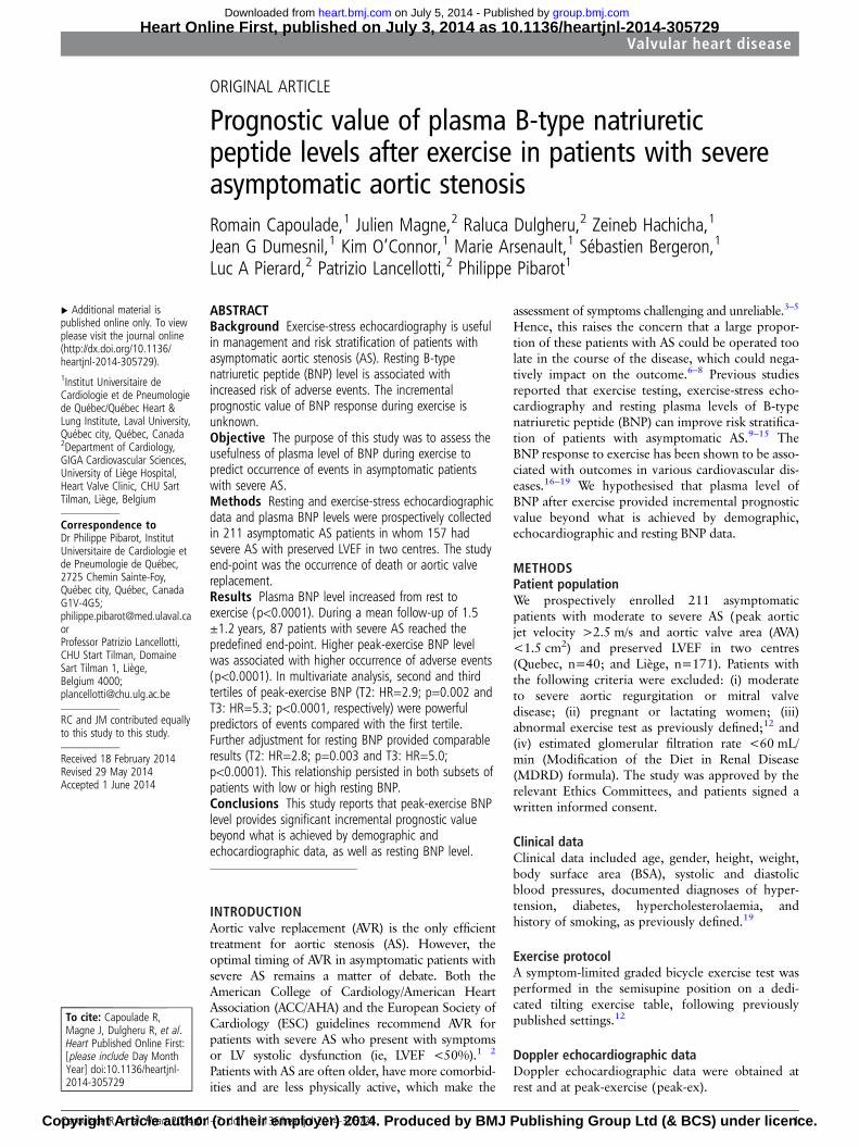
ORIGINAL ARTICLE
Prognostic value of plasma B-type natriureticpeptide levels after exercise in patients with severeasymptomatic aortic stenosisRomain Capoulade,1 Julien Magne,2 Raluca Dulgheru,2 Zeineb Hachicha,1
Jean G Dumesnil,1 Kim O’Connor,1 Marie Arsenault,1 Sébastien Bergeron,1
Luc A Pierard,2 Patrizio Lancellotti,2 Philippe Pibarot1
▸ Additional material ispublished online only. To viewplease visit the journal online(http://dx.doi.org/10.1136/heartjnl-2014-305729).1Institut Universitaire deCardiologie et de Pneumologiede Québec/Québec Heart &Lung Institute, Laval University,Québec city, Québec, Canada2Department of Cardiology,GIGA Cardiovascular Sciences,University of Liège Hospital,Heart Valve Clinic, CHU SartTilman, Liège, Belgium
Correspondence toDr Philippe Pibarot, InstitutUniversitaire de Cardiologie etde Pneumologie de Québec,2725 Chemin Sainte-Foy,Québec city, Québec, CanadaG1V-4G5;[email protected] Patrizio Lancellotti,CHU Start Tilman, DomaineSart Tilman 1, Liège,Belgium 4000;[email protected]
RC and JM contributed equallyto this study to this study.
Received 18 February 2014Revised 29 May 2014Accepted 1 June 2014
To cite: Capoulade R,Magne J, Dulgheru R, et al.Heart Published Online First:[please include Day MonthYear] doi:10.1136/heartjnl-2014-305729
ABSTRACTBackground Exercise-stress echocardiography is usefulin management and risk stratification of patients withasymptomatic aortic stenosis (AS). Resting B-typenatriuretic peptide (BNP) level is associated withincreased risk of adverse events. The incrementalprognostic value of BNP response during exercise isunknown.Objective The purpose of this study was to assess theusefulness of plasma level of BNP during exercise topredict occurrence of events in asymptomatic patientswith severe AS.Methods Resting and exercise-stress echocardiographicdata and plasma BNP levels were prospectively collectedin 211 asymptomatic AS patients in whom 157 hadsevere AS with preserved LVEF in two centres. The studyend-point was the occurrence of death or aortic valvereplacement.Results Plasma BNP level increased from rest toexercise (p<0.0001). During a mean follow-up of 1.5±1.2 years, 87 patients with severe AS reached thepredefined end-point. Higher peak-exercise BNP levelwas associated with higher occurrence of adverse events(p<0.0001). In multivariate analysis, second and thirdtertiles of peak-exercise BNP (T2: HR=2.9; p=0.002 andT3: HR=5.3; p<0.0001, respectively) were powerfulpredictors of events compared with the first tertile.Further adjustment for resting BNP provided comparableresults (T2: HR=2.8; p=0.003 and T3: HR=5.0;p<0.0001). This relationship persisted in both subsets ofpatients with low or high resting BNP.Conclusions This study reports that peak-exercise BNPlevel provides significant incremental prognostic valuebeyond what is achieved by demographic andechocardiographic data, as well as resting BNP level.
INTRODUCTIONAortic valve replacement (AVR) is the only efficienttreatment for aortic stenosis (AS). However, theoptimal timing of AVR in asymptomatic patients withsevere AS remains a matter of debate. Both theAmerican College of Cardiology/American HeartAssociation (ACC/AHA) and the European Society ofCardiology (ESC) guidelines recommend AVR forpatients with severe AS who present with symptomsor LV systolic dysfunction (ie, LVEF <50%).1 2
Patients with AS are often older, have more comorbid-ities and are less physically active, which make the
assessment of symptoms challenging and unreliable.3–5
Hence, this raises the concern that a large propor-tion of these patients with AS could be operated toolate in the course of the disease, which could nega-tively impact on the outcome.6–8 Previous studiesreported that exercise testing, exercise-stress echo-cardiography and resting plasma levels of B-typenatriuretic peptide (BNP) can improve risk stratifica-tion of patients with asymptomatic AS.9–15 TheBNP response to exercise has been shown to be asso-ciated with outcomes in various cardiovascular dis-eases.16–19 We hypothesised that plasma level ofBNP after exercise provided incremental prognosticvalue beyond what is achieved by demographic,echocardiographic and resting BNP data.
METHODSPatient populationWe prospectively enrolled 211 asymptomaticpatients with moderate to severe AS (peak aorticjet velocity >2.5 m/s and aortic valve area (AVA)<1.5 cm2) and preserved LVEF in two centres(Quebec, n=40; and Liège, n=171). Patients withthe following criteria were excluded: (i) moderateto severe aortic regurgitation or mitral valvedisease; (ii) pregnant or lactating women; (iii)abnormal exercise test as previously defined;12 and(iv) estimated glomerular filtration rate <60 mL/min (Modification of the Diet in Renal Disease(MDRD) formula). The study was approved by therelevant Ethics Committees, and patients signed awritten informed consent.
Clinical dataClinical data included age, gender, height, weight,body surface area (BSA), systolic and diastolicblood pressures, documented diagnoses of hyper-tension, diabetes, hypercholesterolaemia, andhistory of smoking, as previously defined.19
Exercise protocolA symptom-limited graded bicycle exercise test wasperformed in the semisupine position on a dedi-cated tilting exercise table, following previouslypublished settings.12
Doppler echocardiographic dataDoppler echocardiographic data were obtained atrest and at peak-exercise (peak-ex).
Capoulade R, et al. Heart 2014;0:1–7. doi:10.1136/heartjnl-2014-305729 1
Valvular heart disease Heart Online First, published on July 3, 2014 as 10.1136/heartjnl-2014-305729
Copyright Article author (or their employer) 2014. Produced by BMJ Publishing Group Ltd (& BCS) under licence.
group.bmj.com on July 5, 2014 - Published by heart.bmj.comDownloaded from
The Doppler echocardiographic indices of AS severityincluded peak aortic jet velocity, peak and mean transvalvularpressure gradients, and AVA indexed to BSA. Severe AS wasdefined according to ACC/AHA—ESC guidelines.1 2
The relative wall thickness and LV mass indexed to BSA werecalculated.20 LVEF was measured with the use of biplaneSimpson method.
Left atrial (LA) area was obtained by planimetry at end systolefrom apical four-chamber view and was indexed to BSA. E to e0
ratio was calculated from the E-wave of mitral inflow and theaverage of septal and lateral mitral annulus e0-wave. Systolic pul-monary arterial pressure (SPAP) was evaluated from the regurgitantjet of the tricuspid insufficiency.12 Resting and exercise pulmonaryhypertension (PHT) were defined as SPAP >50 and >60 mmHg,respectively. As a measure of global LV haemodynamic load, we cal-culated the valvulo-arterial impedance (Zva).
21
Plasma BNP levelVenous blood samples for BNP measurements were drawnbefore echocardiography after 20 min of supine rest (restingBNP) and at peak-ex, that is, within the 3 min following theend of exercise (peak-ex BNP) as previously validated.19
Study end-pointThe study end-point was the occurrence of death or AVR moti-vated by development of symptoms or LV dysfunction (ie, classI indication). To ensure blinding, both resting and peak-ex BNPlevels were not transmitted to the treating physician or surgeon.
Statistical analysisPatients were divided into three groups according to tertiles ofpeak-ex BNP level. Continuous data were expressed as mean±SDand were tested for normality of distribution and homogeneity ofvariances with the Shapiro–Wilk and Levene tests, respectively.Groups were compared with one-way Analysis of Variance(ANOVA) followed by a Tukey’s post hoc test when appropriate.Categorical data were expressed as percentage and groups werecompared with the χ2 or Fischer’s exact tests when appropriate.BNP levels were reported as median (IQR) and the natural loga-rithm transformation was applied to BNP levels. Differencebetween resting and peak-ex BNP levels was obtained with paired ttest and correlations between resting BNP and peak-ex BNPor abso-lute change in BNP (δ BNP) were determined using Pearson’sproduct-moment correlations. Multivariate linear regression ana-lysis, including age, gender, peak-ex workload and exercise-inducedincrease in mean gradient, stroke volume index and Zva, was per-formed to identify the independent determinants of peak-ex BNPlevel. Kaplan–Meier curves and log-rank tests of the time-to-eventdata were used to assess the effect of tertiles of peak-ex BNP and ofδ BNP on the composite end-point of death or AVR. The relation-ships among resting, peak-ex, δ BNP levels and event-free survivalwere assessed with the use of individual and multivariate Coxproportional-hazard analyses. The traditional risk factors of thecomposite of death or AVR and all variables with p value <0.10 inindividual Cox analysis (ie, age, gender, resting mean gradient,resting Zva, resting indexed LA area and exercise-induced increasein heart rate, mean gradient and Zva) were entered in the multivari-ate Cox model. The proportional-hazards assumption was checkedwith the use of Schoenfeld residuals. The incremental predictive
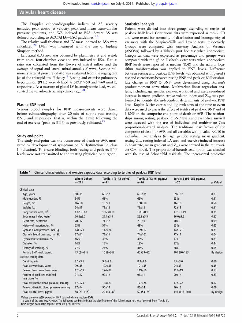
Table 1 Clinical characteristics and exercise capacity data according to tertiles of peak-ex BNP level
Whole Cohortn=211
Tertile 1 (6–42 pg/mL)n=71
Tertile 2 (43–91 pg/mL)n=70
Tertile 3 (92–956 pg/mL)n=70 p Value†
Clinical dataAge, years 68±11 65±12 69±10* 69±10* 0.03Male gender, % 64% 63% 66% 63% 0.91
Height, cm 167±8 167±7 168±10 166±8 0.50Weight, kg 74±13 76±12 76±12 72±13 0.21Body surface area, m2 1.82±0.18 1.82±0.18 1.83±0.18 1.81±0.19 0.71Body mass index, kg/m2 26.6±3.7 27.1±3.9 26.8±3.5 26.0±3.6 0.27Heart rate, beats/min 70±12 71±12 70±10 70±13 0.99History of hypertension, % 53% 57% 49% 53% 0.65Systolic blood pressure, mm Hg 141±21 142±24 139±17 142±21 0.71Diastolic blood pressure, mm Hg 77±11 79±11 74±10* 77±11 0.04Hypercholesterolaemia, % 46% 48% 43% 47% 0.83Diabetes, % 14% 13% 12% 17% 0.44History of smoking, % 27% 24% 31% 28% 0.65Resting BNP level, pg/mL 43 (24–81) 16 (9–26) 45 (39–60) 101 (76–133) By design
Exercise testing dataDuration, min 9.1±3.1 9.0±2.6 8.9±2.9 9.4±3.6 0.69Peak-ex workload, watts 99±35 102±38 101±35 94±33 0.35Peak-ex heart rate, beats/min 120±19 124±20 119±16 118±19 0.13Percent of predicted maximalheart rate, %
91±12 92±12 91±11 90±14 0.83
Peak-ex systolic blood pressure, mm Hg 179±23 184±23 177±24 177±22 0.17Peak-ex diastolic blood pressure, mm Hg 87±14 90±14 85±14 86±13 0.09Peak-ex BNP level, pg/mL 58 (29–115) 20 (13–30) 59 (53–74) 146 (115–201) By design
Values are mean±SD except for BNP data which are median (IQR).†p Value of the one-way ANOVA. The following symbols indicate the significance of the Tukey’s post hoc test: *p<0.05 from ‘Tertile 1’.BNP, B-type natriuretic peptide; Peak-ex, peak-exercise.
2 Capoulade R, et al. Heart 2014;0:1–7. doi:10.1136/heartjnl-2014-305729
Valvular heart disease
group.bmj.com on July 5, 2014 - Published by heart.bmj.comDownloaded from
value of peak-ex BNP was assessed by calculating the net reclassifi-cation improvement (NRI) for 1-year events (death or AVR) usingthe NRI & IDI program codes downloaded online (http://www.ucr.uu.se/en/index.php/ucr-statistics/program-code/306-nri-and-idi).22 23 Category cut-points were defined as observedevent rate at 1-year (ie, 28%) or tertiles of risk probabilities deter-mined using the model with established risk factors (ie, <19%,19%–35%, and ≥35%). Patients were reclassified according to1-year predicted probabilities after addition of peak-ex BNP. Thereceiver operating characteristic (ROC) curve analyses were per-formed to determine the accuracy (area under the curve, AUC) aswell as the best cut-point values for resting and peak-ex BNP levelsto predict 1-year events expressed as a binary outcome. Statisticalanalyses were done with Stata Software (V.11.0). A p value <0.05was considered statistically significant.
RESULTSPopulation characteristicsBaseline characteristics of the population are depicted in tables1 and 2. Severe AS was present in 157 (74%) patients. The base-line characteristics (all p>0.05; data not shown) and 1-yearevent rates (65% vs 73%; p=0.87) were similar in the subset ofpatients enrolled from Canada versus those enrolled fromBelgium.
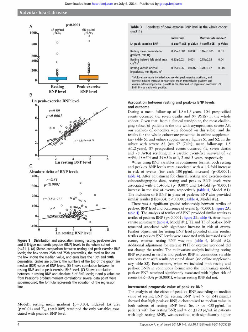
Response of BNP to exerciseBNP level increased significantly from rest to peak-ex(p<0.0001; figure 1A) and both resting and peak-ex BNP levelswere strongly correlated (r=0.89, p<0.0001; figure 1B). Theexercise-induced absolute change in BNP was 9 (2–29) pg/mLand 182 (86%) patients exhibited an increase in BNP duringexercise (ie, δ BNP>0). There was a significant but modest cor-relation between resting BNP and absolute δ BNP (r=0.31,p<0.0001; figure 1C). Peak-ex BNP correlated weakly andnegatively with peak-ex workload (r=−0.16; p=0.03).
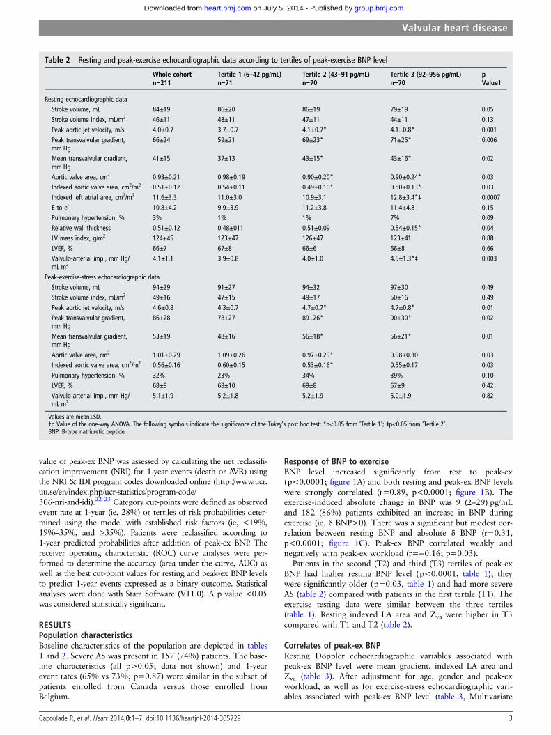
Patients in the second (T2) and third (T3) tertiles of peak-exBNP had higher resting BNP level (p<0.0001, table 1); theywere significantly older (p=0.03, table 1) and had more severeAS (table 2) compared with patients in the first tertile (T1). Theexercise testing data were similar between the three tertiles(table 1). Resting indexed LA area and Zva were higher in T3compared with T1 and T2 (table 2).
Correlates of peak-ex BNPResting Doppler echocardiographic variables associated withpeak-ex BNP level were mean gradient, indexed LA area andZva (table 3). After adjustment for age, gender and peak-exworkload, as well as for exercise-stress echocardiographic vari-ables associated with peak-ex BNP level (table 3, Multivariate
Table 2 Resting and peak-exercise echocardiographic data according to tertiles of peak-exercise BNP level
Whole cohortn=211
Tertile 1 (6–42 pg/mL)n=71
Tertile 2 (43–91 pg/mL)n=70
Tertile 3 (92–956 pg/mL)n=70
pValue†
Resting echocardiographic dataStroke volume, mL 84±19 86±20 86±19 79±19 0.05Stroke volume index, mL/m2 46±11 48±11 47±11 44±11 0.13Peak aortic jet velocity, m/s 4.0±0.7 3.7±0.7 4.1±0.7* 4.1±0.8* 0.001Peak transvalvular gradient,mm Hg
66±24 59±21 69±23* 71±25* 0.006
Mean transvalvular gradient,mm Hg
41±15 37±13 43±15* 43±16* 0.02
Aortic valve area, cm2 0.93±0.21 0.98±0.19 0.90±0.20* 0.90±0.24* 0.03Indexed aortic valve area, cm2/m2 0.51±0.12 0.54±0.11 0.49±0.10* 0.50±0.13* 0.03Indexed left atrial area, cm2/m2 11.6±3.3 11.0±3.0 10.9±3.1 12.8±3.4*‡ 0.0007E to e0 10.8±4.2 9.9±3.9 11.2±3.8 11.4±4.8 0.15Pulmonary hypertension, % 3% 1% 1% 7% 0.09Relative wall thickness 0.51±0.12 0.48±011 0.51±0.09 0.54±0.15* 0.04LV mass index, g/m2 124±45 123±47 126±47 123±41 0.88LVEF, % 66±7 67±8 66±6 66±8 0.66Valvulo-arterial imp., mm Hg/mL m2
4.1±1.1 3.9±0.8 4.0±1.0 4.5±1.3*‡ 0.003
Peak-exercise-stress echocardiographic dataStroke volume, mL 94±29 91±27 94±32 97±30 0.49Stroke volume index, mL/m2 49±16 47±15 49±17 50±16 0.49Peak aortic jet velocity, m/s 4.6±0.8 4.3±0.7 4.7±0.7* 4.7±0.8* 0.01Peak transvalvular gradient,mm Hg
86±28 78±27 89±26* 90±30* 0.02
Mean transvalvular gradient,mm Hg
53±19 48±16 56±18* 56±21* 0.01
Aortic valve area, cm2 1.01±0.29 1.09±0.26 0.97±0.29* 0.98±0.30 0.03Indexed aortic valve area, cm2/m2 0.56±0.16 0.60±0.15 0.53±0.16* 0.55±0.17 0.03Pulmonary hypertension, % 32% 23% 34% 39% 0.10LVEF, % 68±9 68±10 69±8 67±9 0.42Valvulo-arterial imp., mm Hg/mL m2
5.1±1.9 5.2±1.8 5.2±1.9 5.0±1.9 0.82
Values are mean±SD.†p Value of the one-way ANOVA. The following symbols indicate the significance of the Tukey’s post hoc test: *p<0.05 from ‘Tertile 1’; ‡p<0.05 from ‘Tertile 2’.BNP, B-type natriuretic peptide.
Capoulade R, et al. Heart 2014;0:1–7. doi:10.1136/heartjnl-2014-305729 3
Valvular heart disease
group.bmj.com on July 5, 2014 - Published by heart.bmj.comDownloaded from
Model), resting mean gradient (p=0.03), indexed LA area(p=0.04) and Zva (p=0.009) remained the only variables asso-ciated with peak-ex BNP level.
Association between resting and peak-ex BNP levelsand outcomeDuring a mean follow-up of 1.8±1.3 years, 104 prespecifiedevents occurred (ie, seven deaths and 97 AVRs) in the wholecohort. Given that, from a clinical standpoint, the most challen-ging subset of patients is the one with asymptomatic severe AS,our analyses of outcomes were focused on this subset and theresults for the whole cohort are presented in online supplemen-tary table S1 and online supplementary figures S1 and S2. In thesubset with severe AS (n=157 (74%); mean follow-up: 1.5±1.2 years), 87 prespecified events occurred (ie, seven deathsand 78 AVRs) resulting in a cardiac event-free survival of 72±4%, 48±5% and 39±5% at 1, 2 and 3 years, respectively.
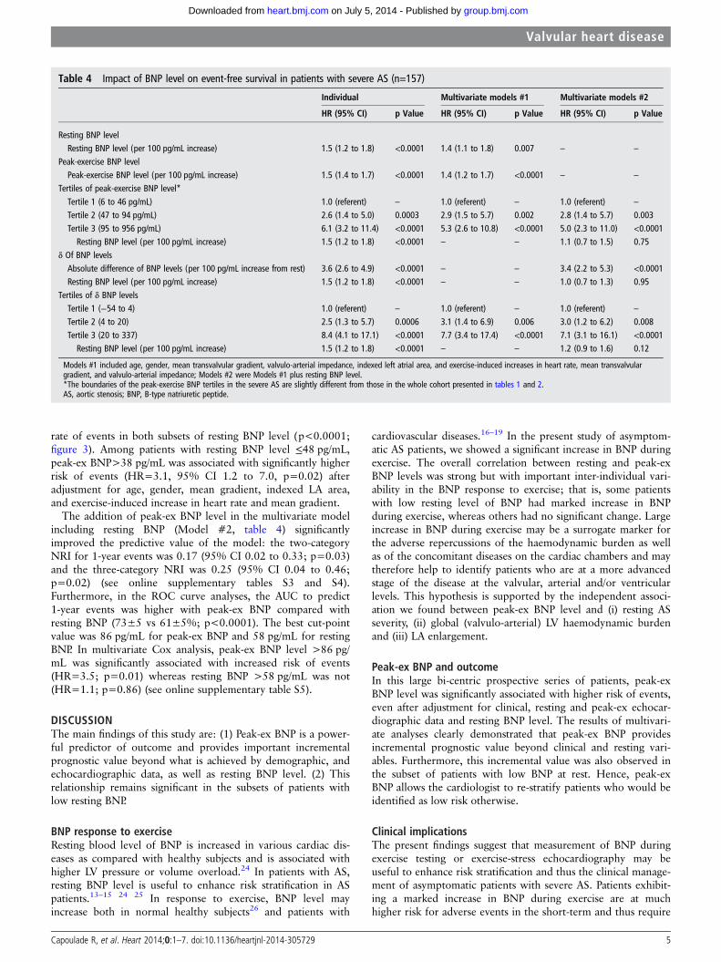
When using BNP variables in continuous format, both restingand peak-ex BNP levels were associated with a 1.5-fold increasein risk of events (for each 100 pg/mL increase) (p<0.0001;table 4). After adjustment for clinical, resting and exercise-stressechocardiographic data, resting and peak-ex BNP levels wereassociated with a 1.4-fold (p=0.007) and 1.4-fold (p<0.0001)increase in the risk of events, respectively (table 4, Model #1).The inclusion of δ BNP in place of peak-ex BNP also providedsimilar results (HR=3.4; p<0.0001; table 4, Model #2).
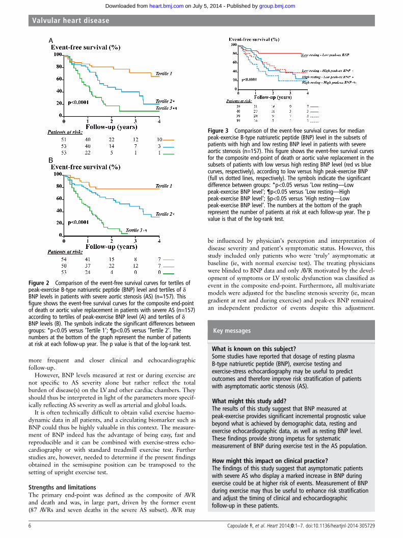
There was a significant graded relationship between tertiles ofpeak-ex BNP level and occurrence of events (p<0.0001; figure 2A;table 4). The analysis of tertiles of δ BNP provided similar results astertiles of peak-ex BNP (p<0.0001; figure 2B; table 4). After multi-variate adjustment (table 4, Model #1), T2 and T3 of peak-ex BNPremained associated with significant increase in risk of events.Further adjustment for resting BNP level provided similar results:tertiles of peak-ex BNP levels were associated with increased risk ofevents, whereas resting BNP was not (table 4, Model #2).Additional adjustment for exercise PHT or exercise workload didnot change the results. The multivariate analysis including restingBNP expressed in tertiles and peak-ex BNP in continuous variablewas consistent with results presented above (see online supplemen-tary table S2). Furthermore, when we included both resting andpeak-ex BNPs in continuous format into the multivariate model,peak-ex BNP remained significantly associated with higher risk ofevents (HR=3.4; p<0.0001), whereas resting BNP did not.
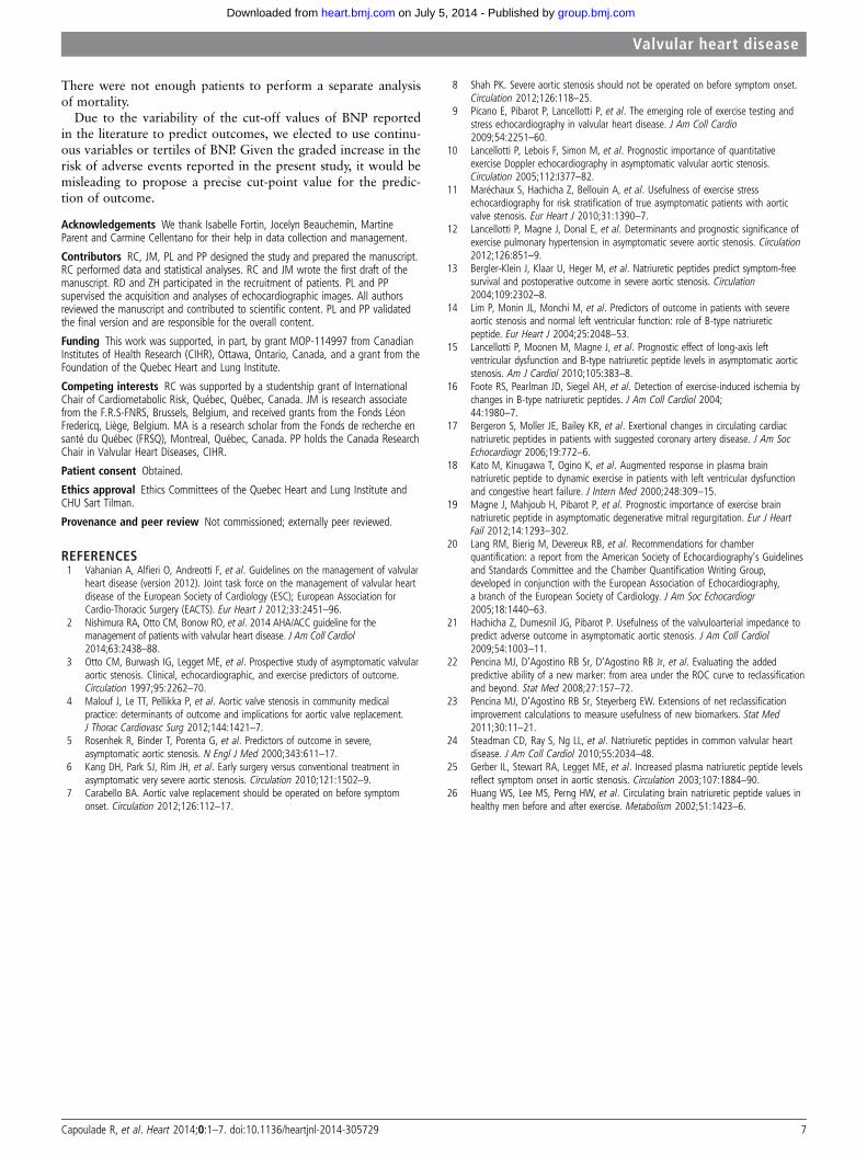
Incremental prognostic value of peak-ex BNPThe analysis of the effect of peak-ex BNP according to medianvalue of resting BNP (ie, resting BNP level > or ≤48 pg/mL)showed that high peak-ex BNP, dichotomised to median value inboth subsets of resting BNP level (ie, > or ≤38 pg/mL inpatients with low resting BNP, and > or ≤120 pg/mL in patientswith high resting BNP), was associated with significantly higher
Figure 1 Distribution and association among resting, peak-exerciseand δ B-type natriuretic peptide (BNP) levels in the whole cohort(n=211). (A) Shows comparison between resting and peak-exercise BNPlevels; the box shows 25th and 75th percentiles, the median line onthe box shows the median value, and error bars the 10th and 90thpercentiles; circles are outliers; the numbers of the top of the graph aremedian (IQR) value of BNP levels. (B) Shows correlation between lnresting BNP and ln peak-exercise BNP level. (C) Shows correlationbetween ln resting BNP and absolute δ of BNP levels; r and p value arefrom Pearson’s product-moment correlations; several data point weresuperimposed; the formula represents the equation of the regressionline.
Table 3 Correlates of peak-exercise BNP level in the whole cohort(n=211)
Ln peak-exercise BNP
Individual Multivariate model*
β coeff.±SE p Value β coeff.±SE p Value
Resting mean transvalvulargradient, mm Hg
0.25±0.004 0.0003 0.16±0.005 0.03
Resting indexed left atrial area,cm2/m2
0.23±0.02 0.001 0.15±0.02 0.04
Resting valvulo-arterialimpedance, mm Hg/mL m2
0.25±0.06 0.0002 0.20±0.07 0.009
*Multivariate model included age, gender, peak-exercise workload, andexercise-induced increase in heart rate, mean transvalvular gradient andvalvulo-arterial impedance. β coeff. is the standardised regression coefficient±SE.BNP, B-type natriuretic peptide.
4 Capoulade R, et al. Heart 2014;0:1–7. doi:10.1136/heartjnl-2014-305729
Valvular heart disease
group.bmj.com on July 5, 2014 - Published by heart.bmj.comDownloaded from
rate of events in both subsets of resting BNP level (p<0.0001;figure 3). Among patients with resting BNP level ≤48 pg/mL,peak-ex BNP>38 pg/mL was associated with significantly higherrisk of events (HR=3.1, 95% CI 1.2 to 7.0, p=0.02) afteradjustment for age, gender, mean gradient, indexed LA area,and exercise-induced increase in heart rate and mean gradient.
The addition of peak-ex BNP level in the multivariate modelincluding resting BNP (Model #2, table 4) significantlyimproved the predictive value of the model: the two-categoryNRI for 1-year events was 0.17 (95% CI 0.02 to 0.33; p=0.03)and the three-category NRI was 0.25 (95% CI 0.04 to 0.46;p=0.02) (see online supplementary tables S3 and S4).Furthermore, in the ROC curve analyses, the AUC to predict1-year events was higher with peak-ex BNP compared withresting BNP (73±5 vs 61±5%; p<0.0001). The best cut-pointvalue was 86 pg/mL for peak-ex BNP and 58 pg/mL for restingBNP. In multivariate Cox analysis, peak-ex BNP level >86 pg/mL was significantly associated with increased risk of events(HR=3.5; p=0.01) whereas resting BNP >58 pg/mL was not(HR=1.1; p=0.86) (see online supplementary table S5).
DISCUSSIONThe main findings of this study are: (1) Peak-ex BNP is a power-ful predictor of outcome and provides important incrementalprognostic value beyond what is achieved by demographic, andechocardiographic data, as well as resting BNP level. (2) Thisrelationship remains significant in the subsets of patients withlow resting BNP.
BNP response to exerciseResting blood level of BNP is increased in various cardiac dis-eases as compared with healthy subjects and is associated withhigher LV pressure or volume overload.24 In patients with AS,resting BNP level is useful to enhance risk stratification in ASpatients.13–15 24 25 In response to exercise, BNP level mayincrease both in normal healthy subjects26 and patients with
cardiovascular diseases.16–19 In the present study of asymptom-atic AS patients, we showed a significant increase in BNP duringexercise. The overall correlation between resting and peak-exBNP levels was strong but with important inter-individual vari-ability in the BNP response to exercise; that is, some patientswith low resting level of BNP had marked increase in BNPduring exercise, whereas others had no significant change. Largeincrease in BNP during exercise may be a surrogate marker forthe adverse repercussions of the haemodynamic burden as wellas of the concomitant diseases on the cardiac chambers and maytherefore help to identify patients who are at a more advancedstage of the disease at the valvular, arterial and/or ventricularlevels. This hypothesis is supported by the independent associ-ation we found between peak-ex BNP level and (i) resting ASseverity, (ii) global (valvulo-arterial) LV haemodynamic burdenand (iii) LA enlargement.
Peak-ex BNP and outcomeIn this large bi-centric prospective series of patients, peak-exBNP level was significantly associated with higher risk of events,even after adjustment for clinical, resting and peak-ex echocar-diographic data and resting BNP level. The results of multivari-ate analyses clearly demonstrated that peak-ex BNP providesincremental prognostic value beyond clinical and resting vari-ables. Furthermore, this incremental value was also observed inthe subset of patients with low BNP at rest. Hence, peak-exBNP allows the cardiologist to re-stratify patients who would beidentified as low risk otherwise.
Clinical implicationsThe present findings suggest that measurement of BNP duringexercise testing or exercise-stress echocardiography may beuseful to enhance risk stratification and thus the clinical manage-ment of asymptomatic patients with severe AS. Patients exhibit-ing a marked increase in BNP during exercise are at muchhigher risk for adverse events in the short-term and thus require
Table 4 Impact of BNP level on event-free survival in patients with severe AS (n=157)
Individual Multivariate models #1 Multivariate models #2
HR (95% CI) p Value HR (95% CI) p Value HR (95% CI) p Value
Resting BNP levelResting BNP level (per 100 pg/mL increase) 1.5 (1.2 to 1.8) <0.0001 1.4 (1.1 to 1.8) 0.007 – –
Peak-exercise BNP levelPeak-exercise BNP level (per 100 pg/mL increase) 1.5 (1.4 to 1.7) <0.0001 1.4 (1.2 to 1.7) <0.0001 – –
Tertiles of peak-exercise BNP level*
Tertile 1 (6 to 46 pg/mL) 1.0 (referent) – 1.0 (referent) – 1.0 (referent) –
Tertile 2 (47 to 94 pg/mL) 2.6 (1.4 to 5.0) 0.0003 2.9 (1.5 to 5.7) 0.002 2.8 (1.4 to 5.7) 0.003Tertile 3 (95 to 956 pg/mL) 6.1 (3.2 to 11.4) <0.0001 5.3 (2.6 to 10.8) <0.0001 5.0 (2.3 to 11.0) <0.0001Resting BNP level (per 100 pg/mL increase) 1.5 (1.2 to 1.8) <0.0001 – – 1.1 (0.7 to 1.5) 0.75
δ Of BNP levelsAbsolute difference of BNP levels (per 100 pg/mL increase from rest) 3.6 (2.6 to 4.9) <0.0001 – – 3.4 (2.2 to 5.3) <0.0001Resting BNP level (per 100 pg/mL increase) 1.5 (1.2 to 1.8) <0.0001 – – 1.0 (0.7 to 1.3) 0.95
Tertiles of δ BNP levelsTertile 1 (−54 to 4) 1.0 (referent) – 1.0 (referent) – 1.0 (referent) –
Tertile 2 (4 to 20) 2.5 (1.3 to 5.7) 0.0006 3.1 (1.4 to 6.9) 0.006 3.0 (1.2 to 6.2) 0.008Tertile 3 (20 to 337) 8.4 (4.1 to 17.1) <0.0001 7.7 (3.4 to 17.4) <0.0001 7.1 (3.1 to 16.1) <0.0001Resting BNP level (per 100 pg/mL increase) 1.5 (1.2 to 1.8) <0.0001 – – 1.2 (0.9 to 1.6) 0.12
Models #1 included age, gender, mean transvalvular gradient, valvulo-arterial impedance, indexed left atrial area, and exercise-induced increases in heart rate, mean transvalvulargradient, and valvulo-arterial impedance; Models #2 were Models #1 plus resting BNP level.*The boundaries of the peak-exercise BNP tertiles in the severe AS are slightly different from those in the whole cohort presented in tables 1 and 2.AS, aortic stenosis; BNP, B-type natriuretic peptide.
Capoulade R, et al. Heart 2014;0:1–7. doi:10.1136/heartjnl-2014-305729 5
Valvular heart disease
group.bmj.com on July 5, 2014 - Published by heart.bmj.comDownloaded from
more frequent and closer clinical and echocardiographicfollow-up.
However, BNP levels measured at rest or during exercise arenot specific to AS severity alone but rather reflect the totalburden of disease(s) on the LVand other cardiac chambers. Theyshould thus be interpreted in light of the parameters more specif-ically reflecting AS severity as well as arterial and global loads.
It is often technically difficult to obtain valid exercise haemo-dynamic data in all patients, and a circulating biomarker such asBNP could thus be highly valuable in this context. The measure-ment of BNP indeed has the advantage of being easy, fast andreproducible and it can be combined with exercise-stress echo-cardiography or with standard treadmill exercise test. Furtherstudies are, however, needed to determine if the present findingsobtained in the semisupine position can be transposed to thesetting of upright exercise test.
Strengths and limitationsThe primary end-point was defined as the composite of AVRand death and was, in large part, driven by the former event(87 AVRs and seven deaths in the severe AS subset). AVR may
be influenced by physician’s perception and interpretation ofdisease severity and patient’s symptomatic status. However, thisstudy included only patients who were ‘truly’ asymptomatic atbaseline (ie, with normal exercise test). The treating physicianswere blinded to BNP data and only AVR motivated by the devel-opment of symptoms or LV systolic dysfunction was classified asevent in the composite end-point. Furthermore, all multivariatemodels were adjusted for the baseline stenosis severity (ie, meangradient at rest and during exercise) and peak-ex BNP remainedan independent predictor of events despite this adjustment.
Figure 3 Comparison of the event-free survival curves for medianpeak-exercise B-type natriuretic peptide (BNP) level in the subsets ofpatients with high and low resting BNP level in patients with severeaortic stenosis (n=157). This figure shows the event-free survival curvesfor the composite end-point of death or aortic valve replacement in thesubsets of patients with low versus high resting BNP level (red vs bluecurves, respectively), according to low versus high peak-exercise BNP(full vs dotted lines, respectively). The symbols indicate the significantdifference between groups: *p<0.05 versus ‘Low resting—Lowpeak-exercise BNP level’; ¶p<0.05 versus ‘Low resting—Highpeak-exercise BNP level’; §p<0.05 versus ‘High resting—Lowpeak-exercise BNP level’. The numbers at the bottom of the graphrepresent the number of patients at risk at each follow-up year. The pvalue is that of the log-rank test.
Figure 2 Comparison of the event-free survival curves for tertiles ofpeak-exercise B-type natriuretic peptide (BNP) level and tertiles of δBNP levels in patients with severe aortic stenosis (AS) (n=157). Thisfigure shows the event-free survival curves for the composite end-pointof death or aortic valve replacement in patients with severe AS (n=157)according to tertiles of peak-exercise BNP level (A) and tertiles of δBNP levels (B). The symbols indicate the significant differences betweengroups: *p<0.05 versus ‘Tertile 1’; ¶p<0.05 versus ‘Tertile 2’. Thenumbers at the bottom of the graph represent the number of patientsat risk at each follow-up year. The p value is that of the log-rank test.
Key messages
What is known on this subject?Some studies have reported that dosage of resting plasmaB-type natriuretic peptide (BNP), exercise testing andexercise-stress echocardiography may be useful to predictoutcomes and therefore improve risk stratification of patientswith asymptomatic aortic stenosis (AS).
What might this study add?The results of this study suggest that BNP measured atpeak-exercise provides significant incremental prognostic valuebeyond what is achieved by demographic data, resting andexercise echocardiographic data, as well as resting BNP level.These findings provide strong impetus for systematicmeasurement of BNP during exercise test in the AS population.
How might this impact on clinical practice?The findings of this study suggest that asymptomatic patientswith severe AS who display a marked increase in BNP duringexercise could be at higher risk of events. Measurement of BNPduring exercise may thus be useful to enhance risk stratificationand adjust the timing of clinical and echocardiographicfollow-up in these patients.
6 Capoulade R, et al. Heart 2014;0:1–7. doi:10.1136/heartjnl-2014-305729
Valvular heart disease
group.bmj.com on July 5, 2014 - Published by heart.bmj.comDownloaded from
There were not enough patients to perform a separate analysisof mortality.
Due to the variability of the cut-off values of BNP reportedin the literature to predict outcomes, we elected to use continu-ous variables or tertiles of BNP. Given the graded increase in therisk of adverse events reported in the present study, it would bemisleading to propose a precise cut-point value for the predic-tion of outcome.
Acknowledgements We thank Isabelle Fortin, Jocelyn Beauchemin, MartineParent and Carmine Cellentano for their help in data collection and management.
Contributors RC, JM, PL and PP designed the study and prepared the manuscript.RC performed data and statistical analyses. RC and JM wrote the first draft of themanuscript. RD and ZH participated in the recruitment of patients. PL and PPsupervised the acquisition and analyses of echocardiographic images. All authorsreviewed the manuscript and contributed to scientific content. PL and PP validatedthe final version and are responsible for the overall content.
Funding This work was supported, in part, by grant MOP-114997 from CanadianInstitutes of Health Research (CIHR), Ottawa, Ontario, Canada, and a grant from theFoundation of the Quebec Heart and Lung Institute.
Competing interests RC was supported by a studentship grant of InternationalChair of Cardiometabolic Risk, Québec, Québec, Canada. JM is research associatefrom the F.R.S-FNRS, Brussels, Belgium, and received grants from the Fonds LéonFredericq, Liège, Belgium. MA is a research scholar from the Fonds de recherche ensanté du Québec (FRSQ), Montreal, Québec, Canada. PP holds the Canada ResearchChair in Valvular Heart Diseases, CIHR.
Patient consent Obtained.
Ethics approval Ethics Committees of the Quebec Heart and Lung Institute andCHU Sart Tilman.
Provenance and peer review Not commissioned; externally peer reviewed.
REFERENCES1 Vahanian A, Alfieri O, Andreotti F, et al. Guidelines on the management of valvular
heart disease (version 2012). Joint task force on the management of valvular heartdisease of the European Society of Cardiology (ESC); European Association forCardio-Thoracic Surgery (EACTS). Eur Heart J 2012;33:2451–96.
2 Nishimura RA, Otto CM, Bonow RO, et al. 2014 AHA/ACC guideline for themanagement of patients with valvular heart disease. J Am Coll Cardiol2014;63:2438–88.
3 Otto CM, Burwash IG, Legget ME, et al. Prospective study of asymptomatic valvularaortic stenosis. Clinical, echocardiographic, and exercise predictors of outcome.Circulation 1997;95:2262–70.
4 Malouf J, Le TT, Pellikka P, et al. Aortic valve stenosis in community medicalpractice: determinants of outcome and implications for aortic valve replacement.J Thorac Cardiovasc Surg 2012;144:1421–7.
5 Rosenhek R, Binder T, Porenta G, et al. Predictors of outcome in severe,asymptomatic aortic stenosis. N Engl J Med 2000;343:611–17.
6 Kang DH, Park SJ, Rim JH, et al. Early surgery versus conventional treatment inasymptomatic very severe aortic stenosis. Circulation 2010;121:1502–9.
7 Carabello BA. Aortic valve replacement should be operated on before symptomonset. Circulation 2012;126:112–17.
8 Shah PK. Severe aortic stenosis should not be operated on before symptom onset.Circulation 2012;126:118–25.
9 Picano E, Pibarot P, Lancellotti P, et al. The emerging role of exercise testing andstress echocardiography in valvular heart disease. J Am Coll Cardio2009;54:2251–60.
10 Lancellotti P, Lebois F, Simon M, et al. Prognostic importance of quantitativeexercise Doppler echocardiography in asymptomatic valvular aortic stenosis.Circulation 2005;112:I377–82.
11 Maréchaux S, Hachicha Z, Bellouin A, et al. Usefulness of exercise stressechocardiography for risk stratification of true asymptomatic patients with aorticvalve stenosis. Eur Heart J 2010;31:1390–7.
12 Lancellotti P, Magne J, Donal E, et al. Determinants and prognostic significance ofexercise pulmonary hypertension in asymptomatic severe aortic stenosis. Circulation2012;126:851–9.
13 Bergler-Klein J, Klaar U, Heger M, et al. Natriuretic peptides predict symptom-freesurvival and postoperative outcome in severe aortic stenosis. Circulation2004;109:2302–8.
14 Lim P, Monin JL, Monchi M, et al. Predictors of outcome in patients with severeaortic stenosis and normal left ventricular function: role of B-type natriureticpeptide. Eur Heart J 2004;25:2048–53.
15 Lancellotti P, Moonen M, Magne J, et al. Prognostic effect of long-axis leftventricular dysfunction and B-type natriuretic peptide levels in asymptomatic aorticstenosis. Am J Cardiol 2010;105:383–8.
16 Foote RS, Pearlman JD, Siegel AH, et al. Detection of exercise-induced ischemia bychanges in B-type natriuretic peptides. J Am Coll Cardiol 2004;44:1980–7.
17 Bergeron S, Moller JE, Bailey KR, et al. Exertional changes in circulating cardiacnatriuretic peptides in patients with suggested coronary artery disease. J Am SocEchocardiogr 2006;19:772–6.
18 Kato M, Kinugawa T, Ogino K, et al. Augmented response in plasma brainnatriuretic peptide to dynamic exercise in patients with left ventricular dysfunctionand congestive heart failure. J Intern Med 2000;248:309–15.
19 Magne J, Mahjoub H, Pibarot P, et al. Prognostic importance of exercise brainnatriuretic peptide in asymptomatic degenerative mitral regurgitation. Eur J HeartFail 2012;14:1293–302.
20 Lang RM, Bierig M, Devereux RB, et al. Recommendations for chamberquantification: a report from the American Society of Echocardiography’s Guidelinesand Standards Committee and the Chamber Quantification Writing Group,developed in conjunction with the European Association of Echocardiography,a branch of the European Society of Cardiology. J Am Soc Echocardiogr2005;18:1440–63.
21 Hachicha Z, Dumesnil JG, Pibarot P. Usefulness of the valvuloarterial impedance topredict adverse outcome in asymptomatic aortic stenosis. J Am Coll Cardiol2009;54:1003–11.
22 Pencina MJ, D’Agostino RB Sr, D’Agostino RB Jr, et al. Evaluating the addedpredictive ability of a new marker: from area under the ROC curve to reclassificationand beyond. Stat Med 2008;27:157–72.
23 Pencina MJ, D’Agostino RB Sr, Steyerberg EW. Extensions of net reclassificationimprovement calculations to measure usefulness of new biomarkers. Stat Med2011;30:11–21.
24 Steadman CD, Ray S, Ng LL, et al. Natriuretic peptides in common valvular heartdisease. J Am Coll Cardiol 2010;55:2034–48.
25 Gerber IL, Stewart RA, Legget ME, et al. Increased plasma natriuretic peptide levelsreflect symptom onset in aortic stenosis. Circulation 2003;107:1884–90.
26 Huang WS, Lee MS, Perng HW, et al. Circulating brain natriuretic peptide values inhealthy men before and after exercise. Metabolism 2002;51:1423–6.
Capoulade R, et al. Heart 2014;0:1–7. doi:10.1136/heartjnl-2014-305729 7
Valvular heart disease
group.bmj.com on July 5, 2014 - Published by heart.bmj.comDownloaded from
doi: 10.1136/heartjnl-2014-305729 published online July 3, 2014Heart
Romain Capoulade, Julien Magne, Raluca Dulgheru, et al. stenosispatients with severe asymptomatic aorticnatriuretic peptide levels after exercise in Prognostic value of plasma B-type
http://heart.bmj.com/content/early/2014/07/03/heartjnl-2014-305729.full.htmlUpdated information and services can be found at:
These include:
Data Supplement http://heart.bmj.com/content/suppl/2014/07/03/heartjnl-2014-305729.DC1.html
"Supplementary Data"
References http://heart.bmj.com/content/early/2014/07/03/heartjnl-2014-305729.full.html#ref-list-1
This article cites 26 articles, 10 of which can be accessed free at:
P<P Published online July 3, 2014 in advance of the print journal.
serviceEmail alerting
the box at the top right corner of the online article.Receive free email alerts when new articles cite this article. Sign up in
(DOIs) and date of initial publication. publication. Citations to Advance online articles must include the digital object identifier citable and establish publication priority; they are indexed by PubMed from initialtypeset, but have not not yet appeared in the paper journal. Advance online articles are Advance online articles have been peer reviewed, accepted for publication, edited and
http://group.bmj.com/group/rights-licensing/permissionsTo request permissions go to:
http://journals.bmj.com/cgi/reprintformTo order reprints go to:
http://group.bmj.com/subscribe/To subscribe to BMJ go to:
group.bmj.com on July 5, 2014 - Published by heart.bmj.comDownloaded from
CollectionsTopic
(7843 articles)Drugs: cardiovascular system � (330 articles)Aortic valve disease �
(1905 articles)Echocardiography � (4347 articles)Clinical diagnostic tests �
Articles on similar topics can be found in the following collections
Notes
(DOIs) and date of initial publication. publication. Citations to Advance online articles must include the digital object identifier citable and establish publication priority; they are indexed by PubMed from initialtypeset, but have not not yet appeared in the paper journal. Advance online articles are Advance online articles have been peer reviewed, accepted for publication, edited and
http://group.bmj.com/group/rights-licensing/permissionsTo request permissions go to:
http://journals.bmj.com/cgi/reprintformTo order reprints go to:
http://group.bmj.com/subscribe/To subscribe to BMJ go to:
group.bmj.com on July 5, 2014 - Published by heart.bmj.comDownloaded from
Copyright © 2022 FDOKUMEN