Probing the structure of Lhca3 by mutation analysis
7
Probing the structure of Lhca3 by mutation analysis Milena Mozzo a,b , Tomas Morosinotto b,c , Roberto Bassi b,c , Roberta Croce a,d, ⁎ a Istituto di Biofisica. CNR. C/o ITC via Sommarvie 18. 38100 Povo. Trento, Italy b Dipartimento Scientifico e Tecnologico, Università di Verona, Strada Le Grazie 15-37134 Verona, Italy c Université Aix-Marseille II, LGBP-Faculté de Sciences de Luminy, Département de Biologie-Case 901-163 Avenue de Luminy, 13288 Marseille, France d Department of Biophysical Chemistry, Groningen Bimolecular Sciences and Biotechnology Institute, University of Groningen, Nijenborgh 4, 9747 AG Groningen, The Netherlands Received 18 May 2006; received in revised form 28 June 2006; accepted 29 June 2006 Available online 21 July 2006 Abstract Lhc proteins constitute a family of transmembrane proteins which share homology in sequence and similarity in the general organisation although members can be strongly differentiated such as in the case of PsbS and ELIPs. In this work, we report on the structure of Lhca3, a pigment-protein subunit component of the antenna system of higher plants Photosystem I, through the effect of point mutations in critical sites. Based on the structure of PSI-LHCI (Ben Shem et al., PDB file 1QZV remark 999) it has been suggested that Lhca3 may have different folding as compared to other members of the Lhc family. In particular, it was proposed that the two central helices may be swapped and chlorophylls in sites 1013 and 1023 are not present. This different folding would imply that the chlorophylls coordinated to the two central helices have different ligands in Lhca3 with respect to the other Lhc complexes. The structural model was tested by substituting the putative binding residues with residues unable to coordinate chlorophyll and the spectroscopic properties of the individual pigments were used as structural probes. The results indicate that Lhca3 folds in the same way as the other antenna proteins. Moreover, the low-energy absorption form originates from interaction between chlorophylls in site 1015 and 1025, like for the other PSI antenna subunits. Evidence is also shown for the presence in Lhca3 of chlorophylls in sites 1013 and 1023. © 2006 Elsevier B.V. All rights reserved. Keywords: Photosystem I; Light-harvesting complex; Lhca3; Mutant; Fluorescence; Red form Higher plant Photosystem I, the plastocyanin/ferredoxin oxido-reductase, is composed of a core complex and of an outer antenna system, consisting of four major polypeptides, the pro- ducts of the genes Lhca1–4 [1,2]. A breakthrough in the study of the PSI-LHCI was the resolution of the structure of this super- complex at 4.4 Å [3]. This represents the largest structure of a membrane complex resolved up to now and it provides essential information about the organisation of the complexes and the localisation of most Chl molecules. It was shown that Lhca1–4 subunits are bound on one side of the core in agreement with a previous model [4], and that a single copy of each polypeptide is present per P700. Moreover, it was shown that several Chl molecules are bound at the interface between the reaction centre and the antennas, possibly favouring the energy transfer to P700 [3,5–7]. Lhca proteins belong to the Lhc multigenic family, which also includes the Lhcb antenna complexes of Photosystem II. These pigment–protein complexes are characterised by three trans- membrane helices sharing high similarity. Comparison of the Chl organisation in LHCII, a complex whose structure has been analysed in detail [8,9], and most Lhca complexes reveals a high level of homology, consistent with the conservation of all Chl binding residues [10]. One exception seems to be Lhca3: based on the analysis of the electron density map, it has been suggested that the two central helices might be swapped ([3], PDB file 1QZV). In the remark 999 of the 1QZV structure it is reported: “in Lhca3 only, the transmembrane helix closest to helix C is not the longer of the two tilted transmembrane helices (labelling as in Kuhlbrandt et al. 1994 [29]). This might indicate that in Lhca3 the helix closes to helix C is not the first helix (from the N- terminus) but the third one. The electron density map is not conclusive here but seems to suggest that this might be the case. Biochimica et Biophysica Acta 1757 (2006) 1607 – 1613 www.elsevier.com/locate/bbabio ⁎ Corresponding author. Department of Biophysical Chemistry, Groningen Bimolecular Sciences and Biotechnology Institute, University of Groningen, Nijenborgh 4, 9747 AG Groningen, The Netherlands. Tel.: +31 503634214; fax: +31 50 3634800. E-mail address: [email protected] (R. Croce). 0005-2728/$ - see front matter © 2006 Elsevier B.V. All rights reserved. doi:10.1016/j.bbabio.2006.06.018
Transcript of Probing the structure of Lhca3 by mutation analysis
1757 (2006) 1607–1613www.elsevier.com/locate/bbabio
Biochimica et Biophysica Acta
Probing the structure of Lhca3 by mutation analysis
Milena Mozzo a,b, Tomas Morosinotto b,c, Roberto Bassi b,c, Roberta Croce a,d,⁎
a Istituto di Biofisica. CNR. C/o ITC via Sommarvie 18. 38100 Povo. Trento, Italyb Dipartimento Scientifico e Tecnologico, Università di Verona, Strada Le Grazie 15-37134 Verona, Italy
c Université Aix-Marseille II, LGBP-Faculté de Sciences de Luminy, Département de Biologie-Case 901-163 Avenue de Luminy, 13288 Marseille, Franced Department of Biophysical Chemistry, Groningen Bimolecular Sciences and Biotechnology Institute, University of Groningen, Nijenborgh 4,
9747 AG Groningen, The Netherlands
Received 18 May 2006; received in revised form 28 June 2006; accepted 29 June 2006Available online 21 July 2006
Abstract
Lhc proteins constitute a family of transmembrane proteins which share homology in sequence and similarity in the general organisation althoughmembers can be strongly differentiated such as in the case of PsbS and ELIPs. In this work, we report on the structure of Lhca3, a pigment-proteinsubunit component of the antenna system of higher plants Photosystem I, through the effect of point mutations in critical sites. Based on the structureof PSI-LHCI (Ben Shem et al., PDB file 1QZV remark 999) it has been suggested that Lhca3 may have different folding as compared to othermembers of the Lhc family. In particular, it was proposed that the two central helices may be swapped and chlorophylls in sites 1013 and 1023 are notpresent. This different folding would imply that the chlorophylls coordinated to the two central helices have different ligands in Lhca3 with respect tothe other Lhc complexes. The structural model was tested by substituting the putative binding residues with residues unable to coordinate chlorophylland the spectroscopic properties of the individual pigments were used as structural probes. The results indicate that Lhca3 folds in the sameway as theother antenna proteins. Moreover, the low-energy absorption form originates from interaction between chlorophylls in site 1015 and 1025, like for theother PSI antenna subunits. Evidence is also shown for the presence in Lhca3 of chlorophylls in sites 1013 and 1023.© 2006 Elsevier B.V. All rights reserved.
Keywords: Photosystem I; Light-harvesting complex; Lhca3; Mutant; Fluorescence; Red form
Higher plant Photosystem I, the plastocyanin/ferredoxinoxido-reductase, is composed of a core complex and of an outerantenna system, consisting of four major polypeptides, the pro-ducts of the genes Lhca1–4 [1,2]. A breakthrough in the study ofthe PSI-LHCI was the resolution of the structure of this super-complex at 4.4 Å [3]. This represents the largest structure of amembrane complex resolved up to now and it provides essentialinformation about the organisation of the complexes and thelocalisation of most Chl molecules. It was shown that Lhca1–4subunits are bound on one side of the core in agreement with aprevious model [4], and that a single copy of each polypeptide ispresent per P700. Moreover, it was shown that several Chlmolecules are bound at the interface between the reaction centre
⁎ Corresponding author. Department of Biophysical Chemistry, GroningenBimolecular Sciences and Biotechnology Institute, University of Groningen,Nijenborgh 4, 9747 AG Groningen, The Netherlands. Tel.: +31 503634214; fax:+31 50 3634800.
E-mail address: [email protected] (R. Croce).
0005-2728/$ - see front matter © 2006 Elsevier B.V. All rights reserved.doi:10.1016/j.bbabio.2006.06.018
and the antennas, possibly favouring the energy transfer to P700[3,5–7].
Lhca proteins belong to the Lhcmultigenic family, which alsoincludes the Lhcb antenna complexes of Photosystem II. Thesepigment–protein complexes are characterised by three trans-membrane helices sharing high similarity. Comparison of theChl organisation in LHCII, a complex whose structure has beenanalysed in detail [8,9], and most Lhca complexes reveals a highlevel of homology, consistent with the conservation of all Chlbinding residues [10]. One exception seems to be Lhca3: basedon the analysis of the electron density map, it has been suggestedthat the two central helices might be swapped ([3], PDB file1QZV). In the remark 999 of the 1QZV structure it is reported:“in Lhca3 only, the transmembrane helix closest to helix C is notthe longer of the two tilted transmembrane helices (labelling asin Kuhlbrandt et al. 1994 [29]). This might indicate that in Lhca3the helix closes to helix C is not the first helix (from the N-terminus) but the third one. The electron density map is notconclusive here but seems to suggest that this might be the case.

1608 M. Mozzo et al. / Biochimica et Biophysica Acta 1757 (2006) 1607–1613
It this is true and Lhca3 folds differently, then the numbering ofresidues in Lhca3 must change accordingly”. Moreover, itappears that in the Lhca3 structure the Chls in sites 1013 and1023 are missing while a “new” Chl (1041) in an intermediateposition [3] was resolved. In this work we have checked thesesuggestions by mutation analysis. In the following the Chlnomenclature by Ben-Shem et al. [3] is used. For clarity, thecorrespondence to the nomenclature of LHCII [8] is reported inbrackets.
Each Chl coordinated to an Lhc complex has specificspectroscopic properties due to interactions with the protein andneighbouring pigments. Mutation analysis has been performedon two Lhcb complexes [11–13] and three Lhca complexes[14–16] revealing conservancy of the chromophore propertiesin several binding sites. For example, the interacting dimer Chl1012 (612)/Chl 1022 (611) displays its main absorption at 681–682 nm in all complexes analysed so far, as can be inferredfrom mutation of the ligand for Chl 1012. Lhca proteinsubfamily is characterised by the presence of low-energyabsorption forms yielding fluorescence emission above700 nm. In the case of Lhca1, 2 and 4 it was shown thatthese forms originate from an excitonic interaction involvingChl a molecules in sites 1015 (603) and 1025 (609) [14–16]. Inthis work we integrate the model obtained for Lhca3 from X-ray crystallography with the results from mutation analysis,using the spectroscopic properties of the individual chloro-phylls as structural probes, in order to get details on the overallstructure of the complex and on the spectroscopic properties ofthe individual chromophores coordinated to it. We foundevidence for the presence of Chls in binding sites 1013 and1023 in Lhca3. Moreover, the Chls responsible for the lowenergy absorption in Lhca3 are the same as found in the otherLhca complexes. We conclude that Lhca3 shares its overallstructural organisation with the other members of the Lhca sub-family.
1. Materials and methods
1.2. Sample preparation
cDNA of Lhca3 from Arabidopsis thaliana [17] was mutated with theQuickChange© Site directed Mutagenesis Kit, by Stratagene©. WT andmutants apoproteins were isolated from the SG13009 strain of E. colitransformed with constructs following a protocol previously described [18].
Table 1Pigment composition of Lhca3 WT and mutated at different chlorophyll binding sit
Sample Putative Chl binding site Chl tot Chl a/b
WT (12) 10 6.2±0.8R64L/E184V (1) 1011–610 – –N187V (3) 1012–612 8 4.7±0.4Q201L (9) 1013–613 – –E59V/R189L (3) 1014–602 – –N62F (3) 1015–603 8 7.3±0.3H216F (4) 1023–614 9 5.5±0.6E126V/R129L (6) 1025–609 8 9.2±3.2E118V (5) 1026–606 9 5.9±0.8
The putative Chl binding sites affected by the mutation are indicated, as identified fromIn brackets are reported the number of reconstitutions performed for each complex.
Reconstitution and purification of protein–pigment complexes were performedas described in [19] with the modification reported in [20].
1.3. Protein and pigment concentration
HPLC analysis was done as in [21]. The chlorophyll to carotenoid ratio andthe Chl a/b ratio were measured independently by fitting the spectrum ofacetone extracts with the spectra of individual purified pigments [22].
1.4. Spectroscopy
The absorption spectra at RT and 77 K were recorded using a SLM-AmincoDK2000 spectrophotometer, in 10 mMHEPES pH 7.5, 60% glycerol and 0.06%n-dodecyl-β-D-maltopyranoside. The wavelength sampling step was 0.4 nm, thescan rate 100 nm/min, the optical pathlength 1 cm. Fluorescence emissionspectra were measured using a Jasco FP-777 spectrofluorimeter and correctedfor the instrumental response. The samples were excited at 440, 475 and 500 nm.The spectral bandwidth was 5 nm (excitation) and 3 nm (emission). Chlorophyllconcentration was about 0.02 μg/ml in 60% glycerol and 0.03% n-dodecyl-β-D-maltopyranoside.
2. Results
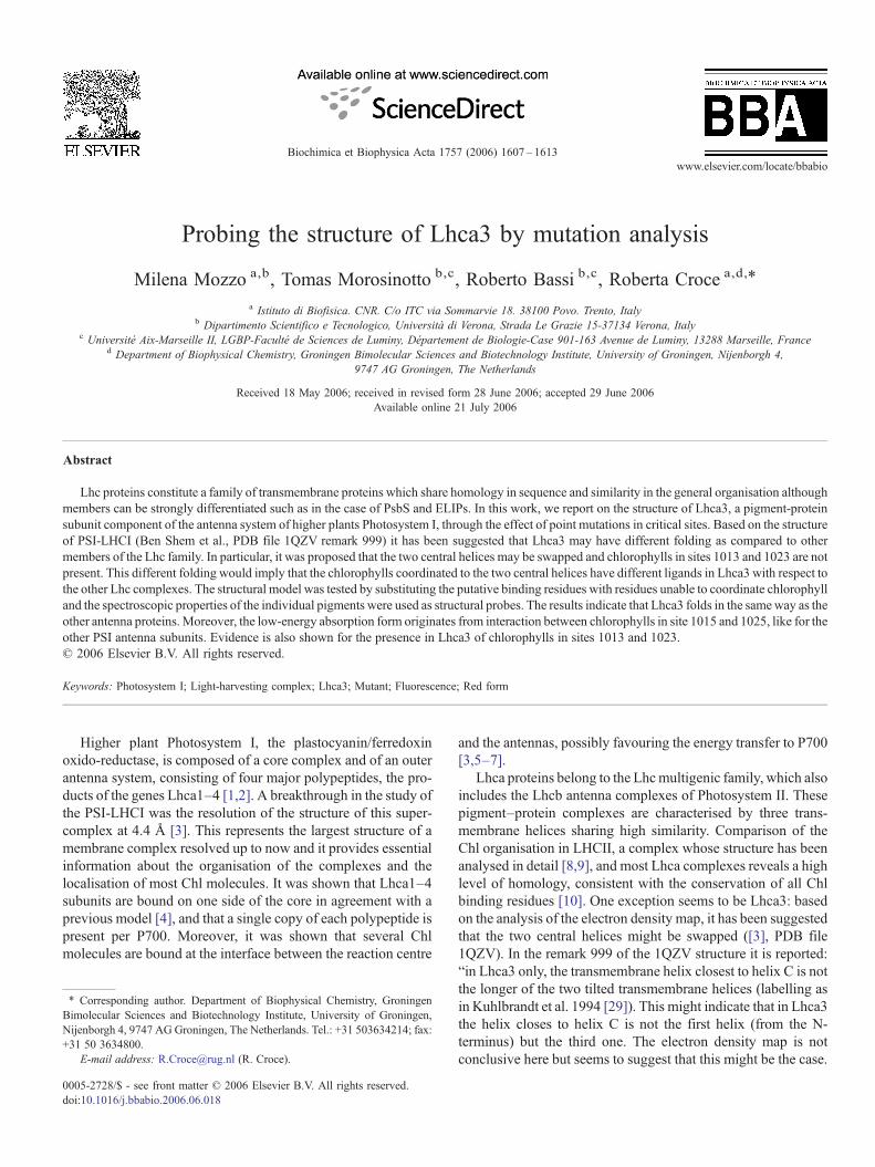
The cDNA encoding Lhca3 from Arabidopsis thaliana wasmutated at the putative Chl binding sites by substitutingnucleophilic residues with apolar amino acids, which cannotcoordinate the central Mg of the Chls. The list of mutationsperformed is reported in Table 1 (Chl binding sites nomenclaturefrom [8] and [3]). In Fig. 1 a schematic representation of thestructure of Lhca3 from Ben-Shem et al. [3] is reported, the Chlsin color are the target of the mutations. WT and mutantapoproteins were overexpressed in E. coli and reconstituted invitro adding pigments [23]. The refolded complexes were purifiedfrom the excess of pigments used in the reconstitution by sucrosegradient ultracentrifugation and ionic exchange chromatography.
All mutants yielded stable reconstituted monomeric productsexcept in the case of R64L/E184V and E59V/R189L. Formutant Q201L a faint green band was obtained in the sucrosegradient, but the complex was highly instable and unable tosurvive the following purification steps.
The pigment content of the complexes is reported in Table 1.We have to mention that the number of Chls coordinated toreconstitute Lhca3 is lower than in the native complex [3]. This isprobably due to the fact that the binding of few peripheral Chls isstabilised by interactions between two different complexes, while
es
Chl a Chl b Violax Lutein β-Car
8.6±0.15 1.4±0.15 0.5±0.04 1.9±0.2 0.36±0.25– – – – –6.6±0.1 1.4±0.1 0.5±0.01 1.4±0.12 0.38±0.14– – – – –– – – – –7.0±0.04 0.96±0.04 0.26±0.004 1.71±0.04 –7.6±0.12 1.4±0.12 0.62±0.08 1.77±0.17 0.34±0.256.3±0.15 0.73±0.16 0.33±0.04 1.52±0.26 –7.7±0.18 1.32± 0.18 0.38±0.08 1.88±0.15 0.1±0.09
sequence homologies. Nomenclature from [8] and [3] are reported for the Chls.
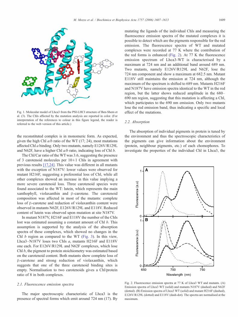
Fig. 2. Fluorescence emission spectra at 77 K of Lhca3 WT and mutants. (A)Emission spectra of Lhca3 WT (solid) and mutants N187V (dashed) and N62F(dotted). (B) Emission spectra of Lhca3WT (solid) and mutant H216F (dashed),E126V/R129L (dotted) and E118V (dash-dot). The spectra are normalised at themaximum.
Fig. 1. Molecular model of Lhca3 from the PSI-LHCI structure of Ben-Shem etal. (3). The Chls affected by the mutation analysis are reported in color. (Forinterpretation of the references to colour in this figure legend, the reader isreferred to the web version of this article.)
1609M. Mozzo et al. / Biochimica et Biophysica Acta 1757 (2006) 1607–1613
the reconstituted complex is in monomeric form. As expected,given the high Chl a/b ratio of the WT (17; 24), most mutationsaffected Chl a binding. Only two mutants, namely E126V/R129Land N62F, have a higher Chl a/b ratio, indicating loss of Chl b.
The Chl/Car ratio of theWTwas 3.6, suggesting the presenceof 3 carotenoid molecules per 10±1 Chls in agreement withprevious results [17,24]. This value was different in all mutantswith the exception of N187V: lower values were observed formutant H216F, suggesting a preferential loss of Chl, while allother complexes showed an increase in this value implying amore severe carotenoid loss. Three carotenoid species werefound associated to the WT: lutein, which represents the mainxanthophyll, violaxanthin and β-carotene. The carotenoidcomposition was affected in most of the mutants: completeloss of β-carotene and reduction of violaxanthin content wereobserved in mutants N62F, E126V/R129L and E118V. Reducedcontent of lutein was observed upon mutation at site N187V.
In mutant N187V, H216F and E118V the number of the Chlslost was estimated assuming a constant amount of Chl b. Thisassumption is supported by the analysis of the absorptionspectra of these complexes, which showed no changes in theChl b region as compared to the WT (Fig. 3). In this view,Lhca3–N187V loses two Chls a, mutants H216F and E118Vone each. For E126V/R129L and N62F complexes, which loseChl b, the pigment to protein stoichiometry was estimated basedon the carotenoid content. Both mutants show complete loss ofβ-carotene and strong reduction of violaxanthin, whichsuggests that one of the three carotenoid binding sites isempty. Normalisation to two carotenoids gives a Chl/proteinratio of 8 in both complexes.
2.1. Fluorescence emission spectra
The major spectroscopic characteristic of Lhca3 is thepresence of spectral forms which emit around 724 nm (17). By
mutating the ligands of the individual Chls and measuring thefluorescence emission spectra of the mutated complexes it ispossible to detect which are the pigments responsible for the redemission. The fluorescence spectra of WT and mutatedcomplexes were recorded at 77 K where the contribution ofthe red forms is enhanced (Fig. 2). At 77 K the fluorescenceemission spectrum of Lhca3-WT is characterised by amaximum at 724 nm and an additional band around 689 nm.Two mutants, namely E126V/R129L and N62F, lose the724 nm component and show a maximum at 682.5 nm. MutantE118V still maintains the emission at 724 nm, although themaximum of the spectrum is shifted to 689 nm. Mutants H216Fand N187V have emission spectra identical to the WT in the redregion, but the latter shows reduced amplitude in the 680–690 nm region, suggesting that this mutation is affecting a Chl,which participates to the 690 nm emission. Only two mutantslose the red emission band, thus indicating a specific and localeffect of the mutations.
2.2. Absorption
The absorption of individual pigments in protein is tuned bythe environment and thus the spectroscopic characteristics ofthe pigments can give information about the environment(protein, neighbour pigments, etc.) of each chomophores. Toinvestigate the properties of the individual Chl in Lhca3, the
Table 2Correspondence of chlorophyll ligands and binding sites
Ligands “Alignment-based” conformation “Swapped” conformation
R64L/E184V 1011-A1-610 A4-1014-602N187V 1012-A2-612 A5-1015-603Q201L 1013-A3-613 –E59V/R189L 1014-A4-602 A1-1011-610N62F 1015-A5-603 A2-1012-612H216F 1023-B3-614 –E126V/R129L 1025-B5-609 B5-1025-609E118V 1026-B6-606 B6-1026-606
In the “alignment-based” conformation, the binding sites identified in LHCIIstructures [29] and [8] have been located in Lhca3 by sequence alignment. The“swapped” conformation, instead, indicates the correspondence following thehypothesis that helices A and B are swapped in Lhca3 [3]. The Chl nomenclaturefrom [3,29] and [8] is reported.
Fig. 3. Absorption spectra at 77 K of Lhca3 WT and mutants. (A) Absorptionspectra of Lhca3 WT (solid) and mutants N187V (dashed) and N62F (dotted).(B) Absorption of Lhca3 WT (solid) and mutant H216F (dashed), E126V/R129L (dotted), E118V (dash-dot). The spectra are normalised to the Chlcontent reported in Table 1.
1610 M. Mozzo et al. / Biochimica et Biophysica Acta 1757 (2006) 1607–1613
absorption spectra of all mutants were measured at 77 K andthey are reported in Fig. 3.
Mutants E126V/R129L and N62F lose completely the redabsorption tail, in accordance with the fluorescence data. Allother complexes exhibit absorption above 700 nm, although inthe case of the Lhca3–E118V complex the intensity is stronglyreduced as compared to the WT. In agreement with the pigmentanalysis, the only two mutations affecting the Chl b absorptionregion are E126V/R129L and N62F. Clear loss of absorptionaround 660 and 670 nm is observed in mutant H216F, while theLhca3–N187V complex is depleted in absorption around680 nm.
3. Discussion
Based on the electron density map, it has been suggestedthat Lhca3 may fold differently as compared with the other Lhccomplexes, with the helix closest to helix C not being helix B,but helix A. However, the structure also shows that thepositions of most of the Chls in Lhca3 are conserved ascompared to the other Lhc complexes [3]. If this suggestion iscorrect, then the ligands for the Chls coordinated to residues ofhelices A and B, namely Chls 1011 (610), 1012 (612), 1013(613), 1014 (602) and 1015 (603) should be swapped withrespect to the ligands in all other Lhc complexes. For clarity theChl ligands in the two possible conformations are reported inTable 2. Moreover, the Lhca3 structural model does not showChls in sites 1013 (613) and 1023 (614), but a new Chl, 1041,
located in an intermediate position. For this study we have usedthe Lhca3 gene of Arabidopsis thaliana, while the structure hasbeen obtained on PSI-LHCI from pea. The proteins from peaand Arabidopsis shows 91.8% identity and most of thesubstitution are homologous, moreover, all the putative Chlbinding residues are conserved, thus strongly suggesting thatthe overall structure and the pigment properties are conservedbetween the two species.
In order to probe the chromophore coordination in Lhca3,mutation analysis of the putative Chl binding sites has beenperformed. It has been shown that the spectroscopic propertiesof several chromophores are conserved throughout the Lhcfamily: the interacting dimer Chl 1012 (612)/Chl 1022 (611)displays the main absorption at 680–681 nm in all complexes sofar analysed, as can be inferred by mutation of the ligand for Chl1012 [12,14]. Similarly, in Lhca complexes, Chl 1015 (603)was shown to be involved in pigment–pigment interaction withChl 1025 (609), leading to the low-energy absorption forms[20,25,26]. If the folding of Lhca3 is different from that of theother Lhc complexes, one can expect that a mutation at residueN187 (ligand for Chl 1012 according to sequence alignment)does not affect Chl 1012, but instead Chl 1015, while a mutationat N62 would influence Chl 1012. Considering that the Chlorganisation and the primary structure are very similar in theLhc complexes we can thus expect to see the “signature” of Chl1012 upon mutation at site N62 and the “signature” of Chl 1025upon mutation at N187.
3.1. Mutation N187V
In Lhc complexes, Chl 1012 (612) is usually coordinated to aN (or H) in the helix A. Mutation at this site in Lhc complexesleads to the loss of the absorption at 680–681 nm (values forroom temperature). This red absorption originates from anexcitonic interaction which involves Chl 1012 and Chl 1022 asshown by mutation analysis and calculations based on thestructure of LHCII [12,22,27].
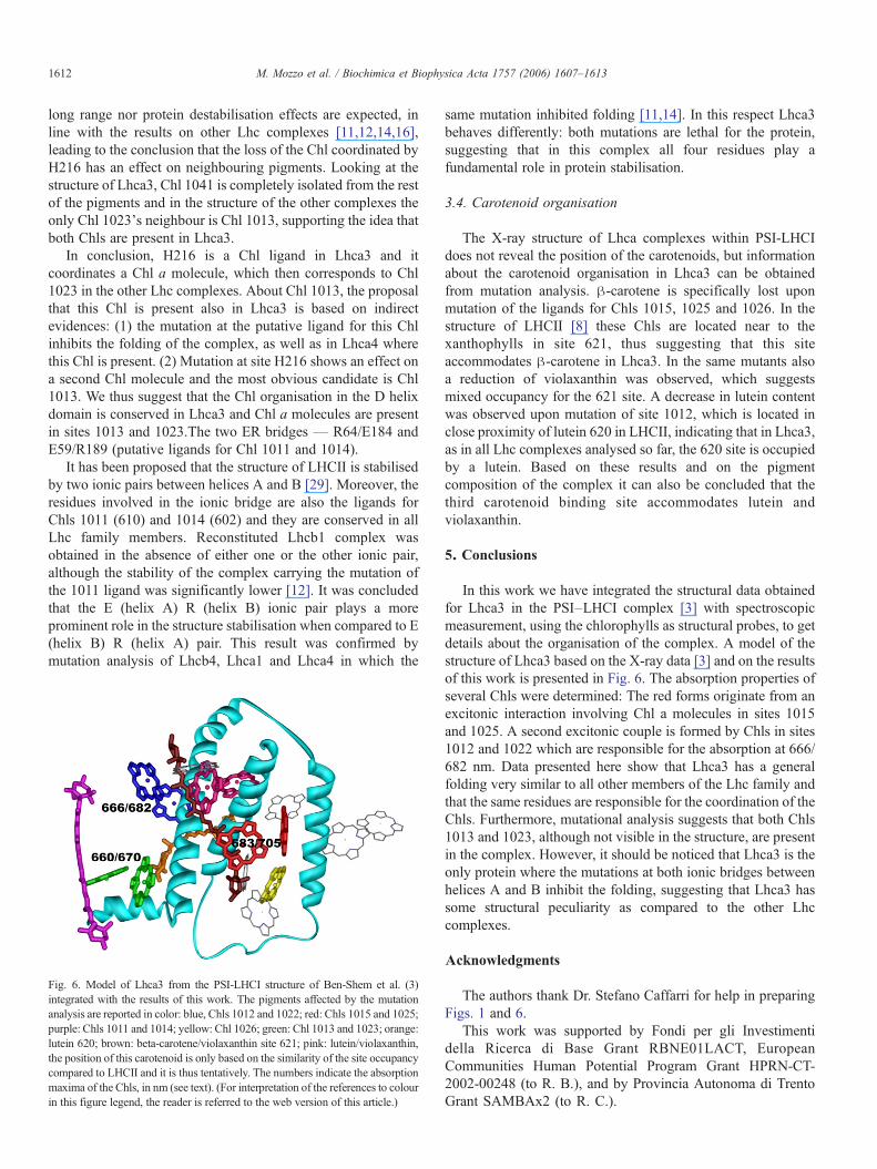
In Lhca3, the N187V mutation leads to the loss of 2 Chl amolecules. In the absorption difference spectrum at lowtemperature, two bands are present at 666 nm and 679.5 nm(Fig. 4) (the difference at RT shows the maximum at 681.5 nm),
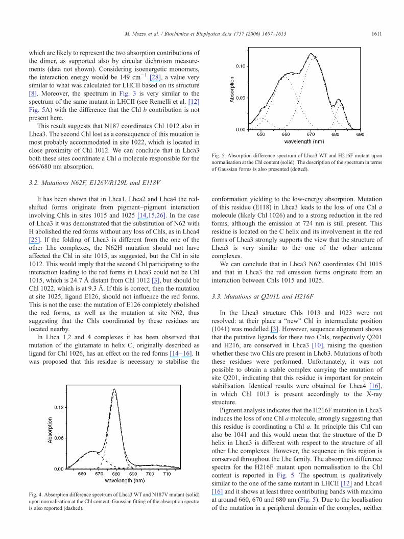
Fig. 5. Absorption difference spectrum of Lhca3 WT and H216F mutant uponnormalisation at the Chl content (solid). The description of the spectrum in termsof Gaussian forms is also presented (dotted).
1611M. Mozzo et al. / Biochimica et Biophysica Acta 1757 (2006) 1607–1613
which are likely to represent the two absorption contributions ofthe dimer, as supported also by circular dichroism measure-ments (data not shown). Considering isoenergetic monomers,the interaction energy would be 149 cm−1 [28], a value verysimilar to what was calculated for LHCII based on its structure[8]. Moreover, the spectrum in Fig. 3 is very similar to thespectrum of the same mutant in LHCII (see Remelli et al. [12]Fig. 5A) with the difference that the Chl b contribution is notpresent here.
This result suggests that N187 coordinates Chl 1012 also inLhca3. The second Chl lost as a consequence of this mutation ismost probably accommodated in site 1022, which is located inclose proximity of Chl 1012. We can conclude that in Lhca3both these sites coordinate a Chl a molecule responsible for the666/680 nm absorption.
3.2. Mutations N62F, E126V/R129L and E118V
It has been shown that in Lhca1, Lhca2 and Lhca4 the red-shifted forms originate from pigment–pigment interactioninvolving Chls in sites 1015 and 1025 [14,15,26]. In the caseof Lhca3 it was demonstrated that the substitution of N62 withH abolished the red forms without any loss of Chls, as in Lhca4[25]. If the folding of Lhca3 is different from the one of theother Lhc complexes, the N62H mutation should not haveaffected the Chl in site 1015, as suggested, but the Chl in site1012. This would imply that the second Chl participating to theinteraction leading to the red forms in Lhca3 could not be Chl1015, which is 24.7 Å distant from Chl 1012 [3], but should beChl 1022, which is at 9.3 Å. If this is correct, then the mutationat site 1025, ligand E126, should not influence the red forms.This is not the case: the mutation of E126 completely abolishedthe red forms, as well as the mutation at site N62, thussuggesting that the Chls coordinated by these residues arelocated nearby.
In Lhca 1,2 and 4 complexes it has been observed thatmutation of the glutamate in helix C, originally described asligand for Chl 1026, has an effect on the red forms [14–16]. Itwas proposed that this residue is necessary to stabilise the
Fig. 4. Absorption difference spectrum of Lhca3 WT and N187V mutant (solid)upon normalisation at the Chl content. Gaussian fitting of the absorption spectrais also reported (dashed).
conformation yielding to the low-energy absorption. Mutationof this residue (E118) in Lhca3 leads to the loss of one Chl amolecule (likely Chl 1026) and to a strong reduction in the redforms, although the emission at 724 nm is still present. Thisresidue is located on the C helix and its involvement in the redforms of Lhca3 strongly supports the view that the structure ofLhca3 is very similar to the one of the other antennacomplexes.
We can conclude that in Lhca3 N62 coordinates Chl 1015and that in Lhca3 the red emission forms originate from aninteraction between Chls 1015 and 1025.
3.3. Mutations at Q201L and H216F
In the Lhca3 structure Chls 1013 and 1023 were notresolved: at their place a “new” Chl in intermediate position(1041) was modelled [3]. However, sequence alignment showsthat the putative ligands for these two Chls, respectively Q201and H216, are conserved in Lhca3 [10], raising the questionwhether these two Chls are present in Lhcb3. Mutations of boththese residues were performed. Unfortunately, it was notpossible to obtain a stable complex carrying the mutation ofsite Q201, indicating that this residue is important for proteinstabilisation. Identical results were obtained for Lhca4 [16],in which Chl 1013 is present accordingly to the X-raystructure.
Pigment analysis indicates that the H216F mutation in Lhca3induces the loss of one Chl amolecule, strongly suggesting thatthis residue is coordinating a Chl a. In principle this Chl canalso be 1041 and this would mean that the structure of the Dhelix in Lhca3 is different with respect to the structure of allother Lhc complexes. However, the sequence in this region isconserved throughout the Lhc family. The absorption differencespectra for the H216F mutant upon normalisation to the Chlcontent is reported in Fig. 5. The spectrum is qualitativelysimilar to the one of the same mutant in LHCII [12] and Lhca4[16] and it shows at least three contributing bands with maximaat around 660, 670 and 680 nm (Fig. 5). Due to the localisationof the mutation in a peripheral domain of the complex, neither
1612 M. Mozzo et al. / Biochimica et Biophysica Acta 1757 (2006) 1607–1613
long range nor protein destabilisation effects are expected, inline with the results on other Lhc complexes [11,12,14,16],leading to the conclusion that the loss of the Chl coordinated byH216 has an effect on neighbouring pigments. Looking at thestructure of Lhca3, Chl 1041 is completely isolated from the restof the pigments and in the structure of the other complexes theonly Chl 1023's neighbour is Chl 1013, supporting the idea thatboth Chls are present in Lhca3.
In conclusion, H216 is a Chl ligand in Lhca3 and itcoordinates a Chl a molecule, which then corresponds to Chl1023 in the other Lhc complexes. About Chl 1013, the proposalthat this Chl is present also in Lhca3 is based on indirectevidences: (1) the mutation at the putative ligand for this Chlinhibits the folding of the complex, as well as in Lhca4 wherethis Chl is present. (2) Mutation at site H216 shows an effect ona second Chl molecule and the most obvious candidate is Chl1013. We thus suggest that the Chl organisation in the D helixdomain is conserved in Lhca3 and Chl a molecules are presentin sites 1013 and 1023.The two ER bridges — R64/E184 andE59/R189 (putative ligands for Chl 1011 and 1014).
It has been proposed that the structure of LHCII is stabilisedby two ionic pairs between helices A and B [29]. Moreover, theresidues involved in the ionic bridge are also the ligands forChls 1011 (610) and 1014 (602) and they are conserved in allLhc family members. Reconstituted Lhcb1 complex wasobtained in the absence of either one or the other ionic pair,although the stability of the complex carrying the mutation ofthe 1011 ligand was significantly lower [12]. It was concludedthat the E (helix A) R (helix B) ionic pair plays a moreprominent role in the structure stabilisation when compared to E(helix B) R (helix A) pair. This result was confirmed bymutation analysis of Lhcb4, Lhca1 and Lhca4 in which the
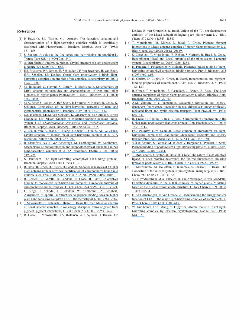
Fig. 6. Model of Lhca3 from the PSI-LHCI structure of Ben-Shem et al. (3)integrated with the results of this work. The pigments affected by the mutationanalysis are reported in color: blue, Chls 1012 and 1022; red: Chls 1015 and 1025;purple: Chls 1011 and 1014; yellow: Chl 1026; green: Chl 1013 and 1023; orange:lutein 620; brown: beta-carotene/violaxanthin site 621; pink: lutein/violaxanthin,the position of this carotenoid is only based on the similarity of the site occupancycompared to LHCII and it is thus tentatively. The numbers indicate the absorptionmaxima of the Chls, in nm (see text). (For interpretation of the references to colourin this figure legend, the reader is referred to the web version of this article.)
same mutation inhibited folding [11,14]. In this respect Lhca3behaves differently: both mutations are lethal for the protein,suggesting that in this complex all four residues play afundamental role in protein stabilisation.
3.4. Carotenoid organisation
The X-ray structure of Lhca complexes within PSI-LHCIdoes not reveal the position of the carotenoids, but informationabout the carotenoid organisation in Lhca3 can be obtainedfrom mutation analysis. β-carotene is specifically lost uponmutation of the ligands for Chls 1015, 1025 and 1026. In thestructure of LHCII [8] these Chls are located near to thexanthophylls in site 621, thus suggesting that this siteaccommodates β-carotene in Lhca3. In the same mutants alsoa reduction of violaxanthin was observed, which suggestsmixed occupancy for the 621 site. A decrease in lutein contentwas observed upon mutation of site 1012, which is located inclose proximity of lutein 620 in LHCII, indicating that in Lhca3,as in all Lhc complexes analysed so far, the 620 site is occupiedby a lutein. Based on these results and on the pigmentcomposition of the complex it can also be concluded that thethird carotenoid binding site accommodates lutein andviolaxanthin.
5. Conclusions
In this work we have integrated the structural data obtainedfor Lhca3 in the PSI–LHCI complex [3] with spectroscopicmeasurement, using the chlorophylls as structural probes, to getdetails about the organisation of the complex. A model of thestructure of Lhca3 based on the X-ray data [3] and on the resultsof this work is presented in Fig. 6. The absorption properties ofseveral Chls were determined: The red forms originate from anexcitonic interaction involving Chl a molecules in sites 1015and 1025. A second excitonic couple is formed by Chls in sites1012 and 1022 which are responsible for the absorption at 666/682 nm. Data presented here show that Lhca3 has a generalfolding very similar to all other members of the Lhc family andthat the same residues are responsible for the coordination of theChls. Furthermore, mutational analysis suggests that both Chls1013 and 1023, although not visible in the structure, are presentin the complex. However, it should be noticed that Lhca3 is theonly protein where the mutations at both ionic bridges betweenhelices A and B inhibit the folding, suggesting that Lhca3 hassome structural peculiarity as compared to the other Lhccomplexes.
Acknowledgments
The authors thank Dr. Stefano Caffarri for help in preparingFigs. 1 and 6.
This work was supported by Fondi per gli Investimentidella Ricerca di Base Grant RBNE01LACT, EuropeanCommunities Human Potential Program Grant HPRN-CT-2002-00248 (to R. B.), and by Provincia Autonoma di TrentoGrant SAMBAx2 (to R. C.).
1613M. Mozzo et al. / Biochimica et Biophysica Acta 1757 (2006) 1607–1613
References
[1] P. Haworth, J.L. Watson, C.J. Arntzen, The detection, isolation andcharacterisation of a light-harvesting complex which id specificallyassociated with Photosystem I, Biochim. Biophys. Acta 724 (1983)151–158.
[2] S. Jansson, A guide to the Lhc genes and their relatives in Arabidopsis,Trends Plant Sci. 4 (1999) 236–240.
[3] A. Ben Shem, F. Frolow, N. Nelson, Crystal structure of plant photosystemI, Nature 426 (2003) 630–635.
[4] E.J. Boekema, P.E. Jensen, E. Schlodder, J.F. van Breemen, H. van Roon,H.V. Scheller, J.P. Dekker, Green plant photosystem I binds light-harvesting complex I on one side of the complex, Biochemistry 40 (2001)1029–1036.
[5] M. Ballottari, C. Govoni, S. Caffarri, T. Morosinotto, Stoichiometry ofLHCI antenna polypeptides and characterization of gap and linkerpigments in higher plants Photosystem I, Eur. J. Biochem. 271 (2004)4659–4665.
[6] M.K. Sener, C. Jolley, A. Ben Shem, P. Fromme, N. Nelson, R. Croce, K.Schulten, Comparison of the light-harvesting networks of plant andcyanobacterial photosystem I, Biophys. J. 89 (2005) 1630–1642.
[7] J.A. Ihalainen, I.H.M. van Stokkum, K. Gibasiewicz, M. Germano, R. vanGrondelle, J.P. Dekker, Kinetics of excitation trapping in intact Photo-system I of Chlamydomonas reinhardtii and Arabidopsis thaliana,Biochim. Biophys. Acta, Bioenerg. 1706 (2005) 267–275.
[8] Z. Liu, H. Yan, K. Wang, T. Kuang, J. Zhang, L. Gui, X. An, W. Chang,Crystal structure of spinach major light-harvesting complex at 2. 72 Aresolution, Nature 428 (2004) 287–292.
[9] R. Standfuss, A.C.T. van Scheltinga, M. Lamborghini, W. Kuhlbrandt,Mechanisms of photoprotection and nonphotochemical quenching in pealight-harvesting complex at 2. 5A resolution, EMBO J. 24 (2005)919–928.
[10] S. Janssonm, The light-harvesting chlorophyll a/b-binding proteins,Biochim. Biophys. Acta 1184 (1994) 1–19.
[11] R. Bassi, R. Croce, D. Cugini, D. Sandona, Mutational analysis of a higherplant antenna protein provides identification of chromophores bound intomultiple sites, Proc. Natl. Acad. Sci. U. S. A. 96 (1999) 10056–10061.
[12] R. Remelli, C. Varotto, D. Sandona, R. Croce, R. Bassi, Chlorophyllbinding to monomeric light-harvesting complex, a mutation analysis ofchromophore-binding residues, J. Biol. Chem. 274 (1999) 33510–33521.
[13] H. Rogl, R. Schodel, H. Lokstein, W. Kuhlbrandt, A. Schubert,Assignment of spectral substructures to pigment-binding sites in higherplant light-harvesting complex LHC-II, Biochemistry 41 (2002) 2281–2287.
[14] T.Morosinotto, S. Castelletti, J. Breton, R. Bassi, R. Croce,Mutation analysisof Lhca1 antenna complex—Low energy absorption forms originate frompigment–pigment interactions, J. Biol. Chem. 277 (2002) 36253–36261.
[15] R. Croce, T. Morosinotto, J.A. Ihalainen, A. Chojnicka, J. Breton, J.P.
Dekker, R. van Grondelle, R. Bassi, Origin of the 701-nm fluorescenceemission of the Lhca2 subunit of higher plant photosystem I, J. Biol.Chem. 279 (2004) 48543–48549.
[16] T. Morosinotto, M. Mozzo, R. Bassi, R. Croce, Pigment–pigmentinteractions in Lhca4 antenna complex of higher plants photosystem I, J.Biol. Chem. 280 (2005) 20612–20619.
[17] S. Castelletti, T. Morosinotto, B. Robert, S. Caffarri, R. Bassi, R. Croce,Recombinant Lhca2 and Lhca3 subunits of the photosystem I antennasystem, Biochemistry 42 (2003) 4226–4234.
[18] H. Paulsen, B. Finkenzeller, N. Kuhlein, Pigments induce folding of light-harvesting chlorophyll alpha/beta-binding protein, Eur. J. Biochem. 215(1993) 809–816.
[19] E. Giuffra, D. Cugini, R. Croce, R. Bassi, Reconstitution and pigment-binding properties of recombinant CP29, Eur. J. Biochem. 238 (1996)112–120.
[20] R. Croce, T. Morosinotto, S. Castelletti, J. Breton, R. Bassi, The Lhcaantenna complexes of higher plants photosystem I, Bioch. Biophys. Acta,Bioenerg. 1556 (2002) 29–40.
[21] A.M. Gilmore, H.Y. Yamamoto, Zeaxanthin formation and energy-dependent fluorescence quenching in pea chloroplasts under artificiallymediated linear and cyclic electron transport, Plant Physiol. 96 (1991)635–643.
[22] R. Croce, G. Canino, F. Ros, R. Bassi, Chromophore organization in thehigher-plant photosystem II antenna protein CP26, Biochemistry 41 (2002)7334–7343.
[23] F.G. Plumley, G.W. Schmidt, Reconstitution of chloroform a/b light-harvesting complexes: Xanthophyll-dependent assembly and energytransfer, Proc. Natl. Acad. Sci. U. S. A. 84 (1987) 146–150.
[24] V.H.R. Schmid, S. Potthast, M. Wiener, V. Bergauer, H. Paulsen, S. Storf,Pigment binding of photosystem I light-harvesting proteins, J. Biol. Chem.277 (2002) 37307–37314.
[25] T. Morosinotto, J. Breton, R. Bassi, R. Croce, The nature of a chlorophyllligand in Lhca proteins determines the far red fluorescence emissiontypical of photosystem I, J. Biol. Chem. 278 (2003) 49223–49229.
[26] T. Morosinotto, M. Ballottari, F. Klimmek, S. Jansson, R. Bassi, Theassociation of the antenna system to photosystem I in higher plants, J. Biol.Chem. 280 (2005) 31050–31058.
[27] V.I. Novoderezhkin, M.A. Palacios, H. Van Amerongen, R. van Grondelle,Excitation dynamics in the LHCII complex of higher plants: Modelingbased on the 2. 72 angstrom crystal structure, J. Phys. Chem. B 109 (2005)10493–10504.
[28] H. Van Amerongen, R. van Grondelle, Understanding the energy transferfunction of LHCII, the major light-harvesting complex of green plants, J.Phys. Chem. B 105 (2001) 604–617.
[29] W. Kühlbrandt, D.N. Wang, Y. Fujiyoshi, Atomic model of plant light-harvesting complex by electron crystallography, Nature 367 (1994)614–621.