Calcium-activated proteolysis of neurofilament proteins in goldfish Mauthner axons
Upload
independentCategory
view
1download
0
Eur. J . Biochem. 226, 323-333 (1994) 0 FEBS 1994
Probing the structure of hirudin from Hirudinaria manillensis by limited proteolysis Isolation, characterization and thrombin-inhibitory properties of N-terminal fragments
Alessandro VINDIGNI', Vincenzo DE FlLIPPIS ', Giuseppe ZANOTTI', Carlo VISCO*, Gaetano ORSINI' and Angelo FONTANA' ' CRIBI Biotechnology Centre and Department of Organic Chemistry, University of Padua, Italy
Pharmacia-Farmitalia, Bioscience Centre, Nerviano, Milan, Italy
(Received August 3, 1994) - EJB 94 1188/3
Hirudin is the most potent and specific inhibitor of the blood-clotting enzyme thrombin so far known. Several hirudin variants were isolated mostly from Hirudo medicinalis and shown to be polypeptide chains of approximately 7 kDa with three internal disulfide bridges. In this study, lim- ited proteolysis has been used to probe aspects of the structure and dynamics of a hirudin variant HM2 isolated from Hirudinaria manillensis. Proteolysis of the polypeptide chain of 64-amino-acid residues of hirudin HM2 by protease from Staphylococcus aureus V8, trypsin, thermolysin and subtilisin occurs at region 41 -49 of the chain. The N-terminal fragments 1-41 and 1-47 were isolated to homogeneity and shown to maintain inhibitory action on thrombin, though much lower than the intact protein. The results were interpreted on the basis of a proposed three-dimensional structure of hirudin HM2 deduced by protein modelling the known structure of hirudin variant HV1 from Hirudo medicinalis (75% sequence similarity between HM2 and HV1). Both proteolysis experiments and protein modelling provide evidence for the existence in hirudin HM2 of a N- terminal well-structured domain (core) and a C-terminal flexible polypeptide segment. Determina- tion of the accessible surface area of the three-dimensional model of hirudin HM2 showed that the sites of preferential cleavages are at the surface of the polypeptide molecule.
Hirudin is a potent and specific thrombin inhibitor iso- lated from the salivary gland of the blood sucking leech (Markwardt, 1991). A number of hirudin variants have been isolated, mostly from Hirudo medicinalis. All of them consist of a single polypeptide chain of 63 -66 amino-acid residues with three disulfide bridges (Dodt et al., 1985; Tripier, 1988; Stringer and Lindenfeld, 1992). In recent years, hirudin has been intensively investigated as a potential protein drug with anticoagulant activity, as well as an interesting model for addressing structure/function relationships and biorecogni- tion mechanisms of proteins (Tapparelli et al., 1993). Recom- binant methods have been used to produce large amounts of the polypeptide (Dodt et al., 1986; Harvey et al., 1986; Riehl-Bellon et al., 1989), thus enabling a variety of studies aimed at elucidating its mechanism of interaction with thrombin (Vu et al., 1991 ; Ayah and Di Cera, 1994), as well as its three-dimensional structure both in the free form or complexed with thrombin. NMR (Folkers et al., 1989; Haru- yama and Wiitrich, 1989; Haruyama et al., 1989) and X-ray (Rydel et al., 1990, 1991; Vitali et al., 1992; Karshikov et al., 1992; Priestle et al., 1993) analyses of hirudin from H.
Correspondence to A. Fontana, CRIBI Biotechnology Centre, Via Trieste 75, 1-35121 Padua, Italy
Abbreviations. HM2 and HVI, hirudin variants from Hirudina- ria manillensis and Hirudo medicinalis, respectively ; RP, reverse- phase; S-2238, synthetic thrombin substrate H-o-Phe-pipecolyl-Arg- p-nitroanilide ; ASA, accessible surface area.
Enzymes. Trypsin (EC 3.4.21.4); thrombin (EC 3.4.21.5); thermolysin (EC 3.4.24.4); subtilisin (EC 3.4.21.14); Glu-C prote- ase from Staphylococcus aureus V8 (EC 3.4.21.19).
medicinalis provided evidence for a well-structured N-tenni- nal domain, tightly packed by the three disulfide bridges of the polypeptide, and a disordered C-terminal tail abundant in acidic residues. While hirudin isolated from H. medicinalis contains a sulphated Tyr63, recombinant hirudin is not sul- phated and shows a somewhat lower inhibitory activity towards thrombin (Braun et al., 1988). Both domains of hiru- din, the N-terminal core and the C-terminal tail, contribute to the formation of a very tight complex with thrombin (Stone and Hofsteenge, 1986). The N-terminal core binds near the active site of thrombin, while the C-terminal tail recognizes the fibrinogen-binding site of thrombin (Rydel et al., 1990, 1991).
In recent studies (Electricwala et al., 1991 ; Steiner et al., 1992; Scacheri et al., 1993), hirudin variants have been iso- lated from Hirudinaria manillensis, called buffalo leech, a species which is significantly more specialized for mamma- lian parasitism than H. medicinalis (Sawyer, 1986). The pri- mary structures of hirudin isoforms from H. manillensis (Steiner et al., 1992) show significant (60-75%) sequence similarity to those of H. medicinalis (Dodt et al., 1985; Scharf et al., 1989), and, in particular, contain six cysteine residues at highly conserved positions, thus signifying the same pattern of disulfide formation (Thornton, 1981). Hiru- din from H. manillensis was found to be glycosylated and not sulphated (Steiner et al., 1992). A 64-residue hirudin variant, HM2, from H. manillensis was expressed in Escherichia coli utilizing a synthetic gene (Scacheri et al., 1993). The recom- binant HM2, lacking post-translational modifications, pos-

324
sessed an inhibitory activity for thrombin comparable to that of other recombinant hirudins.
In this report, limited proteolysis of hirudin HM2 was used to probe its structure and dynamics, with the view that proteolysis could dissect this inhibitory protein into indivi- dual domains, in analogy to previous work conducted on hi- rudin from H. medicinalis (Chang, 1990; Chang et al., 1990; Dennis et al., 1990). Indeed, the polypeptide chain of the HM2 variant can be cleaved by the glutamate-specific prote- ase from Staphylococcus aureus V8, trypsin, subtilisin and thermolysin at chain region 41 -49, thus allowing us to iso- late and characterize N-terminal core fragments of hirudin HM2, which were shown to maintain inhibitory action towards thrombin. Moreover, a three-dimensional model of hirudin HM2 was produced on the basis of the NMR solution structure of the HV1 variant (Folkers et al., 1989). The re- sults of proteolysis experiments, as well as the thrombin in- hibitory activities of the N-terminal proteolytic fragments, were interpreted on the basis of the proposed three-dimen- sional model of hirudin HM2.
EXPERIMENTAL PROCEDURES Materials
Recombinant hirudin HM2 from H. manillensis was pro- duced at the Bioscience Centre of the Pharmacia-Farmitalia and stored at -20°C (Scacheri et al., 1993). A sample of recombinant hirudin variant HV1 from H. medicinalis was purchased from Sigma Chem. Co. The Glu-C protease from S. aureus V8, trypsin and subtilisin were purchased from Boehringer. Thermolysin from Bacillus thermoproteolyticus and thrombin from human plasma were from Sigma. Triflu- oroacetic acid, phenylisothiocyanate and constant boiling HC1 were purchased from Pierce Chem. Co. Solvents and reagents used for peptide/protein sequence analysis were from Applied Biosystem. Deionized water obtained from a tandem Milli-RO and Milli-Q system (Millipore) was used for all solutions. All organic solvents were HPLC grade and other compounds were of analytical grade purchased from C. Erba or Merck.
Methods
Reverse-phase high-performance liquid chromatography (RP-HPLC) was performed on a HPLC instrument from Pharmacia-LKE3 model 2249 utilizing a Vydac C,, column (4.6 mmX 150 mm) purchased from The Separations Group. Eluted material from the column was collected manually in 1 .5-ml Eppendorf tubes and dried in a Speedvac concentrator from Savant.
Capillary electrophoresis was conducted on a Bio-Rad instrument HPE-100 utilizing a fused-silica capillary of in- side diameter 25 pm. Peptide/protein samples were dissolved in 0.1 M potassium phosphate, pH 2.5, and injected onto the capillary applying an electric field of 5 KV for 8 s. The electrophoretic separation was carried out at room temper- ature with a field strength of 400 volts/cm. The absorbance of the effluent was monitored at 200 nm.
Amino acid analysis was performed using the Millipore- Waters workstation and the Pico-Tag C,, column (4.6 mmX15O mm). Lyophilized samples of protein frag- ments (10-500 pmol), contained in heat-treated borosilicate tubes (4mmX5Omm), were acid hydrolyzed on the Pico- Tag workstation for 1 h at 150°C using 200 p16 M HCl con-
taining 0.1 % phenol, then derivatized with phenylisothiocya- nate (Heinrikson and Meredith, 1984). The phenylthiocarba- moyl derivatives were analyzed by HPLC.
N-terminal sequences were determined with an Applied Biosystems sequencer 477A equipped with an on-line phe- nylthiohydantoin analyzer 120A. Standard manufacturer's procedures and programs were used with minor modifica- tions.
Mass-spectrometry analyses of purified fragments were carried out on a time-of-flight ReflexTM mass spectrometer from Bruker, equipped with a matrix-assisted laser desorp- tion ion source (Karas and Hillenkamp, 1988). Mass spectra were recorded in the linear mode using a 30-kV electric field. Raw data were analysed and stored by the X-MassTM soft- ware provided by Bruker.
Limited proteolysis Hirudin HM2 was subjected to proteolysis in 50mM
NH,HCO, buffer, pH 7.8 (2 mg/ml), at a protease/substrate ratio of 1 : 10 (by mass) with the Glu-C protease from S. aureus V8 (Houmard and Drapeau, 1972; Drapeau, 1977). The reaction mixture was incubated at 37°C and samples were removed at timed intervals for analyzing the proteolytic products by RP-HPLC. Proteolysis was stopped by diluting an aliquot (10 pl) of the digestion mixture with 0.1 % aque- ous trifluoroacetic acid (50 pl). Samples were concentrated using the Speedvac system, then analyzed by RP-HPLC. The identity of the proteolytic fragments thus purified was deter- mined on the basis of their amino acid composition after acid hydrolysis and N-terminal sequencing (see Results).
Hirudin HM2 was digested with trypsin, subtilisin and thermolysin at room temperature and a protease/substrate ra- tio of 1 : 20 (by mass), utilizing the same buffer and protein concentrations used for the digestion with V8 protease. In the case of thermolysin, the buffer was 50 mM Tris/HCl, pH 7.8, containing 5 mM CaCI,.
Amidolytic assay of thrombin Aniithrombin activity of recombinant hirudin variants
HM2 or HV1 was determined (Stone and Hofsteenge, 1986; Braun et al., 1988) with H-D-Phe-pipecolyl-Arg-p-nitroani- lide (S-2238) (Kabi Vitrum) as a substrate. Assays were per- formed in a 1-cm pathlength quartz cuvette at 37°C in SO mM Tris/HCI, pH 7.8, containing 0.1 M NaCl and 0.1% (by mass) poly(ethy1ene glycol) 6000. The assay solution containing substrate and hirudin HM2 was incubated at 37 "C for 5 min, then the assay was started by the addition of thrombin. The release of p-nitroaniline that resulted from the hydrolysis of the substrate was monitored by the increase in absorbance at 405 nm ( E ~ , , ~ rill, = 9920 M-' . cm-' for p - nitroaniline). The concentration of the substrate S-2238 was determined spectrophotometrically ( E ~ ~ ~ = 8270 M-' . cm-' ; Lottenberg and Jackson, 1983). Antithrombin activity of hi- rudin HM2 fragments 1-41 and 1-47 was measured in the same assay solution with 100 pM thrombin and 0.2-0.5 pM fragment and increasing the substrate S-2238 concentration from 4 pM to 30 pM.
Kinetic parameters for the thrombin inhibition by frag- ments 1-41 and 1-47 were calculated by the Michaelis- Menten treatment of competitive inhibition (Fromm, 1975). The data were fitted to the Lineweaver-Burk equation 1/V = l/V,,,, + K,,,/V,,,,, . [S], where V,,,, is the hydrolysis rate in the absence of inhibitor. Since the formation of the thrombin-

325
inhibitor complex can be represented by E + I ts EI, the standard Gibbs free energy of complex formation (denoted as binding energy AG;) is related to the dissociation con- stant, K,, of the thrombin-inhibitor complex by equation AG; = -RirlnK,, where R is the gas constant (8.314 J . mol-’ . K-’) and T is the absolute temperature.
Peptide and protein concentrations were determined by UV absorption spectroscopy using a double-beam Perkin-El- mer spectrophotometer 11-2. Absorption coefficients for intact himdin HM2 (c~~,,,,,,, = 0.43 mg-’ . cm2) and its fragments 1-41 (C28nnm= 0.69 mg-’ . cm’) and 1-47 (cZXOn,,, - 0.60 mg-’ . cm’) were determined using molar absorption coefficients of 1280 M-’ . cm-’ and 120 M-‘ . cm-’ for tyro- sine and cystine residues, respectively (Gill and von Hippel, 1989).
-
Model building The structure of hirudin HM2 from H. manillensis has
been modelled on the NMR-derived solution structure in the Brookhaven Protein Data Bank of hirudin HV1 from H. me- dicinalis (Folkers et al., 1989) and deposited as 6HIR. The model was built using the program Insight-11 (Biosym Tech- nologies) run on a Silicon Graphics IRIS SD/35 workstation. The modelling of residues 1-47 of hirudin HM2 was ob- tained by replacing the amino acid side chains of the HV1 1-49 domain with the corresponding side chains, when dif- ferent, in the HM2 1-47 domain. Side-chain conformations were initially set to the most preferred side-chain rotamer and subsequentely manually optimized. The reverse-turn-en- compassing residues 3 1 - 34 in the HM2 sequence (residues 32-35 in the HVI sequence) is ill-defined in the NMR- derived solution structure of both intact HV1 (Folkers et al., 1989; Hamyama and Wiitrich, 1989) or HV1 fragment 1-51 (Szyperski et al., 1992a), as well as in the X-ray struc- ture of HV1 thrombin (Vitali et al., 1992) or [Lys47]HV2- thrombin complex (Rydel et al., 1991). In our case, it was modelled using a distance geometry-based algorithm (Search-Loop option in Biopolymer module of the program Insight 11). The refinement of the model was achieved by energy-minimization procedure (Brunger, 1988, 1990) in or- der to compensate for unfavorable intramolecular contacts introduced during the modelling procedure. Accessible sur- face area (Lee and Richards, 1971) on the HM2 model was calculated using the program ACCESS (Richards, 198.3, and using a water probe radius of 0.14 nm.
RESULTS Proteolysis of hirudin HM2
Limited proteolysis of recombinant hirudin HM2 from H. manillensis (Scacheri et al., 1993) was conducted using sev- eral proteases of known properties and substrate specificity. Since the aim of these experiments was to probe the structure and dynamics of the hirudin molecule, proteolytic enzymes of restricted specificity such as the Glu-C-specific protease from S. aureus V8 (Houmard and Drapeau, 1972; Drapeau, 1977) and trypsin (Walsh, 1970), as well as of broad specific- ity, such as subtilisin (Ottesen ans Svendsen, 1970) and thermolysin (Heinrikson, 1977), were employed. It was an- ticipated that regions of the hirudin molecule characterized by exposure and flexibility would be sites of proteolytic at- tack, whereas the actual peptide bond(s) fission within these regions would be dictated by the specificity of the protease
G S N V C G E G K N C Q L S S S G ! G l Q [ N K ~ I ~ G ~ D ~
Fig. 1. Sequence alignment of hirudin variant HM2 from H. ma- nillensis (Scacheri et al., 1993) and variant HV1 from H. medici- nalis (Dodt et al., 1985). Identical residues are boxed and gaps are introduced for maximal alignment. Disulfide bonds occur between cysteine residues at positions 6-14, 16-28 and 22-37 in hirudin HM2 and at positions 6-14, 16-28 and 22-39 in hirudin HV1.
1-41
I
I I I
0 10 20 30 40 RETENTION TIME, min
Fig. 2. Separation by RP-HPLC of protein fragments obtained by proteolysis of hirudin HM2 with V8 protease. An aliquot of the proteolytic mixture, obtained after a 1-h reaction at 37°C with an enzymehubstrate ratio of 1 : 10 (by mass; see Materials and Methods), was applied to a Vydac C,, column (4.6 mmXl.50 mm). Elution was carried out at a flow rate of 0.8 ml/min with a gradient of acetonitrile in 0.05% trifluoroacetic acid. The effluent was moni- tored at 226 nm. Numbers near the chromatographic peaks refer to the identity of the peptide material eluted from the column (see text). Fragment (1 -41)* derives from internal nicking by V8 protease of fragment 1-41 at the GluS-Ser9 peptide bond (see Fig. 1) .
(Mihalyi, 1978; Neurath, 1980; Fontana et al., 1986, 1989, 1993).
Fig. 2 shows a typical RP-HPLC separation of the pep- tide fragments produced upon treatment of hirudin HM2 by the Glu-specific V8 protease at 37°C for 1 h in 50 mM am- monium bicarbonate, pH 7.8. Despite the fact that there are seven glutamate residues in hirudin HM2 (see Fig. 1) as pos-

326
Table 1. Amino acid composition of hirudin HM2 from H. manillensis and its proteolytic fragments. Theoretical values are givtn in parenthesis and were calculated from the amino acid sequence of hirudin variant HM2 (Scacheri et al., 1993). The various proteolytic fragments of hirudin HM2 were prepared and isolated to homogeneity as described in the text. Fragment (1-41)" is constituted by fragments 1-8 and 9-41, linked by a disulfide bridge. n.d., not determined.
Amino acid Composition of proteolytic fragment
HM2 1-41 (1-41)* 42-64 42-51 52-64 1-47 48-64 1-49
residues/mol
Asx 8.6 (9) 4.7 ( 5 ) 4.8 ( 5 ) 4.1 (4) 4.1 (4) 4.9 ( 5 ) 3.8 (4) 5.3 ( 5 )
GIY 7.7 (8) 5.6 (6) 5.6 (6) 2.2 (2) 1.0 (1) 1.0 (I) 7.2 (7) 1.4 (I) 7.4 ( 7 ) His 1.0 (1) 0.9 (1) 0.9 (1) 1.2 (1) 1.0 ( I )
Pro 3.0 (3) 2.6 (3) 1.8 (2) 1.0 (1) 2.1 (2) 1.1 (1) 2.2 (-')
Val 3.9 (4) 3.7 (4) 3.6 (4) 3.8 (4) 3.9 (4)
Leu 3.2 (3) 2.0 (2) 2.0 (2) 1.0 (1) 1.0 (1) 2.0 (2) 1.0 (1) 2.1 ( 2 )
Glx 11.5 (11) 6.1 (6) 6.1 (6) 5.2 ( 5 ) 2.1 (2) 3.3 (3) 6.1 (6) 5.0 ( 5 ) 7.2 (5) Ser 7.0 (7) 5.8 (6) 5.7 (6) 1.2 (1) 0.9 (I) 6.0 (6) 1.1 (1) 7.0 (7)
Thr 4.0 (4) 1.6 (2) 1.5 (2) 1.7 (2) 1.7 (2) 3.0 (3) 0.8 (1) 2.8 ( 'j)
TYr 2.1 (2) 1.9 (2) 1.9 (2) 1.8 (2) 1.8 ( 3 )
cys n.d. n.d. n.d. n.d. n.d. Ile 2.0 (2) 1.7 (2) 2.0 (2) 1.8 (2)
Phe 1.1 (1) 0.8 (1) 1.0 (1) 0.9 (1) LYS 3.6 (3) 1.2 (1) 1.2 (1) 1.9 (2) 2.1 (2) 3.0 (3) 3.0 ( 3 )
Table 2. N-terminal sequences of peptide fragments derived from proteolytic digestion of hirudin HM2 from H. manillensis. Frag- ments were prepared and purified to homogeneity by RP-HPLC as described in the text. The results of sequence analysis are reponed as yields of the phenylthiohydantoin derivatives of the amino acid (PTH-Xaa) recovered on the sequencer at each cycle of the Edman degradation. Data for intact hirudin HM2 are also included for reference. n.d., unidentified PTH-amino-acid.
Cycle number PTH-Xaa
HM2 1-41 (1 -41)" 1-47 42 - 64 48-64 1 -49
pmol
1 2 3 4 5 6 7 8 9 10 11 12 13 14 15 16 17
Val (906) Ser (291) Tyr (523) Thr (426) Asp (192) n.d. Thr (409) Glu (247) Ser (151) Gly (332) Gln (272) Asn (282) Tyr (311) n.d. Leu (273) n.d. Val (255)
Val (55) Val (181)/Ser (100) Val (49) G ~ Y (43)
Thr (44) Thr (80)/Asn (97) Thr (40) LYS (15)
LYS (11) Ser (8) Gln (14) Thr (8) Glu (8)
Ser (32) Ser (83)/Gly (154) Ser (38) Thr (26) T Y ~ (66) Tyr (119)/Gln (128) Tyr (46) Pro (29)
Asp (36) Asp (53)/Tyr (1 17) Asp (20) Pro (25)
GlY (11) Asp (5)
Glu ( 5 ) Phe (9)
Glu (9)
Ser (35) Val (78) Gln (33) Ser (42)
Thr ( S O ) Glu (1 7) GlY (28) Asp (43)
Phe (25) Glu (1 7) Glu (18) Ile (21) Pro (17) Asp (4) Glu (10) Asp (2) Ile (13) Leu (9) Asn (5)
Thr (14) TYr (59)
Asp (13)
sible sites of proteolytic cleavage, it appears that V8 protease cleaves the HM2 molecule quite selectively, since a major peak of peptide material is seen in the RP-HPLC chromato- gram (Fig. 2). The peptide material corresponding to most of the chromatographic peaks was isolated and analyzed in terms of amino acid composition after acid hydrolysis (Table 1) and N-terminal sequencing (Table 2). These data were compared with the known sequence of hirudin HM2 (Fig. I) , thus allowing unambigous identification of the fragments produced by V8-protease treatment of hirudin HM2 (see Fig. 2, and Tables 1 and 2).
Since, in this study, we were interested also in probing the most reactive site(s) of proteolysis, also the kinetics of the peptide-bond fissions by V8 protease were determined by quantitative RP-HPLC analysis of the time-course torma- tion of the various fragments. The results of these analyses (Fig. 3) show that the most susceptible site to hydroljsis by V8 protease is the peptide bond, Glu41 -Gly42, thus leading to the early formation of fragments 1-41 and 42-64. In turn, fragment 42-64 is cleaved at the GluS1-GlyS2 peptide bond leading to fragments 42-51 and 52-64 (see Fig. 2 and Tables 1 and 2). Moreover, fragment 1-41 is slowly

327
Inhibition of thrombin activity by hirudin HM2 fragments 1-41 and 1-47
In the course of this study, the anti-thrombin activity of recombinant hirudin HV1 and HM2 variants was determined using the S-2238 substrate (see Experimental Procedures). The calculated figures for the inhibition constant (K,) were 1.1550.01 pM and 0.78+0.02 pM for hirudin HV1 and HM2, respectively, thus implying that the two polypeptides display quite similar anti-thrombin activity.
As shown in Fig. 6, hirudin HM2 fragments 1-41 and 1 -47 were competitive inhibitors of thrombin. However, the calculated figures for K,, given in Table 3, indicate that the binding of fragments 1-41 and 1-47 to thrombin is much less efficient than that of intact hirudin HM2 and that frag- ment 1-47 is a better inhibitor than fragment 1-41. From the K, values, the free energy of complex formation (-dG,”) with thrombin was calculated for both hirudin HM2 and its fragments 3 -41 and 1-47 (see Experimental Pro- cedures). The figures shown in Table 3 indicate that about half of the free energy of binding to thrombin is lost upon removal of the C-terminal portion of hirudin HM2.
I I I I I
’ ’3 I 100
0 4 1-41 @.-A 42-64
c 60 5
50 I
40 2 5 30 a
20
10
0
2
0 20 40 60 80 I00 120 240 340 440 REACTION TIME, min
Fig. 3. The time-course of the formation of protein fragments during proteolysis of hirudin HM2 by V8 protease. Proteolysis was monitored at 37°C with an enzymehubstrate ratio of 1 : 10, by mass. At time intervals, aliquots were removed for RP-HPLC (see Fig. 2). Quantification of the proteolytic fragments was performed by integrating the area under the corresponding chromatographic peaks. The area of the peak of hirudin HM2, before proteolysis, was taken as 100%. No area normalization factor was used to account for different absorption coefficients at 226 nm of protein fragments. 0, hirudin HM2; 0, fragment 1-41 ; A, fragment 42-64; A, frag- ment (1 -41) nicked at Glu8-Ser9.
hydrolyzed by V8 protease at Glu8-Ser9 to a fragment spe- cies constituted by peptides 1-8 and 9-41, crosslinked by disulfide bridges (see Fig. 1). Evidence for the identity of this nicked fragment, denoted (1-41)*, is given by the fact that a peptide of different RP-HPLC chromatographic behav- iour (Fig. 2) shows the same amino acid composition of that of fragment 1-41 (Table l ) , but two N-terminal sequences (Val-Ser-Tyr-Thr-Asp and Ser-Gly-Gln-Asn-Tyr, see Table 2 and Fig. 1).
Fig. 4 shows the RP-HPLC chromatograms of proteolytic digests of hirudin HM2 obtained by trypsin, subtilisin and thermolysin. Trypsin cleaves hirudin HM2 very selectively at Lys47-Ser48, leading to fragments 1-47 and 48-64 only (see Fig. 4A and the data given in Tables 1 and 2), despite the fact that there are two additional lysine-residues at posi- tions 26 and 45 of the chain (Fig. 1). The tryptic hydrolysis at Lys47 was fast and selective and measurements of the hydrolysis kinetics were not conducted. Both subtilisin and thermolysin cleave hirudin HM2 at the Glu49-Thr50 peptide bond, leading to fragment 1-49 as a major product, which is quite resistant to further proteolysis (see Fig. 4B and C and the analytical data of Tables 1 and 2).
Since the proteolytic fragments 1-47 and 1-49 can be produced easily and in high yields by proteolysis of hirudin HM2 with V8 protease and trypsin, respectively, these frag- ments were isolated by micropreparative RP-HPLC in suffi- cient amounts for subsequent functional studies. Fragments 1-41 and 1-47 thus isolated were shown to be homoge- neous by analytical RP-HPLC and capillary electrophoresis (Fig. 5 ) . Moreover, time-of-flight mass spectrometry analysis (Karas and Hillenkamp, 1988) gave molecular masses of 4255 ? 2.1 Da and 4864 % 2.7 Da for fragments 1-41 and 1-47, respectively, in agreement with the calculated masses of 4255.3 Da and 4864.7 Da (average isotope composition) for fragment 1-41 and 1-47, respectively.
Protein modelling of hirudin HM2
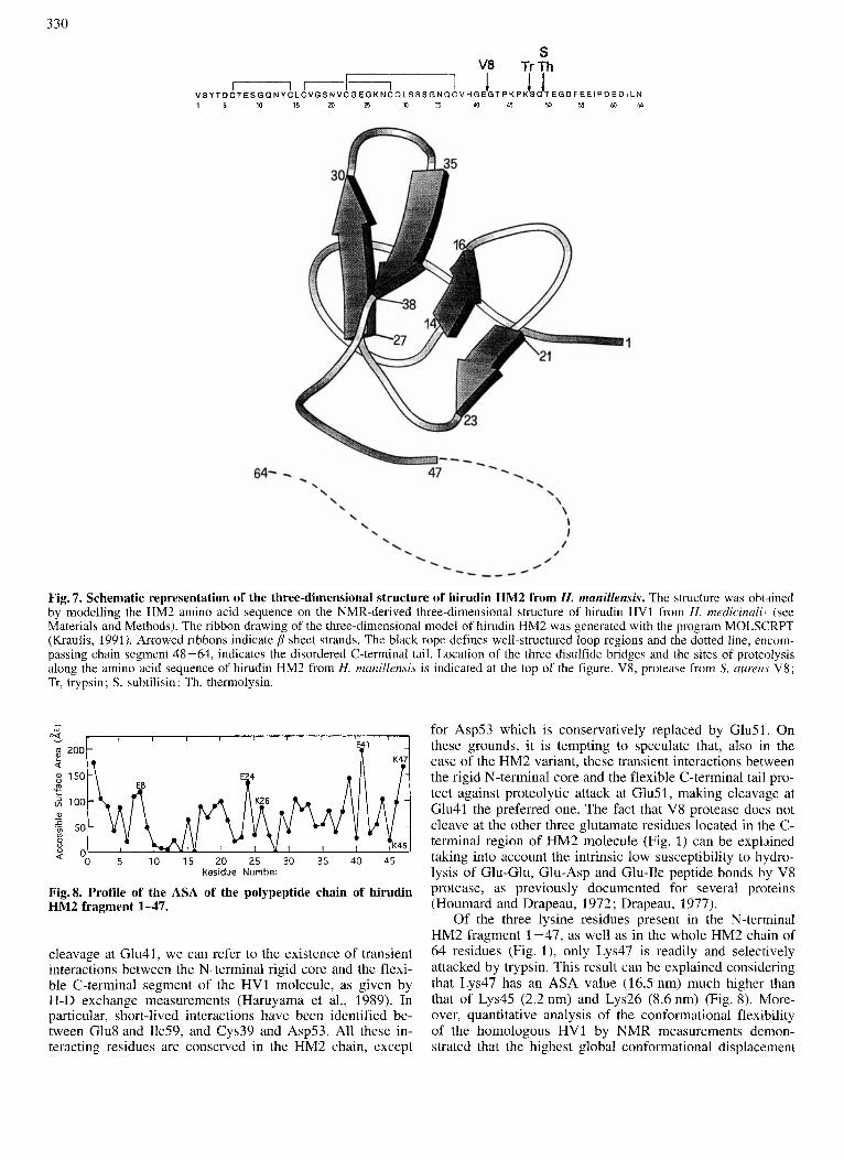
The model of the three-dimensional structure of hirudin HM2 was built on the basis of the solution structure of hiru- din variant HV1 from H. medicinalis, determined by NMR. Only the coordinates of the N-terminal chain segment 1-49 of hirudin HV1 are available, the C-terminal tail segment 50-65 being highly flexible and disordered in solution (Folkers et al., 1989). The N-terminal segment 1-47 of hiru- din HM2 shows high sequence similarity (75%) with the cor- responding segment 1-49 of the HV1 variant (see Fig. 1). In particular, the sequence of fragment 1-47 of HM2 shows two amino acid deletions and 11 substitutions. Since seven are conservative substitutions, the global sequence similarity between the two proteins can be considered approximately 90%. Moreover, all hirudin variants reported to date, includ- ing variants HM2 and HV1, possess six cysteine residues at highly conserved positions (Steiner et al., 1992), thus imply- ing the same pattern of disulfide bonds (Thornton, 1981). Indeed, limited proteolysis of hirudin HM1 with thermolysin allowed us to isolate and characterize disulfide-containing peptides in full agreement with disulfide linkages occurring between residues 6-14, 16-28 and 22-37 (unpublished re- sults).
The model of hirudin HM2 is shown in Fig. 7. The pro- tein shows a tightly packed N-terminal 1-47 segment cross- linked by the three disulfide bridges and characterized by the presence of two p sheets. The first seven residues form an irregular strand which leads into a type-11 reverse turn ex- tending over residues 8 - 11. This is followed by an antiparal- lel p sheet formed by residues 14-16 (strand I) and 20-22 (strand I’), connected by a type-11’ reverse turn, consisting of residues 17-20. A type-I1 reverse turn (residues 23-26) leads into the second p sheet constituted by residues 26-30 (strand 11) and 35-39 (strand 11’) connected by a reverse turn (residues 31-34). Of note, the C-terminal region (residues 40-47) of the core domain 1-47 is brought in close proxim- ity to the N-terminal region by hydrogen-bonding interac- tions of Lys47 with Thr4, Asp5 and Asnl2.
In order to possibly correlate sites of surface exposure with sites of proteolysis along the chain of the HM2 mole- cule, we calculated the accessible surface area (ASA) (Lee

328
1-47 A - I
I I I I 0 10 20 30 40
RETENTION TIME, rnin
I I I I
RETENTION TIME, rnin
I I I I
RETENTION TIME, rnin
Fig. 4. RP-HPLC analysis of proteolytic digests of hirudin HM2. Proteolysis was conducted at room temperature as described in Materials and Methods. An aliquot of the reaction mixture was diluted with aqueous trichloroactic acid in order to stop proteolysis, then analyzed by RP-HPLC using a Vydac C,, column. The chromatographic conditions are the same as those described in Fig. 2. (A) Proteolysis with trypsin, 5-min reaction; (B) Proteolysis with subtilisin, 3-h reaction; (C) proteolysis with thermolysin, 24-h reaction.
and Richards, 1971; Richards, 1985) of the three-dimen- sional model of fragment 1-47 with the view that the most accessible sites in the polypeptide molecule should be fa- vored sites for proteolysis (Fontana et al., 1993 ; Novotni and Bruccoleri, 1986; Hubbard et al., 1991). The ASA profile along the polypeptide chain of HM2 fragment 1-47 is shown in Fig. 8 and is used for discussing the data of limited proteolysis of the HM2 molecule.
DISCUSSION The aim of this work was to deduce aspects of structure
and dynamics of hirudin HM2 from H. manillensis by limited proteolysis experiments and possibly to produce and isolate useful fragments for addressing structure/function relation- ships and biorecognition mechanisms between thrombin and its polypeptide inhibitors. Indeed, the use of proteolytic en- zymes as probes of protein structure and dynamics is a well recognized and often employed approach (Mihalyi, 1978 ; Neurath, 1980; Fontana et al., 1989, 1993; Signor et al., 1990). In analogy to all enzymic reactions, a proteolytic en- zyme can cleave a polypeptide chain only if the site(s) of chain fission can bind and adapt to the specific stereo- chemistry of the enzyme active site. Normally, native globu-
lar proteins are quite resistant to proteolysis, wherea\ un- folded protein species are degraded faster by several orders of magnitude. However, there is ample evidence that native proteins can act as substrates for proteolysis, often leading to limited proteolysis at one or very few peptide bonds located at the most exposed and flexible sites in the protein structure. We have previously emphasized that chain flexibility is a key parameter in dictating selective peptide-bond fission by limited proteolysis, since a striking correlation has been ob- served between the sites of proteolysis and the sites of high segmental mobility of the protein chain, this last given by the thermal factor (B factor) determined by X-ray methods (Fontana et al., 1986).
Several proteolytic enzymes preferentially cleave the 64- residue chain of hirudin HM2 at chain segment 41 -49. lead- ing to the formation of fragments encompassing the N-termi- nal core domain crosslinked by three disulfide bridges and quite resistant to further proteolysis. The easy and selective removal of the C-terminal tail of hirudin HM2 by proteolytic digestion indicates that this portion of the molecule is highly flexible (see above). This is fully consistent with previous NMR data (Folkers et al., 1989; Haruyama and Wutrich, 1989) showing that the C-terminal tail 50-65 of the related hirudin HV1 is highly flexible and disordered in solution. Thus, the results of this study provide evidence that the over-

329
250 c
RETENTION TIME, min
I I I I I I
1-84
h 10 12 I
MINUTES
Fig. 5. Analytical RP-HPLC (A) and capillary electrophoresis (B) of hirudin HM2 and its proteolytic fragments 1 4 1 and 1-47. The experimental conditions of the RP-HPLC analysis utilizing a Vydac C,, column are those described in Fig. 2. The capillary elec- trophoresis analysis was carried out at room temperature using a fused-silica capillary (25 pmX20 cm) with a field strength of 400 Volt/cm (see also Materials and Methods).
all topology and dynamics of hirudin HM2 are similar to those of variant HV1.
In order to highlight structural aspects of hirudin HM2, as well as to interpret the results of proteolysis experiments, we constructed a three-dimensional model of hirudin HM2, based on the NMR-derived structure of the HV1 variant (Folkers et al., 1989). It has been argued that the errors in a three-dimensional model of a protein, built on the basis of a structure with 90% sequence identity, may be as low as the errors in crystallographically determined structures (Blundell et al., 1987; Chothia and Lesk, 1986). Therefore, the accu- racy of the HM2 model, shown in Fig. 7, is probably good enough to provide a useful structural framework for a discus- sion of the proteolysis experiments, as well as of the struc-
I I I I I 1 0 0.05 0.10 0.15 0.20 0.25
II[S], pM-’
Fig.6. Effect of hirudin HM2 fragments 1-41 (0) and 1-47 (0) on the amidolytic activity of thrombin. Assays were performed at 37°C as described in Materials and Methods utilizing, as substrate, S-2238. The lines represent the best fit of the experimental data to the equation for competitive inhibition according to the Michaelis- Menten model.
Table 3. Binding constants for the complexes between thrombin and hirudin fragments. Assays were performed and the data ana- lysed as described in the text. The estimates of the dissociation con- stant (K,) for hirudin HM2 and its fragments are given together with their standard errors.
Inhibitor KI -AG:
kJ . mol-l
HM2 0.78 ? 0.02 pM 71.9 1-47 150 5 20 nM 40.5 1-41 400 ? 50 nM 38.0
ture/activity relationships of both HM2 and its N-terminal fragments reported in this study. Of direct interest for the purposes of this study is also the calculated ASA profile of the HM2 molecule (Fig. S), since exposure is a surface prop- erty of a globular protein (Thornton et al., 1986) usually strongly correlated with both flexibility and hydrophilicity (Ringe and Petsko, 1985). Of course, proteolysis is predicted to occur at protein sites exposed at the surface and not em- bedded in the protein interior (Fontana et al., 1986, 1993; Novotni and Bruccoleri, 1986; Hubbard et al., 1991).
The initial cleavage of the HM2 chain by V8 protease occurs at Glu41, producing fragments 1-41 and 42-64 (see Fig. 3). ASA calculations carried out on the three-dimen- sional model of HM2 (Fig. 8) reveal that the surface accessi- bility of Glu41 (19.7 nm) is much higher than that of other two glutamate residues in the N-terminal domain of HM2, thus providing a clear-cut explanation of the enhanced susceptibility to proteolysis of the Glu41-Gly42 peptide bond in respect to those involving Glu8 and Glu24. Nevertheless, there are an additional four glutamate residues located in po- sitions 51, 55, 56 and 60 of the disordered and flexible C- terminal tail of HM2 (see Fig. l) , thus one would expect, in addition, an even more easy proteolysis at these glutamate residues. However, V8 protease cleaves solely at Glu51 and only after the fragment 42-64 has been cleaved off from the HM2 molecule, as shown by the kinetics of the proteolysis (see Fig. 3). To possibly interpret the initial and preferential
330
S V8 TrTh
50 56 a t 4 V S Y T D C T E S G a N Y C L C V G S N V C G E G K N C Q L S S S G N Q C V H G E G T P K P K S Q T E G D F E E l P D E D l L N 1 5 10 15 ZD Z5 3 3 5 4 0 4 5
1 I I
Fig. 7. Schematic representation of the three-dimensional structure of hirudin HM2 from H. manillensis. The structure was obtined by modelling the HM2 amino acid sequence on the NMR-derived three-dimensional structure of hirudin HV1 from H. rnedicinali, (see Materials and Methods). The ribbon drawing of the three-dimensional model of hirudin HM2 was generated with the program MOLSCRPT (Kraulis, 1991). Arrowed ribbons indicate p sheet strands. The black rope defines well-structured loop regions and the dotted line, encom- passing chain segment 48-64, indicates the disordered C-terminal tail. Location of the three disulfide bridges and the sites of proteolysis along the amino acid sequence of hirudin HM2 from H. manillensis is indicated at the top of the figure. V8, protease from S. aureu.y V8; Tr, trypsin; S, subtilisin; Th, thermolysin.
"0 5 1 0 15 20 25 30 35 40 45 Residue Number
Fig.8. Profile of the ASA of the polypeptide chain of hirudin HM2 fragment 1-47.
cleavage at Glu41, we can refer to the existence of transient interactions between the N-terminal rigid core and the flexi- ble C-terminal segment of the HV1 molecule, as given by H-D exchange measurements (Haruyama el al., 1989). In particular, short-lived interactions have been identified be- tween Glu8 and Ile59, and Cys39 and Asp53. All these in- teracting residues are conserved in the HM2 chain, except
for Asp53 which is conservatively replaced by Glu51. On these grounds, it is tempting to speculate that, also in the case of the HM2 variant, these transient interactions between the rigid N-terminal core and the flexible C-terminal tail pro- tect against proteolytic attack at Glu51, making cleavage at Glu41 the preferred one. The fact that V8 protease does not cleave at the other three glutamate residues located in the C- terminal region of HM2 molecule (Fig. 1) can be explained taking into account the intrinsic low susceptibility to hydro- lysis of Glu-Glu, Glu-Asp and Glu-Ile peptide bonds hy V8 protease, as previously documented for several proteins (Houmard and Drapeau, 1972 ; Drapeau, 1977).
Of the three lysine residues present in the N-terminal HM2 fragment 1-47, as well as in the whole HM2 chain of 64 residues (Fig. l) , only Lys47 is readily and selectively attacked by trypsin. This result can be explained considering that Lys47 has an ASA value (16.5 nm) much higher than that of Lys45 (2.2 nm) and Lys26 (8.6 nm) (Fig. 8). More- over, quantitative analysis of the confornational flexibility of the homologous HV1 by NMR measurements demon- strated that the highest global conformational displacement

331
relative to the mean NMR structure occurs at Lys49 (Lys47 in HM2; Szyperslu et al., 1992b). Thus, exposure and flexi- bility characteristics appear to dictate the exclusive peptide- bond fission at Lys47 in the HM2 molecule. Nevertheless, the observed selective peptide-bond hydrolysis at Lys47 by trypsin can be dictated, in part, also by the substrate speci- ficity of the protease (Walsh, 1972; Keil, 1982). This possi- bility should be considered, especially to explain the resis- tance to hydrolysis of the peptide bond Lys45-Pro46 (Keil, 1982).
Thermolysin (Heinrikson, 1977) and subtilisin (Ottesen and Svendsen, 1970), which similarly show broad substrate specificity, remove the C-terminal flexible tail from the HM2 molecule, leading to fragment 1-49 as the major proteolytic product quite resistant to further degradation. In the case of thermolysin, it is surprising to observe that the preferential cleavage occurs at the Glu49-Thr50 peptide bond, con- sidering that thermolysin cleaves at amino acid residues with bulky hydrophobic side chains (Heinrikson, 1977 ; Keil, 1982). This would imply that, on one side, the C-terminal tail of HM2 is highly flexible and, on the other, that the N-terminal core 1-49 is characterized by compactness and rigidity preventing its further degradation. Since we do not have experimental data on the flexibility of HM2 N-terminal core domain, we can use the correlated physical property of surface accessibility (Ringe and Pesko, 1985) to possibly interpret the resistance to proteolysis of fragment 1 -49. Based on the substrate specificity of thermolysin (Heinrik- son, 1977; Keil, 1982), at least seven sites of preferential proteolytic attack by thermolysin can be proposed in the amino acid sequence of HM2 fragment 1-49 (Tyr3, Tyrl3, Leul5, Va117, Va121, Leu30 and Va140). However, the ASA profile of the polypeptide chain of HM2 fragment 1-47 (see Fig. 8) clearly shows that all these sites are characterized by an average lower surface accessibility and thus are most likely not prone to attack by the protease. Moreover, five of the above listed sites of possible proteolysis are adjacent in the amino acid sequence to deeply buried cystine residues (see Fig. 1). Disulfide crosslinking, therefore, can inhibit pro- teolysis by steric constraints.
The interest in hirudin fragments was stimulated by the observation that the mechanism of enzyme inhibition in- volves binding of the N-terminal core domain of hirudin to the thrombin active site, whereas the C-terminal tail binds at the fibrinogen-binding site of thrombin (Rydel et al., 1991). Thus, N-terminal fragments of hirudin HV1 have been pro- duced by proteolysis experiments (Chang, 1990; Chang et al.. 1990; Dennis et al., 1990) similar to those described in this report, whereas the much shorter C-terminal fragments were amenable to chemical synthesis (Maraganore et al., 1989, 1990; Krstenansky and Mao, 1987; Ni et al., 1992). For an efficient preparation of fragment 1 - 52 of hirudin HV1 , an engineered recombinant protein was produced in E. coli with a Gln-Met substitution in position 52 of the chain (Dennis et al., 1990). The Met52 mutant hirudin was then subjected to cyanogen bromide cleavage at Met52, producing fragment 1 -52. The thrombin-inhibitory properties of this fragment (Dennis et al., 1990), as well as its NMR solution structure (Szyperski et al., 1992a,b), have been reported. The clean and high-yield production of fragment 1-47 (see Fig. 4A) from hirudin HM2 indicates that there is no need to engineer a special mutant hirudin HM2 for producing an interesting and biologically active N-terminal domain of hi- rudin variant HM2. Nevertheless recently, in our laboratory, we have achieved the solid-phase chemical synthesis of ana-
logues of fragment 1-47 from hirudin HM2, thus enabling us to efficiently produce correctly folded and biologically active fragment species (unpublished results).
In view of the numerous non-enzymic (hormonal) activi- ties of thrombin (Fenton, 1988; Davie et al., 1991; Tsiang et al., 1990; Vu et al., 1991; Tapparelli et al., 1993), hirudin fragments can possibly be usefully employed for dissecting molecular aspects of both the dual-type binding of hirudin to thrombin, as well as of other biological functions of this en- zyme in vivo. For example, the data of this study show that more than half of the free energy of binding of hirudin HM2 to thrombin is contributed by the N-terminal core domain, as also reported for hirudin HV1 (Betz et al., 1992). Fragments 1-41 and 1-47 of hirudin HM2 show similar thrombin- inhibitory properties, as well as energetics of binding to the target enzyme (see Table 3), with fragment 1-47 being a better inhibitor than fragment 1 -41. This can be understood by considering that previous NMR studies (Folkers et al., 1989; Szyperski et al., 1992a,b) showed that Lys47, as well as Pro46 and Pro48, of the related HV1 chain are critical in dictating the interactions bringing the N-terminal and C- terminal parts of hirudin HV1 core domain 1-51 into close proximity, and thus positioning the N-terminal tripeptide in a suitable conformation for its binding to the active site of thrombin.
We gratefully acknowledge Dr P. Polverino de Laureto for the peptide/protein sequencing analyses. We also thank Mr M. Zam- bonin for excellent technical assistance and Mrs A. Mocavero for typing the manuscript.
REFERENCES Ayala, Y. & Di Cera, E. (1994) Molecular recognition by thrombin.
Role of the slow - fast transition, site-specific ion binding ener- getics and thermodynamic mapping of structural components, J. Mol. Biol. 23.5, 733-746.
Betz, A,, Hofsteenge, J. & Stone, S. R. (1992) Interaction of the N- terminal region of hirudin with the active-site cleft of thrombin, Biochemistry 31,4557-4562.
Blundell, T. L., Sibanda, B. L., Sternberg, M. J. E. & Thornton, J. M. (1987) Knowledge-based prediction of protein structures and the design of novel molecules, Nature 326, 347-352.
Braun, P. J., Dennis, S., Hofsteenge, J. & Stone, S. R. (1988) Use of site-directed mutagenesis to investigate the basis for the speci- ficity of hirudin, Biochemistry 27, 6517-6522.
Brunger, A. T. (1988) Crystallographic refinement by simulated an- nealing. Application to a 2.8 A resolution structure of aspartate aminotransferase, J. Mol. Bid. 203, 803-816.
Brunger, A. T. (1990) X-PLOR, Manual version 2.1, Yale Univer- sity, New Haven, CT, USA.
Chang, J.-Y., Schlaeppi, J.-M. & Stone, S. R. (1990) Antithrombin activity of the hirudin N-terminal core domain residues 1-43, FEBS Lett. 260, 209-212.
Chang, J.-Y. (1990) Production, properties and thrombin inhibitory mechanism of hirudin amino-terminal core fragments, J. Biol. Chem. 265, 22159-22166.
Chothia, C. & Lesk, A. M. (1986) The relation between the diver- gence of sequence and structure in proteins, EMBO J. 5, 823- 836.
Davie, E. W., Fujikawa, K. & Kisiel, W. (1991) The coagulation cascade : Initiation, maintenance and regulation, Biochemistry 30, 10363-10370.
Dennis, S., Wallace, A., Hofsteenge, J. & Stone, S. R. (1990) Use of fragments of hirudin to investigate thrombin-hirudin interac- tion, Eur: J. Biochem. 188, 61-66.
Dodt, J., Schnitz, T., Schafer, T. & Bergmann, T. (1986) Expression, secretion and processing of hirudin in E. coli using the alkaline phoxphatase signal sequence, FEBS Lett. 202, 373 -377.

332
Dodt, J., Seemuller, U., Maschler, R. & Fritz, H. (1985) The com- plete covalent structure of hirudin : Localization of the disulfide bonds, Biol. Chem. Hoppe-Seyler 366, 379-385.
Drapeau, G. R. (1977) Cleavage at glutamic acid with staphylococ- cal protease, Methods Enzymol. 47, 189-191.
Electricwala, A,, Sawyer, R. T., Powell Jones, C. & Atkinson, A. (1991) Isolation of a thrombin inhibitor from the leech Hirudina- ria manillensis, Blood Coag. Fibrinolysis 2, 83 - 89.
Fenton, J. W. (1 988) Thrombin bioregulatory functions, Adv. Clin. Enzymol. 6 , 186-193.
Folkers, P. J. M., Clore, G. M., Driscoll, P. C., Dodt, J., Kohler, S. & Gronenborn, A. M. (1 989) Solution structure of recombinant hi- rudin and the Lys47 - Glu mutant: A nuclear magnetic reso- nance and hybrid geometry-dynamical simulated annealing study, Biochemistry 28, 2601 -2617.
Fontana, A,, Fassina, G., Vita, C., Dalzoppo, D., Zamai, M. & Zam- bonin, M. (1986) Correlation between segmental mobility and sites of limited proteolysis in thermolysin, Biochemistry 25, 1 847 - 1851.
Fontana, A,, Polverino de Laureto, P. & De Filippis, V. (1993) Mo- lecular aspects of proteolysis of globular proteins, in Protein stability and stabilization (Van den Tweel, W., Harder, A. & Buitelaar, M., eds) pp. 101 - 110, Elsevier Science Publ., Amster- dam.
Fontana, A,, Vita, C., Dalzoppo, D. & Zambonin, M. (1989) Limited proteolysis as a tool to detect structure and dynamic features of globular proteins: Studies on thermolysin, in Methods in protein sequence analysis (Wittmann-Liebold, B., ed.) pp. 315-324, Springer-Verlag, Berlin.
Fromm, H. J. (1975) Enzyme kinetics, Springer-Verlag, Berlin. Gill, S. G. & von Hippel, P. H. (1989) Calculation of protein extinc-
tion coefficients from amino acid sequence data, Anal. Biochem.
Haruyama, H. & Wiitrich, K. (1989) Conformation of recombinant desulfatohirudin in aqueous solution determined by nuclear mag- netic resonance, Biochemistry 28, 4301 -4312.
Haruyama, H., Qian, Y.-Q. & Wiitrich, K. (1989) Static and transient hydrogen-binding interactions in recombinant desulfatohirudin studied by ‘H nuclear magnetic resonance measurements of am- ide proton exchange rates and pH-dependent chemical shifts, Biochemistry 28, 4312-4317.
Harvey, R., Degryse, E., Stefani, L., Schamber, F., Cazenave, J. P., Courtney, M., Tolstoshev, P. & Lecocq, J. P. (1986) Cloning and expression of cDNA coding for the anticoagulant hirudin from the blood sucking leech Hirudo medicinalis, Proc. Natl Acad. Sci. USA 83, 1084-1088.
Heinrikson, R. L. & Meredith, S. C. (1984) Amino acid analysis by reverse-phase high-performance liquid chromatography: Precol- umn derivatization with phenylisothiocyanate, Anal. Biochem. 136, 65-74.
Heinrikson, R. L. (1977) Applications of thermolysin in protein structural analysis, Methods Enzymol. 47, 175 - 189.
Houmard, J. & Drapeau, G. R. (1972) Staphylococcal protease: A proteolytic enzyme specific for glutamyl bonds, Proc. Nut1 Acad. Sci. USA 69, 3506-3509.
Hubbard, S. J., Campbell, S. F. & Thornton, J. M. (1991) Molecular recognition. Conformational analysis of limited proteolytic sites and serine proteinase protein inhibitors, J. Mol. Biol. 220, 507- 530.
Karas, M. & Hillenkamp, F. (1988) Laser desorption ionization of proteins with molecular masses exceeding 1 0,000 Daltons, Anal. Chem. 60, 2299-2301.
Karshikov, A., Bode, W., Tulinsky, A. & Stone, S. R. (1992) Electro- static interactions in the association of proteins: An analysis of the thrombin-hirudin complex, Protein Sci. I , 727-735.
Keil, B. (1982) Enzymic cleavage of proteins, in Methods in protein sequence analysis (Elzinga, M., ed.) pp. 291 -304, Humana Press, Clifton, NJ, USA.
Kraulis, P. J. (1991) MOLSCRIPT: A program to produce both de- tailed and schematic plots of protein structures, J. Appl. Crystal-
182, 319-326.
1ogK 24, 946-950.
Krstenansky, J. L. & Mao, S. J. T. (1987) Antithrombin properties of C-terminus of hirudin using synthetic unsulfated N,-acetyl- hirudin (45-65), FEBS Lett. 211, 10-16.
Lee, B. K. & Richards, F. M. (1971) The interpretation of protein structures: estimation of static accessibility, J. Mol. Biol. 55, 379-400.
Lottenberg, R. & Jackson, C. M. (1983) Solution composition de- pendent variation in extinction coefficients for p-nitroanilide, Biochim. Biophys. Acta 742, 558-564.
Maraganore, J. M., Bourdon, P., Jablonski, J., Ramachandran K. L. & Fenton, J. W. (1990) Design and characterization of hiru- logs: A novel class of bivalent peptide inhibitors of thrombin, Biochemistry 29,7095 -7101.
Maraganore, J. M., Chao, B., Joseph, M. L., Jablonski, J. & Ra- machandran, K. L. (1989) anticoagulant activity of synthetic hi- rudin peptides, J. Biol. Chem. 24, 8692-8698.
Markwardt, F. (1991) Past, present and future of hirudin, Huemosra- sis 21, 11 -26.
Mihalyi, E. (1 978) Application of proteolytic enzymes to protein structure studies, vols 1 and 2, CRC Press, West Palm Beach, Florida, USA.
Neurath, H. (1980) Limited proteolysis, protein folding and physio- logical regulation, in Protein folding (Jaenicke, R., ed.) pp. 501 - 525, ElsevierNorth Holland Biomedical Press, Amsterdam-New York.
Ni, F., Ripoll, D. R. & Purisima, E. 0. (1992) Conformational sta- bility of a thrombin-binding peptide derived from the hirudin C- terminus, Biochemistry 31, 2545-2554.
Novotny, J. & Bruccoleri, R. E. (1986) Correlation among sites of limited proteolysis, enzyme accessibility and segmental mobility, FEBS Lett. 211, 185-189.
Ottesen, M. & Svendsen, I. (1970) The subtilisins, Methods Enzy- mol. 19, 199-215.
Priestle, J. P., Rahuel, J., Rink, H., Tones, M. & Griitter, M. G. (1993) Changes in interactions in complexes of hirudin deriva- tives and human a-thrombin due to different crystal forms. Pro- tein Sci. 2, 1630- 1642.
Richards, F. R. (1985) Calculations of molecular volumes and areas for structures of known geometry, Methods Enzymol. 115, 440- 464.
Riehl-Bellon, N., Carvallo, D., Acker, M., Van Dorsselaer, A,, Marquet, M., Loison, G., Lemoine, Y., Brown, S. W., Courtney, M. & Roitsch, C. (1989) Purification and biochemical charxter- ization of recombinant hirudin produced by Saccharomyces cere- visiae, Biochemistry 28, 2941 -2949.
Ringe, D. & Petsko, G. A. (1985) Mapping protein dynamics by X- ray diffraction, Prog. Biophys. Mol. Biol. 45, 197-235.
Rydel, T. J., Ravichandran, K. G., Tulinsky, A., Bode, W., Huber, R., Roitsch, C. & Fenton, J. W. (1990) The structure of a complex of recombinant hirudin and human rr-thrombin, Science 249. 277- 280.
Rydel, T. J., Tulinsky, A., Bode, W. & Huber, R. (1991) Refined structure of the hirudin-thrombin complex, J. Mol. Biol. 221,
Sawyer, R. T. (1986) in Leech biology and behaviour, vol. 2, p. 420, Oxford University Press, Oxford.
Scacheri, E., Nitti, G., Valsasina, B., Orsini, G., Visco, C., Ferreia, M., Sawyer, R. T. & Sarmientos, P. (1993) Novel hirudin variants from the leech Hirudinaria manillensis: Amino acid sequence, cDNA cloning and genomic organization, Eur. J. Biochem. 214,
Scharf, M., Engels, J. & Tripier, D. (1989) Primary structure of new iso-hirudins, FEBS Lett. 225, 105-110.
Signor, G., Vita, C., Fontana, A., Frigerio, R., Bolognesi, M., Toma, S., Gianna, R., De Gregoriis, E. & Grandi, G. (1990) Structural features of neutral protease from Bacillus subtilis deduced from model building and limited proteolysis experiments, Eur. J. Bio- chem. 189, 221 -227.
Steiner, V., Knecht, R., Borsen, K. O., Gassmann, E., Stone. S. R., Raschdorf, F., Schlaeppi, J.-M. & Maschler, R. (1992) Primary structure and function of novel 0-glycosylated hirudins from the leech Hirudinaria manillensis, Biochemistry 31, 2294-2298.
583 -601.
295-304.

333
Stone, S. R. & Hofsteenge, J. (1986) Kinetics of the inhibition of thrombin by hirudin, Biochemistry 25, 4622-4624.
Stringer, K. A. & Lindenfeld, J. A. (1992) Hirudins: Antithrombin anticoagulants, Ann. Pharmacother: 26, 1535 - 1540.
Szyperski, T., Giintert, P., Stone, S. R. & Wiitrich, K. (1992a) Nuclear magnetic resonance solution structure of hirudin (1-51) and comparison with corresponding three-dimensional structures determined using the complete 65-residue hirudin polypeptide chain, J. Mol. Biol. 228, 1193-1205.
Szyperski, T., Giintert, P., Stone, S. R., Tulinski, A,, Bode, W., Huber, R. & Wiitrich, K. (1992b) Impact of protein-protein con- tacts on the conformation of thrombin-bound hirudin studied by comparison with the nuclear magnetic resonance solution struc- ture of hirudin (I-51), J. MoZ. Biol. 228, 1206-1211.
Tapparelli, C., Metternich, R., Ehrhardt, C. & Cook, N. S. (1993) Synthetic low molecular mass thrombin inhibitors : Molecular design and pharmacological profile. Trends Pharmacol. Sci. 14, 366-376.
Thornton, J. M. (1981) Disulfide bridges in globular proteins, J. Mol. Biol. 151, 261-287.
Thornton, J. M., Edwards, M. S., Taylor, W. R. & Barlow, D. J. (1 986) Location of continuous antigenic determinants in the pro- truding regions of proteins. EMBO J. 5, 409-413.
Tripier, D. (1988) A family of iso-proteins: Isolation and sequence determination of new hirudin, Folia Haematol. (Leipzig) 115, 30-35.
Tsiang, M., Lentz, S. A., Dittman, W. A,, Wen, D., Scarpati, E. M. & Sadler, J . E. (1990) Equilibrium binding of recombinant human trombomodulin: Effect of hirudin, fibrinogen, factor Va and pep- tide analogues, Biochemistry 29, 10602- 10612.
Vitali, J., Martin, P. D., Mulkowski, M. G., Robertson, W. D., Lazar, J. B., Winant, R. C., Johnson, P. H. & Edwards, B. F. P. (1992) The structure of a cqmplex of bovine a-thrombin and recombi- nant hirudin at 2.8 A resolution, J. Biol. Chem. 267. 17670- 17678.
Vu, T. K. H., Wheaton, V. J., Hung, D. T., Charo, I. & Coughlin, S. R. (1991) Domains specifying thrombin-receptor interaction, Nature 353, 674-677.
Walsh, K. A. (1970) Trypsinogens and trypsins of various species, Methods Enzymol. 19, 41 -63.
Copyright © 2022 FDOKUMEN