Properties of external plexiform layer interneurons in mouse olfactory bulb slices
Upload
khangminh22Category
view
0download
0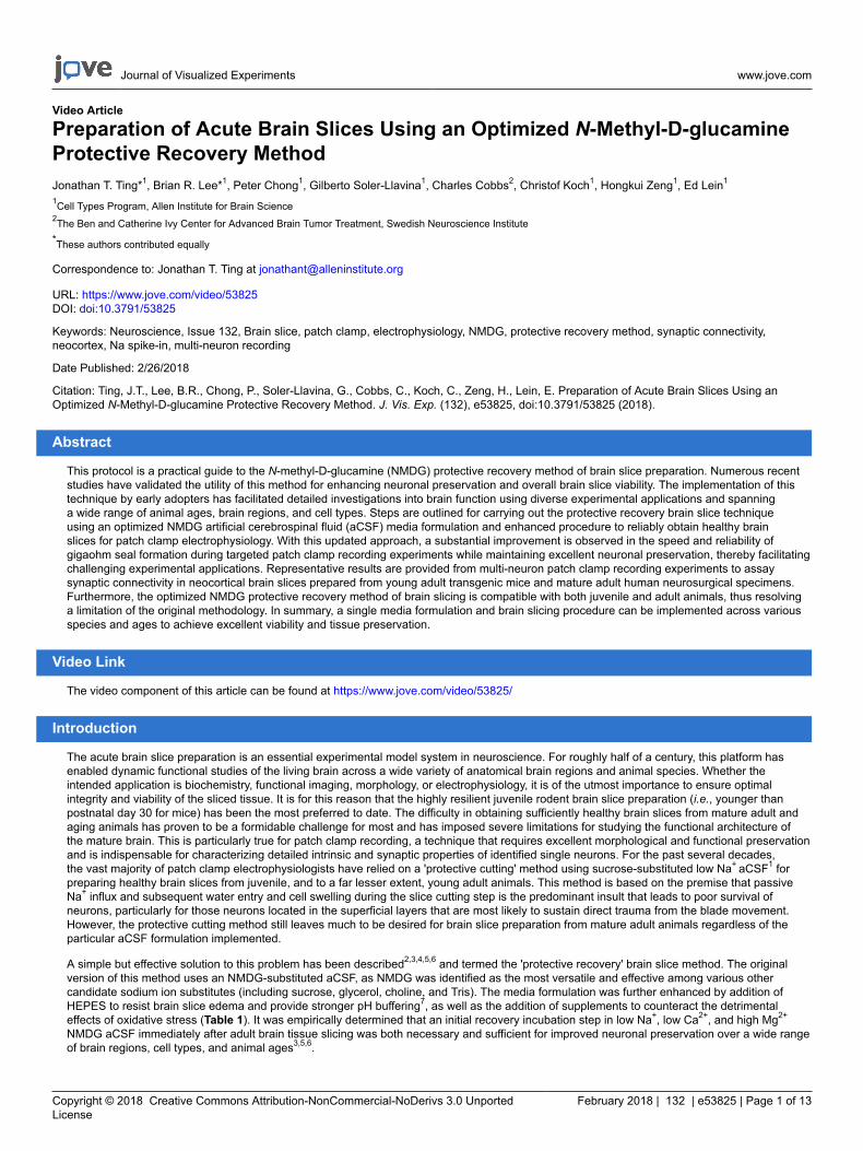
Journal of Visualized Experiments www.jove.com
Copyright © 2018 Creative Commons Attribution-NonCommercial-NoDerivs 3.0 UnportedLicense
February 2018 | 132 | e53825 | Page 1 of 13
Video Article
Preparation of Acute Brain Slices Using an Optimized N-Methyl-D-glucamineProtective Recovery MethodJonathan T. Ting*1, Brian R. Lee*1, Peter Chong1, Gilberto Soler-Llavina1, Charles Cobbs2, Christof Koch1, Hongkui Zeng1, Ed Lein1
1Cell Types Program, Allen Institute for Brain Science2The Ben and Catherine Ivy Center for Advanced Brain Tumor Treatment, Swedish Neuroscience Institute*These authors contributed equally
Correspondence to: Jonathan T. Ting at [email protected]
URL: https://www.jove.com/video/53825DOI: doi:10.3791/53825
Keywords: Neuroscience, Issue 132, Brain slice, patch clamp, electrophysiology, NMDG, protective recovery method, synaptic connectivity,neocortex, Na spike-in, multi-neuron recording
Date Published: 2/26/2018
Citation: Ting, J.T., Lee, B.R., Chong, P., Soler-Llavina, G., Cobbs, C., Koch, C., Zeng, H., Lein, E. Preparation of Acute Brain Slices Using anOptimized N-Methyl-D-glucamine Protective Recovery Method. J. Vis. Exp. (132), e53825, doi:10.3791/53825 (2018).
Abstract
This protocol is a practical guide to the N-methyl-D-glucamine (NMDG) protective recovery method of brain slice preparation. Numerous recentstudies have validated the utility of this method for enhancing neuronal preservation and overall brain slice viability. The implementation of thistechnique by early adopters has facilitated detailed investigations into brain function using diverse experimental applications and spanninga wide range of animal ages, brain regions, and cell types. Steps are outlined for carrying out the protective recovery brain slice techniqueusing an optimized NMDG artificial cerebrospinal fluid (aCSF) media formulation and enhanced procedure to reliably obtain healthy brainslices for patch clamp electrophysiology. With this updated approach, a substantial improvement is observed in the speed and reliability ofgigaohm seal formation during targeted patch clamp recording experiments while maintaining excellent neuronal preservation, thereby facilitatingchallenging experimental applications. Representative results are provided from multi-neuron patch clamp recording experiments to assaysynaptic connectivity in neocortical brain slices prepared from young adult transgenic mice and mature adult human neurosurgical specimens.Furthermore, the optimized NMDG protective recovery method of brain slicing is compatible with both juvenile and adult animals, thus resolvinga limitation of the original methodology. In summary, a single media formulation and brain slicing procedure can be implemented across variousspecies and ages to achieve excellent viability and tissue preservation.
Video Link
The video component of this article can be found at https://www.jove.com/video/53825/
Introduction
The acute brain slice preparation is an essential experimental model system in neuroscience. For roughly half of a century, this platform hasenabled dynamic functional studies of the living brain across a wide variety of anatomical brain regions and animal species. Whether theintended application is biochemistry, functional imaging, morphology, or electrophysiology, it is of the utmost importance to ensure optimalintegrity and viability of the sliced tissue. It is for this reason that the highly resilient juvenile rodent brain slice preparation (i.e., younger thanpostnatal day 30 for mice) has been the most preferred to date. The difficulty in obtaining sufficiently healthy brain slices from mature adult andaging animals has proven to be a formidable challenge for most and has imposed severe limitations for studying the functional architecture ofthe mature brain. This is particularly true for patch clamp recording, a technique that requires excellent morphological and functional preservationand is indispensable for characterizing detailed intrinsic and synaptic properties of identified single neurons. For the past several decades,the vast majority of patch clamp electrophysiologists have relied on a 'protective cutting' method using sucrose-substituted low Na+ aCSF1 forpreparing healthy brain slices from juvenile, and to a far lesser extent, young adult animals. This method is based on the premise that passiveNa+ influx and subsequent water entry and cell swelling during the slice cutting step is the predominant insult that leads to poor survival ofneurons, particularly for those neurons located in the superficial layers that are most likely to sustain direct trauma from the blade movement.However, the protective cutting method still leaves much to be desired for brain slice preparation from mature adult animals regardless of theparticular aCSF formulation implemented.
A simple but effective solution to this problem has been described2,3,4,5,6 and termed the 'protective recovery' brain slice method. The originalversion of this method uses an NMDG-substituted aCSF, as NMDG was identified as the most versatile and effective among various othercandidate sodium ion substitutes (including sucrose, glycerol, choline, and Tris). The media formulation was further enhanced by addition ofHEPES to resist brain slice edema and provide stronger pH buffering7, as well as the addition of supplements to counteract the detrimentaleffects of oxidative stress (Table 1). It was empirically determined that an initial recovery incubation step in low Na+, low Ca2+, and high Mg2+
NMDG aCSF immediately after adult brain tissue slicing was both necessary and sufficient for improved neuronal preservation over a wide rangeof brain regions, cell types, and animal ages3,5,6.
Journal of Visualized Experiments www.jove.com
Copyright © 2018 Creative Commons Attribution-NonCommercial-NoDerivs 3.0 UnportedLicense
February 2018 | 132 | e53825 | Page 2 of 13
Notably, earlier incarnations of what is now dubbed the protective recovery method can be found in the literature1,8,9,10,11,12,13, althoughthe full potential for mature adult and aging animal brain slice and patch clamp recording was not recognized or demonstrated in theseearlier works. In addition, nuanced procedural variations continue to emerge in support of specific experimental applications4,14,15,16. Thecollective body of work of these numerous research groups imparts high confidence in the robustness of the protective recovery method forimproved tissue preservation. The NMDG protective recovery method has now been widely adopted and implemented in numerous publishedresearch studies utilizing adult animal brain slice preparations. These acute slice studies span neocortical3,17,18, hippocampal15,19,20,21,striatal22,23,24, midbrain25,26,27,28,29, and hindbrain30,31,32,33,34 regions, and a variety of neurotransmitter and neuromodulator types includingglutamatergic4,30, GABAergic18,20,31,35,36, dopaminergic24,29,37,38, cholinergic14,37,38,39, noradrenergic40, and serotonergic27,28 neurotransmission.The method is also well suited for optogenetic control of neuronal activity in slices derived from transgenic animals3,39 or following in vivoviral injections17,27,28,40,41,42,43, as well as functional Ca2+ imaging of neuronal activity2,44,45,46. Analyses of both short term plasticity4,47,48 anddiverse forms of long-term plasticity16,35,48 have been reported. A recent study applied the NMDG protective recovery method to facilitateextensive and systematic probing of synaptic connectivity in the visual cortex in mature adult mouse brain slices using the octopatch recordingconfiguration49 — a powerful demonstration of the utility and robustness of this method. The protective recovery method has even been appliedsuccessfully in previously unforeseen experimental contexts, such as, improved preservation of vasculature and pericytes in adult cortical brainslices50, patch clamp recording from transplanted interneuron populations in 1–1.5 year old Alzheimer's Disease mouse models20, and an adultbrain slice receptor trafficking assay51.
The following protocol describes step-by-step procedures for implementing an optimized NMDG protective recovery method of brain slicepreparation to improve the viability of acute brain slices. The principles for improved neuronal preservation are discussed, as well asdemonstration of the clear benefits of this methodology for complex multi-neuron patch clamp recording experiments in both young adulttransgenic mouse brain slices and mature adult human neurosurgical brain slices. The following protocol has been validated for mice from 21days old to more than one years old, as well as for human neurosurgical specimens derived from adult patients.
Protocol
Procedures involving transgenic mice have been approved by the Institutional Animal Care and Use Committee (IACUC) at the Allen Institute forBrain Science. Both male and female C57BL/6 mice (weight range 10-30 g) were used in these experiments. Some of the representative resultsdescribe data collected from living human brain slices. Neocortical tissue specimens were obtained during neurosurgeries for tumor removal. It wasnecessary to remove the overlying neocortical tissue to gain access to the diseased tissue. Informed patient consent was obtained in all cases forthe use of neurosurgical tissue for research purposes under a protocol approved by the institutional review board of Swedish Medical Center.
1. Preparation of Media and Reagents (Table 1)
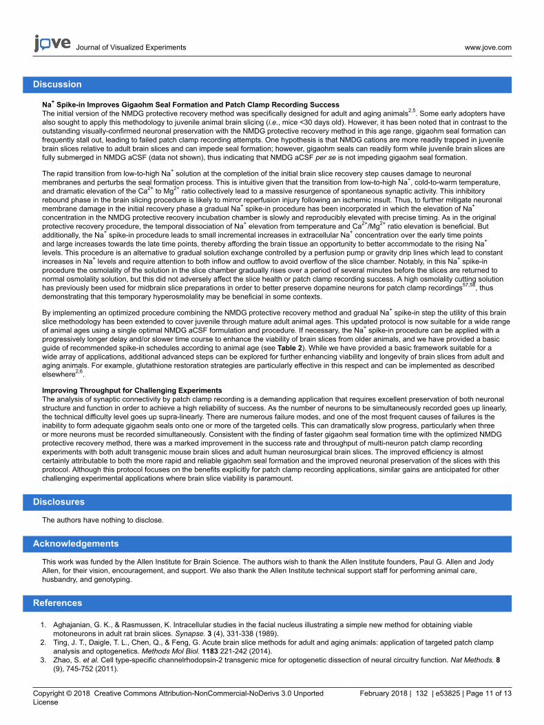
NOTE: Solutions should be made up in purified water that is free of trace metals and other impurities. It is recommended that solutions be madefreshly on the day of the experiment, although unused solutions may be stored at 4 °C for up to 1 week, if desired. 1 L of each formulation aboveis sufficient for 1–2 slicing procedures. All aCSF solutions must be saturated with carbogen (95% O2/5% CO2) prior to use to ensure stable pHbuffering and adequate oxygenation. The pH of all solutions should be adjusted to 7.3-7.4 and osmolality measured and adjusted to 300-310mOsmol/kg.
1. Prepare NMDG-HEPES aCSF (in mM): 92 NMDG, 2.5 KCl, 1.25 NaH2PO4, 30 NaHCO3, 20 HEPES, 25 glucose, 2 thiourea, 5 Na-ascorbate,3 Na-pyruvate, 0.5 CaCl2·2H2O, and 10 MgSO4·7H2O. Titrate pH to 7.3–7.4 with 17 mL +/- 0.5 mL of 5 M hydrochloric acid.
NOTE: This titration step should ideally be performed prior to the addition of divalent cations to avoid precipitation; however, the precipitationcan be reversed upon adjustment of the pH to the physiological range.
2. Prepare HEPES holding aCSF (in mM): 92 NaCl, 2.5 KCl, 1.25 NaH2PO4, 30 NaHCO3, 20 HEPES, 25 glucose, 2 thiourea, 5 Na-ascorbate, 3Na-pyruvate, 2 CaCl2·2H2O, and 2 MgSO4·7H2O. Titrate pH to 7.3–7.4 with several drops of concentrated 10 N NaOH.
3. Prepare recording aCSF (in mM): 124 NaCl, 2.5 KCl, 1.25 NaH2PO4, 24 NaHCO3, 12.5 glucose, 5 HEPES, 2 CaCl2·2H2O, and 2MgSO4·7H2O. Titrate pH to 7.3-7.4 with a few drops of concentrated 10 N NaOH.
4. Prepare Na+ spike-in solution (2 M): 580 mg of NaCl dissolved in 5 mL of freshly prepared NMDG-HEPES aCSF. This is enough solution forone brain slice prep.
5. Prepare 2% agarose to be used for tissue embedding. Dissolve 2 g of agarose type 1B (see Table of Materials) in 100 mL of 1x PBS andmicrowave until just boiling. Swirl to mix, then pour the mixture into a sterile 10 cm Petri dishes and allow to solidify. Store the agarose platein a sealed plastic bag at 4 °C until use.
6. Prepare injectable anesthetic working stock solution. Mix 2.5 g of 2,2,2-Tribromoethanol with 5 mL of 2-methyl-2-butanol and then graduallydissolve in 200 mL PBS, pH 7.0–7.3. Filter the stock solution with a 0.22 µm filter before use and store at 4 °C protected from light.
NOTE: Consult respective animal use committee guidelines and rules for determining the expiration and disposal procedure for the anestheticworking stock solution.
2. Setup of the Slicing Station
1. Set up the slicing station with the tissue slicer machine and surgical instruments (see Table of Materials). To calibrate the slicer machine,attach a zirconium ceramic injector blade to the blade arm using fast-adhesive glue, then insert the specimen holder and align the leadingedge of the blade to the specimen holder rim leaving a tiny gap to ensure the blade does not scrape the metal.
NOTE: If the blade edge is not physically damaged it can be reused for many weeks or even months without replacement.Various tissueslicer models are commercially available, many of which can provide excellent performance when optimally calibrated. The ideal instrumentshould have minimal z-axis deflection, either directly measured and tuned or empirically observed.
2. Set up a 250 mL beaker filled with 200 mL of NMDG-HEPES aCSF and pre-chill on ice with constant carbogenation (applied via a gasdiffuser stone immersed in the media) for >10 min.
NOTE: This solution will be used for transcardial perfusion and for filling the slicing machine reservoir during sectioning.
Journal of Visualized Experiments www.jove.com
Copyright © 2018 Creative Commons Attribution-NonCommercial-NoDerivs 3.0 UnportedLicense
February 2018 | 132 | e53825 | Page 3 of 13
3. Set up the initial brain slice recovery chamber filled with 150 mL of NMDG-HEPES aCSF (maintain constant carbogenation) and place thechamber in a heated water bath maintained at 32–34 °C.
NOTE: A slice chamber after the design of Edwards and Konnerth (1992)52 is recommended for this step. These chambers can be made withreadily available laboratory items (250 mL beaker, nylon mesh netting, 50 mL conical tube, 35 mm plastic round dish). Care must be takento ensure that the slice netting remains free of air bubbles, particularly those continuously produced by the carbogen gas bubble stones asthese can cause slices to float up and become damaged. The netting should be submerged roughly 1 cm under the liquid surface.
4. Set up a brain slice holding chamber; a design with multiple independent wells in a larger reservoir is recommended (see Table ofMaterials). Fill the reservoir with 450 mL of HEPES aCSF and warm to room temperature under constant carbogenation until use.
NOTE: The brain slices will be transferred from the initial recovery chamber to this chamber for long-term storage before electrophysiologicalrecording. Care must be taken to ensure that the slice netting remains free of air bubbles at all times.
5. Prepare molten agarose for tissue embedding. Use the open end of a 50 mL conical vial like a cookie cutter to cut out a block of 2% agarosefrom the previously prepared dish. Loosely cap the conical vial, then microwave for 10–30 s until the agarose is just melted. Do not overheat.
6. Pour the molten agarose into 1.5 mL tubes. Maintain the agarose in the molten state using a thermomixer set to 42 °C with vigorous shaking.Carefully ensure that the molten agarose does not solidify prematurely.
7. Place the accessory chilling block for the slicer on ice to pre-cool at this time.
3. Transcardial Perfusion
NOTE: The transcardial perfusion procedure is an important step when working with adult animals and is important to achieve rapid cooling of thebrain and slowed metabolism via brain infusion of low Na+, low Ca2+/high Mg2+ aCSF solution. The transcardial perfusion also serves to clear redblood cells from brain vasculature, which reduces autofluorescence that might interfere with visualization and targeting of fluorescently labeled cellpopulations in transgenic lines. It is not advisable to omit transcardial perfusion.
1. Deeply anesthetize mice by intraperitoneal injection of anesthetic working stock solution (250 mg/kg: 0.2 mL of 1.25% anesthetic workingstock solution per 10 g body weight, see the Table of Materials). After ~2–3 min, verify sufficient depth of anesthesia by assessing toe pinchreflex. If required, inject an additional volume of anesthetic working stock and reassess toe pinch reflex after another 2-3 min.
2. Load a 30 mL syringe with 25 mL of carbogenated NMDG-HEPES aCSF from the pre-chilled 250 mL beaker (2–4 °C is optimal, as opposedto slushy or frozen solution). Attach a 25 5/8 gauge needle.
3. With the mouse on its back, pin down the forepaws and hind paws for stability. A 15 cm glass Petri dish filled with hardened silicone workswell as the base.
4. Using a scalpel, make a lateral incision to open the thoracic cavity at the level of the diaphragm. Use fine scissors to cut through the rib cageon either side taking care to avoid clipping the heart and lungs.
5. Pin back the center portion of the rib cage to expose the heart. Insert the needle of the 30 mL syringe into the left ventricle and cut the rightatrium with fine scissors to allow blood to exit the heart.
6. Depress the syringe plunger using manual constant pressure and perfuse the animal with the chilled NMDG-HEPES aCSF at a rate of ~10mL/min.
NOTE: If the perfusion is successful the liver will change in color from deep red to pale yellow, and in some cases clear fluids can beobserved exiting the nostrils towards the end of the procedure.
4. Brain Dissection and Slicing
1. Decapitate the animal. Use a scalpel to open the skin on the head and expose the skull cap.2. Use fine super-cut scissors to cut away the skin over the skull cap and make small incisions laterally on either side at the caudal/ventral base
of the skull. Make additional shallow cuts starting at the caudal/dorsal aspect of the skull moving in the rostral direction up the dorsal midlinetaking care not to damage the underlying brain. Make a final 'T' cut perpendicular to the midline at the level of the olfactory bulbs.
NOTE: Care must be taken to ensure no damage is done to the brain region(s) of interest. In particular, at no time should there be anycompressive force applied to the brain itself.
3. Use the round-tip forceps to grasp the skull starting at the rostral-medial aspect and peel back towards the caudal-lateral direction. Repeat forboth sides to crack open and remove the dorsal halves of the skull cap to expose the brain. Gently scoop out the intact brain into the beakerof pre-chilled NMDG-HEPES aCSF. Allow the brain to uniformly cool for ~1 min.
4. Use the large spatula to lift the brain out of the beaker and onto the Petri dish covered with filter paper. Trim and block the brain according tothe preferred angle of slicing and desired brain region of interest. Work quickly to avoid prolonged oxygen deprivation during handling.
NOTE: Many slicing angles are possible. The exact blocking method and slicing angle will depend on the exact brain region, cell type, andcircuit to be studied.
5. Affix the brain block to the specimen holder using adhesive glue. Retract the inner piece of the specimen holder enough to withdraw thebrain block fully inside. Pour the molten agarose directly into the holder until the brain block is fully covered in agarose. Clamp the pre-cooledaccessory chilling block around the specimen holder for ~10 s until the agarose has solidified.
6. Insert the specimen holder into the receptacle on the slicer machine and verify proper alignment. Fill the reservoir with remaining pre-chilled,oxygenated NMDG-HEPES aCSF from the 250 mL beaker and move a bubble stone into the reservoir for the duration of slicing to ensureadequate oxygenation.
7. Adjust the micrometer to begin advancing the agarose-embedded brain specimen. Start the slicer and empirically adjust the advance speedand oscillation frequency to the desired level.
NOTE: Both settings should be in the low range. For best results, a single pass of the blade arm should take approximately 20 s and theoscillation should produce a very smooth and gentle humming noise with no overt buzzing.
8. Continue advancing and slicing the tissue in 300 µm increments (or other preferred thickness) until the brain region of interest is fullysectioned; the total time for the slicing procedure should be less than 15 min.

Journal of Visualized Experiments www.jove.com
Copyright © 2018 Creative Commons Attribution-NonCommercial-NoDerivs 3.0 UnportedLicense
February 2018 | 132 | e53825 | Page 4 of 13
5. Optimized NMDG Protective Recovery Procedure
1. Initial NMDG recovery step (critical step): Upon completion of the sectioning procedure, collect up all of the slices using a cut-off plasticPasteur pipe t and transfer them into a pre-warmed (34 °C) initial recovery chamber filled with 150 mL of NMDG-HEPES aCSF. Transfer allslices in short succession and start a timer as soon as all slices are moved into the recovery chamber.
2. Consult Table 2 to determine the optimal Na+ spike-in schedule according to mouse age.
NOTE: This procedure is a practical method to achieve a controlled rate of reintroduction of Na+ into the brain slice chamber and is optimizedfor a specific brain slice chamber geometry and reservoir volume and type (see Table of Materials).
3. Carry out the stepwise Na+ spike-in procedure by adding the indicated volumes of Na+ spike-in solution at the indicated times. Add the Na+
spike-in solution directly into the bubbler chimney of the initial recovery chamber to facilitate rapid mixing.4. Transfer all slices to the HEPES aCSF long-term holding chamber maintained at room temperature. Allow slices to recover for an additional 1
h in the HEPES holding chamber prior to initiating patch clamp recording experiments.
6. Patch Clamp Recording
NOTE: The following basic procedures merely provide some practical considerations and are not intended to represent detailed protocols for patchclamp recordings, as these can be found elsewhere53,54. A patch clamp electrophysiology rig is required for this application. This will generallybe composed of an upright microscope equipped with infrared differential interference contrast (IR-DIC) optics and a fluorescence illuminationsystem, a patch clamp amplifier and data digitizer, motorized micromanipulator and microscope platform, vibration isolation table, Faraday cage,and solution heating and perfusion system. The sample chamber and platform should be designed for submerged slice recording. For multi-neuronpatch clamp recordings, a rig equipped with multiple amplifiers, head stages, and high quality micromanipulators is required. In addition, for bestresults, a rig equipped with a 900 nm IR band pass filter and matching optical components is highly recommended to ensure adequate visualizationof cells located >50 µm deep in the brain slices. Proper alignment for Kӧhler illumination is also important for clear visualization.
1. Prepare intracellular pipette solution (in mM): 130 K-Gluconate, 4 KCl, 10 HEPES, 0.3 EGTA, 10 phosphocreatine-Na2, 4 MgATP, 0.3 Na2-GTP, and 13.4 biocytin. Adjust the pH to 7.35 with 1 M KOH and the osmolality to 285–290 mOsmol/kg using sucrose as needed.
2. Prepare patch clamp electrodes from thick-walled borosilicate glass capillaries; the ideal patch clamp electrode has a relatively short andstubby taper with a ~3–6 MOhm filled tip resistance in the bath.
3. Ensure that the silver electrode wires are properly chlorided in order to ensure stable recordings. Do this (typical) by submerging the last 3-4mm of the silver wire in liquid bleach for approximately 30 min or until the wire turns black.
4. Establish solution perfusion using a peristaltic pump set to 3–4 mL/min. Circulate carbogenated recording aCSF through the recordingchamber taking care to match inflow and outflow to avoid overflow.
5. Transfer a single brain slice into the submersion recording chamber and secure in place using a U-shaped slice anchor with nylon crossstrings. Identify the target brain region using a 4X air objective before switching to a higher power objective (e.g., high numerical aperture 40Xor 60X water immersion objectives).
6. Visually identify a healthy target cell. The appearance of the neuronal membrane at the soma, as visualized by IR-DIC microscopy, is used tojudge the suitability of a candidate cell for patch clamp recording.
NOTE: Healthy neurons typically exhibit the following features: neither shrunken nor swollen somata, soft contrast of membrane edges, andsmooth membrane appearance. In addition, the majority of healthy cells are located >30 µm deep in the slice, as superficial neurons are likelyto be damaged with severed dendritic processes. Neurons that exhibit a crinkled appearance, clearly visible nuclei or 'fried egg' appearance,or very dark or high contrast membrane edges should be avoided.
7. Back-fill the patch pipette with ~5 µL of intracellular solution and load it onto the electrode holder. Move the pipette into the recording bathabove the brain slice and apply light positive pressure to clear any obstructions in the tip. Zero the pipette offset and monitor the tip resistanceusing a membrane test function.
8. Move the pipette tip into contact with the target neuron's cell body; a small dimple should form on the membrane surface due to the lightpositive pressure.
9. As soon as a membrane dimple is observed, swiftly remove the positive pressure and apply gentle suction to facilitate seal formation. Oncethe pipette resistance increases to >100 MOhm, turn on a holding command to a level that matches the anticipated resting membranepotential for the targeted cell type (-70 mV is a good starting point).
10. Once the tip resistance reaches ≥1 gigaohm, attempt to break into the cell by rupturing the membrane underneath the patch pipette usingbrief bouts of sharp suction; the 'zap' feature may be utilized to facilitate break-in as needed.
NOTE: A holding potential of -70 mV is suggested for voltage clamp experiments on cortical neurons. Healthy neurons will have a leakcurrent no more negative than -100 pA for the duration of the experiment, but this is in part dependent on the cell type. A neuron would beexcluded from analysis if the resting membrane potential was more depolarized than -50 mV, or if the access resistance changed by morethan 20%.
11. Once a stable whole-cell patch clamp recording is obtained, target additional neurons for recording by repeating steps 6.6–6.10.Take care to avoid mechanical disturbances that would result in losing the first recording.
1. Select additional neurons within 100 µm from the first neuron to ensure a reasonable probability of finding synaptically-coupledneurons.
NOTE: It may be helpful in some cases to identify multiple candidate neurons up front and to pre-load and pre-position all pipettesnearby to the various targeted cells before establishing the first patch clamp recording.

Journal of Visualized Experiments www.jove.com
Copyright © 2018 Creative Commons Attribution-NonCommercial-NoDerivs 3.0 UnportedLicense
February 2018 | 132 | e53825 | Page 5 of 13
Representative Results
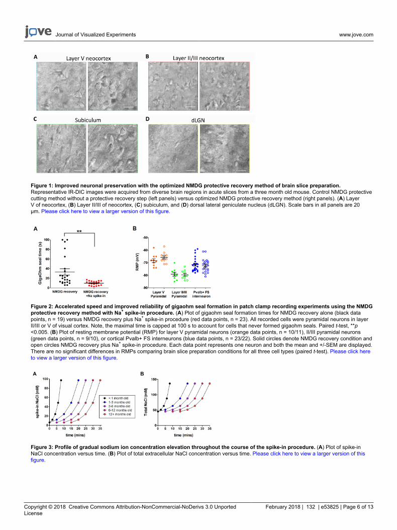
This section provides representative results for routine brain slice preparation and patch clamp electrophysiology experiments usingthe optimized NMDG protective recovery method (i.e., NMDG protective recovery combined with gradual Na+ spike-in procedure). First,morphological preservation of neurons was evaluated in various brain regions of brain slices prepared with or without implementing theoptimized NMDG protective recovery method (Figure 1). Three month old adult mice were selected for these experiments, and we used IR-DICmicroscopy to determine neuron health based on the shape and overall appearance of the somata and proximal dendrites. Note the shriveled,pyknotic appearance of most neurons in the representative images of brain slices prepared without the protective recovery method (all imageswere obtained 1–2 h after slice preparation). These control slices were prepared using NMDG aCSF for transcardial perfusion and slicing stepsbut were initially recovered in high Na+-containing HEPES aCSF. In contrast, the representative images from the slices prepared using theoptimized NMDG protective recovery method reveal neurons with improved morphologies (smoother, fuller, less crinkled appearance) thatare suitable for patch clamp recording (Figure 1). The improved neuronal preservation was observed across multiple brain regions includingneocortical layers II/III and V, subiculum, and dorsal lateral geniculate nucleus (dLGN).
Next, the optimized NMDG protective recovery method was compared with the original NMDG protective recovery method (i.e., withoutthe gradual Na+ spike-in procedure). The average time for gigaohm seal formation in patch clamp recording attempts was dramatically andsignificantly reduced (9.9 s versus 33.3 s, **p <0.005, paired t-test) when the gradual Na+ spike-in procedure was applied together with theNMDG protective recovery step (Figure 2). The faster and more reliable membrane sealing times greatly improved the throughput of patchclamp recording in young adult brain slices. The optimal Na+ spike-in schedule was further modified according to animal age (Table 2) and wasbeneficial for all ages tested (3 weeks to 1 year old mice). The profile of gradual sodium ion concentration elevation throughout the course of thespike-in procedure is provided (Figure 3) to accompany the schedules shown in Table 2.
As part of the Allen Institute Cell Types Program (http://celltypes.brain-map.org/) a large scale effort is underway to systematically characterizethe intrinsic electrophysiological properties of individual neurons in young adult (postnatal day 40-80) mouse visual cortical brain slices derivedfrom transgenic lines with cell type-specific fluorescent marker expression in genetically-defined neuronal populations (cortical layer and cell typespecific Cre driver lines crossed to a Cre-dependent fluorescent reporter line55). Figure 4 shows example traces of the firing patterns recordedfrom Parvalbumin (Pvalb)-expressing cortical fast-spiking (FS) interneurons (Pvalb-IRES-Cre/Ai14 mice) in response to a series of 1 s currentinjection steps that cover the dynamic range of neuron firing. The F-I curve for a dataset of 22 cortical FS interneurons is shown at right. Similartargeted patch clamp recording experiments were performed to characterize 23 Rorb-expressing excitatory neurons in layer IV from Rorb-IRES-Cre/Ai14 mice (Figure 4). Diverse healthy neuron types including FS interneurons and pyramidal neurons across cortical regions and layers canroutinely and reliably be targeted for patch clamp recording for at least 6-8 h after slice preparation using this optimized protocol.
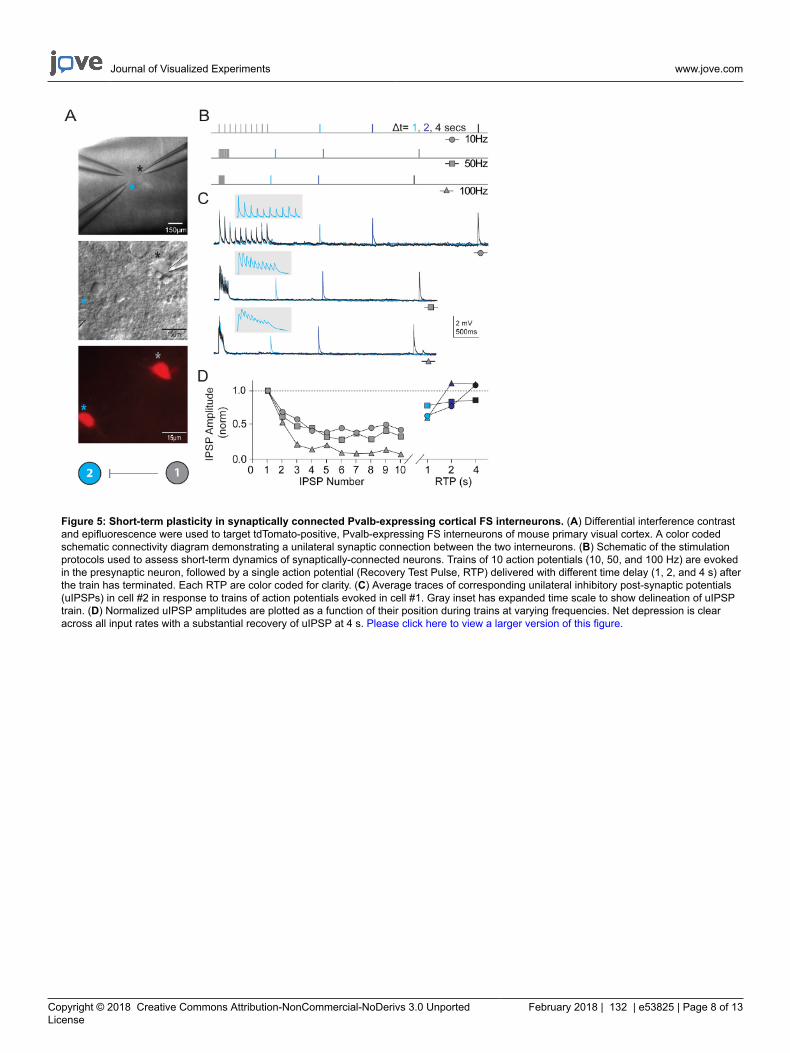
In addition to measuring intrinsic neuronal properties, synaptic connectivity was probed between multiple simultaneously recorded neurons ofdefined types in visual cortical microcircuits. The multi-neuron patch clamp recording technique is exceptionally demanding, as numerous healthycandidate neurons of defined types must be present within a relatively small field of the brain slice in order to ensure a reasonable chance ofobtaining high quality simultaneous recordings and identifying bona fide synaptic connections. Figure 5 shows paired recording of two tdTomato+ FS interneurons in the visual cortex of brain slices derived from young adult Pvalb-IRES-Cre/Ai14 mice. A strong unidirectional inhibitorysynaptic connection was detected (recorded with high chloride internal pipette solution). Example recordings and protocols for measuringproperties of short-term synaptic plasticity are presented. Bouts of high frequency train stimulation (10 pulses each at 10, 50, and 100 Hz) werefollowed by single recovery test pulses at various time intervals (1, 2, or 4 s) to measure the time course of recovery from synaptic depression.
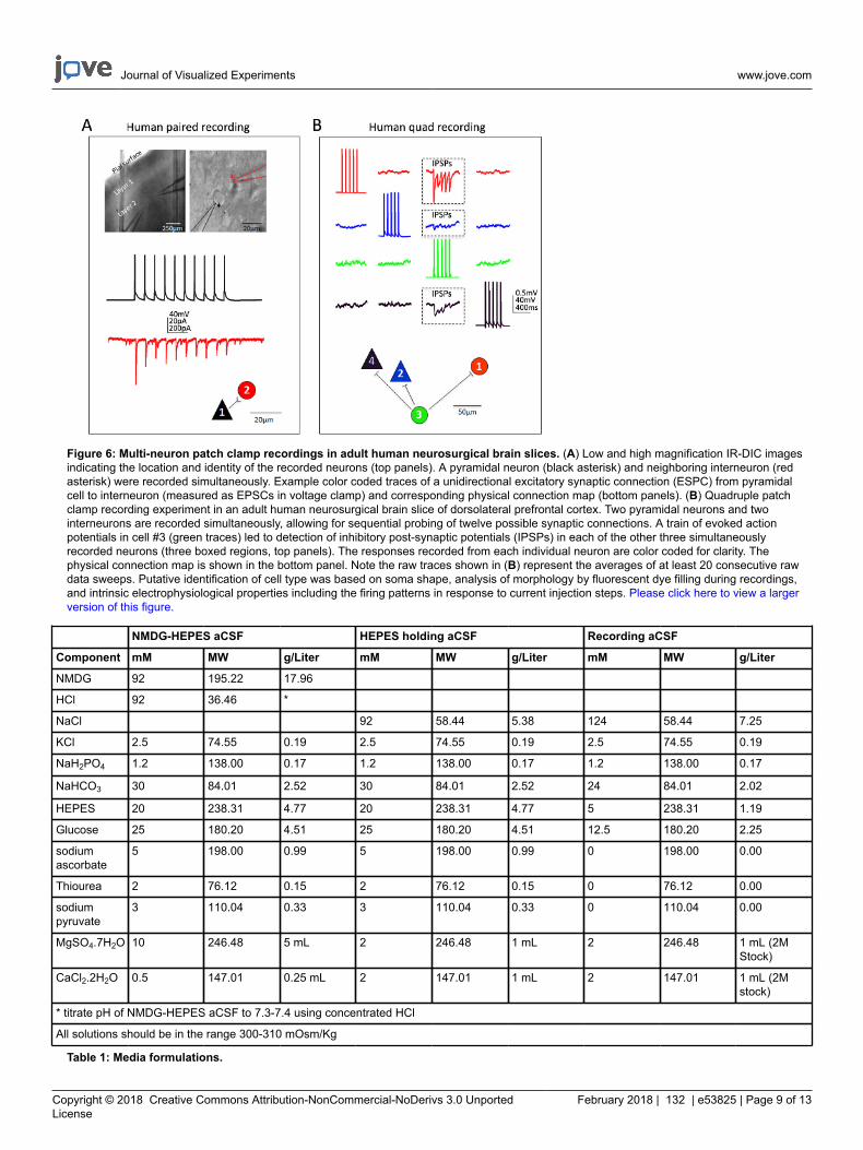
Excellent success has also been obtained for human neocortical neurons in mature adult ex vivo brain slices. Neurosurgical specimens areobtained from patients undergoing scheduled surgeries for tumor removal at local hospitals. The procedures for human neurosurgical tissuecollection and brain slice preparation differ from the mouse brain slice procedures in a few practical ways. In brief, the resected neocorticaltissue (distal to the site of pathology) is collected from the operating room and immersed into ice-cold oxygenated NMDG-HEPES aCSF andtransported with continuous chilling and oxygenation from the operating room to the laboratory within 30 min or less. The brain slices areprepared using the NMDG protective recovery procedure and allowed to recover for an extended time of approximately 3 h before initiating patchclamp recordings. Figure 6 shows a successful paired recording experiment and a successful quadruple patch clamp recording experiment fromhuman ex vivo brain slices prepared in this manner from the frontal cortex region. The paired recording demonstrates unidirectional excitatorysynaptic input from a cortical pyramidal neuron onto a cortical interneuron (recorded as excitatory postsynaptic currents). In the quad patchexperiment two excitatory and two inhibitory neurons were recorded simultaneously, and three inhibitory synaptic connections were detected(recorded as inhibitory postsynaptic potentials) out of twelve total connections probed. Thus, this optimized brain slice methodology allowsreliable experimental success in the most challenging of brain slice applications, including multi-neuron patch clamp experiments to study circuitconnectivity in acutely resected mature adult human brain tissue.
Journal of Visualized Experiments www.jove.com
Copyright © 2018 Creative Commons Attribution-NonCommercial-NoDerivs 3.0 UnportedLicense
February 2018 | 132 | e53825 | Page 6 of 13
Figure 1: Improved neuronal preservation with the optimized NMDG protective recovery method of brain slice preparation.Representative IR-DIC images were acquired from diverse brain regions in acute slices from a three month old mouse. Control NMDG protectivecutting method without a protective recovery step (left panels) versus optimized NMDG protective recovery method (right panels). (A) LayerV of neocortex, (B) Layer II/III of neocortex, (C) subiculum, and (D) dorsal lateral geniculate nucleus (dLGN). Scale bars in all panels are 20µm. Please click here to view a larger version of this figure.
Figure 2: Accelerated speed and improved reliability of gigaohm seal formation in patch clamp recording experiments using the NMDGprotective recovery method with Na+ spike-in procedure. (A) Plot of gigaohm seal formation times for NMDG recovery alone (black datapoints, n = 19) versus NMDG recovery plus Na+ spike-in procedure (red data points, n = 23). All recorded cells were pyramidal neurons in layerII/III or V of visual cortex. Note, the maximal time is capped at 100 s to account for cells that never formed gigaohm seals. Paired t-test, **p<0.005. (B) Plot of resting membrane potential (RMP) for layer V pyramidal neurons (orange data points, n = 10/11), II/III pyramidal neurons(green data points, n = 9/10), or cortical Pvalb+ FS interneurons (blue data points, n = 23/22). Solid circles denote NMDG recovery condition andopen circles NMDG recovery plus Na+ spike-in procedure. Each data point represents one neuron and both the mean and +/-SEM are displayed.There are no significant differences in RMPs comparing brain slice preparation conditions for all three cell types (paired t-test). Please click hereto view a larger version of this figure.
Figure 3: Profile of gradual sodium ion concentration elevation throughout the course of the spike-in procedure. (A) Plot of spike-inNaCl concentration versus time. (B) Plot of total extracellular NaCl concentration versus time. Please click here to view a larger version of thisfigure.

Journal of Visualized Experiments www.jove.com
Copyright © 2018 Creative Commons Attribution-NonCommercial-NoDerivs 3.0 UnportedLicense
February 2018 | 132 | e53825 | Page 7 of 13
Figure 4: Intrinsic electrophysiological properties of genetically-defined cortical cell types. (A) Example traces of neuronal firing inresponse to current injection steps for Pvalb+ cortical FS interneurons (left panel). tdTomato+ neurons were targeted for recordings in brainslices from Pvalb-IRES-Cre/Ai14 mice. Summary data for firing rate-current injection relationship (F-I curve) are shown at right (n = 22). (B)Example traces of neuronal firing in response to current injection steps for Rorb-expressing cortical layer IV excitatory neurons (left panel).tdTomato+ neurons were targeted for recordings in brain slices from Rorb-IRES-Cre/Ai14 mice. Summary data for firing rate-current injectionrelationship (F-I curve) are shown at right (n = 23). Each thin colored line represents the F-I curve for a single neuron; whereas, the thick blacklines represents the average for each group +/-SEM. Please click here to view a larger version of this figure.
Journal of Visualized Experiments www.jove.com
Copyright © 2018 Creative Commons Attribution-NonCommercial-NoDerivs 3.0 UnportedLicense
February 2018 | 132 | e53825 | Page 8 of 13
Figure 5: Short-term plasticity in synaptically connected Pvalb-expressing cortical FS interneurons. (A) Differential interference contrastand epifluorescence were used to target tdTomato-positive, Pvalb-expressing FS interneurons of mouse primary visual cortex. A color codedschematic connectivity diagram demonstrating a unilateral synaptic connection between the two interneurons. (B) Schematic of the stimulationprotocols used to assess short-term dynamics of synaptically-connected neurons. Trains of 10 action potentials (10, 50, and 100 Hz) are evokedin the presynaptic neuron, followed by a single action potential (Recovery Test Pulse, RTP) delivered with different time delay (1, 2, and 4 s) afterthe train has terminated. Each RTP are color coded for clarity. (C) Average traces of corresponding unilateral inhibitory post-synaptic potentials(uIPSPs) in cell #2 in response to trains of action potentials evoked in cell #1. Gray inset has expanded time scale to show delineation of uIPSPtrain. (D) Normalized uIPSP amplitudes are plotted as a function of their position during trains at varying frequencies. Net depression is clearacross all input rates with a substantial recovery of uIPSP at 4 s. Please click here to view a larger version of this figure.
Journal of Visualized Experiments www.jove.com
Copyright © 2018 Creative Commons Attribution-NonCommercial-NoDerivs 3.0 UnportedLicense
February 2018 | 132 | e53825 | Page 9 of 13
Figure 6: Multi-neuron patch clamp recordings in adult human neurosurgical brain slices. (A) Low and high magnification IR-DIC imagesindicating the location and identity of the recorded neurons (top panels). A pyramidal neuron (black asterisk) and neighboring interneuron (redasterisk) were recorded simultaneously. Example color coded traces of a unidirectional excitatory synaptic connection (ESPC) from pyramidalcell to interneuron (measured as EPSCs in voltage clamp) and corresponding physical connection map (bottom panels). (B) Quadruple patchclamp recording experiment in an adult human neurosurgical brain slice of dorsolateral prefrontal cortex. Two pyramidal neurons and twointerneurons are recorded simultaneously, allowing for sequential probing of twelve possible synaptic connections. A train of evoked actionpotentials in cell #3 (green traces) led to detection of inhibitory post-synaptic potentials (IPSPs) in each of the other three simultaneouslyrecorded neurons (three boxed regions, top panels). The responses recorded from each individual neuron are color coded for clarity. Thephysical connection map is shown in the bottom panel. Note the raw traces shown in (B) represent the averages of at least 20 consecutive rawdata sweeps. Putative identification of cell type was based on soma shape, analysis of morphology by fluorescent dye filling during recordings,and intrinsic electrophysiological properties including the firing patterns in response to current injection steps. Please click here to view a largerversion of this figure.
NMDG-HEPES aCSF HEPES holding aCSF Recording aCSF
Component mM MW g/Liter mM MW g/Liter mM MW g/Liter
NMDG 92 195.22 17.96
HCl 92 36.46 *
NaCl 92 58.44 5.38 124 58.44 7.25
KCl 2.5 74.55 0.19 2.5 74.55 0.19 2.5 74.55 0.19
NaH2PO4 1.2 138.00 0.17 1.2 138.00 0.17 1.2 138.00 0.17
NaHCO3 30 84.01 2.52 30 84.01 2.52 24 84.01 2.02
HEPES 20 238.31 4.77 20 238.31 4.77 5 238.31 1.19
Glucose 25 180.20 4.51 25 180.20 4.51 12.5 180.20 2.25
sodiumascorbate
5 198.00 0.99 5 198.00 0.99 0 198.00 0.00
Thiourea 2 76.12 0.15 2 76.12 0.15 0 76.12 0.00
sodiumpyruvate
3 110.04 0.33 3 110.04 0.33 0 110.04 0.00
MgSO4.7H2O 10 246.48 5 mL 2 246.48 1 mL 2 246.48 1 mL (2MStock)
CaCl2.2H2O 0.5 147.01 0.25 mL 2 147.01 1 mL 2 147.01 1 mL (2Mstock)
* titrate pH of NMDG-HEPES aCSF to 7.3-7.4 using concentrated HCl
All solutions should be in the range 300-310 mOsm/Kg
Table 1: Media formulations.

Journal of Visualized Experiments www.jove.com
Copyright © 2018 Creative Commons Attribution-NonCommercial-NoDerivs 3.0 UnportedLicense
February 2018 | 132 | e53825 | Page 10 of 13
Animal Age
Time (min)* <1 month 1-3 months 3–6 months 6–12 months 12+ months
0 250 µL 250 µL
1
2 250 µL
3
4 500 µL
5 250 µL 250 µL
6 1000 µL
7
8 2000 µL
9
10 transfer 500 µL 250 µL 250 µL
11
12
13
14
15 1000 µL 500 µL 250 µL 250 µL
16
17
18
19
20 2000 µL 1000 µL 500 µL 250 µL
21
22
23
24
25 transfer 2000 µL 1000 µL 500 µL
26
27
28
29
30 transfer 2,000 µL 1,000 µL
31
32
33
34
35 transfer 2,000 µL
36
37
38
39
40 transfer
*Time zero is the moment slices are transferred into the initial recovery chamber
Table 2: Recommended schedule for gradual Na+ spike-in procedure according to mouse age.
Journal of Visualized Experiments www.jove.com
Copyright © 2018 Creative Commons Attribution-NonCommercial-NoDerivs 3.0 UnportedLicense
February 2018 | 132 | e53825 | Page 11 of 13
Discussion
Na+ Spike-in Improves Gigaohm Seal Formation and Patch Clamp Recording Success
The initial version of the NMDG protective recovery method was specifically designed for adult and aging animals2,5. Some early adopters havealso sought to apply this methodology to juvenile animal brain slicing (i.e., mice <30 days old). However, it has been noted that in contrast to theoutstanding visually-confirmed neuronal preservation with the NMDG protective recovery method in this age range, gigaohm seal formation canfrequently stall out, leading to failed patch clamp recording attempts. One hypothesis is that NMDG cations are more readily trapped in juvenilebrain slices relative to adult brain slices and can impede seal formation; however, gigaohm seals can readily form while juvenile brain slices arefully submerged in NMDG aCSF (data not shown), thus indicating that NMDG aCSF per se is not impeding gigaohm seal formation.
The rapid transition from low-to-high Na+ solution at the completion of the initial brain slice recovery step causes damage to neuronalmembranes and perturbs the seal formation process. This is intuitive given that the transition from low-to-high Na+, cold-to-warm temperature,and dramatic elevation of the Ca2+ to Mg2+ ratio collectively lead to a massive resurgence of spontaneous synaptic activity. This inhibitoryrebound phase in the brain slicing procedure is likely to mirror reperfusion injury following an ischemic insult. Thus, to further mitigate neuronalmembrane damage in the initial recovery phase a gradual Na+ spike-in procedure has been incorporated in which the elevation of Na+
concentration in the NMDG protective recovery incubation chamber is slowly and reproducibly elevated with precise timing. As in the originalprotective recovery procedure, the temporal dissociation of Na+ elevation from temperature and Ca2+/Mg2+ ratio elevation is beneficial. Butadditionally, the Na+ spike-in procedure leads to small incremental increases in extracellular Na+ concentration over the early time pointsand large increases towards the late time points, thereby affording the brain tissue an opportunity to better accommodate to the rising Na+
levels. This procedure is an alternative to gradual solution exchange controlled by a perfusion pump or gravity drip lines which lead to constantincreases in Na+ levels and require attention to both inflow and outflow to avoid overflow of the slice chamber. Notably, in this Na+ spike-inprocedure the osmolality of the solution in the slice chamber gradually rises over a period of several minutes before the slices are returned tonormal osmolality solution, but this did not adversely affect the slice health or patch clamp recording success. A high osmolality cutting solutionhas previously been used for midbrain slice preparations in order to better preserve dopamine neurons for patch clamp recordings57,58, thusdemonstrating that this temporary hyperosmolality may be beneficial in some contexts.
By implementing an optimized procedure combining the NMDG protective recovery method and gradual Na+ spike-in step the utility of this brainslice methodology has been extended to cover juvenile through mature adult animal ages. This updated protocol is now suitable for a wide rangeof animal ages using a single optimal NMDG aCSF formulation and procedure. If necessary, the Na+ spike-in procedure can be applied with aprogressively longer delay and/or slower time course to enhance the viability of brain slices from older animals, and we have provided a basicguide of recommended spike-in schedules according to animal age (see Table 2). While we have provided a basic framework suitable for awide array of applications, additional advanced steps can be explored for further enhancing viability and longevity of brain slices from adult andaging animals. For example, glutathione restoration strategies are particularly effective in this respect and can be implemented as describedelsewhere2,6.
Improving Throughput for Challenging Experiments
The analysis of synaptic connectivity by patch clamp recording is a demanding application that requires excellent preservation of both neuronalstructure and function in order to achieve a high reliability of success. As the number of neurons to be simultaneously recorded goes up linearly,the technical difficulty level goes up supra-linearly. There are numerous failure modes, and one of the most frequent causes of failures is theinability to form adequate gigaohm seals onto one or more of the targeted cells. This can dramatically slow progress, particularly when threeor more neurons must be recorded simultaneously. Consistent with the finding of faster gigaohm seal formation time with the optimized NMDGprotective recovery method, there was a marked improvement in the success rate and throughput of multi-neuron patch clamp recordingexperiments with both adult transgenic mouse brain slices and adult human neurosurgical brain slices. The improved efficiency is almostcertainly attributable to both the more rapid and reliable gigaohm seal formation and the improved neuronal preservation of the slices with thisprotocol. Although this protocol focuses on the benefits explicitly for patch clamp recording applications, similar gains are anticipated for otherchallenging experimental applications where brain slice viability is paramount.
Disclosures
The authors have nothing to disclose.
Acknowledgements
This work was funded by the Allen Institute for Brain Science. The authors wish to thank the Allen Institute founders, Paul G. Allen and JodyAllen, for their vision, encouragement, and support. We also thank the Allen Institute technical support staff for performing animal care,husbandry, and genotyping.
References
1. Aghajanian, G. K., & Rasmussen, K. Intracellular studies in the facial nucleus illustrating a simple new method for obtaining viablemotoneurons in adult rat brain slices. Synapse. 3 (4), 331-338 (1989).
2. Ting, J. T., Daigle, T. L., Chen, Q., & Feng, G. Acute brain slice methods for adult and aging animals: application of targeted patch clampanalysis and optogenetics. Methods Mol Biol. 1183 221-242 (2014).
3. Zhao, S. et al. Cell type-specific channelrhodopsin-2 transgenic mice for optogenetic dissection of neural circuitry function. Nat Methods. 8(9), 745-752 (2011).

Journal of Visualized Experiments www.jove.com
Copyright © 2018 Creative Commons Attribution-NonCommercial-NoDerivs 3.0 UnportedLicense
February 2018 | 132 | e53825 | Page 12 of 13
4. Peca, J. et al. Shank3 mutant mice display autistic-like behaviours and striatal dysfunction. Nature. 472 (7344), 437-442 (2011).5. Ting, J. T. F., G. Improved methods for acute brain slice preparation from adult and aging animals. Society for Neuroscience Abstracts.
520.29 (2011).6. Ting, J. T. F., G. Improved methods for acute brain slice preparation from adult and aging animals (Part II): Glutathione depletion underlies
rapid deterioration of adult brain slices. Society for Neuroscience Abstracts. 505.12 (2012).7. MacGregor, D. G., Chesler, M., & Rice, M. E. HEPES prevents edema in rat brain slices. Neurosci Lett. 303 (3), 141-144 (2001).8. Liu, Y. B., Guo, J. Z., & Chiappinelli, V. A. Nicotinic receptor-mediated biphasic effect on neuronal excitability in chick lateral spiriform
neurons. Neuroscience. 148 (4), 1004-1014 (2007).9. Xiang, Z., Huguenard, J. R., & Prince, D. A. GABAA receptor-mediated currents in interneurons and pyramidal cells of rat visual cortex. J
Physiol. 506 ( Pt 3) 715-730 (1998).10. Bischofberger, J., Engel, D., Li, L., Geiger, J. R., & Jonas, P. Patch-clamp recording from mossy fiber terminals in hippocampal slices. Nat
Protoc. 1 (4), 2075-2081 (2006).11. Contractor, A. et al. Loss of kainate receptor-mediated heterosynaptic facilitation of mossy-fiber synapses in KA2-/- mice. J Neurosci. 23 (2),
422-429 (2003).12. Pita-Almenar, J. D., Collado, M. S., Colbert, C. M., & Eskin, A. Different mechanisms exist for the plasticity of glutamate reuptake during early
long-term potentiation (LTP) and late LTP. J Neurosci. 26 (41), 10461-10471 (2006).13. Ito, K., Contractor, A., & Swanson, G. T. Attenuated plasticity of postsynaptic kainate receptors in hippocampal CA3 pyramidal neurons. J
Neurosci. 24 (27), 6228-6236 (2004).14. Van Dort, C. J. et al. Optogenetic activation of cholinergic neurons in the PPT or LDT induces REM sleep. Proc Natl Acad Sci U S A. 112 (2),
584-589 (2015).15. Ferando, I., & Mody, I. Altered gamma oscillations during pregnancy through loss of delta subunit-containing GABA(A) receptors on
parvalbumin interneurons. Front Neural Circuits. 7 144 (2013).16. Walker, A. G. et al. Metabotropic glutamate receptor 3 activation is required for long-term depression in medial prefrontal cortex and fear
extinction. Proc Natl Acad Sci U S A. 112 (4), 1196-1201 (2015).17. Takahashi, Y. K. et al. Neural estimates of imagined outcomes in the orbitofrontal cortex drive behavior and learning. Neuron. 80 (2), 507-518
(2013).18. Pan, G. et al. Preserving GABAergic interneurons in acute brain slices of mice using the N-methyl-D-glucamine-based artificial cerebrospinal
fluid method. Neurosci Bull. 31 (2), 265-270 (2015).19. LaSarge, C. L., Santos, V. R., & Danzer, S. C. PTEN deletion from adult-generated dentate granule cells disrupts granule cell mossy fiber
axon structure. Neurobiol Dis. 75 142-150 (2015).20. Tong, L. M. et al. Inhibitory interneuron progenitor transplantation restores normal learning and memory in ApoE4 knock-in mice without or
with Abeta accumulation. J Neurosci. 34 (29), 9506-9515 (2014).21. Fleming, R. L. et al. Binge-pattern ethanol exposure during adolescence, but not adulthood, causes persistent changes in GABAA receptor-
mediated tonic inhibition in dentate granule cells. Alcohol Clin Exp Res. 37 (7), 1154-1160 (2013).22. Kummer, K. K., El Rawas, R., Kress, M., Saria, A., & Zernig, G. Social interaction and cocaine conditioning in mice increase spontaneous
spike frequency in the nucleus accumbens or septal nuclei as revealed by multielectrode array recordings. Pharmacology. 95 (1-2), 42-49(2015).
23. Qi, J. et al. A glutamatergic reward input from the dorsal raphe to ventral tegmental area dopamine neurons. Nat Commun. 5 5390 (2014).24. Wang, L. et al. Modulation of dopamine release in the striatum by physiologically relevant levels of nicotine. Nat Commun. 5 3925 (2014).25. Siuda, E. R. et al. Spatiotemporal Control of Opioid Signaling and Behavior. Neuron. (2015).26. Tucker, K. R., Huertas, M. A., Horn, J. P., Canavier, C. C., & Levitan, E. S. Pacemaker rate and depolarization block in nigral dopamine
neurons: a somatic sodium channel balancing act. J Neurosci. 32 (42), 14519-14531 (2012).27. McDevitt, R. A. et al. Serotonergic versus nonserotonergic dorsal raphe projection neurons: differential participation in reward circuitry. Cell
Rep. 8 (6), 1857-1869 (2014).28. Wang, D. V. et al. Mesopontine median raphe regulates hippocampal ripple oscillation and memory consolidation. Nat Neurosci. 18 (5),
728-735 (2015).29. Engle, S. E., Shih, P. Y., McIntosh, J. M., & Drenan, R. M. alpha4alpha6beta2* nicotinic acetylcholine receptor activation on ventral tegmental
area dopamine neurons is sufficient to stimulate a depolarizing conductance and enhance surface AMPA receptor function. Mol Pharmacol.84 (3), 393-406 (2013).
30. Vance, K. M., Ribnicky, D. M., Rogers, R. C., & Hermann, G. E. Artemisia santolinifolia enhances glutamatergic neurotransmission in thenucleus of the solitary tract. Neurosci Lett. 582 115-119 (2014).
31. Dergacheva, O., Boychuk, C. R., & Mendelowitz, D. Developmental changes in GABAergic neurotransmission to presympathetic and cardiacparasympathetic neurons in the brainstem. J Neurophysiol. 110 (3), 672-679 (2013).
32. Dergacheva, O. Chronic intermittent hypoxia alters neurotransmission from lateral paragigantocellular nucleus to parasympathetic cardiacneurons in the brain stem. J Neurophysiol. 113 (1), 380-389 (2015).
33. Dyavanapalli, J. et al. Chronic intermittent hypoxia-hypercapnia blunts heart rate responses and alters neurotransmission to cardiac vagalneurons. J Physiol. 592 (Pt 13), 2799-2811 (2014).
34. Apostolides, P. F., & Trussell, L. O. Regulation of interneuron excitability by gap junction coupling with principal cells. Nat Neurosci. 16 (12),1764-1772 (2013).
35. Graziane, N. M., Polter, A. M., Briand, L. A., Pierce, R. C., & Kauer, J. A. Kappa opioid receptors regulate stress-induced cocaine seeking andsynaptic plasticity. Neuron. 77 (5), 942-954 (2013).
36. Ferando, I., & Mody, I. In vitro gamma oscillations following partial and complete ablation of delta subunit-containing GABAA receptors fromparvalbumin interneurons. Neuropharmacology. 88 91-98 (2015).
37. Foster, D. J. et al. M5 receptor activation produces opposing physiological outcomes in dopamine neurons depending on the receptor'slocation. J Neurosci. 34 (9), 3253-3262 (2014).
38. Engle, S. E., Broderick, H. J., & Drenan, R. M. Local application of drugs to study nicotinic acetylcholine receptor function in mouse brainslices. J Vis Exp. (68), e50034 (2012).
39. Wang, L. et al. Temporal components of cholinergic terminal to dopaminergic terminal transmission in dorsal striatum slices of mice. JPhysiol. 592 (Pt 16), 3559-3576 (2014).

Journal of Visualized Experiments www.jove.com
Copyright © 2018 Creative Commons Attribution-NonCommercial-NoDerivs 3.0 UnportedLicense
February 2018 | 132 | e53825 | Page 13 of 13
40. Holloway, B. B. et al. Monosynaptic glutamatergic activation of locus coeruleus and other lower brainstem noradrenergic neurons by the C1cells in mice. J Neurosci. 33 (48), 18792-18805 (2013).
41. DePuy, S. D. et al. Glutamatergic neurotransmission between the C1 neurons and the parasympathetic preganglionic neurons of the dorsalmotor nucleus of the vagus. J Neurosci. 33 (4), 1486-1497 (2013).
42. Abbott, S. B., Holloway, B. B., Viar, K. E., & Guyenet, P. G. Vesicular glutamate transporter 2 is required for the respiratory andparasympathetic activation produced by optogenetic stimulation of catecholaminergic neurons in the rostral ventrolateral medulla of mice invivo. Eur J Neurosci. 39 (1), 98-106 (2014).
43. Dergacheva, O., Dyavanapalli, J., Pinol, R. A., & Mendelowitz, D. Chronic intermittent hypoxia and hypercapnia inhibit the hypothalamicparaventricular nucleus neurotransmission to parasympathetic cardiac neurons in the brain stem. Hypertension. 64 (3), 597-603 (2014).
44. Chen, Q. et al. Imaging neural activity using Thy1-GCaMP transgenic mice. Neuron. 76 (2), 297-308 (2012).45. Luongo, F. H., Horn, M.E.; Sohal V.S. Putative microcircuit-level substrates for attention are disrupted in mouse models of autism. Biological
Psychiatry.79(8), 667-675 (2016).46. Vance, K. M., Rogers, R. C., & Hermann, G. E. PAR1-activated astrocytes in the nucleus of the solitary tract stimulate adjacent neurons via
NMDA receptors. J Neurosci. 35 (2), 776-785 (2015).47. Feliciano, P., Andrade, R., & Bykhovskaia, M. Synapsin II and Rab3a cooperate in the regulation of epileptic and synaptic activity in the CA1
region of the hippocampus. J Neurosci. 33 (46), 18319-18330 (2013).48. Tomioka, N. H. et al. Elfn1 recruits presynaptic mGluR7 in trans and its loss results in seizures. Nat Commun. 5 4501 (2014).49. Jiang, X. et al. Principles of connectivity among morphologically defined cell types in adult neocortex. Science. 350 (6264), aac9462 (2015).50. Mishra, A. et al. Imaging pericytes and capillary diameter in brain slices and isolated retinae. Nat Protoc. 9 (2), 323-336 (2014).51. Gabriel, L. R., Wu, S., & Melikian, H. E. Brain slice biotinylation: an ex vivo approach to measure region-specific plasma membrane protein
trafficking in adult neurons. J Vis Exp. (86) (2014).52. Edwards, F. A., & Konnerth, A. Patch-clamping cells in sliced tissue preparations. Methods Enzymol. 207 208-222 (1992).53. Qi, G., Radnikow, G., & Feldmeyer, D. Electrophysiological and morphological characterization of neuronal microcircuits in acute brain slices
using paired patch-clamp recordings. J Vis Exp. (95), 52358 (2015).54. Booker, S. A., Song, J., & Vida, I. Whole-cell patch-clamp recordings from morphologically- and neurochemically-identified hippocampal
interneurons. J Vis Exp. (91), e51706 (2014).55. Madisen, L. et al. A robust and high-throughput Cre reporting and characterization system for the whole mouse brain. Nat Neurosci. 13 (1),
133-140 (2010).56. Hille, B. The permeability of the sodium channel to organic cations in myelinated nerve. J Gen Physiol. 58 (6), 599-619 (1971).57. Martin, M., Chen, B. T., Hopf, F. W., Bowers, M. S., & Bonci, A. Cocaine self-administration selectively abolishes LTD in the core of the
nucleus accumbens. Nat Neurosci. 9 (7), 868-869 (2006).58. Chen, B. T. et al. Cocaine but not natural reward self-administration nor passive cocaine infusion produces persistent LTP in the VTA. Neuron.
59 (2), 288-297 (2008).
Copyright © 2022 FDOKUMEN