Electrically conductive carbon nanopipe-graphite nanosheet/polyaniline composites
Upload
independentCategory
view
3download
0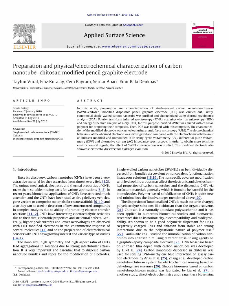
Pn
TD
a
ARRAA
KSCD
1
aTmragairdewss[
ltn
(
0d
Applied Surface Science 257 (2010) 622–627
Contents lists available at ScienceDirect
Applied Surface Science
journa l homepage: www.e lsev ier .com/ locate /apsusc
reparation and physical/electrochemical characterization of carbonanotube–chitosan modified pencil graphite electrode
ayfun Vural, Filiz Kuralay, Cem Bayram, Serdar Abaci, Emir Baki Denkbas ∗
epartment of Chemistry, Faculty of Science, Hacettepe University, 06800 Beytepe, Ankara, Turkey
r t i c l e i n f o
rticle history:eceived 7 January 2010eceived in revised form 15 July 2010ccepted 15 July 2010vailable online 21 July 2010
a b s t r a c t
In this work, preparation and characterization of single-walled carbon nanotube–chitosan(SWNT–chitosan) modified disposable pencil graphite electrode (PGE) was carried out. Firstly,commercial single-walled carbon nanotube was purified and characterized using thermal gravimetricanalysis (TGA), Fourier transform infrared spectroscopy (FT-IR), scanning electron microscopy (SEM)and energy dispersive analysis of X-ray (EDX) for this purpose. Purified SWNT was mixed with chitosan
eywords:ingle-walled carbon nanotube (SWNT)hitosanisposable pencil graphite electrode (PGE)
polymer for preparing their composite. Then, PGE was modified with this composite. The characteriza-tion of the modified electrode was carried out using atomic force microscopy (AFM). The electrochemicalbehaviour of the obtained electrode was investigated and compared with the electrochemical behaviourof chitosan modified and unmodified PGEs using cyclic voltammetry (CV), differential pulse voltam-metry (DPV) and alternative current (AC) impedance spectroscopy. In order to obtain more sensitiveelectrochemical signals, the effect of SWNT concentration was studied. This modified electrode alsoshowed electrocatalytic effect for hydrogen evolution.
. Introduction
Since its discovery, carbon nanotubes (CNTs) have been a veryttractive material for the researches from almost every field [1,2].he unique mechanical, electronic and thermal properties of CNTsake them suitable missing parts for various applications [3–5]. In
ecent years, biomedical applications of CNTs have attracted muchttention and the CNTs have been used as drug delivery vehicles,ene vectors or composite materials for tissue scaffolds [6–10] andlso they can be used in detection of low concentrated compoundsn complex analytes due to ability of promoting electron transfereactions [11,12]. CNTs have interesting electrocatalytic activitiesue to their size, electronic properties and structural defects. Gen-rally, higher peak currents and lower overvoltages are observedith CNT modified electrodes in the voltammetric response of
everal molecules [13] and so the preparation of electrochemicalensors with CNTs has a growing interest and various type of studies14–17].
The nano size, high symmetry and high aspect ratio of CNTsead aggregations in solutions due to strong intertubular attrac-ion. It is very important and essential that to disperse carbonanotube bundles and ropes for the modification of electrodes.
∗ Corresponding author. Tel.: +90 312 297 7992; fax: +90 312 299 2163.E-mail addresses: [email protected], [email protected]
E.B. Denkbas).
169-4332/$ – see front matter © 2010 Elsevier B.V. All rights reserved.oi:10.1016/j.apsusc.2010.07.048
© 2010 Elsevier B.V. All rights reserved.
Single-walled carbon nanotubes (SWNTs) can be individually dis-persed from bundles via covalent or noncovalent functionalizationin aqueous solutions [18,19]. The nonspecific covalent modificationwith hydrophilic groups may affect the electronic and physiochem-ical properties of carbon nanotubes and the dispersing CNTs viasurfactant materials generally which is found to be harmful for thebiomolecules. Polymer based solubilization of CNTs is quite newand minimalizes the disadvantages mentioned above [20].
The dispersion of functionalized CNTs is much better in chargedpolyelectrolyte solutions like chitosan than the organic solvents[21]. Chitosan is a naturally abundant polysaccharide and it hasbeen applied in numerous biomedical studies and biomaterialresearches due to its nontoxicity, biocompatibility, and biodegrad-ability. It’s shown to be a good polymeric dispersant for CNTs.Negatively charged CNTs and chitosan form stable and stronginteractions due to the polycationic nature of polymer itself[22]. Pauliukaite et al. studied the immobilization of carbon nan-otubes into chitosan film using different cross-linking agents ona graphite–epoxy composite electrode [23]. DNA biosensor basedon chitosan film doped with carbon nanotubes was developedby Li et al. [24]. Carbon nanotubes dispersed in chitosan wasused for sensing DNA–methylene blue interaction on glassy car-
bon electrodes by Arias et al. [25]. Zhang et al. developed carbonnanotube–chitosan system for electrochemical sensing based ondehydrogenase enzymes [26]. Glucose biosensor based on carbonnanotubes/chitosan matrix was fabricated by Liu et al. [27]. Inanother study, direct electrochemistry and reagentless biosensing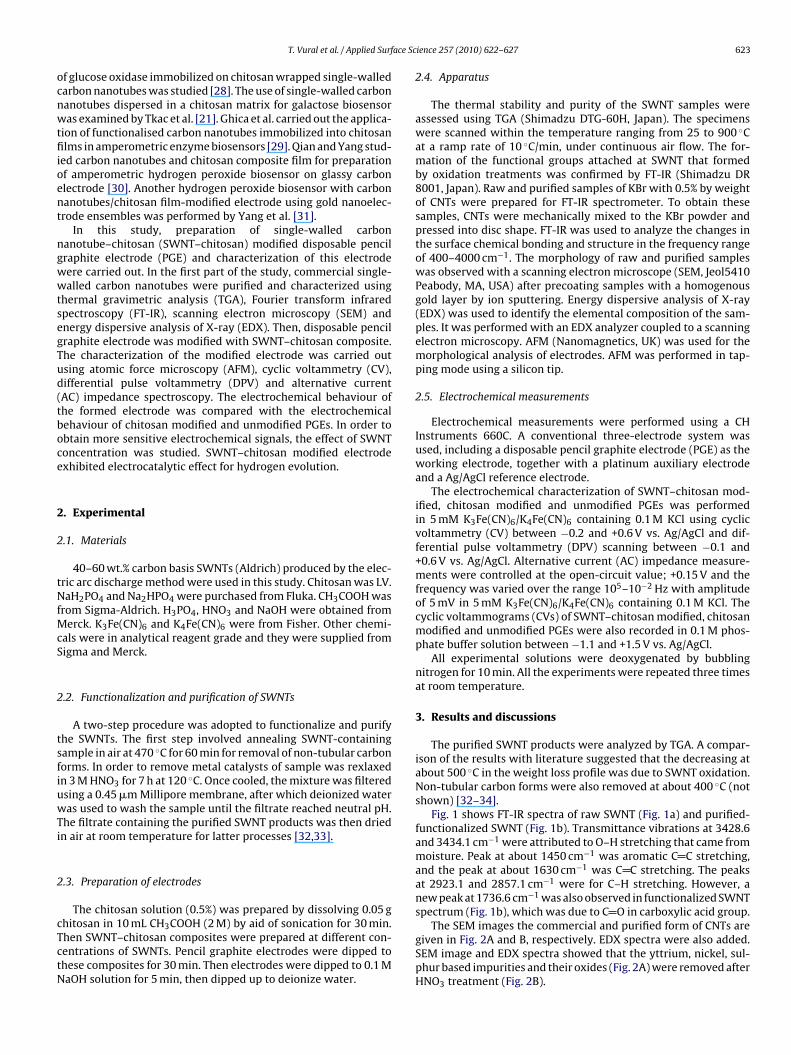
ace Sc
ocnwtfiioent
ngwwtsegTud(tboce
2
2
tNfMcS
2
tsfiuwTi
2
cTctN
T. Vural et al. / Applied Surf
f glucose oxidase immobilized on chitosan wrapped single-walledarbon nanotubes was studied [28]. The use of single-walled carbonanotubes dispersed in a chitosan matrix for galactose biosensoras examined by Tkac et al. [21]. Ghica et al. carried out the applica-
ion of functionalised carbon nanotubes immobilized into chitosanlms in amperometric enzyme biosensors [29]. Qian and Yang stud-
ed carbon nanotubes and chitosan composite film for preparationf amperometric hydrogen peroxide biosensor on glassy carbonlectrode [30]. Another hydrogen peroxide biosensor with carbonanotubes/chitosan film-modified electrode using gold nanoelec-rode ensembles was performed by Yang et al. [31].
In this study, preparation of single-walled carbonanotube–chitosan (SWNT–chitosan) modified disposable pencilraphite electrode (PGE) and characterization of this electrodeere carried out. In the first part of the study, commercial single-alled carbon nanotubes were purified and characterized using
hermal gravimetric analysis (TGA), Fourier transform infraredpectroscopy (FT-IR), scanning electron microscopy (SEM) andnergy dispersive analysis of X-ray (EDX). Then, disposable pencilraphite electrode was modified with SWNT–chitosan composite.he characterization of the modified electrode was carried outsing atomic force microscopy (AFM), cyclic voltammetry (CV),ifferential pulse voltammetry (DPV) and alternative currentAC) impedance spectroscopy. The electrochemical behaviour ofhe formed electrode was compared with the electrochemicalehaviour of chitosan modified and unmodified PGEs. In order tobtain more sensitive electrochemical signals, the effect of SWNToncentration was studied. SWNT–chitosan modified electrodexhibited electrocatalytic effect for hydrogen evolution.
. Experimental
.1. Materials
40–60 wt.% carbon basis SWNTs (Aldrich) produced by the elec-ric arc discharge method were used in this study. Chitosan was LV.aH2PO4 and Na2HPO4 were purchased from Fluka. CH3COOH was
rom Sigma-Aldrich. H3PO4, HNO3 and NaOH were obtained fromerck. K3Fe(CN)6 and K4Fe(CN)6 were from Fisher. Other chemi-
als were in analytical reagent grade and they were supplied fromigma and Merck.
.2. Functionalization and purification of SWNTs
A two-step procedure was adopted to functionalize and purifyhe SWNTs. The first step involved annealing SWNT-containingample in air at 470 ◦C for 60 min for removal of non-tubular carbonorms. In order to remove metal catalysts of sample was rexlaxedn 3 M HNO3 for 7 h at 120 ◦C. Once cooled, the mixture was filteredsing a 0.45 �m Millipore membrane, after which deionized wateras used to wash the sample until the filtrate reached neutral pH.
he filtrate containing the purified SWNT products was then driedn air at room temperature for latter processes [32,33].
.3. Preparation of electrodes
The chitosan solution (0.5%) was prepared by dissolving 0.05 g
hitosan in 10 mL CH3COOH (2 M) by aid of sonication for 30 min.hen SWNT–chitosan composites were prepared at different con-entrations of SWNTs. Pencil graphite electrodes were dipped tohese composites for 30 min. Then electrodes were dipped to 0.1 MaOH solution for 5 min, then dipped up to deionize water.ience 257 (2010) 622–627 623
2.4. Apparatus
The thermal stability and purity of the SWNT samples wereassessed using TGA (Shimadzu DTG-60H, Japan). The specimenswere scanned within the temperature ranging from 25 to 900 ◦Cat a ramp rate of 10 ◦C/min, under continuous air flow. The for-mation of the functional groups attached at SWNT that formedby oxidation treatments was confirmed by FT-IR (Shimadzu DR8001, Japan). Raw and purified samples of KBr with 0.5% by weightof CNTs were prepared for FT-IR spectrometer. To obtain thesesamples, CNTs were mechanically mixed to the KBr powder andpressed into disc shape. FT-IR was used to analyze the changes inthe surface chemical bonding and structure in the frequency rangeof 400–4000 cm−1. The morphology of raw and purified sampleswas observed with a scanning electron microscope (SEM, Jeol5410Peabody, MA, USA) after precoating samples with a homogenousgold layer by ion sputtering. Energy dispersive analysis of X-ray(EDX) was used to identify the elemental composition of the sam-ples. It was performed with an EDX analyzer coupled to a scanningelectron microscopy. AFM (Nanomagnetics, UK) was used for themorphological analysis of electrodes. AFM was performed in tap-ping mode using a silicon tip.
2.5. Electrochemical measurements
Electrochemical measurements were performed using a CHInstruments 660C. A conventional three-electrode system wasused, including a disposable pencil graphite electrode (PGE) as theworking electrode, together with a platinum auxiliary electrodeand a Ag/AgCl reference electrode.
The electrochemical characterization of SWNT–chitosan mod-ified, chitosan modified and unmodified PGEs was performedin 5 mM K3Fe(CN)6/K4Fe(CN)6 containing 0.1 M KCl using cyclicvoltammetry (CV) between −0.2 and +0.6 V vs. Ag/AgCl and dif-ferential pulse voltammetry (DPV) scanning between −0.1 and+0.6 V vs. Ag/AgCl. Alternative current (AC) impedance measure-ments were controlled at the open-circuit value; +0.15 V and thefrequency was varied over the range 105–10−2 Hz with amplitudeof 5 mV in 5 mM K3Fe(CN)6/K4Fe(CN)6 containing 0.1 M KCl. Thecyclic voltammograms (CVs) of SWNT–chitosan modified, chitosanmodified and unmodified PGEs were also recorded in 0.1 M phos-phate buffer solution between −1.1 and +1.5 V vs. Ag/AgCl.
All experimental solutions were deoxygenated by bubblingnitrogen for 10 min. All the experiments were repeated three timesat room temperature.
3. Results and discussions
The purified SWNT products were analyzed by TGA. A compar-ison of the results with literature suggested that the decreasing atabout 500 ◦C in the weight loss profile was due to SWNT oxidation.Non-tubular carbon forms were also removed at about 400 ◦C (notshown) [32–34].
Fig. 1 shows FT-IR spectra of raw SWNT (Fig. 1a) and purified-functionalized SWNT (Fig. 1b). Transmittance vibrations at 3428.6and 3434.1 cm−1 were attributed to O–H stretching that came frommoisture. Peak at about 1450 cm−1 was aromatic C C stretching,and the peak at about 1630 cm−1 was C C stretching. The peaksat 2923.1 and 2857.1 cm−1 were for C–H stretching. However, anew peak at 1736.6 cm−1 was also observed in functionalized SWNTspectrum (Fig. 1b), which was due to C O in carboxylic acid group.
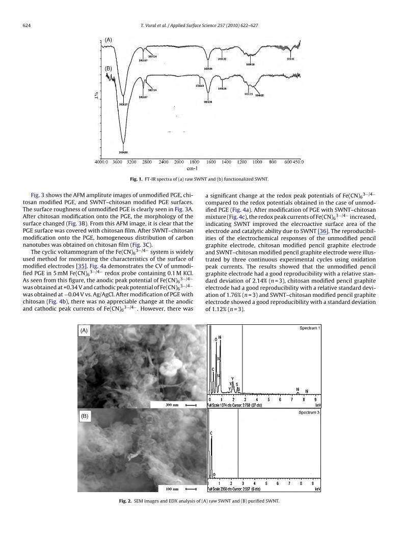
The SEM images the commercial and purified form of CNTs aregiven in Fig. 2A and B, respectively. EDX spectra were also added.SEM image and EDX spectra showed that the yttrium, nickel, sul-phur based impurities and their oxides (Fig. 2A) were removed afterHNO3 treatment (Fig. 2B).
624 T. Vural et al. / Applied Surface Science 257 (2010) 622–627
SWNT
tTAsPmn
umfiAwwca
Fig. 1. FT-IR spectra of (a) raw
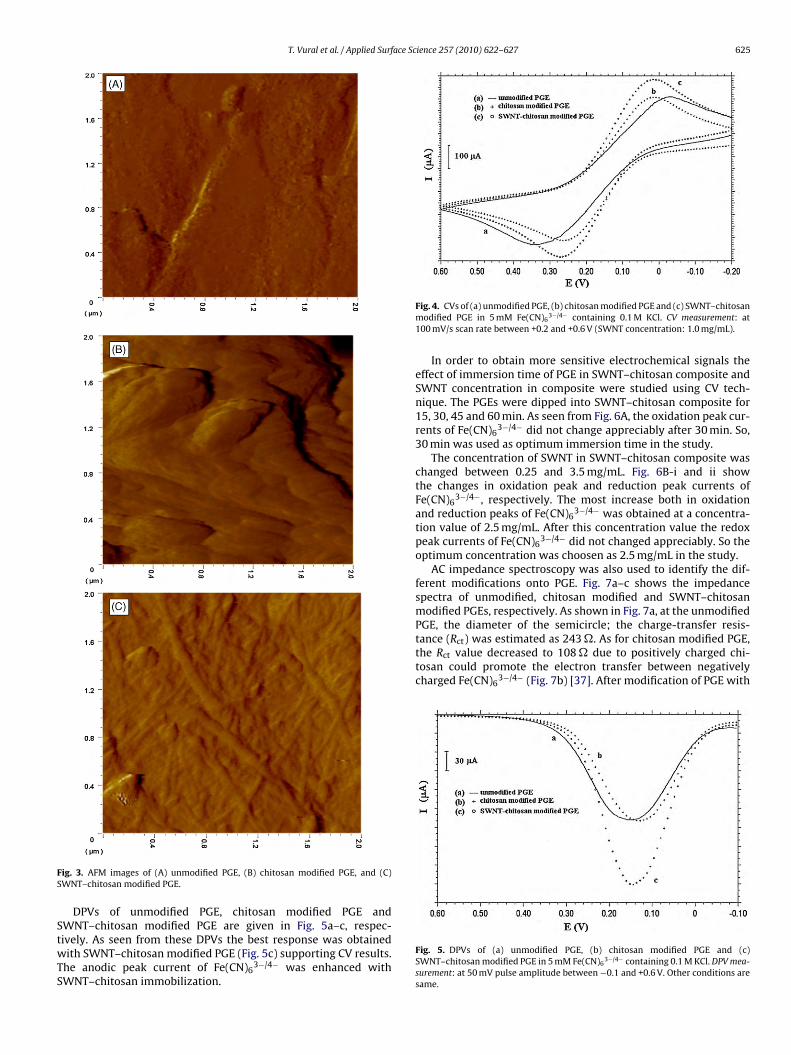
Fig. 3 shows the AFM amplitute images of unmodified PGE, chi-osan modified PGE, and SWNT–chitosan modified PGE surfaces.he surface roughness of unmodified PGE is clearly seen in Fig. 3A.fter chitosan modification onto the PGE, the morphology of theurface changed (Fig. 3B). From this AFM image, it is clear that theGE surface was covered with chitosan film. After SWNT–chitosanodification onto the PGE, homogeneous distribution of carbon
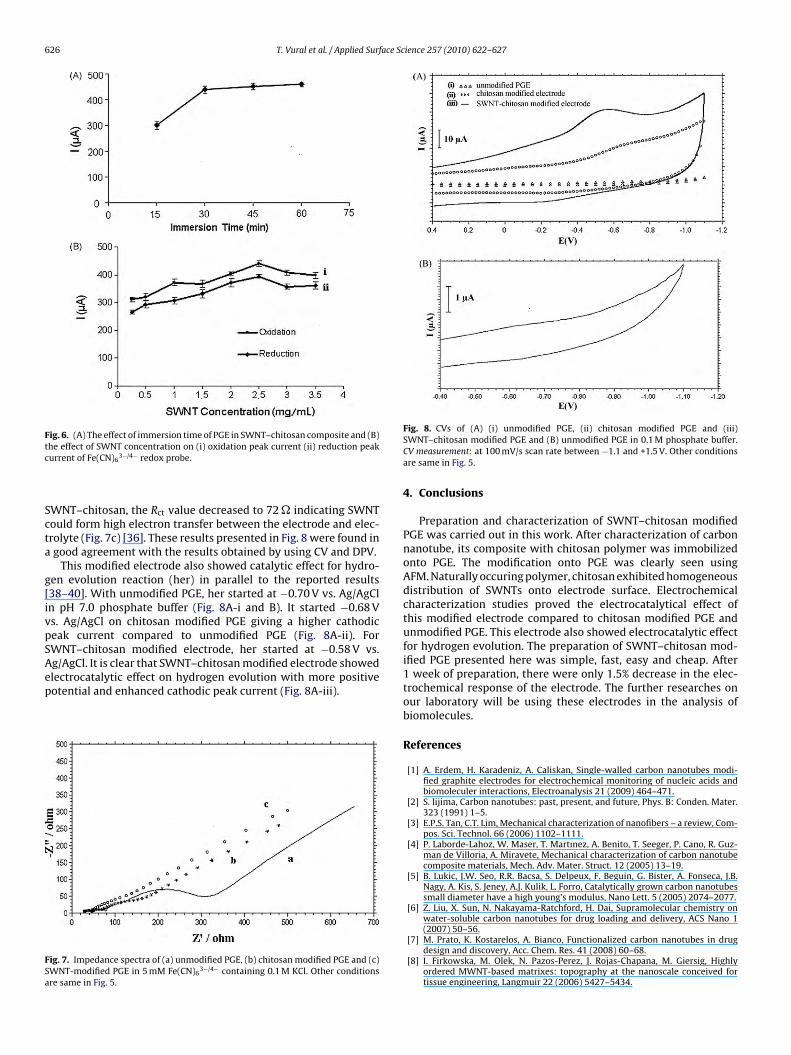
anotubes was obtained on chitosan film (Fig. 3C).The cyclic voltammogram of the Fe(CN)6
3−/4− system is widelysed method for monitoring the characteristics of the surface ofodified electrodes [35]. Fig. 4a demonstrates the CV of unmodi-
ed PGE in 5 mM Fe(CN)63−/4− redox probe containing 0.1 M KCl.
s seen from this figure, the anodic peak potential of Fe(CN)63−/4−
as obtained at +0.34 V and cathodic peak potential of Fe(CN)63−/4−
as obtained at −0.04 V vs. Ag/AgCl. After modification of PGE withhitosan (Fig. 4b), there was no appreciable change at the anodicnd cathodic peak currents of Fe(CN)6
3−/4−. However, there was
Fig. 2. SEM images and EDX analysis of (A
and (b) functionalized SWNT.
a significant change at the redox peak potentials of Fe(CN)63−/4−
compared to the redox potentials obtained in the case of unmod-ified PGE (Fig. 4a). After modification of PGE with SWNT–chitosanmixture (Fig. 4c), the redox peak currents of Fe(CN)6
3−/4− increased,indicating SWNT improved the elecroactive surface area of theelectrode and catalytic ability due to SWNT [36]. The reproducibil-ities of the electrochemical responses of the unmodified pencilgraphite electrode, chitosan modified pencil graphite electrodeand SWNT–chitosan modified pencil graphite electrode were illus-trated by three continuous experimental cycles using oxidationpeak currents. The results showed that the unmodified pencilgraphite electrode had a good reproducibility with a relative stan-
dard deviation of 2.14% (n = 3), chitosan modified pencil graphiteelectrode had a good reproducibility with a relative standard devi-ation of 1.76% (n = 3) and SWNT–chitosan modified pencil graphiteelectrode showed a good reproducibility with a standard deviationof 1.12% (n = 3).) raw SWNT and (B) purified SWNT.
T. Vural et al. / Applied Surface Science 257 (2010) 622–627 625
FS
StwTS
tance (Rct) was estimated as 243 �. As for chitosan modified PGE,the Rct value decreased to 108 � due to positively charged chi-tosan could promote the electron transfer between negativelycharged Fe(CN)6
3−/4− (Fig. 7b) [37]. After modification of PGE with
ig. 3. AFM images of (A) unmodified PGE, (B) chitosan modified PGE, and (C)WNT–chitosan modified PGE.
DPVs of unmodified PGE, chitosan modified PGE and
WNT–chitosan modified PGE are given in Fig. 5a–c, respec-ively. As seen from these DPVs the best response was obtainedith SWNT–chitosan modified PGE (Fig. 5c) supporting CV results.he anodic peak current of Fe(CN)63−/4− was enhanced with
WNT–chitosan immobilization.
Fig. 4. CVs of (a) unmodified PGE, (b) chitosan modified PGE and (c) SWNT–chitosanmodified PGE in 5 mM Fe(CN)6
3−/4− containing 0.1 M KCl. CV measurement: at100 mV/s scan rate between +0.2 and +0.6 V (SWNT concentration: 1.0 mg/mL).
In order to obtain more sensitive electrochemical signals theeffect of immersion time of PGE in SWNT–chitosan composite andSWNT concentration in composite were studied using CV tech-nique. The PGEs were dipped into SWNT–chitosan composite for15, 30, 45 and 60 min. As seen from Fig. 6A, the oxidation peak cur-rents of Fe(CN)6
3−/4− did not change appreciably after 30 min. So,30 min was used as optimum immersion time in the study.
The concentration of SWNT in SWNT–chitosan composite waschanged between 0.25 and 3.5 mg/mL. Fig. 6B-i and ii showthe changes in oxidation peak and reduction peak currents ofFe(CN)6
3−/4−, respectively. The most increase both in oxidationand reduction peaks of Fe(CN)6
3−/4− was obtained at a concentra-tion value of 2.5 mg/mL. After this concentration value the redoxpeak currents of Fe(CN)6
3−/4− did not changed appreciably. So theoptimum concentration was choosen as 2.5 mg/mL in the study.
AC impedance spectroscopy was also used to identify the dif-ferent modifications onto PGE. Fig. 7a–c shows the impedancespectra of unmodified, chitosan modified and SWNT–chitosanmodified PGEs, respectively. As shown in Fig. 7a, at the unmodifiedPGE, the diameter of the semicircle; the charge-transfer resis-
Fig. 5. DPVs of (a) unmodified PGE, (b) chitosan modified PGE and (c)SWNT–chitosan modified PGE in 5 mM Fe(CN)6
3−/4− containing 0.1 M KCl. DPV mea-surement: at 50 mV pulse amplitude between −0.1 and +0.6 V. Other conditions aresame.
626 T. Vural et al. / Applied Surface Science 257 (2010) 622–627
Fig. 6. (A) The effect of immersion time of PGE in SWNT–chitosan composite and (B)tc
Scta
g[ivpSAep
FSa
he effect of SWNT concentration on (i) oxidation peak current (ii) reduction peakurrent of Fe(CN)6
3−/4− redox probe.
WNT–chitosan, the Rct value decreased to 72 � indicating SWNTould form high electron transfer between the electrode and elec-rolyte (Fig. 7c) [36]. These results presented in Fig. 8 were found ingood agreement with the results obtained by using CV and DPV.
This modified electrode also showed catalytic effect for hydro-en evolution reaction (her) in parallel to the reported results38–40]. With unmodified PGE, her started at −0.70 V vs. Ag/AgCln pH 7.0 phosphate buffer (Fig. 8A-i and B). It started −0.68 Vs. Ag/AgCl on chitosan modified PGE giving a higher cathodiceak current compared to unmodified PGE (Fig. 8A-ii). ForWNT–chitosan modified electrode, her started at −0.58 V vs.g/AgCl. It is clear that SWNT–chitosan modified electrode showedlectrocatalytic effect on hydrogen evolution with more positive
otential and enhanced cathodic peak current (Fig. 8A-iii).ig. 7. Impedance spectra of (a) unmodified PGE, (b) chitosan modified PGE and (c)WNT-modified PGE in 5 mM Fe(CN)6
3−/4− containing 0.1 M KCl. Other conditionsre same in Fig. 5.
Fig. 8. CVs of (A) (i) unmodified PGE, (ii) chitosan modified PGE and (iii)SWNT–chitosan modified PGE and (B) unmodified PGE in 0.1 M phosphate buffer.CV measurement: at 100 mV/s scan rate between −1.1 and +1.5 V. Other conditionsare same in Fig. 5.
4. Conclusions
Preparation and characterization of SWNT–chitosan modifiedPGE was carried out in this work. After characterization of carbonnanotube, its composite with chitosan polymer was immobilizedonto PGE. The modification onto PGE was clearly seen usingAFM. Naturally occuring polymer, chitosan exhibited homogeneousdistribution of SWNTs onto electrode surface. Electrochemicalcharacterization studies proved the electrocatalytical effect ofthis modified electrode compared to chitosan modified PGE andunmodified PGE. This electrode also showed electrocatalytic effectfor hydrogen evolution. The preparation of SWNT–chitosan mod-ified PGE presented here was simple, fast, easy and cheap. After1 week of preparation, there were only 1.5% decrease in the elec-trochemical response of the electrode. The further researches onour laboratory will be using these electrodes in the analysis ofbiomolecules.
References
[1] A. Erdem, H. Karadeniz, A. Caliskan, Single-walled carbon nanotubes modi-fied graphite electrodes for electrochemical monitoring of nucleic acids andbiomoleculer interactions, Electroanalysis 21 (2009) 464–471.
[2] S. lijima, Carbon nanotubes: past, present, and future, Phys. B: Conden. Mater.323 (1991) 1–5.
[3] E.P.S. Tan, C.T. Lim, Mechanical characterization of nanofibers – a review, Com-pos. Sci. Technol. 66 (2006) 1102–1111.
[4] P. Laborde-Lahoz, W. Maser, T. Martınez, A. Benito, T. Seeger, P. Cano, R. Guz-man de Villoria, A. Miravete, Mechanical characterization of carbon nanotubecomposite materials, Mech. Adv. Mater. Struct. 12 (2005) 13–19.
[5] B. Lukic, J.W. Seo, R.R. Bacsa, S. Delpeux, F. Beguin, G. Bister, A. Fonseca, J.B.Nagy, A. Kis, S. Jeney, A.J. Kulik, L. Forro, Catalytically grown carbon nanotubessmall diameter have a high young’s modulus, Nano Lett. 5 (2005) 2074–2077.
[6] Z. Liu, X. Sun, N. Nakayama-Ratchford, H. Dai, Supramolecular chemistry onwater-soluble carbon nanotubes for drug loading and delivery, ACS Nano 1
(2007) 50–56.[7] M. Prato, K. Kostarelos, A. Bianco, Functionalized carbon nanotubes in drugdesign and discovery, Acc. Chem. Res. 41 (2008) 60–68.
[8] I. Firkowska, M. Olek, N. Pazos-Perez, J. Rojas-Chapana, M. Giersig, Highlyordered MWNT-based matrixes: topography at the nanoscale conceived fortissue engineering, Langmuir 22 (2006) 5427–5434.
ace Sc
[
[
[
[
[
[
[
[
[
[
[
[
[
[
[
[
[
[
[
[
[
[
[
[
[
[
[
[
[
T. Vural et al. / Applied Surf
[9] J.L. McKenzie, M.C. Waid, R. Shi, T.J. Webster, Decreased functions ofastrocytes on carbon nanofiber materials, Biomaterials 25 (2004) 1309–1317.
10] H. Hu, Y. Ni, S.K. Mandal, V. Montana, B. Zhao, R.C. Haddon, V. Parpura,Polyethyleneimine functionalized single-walled carbon nanotubes as a sub-strate for neuronal growth, J. Phys. Chem. B 109 (2005) 4285–4289.
11] L. Agüi, P. Yanez-Sedeno, J.M. Pingarron, Role of carbon nanotubes in electro-analytical chemistry, Anal. Chim. Acta 622 (2008) 11–47.
12] S. Carrara, V.V. Shumyantsevab, A.I. Archakovb, B. Samori, Screen-printed elec-trodes based on carbon nanotubes and cytochrome P450scc for highly sensitivecholesterol biosensors, Biosens. Bioelectron. 24 (2008) 148–150.
13] G.A. Rivas, M.D. Rubianes, M.C. Rodriguez, N.C. Ferreyya, G.L. Luque, M.L.Pedano, S.A. Miscoria, C. Parrado, Carbon nanotubes for electrochemical sens-ing, Talanta 74 (2007) 291–307.
14] S.P. Zhang, Y. Zheng, L.G. Shan, L.Y. Shi, K.L. Leng, Complex and new modificationtechniques of thiocholine detection electrodes with carbon nanotubes, Appl.Surf. Sci. 255 (2008) 439–441.
15] A. Erdem, P. Papakonstantinou, H. Murphy, Direct DNA hybridization at dis-posable graphite electrodes modified with carbon nanotubes, Anal. Chem. 78(2006) 6656–6659.
16] A. Merkoci, Carbon nanotubes in analytical sciences, Microchim. Acta 152(2006) 157–174.
17] S. Timur, U. Anik, D. Odaci, L. Gorton, Development of a microbial biosensorbased on carbon nanotube (CNT) modified electrodes, Electron. Commun. 9(2007) 1810–1815.
18] H. Kitano, K. Tachimoto, Y. Anraku, Functionalization of single-walled carbonnanotube by the covalent modification with polymer chains, J. Colloid InterfaceSci. 306 (2007) 28–33.
19] X. Xie, Y. Mai, X. Zhou, Dispersion and alignment of carbon nanotubes in poly-mer matrix, Mater. Sci. Eng. R 49 (2005) 89–112.
20] Y. Lin, S. Taylor, H.L.K.A.S. Fernando, L. Qu, W. Wang, L. Gu, B. Zhou, Y.P. Sun,Advances toward bioapplications of carbon nanotubes, J. Mater. Chem. 14(2004) 527–541.
21] J. Tkac, J.W. Whittaker, T. Ruzgas, The use of single walled carbon nanotubesdispersed in a chitosan matrix for preparation of a galactose biosensor, Biosens.Biolectron. 22 (2007) 1820–1824.
22] Y. Liu, J. Tang, X. Chen, J.H. Xin, Decoration of carbon nanotubes with chitosan,Carbon 43 (2005) 3178–3180.
23] R. Pauliukaite, M.E. Ghica, O. Fatibello-Filho, C.M.A. Brett, Comparative study ofdifferent cross-linking agents for the immobilization of functionalized carbonnanotubes within a chitosan film supported on a graphite–epoxy compositeelectrode, Anal. Chem. 81 (2009) 5364–5372.
24] J. Li, Q. Liu, Y. Liu, S. Liu, S. Yao, DNA biosensor based on chitosan film dopedwith carbon nanotubes, Anal. Biochem. 346 (2005) 107–114.
[
[
ience 257 (2010) 622–627 627
25] P. Arias, N.F. Ferreyra, G.A. Rivas, S. Bollo, Glassy carbon electrodes modifiedwith CNT-dispersed in chitosan: analytical applications for sensing DNA-methylene blue interaction, J. Electroanal. Chem. 634 (2009) 123–126.
26] M. Zhang, A. Smith, W. Gorski, Carbon nanotube–chitosan system for electro-chemical sensing based on dehydrogenase enzymes, Anal. Chem. 76 (2004)5045–5050.
27] Y. Liu, M. Wang, F. Zhao, Z. Xu, S. Dong, The direct electron transfer of glucoseoxidase and glucose biosensor based on carbon nanotubes/chitosan matrix,Biosens. Bioelectron. 21 (2005) 984–988.
28] Y. Zhou, H. Yng, H.Y. Chen, Direct electrochemistry and reagentless biosens-ing of glucose oxidase immobilized on chitosan wrapped single-walled carbonnanotubes, Talanta 76 (2008) 419–423.
29] M.E. Ghica, R. Pauliukaite, O. Fatibello-Filho, C.M.A. Brett, Application of func-tionalized carbon nanotubes immobilized into chitosan films in amperometricenzyme biosensors, Sens. Actuators B: Chem. 142 (2009) 308–315.
30] L. Qian, X. Yang, Composite film of carbon nanotubes and chitosan for prepa-ration of amperometric hudrogen peroxide biosensor, Talanta 68 (2006)721–727.
31] Y. Yang, G. Yang, Y. Huang, H. Bai, X. Lu, A new hydrogen peroxidebiosensor based on gold nanoelectrode ensembles/multiwalled carbon nan-otubes/chitosan film-modified electrode, Coll. Surf. A: Physicochem. Eng.Aspects 340 (2009) 50–55.
32] H. Hu, B. Zhao, M.E. Itkis, R.C. Haddon, Nitric acid purification of single-walledcarbon nanotubes, J. Phys. Chem. B 107 (2003) 13838–13842.
33] J.M. Moon, K.H. An, Y.H. Lee, Y.S. Park, D.J. Bae, G.S. Park, High yield purifica-tion process of singlewalled carbon nanotubes, J. Phys. Chem. B 105 (2001)5677–5681.
34] J. Ma, J.N. Wang, Purification of single-walled carbon nanotubes by a higlyefficient and nondestructive approach, Chem. Mater. 20 (2008) 2895–2902.
35] P. Tomcik, C.E. Banks, T.J. Davies, R.G. Compton, A self-catalytic carbon pasteelectrode for the detection of vitamin B12, Anal. Chem. 76 (2004) 161–165.
36] C. Xiang, Y. Zou, L.X. Sun, F. Xu, Direct electrochemistry and electrocatalysis ofcytochrome c immobilized on gold nanoparticles–chitosan–carbon nanotubesmodified electrode, Talanta 74 (2007) 206–211.
37] M. Li, S. Huang, P. Zhu, L. Kong, B. Peng, H. Gao, A novel DNA biosensor based onssDNA/Cyt c/l-Cys/GNOs/Chits/GCE, Electrochim. Acta 54 (2009) 2284–2289.
38] A. Koca, Hydrogen evolution reaction on glassy carbon electrode modified withtitanyl phthalocyanines, Int. J. Hydrogen Energy 34 (2009) 2107–2112.
39] P.P. Prosini, A. Pozio, S. Botti, R. Ciardi, Electrochemical studies of hydrogen evo-lution, storage and oxidation on carbon nanotube electrodes, J. Power Sources118 (2003) 265–269.
40] P.S. Fernandez, E.B. Catro, S.G. Real, M.E. Martins, Electrochemical behaviourof single walled carbon nanotubes-hydrogen storage and hydrogen evolutionreaction, Int. J. Hydrogen Energy 34 (2009) 8115–8126.
Copyright © 2022 FDOKUMEN