Predominance of spliceosomal complex formation over polyadenylation site selection in TDP-43...
10
Predominance of spliceosomal complex formation over polyadenylation site selection in TDP-43 autoregulation Sara Bembich, Jeremias S. Herzog, Laura De Conti, Cristiana Stuani, S. Ere ´ ndira Avendan ˜ o-Va ´ zquez, Emanuele Buratti, Marco Baralle and Francisco E. Baralle* International Centre for Genetic Engineering and Biotechnology (ICGEB), 34149 Trieste, Italy Received May 2, 2013; Revised November 27, 2013; Accepted December 3, 2013 ABSTRACT TDP-43 is a nuclear protein involved in many aspects of RNA metabolism. To ensure cellular via- bility, its expression levels within cells must be tightly regulated. We have previously demonstrated that TDP-43 autoregulation occurs through the acti- vation of a normally silent intron in its 3 0 -UTR sequence that results in the use of alternative polyadenylation sites. In this work, we analyse which is the dominant event in autoregulation: the recognition of the splice sites of 3 0 -UTR intron 7 or the intrinsic quality of the alternative poly- adenylation sites. A panel of minigene constructs was tested for autoregulation functionality, protein production and subcellular messenger RNA local- ization. Our data clearly indicate that constitutive spliceosome complex formation across intron 7 does not lead to high protein production but, on the contrary, to lower TDP-43 messenger RNA and protein levels. This is due to altered nucleo- cytoplasmic distribution of the RNA that is mostly retained in the nucleus and degraded. This study provides a novel in-depth characterization of how RNA binding proteins can autoregulate their own levels within cells, an essential regulatory process in maintaining cellular viability. INTRODUCTION Cell viability relies on the correct protein concentration levels within the various cellular compartments (1) and prevents the development of disease, especially at the neuronal level (2). There are several pathways used by the cell to achieve this, with protein expression regulation at the messenger RNA (mRNA) level being one of the most common due to its ability to act in an efficient and rapid manner. This type of regulation is often seen in genes encoding for RNA binding proteins due to the fact that many of these are able to bind their own RNA. Such an arrangement, in fact, allows cells to set up effect- ive negative feedback mechanism that will raise protein production rapidly when cellular levels drop below a critical threshold and inhibit protein production when cellular concentrations become too high. Several pathways where RNA binding proteins regulate their own expression through direct binding to their transcript have been described. These include proteins such as HuR (3), PTB (hnRNP I) (4), hnRNP L (5), hnRNP A/B (6), TIA-1/TIAR (7), SRSF3 (SRp20) (8), SRSF2 (SC-35) (9) and Tra2 (10). For recent reviews on the subject, the reader is referred to Buratti and Baralle (11) and to Yap and Makeyev (12). In the majority of these cases, the autoregulatory processes for these proteins are based on the selective triggering of a specific RNA degradation mechanism called nonsense-mediated decay (NMD) (13). Exceptions to this rule are represented by HuR (3) and possibly Tra2 (10) proteins where polyadenylation and translational mechanisms may be prevalent. Another notable exception to this NMD rule is represented by the mechanism described to occur for the nuclear factor TDP-43 (14,15). TDP-43 was initially identified as a transcriptional regulator (16) and subsequently as a regulator of Cystic fibrosis transmembrane conductance regulator (CFTR) exon 9 splicing (17). The importance of TDP-43 in the *To whom correspondence should be addressed. Tel: +39 040 3757337; Fax:+39 040 3757361; Email: [email protected] The authors wish it to be known that, in their opinion, the first two authors should be regarded as Joint First Authors. Present address: S. Ere´ndira Avendan˜o-Va´zquez, Department of Biochemistry and Molecular Pharmacology, University of Massachusetts Medical School, Worcester, MA 01605, USA. Nucleic Acids Research, 2013, 1–10 doi:10.1093/nar/gkt1343 ß The Author(s) 2013. Published by Oxford University Press. This is an Open Access article distributed under the terms of the Creative Commons Attribution License (http://creativecommons.org/licenses/by/3.0/), which permits unrestricted reuse, distribution, and reproduction in any medium, provided the original work is properly cited. Nucleic Acids Research Advance Access published December 24, 2013 by guest on June 1, 2016 http://nar.oxfordjournals.org/ Downloaded from
-
Upload
independent -
Category
Documents
-
view
3 -
download
0
Transcript of Predominance of spliceosomal complex formation over polyadenylation site selection in TDP-43...

Predominance of spliceosomal complex formationover polyadenylation site selection in TDP-43autoregulationSara Bembich Jeremias S Herzog Laura De Conti Cristiana Stuani
S Erendira Avendano-Vazquez Emanuele Buratti Marco Baralle and
Francisco E Baralle
International Centre for Genetic Engineering and Biotechnology (ICGEB) 34149 Trieste Italy
Received May 2 2013 Revised November 27 2013 Accepted December 3 2013
ABSTRACT
TDP-43 is a nuclear protein involved in manyaspects of RNA metabolism To ensure cellular via-bility its expression levels within cells must betightly regulated We have previously demonstratedthat TDP-43 autoregulation occurs through the acti-vation of a normally silent intron in its 30-UTRsequence that results in the use of alternativepolyadenylation sites In this work we analysewhich is the dominant event in autoregulationthe recognition of the splice sites of 30-UTR intron7 or the intrinsic quality of the alternative poly-adenylation sites A panel of minigene constructswas tested for autoregulation functionality proteinproduction and subcellular messenger RNA local-ization Our data clearly indicate that constitutivespliceosome complex formation across intron 7does not lead to high protein production buton the contrary to lower TDP-43 messenger RNAand protein levels This is due to altered nucleo-cytoplasmic distribution of the RNA that is mostlyretained in the nucleus and degraded This studyprovides a novel in-depth characterization of howRNA binding proteins can autoregulate their ownlevels within cells an essential regulatory processin maintaining cellular viability
INTRODUCTION
Cell viability relies on the correct protein concentrationlevels within the various cellular compartments (1) and
prevents the development of disease especially at theneuronal level (2) There are several pathways used bythe cell to achieve this with protein expression regulationat the messenger RNA (mRNA) level being one of themost common due to its ability to act in an efficient andrapid manner This type of regulation is often seen ingenes encoding for RNA binding proteins due to thefact that many of these are able to bind their own RNASuch an arrangement in fact allows cells to set up effect-ive negative feedback mechanism that will raise proteinproduction rapidly when cellular levels drop below acritical threshold and inhibit protein production whencellular concentrations become too high Severalpathways where RNA binding proteins regulate theirown expression through direct binding to their transcripthave been described These include proteins such as HuR(3) PTB (hnRNP I) (4) hnRNP L (5) hnRNP AB (6)TIA-1TIAR (7) SRSF3 (SRp20) (8) SRSF2 (SC-35) (9)and Tra2 (10) For recent reviews on the subject thereader is referred to Buratti and Baralle (11) and to Yapand Makeyev (12) In the majority of these cases theautoregulatory processes for these proteins are based onthe selective triggering of a specific RNA degradationmechanism called nonsense-mediated decay (NMD) (13)Exceptions to this rule are represented by HuR (3) andpossibly Tra2 (10) proteins where polyadenylation andtranslational mechanisms may be prevalent Anothernotable exception to this NMD rule is represented bythe mechanism described to occur for the nuclear factorTDP-43 (1415)TDP-43 was initially identified as a transcriptional
regulator (16) and subsequently as a regulator of Cysticfibrosis transmembrane conductance regulator (CFTR)exon 9 splicing (17) The importance of TDP-43 in the
To whom correspondence should be addressed Tel +39 040 3757337 Fax +39 040 3757361 Email baralleicgeborg
The authors wish it to be known that in their opinion the first two authors should be regarded as Joint First Authors
Present addressS Erendira Avendano-Vazquez Department of Biochemistry and Molecular Pharmacology University of Massachusetts Medical School WorcesterMA 01605 USA
Nucleic Acids Research 2013 1ndash10doi101093nargkt1343
The Author(s) 2013 Published by Oxford University PressThis is an Open Access article distributed under the terms of the Creative Commons Attribution License (httpcreativecommonsorglicensesby30) whichpermits unrestricted reuse distribution and reproduction in any medium provided the original work is properly cited
Nucleic Acids Research Advance Access published December 24 2013 by guest on June 1 2016
httpnaroxfordjournalsorgD
ownloaded from
neurodegeneration field was established in 2006 when itwas described as the major protein component of theintracellular inclusions occurring in the neuronal tissuesof patients affected by amyotrophic lateral sclerosisand frontotemporal dementia (1819) In the patientrsquosaffected neurons TDP-43 is abnormally mislocalized inthe cytoplasm ubiquitinated hyperphosphorylated andcleaved to generate C-terminal fragments (20)Currently one hypothesis is that such mislocalizationplays a pivotal role in neurodegeneration through theloss of proper TDP-43 functions in the nucleusalthough gain-of-function mechanisms may be active aswell (21ndash26)The autoregulatory process of TDP-43 is totally
dependent on a region called TDP-43 binding region(TDPBR) that contains several Cross-Linking andImmunoprecipitation (CLIP) sequences that act astargets for TDP-43 binding (27) This region is localizedin TDP-43 30-UTR and spans a normally silent intron 7that contains the pA1 site (Figure 1A) In steady stateconditions pA1 is the major polyadenylation site (PAS)used by the TDP-43 mRNA The pA4 site is also usedhowever to a much lower extentWe have previously shown that a 3-fold increase in
TDP-43 nuclear levels results in an increased occupancyof the TDPBR that in turn causes a rise in polymerase IIdensity in this sequence (15) Such a pausing effect inducesan increased processing of intron 7 an effect that has beenobserved to occur in many experimental systems (28)Removal of intron 7 and hence of pA1 enforces pA2 andpA4 selection and mRNAs using these PAS were shownto be partially retained in the nucleus (15) As a resultof all these events in the presence of high TDP-43 con-centrations a reduction of TDP-43 cellular mRNA levelsoccurs quickly The relative importance in this process ofTDP-43 binding to the TDPBR spliceosome assemblysplicing of intron 7 and pA1-pA2-pA4 polyadenylationis still unclear In this work our results show that intron7 processing andor spliceosome assembly induced byTDP-43 is the key event in the reduction of the amountof TDP-43 mRNA rather than the intrinsic quality of thevarious PASs
MATERIALS AND METHODS
Cell lines
The HEK-293 TDP-43 wild-type inducible cell line hasbeen previously described (14) Cells were cotransfectedusing the calcium phosphate method with 3 mg of eachGreen Fluorescent Protein (GFP) construct variants and05mg DiGFP generously donated by Dr Christian BeetzIt should also be noted that the reliability of our experi-mental conditions in the transfection assays to correctlymeasure GFP expression levels was determined bymaking a titration curve that show a proportionalincrease in the signal using increasing concentrations ofcellular extracts from our reference X7 construct(Supplementary Figure S1)
Plasmid construction 30 RACE and reversetranscriptase-polymerase chain reaction analysis
The X7 wild-type and TDPBR recombinant constructshave been previously described (15) The otherX7 constructvariants were made by polymerase chain reaction (PCR)amplification using quick change (Stratagene) accordingto manufacturersquos instructions (primer sequences are avail-able upon request) For 30RACE (Rapid amplification ofcDNA ends) analysis total RNA was reverse-transcribedusing an oligonucleotide (dT)20V anchor (50-ctagtctagatct-gaatatattcgttttttttttttttttttttv-30) and amplified with theanchor (50-cgaatatattcagatctagactag-30) and GFPfw (50-tctcggcatggacgagctgtacaag-30) Cycloheximide (CHX)treatment was performed by adding 50 mgml of this antibi-otic for 3 h before mRNA extraction
Immunoblotting
Cells extracts were prepared in 15mM HEPES pH 75025M NaCl 05 NP-40 10 glycerol 1 proteaseinhibitor (Roche 1873580) 25mM NaF 10mM b-glycerolphosphate 02mM Na3VO4 and 1mM phenyl-methylsulphonyl fluoride Proteins were separated bysodium dodecyl sulphatendashpolyacrylamide gel electrophor-esis and transferred to nitrocellulose (045mM AmershamBiosciences) and protein detection was carried out withstandard western blotting techniques using an antibody todetect GFP (Santa Cruz Biotechnology sc-8334)
Northern blotting
For northern blotting RNA was isolated withEuroGoldtriFast (Euroclone) following manufacturerrsquosinstructions RNA samples were loaded on 12 formal-dehyde agarose gels transferred toHybond-N+nylonmem-branes (Amersham Biosciences) and probed with internally32P-labelled sequences following prehybridization inULTRAhyb Ultrasensitive Hybridization Buffer(Ambion) Prehybridization and hybridization werecarried out at 65C The probes were generated by PCRusing primers GFP813FW (50-cggcgtgcagtgcttcagccgctac-30) and GFPend RV (50-cttgtacagctcgtccatgccgagag-30)and 32P-labelled with Rediprime II DNA LabelingSystem (GE Healthcare) Visualization of transcripts wascarried out with a Cyclone Plus Storage Phosphor Scannerthat included OptiQuant Software (Perkin Elmer)
RNA in situ hybridization
HEK-293 FlpIn cells were fixed for 30min at roomtemperature in 2 Paraformaldehyde (PFA) 24 h post-transfection After three washes in phosphate-bufferedsaline cells were permeabilized by treatment with 01triton in phosphate-buffered saline for 5min andhybridized overnight at 37C in 20 ml of a mixture contain-ing 10dextran sulphate 10 mg yeast tRNA 1 SSC 20formamide and 20 ng of digoxigenin-11-dUTP (Cat No11573152910 Roche Applied Science) labelled PCRproduct probe against GFP coding sequence Cells werethen washed three times in 2 SSC and once in 4 SSCand incubated for 1 h at room temperature with 1200 anti-digoxigenin-rhodamine Fab fragments (Cat No 1120775
2 Nucleic Acids Research 2013
by guest on June 1 2016httpnaroxfordjournalsorg
Dow
nloaded from
0910 Roche Applied Science) in 4 SSC Following threewashes in 4 SSC cells were mounted and the images werecaptured on a Zeiss LSM 510 META confocal microscope(Carl Zeiss Microimaging Inc) with a 63NA 14 Plan-Apochromat oil objective Cells nuclei were visualizedusing TO-PRO-3 (Invitrogen T3605)
RESULTS
Evaluating the effects of PAS shuffling on TDP-43autoregulation
Figure 1A shows a schematic representation of theTARDBP gene illustrating the locations of the stopcodon (tag) the various PASs (pA1ndash4) and the TDPBR
region that regulates intron 7 splicing within the non-coding 30-UTR This region represents an essentialelement for the negative feedback loop that allows TDP-43 to control its own expression levels within cells (1415)We have previously shown that a minimal transcript con-sisting in a portion of the TDP-43 30-UTR region fused tothe GFP reporter protein displays similar autoregulatoryproperties to those observed for endogenous TARDBPgene (Figure 1B X7 construct) (1415) In fact productionof the GFP reporter protein from this transcript followingtransfection in HEK-293 cells that carry an integratedtetracycline (Tet)-inducible TDP-43 complementaryDNA (cDNA) is 4-fold decreased on overexpression ofTDP-43 (+Tet conditions) as determined by western blotanalysis (Figure 1C GFP reporter lanes 1ndash2) and
Figure 1 Cis acting elements and importance of PAS sequences in TDP-43 autoregulation (A) shows a schematic diagram of TDP-43 illustratinglocations of stop codon (tag) PASs (pA1ndash4) TDPBR region and splicing events (in coding sequences by filled lines in the 30-UTR region by dottedlines) Coding regions (black boxes) untranslated sequences (grey boxes) and introns (connecting black lines) are indicated A schematic represen-tation of each reporter used in this experiment is shown in (B) (C) shows the ability to autoregulate of these various TDP-43 30-UTR constructsfused to the GFP protein and transfected in HEK-293 cells stably expressing a TDP-43 transgene following tetracycline induction (+Tet lanes)Western blots of GFP reporter protein in normal (Tet) and TDP-43 overexpression conditions (+Tet) are shown in the centre With regards to thetechnical aspects of these experiments it should be noted that in all these cases autoregulation efficiency of the GFP reporters was normalized bycotransfection with a plasmid expressing DiGFP (C DiGFP upper panel) and further confirmed by western blot against tubulin (C lower panels)Normalization using tubulin levels in fact yielded similar results to those obtained using diGFP (data not shown) In parallel correct overexpressionof the TDP-43 transgene (Flag-TDP-43) and downregulation of the endogenous TDP-43 (TDP-43 end) was also assessed (C lower panels) Finally(D) shows the quantification of three independent experiments to quantify GFP protein expression levels in Tet and+Tet conditions normalizedaccording to DiGFP expression Mean values are reported on the bar chart and error bars indicate standarddeviation from at least three inde-pendent experiments
Nucleic Acids Research 2013 3
by guest on June 1 2016httpnaroxfordjournalsorg
Dow
nloaded from
densitometric quantification (Figure 1D) Using thisminimal transcript we have previously reported thecentral role played by the TDPBR sequence and by thesplice sites of intron 7 This was achieved by engineeringmutants where the TDPBR was deleted (Figure 1Bmutants X7-TDPBR) and another construct where thesplice sites were disrupted (Figure 1B X7-gt-ag respect-ively) (15) As shown in Figure 1C no GFP downregula-tion could be observed following tetracycline induction ofTDP-43 expression for neither of these mutants(Figure 1C lanes 3ndash6 and Figure 1D)A question that remained open from these experiments
was the eventual role played by pA4 in TDP-43 auto-regulation process Therefore to assess its eventualimportance we engineered a mutant GFP reporter(Supplementary Figure S2A X7-inpA2) in which theentire TDP-43 30-UTR sequence was deleted from 33 ntbeyond pA2 till the pA4 and compared its protein expres-sion levels with those of X7 both in Tet and+Tet condi-tions This experiment showed that in +Tet conditionsthe X7-inpA2 could efficiently autoregulate its own levelssimilarly to the wild-type X7 construct (SupplementaryFigure S2B) even in the absence of pA4 indicating thatthis PAS does not contribute significantly to the TDP-43autoregulatory process Further work is required to under-stand the biological function of the 30-UTR beyond pA2What remained unclear from our previous results
however was an assessment of the relative importance ofPAS switch from pA1 to pA2 compared with the actualevent of intron 7 processing To address the relative im-portance of pA1 and pA2 in the autoregulatory process weengineered two mutants one in which the pA2 wasremoved (X7-pA2) and another in which pA2 wasreplaced with a second pA1 site (X7-2pA1) (Figure 1B)Following transfection of these mutants in HEK-293 cells(Tet lanes) and induction of the transgene (+Tet) it wasobserved that both constructs displayed efficientautoregulation (Figure 1C lanes 7ndash8 and 9ndash10 respect-ively and 1D) These results indicate that the use of pA2
is not intrinsically required to achieve TDP-43autoregulation and that it is not inherently defective butcapable of sustaining efficient protein translation Tofurther corroborate this we engineered a mutant X7 con-struct X7-in7cDNA (Figure 1B) in which intron 7 wasremoved artificially and the pA2 sequence was kept as theonly available PAS (artificially mimicking the transcriptthat would naturally originate following intron 7 removalby the spliceosome) Following transfection in HEK-293cells of this mutant it was observed that X7-in7cDNAproduced TDP-43 at an analogous level than X7 in Tetconditions (Figure 1C lane 11 and Figure 1D) Moreoverin contrast X7 it was no longer capable of autoregulationfollowing transgene induction (Figure 1C and D)
The 30RACE analysis of the PAS-modified constructs
To gain better insight regarding PAS usage in these con-structs 30 RACE analysis with primers specific for theGFP reporter sequence (GFPfw) and a polyT anchor asreverse primer was performed (Figure 2A) As previouslyreported for the X7 plasmid (15) in normal conditions a
dominant transcript was obtained (band 1 Tet lane) thatcontained the TDP-43 30-UTR sequence ending at pA1
(Figure 2B) On induction of the transgene the X7 band1 diminished and a new product appeared (band 2) thatwas observed to derive from intron 7 removal and the useof pA2 (Figure 2B+Tet lane)
A decrease of band 1 was also obtained for the othertwo constructs X7-pA2 and X7-2pA1 that displayedautoregulation at the protein level following transgeneinduction In fact Figure 2CndashD shows that in both casesthe transgene induction caused a drop in the mRNA thatcontain the unspliced intron 7 and resulted in the appear-ance of novel mRNA (compare Tet and +Tet lanes)Sequencing of these bands revealed that these spliced tran-scripts used alternative PAS In the case of X7-pA2 thedecrease in band 1 is associated with the use of a crypticPAS within the plasmid after unusual splicing eventswithin the 30-UTR (band 3 Figure 2C) and oneoriginating from a cryptic splicing event within intron 7(band 4 Figure 2C) X7-2pA1 on the other hand uses thesecond pA1 after the intron 7 30ss that is uniquely presentin this mutant (bands five and six Figure 2D) In fact theX7-2pA1 construct has the option of using also the secondpA1 (placed at the site of pA2) in Tet conditions whenthere is no intron 7 splicing As a substantial amount oftranscript made use of this second pA1 sequence it wouldappear that the pA1 is more efficient than pA2 with run-through transcripts that do not use the first pA1 usingthe second pA1 as well possibly also detrimental to pA4
use (that cannot be detected in this 30RACE analysisbecause it is gt3 kb away and a fragment this long doesnot amplify as efficiently as the shorter ones)
The 30 RACE of the X7-in7cDNA showed that thepA2 contained in this construct was used in all themRNAs transcribed both under Tet and +Tet condi-tions and that no further processing of this transcriptcould be observed (Figure 2E) This situation is similarto that previously described in the X7-gt-ag mutantthat does not autoregulate even if in the case the pA1 isused (15) (Figure 2F) In this mutant intron 7 splicingis completely inhibited following the inactivation ofits donor and acceptor splice sites As a result the onlytranscript observed for this construct uses pA1 in a con-stitutive manner both in Tet and+Tet conditions (band1 Figure 2F) A similar situation is also observed for theX7-TDPBR construct in which deletion of the TDPBRregion leads to loss of autoregulation (Figure 2G)
These constructs allow us to conclude that auto-regulation is not strictly linked to the quality of the PASsFurthermore it is interesting to note that autoregulationonly takes place when splicing is occuring suggesting thatthe key feature that allows TDP-43 autoregulation is repre-sented by spliceosome assembly on intron 7 splice sites
Comparing the mRNA and protein expression levels of theX7-in7cDNA and X7 constructs
One of the most interesting findings of the mutantsanalysed in Figure 1 was the relatively equal protein pro-duction stemming from differential pA1 and pA2 usage bythe X7 and X7-in7cDNA (compare Figure 1B lanes 1
4 Nucleic Acids Research 2013
by guest on June 1 2016httpnaroxfordjournalsorg
Dow
nloaded from
and 11 and the Tet lanes in Figure 2B and E) In Tetconditions in fact the X7 construct uses almost exclu-sively pA1 as opposed to X7-in7cDNA that uses exclu-sively the pA2 site This allowed us to conclude that theuse of pA2 is not the main factor responsible for theautoregulation process
To analyse further eventual functional differences inmRNAs carrying these two different PASs exists wethen decided to compare the level of mRNA in cellswithout transgene induction for the constructs X7 andX7-in7cDNA
The comparative RNA expression levels from thesetwo constructs were analysed by northern blot followingtransfection of the two constructs in HEK-293 cells eitherseparately or together (Figure 3A) Quantification of thevarious band intensities detected only a slightly lowermRNA level of the X7-in7cDNA construct withrespect to the X7 constructs (Figure 3B) However thisdecrease failed to reach statistical significance consistentwith protein expression levels (Figure 1 lanes 1 and 11)
Therefore taken together these results suggest that theuse of pA1 instead of pA2 does not result in any significantdifference with regards to mRNA and protein expressionlevels and that the two PASs are perfectly comparable interms of protein production efficiency
Improving intron 7 splice site strength to induce itsconstitutive splicing
The central importance of intron 7 in the autoregulationprocess of TDP-43 was previously shown in a X7 gt-agmutant in which loss of autoregulation following expres-sion of the transgene was achieved by simply deleting bothsplice sites from the wild-type X7 construct (15) andFigure 1 lanes 5 and 6To complement the X7 gt-ag mutant result and further
extend our insight into the role of splicing of this intron inautoregulation it was then decided to mutate both thesesequences to improve the strength of the splice sites In thisrespect it should be noted that in normal conditions intron7 splicing is not observed This is not surprising as its 50
and 30 splice sites are not of optimal strength according toMaxentscan weight matrix model (29) and NNSplice (30)(Figure 4A upper schematic diagram) A considerable im-provement was then achieved in mutant X7-sup50-30
(Figure 4A lower schematic diagram) The purpose ofthese mutations was to make the splicing of intron 7 asconstitutive as possible without the need for any externalenhancer factor (like high levels of TDP-43)As shown in the 30RACE analysis of Figure 4B this
objective was successfully achieved and transfection
Figure 2 30 RACE analyses (A) shows a schematic diagram of the X7 reporter construct with an indication of the primers used for 30 RACEanalysis (GFPfw and anchor primers) (BndashG) show the results of the 30 RACE analyses from the various hybrid constructs under normal (Tet) andfollowing TDP-43 transgene overexpression (+Tet) The amplified bands were all subjected to sequencing and the schematic diagrams on the rightshow which PAS become activated following transgene induction
Nucleic Acids Research 2013 5
by guest on June 1 2016httpnaroxfordjournalsorg
Dow
nloaded from
in Tet conditions immediately led to the appearance of atotally processed transcript in which pA2 was used as theonly PAS available As expected this situation was un-changed following transgene induction (Figure 4B +Tetlane) In addition the transcripts originated fromX7-sup50-30 are not subjected to any change followingCHX treatment (Figure 4B CHX Tet and+Tet lanes)In terms of the qualitative nature of the transcript
species being generated the mRNA is the same as theone produced by the X7-in7cDNA transcript (compareFigure 4B with Figure 2E) with the difference that themRNA of X7-sup50-30 is generated by transcriptionfollowed by splicing whereas the mRNA of X7-in7cDNA only by transcription However quantita-tively the results were strikingly different as in northernblot analysis the amount of pA2-using transcript from
X7-sup50-30 was significantly lower when compared withX7 (Figure 4CndashE)
Taking in consideration the reduction in the amount oftotal RNA for the X7-sup50-30 construct (Figure 4CndashE) itwas then interesting to measure whether these differencescould result in a reduction of GFP protein expression Inkeeping with this hypothesis the western blot resultreported in Figure 4D showed that the amount of GFPprotein produced by the X7-sup50-30 construct was muchlower with respect to the X7 wild-type construct both inTet and+Tet conditions Quantification of this drop inexpression confirmed that X7-sup50-30 GFP protein levelswere 25 of normal X7 GFP production levels(Figure 4F) a significant reduction compared with theestimated change in mRNA levels suggesting a differencein translational ability andor in nuclear export of the pA2
mRNA
Altered nucleocytoplasmic distribution of the X7-sup50-30
transcript
One possibility to explain the differences mentioned earlierin the text may lie on the availability of the mRNAs inthe cytoplasm RNA fluorescence in situ hybridizationanalysis of the various transcripts originating from theX7 X7-in7cDNA and X7-sup50-30 constructs was thenperformed This analysis is not suited to detect quantita-tive differences in expression but rather differential distri-butions of RNAs in the cell body As shown in Figure 5Aby the Anti-digox-rhod signal in red the transcripts fromX7 are predominantly localized in the cytoplasm with aconsequently high efficiency of GFP production A similarsituation is also observed for the x7-in7cDNA construct(Figure 5C) further highlighting our observation that useof pA1 (X7) or pA2 (X7-in7cDNA) does not make anydifference to the resulting protein production process
On the other hand the RNA transcribed from the X7-sup50-30 shows a very different nucleuscytoplasm ratiofrom the one observed in both X7 and X7-in7cDNAnotwithstanding the latter being identical to the transcriptproduced by X7-sup50-30 (Figure 5B) This data in factclearly shows that instead of being mainly localized in thecytoplasm the RNA produced by X7-sup50-30 is uniformlydistributed between the nucleus and cytoplasm This resultsuggests that the 30-UTR intron 7 recognition by thesplicing machinery somehow marks the bulk of the tran-script for nuclear retention and degradation resultingin lower amounts of cytoplasmic mRNA and henceprotein level
DISCUSSION
TDP-43 is an ubiquitously expressed protein (173132)whose production is tightly autoregulated in cells (33)Even though the exact identification of the various rolesplayed by TDP-43 has not proved to be an easy task (34)there is no doubt that correctly regulating TDP-43 con-centrations within cells is essential for maintaining cellviability It has been recently described that TDP-43levels and intracellular localization can be transientlyaltered following axotomy (35) nerve crush (36) axonal
Figure 3 Comparison of RNA and protein levels in the X7-in7cDNA and X7 constructs (A) shows the northern blots followingtransfection of the X7-in7cDNA and X7 constructs in HEK-293 cellsunder Tet conditions Both plasmids were transfected alone andtogether (in triplicate) to allow for normalization Schematicdiagrams for each band are shown on the right (B) shows the quanti-fication of the RNAs derived from X7-in7cDNA normalized to theintensity of the X7 transcript Standard deviations from the threeindependent experiments are reported
6 Nucleic Acids Research 2013
by guest on June 1 2016httpnaroxfordjournalsorg
Dow
nloaded from
ligation (37) acute ischemic stroke in rats (38) and humantraumatic brain injury (39) as part of a normal physio-logical response to injury Therefore the presence of anRNA autoregulatory mechanism can help to control allthese tasks quickly and then swiftly return to steady-stateexpression levels once the need is over In additionkeeping a tight control on TDP-43 may also be especially
important in a pathological setting as already discussed indetail elsewhere (1140) In this case in fact pathologicalaggregates may act as a protein lsquosinkrsquo of TDP-43 loweringits nuclear levels and triggering through the TDP-43self-regulation loop and ever increasing rate of TDP-43production that might prove harmful to cells especiallyneurons
Figure 4 Effects on mRNA and protein level production of improving intron 7 donor and acceptor splice sites (A) shows a schematic diagram ofthe intron 7 splice sites both in their natural setting (X7) and in the improved version (X7-sup50-30) (B) shows a 30 RACE analysis of X7-sup50-30
either in normal conditions (CHX) or following CHX treatment (+CHX) to rule out eventual effects by NMD (C) shows a northern blot analysisof X7 and the X7-sup50-30 mutant transfected alone or together (in triplicate) to allow for quantification (E) With regards to GFP proteinproduction (D) shows the immunoblots of the X7-sup50-30 construct with respect to the X7 construct under Tet and +Tet conditions As inFigure 1 these constructs were cotransfected with DiGFP to allow for internal normalization Below this figure a western blot is also reported toshow the correct overexpression of the transgene (Flag-TDP-43) and the consequent shut down of the endogenous TDP-43 (TDP-43 end) in the+Tetconditions Finally all western blots were also hybridized with an antibody against tubulin to act as a further normalizing control (F) shows thequantification of three independent experiments of the X7-sup50-30 versus X7 protein expression levels (error bars indicate standarddeviation from atleast three independent experiments)
Nucleic Acids Research 2013 7
by guest on June 1 2016httpnaroxfordjournalsorg
Dow
nloaded from
Figure 5 Cellular distribution of GFP different constructs RNA (A) First column shows the GFP protein cellular localization of the differentconstructs where it can be seen that the signal is homogeneously distributed between nucleus and cytoplasm (B) The second column shows the RNAcellular distribution of the constructs Specific RNA signal is mainly cytoplasmic for the X7 and the X7-in7cDNA constructs whilst it is presentalso in the nucleus for the X7-sup50-30 construct Specific RNA detection was performed using anti-digoxigenin-rhodamin Fab fragments as describedin the lsquoMaterials and Methodsrsquo section (C) Finally the third column shows cells nuclei stained with TO-PRO-3 The panels on the right show themerge of these three signals Scale bar 9 mm
Figure 6 Schematic representation of the importance of intron 7 processing in autoregulation (A) shows a schematic representation of the pro-cessing events of the X7-sup50-30 construct In this construct spliceosomal assembly andor constitutive processing of intron 7 leads to degradation ofthe resulting transcript through a NMD-independent mechanism and inefficient export to the cytoplasm As detected by 30 RACE analysis a smallamount of transcripts results in pA2 usage that is also nuclear retained (as observed in immunocytochemistry) This results in a reduced amount ofprotein production (B) shows the opposite situation where intron 7 is removed artificially in the X7-in7cDNA construct before transfection intocells In this case pA2 usage does not cause degradationexport problems and leads to efficient TDP-43 protein production In both 6A and B thecoding regions (black boxes) untranslated sequences (grey boxes) and various PAS are indicated
8 Nucleic Acids Research 2013
by guest on June 1 2016httpnaroxfordjournalsorg
Dow
nloaded from
The results presented in this manuscript advance ourknowledge of the mechanism of TDP-43 self-regulationin several ways Previously we have demonstrated thatTDP-43 autoregulation involves the activation of thenormally silent intron 7 in its 30-UTR sequence and thatremoval of this intron results in the use of alternative PAS(1415) However the importance of each event was notclear as originally it was thought that pA1 and pA2 hadintrinsic different qualities with pA2 usage being detri-mental to proper nuclear export andor translationNow the results presented in this work clearly indicatethat constitutive splicing of intron 7 does not lead tohigh protein production but on the contrary to lowermRNA and protein levels In addition we show herethat an mRNA ending in pA2 that is produced only bytranscription without involving spliceosome assemblydoes not autoregulate Furthermore this mRNA isexported to the cytoplasm and translated producingequivalent levels of mRNA (ending in pA1 Figure 3)and protein than the wild-type X7 configuration(Figure 1 compare lanes 11 and 1) Instead the constructwhere the splicing of intron 7 is constitutive (X7-sup50-30)results in low mRNA and protein production (Figure 4B)Of course additional modifier factors may be acting at thelevel of splicing andor transcription and can also have animpact on regulating TDP-43 expression levels withincells In fact the splicing of introns is generally a co-operative process that involves many auxiliary factors indetermining the final outcome (41ndash43)
These results refine substantially the previous proposalof the self-regulation determinants and mechanismThe spliceosome assembly in intron 7 either induced byTDP-43 binding to the TDPBR or by improvement ofintron 7 splice sites is responsible of the downregulationof mRNA and protein production
These data suggest that the autoregulation mechanismcan be interpreted as described in Figure 6A In this figurewe propose that most of the spliceosomal complex is notproductive and the pre-mRNA transcript is not fully pro-cessed As a consequence it is retained in the nucleus anddegraded The molecules that do undergo splicing areeither exported to the cytoplasm translated and representthe source for the reduced protein production or as wehave shown before the mature pre-mRNA bearing pA2
produced by the splicing process is marked somehow forsubstantial nucleus retention and degraded (Figure 6A)On the other hand in the case of cDNA expression(Figure 6B) where the spliceosome is not involved thepA2 mRNA is exported to the cytoplasm with reasonableefficiency and translated to protein giving similar amountof protein compared with the pA1 mRNA This lattermRNA is always produced by transcription andpolyadenylation without involvement of the intron 7splicing process when intracellular levels of TDP-43 arenormal
In conclusion our data indicate that in the endogenousgene the key event that mediates autoregulation of TDP-43 mRNA levels in the nucleus is represented by thespliceosomal assembly process across 30-UTR intron 7induced by TDP-43 binding to the TDPBR
SUPPLEMENTARY DATA
Supplementary Data are available at NAR Online
FUNDING
Associazione Ricerca Italiana sulla Sclerosi LateraleAmiotrofica (AriSLA) [TARMA to FB] and Thierry-Latran Fondation [project REHNPALS to EB]Funding for open access charge AriSLA (AssociazioneRicerca Italiana sulla Sclerosi Laterale Amiotrofica)
Conflict of interest statement None declared
REFERENCES
1 BalchWE MorimotoRI DillinA and KellyJW (2008)Adapting proteostasis for disease intervention Science 319916ndash919
2 DouglasPM and DillinA (2010) Protein homeostasis andaging in neurodegeneration J Cell Biol 190 719ndash729
3 DaiW ZhangG and MakeyevEV (2012) RNA-binding proteinHuR autoregulates its expression by promoting alternativepolyadenylation site usage Nucleic Acids Res 40 787ndash800
4 WollertonMC GoodingC WagnerEJ Garcia-BlancoMAand SmithCW (2004) Autoregulation of polypyrimidine tractbinding protein by alternative splicing leading to nonsense-mediated decay Mol Cell 13 91ndash100
5 RossbachO HungLH SchreinerS GrishinaI HeinerMHuiJ and BindereifA (2009) Auto- and cross-regulation of thehnRNP L proteins by alternative splicing Mol Cell Biol 291442ndash1451
6 McGlincyNJ TanLY PaulN ZavolanM LilleyKS andSmithCW (2010) Expression proteomics of UPF1 knockdown inHeLa cells reveals autoregulation of hnRNP A2B1 mediated byalternative splicing resulting in nonsense-mediated mRNA decayBMC Genomics 11 565
7 Le GuinerC LejeuneF GalianaD KisterL BreathnachRSteveninJ and Del Gatto-KonczakF (2001) TIA-1 and TIARactivate splicing of alternative exons with weak 50 splice sitesfollowed by a U-rich stretch on their own pre-mRNAs J BiolChem 276 40638ndash40646
8 JumaaH and NielsenPJ (1997) The splicing factor SRp20modifies splicing of its own mRNA and ASFSF2 antagonizesthis regulation EMBO J 16 5077ndash5085
9 SureauA GattoniR DoogheY SteveninJ and SoretJ (2001)SC35 autoregulates its expression by promoting splicing eventsthat destabilize its mRNAs EMBO J 20 1785ndash1796
10 StoilovP DaoudR NaylerO and StammS (2004) Humantra2-beta1 autoregulates its protein concentration by influencingalternative splicing of its pre-mRNA Hum Mol Genet 13509ndash524
11 BurattiE and BaralleFE (2011) TDP-43 new aspects ofautoregulation mechanisms in RNA binding proteins and theirconnection with human disease FEBS J 278 3530ndash3538
12 YapK and MakeyevEV (2013) Regulation of gene expressionin mammalian nervous system through alternative pre-mRNAsplicing coupled with RNA quality control mechanismsMol Cell Neurosci 56 420ndash428
13 MaquatLE (2005) Nonsense-mediated mRNA decay inmammals J Cell Sci 118 1773ndash1776
14 AyalaYM De ContiL Avendano-VazquezSE DhirARomanoM DrsquoAmbrogioA TollerveyJ UleJ BaralleMBurattiE et al (2011) TDP-43 regulates its mRNA levelsthrough a negative feedback loop EMBO J 30 277ndash288
15 Avendano-VazquezSE DhirA BembichS BurattiEProudfootN and BaralleFE (2012) Autoregulation of TDP-43mRNA levels involves interplay between transcription splicingand alternative polyA site selection Genes Dev 26 1679ndash1684
16 OuSH WuF HarrichD Garcia-MartinezLF andGaynorRB (1995) Cloning and characterization of a
Nucleic Acids Research 2013 9
by guest on June 1 2016httpnaroxfordjournalsorg
Dow
nloaded from
novel cellular protein TDP-43 that binds to humanimmunodeficiency virus type 1 TAR DNA sequence motifsJ Virol 69 3584ndash3596
17 BurattiE DorkT ZuccatoE PaganiF RomanoM andBaralleFE (2001) Nuclear factor TDP-43 and SR proteinspromote in vitro and in vivo CFTR exon 9 skipping EMBO J20 1774ndash1784
18 AraiT HasegawaM AkiyamaH IkedaK NonakaTMoriH MannD TsuchiyaK YoshidaM HashizumeY et al(2006) TDP-43 is a component of ubiquitin-positive tau-negativeinclusions in frontotemporal lobar degeneration and amyotrophiclateral sclerosis Biochem Biophys Res Commun 351 602ndash611
19 NeumannM SampathuDM KwongLK TruaxACMicsenyiMC ChouTT BruceJ SchuckT GrossmanMClarkCM et al (2006) Ubiquitinated TDP-43 in frontotemporallobar degeneration and amyotrophic lateral sclerosis Science 314130ndash133
20 BurattiE and BaralleFE (2012) TDP-43 gumming up neuronsthrough protein-protein and protein-RNA interactions TrendsBiochem Sci 37 237ndash247
21 HallidayG BigioEH CairnsNJ NeumannMMackenzieIR and MannDM (2012) Mechanisms of disease infrontotemporal lobar degeneration gain of function versus loss offunction effects Acta Neuropathol 124 373ndash382
22 BurattiE and BaralleFE (2009) The molecular links betweenTDP-43 dysfunction and neurodegeneration Adv Genet 661ndash34
23 LeeEB LeeVM and TrojanowskiJQ (2012) Gains or lossesmolecular mechanisms of TDP43-mediated neurodegenerationNat Rev Neurosci 13 38ndash50
24 XuZS (2012) Does a loss of TDP-43 function causeneurodegeneration Mol Neurodegener 7 27
25 ArnoldES LingSC HuelgaSC Lagier-TourenneCPolymenidouM DitsworthD KordasiewiczHB McAlonis-DownesM PlatoshynO ParonePA et al (2013) ALS-linkedTDP-43 mutations produce aberrant RNA splicing andadult-onset motor neuron disease without aggregation or loss ofnuclear TDP-43 Proc Natl Acad Sci USA 110 E736ndashE745
26 GuoW ChenY ZhouX KarA RayP ChenX RaoEJYangM YeH ZhuL et al (2011) An ALS-associatedmutation affecting TDP-43 enhances protein aggregation fibrilformation and neurotoxicity Nat Struct Mol Biol 18 822ndash830
27 BhardwajA MyersMP BurattiE and BaralleFE (2013)Characterizing TDP-43 interaction with its RNA targetsNucleic Acids Res 41 5062ndash5074
28 KornblihttAR (2007) Coupling transcription and alternativesplicing Adv Exp Med Biol 623 175ndash189
29 YeoG and BurgeCB (2004) Maximum entropy modeling ofshort sequence motifs with applications to RNA splicing signalsJ Comput Biol 11 377ndash394
30 ReeseMG EeckmanFH KulpD and HausslerD (1997)Improved splice site detection in Genie J Comput Biol 4311ndash323
31 HuangC XiaPY and ZhouH (2010) Sustained expression ofTDP-43 and FUS in motor neurons in rodentrsquos lifetime IntJ Biol Sci 6 396ndash406
32 DissetA MichotC HarrisA BurattiE ClaustresM andTuffery-GiraudS (2005) A T3 allele in the CFTR geneexacerbates exon 9 skipping in vas deferens and epididymal celllines and is associated with Congenital Bilateral Absence of VasDeferens (CBAVD) Hum Mutat 25 72ndash81
33 BurattiE and BaralleFE (2010) The multiple roles of TDP-43in pre-mRNA processing and gene expression regulationRNA Biol 7 420ndash429
34 BurattiE RomanoM and BaralleFE (2013) TDP-43 highthroughput screening analyses in neurodegeneration advantagesand pitfalls Mol Cell Neurosci 56 465ndash474
35 MoisseK VolkeningK Leystra-LantzC WelchI HillT andStrongMJ (2009) Divergent patterns of cytosolic TDP-43 andneuronal progranulin expression following axotomy implicationsfor TDP-43 in the physiological response to neuronal injuryBrain Res 1249 202ndash211
36 SwarupV AudetJN PhaneufD KrizJ and JulienJP (2012)Abnormal regenerative responses and impaired axonal outgrowthafter nerve crush in TDP-43 transgenic mouse models ofamyotrophic lateral sclerosis J Neurosci 32 18186ndash18195
37 SatoT TakeuchiS SaitoA DingW BambaH MatsuuraHHisaY TooyamaI and UrushitaniM (2009) Axonal ligationinduces transient redistribution of TDP-43 in brainstem motorneurons Neuroscience 164 1565ndash1578
38 KanazawaM KakitaA IgarashiH TakahashiTKawamuraK TakahashiH NakadaT NishizawaM andShimohataT (2011) Biochemical and histopathological alterationsin TAR DNA-binding protein-43 after acute ischemic stroke inrats J Neurochem 116 957ndash965
39 JohnsonVE StewartW TrojanowskiJQ and SmithDH(2011) Acute and chronically increased immunoreactivity tophosphorylation-independent but not pathological TDP-43 after asingle traumatic brain injury in humans Acta Neuropathol 122715ndash726
40 BudiniM and BurattiE (2011) TDP-43 autoregulationimplications for disease J Mol Neurosci 45 473ndash479
41 SmithCW and ValcarcelJ (2000) Alternative pre-mRNAsplicing the logic of combinatorial control Trends Biochem Sci25 381ndash388
42 BurattiE BaralleM and BaralleFE (2006) Defective splicingdisease and therapy searching for master checkpoints in exondefinition Nucleic Acids Res 34 3494ndash3510
43 RocaX KrainerAR and EperonIC (2013) Pick one but bequick 50 splice sites and the problems of too many choicesGenes Dev 27 129ndash144
10 Nucleic Acids Research 2013
by guest on June 1 2016httpnaroxfordjournalsorg
Dow
nloaded from
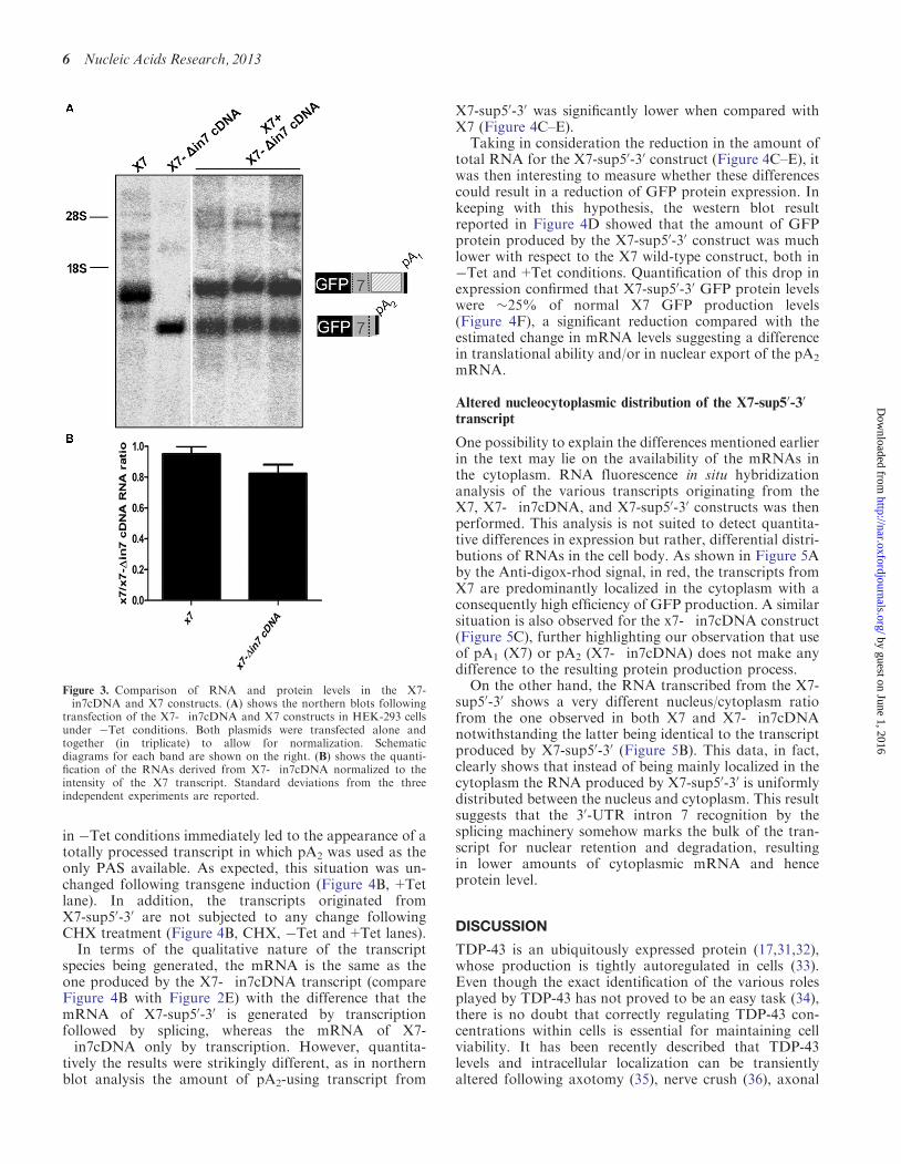
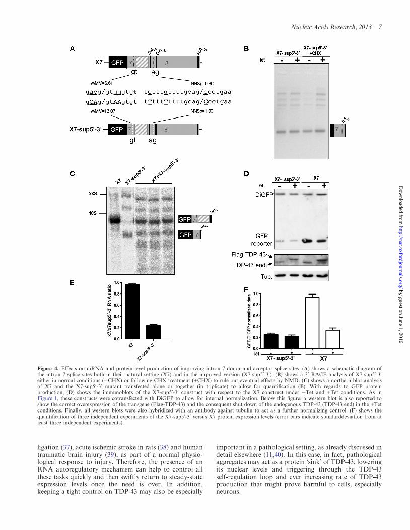