Predictive factors for postoperative outcome in temporal lobe epilepsy according to two different...
12
Predictive factors for postoperative outcome in temporal lobe epilepsy according to two different classifications F. Irsel Tezer a, * , Nejat Akalan b , Kader K. Oguz c , Erdem Karabulut d , Nese Dericioglu a , Abdurrahman Ciger a , Serap Saygı e a Institute of Neurological Sciences and Psychiatry, Hacettepe University School of Medicine, Ankara, Turkey b Department of Neurosurgery, Hacettepe University School of Medicine, Ankara, Turkey c Department of Radiology, Hacettepe University School of Medicine, Ankara, Turkey d Department of Biostatistics, Hacettepe University School of Medicine, Ankara, Turkey e Department of Neurology, Hacettepe University School of Medicine, Ankara, Turkey Received 20 May 2007; received in revised form 25 January 2008; accepted 29 February 2008 Seizure (2008) 17, 549—560 www.elsevier.com/locate/yseiz KEYWORDS Temporal lobe epilepsy; Hippocampal sclerosis; Epilepsy surgery; Outcome; Risk factors; Early seizures Summary Purpose: The determination of prognostic factors is important for predicting out- come after epilepsy surgery. We investigated the factors related to surgical outcome within a homogeneous group of patients suffering from pathologically proven mesial temporal lobe epilepsy with hippocampal sclerosis (MTLE-HS), and compared Engel’s outcome classification system with the latest one proposed by the ILAE. Method: We included 109 patients with MTLE-HS who were followed-up for at least 1 year after epilepsy surgery. A retrospective chart review was performed to extract patients’ demographic details, and potential pre-postoperative risk factors. Outcome of surgery was defined by the Engel’s and ILAE classifications. In addition, the course of prognosis was determined according to the changes in ILAE classifications on an annual basis. Univariate and multivariate logistic regression analyses were used for the latest available outcomes and two different courses of prognosis. Results: The average duration of follow up was 4.78 2.55 years in the 109 patients with MTLE-HS. The univariate and multiple logistic regression analyses showed that the occurrence of seizures during the first month after surgery was a significant risk factor for a poor outcome. A history of trauma was also significant for patients with late recurrence of postsurgical seizures after at least 1-year seizure remission. * Corresponding author at: Hacettepe University Faculty of Medicine, Department of Neurology, 06100 Sıhhıye, Ankara, Turkey. Tel.: +90 312 305 11 82; fax: +90 312 309 34 51. E-mail address: [email protected] (F.I. Tezer). 1059-1311/$ — see front matter # 2008 British Epilepsy Association. Published by Elsevier Ltd. All rights reserved. doi:10.1016/j.seizure.2008.02.003
Transcript of Predictive factors for postoperative outcome in temporal lobe epilepsy according to two different...

Predictive factors for postoperative outcomein temporal lobe epilepsy according to twodifferent classifications
F. Irsel Tezer a,*, Nejat Akalan b, Kader K. Oguz c, Erdem Karabulut d,Nese Dericioglu a, Abdurrahman Ciger a, Serap Saygı e
a Institute of Neurological Sciences and Psychiatry, Hacettepe University School of Medicine,Ankara, TurkeybDepartment of Neurosurgery, Hacettepe University School of Medicine, Ankara, TurkeycDepartment of Radiology, Hacettepe University School of Medicine, Ankara, TurkeydDepartment of Biostatistics, Hacettepe University School of Medicine, Ankara, TurkeyeDepartment of Neurology, Hacettepe University School of Medicine, Ankara, Turkey
Received 20 May 2007; received in revised form 25 January 2008; accepted 29 February 2008
Seizure (2008) 17, 549—560
www.elsevier.com/locate/yseiz
KEYWORDSTemporal lobeepilepsy;Hippocampal sclerosis;Epilepsy surgery;Outcome;Risk factors;Early seizures
Summary
Purpose: The determination of prognostic factors is important for predicting out-come after epilepsy surgery. We investigated the factors related to surgical outcomewithin a homogeneous group of patients suffering from pathologically proven mesialtemporal lobe epilepsy with hippocampal sclerosis (MTLE-HS), and compared Engel’soutcome classification system with the latest one proposed by the ILAE.Method: We included 109 patients with MTLE-HS who were followed-up for at least 1year after epilepsy surgery. A retrospective chart review was performed to extractpatients’ demographic details, and potential pre-postoperative risk factors. Outcomeof surgery was defined by the Engel’s and ILAE classifications. In addition, the courseof prognosis was determined according to the changes in ILAE classifications on anannual basis. Univariate and multivariate logistic regression analyses were used forthe latest available outcomes and two different courses of prognosis.Results: The average duration of follow up was 4.78 � 2.55 years in the 109 patientswith MTLE-HS. The univariate and multiple logistic regression analyses showed thatthe occurrence of seizures during the first month after surgery was a significant riskfactor for a poor outcome. A history of trauma was also significant for patients with
late recurrence of postsurgical seizures after at least 1-year seizure remission.* Corresponding author at: Hacettepe University Faculty of Medicine, Department of Neurology, 06100 Sıhhıye, Ankara, Turkey.Tel.: +90 312 305 11 82; fax: +90 312 309 34 51.
E-mail address: [email protected] (F.I. Tezer).
1059-1311/$ — see front matter # 2008 British Epilepsy Association. Published by Elsevier Ltd. All rights reserved.doi:10.1016/j.seizure.2008.02.003

550 F.I. Tezer et al.
Conclusion: The occurrence of seizures during the first month after surgery is asignificant prognostic factor in patients with MTLE-HS. Ignoring early postoperativeseizures in classification systems may result in difficulty in identifying the course ofepilepsy after surgery.# 2008 British Epilepsy Association. Published by Elsevier Ltd. All rights reserved.
Introduction
The rate of complete remission of seizures afterepilepsy surgery for temporal lobe epilepsy wasreported as 68—85%.1 Almost one third of patientscontinue to have seizures after surgery. The out-come in the long term is worse than that in the shortterm and 48—55% of patients did not become sei-zure-free more than 5 years after the operation.2—4
Therefore, the determination of prognostic factorsfor epilepsy surgery is important for the counsellingof patients in everyday practice.
Several previous studies have reported predictivevariables of outcome after epilepsy surgery butmosthave included a variety of temporal lobe pathologiesincluding indeterminate findings and normal or non-specific abnormality,5—7 some have no pathologicaldata,8,9 and some have included both temporal andextratemporal cases.6,7,10 Mesial temporal lobe epi-lepsy (MTLE) pathologically characterized by hippo-campal sclerosis (HS) is the prototype for surgicallyremediable epileptic syndromes.11 There have beenfew studies of the prognostic factors within thissubgroup of patients with MTLE-HS.12—17 To the bestof our knowledge, only four studies have examinedthe outcome of surgery in a homogeneous groupwith MTLE-HS that was defined histopathologi-cally.12,14,15,17
Another problem in most previous studies aboutoutcome after epilepsy surgery is the use of differ-ent methods of classification and cross-sectionalanalysis of outcomes during different time periods.The calculation of the proportion of patients whoare seizure-free for some years after surgery pro-vides only a snapshot of cohort outcomes during thatperiod. The cross-sectional method of analysis maybe problematic as the correlation of variables withoutcome may yield different results depending onthe stage of the postoperative course at which out-come was assessed.2 To avoid this problem, weexamined outcome in a group with MTLE-HS forthe whole follow-up period in each patient, in addi-tion to the last available outcome. The last availableoutcome of each patient was also defined using bothEngel’s outcome classification18 and the new ILAEclassification.19
We investigated the factors related to surgicaloutcome within a group of pathologically proven
MTLE-HS patients, comparing the two classificationsystems. Besides we also tried to determine howpostoperative prognosis was altered by time.
Methods
All patients who received surgical treatment forintractable temporal lobe seizures at our centerbetween 1995 and 2006 were reviewed for possibleinclusion in the study.
Presurgical evaluation and patientselection
A detailed clinical history was obtained frompatients considered possible candidates for epilepsysurgery. Resistance to first-line anti-epileptic drugs(AED) was evaluated. As a rule, high-resolution brainmagnetic resonance (MR) imaging was performed.With most patients, MR images were obtained usingeither 1.5 or 3.0T scanners (Symphony and Allegra,respectively, Siemens, Erlangen, Germany). The MRimaging protocol performed for patients with epi-lepsy in our institution includes coronal 3D T1-weighted (W) gradient-echo imaging (MPRAGE)obtained parallel to the brainstem, and fluid-atte-nuated inversion recovery (FLAIR), and T2-W turbospin-echo and T1-W inversion recovery imagesobtained perpendicular to the hippocampi in addi-tion to routine brain imaging. A 32-channel EEGrecording system was used. Scalp electrodes includ-ing T1—T2 electrodes were placed according to 10—20 systems. Patients underwent continuous video-EEG monitoring lasting 3—10 days. Findings frompresurgical evaluations were discussed at a multi-disciplinary case conference, where decisions weremade concerning the possibility and type of surgery.For patients in whom hippocampal sclerosis waspresent on imaging, an operation was performedwhen interictal and ictal EEG findings were concor-dant with MR imaging. Our standard procedure fortreating MTLE was anterior temporal lobectomywith removal of the medial structures includingthe amygdala, hippocampus and parahippocampalgyrus. We performed standard anterior temporallobe resection in all patients. We resected 3,5 cmof lateral temporal lobe from the anterior temporal

Predictive factors for postoperative outcome in temporal lobe epilepsy 551
tip for the dominant hemisphere and 5 cm for thenondominant one. Patients who underwent epilepsysurgery were re-examined every 6 months afterthat, with an assessment of the seizure outcomeas well as an evaluation of the psychiatric and socialstatus. Our protocol is for patients to receive AEDsfor a minimum of 2 years post-operatively. The typeof AEDs may change or the dosage may decreaseafter 1 year. Two years after successful surgery, theoption of complete AED discontinuation is tailoredto the individual patient.
This study included 109 patients who were fol-lowed-up for at least 1 year and who had HS shownon MR imaging and confirmed by postoperativepathological analysis. The hippocampal subfieldsand dentate gyrus were examined for neuronal lossand gliosis by visual analysis. Hippocampal sclerosiswas defined as more than 50% neuron loss in the CA1subfield.
Data collection
A retrospective chart review was performed toextract patients’ demographic details, and poten-tial preoperative and postoperative risk factors. Thefollowing variables were investigated: gender; pre-sence of mental retardation; age at epilepsy onset(onset of habitual seizures, excluding febrile sei-zures); presence of family history and consanguinityof parents; history of natal or perinatal injury asperinatal risk factors; histories of febrile seizures,trauma and status epilepticus, aura; history of sec-ondarily generalized tonic-clonic seizures (SGTCS);number of AEDs used before surgery; age at opera-tion; duration of epilepsy; interictal scalp electro-encephalographic findings (EEG); follow-up periods;presence of surgical complications; frequency ofpostoperative seizure days on an annual basis oneach anniversary of the surgery and usage of AEDsfor each postoperative year. The annual and latestavailable seizure outcomes of patients described indetail using both the classification system of Engel’sClasses I—IV18 and the new ILAE classification systemClasses 1—6.19 Seizures occurring in first postopera-tive month were ignored in classifications. We alsolooked at the occurrence of seizures within the first2 days and first month (between days 3 and 30). Ithas been suggested that acute early seizures withina few days, mostly in the first 48 h after surgery maybe less prognostically important because this iswhen the acute surgical effects like electrolyteimbalance, hypoxia, hypoglycemia or hypocapniaare the most pronounced and the risk of a lowAED level is frequent.18,20 Because of that seizureoutcome for the first 2 days were not included in thefirst month outcome. Patients’ previous medical
charts were reviewed and the missing variableswere ascertained by asking the patients and theirrelatives. The location and frequency of interictalepileptiform discharges were assessed by visualanalysis of interictal EEG samples. Unilateral inter-ictal epileptiform discharges were defined if at least70% of interictal discharges appeared over one tem-poral lobe. The detailed ictal EEG findings of thesepatients will be reported in a parallel study that iscurrently being performed.
Outcome assessment
At each visit, the information collected includedseizure frequency, medication intake and compli-ance, and the effect of seizures on daily activitiesor social status. Neuropsychological outcomeswere not included. To minimize the lack of con-tribution to outcome determination, we obtainedthe latest information by telephone calls in 94patients. Outcome of surgery was defined accord-ing to the Engel’s and ILAE classifications.18,19 Out-come status was determined every year for up to11 years after surgery by outpatient clinical inter-view or telephone interview. Patients were cate-gorized as seizure-free (Engel’s Class I or ILAE Class1 or 2) or not (Engel’s Classes II—IV or ILAE Classes3—5). Also the patients who were completelyfree from both seizures and auras21,22 at the lastavailable outcome (ILAE Class 1a which corre-sponds to Engel’s Class IA) were determined. Fif-teen patients who could not be contacted byoutpatient or telephone interview were categor-ized according to their medical charts at their lastfollow-up year.
Furthermore, the changes in ILAE classificationthat occurred on an annual basis were documentedgraphically for each patient, except for 4 patients(n = 105) who were followed-up for a year only. Inpatients with changes in outcomes, it was attemptedto isolate clinical characteristics related to specificpatterns such as the running down (RD) phenomenon(becoming seizure free after some early postopera-tive seizures) or the reverse form of it (late seizurerecurrence after an initial, at least 1 year, seizurefree period).23,24
Statistical methods
An independent samples t test was used for con-tinuous variables: age at operation, age at epilepsyonset (onset of habitual seizures), epilepsy durationbefore surgery (defined by the difference betweenage at operation and age at onset), number of drugsused before surgery, follow-up period in months andseizure frequency per month before surgery.

552 F.I. Tezer et al.
A Chi-square or Fisher’s exact test of indepen-dence was used for univariate analysis of the follow-ing categoric variables: gender, side of surgery,presence of mental retardation and perinatal risk
Table 1 Clinical features of 109 patients with TLE-HS
IE, ipsilateral epileptiform abnormality; iiEEG, interictal EEG.
factors, presence of family history and consanguinityof parents, histories of febrile seizures, trauma,status epilepticus, presence of aura, history of SGTCSbefore surgery, presence of ipsilateral interictal

Predictive factors for postoperative outcome in temporal lobe epilepsy 553
epileptiform discharges on interictal EEG and pre-sence of surgical complications.
Backward stepwise multiple logistic regressionanalysis was used to assess the prognostic impor-tance of the clinical variables. Odds ratios (ORs)were calculated for each surgical outcome group:Engel’s Class I and others (Classes II—IV), and ILAEClasses 1 and 2 and others (Classes 3—5). Multi-variate analysis was performed on variables thatwere p < 0.15 in the univariate analysis.
Table 2 Last available outcome of patients after surgery
aThe changes in ILAE classification occurred on annual basis (coursewere followed up for a year only.bLate seizure recurrence after an initial, at least 1 year, seizure fr
Results
The present study included 109 patients with medi-cally intractable seizures who underwent temporallobe resection in whom pathological examination ofthe excised material revealed HS and who werefollowed-up for at least 1 year.
Tables 1 and 2 show details of the clinical andoutcome findings. Of the 109 patients 67 (61%) werefemale. The mean age at surgery was 28.14 � 2.55
of prognosis) were documented for each patient, except 4 who
ee period.
554 F.I. Tezer et al.
years (range 15—52 years). The age at epilepsy onsetranged from 7 to 32 years (mean 9.64 � 6.53 years).The mean epilepsy duration was 18.55 � 7.78 years.The average duration of follow-up was 52.28 �30.75 months or 4.78 � 2.55 years (1—11 years).The mean number of AEDs used before surgerywas 2.43 � 1.49. In 51 patients, the resection wason the left side (47%) and in the other 58 it was onthe right.
Interictal EEG reports of 100 patients were avail-able in their charts. In 96 patients interictal EEGshowed ipsilateral epileptiform discharges. Surgicalcomplications were reported in 22 (20%) of 109patients, but only one had a major complication.Ninety-four patients (86%) were reevaluated by tel-ephone interview. During the follow-up period, onepatient committed suicide. The other reasons whypatients were lost to follow-up are unknown.
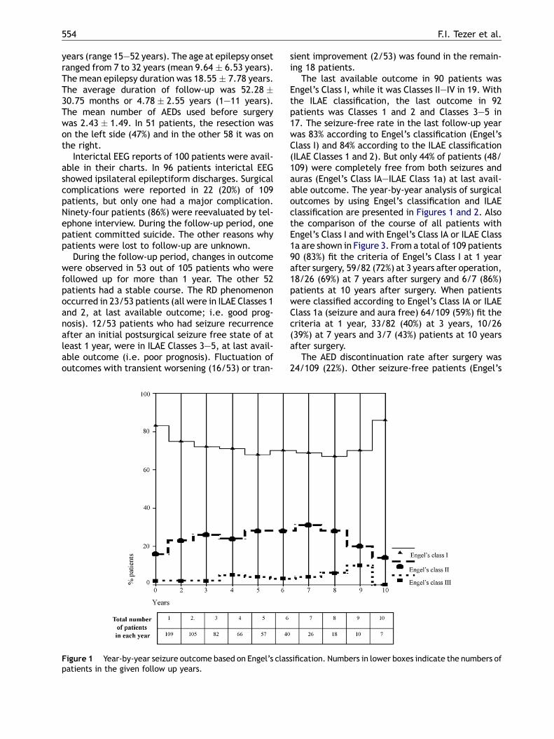
During the follow-up period, changes in outcomewere observed in 53 out of 105 patients who werefollowed up for more than 1 year. The other 52patients had a stable course. The RD phenomenonoccurred in 23/53 patients (all were in ILAE Classes 1and 2, at last available outcome; i.e. good prog-nosis). 12/53 patients who had seizure recurrenceafter an initial postsurgical seizure free state of atleast 1 year, were in ILAE Classes 3—5, at last avail-able outcome (i.e. poor prognosis). Fluctuation ofoutcomes with transient worsening (16/53) or tran-
Figure 1 Year-by-year seizure outcome based on Engel’s claspatients in the given follow up years.
sient improvement (2/53) was found in the remain-ing 18 patients.
The last available outcome in 90 patients wasEngel’s Class I, while it was Classes II—IV in 19. Withthe ILAE classification, the last outcome in 92patients was Classes 1 and 2 and Classes 3—5 in17. The seizure-free rate in the last follow-up yearwas 83% according to Engel’s classification (Engel’sClass I) and 84% according to the ILAE classification(ILAE Classes 1 and 2). But only 44% of patients (48/109) were completely free from both seizures andauras (Engel’s Class IA—ILAE Class 1a) at last avail-able outcome. The year-by-year analysis of surgicaloutcomes by using Engel’s classification and ILAEclassification are presented in Figures 1 and 2. Alsothe comparison of the course of all patients withEngel’s Class I and with Engel’s Class IA or ILAE Class1a are shown in Figure 3. From a total of 109 patients90 (83%) fit the criteria of Engel’s Class I at 1 yearafter surgery, 59/82 (72%) at 3 years after operation,18/26 (69%) at 7 years after surgery and 6/7 (86%)patients at 10 years after surgery. When patientswere classified according to Engel’s Class IA or ILAEClass 1a (seizure and aura free) 64/109 (59%) fit thecriteria at 1 year, 33/82 (40%) at 3 years, 10/26(39%) at 7 years and 3/7 (43%) patients at 10 yearsafter surgery.
The AED discontinuation rate after surgery was24/109 (22%). Other seizure-free patients (Engel’s
sification. Numbers in lower boxes indicate the numbers of

Predictive factors for postoperative outcome in temporal lobe epilepsy 555
Figure 2 Year-by-year seizure outcome based on ILAE classification. Numbers in lower boxes indicate the numbers ofpatients in the given follow up years.
Class I or ILAE Classes 1 and 2) continued the AEDs.Mean time of discontinuation of AEDs after surgerywas 2.83 � 0.70 years.
Fifty-seven (52%) of the 109 patients had at leastone seizure during the entire postoperative follow-up. Eleven (19%) of these 57 experienced theirinitial seizure recurrence during the first month,21 (37%) during the first year, and 13 (23%) duringthe second year. Three (27%) of the 11 patients whohad seizures during the first month had no furtherseizures after that.
Univariate analyses
Table 3 shows the results of univariate analyses ofidentifying variables between patients with good(ILAE Classes 1 and 2 and Engel’s Class I) and poor(ILAE Classes 3—5 and Engel’s Classes II—IV) outcomeat last follow up. The demographic characteristicswere similar among different outcome groups.
The presence of preoperative risk factors includ-ing the presence of mental retardation; histories offebrile seizures, trauma, infection, status epilepti-cus; and the presence of seizures during sleep; auraand SGTCS were not associated with a poor prog-nosis at last available outcome. Interictal EEG find-ings with scalp electrodes and the presence ofsurgical complications did not predict the surgical
outcome. Resections on the left side did not differfrom those on the other side in terms of surgicaloutcome.
Univariate analysis revealed that the presence ofseizures during the first month (3—30 days) aftersurgery was a significant risk factor for postsurgicaloutcome in the last follow-up period. During the firstmonth after surgery, 6/19 patients in Engel’s ClassesII—IV ( p = 0.004) and 5/17 patients in ILAE Classes3—5 (p = 0.01) had recurrence of seizures (Table 3).
After the findings about the significance of theseizures during first month, some features of 11patientswhohad seizures during thefirstmonthwerere-evaluated. Five of these patients were classifiedas seizure free (Engel’s Class I) and6of themwerenotseizure free at the last available outcome. Seizures inthe first month (3—30 days) occurred mostly after aweek from the surgery. The exact day of these sei-zures could not be obtained in eight patients. Thenumberof seizures changed from1 to 4 times for eachpatient during that period, totally 24 seizures werereported for all 11 patients. Seizures of 7 patientswere resembling the habitual preoperative seizuresin mild form but features of seizures were notdescribed in 4 patients. All of the patients used theirAEDs regularly during these periods but AEDs serumlevels were not noted at these times. Furthermorepreoperative electrophysiological and neuroradiolo-

556 F.I. Tezer et al.
Table 3 Univariate analysis of the variables between groups which include Engel’s Class I and Engel’s Classes II, III, IVor new classification of ILAE Classes 1 and 2 and Classes 3—5 at last outcome
Engel’s Class I(n = 90)
Engel’s ClassesII—III—IV (n = 19)
— ILAE Classes 1and 2 (n = 92)
ILAE Classes3—5 (n = 17)
p
Age at surgery(year; mean � S.D.)
27.73 � 7.51 30.05 � 7.63 0.22 27.81 � 7.43 29.88 � 8.14 0.30
Duration of epilepsy(year; mean � S.D.)
17.79 � 7.26 21.94 � 9.49 0.39 17.90 � 7.36 21.58 � 9.38 0.07
Duration ofepilepsy >20 years
31/90 11/19 0.06 32/92 10/17 0.10
Site of surgery(right temporal)
50/90 8/19 0.32 52/92 6/17 0.10
Gender (male) 36/90 6/19 0.60 35/92 7/17 0.79Marital status
(married)20/90 5/19 0.76 20/92 5/17 0.53
Right handed 78/90 14/19 0.15 80/92 12/17 0.13Presence of mental
retardation18/90 5/19 0.53 19/92 4/17 1.0
First seizure at theage of �6 years old
22/90 2/19 0.23 22/92 2/17 0.35
Presence of familyhistory
39/90 8/19 1.00 42/92 5/17 0.29
Presence perinatalrisk factors
15/90 5/19 0.34 17/92 3/17 1.00
History of trauma 34/90 9/19 0.60 33/92 10/17 0.11History of febrile
seizures60/90 13/19 1.0 63/92 10/17 0.57
Consanguinity ofparents
18/90 3/19 1.0 19/92 2/17 0.52
Recurrentseizures at theage of �6 years
63/90 9/19 0.10 63/92 9/17 0.25
History of statusepilepticus
18/90 5/19 0.53 18/92 5/17 0.52
History of SGTCS 53/90 13/19 1.0 55/92 11/17 0.79Presence of aura 80/90 17/19 1.0 81/92 16/17 0.68IE abnormality
on iiEEG80/84 16/16 1.0 82/86 14/14 1.0
Complicationof surgery
21/90 1/19 0.11 21/92 1/17 0.18
Presence of seizureat first month
5/90 6/19 0.004 6/92 5/17 0.01
Withdrawal ofantiepileptics aftersurgery
23/90 1/19 0.07 22/92 2/17 0.35
IE, ipsilateral epileptiform abnormality; iiEEG, interictal EEG.
gical findings of these11patientswith seizures duringthe first month after surgery were re-evaluated. Allof the patients except one had unilateral interictalepileptiformdischarges (more than>80%) on the sideof operation. Only one patient had 60% ipsilateralinterictal epileptiform discharges. Ictal EEG dis-closed lateralized temporal rhythmic discharges onthe side of surgery in all patients. On MR imaging allpatients had unilateral hippocampal atrophy at theside of operation. There were no complications
related to surgical procedure during the postopera-tive period.
Univariate analysis was also performed on theserisk factors for patients with RD phenomenon orpatients with late postsurgical seizure recurrencewho were seizure free at least for a year aftersurgery. Only history of trauma was significantlydifferent between two groups. Trauma history waspresent in 21% of those with the RD phenomenon andin 75% of patients in the latter group ( p = 0.003).

Predictive factors for postoperative outcome in temporal lobe epilepsy 557
Multivariate analysis
We performed a multivariate analysis for the pre-sence of seizures in the first postoperative month.Standard stepwise multiple logistic regression alsoshowed that the presence of seizures during the firstmonth after surgery was independently predictivefor worsening of outcome. (For last available out-come according to Engel’s classification: p = 0.008OR = 6.38 95% CI = 1.62—25.13, and for last avail-able outcome according to the new ILAE classifica-tion: p = 0.006 OR = 0.14 95% CI = 1.76—27.80.) Thepresence of a history of trauma in patients with latepostsurgical seizure recurrence after an initial sei-zure free period (p = 0.05 OR = 0.33 95% CI = 0.003—0.38) was also an independent factor.
Discussion
Seizure outcomes following temporal lobe surgeryhave been studied extensively in recent years. How-ever, the interpretation of published reports onlong-term outcome has been difficult because ofseveral methodological problems.2 These studiesusually use a modified Engel’s classification or anew modified form, lacking appropriate standar-dized classification systems for seizure outcome.In our study, the last available outcomes of eachpatient were defined as seizure-free or not, accord-ing to both worldwide acceptable classification sys-tems, Engel’s and the ILAE. Statistical analysis wasperformed in these two different classification sys-tems. We found that the only predictor of post-operative poor outcome in patients with MTLE-HSshown by univariate and multivariate analysiswas recurrence of seizures in the first month afterepilepsy surgery in either classification system.Remarkably, no other pre- or perioperative riskfactors examined were found to have definite prog-nostic value in these patients with pathologicallyproven HS.
The occurrence of early postoperative seizures orauras in the first week after epilepsy surgery isrelatively common (up to 49%).20,25—27 It has beensuggested that these early seizures may result fromthe effects of acute surgical injury and they havebeen termed neighborhood seizures. Some authorshad the opinion that transient neighborhood sei-zures did not necessarily infer a bad prog-nosis.18,19,25 Therefore, in most previous studiesof outcome following epilepsy surgery, the earlypostoperative seizures that occurred during thehospital stay have been ignored. The most com-monly used seizure outcome classification18 is notspecific for the time period of early seizures. In
2001, the Commission on Neurosurgery of the ILAE19
suggested a 4-week period for this.In the literature, most series showed that in 20—
40% of patients with TLE the earliest time period forthe risk of recurrence and persistence of seizureswaswithin 6—12 months of surgery.27—29 However, someprevious studies support our findings showing thepre-dictive value of earlier postoperative seizures afterTLE surgery on poor prognosis.4,7,20,23,26,27,30—33
Armon et al. reported that the presence of seizuresin 25% of patients within 2 months of surgery was asignificant postoperative predictor for poor outcomeover 2 years in a heterogeneous group with corticalresections.7 McIntosh et al. reported that 21.5% oftheir patients had early postoperative seizures in thefirst month,31 and 77% of them experienced subse-quent seizures in the first year. They found thatpatients with early seizures had a risk of seizurerecurrence that was almost 3—8 times that ofpatients without early seizures. They suggested,however, that adjustment of preoperative pathologywas important for thepredictionof theeffect of earlypostoperative seizures onoutcome.Thedifference inthe presence of early seizures ratesmaybe related tothe differences in the pathological substrate of thestudy population and the definition of the earlyperiod. Our study was limited to a single pathologyMTLE-HS, whereas the earlier studies were not lim-ited. In our homogeneous group of patients withpathologically provenTLE-HS, 10% (11/109) hadearlyseizures between the 3rd and 30th postoperativedays, and only 21% of them had no recurrence ofseizures after the first month. In our study, involve-ment of only patients with HS may support thehypothesis that early seizures are a marker forremaining primary or secondary epileptogenesisand intractability of TLE-HS.33,34 However, furtherstudies with postoperative MR imaging and EEG find-ings for residual pathology or epileptogenic region inlarger numbers of patients are required.
There is a limitation of our retrospective dataabout features of early seizures like type and timing,and their risk factors such as AEDs serum levels. In theliterature, however, the importance of these char-acteristics of early seizures, which are associatedwith poor seizure outcome,4,20,21,23,26,27,31,32 isunclear. Malla et al. reported that patients whohad early seizures similar to their preoperative habi-tual seizures within first month after anterior tem-poral lobectomy had worse outcome than those withauras, or focal or generalised seizures.26 HoweverGarcia et al.,20 found no difference in long termoutcomes between patients with postoperative sei-zures that were similar to preoperative seizures andthose seizures different from preoperative seizures.In our study we confirmed that 7 of 11 patients who

558 F.I. Tezer et al.
had early seizures during the first month had seizuressimilar to preoperative habitual seizures. Five ofthese 7 patients were classified as poor seizure out-come (Engel’s Class II) whereas 1 of 4 patients whoseseizure characteristics could not be determined hadpoor prognosis. In another study, it was suggestedthat there was a trend toward higher recurrenceamong patients who experienced a first seizure inthe first week rather than in the subsequent 3 weeksafter anterior temporal lobectomy with any pathol-ogy.31 Our group of 11 patients with early seizuresmostly had seizures after first week but exact time ofthese were not known.
Contrary to what might be expected, the resultsrelated to the significance of other preoperative riskfactors were not consistent with the numerous pre-vious studies of the overall group of patients receiv-ing temporal lobe resection for epilepsy. Identifiedfactors that have been shown to predict outcome inthese large series includedurationofepilepsy,6,13 ageat operation,5,6,13 history of febrile seizures,5 con-cordant interictal epileptiform abnormalities,9,16,34
and presence of status epilepticus or SGTCS beforesurgery.4,15,16 The differences in these predictivefactors may be related to the differences in thepathological substrate of the study population. Tothe best of our knowledge, there are only a fewreports that specifically address the surgical outcomeof patientswith the syndromeMTLE-HS.12,14—17 In theMTLE subgroup, like in our patients, most of thesefactors lose their predictive value.
In the literature, very few systematic studieshave been performed on patients’ natural historyand prognostic factors predicting the continuedremission of seizures or relapse after surgery.2 Usingonly cross-sectional analyses leads to loss of infor-mation related to course of prognosis like the run-ning down phenomenon. This approach may also beproblematic for correlation of findings depending onthe stage of the postoperative period. For thatreason, firstly we analysed annual changes in out-come according to the new ILAE classification foreach patient. Although the general proportion ofseizure-free patients remained stable in our series(52/109), as in the study by Jeong et al. (73/121),1
some patients showed the RD phenomenon or lateseizure recurrence after an initial postsurgical sei-zure free period in the group with fluctuatingcourse.
The rate of TLE patients with late improvement inpostoperative seizure control was reported as 5—44%.23,24 The RD phenomenon was also observed in21% (23/109) of our patients. Late seizure recur-rence after an initial seizure free period, occurredin 13 of our patients (12%). This type of course wasalso observed in a few reports.1,28 There is a sugges-
tion that patients with that course of prognosis whohad failed TLE surgery are more likely to have largeepileptogenic zones and more diffuse lesions of thetype produced by encephalitis and head injury,which often involve the posterior temporal region,and are difficult to eradicate with surgery.35,36 Insupport of that we found only the presence oftrauma history as a predictive risk factor for lateseizure recurrence after an initial seizure free per-iod. This may indicate that changes occur in thefunctional anatomy of the brain and it becomessusceptible to intractability. However, to the bestof our knowledge there is no report related to theclinical details and comparisons of both of thesespecific outcome patterns. Only Salanova et al.reported that patients with the RD phenomenonwere more likely to have a history of febrile sei-zures, unilateral temporal spiking and almost com-plete hippocampal resection compared withpatients who continued to have seizures.23 Theysuggested that a less autonomous residual epilepto-genic area causes this clinical phenomenon and runsdown over the course of months or years. Futureprospective studies that include a large group ofpatients with a long follow-up period in addition toknowledge about postoperative MR imagings, EEGfindings and usage of AEDs would be needed for theanalysis of the features of the RD phenomenon orthe reverse of that phenomenon, i.e. late seizurerecurrence after an initial seizure free period.
In the present study, both classification systemswere used for analysis. They have different advan-tages and disadvantages. Although the 2001 ILAEclassification is a modification of Engel’s classifica-tion, it is more standardized and has more objectivecriteria. The definitions in the last classification,like frequency of seizures for a year and ratio ofseizures according to ‘‘baseline seizure days’’, aremore easily applicable. It is useful for annual followup and for showing the time course of prognosis. InEngel’s Class I seizure-free patients are includedwith those who still have seizures. Although Engel’sClass IA refers to absolutely seizure-free patients,like the new classification of ILAE Class 1a, in realitymost centers do not report outcomes using Engel’ssubcategories. Therefore, the actual number ofseizure-free patients is frequently obscure.21,22 Inour study when analyses were restricted to Engel’sClass IA (or ILAE Class 1a) patients, seizure-aura freeratios were 59% (64/109) and 44% (3/7) at the firstand tenth years. However in analyses of Engel’sClass I patients, seizure outcome appeared to bebetter at first year (90/109; 83%) and tenth year (6/7; 86%) follow up, and be more stable with time(Figure 3). But in that stable course we could losethe patients with simple (Engel’s Class IB) or dis-

Predictive factors for postoperative outcome in temporal lobe epilepsy 559
Figure 3 Year-by-year number of patients with ILAE 1a (Engel’s Class IA) and Engel’s Class; for comparison of twodifferent definitions of seizure freedom. Numbers in lower boxes indicate the numbers of patients in the given follow upyears.
abling seizures (Engel’s Class IC) and with seizuresrelated to AED discontinuation (Engel’s Class ID).Moreover, the classification of patients having somedisabling seizures after surgery (Engel’s Class IC) asseizure-free may cause the special course of prog-nosis like the RD phenomenon to be missed. In theyear-by-year reporting of Class 1 outcomes recom-mended subgroups like ‘‘ILAE Class 1a: completelyseizure-free since surgery’’ resembling to Engel’sClass IA might be used to indicate the RD phenom-enon. Furthermore, recurrence of seizures aftermore than 1 year seizure-free period might bementioned for defining the late recurrence of sei-zures or fluctuating course. In addition, the defini-tion of nocturnal seizures that are less disabling inpatient’s life is important in classification. However,as these groups of seizures are defined in ‘‘seizuredays’’ in the new classification, it could not bespecifically mentioned. As a result, the ILAE classi-fication for annual follow up in determination ofcourse of prognosis and Engel’s classification for lastavailable outcome may give more realistic results.
The findings of the present study suggest thatignoring early postoperative seizures in both classi-fication systems may result in difficulty in identify-ing the course of refractory epilepsy after surgery.Early seizures may be a marker for remaining epi-
leptogenesis or intractability in patients with hip-pocampal sclerosis. They may also show theprogressive course of the disease after surgery.The protocol of discontinuation of AEDs or MR ima-ging follow up after surgery may be reviewed. More-over, the pathological differences for the functionaland anatomical reorganization of limbic structurebetween children and adults with HS may giveinformation about the chronicity of this diseaseand may support its progressive form although itwas surgically removed.
References
1. Jeong SW, Lee SK, Hong KS, Kim KK, Chung CK, Kim H.Prognostic factors for the surgery for mesial temporal lobeepilepsy: longitudinal analysis. Epilepsia 2005;46:1273—9.
2. McIntosh AM, Wilson SJ, Berkovic SF. Seizure outcome aftertemporal lobectomy: current research practice and findings.Epilepsia 2001;42:1288—307.
3. Spencer SS. Long-term outcome after epilepsy surgery. Epi-lepsia 1996;37:807—13.
4. Spencer SS, Berg AT, Vickery BG, Sperling MR, Bazil CW,Shinnar S, et al. Predicting long term seizure outcome afterresective epilepsy surgery. Neurology 2005;65:912—8.
5. Blume WT, Desai HB, Girvin JP, McLahlan RS, Lemieux JV.Effectiveness of temporal lobectomy measured by yearly fol-low-up and multivariate analysis. J Epilepsy 1994;7:203—14.

560 F.I. Tezer et al.
6. Guldvog B, Loyning Y, Hauglie-Hanssen E, Flood S, Bjornaes H.Predictive factors for success in surgical treatment for partialepilepsy: a multivariate analysis. Epilepsia 1994;35:566—78.
7. Armon C, Radtke RA, Friedman AH, Dawson DV. Predictors ofoutcome of epilepsy surgery: multivariate analysis with vali-dation. Epilepsia 1996;37:814—21.
8. Walczak TS, Radtke RA, McNamara JO, Lewis DV, Luther JS,Thompson E, et al. Anterior temporal lobectomy for complexpartial seizures: evaluation, results, and long-term follow-upin 100 cases. Neurology 1990;40:413—8.
9. Radhakrishnan K, So EL, Silbert PL, Jack Jr CR, Cascino GD,Sharbrough FW, et al. Predictors of outcome of anteriortemporal lobectomy for intractable epilepsy: a multivariatestudy. Neurology 1998;51:465—71.
10. Berg AT, Walczak T, Hirsch LJ, Spencer SS. Multivariableprediction of seizure outcome one year after resective epi-lepsy surgery: development of a model with independentvalidation. Epilepsy Res 1998;29:185—94.
11. Engel Jr J, Williamson P, Wieser HG. Mesial temporal lobeepilepsy. In: Engel Jr J, Pedley TA, editors. Epilepsy, a compre-hensive textbook. New York: Raven Press; 1997. p. 2417—26.
12. Kilpatrick C, Cook M, Matkovic Z, O’Brien T, Kaye A, MurphyM. Seizure frequency and duration of epilepsy are not riskfactors for post-operative seizure outcome in patients withhippocampal sclerosis. Epilepsia 1999;40:899—903.
13. Jeong SW, Lee SK, Kim KK, Kim H, Kim JY, Chung CK. Prog-nostic factors in anterior temporal lobe resections for mesialtemporal lobe epilepsy: multivariate analysis. Epilepsia1999;40:1735—9.
14. Hennessy MJ, Elwes RDC, Rabe-Hesketh S, Binnie CD, PolkeyCE. Prognostic factors in the surgical treatment of medicallyintractable epilepsy associated with mesial temporal sclero-sis. Acta Neurol Scand 2001;103:344—50.
15. Hardy SG, Miller JW, Holmes MD, Born DE, Ojemann GA,Dodrill CB, et al. Factors predicting outcome of surgery forintractable epilepsy with pathologically verified mesial tem-poral sclerosis. Epilepsia 2003;44:565—8.
16. Janszky J, Janszky I, Schulz R, Hoppe M, Behne F, Pannek HW,et al. Temporal lobe epilepsy with hippocampal sclerosis:predictors for long-term surgical outcome. Brain 2005;128:395—404.
17. Lee SA, Yim SB, Lim YM, Kang JK, Lee JK. Factors predictingseizure outcome of anterior temporal lobectomy for patientswith mesial temporal sclerosis. Seizure 2006;15:397—404.
18. Engel Jr J, Van Ness P, Rasmussen T, Ojemann LM. Outcomewith respect to epileptic seizures. In: Engel Jr J, editor.Surgical treatment of the epilepsies. 2nd ed. New York:Raven Pres; 1993. p. 609—21.
19. Wieser HG, BlumeWT, Fish D, Goldenshon E, Hufnagel A, KingD, et al. ILAE Commission Report. Proposal for a new classi-fication of outcome with respect to epileptic seizures follow-ing epilepsy surgery. Epilepsia 2001;42:282—6.
20. Garcia PA, Barbaro NM, Laser KD. The prognostic value ofpost-operative seizures following epilepsy surgery. Neurol-ogy 1991;41:1511—2.
21. Wieser HG, Ortega M, Friedman A, Yonekewa Y. Long termseizure outcome following amygdalohippocampectomy. JNeurosurg 2003;98:751—63.
22. Dupont S, Tanguy ML, Clemenceau S, Adam C, Hazemann P,Baulac M. Long term prognosis and psychosocial outcomesafter surgery for MTLE. Epilepsia 2006;47:2115—24.
23. Salanova V, Andermann F, Rasmussen T, Olivier A, Quesney L.The running down phenomenon in temporal lobe epilepsy.Brain 1996;119:989—96.
24. Ficker DM, So EL, Mosewich RK, Radhakrishnan K, Cascino GD,Sharbrough FW. Improvement and deterioration of seizurecontrol during the postsurgical course of epilepsy surgerypatients. Epilepsia 1999;40:62—7.
25. Falconer MA, Serafetinides EA. A follow up study of surgery intemporal lobe epilepsy. J Neurol Neurosurg Psychiatry1963;26:154—65.
26. Malla BR, O’Brien TJ, Cascino GD, So EL, Radhakrishnan K,Silbert P, et al. Acute postoperative seizures following ante-rior temporal lobectomy for intractable partial epilepsy. JNeurosurg 1998;89:177—82.
27. Radhakrishnan K, So EL, Silbert PL, Cascino GD, Marsh WR,Cha R, et al. Prognostic implications of seizure recurrence inthe first year after anterior temporal lobectomy. Epilepsia2003;44:77—80.
28. Wingkun EC, Awad IA, Luders H, Awad CA. Natural history ofrecurrent seizures after resective surgery for epilepsy. Epi-lepsia 1991;32:851—6.
29. So EL, Radhakrishnan K, Silbert PL, Cascino GD, SharbroughFW, O’Brien PC. Assessing changes over time in temporallobectomy: outcome by scoring seizure frequency. EpilepsyRes 1997;27:119—25.
30. McIntosh AM, Kalnins RM, Michell LA, Fabinyi GCA, BriellmannRS, Bekovic SF. Temporal lobectomy: long term seizure out-come, late recurrence and risks for seizure recurrence. Brain2004;127:2018—30.
31. McIntosh AM, Kalnins RM, Mitchell LA, Berkovic SF. Earlyseizures after temporal lobectomy predict subsequent sei-zure recurrence. Ann Neurol 2005;57:283—8.
32. McIntosh AM, Berkovic SF. What happens now? Ongoing out-come after posttemporal lobectomy seizure recurrence.Neurology 2006;67:1671—3.
33. Wieser HG. ILAE commission on neurosurgery of epilepsy.ILAE Commission report: Mesial temporal lobe epilepsywith hippocampal sclerosis. Epilepsia 2004;45:695—714.
34. Berkovic SF, McIntosh AM, Kalnins RM. Pre-operative MRI pre-dicts outcome of temporal lobectomy. Neurology 1995;45:1358—63.
35. Salanova V, Markand O, Worth R. Temporal lobe epilepsy:analysis of failures and the role of reoperation. Acta NeurolScand 2005;111:126—33.
36. Spencer DD, Spencer SS, Mattson RH, Williamson PD, NovellyRA. Access to the posterior medial temporal lobe structuresin the surgical treatment of temporal lobe epilepsy. Neuro-surgery 1984;15:667—71.