Pre-Clinical Models in Implant Dentistry: Past, Present, Future
40
Biomedicines 2021, 9, 1538. https://doi.org/10.3390/biomedicines9111538 www.mdpi.com/journal/biomedicines Review Pre-Clinical Models in Implant Dentistry: Past, Present, Future Nicolas Blanc-Sylvestre 1,2 , Philippe Bouchard 1,2 , Catherine Chaussain 1,3 and Claire Bardet 1, * 1 Université de Paris, Institut des Maladies Musculo-Squelettiques, Orofacial Pathologies, Imaging and Biotherapies Laboratory URP2496 and FHU-DDS-Net, Dental School, and Plateforme d’Imagerie du Vivant (PIV), 92120 Montrouge, France; [email protected] (N.B.-S.); [email protected] (P.B.); [email protected] (C.C.) 2 AP-HP, Department of Periodontology, Rothschild Hospital, European Postgraduate in Periodontology and Implantology, Université de Paris, 75012 Paris, France 3 AP-HP, Reference Center for Rare Disorders of the Calcium and Phosphate Metabolism, Dental Medicine Department, Bretonneau Hospital, GHN-Université de Paris, 75018 Paris, France * Correspondence: [email protected]; Tel.: +331-5807-6724 Abstract: Biomedical research seeks to generate experimental results for translation to clinical set- tings. In order to improve the transition from bench to bedside, researchers must draw justifiable conclusions based on data from an appropriate model. Animal testing, as a prerequisite to human clinical exposure, is performed in a range of species, from laboratory mice to larger animals (such as dogs or non-human primates). Minipigs appear to be the animal of choice for studying bone surgery around intraoral dental implants. Dog models, well-known in the field of dental implant research, tend now to be used for studies conducted under compromised oral conditions (biofilm). Regarding small animal models, research studies mostly use rodents, with interest in rabbit models declining. Mouse models remain a reference for genetic studies. On the other hand, over the last decade, scientific advances and government guidelines have led to the replacement, reduction, and refinement of the use of all animal models in dental implant research. In new development strate- gies, some in vivo experiments are being progressively replaced by in vitro or biomaterial ap- proaches. In this review, we summarize the key information on the animal models currently avail- able for dental implant research and highlight (i) the pros and cons of each type, (ii) new levels of decisional procedures regarding study objectives, and (iii) the outlook for animal research, discuss- ing possible non-animal options. Keywords: pre-clinical research; murine dental implant; human-sized dental implant; osseointegra- tion; biocompatibility; implant models 1. Introduction In the field of dental implant research, experiments have been mainly limited to in vivo studies in so far as translational studies are a prerequisite for any clinical research. Identification of the reasons for the failure and success of dental implant treatments re- mains the most frequent question in clinical practice. By mimicking the biological condi- tion of an implant, pre-clinical research makes it possible to investigate aspects of peri- implant tissue healing and peri-implant disease development [1]. Traditionally, two types of in vivo implant studies have been conducted depending on the implant size used: (1) experiments in large animal models (dogs, pigs, and non- human primates [NHPs]) for studying human-sized implants and (2) experiments in small animal models (rabbits, mice, and rats) for studying adapted “implants” (Figure 1). Nonetheless, taken separately, no animal model is able to shed light on all three levels of implant osseointegration (macro/micro/nano). For example, assessment of implant occlu- sion (macro level) is limited to large animal models, but these models do not allow the analysis of molecular interaction at the bone/implant interface (micro level). Moreover, Citation: Blanc-Sylvestre, N.; Bouchard, P.; Chaussain, C.; Bardet, C. Pre-Clinical Models in Implant Dentistry: Past, Present, Future. Biomedicines 2021, 9, 1538. https://doi.org/10.3390/ biomedicines9111538 Academic Editors: Federico Mussano, Giulio Preti, Davide Cavagnetto and Gianluca Gambarini Received: 9 September 2021 Accepted: 15 October 2021 Published: 26 October 2021 Publisher’s Note: MDPI stays neu- tral with regard to jurisdictional claims in published maps and institu- tional affiliations. Copyright: © 2021 by the authors. Li- censee MDPI, Basel, Switzerland. This article is an open access article distributed under the terms and con- ditions of the Creative Commons At- tribution (CC BY) license (https://creativecommons.org/li- censes/by/4.0/).
-
Upload
khangminh22 -
Category
Documents
-
view
4 -
download
0
Transcript of Pre-Clinical Models in Implant Dentistry: Past, Present, Future

Biomedicines 2021, 9, 1538. https://doi.org/10.3390/biomedicines9111538 www.mdpi.com/journal/biomedicines
Review
Pre-Clinical Models in Implant Dentistry: Past, Present, Future Nicolas Blanc-Sylvestre 1,2, Philippe Bouchard 1,2, Catherine Chaussain 1,3 and Claire Bardet 1,*
1 Université de Paris, Institut des Maladies Musculo-Squelettiques, Orofacial Pathologies, Imaging and Biotherapies Laboratory URP2496 and FHU-DDS-Net, Dental School, and Plateforme d’Imagerie du Vivant (PIV), 92120 Montrouge, France; [email protected] (N.B.-S.); [email protected] (P.B.); [email protected] (C.C.)
2 AP-HP, Department of Periodontology, Rothschild Hospital, European Postgraduate in Periodontology and Implantology, Université de Paris, 75012 Paris, France
3 AP-HP, Reference Center for Rare Disorders of the Calcium and Phosphate Metabolism, Dental Medicine Department, Bretonneau Hospital, GHN-Université de Paris, 75018 Paris, France
* Correspondence: [email protected]; Tel.: +331-5807-6724
Abstract: Biomedical research seeks to generate experimental results for translation to clinical set-tings. In order to improve the transition from bench to bedside, researchers must draw justifiable conclusions based on data from an appropriate model. Animal testing, as a prerequisite to human clinical exposure, is performed in a range of species, from laboratory mice to larger animals (such as dogs or non-human primates). Minipigs appear to be the animal of choice for studying bone surgery around intraoral dental implants. Dog models, well-known in the field of dental implant research, tend now to be used for studies conducted under compromised oral conditions (biofilm). Regarding small animal models, research studies mostly use rodents, with interest in rabbit models declining. Mouse models remain a reference for genetic studies. On the other hand, over the last decade, scientific advances and government guidelines have led to the replacement, reduction, and refinement of the use of all animal models in dental implant research. In new development strate-gies, some in vivo experiments are being progressively replaced by in vitro or biomaterial ap-proaches. In this review, we summarize the key information on the animal models currently avail-able for dental implant research and highlight (i) the pros and cons of each type, (ii) new levels of decisional procedures regarding study objectives, and (iii) the outlook for animal research, discuss-ing possible non-animal options.
Keywords: pre-clinical research; murine dental implant; human-sized dental implant; osseointegra-tion; biocompatibility; implant models
1. Introduction In the field of dental implant research, experiments have been mainly limited to in
vivo studies in so far as translational studies are a prerequisite for any clinical research. Identification of the reasons for the failure and success of dental implant treatments re-mains the most frequent question in clinical practice. By mimicking the biological condi-tion of an implant, pre-clinical research makes it possible to investigate aspects of peri-implant tissue healing and peri-implant disease development [1].
Traditionally, two types of in vivo implant studies have been conducted depending on the implant size used: (1) experiments in large animal models (dogs, pigs, and non-human primates [NHPs]) for studying human-sized implants and (2) experiments in small animal models (rabbits, mice, and rats) for studying adapted “implants” (Figure 1). Nonetheless, taken separately, no animal model is able to shed light on all three levels of implant osseointegration (macro/micro/nano). For example, assessment of implant occlu-sion (macro level) is limited to large animal models, but these models do not allow the analysis of molecular interaction at the bone/implant interface (micro level). Moreover,
Citation: Blanc-Sylvestre, N.;
Bouchard, P.; Chaussain, C.; Bardet,
C. Pre-Clinical Models in Implant
Dentistry: Past, Present, Future.
Biomedicines 2021, 9, 1538.
https://doi.org/10.3390/
biomedicines9111538
Academic Editors: Federico
Mussano, Giulio Preti, Davide
Cavagnetto and Gianluca Gambarini
Received: 9 September 2021
Accepted: 15 October 2021
Published: 26 October 2021
Publisher’s Note: MDPI stays neu-
tral with regard to jurisdictional
claims in published maps and institu-
tional affiliations.
Copyright: © 2021 by the authors. Li-
censee MDPI, Basel, Switzerland.
This article is an open access article
distributed under the terms and con-
ditions of the Creative Commons At-
tribution (CC BY) license
(https://creativecommons.org/li-
censes/by/4.0/).

Biomedicines 2021, 9, 1538 2 of 40
limiting the selection criteria to animal size amounts to ignoring other species-specific characteristics (anatomy, physiology, etc.) of each model. Overall, animal model justifica-tion in implant studies is complex and requires decision support tools.
Since the 1950s, researchers have been widely encouraged to find alternatives to an-imal testing and improve animal welfare and research quality. In many countries, the principles of Replacement, Reduction, and Refinement (the “3Rs”) are now embedded in national and international legislation, redefining the use of animals in scientific proce-dures with the establishment of the ARRIVE (Animal Research: Reporting of In Vivo Ex-periments) guidelines [1–3]. Nowadays, in vitro studies play a leading role in the devel-opment of tissue bioprinting, organoids, or organ-on-a-chip, which have emerged as promising approaches for replacing animal experiments in basic research. Guided by the principles of the 3Rs, trends in use as a function of this size-based classification have changed, with large animal models mainly being used for clinical studies, small animal models employed preferentially for pathophysiological pathway analysis [4], and the sub-stantial development of in vitro methods.
The present review aims to clarify these trends in the use of animal models in dental implant research and highlights the pros and cons of each of these models. It also discusses the outlook for animal research and emerging decisional procedures regarding study ob-jectives, as well as currently available and promising non-animal options.
Figure 1. Conventional decision tree for animal model selection in dental implant research: large vs. small animal models.
2. Large Animal Models in Implantology 2.1. Non-Human Primate Models
Many species of NHPs have been used as bone disease models due to similarities of their physiology to that of humans. NHPs have considerable genetic homology with hu-mans, which allows the use of numerous human probes for genetic studies [5]. Further, they develop similar bone diseases to humans, such as osteoporosis and age-related bone loss [6,7].
Pros and Cons of the Models NHPs were naturally chosen for dental procedures for their dental similarities with
two dentitions (deciduous and permanent teeth). Even if periodontitis does not often oc-cur naturally, plaque accumulation may occur, potentially progressing to gingival inflam-mation [8]. They are therefore one of the best models for oral procedures, including dental implant surgery. On the other hand, these similarities are also considered a disadvantage in fundamental research where procedures tend to be as short as possible, protocol dura-tion in NHPs being the longest compared to all other models (Figure 2, Appendix A1).

Biomedicines 2021, 9, 1538 3 of 40
Figure 2. Representative duration of dental implant protocols by type of animal model. Time is ex-pressed in months (unless indicated otherwise) from birth to sacrifice (values for large animals adapted from Schwarz et al. [9]).
Furthermore, for ethical reasons, in addition to costs and housing difficulties, NHPs have almost completely stopped being used [10] in accordance with international legisla-tion, except for the assessment of major innovations or new treatments already validated in another large animal model. Consequently, the number of research studies using NHPs has decreased (Figure 3A) with few articles on dental implant procedures, most published before 2015 (Figure 3B). Pros and cons are summarized in Figure 4.
Figure 3. Distribution of animal model studies in implant research (A) from 1990 to 2011 (adapted from Stadlinger et al. [2]) and from 2010 to 2020 (NHP: non-human primate); (B) distribution of publications over the past 10 years.

Biomedicines 2021, 9, 1538 4 of 40
Figure 4. Key advantages and disadvantages of the different types of models used in dental implan-tology research (adapted from Kleinert et al. [11]).
2.2. Canine Models Before the development of implantology and in contrast to NHPs, dogs were consid-
ered the natural model for periodontitis. Indeed, most canines can develop periodontal disease with a conventional transition process from gingivitis to periodontitis [12,13]. This natural periodontitis reproduces human periodontitis from both the microbiological [14] and clinical [10] points of view. Clinically, this model makes it possible to investigate com-monly used grading criteria: pocket depth with marginal alveolar bone loss and marginal recessions [10]. Periodontitis severity normally decreases from the first premolar to the first molar [15].
Pros and Cons of the Models A recent report published by the National Association for Biomedical Research based
on the US Food and Drug Administration data has shown that dogs were key in develop-ing 22 out of the 25 drugs most prescribed in the US in 2014 [16,17]. In implantology, successful pre-clinical study designs in dogs have been used to test general approaches and regenerative therapies, such as the use of growth factors and barrier membranes [18]. Advantages of dog models include the ease of management and manipulation before sur-gery and during postoperative oral hygiene procedures and reducing bias between ani-mals; however, they have the disadvantage of dogs being companion animals with the associated ethical implications [19]. As the use of these models was well established in the periodontal research community, they were naturally transposed to the implantology field [20,21] and validated in peri-implantitis models [22–24]. Such natural periodontal lesions appear after several years which is a disadvantage, but they are usually accelerated with a soft diet and submarginal ligatures [9].
Thanks to this extensive history of use in basic research over more than 40 years, canine models have gained prominence and are now widely used in implantology, having been employed in 215 out of 479 studies reported this last decade (Table A2). Dogs are considered large animal models in implantology and the use of human-sized implants is

Biomedicines 2021, 9, 1538 5 of 40
common [4]. Bones are similar to those of humans in terms of water, organic, volatile in-organic, and ash fractions [25]. In addition, dogs have a mixed microstructure bone with secondary osteons mainly in the center of the cortical bone, with plexiform organization on each side [26]. Plexiform bone is characterized by a rapid bone apposition process. In humans, this organization is only found in children during rapid growth to improve me-chanical strength against fracture.
There are, however, notable differences in terms of weight and size between dog breeds, in some cases, increasing discrepancies with human bones [19]. Furthermore, the rate of trabecular bone remodeling differs between humans and dogs, and also between bones in the same animal (with bone turnover rates from 12% for the talus to nearly 200% for the lumbar vertebral body [19,27]). Similar differences have been observed in cortical bone [19,28] and between oral bones, with a bone remodeling rate two-fold higher in the mandible than the maxilla [29]. Aside from age, which affects bone turnover and response to implants [30], this specificity has to be considered for determining the implantation site.
Experiments are usually performed in 1-year-old individuals with full adult denti-tion but can be performed in dogs up to 2 years of age [31] (Figure 2, Appendix A2, Table A1). Pros and cons are summarized in Figure 4.
2.3. Swine Models Swine, both pigs and minipigs, is one of the main species used in translational re-
search. Pigs have the advantage of anatomical, physiological, metabolic, and genetic sim-ilarities to humans. Bone studies were mostly conducted on porcine models in the 1970s for studies on infectious bone diseases, [32] surgery training and toxicology testing, [33] and researchers rapidly extended this model to the field of implantology in the 1990s [34]. Their use in biomedical research has been growing considerably in recent years as it has come to be considered an optimal model for many human diseases. Pigs are now used in various fields of biomedical research, including genetics [35] and clinical research (e.g., organ transplantation and cancer [36]).
Pros and Cons of the Models The use of pig models is justified in dental implantology by the similarities of the
periodontium to that of humans both anatomically and physiologically [37]. Indeed, pig bone has a similar Haversian structure to that of humans [38] and also a similar bone mineral density, [25] with minimal differences in minimum diameter and number of la-cunae per osteon [39], and bone remodeling rate (1.2–1.5 mL/day in pigs vs. 1.0–1.5 mL/day in humans) [19]. Nonetheless, there are some differences: notably, pigs have a denser trabecular network and a higher bone mass, [40] and the maximum diameter, pe-rimeter, area, and circularity of the osteons also differ [39].
Generally, from a research point of view, commercial breeds of pig, or farm pigs, have multiple disadvantages. First, the development of pigs results in rapid growth rates and excessive bone weight which is a disadvantage compared to other species [6]. Sec-ondly, pigs tend to be difficult to handle due to their potentially aggressive temperament, heavy weight (up to 350 kg in the case of an adult domestic pig [5]) and high housing costs. Any repetitive procedure, such as oral hygiene maintenance, cannot be carried out without trained technicians [41]. Further, it is difficult to train pigs and postoperative heal-ing may be jeopardized if they can access materials to chew (e.g., metal grid bars) [42]. The breeding of minipigs has resolved some of these issues [43] and has considerably helped to widen the use of pig models.
Minipig models are nowadays a standard tool for dentistry research [4]. There are almost 50 breeds of minipigs available worldwide [44]. As adults, depending on the breed, mini-pigs weigh between only 35 kg (Göttingen breed) and 95 kg (Hanford breed) [45], facilitating their housing. They reach sexual maturity early, at 4 to 6 months of age (Table A1). As they result from selective breeding, they are not considered transgenic or genet-ically modified animals [46] and their physiology and anatomy are not far different from

Biomedicines 2021, 9, 1538 6 of 40
those of conventional pigs [33]. In implantology, both pigs and minipigs show anatomic characteristics close to those of humans, allowing the placement of commonly used dental implants (6–10 mm in length/3 to 4.8 mm in diameter) which is a tremendous advantage compared to small animal models [4]. Another major advantage of minipigs is the ability to perform long follow-up with multiple surgical steps [47] and age-related studies [48]. On the other hand, to avoid the costs of breeding, animals are usually ordered from com-mercial laboratories for each experiment.
The duration of a conventional protocol differs between studies and between pig models (farm pigs versus minipigs) (see Appendix A3). Pros and cons are summarized in Figure 4.
2.4. Other Large Animal Models Other large animal models have not been widely used in implantology (Table A2).
The ovine long-bone model has been mainly used for research on surgical techniques (ver-tical ridge augmentation [49], drilling procedures [50–52]), and implant surface properties [53]. Overall, in sheep, there have been relatively few studies with differing protocols and objectives. Only one study has been conducted in the mandibular bone to assess the effect of implant coating on a titanium implant [54]. Other large animal models have been tested, such as the sika deer for their antlers [55], and the goat for surgical experimentation [56] and implant osseointegration (mainly in the pelvic region) [57,58].
2.5. Conclusion on the Use of Large Animal Models in Dental Implant Research Nowadays, within the binary “large vs. small animal models” classification (Figure
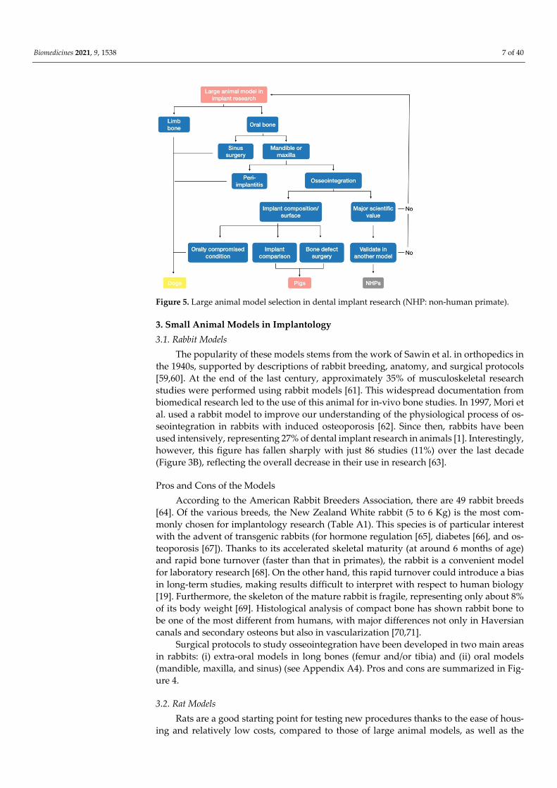
1), a new level of decisional procedures has emerged regarding study objectives, with the establishment of sub-classes according to species-specific characteristics of each large an-imal model. Pre-clinical surgical procedures (e.g., sinus or bone augmentation) with hu-man-sized implants on large animals can be categorized (Figure 5): • NHPs are no longer used in Europe and are only used elsewhere in already accred-
ited procedures. NHP models, particularly the baboon, should be considered a con-firmation model reserved for studies on major advances providing substantial added scientific value, already validated in another model.
• Pigs and minipigs are the new pioneers, having replaced dogs in procedures. The minipig appears to be an ideal model for studies of bone regeneration around dental implants when placed at intraoral sites.
• Dogs should only be used when pigs cannot be used to address the question of inter-est (mainly for compromised oral conditions, sinus surgery, and peri-implantitis pro-cedures). In particular, dog models should be preferentially employed for studies conducted under compromised oral conditions (biofilm).
Biomedicines 2021, 9, 1538 7 of 40
Figure 5. Large animal model selection in dental implant research (NHP: non-human primate).
3. Small Animal Models in Implantology 3.1. Rabbit Models
The popularity of these models stems from the work of Sawin et al. in orthopedics in the 1940s, supported by descriptions of rabbit breeding, anatomy, and surgical protocols [59,60]. At the end of the last century, approximately 35% of musculoskeletal research studies were performed using rabbit models [61]. This widespread documentation from biomedical research led to the use of this animal for in-vivo bone studies. In 1997, Mori et al. used a rabbit model to improve our understanding of the physiological process of os-seointegration in rabbits with induced osteoporosis [62]. Since then, rabbits have been used intensively, representing 27% of dental implant research in animals [1]. Interestingly, however, this figure has fallen sharply with just 86 studies (11%) over the last decade (Figure 3B), reflecting the overall decrease in their use in research [63].
Pros and Cons of the Models According to the American Rabbit Breeders Association, there are 49 rabbit breeds
[64]. Of the various breeds, the New Zealand White rabbit (5 to 6 Kg) is the most com-monly chosen for implantology research (Table A1). This species is of particular interest with the advent of transgenic rabbits (for hormone regulation [65], diabetes [66], and os-teoporosis [67]). Thanks to its accelerated skeletal maturity (at around 6 months of age) and rapid bone turnover (faster than that in primates), the rabbit is a convenient model for laboratory research [68]. On the other hand, this rapid turnover could introduce a bias in long-term studies, making results difficult to interpret with respect to human biology [19]. Furthermore, the skeleton of the mature rabbit is fragile, representing only about 8% of its body weight [69]. Histological analysis of compact bone has shown rabbit bone to be one of the most different from humans, with major differences not only in Haversian canals and secondary osteons but also in vascularization [70,71].
Surgical protocols to study osseointegration have been developed in two main areas in rabbits: (i) extra-oral models in long bones (femur and/or tibia) and (ii) oral models (mandible, maxilla, and sinus) (see Appendix A4). Pros and cons are summarized in Fig-ure 4.
3.2. Rat Models Rats are a good starting point for testing new procedures thanks to the ease of hous-
ing and relatively low costs, compared to those of large animal models, as well as the

Biomedicines 2021, 9, 1538 8 of 40
extensive history of their use in scientific experiments [25]. Rat physiology, especially in bone tissue, suggests it would be useful for research in certain areas. The growing rat is a well-known model for evaluating the effects of endocrine, nutritional and environmental factors on peak bone mass but is not appropriate for adult human skeleton studies due to the presence of cellular pathways not present in human adults [72]. Bone mass gain, in parallel with the long bone elongation, mainly occurs during the first 6 months of life [72], though some authors consider that the long bone grows continuously for at least 1 year with a gradual transition from modeling to remodeling with age [73], a transition that does not occur uniformly across bones [74]. Due to this longitudinal bone growth, a mar-gin of at least 1 mm from the growth plate of the tibia should be left intact if experimen-tation starts around 10 months of age, an issue to be considered in dental implant studies [74]. Sex and hormones are also key parameters in rat research. At 8 months of age, males were found to have 22% greater bone width and 33% greater breaking strength than fe-males in the tibia [75]. The role of hormones has been put to good use in an ovariectomy model. Rats, as for any rodents, do not have natural menopause, but the ablation of the gonad is a good model for artificial menopause [76] and therefore for the analysis of osse-ointegration in a model of pathological bone.
Pros and Cons of the Models For implantology purposes, the size of the rat is a mixed blessing. The lower costs
and ease of housing and handling favor the use of this animal. On the other hand, only small human implants can be used, and most of the time, implants need to be adapted.
Two rat breeds are commonly used: (1) Wistar rats (from the Wistar Institute) are one of the oldest and still considered one of the best rat models. By adulthood, they reach 500 g [77] which places them in the mid-upper range of small laboratory animals. (2) Sprague Dawley rats, developed from Wistar rats, have an adult weight of up to 300 g [5] and are one of the breeds most widely used in pre-clinical studies [78]. They have been used as a model for osteoporosis, and for analyzing the effects of calcium supplementation on bones [79] (Table A1). Various protocols have been developed in rats depending on the implan-tation site (see Appendix A5). The number of research studies using rats has increased during the last decade (Figure 3A) and confirms the scientific interest in this model [80]. Pros and cons are summarized in Figure 4.
3.3. Mouse Models The mouse is the animal most commonly used in laboratory research. It was the first
laboratory animal model established for genetic- and aged-related changes in bone [81] and used for full genomic analysis.
Pros and Cons of the Models Among all mouse strains, C57BL/6 is the most commonly used, almost 20,000 papers
having been published referring to research using this strain in 2019 [82]. Within the same strain, different sub-strains show notable genetic and phenotypic differences [83]. It is therefore important to determine, when planning a research study, which type of animal is needed.
From a bone point of view, mice have similar growth characteristics to rats, with even more marked size-related advantages and disadvantages. It is therefore inappropriate to carry out an implant study in mice if the same model has been developed in rats. None-theless, mice have some characteristics which distinguish them from other laboratory an-imals.
The main advantage they offer over other small animal models is the existence of numerous knockout and transgenic mice. This factor is even more important with the emergence of new tools to develop genetically engineered mouse models. Transfection or viral vector transduction are routinely applied methods for random DNA integration [84],
Biomedicines 2021, 9, 1538 9 of 40
while the CRISPR-cas9 system for gene editing [85] is an emerging technology that ex-tends the scope of research in this field [86].
In implant studies, this model has been used for a long time but primarily for extra-oral approaches due to technical and surgical complications [4], the reason most often mentioned being the difficulty of access due to the mouth size and range of opening of mice. Some authors opted to develop a more accurate model by working on the mouse maxilla [87]. A limitation of this model is the limited cortical bone remodeling and the lack of the Haversian structure in cortical bones. Indeed, rodent long bones are mainly composed of primary bone and a minimal proportion of cancellous bone [6]. The counter-part of this biological issue is the small amount of cancellous bone site for implantation studies [88].
As for rats, protocols are markedly heterogeneous, and it has not been possible to establish a gold standard (See Appendix A6). Pros and cons are summarized in Figure 4.
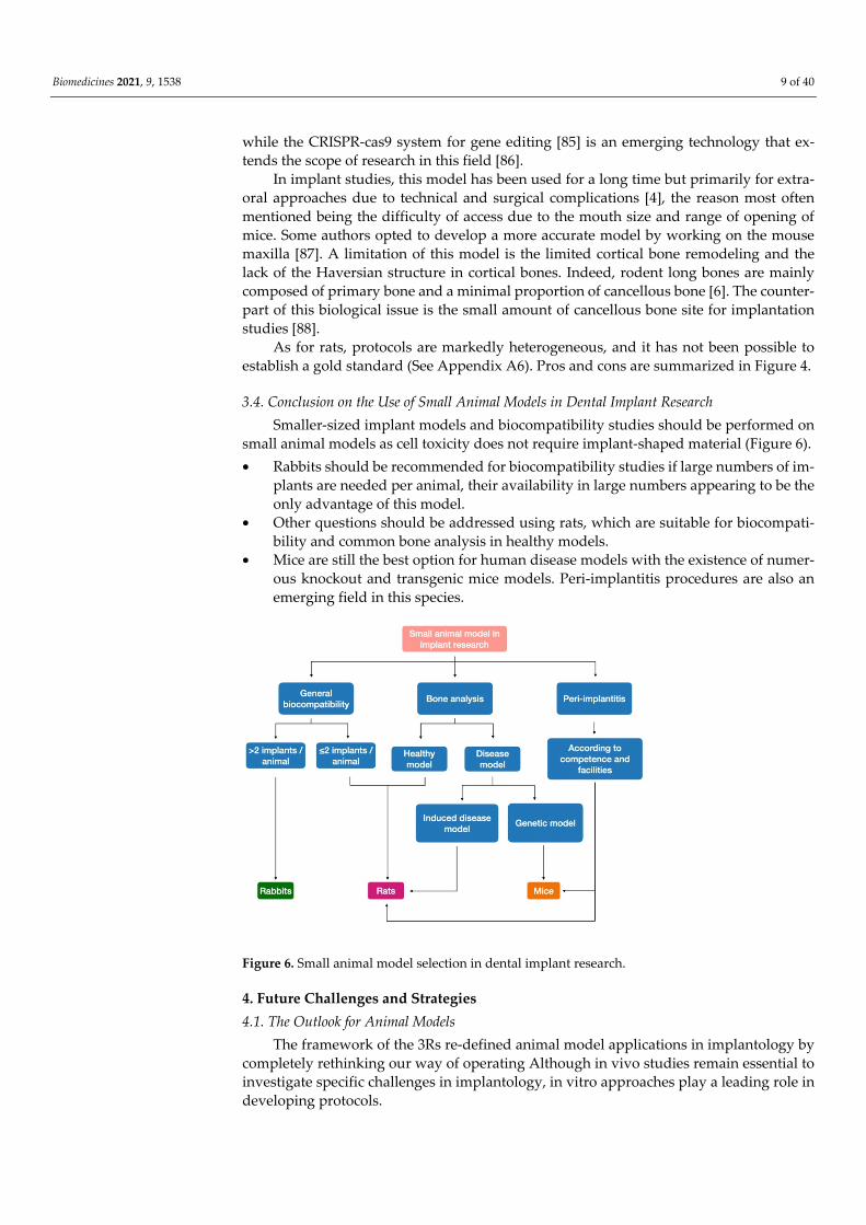
3.4. Conclusion on the Use of Small Animal Models in Dental Implant Research Smaller-sized implant models and biocompatibility studies should be performed on
small animal models as cell toxicity does not require implant-shaped material (Figure 6). • Rabbits should be recommended for biocompatibility studies if large numbers of im-
plants are needed per animal, their availability in large numbers appearing to be the only advantage of this model.
• Other questions should be addressed using rats, which are suitable for biocompati-bility and common bone analysis in healthy models.
• Mice are still the best option for human disease models with the existence of numer-ous knockout and transgenic mice models. Peri-implantitis procedures are also an emerging field in this species.
Figure 6. Small animal model selection in dental implant research.
4. Future Challenges and Strategies 4.1. The Outlook for Animal Models
The framework of the 3Rs re-defined animal model applications in implantology by completely rethinking our way of operating Although in vivo studies remain essential to investigate specific challenges in implantology, in vitro approaches play a leading role in developing protocols.

Biomedicines 2021, 9, 1538 10 of 40
Overlaying diagrams by model size (small or large) shows that models within each category offer similar characteristics. Interestingly, rating comparable criteria for each model, (i) although mice are small, the mouse model seems to have numerous advantages for implant studies and (ii) in vitro/in silico models and analyses of biomaterial properties are ranked top, highlighting their great potential in the field of dental implantology (Fig-ure 7).
Figure 7. Overall appeal assessed by animal model. Diagram based on key elements of interest in pre-clinical research: cost, housing and husbandry requirements, protocol duration, biological relevance, N [implants] per animal, ethical issues, surgical relevance, and resemblance to the human implant model. For each criterion, the models were ranked (from best [1, outer line] to worst [6, inner line] (Table A3). Overlaying diagrams by animal size also shows that small and large models have very similar characteristics to others in the same size group. The diagrams indicate that the in vitro/in silico and biomaterials study type was ranked first for four criteria (cost, housing and husbandry requirements, ethical issues, and protocol duration) and last for two criteria (surgical relevance and implant model), as they are not addressed by such studies, while two criteria were not applicable to this last category (biological relevance and number of implants per ani-mal). Comparison with the global diagram shows that the mouse model diagram is the closest to that of the in vitro/in silico/biomaterial models.
4.2. Development of Replacement Strategies The principles of the 3Rs were developed over 50 years ago, aiming to encourage
alternatives to animal testing and improve animal welfare and research quality where the use of animals could not be avoided.
4.2.1. In vitro Biocompatibility and Cytotoxicity Analyses In vitro human or animal cell-based studies on modified surfaces for dental implants
allow the assessment of toxicity and characterization of osteoblast adhesion to the im-plant, or the impact of any added processing steps on the implant surface [89,90]. For ex-ample, when a new nanoparticle treatment is developed, in vitro studies are needed to test implant treatment viability during cell interaction [91]. Similar studies are needed to

Biomedicines 2021, 9, 1538 11 of 40
test bioactive [92–94] or peptide (e.g., RGD [95]) coatings, or the incorporation of antibiot-ics [96] or growth factors (e.g., bone morphogenetic proteins [97]).
In vitro studies can also assess the impact of given clinical methods on a device [98]. By applying a procedure directly to the implant, for example, studying the effect of vari-ous polishing methods on bacterial colonization [99–101], the need for an animal model can be reduced and it may be easier to focus on the interaction of interest. As titanium is the main material used in dental implant surgery, experiments have mainly focused on titanium powder or titanium disks. For example, in vitro studies showed that some sur-faces induce the generation of toxic particles, certain surfaces being more toxic than others to oral epithelial cells [102]. Other materials, such as zirconium implants, have also been tested for cell biocompatibility and mechanical properties [103].
4.2.2. In Vitro Models of Response to Implant and Associated Biofilm For more biological issues, in vitro models were developed at the end of the 1990s to
assess the possibility of answering simple questions without the use of animals [104,105]. According to Mombelli et al., in vitro models were relevant for studying: (i) the reaction of micro-organisms to the presence of implants, (ii) the reaction of implant-associated mi-cro-organisms to antimicrobial agents, and finally, (iii) the reaction of the host tissues to the presence of implants contaminated with micro-organisms [105,106]. In vitro studies have subsequently been applied to the issues of hypersensitivity, and immune and pro-inflammatory responses [107]. Biofilm assays are also a common in vitro procedure for the analysis of antibiotic resistance. Adapted to implantology, complete implants or tita-nium chips are used in cell culture in the presence of bacteria, allowing the analysis of bacteria adhesion and biofilm construction. Once the biofilm is stable, chlorhexidine [108] or antibiotics can be added to the medium to observe their performance. In the same pro-cess, infected implants can be placed in contact with animal or human cells to study their interaction [106].
4.2.3. In Vitro Physical and Mechanical Evaluation Once biocompatibility and cytotoxicity have been demonstrated, protocols for phys-
ical and mechanical testing can assess the resilience to different loads in pre-load models, monitoring numerous variables related to the implant–abutment connection [109], such as force used for screw tightening [110], ability to withstand a long-term load (assessed by direct strength testing) [111], and chewing cycle (simulated by an artificial mouth) [112,113]. Further, esthetic comparisons can be made in terms of abutment titanium visi-bility [114].
Finally, in vitro rather than animal models should be used to study the improvement and/or development of future technologies, such as computer-guided navigation for im-plant placement [115], laser procedures [116], or comparisons between different scanning methods [117].
The 3Rs are now widely embedded in national and international legislation and reg-ulations on the use of animals in scientific procedures. Following these principles, before conducting an in vivo study, it has to be considered whether it is possible to replace the use of animals with alternative methods. Indeed, in implantology, before any in vivo test, in vitro analysis is essential for implant development. Before choosing which animal is needed for a procedure, the first decision is whether we can avoid using animals at all (Figure 8).

Biomedicines 2021, 9, 1538 12 of 40
Figure 8. Research protocol selection in dental implant research: in vivo vs. in vitro.
5. Conclusions Biological advances in large animals have narrowed the gap between large and small
animal model applications, as it is now possible to perform genetic analysis in dogs while it was previously only possible in small animal models, and at the same time, technolog-ical advances have enabled reductions in instrument size, and therefore the manufactur-ing of small implants is compatible with mouse size. The distinctions between these two groups are small, but the specificities of dental implant models allow rational decisions concerning their use to maximize scientific impact and benefits.
Last but not least, nowadays any decision-making process dealing with animal sac-rifice in research raises the key question of its scientific necessity, especially in the devel-opment of dental implant protocols dealing with elective surgeries (Figure 4). Considera-ble efforts have been recently made to replace animal studies with in vitro studies, which enable mechanical and physical characterization of dental implants. Thus, when the ques-tion of the use of animals in implant surgery research nowadays arises, another question must always follow: “can we do otherwise?”
Funding: This study was funded by the Agence Nationale de la Recherche (grant Hyposkel 18-CE14-0018-01); and Fondation pour la Recherche Médicale (grant DGE20111123012); Nicolas Banc-Sylvestre was supported by the Fondation pour la Recherche Médicale (grant FDM201906008500).
Acknowledgments: The authors thank the support of the Agence Nationale de la Recherche and Fondation pour la Recherche Médicale
Conflicts of Interest: The authors declare no conflict of interest.
Appendix A Appendix A1. Research in Non-Human Primates
In 2010, the European Parliament issued a directive which is still in force: “The use of non-human primates should be permitted only in those biomedical areas essential for the benefit of human beings, for which no other alternative replacement methods are yet available” [118].
Furthermore, there are risks associated with handling due to the possibility of zoon-otic disease transmission [41,119] but also biological and behavioral responses due to stressors such as separation from their familial environment [120].

Biomedicines 2021, 9, 1538 13 of 40
The duration of a conventional protocol is around 6 to 9 months with a first healing time of 3 months after tooth extraction and 3 to 6 months after implantation (Figure 2). Protocols are usually performed on adult animals, from 7 to 10 years old, this allowing the use of human-sized implants (Figure 5). Laboratory breeding and reproduction are therefore not feasible, and animals are acquired for the protocol. Such studies have inves-tigated the healing process after sinus floor elevation [121–123], improvements of analysis techniques [124], and clinical questions concerning soft-tissue response around combined tooth–implant-supported prostheses [125,126]. Old World monkeys such as baboons, mandrills, and macaques are preferred, as their long bones have a dense Haversian struc-ture, with thin layers of endosteal and periosteal bone [127]. For anatomical reasons, the use of Rhesus macaques has to be avoided, their adult size and weight (6.5 to 12 kg vs. 21.5 kg for male baboon) [5] being too small to be considered a “large animal model”.
Summary: In accordance with international legislation, NHPs should no longer be used except for the assessment of major innovations or new treatments already validated in another large animal model (Figure 5).
Appendix A2. Research in Canine Models The most commonly used dog breed is the beagle due to the ease of care. Its mean
adult weight is around 16 kg [5] allowing the use of human-sized implants. For this type of study, all animals are acquired from accredited laboratories. Mainly carried out in long bones in the past, protocols have now been developed for oral bone models which have more appropriate characteristics for the analysis of osseointegration. Similarly, peri-im-plantitis has been successfully transposed to oral implant studies, facilitating the use of oral bone models (Table A1).
The duration of a conventional protocol is slightly shorter than in NHPs, with a first phase of 3 months after tooth extraction and 2 to 3 months of implant healing (Figure 2).
Table A1. Summary of species characteristics and use in implant protocols.
Non-Human Primates Pigs Canines Rabbits Rats Mice
Species most frequently
used
Baboon, mandrill and
macaques
Pigs: Domestic pigs Minipigs:
Hanford of Göttingen breed
Beagle New Zealand White rabbit
Wistar rats, Sprague Dawley rats
C57 Black/6
Age of use 7 to 10 years
old 2 to 3 years old 1 to 2 years old
6 to 9 months old
2 to 3 months old 8 weeks old
Protocol duration
6 to 9 months 12 months 5 months
2 to 4 weeks (long bone)
Up to 3 months (oral
bone)
2 to 6 weeks (long bone) 2.5 months(oral bone)
4 weeks (long bone) 2 to 3 months (oral bone)
Weight 21.5 kg Pig: 350 kg
Mini-Pig: 35 to 95 kg
15 kg 5 to 6 kg Sprague dawley: 70 to 300 g Wistar rats: up to 500 g
30 g
Implant size Human-sized Human-sized Human-sized Human-sized
Adapted implant
Adapted implant: 1.5 mm diameter, 2.5 mm length
Adapted implant: 1 mm diameter, 2 to 3 mm length
(long bone) 0.6 mm diameter, 2 mm length
(maxilla)
Trend Falling into
disuse
Any study related to
implant surgery under healthy
conditions
Peri-implantitis, sinus and
genetic studies
Falling into disuse
Systemic conditions (diabetes, hormones), poor bone quality models, ease
of breed and use
Genetic studies, knock-out protocols, peri-implantitis

Biomedicines 2021, 9, 1538 14 of 40
Appendix A2.1. Long Bone Models To evaluate implant osseointegration, dental implants were placed in dog leg bones,
but this is rare nowadays, less than 20 studies having been reported over the past 10 years (Table A2).
Despite an obvious bias of studying implants loaded on a quadrupedal gait model, protocols involving implant placement on limbs allow the use of a large number of im-plants, thus reducing the sample size (in one study, up to 75 implants having been tested in the radius of just 6 dogs [128]). Though front limbs can also be used, [128] most of the time both tibias are used, as they offer a large quantity of bone. For this kind of study, implants 3.75 mm in diameter and 10 mm in length are the most common, two to three implants being used per tibia [129]. The large amount of bone available allows the creation of surgically created defects to analyze bone regeneration associated with dental implants. Properties of membranes [129] or new grafting compounds [130] have been tested. New implant devices such as implant extenders have also been tested before clinical use [131]. The proximal tibia is commonly used for drilling protocols, to test the impact of drilling in early stages of osseointegration and implant stability [132–137], different implant sur-faces [138–142], and biomechanical properties (insertion torque [133], response to com-pressive stress [143]).
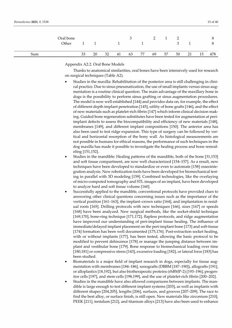
Table A2. Distribution of implant location by animal and year over the past 10 years based on research articles in MED-LINE, including all experimental implant studies on animals.
2010 2011 2012 2013 2014 2015 2016 2017 2018 2019 2020 Sum Canines 12 4 17 18 24 40 37 26 20 10 7 215
Long bone 1 5 3 2 3 2 1 2 19 Oral bone 12 3 12 15 22 37 35 25 18 10 7 196
Non-human primates 1 1 1 1 4 Oral bone 1 1 1 1 4
Mice 1 2 3 3 1 5 5 2 1 23 Long bone 1 1 1 1 1 1 1 7 Oral bone 1 2 2 1 4 4 1 15 Other 1 1
Pigs 4 1 4 6 10 7 11 7 3 1 54 Long bone 1 1 2 4 Oral bone 4 1 3 2 9 4 10 2 3 38 Skull 1 4 1 2 3 1 12
Rabbits 6 6 5 4 14 12 8 10 11 6 4 86 Long bone 6 6 5 4 11 9 6 10 9 4 3 73 Oral bone 3 2 2 2 2 1 12 Skull 1 1
Rats 6 7 5 9 7 11 7 7 5 1 3 68 Long bone 3 5 4 7 4 9 6 6 4 1 1 50 Oral bone 3 2 2 1 2 1 1 1 2 15 Skull 1 2 3
Other 3 2 2 5 3 4 2 6 1 28 Long bone 2 1 1 2 2 2 1 1 12
Biomedicines 2021, 9, 1538 15 of 40
Oral bone 3 2 1 2 8 Other 1 1 1 1 3 1 8
Sum 33 20 32 41 63 77 69 57 50 21 15 478
Appendix A2.2. Oral Bone Models Thanks to anatomical similarities, oral bones have been intensively used for research
on surgical techniques (Table A2). • Studies in the maxilla: Rehabilitation of the posterior area is still challenging in clini-
cal practice. Due to sinus pneumatization, the use of small implants versus sinus aug-mentation is a routine clinical question. The main advantage of the maxillary bone in dogs is the possibility to perform sinus grafting or sinus augmentation procedures. The model is now well established [144] and provides data on, for example, the effect of different depth implant penetration [145], utility of bone grafts [146], and the effect of new materials such as platelet-rich fibrin [147] which inform clinical decision mak-ing. Guided bone regeneration substitutes have been tested for augmentation at peri-implant defects to assess the biocompatibility and efficiency of new materials [148], membranes [149], and different implant compositions [150]. The anterior area has also been used to test ridge expansion. This type of surgery can be followed by ver-tical and horizontal resorption of the bony wall. As histological measurements are not possible in humans for ethical reasons, the performance of such techniques in the dog maxilla has made it possible to investigate the healing process and bone remod-eling [151,152].
• Studies in the mandible: Healing patterns of the mandible, both of the bone [31,153] and soft tissue compartment, are now well characterized [154–157]. As a result, new techniques have been developed to standardize or even to automate [158] osseointe-gration analysis. New robotization tools have been developed for biomechanical test-ing in parallel with 3D modeling [159]. Combined technologies, like the overlaying of micro-computed tomography and STL images of an implant, have been developed to analyze hard and soft tissue volume [160].
• Successfully applied to the mandible, conventional protocols have provided clues to answering other clinical questions concerning issues such as the importance of the vertical position [161–163], the implant–crown ratio [164], and implantation in resid-ual roots [165]. Drilling protocols with new techniques [166], sizes [167] or speeds [168] have been analyzed. New surgical methods, like the socket-shield technique [169,170], bone-ring technique [171,172], flapless protocols, and ridge augmentation have improved our understanding of peri-implant tissue healing. The influence of immediate/delayed implant placement on the peri-implant bone [173] and soft-tissue [174] formation has been well documented [175,176]. Post-extraction socket healing, with or without implants [177], has been tested, allowing the basic protocol to be modified to prevent dehiscence [178] or manage the jumping distance between im-plant and vestibular bone [179]. Bone response to biomechanical loading over time [180,181] or compressive stress [143], excessive loading [182], or lateral force [183] has been studied.
• Biomaterials is a major field of implant research in dogs, especially for tissue aug-mentation with membranes [184–186], xenografts (DBBM [187–190]), allografts [191], or alloplastics [18,192], but also biotherapeutic proteins (rhBMP-2) [193–196], progen-itor cells [197], and stem-cells [198,199], and the use of platelet-rich fibrin [200–202].
• Studies in the mandible have also allowed comparisons between implants. The man-dible is large enough to test different implant systems [203], as well as implants with different shapes [204,205], lengths [206], surfaces, and grooves [207–209]. The race to find the best alloy, or surface finish, is still open. New materials like zirconium [210], PEEK [211], tentalum [212], and titanium alloys [213] have also been used to enhance

Biomedicines 2021, 9, 1538 16 of 40
osseointegration. Implant surface properties is a field that attracts the attention of many researchers. Old techniques have been improved with the addition of mole-cules like magnesium [214], plasma projection [215], or chemical treatment [216] and new techniques have been developed with nanocoatings [217] or biofunctionaliza-tion [218]. Comparisons have been made between implants with differing abutment shape [219,220], composition [221,222], or microstructure, [223] and different proto-cols, e.g., platform switching [224,225], have been tested to support the best peri-im-plant tissue healing.
• Finally, only a few articles were found combining implants and drugs. Pilot studies have been performed to test topical use of implant surface treatments with melatonin coating or vitamin D [226,227] and the efficacy of mouth rinses for prevention of peri-implant mucositis and peri-implantitis, and more recently, the impact of hyperbaric oxygen on tissue healing was analyzed [228]. From a systemic point of view, vaccines have been developed seeking to prevent alveolar bone loss in peri-implantitis [229,230].
• Contribution of peri-implantitis studies in dog models to implantology: A specific strength of this model is the ability to perform periodontitis and peri-implantitis pro-tocols, the dog being the large animal model most widely used in periodontitis stud-ies [231]. A new line of research is the characterization of the peri-implantitis micro-biota and changes therein during and after ligature placement, as well as after treat-ment [232–235]. Silk and cotton ligatures have been extensively used to initiate plaque formation and therefore an inflammatory process in gingival tissues [236]. New protocols have been developed to accelerate or exacerbate the inflammatory process [237]. For their flexibility and ease of handling, stainless steel ligatures have been proposed to replace soft ligatures which can be tricky to place and retain on the implantation site in the long term. Immediate induction of peri-implantitis has shown similar results to conventional methods with a shorter 6-month protocol [238].
• A better understanding has been obtained of implants’ susceptibility to bacterial con-tamination depending on the surface condition or composition [239] and character-istics [240–242] and tools have been developed to treat them mechanically (Ti-Brush [243], ER:YAG laser [243]) or with drugs (antimicrobials [244], chlorexidine [245], mouth rinse [246], or even plasma [247]) and to reconstruct bone tissue lost [248]. Summary: A natural tendency to develop periodontal disease and the ease of repro-
ducing peri-implantitis make the dog a strong model for dental implant research. None-theless, animal care regulations and husbandry requirements (exercise, need for environ-mental enrichment) [249] limit the use of dog models to research for which other animal models cannot be used (e.g., large models of peri-implantitis, microbiological studies) (Figure 5, Table A3). Interestingly, the emergence of new evidence demonstrating the sim-ilarity of dogs and humans in rare diseases [250] widens the field of application of this model, building a dual model with advantages of both large models (clinically relevant) and small models (genetic disorders).
Table A3. Overall appeal weighted by animal model. Rankings for each criterion from 1 to 6.
Models Cost Housing/Hus-
bandry Require-ment
Biological Interest
N per Ani-mal
Ethical Is-sues
Protocol Duration
Surgical Relevance
Implant Mo-del Total
Mice 1 1 1 5 1 1 5 6 21 Rats 2 1 4 4 2 2 4 6 25
Rabbits 3 3 6 3 3 3 3 3 27 Pigs 4 4 4 1 4 5 2 1 25
Canines 5 5 3 2 5 4 2 1 27 Non-human pri-
mates 6 6 2 2 6 6 1 1 34

Biomedicines 2021, 9, 1538 17 of 40
In vitro/in silico/bio-materials
1 1 - - 1 1 7 7 /
Appendix A3. Research in Pig Models Usually, 2- to 3-year-old animals [250] are used for implant insertion 3.5 months after
tooth extraction and analysis 8.5 weeks after implantation, if a second stage is needed (Figure 2). Due to the substantial bone thickness and the presence of numerous adequate sites in the oral region, tibias have only been used in a few studies. Similarly, several pro-tocols were developed in the skull (frontal bone or calvaria) but have now mainly been transposed to oral bone. The large amount of bone in the maxilla and mandible allows the use of several implants in a single protocol, and the performance of implant surgery in bone with more appropriate properties.
Appendix A3.1. Oral Bone Models Due to substantial oral bone thickness and the presence of other more appropriate
areas, tibias have only been used in a few studies (Table A2). On the other hand, they allow researchers to (i) insert multiple implants in the same bone (up to 6 per tibia [251,252]), given their large surface area, and (ii) use a split design with both tibias, reduc-ing the number of animals required.
Implant surgery can be performed following human protocols and using human-sized implants (mean of 4 mm in diameter and 11 mm in length). Therefore, primary sta-bility, implant stability quotient, and removal torque can be measured in the same way as in clinical practice [252]. This model can also be used to test the viability and mechanical properties of new implants [253] or compare different models [251,254].
Appendix A3.2. Skull Bone Models Reports have been found of several studies the skull (frontal [255] or in calvaria
[256]). These models allow implant osseointegration and healing without mastication con-straints or tongue movement and are suitable for multiple-step protocols [257] or surgery with large instruments. This area also permits the implantation of up to 12 devices in the same animal. Nonetheless, the length of the implant has to be carefully selected to avoid brain damage. In terms of protocols, the scope for research is similar to that for the man-dible: surface conditioning [258–261], implant composition [262], implant macro-design [263] and abutment [264], pull-out strength [265], and bone augmentation with [256] or without grafts [266], but also diseases (e.g., diabetes [267]).
Appendix A3.3. Oral Bone Models Most pig oral bone models use either the maxilla or mandible for implantology re-
search. Not surprisingly, the maxillary sinus has been poorly studied due to its position (distal to the last molar) making it difficult to access. The maxilla and mandible can be used in the same protocol, often with teeth extractions in all four quadrants to maximize the number of implants [268]. In systemic disease processes, it seems that the pig model is poorly suited and only used for obesity/metabolic syndrome and/or diabetes [269–271]. Peri-implantitis also appears to have been poorly investigated [272].
A range of surgical techniques can be performed, such as (i) site preparation with different osteotomies [273], (ii) flap procedures [274,275], or (iii) bone surgery with oste-odistraction [276], creation of bone defects [277], and bone grafting [271,278,279]. Bio-materials such as scaffold [280] and disc-shaped matrices for vertical bone regeneration [254,280] have been tested in pigs prior to use in human surgery. Biological coating func-tionalization with chondroitin sulfate and sulfated hyaluronan with collagen molecules [281–283] is often tested on this model, avoiding the process of miniaturization of a new model to be put on the market.

Biomedicines 2021, 9, 1538 18 of 40
In oral bones, implants are almost always placed in a socket or healed site after tooth extraction [284]. There are numerous protocols for healing time (ranging from 9 weeks [285] to 8 months [273]), with a mean time of 3–4 months after tooth extraction. Notably, 3 to 4 implants can be anchored per quadrant [284] with human or human-like implants 4 mm in diameter and 11 mm in length [286].
As for the tibia, the oral model may be useful for histological analyses such as cyto-toxicity and viability of new compositions [287–290] but also for assessments on 1:1 scale models of mechanical properties [47,268,291], stress distribution [287,288], and the effects of different implant and abutment shapes [264,284,292–295]. Thanks to pigs having similar bone properties to those of humans, resistance to insertion [273] or removal torque can be measured during [296] or after healing [268]. It is possible to perform classical analysis such as measurement of bone area [273], marginal bone level [273], crestal bone loss [275],, resonance frequency [273], or implant stability quotient [297]. However, the main reason for selecting this site is almost always its suitability for bone-to-implant contact analysis [273,281,282,286,289,290,294]. This model is also used to assess the accuracy of new tech-niques, such as ultrasound imaging [298], comparison of intra-oral and cone beam com-puted tomography [299,300], magnetic resonance imaging [301], and resonance frequency analysis [302]. In addition, it can be used to investigate not only bone but also soft tissues [295] as in immunohistochemical analysis of blood vessels in peri-implant mucosa [274], cell quantification, and fiber orientation [292], or pangenomic gene-expression analysis [293] of implant tissues.
Summary: Supported by a strong history of use, pig oral bone models are suitable for pre-clinical studies, allowing the testing of different implant surface properties or sur-gical procedures under physiological conditions. The benefits of minipigs in terms of costs and ease of housing make them the model of choice, overcoming the limitations of other large animal models (Figure 7).
Appendix A4. Research in Rabbit Models Experiments are usually shorter than those in large animal models: (i) as previously
described, bone maturity occurs at around 6 months of age, and hence, protocols use rab-bits from 6 to 9 months of age, (ii) surgery is mainly performed in long bones, and hence, no healing time is needed, unlike protocols involving tooth extraction, and (iii) bone heal-ing is faster. Protocols can stop 2 to 4 weeks after implantation for bone-to-implant contact analysis (Figure 2).
Appendix A4.1. Long Bone Models The great majority of studies in rabbits correspond to long bone experiments (69/86),
with 50/69 on the tibia and 19/69 on the femur (Table A2). • Protocols using the tibia: The anatomy and histology of rabbit tibia are well known,
the rabbit being used for the first attempt to develop an animal model of osteomye-litis [303] for bone fracture analysis [304]. Numerous research protocols have been developed on the tibia, as the relatively good volume accessible has allowed analysis of as many as 112 implants in 28 rabbits in one study [305]. In this area, 3- to 4-mm diameter implants can be used with lengths of up to 7 mm [306]. Rabbit tibia has been widely used to analyze the osseointegration of zirconia implants [307], titanium–zir-conium implants [308], implants coated with calcium carbonate [309], and implants with surface modifications [306,310,311].Bilateral procedures are generally described including (i) two implants per animal with one implant in each tibia [311]; (ii) four implants per animal with two implants in each [312] or (iii) six implants per animal with three implants per tibia [313,314]. The metaphysis and diaphysis of the bone can be used. Thanks to fast healing, osseointegration can be analyzed 1 month after im-plantation [312]. The tibia has also been used for drilling studies seeking to improve implant stability [315] with drilling speed [316] or drill diameter and implant torque

Biomedicines 2021, 9, 1538 19 of 40
[312] analysis. The large volume of the tibia and ease of surgery have allowed this bone to be used for the creation of peri-implant defects [317] and the use of a bone substitute model [318] and spacers [319], as well as for pathophysiological purposes, mainly for reduced bone models (osteopenic or osteoporotic conditions) [320–323].
Environmental parameters have been investigated in contexts such as a high-fat diet [324] and irradiation [325].
• Protocols using the femur: the femoral bone has been chosen by researchers for many reasons: Easy access and the small amount of soft tissue [326]. Rabbit long bones are composed of 70%–80% compact bone [68], allowing good implant stability.
• The femur is thicker than the tibia and the medullary space is large [326], allowing multiple implant fixations [63]. Experiments can also be performed on both sides of the knee (distal part of the femur and proximal part of the tibia) [327]. The disad-vantages of this model are related to the general differences between humans and rabbits as mentioned above. In particular, rabbit long bones show a distinctive phys-iological variability of the bone architecture with a longitudinal vascular pattern [26]. Another point to consider is the age of the animal. Indeed, due to endosteal bone remodeling, the bone shows cortical thinning and an increase in bone marrow vol-ume by as much as 24% with age [328]. It has also been reported that rabbit bone marrow contains a significant proportion of adipose tissue [5], a characteristic not present in the oral cavity in humans, and this reduces the usefulness of the model.
Appendix A4.2. Skull Bone Models For the calvaria, only one study has been found. It sought to analyze the influence of
nonsteroidal anti-inflammatory drugs on osseointegration of dental implants in the cal-varia [329]. The model found no significant differences in the use of this type of drug and only one rabbit out of the 19 used died in the postoperative period.
Appendix A4.3. Oral Bone Models The rabbit skull is mainly composed of spongy bone and contains wide spaces [69].
The mandible is formed by two symmetrical bones joined by a fibrous or fibrocartilagi-nous symphysis. As for humans, two parts can be described: (i) the horizontal part, which houses the teeth, and (ii) the vertical posterior part, called the ramus. The maxilla is also formed by two bones fused on the sagittal line. The dental formula of the rabbit is: two pairs of incisors on the maxilla and one on the mandible; no canines; three pairs of pre-molars on the maxilla and two on the mandible; and two to three pairs of molars on the maxilla and three on the mandible. The total number of teeth ranges from 26 to 28 [69]. Rabbit incisors grow continuously [330], which is of great interest for longitudinal miner-alization studies [331].
As previously described, oral bone, in addition to the obvious difficulty of access due to the small mouth opening, has a poor cortical/spongy bone ratio with mainly spongy bone and wide spaces. There is a paucity of literature in this area and the models seem to be avoided by researchers (Table A1). Out of the 86 studies, 12 were performed in oral bone: 3 in the maxilla, 1 for sinus augmentation, and 8 in the mandible (Table A2). • Studies in the maxilla: Studies in the maxilla are mainly used for related sinus aug-
mentation therapies. Medications such as anti-inflammatory drugs and painkillers can be tested for postoperative pain [332]. Newly formed bone height is measurable following sinus floor elevation using a blood clot [333] or for pre-clinical testing of new bone substitute [334], giving an idea of how such materials are accepted in in vivo models. The poor bone quality of the maxillary sinus is also exploited for stud-ying the impact of innovative surface properties in poor quality bone [335].
• Studies in the mandible: Procedures are short, immediate extraction/implantation protocols being the most common. The healing period after implantation is at least 3 weeks [336] and up to 3 months [337]. The incisor area provides a great volume for

Biomedicines 2021, 9, 1538 20 of 40
osseointegration in immediate extraction/implantation protocols with 3-mm diame-ter and 12-mm length implants [338]. Except for the study by Schiegnitz et al. using 9-month-old New Zealand rabbits (4–5 kg) [336], the age of rabbits is generally not specified accurately; rather, it is reported as “adult age” which corresponds to 2.5 to 6 Kg. Only one study found was performed in younger rabbits (4 months old), these having been exposed to fluoride since 2 months of age [337]. Studies in the rabbit mandible have been used to assess osseointegration of implants with different sur-face properties [339] or positions [336,340], and the systemic effect of exposure to molecules like fluoride [337] or the effect of thyroid hormone production [65]. It should be noted that one proof of concept study for a peri-implantitis model was conducted on the first mandible anterior tooth with silk ligatures in 2015 (reported only in Chinese, except for the abstract [341]). In the mandible, an extra-oral approach by opening a flap from the skin to the mandible angle has been used for vertical bone growth, making it possible to extend the scope of already known materials [342]. The great volume available in this area allows the use of human-sized implants, with a length of 8 mm and a diameter of 4.1 mm. Summary: Rabbit models have an extensive history of use and well-developed pro-
tocols in studies in orthopedics and more recently periodontal diseases. The weak bone quality around the oral cavity, however, limits the use of long bone in this model for im-plantology, biocompatibility, or osteoinduction [6].
Nonetheless, rabbit models allow analysis of numerous implants per animal, thereby reducing the total number of animals needed per protocol (Table A1). Their application remains limited due to the small number of genetic models.
Appendix A5. Research in Rat Models Protocol durations depend on the site, with substantial differences between studies.
The average age of animals is around 2 to 3 months for the first surgery. For long bone procedures, 2 to 6 weeks is needed before assessing osseointegration. In the case of im-plant placement at a healed extraction site, 1.5 months of healing is generally allowed after extraction and another month for implant osseointegration (Figure 2). In the maxilla, pro-tocols are shortened, with implantation performed immediately after extraction.
Appendix A5.1. Long Bone Models • Studies in the tibia: The rat tibia is suitable for bone implantation due to the ease of
access and relatively good volume. Notably, 32 out of the 68 studies found have been performed with this bone. The medial tibial plate of the bone is commonly used as it is flat and can receive implants [343]. Depending on the prototype used, the number of tests and their size differ. At least one implant per tibia can be tested with a nearly human-size implant (2.0 mm in diameter and 4- to 5-mm in length [83,344–346]). On this kind of model, bi-cortical anchorage can be achieved. For multi-implant proto-cols, a diameter of 1.5 mm and length of 2.5 mm are more appropriate [343,347].
• Research using the rat tibia model has commonly investigated the effects of the im-plant surface on osseointegration [343,344], but a new trend has emerged, with grow-ing numbers of studies in the areas of the drug delivery and/or physiopathology: effects on osseointegration of different doses of drugs in rats that are healthy [345] or have certain diseases, e.g., diabetes [348–354], or in peri-implant bone defects [346]. Nevertheless, the framework of choice is implantation osseointegration in poor qual-ity bone with drug treatments [355,356]. Research into bisphosphonates [347,355,357–359] or agonists like selective estrogen receptor modulators [347,360] is typically con-ducted in the rat as it is an excellent model of osteoporosis. Other diseases, such as arthritis [361] or Crohn’s disease [362] and the effects of severe dietary magnesium deficiency on systemic bone density, have been investigated [363]. Finally, the rat tibia can also be used for mechanical testing, including in dynamic loading models

Biomedicines 2021, 9, 1538 21 of 40
[364,365], as well as for exploring the effects of pulsed ultrasound [366] or laser ther-apy [367].
• Studies in the femur: Only a few studies have been conducted in femoral bone mainly due to (i) the short length of the bone and (ii) the amount of muscle and tissue sur-rounding it. Nonetheless, titanium mesh [368] and implants have been placed for surface testing [369] in diabetic rats [370–372] or in combination with dietary supple-ments [373] or cell therapy [374].
Appendix A5.2. Oral Bone Models Research in implantology tends to focus on oral bone for the sake of model legiti-
macy. In small animals, like rats, protocols have to be adapted to be suitable. Various strategies for the maxilla and mandible have been developed in the rat and illustrate some ways protocols can be adapted. Tooth extraction and subsequent dental implant place-ment is the best proxy procedure in terms of “human-like protocols”. • Studies in the maxilla: Numerous studies have assessed the validity of the maxillary
molar site, but with no established guidelines and considerable heterogeneity be-tween protocols. For this procedure, the implants or “mini-screws” measure approx-imately 1 mm in width and 2 mm in length, though some authors prefer longer and wider implants for good primary stability despite the increase in the risk of sinus perforation [375]. In any case, maxillary molars seem to be an adequate place for im-plant–prototype anchorage immediately after extraction [376] or with a delay [375,377]. Another possibility is to use the maxillary diastema, mesial to the molars. A recent study has successfully shown a model of peri-implantitis in this area [378], but the validity of this model has yet to be demonstrated [379]. The maxilla is also used for classic implant surface comparisons [376], mechanical testing [380], and analysis of pathophysiological processes [381–385].
• Studies in the mandible: Beyond the maxilla, two studies have been found that used the mandibular region: one protocol used the posterior part of the mandible in the ramus through an extra-oral approach [386]; and in the other, the implant took the place of the first mandibular molar after extraction and healing for a month [387]. Both protocols led to significant osseointegration, but the latter seems to be more physiologically relevant as it replaces a previous tooth in alveolar bone.
• Contribution of peri-implantitis studies in rat models to implantology: In periodon-tology, in humans, as in many other animals, chronic inflammation needs to be pro-duced at the sulci of the tooth (with silk ligatures and/or microbial gavage) to pro-duce periodontal destruction [388]. In the same way, peri-implantitis can be induced with a mixed bacterial infection (Streptococcus oralis and Aggregatibacter actinomy-cetemcomitans) [378]. Summary: The rat offers numerous advantages for implantology studies: it is a well-
known model for systemic variation (diabetes, hormones), and therefore, for osseointe-gration in models of poor bone quality (Table A1). On the other hand, opportunities for genetic studies remain limited compared to those with mouse models.
Appendix A6. Research in Mouse Models It seems that 8 weeks is the minimum age of mice for implantation. For protocols in
oral bone, 4 to 8 weeks are needed for alveolar healing and 3–4 weeks for implant osse-ointegration. This is the same length of time as for osseointegration in the tibia (Figure 2).
Appendix A6.1. Back of the Mouse Only one study was found using the back of mice. It sought to analyze the potential
bacterial infection originating from the implant itself during surgery. This model was used as it offered a closed environment with no potential interaction, unlike in the case of the mouth [389].

Biomedicines 2021, 9, 1538 22 of 40
Appendix A6.2. Long Bone Models • Studies in the tibia: with only two studies found from this decade, it seems that the
shin has fallen into disuse. One was considered a mouse study but the animal species used was actually Rattus norvegicus, that is, the article had been erroneously classified [390], and the other was conducted to analyze peri-implant bone density in senes-cence-accelerated mice, but the choice of the shin over femur was not explained in detail [391].
• • Studies in the femur: The femur is more commonly used, but nonetheless only a few
studies were found. The most common topic is the evaluation of implant osseointe-gration associated with disease. Diabetes is the most studied disease in association, for example, with drug therapies (1α,25-Dihydroxyvitamin D3 [392]; transcription factors [HIF-1α] [393]). Innovative genetic technologies for lentiviral vector transfec-tion are also useful for testing new treatments [394], or a specific molecular pathway [395]. For this bone, the common prototype is an implant-like model, mostly with a pin-shaped implant 1 mm in diameter and 2- to 3-mm in length [392,394]. Titanium discs are also used if the aim is to test the biocompatibility of the material [395].
Appendix A6.3. Oral Bone Models • Studies in the maxilla: No studies were found on the mandible due to the difficulty
of access and mechanical difficulties for manufacturing miniature implants [4]. The maxilla is a relatively recent model, having been developed during the last 10 years [396]. It has become the most common model used in mice (Table A2). Nonetheless, due to the recent description of this model, research is focused on the development of the model itself more than on its implementation. The first model in the dental area, reported in 2014, used “retopins” (0.6-mm diameter cut to a 2-mm length, NTI Kahla GmbH, Germany) positioned in the mesial part of the first maxillary molar. This model demonstrated that osseointegration in oral bone cannot be compared to long bone studies [87,397], but it has recently been shown to be a suitable tool for the assessment of biological events associated with the osseointegration process [398]. This protocol has also been adapted for analysis of (i) immediate post-extraction im-plant placement [399–401] and (ii) the involvement of different pathways [397,402]. This site can also be used to test new implant compositions, such as a bio-implant [403].
• Contribution of peri-implantitis studies in mouse models to implantology: In order to investigate how to manage infectious conditions, there is also a need to identify a new model of peri-implantitis in the maxilla. Five different studies have proposed different peri-implantitis models: o The first one was also the first study to anchor an implant in the oral cavity [396].
The pin-shaped implant was placed in the medial line of the hard palate and peri-implantitis was induced with a special diet enriched with sugar and flavorings.
o Pirih et al. developed two new peri-implantitis models with a 1-mm length/0.5-mm diameter implant placed in the second and third maxillary molar area: (1) using silk ligatures placed apically to the implant head [404] and (2) using lipo-polysaccharide injections on the implant surface [405].
o Another silk model validated the maxillary molars as a potential site for peri-implantitis [406]. This model was later used for the comparison of peri-implanti-tis and periodontitis progression [407].
o Peri-implantitis was also obtained in a recent study of oral infection with Porphy-romonas gingivalis [408]. Since the model has been well established, some appli-cations have emerged, namely, analysis of the impact of different implant sur-faces on peri-implantitis [409] or inflammation pathways [410].

Biomedicines 2021, 9, 1538 23 of 40
Summary: Mice seem to be a promising model for genetic research thanks to the abil-ity to perform knock-out studies and the availability of models of human disease. New approaches are emerging, such as peri-implantitis protocols, extending the range of pos-sibilities in this model (Table A1).
On the other hand, mice have obvious limitations typical of small animal models. Implant adaptation in the oral cavity is a limitation both from a surgical and industrial point of view (Figure 4). This disadvantage should be seen as a challenge and may be addressed thanks to technological advances.
References 1. Vignoletti, F.; Abrahamsson, I. Quality of reporting of experimental research in implant dentistry. Critical aspects in design,
outcome assessment and model validation. J. Clin. Periodontol. 2012, 39, 6–27, https://doi.org/10.1111/j.1600-051X.2011.01830.x. 2. Stadlinger, B.; Pourmand, P.; Locher, M.C.; Schulz, M.C. Systematic review of animal models for the study of implant integra-
tion, assessing the influence of material, surface and design. J. Clin. Periodontol. 2012, 39, 28–36, https://doi.org/10.1111/j.1600-051X.2011.01835.x.
3. Thoma, D.S.; Martin, I.S.; Mühlemann, S.; Jung, R.E. Systematic review of pre-clinical models assessing implant integration in locally compromised sites and/or systemically compromised animals. J. Clin. Periodontol. 2012, 39, 37–62, https://doi.org/10.1111/j.1600-051X.2011.01833.x.
4. Dard, M. Methods and interpretation of performance studies for dental implants. In Biocompatibility and Performance of Medical Devices; Elsevier: Amsterdam, The Netherlands, 2012; Volume 13, p. 308.
5. Mushcler, G.; Raut, V.; Patterson, T. The design and use of animal models for translational research in bone tissue engineering and regenerative medicine. Tissue Eng. Part B Rev. 2010, 16, 123–145.
6. Wancket, L.M. Animal Models for Evaluation of Bone Implants and Devices: Comparative Bone Structure and Common Model Uses. Vet. Pathol. 2015, 52, 842–850, https://doi.org/10.1177/0300985815593124.
7. Jerome, C.P.; Peterson, P.E. Nonhuman primate models in skeletal research. Bone 2001, 29, 1–6, https://doi.org/10.1016/S8756-3282(01)00477-X.
8. Schou, S.; Holmstrup, P.; Kornman, K. Non-human primates used in studies of periodontal disease pathogenesis: A review of the literature. J. Periodontol. 1993, 64, 497–508.
9. Schwarz, F.; Sculean, A.; Engebretson, S.P.; Becker, J.; Sager, M. Animal models for peri-implant mucositis and peri-implantitis. Periodontol. 2000 2015, 68, 168–181, https://doi.org/10.1111/prd.12064.
10. Pellegrini, G.; Seol, Y.J.; Gruber, R.; Giannobile, W.V. Pre-clinical Models for Oral and Periodontal Reconstructive Therapies. J. Dent. Res. 2009, 1065–1076, https://doi.org/10.1177/0022034509349748.
11. Kleinert, M.; Clemmensen, C.; Hofmann, S.M.; Moore, M.C.; Renner, S.; Woods, S.C.; Huypens, P.; Beckers, J.; Angelis, M.H. De; Schürmann, A.; et al. Animal models of obesity and diabetes mellitus. Nat. Publ. Gr. 2018, 14, 140–162, https://doi.org/10.1038/nrendo.2017.161.
12. Schroeder, H.E.; Lindhe, J. Conversion of stable established gingivitis in the dog into destructive periodontitis. Arch. Oral Biol. 1975, 20, https://doi.org/10.1016/0003-9969(75)90052-7.
13. Ericsson, I.; Lindhe, J.; Rylander, H.; Okamoto, H. Experimental periodontal breakdown in the dog. Eur. J. Oral Sci. 1975, 83, 189–192, https://doi.org/10.1111/j.1600-0722.1975.tb01198.x.
14. Syed, S.; Svanberg, M.; Svanberg, G. The predominant cultivable dental plaque flora of beagle dogs with periodontitis. J. Clin. Periodontol. 1981, 8, 45–56.
15. Page, R.; Schroeder, H. Spontaneous Chronic Periodontitis in Adult Dogs. J. Periodontol. 1981, 52, 60–73. 16. Food and Drug Administration. New Drug Application Database. Available online: https://www.fda.gov/animal-veteri-
nary/animal-health-literacy/idea-marketplace-journey-animal-drug-through-approval-process#approved (accessed on 9 Sep-tember 2021).
17. Brooks, M. TOP 100 most prescribed, Top-selling drugs. Medscape, 2015. Available online: https://www.medscape.com/viewar-ticle/825053 (accessed on 9 September 2021).
18. Batas, L.; Stavropoulos, A.; Papadimitriou, S.; Nyengaard, J.R.; Konstantinidis, A. Evaluation of autogenous PRGF+β-TCP with or without a collagen membrane on bone formation and implant osseointegration in large size bone defects. A preclinical in vivo study. Clin. Oral Implants Res. 2016, 27, 981–987, https://doi.org/10.1111/clr.12742.
19. Pearce, A.; Richards, R.; Milz, S.; Schneider, E.; Pearce, S. Animal models for implant biomaterial research in bone: A review. Eur. Cells Mater. 2007, 13, 1–10, https://doi.org/10.22203/eCM.v013a01.
20. Berglundh, T.; Lindhe, J.; Marinell, C.; Ericsson, I.; Liljenberg, B. Soft tissue reaction to de novo plaque formation on implants and teeth. An experimental study in the dog. Clin. Oral Implants Res. 1992, 3, 1–8, https://doi.org/10.1034/j.1600-0501.1992.030101.x.
21. Lindhe, J.; Berglundh, T.; Ericsson, I.; Liljenberg, B.; Marinello, C. Experimental breakdown of peri-implant and periodontal tissues. A study in the beagle dog. Clin. Oral Implants Res. 1992, 3, 9–16, https://doi.org/10.1034/j.1600-0501.1992.030102.x.
22. Zitzmann, N.U.; Berglundh, T.; Ericsson, I.; Lindhe, J. Spontaneous progression of experimentally induced periimplantitis. J. Clin. Periodontol. 2004, 31, 845–849, https://doi.org/10.1111/j.1600-051X.2004.00567.x.

Biomedicines 2021, 9, 1538 24 of 40
23. Albouy, J.P.; Abrahamsson, I.; Persson, L.G.; Berglundh, T. Spontaneous progression of peri-implantitis at different types of implants. An experimental study in dogs. I: Clinical and radiographic observations. Clin. Oral Implants Res. 2008, 19, 997–1002, https://doi.org/10.1111/j.1600-0501.2008.01589.x.
24. Albouy, J.P.; Abrahamsson, I.; Persson, L.G.; Berglundh, T. Spontaneous progression of ligatured induced peri-implantitis at implants with different surface characteristics. An experimental study in dogs II: Histological observations. Clin. Oral Implants Res. 2009, 20, 366–371, https://doi.org/10.1111/j.1600-0501.2008.01645.x.
25. Aerssens, J.; Boonen, S.; Lowet, G.; Dequeker, J. Interspecies Differences in Bone Composition, Density, and Quality: Potential Implications for in Vivo Bone Research. Endocrinology 1998, 139, 663–670.
26. Wang, X.; Mabrey, J.; Agrawal, C. An interspecies comparison of bone fracture properties. Biomed. Mater. Eng. 1998, 8, 1–9. 27. Kimmel, D.; Jee, W. A quantitative histologic study of bone turnover in young adult beagles. Anat. Rec. 1982, 203, 31–45. 28. Polig, E.; Jee, W. Bone structural parameters, dosimetry, and relative radiation risk in the beagle skeleton. Radiat. Res. 1989, 120,
83–101. 29. Huja, S.S.; Beck, F.M. Bone remodeling in maxilla, mandible, and femur of young dogs. Anat. Rec. 2008, 291, 1–5,
https://doi.org/10.1002/ar.20619. 30. Magee, F.; Longo, J.; Hedley, A. The effect of age on the interface strength between porous coated implants and bone. Trans.
Orthopaed. Res. Soc. 1989, 14, 575. 31. Caroprese, M.; Lang, N.P.; Rossi, F.; Ricci, S.; Favero, R.; Botticelli, D. Morphometric evaluation of the early stages of healing at cortical
and marrow compartments at titanium implants: An experimental study in the dog. Clin. Oral Implants Res. 2017, 28, 1030–1037, https://doi.org/10.1111/clr.12913.
32. Koschmieder, R.; Ritzerfield, W.; Homeyer, L. Addition of gentamicine to polymethyl methacrylate for therapy of infectious bone diseases. Experimental in vivo tests. Orthop Ihre Grenzgeb 1975, 113, 147–149.
33. Swindle, M.M.; Makin, A.; Herron, A.J.; Clubb, F.J.; Frazier, K.S. Swine as Models in Biomedical Research and Toxicology Test-ing. Vet. Pathol. 2011, 49, 344–356, https://doi.org/10.1177/0300985811402846.
34. Buser, D.; Schenk, R.K.; Steinemann, S.; Fiorellini, J.P.; Fox, C.H. Influence of Surface Characteristcs on Bone Integration of Titanium Implants. J. Biomed. Mater. Res. 1991, 25, 889–902.
35. Xing, K.; Zhu, F.; Zhai, L.; Chen, S.; Tan, Z.; Sun, Y.; Hou, Z.; Wang, C. Identification of genes for controlling swine adipose deposition by integrating transcriptome, whole-genome resequencing, and quantitative trait loci data. Sci. Rep. 2016, 6, 23219, https://doi.org/10.1038/srep23219.
36. McAnulty, P.; Dayan, A.; Ganderup, N.; Hastings, K. The Minipig in Biomedical Research; CRC Press: Boca Raton, FL, USA, 2012. 37. Reichert, J.; Saifzadeh, S.; Wullschleger, M. The challenge of establishing preclinical models for segmental bone defect research.
Biomaterials 2009, 30, 2149–2163. 38. Hillier, M.L.; Bell, L.S. Differentiating Human Bone from Animal Bone: A Review of Histological Methods. J. Forensic Sci. 2007,
52, 249–263, https://doi.org/10.1111/j.1556-4029.2006.00368.x. 39. Cummaudo, M.; Cappella, A.; Giacomini, F.; Raffone, C.; Màrquez-Grant, N.; Cattaneo, C. Histomorphometric analysis of os-
teocyte lacunae in human and pig: Exploring its potential for species discrimination. Int. J. Legal Med. 2019, 133, 711–718, https://doi.org/10.1007/s00414-018-01989-9.
40. Inui, A.; Itamoto, K.; Takuma, T.; Tsutsumi, H.; Tanigawa, M.; Hayasaki, M.; Taura, Y.; Mamba, K. Age-related changes of bone mineral density and microarchitecture in miniature pigs. J. Vet. Med. Sci. 2004, 66, 599–609, https://doi.org/10.1292/jvms.66.599.
41. Turner, S.; Turner, A.S.; Turner, A.S.; Turner, A.S. Animal models of osteoporosis—necessity and limitations. 2001, 13, 66–81, https://doi.org/10.22203/eCM.v001a08.
42. Olsen, M.L.; Aaboe, M.; Hjørting-Hansen, E.; Hansen, A.K. Problems related to an intraoral approach for experimental surgery on minipigs. Clin. Oral Implants Res. 2004, 15, 333–338, https://doi.org/10.1111/j.1600-0501.2004.01016.x.
43. Hönig, J.; Merten, H. Das Göttinger Miniatureschwein (GMS) als Versuchstier in der humanmedizinischen osteologischen Grundlagenforschung. Z Zahnärztl Implant. 1993, 13, 418–426.
44. Smith, A.C.; Swindle, M.M. Preparation of Swine for the Laboratory. ILAR J. 2006, 47, 358–363, https://doi.org/10.1093/ilar.47.4.358.
45. Rozkot, M.; Václavková, E.; Bělková, J. Minipigs as Laboratory Animals-Review. Res. Pig Breed. 2015, 9, 10–14. 46. Gutierrez, K.; Dicks, N.; Glanzner, W.G.; Agellon, L.B.; Bordignon, V. Efficacy of the porcine species in biomedical research.
Front. Genet. 2015, 6, 1–9, https://doi.org/10.3389/fgene.2015.00293. 47. Chappuis, V.; Maestre, L.; Bürki, A.; Barré, S.; Buser, D.; Zysset, P.; Bosshardt, D. Osseointegration of ultrafine-grained titanium
with a hydrophilic nano-patterned surface: An in vivo examination in miniature pigs. Biomater. Sci. 2018, 6, 2448–2459, https://doi.org/10.1039/c8bm00671g.
48. Wen, B.; Chen, J.; Dard, M.; Cai, Z. The Performance of Titanium-Zirconium Implants in the Elderly: A Biomechanical Compar-ative Study in the Minipig. Clin. Implant Dent. Relat. Res. 2016, 18, 1200–1209, https://doi.org/10.1111/cid.12389.
49. Çolpak, H.A.; Gönen, Z.B.; Özdamar, S.; Alkan, A.; Kütük, N. Vertical ridge augmentation using guided bone regeneration procedure and dental pulp derived mesenchymal stem cells with simultaneous dental implant placement: A histologic study in a sheep model. J. Stomatol. Oral Maxillofac. Surg. 2019, 120, 216–223, https://doi.org/10.1016/j.jormas.2018.12.011.
50. Yurttutan, M.E.; Kestane, R.; Keskin, A.; Dereci, O. Biomechanical evaluation of oversized drilling on implant stability—An experimental study in sheep. J. Pak. Med. Assoc. 2016, 66, 147–150.
51. Jimbo, R.; Tovar, N.; Yoo, D.Y.; Janal, M.N.; Anchieta, R.B.; Coelho, P.G. The effect of different surgical drilling procedures on full

Biomedicines 2021, 9, 1538 25 of 40
laser-etched microgrooves surface-treated implants: An experimental study in sheep. Clin. Oral Implants Res. 2014, 25, 1072–1077, https://doi.org/10.1111/clr.12216.
52. Abdel-Haq, J.; Karabuda, C.Z.; Arısan, V.; Mutlu, Z.; Kürkçü, M. Osseointegration and stability of a modified sand-blasted acid-etched implant: An experimental pilot study in sheep. Clin. Oral Implants Res. 2011, 22, 265–274, https://doi.org/10.1111/j.1600-0501.2010.01990.x.
53. Sartoretto, S.C.; Calasans-Maia, J. de A.; Costa, Y.O. da; Louro, R.S.; Granjeiro, J.M.; Calasans-Maia, M.D. Accelerated Healing Period with Hydrophilic Implant Placed in Sheep Tibia. Braz. Dent. J. 2017, 28, 559–565, https://doi.org/10.1590/0103-6440201601559.
54. Varoni, E.; Canciani, E.; Palazzo, B.; Varasano, V.; Chevallier, P.; Petrizzi, L.; Dellavia, C.; Mantovani, D.; Rimondini, L. Effect of Poly-L-Lysine coating on titanium osseointegration: From characterization to in vivo studies. J. Oral Implantol. 2015, 41, 626–631, https://doi.org/10.1563/AAID-JOI-D-13-00036.
55. He, Y.; Hasan, I.; Keilig, L.; Fischer, D.; Ziegler, L.; Abboud, M.; Wahl, G.; Bourauel, C. Biomechanical characteristics of imme-diately loaded and osseointegration dental implants inserted into Sika deer antler. Med. Eng. Phys. 2018, 59, 8–14, https://doi.org/10.1016/j.medengphy.2018.04.021.
56. Tabassum, A.; Meijer, G.J.; Walboomers, X.F.; Jansen, J.A. Evaluation of primary and secondary stability of titanium implants using different surgical techniques. Clin. Oral Implants Res. 2014, 25, 487–492, https://doi.org/10.1111/clr.12180.
57. Tabassum, A.; Meijer, G.J.; Walboomers, X.F.; Jansen, J.A. Biological limits of the undersized surgical technique: A study in goats. Clin. Oral Implants Res. 2011, 22, 129–134, https://doi.org/10.1111/j.1600-0501.2010.02016.x.
58. Schouten, C.; Meijer, G.J.; van den Beucken, J.J.J.P.; Spauwen, P.H.M.; Jansen, J.A. A novel implantation model for evaluation of bone healing response to dental implants: The goat iliac crest. Clin. Oral Implants Res. 2010, 21, 414–423, https://doi.org/10.1111/j.1600-0501.2009.01872.x.
59. Sawin, P.B. Morphogenetic studies of the rabbit; regional specificity of hereditary factors affecting homoeotic variations in the axial skeleton. J. Exp. Zool. 1945, 100, 301–329.
60. Sawin, P.; Crary, D. Genetics of Skeletal Deformities in the Domestic rabbit (Oryctolagus cuniculus). Clin. Orthop. Relat. Res. 1964, 33, 71–90.
61. Neyt, J.G.; Buckwalter, J.A.; Carroll, C.N. Use of animal models in musculoskeletal research. Iowa Orthop. 1998, 18, 118–123. 62. Mori, H.; Manabe, M.; Kurachi, Y.; Nagumo, M. Osseointegration of dental implants in rabbit bone with low mineral density. J.
Oral Maxillofac. Surg. 1997, 55, 351–361, https://doi.org/10.1016/S0278-2391(97)90124-5. 63. Burkholder, T.H.; Linton, G.; Hoyt, R.F.; Young, R. The Rabbit as an Experimental Model; 1st ed.; Elsevier Inc.: Amsterdam, The
Netherlands, 2012; ISBN 9780123809209. 64. Association, A.R.B. ARBA Regnition Breed 2011. Available online: https://www.arbadistricts.net/breeds.htm (accessed on 9 Sep-
tember 2021). 65. Weber, J.B.B.; Mayer, L.; Cenci, R.A.; Baraldi, C.E.; Ponzoni, D.; Gerhardt de Oliveira, M. Effect of Three Different Protocols of
Low-Level Laser Therapy on Thyroid Hormone Production After Dental Implant Placement in an Experimental Rabbit Model. Photomed. Laser Surg. 2014, 32, 612–617, https://doi.org/10.1089/pho.2014.3756.
66. Altug, H.A.; Tatli, U.; Coskun, A.T.; Erdogan, Ö.; Özkan, A.; Sencimen, M.; Kürkçü, M. Effects of hyperbaric oxygen treatment on implant osseointegration in experimental diabetes mellitus. J. Appl. Oral Sci. 2018, 26, e20180083–e20180083, https://doi.org/10.1590/1678-7757-2018-0083.
67. Oki, Y.; Doi, K.; Makihara, Y.; Kobatake, R.; Kubo, T.; Tsuga, K. Effects of continual intermittent administration of parathyroid hormone on implant stability in the presence of osteoporosis: An in vivo study using resonance frequency analysis in a rabbit model. J. Appl. Oral Sci. 2017, 25, 498–505, https://doi.org/10.1590/1678-7757-2016-0561.
68. Gilsanz, V.; Roe, T.; Gibbens, D. Effect of sex steroids on peak bone density of growing rabbits. J. Physiol. 1988, 255, E416–E421. 69. Brewer, N.R. Biology of the Rabbit. J. Am. Assoc. Lab. Anim. Sci. 2006, 45, 25–26. 70. Martiniakova, M.; Grosskopf, B.; Omelka, R. Histological study of compact bone tissue in some mammals: A method for species
determination. Int. J. Osteoarch. 2007, 17, 82–90. 71. Martiniakova, M.; Grosskopf, B.; Omelka, R. Differences among species in compact bone tissue microstructure of mammalian
skeleton: Use of a discriminant function analysis for species identification. J. Forensic Sci. 2006, 51, 1235–1239. 72. Turner, R.T.; Maran, A.; Lotinun, S.; Hefferan, T.; Evans, M.G.L.; Zhang, M.M. Animal Models for Osteoporosis. Endocr. Metab.
Disord. 2001, 2, 117–127. 73. Dennison, E.; Cole, Z.; Cooper, C. Diagnosis and epidemiology of osteoporosis. Curr. Opin. Rheumatol. 2005, 17, 456–461. 74. Jee, W.S.S.; Yao, W. Overview: Animal models of osteopenia and osteoporosis. J. Musculoskel. Neuron. Interact. 2001, 1, 193–207. 75. KIM, B.-T.; Tornvig, L.; Thomsen, J.S.; Seeman, E.G.O. The Structural and Hormonal Basis of Sex Differences in Peak Appen-
dicular Bone Strength in Rats. J. Bone Miner. Res. 2003, 18, 150–155. 76. Koebele, S.V.; Bimonte-Nelson, H.A. Modeling menopause: The utility of rodents in translational behavioral endocrinology
research. Maturitas 2016, 87, 5–17, https://doi.org/10.1016/j.maturitas.2016.01.015. 77. Luiz, F.; Carlos, M.S.; Alves, A.; Peruzzo, D.C.; Ângelo, V.; Montalli, M.; Duarte, P.M.; Napimoga, M.H. Preliminary Findings
on the Role of Sclerostin in the Osseointegration Process Around Titanium Implants. JOMI 2016, 31, 1298–1302, https://doi.org/10.11607/jomi.5093.
78. Aleman, C.; Mas, R.; Rodeiro, I.; Noa, M.; Hernandez, C.; Gamez, R. Reference database of the main physiological parameters in Sp rague-Dawley rats from 6 to 32 months. Lab. Anim. 1998, 32, https://doi.org/10.1258/002367798780599802.

Biomedicines 2021, 9, 1538 26 of 40
79. Cho, J.; Cho, D.; Kim, K. Effect of dietary calcium on spinal bone fusion in an ovariectomized rat model. J. Korean Neurosurg. Soc. 2012, 52, 281–287.
80. Staubli, N.; Schmidt, J.C.; Rinne, C.A.; Signer-Buset, S.L.; Rodriguez, F.R.; Walter, C. Animal experiments in periodontal and peri-implant research: Are there any changes? Dent. J. 2019, 7, 1–9, https://doi.org/10.3390/dj7020046.
81. Beamer, W.; Donahue, L.; Rosen, C.; Baylink, D. Genetic variability in adult bone density among imbred strains of mice. Bone 1996, 18, 397–403.
82. Johnson, M. Laboratory Mice and rats. Mater. Methods 2020, 2, 13070, https://doi.org/10.13070/mm.en.2.113. 83. Simon, M.M.; Greenaway, S.; White, J.K.; Fuchs, H.; Gailus-Durner, V.; Wells, S.; Sorg, T.; Wong, K.; Bedu, E.; Cartwright, E.J.;
et al. A comparative phenotypic and genomic analysis of C57BL/6J and C57BL/6N mouse strains. Genome Biol. 2013, 14, R82, https://doi.org/10.1186/gb-2013-14-7-r82.
84. Garcia-Perez, M.M.-L. and J.L. DNA Transposons: Nature and Applications in Genomics. Curr. Genomics 2010, 11, 115–128. 85. Mizuno, N.; Mizutani, E.; Sato, H.; Kasai, M.; Ogawa, A.; Suchy, F.; Yamaguchi, T.; Nakauchi, H. Intra-embryo Gene Cassette
Knockin by CRISPR/Cas9-Mediated Genome Editing with Adeno-Associated Viral Vector. iScience 2018, 9, 286–297, https://doi.org/10.1016/j.isci.2018.10.030.
86. Chen, J.S.; Dagdas, Y.S.; Kleinstiver, B.P.; Welch, M.M.; Sousa, A.A.; Harrington, L.B.; Sternberg, S.H.; Joung, J.K.; Yildiz, A.; Doudna, J.A. Enhanced proofreading governs CRISPR-Cas9 targeting accuracy. Nature 2017, 550, 407–410, https://doi.org/10.1038/nature24268.
87. Mouraret, S.; Hunter, D.J.; Bardet, C.; Brunski, J.B.; Bouchard, P.; Helms, J.A. A pre-clinical murine model of oral implant osse-ointegration. Bone 2014, 58, 177–184, https://doi.org/10.1016/j.bone.2013.07.021.
88. Bagi, C.M.; Berryman, E.; Moalli, M.R. Comparative Bone Anatomy of Commonly Used Laboratory Animals: Implications for Drug Discovery. Comp. Med. 2011, 61, 76–85.
89. Ting, M.; Jefferies, S.R.; Xia, W.; Engqvist, H.; Suzuki, J.B. Classification and effects of implant surface modification on the bone: Human cell-based in vitro studies. J. Oral Implantol. 2017, 43, 58–83, https://doi.org/10.1563/aaid-joi-D-16-00079.
90. Ting, M.; Whitaker, E.J.; Albandar, J.M. Systematic review of the in vitro effects of statins on oral and perioral microorganisms. Eur. J. Oral Sci. 2016, 124, 4–10, https://doi.org/10.1111/eos.12239.
91. Webster, T.J.; Ejiofor, J.U. Increased osteoblast adhesion on nanophase metals: Ti, Ti6Al4V, and CoCrMo. Biomaterials 2004, 25, 4731–4739, https://doi.org/10.1016/j.biomaterials.2003.12.002.
92. Li, Q.; Wang, Z. Involvement of fak/p38 signaling pathways in mediating the enhanced osteogenesis induced by nano-graphene oxide modification on titanium implant surface. Int. J. Nanomed. 2020, 15, 4659–4676, https://doi.org/10.2147/IJN.S245608.
93. Kokubo, T.; Yamaguchi, S. Bioactive Titanate Layers Formed on Titanium and Its Alloys by Simple Chemical and Heat Treat-ments. Open Biomed. Eng. J. 2015, 9, 29–41, https://doi.org/10.2174/1874120701509010029.
94. Pattanayak, D.K.; Yamaguchi, S.; Matsushita, T.; Nakamura, T.; Kokubo, T. Apatite-forming ability of titanium in terms of pH of the exposed solution. J. R. Soc. Interface 2012, 9, 2145–2155, https://doi.org/10.1098/rsif.2012.0107.
95. Smeets, R.; Stadlinger, B.; Schwarz, F.; Beck-Broichsitter, B.; Jung, O.; Precht, C.; Kloss, F.; Gröbe, A.; Heiland, M.; Ebker, T. Impact of Dental Implant Surface Modifications on Osseointegration. Biomed Res. Int. 2016, 2016, https://doi.org/10.1155/2016/6285620.
96. Stigter, M.; Bezemer, J.; de Groot, K.; Layrolle, P. Incorporation of different antibiotics into carbonated hydroxyapatite coatings on titanium implants, release and antibiotic efficacy. J. Control. Release 2004, 99, 127–137, https://doi.org/10.1016/j.jcon-rel.2004.06.011.
97. Boyne, P.; Jones, S.D. Demonstration of the osseoinductive effect of bone morphogenetic protein within endosseous dental implants. Implant Dent. 2004, 13, 180—184, https://doi.org/10.1097/01.id.0000127520.06443.42.
98. Al-Hashedi, A.A.; Laurenti, M.; Benhamou, V.; Tamimi, F. Decontamination of titanium implants using physical methods. Clin. Oral Implants Res. 2017, 28, 1013–1021, https://doi.org/10.1111/clr.12914.
99. John, G.; Becker, J.; Schwarz, F. Rotating titanium brush for plaque removal from rough titanium surfaces—An in vitro study. Clin. Oral Implants Res. 2014, 25, 838–842, https://doi.org/10.1111/clr.12147.
100. Sahrmann, P.; Ronay, V.; Hofer, D.; Attin, T.; Jung, R.E.; Schmidlin, P.R. In vitro cleaning potential of three different implant debridement methods. Clin. Oral Implants Res. 2015, 26, 314–319, https://doi.org/10.1111/clr.12322.
101. Barbour, M.E.; O’Sullivan, D.J.; Jenkinson, H.F.; Jagger, D.C. The effects of polishing methods on surface morphology, roughness and bacterial colonisation of titanium abutments. J. Mater. Sci. Mater. Med. 2007, 18, 1439–1447, https://doi.org/10.1007/s10856-007-0141-2.
102. Suárez-López del Amo, F.; Rudek, I.; Wagner, V.; Martins, M.; O’Valle, F.; Galindo-Moreno, P.; Giannobile, W.; Wang, H.-L.; Castilho, R. Titanium Activates the DNA Damage Response Pathway in Oral Epithelial Cells: A Pilot Study. Int. J. Oral Maxillofac. Implants 2017, 32, 1413–1420, https://doi.org/10.11607/jomi.6077.
103. Sivaraman, K.; Chopra, A.; Narayan, A.I.; Balakrishnan, D. Is zirconia a viable alternative to titanium for oral implant? A critical review. J. Prosthodont. Res. 2018, 62, 121–133, https://doi.org/10.1016/j.jpor.2017.07.003.
104. Simion, M.; Baldoni, M.; Rossi, P. A study on the attachment of human gingival cell structures to oral implant materials. Int. J. Prosthodont. 1991, 4, 543–547.
105. Mombelli, A. In vitro models of biological responses to implant microbiological models. Adv. Dent. Res. 1999, 13, 67–72, https://doi.org/10.1177/08959374990130011701.

Biomedicines 2021, 9, 1538 27 of 40
106. Irshad, M.; Scheres, N.; Crielaard, W.; Loos, B.G.; Wismeijer, D.; Laine, M.L. Influence of titanium on in vitro fibroblast-Porphy-romonas gingivalis interaction in peri-implantitis. J. Clin. Periodontol. 2013, 40, 841–849, https://doi.org/10.1111/jcpe.12136.
107. Mombelli, A.; Hashim, D.; Cionca, N. What is the impact of titanium particles and biocorrosion on implant survival and com-plications? A critical review. Clin. Oral Implants Res. 2018, 29, 37–53, https://doi.org/10.1111/clr.13305.
108. Yan Lin, H.; Liu, Y.; Wismeijer, D.; Crielaard, W.; Mei Deng, D. Effects of Oral Implant Surface Roughness on Bacterial Biofilm Formation and Treatment Efficacy. Int. J. Oral Maxillofac. Implants 2013, 28, 1226–1231, https://doi.org/10.11607/jomi.3099.
109. Streckbein, P.; Streckbein, R.G.; Wilbrand, J.F.; Malik, C.Y.; Schaaf, H.; Howaldt, H.P.; Flach, M. Non-linear 3D evaluation of different oral implant-abutment connections. J. Dent. Res. 2012, 91, 1184–1189, https://doi.org/10.1177/0022034512463396.
110. Duyck, J.; Van Oosterwyck, H.; Vander Sloten, J.; De Cooman, M.; Puers, R.; Naert, I. Pre-load on oral implants after screw tightening fixed full prostheses: An in vivo study. J. Oral Rehabil. 2001, 28, 226–233, https://doi.org/10.1111/j.1365-2842.2001.00665.x.
111. Marchetti, E.; Ratta, S.; Mummolo, S.; Tecco, S.; Pecci, R.; Bedini, R.; Marzo, G. Mechanical Reliability Evaluation of an Oral Implant-Abutment System According to UNI en ISO 14801 Fatigue Test Protocol. Implant Dent. 2016, 25, 613–618, https://doi.org/10.1097/ID.0000000000000453.
112. Blum, K.; Wiest, W.; Fella, C.; Balles, A.; Dittmann, J.; Rack, A.; Maier, D.; Thomann, R.; Spies, B.C.; Kohal, R.J.; et al. Fatigue induced changes in conical implant-abutment connections. Dent. Mater. 2015, 31, 1415–1426, https://doi.org/10.1016/j.den-tal.2015.09.004.
113. Kohal, R.J.; Klaus, G.; Strub, J.R. Zirconia-implant-supported all-ceramic crowns withstand long-term load: A pilot investigation. Clin. Oral Implants Res. 2006, 17, 565–571, https://doi.org/10.1111/j.1600-0501.2006.01252.x.
114. Wang, T.; Wang, L.; Lu, Q.; Fan, Z. Changes in the esthetic, physical, and biological properties of a titanium alloy abutment treated by anodic oxidation. J. Prosthet. Dent. 2019, 121, 156–165, https://doi.org/10.1016/j.prosdent.2018.03.024.
115. Kramer, F.-J.; Baethge, C.; Swennen, G.; Rosahl, S. Navigated vs. conventional implant insertion for maxillary single tooth re-placement. Clin. Oral Implants Res. 2004, 16, 60–68, https://doi.org/10.1111/j.1600-0501.2004.01058.x.
116. Romanos, G.E.; Gutknecht, N.; Dieter, S.; Schwarz, F.; Crespi, R.; Sculean, A. Laser wavelengths and oral implantology. Lasers Med. Sci. 2009, 24, 961–970, https://doi.org/10.1007/s10103-009-0676-1.
117. Mangano, F.G.; Veronesi, G.; Hauschild, U.; Mijiritsk, E.; Mangano, C. Trueness and precision of four intraoral scanners in oral implantology: A comparative in vitro study. PLoS ONE 2016, 11, 1–18, https://doi.org/10.1371/journal.pone.0163107.
118. European Parliament. Directive 2010/63/EU of the European Parliament and of the Council of 22 September 2010 on the protec-tion of animals used for scientific purposes (text with EEA relevance). Off. J. Eur. Union 2010, 28, 82–128.
119. Weiss, R.A. Retroviral zoonoses. Nat. Med. 1998, 4, 391–392, https://doi.org/10.1038/nm0498-391. 120. Suomi, S.J. Risk, resilience, and gene-environment interplay in primates. J. Can. Acad. Child Adolesc. Psychiatry 2011, 20, 289–297. 121. Scala, A.; Botticelli, D.; Rangel, I.G.; De Oliveira, J.A.; Okamoto, R.; Lang, N.P. Early healing after elevation of the maxillary
sinus floor applying a lateral access: A histological study in monkeys. Clin. Oral Implants Res. 2010, 21, 1320–1326, https://doi.org/10.1111/j.1600-0501.2010.01964.x.
122. Scala, A.; Botticelli, D.; Faeda, R.S.; Garcia Rangel, I.; Américo de Oliveira, J.; Lang, N.P. Lack of influence of the Schneiderian membrane in forming new bone apical to implants simultaneously installed with sinus floor elevation: An experimental study in monkeys. Clin. Oral Implants Res. 2012, 23, 175–181, https://doi.org/10.1111/j.1600-0501.2011.02227.x.
123. Jungner, M.; Cricchio, G.; Salata, L.A.; Sennerby, L.; Lundqvist, C.; Hultcrantz, M.; Lundgren, S. On the Early Mechanisms of Bone Formation after Maxillary Sinus Membrane Elevation: An Experimental Histological and Immunohistochemical Study. Clin. Implant Dent. Relat. Res. 2015, 17, 1092–1102, https://doi.org/10.1111/cid.12218.
124. Stokholm, R.; Spin-Neto, R.; Nyengaard, J.R.; Isidor, F. Comparison of radiographic and histological assessment of peri-implant bone around oral implants. Clin. Oral Implants Res. 2016, 27, 782–786, https://doi.org/10.1111/clr.12683.
125. Siar, C.H.; Toh, C.G.; Ali, T.B.T.; Seiz, D.; Ong, S.T. Dimensional profile of oral mucosa around combined tooth-implant-sup-ported bridgework in macaque mandible. Clin. Oral Implants Res. 2012, 23, 438–446, https://doi.org/10.1111/j.1600-0501.2010.02145.x.
126. Stokholm, R.; Isidor, F.; Nyengaard, J.R. Histologic and histomorphometric evaluation of peri-implant bone of immediate or delayed occlusal-loaded non-splinted implants in the posterior mandible—An experimental study in monkeys. Clin. Oral Im-plants Res. 2014, 25, 1311–1318, https://doi.org/10.1111/clr.12274.
127. Singh, I.; Tonna, E.; Gandel, C. A comparative histological study of mammalian bone. J. Morphol. 1974, 144, 421–437. 128. Coelho, P.G.; Bonfante, E.A.; Pessoa, R.S.; Marin, C.; Granato, R.; Giro, G.; Witek, L.; Suzuki, M. Characterization of Five Differ-
ent Implant Surfaces and Their Effect on Osseointegration: A Study in Dogs. J. Periodontol. 2011, 82, 742–750, https://doi.org/10.1902/jop.2010.100520.
129. Lopez, M. de A.; Olate, S.; Lanata-Flores, A.; Pozzer, L.; Cavalieri-Pereira, L.; Cantín, M.; Vásquez, B.; de Albergaria-Barbosa, J. New bone formation in a bone defect associated to dental implant using absorbable or non-absorbable membrane in a dog model. Int. J. Clin. Exp. Pathol. 2013, 6, 2292–2299.
130. Tonetto, A.; Lago, P.W.; Borba, M.; Rosa, V. Effects of chrondro-osseous regenerative compound associated with local treat-ments in the regeneration of bone defects around implants: An in vivo study. Clin. Oral Investig. 2016, 20, 267–274, https://doi.org/10.1007/s00784-015-1509-1.
131. Cardoso, Á.L.; Lima, C.A. de P.; Montebello Filho, A.; Pereira, A.A. Radiographic analysis of dental implant extensions using bone grafts on dogs. Clin. Implant Dent. Relat. Res. 2018, 20, 222–228, https://doi.org/10.1111/cid.12581.

Biomedicines 2021, 9, 1538 28 of 40
132. Baires-Campos, F.E.; Jimbo, R.; Bonfante, E.A.; Fonseca-Oliveira, M.T.; Moura, C.; Zanetta-Barbosa, D.; Coelho, P.G. Drilling dimension effects in early stages of osseointegration and implant stability in a canine model. Med. Oral Patol. Oral Cir. Bucal 2015, 20, e471–e479, https://doi.org/10.4317/medoral.20557.
133. Campos, F.E.B.; Jimbo, R.; Bonfante, E.A.; Barbosa, D.Z.; Oliveira, M.T.F.; Janal, M.N.; Coelho, P.G. Are insertion torque and early osseointegration proportional? A histologic evaluation. Clin. Oral Implants Res. 2015, 26, 1256–1260, https://doi.org/10.1111/clr.12448.
134. Coelho, P.G.; Marin, C.; Teixeira, H.S.; Campos, F.E.; Gomes, J.B.; Guastaldi, F.; Anchieta, R.B.; Silveira, L.; Bonfante, E.A. Bio-mechanical evaluation of undersized drilling on implant biomechanical stability at early implantation times. J. Oral Maxillofac. Surg. 2013, 71, e69–e75, https://doi.org/10.1016/j.joms.2012.10.008.
135. Gil, L.; Sarendranath, A.; Neiva, R.; Marão, H.; Tovar, N.; Bonfante, E.; Janal, M.; Castellano, A.; Coelho, P. Bone Healing Around Dental Implants: Simplified vs Conventional Drilling Protocols at Speed of 400 rpm. Int. J. Oral Maxillofac. Implants 2017, 32, 329–336, https://doi.org/10.11607/jomi.4802.
136. Jimbo, R.; Janal, M.N.; Marin, C.; Giro, G.; Tovar, N.; Coelho, P.G. The effect of implant diameter on osseointegration utilizing simplified drilling protocols. Clin. Oral Implants Res. 2014, 25, 1295–1300, https://doi.org/10.1111/clr.12268.
137. Patel, A.; Gil, L.; Castellano, A.; Freitas, G.; Navarro, D.; Peredo, A.; Tovar, N.; Coelho, P. Effect of Simplified One-Step Drilling Protocol on Osseointegration. Int. J. Periodontics Restorative Dent. 2016, e82–e87, https://doi.org/10.11607/prd.2755.
138. Coelho, P.G.; Granato, R.; Marin, C.; Jimbo, R.; Lin, S.; Witek, L.; Suzuki, M.; Bonfante, E.A. Effect of Si addition on Ca- and P-impregnated implant surfaces with nanometer-scale roughness: An experimental study in dogs. Clin. Oral Implants Res. 2012, 23, 373–378, https://doi.org/10.1111/j.1600-0501.2010.02150.x.
139. Coelho, P.G.; Marin, C.; Granato, R.; Giro, G.; Suzuki, M.; Bonfante, E.A. Biomechanical and histologic evaluation of non-washed resorbable blasting media and alumina-blasted/acid-etched surfaces. Clin. Oral Implants Res. 2012, 23, 132–135, https://doi.org/10.1111/j.1600-0501.2010.02147.x.
140. Teixeira, H.S.; Marin, C.; Witek, L.; Freitas, A.; Silva, N.R.F.; Lilin, T.; Tovar, N.; Janal, M.N.; Coelho, P.G. Assessment of a chair-side argon-based non-thermal plasma treatment on the surface characteristics and integration of dental implants with textured surfaces. J. Mech. Behav. Biomed. Mater. 2012, 9, 45–49, https://doi.org/10.1016/j.jmbbm.2012.01.012.
141. de Jesus, R.N.R.; Carrilho, E.; Antunes, P.V.; Ramalho, A.; Moura, C.C.G.; Stavropoulos, A.; Zanetta-Barbosa, D. Interfacial biomechanical properties of a dual acid-etched versus a chemically modified hydrophilic dual acid-etched implant surface: An experimental study in Beagles. Int. J. Implant Dent. 2018, 4, 1–10, https://doi.org/10.1186/s40729-018-0139-1.
142. Marin, C.; Granato, R.; Bonfante, E.A.; Suzuki, M.; Janal, M.N.; Coelho, P.G. Evaluation of a nanometer roughness scale resorb-able media-processed surface: A study in dogs. Clin. Oral Implants Res. 2012, 23, 119–124, https://doi.org/10.1111/j.1600-0501.2010.02155.x.
143. Ikumi, N.; Suzawa, T.; Yoshimura, K.; Kamijo, R. Bone Response to Static Compressive Stress at Bone-Implant Interface: A Pilot Study of Critical Static Compressive Stress. Int. J. Oral Maxillofac. Implants 2015, 30, 827–833, https://doi.org/10.11607/jomi.3715.
144. Zhu, L.; Yang, J.; Gong, J.; Zhang, C.; Wang, H. Optimized beagle model for maxillary sinus floor augmentation via a mini-lateral window with simultaneous implant placement. J. Int. Med. Res. 2018, 46, 4684–4692, https://doi.org/10.1177/0300060518796759.
145. Elhamruni, L.M.M.; Marzook, H.A.M.; Ahmed, W.M.S.; Abdul-Rahman, M. Experimental study on penetration of dental im-plants into the maxillary sinus at different depths. Oral Maxillofac. Surg. 2016, 20, 281–287, https://doi.org/10.1007/s10006-016-0568-z.
146. Si, M.S.; Mo, J.J.; Zhuang, L.F.; Gu, Y.X.; Qiao, S.C.; Lai, H.C. Osteotome sinus floor elevation with and without grafting: An animal study in Labrador dogs. Clin. Oral Implants Res. 2015, 26, 197–203, https://doi.org/10.1111/clr.12313.
147. Jeong, S.M.; Lee, C.U.; Son, J.S.; Oh, J.H.; Fang, Y.; Choi, B.H. Simultaneous sinus lift and implantation using platelet-rich fibrin as sole grafting material. J. Cranio-Maxillofacial Surg. 2014, 42, 990–994, https://doi.org/10.1016/j.jcms.2014.01.021.
148. Thoma, D.S.; Jung, U.W.; Park, J.Y.; Bienz, S.P.; Hüsler, J.; Jung, R.E. Bone augmentation at peri-implant dehiscence defects comparing a synthetic polyethylene glycol hydrogel matrix vs. standard guided bone regeneration techniques. Clin. Oral Im-plants Res. 2017, 28, e76–e83, https://doi.org/10.1111/clr.12877.
149. Seo, G.Y.; Thoma, D.S.; Jung, U.W.; Lee, J.S. Increasing the tissue thickness at implant sites using guided bone regeneration and an additional collagen matrix: Histologic observations in beagle dogs. J. Biomed. Mater. Res.—Part B Appl. Biomater. 2019, 107, 741–749, https://doi.org/10.1002/jbm.b.34168.
150. Benic, G.I.; Thoma, D.S.; Sanz-Martin, I.; Munoz, F.; Hämmerle, C.H.F.; Cantalapiedra, A.; Fischer, J.; Jung, R.E. Guided bone re-generation at zirconia and titanium dental implants: A pilot histological investigation. Clin. Oral Implants Res. 2017, 28, 1592–1599, https://doi.org/10.1111/clr.13030.
151. Beolchini, M.; Lang, N.P.; Viganò, P.; Bengazi, F.; Triana, B.G.; Botticelli, D.D. The edentulous ridge expansion (ERE) technique an experimental study in the dog. Clin. Oral Implants Res. 2014, 25, 1207–1211, https://doi.org/10.1111/clr.12263.
152. Beolchini, M.; Lang, N.P.; Ricci, E.; Bengazi, F.; Triana, B.G.; Botticelli, D. Influence on alveolar resorption of the buccal bony plate width in the edentulous ridge expansion (E.R.E.)—An experimental study in the dog. Clin. Oral Implants Res. 2015, 26, 109–114, https://doi.org/10.1111/clr.12308.
153. Rossi, F.; Lang, N.P.; De Santis, E.; Morelli, F.; Favero, G.; Botticelli, D. Bone-healing pattern at the surface of titanium implants: An experimental study in the dog. Clin. Oral Implants Res. 2014, 25, 124–131, https://doi.org/10.1111/clr.12097.
154. Canullo, L.; Tallarico, M.; Botticelli, D.; Alccayhuaman, K.A.A.; Martins Neto, E.C.; Xavier, S.P. Hard and soft tissue changes

Biomedicines 2021, 9, 1538 29 of 40
around implants activated using plasma of argon: A histomorphometric study in dog. Clin. Oral Implants Res. 2018, 29, 389–395, https://doi.org/10.1111/clr.13134.
155. Teng, F.; Chen, H.; Xu, Y.; Liu, Y.; Ou, G. Polydopamine deposition with anodic oxidation for better connective tissue attach-ment to transmucosal implants. J. Periodontal Res. 2018, 53, 222–231, https://doi.org/10.1111/jre.12509.
156. Gehrke, S.A.; Bragança, L.K.; Velasco-Ortega, E.; Calvo-Guirado, J.L. Evaluation of dimensional behavior of peri-implant tissues in implants immediately exposed or submerged in fresh extraction and healed sites: A histological study in dogs. Int. J. Implant Dent. 2018, 4, https://doi.org/10.1186/s40729-018-0120-z.
157. Sanz-Martin, I.; Vignoletti, F.; Nuñez, J.; Permuy, M.; Muñoz, F.; Sanz-Esporrín, J.; Fierravanti, L.; Shapira, L.; Sanz, M. Hard and soft tissue integration of immediate and delayed implants with a modified coronal macro-design. Histologic, Micro-CT and volumetric soft tissues changes from a pre-clinical in-vivo study. J. Clin. Periodontol. 2017, 44, 842–853.
158. Manresa, C.; Bosch, M.; Manzanares, M.C.; Carvalho, P.; Echeverría, J.J. A new standardized-automatic method for bone-to-implant contact histomorphometric analysis based on backscattered scanning electron microscopy images. Clin. Oral Implants Res. 2014, 25, 702–706, https://doi.org/10.1111/clr.12129.
159. Becker, K.; Drescher, D.; Hönscheid, R.; Golubovic, V.; Mihatovic, I.; Schwarz, F. Biomechanical, micro-computed tomographic and immunohistochemical analysis of early osseous integration at titanium implants placed following lateral ridge augmenta-tion using extracted tooth roots. Clin. Oral Implants Res. 2017, 28, 334–340, https://doi.org/10.1111/clr.12803.
160. Sanz-Martín, I.; Permuy, M.; Vignoletti, F.; Nuñez, J.; Muñoz, F.; Sanz, M. A novel methodological approach using superim-posed Micro-CT and STL images to analyze hard and soft tissue volume in immediate and delayed implants with different cervical designs. Clin. Oral Implants Res. 2018, 29, 986–995, https://doi.org/10.1111/clr.13365.
161. Fetner, M.; Fetner, A.; Koutouzis, T.; Clozza, E.; Tovar, N.; Sarendranath, A.; Coelho, P.; Neiva, K.; Janal, M.; Neiva, R. The Effects of Subcrestal Implant Placement on Crestal Bone Levels and Bone-to-Abutment Contact: A Microcomputed Tomo-graphic and Histologic Study in Dogs. Int. J. Oral Maxillofac. Implants 2015, 30, 1068–1075, https://doi.org/10.11607/jomi.4043.
162. Cesaretti, G.; Lang, N.P.; Salata, L.A.; Schweikert, M.T.; Gutierrez Hernandez, M.E.; Botticelli, D. Sub-crestal positioning of implants results in higher bony crest resorption: An experimental study in dogs. Clin. Oral Implants Res. 2015, 26, 1355–1360, https://doi.org/10.1111/clr.12467.
163. Huang, B.; Meng, H.; Zhu, W.; Witek, L.; Tovar, N.; Coelho, P.G. Influence of placement depth on bone remodeling around tapered internal connection implants: A histologic study in dogs. Clin. Oral Implants Res. 2015, 26, 942–949, https://doi.org/10.1111/clr.12384.
164. Okada, S.; Koretake, K.; Miyamoto, Y.; Oue, H.; Akagawa, Y. Increased Crown-To-Implant Ratio May Not Be a Risk Factor for Dental Implant Failure under Appropriate Plaque Control. PLoS ONE 2013, 8, 1–7, https://doi.org/10.1371/journal.pone.0063992.
165. De Carvalho Cardoso, L.; Poi, W.R.; Botticelli, D.; Junior, I.R.G.; Pantani, F.; Pereira, C.C.S. Osseointegration at implants placed into delayed reimplanted roots: An experimental study in dogs. Clin. Oral Implants Res. 2014, 25, 610–615, https://doi.org/10.1111/clr.12092.
166. Calvo-Guirado, J.L.; Delgado-Peña, J.; Maté-Sánchez, J.E.; Mareque Bueno, J.; Delgado-Ruiz, R.A.; Romanos, G.E. Novel hybrid drilling protocol: Evaluation for the implant healing—Thermal changes, crestal bone loss, and bone-to-implant contact. Clin. Oral Implants Res. 2015, 26, 753–760, https://doi.org/10.1111/clr.12341.
167. Rea, M.; Lang, N.P.; Ricci, S.; Mintrone, F.; González González, G.; Botticelli, D. Healing of implants installed in over- or under-prepared sites—An experimental study in dogs. Clin. Oral Implants Res. 2015, 26, 442–446, https://doi.org/10.1111/clr.12390.
168. Seo, D.U.; Kim, S.G.; Oh, J.S.; Lim, S.C. Comparative study on early osseointegration of implants according to various drilling speeds in the mandible of dogs. Implant Dent. 2017, 26, 841–847, https://doi.org/10.1097/ID.0000000000000673.
169. Guirado, J.L.C.; Troiano, M.; López-López, P.J.; Ramírez-Fernandez, M.P.; de Val, J.E.M.S.; Marin, J.M.G.; Gehrke, S.A. Different configuration of socket shield technique in peri-implant bone preservation: An experimental study in dog mandible. Ann. Anat. 2016, 208, 109–115, https://doi.org/10.1016/j.aanat.2016.06.008.
170. Tan, Z.; Kang, J.; Liu, W.; Wang, H. The effect of the heights and thicknesses of the remaining root segments on buccal bone resorption in the socket-shield technique: An experimental study in dogs. Clin. Implant Dent. Relat. Res. 2018, 20, 352–359, https://doi.org/10.1111/cid.12588.
171. Haga-Tsujimura, M.; Nakahara, K.; Kobayashi, E.; Igarashi, K.; Schaller, B.; Saulacic, N. Single-staged implant placement using bone ring technique with and without membrane placement: An experimental study in the Beagle dog. Clin. Oral Implants Res. 2018, 29, 263–276, https://doi.org/10.1111/clr.13111.
172. Nakahara, K.; Haga-Tsujimura, M.; Igarashi, K.; Kobayashi, E.; Schaller, B.; Lang, N.P.; Saulacic, N. Single-staged implant place-ment using the bone ring technique with and without membrane placement: Micro-CT analysis in a preclinical in vivo study. Clin. Oral Implants Res. 2020, 31, 29–36, https://doi.org/10.1111/clr.13543.
173. Passoni, B.B.; Marques de Castro, D.S.; de Araújo, M.A.R.; de Araújo, C. d. R.P.; Piatelli, A.; Benfatti, C.A.M. Influence of imme-diate/delayed implant placement and implant platform on the peri-implant bone formation. Clin. Oral Implants Res. 2016, 27, 1376–1383, https://doi.org/10.1111/clr.12750.
174. Negri, B.; López Marí, M.; Maté Sánchez de Val, J.E.; Iezzi, G.; Bravo González, L.A.; Calvo Guirado, J.L. Biological width for-mation to immediate implants placed at different level in relation to the crestal bone: An experimental study in dogs. Clin. Oral Implants Res. 2015, 26, 788–798, https://doi.org/10.1111/clr.12345.
175. Vignoletti, F.; Sanz-Esporrin, J.; Sanz-Martin, I.; Nuñez, J.; Luengo, F.; Sanz, M. Ridge alterations after implant placement in fresh extraction sockets or in healed crests: An experimental in vivo investigation. Clin. Oral Implants Res. 2019, 30, 353–363,

Biomedicines 2021, 9, 1538 30 of 40
https://doi.org/10.1111/clr.13421. 176. Favero, G.; Lang, N.P.; Romanelli, P.; Pantani, F.; Caneva, M.; Botticelli, D. A digital evaluation of alveolar ridge preservation at
implants placed immediately into extraction sockets: An experimental study in the dog. Clin. Oral Implants Res. 2015, 26, 102–108, https://doi.org/10.1111/clr.12307.
177. Discepoli, N.; Vignoletti, F.; Laino, L.; de Sanctis, M.; Muñoz, F.; Sanz, M. Fresh extraction socket: Spontaneous healing vs. immediate implant placement. Clin. Oral Implants Res. 2015, 26, 1250–1255, https://doi.org/10.1111/clr.12447.
178. Bressan, E.; Sivolella, S.; Stellini, E.; Almagro Urrutia, Z.; Lang, N.P.; Botticelli, D. Healing of buccal dehiscence defects at im-plants installed immediately into extraction sockets—An experimental study in dogs. Clin. Oral Implants Res. 2013, 24, 270–277, https://doi.org/10.1111/clr.12007.
179. Pereira, F.P.; Hochuli-Vieira, E.; Maté Sánchez de Val, J.E.; De Santis, E.; Salata, L.A.; Botticelli, D. Bone Ceramic® at Implants Installed Immediately into Extraction Sockets in the Molar Region: An Experimental Study in Dogs. Clin. Implant Dent. Relat. Res. 2016, 18, 360–368, https://doi.org/10.1111/cid.12312.
180. Mainetti, T.; Lang, N.P.; Bengazi, F.; Sbricoli, L.; Soto Cantero, L.; Botticelli, D. Immediate loading of implants installed in a healed alveolar bony ridge or immediately after tooth extraction: An experimental study in dogs. Clin. Oral Implants Res. 2015, 26, 435–441, https://doi.org/10.1111/clr.12389.
181. Rea, M.; Botticelli, D.; Ricci, S.; Soldini, C.; González, G.G.; Lang, N.P. Influence of immediate loading on healing of implants installed with different insertion torques—An experimental study in dogs. Clin. Oral Implants Res. 2015, 26, 90–95, https://doi.org/10.1111/clr.12305.
182. Lima, L.A.; Bosshardt, D.D.; Chambrone, L.; Araújo, M.G.; Lang, N.P. Excessive occlusal load on chemically modified and mod-erately rough titanium implants restored with cantilever reconstructions. An experimental study in dogs. Clin. Oral Implants Res. 2019, 30, 1142–1154, https://doi.org/10.1111/clr.13539.
183. Ferrari, D.S.; Piattelli, A.; Iezzi, G.; Faveri, M.; Rodrigues, J.A.; Shibli, J.A. Effect of lateral static load on immediately restored implants: Histologic and radiographic evaluation in dogs. Clin. Oral Implants Res. 2015, 26, e51–e56, https://doi.org/10.1111/clr.12331.
184. Caneva, M.; Botticelli, D.; Salata, L.A.; Scombatti Souza, S.L.; Carvalho Cardoso, L.; Lang, N.P. Collagen membranes at imme-diate implants: A histomorphometric study in dogs. Clin. Oral Implants Res. 2010, 21, 891–897, https://doi.org/10.1111/j.1600-0501.2010.01946.x.
185. Schwarz, F.; Mihatovic, I.; Shirakata, Y.; Becker, J.; Bosshardt, D.; Sculean, A. Treatment of soft tissue recessions at titanium implants using a resorbable collagen matrix: A pilot study. Clin. Oral Implants Res. 2014, 25, 110–115, https://doi.org/10.1111/clr.12042.
186. Vierra, M.; Mau, L.P.; Huynh-Ba, G.; Schoolfield, J.; Cochran, D.L. A lateral ridge augmentation study to evaluate a synthetic membrane for guided bone regeneration: An experiment in the canine mandible. Clin. Oral Implants Res. 2016, 27, 73–82, https://doi.org/10.1111/clr.12517.
187. Antunes, A.A.; Grossi-Oliveira, G.A.; Martins-Neto, E.C.; De Almeida, A.L.G.; Salata, L.A. Treatment of circumferential defects with osseoconductive xenografts of different porosities: A histological, histometric, resonance frequency analysis, and micro-ct study in dogs. Clin. Implant Dent. Relat. Res. 2013, 17, e202–e220, https://doi.org/10.1111/cid.12181.
188. Benic, G.I.; Thoma, D.S.; Muñoz, F.; Sanz Martin, I.; Jung, R.E.; Hämmerle, C.H.F. Guided bone regeneration of peri-implant defects with particulated and block xenogenic bone substitutes. Clin. Oral Implants Res. 2016, 27, 567–576, https://doi.org/10.1111/clr.12625.
189. Sivolella, S.; Bressan, E.; Salata, L.A.; Quiñones, M.E.; Lang, N.P.; Botticelli, D. Deproteinized bovine bone mineral particles and osseointegration of implants without primary bone contact: An experimental study in dogs. Clin. Oral Implants Res. 2014, 25, 296–303, https://doi.org/10.1111/clr.12154.
190. Jung, U.-W.; Cha, J.-K.; Vignoletti, F.; Sanz, J.; Sanz, M. Simultaneous lateral bone augmentation and implant placement using a particulated synthetic bone substitute around chronic peri-implant dehiscence defects in dogs. J. Clin. Periodontol. 2017, 44, 1172–118, xdoi:10.1111/ijlh.12426.
191. Orti, V.; Bousquet, P.; Tramini, P.; Gaitan, C.; Mertens, B.; Cuisinier, F. Benefits of mineralized bone cortical allograft for immediate implant placement in extraction sites: An in vivo study in dogs. J. Periodontal Implant Sci. 2016, 46, 291–302, https://doi.org/10.5051/jpis.2016.46.5.291.
192. Doi, K.; Kubo, T.; Takeshita, R.; Kajihara, S.; Kato, S.; Kawazoe, Y.; Shiba, T.; Akagawa, Y. Inorganic polyphosphate adsorbed onto hydroxyapatite for guided bone regeneration: An animal study. Dent. Mater. J. 2014, 33, 179–186, https://doi.org/10.4012/dmj.2013-275.
193. Benic, G.I.; Joo, M.J.; Yoon, S.R.; Cha, J.K.; Jung, U.W. Primary ridge augmentation with collagenated xenogenic block bone substitute in combination with collagen membrane and rhBMP-2: A pilot histological investigation. Clin. Oral Implants Res. 2017, 28, 1543–1552, https://doi.org/10.1111/clr.13024.
194. Cha, J.K.; Jung, U.W.; Thoma, D.S.; Hämmerle, C.H.F.; Jung, R.E. Osteogenic efficacy of BMP-2 mixed with hydrogel and bone substitute in peri-implant dehiscence defects in dogs: 16 weeks of healing. Clin. Oral Implants Res. 2018, 29, 300–308, https://doi.org/10.1111/clr.13117.
195. Amara, H.; Lee, J.-W.; Kim, J.-J.; Kang, Y.-M.; Kang, E.-J.; Koo, K.-T. Influence of rhBMP-2 on Guided Bone Regeneration for Placement and Functional Loading of Dental Implants: A Radiographic and Histologic Study in Dogs. Int. J. Oral Maxillofac. Implants 2017, 32, e265–e267, https://doi.org/10.11607/jomi.5986.

Biomedicines 2021, 9, 1538 31 of 40
196. Pan, H.; Han, J.J.; Park, Y.D.; Cho, T.H.; Hwang, S.J. Effect of sustained release of rhBMP-2 from dried and wet hyaluronic acid hydrogel carriers compared with direct dip coating of rhBMP-2 on peri-implant osteogenesis of dental implants in canine man-dibles. J. Cranio-Maxillofacial Surg. 2016, 44, 116–125, https://doi.org/10.1016/j.jcms.2015.11.018.
197. Machtei, E.E.; Kim, D.M.; Karimbux, N.; Zigdon-Giladi, H. The use of endothelial progenitor cells combined with barrier mem-brane for the reconstruction of peri-implant osseous defects: An animal experimental study. J. Clin. Periodontol. 2016, 43, 289–297, https://doi.org/10.1111/jcpe.12511.
198. Sánchez-Garcés, M.; Alvira-González, J.; Sánchez, C.; Cairó, J.; del Pozo, M.; Gay-Escoda, C. Bone Regeneration Using Adipose-Derived Stem Cells with Fibronectin in Dehiscence-Type Defects Associated with Dental Implants: An Experimental Study in a Dog Model. Int. J. Oral Maxillofac. Implants 2017, 32, e97–e106, https://doi.org/10.11607/jomi.5169.
199. Han, X.; Liu, H.; Wang, D.; Su, F.; Zhang, Y.; Zhou, W.; Li, S.; Yang, R. alveolar bone regeneration around immediate implants using an injectable nHAC/CSH loaded with autogenic blood-acquired mesenchymal progenitor cells: An experimental study in the dog mandible. Clin. Implant Dent. Relat. Res. 2013, 15, 390–401, https://doi.org/10.1111/j.1708-8208.2011.00373.x.
200. Song, D.; Huang, Y.; Van Dessel, J.; Shujaat, S.; Orhan, K.; Vangansewinkel, T.; Van den Eynde, K.; Lambrichts, I.; Roskams, T.; Politis, C.; et al. Effect of platelet-rich and platelet-poor plasma on peri-implant innervation in dog mandibles. Int. J. Implant Dent. 2019, 5, https://doi.org/10.1186/s40729-019-0193-3.
201. Huang, Y.; Li, Z.; Van Dessel, J.; Salmon, B.; Huang, B.; Lambrichts, I.; Politis, C.; Jacobs, R. Effect of platelet-rich plasma on peri-implant trabecular bone volume and architecture: A preclinical micro-CT study in beagle dogs. Clin. Oral Implants Res. 2019, 30, 1190–1199, https://doi.org/10.1111/clr.13532.
202. Streckbein, P.; Kleis, W.; Buch, R.S.R.; Hansen, T.; Weibrich, G. Bone healing with or without platelet-rich plasma around four different dental implant surfaces in Beagle Dogs. Clin. Implant Dent. Relat. Res. 2014, 16, 479–486, https://doi.org/10.1111/cid.12026.
203. De Sanctis, M.; Vignoletti, F.; Discepoli, N.; Muñoz, F.; Sanz, M. Immediate implants at fresh extraction sockets: An experimental study in the beagle dog comparing four different implant systems. Soft tissue findings. J. Clin. Periodontol. 2010, 37, 769–776, https://doi.org/10.1111/j.1600-051X.2010.01570.x.
204. Negri, B.; Calvo-Guirado, J.L.; Maté Sánchez de Val, J.E.; Delgado Ruiz, R.A.; Ramírez Fernández, M.P.; Gómez Moreno, G.; Aguilar Salvatierra, A.; Guardia, J.; Muñoz Guzón, F. Biomechanical and Bone Histomorphological Evaluation of Two Surfaces on Tapered and Cylindrical Root Form Implants: An Experimental Study in Dogs. Clin. Implant Dent. Relat. Res. 2013, 15, 799–808, https://doi.org/10.1111/j.1708-8208.2011.00431.x.
205. Calvo-Guirado, J.L.; Gomez Moreno, G.; Aguilar-Salvatierra, A.; Mate Sanchez de Val, J.E.; Abboud, M.; Nemcovsky, C.E. Bone remodeling at implants with different configurations and placed immediately at different depth into extraction sockets. Exper-imental study in dogs. Clin. Oral Implants Res. 2015, 26, 507–515, https://doi.org/10.1111/clr.12433.
206. Caneva, M.; Salata, L.A.; de Souza, S.S.; Bressan, E.; Botticelli, D.; Lang, N.P. Hard tissue formation adjacent to implants of various size and configuration immediately placed into extraction sockets: An experimental study in dogs. Clin. Oral Implants Res. 2010, 21, 885–890, https://doi.org/10.1111/j.1600-0501.2010.01931.x.
207. Chang, Y.Y.; Kim, S.H.; Park, K.O.; Yun, J.H. Effect of microthreads on coronal bone healing of narrow-diameter implants with reverse-tapered design in beagle dogs. Clin. Oral Implants Res. 2017, 28, 1532–1542, https://doi.org/10.1111/clr.13022.
208. Choi, J.Y.; Moon, I.S.; Yun, J.H.; Park, K.H.; Huh, J.K.; Lee, D.W. Effects of thread size in the implant neck area on peri-implant hard and soft tissues: An animal study. Clin. Oral Implants Res. 2016, 27, 1187–1192, https://doi.org/10.1111/clr.12720.
209. Igarashi, K.; Nakahara, K.; Kobayashi, E.; Watanabe, F.; Haga-Tsujimura, M. Hard and soft tissue responses to implant made of three different materials with microgrooved collar in a dog model. Dent. Mater. J. 2018, 37, 964–972, https://doi.org/10.4012/dmj.2017-197.
210. Mihatovic, I.; Golubovic, V.; Becker, J.; Schwarz, F. Bone tissue response to experimental zirconia implants. Clin. Oral Investig. 2017, 21, 523–532, https://doi.org/10.1007/s00784-016-1904-2.
211. El Awadly, T.A.; Wu, G.; Ayad, M.; Radi, I.A.W.; Wismeijer, D.; Abo El Fetouh, H.; Osman, R.B. A Histomorphometric Study on Treated and Untreated Ceramic Filled PEEK Implants Versus Titanium Implants: Preclinical In Vivo Study. Clin. Oral Implant. Res. 2020, 31, 246–254.
212. Lee, J.; Wen, H.; Battula, S.; Akella, R.; Collins, M.; Romanos, G. Outcome After Placement of Tantalum Porous Engineered Dental Implants in Fresh Extraction Sockets: A Canine Study. Int. J. Oral Maxillofac. Implants 2015, 30, 134–142, https://doi.org/10.11607/jomi.3692.
213. Lee, J.; Hurson, S.; Tadros, H.; Schüpbach, P.; Susin, C.; Wikesjö, U.M.E. Crestal remodelling and osseointegration at surface-modified commercially pure titanium and titanium alloy implants in a canine model. J. Clin. Periodontol. 2012, 39, 781–788, https://doi.org/10.1111/j.1600-051X.2012.01905.x.
214. Song, W.W.; Heo, J.H.; Lee, J.H.; Park, Y.M.; Kim, Y.D. Osseointegration of magnesium-incorporated sand-blasted acid-etched implant in the dog mandible: Resonance frequency measurements and histomorphometric analysis. Tissue Eng. Regen. Med. 2016, 13, 191–199, https://doi.org/10.1007/s13770-016-9126-x.
215. Qiao, S.; Cao, H.; Zhao, X.; Lo, H.; Zhuang, L.; Gu, Y.; Shi, J.; Liu, X.; Lai, H. Ag-plasma modification enhances bone apposition around titanium dental implants: An animal study in labrador dogs. Int. J. Nanomed. 2015, 10, 653–664, https://doi.org/10.2147/IJN.S73467.
216. Simion, M.; Benigni, M.; Al-Hezaimi, K.; Kim, D. Early Bone Formation Adjacent to Oxidized and Machined Implant Surfaces: A Histologic Study. Int. J. Periodontics Restor. Dent. 2015, 35, 9–17, https://doi.org/10.11607/prd.2217.

Biomedicines 2021, 9, 1538 32 of 40
217. Choi, J.Y.; Jung, U.W.; Kim, C.S.; Jung, S.M.; Lee, I.S.; Choi, S.H. Influence of nanocoated calcium phosphate on two different types of implant surfaces in different bone environment: An animal study. Clin. Oral Implants Res. 2013, 24, 1018–1022, https://doi.org/10.1111/j.1600-0501.2012.02492.x.
218. Schmitt, C.M.; Koepple, M.; Moest, T.; Neumann, K.; Weisel, T.; Schlegel, K.A. In vivo evaluation of biofunctionalized implant surfaces with a synthetic peptide (P-15) and its impact on osseointegration. A preclinical animal study. Clin. Oral Implants Res. 2016, 27, 1339–1348, https://doi.org/10.1111/clr.12723.
219. Caram, S.; Huynh-Ba, G.; Schoolfield, J.; Jones, A.; Cochran, D.; Belser, U. Biologic Width Around Different Implant-Abutment Interface Configurations. A Radiographic Evaluation of the Effect of Horizontal Offset and Concave Abutment Profile in the Canine Mandible. Int. J. Oral Maxillofac. Implants 2014, 29, 1114–1122, https://doi.org/10.11607/jomi.3068.
220. López-López, P.J.; Mareque-Bueno, J.; Boquete-Castro, A.; Aguilar-Salvatierra Raya, A.; Martínez-González, J.M.; Calvo-Guir-ado, J.L. The effects of healing abutments of different size and anatomic shape placed immediately in extraction sockets on peri-implant hard and soft tissues. A pilot study in foxhound dogs. Clin. Oral Implants Res. 2016, 27, 90–96, https://doi.org/10.1111/clr.12516.
221. Rea, M.; Ricci, S.; Ghensi, P.; Lang, N.P.; Botticelli, D.; Soldini, C. Marginal healing using Polyetheretherketone as healing abut-ments: An experimental study in dogs. Clin. Oral Implants Res. 2017, 28, e46–e50, https://doi.org/10.1111/clr.12854.
222. Blanco, J.; Caneiro, L.; Liñares, A.; Batalla, P.; Muñoz, F.; Ramos, I. Peri-implant soft tissue analyses comparing Ti and ZrO2 abutments: An animal study on beagle dogs. Clin. Oral Implants Res. 2016, 27, 1221–1226, https://doi.org/10.1111/clr.12729.
223. Schwarz, F.; Mihatovic, I.; Golubovich, V.; Schär, A.; Sager, M.; Becker, J. Impact of abutment microstructure and insertion depth on crestal bone changes at nonsubmerged titanium implants with platform switch. Clin. Oral Implants Res. 2015, 26, 287–292, https://doi.org/10.1111/clr.12478.
224. Alves, C.C.; Muñoz, F.; Cantalapiedra, A.; Ramos, I.; Neves, M.; Blanco, J. Marginal bone and soft tissue behavior following platform switching abutment connection/disconnection- a dog model study. Clin. Oral Implants Res. 2015, 26, 983–991, https://doi.org/10.1111/clr.12385.
225. Bressan, E.; Lang, N.P.; Corazza, B.; Ricci, S.; Urrutia, Z.A.; Botticelli, D. The platform switching concept revisited. An experi-mental study in dogs. Clin. Oral Implants Res. 2014, 25, 1200–1206, https://doi.org/10.1111/clr.12262.
226. Salomó-Coll, O.; Maté-Sánchez de Val, J.E.; Ramírez-Fernández, M.P.; Satorres-Nieto, M.; Gargallo-Albiol, J.; Calvo-Guirado, J.L. Osseoinductive elements for promoting osseointegration around immediate implants: A pilot study in the foxhound dog. Clin. Oral Implants Res. 2016, 27, e167–e175, https://doi.org/10.1111/clr.12596.
227. Salomó-Coll, O.; Maté-Sánchez de Val, J.E.; Ramírez-Fernandez, M.P.; Hernández-Alfaro, F.; Gargallo-Albiol, J.; Calvo-Guirado, J.L. Topical applications of vitamin D on implant surface for bone-to-implant contact enhance: A pilot study in dogs part II. Clin. Oral Implants Res. 2016, 27, 896–903, https://doi.org/10.1111/clr.12707.
228. Liao, J.; Wu, M.J.; Mu, Y.D.; Li, P.; Go, J. Impact of hyperbaric oxygen on tissue healing around dental implants in Beagles. Med. Sci. Monit. 2018, 24, 8150–8159, https://doi.org/10.12659/MSM.912784.
229. Fan, X.; Wang, Z.; Ji, P.; Bian, Y.; Lan, J. rgpA DNA Vaccine Induces Antibody Response and Prevents Alveolar Bone Loss in Experimental Peri-Implantitis. J. Periodontol. 2013, 84, 850–856, https://doi.org/10.1902/jop.2012.120251.
230. Guo, M.; Wang, Z.; Fan, X.; Bian, Y.; Wang, T.; Zhu, L.; Lan, J. kgp, rgpA, and rgpB DNA Vaccines Induce Antibody Responses in Experimental Peri-Implantitis. J. Periodontol. 2014, 85, 1575–1581, https://doi.org/10.1902/jop.2014.140240.
231. Hamp, S.E.; Lindberg, R. Histopathology of spontaneous periodontitis in dogs. J. Periodontal Res. 1977, 12, 46–54, https://doi.org/10.1111/j.1600-0765.1977.tb00108.x.
232. Charalampakis, G.; Abrahamsson, I.; Carcuac, O.; Dahlén, G.; Berglundh, T. Microbiota in experimental periodontitis and peri-implantitis in dogs. Clin. Oral Implants Res. 2014, 25, 1094–1098, https://doi.org/10.1111/clr.12235.
233. Zhu, B.; Meng, H.; Huang, B.; Chen, Z. Bin; Lu, R. Detection of T. forsythia and other important bacteria in crestal and subcrestal implants with ligature-induced peri-implant infection in dogs. J. Periodontol. 2019, 90, 306–313, https://doi.org/10.1002/JPER.18-0223.
234. Seong, W.J.; Kotsakis, G.; Huh, J.K.; Jeong, S.C.; Nam, K.Y.; Kim, J.R.; Heo, Y.C.; Kim, H.C.; Zhang, L.; Evans, M.D.; et al. Clinical and microbiologic investigation of an expedited peri-implantitis dog model: An animal study. BMC Oral Health 2019, 19, 1–11, https://doi.org/10.1186/s12903-019-0837-y.
235. Madi, M.; Alagl, A.S. The Effect of Different Implant Surfaces and Photodynamic Therapy on Periodontopathic Bacteria Using TaqMan PCR Assay following Peri-Implantitis Treatment in Dog Model. Biomed Res. Int. 2018, 2018, https://doi.org/10.1155/2018/7570105.
236. Carcuac, O.; Abrahamsson, I.; Albouy, J.P.; Linder, E.; Larsson, L.; Berglundh, T. Experimental periodontitis and peri-implantitis in dogs. Clin. Oral Implants Res. 2013, 24, 363–371, https://doi.org/10.1111/clr.12067.
237. Lin, X.; Liu, T.; Wu, G.; Zheng, Y.; Wismeijer, D.; Liu, Y. Peri-implantitis Induced by Stainless Steel Ligature in Beagle Dogs. Int. J. Periodontics Restorative Dent. 2017, 37, e170–e179, https://doi.org/10.11607/prd.3153.
238. Park, S.Y.; Kim, K.H.; Rhee, S.H.; Lee, J.C.; Shin, S.Y.; Lee, Y.M.; Seol, Y.J. An immediate peri-implantitis induction model to study regenerative peri-implantitis treatments. Clin. Oral Implants Res. 2017, 28, 36–42, https://doi.org/10.1111/clr.12611.
239. Schwarz, F.; Mihatovic, I.; Golubovic, V.; Bradu, S.; Sager, M.; Becker, J. Impact of plaque accumulation on the osseointegration of titanium-zirconium alloy and titanium implants. A histological and immunohistochemical analysis. Clin. Oral Implants Res. 2015, 26, 1281–1287, https://doi.org/10.1111/clr.12452.
240. Roehling, S.; Gahlert, M.; Janner, S.; Meng, B.; Woelfler, D.D.S.H.; Cochran, D.L. Ligature-Induced Peri-implant Bone Loss

Biomedicines 2021, 9, 1538 33 of 40
Around Loaded Zirconia and Titanium implants. Int. J. Oral. Maxillofac Implants 2019, 34, 357–365, https://doi.org/10.11607/jomi.7015.
241. Yang, H.W.; Tang, X.S.; Tian, Z.W.; Wang, Y.; Yang, W.Y.; Hu, J.Z. Effects of nano-hydroxyapatite/ polyetheretherketone-coated, sandblasted, large-grit, and acid-etched implants on inflammatory cytokines and osseointegration in a peri-implantitis model in beagle dogs. Med. Sci. Monit. 2017, 23, 4601–4611, https://doi.org/10.12659/MSM.903048.
242. Morelli, F.; Apaza Alccayhuaman, K.A.; Viganò, P.; Bengazi, F.; Urbizo, J.; Cesaretti, G.; Botticelli, D. Peri-implantitis at implants with different diameters: A pilot study in dogs. Int. J. Implant Dent. 2019, 5, 21, https://doi.org/10.1186/s40729-019-0177-3.
243. Yamamoto, A.; Tanabe, T. Treatment of Peri-implantitis Around TiUnite-Surface Implants Using Er:YAG Laser Microexplosions. Int. J. Periodontics Restor. Dent. 2013, 33, 21–29, https://doi.org/10.11607/prd.1593.
244. Ramos, U.D.; Suaid, F.A.; Wikesjö, U.M.E.; Susin, C.; Taba, M.; Novaes, A.B. Comparison between two antimicrobial protocols with or without guided bone regeneration in the treatment of peri-implantitis. A histomorphometric study in dogs. Clin. Oral Implants Res. 2017, 28, 1388–1395, https://doi.org/10.1111/clr.12998.
245. Carcuac, O.; Abrahamsson, I.; Charalampakis, G.; Berglundh, T. The effect of the local use of chlorhexidine in surgical treatment of experimental peri-implantitis in dogs. J. Clin. Periodontol. 2015, 42, 196–203, https://doi.org/10.1111/jcpe.12332.
246. Levin, L.; Barbu, H.; Kurgan, S.; Comăneanu, R.M.; Referendaru, D.; Lorean, A. Evaluation of 0.2% delmopinol mouth rinse for prevention of peri-implant mucositis and peri-implantitis: A randomized controlled canine study. Clin. Implant Dent. Relat. Res. 2019, 21, 46–51, https://doi.org/10.1111/cid.12692.
247. Shi, Q.; Song, K.; Zhou, X.; Xiong, Z.; Du, T.; Lu, X.; Cao, Y. Effects of non-equilibrium plasma in the treatment of ligature-induced peri-implantitis. J. Clin. Periodontol. 2015, 42, 478–487, https://doi.org/10.1111/jcpe.12403.
248. Xu, L.; Sun, X.; Bai, J.; Jiang, L.; Wang, S.; Zhao, J.; Xia, L.; Zhang, X.; Wen, J.; Li, G.; et al. Reosseointegration Following Regen-erative Therapy of Tissue-Engineered Bone in a Canine Model of Experimental Peri-Implantitis. Clin. Implant Dent. Relat. Res. 2016, 18, 379–391, https://doi.org/10.1111/cid.12308.
249. Oz, H.; Puleo, D. Animal models for periodontal disease. J Biomed Biotechnol 2011, 2011, 754857. 250. Hytönen, M.K.; Lohi, H. Canine models of human rare disorders. Rare Dis. 2016, 4, e1241362,
https://doi.org/10.1080/21675511.2016.1241362. 251. Kalemaj, Z.; Scarano, A.; Valbonetti, L.; Rapone, B.; Grassi, F. Bone Response to Four Dental Implants with Different Surface
Topographies: A Histologic and Histometric Study in Minipigs. Int. J. Periodontics Restorative Dent. 2016, 745–754, https://doi.org/10.11607/prd.2719.
252. Fabbro, M. Del; Taschieri, S.; Canciani, E.; Addis, A.; Musto, F.; Weinstein, R.; Dellavia, C. Osseointegration of Titanium Im-plants with Different Rough Surfaces: A Histologic and Histomorphometric Study in an Adult Minipig Model. Implant Dent. 2017, 26, 357–366, https://doi.org/10.1097/ID.0000000000000560.
253. Schierano, G.; Mussano, F.; Faga, M.G.; Menicucci, G.; Manzella, C.; Sabione, C.; Genova, T.; Degerfeld, M.M. Von; Peirone, B.; Cassenti, A.; et al. An alumina toughened zirconia composite for dental implant application: In vivo animal results. Biomed Res. Int. 2015, 2015, https://doi.org/10.1155/2015/157360.
254. Schorn, L.; Sproll, C.; Ommerborn, M.; Naujoks, C.; Kübler, N.R.; Depprich, R. Vertical bone regeneration using rhBMP-2 and VEGF. Head Face Med. 2017, 13, 1–11, https://doi.org/10.1186/s13005-017-0146-0.
255. Erdogan, Ö.; Üstün, Y.; Tatli, U.; Damlar, I.; Daglioglu, K. A pig model for the histomorphometric evaluation of hard tissue around dental implants. J. Oral Implantol. 2013, 39, 551–557, https://doi.org/10.1563/AAID-JOI-D-11-00009.
256. Moest, T.; Koehler, F.; Prechtl, C.; Schmitt, C.; Watzek, G.; Schlegel, K.A. Bone formation in peri-implant defects grafted with microparticles: A pilot animal experimental study. J. Clin. Periodontol. 2014, 41, 990–998, https://doi.org/10.1111/jcpe.12295.
257. Mehl, C.; Becker, S.T.; Acil, Y.; Harder, S.; Wiltfang, J.; Dabbagh-Afrouz, A.A.; De Buhr, W.; Kern, M. Impact of vertical loading on the implant-bone interface. Clin. Oral Implants Res. 2013, 24, 949–956, https://doi.org/10.1111/j.1600-0501.2012.02487.x.
258. Naujokat, H.; Harder, S.; Schulz, L.Y.; Wiltfang, J.; Flörke, C.; Açil, Y. Surface conditioning with cold argon plasma and its effect on the osseointegration of dental implants in miniature pigs. J. Cranio-Maxillofacial Surg. 2019, 47, 484–490, https://doi.org/10.1016/j.jcms.2018.12.011.
259. Cardoso, M.; de Rycker, J.; Chaudhari, A.; Duyck, J. Titanium implant functionalization with phosphate-coating polymers may favor in vivo osseointegration. J. Clin. Periodontol. 2016, 38, 42–49, https://doi.org/10.1111/ijlh.12426.
260. Cardoso, M.; Chaudhari, A.; Yoshihara, K.; Mesquita, M.; Yoshida, Y.; Van Meerbeek, B.; Vandamme, K.; Duyck, J. Phosphory-lated Pullulan Coating Enhances Titanium Implant Osseointegration in a Pig Model. Int. J. Oral Maxillofac. Implants 2017, 32, 282–290, https://doi.org/10.11607/jomi.5074.
261. Ramazanoglu, M.; Lutz, R.; Rusche, P.; Trabzon, L.; Kose, G.T.; Prechtl, C.; Schlegel, K.A. Bone response to biomimetic implants delivering BMP-2 and VEGF: An immunohistochemical study. J. Cranio-Maxillofacial Surg. 2013, 41, 826–835, https://doi.org/10.1016/j.jcms.2013.01.037.
262. Möller, B.; Terheyden, H.; Ail, Y.; Purcz, N.M.; Hertrampf, K.; Tabakov, A.; Behrens, E.; Wiltfang, J. A comparison of biocom-patibility and osseointegration of ceramic and titanium implants: An in vivo and in vitro study. Int. J. Oral Maxillofac. Surg. 2012, 41, 638–645, https://doi.org/10.1016/j.ijom.2012.02.004.
263. Vivan Cardoso, M.; Vandamme, K.; Chaudhari, A.; De Rycker, J.; Van Meerbeek, B.; Naert, I.; Duyck, J. Dental Implant Macro-Design Features Can Impact the Dynamics of Osseointegration. Clin. Implant Dent. Relat. Res. 2015, 17, 639–645, https://doi.org/10.1111/cid.12178.
264. Mehl, C.; Gaβling, V.; Schultz-Langerhans, S.; Açil, Y.; Bähr, T.; Wiltfang, J.; Kern, M. Influence of Four Different Abutment

Biomedicines 2021, 9, 1538 34 of 40
Materials and the Adhesive Joint of Two-Piece Abutments on Cervical Implant Bone and Soft Tissue. Int. J. Oral Maxillofac. Implants 2016, 31, 1264–1272, https://doi.org/10.11607/jomi.5321.
265. Nonhoff, J.; Moest, T.; Schmitt, C.M.; Weisel, T.; Bauer, S.; Schlegel, K.A. Establishment of a new pull-out strength testing method to quantify early osseointegration—An experimental pilot study. J. Cranio-Maxillofacial Surg. 2015, 43, 1966–1973, https://doi.org/10.1016/j.jcms.2015.10.005.
266. Lutz, R.; Sendlbeck, C.; Wahabzada, H.; Tudor, C.; Prechtl, C.; Schlegel, K.A. Periosteal elevation induces supracortical peri-implant bone formation. J. Cranio-Maxillofacial Surg. 2017, 45, 1170–1178, https://doi.org/10.1016/j.jcms.2017.05.009.
267. Schlegel, K.A.; Prechtl, C.; Möst, T.; Seidl, C.; Lutz, R.; von Wilmowsky, C. Osseointegration of SLActive implants in diabetic pigs. Clin. Oral Implants Res. 2013, 24, 128–134, https://doi.org/10.1111/j.1600-0501.2011.02380.x.
268. Brockmeyer, P.; Krohn, S.; Thiemann, C.; Schulz, X.; Kauffmann, P.; Tröltzsch, M.; Schlottig, F.; Schliephake, H.; Gruber, R.M. Primary stability and osseointegration of dental implants in polylactide modified bone—A pilot study in Goettingen minipigs. J. Cranio-Maxillofacial Surg. 2016, 44, 1095–1103, https://doi.org/10.1016/j.jcms.2016.05.025.
269. Coelho, P.G.; Pippenger, B.; Tovar, N.; Koopmans, S.J.; Plana, N.M.; Graves, D.T.; Engebretson, S.; van Beusekom, H.M.M.; Oliveira, P.G.F.P.; Dard, M. Effect of Obesity or Metabolic Syndrome and Diabetes on Osseointegration of Dental Implants in a Miniature Swine Model: A Pilot Study. J. Oral Maxillofac. Surg. 2018, 76, 1677–1687, https://doi.org/10.1016/j.joms.2018.02.021.
270. Tan, N.; Liu, X.; Cai, Y.; Zhang, S.; Jian, B.; Zhou, Y.; Xu, X.; Ren, S.; Wei, H.; Song, Y. The influence of direct laser metal sintering implants on the early stages of osseointegration in diabetic mini-pigs. Int. J. Nanomed. 2017, 12, 5433–5442, https://doi.org/10.2147/IJN.S138615.
271. Vasak, C.; Busenlechner, D.; Schwarze, U.Y.; Leitner, H.F.; Munoz Guzon, F.; Hefti, T.; Schlottig, F.; Gruber, R. Early bone ap-position to hydrophilic and hydrophobic titanium implant surfaces: A histologic and histomorphometric study in minipigs. Clin. Oral Implants Res. 2014, 25, 1378–1385, https://doi.org/10.1111/clr.12277.
272. Stubinger, S.; Bucher, R.; Kronen, P.; Schlottig, F.; Rechenberg, B. von Ligature-Induced Peri-Implantitis in Minipigs Revisited. Periodontics Prosthodont. 2016, 02, 1–5, https://doi.org/10.21767/2471-3082.100008.
273. Eom, T.-G.; Kim, H.; Jeon, G.-R.; Yun, M.-J.; Huh, J.-B.; Jeong, C.-M. Effects of Different Implant Osteotomy Preparation Sizes on Implant Stability and Bone Response in the Minipig Mandible. Int. J. Oral Maxillofac. Implants 2016, 997–1006, https://doi.org/10.11607/jomi.4165.
274. Lazić, Z.; Golubović, M.; Marković, A.; Šćepanović, M.; Mišić, T.; Vlahović, Z. Immunohistochemical analysis of blood vessels in peri-implant mucosa: A comparison between mini-incision flapless and flap surgeries in domestic pigs. Clin. Oral Implants Res. 2015, 26, 775–779, https://doi.org/10.1111/clr.12337.
275. Pérez-Albacete Martínez, C.; Vlahović, Z.; Šćepanović, M.; Videnović, G.; Barone, A.; Calvo-Guirado, J.L. Submerged flapless technique vs. conventional flap approach for implant placement: Experimental domestic pig study with 12-month follow-up. Clin. Oral Implants Res. 2016, 27, 964–968, https://doi.org/10.1111/clr.12665.
276. Stricker, A.; Fleiner, J.; Dard, M.; Voss, P.; Sauerbier, S.; Bosshardt, D.D. Evaluation of a new experimental model to study bone healing after ridge expansion with simultaneous implant placement-a pilot study in minipigs. Clin. Oral Implants Res. 2014, 25, 1265–1272, https://doi.org/10.1111/clr.12265.
277. Friedmann, A.; Friedmann, A.; Grize, L.; Obrecht, M.; Dard, M. Convergent methods assessing bone growth in an experimental model at dental implants in the minipig. Ann. Anat. 2014, 196, 100–107, https://doi.org/10.1016/j.aanat.2014.02.001.
278. Verket, A.; Lyngstadaas, S.P.; Rønold, H.J.; Wohlfahrt, J.C. Osseointegration of dental implants in extraction sockets preserved with porous titanium granules—An experimental study. Clin. Oral Implants Res. 2014, 25, 100–108, https://doi.org/10.1111/clr.12070.
279. Verket, A.; Müller, B.; Wohlfahrt, J.C.; Lyngstadaas, S.P.; Ellingsen, J.E.; Jostein Haugen, H.; Tiainen, H. TiO2 scaffolds in peri-implant dehiscence defects: An experimental pilot study. Clin. Oral Implants Res. 2015, 1–7, https://doi.org/10.1111/clr.12725.
280. Catros, S.; Wen, B.; Schleier, P.; Shafer, D.; Dard, M.; Obrecht, M.; Freilich, M.; Kuhn, L. Use of a Perforated Scaffold-Retaining Abutment to Achieve Vertical Bone Regeneration Around Dental Implants in the Minipig. Int. J. Oral Maxillofac. Implants 2013, 28, 432–443, https://doi.org/10.11607/jomi.2782.
281. Korn, P.; Schulz, M.; Hintze, V.; Stadlinger, B. Chondroitin sulfate and sulfated hyaluronan containing collagen coatings of titanium implants influence peri-implant bone formation in a minipig model. J Biomed. Mater Res. 2014, 2–31, https://doi.org/10.1002/acr.22212.
282. Schulz, M.C.; Korn, P.; Stadlinger, B.; Range, U.; Möller, S.; Becher, J.; Schnabelrauch, M.; Mai, R.; Scharnweber, D.; Eckelt, U.; et al. Coating with artificial matrices from collagen and sulfated hyaluronan influences the osseointegration of dental implants. J. Mater. Sci. Mater. Med. 2014, 25, 247–258, https://doi.org/10.1007/s10856-013-5066-3.
283. Stadlinger, B.; Hintze, V.; Bierbaum, S.; Möller, S.; Schulz, M.C.; Mai, R.; Kuhlisch, E.; Heinemann, S.; Scharnweber, D.; Schna-belrauch, M.; et al. Biological functionalization of dental implants with collagen and glycosaminoglycans-A comparative study. J. Biomed. Mater. Res.—Part B Appl. Biomater. 2012, 100 B, 331–341, https://doi.org/10.1002/jbm.b.31953.
284. Cochran, D.; Stavropoulos, A.; Obrecht, M.; Pippenger, B.; Dard, M. A Comparison of Tapered and Nontapered Implants in the Minipig. Int. J. Oral Maxillofac. Implants 2016, 31, 1341–1347, https://doi.org/10.11607/jomi.4712.
285. Vlahović, Z.; Marković, A.; Lazić, Z.; Šćepanović, M.; Đinić, A.; Kalanović, M. Histopathological comparative analysis of pe-riimplant bone inflammatory response after dental implant insertion using flap and flapless surgical technique. An experi-mental study in pigs. Clin. Oral Implants Res. 2017, 28, 1067–1073, https://doi.org/10.1111/clr.12919.
Biomedicines 2021, 9, 1538 35 of 40
286. Huang, M.S.; Chen, L.K.; Ou, K.L.; Cheng, H.Y.; Wang, C.S. Rapid Osseointegration of Titanium Implant with Innovative Nanopo-rous Surface Modification: Animal Model and Clinical Trial. Implant Dent. 2015, 24, 441–447, https://doi.org/10.1097/ID.0000000000000258.
287. Chiang, H.J.; Hsu, H.J.; Peng, P.W.; Wu, C.Z.; Ou, K.L.; Cheng, H.Y.; Walinski, C.J.; Sugiatno, E. Early bone response to machined, sandblasting acid etching (SLA) and novel surface-functionalization (SLAffinity) titanium implants: Characterization, biomechan-ical analysis and histological evaluation in pigs. J. Biomed. Mater. Res.—Part A 2016, 104, 397–405, https://doi.org/10.1002/jbm.a.35577.
288. Ou, K.L.; Weng, C.C.; Wu, C.C.; Lin, Y.H.; Chiang, H.J.; Yang, T. Sen; Wang, J.; Yen, Y.; Cheng, H.Y.; Sugiatno, E. Research of StemBios cell therapy on dental implants containing nanostructured surfaces: Biomechanical behaviors, microstructural char-acteristics, and clinical trial. Implant Dent. 2016, 25, 63–73, https://doi.org/10.1097/ID.0000000000000337.
289. Rodella, L.F.; Favero, G.; Sala, L.; Buffoli, B.; Castelli, G.; Belloni, V.; Paganelli, C.; Rezzani, R. A comparative pilot study of two dental implant metals in a pig model. Implant Dent. 2010, 19, 532–538, https://doi.org/10.1097/ID.0b013e318200301a.
290. Schliephake, H.; Hefti, T.; Schlottig, F.; Gédet, P.; Staedt, H. Mechanical anchorage and peri-implant bone formation of surface-modified zirconia in minipigs. J. Clin. Periodontol. 2010, 37, 818–828, https://doi.org/10.1111/j.1600-051X.2010.01549.x.
291. Anchieta, R.B.; Baldassarri, M.; Guastaldi, F.; Tovar, N.; Janal, M.N.; Gottlow, J.; Dard, M.; Jimbo, R.; Coelho, P.G. Mechanical property assessment of bone healing around a titanium-zirconium alloy dental implant. Clin. Implant Dent. Relat. Res. 2014, 913–919, https://doi.org/10.1111/cid.12061.
292. Liñares, A.; Muñoz, F.; Permuy, M.; Dard, M.; Blanco, J. Soft tissue histomorphology at implants with a transmucosal modified surface. A study in minipigs. Clin. Oral Implants Res. 2015, 26, 996–1005, https://doi.org/10.1111/clr.12417.
293. Mueller, C.K.; Thorwarth, M.; Schultze-Mosgau, S. Influence of insertion protocol and implant shoulder design on inflammatory infiltration and gene expression in peri-implant soft tissue during nonsubmerged dental implant healing. Oral Surgery, Oral Med. Oral Pathol. Oral Radiol. Endodontology 2010, 109, e11–e19, https://doi.org/10.1016/j.tripleo.2010.01.003.
294. Sivan-Gildor, A.; Machtei, E.E.; Gabay, E.; Frankenthal, S.; Levin, L.; Suzuki, M.; Coelho, P.G.; Zigdon-Giladi, H. Novel Implant Design Improves Implant Survival in Multirooted Extraction Sites: A Preclinical Pilot Study. J. Periodontol. 2014, 85, 1458–1463, https://doi.org/10.1902/jop.2014.140042.
295. Farronato, D.; Santoro, G.; Canullo, L.; Botticelli, D.; Maiorana, C.; Lang, N.P. Establishment of the epithelial attachment and connective tissue adaptation to implants installed under the concept of “platform switching”: A histologic study in minipigs. Clin. Oral Implants Res. 2012, 23, 90–94, https://doi.org/10.1111/j.1600-0501.2011.02196.x.
296. Silva, R. de O.; Passador, F.; Caria, P.H.F. Twist removal of healed vs. nonhealed implants—a mechanical and histological study in mini pigs. Int. J. Implant Dent. 2016, 2, https://doi.org/10.1186/s40729-016-0059-x.
297. Mehl, C.; Kern, M.; Neumann, F.; Bähr, T.; Wiltfang, J.; Gassling, V. Effect of ultraviolet photofunctionalization of dental tita-nium implants on osseointegration. J. Zhejiang Univ. Sci. B 2018, 19, 525–534, https://doi.org/10.1631/jzus.B1600505.
298. Choi, M.; Culjat, M.O.; Singh, R.S.; White, S.N. Ultrasound imagery for dental implant diagnosis and treatment planning in a porcine model. J. Prosthet. Dent. 2012, 108, 344–353, https://doi.org/10.1016/S0022-3913(12)60190-5.
299. Dos Santos Corpas, L.; Jacobs, R.; Quirynen, M.; Huang, Y.; Naert, I.; Duyck, J. Peri-implant bone tissue assessment by compar-ing the outcome of intra-oral radiograph and cone beam computed tomography analyses to the histological standard. Clin. Oral Implants Res. 2011, 22, 492–499, https://doi.org/10.1111/j.1600-0501.2010.02029.x.
300. Elian, N.; Bloom, M.; Dard, M.; Cho, S.C.; Trushkowsky, R.D.; Tarnow, D. Radiological and micro-computed tomography anal-ysis of the bone at dental implants inserted 2, 3 and 4 mm apart in a minipig model with platform switching incorporated. Clin. Oral Implants Res. 2014, 25, 1–8, https://doi.org/10.1111/clr.12037.
301. Korn, P.; Elschner, C.; Schulz, M.C.; Range, U.; Mai, R.; Scheler, U. MRI and dental implantology: Two which do not exclude each other. Biomaterials 2015, 53, 634–645, https://doi.org/10.1016/j.biomaterials.2015.02.114.
302. Vlahović, Z.; Mihailović, B.; Lazić, Z.; Golubović, M. Comparative radiographic and resonance frequency analyses of the peri-implant tissue after dental implants placement using flap and flapeless techniques: An experimental study on domestic pigs. Vojnosanit. Pregl. 2013, 70, 586–594, https://doi.org/10.2298/VSP1306586V.
303. Norden, C. Experimental osteomyelitis. I. A description of the model. J. Infect. Dis. 1970, 122, 410–418. 304. An, Y. Animal models of orthopedic implant infection. J. Invest. Surg. 1998, 139–146, https://doi.org/10.3109/08941939809032193. 305. Caneva, M.; Lang, N.P. Bone healing at bicortically installed implants with different surface configurations. An experimental
study in rabbits. Clin. Oral Implants Res. 2014, 293–300, https://doi.org/10.1111/clr.12475. 306. Gehrke, S.A.; Dedavid, B.A.; Aramburú, J.S.; Pérez-Díaz, L.; Guirado, J.L.C.; Canales, P.M.; De Aza, P.N. Effect of Different
Morphology of Titanium Surface on the Bone Healing in Defects Filled Only with Blood Clot: A New Animal Study Design. Biomed Res. Int. 2018, 2018, 4265474, https://doi.org/10.1155/2018/4265474.
307. Martins, R.; Cestari, T.M.; Arantes, R.V.N.; Santos, P.S.; Taga, R.; Carbonari, M.J.; Oliveira, R.C. Osseointegration of zirconia and titanium implants in a rabbit tibiae model evaluated by microtomography, histomorphometry and fluorochrome labeling analyses. J. Periodontal Res. 2018, 53, 210–221, https://doi.org/10.1111/jre.12508.
308. Kämmerer, P.W.; Palarie, V.; Schiegnitz, E.; Hagmann, S.; Alshihri, A.; Al-Nawas, B. Vertical osteoconductivity and early bone formation of titanium–zirconium and titanium implants in a subperiosteal rabbit animal model. Clin. Oral Implants Res. 2014, 25, 774–780, https://doi.org/10.1111/clr.12175.
309. Liu, Y.; Zhou, Y.; Jiang, T.; Liang, Y.-D.; Zhang, Z.; Wang, Y.-N. Evaluation of the osseointegration of dental implants coated with calcium carbonate: An animal study. Int. J. Oral Sci. 2017, 9, 133–138, https://doi.org/10.1038/ijos.2017.13.

Biomedicines 2021, 9, 1538 36 of 40
310. Yang, D.H.; Moon, S.W.; Lee, D.-W. Surface Modification of Titanium with BMP-2/GDF-5 by a Heparin Linker and Its Efficacy as a Dental Implant. Int. J. Mol. Sci. 2017, 18, 229, https://doi.org/10.3390/ijms18010229.
311. Thiem, D.G..; Adam, M.; Ganz, C.; Gerber, T. The implant surface and its role in affecting the dynamic processes of bone re-modeling by means of distance osteogenesis: A comparative in vivo study. JOMI 2018, 1–20, https://doi.org/10.11607/jomi.6729.
312. Duyck, J.; Roeseems, R.; Vandamme, K. Effect of insertion torque on titanium implant osseointegration: An animal experimental study. Clin. Oral Implants Res. 2013, 191–196, https://doi.org/10.1111/clr.12316.
313. Sergio, A.G.; Giovanni Wiel, M. Biomechanical evaluation of dental implants with three different designs: Removal torque and resonance frequency analysis in rabbits. Ann. Anat. 2014, 119, 30–35, https://doi.org/10.1016/j.aanat.2014.07.009.
314. Vivan Cardoso, M.; Chaudhari, A.; Yoshida, Y.; Van Meerbeek, B.; Naert, I.; Duyck, J.; Polymers, B. tissue response to implant surfaces functionalized with phosphate-containing; Authors’ Bone tissue response to implant surfaces functionalized with phosphate-containing polymers. Clin. Oral Implants Res. 2012, 91–100, https://doi.org/10.1111/clr.12053.
315. Halldin, A.; Jimbo, R.; Johansson, C.B.; Wennerberg, A.; Jacobsson, M.; Albrektsson, T.; Hansson, S. Implant Stability and Bone Remodeling after 3 and 13 Days of Implantation with an Initial Static Strain. Clin. Implant Dent. Relat. Res. 2014, 16, 383–393, https://doi.org/10.1111/cid.12000.
316. Landazuri-Del Barrio, R.A.; Nunes de Paula, W.; Spin-Neto, R.; Chaves de Souza, J.A.; Pimentel Lopes de Oliveira, G.J.; Mar-cantonio-Junior, E. Effect of 2 Different Drilling Speeds on the Osseointegration of Implants Placed with Flapless Guided Sur-gery: A Study in Rabbits. Implant Dent. 2017, 26, 882–887.
317. Lee, J.-W.; Kim, S.-G.; Kim, J.-Y.; Lee, Y.-C.; Choi, J.-Y.; Dragos, R.; Rotaru, H. Restoration of a peri-implant defect by platelet-rich fibrin. Oral Surg. Oral Med. Oral Pathol. Oral Radiol. 2012, 113, 459–463, https://doi.org/10.1016/j.tripleo.2011.03.043.
318. Cohen, D.J.; Scott, K.M.; Kulkarni, A.N.; Wayne, J.S.; Boyan, B.D.; Schwartz, Z. Acellular mineralized allogenic block bone graft does not remodel during the 10 weeks following concurrent implant placement in a rabbit femoral model. Clin. Oral Implants Res. 2020, 31, 37–48, https://doi.org/10.1111/clr.13544.
319. Casap, N.; Laster, Z.; Laviv, D.M.D.A.; Debecco, D.M.D.M. Recombinant Human Bone Morphogenetic Protein-2 Confined by an Imperforate Titanium Shell over High-Profile Dental Implants in Rabbit Tibiae: A Pilot Bone Augmentation Study. Int. J. Oral Maxillofac. Implants 2013, https://doi.org/10.11607/jomi.te15.
320. Oki, Y.; Doi, K.; Makihara, Y.; Kubo, T.; Oue, H.; Tsuga, K. Intermittent administration of parathyroid hormone enhances pri-mary stability of dental implants in a bone-reduced rabbit model. J. Oral Sci. 2016, 58, 241–246, https://doi.org/10.2334/jos-nusd.15-0717.
321. Carvalho, C.; Carvalho, L.; Costa, L.; Sá, M.; Figueiredo, C.; Azevedo, A. Titanium implants: A removal torque study in osteo-penic rabbits. Indian J. Dent. Res. 2010, 21, 349–352, https://doi.org/10.4103/0970-9290.70798.
322. Martin-monge, E.; Tresguerres, D.D.S.I.F.; Clemente, C.; Tresguerres, D.D.S.J.A.F. Local Application of Growth Hormone to Enhance Osseointegration in Osteoporotic Bones: A Morphometric and Densitometric Study. Int. J. Oral Maxillofac. Implants 2017, 751–758, https://doi.org/10.11607/jomi.5320.
323. Almagro, M.I.; Roman-Blas, J.A.; Bellido, M.; Castañeda, S.; Cortez, R.; Herrero-Beaumont, G. PTH [1–34] enhances bone re-sponse around titanium implants in a rabbit model of osteoporosis. Clin. Oral Implants Res. 2013, 24, 1027–1034, https://doi.org/10.1111/j.1600-0501.2012.02495.x.
324. Dündar, S.; Yaman, F.; Ozupek, M.F.; Saybak, A.; Gul, M.; Asutay, F.; Kirtay, M.; Ozercan, I.H. The effects of high-fat diet on implant osseointegration: An experimental study. J. Korean Assoc. Oral Maxillofac. Surg. 2016, 42, 187–192, https://doi.org/10.5125/jkaoms.2016.42.4.187.
325. Li, J.Y.; Pow, E.H.N.; Zheng, L.W.; Ma, L.; Kwong, D.L.W.; Cheung, L.K. Dose-dependent effect of radiation on titanium im-plants: A quantitative study in rabbits. Clin. Oral Implants Res. 2014, 25, 260–265, https://doi.org/10.1111/clr.12116.
326. An, Y.H.; Kang, Q.K.; Arciola, C.R. Animal models of osteomyelitis. 2006, 29, 407–420. 327. Stefan Stubinger; Dard, M. The Rabbit as Experimental Model for Research in Implant. J. Investig. Surg. 2013, 266–282,
https://doi.org/10.3109/08941939.2013.778922. 328. Stoker, N.G.; Epker, B.N. Age Changes in Endosteal Bone Remodeling and Balance in the Rabbit. J. Dent. Res. 1970, 1570–1574,
https://doi.org/10.1177/00220345710500063701. 329. Cai, W.X.; Ma, L.; Zheng, L.W.; Kruse-Gujer, A.; Stübinger, S.; Lang, N.P.; Zwahlen, R.A. Influence of non-steroidal anti-inflam-
matory drugs (NSAIDs) on osseointegration of dental implants in rabbit calvaria. Clin. Oral Implants Res. 2015, 26, 478–483, https://doi.org/10.1111/clr.12392.
330. Wyss, F.; Mu ller, J.; Clauss, M.; Kircher, P.; Geyer, H.; Rechenberg, B.; Hatt, J.-M. Measuring Rabbit (Oryctolagus cuniculus) Tooth Growth and Eruption by Fluorescence Markers and Bur Marks. J. Vet. Dent. 2016, 33, 39–46, https://doi.org/10.1177/0898756416640956.
331. Popoff, S.N.; Marks, S.C., Jr. Relationship of abnormalities in dental and skeletal development in the osteopetrotic (os) rabbit. J. Oral Pathol. Med. 1990, 19, 5–12, https://doi.org/10.1111/j.1600-0714.1990.tb00774.x.
332. Hedenqvist, P.; Trbakovic, A.; Thor, A.; Ley, C.; Ekman, S.; Jensen-Waern, M. Carprofen neither reduces postoperative facial expression scores in rabbits treated with buprenorphine nor alters long term bone formation after maxillary sinus grafting. Res. Vet. Sci. 2016, 107, 123–131, https://doi.org/10.1016/j.rvsc.2016.05.010.
333. Thoma, D.S.; Yoon, S.-R.; Cha, J.-K.; Lim, H.-C.; Lee, J.-S.; Choi, S.-H.; Jung, U.-W. Sinus floor elevation using implants coated with recombinant human bone morphogenetic protein-2: Micro-computed tomographic and histomorphometric analyses. Clin. Oral Investig. 2018, 22, 829–837, https://doi.org/10.1007/s00784-017-2158-3.

Biomedicines 2021, 9, 1538 37 of 40
334. Trbakovic, A.; Hedenqvist, P.; Mellgren, T.; Ley, C.; Hilborn, J.; Ossipov, D.; Ekman, S.; Johansson, C.B.; Jensen-Waern, M.; Thor, A. A new synthetic granular calcium phosphate compound induces new bone in a sinus lift rabbit model. J. Dent. 2018, 70, 31–39, https://doi.org/10.1016/j.jdent.2017.12.009.
335. Park, S.-H.; Choi, H.; Lee, S.-B.; Zhang, C.; Otgonbold, J.; Cho, J.-G.; Han, J.S. A rabbit maxillary sinus model with simultaneous customized-implant placement: Comparative microscopic analysis for the evaluation of surface-treated implants. Microsc. Res. Tech. 2015, 78, 697–706, https://doi.org/10.1002/jemt.22527.
336. Schiegnitz, E.; Palarie, V.; Nacu, V.; Al-Nawas, B.; Kämmerer, P.W. Vertical Osteoconductive Characteristics of Titanium Im-plants with Calcium-Phosphate-Coated Surfaces—A Pilot Study in Rabbits. Clin. Implant Dent. Relat. Res. 2014, 16, 194–201, https://doi.org/10.1111/j.1708-8208.2012.00469.x.
337. Yu, Y.; Zhu, W.; Xu, L.; Ming, P.; Shao, S.; Qiu, J. Osseointegration of titanium dental implant under fluoride exposure in rabbits: Micro-CT and histomorphometry study. Clin. Oral Implants Res. 2019, 30, 1038–1048, https://doi.org/10.1111/clr.13517.
338. Zhu, X.; Deng, T.; Pang, J.; Liu, B.K. Effect of High Positive Acceleration (+Gz) Environment on Dental Implant Osseointegra-tion:A Preliminary Animal Study. Biomed envir Sci. 2019, 32, 687–698.
339. Pinheiro, F.A.L.; Mourão, C.F. de A.B.; Diniz, V.S.; Silva, P.C.; Meirelles, L.; Santos Junior, E.; Schanaider, A. In-vivo bone re-sponse to titanium screw implants anodized in sodium sulfate. Acta Cir. Bras. 2014, 29, 376–382.
340. Kang, S.; Park, J.; Kim, I.; Lee, W.; Kim, H. Evaluation of apically positioned dental implants with a sandblasted with large grit and acid-etched surface 1 day after initial placement using micro-CT images and nondecalcified tissue slide images in a rabbit model Authors and Affiliations. JOMI 2019, 34, 1078–1083.
341. Shen, L.; Meng, L.; Wang, X.; Jiang, H.; Wang, S. Establishment of animal model with peri-implantitis after immediate implan-tation in rabbits. Shanghai Kou Qiang Yi Xue 2015, 24, 37–40.
342. Wen, B.; Li, Z.; Nie, R.; Liu, C.; Zhang, P.; Miron, R.J.; Dard, M.M. Influence of biphasic calcium phosphate surfaces coated with Enamel Matrix Derivative on vertical bone growth in an extra-oral rabbit model. Clin. Oral Implants Res. 2016, 27, 1297–1304, https://doi.org/10.1111/clr.12740.
343. Alenezi, A.; Galli, S.; Atefyekta, S.; Andersson, M.; Wennerberg, A. Osseointegration effects of local release of strontium ranelate from implant surfaces in rats. J. Mater. Sci. Mater. Med. 2019, 30, 1–12, https://doi.org/10.1007/s10856-019-6314-y.
344. Schliephake, H.; Rublack, J.; Förster, A.; Schwenzer, B.; Reichert, J.; Scharnweber, D. Functionalization of titanium implants using a modular system for binding and release of VEGF enhances bone-implant contact in a rodent model. J. Clin. Periodontol. 2015, 42, 302–310, https://doi.org/10.1111/jcpe.12370.
345. Eduardo, C.; Mafra, S.; Sirolli, M.; Cabral, M. Effect of Different Doses of Synthetic Parathyroid Hormone ( 1-34 ) on Bone around Implants : A Preclinical Rat Model. Brazilian Dent. J. 2019, 30, 43–46.
346. Cirano, F.; Pimentel, S.; Casati, M.; Correa, M.; Ribeiro, F. Effect of curcumin on bone tissue in the diabetic rat: Repair of peri-implant and critical-sized defects. Oral Maxillofac. Surg. 2018, 47, 1495–1503, https://doi.org/10.1016/j.ijom.2018.04.018.
347. de Oliveira, M.A.; Asahi, D.A.; Silveira, C.A.E.; Lima, L.A.P.A.; Glick, M.; Gallottini, M. The effects of zoledronic acid and dex-amethasone on osseointegration of endosseous implants : Histological and histomorphometrical evaluation in rats. Clin. Oral Implants Res. 2015, 26, e17-e21, https://doi.org/10.1111/clr.12335.
348. Rybaczek, T.; Tangl, S.; Reinhard, G.; Kuchler, U. The Effect of Parathyroid Hormone on Osseointegration in Insulin-Treated Diabetic Rats. Implant Dent. 2015, 1–5, https://doi.org/10.1097/ID.0000000000000288.
349. Hashiguchi, C.; Kawamoto, S.; Nishi, Y.; Nagaoka, E. In fluence of an Antidiabetic Drug on Biomechanical and Histological. Implant Dent. 2014, 23, 264–269, https://doi.org/10.1097/ID.0000000000000021.
350. Kuchler, U.; Watzek, G.; Kuchler, U.; Spilka, T.; Baron, K.; Kuchler, U.; Spilka, T.; Baron, K.; Gruber, R. Intermittent parathyroid hormone fails to stimulate osseointegration in diabetic rats. Clin. oral Implant Res. 2011, 518–523, https://doi.org/10.1111/j.1600-0501.2010.02047.x.
351. Zhang, J.; Shirai, M.; Yamamoto, R.; Yamakoshi, Y. Effect of Nerve Growth Factor on Osseointegration of Titanium Implants in Type 2 Diabetic Rats. Int. J. Oral Maxillofac. Implants 2016, 13–15, https://doi.org/10.11607/jomi.4455.
352. Han, Y.; Zeng, Q.; Lingling, E.; Liu, H. Sustained Topical Delivery of Insulin From Fibrin Gel Loaded With Poly ( Lactic- Co-Glycolic Acid ) Microspheres Improves the Biomechanical Retention of Titanium Implants in Type 1 Diabetic Rats. Dent. Implant. 2012, 2299–2308, https://doi.org/10.1016/j.joms.2012.05.028.
353. Molon, R.S.D.; Morais-camilo, D.; Faeda, R.S.; Pepato, M.T.; Marcantonio-Junior, E. Impact of diabetes mellitus and metabolic control on bone healing around osseointegrated implants : Removal torque and histomorphometric analysis in rats. Clin. Oral Implants Res. 2012, 1–7, https://doi.org/10.1111/j.1600-0501.2012.02467.x.
354. Zou, G.; Song, Y.; Zhou, W.; Yu, M.; Liang, L.; Zhu, W.-Z. Effects of local delivery of bFGF from PLGA microspheres on osse-ointegration around implants in diabetic rats. Oral Surg Oral Med Oral Pathol Oral Radiol. 2012, 114, 284–289, https://doi.org/10.1016/j.tripleo.2011.07.006.
355. Faverani, L.P.; Ocon, T.; Polo, B.; Ramalho-ferreira, G.; Freire, A.R.; Prado, F.B.; Luvizuto, E.R. Raloxifene but not alendronate can compensate the impaired osseointegration in osteoporotic rats. Clin. Oral. Invest. 2018, 22, 255–265, https://doi.org/10.1007/s00784-017-2106-2.
356. Du, Z.; Chen, J.; Yan, F.; Doan, N.; Ivanovski, S.; Xiao, Y. Serum bone formation marker correlation with improved osseointe-gration in osteoporotic rats treated with simvastatin. Clin. Oral Implants Res. 2011, 1–6, https://doi.org/10.1111/j.1600-0501.2011.02341.x.
357. Dikicier, E.; Karaçayl, Ü.; Dikicier, S.; Günaydin, Y. Effect of systemic administered zoledronic acid on osseointegration of a

Biomedicines 2021, 9, 1538 38 of 40
titanium implant in ovariectomized rats. J. Cranio-Maxiollo Facial Surg. 2014, 1–6, https://doi.org/10.1016/j.jcms.2014.01.039. 358. Pyo, S.W.; Kim, Y.M.; Kim, C.S.; Lee, I.S.; Park, J.U. Phosphate—Coated and Zoledronate-Immobilized Titanium Implants in
Osteoporotic Rat Tibiae. Jomi 2014, 29, 478–484. 359. Giro, G.; Coelho, P.G.; Rodrigues, R.M.; Jorgetti, V.; Orrico, S.R.P. The effect of oestrogen and alendronate therapies on post-
menopausal bone loss around osseointegrated titanium implants. Clin. oral Implant Res. 2010, 259–264, https://doi.org/10.1111/j.1600-0501.2010.01989.x.
360. Faverani, L.P.; Prado, F.B.; Garcia, I.R., Jr.; Okamoto, R. Raloxifene enhances peri-implant bone healing in osteoporotic rats. Int. J. Oral Maxillofac. Surg. 2015, 44, 798–805, https://doi.org/10.1016/j.ijom.2015.02.018.
361. Freitas de Paula, L.G.; Lopes de Oliveira, G.J.P.; Pinotti, F.E.; Grecchi, B.B.; Garcia de Aquino, S.; Rosemary Adriana Chiérici Marcantonio Osseointegration in Rats with Experimental Arthritis. Int. J. Oral Maxillofac. Implants 2018, https://doi.org/10.11607/jomi.6124.
362. Kuchler, U.; Luvizuto, E.; Watzek, G.; Gruber, R. Bone healing around titanium implants in two rat colitis models. Clin. oral Implant Res. 2012, 1–6, https://doi.org/10.1111/j.1600-0501.2012.02454.x.
363. Belluci, M.M.; Andre, R.; Maria, R.; Pereira, R.; Belluci, M.M.; Giro, G.; Regina, S.; Orrico, P. Effects of magnesium intake defi-ciency on bone metabolism and bone tissue around osseointegrated implants. Clin. Oral Implants Res. 2010, 716–721, https://doi.org/10.1111/j.1600-0501.2010.02046.x.
364. Yagihara, A.; Kawasaki, D.D.S.R.; Mita, M.A.; Takakuda, K. Impact of Dynamic and Static Load on Bone Around Implants: An Experimental Study in a Rat Model. Jomi 2016, 49–56, https://doi.org/10.11607/jomi.4372.
365. Anselm Wiskott, H.; Bonhote, P.; Cugnoni, J.; Zacchetti, G.; Scherrer, S.S.; Ammann, P. Implementation of the “ loaded implant ” model in the rat using a miniaturized setup—Description of the method and first results. Clin. Oral Implant Res. 2011, 1–8, https://doi.org/10.1111/j.1600-0501.2011.02349.x.
366. Zhou, H.; Hou, Y.; Zhu, Z.; Xiao, W.; Xu, Q.; Li, L.; Li, X.; Chen, W. Effects of Low-Intensity Pulsed Ultrasound on Implant Osseointegration in Ovariectomized Rats. J. Ultrasound Med. 2 2016, 53–60, https://doi.org/10.7863/ultra.15.01083.
367. Boldrini, C.; Almeida, J.M. De; Garcia, V.G.; Theodoro, L.H. Biomechanical effect of one session of low-level laser on the bone—Titanium implant interface. Lasers Med. Sci. 2013, 349–352, https://doi.org/10.1007/s10103-012-1167-3.
368. Hirota, M.; Ozawa, T. Effects of ultraviolet Photofunctionalization on bone augmentation and integration capabilities of tita-nium mesh and implants. Oral Maxillofac. Implant. 2017, 32, https://doi.org/10.11607/jomi.4891.
369. Alghamdi, H.; Jansen, J. Calcium-phosphate-coated Oral Implants Promote Osseointegration in Osteoporosis. J. Dent. Res. 2013, 982–988, https://doi.org/10.1177/0022034513505769.
370. Trandafir, V.; Pașca, S.; Sindilar, E.-V.; Dragan, E.; Odri, G.; Nemtoi, A.; Haba, D.; Elena, S.; Nemtoi, A. Osseointegration of chemically modified sandblasted and acid-etched titanium implant surface in diabetic rats: A histological and scanning electron microscopy study. Rom. J. Morphol. Embryol. 2017, 58, 881–886.
371. Liu, Z.; Zhou, W.; Tangl, S.; Liu, S.; Xu, X. Potential mechanism for osseointegration of dental implants in Zucker diabetic fatty rats. Br. J. Oral Maxillofac. Surg. 2015, 4–9, https://doi.org/10.1016/j.bjoms.2015.05.023.
372. Zhou, W.; Liu, Z.; Yao, J.; Chi, F.; Dong, K. The Effects of Exenatide Microsphere on Serum BGP and ALP Levels in ZDF Rats. Clin. Implant. Dent. 2015, 765–770, https://doi.org/10.1111/cid.12184.
373. Watanabe, T.; Nakada, H.; Takahashi, T.; Fujita, K.; Tanimoto, Y. Potential for acceleration of bone formation after implant surgery by using a dietary supplement: An animal study. Oral Rehabil. 2015, 46, 447–453, https://doi.org/10.1111/joor.12270.
374. Okamoto, Y.; Tateishi, H.; Kinoshita, K.; Hibi, H.; Ueda, M. An Experimental Study of Bone Healing Around the Titanium Screw Implants in Ovariectomized Rats: Enhancement of Bone Healing by Bone Marrow Stromal. Implant Dent. 2011, 20, 236–245, https://doi.org/10.1097/ID.0b013e3182199543.
375. DU, Z.; Lee, R.S.B.; Xiao, Y. Evaluation of the first maxillary molar post-extraction socket as a model for dental implant osseoin-tegration. Clin. Oral Implant Res. 2015, 1–10, https://doi.org/10.1111/clr.12571.
376. Miyamoto, N.; Yamachika, R.; Sakurai, T.; Hayakawa, T.; Hosoya, N. Bone Response to Titanium Implants Coated with Double- or Single-Stranded DNA. Biomed Res. Int. 2018, 2018, 9204391.
377. Shah, F.; Stål, P.; Li, J.; Sessle, B.J.; Avivi-arber, L. Tooth extraction and subsequent dental implant placement in Sprague- Dawley rats induce differential changes in anterior digastric myofibre size and myosin heavy chain isoform expression. Arch. Oral Biol. 2019, 99, 141–149, https://doi.org/10.1016/j.archoralbio.2019.01.009.
378. Sun, J.; Eberhard, J.; Glage, S.; Held, N.; Voigt, H.; Schwabe, K.; Winkel, A.; Stiesch, M. Development of a peri-implantitis model in the rat. Clin. Oral Implants Res. 2020, 31, 203–214, https://doi.org/10.1111/clr.13556.
379. Yue, G.; Edani, H.; Sullivan, A.; Jiang, S.; Kazerani, H.; Saghiri, M.A. Is maxillary diastema an appropriate site for implantation in rats? Int. J. Implant Dent. 2020, 6, 8, https://doi.org/10.1186/s40729-019-0203-5.
380. Nagasawa, M.; Takano, R. Observation of the Bone Surrounding an overlpaded implant in a Novel rat model. Int. J. Oral Max-illofac. Implants 2013, 109–116, https://doi.org/10.11607/jomi.2388.
381. Avivi-arber, L.; Lee, J.; Sood, M.; Lakschevitz, F.; Fung, M.; Barashi-gozal, M.; Glogauer, M.; Sessle, B.J. Long-Term Neuroplas-ticity of the Face Primary Motor Cortex and Adjacent Somatosensory Cortex Induced by Tooth Loss Can Be Reversed Following Dental Implant Replacement in Rats. J. Comp. Neurol. 2015, 2389, 2372–2389, https://doi.org/10.1002/cne.23793.
382. Oh, K.C.; Moon, H.S.; Lee, J.H.; Park, Y.B.; Kim, J. Effects of alendronate on the peri-implant bone in rats. Oral Dis. 2015, 248–256, https://doi.org/10.1111/odi.12258.
383. Inouye, K.A.S.; Bisch, F.C.; Elsalanty, M.E.; Zakhary, I.; Khashaba, R.M.; Borke, J.L. Effect of Metformin on Periimplant Wound

Biomedicines 2021, 9, 1538 39 of 40
Healing in a Rat Model of Type 2 Diabetes. Implant Dent. 2014, 23, 319–327, https://doi.org/10.1097/ID.0000000000000069. 384. Quintero, D.G.; Winger, J.N.; Khashaba, R.; Borke, J.L. Advanced Glycation Endproducts and rat dental implant osseointegra-
tion. J. Oral Implantol. 2010, 97–103, https://doi.org/10.1563/AAID-JOI-D-09-00032. 385. Freira, M.; Zadeh, H. Development of an Animal Model for Aggregatibacter Actinomycetemcomitans Biofilm-Mediated Oral
Osteolytic Infection: A. J. Periodontol. 2010, 82, https://doi.org/10.1902/jop.2010.100263. 386. Wen, B.; Karl, M.; Pendrys, D.; Shafer, D.; Freilich, M.; Kuhn, L. An evaluation of BMP-2 delivery from scaffolds with miniatur-
ized dental implants in a novel rat mandible model. J. Biomed. Mater. Res. B 2011, 97, 315–326, https://doi.org/10.1002/jbm.b.31817. 387. Lee, Y.; Bhattarai, G.; Park, I.; Kim, G.; Kim, G.; Lee, M.; Yi, H. Biomaterials Bone regeneration around N -acetyl cysteine-loaded
nanotube titanium dental implant in rat mandible. Biomaterials 2013, 34, 10199–10208, https://doi.org/10.1016/j.biomateri-als.2013.08.080.
388. Jin, Q.; Cirelli, J.A.; Park, C.H.; Sugai, J.V.; Taba, M., Jr.; Kostenuik, P.J.; Giannobile, W. V RANKL inhibition through osteopro-tegerin blocks bone loss in experimental periodontitis. J. Periodontol. 2007, 78, 1300–1308, https://doi.org/10.1902/jop.2007.070073.
389. Riool, M.; De Boer, L.; Jaspers, V.; Van Der Loos, C.M.; Van Wamel, W.J.B.; Wu, G.; Kwakman, P.H.S.; Zaat, S.A.J. Staphylococcus epidermidis originating from titanium implants infects surrounding tissue and immune cells. Acta Biomater. 2014, 10, 5202–5212, https://doi.org/10.1016/j.actbio.2014.08.012.
390. Maluf, A.P.; Maluf, R.P.; Da Rocha Brito, C.; França, F.M.G.; De Brito, R.B. Mechanical evaluation of the influence of low-level laser therapy in secondary stability of implants in mice shinbones. Lasers Med. Sci. 2010, 25, 693–698, https://doi.org/10.1007/s10103-010-0778-9.
391. Beppu, K.; Kido, H.; Watazu, A.; Teraoka, K.; Matsuura, M. Peri-implant bone density in senile osteoporosis-changes from implant placement to osseointegration. Clin. Implant Dent. Relat. Res. 2013, 15, 217–226, https://doi.org/10.1111/j.1708-8208.2011.00350.x.
392. Xiong, Y.; Zhang, Y.; Guo, Y.; Yuan, Y.; Guo, Q.; Gong, P.; Wu, Y. 1α,25-Dihydroxyvitamin D 3 increases implant osseointegra-tion in diabetic mice partly through FoxO1 inactivation in osteoblasts. Biochem. Biophys. Res. Commun. 2017, 494, 626–633, https://doi.org/10.1016/j.bbrc.2017.10.024.
393. Oh, S.M.; Shin, J.S.; Kim, I.K.; Kim, J.H.; Moon, J.S.; Lee, S.K.; Lee, J.H. Therapeutic effects of HIF-1α on bone formation around implants in diabetic mice using cell-penetrating DNA-binding protein. Molecules 2019, 24, https://doi.org/10.3390/mole-cules24040760.
394. Xiang, L.; Ma, L.; Wei, N.; Wang, T.; Yao, Q.; Yang, B.; Xiong, Y.; Wu, Y.; Gong, P. Effect of lentiviral vector overexpression α-calcitonin gene-related peptide on titanium implant osseointegration in α-CGRP-deficient mice. Bone 2017, 94, 135–140, https://doi.org/10.1016/j.bone.2015.08.009.
395. Morinaga, K.; Sasaki, H.; Park, S.; Hokugo, A.; Okawa, H.; Colwell, C.S.; Nishimura, I.; Angeles, L.; Implantology, M.; Angeles, L.; et al. Neuronal PAS domain 2 (Npas2) facilitated osseointegration of titanium implant with rough surface through a neuro-skeletal mechanism. Biomaterials 2020, 62–74, https://doi.org/10.1016/j.biomaterials.2018.11.003.Neuronal.
396. Becker, S.T.; Föge, M.; Beck-Broichsitter, B.E.; Gavrilova, O.; Bolte, H.; Rosenstiel, P.; Wiltfang, J. Induction of periimplantitis in dental implants. J. Craniofac. Surg. 2013, 24, 15–18, https://doi.org/10.1097/SCS.0b013e318266fb2d.
397. Mouraret, S.; Hunter, D.J.; Bardet, C.; Popelut, A.; Brunski, J.B.; Chaussain, C.; Bouchard, P.; Helms, J.A. Improving oral implant osseointegration in a murine model via Wnt signal amplification. J. Clin. Periodontol. 2014, 41, 172–180, https://doi.org/10.1111/jcpe.12187.
398. Biguetti, C.C.; Cavalla, F.; Silveira, E.M.; Fonseca, A.C.; Vieira, A.E.; Tabanez, A.P.; Rodrigues, D.C.; Trombone, A.P.F.; Garlet, G.P. Oral implant osseointegration model in C57Bl/6 mice: Microtomographic, histological, histomorphometric and molecular characterization. J. Appl. oral Sci. 2018, 26, e20170601, https://doi.org/10.1590/1678-7757-2017-0601.
399. Yuan, X.; Pei, X.; Zhao, Y.; Li, Z.; Chen, C.H.; Tulu, U.S.; Liu, B.; Van Brunt, L.A.; Brunski, J.B.; Helms, J.A. Biomechanics of Immediate Postextraction Implant Osseointegration. J. Dent. Res. 2018, 97, 987–994, https://doi.org/10.1177/0022034518765757.
400. Watanabe, T.; Nakagawa, E.; Saito, K.; Ohshima, H. Differences in Healing Patterns of the Bone-Implant Interface between Immediately and Delayed-Placed Titanium Implants in Mouse Maxillae. Clin. Implant Dent. Relat. Res. 2016, 18, 146–160, https://doi.org/10.1111/cid.12280.
401. Wang, L.; Wu, Y.; Perez, K.C.; Hyman, S.; Brunski, J.B.; Tulu, U.; Bao, C.; Salmon, B.; Helms, J.A. Effects of Condensation on Peri-implant Bone Density and Remodeling. J. Dent. Res. 2017, 96, 413–420, https://doi.org/10.1177/0022034516683932.
402. Heyman, O.; Koren, N.; Mizraji, G.; Capucha, T.; Wald, S.; Nassar, M.; Tabib, Y.; Shapira, L.; Hovav, A.H.; Wilensky, A. Impaired differentiation of Langerhans cells in the murine oral epithelium adjacent to titanium dental implants. Front. Immunol. 2018, 9, 1–14, https://doi.org/10.3389/fimmu.2018.01712.
403. Lee, D.J.; Lee, J.M.; Kim, E.J.; Takata, T.; Abiko, Y.; Okano, T.; Green, D.W.; Shimono, M.; Jung, H.S. Bio-implant as a novel restoration for tooth loss. Sci. Rep. 2017, 7, 1–10, https://doi.org/10.1038/s41598-017-07819-z.
404. Pirih, F.Q.; Hiyari, S.; Barroso, A.D.V.; Jorge, A.C.A.; Perussolo, J.; Atti, E.; Tetradis, S.; Camargo, P.M. Ligature-induced peri-implantitis in mice. J. Periodontal Res. 2015, 50, 519–524, https://doi.org/10.1111/jre.12234.
405. Pirih, F.; Hiyari, S.; Tetradis, S.; Camargo, P. A Murine Model of Lipopolysaccharide-Induced Peri-Implant Mucositis and Peri-implantitis. J. Oral Implantol. 2015, 41, e158–e164.
406. Nguyen Vo, T.N.; Hao, J.; Chou, J.; Oshima, M.; Aoki, K.; Kuroda, S.; Kaboosaya, B.; Kasugai, S. Ligature induced peri-implan-titis: Tissue destruction and inflammatory progression in a murine model. Clin. Oral Implants Res. 2017, 28, 129–136, https://doi.org/10.1111/clr.12770.

Biomedicines 2021, 9, 1538 40 of 40
407. Pirih, F.; Camargo, P. Ligature-induced peri-implantitis in mice. J. Periodontal Res. 2017, 176, 139–148. 408. Tzach-Nahman, R.; Mizraji, G.; Shapira, L.; Nussbaum, G.; Wilensky, A. Oral infection with P. gingivalis induces peri-implan-
titis in a murine model: Evaluation of bone loss and the local inflammatory response. J. Clin. Periodontol. 2017, 44, 739–748. 409. Ding, L.; Zhang, P.; Wang, X.; Kasugai, S. A doxycycline-treated hydroxyapatite implant surface attenuates the progression of
peri-implantitis: A radiographic and histological study in mice. Clin. Implant Dent. Relat. Res. 2019, 21, 154–159, https://doi.org/10.1111/cid.12695.
410. Yu, X.; Hu, Y.; Freire, M.; Yu, P.; Kawai, T.; Han, X. Role of toll-like receptor 2 in inflammation and alveolar bone loss in exper-imental peri-implantitis versus periodontitis. J. Periodontal. Res. 2018, 53, 98–106, https://doi.org/10.1111/jre.12492.