PRACTICAL NOTIONS OF GROWTH AND DEVELOPMENT
226
1 Mărioara Boia Daniela Iacob Anikó Manea Camelia Budișan Ileana Enătescu Mirabela Dima Oana Costescu PRACTICAL NOTIONS OF GROWTH AND DEVELOPMENT COURSE Editura „Victor Babeş” Timişoara, 2020
-
Upload
khangminh22 -
Category
Documents
-
view
3 -
download
0
Transcript of PRACTICAL NOTIONS OF GROWTH AND DEVELOPMENT

1
Mărioara Boia
Daniela Iacob
Anikó Manea
Camelia Budișan
Ileana Enătescu
Mirabela Dima
Oana Costescu
PRACTICAL NOTIONS OF
GROWTH AND DEVELOPMENT
COURSE
Editura „Victor Babeş”
Timişoara, 2020

2
Editura „Victor Babeş”
Piaţa Eftimie Murgu nr. 2, cam. 316, 300041 Timişoara
Tel./ Fax 0256 495 210
e-mail: [email protected]
www.umft.ro/editura
Director general: Prof. univ. emerit dr. Dan V. Poenaru
Colecţia: MANUALE
Coordonator colecţie: Prof. univ. dr. Sorin Eugen Boia
Indicativ CNCSIS: 324
© 2020
Toate drepturile asupra acestei ediţii sunt rezervate.
Reproducerea parţială sau integrală a textului, pe orice suport, fără acordul scris al autorilor
este interzisă şi se va sancţiona conform legilor în vigoare.
Traducere: Dr. Maciu Ana Maria Cristina - medic rezident Neonatologie
ISBN 978-606-786-182-2

3
Table of contents
CHAPTER I ........................................................................................................................... 4 INTRODUCTION ...................................................................................................................... 4
CHAPTER II .......................................................................................................................... 5
CLASSIFICATION OF INFANT CARE .................................................................................. 5 CHAPTER III ......................................................................................................................... 9
GROWTH AND DEVELOPMENT .......................................................................................... 9 CHAPTER IV ..................................................................................................................... 25
CHILDHOOD STAGES .......................................................................................................... 25
CHAPTER V ........................................................................................................................ 30 TERM NEWBORN .................................................................................................................. 30
CHAPTER VI ...................................................................................................................... 72 HIGH RISK NEWBORN ......................................................................................................... 72
CHAPTER VII ..................................................................................................................... 78 SMALL BIRTH WEIGHT NEWBORN ................................................................................. 78
PRETERM NEWBORN ...................................................................................................... 78
INTRAUTERINE GROWTH RESTRICTION ................................................................ 97
CHAPTER VIII ................................................................................................................. 105 VITAL SIGNS MONITORING IN NEONATOLOGY AND PEDIATRICS ...................... 105
CHAPTER IX .................................................................................................................... 118 PAIN MANAGEMENT OF A NEWBORN ......................................................................... 118
CHAPTER X ...................................................................................................................... 125 CLINICAL AND BIOLOGICAL PARTICULARITIES OF INFANTS AND SMALL
CHILDREN ............................................................................................................................ 125 CHAPTER XI ................................................................................................................... 133
CLINICAL AND BIOLOGICAL PARTICULARITIES OF A PRESCHOOLER AND
SCHOOL-AGE CHILD ......................................................................................................... 133 CHAPTER XII ................................................................................................................... 141
PUBERTY AND ITS INFLUENCE ON GROWTH. PUBERTY LAWS ............................ 141
PART II ................................................................................................................................. 157 CHAPTER I ....................................................................................................................... 158
GENERAL PRINCIPLES OF NUTRITION AND ALIMENTATION ................................ 158 CHAPTER II ...................................................................................................................... 176
PARENTERAL NUTRITION ............................................................................................... 176
CHAPTER III ..................................................................................................................... 186
BREASTFEEDING ............................................................................................................... 186 CHAPTER IV .................................................................................................................... 199
ARTIFICIAL FEEDING ........................................................................................................ 199 CHAPTER V ..................................................................................................................... 208
COMPLEMENTARY FEEDING .......................................................................................... 208
CHAPTER VI .................................................................................................................... 213 SMALL CHILD NUTRITION .............................................................................................. 213
CHAPTER VII ................................................................................................................... 217 NUTRITIONAL DISORDERS.............................................................................................. 217
CHAPTER VIII .................................................................................................................. 220
THE IMPORTANT ROLE OF PARENT-CHILD RELATIONSHIP IN THE CHILD'S
SOMATIC AND MENTAL DEVELOPMENT .................................................................... 220
BIBLIOGRAPHY .............................................................................................................. 223

4
CHAPTER I
INTRODUCTION
Infant care represents a prophylactic side of pediatrics and consists of an assembly of
means which ensure both somatic and mental growth and development of a child, from birth
until adolescence, in a way that it maintains the child's good health. Infant care interests the
child and its family, the socioeconomic environment, for maintaining both a good health and
development of physical and intellectual performances of an individual.

5
CHAPTER II
CLASSIFICATION OF INFANT CARE
1. Preconceptional care
Represents all the preventive measures which can be taken by a couple, in order to conceive a
healthy foetus and it targets the avoidance of teratogenic factors that determine prenatal
insults:
a) Infectious factors: Rubella virus, Cytomegalovirus and Toxoplasma Gondi
infection lead to different major anomalies and determine almost 2% of the total
complex malformations. Other infectious diseases related to malformations are
as follows: Herpes virus, Coxsackie virus, Varicella Zoster virus, Treponema
palidum, urinary tract infections.
The following are examples of plurimalformative syndromes which appear due
to certain infectious diseases:
Congenital Rubella- causes the following anomalies: congenital cataract (fig.
1), congenital heart defects, IUGR, hearing loss, prolonged and severe
jaundice, intellectual disability, liver and spleen diseases, pneumonias.
Fig. 1. Newborn with congenital rubella - congenital cataract.
CMV infection - the most severe anomalies: sensorineural hearing loss, visual
acuity insult, neuropsychic and neuromotor disorder such as seizures,
tetraparesis, diplegia, dyslexia, dysgraphia, adjustment disorders, intellectual
disability, demise (20-30%), liver failure (fig. 2), DIC.

6
Fig. 2. Newborn with severe congenital CMV infection, hydrops fetalis
Congenital toxoplasmosis - frequent anomalies: prematurity, IUGR, sight
loss, brain calcifications, hydrocephalus, congenital heart defects, jaundice,
hearing loss.
b) Non-infectious maternal diseases: manifest diabetus mellitus may lead to various
malformations; phenylketonuriamay lead to heart defects and intellectual
dissability; spotting during the first trimester of pregnancy.
c) Actinic factors (ionizing radiation): these have a mutagen effect when they act
upon the zygote (chromosomal aberrations).
d) Chemical factors: thalidomide which leads to various malformations;
organomercury leads to neurological lesions, nicotinecauses small gestational
age newborns; alcoholcauses small gestational age newborns, dismorphism/
malformations, intellectual dissability; drugs- anticonvulsants given during the
first trimester; anticoagulants given during the first trimester; heparin may cause
intrauterine fetal death, cytostatics always lead to malformations.
e) Other teratogenic factors:
Nutrition: amino acid deficiency; vitamins E, A, folic acid;
oligoelements: Zn, Iodine;
The ideal maternal age for conceiving is 25 to 35 years.

7
Fig. 3. Newborn with Down Syndrome
Fig. 4. Occipital myelomeningocele
2. Prenatal infant care
Prenatal infant care targets the protection of the expectant mother and the intrauterine
development of the foetus.
Goals: early pregnancy detection; follow-up of the expectant mother's health state
(monitoring, early detection and treatment of the mother's possible diseases, admission of the
high risk patients), following a regular life and work schedule; vitamin prophylaxis, proper
alimentation.
The normal pregnancy timeline consists of 280 ±10 days.

8
3. Postnatal infant care
Postnatal infant care consists of an ensemble of measures taken for the supervision, correct
alimentation, environmental hygiene, psychoaffective state within family and community.
The main purpose is decreasing infant mortality which acts upon the average life
expectancy and rate of natural increase.
Infant mortality (I.M.) can be measured using the following formula:
I.M. = (Number of deaths to live born infants under one year of age) / 1000
Infant mortality, is still high in Europe, even if it has been significantly decreasing
(26,9‰ in 1990 - 11‰ in 2008). The average infant mortality in the EU is 4,59 to one
thousand live born infants. In our country, the mortality rate dropped to 6‰.
Among the states with a low mortality rate are Sweden, Slovenia, Norway, Japan, Italy,
Finland, Czech Republic, Switzerland, Spain.

9
CHAPTER III
GROWTH AND DEVELOPMENT
Postnatal growth and development
Growth represents a normal process of cell multiplication and volume growth, which
leads to body size upsurge and the apparition of new tissue, respectively.
Development represents a normal process which includes functional complexities like
the formation of new structures and enzymatic maturation.
Growth and development follow a neat pattern, almost the same for all children, but
with certain variations between normal ones, depending on age and reflecting the growing
body's response to multiple factors.
In humans, maturity is slowly obtained, 1/3 of life is a preparation for the other 2/3 of it.
A. Growth general laws
Growth general laws were assessed according to the general aspects of growth, but also
through careful observation and investigation of each segment of the body:
Alternation laws - refers to the elongation of the bones, followed by callosity.
Proportion laws - from birth until maturity, each body segment has its own growth
behaviour compared to height.
Puberty laws - before puberty, height rises especially in the inferior members area,
and then in the trunk area. At puberty, elongation is seen first, and after that the
thickening of the body.
B. Growth factors
During the process of growth and development, several categories of factors arise:
1. Genetic factors (hereditary, intrinsic)
These factors hold an important role in determining the growth rhythm due to
genetic controlling of the structural proteins and their enzymes.
Genetic pathology is quite complex and multifarious, but generally the following
genetic disorders are seen (see "Growth disorders due to genetic causes").
- aneuploidies (abnormal number of chromosomes), which can be classified as
follows: monosomy - the absence of one chromosome of a pair; trisomy and
polysomy - more than one chromosome added to a normal pair.

10
- structural anomalies (morphological), from this category there are: deletions
- a portion of the chromosome is missing; translocation - a portion of the
chromosome is reattached to a different chromosome; inversions;
isochromosomes.
2. Hormonal growth factors
Hormonal factors have an important role in the modulation of both growth and
development processes; these act in several phases of life due to genetic
information. The main growth hormones are:
Growth hormone (G.H.) - has a role in cell multiplication and chondrogenesis,
acts upon the cartilage with the help of somatomedin. Hyperfunction leads to
gigantism. Hypofunction leads to harmonic dwarfism (short stature, normal
intellect).
Thyroxine and triiodothyronine, with the help of the hypothalamic-pituitary
system act upon the growth cartilage, realizing bone mineralisation, but also
upon the central nervous system and dental maturation.
Hypersecretionmay lead to hyperthyroidism. Hypofunction leads to
disharmonic dwarfism and intellectual disabillity (imbecility).
Parathyroid hormone controls the calcium homeostasis and is involved in the
skeletal calcification.
Insulin is involved in the protein, fatty acids and glycogen synthesis.
Androgynous hormones - initially they activate the growth mechanism, but
during puberty they limit it, by accelerating the bone maturation.
Estrogens and progesterone contribute to the growth cartilage calcification.
Cortizone has a negative effect (diminishes the number of cells) and a positive
one (favours the enzyme maturation).
3. Environmental factors
Environmental factors act upon the growth process both in the intrauterine life, as
well as after birth. During the intrauterine period, these factors act by chemical,
actinic and infectious agents. After birth, the growth process may be influenced by
acute and chronic diseases, alimentation and geographic environment (pollution,
sunshine, UV, humidity). These factors mainly influence the first 5 years of life.

11
Other factors:
a. Socioeconomic factors: financial status, home quality, hygiene, medical
facilities and civilization;
b. Cultural and educational factors;
c. Emotional factors (maternal deprivation);
d. Physical exercises: circulation activation, increase the O2 flow to the
tissues.
4. Pathologic factors
Pathologic factors, which act both in the intrauterine period, as well as after birth,
have a restrictive effect on growth and development. For example:
intrauterine: maternal-fetal infections;
postnatal: acute and chronic digestive disorders, chronic organ injuries,
metabolic and endocrine disorders, infantile chronic encephalopathy,
congenital heart defects, of the central nervous system, etc.
C. Types (growth charts)
During the growth period, 4 types of growth can be seen:
1. Neural type - defining for the central nervous system, which grows very fact
during intrauterine life. After birth, there is an intense increase until the age of 5,
and after that a slower rate until adolescence.
2. Skeletal type - rapid growth during intrauterine life and until the age of 2, slow
growth until puberty, rapid growth during puberty and then a slow rate during
adolescence.
3. Lymphatic type - progressive development until 5-6 years of age (lymph nodes,
tonsils and thymus), then it reduces and the thymus regresses.
4. Adipose tissue- rapid growth until 1 year of age, after that with a slower rate
between 1 and 2 years, and then with a steady rate.
D. Growth and development assessment
Growth and development assessment is based upon the comparison between the
somatometric parameters of a certain child and the ones of other healthy children, of the same
age, gender, geographic region, country.

12
Growth assessment uses different parameters.
For comparing the parameters, certain charts were made, based on measurements
obtained after evaluating lots of healthy children, of all ages, both genders, from different
countries.
Based on the given data, certain „gaussian” charts and dynamic charts were made, or
certain „continous” channels, mathematically derived, so called „percentiles„ or standard
deviations (after the used formula).
1. On the „gaussian” charts the weight, height, head circumference values, at a given age, are
as follows:
The mean value is on top, the negative values on the ascending branch, the
positive values on the descending branch.
Normal values = + 2 S.D. (standard deviations)
1. S.D. includes 66,6%;
2. S.D. includes 95%;
3. S.D. includes 99,7%.
2. The percentiles indicate the presence of a typical seriated measurement of 100.
10th
percentile = the child is bigger than 9% of the same age and gender children.
50th
percentile = an equal number of children are smaller than the measured one.
1st
percentile = -3 S.D. P. „84" = + 1 S.D.
3rd
percentile = -2 S.D. P. „97" = + 2 S.D.
16th
percentile = -1 S.D. P. „99" = + 3 S.D.
By comparing the measured child's data with the charts, these can evaluate the growth
pattern, which can be:
Regular - if it is constantly situated on the same channel;
Irregular- with periods of slow down and acceleration.

13
3. Growth charts
Fig. 5. Postnatal growth charts, by gender
For the evaluation of growth and development, the most used parameters are:
1. Weight
Is the most useful index in appreciating a child's growth and nutritional status.
Parameters that influence growth are: genetic factors, gestational age, maternal malnutrition,
alcoholism, tabacism, drug addiction, intrauterine infectious diseases, placental insufficiency,
multiple pregnancies, diabetus mellitus.
The mean annual spore of ponderal growth in the first year of life is:
Between 0-4 months = 750 g/month;
Between 5-9 months = 500 g/month;
Between 9-12 months = 250 g/month.
Weight is doubled by 4 months, tripled by 1 year of age, quadrupled at 2 years of age, it
increases 6 times by the age of 5, 7 times by the age of 7, 10 times by the age of 10.
In the first 2 years of life, boys have a plus of 0,5 kg compared to girls.
In the second year of life, children have a growth rate of 200 - 250 g/month (2-3
kg/year), between 3-5 years with 2 kg/year, between 6-10 years boys have a growth rate of 3-
3,5 kg/year. Between 6-12 years of age, girls have a growth rate of 3-3,5 kg/year.

14
The evaluation of a child's weight can be done using Herman's formula:
W = 9 x 2A
W = weight;
9 = weight by the age of 1;
A = age in years.
2. Length or height
The measurement is made in the dorsal decubitus position by the age of 5-6.
At birth the normal length is 50 + 2 cm.
The minimal increment if height increase in the first year of life is:
Ist months = 4 cm
IInd
- IIIrd
months = 3 cm
IVth
month = 2 cm
Vth
- XIIth
month = 1 cm
At 1-year-old, length is 50% higher than the one at birth. The statural increment in the
first year is 20-30 cm.
Between 1 - 2 years of life it is of 1 cm/month (12 cm/year).
In the first two years of life, boys are higher by 0,5 cm.
Between 2 - 5 years it increases by 6 - 8 cm/year.
Height is doubled by the age of 4 and tripled by the age of 13.
Height growth stops between 17-19 years in girls and by 20 years in boys.
The evaluation of height can be done using Geldrich's formula:
80 = lengthat the age of 1 in cm;
A = age in years.
It represents an advisory tool.
3. Head circumference (H.C.)
Head circumference measurement is important until the age of 2-3.
At birth, the head circumference is between 34- 36 cm.
H = 80 + 5 A

15
The head circumference grows by 10 cm during the first year of life, of which 50 %
during the first 4 months. From 1 to 17 years it grows about 10 cm. The central nervous
system represents by 6 months 50 % of the adult's dimension, at by 12 years 75 %.
During adolescence, head circumference grows on behalf of the bone tissue and soft
tissues.Head circumference growth is possible due to cranial sutures and fontanels.
Head circumference can rapidly grow because of certain severe diseases: hydrocephaly,
intracerebral hemorrhage, intracranial tumors, cerebral malformations; it can be smaller in
cases ofcraniostenosis, intrauterine chronic hypoxia, genetic disorders. Both micro and
macrocephaly can be inherited.
4. Dental maturation
The major determinants of the dental morphology and dimensions are genetic factors, but
other factors can be involved as well: nutritional disorders, rickets, endocrinopathies, and
some antibiotics.
a. The first dentition (milk teeth) is formed by 20 teeth.
The moment of eruption has wide limits, between 1 and 15 months. Exceptionally, at birth
there can be present an erupt tooth, between 1/1500 - 1/3000 of births(fig. 6), with no
pathological meaning or semnification of future dental eruption.
The order of apparition in most cases is as follows:
6th
-8th
month - inferior central incisors
8th
-10th
month - superior central incisors
10th
-12th
month - lateral incisors
1st
year -1st and a half year - first molars
1st and a half year-2
nd year - 4 canine teeth
2nd
year - 2nd
and a half year (3rd
year) - second molars.
Rarely, there can be a precocious eruption (4th
month), usually with genetic cause,
and the eruption can be seen in rickets, 21 trisomy, myxedema, chronic metabolic
diseases.
The associated symptoms with dental eruption are: agitation, slight digestive disorder,
subfebrility.
Calcification starts in the 5th
month of pregnancy and ends by 2 1/2 - 3 years of life.

16
Fig. 6. Newborn with dental eruption present at birth.
b. Permanent dentition
Permanent dentition starts by the age of 6 with the first molar (the 6th
year
molar).Calcification starts in the first postnatal month and goes on until 25 years of life.
Between 7-12 years of life, the milk teeth shed in the same sequence they appeared.
The replacement of first dentition with the permanent one is generally as follows:
Between 6-8 years - central incisors;
Between 7-9 years - lateral incisors;
Between 9-13 years - canine teeth;
Between 9-12 years - first premolars;
Between 10-12 years - second premolars;
Between 10-14 years - second molars;
Between 18-20-25 years - third molars.
Dental maturation needs input of:Ca, P, vitamins A, C, D. The first molar, the one from
6 years, consists the foundation for the development of permanent dentition, which
consequently determines the final form of the maxilla and the neat arrangement of teeth.
5. Bone maturation
It is the most loyal indicator of growth, bone age must be concordant to the
chronological age.
The ossification process starts in the 5th
month of pregnancy, ends with adolescence and
starts at the ossification nuclei. The term newborn has 3 to 4 ossification nuclei:
Beclard nucleus - situated on the distal femoral epiphysis;
Tappon nucleus - situated on the proximal epiphysis of the tibia;
Cuboid bone nucleus;
Humeral head nucleus (inconsistent).

17
The evaluation of the ossification is done by radiographic examination: under the age of
1 at the lower limb level, after the age of 1 at the upper limb level, or at the radiocarpal joint
level. In the first year, 10 ossification nuclei appear, for one hemiskeleton.
Establishment of bone age is based on the number and dimension of the nuclei, their
form and density and by the delimitation of the bone extremities edges, the distance which
splits the epiphyseal centers.
Bone age is finalized around 12 years for girls and around 13 years for boys. Bone
maturation is estimated also by studying the number of bone sutures, which appear between
13 and 18 years.
6. Other growth appreciation criterias
In the current medical practice, other criterias can be used for growth appreciation:
measurement of the biacromial and biiliac diameter;
arm and thigh circumference;
skinfold thickness (tricipital andsubscapular). This type of measurement is a
valuable criteria for appreciating the nutritional status;
antimicrobial resistance appreciation;
neurologic development assessment.
Essential characteristics of these measurements
For their applicability in the current medical practice, the measurements must fulfill
certain criterias:
- to be obtained through standardised methods
- compare the results to normal values obtained from the significant statistical cross-
section, from one infant population of the same age, gender, geographic zone, comparable
socioeconomic status.
Any deviation will be appreciated based upon the somatic traits of the genitors (growth
genetic potential) and the child's own growth chart.

18
GENETIC GROWTH DISORDERS
Approximately 2-3% of the newborns are seen with a congenital defect which is
diagnosed at birth; a higher percentage can be seen in countries where the diagnose is given
later. The modern methods of early diagnosis and intensive care make survival options
possible for a large number of these patients. The first step is the development of the
classification methods used to identify certain anomalies, as well as understanding of the
terminology used to describe them.
Classification and definition of congenital anomalies
Malformation- morphogenetic anomaly determined by intrinsic factors which intervene
in the development of a certain structure.
Mechanism: formation, development ortissue differentiation realized by genetic, mesologic or
both causes.
Examples: spina bifida, cleft lip or cleft palate, congenital heart defects, syndactyly.
Deformation - morphogenetic anomaly determined by extrinsic factors which intervene
in the formation and development of a certain structure.
Mechanism: intrauterine fetal constraint.
Examples: craniofacial asymmetry, ear anomalies, feet deformities, clubfoot.
Disruption- morphogenetic anomaly, given by distructive forces which act upon the
development of a structure.
Mechanism: cell death or tissue distruction determined by vascular, microbial or mechanical
factors.
Examples: absence of fingers or members, facial cleft.
Dysplasia - morphogenetic anomaly determined by the abnormal arrangement and
function of cells from a certain tissue.
Mechanism: major mutant genes.
Examples: skeletal disorders, ectodermal dysplasia, connective tissue disorders.

19
Diagnosis of congenital anomalies
Specifying the correct diagnosis of different congenital anomalies supposes the main
following steps:
1. Family medical history (as well as personal history);
2. Clinical examination;
3. Paraclinical investigation;
4. Formulating the complete diagnosis;
5. Establishing a treatment schedule, follow-up, making sure the family benefits from
adequate examination and genetic advice.
Family medical history (and personal history)
The target of this is following the next aspects:
the diagnosis of patients with obvious problems;
identifying other potential problems that patients might have;
identifying other individuals with high risk from the same family;
identifying the conditions which raise the suspicion of a genetic disorder (genic or
chromosomial) within the family:
consanguinity: increases the risk of autosomal recessive disorders;
the abnormal traits of one or both parents, which are also present in our
patient, can be transmitted autosomal dominant;
the similar impairment transmitted through the maternal line and its
presence in boys suggests the X-linked transmission;
history of reproductive failure (miscarriage, sterility, stillbirth) or death in
the neonatal period suggests the possibility of balanced chromosomal
rearrangement from the genitors.
Pre- and perinatal historyrefers to: genitors age, ethnicity, socioeconomic and
psychosocial status of the family (alcoholism, tabacism, substance abuse, depression, suicide);
parity (number of pregnancies); uterine malformations or reduced pelvic dimensions; data
from the actual pregnancy:
fetal position,
beginning and intensity of the fetal movement,
gestational age,

20
quantity of amniotic fluid,
placental morphology and length of umbilical cord,
spotting or infectious episodes.
hazardous environment for the fetus through maternal exposure and injury:
infectious agents: viruses (rubella, CMV, herpes, herpes-zoster), bacterias
(treponema pallidum), parasites (toxoplasma Gondii);
physical agents: radiation, high body temperature (internal - through fever
or external - solarium, sauna);
drugs and chemical substances, prescribed (anticoagulants, antineoplastics,
anticonvulsants, isotretionin), or unprescribed (alcohol, cocaine);
maternal diseases: diabetus mellitus, phenylketonuria, myotonic dystrophy,
systemic lupus erythematosus, epilepsy.
birth data:
type of birth (normal, c-section, forceps extraction),
labor complications,
the newborn's presentation,
placenta: size, aspect.
newborn's state:
APGAR score,
birth weight (small/large for gestational age),
somatometric indexes.
Clinical examination
A. Morphologic alteration inventory
Any deviation from normal will be carefully observed and noted, even if it seems
unique and has only a cosmetic connotation. The spectrum of craniofacial dysmorphism can
be very comprehensive: facial or general asymmetry, small, dysmorphic and low-set ears,
shortened palpebral fissures, with mongoloid or anti mongoloid slits, hypertelorism, or
hypotelorism, ptosis or palpebral edema, coloboma, nystagmus, strabismus, microphtalmia,
macroglossia or pseudomacroglossia, lip cleft or palate cleft, facial cleft, micrognathism,
retrognathism, micro or macrocephaly etc.

21
Trunk and members alterations will also be observed: limb agenesis, ectrodactyly,
brachydactyly, syndactyly, polydactyly, arachnodactyly, segmental hypoplasia, column,
sternum, limb deformations, the presence of certain hyperpigmented or hypopigmented areas.
Following the nervous system's functionality, certain anomalies can be seen, like:
muscle tone anomalies, archaic reflexes, motility, the presence of facial paresis or limb
paresis.
The examination of the cardiovascular system and morphology of the external genital
organs must not be neglected; a certain degree of sexual ambiguity could be revealed.
B. Congenital anomalies classification
1. Major and minor anomalies or normal versions
a. Major anomalies – appear at approximately 3-5% of newborns and they usually
need medical or surgical intervention.
b. Minor anomalies – are versions that have or not a certain medical or cosmetical
signification; these can be seen at approximately 4% of the newborns.
c. Normal versions– traits that do not exceed the spectrum of normality.
Minor anomalies and normal versions can serve as an indicator of altered
morphogenesis and may lead to the anomaly pattern.
2. Performing the somatometrical measurements, which allow the quantitative
appreciation of the physical anomaly, whenever possible.
3. Comparing the patient with other family members.
4. Brief appreciation, but correct, of children with life potential, which will need
specific treatment.
Laboratory testing
cytogenetic investigations (karyotype, sex chromatin);
biochemical testing;
radiographies, tomographies;
echographies, EKG;
any other paraclinical explorations which can complete the diagnosis of the disorders
found at birth, with specifying the complexity and severity of the anomalies,
establishing the prognosis and formulating the differential diagnosis;
postmortem examinations and explorations.

22
Formulating a complete diagnosis
The necessary studies and investigations needed for formulating a diagnosis are usually
done by a team of specialists and conclusions are debated with a multidisciplinary team
(obstetrician, neonatologist, pediatrician, geneticist).
When the diagnosis is completely formulated, a conference type discussion is held with
both parents, which has to allow the explaining of the diagnosis, immediate and long term
implications and the possibilities of recurrence on other descendants, or manifesting disease
in other family members. Giving appropriate genetic advice can remain in the geneticist's
care, but we must ensure this has been taken care of, if the geneticist is not participating when
the discussion with the family takes place.
Examples of common dysmorphic syndromes
1. Dysmorphic syndromes of chromosomial cause
Down Syndrome (trisomy 21)
Characteristic dysmorphisms: round face, brachycephaly, mongoloid palpebral fissures,
hypertelorism, epicantus, flat nasal bridge, pseudomacroglossia, short, thick neck, hypotonia,
brachydactyly.
Pätau Syndrome(trisomy 13)
Facial dysmorphisms: beveled forehead, microphthalmia, coloboma, hypertelorism, lip
or cleft palate, severe central nervous system anomalies, renal and skeletal anomalies, genital
organs anomalies.
Edwards Syndrome(trisomy 18)
Diverse and nonspecific facial dysmorphisms, marked muscular hypertonicity, occipital
anomalies, calcaneal anomalies, pectus excavatum, finger anomalies, cardiac malformations.
2. Dysmorphic syndromes of genetic cause
Treacher Collins Franceschetti Syndrome(mandibulofacial dysostosis)
Facial dysmorphisms: hypoplastic facial bones, especially the superior maxilla,
mandible and zygomatic arch, macrostomia, micrognatism, aquiline nose, coloboma,
antimongoloid palpebral fissures, ear malformation, hypoplastic ear lobe or absent, big "fish
like" mouth, high-arched palate, dental anomalies.

23
Pierre Robin Syndrome(mandible hypoplasia)
Craniofacial dysmorphysms: micrognathism, glossoptosis, various types of cleft palate,
low inserted ears, microphthalmia, multiple skeletal anomalies.
Mucopolysaccharidosis type I (Hurler disease)
Characteristic face traits, with gargoylic dysmorphism: grotesque, rough, with bulgy
forehead, faded nasal bridge, large nostrils, thick lips, bulky tongue, small teeth. The trunk
presents with a large anteroposterior diameter, dorsolumbar kyphosis. Big abdomen,
deformed limbs, limitation of the joint movement, big "claw-like" fingers, dwarfism.
3. Dysmorphic syndromes of multifactorial polygenic cause
Anencephaly(acrania)– monstruosity incompatible with life.
Congenital lip-palate cleft
Studies have shown families with high incidence of this defect, and so, with a higher
risk, which sustained the multifactorial polygenic determinism. The anomaly can be
determined by chromosomes (trisomy 13, 18) or by teratogen factors action during
intrauterine development.
Spina bifida
This represents a congenital anomaly which consists of a lack of closing of the vertebral
arches in different parts of the spinal column. The anomaly can be open (aperta), when in the
place of defect the soft tissue is missing and closed (oculta), when the skeletal defect is
covered by muscle and skin; the defect is usually covered by a strange pilosity in the area.
4. Dysmorphic syndromes of multifactorial polygenic cause
a. Rubeolic embryopathy
Is characterised by 4 types of anomalies: ocular (microphthalmia, cataract, strabismus,
nystagmus), auricular (malformations of the external ear), heart defects (septal defect, persistent
arterial duct, tetralogy of Fallot) and dental (dental hypoplasia - so severe that it leads to
edentation; enamel alteration). Sometimes, facial and skeletal anomalies can be present.
b. Diabetic embryopathy
Large for gestational age, with "swollen" aspect, moon shaped facies, skin edema,
syndactyly, ophtalmic anomalies, heart defects, congenital foot anomalies.

24
c. Syphilitic embryopathy (congenital syphilis)
Specific facial dysmorphism (high forehead, small nose, "saddle nose", altered teeth -
Hutchinson teeth, mulberry molars, palatoschisis), nail deformities, maculopapular or
erythematous rash.

25
CHAPTER IV
CHILDHOOD STAGES
Childhood represents a stage of human life. One third of it represents the grooming of the
other two thirds of it. Out of morphofunctional and pathological reasons, childhood is divided
into 3 periods, each having its own subperiods.
Infancy- 0-3 years
Infancy has the following periods:
1. The neonatal period which represents the first 28 days, of which the first 7 days
represent the early neonatal period. It is a period of adaption of all aparatus and
systems to the extrauterine life, along with various pathology and high mortality.
2. Infant period = 1 month - 1 year and is characterized by:
accelerated statural growth - from 50 ± 2 cm at birth to 80 cm at 1 year of age
accelerated ponderal growth - from 3250 g (medium weight) at birth to 9000g
at 1 year of age
head circumference growth from 34-36 cm at birth to 44 cm by the age of 1
high metabolic demands
digestive functions development (for complementary feeding)
appearance of the first teeth
neuropsychic development
self immunity development
high morbidity, high mortality
maternal attachment.
3. Small child period: from 1 to 3 years and has the following traits: head - trunk - limbs
ration is modified, completing the first teeth eruption, diverse alimentation; perfection
of the motorfunctions, vocabulary enrichment, intensification of the affective
relationships, development of certain habits (sleep, eating schedule, sphincters
control); reduced morbidity (respiratory, contagious, intoxications, insults); reduced
mortality.
26
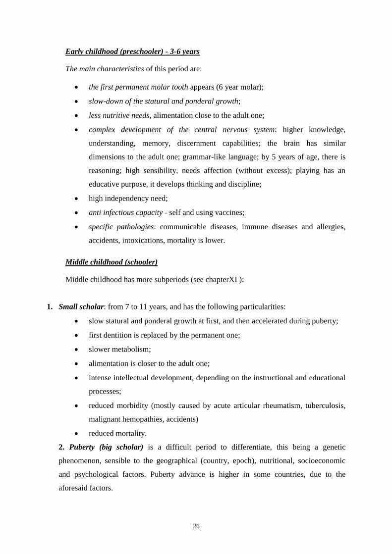
Early childhood (preschooler) - 3-6 years
The main characteristics of this period are:
the first permanent molar tooth appears (6 year molar);
slow-down of the statural and ponderal growth;
less nutritive needs, alimentation close to the adult one;
complex development of the central nervous system: higher knowledge,
understanding, memory, discernment capabilities; the brain has similar
dimensions to the adult one; grammar-like language; by 5 years of age, there is
reasoning; high sensibility, needs affection (without excess); playing has an
educative purpose, it develops thinking and discipline;
high independency need;
anti infectious capacity - self and using vaccines;
specific pathologies: communicable diseases, immune diseases and allergies,
accidents, intoxications, mortality is lower.
Middle childhood (schooler)
Middle childhood has more subperiods (see chapterXI ):
1. Small scholar: from 7 to 11 years, and has the following particularities:
slow statural and ponderal growth at first, and then accelerated during puberty;
first dentition is replaced by the permanent one;
slower metabolism;
alimentation is closer to the adult one;
intense intellectual development, depending on the instructional and educational
processes;
reduced morbidity (mostly caused by acute articular rheumatism, tuberculosis,
malignant hemopathies, accidents)
reduced mortality.
2. Puberty (big scholar) is a difficult period to differentiate, this being a genetic
phenomenon, sensible to the geographical (country, epoch), nutritional, socioeconomic
and psychological factors. Puberty advance is higher in some countries, due to the
aforesaid factors.

27
During puberty, there can be:
Adrenal puberty determined by the androgynous stimulation.
Clinically, no changes can be seen, or the possible apparition of pubic hair;
Biologically, the secretion of androgynous hormones begins.
Gonadal puberty - appears 2 years after the adrenal one. The gonadotropic secretion
begins gradually, as well as the steroid hormone secretion.
Normal puberty's course
The beginning of puberty and the age of debut depend on a variety of factors and range as
follows: girls 9 - 13 years (mean age 11 years) and boys 10 - 15 years (mean age 12 years).
There is a correlation between the beginning of puberty and the degree of bone
maturation (for example, the concordance between the beginning of puberty and the start of
the ossification of the sesamoid bone).
During this period, statural growth is intense: for boys, in the first year it is of 8,7 cm
and the second year 6,5 cm and ends after approximately 5 years after its beginning. For
girls, the growth increment is 7,5 cm in the first year and 5,5 cm in the second one.
Characteristic psychological changes are:
- affective maturity
- presence of libido (adolescence crisis);
- accepting the body changes;
-intellectual maturity;
- development of abstract thinking.
The evolution of puberty, for both the physical and psycho-affective traits for both
genders are summarized in the Tanner stages.
a. Clinical stages of pubertal development (Tanner)
For girls:
Stage I: infantile - S1 (breasts), P1 (pilosity);
Stage II: breast budding, elevation of the papillae; increased diameter of the areolae (S2),
apparition of pubic pilosity, and then axilar pilosity (P2);
Stage III: development of the mammary glands, enlargement of the areolae (S3), pilosity
becomes coarser (P3), development of the labia (minora and majora);
Stage IV: complete breast development, the areola becomes bulgy (S4), complete pilosity (P4);
Stage V: the first menstrual cycle appears(2 years after the beginning of puberty);
regularization of the menstrual cycles (ovulation appears approximatelly 2 years later).

28
For boys:
Stage I: infantile;
Stage II: the beginning of testes enlargement, pubic pilosity appears, then the axilar one;
Stage III: coarse pubic hair, upper lip pilosity appears, voice changes start to show;
Stage IV: adult type pilosity, muscle mass development, widening of the back;
Stage V: beard growth, ejaculations appear.
b. Biologic stages of puberty
They can be delimitated using hormonal dosing. These are applied only in puberty
anomalies, they are difficult to interpret and are expensive.
1. Pituitary gonadotropins
These hormones are without gender specificity, they realize gonadic growth, gamete
maturation, adrenal C androgen sexual hormones secretion, for both genders. The level of
the gonadotropins increases progressively in blood and urine, all along puberty.
For girls, the plasmatic level of FSH rises, which begins by 11 years, and the
maximum level is reached within 2 years from the beginning of secretion. L.H. level
increase appears later, has a higher duration, with fluctuations until the menstrual cycle
becomes regular.
For boys, the increase of the FSH level begins by the age of 12 and is paralel to the
enlargement of the testicles.
2. Sex steroids
Estrogen levels increase with the development of the mammary glands. From
stage III there are hormonal fluctuation present, in correlation to the cyclical
activity of the ovaries.
Androgens begin to increase in level from 6-7 years of age (beginning of
adrenal maturation). These hormones rise along with testosterone levels during
puberty (gonadic maturation).
Plasmatic progesterone begins to rise with the prepubertary stage; its level
remains low until the apparition of menstrual cycles.
c. Physiological variations of puberty
Recognizing these variations is possible along with the apparition of the secondary traits
which match the skeletal age, so the chronological one.

29
Differential diagnosis is done with:
1) Real sexual precocityin which the sexual traits appear before the skeletal age of normal
puberty, along with bone maturation. In this situation, there is a risk of definitive short stature.
2) Real delayed puberty is characterized by: absence of the secondary sexual traits, bone
pubertar age is overcome. The etiology can be within the family history or a pathologic
growth reason. In this situation, psychological problems appear.
3) Dissociation of puberty arises prematurely and isolated, as a secondary trait. The child's
development is normal. Examples: premature development of the pubic pilosity, of breasts,
with no other signs of pubertal evolution.
Pubertal gynecomastia in adolescent males
It can appear during a normal pubertal process. The testes volume is normal for the pubertal
stage. The prognosis is good if the phenomenon regresses in a few months (at most a few
years). It induces important psychologic consequences.

30
CHAPTER V
TERM NEWBORN
The neonatal period includes the first 28 days of life and represents the transition from
the intrauterine life to the extrauterine one. It's an extremely vulnerable period for the
newborn, who finalizes a lot of the physiologic traits needed for the extrauterine survival.
The transition from the intrauterine to the extrauterine life requires multiple biochemical
and physiological readjustments.
The disparition of the maternal circulation through the placenta presumes a series of
readjustments. Otherwise, the newborn lung takes the gas exchange function, the intestinal
tract takes the absorption of the nutritional factors, the kidney takes the excretion function and
the sustaining of the hydroelectrolytic and acid-base balance, the liver takes the neutralization
function and excretion of the toxic substances and the immune system the protection against
infections. The cardiovascular system and the endocrine system adapt, as well, for functioning
independently.
The first 7 days of life are called the perinatal period or early neonatal period.
Evaluation of the gestational age
Fetal evaluation using various means, more or less invasive, is justified with fetal and
neonatal risk assessment. For this purpose, gestational age is being evaluated, along with the
functional maturity, intrauterine growth and development and prenatal specific diagnosable
diseases. Gestational age is evaluated using several methods:
last menstrual period: Mc Donald formula – 7 days are added to the last
menstrual period, starting with the first day and then 3 months are substracted;
clinical examination of the newborn.
After birth, the best way to evaluate gestational age is using the Ballard score, modified
by Dubowitz (table 1 and 2). The error extent of this estimation is +/- 2 weeks. This method
is not accurate enough for the premature newborn or the sick one.
Dubowitz/Ballard score– evaluates the general aspect, skin texture, motor function, reflexes.
Physical maturity - general examination- can be appreciated well in the first 2
hours of life (table 1).
Examination of the neuromuscular maturity (table 2)- is realized in the first 24
hours.

31
Classification of newborns
Based on gestational age, newborns as classified as follow:
Preterm: gestational age < 37 weeks;
Term: gestational age between 38-42 weeks;
Postterm: gestational age > 42 weeks.
Based of birthweight:
Newborn with appropriate birthweight ≈ 2500 g;
Newborn with very small birthweight< 1500 g;
Newborn with small birthweight< 2500 g.
Newborns with small birthweight will be evaluated also by maturity and gestational age, as
follows:
Appropriate for gestational age preterm newborn;
Small for gestational age preterm newborn;
Very small for gestational age preterm newborn;
Table 1. Ballard score. Physical maturity
0 1 2 3 4 5 6
Skin Sticky,
friable,
transparent
Gelatinous,
red,
translucent
Smooth,
pink;
visible
veins
Superficial
peeling
and/or rash;
few veins
Cracking
pale areas;
rare veins
Parchment,
deep cacking;
no vessels
Leathery,
creacked,
wrinkled
Lanugo None Sparse Abundant Thinning Bald areas Mostly bald
Plantar
surface None Almost
visible Faint red
marks Anterior
transverse
crease only
Creases
anterior 2/3 Creases over
entire sole
Breast Impercepti
ble Barely
perceptible Flat areola,
no bud Stippled
areola, 1-2
mm bud
Raised
areola, 3-4
mm bud
Full areola, 5-
10 mm bud
Eye/ Ear Lids fused
loosely or
tightly
Lids open;
flat pinna;
stays
folded
Slightly
curved
pinna; soft
slow recoil
Well curved
pinna; soft
but steady
recoil
Formed and
firm, instant
recoil
Thick
cartilage, stiff
ear
Genitals
(male) Flat,
smooth
scrotum
Empty
scrotum,
faint rugae
Testes in
the upper
canal, rare
rugae
Descending
testes, few
rugae
Testes are
down, good
rugae
Pendulous
testes, deep
rugae
Genitals
(female) Prominent
clitoris, flat
labia
Prominent
clitoris,
small labia
minora
Prominent
clitoris,
enlarged
labia
minora
Majora and
minora
usually
prominent
Majora
larga,
minora
small
Labia majora
covers clitoris
and labia
minora

32
Table2.Ballard score. Neuromuscular maturation
Table 3. Ballard score and gestational age ratio
SCORE
WEEK -10 20 -5 22 0 24 5 26 10 28 15 30 20 32 25 34 30 36 35 38 40 40 45 42 50 44
a)

33
Morphofunctional particularities
Based on gestational age, the term newborn is born between 38-42 weeks, and based on
birthweight between 2500 – 4000 g.
Other important parameters, which characterize the term newborn are:
length, with values between 48-52 cm;
head circumference, with values between 34-36 cm;
chest circumference, with values between 33-34 cm;
abdominal circumference, with values between 31-32 cm.
The initial examination of a newborn must be done as soon as possible, after birth, for
the identification of a certain anomaly and to establish a base for the next examinations to
come. For high risk births, this examination must happen in the labor room, and must
concentrate upon congenital anomalies and physiopathological alterations which can interfere
with a normal cardiovascular and metabolic adaptation to the extrauterine life. Congenital
anomalies can be present at 3-5% of births.
The second, more detailed examination must be done within 24 hours of birth, in the
Neonatal Care Unit.
The examination of a newborn takes patience, softness and procedural flexibility.
The clinical exam of a newborn must follow the antropometric parameters, on one hand
(weight, height/length, head, chest and abdominal circumference) and the complete
examination on apparatus and systems on the other hand. All followed aspects are carefully
watched and noted: pulse (normal: 120-160 bpm), respiratory rate (normal: 30-60 rpm),
temperature, weight, length, head circumference and the dimensions of any visible or palpable
structural anomaly.
Blood pressure is determined only in the ill newborn's case.
Skin
Skin is completely developed, both anatomically and functionally and is pinkish-red
(fig. 7).
At birth, we can observe that the skin is covered in a waxy, yellow substance called vernix
caseosa, which is high in cholesterol, glycogen, fatty acids and proteins. It is produced by the
amniotic epithelium and the fetus sebaceous glands. It has a lubrication role, protects against
bacteria and the cold and is usually covering the pits, or in the dorsal region or shoulders.

34
On the skin, soft hair, named lanugo can be observed, usually on the forehead,
shoulders and back. On the face and nose «facial milia» (miliaria sebaceous)is remarked,
represented by numerous sebaceous glands of gray-white color. Upon skin examination,
certain nevi can be seen as well:
Fig. 7. Newborn skin aspect. Color, miliaria sebaceous, lanugo.
The capillary hemangioma is frequently seen on the eyelids, base of nose and the
occipital area. This vascular mark fades and completely disappears after several
months.
Prominent vascular hemangiona - can be present at birth, it continuously grows
for a few months and later regresses spontaneously by the age of 1 or 2.
Cavernous hemangiomas – do not have a spontaneously regressing evolution
and can easily complicate with thrombosis, ulceration or consumption
coagulopathy (fig. 8).
Pigmented nevi – often covered in hair, are placed on the back or the gluteal
region. A variation is represented by a blue area situated on the skin, named
"mongoloid spot", specific for the hyperpigmented race (fig. 9).
Petechiae - can show up on the scalp or face, after a difficult delivery.

35
Fig. 8. Gigantic cavernous hemangioma.
Fig. 9. Mongoloid spot
The vasomotor instability and scarce peripheral circulation are brought out by deep
redness or cyanosis when the newborn is crying, the intensity of the violaceous color can
intensify the glottic closure during crying.
The mottled aspect of skin can follow a general circulating instability and can be
associated with serious diseases or can be connected to a transitory cutaneos temperature
fluctuation.
Cyanosis can be masked by the palour from circulatory insufficiency or by anemia.
Emphasised palor of the skin can indicate a severe asphyxia, shock or edema. Severe
anemia with an early debut can be seen in: fetal erythroblastosis, subcapsular liver or spleen
hematoma, subdural hemorrhage, fetomaternal transfusion or twin-twin transfusion syndrome.
Postterm newborns can have paler and thicker skin than the preterm or term newborns, with
no signs of anemia.

36
The intense red color of the skin is seen in polycythemia.
In the area the vernix is situated, and around the umbilical cord the skin can have areas
of yellowish-brown color; if this happens, that means the passing of the meconium took place
before or during labor, the most frequent cause being intrauterine hypoxia.
Preterm babies have an intense red color of the skin, thin skin and very sensible to
physical, chemical and infectious agents. The ones that are extremely preterm have an almost
gelatinous skin, with high risk of bleeding and diverse traumatic lesions.
The preterm can have an intensification of lanugo, especially on the head, forehead and
face area. The areas covered with pilosity from the lumbosacral region can raise suspicion,
like spina bifida, sinus tracts or tumors.
Nails are rudimental for the preterm newborns, but can be over the curve of the fingers
in postterm newborn; these can also have pergamentos skin with descuamation.
After approximately 3 days of life, there can be seen an erythematous area, followed by
a papular rash, the pustular and vesicular rash being of white color. This benign skin rash,
named toxic erythema, lasts for about a week, is high in eosinophils and is distributed to the
face, upper body and limbs.
Transient neonatal pustular melanosis is a benign lesion which is frequently seen in the
dark-skinned newborns, is filled with neutrophils and is seen at birth as a vesicular and pustular
rash, located on the chin, neck, back, limbs, hand, sole and usually passes after 2 or 3 days.
Both lesions must be differentiated by other infectious, more severe, vesicular
eruptions, like the ones caused by herpes simplex and staphylococcus aureus.
The presence of amniotic bands in the intrauterine life can lead to major skin and limb
anomalies. As such, the amniotic bands can separate skin, limbs (amputation, constriction,
syndactyly), face (fissures) or trunk (abdominal or thoracic wall defect). Their etiology is
uncertain, but can be associated with a rupture of amniotic membranes or their vascular
compromise, along with the formation of fibrous bands.
The excessive fragility and elasticity of the skin, associated with joint hypermobility is
seen in severe diseases: Ehlers-Danlos syndrome, Marfan syndrome, congenital
arachnodactyly or other colagen synthesis disorders.
The epidermis is thin, the stratum corneum is thin, the elastic connective structure of the
dermis is underdeveloped, the basal and germinal layers are less developed.
The sweat glands have no activity during the neonatal period.

37
The head
The head is voluminous and represents 1/4 of the newborn's length; the neurocranium is
bigger. The skull has 8 membraneous sutures and 6 fontanelles. The palpable fontanelles are:
anterior fontanelle of rhomboidal shape, has a diameter of 2,5/3,5 cm, closes at 14-16 months;
posterior fontanelle, which has a triangular shape, is present at 25% of the newborns and has
diameters of 1,5/1 cm; it closes in the first 4-8 weeks of life.
The head circumference is between 34-36 cm.
The head can be big (macrocephaly), in case of: hydrocephaly, or familial large head, or
macrocephaly associated with intellectual disability (rarely), achondroplasia, cerebral
gigantism, neurocutaneous syndromes, inborn metabolic error or hereditary (fig. 10).
The head of a preterm baby can suggest hydrocephaly due to the accelerated brain
growth rate compared to the other organs and a larger head to body proportion (the head
represents 1/3 of the body, while in a term newborn the head is 1/4 of the body).
A skull depression (indentation, fracture, "ping pong ball sign").
Fig. 10. Term newborn, caput succedaneum, depresible, with edema.
Microcephaly (small head) can be «apparently small» in the newborn from diabetic
mother, but the head circumference is within normal limits.
Microcephaly can be hereditary and is not accompanied by intellectual disability or can
be secondary to the following anomalies: intrauterine growth restriction, cytomegalic
inclusion disease, congenital toxoplasmosis, chromosome anomalies (fig. 12).
The presence of large fontanelles, associated with large sutures can lead to congenital
hypothyroidism suspicion, congenital rubella or chromosomal anomaly.

38
The head can have certain mechanical alterations, due to edema, intrauterine
positioning or laborious births (caput succedaneum) (fig. 10).
The head can be deformed in the following situations as well: caput succedaneum is
produced by a hemorrhagic edema of the scalp tissue and is usually situated in the occipital
area, does not respect the suture limit and is rapidly reabsorbed; cephalhematoma (fig 13) is
produced by the subperiostal hemorrhage, is well defined within the skull sutures and can
persist for months; sometimes it has a calcification tendency.
The fontanelles and suture dimensions, sunken of bulged fontanelles can be evaluated
best after the first few days of life.
Bulged fontanelles can be seen in meningitis, cerebral hemorrhage, cerebral edema,
hydrocephaly, cerebral tumors, cerebral malformations. The bulging of the anterior fontanelle
suggests the rise of intracranial pressure. High intracranial pressure can be also seen during
crying.
Examination of the cranial sutures (sagital, metopic and coronalsutue) shows their
dimensions, if the parietal bones are overlapped, having a tendency to overlap the occipital
and frontal bones, especially in the preterm newborn, which can lead to severe, particular
situation, even the apparition of craniostenosis. Craniostenosis represents the premature
closure of the fontanelles, followed by the ossification of the sutures, which leads to
microcephaly and neuropsychomotor disability.
Head asymmetry can be cause by multiple factors.
The skull can be elongated due to the intrauterine position, if it is the first one born or if it was
engaged for a long period of time.
Craniotabes is sometimes identified in the parietal bones area, near the sagital suture. It
is frequently seen in preterm babies and the ones exposed to uterine compressions. Usually,
the evolution is good, but if it is persistent, tests must be done and the treatment and cause
must be established.
Generalized craniotabes can appear in calcification disorders, like the ones from
osteogenesis imperfecta, cleidocranial dysostosis, lacunar skull, imbecility, and, occasionally
Down Syndrome.
A positive diagnosis is established using paraclinical investigations, the most useful
being cranial transillumination and transfontanelar ultrasound.

39
Fig. 11. Secondary macrocephaly to evolutive hydrocephaly.
Fig. 12. Microcephaly
Fig. 13. Bilateral arieto - occipital cephalhematoma

40
Facies
General examination looks for the general aspect of facies, with careful observation of
the dysmorphic anomalies, like hyperpilosity, protruding forehead, epicantus, hypertelorism,
microphtalmia, low-set ears, which are associated with congenital malformations, most of the
time.
Facial asymmetries are seen in: paralysis of the VIIth
nerve, hypoplasia of the depressor
anguli oris muscle, abnormal fetal posture; if the maxilla was fixed on the opposite shoulder
side during intrauterine life, the mandible can easily deviate from the median line.(fig. 14).
The symmetrical facial paralysis suggests the absence or the hypoplasia of the VIIth
nerve
nucleus (Mobius syndrome).
Fig. 14. Facial paralysis
Fig. 15. Facial deformity
Eyes
Eye examination of a newborn is difficult because newborns have a tendency of keeping
them closed.
Based on inspection, several modifications can be seen: eye agenesis (fig. 16), palpebral
ptosis, congenital cataract, eyelid coloboma.

41
Fig. 16. Unilateral eye agenesis.
Eyes are opening, spontaneously most of the tine, if the newborn is held upward and
lightly moved back and forth. This maneuver, the result of the tonic labyrinthine reflex and
tonic neck reflex is the most useful tool for eye examination, rather than forced opening of the
eyes. A good examination can be done by face illumination using a flashlight when the
newborn opens its eyes.
Conjunctival hemorrhages and retinal ones do not havea pathologic semnification. The
retinal hemorrhage can be seen in: severe hypoxia, subdural hematoma or subarachnoidal
hemorrhages.
The conjugated eye movement are present immediately after birth, but they become
permanent only after a few weeks.
Nystagmus is a common manifestation immediately after birth. In the first 10 days, the
eye have a fix position, following the movement direction of the head - the test of "doll eyes".
The presence of nystagmus after a few days of life is a pathological sign present in a
neurologic or ocular disorder and usually indicates vision disorders.
Pupillary reflex is present after 28-30 gestational age. The pupils must be equal in size,
symmetrical and reactive to light.
Blinking and corneal reflexes are present at birth.
A cornea with more than 1 cm diameter, in a term newborn, suggests congenital
glaucoma and needs emergency ophthalmologic consult.
The iris must be examined for finding eventual coloboma and heterochromia.
The apparition of a white outline around the iris isn't always a pathological sign. It is
usually seen in newborns with Down Syndrome, but can also appear at a healthy newborn..
Leukocoria (white pupillary reflex) appears in a series of severe disorders, like: cataract,
tumors, chorioretinitis, retinopathy of prematurity or persistent hyperplastic primary vitreous
and requires emergency ophthalmologic consult.

42
During the ophtalmoscopic examination, there can be identified: retinal anomalies,
chorioretinitis and retinoblastoma.
During the first 6 months of life, intermittent convergent strabismus can be present,
which has no pathologic semnification. Its persistence after 6 months of life, its unilateral
persistence or the presence of a divergent strabismus can indicate a diminished visual acuity
or diminished ocular muscles motility.
Conjunctivitis can be present in the first few days of life. The chemical conjunctivitis
usually appears after the instilation of silver nitrate 1% drops in the conjunctival sac for the
prevention of gonococcal conjunctivitis. Is manifests as eyelid edema and conjunctival
inflamation along with purulent drainage (fig. 17).
Fig. 17. Purulent conjunctivitis
Visual acuity is appreciated using the photomotor reflex, the corneal reflex and the
pupillary reflex. Central vision progresses from birth, when the newborn perceives only light,
until the age of 6, when he reaches the adult's level of acuity.
Visual tracking and fixation are progressively developing: at 2-4 weeks he can fixate
certain objects placed in his visual field for a few seconds; by 6 weeks he can track an object
placed in his visual field; by 3 months, the infant has convergent eye movements and starts to
distinguish the shapes and colors of different objects.
The nose
The nose can be slightly obstructed by the mucus collected in the tight nostrils. The
nostrils must be identical. Nostril permeability can be tested using a catheter which easily
passes through the nasal duct. Complete obstruction can be seen in bilateral choanal atresia,
which can be a cause of respiratory insufficiency (fig. 18).

43
Fig. 18. Choanal atresia
Ears
The ear examination can bring both pathological data, but also elements of maturation
level. The ear examination must establish the presence of the auditory canal, the dimensions
of the pinnas, the parched or normal aspect.
The presence of preauricular small tumors can lead the examiner to a certain renal
malformation.
Hearing
In the first days of life, hearing is tested with extreme difficulty. From 2 weeks, the
cochleopalpebral reflex can be tested, which consists of a flinch and a wink of the eyelids as a
response to unexpected sounds.
Ear loss screening using special tools is more accurate.
Mouth
During mouth and oral cavity inspection, we are interested first of all in color, which
can be a great indicator of cyanosis.
Normally, early dentition appears rarely (inferior incisor or aberrant location). In
pathologic conditions, neonatal dentition appears in Ellis-van Creveld syndrome, Hallermann-
Streiff syndrome, and others. Extraction is not recommended.
Hard and soft palate must be inspected for identifying an eventual cleft (complete or
partial), ogival arch or bifid uvula (fig. 19).On the hard palate there can be seen epithelial
cells accumulation, named Ebstein pearls. Cystic retention, of similar aspect, can also be
observed on the gums. Both disappear spontaneously, a few weeks after birth.

44
Fig. 19.Bilateral cheilopalatoschisis
On the anterior tonsil pillars, in the first 2-3 days of life, there can frequently be seen
small follicles, of white or yellow color, or ulcerous areas on erythematous base. They are of
unknown cause and disappear in 2-4 days without any treatment.
There is no active salivation in the first 3 weeks of life. The presence of a small quantity
of saliva can suggest the presence of a tracheoesophageal fistula.
The tongue seems relatively large, the frenulum can be short, but must rarely be incised.
Ocasionally, the sublingual mucous membrane forms a prominent fold.
The cheeks are abundant on both sides, internal and external, as a result of fat
acummulation, which contributes to the development of the sucking apparatus. These, along
with the labial tubercle from the upper lip disappear when breastfeeding stops, especially after
1 year of life.
The newborn's pharynx is hard to differentiate due to its arched palate shape, but even
so, it must be carefully inspected for indentifying certain anomalies of the posterior palate or
uvula. The tonsils are small.
The neck
The neck appears relatively short. The anomalies at this level are not frequent. They
include goiter, cystic hygroma, residual brachial cleft, sternocleidomastoid muscle lesions,
traumatic or due to intrauterine position, which can lead to hematomas and fibrosis.
Congenital torticollis (fig. 20) establishes the head orientation toward the affected part,
and the face toward the opposite direction. If it is not treated, plagiocephaly, facial asymmetry
and hemihypoplasia can appear.
For fracture identification, both clavicles should be palpated.

45
Fig. 20. Congenital torticollis. Sternocleidomastoid tumor.
Mammary glands
Breast hypertrophy is frequently seen and sometimes, in the presence of a genital crisis
(under the influence of maternal estrogens), lactation can be present as well. Asymmetry,
erythema, induration and sensitivity suggests a mammary abcess. Supranumerary nipples or
large internipple distance, along with shield shaped chest can be present in the Turner
syndrome.
Pelvic genital region
The external genital organs have a characteristic aspect for each gender. For boys,
fimosis is physiological; the testes are frequently palpated in the inguinal canal; uni or
bilateral hydrocele can be observed.
For girls, the labia minora appears slightly more developed. There is often observed a
mucous, white or sanguinolent secretion, secondary to the transplacentary hyperestrogenism.
Meconium is usually eliminated in the first 12 hours after birth; 99% of the term
newborns and 95% of the preterm ones pass the meconium 48 hours after birth.
Anal imperforation is not always visible; it can easily be detected by rectal examination
(usually with the help of a tube).
The presence of an irregular skin fold on the sacrococcygeal median line, can be easily
mistaken for neurocutaneous sinuses.
Limbs
The limb examination observes the existence of certain posture alterations, especially in
the case of pelvic fetal presentation. The suspicion of a fracture or nerve lesion, birth
associated, is brought out by observing the limb movement during spontaneous movement or
stimulated ones. Hands and feet must be checked for anomalies: polydactyly, syndactyly,
abnormal dermatoglyphics, such as simian crease.

46
Functional adaptation
Respiratory system
• The respiratory system has 4 anatomic components:
– superior respiratory tract: nasal cavities, pharyngeal sinuses, larynx, trachea –
which warms, mixes and filters the inspired air
– inferior respiratory tract: bronchi and bronchioles – which distribute the air
toward the lung
– respiratory parenchima: respiratory bronchioles, alveolar ducts, alveoles–
where gas exchanges happen
– muscular and elastic structures: intercostal muscles, diaphragm, elastic lung
tissue - which realize the respiratory excursions.
Knowing the normal lung development is important for understanding the way in
which this can be alterated by different factors and for picking the best treatment
methods which can prevent pulmonary injury. The degree of lung development is the
main factor in determining the survival of the newborns, especially the preterm, and
response to intensive treatment.
Fetal lung development has 5 stages until final maturation:
– embrionar phase which lasts until 5-6 weeks
• the respiratory primordium appears on day 22 (postconception) by
widening of the caudal end of the lanryngo-tracheal canal
• during this period, developmental anomalies are determined by the
formation of the lung bud, the separation of the trachea and esophagus,
formation of the superior aerial ducts and initiation of the pulmonary
lobes formation. These anomalies can manifest by: laryngeal,
pharyngeal, tracheal, esophageal atresia, tracheal and bronchi stenosis,
esotracheal fistula, pulmonary agenesis, ectopic lobes, bronchogenic
cysts, arteriovenous malformation.
– pseudoglandulary phase which lasts until week 17.
• major events: tracheobronchial treecomplete development until 16
weeks of gestation, development of the pulmonary arterial bed parallel

47
to the bronchial ramification, differentiation of the basal, ciliated,
Goblet and neuroepithelial cells.
• only the pulmonary limph system, the cartilage, the mucous glands and
the smooth muscle is developed. Pleuroperitoneal cavities are closing.
• the anomalies met during this stage are: renal agenesis with pulmonary
hypoplasia, pulmonary intralobar sequestration, cystic adenoid
malformation, congenital diaphragmatic hernia.
– canalicular phase (acinar) – between weeks 16-26.
• pulmonary acini, formation of the alveolocapillary membrane and the
capillary network. Differentiation of the type I and II alveolar cells and
the apparition of the lamellar cells within the type II cells is produced
during this stage.
• anomalies: renal dysplasia and renal hypoplasia, alveolocapillar
dysplasia, respiratory insufficiency, surfactant deficiency.
– saccular phase – weeks 24 – 38.
• air space distension continues along with their growth, formation of the
sacs, thinning of the mesenchyme, septal walls contain a double
network of capillaries, surfactant synthesis begins in the type II
alveolar cells and fetal breathing occurs.
• type II cell maturation is associated with an increase of phospholipid
synthesis from the surfactant and associated proteins (A, B, C, D).
• glycogen content is reduced, mitochondrial enzymatic activity increases,
indicating a movement toward the aerobic cycle of the oxidation.
• surfactant concentration is still low and the composition of its
phospholipids is significantly different from the newborn one.
• for the newborn and adult, the width of the alveolocapillary membrane
is 0,6 mycrons, which allows the passive diffusion of O2 and CO2
between the alveolar and the capillary bed.
• the immature lung contains relatively small amounts of elastin fibers
and collagen and can easily rupture, having a low elasticity coefficient.

48
• anomalies associated to this stage: alveolocapillar dysplasia, respiratory
insufficiency, surfactant deficit, hyaline membrane disease, transient
tachypnea, prematurity apnea.
– alveolar phase– can start by 30 weeks but usually around 36 weeks of gestation
and lasts until the age of 3.
• a term newborn has approximately 50 million alveoli.
• this phase is characterized by: secondary sept formation along with
subdivization of the alveolar sacs, loss of connective tissue and
narrowing of the alveolar septs, and as a result an increase in the
pulmonary alveolar bed. The capillary network fuses to a single
network and the actual maturation is produced by: collagen, elastin and
fibronectin deposit and an increase in the surfactant production within
type 2 alveolar cells.
• in the first 3 months after birth there takes place a slow down in the
alveolar development, and then a rapid increase in its numbers in the
first year of life, so that by the age of 3 they reach 300 millions
comparable to the adult alveolar number.
• associated anomalies: persistent fetal circulation and pulmonary
hypertension, lobar emphisema, meconium aspiration syndrome,
pneumonia, respiratory distress syndrome associated with protein B
surfactant defficiency.
• the anatomic traits of the respiratory tract of a newborn contributing to respiratory
adaptation are:
– big head – short neck
– narrow nostrils– big mouth
– soft palate – big epiglottis
– high glottis position (C3- C4)
– oblique vocal chords – narrow cricoidian ring
– posterior angulation of the subglottic cavity
– small lung –orizontal movement of the thoracic cage

49
• the newborn has a nasal respiration. Flux resistance through the nasal passage
represents almost 45% of the total resistance.
• big and soft epiglottis, relatively high situated in the pharynx, big tongue, in contact
with the soft palate, it occupies a great part of the oropharynx, so it makes the mouth
breathing more difficult.
• the subglottic cricoidian region is narrow, there is a risk of stridor from the mucous
edema after detubation.
• the tracheal bifurcation angle is different to the left and to the right, so there is always
a difficulty of intubation, there is a risk of intubating the right bronchus.
• main breathing muscles: diaphragm and intercostal muscles are normally innervated,
but in the preterm newborns there appears muscular fatigue after stimulation. This is
mainly because there is little muscular mass and a relatively small percentage of type I
muscular fibers with oxidative capacity (10% compared to 25 % in the term newborn
and 50-55% in the adult).
• during inspiration, the inferior ribs are moving inward at an angle that in more
orizontal then the insertion of the diaphragm, resulting alterations of the pulmonary
volumes and costal retraction.
• abdominal distension can lead to respiratory insufficiency due to the fact that the
diaphragm is being pushed up.
Before birth, the lung is filled with a liquid which is secreted at this level, and which
composition is different from the one the amniotic fluid has. During the passing through the
birth canal, a small part of the pulmonary fetal liquid is eliminated with the help of thoracic
compression, but the most of it is absorbed through the sanguine and limphatic vessels in the
first minutes after birth. Absorption of the intrapulmonary liquid is made due to the rapid
increase of the sanguine flow and the decrease of the pulmonary vascular resistance. The
increase of PaO2, decrease of PCO2 and increase of pH favours the decrease of the pressure in
the pulmonary circulation, so the sanguine flow rises.
The first breath is induced by a series of factors: acidosis and moderate hypoxia, rise
of PCO2, hypothermia and the increase of blood pressure in the general circulation by
clamping of the umbilical cord. The difference between intrauterine pressure and the
atmospheric one, the pleural void, produces air aspiration, which reaches the alveoli and
excites the vagus nerve pulmonary endings, leading to expiratory reflex. Tactile stimuli

50
induce gasping type breathing, without initiating a sustained respiration. Pulmonary
compliance and vital capacity increase abruptly and after 12 hours reach values that are
proportional to the ones of adults.
During the first 60 minutes after birth, the newborn has an irregular breathing, with
tachypnea (60-90 breaths/min), intermittent grunting, nasal flaring, subcostal and intercostal
retractions, bronchus rales and bubbly secretion in the oral area. After this period of high
reactivity, there comes a sleeping period, with rapid and superficial breathing. Next, the
respiratory rhythm is stabilized at a frequency of 30-50 breaths/min.
Using inspection, and observing the breaths of the newborn, several aspects can be
identified. Amplitude and rhythm fluctuations depend on the physical activity, wakefulness
state or crying. Because these fluctuations are rapid, the respiratory frequency must be
recorded during a whole minute, in rest state, preferably sleep state of the newborn. Under
these circumstances, the normal respiratory frequency of a term newborn is 30-50
breaths/minute.
For preterm babies the rate is bigger and with larger fluctuations. A constant frequency
is defined at 60 breaths/minute. During the regular periods of breathing, this usually indicates
cardiac or lung disorders. A preterm baby can breathe in a Cheyne-Stokes rhythm, known
under the name of periodic breathing, or with a complete irregular rhythm. The periodic
breathing is rare during the first 24 hours of life.
Irregular gasping, followed by spasmodic movement of the mouth and chin, indicates
respiratory center disturbances.
Newborn's breathing is almost entirely diaphragmatic, so that during the inspiration
time, the soft tissue of the thorax is drawn inward, and the abdomen is protruding. If the child
is calm, relaxed, has a normal skin tone, this "paradoxal movement" does not necessarily
indicate respiratory insufficiency. On the other hand, a difficult respiration is an important
sign of respiratory distress syndrome, pneumonia, anomalies or mechanic lung perturbation.
Nasal flare, subcostal and intercostal retractions, along with polypnea, peripheral or
generalized cyanosis are signs of lung injury.
Pulmonary injury suspicion is based on the vesicular murmur, rales and anomalies
found at percution, which must always be confirmed by cardiopulmonary radiology or other
paraclinical investigations.
Lung ventilation includes at first the anterior and superior parts, then followed by basal
and paravertebral ones.

51
Usually, immediately after birth, massive quantities of surfactant are released in the
respiratory tract (from type II alveoli). Once released, the surfactant is continously mobilized
through the bronchial tree, with the help of vibrating cilia.
Fetal lung maturation can be accelerated by the administration of glucocorticoids
(betamethasone) to the pregnant women, which has a threatened preterm labor. The
administration of this therapy, at least 2 days before birth, is followed by the reduction of
idiopatic respiratory distress syndrome incidence. Crepitant rales can be heard during
auscultation.
The oxygen saturation rises from 65% to 95% after 3 hours of life. The current
respiratory volume is 20-25 ml, total respiratory volume is 160 ml, vital capacity during
crying is 120 ml, total inspiratory volume is 80 ml. Functional residual capacity is 80 ml.
Cardiovascular system
Prenatal development of the cardiovascular system
This is the first functional apparatus of the embryo, it starts to function at the end of the 3rd
week of intrauterine life:
between the 13th
and the 15th
day, from the embryonic mesoderm starts angiogenesis.
after the 17th
day the intraembryonary angiogenesis starts as well.
by the end of the 3rd
week, in the cardiogenic mesenchymal plate 2 endocardial tubes
start to show. These fusionate in a single endocardial tube which receives connections
with the intra and extraembryonary vessels, around the 21st day.
by the 24th
day, the cranial-caudal embryonic curve is seen.
The primitive heart tube, almost rectilinear, has a cranial (arterial) pole and a caudal (venous)
one. In cranial-caudal way, the segments can be seen: the bulb of the heart, which continues
with the arterial trunk, the primitive ventricle, the primitive atrium, the venous sinus, the 2
horns of the venous sinus. During this phase, the embryo is sustained with the help of
nutrients received at the ombilical vesicle level, through the vitelline or omphalomessenteric
circulation.
The primitive cardiac tube has an accelerated growth rhythm, by 23 days it takes the position
of a "laid down S letter" and forms a bulboventricular loop with 2 components: a venous
extremity, from which the venous and atrial sinuses derive and arterial extremity with 2 other
components - the ventricle and the bulb.

52
From the primitive atrium 2 big lateral evaginations will develop, the following definitive
atriums. The right ventricle will develop from the primitive ventricle wall.
The arterial trunk is a fibrous segment, situated in the upper part of the valves, continued
by the ventral aorta (aortic sac), from which the next great arterial vessels will develop.
At birth, along with the umbilical cord clamping and exclusion of the placental
circulation, the blood pressure and the peripheral vascular resistance increase abruptly. Along
with the disruption of this circulation, the Arantius venous duct is closed.
Once the oxygen reaches the pulmonary tissue, this determines a decrease of pulmonary
spasm, and increase of blood flow.
Immediately after birth, the resistance from the pulmonary circulation becomes almost
equal to the one from the systemic circulation. This way, through the arterial duct, there is
produced a small left-to-right shunt. along with its complete closure, the shunt diminishes and
disappears. During the first day of life, pulmonary resistance drops by half, reaching the adult
level at the end of the first week.
The apexian shock can be observed in the 3rd
-4th
left intercostal space, outside the inter-
nipple line; the cardiac sounds are equal, the blood pressure is 75/40 mmHg.
The peripheral circulation is slow, the extremities are cold and cyanotic most of the
time, even though the oxygen concentration is normal. The capillary fragility and vascular
permeability are increased.
The presence of a murmur in the first day of life is benign, but its persistance may need
further investigation.
Congenital heart diseases may not determine a murmur initially, this can appear later in
evolution; there is a risk of 1:12 that a murmur identified at birth can mean a heart disease.
Radiographic, echographic and electrocardiographic cardiac evaluation are essential
when a heart disease is suspicioned.
Heart rate can normally vary, from 90 beats/min to 180 beats/min, stabilising after a few
hours around 120-140 beats/min. A higher heart rate must be evaluated using an EKG.
Preterm babies, whose normal heart rate is usually 140-150 beats/min, can have an
abrupt debut of sinus bradycardia.
The pulse should be palpated at both the upper extremities and the lower ones, for the
eventual detection of a coarctation of the aorta. Measuring of the blood pressure can be very
helpful for a sick newborn. The auscultatory method is acceptable, most of the times, if the
stethscope is adapted to the newborn.

53
The Doppler method has a higher accuracy in the examination of the diastolic and
systolic pressure. The oscillometric method is the easiest one at the moment and is the most
precise non-invasive method, disponible in the NICU.
Hematologic adaptation
Blood volume is approximately 85 ml/kg (limits 50-100 ml/kg). The red cell
particularities during the neonatal period are:
erythrocytes have a diameter of 8-8,5 microns,
anisocytosis is frequent,
red blood cells have a decreased quantity of reduced glutathione,
there is a transitory deficit of glucose-6-phosphate-dehydrogenase, favoring a
severe hemolysis,
the number of red blood cells is 5,6-6 millions/ mm3, of which the number of
nucleated red cells is 500/mm3. Red blood cells have a high fragility, a shorter
life span and a high metabolic activity
reticulocytes values during the first 3 days are of 3-5% and stabilize at values of
1% after 7 days of life.
Hemoglobin is of 17-18g/dl in the blood drawn from the umbilical chord. Hemoglobin
is of fetal type, which represents 80% of the total Hb, is more oxydable, and by 3 month is
transformed in the adult type. Erythrocytes and hemoglobin drop in the first 2-3 months, but
then normalize until 2 years of life. The values are influenced by the volume of placental
transfusion; if the clamping of the umbilical cord is done belatedly, the level of Hb and
hematocrit level are higher; these values also differ based on birthweight and gestational age.
Hematocrit at birth is 55% and 45% after 10 days of life. The serum iron level
decreases gradually.
White blood cell count and differential count is rapidly modifying during the first days
of life (table 4):
Table 4. White blood cell count and differential count
FIRST DAY 1 WEEK
Leukocytes (mm3) 18000-20000 12000
Polymorphonuclears (%) 55-60 45-50
Lymphocytes (%) 30 40
Monocytes (%) 10 5-10
Eosinophils (%) 2 2

54
Platelets have after birth values between 150 000 – 300 000/mm3. Coagulation
mechanisms are imperfect, so the hemorrhagic disease prophylaxis must be done by
administering vitamine K. RBC velocity is of 6 mm/hour in the first 3 days of life and the
below 11 mm/hour after the first week.
The newborn bone marrow is initially hypercellular, then it decreases by the end of the
first week.
Gastrointestinal function
Even though the gastrointestinal tract is sufficiently developed from 28-30 gestational
weeks, for allowing absorption and nutritive substances use, this function is not sufficiently
mature in the moment of birth. So, the coordination of deglutition and esophageal peristaltis
is weak and predisposes toward regurgitation.
Most of the enzymes are present at birth, with one exception, the ones that break down
starch.
Digestion and absorption are very good for carbohydrates, good for proteins and low for
lipids. Biliary secretion is present from the 3rd
month of pregnancy, but the bile is deficitary in
salts, and its elimination is inappropriate.
The intestinal lactase and pancreatic lipase are present from the 8th
month of pregnancy.
The first stool is named meconium, has a dark green color and contains residues from the
digestive tract secretions, desquamation cells and bile. Its elimination appears after 24 hours
and is final after 1-3 days. The following days "transitional diarrhea" is present, which
consists of gelatinous, greenish or faded stools, 6-7 times per day. This false diarrhea is the
consequence of microbial invasion at the intestinal level, of the laxative effect of colostrum
and immaturity of the digestive function.
Digestive tract invasion with microbial flora is produced after 12-20 hours, while the
intestinal tube is sterile. This invasion is produced upward and downward as well. There
follows a period of characteristic constitution of flora, with bifidus bacillus, which represents
85-90% of the flora, and is specific to the breastfed newborn.
Intestinal absorption of immunoglobulins, iron and vitamin B12 is good for newborns.
The liver can be palpated at 1-2 cm under the right costal margin.

55
Renal function
I. Renal embryogenesis and functional development
A. Embryogenesis
The mature human kidney is the final stage of three phases of embryonary development:
pronephros, mesonephros, metanephros.
The transitory pronephros, the first structure that holds the rudimentary tubes,
disappears by the end of the 4th
gestational week.
After that, the mesonephros is developing, which holds well developed nephrons, that
have vascularized glomeruli connected to the distal and proximal renal tubes and which drain
the mesonephric duct.Eventually, the mesonephros fusionates with the cloaca, contributing to
the formation of the urinary bladder, and for males, contributing to the formation of the
reproductive organs.
The metanephros can be identified around the 5th
or 7th
week of gestation, this being the
last element from the developmental period. It has 2 components: ureteric bud and the
metanephric mesenchyme. With each division of the metanephric bud, the stem cells produce a
new layer of nephrons. The metanephros reached the lumbar position after 8 weeks of gestation.
The development of the nephronal system and the collecting system differs: the
nephronal system comes from the mesenchymal cells, and the tubes are formed starting with
the ramification of the ureteric bud. The proximal portion of the proximal ureteral bud
appears in the 28th
day of gestation, and it branches with a nephron at each tip. These
branches form the the final collecting system (tubes, renal pelvis, ureter, urinary bladder
trigone).
Most nephrons are formed from the 36th
week of gestation. Their number varies from
300.000 to 1.800.000 (mean of 900.000) nephrons per kidney; nephrons cannot regenerate.
There are 4 stages of nephron development:
- Stage I – the renal vesicle appears;
- Stage II – transformation of the renal vesicle in a "S" shaped body
- Stage III – capillary loop
- Stage IV – maturation stage, including the proximal tubes, Henle's loop, distal tubes,
juxtaglomerular complex development and a part of the afferent arteriole.
During this stage, the renal interstitium is differentiated into several components: cortical,
medular, etc. A perturbance of any part of this sequence can lead to a reduction in the
nephrone number.
Once a number of nephrones is formed, the postnatal factors (injuries from acute or chronic
renal failure) can only decrease their number.

56
B. Functional development
At birth, the kidneys replace the placental function as the main homeostasis organ,
maintaining the fluid, electrolyte balance and elimination of harmful substances.
This transition appears along with the increase of renal blood flow, glomerular filtration
rate and tubular functions.
Due to this postnatal transition, the level of renal function is correlated more with
postnatal age rather than gestational age.
1. Renal blood flow
During fetal development, the blood flow remains low, representing only 2-3% of the
cardiac output, then it rapidly rises, to 15-18% of the cardiac output.
This is due to the: decrease in renal vascular resistance, rise in the systemic blood pressure,
and increase from the inner part to the outer part of the cortical blood flow.
2. Glomerular filtration
Starts immediately after the formation of the first nephrons, the eGFR increasing along
with body growth and renal growth (1ml/minute/kg body count).
At birth, it is smaller at most of the preterm babies, and increases dependent on the
prematurity grade. At term babies, the GFR rises rapidly, doubling by 2 weeks of age and
reaches the adult level at 1 year of age.
3. Tubular function
a. Sodium control (Na+). Renal capacity of natrium reabsorption is developing after 24
weeks of gestation, even though the tubular reabsorption is low until 34 weeks of gestation.
After 34 weeks gestational age, sodium reabsorption becomes more efficient, so that 99% of
the filtered Na+ can be reabsorbed, resulting a FENa of < 1%, compared to the renal
hypoperfusion (prerenal state).
b. Water control. The newborn has a limited capacity of urine concentration, due to the
limited concentration of urea in the renal interstitium (reduced input of protein and anabolic
growth). The result is a decrease in the interstitial osmolarity which leads to a decrease of con-
centration capacity, and so a reduced capacity of water absorption. The maximum urine con-
centration (osmolarity) is only 500 mOsm/l for preterm babies and 800 mOsm/l for term babies.
c. Potasium control (K+). The limited capacity of preterm babies to excrete large quantities
of potasium is strictly tied to the distal tubular secretion of potasium, as a result of low

57
sensitivity to aldosterone, reduced activity of Na+- K+ -ATP – ase and reduced GFR. Preterm
babies have higher concentrations of serum K+ compared to infants or bigger children.
d. Bicarbonate and acidity control. It is limited by a low serum level of bicarbonate in
the proximal tube (14-16 mEq/l for preterm and 18-21 mEq/l for term newborns), which
advances along with the Na+- K+ -ATP – ase maturation and Na+- H antiporter. In essence,
preterm babies are born with a light renal proximal tubular acidosis which advances along
with maturation.
e. Phosphorus and calcium control. The input and ultrafiltration of the phosphate,
parathyroid hormone and growth factors adjust the renal phosphate transportation. Also,
phosphorus renal reabsorption is modified based on gestational age, rising from 85% by 28
weeks to 98% at 40 gestational weeks.
Calcium urinary excretion is lower in preterm babies and is correlated with gestational
age. For term newborns, calcium urinary excretion rises and persists until 96 months,
approximately.
Neonatal stress and therapies that involveintense use of fluids and furosemide
administration increase Ca2+
excretion, worsening the tendency toward hypocalcemia or
nephrocalcinosis.
4. The contribution of fetal urine to amniotic fluid volumeis minimal in the first
half of the pregnancy (10ml/h), but increases significantly at an average of 50 ml/h.
Oligohydramnios or polyhydramnios can relate to renal developmental disfunctions.
Oligohydramnios can be associated to: renal agenesis, renal dysplasia. Polycystic kidney
disease leads to severe obstruction of the urinary tract.
Polyhydramnios can be observed in complicated pregnancies due to gestational diabetes or
fetal anomalies (esophageal atresia, anencephaly).
Overall, in the first days, the renal function is deficitary: the GFR is reduced, the
permeability of the filtration membrane is high, the tubular reabsorption is high, the secretion
is deficitary, the dilution and concentration insufficient, excretion of the acids and electrolytes
decreased, bases conservation reduced.
The newborn's metabolism is more intense, reported to the renal excretion capacity.
There is a relatively renal inefficiency, predisposing the newborn to water and salt retention
and edemas occurance (precaution to the dose and nature of renal elimination of drugs).
The urine macroscopic aspect is cloudy, there is a transient albuminuria in the first few
days. The diuresis is approximately 100 ml/kg/24h.

58
Immunologic adaptation
The apparition and maturation of the immunologic mechanisms of anti-infectious
defense is realized during the intrauterine life.
Nonspecific defense is realized through:
cutaneous defense – deficitary, permeable to germs;
mucous defense – deficitary, secretory IgA appear after 2 weeks;
phagocytosis is reduced, and the chemotactic response of the leukocytes is weak
in newborns;
nonspecific serum factors – opsonization is relatively good (deficitary for
E.coli), propertine (bactericide, antiviral and opsonizing activity) is ½
compared to the adult; the serum complement is 70 mg for 100 ml (50% less
than the adult), lizozyme (unties the mucous polipeptidic complexes of certain
bacterial membranes)is present.
Specific immunitary defense:
T dependent cellular immunity is not elaborated in the newborn;
specific humoral immunity (immunoglobulins):
most antibodies from the fetal blood are of maternal origin and are represented
by IgG; this passes the placenta in the last trimester;
during intrauterine life, the antitetanic, anti-diphteria, anti-poliomyelitis
antibodies and stuck Rh antibodies pass the placenta;
anti-salmonella, anti E.coli antibodies, reagines and complete anti-Rh antobodies
do not pass the placenta;
IgG of maternal origins decrease in the first 3 months after birth, determining the
aparition of a "physiologic hypogammaglobulinemia", which is low at 3 months;
IgM of maternal origin do not pass the placenta. From the 4th
day of life a rapid
increase of IgM is produced. IgA is absent at birth. Secretory IgA, appear after 2
weeks in tears, bronchus, digestive tract, etc.
Immunologic differences of a newborn determine infection generalization, giving them
a septicemic aspect.

59
Thermal adaptation
The healthy, newborn with normal weight can maintain a constant temperature of 36,5
Celsius degrees at the abdomen skin level, or 36,5-37 Celsius degrees if the newborn is being
placed in a warm room (25 degree Celsius) and is wrapped in tight clothing. Generally, the
newborn must be held in a neutral thermal environment. This is defined by the interval of
external temperature and the oxygen consumption at a minimum, while the newborn
maintains its body temperature within range. A normal body temperature imposes a balance
between heat production and heat loss.
Heat production
The newborn's capacity of heat production, using muscular contraction (shiver) is null.
The necessary amount of heat is provided by the metabolism. Thermogenesis is realized at the
brown adipose tissue level. This represents 2-6% of the total body weight and is distributed
along the neck, subscapular region, mediastinum and perirenaly. The cells of this type of fat
differ from the normal adipose tissue by the fact that they are richer in mitochondrias and
contain a high number of vacuoles. This areas are very well vascularized and when the
newborn is placed in a cold area, with the help of cutaneous receptors, norepinephrine
secretion is boosted and the eliberation of fatty acids, as well. These are oxidized or re-
esterificated, their reactions being accompanied by heat loss.
Heat loss
Heat loss within inside the body to its surface (internal gradient) are vasomotor
adjusted. For stabilisation of a thermal comfort zone, the rectal temperature control is
insufficient. This is an indicator that modifies when the thermic adaptability of the organism
has been exceeded. Capturing the thermal adaption efort early can be realized by
simultaneously measuring the central and cutaneous temperature.
Heat loss from the body surface (external gradient) is realized only by physical
mechanism: radioation, convection, conduction and evaporation.
In the first hours after birth, the temperature drops by 1-3 Celsius degrees and comes to
normal in the next 8 hours. A tendency toward hypothermia or overheating is observed.

60
Hypothermia complications appear when the body capacity of compensating heat loss is
surpassed:
coagulation disturbances such as intravascular disseminated coagulation and
pulmonary hemorrhage
shock with drop of systemic arterial blood pressure, plasmatic volume and cardiac
output
intraventricular hemorrhage
severe sinus bradycardia
high neonatal mortality
low oxygen reserve and hypoxia due to the high oxygen consumption
secondary hypoglycemia due to the glycogen reserve depletion
metabolic acidosis caused by hypoxia and peripheral vasoconstriction
slow growth
apnea crisis
pulmonary hypertension due to hypoxia and metabolic acidosis.
Central nervous system
Embryonary development of the central nervous system has 3 important stages:
neurulation, formation of the cerebral vesicles (prosencephalisation) and neurogenesis
(maturation).
1. Neurulation represents a complex process which consists of the formation and closure
of the neural tube, which was previously floating through the amniotic fluid in the form of
neural tube.
Neurulation takes place at the end of the pre embryonary period, starting with day 14
and ending by the 4th
week. Neurulation defects from the neonatal pathology consist of:
anencephaly, encephalocele, spina bifida.
2. Prosencephalisation starts from the first 25-30 days and continues until day 80-90,
when the cerebral hemispheres are formed. Initially 3 evaginations are produced:
prosencephalon (anterior brain), mesencephalon (midbrain) and rhombencephalon (hindbrain)
Over time, the lateral parts of the prosencephalon appear as 2 evaginations - out of which the
optical nerves and a part of the eyes will develop.

61
The following processes take place:
By day 36, the prosencephalon is posteriorly divided in diencephalon and
anteriorly in 2 telencephalic vesicles. The vesicles form the 2 cerebral
hemispheres.
The telencephalic cavities appear out of which the cerebral ventricles are
formed.
Rhombencephalon is divided into 2 layers: anterior one, which constitutes the next
pons and cerebellum and posterior one - the next bulb and medulla oblongata.
The cavities become the 4th
ventricle.
From the mesencephalus the cerebral peduncles and the quadrigeminal plate are
being formed.
The perturbances that appeared during this stage can determine major morphologic
malformations: holoprosencephaly, arhinencephaly, agenesis of corpus calossum.
3. Neurogenesis starts at 8-10 weeks, continues after birth and consists of prolipheration and
migration of neurons. At 20 weeks gestational age the total number of neurons is formed by
multiplication of the neuroblasts from the periventricular germinative areas. The last
migrating neurons are settled on the external side, so that the superficial structures of the
cortex can later develop, rather than the profound ones.
Anomalies during this period lead to:
- microcephaly, agyria and lissencephaly - caused by migration reduction
- cerebral dysplasia - disorders in the placeoccupied by neurons in the cerebral cortex
- heterotopias - through agglomerations of neurons in the white substance, which do not
arrive at their place, in the cerebral cortex.
Maturation of the nervous system is finished several years after birth, even though it
starts in the fetal period. During the 3rd
intrauterine month, the central nervous system appears
drafted, as a general structure. Even in rudimental form, all the brain, medulla and sense
organs (eye and ear), tight to the nervous system development, exist. During the next step,
connections between the lateral ventricles start to show and the 3rd
ventricle reduces in size.
Next, there comes the differentiation of the other structures: interhemispheric commissures,
corpus callosum and the anterior white comissure.

62
During the last 20 weeks, which represent the second period of maturation, the
cerebral growth is realized, tied on one hand by the multiplication of the glial cells, and on the
other hand by the start of myelination and growth of the cellular extensions (axonal). In
parallel, the cerebral enzymologic system develops.
Myelination is considered the main indicator of the nervous system maturation. It is
underdeveloped at birth, continues after birth, until the age of 3 and is genetically determined.
So, there is an order of myelination of different fascicles, the ascendant ones are myelinated
before the descendant ones. The pathological aspects that appear during this period are
distructive processes, ischemic or infectious and not necessarily malformative.
During the postnatal period, starting from birth, the cerebral hemispheres are
considered well developed (macroscopically), but the maturation process continues, in a more
alert manner.
Structural and functional maturation are complex process, and of duration, and it
unwinds as follows:
for the neurologic functions, the maturation is finished around 10 years of age,
for age functions, around 13-14 years.
the weight a newborn's brain has is around 320-340 g,
it reaches the adult's weight by the age of 12-15;
the exterior aspect can be compared to the adult one at 2 years of age.
cerebellum is maturating later, and by 7-10 years is similar to the adult one
neurons - at birth they are totally present (14-22 billions) - are progressively
maturating (cellular body, neuronal extensions, synapses) realizing more complex
circuits.
the glyal cells are developing during the first 6 months of life and reach maturity by 2-
3 years of age.
At birth, the central nervous system is not completely functionally mature, excepting the
brain stem, which assures the vital functions. That is why, the newborn is said to be a
subcortical being, but with a rapid developmental rhythm.
Gradually, there appear new functions, a progressive corticalization appears which implies a
functional hierarchization with a subordonation of the inferior structures.
The neurologic exam of a newborn means appreciating the posture, passive muscle
tone, active muscle tone and primary reflexes (see "Infant Care Practical Course").

63
The newborn's posture is that of hypertonia, with flexor muscles predominance, along
with the appearance of short-tem clonical movement of the mandible and extremities (which
will be differentiated from seizures).
The deep tendon reflexes can be normally absent. The presence of Babinski sign is not
pathological.
In the process of morphofunctional development of the central nervous system, a series
of reactions characteristic to a certain stage take place; these specific transitory reflexes
appear only at a certain age, so the lack of their appearance, as well as their persistence for a
longer than usual period of time defines a pathological state.
The rooting reflex
Is examined by gently touching the infant's cheek with a soft object; the response
consists of the mouth opening and turning of the chin toward the stimulus. In the first
month of life, the response consists of repeated turns of the infant's head, with lateral
oscillations which decrease as amplitude and which end by touching the stimulus
with the lips. After a month, the head turns with one movement toward the stimulus
source. This reflex disappears by the end of the first year of life.
The sucking reflex
Is shown by gently touching the newborn's lips or cheek; the response consists of
opening of the mouth and rhythmical sucking movements. It is present at birth and
disappears at the end of the first year of life. In can appear during sleep until 5-10
years, with no pathological meaning.
The swallow reflex
Usually follows the sucking reflex. Through sucking, saliva accumulates in the
reflexogenous zone of the pharynx. This induces the swallowing reflex, which is
shown by the ascension of the hyoid bone. The disorders of the sucking and/or
swallow reflexes are frequent manifestations of severe cerebral injury, consequences
of hypoxia or obstetrical trauma. The persistence of the swallow reflex after the age
of 1 usually indicates a cortical dysfunction.
Cardinal points reflex
If a finger is moved across a labial comissure, gently touching it, the lower lip will
descend and the tongue will be oriented toward the finger. A sucking movement is
also present and even a slight head rotation. If the finger movement stops, the finger
will be placed upon the lips and sucked. If the finger is progrssively withdrawn, a
head flexion is observed.

64
The support reflex (static attitude test)
Holding an infant below the armpits, with the dorsal foot in direct contact with the
table's edge, he will respond by characteristically rising the foot and placing it on the
examination table. It is present until the end of the first year of life.
The stepping reflex (automatic walking reflex)
It is similar to the support reflex and consists of a series of stepping moves, if the
infant is held below the armpits and if the feet are in direct contact with a flat
surface. It appears at birth and disappears generally after the 6th
- 7th
month.
Ventral suspension reflex
It is observed by the infant's suspension in a vertical position, with the head facing
upward. Until he reaches 4 months of age, the infant flexes the lower extremities.
Crossing the two lower extremities suggests Little disease.
The parachute reflex
The infant is held in the air, vertically, by the trunk; if we make him descend
abruptly, his arms will extend and his fingers will spread.
Fig. 21. Moro reflex
Moro-Freudemberg reflex
A sudden stimulus (abdominal percussion, an intense noise, a sudden blow on the
infant's face, suddenly pulling the diaper from beneath him) determines an abduction
and extension movement of all four extremities, usually followed by a slower reverse
movement. The reflex is present at birth, fades by 4 months, but can also persist until
6 months of age. If so, it can be proof of a chronic cerebral injury. The asymmetrical
movements of the upper extremities can suggest a brachial plexus palsy or a clavicle
fracture.
Landau reflex
The newborn is held horizontally, with the face down. If the head extends, the trunk
and the lower extremities will extend, realizing a quadrant with inferior convexity. If

65
the head is flexed, the trunk and the lower extremities are also going to flex, realizing
a quadrant with superior convexity. This reflex is useful for detecting hypo- or
hypertonia. In case of no response, or of an exaggerated one, cerebral disorders of the
vertebral column or myopathic ones can be suspected. It is not present at birth,
appears after 3 months of life, it is very well noticed between 6 and 10 months and
fades after the second year of life.
Tonic labyrinthine neck reflexes
Changes in the head positioning in relation to the trunk lead to movements or tonic
variation of the infant's extremities. A sudden head spin in one way will determine
the extremities which face the front of the body to extend, and those which face the
back of the body to flex. They are present at birth, more clear at the upper extremities
level than the lower ones; sometimes they are only adumbrated and they fade after
the 6th
or 7th
month of lige. Asymmetrical responses, with stronger reactivity on the
left or the right part, can be the consequence of neurological disorders, with cerebral
origin. The persistence of these reflexes can suggest a brain injury, while their
absence can indicate a spine disorder.
Peiper's tonic reflex
If a strong light is projected against the eyes of a newborn, its head will be thrown
backwars, in the opistotonus posture.
The grasping reflex
From birth, until 3 to 4 months, the infant strongly clenches his fists around an object
which is placed on the skin of the palm. Any attempt to take the object will intensify
the grasp. The grasping reflex starts to fade from the 3rd
- 4th
month, being replaced
by the same movement but while the infant sees the object or exteroceptive stimuli
are present. If the object is taken from the infants hand, the magnet reaction can be
observed, which consists of chasing the object with the upper extremity. The absence
of this reflex at birth can be the consequence of a peripheral motor neuron damage
(brachial palsy, if the absence is unilateral) or severe central motor neuron damage.
Its presence after 4 months reflects the release of cerebral tonigenic mechanism, as
observed in hydrocephaly, or striate nuclei damage.

66
Newborn assessement in the delivery room
In the delivery room, assessing the newborn's status is realized after the score done by
V. Apgar (1953), which must be done at 1 and 5 minutes after birth. It is appreciated based on
5 clinical signs (table 5).
Table 5. Apgar score
0 1 2
A = aspect or color of
the skin
cyanotic/pale all ove peripheral cyanosis only pink
P = pulse 0 <100 b/min >100 b/min
G = grimace no response to
stimulation
grimace (facial movement)/weak cry
when stimulated
cry when stimulated
A = activity floppy some flexion well flexed and
resisting extension
R = respiration apneic slow, irregular breathing strong cry
The score is a practical method of evaluation immediately after birth. A score at 1
minute indicates the need of immediate reanimation and at 5, 10, 15 and 20 minutes the score
indicates the success of a probable reanimation of the newborn.
An Apgar score between 0-3 at 20 minutes predicts high mortality and morbidity.
The care of a newborn with an Apgar score of 7-10 - situation seen at 90% of births,
imposes a rapid oropharyngeal aspiration, tactile stimulation and skin drying.
The care of a newborn with an Apgar score of 5-7 - additionally, oxygen is
administered. If the clinical state does not improve, positive pressure ventilation is
administered, with a rhythm of 30 respirations/min; the acidosis, hypoglicemia and
hypocalcemia must be corrected.
The care of a newborn with an Apgar score of 3-4 - these newborns present with
hypoxia and severe acidosis or have received depressant medication. They need immediate
positive pressure ventilation. The pressure will not exceed 25-30 cm H2O. An umbilical
venous catheter will be placed for the correction of: acidosis with sodium bicarbonate 4,2%;
hypoglicemia by establishing a perfusion line and administering 5% or 10% glucose solution,
with a dose of 6mg/kg/h; hypocalcemia by administering calcium gluconate 10% with a dose
of 1-2 mg/kg.
Gastric dilatation must be avoided by introducing in the stomach a catheter which will
stay in place during ventilation.

67
The basic assistance of a newborn:
Newborns with a low risk must be placed with the head facing downward immediately
after birth for carefully clearing (using gravity) the mouth, pharynx and nose of the mucus,
blood and amniotic fluid; also, careful suction with a catheter must be done.
Clearing of the mouth and pharynx must be done carefully, so that no scratches,
ulcerations or infections will appear (exceptionally, the infection of the dental bud can appear,
or maxillary osteomyelitis and formation of retrobulbar abscess).
If the newborn has a satisfactory state, he can be passed to the mother immediately.
If there is any respiratory problem, newborns must be placed on a radiant warmer and
reanimation should be started.
The newborn's gastric content for the ones born from c-section holds more amniotic
fluid than the ones born vaginally. Their stomach should be emptied using a suction catheter,
for prevention of gastric content aspiration.
Compared to weight, the newborn's body surface is approximately 3 times bigger than
the adult one, and the low birth weight babies have a thinner subcutaneous fat tissue.
Heat loss rate is approximately 4 times higher than the adult. Below the usual medium
temperature of 20-25ºC from the delivery room, the skin temperature drops by 0.3º C/min,
and the central one by 0.1º C/min, immediately after birth, resulting a loss of 2-3 ºC from the
central body temperature(correspondent to a loss of heat of approximately 200 kcal/kg).
Term newborns, that are exposed to cold after birth, can develop metabolic acidosis,
hypoxemia, hypoglicemia and an increase in the water renal excretion due to their effort to
prevent heat loss. They increase the heat production by boosting the metabolism and oxygen
consumption and norepinephrine discharge which results from fat oxydation, especially
brown fat. What's more, the muscle activity can grow. The hypoglicemic or hypoxic child
cannot increase oxygen consumption when placed in a cold environment, so their central body
temperature decreases.
After labor and natural birth, many newborns present with moderate metabolic acidosis,
which will be compensated through hyperventilation, which is a difficult thing to realize if the
newborn is depressed or exposed to thermal stress in the delivery room. This way, we have to
make sure the skin is dry and during the skin-to-skin contact with the mother, he will be
covered in cloth, or placed under a heat source.
For reducing the incidence of skin and periumbilical area infection (omphalitis), the
skin and the umbilical cord should be cleaned in the delivery room of after placing the

68
newborn in an incubator, using sterile fields with warm water or soap. The child can be rinsed
with warm water, dried, clothed and placed in an incubator.
For decreasing the risk of transmission of pathogens in the incubator, the external cloth
can be left near the incubator's door.
For decreasing the risk of contacting Staphylococcus aureus or other pathogenic
bacteria, the umbilical stump will be treated daily with antiseptic solutions. As an alternative,
in cases of S. aureus epidemy, a single bath with hexachlorophene can be done. Repeated
baths can be neurotoxic, especially to the newborn with low birth weight.
The medical staff must use disinfectant or antiseptic soap for routinely hand washing
before touching a newborn. Hands must be washed to the elbows 2 minutes initially, then 15-
30 seconds for the next washings; this is recommended to the visitors as well. Short washes,
but meticulous are necessary between child manipulations.
Prevention of the gonococcal infection is done by instillation drops of silver nitrate 1%,.
Eyedrops of erythromicine (0,5%) or tetracycline (1%), sterile ophtalmic solution in the
conjunctival sac are alternative measures in case of clamydial conjunctivitis.
For preventing the newborn hemorrhagic disease, all newborns have to receive after
birth 1 mg of vitamin K (Phytomenadione). Higher intravenous doses predispose to
hyperbilirubinemia and nuclear jaundice and should be avoided. Administration of vitamin K
to the mother during labor is not recommended due to the unpredictable placentary transfer.
Neonatal screening is available for different genetic, metabolic, hematologic and
endocrine diseases. Common screening tests, taken from several blood droplets from the child's
heel are the ones for: hypothiroidism, sickle cell disease, phenylketonuria, homocystinuria,
galactosemia, androgenital syndrome, HIV infection, maple syrup urine disease.
The newborn without risk, after examination in the delivery room, can be places in an
incubator, or in the mother's room, if the environment allows it.
The incubator should be of transparent plastic (the walls) for a good visibility and care
and must be frequently washed. All caring should be done inside the incubator, including the
physical examination, clothes changing, temperature measuring and other procedures; if there
procedures are done in other place, the infectious risk rises. Clothing and bedding should be
minimal, only necessary to the newborn's comfort; the incubator temperature must be held at
24 ºC.
Temperature monitorization must be done several times per day, by rectal and axillary
measurement; it should be measured at maximum 4 hours in the first 2-3 days and then at 8
hours.

69
The axillary temperature of 36,4-37 ºC is within limits.
Weighing must be done at birth, and after that, once per day.
Vernix disappears spontaneously in 2-3 days, most of it remain attached to clothing,
which must be changed daily. The diaper must be checked before and after feeds or when he
cries. It must be changed whether it is wet or dirty.
Meconium or fecal matter must be cleared with sterile fields and sterile water.
Child-parent connection
The normal newborn development partially depends on the affective relationship
between him and his mother; this kind of relationship binds them psychologically, as well as
physiologically. This bond is facilitated and maintained by a loving husband and family.
Attachment can be important for some mothers, for offering care during the neonatal period
and later, during childhood.
This is initiated before birth, along with planning and pregnancy confirmation and by
accepting the growth of a fetus as an individual.
Immediately after birth and in the weeks to follow, visual and physical contact between
the mother and her child develops a variety of enjoyable interactions, such as touching the
newborn's hands and face or lightly massaging the back.
Touching the newborn's cheek produces the turn of the baby's head toward the mother,
looking for the mother's nipple, and breastfeeding is a strong stimulus for the mother's
prolactine secretion.
The first contact between the mother and her child should take place in the delivery room,
with the opportunity of continuation after one hour, if possible. Any delay in the contact can
appear due to prematurity, pathologies of the newborn or mother, malformations, family
problems, and all these can affect a child's development and a mother's capacity of care.
Specific aspects of the neonatal period
The transition from the intrauterine to the extrauterine life determined several aspects or
specific manifestations otherwise known as "physiologic incidents". The most frequent ones
are:
Physiological jaundice, common or idiopathic newborn jaundice, has an incidence of
60-80% (mean 70%). It has 3 phases of evolution:
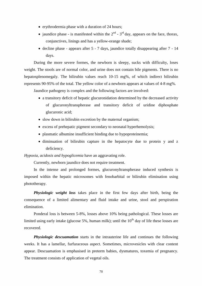
70
erythrodermia phase with a duration of 24 hours;
jaundice phase - is manifested within the 2nd
- 3rd
day, appears on the face, thorax,
conjunctives, linings and has a yellow-orange shade;
decline phase - appears after 5 - 7 days, jaundice totally disappearing after 7 - 14
days.
During the more severe formes, the newborn is sleepy, sucks with difficulty, loses
weight. The stools are of normal color, and urine does not contain bile pigments. There is no
hepatosplenomegaly. The bilirubin values reach 10-15 mg%, of which indirect bilirubin
represents 90-95% of the total. The yellow color of a newborn appears at values of 4-8 mg%.
Jaundice pathogeny is complex and the following factors are involved:
a transitory deficit of hepatic glucuronidation determined by the decreased activity
of glucuronyltranspherase and transitory deficit of uridine diphosphate
glucuronic acid;
slow down in bilirubin excretion by the maternal organism;
excess of prehepatic pigment secondary to neonatal hyperhemolysis;
plasmatic albumine insufficient binding due to hypoproteinemia;
diminuation of bilirubin capture in the hepatocyte due to protein y and z
deficiency.
Hypoxia, acidosis and hypoglicemia have an aggravating role.
Currently, newborn jaundice does not require treatment.
In the intense and prolonged formes, glucuronyltranspherase induced synthesis is
imposed within the hepatic microsomes with fenobarbital or bilirubin elimination using
phototherapy.
Physiologic weight loss takes place in the first few days after birth, being the
consequence of a limited alimentary and fluid intake and urine, stool and perspiration
elimination.
Ponderal loss is between 5-8%, losses above 10% being pathological. These losses are
limited using early intake (glucose 5%, human milk); until the 10th
day of life these losses are
recovered.
Physiologic descuamation starts in the intrauterine life and continues the following
weeks. It has a lamellar, furfuraceous aspect. Sometimes, microvesicles with clear content
appear. Descuamation is emphasised in preterm babies, dysmatures, toxemia of pregnancy.
The treatment consists of application of vegetal oils.

71
Umbilical cord involution is produced in consecutive steps: from the white initial color,
the umbilical stump suffers a mumification process gaining a dark brown color; a delimitation
area is formed, by leukocyte infiltration (aseptic necrosis); the fall out of the stump takes
place after 5-10 days and the healing takes 3 to 4 weeks. Both the stump and the scar need
daily application of sterile fields and cleaning, for preventing infection.
Genital crisis (hormonal) appears between the 3rd
and 6th
day of life and consists of
swelling and growing of the mammary glands (if expressed, a white secretion will appear).
The mammary tumefaction can appear in both genders.
At females, a descuamative vaginitis can appear, and at males a growth in the testicular
volume can appear, with or without associated hydrocele and penile edema.
The duration can be of several weeks, being the consequence of the maternal hormones'
presence in the newborn's blood. No other treatment but local hygiene is necessary.
Sometimes, other phenomena can appear, noted as paraphysiologic:
1. allergic erythema- rare, after 48-72 hours of life, has a polimorph aspect:
erythema, papules, vesicles, localized on the face, eyelids (sometimes palpebral
edema and conjunctivitis). It is associated with micropoliadenomegalies, catarrh,
splenomegaly, hyperexcitability. Blood testing reveals eosinophilia (this is the
main hypothesis - the allergic pathogeny on the phenomena; other consider it a
enterotoxin eliminated by the bacterial flora which invaded the intestines);
2. dehydration fever (thirst) - a febrile state appears after 3 - 4 days of life with
values of 39-40º C, with a duration of 12-14 hours. It is frequently associated with
hyperexcitability, influenced general state, ponderal loss. Most frequently, this is
secondary to overheating and an insufficient fluid intake;
3. difficult adjustment syndrome appears at underweight children and manifests as:
slow reactivity, difficult breastfeeding, deficitary ponderal curve, intense and
prolonged jaundice. This syndrome will differentiate dormant infections of
cerebral hemorrhage.

72
CHAPTER VI
HIGH RISK NEWBORN
The high risk newborn is a term or preterm newborn which is born under pathologic
circumstances, of maternal or fetal cause, these causes being isolated or associated. The
etiological factors most frequently seen, associated to the high-risk newborns and the type of
associated pathology are as follows:
Maternal factors and fetal or neonatal associated risk
1. Mother's age: age over 40 leads to associated neonatal pathology, such as:
chromosomal anomalies, macrosomy, intrauterine growth restriction (IUGR),
blood loss (interruption or previa)
- age below 16 years - IUGR, prematurity, neglected/abused child (the mother
herself could be abused).
2. Social factors are represented by:
- socioeconomic factors -determine prematurity, IUGR, infection
- smoking - high perinatal mortality, IUGR
- substance abuse or alcohol - IUGR, fetal alcohol syndrome, withdrawal
syndrome, sudden death syndrome, neglected/abused child.
- poor diet - slight IUGR to fetal death.
- trauma (acute or chronic). Placental rupture, fetal death, prematurity.
3. Medical afflictions:
diabetus mellitus leads to: fetal death, followed by suffering at birth, macrosomy,
respiratory distress syndrome, hypoglicemia, congenital anomalies.
thyroid disorders - goiter, hypothyroidism, hyperthyroidism.
renal disorders - fetal death, IUGR, prematurity.
urinary tract infections- prematurity, sepsis.
cardiac of pulmonary disorders- fetal death, IUGR, prematurity.
high blood pressure- both chronic or pregnancy related- can determine: fetal death,
IUGR, prematurity, asphyxia.
anemia - fetal death, IUGR, hydrops, prematurity, asphyxia.
isoimmunization - fetal death, hydrops, anemia, jaundice.
alloimmunization (platelet antigen) - fetal death, bleeding.
thrombocytopenia - fetal death, bleeding.

73
2. Obstetrical history
- history of newborn with: prematurity, jaundice, IUGR, anomalies.
- medication during pregnancy.
- bleeding during the first trimester, fetal death, prematurity.
- hyperthermia, fetal death, fetal anomalies.
- bleeding in the 3rd
trimester, fetal death, anemia.
- premature membrane rupture, infection, sepsis.
- TORCH syndrome.
- trauma at birth, fetal death, prematurity.
Fetal factors and associated fetal or neonatal risk
Multiple pregnancy is associated with: IUGR, twin-to-twin transfusion
syndrome, prematurity, birth trauma, asphyxia.
IUGR - fetal death, congenital anomalies, asphyxia, hypoglicemia,
polycythemia.
Macrosomy - congenital anomalies, birth trauma, hypoglicemia.
Abnormal fetal presentation or positioning - congenital anomalies, birth
trauma, hemorrhage.
Heart rate or rythm anomalies - fetal cardiac insufficiency, cardiac blockage,
hydrops, asphyxia.
Activity decrease-fetal death, asphyxia.
Polyhydroamnios - anencephaly, other central nervous system disorders,
neuromuscular disorders, swallow disorder, chylothorax, diaphragmatic hernia,
omphalocele, gastroschisis, trisomy, tumors, hydrops, isoimmunization,
anemia, cardiac insufficiency, urine concentration inability, large for
gestational age, maternal diabetes.
Oligohydroamnios - fetal death, placentary insufficency, IUGR, renal agenesis,
pulmonary hypoplasia, deformations, intrapartum distress, postterm birth.

74
Labor and birth associated factors and risk assessment
of the fetus and newborn
In this category, the following factors are included: preterm birth, postterm birth,
maternal fever, maternal hypotension.
Other frequent factors:
- precipitate delivery - birth trauma, intracranial hemorrhage, lung fluid
retention/transient tachypnea.
- prolonged delivery - fetal death, asphyxia, birth trauma.
- abnormal presentation - birth trauma, asphyxia.
- uterine tetany- acute asphyxia
- meconium - stained amniotic fluid- fetal death, asphyxia, meconium aspiration
syndrome, persistent pulmonary hypertension.
- cardiac failure - fetal death, asphyxia.
- cesarian - RDS, pulmonary fluid retention/transient tachypnea, sanguine loss.
- obstetrical anesthesia and analgesiasi - RDS, neurogenic distress, hypotension,
hypothermia.
- placental anomalies:
- small placenta - IUGR.
- big placenta - hydrops, diabetus mellitus, large infant.
- placental or umbilical vessels rupture - blood loss.
Neonatal high risk associated factors:
- Prematurity - IUGR, other prematurity traits.
- APGAR score under 5 at 5 minutes
- APGAR score under 5 at 15 minutes - neurologic disorders.
- Palor or shock
- Bad amniotic fluid smell - infection
- Small for gestational age.
- Postmaturity syndrome.

75
CLASSIFICATION BASED ON GESTATIONAL AGE AND BIRTH WEIGHT
Newborns should be classified based on gestational age, if possible, because, as a
psychological matter, this is far more important aspect than birth weight.
A. Gestational age classification.
1. The assessment based on obstetrical information is included in CHAPTER I.
Gestational age based on the first trimester ultrasonography evaluation is exact only
for 4 days.
2. For confirming or completing obstetrical data, the Dubowitz examination (Ballard)
can be useful in estimation gestational age. There are certain limitations with this
method, especially in evaluating the neuromuscular component of the sick newborns.
3. Newborn classification based on postmenstrual gestational age
a. Preterm. Less than 37 complete weeks of gestation (259 days).
b. Late preterm. Represents a subgroup of infants born between 34 and 36 weeks of
gestation (238-258 days).
c. Term. Between 37 and 41 6/7 weeks (260-290 days).
d. Postterm. 42 weeks (295 days) or more.
B. Classification based on birth weight. Even though there isn't a universal agreement,
the most common definitions are:
1. Normal birth weight (NBW). From 2.500 to 4.500 g.
2. Low birth weight(LBW). Less than 2.500 g. While most LBW preterm infants
are born preterm, others are term newborns with small gestational age. LBW
infants can be classified as follows:
a. Very low birth weight (VLBW). Less than 1.500 g.
b. Extremely low birth weight (ELBW). Less than 1000 g.
Newborn from twin pregnancy
The incidence of twin pregnancies is estimated around 1:80, and of monozigote or
identical twins of 1:200. Prematurity and/or IUGR are the main risk coordinates of twin
pregnancies. Unlike small fetuses, prematurity and dysmaturity of the twins is due to
mechanical factors, and in small amount due to other factors.

76
The risk of neonatal mortality and morbidity is, also different for the two twins. During
natural birth, the second twin is more affected due to the intranatal stress factors.
The optimal time of delivery of the second child is within 15 minutes after the expulsion
of the first one. After an interval of 30 minutes, if the expulsion of the second twin does not
begin, this will be considered retained, with a high risk of demise or injury.
All twin pregnancy risks develop proportionally, even exponentially with the number of
fetuses.
Congenital malformations or malformative syndromes are more frequent for twins,
especially monozygotes.
There are at least 3 types of structural defects(especially monozygotes twins):
malformations or complex malformative syndromes, with early signs, which
apparently have the same basis that determines twin pregnancies: renal agenesis,
anal atresia, tracheoesophageal fistula, anencephaly, holoprosencephaly, situs
inversus;
vascular anomalies, secondary to abnormal vascular communications within a
single placenta; these are secondary to thrombosis and IDC that produce at a
deceased fetus and can apply the healthy twin: cutaneous aplasia, intestinal atresia;
plastic deformations, due to mechanical action from the uterus; are normally found
at both categories of twins.
twin-to-twin transfusion syndrome - occurs only at monochorionic twinsand is due
to vascular connections which establish between the two fetal circulations. The
donor will suffer a degree of IUGR, with a slowdown of the organs global mass,
anemia and hypotensions, and the receiver will have the following: polyglobulia,
cardiomegaly, arterial blood pressure and polyhydramnios.
Newborn from diabetic mother
The clinical aspect of these infants is particular, with the following aspects: birth
weight>90th
percentile for gestational age (macrosomic); cushing-like aspect, swollen, short
neck, abundant hair; visceromegaly (excepting the thymus and the brain, sometimes smaller
in size) compared to birth weight; head circumference is small (characteristic aspect).
In literature, it is described as a "giant with clay feet", because he is exposed to many
risks, like:
cardio - respiratory adaptation disorders, due to associated polyglobulia;

77
respiratory distress syndrome (an important percent of macrosomes are preterm);
morphologic disorders; frequent and of variable intensity;
renal veins thrombosis, rare, but frequent in the general population;
cytosteatonecrosis;
metabolic disorders: hypoglycemia, hydroelectrolitic imbalance (extracelullar
dehydration, hypocalcemia), hyperbillirubinemia, hypoproteinemia.
The malformative risk is augmented by the lack of therapeutic control and gravity of the
maternal injuries. Congenital malformations are various and can affect any organ or system.
Other categories of risk newborns:
- newborn from epileptic mother (major malformation risk,
hemorrhagic risk, withdrawal syndrome);
- newborn from alcoholic mother(major malformation risk,
neuropsychomotor injury risk);
- newborn from HBP mother (preterm birth risk, IUGR and cerebral
hemorrhage);
- postterm newborn (cerebral injury risk and late neuropsychomotor
disorder risk).

78
CHAPTER VII
SMALL BIRTH WEIGHT NEWBORN PRETERM NEWBORN
The definition of a preterm newborn takes into account two major criterias: gestational
age and birth weight. After the WHO, preterm newborns are those born before 37 weeks
gestational age.
Other classification, by weight and gestational age:
ELBW - Extremely low birth weight is the weight below 1000 g. Most of
these babies are preterm, born before 27 weeks gestational age.
VLBW - Very low birth weight is the weight below 1500 g.
LBW - Low birth weight, are the ones with weight below 2500 g.
AGA - Appropriate for gestational age newborns
LGA - Large for gestational age newborns
SGA - Small for gestational age newborns; also names newborns with
intrauterine growth restriction (IUGR).
ELBW (extremely low birth weight) newborns survive if modern therapeutic means are
used: surfactant therapy, maternal corticosteroids and advanced neonatal technology.
Prematurity and IUGR are associated with high neonatal mortality and morbidity.
Ideally, the definitions of low birth weight newborns should be based on homogenous
genetically and epidemiologically data.
Prematurity holds a major role in determining the perinatal mortality and morbidity.
Generally, a preterm is considered a baby born before 37 weeks gestational age,
weighing under 1500 grams, having a length below 47 cm. The inconvenient of gestational
age appreciation is not knowing exactly, by some women, the last menstrual cycle's date.
Another classification system, elaborated by WHO in 1948 and largely used in the
medical practice, base on the birth weight was:
prematurity grade I birth weight between 2000-2500 g;
prematurity grade II birth weight between 1500-2000 g;
prematurity grade III birth weight between 1000-1500 g;
prematurity grade IV birth weight below 1000 g.

79
A weight of 2500 g as a limit for prematurity is also unsatisfactory, because 1/3 of these
newborns are dysmature (small birth weight, but normal gestation) and 10% of the newborns
with birth weight over 2500 g and gestational age lower than 37 weeks are wrongly called
sick newborns.
Maternal diabetus mellitus can be associated with preterm birth; these newborns, even if
they are large for gestational age, raise the issues of prematurity.
Along with the mentioned aspects, there are several other criteria to define prematurity,
such as: neurologic criteria by Saint-Anne Danassier, EEG criteria (appreciates the electrical
activity of the brain), anatomopathologic criteria which appreciates the brain conformation
(cerebral scissures, circumvolutions), hystologic criteria (appreciates the nucleo-cytoplasmic
index).
The incidence of prematurityvaries based on regions and populational groups between 4
and 14%.
Etiology of prematurity
Has multiple and varied causes, which are frequently associated or intricate, making
their contributional distinction very difficult.
Out of the most common ones, we are mentioning:
preterm births in the mother's history,
infantile uterus,
nuchal cord,
vicious insertions of the placenta,
hydramnios,
placenta praevia,
premature rupture of membranes (PROM),
uterus and adnexial tumors,
uterine malformations,
incompetent cervix
maternal diseases (TBC, lues, nephropathies, heart disease, hepatitis, toxic
pregnancy, acute infections, diabetus mellitus),
short interpregnancy interval,
twins,
primiparity at a young age,

80
high multiparity,
abnormal presentations,
Rh or group incompatibilities,
proteins or vitamins deficiency,
social factors - illegitimate couples, intense professional activity of the mother,
unknown factors (between 30-50%).
Morphologic pathologies of a preterm baby
The head represents 1/3 of the height, the head circumference is below 33 cm, the
cranial sutures are dehiscent, the fontanelles open, the facies is small, of triangular aspect,
with sharp chin and underdeveloped ears, with a poor maturation of the cartilage.
Pectus excavatum is frequently seen (the ribs are cartilaginous), thoracic circumference
is around 31 cm, underdeveloped intercostal muscles, which favors the tirage.
The abdomen is bulky due to abdominal muscle hypotonia and high liver volume;
diastasis recti, inguinal hernia and umbilical hernia are frequently seen. The umbilical cord is
inserted closer to the pubic symphysis.
The skin is thin, with visible vascular drawing, with a large surface compared to the
body volume, reduced or absent vernix, abundant lanugo on the face, limbs and posterior part
of the thorax. Lamellar descuamation is prolonged.
The skin appendages are smooth and do not exceed the curve of the finger.
The subcutaneous tissue is reduced or absent. Bichat's fat pad is underdeveloped in
grade III, IV preterm newborns; their face has an old man's look.
The muscle system is reduced and with hypotonia, the skeletal system is insufficiently
mineralized; the ossification nuclei, Beclard and Tappon are missing.
The genitals are insufficiently developed. In boys, the testicles are not always
descended and in girls, the labia majora does not cover the labia minora and the clitoris. The
genital crisis is of low intensity.

81
Functional particularities
Respiratory system
The respiratory system has the following traits: narrow thoracic cage, horizontal and
incompletely mineralizedribs, hypotonia, high position of the diaphragm which limits
pulmonary extension and favors tirage.
The respiratory tract is narrow, the pulmonary tissue elasticity is low.
Pulmonary alveoli are immature, with thick ealls and distanced from the sanguine
capillares by an abundant mesenchimal tissue, which makes the pulmonary exchange
difficult.
There is a quantity and quality surfactant deficit. Synthesis and eliberation of surfactant
from the pneumocytes is unsatisfactory before 34 weeks gestational age. The surfactant
deficit (maintains the superficial tension of the alveoli) from the alveolar fluid favors alveolar
colabation.
Ventilation disorders determine a high resistance in the pulmonary circulation.
What's more, the immaturity of the respiratory nervous centers contributes as well to
the respiratory function deficiency. During hypoxia, the apneustic center (upper bulb) stops its
activity, and the inferior bulbar center (older) invervenes, which determines a periodic
respiration, realized by a profound, jerky inspiration, along with spasmodic movement of the
mouth and mandible, interrupted by respiratory breaks (gasping). This type of breathing can
be a prelude to respiration shutdown.
Even though hypoxia is partially compensated in the preterm baby, the oxygen is highly
utilized by the fetal hemoglobin (HbF=90%), which has a high affinity for oxygen and
attached difficultly CO2, favoring its elimination; on the other hand, the use of anaerobic
glycolysis as a main source of energy for the central nervous system, while the hypoxia is
severe, drives the depletion of carbohydrates reserves and lactic acids accumulates.
A metabolic acidosis is created, which adds to the respiratory one through hypercapnia.
Preterm acidosis is prolonged because the liver function is not capable of conjugating
intermediar catabolism products, and the renal function is limited by phosphate elimination
H+ and base conservation. Severe acidosis depresses the miocardic, renal and brain activities.
A vicious circle is realized, and acidosis hinders hematosis by pulmonary arterioles
vasoconstriction. The resultant of these respiratory deficiencies is the "respiratory distress"
which means dyspnea, apneic crysis with cyanosis and paradoxal inefficient respiratory
movements.
The number of breaths is of 50-60 /min.
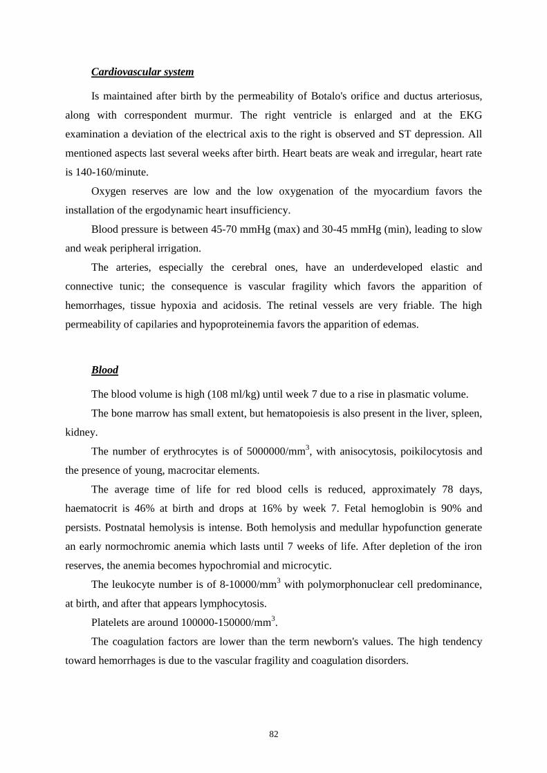
82
Cardiovascular system
Is maintained after birth by the permeability of Botalo's orifice and ductus arteriosus,
along with correspondent murmur. The right ventricle is enlarged and at the EKG
examination a deviation of the electrical axis to the right is observed and ST depression. All
mentioned aspects last several weeks after birth. Heart beats are weak and irregular, heart rate
is 140-160/minute.
Oxygen reserves are low and the low oxygenation of the myocardium favors the
installation of the ergodynamic heart insufficiency.
Blood pressure is between 45-70 mmHg (max) and 30-45 mmHg (min), leading to slow
and weak peripheral irrigation.
The arteries, especially the cerebral ones, have an underdeveloped elastic and
connective tunic; the consequence is vascular fragility which favors the apparition of
hemorrhages, tissue hypoxia and acidosis. The retinal vessels are very friable. The high
permeability of capilaries and hypoproteinemia favors the apparition of edemas.
Blood
The blood volume is high (108 ml/kg) until week 7 due to a rise in plasmatic volume.
The bone marrow has small extent, but hematopoiesis is also present in the liver, spleen,
kidney.
The number of erythrocytes is of 5000000/mm3, with anisocytosis, poikilocytosis and
the presence of young, macrocitar elements.
The average time of life for red blood cells is reduced, approximately 78 days,
haematocrit is 46% at birth and drops at 16% by week 7. Fetal hemoglobin is 90% and
persists. Postnatal hemolysis is intense. Both hemolysis and medullar hypofunction generate
an early normochromic anemia which lasts until 7 weeks of life. After depletion of the iron
reserves, the anemia becomes hypochromial and microcytic.
The leukocyte number is of 8-10000/mm3 with polymorphonuclear cell predominance,
at birth, and after that appears lymphocytosis.
Platelets are around 100000-150000/mm3.
The coagulation factors are lower than the term newborn's values. The high tendency
toward hemorrhages is due to the vascular fragility and coagulation disorders.
83
Digestive system
The mucous membrane of the oral cavity is dry and permeable, the salivary secretion is
low, aspects that favor the appearance of stomatitis.
The sucking reflex is absent at most preterm babies, and others lack the swallow reflex;
salivary amylase is present at birth.
The gastric capacity is reduced (30-90 ml), the mucous layes has a few folds, the glands
and muscular fibers are underdeveloped and gastric evacuation is delayed.
Chlorhidric acid secretion is good. The labferment, pepsin and gastric lipase are present
at birth.
The intestinal musculature is hypotonic, the peristalsis is slow, favoring meteorism and
constipation.
The intestinal mucose has a high permeability. Lactase is deficitary during the first
week after birth, and is stimulated by offering substrate.
The pancreas has a satisfactory activity at birth by the presence of tripsine, lipase
activity is reduced by 20% compared to the term newborn and amilase secretion is absent.
The liver has many deficiencies in seric protein and coagulation factor synthesis.
Glucuronyltransferase activity is reduced (10% compared to normal), and is responsible of the
duration and intensity of preterm physiologic jaundice, which can evolve up to
hyperbilirubinemic encephalopathy.
Bile secretion is deficitary, bile acid values are low, due to the fact that the gallbladder
concentrates the acids insufficiently, and the bile acid loss through stools are big.
Digestion and absorption of the human milk proteins are good.
Digestion and absorption of lipids from milk is deficitary due to the reduced activity of
pancreatic lipase, small quantity of bile acids, phosphorylation disorders and low activity of
serum lipoprotein lipase. Human milk lipids have a superior absorption quality (90%) due to
the presence of two milk lipases (lipoprotein lipase and bile salt-dependent lipase). Cow milk
lipids are absorbed around 60 - 70%.
Digestion and absorption of milk lactose is normal after one week of life.
The establishment of intestinal microbiome is slow and vulnerable for prematures. The
digestive tolerance is generally reduced.

84
Urinary tract and excretion function
More recent data have confined the notion of excretion function deficiency for healthy
preterm newborns, showing that these are capable of assuring the water homeostasis at an
intake of 200 ml/kg/day from the third day of life.
It was shown that the constant maintenance of GFR can be done at 0,92 ml/kg/min
using tubulary reabsorption, appropriate elimination with negative balance and dilution -
concentration capacity in the first 7 days of life, at a normal serum urea concentration.
The oscillations of the urine osmotic pressure between 80-400 mOsm/l, at a constant
filtration rate, confirm the renal homeostatic capacity during the first week of life. In the light
of these events, it appears that the preterm baby's edemas are due to hypoproteinemia rather
than sodium retention.
Studies done in 1987 show that the preterm baby can adequately absorb the HCO3-ion in
moderate acidosis, during the first week of life.
The proximal tubular reabsorption of bicarbonate is dependent on the carbonic
anhydrase activity, which is present in the intrauterine life, before birth.
Hydroelectrolytic metabolism
Maintaining the hydroelectrolytic balance is essential for the organ well functioning. The
hydroelectrolytic imbalances can aggravate certain diseases such as: patent ductus arteriosus,
intraventricular hemorrhage and chronic lung disease (bronchopulmonay dysplasia).
Compared to term newborns, extremely low birth infants have a larger quantity of fluid in
the extracellular compartment and a higher percent of their weight is attributed to the fluid
component.
During the first days after birth, there can be observed a weight loss of 10-20% due to
diuresis, loss that can be aggravated through phototherapy and radiant heat use inside
incubators.
These newborns also have a deficitary renal function, because of the low GFR and low
bicarbonate absorption capacity. Because of the immaturity of the tubular of the renal function,
the capacity to secrete potassium and other ions is decreased, along with a relative incapacity of
urine concentration. Immediately after birth, at the renal tubular level, creatinine reabsorption
takes place; its serum values birth do not represent an abnormal renal function. Water balance is
monitored using daily weighing (sometimes twice per day), water balance (ingestion/excretion)
and ionogram.

85
These newborn are prone to nonolyguric hyperkaliemia.
Hyperkaliemia is defined by potassium levels higher than 6,5 mmol/l. Such values can
cause cardiac arrythmias and demise.
Hypernatremia and hyponatremia are due to free water alteration (dehydration with the
rise of serum sodium or hyperhydration with dillutional hyponatremia). At very low birth
weight infants, dehydration can be produced by exposure to heat or phototherapy. For these
children, hypernatremia can be combatted by administration of hypotone fuids for fluid loss
replacement.
For maintaining an adequate hydration state, one can administer up to 200-250
ml/kg/day. Such quantities of fluids can decompensate cardiac disorders (eg: patent ductus
asrteriosus). Fluid loss by insensible perspiration can be reduced by use of "double wall"
incubators.
In case of hyponatremia, the fluid intake will be restricted and sodium supplements will
be given.
The calcium and phosphate metabolism: prenatal calcium reserves are low due to short
gestation. The intestinal calcium absorption is good, but the alimentar intake does not provide
the adequate necessities of a preterm newborn, so he always has a tendency of clinic
manifested hypocalcemia.
Iron metabolism: is defficitary, even though the intestinal absorption and elimination are
normal, but the iron supplies are low (168 mg), dietary intake from milk is decreased, and the
high intensity hematopoesis generates iron deficiency anemia around 3 months of age.
Nervous system
Circumvolutions and brain scisures are hardly seen and there do not exist clear lines
between the white and grey matter.
The cortical and subcortical areas are low vascularised, in contrast with the rich
vascular network if the periventricular area. This areas are the place of numerous
hemorrhages, causing death by ventricular flood.
The vital function control activity (respiratory, cardiac, thermoregulation) is defficitary.
The preterm baby presents a high muscular excitability, seen as muscular contractions,
present during sleeping as well.
The preterm movements have an athetosic nature. Nystagmus, anisochoria and
strabismus are frequently seen, but they are transient.

86
The osteotendinous and abdominal cutaneous reflexes are present, but the swallowing
reflex is absent at small preterm newborns. The presence of archair reflexes is conditioned by
the duration of the gestation period; it represents a criteria of the nervous system development
assessment.
The EEG shows hypovolted route during the first 5 - 6 months.
The CSF is xantochromial, with albuminorachia of 1,5 g% and a number of 5-6
elements/field.
Thermoregulation
After birth, the preterm's temperature drops by 2-3 degrees and normalizes in 2-7 days.
Preterm newborns have a high thermolability with a tendency toward hypothermia. This
thermolability is the consequence of high thermolysis, deficitary thermogenesis and imaturity
of the thermoregulator centers.
High thermolysis is a consequence of high surface of irradiation through the thin, well
vascularized skin, with a high water content and lack of adipose tissue; this determines an
unsatisfactory thermal insulation.
Thermogenesis is limited by the low lipid reserves, low dietary intake and intestinal
absorption, low basal metabolism and low muscle activity. Maturation of thermoregulation
take place at 3-5 months of age. That's why the preterm newborn is dependent on the
environmental temperature. The thermal comfort zone is of 32 degrees Celsius, which allows
a minimal oxygen consumption.
Protection against cold is done through chemical thermogenesis, more exactly by
general increase of cellular oxydations, especially brown fat. The brow fat triglycerides from
the base of the neck, mediastinum and interscapular area are hydrolized by cathecolamine
action into fatty acids, which, by oxidation, release heat. This type of chemical thermogenesis
is noneconomic, is done with high energy consumption, taking the oxygen from other vital
functions. Overheating determines hyperthermia.
Immunity
Immunity and infection resistance are low. Immunologic deficiency manifests both as
nonspecific and specific defense.
Mucous immunologic deficiency is due to high permeability and secretory IgA
shortage. The fagocytic activity represents 53-60% of the adult values, due to the decreased
chemotactic leukocytic response.
87
The complement, opsonins and properdin reach about 17-50% of the adult values.
IgG from the mother represents 10% of the term newborn's level; the transfer is made
during the last 3 gestational months.
The preterm newborn's infections have a quiescent evolution, without fever, but with
alteration of the general state, with a septicemic trait.
Complications of prematurity
Preterm babies have a series of specific disorders, associated with difficulty of adaption
to the extrauterine life, secondary to different organs and systems immaturity. The most
severe and frequent disorders, that lead an increase in neonatal mortality are:
1. Respiratory distress syndrome
A serious and early prematurity of high risk complication is the respiratory distress
syndrome (RDS), and its main cause is the surfactant deficiency.
The RDS incidence is inversely proportional to gestational age, with an incidence of
60% by 29 weeks of gestation. In the US, RDS affects 40.000 newborns yearly (most of
which have very low birth weight).
Specific clinical signs are: tachypnea (>60 breaths/min.), cyanosis, intercostal
retractions, nasal flaring, expiratory grunting.
Untreated, RDS increases the oxygen need during the first 24-72 hours of life. The
cardio-pulmonary x-ray shows a uniform reticular and granular image, with air bronchogram.
Due to the surfactant shortage, alveoli collapse, edema appears along with a decrease of total
pulmonary capacity.
The surfactant decreases the tension in the small respiratory duct walls, so that the
alveoli remain open, without oxygen supplementation or ventillatory support.
Common complications are: chronic lung disease (bronchopulmonary dysplasia) and
retinopathy of prematurity.
Prophylactic surfactant administration for preterm babies born below 28 weeks of
gestation decreases the ventillation necessity, but does not decrease the risk of
bronchopulmonary dysplasia occurrence. Synthetic surfactants do not have in their structure
certain proteins that are found in the natural surfactant structure, which makes them less
effective.
The RDS incidence can be significantly reduced by the use of prenatal corticosteroids
and early surfactant therapy.

88
Prenatal use of steroids also decreases the indicence of patent ductus arteriosus and
intraventricular hemorrhages, but there still exists the possibility of neuropsychic disorders
during the administration of repeated doses of steroids.
In the last decade, surfactant was often used in the treatment of RDS, so it was
concluded that it should be prophylactically used for all born before 30 weeks of gestation,
which would lead to unnecessary surfactant administration for some preterm babies. While a
small number of newborns are intubated at birth, prophylactic surfactant administration via
intubation cannot be realized. Newborns that are not intubated at birth, can be given oxygen
via nasal canula, with continuous positive pressure (it improves the endogenous production of
surfactant). If this measure does not pay off (rise of PaCO2, clinical signs of respiratory
distress, increase of oxygen need), the next therapeutic option remains intubation and
surfactant administration. Studies (Geary et al.) have shown that such a therapeutic protocol
decreases the incidence of chronic lung disease.
If the surfactant prophylactic administration is wanted, it should be done immediately
after birth. If it is used as a curative treatment, it should be administered as soon as the clinical
signs of respiratory distress start to show or if the respiratory syndrome does not improve after
initial reanimation.
One of the most severe prematurity complications is the chronic lung disease
(bronchopulmonary dysplasia - BPD), which is defined by: oxygen necessity
supplementation or ventillatory support until a gestational age of 36 weeks. An older
definition was: oxygenotherapy dependency after 28 days of life. The incidence is between
19% (1990) to 23% (1996) at newborns with less than 36 weeks gestational age, with a
weight< 1500 g.
2. Intraventricular hemorrhage (IVH)
It's the most common prematurity complication, with the subependimal germinal matrix
as a starting point and it can progress in the presence of aggravating factors, to the whole
ventricular system and/or periventricular parenchyma, leading to peri or intraventricular
hemorrhage.
The incidence and severity of the hemorrhage are inversely correlated with gestational
age. Extremely low birth weight infants have a high risk, due to the high germinal matrix
vulnerability and because the cerebral protective self-regulation is not yet developed.

89
3. Periventricular leukomalacia
Commonly seen pathology in bigger preterm infants, the second pathology after
intraventricular hemorrhages; it represents an area of ischemic necrosis of the white
periventricular substance, localized closely to the external angle of the lateral ventricles. Its
incidence was appreciated around 7-26% within autoptic studies. The frequency is higher for
preterm newborns with a birth weight under 1500 g, with a history of severe hypoxia or
ischemia (80-90% of the cases are seen in preterm babies), newborns from laborious births,
along with cranial trauma or babies with cardiorespiratory disorders during the neonatal
period (heart failure, patent ductus arteriosus, repeated crisis of apnea, septicemia).
4. Apnea of prematurity
Apnea is defined as an interruption of breathing for a period longer than 20 seconds or
any respiratory break with a shorter period but associated with cyanosis, bradycardia or both.
Apnea is frequently seen in a preterm baby. It is caused, most of the time, by a transient
disorder of respiratory control (apnea of prematurity) or rarely by a more severe state, such as:
septicemia, hypoglicemia, intracranial hemorrhage, seizures or mother ingestion of certain
drugs.
5. Patent ductus arteriosus
For a fetus, the blood oxygenation is realized through the placenta, so the blood
passage through the lungs is not necessary. The ductus realizes the connection between the
pulmonary artery and the aorta, so that on the whole pregnancy period, the blood bypasses the
lungs. At term, the ductus closes, generally after the first 48 hours of birth due to the
constriction done by oxygen. This constriction has a decreased effect on the ductus arteriosus
on preterm newborns, so that 80% of the ELBW or VLBW ones still have a ductus at birth,
with important clinical severity, depending on the shunt intensity between the systemic and
pulmonary circulation, left to right shunt.
The clinical symptomatology is diverse: systolic murmur of different degrees,
dyspnea. If the PDA is of a high caliber, a negative effect is produced onto the cerebral
oxygenation, a decrease of the systemic blood flow due to the left to right shunting, decrease
of the urinary flow, food intolerance or hypotension.

90
6. Necrotising enterocolitis (NEC)
NEC represents a digestive disorder, specific for the preterm newborn, which
determines mucous injuries and intestinal vascularisation disorders and it represents the most
frequent intestinal emergency of prematurity. The incidence is directly correlated to
gestational age: 1-8% of the newborns admitted to the NICU, with a mortality rate of 10-50%.
7. Hyperbilirubinemia
Most of the VLBW newborns develop hyperbilirubinemia (jaundice) which needs
treatment. Hyperbilirubinemia is produced under the following circumstances: exaggerated
hemolysis, liver immaturity that is incapable of conjugating and eliminating the resulted
bilirubin. What's more, most premature babies have a low intestinal motility due to the
insufficient oral intake, so the results are: late meconium passing (which contains bilirubin)
and an increase in the enterohepatic circulation of the conjugated bilirubin. Due to these
prematurity complications and other contributory factors (group or Rh incompatibility,
sepsis), these newborns have a high risk of nuclear jaundice at lower bilirubin values than the
term newborns.
8. Retinopathy of prematurity
It represents a disease of the preterm retina, which has not been yet vascularized. Slight
changes in oxygen exposure have been incriminated in the distruction of the vascular
trajectory and can lead to abnormal growth of the vessels, which can lead to retinal
detachment and blindness.
The retinopathy risk factors are represented by prematurity and oxygen exposure. All
VLBW babies should have an ophtalmologic examination by 4 weeks of age (or
postconceptional age of 31 weeks) and, based on the results, every other 2 weeks until the
retina is completely vascularized.
If the disease is present, treatment assessment will be done based on stage and location,
from frequent check-ups to laser intervention or even vitrectomy.
The presence of another disorder or the sinuosity of the retinal vessels represent a poor
prognosis and require immediate treatment.
Preterm newborns with retinopathy have a high risk of developing sequelaes, such as
myopia, strabismus or amblyopia.
91
9. Metabolic disorders
Hypoglycemia (less than 20 mg%) and hypocalcemia (less than 8 mg%) are common.
Fetal euglycemia (normal blood glucose level) is maintained during the whole pregnancy by
the maternal placenta. VLBW newborns can have difficulties in maintaining their glucose
levels immediately after birth when the glucose maternal source has been cut-off. Besides,
these newborns have a higher risk compared to term newborns and do not have sufficient
glycogen reserves.
Hypoglycemia is considered when the plasmatic level of glucose is below 45 mg/dl.
10. Hypothermia
As a result of a high ratio of the body and small weight, the decrease of brown fat
reserves, immaturity of the skin and decrease of glycogen reserves make the ELBW
susceptible to heat loss immediately after birth. Hypothermia can appear as a result of
hypoglycemia, apnea or metabolic acidosis, as well.
Temperature monitorization is important for all categories of preterm newborns and is
realized using radiation heating devices or double wall incubators.
Hypothermia (<35ºC) has been associated with a bad prognosis, including chronic
dependence on oxygen; that's why, special protection measures are imposed. Thereby, after
expulsion, the newborn must be dried and put on a radiant heating sourse, and his head must
be covered with a hat or something else. Studies show that using a plastic bag immediately
after the baby has been dried, seems to minimalize the temperature loss thorugh evaporation
and convection.
For the newborn transportation from the delivery room to the NICU it is necessary that
the baby is covered in warm blankets or cellophane. For long distances transportation, the
newborn must be places in a double-wall heated incubator.
The delivery room and the NICU must be maintained heated for the prevention of
hypothermia of such preterm newborns.
11. Infections
Predisposition to infection is based both on immunologic immaturity, but on other
immaturities as well, that indirectly influence the rise of the infectious risk: nutrition
difficulties, with a risk of pulmonary aspiration, multiple maneuvering or explorations,
catheterisations or extended iv fluid therapy. Infection still remains an important factor of
VLBW neonatal mortality and morbidity.

92
Preterm newborn nutrition
Preterm baby's nutrition must be appropriate for his digestive particularities and high
nutritional needs. Maintaining an ascending growth curve for these newborns remains a
challenge. Weight is monitored continuously (daily weighing) and so are the anthropometric
indexes: length and head circumference (weekly).
The growth curve can be influenced by specific complications, such as pulmonary
disorders or sepsis. Another contributing factor is the inadequate calories and proteins intake.
Initiation of enteral nutrition is frequently delayed, due to the risk of NEC apparition.
For VLBW babies, parenteral nutrition provides the necessary energy and protein
requirements during the first weeks of life.
The optimal parenteral nutrition was realized by the use of special amino acid solution,
glucose, minerals, electrolytes and it names total parenteral nutrition. A 20% lipidic emulsion
is added to the nutritional scheme.
It is considered that the total protein requirement for stimulating the protein anabolism
is of 4 g/kg/day. The nutrition for these preterms must be supplemented with essential amino
acids such as cysteine, glutamine and minerals (iron, iodine, zinc, copper, selenium, floride).
Chrome, molybdenum, manganese and cobalt can be added especially to the newborns with
long term parenteral therapy. In some centers, L-carnitine is added. Lont term parenteral
nutrition use has secondary effects, such as: cholestasis and high trygliceride levels.
Caloric requirements are high compared to the reduced nutritional absorption and
metabolism. During the first week of life, caloric intake requirements are 60-90 cal/kg/day,
during the second week 100-120 cal/kg/day, and after that 130-140 cal/kg/day.
Fluid requirements are influenced by temperature and humidity of the environment.
During the first week, there is a tendency of sodium and water retention, which is why the
fluid intake starts by 60 ml/kg/day and increases up to 150 ml/kg/day; during the second week
of life: 150-180 ml/kg/day, and at the end of the first month of life: 200 ml/kg/day.
Protein requirements are high, between 2,5 – 4 g/kg/day. Cysteine and histidine are
considered essential amino acids. Because the VLBW infants are losing 1,2 g/kg/day of
endogenic proteins, they require the same amino acid quantity per day and 30 kcal/kg/day for
maintaining the protein balance. It is recommended that the nutritional supplementation
should be done using protein substances during the first 12-24 hours, for avoiding protein
catabolism.

93
Lipid needs are of 2-3 g/kg/day, and it is known that preterm babies have a
malabsorption for lipids. Linoleic acid requirement is 5% of the total caloric intake need. The
lipid intake varies depending on tolerance and must be initiated during the first 24 hours of
life for an optimal nutrition. Lipid supplementation can be delayed due to possible infection
or hyperbilirubinemia occurence.
Carbohydrate needs are of 4-6 g/kg/day during the first week of life (when the preterm
has a lactase deficiency) and rise up to 12-18 g/kg/day due to the high glucose requirements,
which is done by the organ functioning and brain glucose need.
Mineral needs are bases on insufficient prenatal reserves and high needs during the
growth process.
The calcium requirement is of 40-50 mg/kg/day, phosphorus of 30-40 mg/kg/day, iron
1-2 mg/kg/day.
Nutrition should be early initiated because the preterm newborn has low reserves, high
energy consumption and the environmental aggressions along with associated disorders
increase the nutritional needs.
Oral nutrition starts with 5% glucose solution for reducing the incidence of
hypoglycemia. After 12 hours, human milk is progressively added, related to his weight and
general state.
The dosing of small milk quantities every 2-4 hours can be initiated from the first day of
life. If the nutrition is well tolerated (low gastric residual), the food ration can be increased by
10-20 ml/kg/day. Bolus nutrition is physiologic, but some preterm newobrns do not tolerate it
and require continuous enteral feeds.
Clinical research shows that the enteral fed preterms, which tolerate higher quantities of
milk have an ascending growth curve and a lower infection risk.
Contrary to the fact that early enteral feeds could favor the apparition of NEC, studies
have not shown a correlation between the nutritional techniques (the age for enteral feeding
initiation, rhythm of ration increase) and NEC.
The number of feeds should be 7 for preterms over 32 - 34 weeks gestational age and
8-10 for VLBW and ELBW, depending on tolerance and associated pathology (see "Infant
care-practical course", chapter IV). When the weight of 2000g is reached, the number of feeds
should drop to 7, no matter the degree of prematurity.
Human milk is considered to be the best choice for enteral feeding and it has been
shown that is has a protective effect on NEC.

94
Human milk is the best food for a preterm baby, due to its protein specificity, low
mineral values, amino acid intake, essential fatty acids, biologic value and optimal absorption.
It can provide, along with the fluid requirements, the caloric needs of proteins, lipids,
carbohydrates and minerals.
LBW newborns have a high requirement of micro and macronutrients, similar to the
one from the intrauterine life. The gastrointestinal immaturity does not allow an adequate
nutritional intake, if we are talking about growth needs.
In spite of multiple immunologic and nutritional advantages, a diet based solely of
human milk, with no supplementation, is energy inefficient and needs calcium, phosphorus
and protein supplementation, so that bone mineralisation and growth rate maintain similar
rates to the intrauterine ones.
Mother's own milk should be supplemented with a caloric, protein and mineral addition,
especially calcium and phosphorus. Without these essential elements, protein - calorie
malnutrition can appear, along with hyponatremia, osteopenia of prematurity or rickets. To
this end, human milk fortifiers are used, with special formulas for preterms, mineral
supplements, vitamin supplements, with different brand names: liquid PreNan, stage I and II,
Prematil, HumanaO, Enfamil, Similac Human Milk Fortifier - powder and Similac Natural
Care Liquid Fortifier - liquid.
Complications of human milk fortifiers use are: nutritional imbalance, high osmolality,
bacterial contamination.
There are a numerous number of formulas, specially designed for preterm babies that
cannot be breastfed.
The nutritional balance is very important during the neonatal period. Studies show that a
high intake of carbohydrates during this period is associated with a growth above limit and
insulin resistance, increasing the risk of metabolic syndrome during the adult life.
The nutrition technique is individualized based on the presence or absence of the
swallowing and sucking reflexes. The vigorous preterm baby will be breastfed, while the ones
that cannot suck, but have the swallowing reflex can be fed using a spoon or using a bottle,
and those with no swallowing reflex using gavage feeding (fig. 22). The ones with RDS are
fed via parenteral nutrition until pulmonary ventilation is normalized and after that oral
nutrition is started.

95
Fig. 22. Preterm newborn, placed in an incubator, fed via gavage.
Vitamins intake: vitamin C, 50 mg/day, necessary for the phenylalanine and tyrosine
metabolism; vitamin D for rickets prophylaxis, 1000 UI per day, starting from day 10; vitamin
E, 10-15 mg/day, prevents hyperhemolisis; vitamin A, 2000 UI/day, necessary because of the
low liver reserves and lipid absorption insufficiency; folic acid, 2,5 mg/day, for establishing the
intestinal microbiome.
Preterm babies catergories
24- 29 weeks old preterm baby (650-1250 g) will be placed in a NICU.
Hypoxia will be prevented by strict monitoring and, at need, assisted
ventilation will be applied for preventing severe complications such as
interventricular hemorrhage. Mortality is between 30-100%.
30-34 weeks old preterm baby (1250-2000 g): hypoxia will be prevented,
along with RDS. Mortality is between 10-40%.
34-37 weeks old preterm baby (2000-2500 g), in most cases has a favorable
outcome; techniques of nutritional care will be applied in the hopes the growth
curves are appropriate. Mortality is around 5-10%.
The ELBW preterm newborn, has a growth chart that maintains below 10%,
sometimes even until school age.

96
Fig. 23. Preterm newborn with physiologic jaundice.
Evolution
Physiologic weight loss is reduced, and the return to the initial weight is slow, by 2-3
weeks.
The rhythm of growth is emphasized: by 2-3 months the weight is doubled, by 6 months
- tripled, by 1 year the weight can be 6-7 times bigger; this is the case if the nutrition and care
are adequate.
The stature and growth deficite will be recovered after 2 years. There aren't specific
normal values for the preterm's normal growth rhythm.
Physiologic jaundice is more intense and prolonged, along with a risk of bilirubin
induced encephalopathy. The hormonal crisis is missing or appears later in evolution.
The neuropsychic development can register several neurologic disorders, sensory,
intellectual deficits, behavioral disorders in 10-50% of cases.
It appears that the preterm baby's evolution is more difficult that the term newborn's.
The smaller the weight, the higher the neurodevelopmental disorder risk.
The most common neurologic disorders are: spastic diplegia, hemiparesis,
hydrocephalus. The sensory deficits are: hypoacusis, strabismus, vision disorders, retinopathy.
Some deficiencies can be prevented or, if present, can be corrected. Behavioral disorders
(anxiety, aggresion) and speech disorders have a major importance in school adaptation.
Prognosis depends upon: grade of prematurity, the cause that determined preterm birth,
hypoxia, care, late morbidity.
Early prognosis is uncertain due to the high neonatal mortality, generated by RDS,
traumatic encephalopathies, hemorrhagic disease.
Late prognosis reveals the frequency of bronchopulmonary dysplasia, NEC, iron
deficiency anemia, neuropsychic deficiencies, statural and growth restriction. The recovery
possibility depends on how early the deficiencies are diagnosed and how well they are treated.

97
INTRAUTERINE GROWTH RESTRICTION
Intrauterine growth restriction, or IUGR, represents a newborn with a weight below the
10th
percentile, related to gestational age; the clinical examination shows signs of intrauterine
malnutrition. It is also known as delayed intrauterine growth (DIUG) or limited intrauterine
growth (LIUG) and the basic feature is represented by the significantly reduction of the fetal
growth and of birth weight.
Other names were used as well: small for gestational age, dysmaturity, fetal
malnutrition, placentary dysfunction syndrome. After WHO, IUGR means a newborn with a
birth weight less than 2500 g and a gestational age higher than 37 weeks.
Etiology of IUGR
In IUGR multiple factors are involved:
maternal causes: toxemia, chronic hypertension, kidney diseases, cyanotic heart
disease, juvenile diabetes, hemoglobinopathies, malnutrition, alcoholism,
tabacism, abnormal uterine structure.
placental causes: structural and inflammatory lesions, twin pregnancy, placental
infarction, twin-to-twin transfusion syndrome, abnormal umbilical cord insertion.
fetal causes: intrauterine infection (toxoplasmosis, rubella, cytomegalovirus,
herpes virus); major fetal anomalies, especially the one interesting the
gastrointestinal tract and the cardiovascular system.
other factors:
Nicotine, acts upon the placental function by: increasing the benzpyrene hydroxylase
activity has degenerative placental influences; a decrease in the amino acid plasmatic level,
carotene, reduction in the pyruvate kinase activity inside the leukocytes and decreasing of
ARN synthesis. It's also noticed a reduction in the respiratory movement of the fetus.
Alcohol is unanimously recognised, as it produces: small birth weight,
microcephaly with facial dysmorphism, plurimalformative syndrome (renal, pulmonary,
cardiovascular, digestive, neurologic, ocular malformations).
Anemia of pregnancy can be a risk factor for small birth weight.
Anti-epileptic drugs can produce a restriction in skull development.
In IUGR secondary to maternal and/or placental causes, it is observed a larger weight
decrease compared to length and head circumference (brain sparing phenomenon). In IUGR
caused by fetal factors, the reducing of weight is proportional to the head circumference.

98
Genetic factors hold an important role; the existence of a single umbilical artery as an
associated anomaly involves a nutritional component of genopathies or cromosomopathies,
by a decrease in placentary flow.
The placentary insufficiency (small, ischemic placenta, placental infarction) represents a
known cause of IUGR.
It is not know how the incriminated factors determine IUGR. 30% of the cases do not
have an exact etiology.
Prenatal conduct during the IUGR pregnancy
Supervising the IUGR pregnancy means a close monitoring of both the mother and the fetus.
A close mother supervision means: detection and treatment of maternal causes (hypertension,
toxemia, infection).
Monitoring the fetal state has three main objectives:
congenital anomalies finding; there is a high risk for the women over 36 years of
age.
evaluation of lung maturity, by determining the lecithin/sphingomyelin ratio in the
amniotic fluid (amniocentesis). If the ratio is 2/1 or above, it represents a mature
lung, with a low probability of RDS.
determining the fetus well being by: serum or urinary estriol determination (in
case of IUGR the estriol is low); serum prolactin for appreciation the feto
placental integrity; following the fetus movement using ultrasonography;
following the fetal breaths (normally, a rythmic respiration); monitoring the fetal
heart by recording or amniocentesis.
When the mother's or fetal state are damaged, but the fetal lung is mature, immediate
birth is recommended. In case of IUGR, prolonging the pregnancy after 38 weeks has a high
mortality risk for the fetus (decline of nutrient intake to the brain).
Diagnosis of IUGR in the prenatal period
Confirmation of the diagnosis is done by several methods:
ultrasonography, which allows the measurement of the head to toe length;
biparietal diameter measurement.
bone densitometry can determine the total intrauterine volume, using
tridimensional measurements integrated in mathematic formulas.

99
By evaluating the ratio between the fetal head and body, 2 types of IUGR occured:
flat, late type, in which the head growth process is reduced only by the end of pregnancy;
newborns are small and asymmetrical, they have a dysproportion between the head
and body. The head is larger and is "protected" by the fact that the circulation is
directed to the brain. In this case, maternal or placental factors are involved.
slow growth type, in which the biparietal diameter growth is slow from the start, resulting
in a small, symmetrical newborn, with a probable fetal cause.
Diagnosis of IUGR at birth
During the clinical examination, the thin aspect draws attention, along with low fat tissue,
skin descuamation, and meconium stained body. Prader (1983) distinguishes 2 forms of
IUGR:
1. Asymmetrical or dysarmonic form, in which:
weight is smaller compared to length, and the length is smaller compared to the
head. The newborn has different degrees of growth deficit, a decrease of adipose
tissue, muscular mass and dry skin;
the onset is produced at the end of gestation;
postnatal recovery is good;
this form corresponds to an intrauterine malformation and can be confirmed by a
restrictive nutritive intake, by a placental insufficiency or a twin pregnancy. It is
seen in 2/3 fetuses with IUGR.
2. The symmetrical form, harmonic, in which:
weight, length and head circumference are proportional and the newborn has an
aspect of "perfect miniature";
the onset is during the early gestation period;
postnatal recovery is difficult;
this form corresponds to some primary fetal anomalies which interest the brain
development. It is due to genetic causes, embripathies, fetopathies, dysmorphic
syndromes. It is seen in 1/3 cases with IUGR.

100
Immediate postnatal conduct
Based on the newborn's clinical state, the attitude in the delivery room is different:
reanimation initiation in the case of neonatal asphyxia;
respiratory tract suction of meconium was found in the amniotic fluid before
birth;
umbilical cord clamping during the first 15 seconds after birth, for avoiding
postnatal placental transfusion; it is dangerous due to high erythropoiesis in
neonatal hypoxia;
thermal comfort assurance.
Specific neonatal complications of SGA newborn
The main issues of IUGR newborn are the causes that determine this disorder, the restriction
degree and clinical form; the most common complications are:
hypoglycemia;
perinatal asphyxia;
meconium aspiration syndrome;
patent ductus arteriosus;
acute renal injury;
thermoregulation disorders;
polycythemia with hyperviscosity;
postasphyxic encephalopathy;
hypocalcemia;
intrauterine infection sequelae;
symptoms related to chromosomal aberrations.
Asphyxia at birth is seen at 2/3 of dysmature newborns, with a low APGAR score and
associated with meconium aspiration.
During the first 48 hours, cerebral edema can be seen, along with seizures. In this cases,
anticonvulsants, mannitol sol 10% and diuretics should be given.
Pulmonary hemorrhage appears during the first 2-4 days, and is fatal in most cases.
Left heart failure is pathogenic in this case and meconium aspiration syndrome.
Hypothermia is due to a decrease of subcutaneous tissue and increase of body surface.
Due to the high amount of brown fat (which appears at 20-30 weeks of gestation) and a high
tone on the flexor muscles, the dysmature baby has a higher resistance toward hypothermia
than the preterm newborn.

101
Hyperhydration is caused by an excess of extracellular water, proportionally to the
malnutrition degree. Simultaneously, a decrease in the cellular volume occurs, due to
intracellular water and solid substances. These are in high contrast to the clinical aspect of
dehydration.
The mixed acidosis is not accented but it is compensated during the first 24 hours. Its
persistence over 24 hours is a bad prognosis sign.
Neonatal hypoglycemia is favored by a tardiness in oral nutrition during the first 24
hours of life. The substrate of this metabolic complication is a reduction of the glycogen
reserves from the liver and myocardium. Glycemia at birth is around 30 mg/dl. The clinical
manifestations are variable, leating to hypoglycemic seizures. Therapy consists of glucose
administration via iv perfusion, in doses of 6mg/kg, until the glycemia is normal.
Early nutrition, if started during the first 6 hours of life, initiates gluconeogenesis.
During perinatal asphyxia, hyperviscosity can occur, due to: high level of erythropoietin
secondary to hypoxia, with erythropoiesis stimulation and increase of eryhtrocyte number
(polycythemia); also, intrauterine transfusion can appear because of late umbilical clamping
after birth.
All these lead to polycythemia, hemoconcentration and increase of hematocrit up to
65%.
Clinical expression of polycythemia is unspecific, as it can lead to heart failure,
respiratory, digestive and neurologic disorders.
Treatment consists of exchange transfusion and replacement of 10% of the blood
volume with albumin, saline solutions, which decrease the risk of hyperbilirubinemia by
reducing the hemoglobin mass.
Respiratory distress syndrome is a rare complication of the IUGR newborn and is more
frequently seen in the preterm below 32 weeks of gestation.
Evolution
In case of no pathologic incidents, the evolution is good, with a growth recovery time of
3 months. The required energetic substrate for growth will be assured by a higher milk
quantity, with a dose of 190 ml/kg or by hypercaloric feeds, of 170 kcal/kg during the first 6
weeks of life. Growth recovery is related to higher protein consumption. Gastric capacity is
high in these newborns.
Sometimes, the evolution is unsatisfactory due to erythrocyte immaturity and by a
reduction of the intestinal mucosal absorption surface.

102
The neurodevelopment, for these newborns with IUGR started before 26 weeks of
gestation, is associated with significantly reduced intelligence tests and learning disabilities.
The following neurologic issues that appear can have many forms, which depend on the
intensity and the duration of the phenomena:
important neurologic anomalies, up to cerebral palsy;
minor neurologic anomalies, represented by minor cerebral dysfunction, for
about 25% of cases, manifested as: hyperactivity, attention disorders,
hyperreflexia, learning difficulties, school adaption deficits. An important role is
held by their manifestation in social environment.
Large for gestational age newborns (LGA)
Represents a category of newborns with a weight above average by two standard
deviations above the 90th
percentile.
The etiology is various, and the most common factors are:
- constitutionally large newborns (large parents)
- newborns from diabetic mothers
- transposition of the great vessels
- fetal erythroblastosis
- Beckwith-Wiedemann syndrome
- parabiotic twin syndrome.
These newborns usually associate with birth trauma, including brachial plexus
injury, perinatal depression and polycythemia. Early starting of feeds is necessary because the
can have hyperinsulinism and therefore are predisposed to hypoglycemia (newborns from
diabetic mothers of Beckwith syndrome or erythroblastosis).
Dysmaturity is defined by exceeding of the normal gestation period over 42 weeks.
The cause remains unknown in most cases. Sometimes, it is associated with anencephaly,
trisomy 13, 18, Seckel syndrome.

103
Dysmature syndrome
In the literature it is described in newborns with normal length and head
circumference. Sometimes it is associated with small birth weight, but compared to SGA
children they have a good evolution, due to the fact that they have a normal development until
the pregnancy reaches 42 weeks.
Dysmature newborns are classified as follows:
1. Stage I
- dry and cracked skin, in bulks or wrinkled;
- painless aspect
- decrease of subcutaneous tissue
- skin too large for the newborn
- eyes wide open, alert newborn
2. Stage II
- all characteristics from stage 1
- meconium stained
- perinatal depression (in some cases)
3. Stage III
- stage I+II
- meconium stained umbilical cord and nails
- higher risk of fetal, intrapartum or neonatal demise.
There is a certain correlation between low placental weight and higher mortality in
dysmature infants. One study has shown that an average placental weight of 452g was seen in
deceased newborns, for surviving ones a weight of 580g and when the placental weight was
above 700g, no demises were seen.
From the therapeutic attitude perspective, the following aspects are pursued:
prepartum: careful estimation of the real gestational age, close fetal monitoring;
intrapartum: fetal monitoring use and preparation for the possible neonatal depression
and meconium aspiration;
postpartum management.
The assessment of the possible dysmaturity complications; the most frequent ones
are:
- congenital anomalies
- perinatal depression

104
- meconium aspiration
- persistent pulmonary hypertension
- hypoglycemia
- hypocalcemia
- polycythemia.
Early initiation of feeds can contribute to both complication prevention but also to
early recovery and achieving the optimal nutritional status.

105
CHAPTER VIII
VITAL SIGNS MONITORING IN NEONATOLOGY AND PEDIATRICS
SPECIFIC PATHOLOGY ANTICIPATION
Vital signs monitoring is an important part of maintaining a normal health status for
the newborns and infants. For early detection of the worsening of clinical state, each patient
needs vital monitoring.
The pulse oximetry can record the vital parameter of respiratory frequency, heart rate,
blood pressure, temperature, and when certain changes appear, the medical staff will be notified.
Vital signs include:
- breath
- pulse
- blood pressure
- temperature
This monitorisation is necessary as an indicator of the health status or disease,
especially under the following circumstances:
- when a change in he general status occurs
- before and after invasive diagnosis procedures
- before and after surgical interventions
- before and after medication administration, with impact on the respiratory and
cardiovascular system
- patient admitted in the ICU
- during transportation
Normal values of the vital signs
A. Respiratory rate - normal values
Table 6 .Normal values of the respiratory rate
Newborn 45-55 per minute
During the first 3 months 40-45 per minute
At the end of the 1st year 35 per minute
At 2 years 32 per minute
At 3 years 30 per minute
At 5 years 27 per minute
At 11 years 24 per minute
Adult 15-16 per minute

106
Type of breathing
Newborn:
- abdominal breathing
- nasal breathing in case the respiratory tract is not permeable
- sometimes irregular rhythm
Infant:
- the breathing becomes thoracoabdominal progressively
- after 6 months of age, he can breath orally
After 2 years of life:
- breathing identical to the adult
B. Heart rate normal values
Table 7. Normal values of heart rate
Age Minimal rate Maximal rate
Newborn 80 160
3 months 120 200
1 year 100 180
8 years 65 120
12 years 60 110
Newborn 140 - 150 beats per min
6 months 130 beats per min
1 year 125 beats per min
2 years 120 beats per min
4 years 100 beats per min
8 years 90 beats per min
12 years 85 beats per min
C. Blood pressure normal values
Table 8. Normal values of the blood pressure
AGE mm/Hg - MAX--MIN
Infant 90-60
2-3 years 95-60
4-6 years 100-65
7-9 years 105-65
10-12 years 110-70
13-15 years 120-80
Average blood pressure values of newborns depending on birth weight

107
Table 9. Average blood pressure values for newborns
< 1000 g 1000-1500 g > 2500 g
At birth (mmHg) 33 ± 15 39 ± 18 49 ± 19
1 week (mmHg) 41 ± 15 47 ± 18 60 ± 19
2 weeks (mmHg) 45 ± 15 50 ± 18 64 ± 19
4 weeks (mmHg) 48 ± 15 53 ± 18 68 ± 19
The optimal measuring method should be simple, noninvasive, painless and should
offer continunous measurements; till now, such a method isn't available.
The most used method for the measurement of blood pressure is the oscillometric one.
The average blood pressure is measured by the oscillations within the arterial wall and then
the systolic and dyastolic pressures are measured based on an algorithm. The method is
accurate enough for everyday use, if the cuff is right for age. The oscillometric method has
the advantage of being easy to use and measures the heart rate as well.
D. Temperature - normal values
Rectal temperature
- normal values: 35,5 – 37,5°C
- is measured using the mercury thermometer, which is introduced intrarectally
at an agle of 30°, 3 cm at a term newborn and 2 cm at a preterm.
- duration: 1 minute
- is not used in NEC
Axillary temperature
- is measured at the armpit level, for 3 minutes
- normal values: 35,6 – 37,3°C
Abdominal cutaneous temperature (skin temperature)
- normal values:
term newborn: 35,5 – 36,5°C
preterm newborn: 36,2 – 37,2°C
- is measured using a cutaneous sensor placed on the abdomen (avoiding the
liver area or the bone of the costal arch). The electrode will be fixed using an
adhesive.

108
Plantar temperature
- normal values: 34,5 - 35°C
Temperature monitoring:
Continuous
- preterm newborn with weight below 1.500 grams
- term newborn with central temperature below 34°C, until its normalization
- severe birth asphyxia
- septic shock
- meningocerebral hemorrhage
Discontinuous (intermittent)
- every hour until the stabilisation of the central temperature during the first 24
hours
- every 4 hours, from day 2-3
- every 8 hours, from day 4-7
- every 12 hours, after the 8th
day
Types of thermometers:
standard Benchmark mercury glass thermometer
- measurement over 3 minutes
- breaking risk, poisoning by mercury evaporation
- is not used anymore in neonatal units
electronic thermometer –the most often used
- thermal sensor can be thermoresistent or thermocoupler
- the signal t0 is processed and displayed digitally
- measurement time below 45 seconds
- the thermometer has small dimensions, easy to handle
infrared electronic thermometer
- infrared sensitive sensor which detects the infrared radiant energy from the
tympanic membrane
- the sensor converts the infrared signal in an electrical one which will be
processed and digitally displayed as t0
- measurement time below 2 seconds.

109
ANTICIPATING SPECIFIC PATHOLOGIES
Important clinical signs of the respiratory system
Thoracic deformities
- emphysematous thorax: asthma, bronchial chronic obstructive pulmonary
diseases
- paralytic chest (phthisic): malnourished children or children with severe
bronchopulmonary disorders
- infundibuliform thorax: Marfan syndrome, Ehles Danlos syndrome
- chest affected by rickets: severe rickets
- kyphotic chest: vertebral column deformation
- unilateral chest bulging: foreign body aspiration with ball-valve mechanism,
pleurodesis, pneumothorax
- unilateral chest retraction: pulmonary atelectasis, unilateral pulmonary fibrosis.
Coughing - reflex act of protection, started by the stimulation of the irritative
receptors within the respiratory tract. Extremely common, the mechanism is always
the same, no matter its cause.
Clinically, several aspects are distinguishable, which are extremely useful in the
etiological diagnosis:
- dry irritating cough: dry nocturnal throat, adenoid vegetations, respiratory
infection debut.
- dry, irritative, progressive cough: inflammatory process of the pharynx, larynx,
trachea, bronchi.
- productive cough, with expectoration (older children): respiratory tract infection,
bronchitis, pneumonia.
- nocturnal cough, barking-like cough: acute laryngitis, laryngotracheitis.
- periodical, suffocating, dry cough: foreign body.
- paroxysmal, tormenting cough, with white mucous expectoration: whooping
cough, adenoviral infections (sometimes).

110
- cough with serious aphonia: diphteria.
- metallic, loud cough: psychiatric causes, tracheitis.
- jerky, painful, contained cough: dry pleurodesis.
- weak, dry, pertussis-like cough: interstitial pneumonia, mucoviscidosis.
- weak, dry cough with aerated expectoration: pulmonary edema, circulatory stasis.
- paroxysmal, dry cough with mucous expectoration: obstructive bronchitis, asthma
- tormenting, dry cough with purulent expectoration: bronchiectasis.
- bitonal cough: foreign body, trachea compression, bronchus compression.
- hemoptoic cough: the ORL area, tongue trauma, foreign bodies, pulmonary,
abscess, cavern, hemangiomas, arteriovenosus disorders.
Stridor – obstruction of the upper respiratory tract, characterized by a rough sound
made by the passing of the turbulent air flow through a partially obstructed segment.
Causes
- acute laryngitis, laryngotracheitis
- allergic laryngitis (pseudocrup)
- laryngeal edema (angioedema)
- epiglotittis
- foreign body aspiration in the upper respiratory tract
- laryngeal malformations
- supraglottic mass
- adenoid vegetation hypertrophy, abscesses, periglottic tumors.
Hemoptysis –the elimination of blood coming from the inferior respiratory tract
(laryngo-tracheo-bronchic or alveolar spaces) by coughing.
Causes
- infections in the bronchopulmonary respiratory system
- staphyloccocal, pneumococcal pneumonia (lobar pneumonia)
- pulmonary destructions, pulmonary abscess, necrotising enterocolitis
- pulmonary tuberculosis
- foreign bodies in the respiratory tract
- trauma of the thoracic organs

111
Tachypnea – acceleration of breath
Causes
- respiratory insufficiency sign
- pneumonia informative sign
- chronic bronchopulmonary process exacerbation
Bradypnea - slow breathing which can influence the inspiratory and expiratory times
Orthopnea: dyspnea that doesn't allow the supine position and forces the child to sit
on a chair of stand up.
Apnea - the breathing stops for a longer or shorter period of time. If it is <10 seconds
and with no bradycardia or cyanosis, it is considered physiological in a newborn.
Prematurity apnea - the interruption of breathing for more than 20 seconds or any
respiratory break of a shorter period of time but associated with cyanosis, bradycardia
or both. It is usually caused by a transitory disorder of the respiratory control
(prematurity apnea) or, rarely, by a severe state like: sepsis, hypoglycemia, intracranial
hemorrhage, seizures or drug ingestion by the mother.
Groan - expiratory sound heard due to the partial occlusion of the glottis, which
maintains an intrathoracic pressure which opposes to the alveolar collapse. It
represents a sign of struggle in case of a diminished compliance when acute newborn
dyspnea happens.
Thoracic cage retractions- the retraction of the thoracic cage, in its lower part.
Causes
- severe respiratory insufficiency signs
- severe pneumonia signs
- asthma attack
- chronic pulmonary diseases.

112
Dyspnea – sensation of difficult breathing reported by the patient. Its etiology is
polymorphic.
Clinical types:
- inspiratory
- expiratory
- mixed
Causes
- phyisiological - effort dispnea of healthy children
- restrictive dyspnea from pulmonary fibrosis, throracic cage deformities
- obstructive dyspnea – asthma, laryngitis, foreign body aspiration
- epiglottitis
- pulmonary parenchyma impairment - pneumonia
- ventilatory disorders in case of pneumothorax, pleural effusion
- upper respiratory tract congenital malformations
- pulmonary hypoplasia
- congenital cysts
- progressive congenital pulmonary emphysema
- hernia, relaxation, diaphragmatic paresis
Nasal flaring - the nostrils widen with every breath. Due to the fact that nasal and
pharyngeal resistance represent 50% of the newborn's respiratory tract resistance, the
effort of breathing is probably decreased by the widening of the nostrils.
Wheezing - high-pitched, whistling breathing conditioned by the bronchial
obstruction (partial, localized, diffuse).
Causes
- recurrent wheezing, bronchial hyperresponsiveness, atopia, cutaneous
allergic reactions
- effort induced asthma
- cough, suffocation during physical practice
- asthma
- broncho obstructive syndrome with anti-inflammatory treatment
- asthma bronchitis

113
- bronchial hyperresponsiveness, irritating cough when exposed to
inhalatory allergens (tabacism, smoke, exhaust gase, fog)
- viral infections (respiratory syncytial virus)
- atypical infections - chlamydiasis, mycoplasma infection
- bacterial infections
Cyanosis – bluish colour of the skin and mucous membrane. Physiopathologically,
cyanosis is an oxygen desaturation of the blood flowing through the cutaneous
capillaries, appearing when the reduced hemoglobin concentration is above 5 grams/dl
in the peripheral circulation.
Causes
- respiratory insufficiency
- oxygen transfer disorder through alveocapillary membrane
- low pulmonary perfusion or the pulmonary territories low
ventilated/not at all
- amniotic fluid aspiration syndrome
- hyaline membrane disease
- pneumonias
- Mikity-Wilson syndrome
- bronchopulmonary dysplasia
- pulmonary arteriovenous shunt
- mechanical causes which interfere with the pulmonary ventilation:
lobar emphysema, diaphragmatic hernia, pneumothorax,
pneumomediastinum, thorax malformations, thracheoesophageal
fistula, bronchogenic cyst, choanal atresia, mediastinum tumors.
Clinical signs in heart rate changes
Sinus bradycardia - heart rate of 100 b/min and less
tachy brady syndrome (sick sinus syndrome)
atrioventricular block:
- Ist degree
- IInd
degree - Mobitz I
- IInd
degree - Mobitz II
- IIIrd
degree - congenital
- IIIrd
degree– acquired

114
Sinus tachycardia - heart rate of 180 beats per minute and more
supraventricular tachycardia:
- atrial flutter,
- atrial fibrillation
- ectopic atrial tachycardia
- multifocal atrial tachycardia
- AV nodal reentrant tachycardia
- WPW syndrome
- accessory pathway entrant tachycardia
ventricular tachycardia (VT)
- monomorphic ventricular tachycardia
- polymorphic ventricular tachycardia
- focal ventricular tachycardia
- torsade de pointes
Cardiac dyspnea
- heart insufficiency
- can appear in case of paroxistic tachycardia
- in different pathological situations of cardiac decompensation
Blood pressure alterations
For a newborn, hypotension is determined by a combination of abnormal
peripheral vascular adjustment, myocardic dysfunction and hypovolemia,
usually having a multifactorial etiology.
Causes
- severe respiratory distress syndrome
- hypovolemia: placental abruption, placental hemorrhages, fetal-
maternal hemorrhage, fetomaternal transfusion syndrome, birth trauma
- cardiogenic shock: asphyxia, arrhythmia, congenital heart defects
(ductal-dependent heart defect and total anomalous pulmonary venous
return), cardiomyopathy, myocarditis.
- sepsis and septic shock
- drugs: prostaglandin E1

115
High blood pressure is represented by the following signs:
- headache
- vertigo
- epistaxis
- dyspnea
- apnea
- drowsiness
Temperature important clinical signs
Hypothermia - body temperature below 36°C (rectal temperature)
Causes
- room temperature below 22°C
- cold scale or diaper
- unheated reanimation source
- cold oxygen into the newborn's face
- handling of a naked newborn in the incubator, in an unheated ward
- open/closed defect incubator
- alteration of the adjusting temperature mechanisms (nervous system
malformations, meningocerebral hemorrhage, severe infections, etc).
Clinical signs of hypothermia in a newborn:
Skin and mucous membranes
usually reddish and cold to touch
can associate with central cyanosis or pallor, edemas or scleroderma on the face or
extremities
Breathing
bradypneic, irregular, superficial, associated with expiratory grunt
relapsing apnea, especially in the small gestational age preterm newborn
Heart
bradycardia

116
Abdomen
abdominal distension
vomiting
Kidneys
oliguria
Behavioral changes
alimentation refusal
weak cry
lethargic, with weak response to pain
tremors, rarely seen in newborns
nervous system depression
Metabolic disorders
hypoglycemia
metabolic acidosis
hyperkalemia
modified coagulation tests, which determine a general hemorrhagic disease, or
frequently pulmonary hemorrhage, which is the main cause of death
Hyperthermia - body temperature above 37,8°C (rectal temperature)
Causes
- excessive clothing
- when room temperature exceeds 36°C
- incubator dysfunction, or of the radiant heater
- local or systemic infection
- dehydration
- alteration of the central thermoregulation mechanisms, such as severe
neonatal asphyxia or malformations (hydranencephaly, holoprosencephaly,
encephalocele)
- hypermetabolism.

117
Body's response to overheat
vasodilatation
tachycardia
hyperpnea
sweating
Clinical signs of overheat hyperthermia
erythematous, warm skin (especially in the thoracic area and extremities)
fever, agitation, grunt, irritability and apnea (Perlstein, Belgaumkar)
diarrhea, disseminated intravascular coagulation, liver and renal insufficiency
(Bacon)
seizures → lethargy → coma
thermic shock death, with severe metabolic modifications
Clinical signs in septic fever
pallor
pallor-cyanotic, cold extremities
central temperature higher than cutaneous temperature

118
CHAPTER IX
PAIN MANAGEMENT OF A NEWBORN
Introduction
The progress made in the latest years has established that the newborn can experience
acute or chronic pain, and that the correct management has benefits on both short term and
long term.
Term newborn and preterm newborn can experience pain and have the right to
efficient analgesic therapy, in safety. That's why, pain prevention is a goal for both doctors
and parents.
Newborn cannot verbalize pain and therefore others must recognize it, evaluate and
manage it; the newborn has a higher sensitivity to stimuli and is predisposed to pain and its
consequences.
As a result of different studies, there are significant changes in biochemical and
physiological markers as a result of pain stimuli, even for preterm newborns, higher than the
adult response.
About 10-15% of the total number of term newborns and a vast majority of the
preterm ones are hospitalized in the NICU, so they represent the highest risk category of
developing neurologic anomalies. Among consequences, a modified sensitivity is seen, with
prologed pain until adolescence, neuroanatomic permanent anomalies and comportamental,
emotional and learning anomalies.
The conception of pain perception
“The concept of pain and suffering comes from the simple sensorial limit
determination. It contains emotional, cognitive, comportamental components, as well as
developmental, environmental and sociocultural components.” (AAP and American Pain
Society policy statement, September, 2001)
Ethics
“Determination and treatment of the algic syndrome - main components in the
pediatric practice, refusal of assuring the adequate analgesis represents an unethical and
unprofessional comportament."

119
Definition
Pain was defined by the International Association for the Definition of Pain as an
"unpleasant sensory and emotional experience associated with actual or potential tissue
damage, or described in terms of such damage."
Also, the incapacity of verbally or non-verbally communicating pain does not deny its
existence.
Stress: a disturbance of the dynamic balance between a newborn and his environment,
resulting a physiologic response.
Stress or response to pain: physiologic response of an individual to pain or stress,
characterized mainly by changes in four areas - endocrine and metabolic, autonomous,
immunologic and or comportamental.
Types of pain in newborns
A. Birth trauma
Neonatal pain associated to birth trauma is a result of vacuum extraction.
Vacuum - face or head bruising as a result of passing through the pelvic canal
Forceps extraction - temporary marks of face/head bruising
Cephalohematoma - frequent in forceps extraction births or vacuum extraction
Clavicle fracture - the most common fractures seen at birth
B. Acute procedural pain
In the NICU, newborns can be exposed to painful procedures, as arterial or venous
punction, lumbar punction, intramuscular injections, peripheric or central cathererisation,
endotracheal intubation. ELBW or VLBW preterm newborns are handled 150 times per day
and have less than 10 minutes of uninterrupted rest.
A newborn can feel pain in the hyperalgesia areas and inflammation around tissue lesions,
abrasions caused by the sensors for oxygen saturation, monitoring electrodes and local
disinfectant agents.
C. Acute pain after surgery
After surgery pain still remains an important issue in the NICU.
Routine pain management should be done using a specific scale, for prolonged post-
surgery pain.

120
D. Chronic pain
Chronic pain is still insufficiently researched in Neonatology.
- chronic pain is an extension of acute, uncontrolled pain.
- means of pain evaluation - validated measurements of chronic pain
Response to pain
Immediate response to pain is different from one newborn to the other, preterms being
more aware of the pain stimulation than the term baby. A healthy newborn can react
energetically to a painful stimulation, while a sick newborn can have no reaction whatsoever.
Preterms respond to pain by a physiologic and comportamental obvious reaction and by
hormonal, metabolic manifestations, which can lead to destabilising effects on the short and
long term.
Comportamental modifications to pain
Facial expression: grimace, nasal flare, chin trembling, nasolabial fold deepening,
atypical tongue movement, frown.
Body movement: body arching, back of the head hypotonia, finger gripping,
hypotonia/trembling of the extremities, squirming; active movement as a way to
withdraw from the painful stimulus.
Physiological modifications to pain
The painful stimulus activates the compensatory mechanisms from the autonomous
nervous system, which as a response, has heart rate changes, respiratory rate changes, blood
pressure, oxygen saturation, cellular metabolism acceleration changes, high oxygen need,
reduction of the tidal volume, pupil dilation.
Neuroendocrine modifications to pain:
Pallor, skin erythema, midriasis, repeated swallowing.
Reduction of cerebrovascular autoregulation, with rise in the intracranial pressure,
with a high risk of intraventricular hemorrhage.
High liberation of: cortisol, norepinephrine, glucagon, GH, renin, angiotensin,
aldosterone, ADH.

121
Pain evaluation in a newborn - the fifth vital sign
Pain evaluation in a newborn is complicated by its incapacity to verbalize pain. There
are numerous scales and scores of pain evaluation, based on different context indicators
(gestational age, sleep/waking status), comportamental response, physiologic response or
combinations of oxygen saturation, BP and facial expression.
Due to communication difficulties, it is necessary to differentiate between pain and
agitation, because each of them must pe treated and assessed differently.
Facial expressions are the most sensitive and specific indicators of pain and are
included in most of the scales and scores of pain evaluation of the neonatal pain. The
following have been subject to comportamental evaluation and have proven valid, reliable and
feasible to neonatal pain assessment.
1. Neonatal Index Pain Scale (NIPS) is used for assessing pain before and after
interventions at normal weight newborns and preterm babies. It uses comportamental and
physiologic variables: facial expression, superior and inferior extremity state; during crying or
the intensity of crying during awakening; breathing pattern.
The baby is appreciated with 1-10 points according to clinical status. A score of 0-3
means an adequate analgesis management.
2. Facial pain score - is assessed by the changing of the face expression and it can be
used by parents, clinicians and researchers.
The baby is appreciated with 1-10 points. 0- the pain is missing; 2-minimal pain.
Fig. 24. Facial pain aspect.
3. Premature infant pain profile (PIPP). It is a method that includes the evaluation
of face expression and physiologic responses in association to gestational age and newborn's
clinical status and is the sole validated method of acute pain assessment for preterm babies.

122
The intensity of pain is determined by measurement of 7 indicators: comportamental indicator
(face expression) and physiologic ones (heart rate and oxygen saturation) and their correlation
to gestational age and sleep/wakening index.
A score less or equal to 6 points indicates the lack of pain or minimal pain. A score of
more than 12 points indicates moderate to severe pain.
4. The score of severe pain appreciation is based on observation of the baby's
behaviour (Douleaur Aigue du Nouveau Né - DAN). The score is used for assessing the
newborn's behaviour and includes grimace, limb movement, crying (non-intubated infants),
crying equivalence (intubated infants); it is the sole method of pain assessment for intubates
babies.
The score interpretation: minimum - 0 (no pain), maximum - 10 (the most powerful
pain).
Table 10. Scales and scores used in pain assessment

123
For acute pain (procedural, after surgery):
Premature Infant Pain Profile (PIPP)
For infants - Neonatal Pain Scale (NIPS)
Douleur Aigue du Nouveau-Né (DAN)
Crying, Requires increased oxygen administration, Increased vital signs, Expression,
Sleeplessness (CRIES)
Neonatal pain, agitation and sedation scale (N-pass)
Pain Assessment tool (PAT)
For chronic pain:
Neonatal pain, agitation and sedation scale (N-pass)
Echelle Douleur Inconfort Nouveau-Né (EDIN)
The medical staff should steer after the institution's guidelines. Each institution should
select an instrument, educate the staff for establishing the an optimal and documented pain
management.
Pain management
Objectives:
stress minimisation.
reducing the number of painful procedures or stimuli that are unnecessary.
prevention of CNS injury.
minimisation of pain mediators eliberation at tissular level.
stress response recovery.
prevention/reducing of acute pain by supplying analgetics as a prevention method for
every painful procedure, which includes a combination of non-pharmacological and
pharmacological methods.
monitoring patient response to analgetics using scales and scores of assessment
validated and provided if needed.
Non-pharmacological actions
The following approach can efficiently reduce pain and discomfort by routine care and
minor procedures.

124
In some situations, these combinations can remove the pharmacologic use or can help
dosage reduction and frequency and, as a consequence, diminishing of the secondary effects.
a. Breastfeeding/mother's own milk administration/non-nutritive sucking.
b. Music therapy.
c. Wrapping, maintaining a satisfactory body position, Kangaroo-care.
d. Invasive procedures are done only with trained staff.
e. Increasing the break period between painful procedures.
Each Neonatology Unit should have and follow a minimal stimulation protocole, that shoud
take into account:
decrease of the number of light and sound stimulus.
permanent monitoring, instead of heart auscultation
a correct positioning of the newborn must be maintained, that allows him to breath
well; if a desaturation occurs, he should be moved.
the number of invasive handling should be brought to a minimum; grouping the
investigation sampling.
an umbilical artery catheter is preferred for newborns that need often arterial gas
assessment.
Pharmacological actions
Pharmacocynetic and pharmacodinamics are different for newborns, especially the
preterm ones, compared to other groups of age. It should be taken into considerration the fact
that the sedative-hypnotic medication is not analgesic as well.
Pharmacological actions vary based on pain severity, from local analgesis to systemic
analgesis.

125
CHAPTER X
CLINICAL AND BIOLOGICAL PARTICULARITIES OF INFANTS AND SMALL CHILDREN
The infant period lasts from the age of 1 month until 1 year of life. The small child
period is from the first year of life until 3 years.
Clinical and biological traits of an infant:
1. Skin
Represents a complex protective shell, both structurally and functionally, being in a
continuous exposure to environmental aggressions.
Color:
white-pink, soft in breastfed newborns
pale in newborns fed with formula
During the infant period, the thickening of the epidermal corneum layer and the
pigmentation of the basal cells; the chromatophore cells start to develop, leading to the
occurence of infection and physicochemical factors increased resistance.
The dermis is maturing, the dermal papillae are multiplying and the capillaries stretch and
take the form of a hairpin, resembling the ones from the adult life.
The sebaceous glands are well developed even from birth and determine a crust on the
scalp.
The sweat glands are developing at 2-3 months and by 2 years of life they reach the whole
functional activity.
The hair changes its color in time, and once puberty starts, facial, axillary, anterior
thoracic level, pubic and limb hair appears.
The infant's skin is elastic, the skin fold comes to its initial form, and in dehydration
states, the skin fold becomes slow or persistent.
The surface area of the human skin is big raported to weight and reduces by age; it's
calculated by Lassabliere's formula:
Surface area of human skin =L2 x 0,92
It represents the ratio between skin surface and weight: surface area of human skin
(cm2)/W(kg).

126
Based on age, the following values occur:
- newborn=660 cm²/kgc
- 1 year=420 cm²/kgc
- adult=220 cm²/kgc
2. The subcutaneous tissue
The adipose tissue is well represented in a newborn; it initially develops on the face,
limbs, thorax and after 6 weeks, on the abdomen; in case of malnutrition, it disappears.
Clinical: nutrition state is appreciated by measuring the skin fold of the thorax (normal
value=1,5-2 cm), of the abdomen (normal values=1,5-2 cm) and the presence of adductors
creases on the internal surface of the thigh and by the presence of turgor.
Turgor is assessed by pressure resistance which the examiner induces between the
index and the thumb on the external surface of the thigh.
In malnutrition, the subcutaneous tissue decreaes, the turgor is diminished and doughy
in infants that receive a lot of farinaceous dishes.
3. Sebaceous and sudoriferous glands
They develop progressively, and by 3 months of age have a normal secretion.
4. Lymphatic node system
It is present from birth, and during the first months of life the lymph vessels are larger,
nodes are less developed, which leads to an easier way for bacteria and toxins to cross into the
blood flow and cause septicemias.
5. Muscular system
Represents 25% of the total body weight, unlike the adult's musculature which
represents 40%.
Its development is unequal, the lower limbs have a well represented musculature.
Muscular tone is increased, with physiologic hypertonia in newborns, that after 1
month decreases, becoming normotonia.
Neuromuscular excitability, researched with galvanic power, shows that at a normal
infant it can be acquired a muscular contraction of an at least 5 mA intensity, with negative
pole toward the opening; the apparition of muscular contraction at a lower intensity, indicates
a neuromuscular hyperexcitability, tetany or hypocalcemia.

127
6. Skeletal system
Represents at birth around 15-20% of the newborn's weight.
In infants, there is a gap between the increase of the skeletal organic substrate and the
body's possibility of calcification using an usual nutritional intake, so that physiologic
osteoporosis occurs; this phenomenon can appear again during puberty; that's why the direct
resistance to trauma is very low.
The skeletal tissue has a high vascularization during the childhood period; this
explains the high recovery capability in children and the intensity of the inflammatory
reactions at that age.
The ossification of the skull begins during intrauterine life; at birth, the fontanelles are
present. The anterior fontanelle has rhomboidal shape, diameters of 2,5/3,5 cm and closes at
14-16 months; the posterior fontanelle has a triangular shape, has diameters of 1,5/1 cm and
closes during the first 4-8 weeks of life.
The vertebral column is rectililear at birth and its curvature appears by mechanical
forces, that develop parallel to motor functions:
- 3 months: cervical lordosis (the infant holds his head)
- 6 months: dorsal kyphosis (the infant sits)
- 9-12 months: lumbar lordosis (the infant begins to walk)
The thorax has a cylindrical shape, the ribs are horizontal, forming a straight angle
with the column after 6 months of age, and the ribs are becoming oblique. The transverse
diameter becomes equal and then exceeds the antero-posterior one, the thorax takes a conical
shape; these modifications change the type of breathing from abdominal-diaphragmatic in
infants to thoracic in children.
The pelvis has a funnel shape, maintaining its form for boys, but for girls it changes,
becoming cylindrical during puberty.
The lower limbs grow faster and have a brackety shape because the internal condyles
of the tibia are less developed than the external ones.
7. Respiratory system
The upper respiratory tract is narrower in infants and their inflammation leads to
mucous tumefaction and breathing difficulties.
The maxillary sinus and the ethmoidal cells are well represented, the frontal sinus has
a slow development until the age of 6 years.

128
The pharynx represents the Waldeyer's tonsillar ring and its hypertrophie happens
easily. The tonsills gradually grow until 4-6 years of age (physiological hypertrophy) and then
regress by 11-12 years along with the regression of the whole lymphatic system.
The larynx is higher than the adult one. Its higher position allows the infant to suck
and breathe at the same time. It develops slowly, from 5 years of age; at puberty, the rythm of
growth accelerates along with voice change during puberty. Glottic spasm is often seen in
children.
Trachea lacks elastic tissue, the pressure resistance is low, weakly attached and easy to
move.
The bronchi are richer in muscular fibres in children compared to adults and larger.
The right bronchus has a smaller angle of bifurcation than the left one. The mucous
membrane is poor in mucous glands.
The respiration is abdominal and diaphragmatic in infants. The diaphragm is raised,
it's dome reaches the 4th
rib.
Lung weight: in newborns it is about 60 g, by 1 year 130 g and at puberty around 1000 g.
Lung aeration and increase of the pulmonary blood flow determines the alveoli
development. The alveoli rise in volume and size which ultimately lead to lung blood flow
increase. The interalveolar septum is thicker in infants and it becomes thinner with age.
The elastic fibers are less developed at infant age so this favors athelectasis.
The lungs have a rich vascularization, which predisposes to infection occurence.
Respiratory rate (RR) – normal values based on age:
infant: 40-45 b/min
1 year: 35 b/min
3 years: 30 b/min
5 years: 25 b/min
15 years: 16 b/min
The breathing amplitude is unequal, with irregular rhythm sometimes. During sleep
certain respiratory breaks can occur, sometimes even Cheyne-Stokes breathing.
Upon physical examination, during pulmonary auscultation, a vesicular murmur is
perceivable, transmitted to both pulmonary areas and tightened murmur due to reduced width
of the thoracic wall.
Hiccups represent a reflex act of diaphragm contraction and occur frequently in
healthy infants during the first month of life, without any pathological significance.

129
8. Cardiovascular system
The heart is placed horizontally in infants, due to the short thorax and high
diaphragmatic dome; after the age of 2, the heart is placed vertically.
Heart apex: Vth
intercostal left space; apex shock: IVth
intercostal left space outside
the medial clavicle line by 1-2 cm.
The heart weighs in infants around 70 g, and during puberty around 400 g.
The atrias are big, the heart has a round shape, with increased transversal diameter.
At birth, the left ventricle wall is thicker than the right one, so that at puberty the left
ventricle is 3 times bigger than the right one. This kind of growth is a result of elastic and
connective tissue development and continues until adult life.
In children, the thin thoracic wall allows during auscultation a stronger perception of
heart sounds. Frequently, mesodyastolic murmurs can be heard, inconsistent and with
rhythmic intensity, with no pathologic semnification.
Newborn pulse= 160 b/min
infant= 120 b/min
2 years= 110 b/min
5 years= 100 b/min
8 years= 90 b/min
14 years= 80 b/min
Blood pressure is low although the cardiac output and the circulation speed are
increased (big vessels, high wall elasticity).
BP during the first year of life = 80/50 mmHg
Systolic BP= 2X 80 (10), X represents age in years
9. Digestive system
The oral cavity is small, with large tongue that exceeds the dental line and is in contact
with the cheek mucous. It is adapted to sucking, having a role of aspiration pump, and the
tongue represents the piston.
The saliva quantity is around 50-100 ml/day, it contains amylase, lipase and secretory
IgA. IgA synthesis provides the infant with antiinfectious defense properties.
The salivary amylase is found in a comparable quantity to the adult at the age of 6-12
months. In case of physiologic deficit of pancreatic amylase in a small infant, the salivary
amylase ensures starch hydrolysis from the decoction or rice mucilage.

130
The lingual lipase intervenes in the digestion of milk lipids, in the stimulation of milk
lipases and pancreatic lipases during the infant period.
The stomach is transversely placed with the great curvature placed caudally, with the pilor at
2 cm to the right of the medial line. After 2-3 years it will be placed vertically. The stomach
glands and crypts grow in size and number under the influence of gastrin. After the start of
nutrition, the parietal gastric cells response to secretory stimulus increase significantly.
Gastric capacity – 1 month – 100 cm3;
– 3 months – 150 cm3;
– 1 year – 300 cm3
The free hydrochloric acid secreted by the parietal cells and pepsinogen are low in
infants. After 3 months of age, as a result of an increased gastrin receptor activity the gastric
pH drops at 2,5-3,5 in breastfed infants, while the infant fed by formula has a higher pH.
The gastric secretion contains:
- proteolytic pherments: pepsin, cathepsin, rennet;
- lypolytic pherments: lipase, lypo kinase.
Gastric evacuation is done after 2 hours in the breastfed infant, after 3 hours in the
formula fed infant, and after 4 hours in complementary fed infants. In bigger children, the
evacuation of gastric content is done by peristaltic movement followed by the opening of the
pilor.
The infant's intestine is about 3,4 m, longer than the adult one related to height, being
in fact an adaption to the growing body. This relation decreases progressively with advanced
age. The lymphatic vessels form a rich network, the lymphatic tissue from the Peyer's patches
and lymph nodes present a hypertrophy by the age of 6 years.
The rectum is long, fixed by the sorrounding organs, that's why the tendency toward
prolaps occurs.
After 3-4 months of life, the facultative anaerobic flora diminishes in the infant's colon
(Enterobacteriacae, Lactobacillus, Streptococcus, Staphilococcus) in favor of the strict
anaerobic one (Bifidobacterium, Eubacterium, Clostridium Bacteriodes); the fecal flora is
close to the adult one.
The bowel movement is done during 6-8 hours through the small intestine, and by the
large intestine in 6 to 12 hours.
The infant stools are from 1 to 4 per day. In the breastfed infant, stools have a yellow-
orange color, are semi consistent, adherent to the diaper, with acid pH. The stools of the

131
artificially fed infant are of yellow-gray color, consistent, do not adhere to the diaper and have
an alcaline pH.
Liver – at birth, the right lobe is twice as big as the left one, and in bigger children,
three times bigger.
The gallbladder reaches the liver's edge by 3 months.
The hystological structure of the liver is definitive around 1 year and a half.
The inferior liver margin is found 2 cm below the right inferior costal margin, on the
right medial clavicle line.
The superior margin is found in the Vth
or VIth
intercostal space, on the
intermamelonar line, in the axilla at the VIIth
intercostal space, and the IXth
intercostal
posterior space.
The liver makes iron supplies and can intervene in case of anemia. After 5 months of age, the
iron deposits are low, so the infant can develop hypochromic anemia if the nutrition is not
iron-sufficient.
Bile secretion is decreased in infants, and the necessary bile salts for lipid emulsion
and absorption are low.
The newborn pancreas weighs 3 g, by 1 year 9 g and by 10 years 30 g. Its histological
structure is characteristic in infants due to the fact that it has a rich interlobullar, interacinar
fibrous tissue and abundant vascularization.
10. Urinary system
The kidneys are lobulated; this lobulation disappears in the second year of life.
The kidney weight is of 35 g.
The ureters have a L=10 cm by 1 year of age. The renal pelvis and ureters have
muscular end elastic fibers that are underdeveloped during the infant period; the walls are
hypotone, which favors the stasis and infection.
The urinary bladder has an anterior wall in relation to the abdominal wall at birth; the
small infant has the apex of the urinary bladder at half the distance between the navel and
pubis. After 1 year, the urinary bladder descends in the pelvis and its anterior wall is found
behind the pelvis.
The partially distanced urinary bladder reaches half into the pelvis, half into the
abdomen, and when it is full, it is entirely in the abdomen.
The female urethra has a L=1,5 cm and diameter of 4 mm, and the male urethra has a
L=5 cm and a diameter of 4 mm. The urinary density is of 1010-1012 in infants.
132
The urinary pH in the breastfed infant is of 6,8-7,8 and in the formula fed it is of 5.
The infant diuresis 75-100 ml/kgc.
Micturition - 10-15-30/day.
When urinary bladder control appears, the micturitions become rare - 6-8/day. In
infants, micturition is done by automatic reflex.
By 1-2 years of age the child has a need to evacuate his bladder and voluntarily
contracts his sphincter.
By the age of 4, the voluntary urinary control is complete. From here starts the
necessity of initiation of certain educational reflex habits and potty training during the infant
period, as well as getting used to ask for the potty after the age of 1.
The hematopoietic system
In infants and small children, hematopoiesis is produced in the bone marrow for the
erythrocitary, granulocytary line and platelets, and for the limphocytary in the lymph nodes
and bone marrow.
From 6 months, in the long bones, the bone marrow starts to be replaced with
embrionar cells, capable of multiplication and to fulfill the hematopoietic potential. This
process of transformation is finished by 12 years of age, so the hematopoiesis in the bigger
child takes place within the short and wide bones: ribs, vertebrae, skull, clavicle as well as the
long bones epiphysis.
The blood volume reduces after birth, so that by 2 months it reaches the adult values
(75-80 ml/kg/day).
The lymph organs are the thymus, lymph nodes, Peyer's patches, tonsils, subepithelial
lymph nodes and spleen, with lymphopoietic and immunity roles.
The bone marrow and lymph organs in children have a capacity of rapid
transformation and reaction to different factors, so that in medullary metaplasia, when the
hematoformator capacity is low, the hematopoiesis is done in the parenchimal organs: liver,
spleen, lymph nodes.

133
CHAPTER XI
CLINICAL AND BIOLOGICAL PARTICULARITIES OF A PRESCHOOLER AND SCHOOL-AGE CHILD
I. Preschool child- the period between 3 and 7 years.
Growth and development during the second childhood period (preschool age)
Physically speaking, the development is obvious during the preschool age period.
Between the age of 3 and 6, growth occurs from a height of +/- 92 cm to +/-116 cm and
weight from +/-14 kg to +/-22 kg. At the same time, the muscle structure develops, the
adipose tissue diminishes, the skin becomes more elastic and thicker, the ossification process
is intense in the long bones epiphysis, thoracic bones and clavicles, the provisory dentition
starts to deteriorate and the permanent dentition to develop. The organism as a whole
becomes more elastic, the movement supple and firm.
Even from this period, a series of differences between girls and boys start to show. In
boys, it can be seen a certain agitation that is less frequent in girls, and a stronger cooperation
in girls, followed by a richer verbal activity; a tendency of isolation in activities of building is
seen in boys.
During this phase, the growth is slower. Growing in length can be calculated using:
L = 5A + 80
where L = length; A = age.
Length growth is about 6-8 cm/year during this phase.
Weight is calculated using the formula:
W = 2A + 9
where W = weight; A = age
Weight growth is done by 2 kg/year. By 2 years of age, the child will weigh 12 kg and
will have 85 cm in length, and by 3 years – 15 kg and 95 cm.
The body configuration changes. The lordosis and prominent abdomen from the first
childhood will disappear. The face grows more than the head. The limbs grow alternatively, at
3 years the upper limbs and at 4 years the lower limbs.
By 3 years of age, he can go up the stairs by alternating the feet, and at 4 years he can
go down the stairs by alternating the feet.

134
Intelligence development enters a new phase. From the symbolic phase, after 3 years
of age, intelligence goes through an inventivity period that prepares the complex operative
thinking. The thinking makes obvious progress, even so. By 3 years of age, the child knows to
use the words "ME", "YOU", "HIM", starts to use verbs at different tenses, understands the
notion of male and female. This is the age of "Why?", "How?", "What for?".
By 4 years he establishes a certain independence and adjusts his program to the
schedule made by adults.
At 4-5 years the child reaches a degree of motor development that allows him to
execute isolated movements with different segments of the body (tricycle riding). He executes
movement with higher degrees of difficulties (jumping, climbing). The language is improved
and the child has a certain level of independence, adapts easily to the daily routine.
Between the age of 4 and 5, the child goes through a period of appetite loss, frequently
determined by the lack of variety in the diet or affective tensions. Affection discomfort
influences the appetite and the child's involvement during meals.
Progressive changes are done in the area of progressive culturalization in the terms of
clothing and hygiene. These conducts involve a lot of skills but the ability to chose the
clothing based on several factors as well, keeping things tidy, washing and using the toilet.
Until the end of this period, the child can adapt efficiently by acquiring a set of adequate
skills.
Around 6 years, almost all motor functions are mastered without difficulties; the
logical thinking occurs, along with anxiety and frustration. Here act aspects related to temper.
Nutritional hygiene, hand washing, hair and teeth brushing reflect de degree of
development and self-image forming. Aspects related to sleep are essential. The child doesn't
want to go to sleep, because of the interest of interacting with others; he becomes more and
more receptive to the thing the adults do, as well as feeling the pleasure of playing. The
protest against going to sleep can be done verbally, elusively, by procrastinating and is filled
with all sorts of transactions, like the necessity of having certain preferences such as: the
mother being present, or a source of lighting, music or silence.
The preschooler crosses the knowledge stage by enlargement of his behavior related to
the social and cultural environment, from which he assimilates life models that determine a
more active integration of the human condition.

135
PSYCHOLOGIC DEVELOPMENT AND PERSONALITY TRAITS SHAPING
The preschool period is one of the most intense stages of psychic development. This
takes place under social, cultural pressure by the influence of mass-media and institution
attendance (kindergarten), where the child is in contact with the multiple demands regarding
the autonomy and adaption to living environment.
The small preschooler (3-4 years) is unstable, cries laughing and passes from one
mood to the other by living explosively and totally the events that impress him. However, the
child is more insensitive toward the meaning of events and adopts conducts that are adequate
to social convergence on a basis of affective fragility with tantrums. The middle aged
preschooler (4-5 years) becomes more sensitive to the events that surround him and is capable
of making relatively correct appreciations about the behavior of others. By structuring
volitional traits, the child can enter longer activities and seeks to help the adults.
The big preschooler (5-6/7 years) manifests a higher adaptability and intelligence,
reluctance to slight awkward situations as a result of a correct understanding of certain
situations and causality reports in the producing of events. The child maintains a certain
opposition toward the adult, that manifests rather spontaneously and is followed by a desire of
reconciliation. The desire to be helpful is seen clearly. Becomes more and more careful,
mimics the adult behavior and takes part in the adult occupational activities. The learning
capacity becomes active and is doubled by knowledge interests.
II. School aged child
The third childhood: - small school child phase: 6-11 years
This period, from the start of school to the end of elementary school is appreciated by
some as the ending of childhood. It's specific that for all this phase there are descriptions
centered on school and learning adaption issues without neglecting the fact that some cerebral
structures develop as a result of the fact that during the early childhood and preschool phase
the most important adaptive and attitude experience takes place.
Therefore, during the small school age period, important traits develop and progress is
made in the psychic activity due to the awareness of the learning process - the learning
becomes the fundamental activity. This means that the educational activity will intensely use
the intellect; a knowledge aquisitional process is realized and the child will be given different
learning strategies, the role of attention and repetition will be emphasised, writing, reading
and calculus skills will be developed.

136
The first 4 years of school will modify the diet, tension and event planning, that
dominate the child's life.
The adaption process amplifies and centers its attention on a new adult, that is not
limited by parents anymore. This adult (teacher) starts to play an important role in the child's
life. For the child he is the representative of high order and ensures that the rules of society and
school are exercised, trains the psychic energy, imposes patterns of behaviour and thinking.
The child becomes highly sensitive to the new dimension of the group and the
affective equality of the school environment, which requires the need to win an independent
statute within the group.
Growth and development during the third childhood and the school period
The growth is slow, but will be emphasised during the puberty phase, when a new
growth spurt will be registered. The mean growth is done by 3,5 kg/year, height by 6 cm/year,
head circumference, very slow, from 51 to 53-54 cm. By the end of this period, the brain size
reaches the adult dimensions.
School years form a period of intense activity. The vertebral column becomes
stronger, but at the same time exposed to dephormations, by incorrect positioning.
Around the age of 7, the first permanent tooth erupts, along with the first molar. Teeth
replacing is done by 4 teeth/year for a period of 5 years. The second permanent molar appears
by 14 years of age. The third molar can appear around 20 years of age, as well.
Once with family detachment, the child starts to spend time outside of it, at school or
on playgrounds. Conflictual situations between parents and children occur.
The daily routine must be maintained and respected.
Unlike the previous program, in the daily program of the small child, several changes
appear:
the activity program is more stable, meaning that the waking hour and the bed time
must be followed strictly, so that the school activity can be carried on optimally.
the time spent at school is much more organized and full of activities that are more
and more different from the ones done in kindergarten.
after the return at home, the child must have a period of studying by himself, that can
be assisted by the parents but not substituted.
the playing must remain in the daily schedule, but the moment and time are dependent
on the school necessities.

137
The small child needs 10 to 11 hours of sleep at night and especially the stability and
following of the bed time. Sleep insufficiency leads to attention span, memory deficiency and
a decrease in thinking performances.
The most important aspects of the physical development are as follows:
growth in length is slightly slower between 6 and 7 years, then the rhythm is increased and by
the end of this phase the mean height in boys is around 132 cm and in girls around 131 cm.
There is a tendency that girls have a slight advance compared to boys; weight growth is
relatively constant and it is up to 29 kg in boys and 28 kg in girls.
The ossification is the most important during this phase and is done in the following
areas: vertebral column, but the lumbar curvature is still unstable and in danger of
deformation if the school child has a bad positioning for writing or carries heavy loads; in the
pelvic area, hands (carpal and phalanges); continous dentition change. The joints are
strengthening and the general resistance of the skeletal system increases.
The most important improvements of the muscle system are at the hand level, more
precisely of the groups implicated in writing.
At the nervous system level, the following changes are important:
a) the brain mass increases up to 1200-1300 g;
b) the neuronal structure becomes almost as the adult one
c) the frontal lobes develop functionally
d) the speed of neuron connection formation increases.
The general motor activity develops and improves, especially autocontrol. The motor
abilities are developing as a result of physical activity. The child aged between 6-7/10-11
years can practice any sport: cycling, swimming, skating, handball, on the terms that he is
sustained and stimulated accordingly.
Attention considerations of small school aged child
Attention is shaped based on school solicitations.
During the first 6-7 years of life, attention is defined as an expression of orientation
and psychic activity concentration. Generally, the child of 6-7 years cannot pay attention for
more than 25-30 minutes during activity time. After entering school, the attention is well
developed (the voluntary attention is less outlined). The stability and the duration is included
in the instructive - educational process by stimulating the child's interest to fulfill an activity
and generally by realizing a positive motivation about learning.

138
The psychic development profile of a small school child
Remarkable event: entering school.
This period is appreciated by some authors as the ending of childhood and the first
year of primary puberty. The problems seen in this stage are linked to the school and learning
adaptation.
Around the age of 6-7 years, the child encounters an important event: entering school.
His whole physical and psychical development will be influenced by this factor. Learning
becomes the fundamental type of activity, determining the developments of learning capacities
and strategies. Parallel to this process, the child realizes important acquisitions: writing and
reading skills, that become the condition and instrument of the other acquisitions to come.
School learning is radically distinguished from all the learning acts until this moment,
both by content and by type of deployment. The volume, quality and diversity of the content
after 7 years of age shape each child's future.
The development of perception and observational capacities appear between 6 and 10
years.
The visual perceptions are shaped by the reading and writing activity.
The evolution of perceptions and observational capacities are marked by the nature of
the learning contents.
The increase of the general visual sensitivity is done by 60% compared to the
preschooler and the differential one by 45%. Under these circumstances, the perceptions
become clearer and more precise: starting with the age of 6, children can rapidly establish
symmetry and asymmetry in the images they start to see, and when they learn how to read and
write, they observe the graphical signs of small dimensions, letter differences, small space
orientation and form perceptive diagrams for small and large letters, hand-written or print,
that ensure the correspondant speed of reading and writing.
The ocular movements during reading: fixation, anticipation, regression, transition
from one row to the other. They increase in speed by 1-3 hundredth of second and, during the
act of reading, the eyes realize the following types of movements:
a) fixation of the letters and sylables that are pronounced at that moment;
b) anticipation of the one to follow by better functioning of the perypheral vision field;
c) regression, meaning coming back to the ones read already for the control and
understanding of the meaning;
d) transition from one line to the other (this transition is done by following the row
with the finger);

139
The other categories of perception increase, the ones referring to objects, mathematics
symbols, geometrical shapes.
The auditory phonetics is highly trained.
Auditory perceptions progress especially around auditory phonetics. This is
sistematically trained in tasks such as:
a) identification of the sounds from one word;
b) identifications of the words from one sentence;
c) analysis of the position of a sound in a word;
d) division into syllables;
e) the correct passing to graphic signs from hearing the correspondent signs. The
musical hearing progresses and the children sing well songs that fit them.
The tactile perceptions become smooth, richer and start to be trained in writing.
Main aspects about language development between 6 and 10 years of age
The appropriation of reading and writing has an effect upon other dimensions of
language, such as:
- an increase of passive vocabulary up to 4000-5000 words, doubling by the ones from
the preschool age, and the active vocabulary over 1000 words. Other progresses refer to the
semnification of words and understanding of the figurative meaning, rigour in the right use of
words.
Perfectioning: speaking, reading, writing.
The language development is very good and ensures a basic condition in the
development of all cognitive processes.
Memory and immagination - characteristic aspects of the small school age.
The voluntary memory and logic dominates. Memorization methods are insured.
Memory refers to fixation of the school information, in such a way that the pupil recognizes
and orally or in writing reproduces what he memorized. Fixation, recognizing and
reproduction are strictly linked to the level of intelligence development in children. All that's
fixated in the memory without the understanding of the child is rapidly forgotten - short term
memory.

140
The specific of affective life in a small school child
The control of emotional and expressive conduct increases - self-control on emotional
and expressive capacities increases.
Motivation and will - specific manifestations.
Motivation for learning is active and in progress.
The small school aged child's motivation is on one hand a premise of well adaptability
in school, and on the other hand, a progress area realized by the support he gets in school.

141
CHAPTER XII
PUBERTY AND ITS INFLUENCE ON GROWTH. PUBERTY LAWS
Puberty is defined by:
rapid initial growth, followed by a slow growth rate;
• modifications of the dimensions and proportion of different body segments;
• apparition of period in girls and sperm in boys;
• intense intellectual development, high psychic lability;
• dental and bone maturation;
• decreased sensitivity and resistance to infection, lower mortality.
Dental maturation
The permanent dentition appears at 6 years of age, along with the first molar. Between
the age of 7 and 12 years, the milk teeth are replaced by permanent ones, as follows:
- 6-8 years - central incisors
- 7-9 years - lateral incisors
- 9-13 years - canines
- 9-12 years - first premolars
- 10-12 years - second premolars
- 10-14 years- first molars
- 18-20 years- second and third molars
The permanent dentition has 32 teeth. The 6 year molar serves as foundation for the
development of the whole permanent dentition and for the neat teeth arrangement. Permanent
dentition calcification starts during the first month of life and ends at 25 years of life. For this
process to unfold naturally, an adequate intake of calcium, phosphorus and vitamins must be
realized (vitamins A, C, D).
Bone maturation
The bone maturation assessment is considered the most truthful indicator of general
growth, the bone age has to be consistent with the chronological age.
Bones are formed based on a cartilaginous matrix, the ossification process starts by the
Vth
month of intrauterine life and ends in adolescence. The ossification appears at the femural

142
bone nucleus and can be seen by radiographic examination after 36-37 weeks of gestation; as
for the superior tibial nucleus - after the 38 week gestational age. At birth, the child has
another nucleus, at the cuboid bone level and sometimes the humeral head.
The assessment of the bone maturity is done by radiologic examination of the lower
limbs by the age of 1, and after this age, other areas can be investigated as well.
Establishing of the bone age is based upon: the dimensions and numbers of the
epiphyseal centers at a given age; the dimensions, form, density and delimitation of the bone
extremities contours; the distance that separates the epiphyseal centers from the calcification
area or the fusion degree of these two elements.
The apparition and fusion of the diverse centers of ossification follows a relatively
well-based "schedule" from birth until maturity. From birth until the age of 5-6, the number of
ossification nuclei increases; after this age, the rhythm of apparition diminishes.
The ossified diaphysis and epiphysis remain for a long time separated by an area
called "conjugation cartilaje"; growth in length is realized at this level. The welding of these
elements takes place in a certain order, at precise ages (more or less) and after this, the natural
stopping of growth takes place.
An essential reference of puberty consists of bone age. Puberty is triggered at an
average of 12 years in boys bone age and 10-11 years in girls.
Bone age is definitive by the apparition of all the nuclei of ossification. There is a
coincidence between the onset of puberty and the appearance of the sesamoid nucleus of the
thumb.
The apparition of secondary sexual traits in puberty
The apparition of secondary sexual traits in puberty represents the last step in the
physiologic process of sexual differentiation. Puberty is defined by the maturation of the
genital function, having as biologic support the hypothalamic-pituitary-gonadal axis; its
consequence is the acquiring of the capacity of procreation.
The onset age is variable depending on gender, and from case to case it is influenced
by the factors mentioned above. Compared to the previous century, a tendency toward
pubertary advance is seen.
The onset of puberty depends on the maturation of the hypothalamic-pituitary-gonadal
axis, the hypothalamus secretes "releasing factors" specific for hypophysis gonadotropic
hormones (FSH, LH). The increase of gonadotropic concentration is practically constant in
boys, but for girls it is a cyclic curve, fact that induces ovulation.

143
Basal values and the one after stimulation of these hormones during different phases
of puberty are seen in the table below (Sizonenko, 1982).
Puberty changes in boys
1. 8-9 years: the infantile body aspect persists
2. 10-11 years: the internal and external genital organs grow
3. 12 years: pubic pilosity appears (pubarche)
4. 13 years: prostatic secretion takes place and breast areola develops (thelarche);
gynecomastia phenomena can occur
5. 14-15 years: axillar pilosity, voice changing
6. 15-16 years: facial pilosity, gonadal cells mature
7. 16-17 years: pubic pilosity gains a male aspect, gynecomastia phenomena disappear;
juvenile acne can occur
8. 17-19 years: the cartilages of conjugation are ossifying.
Puberty changes in girls
1. 8-9 years: the uterus is slowly developing
2. 10-11 years: the uterus is developing rapidly, the myometrium appears, the vaginal
secretion with acid pH, pubic pilosity (pubarche), breast areola develops (thelarche)
3. 11-12 years: axillar pilosity occurs, mammary glands enlarge along with the pelvis
4. 12-14 years: monthly cycle occurs (menarche) - irregular and anovulatory
5. 14-15 years: menstrual cycle becomes ovulatory
6. 15-16 years: juvenile acne can occur
7. 16-17 years: the cartilages of conjugation are ossifying.

144
Tabble 11. Tanner stages of sexual maturation assessment
Stage Boys Girls
External genital organs Pilosity Breasts
I (infantile) 0 0 (smooth hairs) 0
II progressive englargement of
the testicular volume
small hair and
pigmented skin in the
pubic region
breast bud, the breast grows in
diameter, the areola grows
III the penis begins to lengthen hair becomes more
coarse and curly and
begins to extend
lateraly
breast begins to become more
elevated, extends beyond the
borders of the areola, which
continues to widen but remains
in contour with the
surrounding breast
IV penis continues to increase in
length; the scrotum englarges
further
adult-like hair
quality, extending
across pubis but
sparing medial thighs
increased breast sizing and
elevation; areola and papilla
form a secondary mound
projecting from the contour of
the surrounding breast
V adult scrotum and penis hair extends to
medial surface of the
thighs
breast reaches final adult size;
areola returns to contour of the
surrounding breast, with a
projecting central papilla
The gonadotrope hormones stimulate the growth of the gonads, their synthesis
function and eliberation of specific sexual hormones. These hormones are responsible of the
apparition of specific body changes during puberty, also known as secondary sexual traits.
Meanwhile, there is a pubertary maturation of the adrenal glands; the androgens are
responsible of the growth spurt during puberty in both genders and the apparition of pubic and
axillar pilosity in girls.
PHYSIOLOGIC PUBERTY IN GIRLS
The secondary sexual traits that appear during puberty are represented by the ovarian
secretion of estrogens (gonadotropin stimulation) and the high androgen secretion due to
adrenal glands secretion.
The first modification is represented by the onset of thelarche. The mammary gland
development leads to the estrogenic ovarian activity. The onset can be unilateral,
asymmetrical, painful. The complete development is done after 2-3 years.

145
The pubarche, pubic pilosity appears a few months after the telarche onset and the
axillar pilosity soon after the pubic one, by the adrenal gland's action. A spurt of statural
growth is always present, of 6-12 cm/year after the onset of menarche.
At the external genital organ level a slight hypertrophy of the major labia is observed,
along with physiological leucorrhoea. The vaginal mucosa becomes pinkish, matte, with acid
pH. The cytological and vaginal smear show a presence of the acidophilic superficial cells,
which means estrogenic impregnation. The uterus grows as well.
After 2 to 5 years after the onset of thelarche, the menarche appears, which represents
the action of gonadotrope hormones: the FSH determines the growth in volume of the ovaries,
maturation of the Graaf folicles and stimulation of the ovarian secretion of estrogen, while LH
stimulates the secretion of progesterone from the yellow body in the second part of the cycle.
The first cycles are usually anovulatory.
Table 12. Pubertary stages in girls
Stage Thelarche Pubarche and
axillar pilosity
External genital organs
Major labia Minor labia Vaginal mucosa
P1 absent absent infantile undeveloped glossy
P2
incipient incipient incipient
development
incipient
development
slightly matte
P3 average average average
development
average
development
matte
P4 well
developed
abundent well
developed
well
developed
matte
P5 menarche onset
Ovulation
The ovary fulfills two functions: ovulation and ovarian steroidal hormones synthesis
(estrogen and progesterone). Ovulation starts at pubery and is a process that assures
reproduction. During the pubertary period, the ovary has only primary folicles formed by a
layer of glanular cells, which cover an ovogonia found in prophase. The interruption in
prophase of the germinative cells is maintained from puberty until menopause. The cellular
meiosis is a maturation process that occurs during each menstrual cycle.
The number of germinative cells is approximately 2 million from birth, and at puberty
around 100.000. The monthly production of a sole ovule needs around 400 ovogonies. All other
ovogonies are destroyed by an atresia process, during the folicle phase. As a result, 99,6% of the
existant germinative cells at puberty will be destroyed and the ovule which will eventually be
fertilized, is the process of an intense selection. The ratio selection/atresia is of 1/20.000.

146
During the last day of the luteal phase of a cycle and during the first part of the follicle
phase of the next cycle, under the influence of the initial increase of FSH and locally secreted
estrogens, a group of 6-12 follicles start to grow and are transformed in secondary follicles.
These are characterized by the presence of several granular cells layer and by increasing the
dimensions of the oocyte. Of these follicles, only 1 maturates, the rest of them regress. The
"selected" follicle is progressively developing, the oocyte grows and the granular cells
proliferate.
In time, certain modifications in the adjacent ovarian stroma appear, the ajacent stomal
cells arrange in concentric layers around the follicle: the inner sheath (adjacent to the follicle)
and the outer sheath (periphery). At the same time, in the defined area, surrounded by the
granular cells, a fluid starts to accumulate, that contains mucoid substances and steroids.
The sheath and granullary cells secrete higher quantities of estradiol, the most active
estrogenic hormone. This phenomena has two major consequences: the increase of the
intraovarian concentration of estrogenic hormones, which will favor the development of the
adjacent follicles, and the increase of the blood concentration of estrogenic hormones, which
will lead to a feed-back control on the secretion of LH and FSH. The blood concentration of
estradiol increases slowly at first, and then rapidly, being at a maximum right before the
sudden increase of LH and FSH - during ovulation.
In blood, the progesterone concentration decreases during the first days of the
menstrual cycle, is reduced during the middle phase of the follicular stage and then increases
in parallel with the increase of LH.
By complete maturation of the preovulatory follicle, a sudden increase of LH-FSH
induces the ovulation and the transformation of the respective follicle happens, at the corpus
luteum. The ovulation process consists of a slow expulsion of the egg, as a response to the
increased LH secretion and local cellulary changes.
Postovulatory, the granular and inner sheath cells suffer numerous mitosis, a rapid
numerical increase and an intense metabolic activity. Along with this process, the follicle
turns into corpus luteum, which grows progressively, reaching the maximum dimension at 2-8
days after ovulation. If the fertilisation and implantation process don't happen, at 8-9 days
postovulation, the corpus luteum, suffering a hyaline degeneration process turns after 3
months into corpus albicans.
Usually, 10-16 days after ovulation, the corpus luteum actively secretes sterdoids.
147
The ovulation process is under the control of gonadotropic hormones, dependent on a
specific "releasing-factor" (RF), gonadotropin releasing hormone (GRH), a decapeptide
secreted by the neurons from the hypothalamus.
The ovarian hormones biosynthesis starts from cholesterole and the final products are:
estrogens (estradiol, estrone), progesterone and a small amount of androgens (testosterone and
androstenedione).
The menstrual cycle
Under the influence of ovarian hormones, the endometrium suffers modifications
important to the physiology of reproduction. In the first half of each menstrual cycle, the
progesterone concentration is low, while the estradiol one grows gradually, being maximum
at 24 hours before ovulation. This phenomenon stimulates the endometrium, which grows in
width, from 1mm during the first 4-6 cycle days to 3-5mm around ovulation.
At 36 hours after ovulation, under the influence of progesterone, the endometrium,
prepared by estrogen, suffers specific hystologic changes. If the egg is fertilized, its
implantation takes place, usually in the 8th day after ovulation. If the ovulation does not
occur, the glandular secretion decreases, and in the 11th day after ovulation, the endometrium
is invaded by lymphocytes and in the 14th day after ovulation, the egg starts to detach and
remove along with the rapid decrease in the progesterone and estradiol concentrations. These
hormonal changes initiate the gonadotrope secretion, involved in the induction of a new
menstrual cycle.
PHYSIOLOGICAL PUBERTY IN BOYS
The growth of the testicular volume represents the first specific change of boys
puberty, followed by the penis development and the pigmentation of the scrotum. After a few
months, the pubic pilosity shows, then the axyllar one and, variably, the one from face and
trunk. Among the ones mentioned above, several other aspect occur: growth spurge, voice
changing, muscle mass apparition. One third of the cases can present with an intumescence at
the mammary gland level, unilateral or bilateral, which can reach a diameter up to 2 cm and it
can also be painful. This usually regresses after a few months.

148
All these changes are secondary to the increased testosterone synthesis from the
Leydig cells as a response to the LH hypophysary secretion. At the same time, the FSH
stimulates the maturation of the seminifeous tubules and spermatogenesis, this way
completing the sexual maturation.
Table 13. Pubertary stages in boys (after Tanner and Tarib)
Pubertary
stages
Testicles Penis Penis length
(cm)
Pubic and
axillary
pilosity
Facies
(pilosity
acne) Testicular
volume index
(cm)
Length
PI 2.8±0.6 - 3-8 absent absent
P2 4.8±1.7 2.5-3.2 4.5-9 onset absent
P3 9±2.1 3.3-4 4.5-15 average absent
P4 - 4.1-4.5 - developed present
P5 13.2±2.1 4.5 9,18 male pubic
type
present
Spermatogenesis and the testicular hormonal biosynthesis
The parenchyma of testis is formed by two seminiferous tubules, surrounded by
connective tissue which contains Leydig cells, blood and lymph vessels. The cytoplasm of the
Leidig cells contains an abundance of endoplasmic reticulum which is organically involved in
the synthesis of steroids.
The testis has a double function, being responsible for both the spermatogenesis and
the steroid hormonal synthesis, when the androgenic hormone synthesis is predominant.
Spermatogenesis is the process of sperm formation from the immature germ cells. It
takes place in the epithelium of the seminiferous tubules, under the control of FSH. The new
formed sperm is transported into the epididymis, where they are stored until complete
physiologic maturation.
The spermatogenetic cycle starts from the germ cell named spermatogonium. This is
the least differentiated cell. It is divided to form the spermatocyte, which forms, by meiosis or
reductional division, the spermatid. The spermatid is a haploid cell. It will suffer a complex
process of metamorphosis process, which forms a mobile, flagellated cell, named sperm cell.
At the Leydig cell level, the testicle synthetizes a series of steroids, mostly androgens
and in a small amount, estrogens.

149
Androgens are the steroid hormones that realize the differentiation and development of
the male reproductive organs, development of the secondary sexual traits and male behaviour.
The testicles secrete the following androgenic hormones: testosterone,
androstenedione and dehydroepiandrosterone. What's more, the 5-alpha-dihydrotestosterome
is considered the 4th testicular hormone; it comes from the metabolization of testosterone
under the action of 5-alpha-reductase.
The estrogens secreted by the testes are represented by estradiol and estrone. They
come from the transformation of testosterone and androstenedione, respectively. These
hormones can be directly secreted by the testes or can result from the metabolic
transformation of early mentioned androgens. This way are provided 30-95% of the total
circulating estrogen hormones in males. The testes secretes daily 10-15 micrograms of
estradiole and more importantly, estrogen. The estrogens stimulate the growth of the
mammary glands in boys during puberty and have a role in the FSH secretion (males).
The testicular hormonal synthesis is regulated by the hypophyse, by the LH hormone.
This stimulates the androgenous steroidogenesis, having a role in the development and
differentiation of the Leydig cells during puberty, on which they act upon with the help of a
specific receptor (messenger) and cyclic AMP (secondary messenger).
The testosterone determines the differentiation of internal genital organs, the
suppresion of the mammary ridge, the penis growth in length, muscle mass, spermatogenesis,
voice change, male psychosexual orientation.
Dihydrotestosterone determines the differentiation of the external genital organs, the
growth of the prostate gland, pubic and body pilosity development, acne and temporal
alopecia.
The intimate mechanism of action involves the protein-specific cellular receptor,
coded by a gene localized on the X chromosome.
PRECOCIOUS AND LATE PUBERTY
The real precocious puberty manifests by the apparition of menstrual cycles before the
age of 8 years. The cycles are followed by ovulation and preceded by successive and
complete stages, previous to menarche: statural growth, thelarche, adrenarche and pubarche.
Precociuous pseudopuberty is manifested by an early hormonal sexualization, due to
gonadic causes or extragonadic ones, by tumors or adrenal hypertroplasy. It can appear as a
iatrogenic cause, by intempestive estrogenotherapy.

150
Idiopatic precocious puberty: is characterized by the premature maturation of the CNS
with a pulsatile secretion of LRH (gonadotropin releasing hormone).
Neurogenic precocious puberty (nervous lesions)
Precocious pseudopuberty: is determined by a premature secretion; the sexual
hormones usually lead to the apparition of secondary sexual traits, without a pulsatile
secretion of gonadotropes and with no ovulations, by complete lack of gametogenesis.
Incomplete precocious puberty
a. Premature thelarche can be seen from time to time in girls with ages between 6-18
months, when a transient breast enlargement appears, with no other signs of precociuos
puberty.
b. Premature pubarche is charaterized by the apparition of a pubic pilosity, not very
intense, with a slow evolution until the normal apparition of puberty.
Late puberty
It is considered when the apparition of the secondary sexual traits show up after the
age of 13 years, and menarche is not present until 16 or 18 years.
The most common form is the one genetically constitutional and can be taken into
consideration if the family reveals similar delays in other family members.
NUTRITION DURING ADOLESCENCE
Adolescence is a period of dramatic growth and development. During this interval, a
complex of physiologic and psychologic changes take place. All these require special
nutritional needs.
Although the weight gained during adolescence is considerable, the tissue composition
differs from one gender to the other. The male grows more and faster based on muscle mass,
while the female gains weight by increasing the muscle mass and the adipose tissue. The
energetic needs will keep in mind, among other things, the energetic balance. The inactive
subjects can become obese even if their energetic intake is below the recommended one; on
the other hand, the more active ones will require an increased intake.
The protein intake will be between 12-14% of the total energetic needs during
childhood and adolescence. The peak of the protein intake matches the energetic one by the
age of 12 for girls and 16 for boys. This peak is represented by 100 g/day for boys and
80g/day for girls.

151
If the protein intake decreases, the synthesis of new tissue will be negatively
influenced and a decrease of growth occurs.
Studies realized in the USA, on a lot of 5000 subjects show that for ages between 12
and 13 years, the girls consume around 2550 kcal/day, and at the age of 18 years of age the
energetic intake drops by 300 kcal/day. In boys, the average energetic intake until 16 years of
age is about 3470 kcal/day and during the following years 3000 kcal/day. The fact that both
genders have and intake higher than the recommended intake is significant. It is important
that the meals should be consumed based on appetite and not necessarily keep track of
absolutely all the recommendations (table 1).
The minerals requirements (especially calcium, iron and zinc) increase substantially
during the period of growth; the calcium - for the enhancement of the muscle mass; iron - for
the expansion of the muscle mass and blood volume; zinc - for the skeleton and the muscle
tissue.
Calcium retention varies based on the growth rate. The boys acquire an average of
290-400 mg of calcium/day during the growth period, and the girls around 210-240 mg/day.
The recommended diet ration is of 1200mg/day for both genders, being intended for
the coverage of the rapid growth rate during puberty.
It should be mentioned the difficulty of an intake of 1200 mg of calcium because a
daily diet that consists of bread, butter, potatoes, vegetables can provide only 300 mg/day; for
ensuring the difference up to 1200 mg/day, according to recommendations, 6 cups of milk
should be consumed, which is hard to accept by adolescents.
It was noted, as well, that around 5% of girls consume less than 2/3 of the
recommended calcium intake. It is uncertain if the process is compromised by this decreased
intake. We remind of the fact that the ratio between calcium and phosphorus should ideally be
of 1:1. The inadequate intake of iron is reflected upon the high anemia prevalence among
adolescents.
Iron is necessary due to blood volume and muscle mass expansions which realise
during growth. Because this expansion has a more rapid rate in boys, they need around
42mg/day. Girls lose iron via menstruation, which represents around 0,5mg/day; as a result,
the requirement is approximately the same for both genders.
The recommended iron intake for both genders is 180 mg/day. Observation shows that
very few adolescents consume the recommended iron intake. This aspect probably explains
the low level of hemoglobin, of hematocrit and iron in this age category.

152
For the improvement of these events, it is recommended to add to the diet products
rich in iron: meat, beans, green vegetables, peanuts, nuts and other fruit or cereals that are
high in iron. The ingestion of food high in ascorbic acid, along with the ones that contain iron
favors iron absorption.
Zinc has a recognized role in the process of growth and in the sexual maturation of
adolescents. The severe zinc deficit in boys leads to dwarfism syndrome, anemia and
hepatosplenomegaly. High zinc deficiencies seem to determine growth restriction and a delay
of sexual maturity in teenagers.
Zinc retention is estimated at 400 micrograms/day, during adolescence. The
recommended diet intake is 15 mg/day.
Richer sources of zinc are the animal products such as: meat, fish, eggs, milk. In fact,
a correlation has been observed between zinc and protein content of the animal products. The
plant products have a lower zinc content.
The vitamin requirements during adolescence are high compared to small children,
due to a high energy consumption, which requests more thiamine, riboflavin and niacin.
The high rate of tissue synthesis during adolescence requires high quantities of folic
acid and vitamin B12, for the metabolism of the DNA and the RNA. The high rate of skeletal
growth requires high intakes of vitamins D, A, C and iron. The recommended intake is shown
in table 1.
The dietary habits of adolescents
During the last years, in our country, especially in the urban environment a certain
behaviour can be observed: the habit of eating outside of home (bars, stands). The way in
which these adolescents "skip meals" is an accepted aspect by a lot of families.
Breakfast and lunch are the most commonly skipped meals. Scholar and social
activities can as well be a reason of skipping dinner. Due to the tight schedules, adolescents
consume "economically", buying meals that are of inferior quality. These snacks decrease the
appetite and are low in nutrients, vitamins, minerals and proteins, but high in carbohydrates
and lipids.
Low calories aliments are not recommended because during the growth period there is
a high requirement of calories and food principles.
The rapidly served meals, also known as snacks, have little fruit and vegetables,
leading to a decrease in vitamin C, A and folic acid. In exchange, they can be rich in lipids
and sodium, poor in fibers, favoring the apparition of degenerative diseases. In case there are

153
certain motivations regarding the rapid eating of meals and snacks, the nutritional balance
must be improved by supplementation of fresh fruit, vegetables and limitation of the high
lipid food content.
Vegetarian diet
Adolescents turn more and more toward the vegetarian diet. A lot of people adopted
this way of alimentation due to moral, health, religious or ecologic reasons.
During adolescence, the problem with the vegetarian diet lies in the fact that the
growth and development requirements are high. The diet guide from table 2 has more details.
If a lacto-vegetarian diet is adopted, this can provide the recommended intake for all nutrients,
including vitamins and minerals.
The vegetarian diet, which eliminates all animal products is poor in vitamins B6, B12
and riboflavin, minerals, especially calcium, iron and zinc. Vitamin B12 is found only in
animal products.
Calcium can be supplemented by the consumption of fresh greens, beans, nuts, seeds.
Iron can be supplemented by the consumption of vegetables, fruit, beans, nuts, seeds. Zinc
sources are the products of wheat and beans.
Spinnach and parsley contain oxalates which bind the minerals, making them hard to
absorb. The phytates from cereals can decrease the absorption of iron and zinc.
Alimentation and obesity during adolescence
There is a period when the individual forms and develops an image of himself, so
obesity can have a great adverse effect on the psychological development. The obese teenager
is discriminated by his antourage most of the time. This attitude can determine a sentiment of
social isolation and subestimation. Unfortunately, this sentiment frequently leads to an
increase in alimentary consumption and, as a consequence, an aggravation or maintaining of
the obesity condition.
Some of them, teenagers, or adults, look for ways to rapidly lose weight.
One of the most widespread methods includes the exclusive consumption of protein
products, due to the fact that proteins save the muscle tissue, and the fatts are burnt for
assuring the energetic requirements. It must be emphasised that the animal protein does not
contain all the necessary vitamins and minerals. As a result, they should be supplemented by
drug prescriptions.

154
Severe restriction is not recommended during growth period, because it limits the
protein use necessary for tissue synthesis that will be attracted by the catabolic processes. An
obese must eliminate from the diet the excess brought by fats, snacks and desserts.
As an example, the aliments from table 2 will be recommended, except for the fatts.
This diet permits a normal growth, with no adipose tissue buildup.
The programme of obesity control will include physical activity as well. Different
authors report cases of obese adolescents that practically do not eat anything more than non-
obese patients, but are less active. Physical activity will be encouraged and established as a
permanent component of the teenager's life, not only for energy consumption, but for the loss
of adipose tissue accumulation.
Nutrition and sport
In many schools and sport clubs different wrong informations and concepts are heard
regarding the nutrition recommended for athletes.
By some, the solution would be the protein supplementation as a way to cover the
needs during practice, but it is known that the proteins do not constitute the main source of
energy for the muscle tissue. Some additions can be necessary for the synthesis of muscle
mass during workouts, but these demands can be done by a diet that can offer 1 g/kg/day of
proteins for the mature athlete and 2 g/kg/day for the one in growth. Supplementation above
these values is not beneficial for the physical performances.
Another misconception refers to vitamin and mineral supplementation during practice.
If the athletes have an adequate diet, the vitamin supplementation is not necessary. If the
doses of vitamin A and D are above limits, these will have toxic effects, that will reflect upon
athletic performance.
Sodium and potasium are minerals that are frequently lost during hard physical
practices. The ensuring of these minerals is done by non-alcoholic drinks, high in electrolytes,
by the adding of NaCl in diet, as well as the addition of fruits rich in potasium, such as
oranges and bananas. The ingestion of electrolyte tablets is not recommended, except from
rare cases of physical practice.
Iron deficiency occurs in female athletes as a result of an inadequate intake, menstrual
loss and increase of tissue needs. As a result, a supplementation of 30-60mg/day can be
recommended.
During physical exercise, the whole body water can decrease, as a result of sweating,
but from urinary losses and evaporation from the respiratory tract, as well.

155
By increasing the environmental temperature, along with the intensity of the exercises,
the water requirements increase. These losses are recommended to be replaced by an intake of
small and frequent quantities of fluids during practice. Before the event, the teenager should
be well hydrated, because dehydration can lead to fatigue, limitation of the effort capacity,
temperature rise and thermic shock, eventually. The conception that water ingestion is in the
detriment of athletic performances is proven to be dangerous.
Maintaining the body weight by water and food restriction is as well indesirable
because this method can lead to tissue degradation, electrolytic inequity and dehydration.
Alimentation during pregnancy in adolescence
The increase of pregnant teenagers has become a serious health issue. Pregnancy at a
very young age is considered of high risk due to the frequency of preeclampsia, preterm
delivery and small birth weight. 10% of the children born from mothers with age between 15
and 19 years have a small birth weight.
It is known that adolescent girls have variations that interest growth and sexual
maturation. For many girls, the sexual maturation is definitive only 4 years after the onset of
menarche (around 17 years of age).
During the growth period, the needs of a female teenager are high. If at the same time
a pregnancy occurs, the nutritive requirements increase more, for the coverage of the fetal
growth. The pregnant teenager is considered with nutritional risk. The nutritive needs of the
pregnant adolescent can be estimated by adding the necessary supplements, as in adult
pregnancies, related to gestational age (see table 1). Iron supplementation will be added, 30-
60mg/kg/day, necessary for both the maternal and fetal bodies.
The use of oral contraceptives during adolescence and their effect on metabolism
Oral contraceptives can induce a series of metabolic disorders: decrease of plasmatic
albumins, increase of triglycerides. For people with familial hypercholesterolemia the risk of
cholesterol increase after contraceptive use is high. For the ones with diabetes mellitus, an
abnormal glucose tolerance can occur.
For vitamins, a certain increase of the plasmatic level of vitamin A, ascorbic acid,
riboflavin, pyridoxine, folic acid and vitamin B12 is observed. Retention of fluids is also seen,
which influences the weight and body composition.
All these disorders are reversible after the interruption of contraceptive administration.

156
Alcohol abuse and its impact on nutrition
In many countries, the number of teenagers that consume alcohol is rising. Although
teenagers consume less alcohol than adults, the use of narcotics along with alcohol is more
frequent at this age.
Drug use can enhance the dangerous effects of alcohol. The chronic alcoholism can
seriously alter the nutritional status of the adolescent, each gram of alcohol produces 7 kcal
and this energy supplements the one from the diet. This way, a decrease of the protein,
vitamin and minerals is produced. In parallel, an irritation of the gastric mucose occurs, that
by the loss of appetite, reduces aliment consumption.
The urinary zinc excretion is high in alcohol abuse. On the long tern, it can lead to a
reduction of vitamin A, as well as affecting the erythrocite function from the small intestinal
wall, so that the nutrient absorption is altered. The teenager has high nutritional needs for
growth and development and can be severely susceptible to nutritional deficiencies done as a
result of alcohol abuse.
Table 14. Recommended diet for teenagers
Quantity Boys Girls Pregnancy
11-14
years
15-18
years
11-14
years
15-18
years
11-14
years
15-18
years
Weight kg 45 66 46 55 46 55
Energy kcal 2700 2800 2200 2100 2500 2500
Proteins g 45 56 46 46 76 76
Vit.A UI 5000 5000 4000 4000 5000 5000
Vit.D UI 400 400 400 400 600 600
Vit.E mg 8 10 8 8 10 10
Vit.C mg 50 60 50 60 70 80
Folacine μg 400 400 400 400 800 800
Niacine mg 18 15 15 14 17 16
Riboflavin mg 1.6 1.7 1.3 1.3 1.6 1.6
Vit. B6 mg 1.8 2.0 1.8 2.0 2.4 2.6
Vit. B12 μg 3.0 3.0 3.0 3.0 4.0 4.0
Calcium mg 1200 1200 1200 1200 1600 1600
Phosphorus mg 1200 1200 1200 1200 1600 1600
Iodine ug 150 150 150 150 175 175
Iron mg ÎS 18 18 18 18+ 18+
Magneziun mg 350 400 300 300 450 450
Zinc mg 15 15 15 15 20 20

157
PART II

158
CHAPTER I
GENERAL PRINCIPLES OF NUTRITION AND ALIMENTATION
The optimal use of nutritive principles -proteins, lipids and carbohydrates from diet, is
done with the purpose of ensuring a cerebral and somatic growth, so that growth can be
maintained on the specific age graphic.
The major nutrition principles: proteins, lipids and carbohydrates are the main energy
providers, but the ration has a non energetic part as well, represented by water, mineral
substances, oligoelements, fibers and vitamins, with a role of cofactor in the synthesis and
degrading processes, assuring the nutritional yield.
The nutritional needs vary with age, gender, genetic constitution, growth rate of
different tissues, stage of maturation, body structure, physical activity, environment. They
form a curve that is parallel to the weight and body length, leading to an adequate growth.
The recommended dietary ration (RDR) represents the amount of energy and nutrient
principles, considered sufficient for maintaining a health status from a given population (97%
of the individuals). Generally, the RDR exceeds with 20% the minimum need for a specific
aliment. Nutritional imbalances, generated by an insufficient or excessive diet intake can lead
to disorders such as: malnutrition, obesity, degenerative disease (anemia, rickets), early
atherosclerosis, diarrhea, high blood pressure.
Energetic requirements
In infants and children, the caloric requirements is calculated based on: caloric
consumption on age groups, taking into account the ideal weight, body composition,
environment, growth process and physical activity. The caloric needs represent the
correspondent of daily energetic expenditures, such as:
a) Maintenance metabolism, which contains:
basal metabolism, which varies based on age: 35 Kcal/Kg/24h in newborns; 50
Kcal/Kg/24 h in infants; 25-30 Kcal/Kg/24h in big child and teenager; 20
Kcal/Kg/24 h in adult;
the dynamic specific action of aliments represents 10% of the basal metabolism
b) Thermoregulation, requires 20 Kcal/Kg/24 h.

159
c) Growth, requires a different caloric consumption based on age, parallel to the growth
pattern:
1. 50 Kcal/Kg/24 h between 0-2 months;
2. 25-27 Kcal/Kg/24 h between 2-6 months;
3. 10-11 Kcal/Kg/24 h between 6-12 months.
The growth requirements, as a whole, represent 3-4,5 Kcal for each gram of growth spurt.
d) Physical activity, the energy consumption is reduced during the first months of life and
suffers individual variations from one day to the other:
10-20 Kcal/Kg/day during the first 6 months of life (nervous crying infant);
25 Kcal/Kg/day after 6 months.
The caloric requirements during the first year of life are between 90-120 kcal/kg/day,
and then, for each following stage of 3 years, drop by 10 Kcal/Kg/day (table 6). The
maximum admitted limit for a newborn is of 165-180 kcal/kg/day.
Table 15. Caloric requirement based on age
Age N.C./Kg/day
0 - 3 months 120-110
4 - 6 months 110-100
6 - 12 months 100-90
1 - 3 years 90
4 - 6 years 80
7 - 16 years 60-50
For assuring an optimal growth, with a moderate amount of fat (20%) and a maximum
use of nutrient principles, it is imperative an ideal ratio of calories/nutrient principles. For
this, 40-50 % of the calories must come from carbohydrates metabolisation (1 g carbohydrates
= 4,2 Kcal), 35 - 40 % from lipid metabolisation (1 g lipids = 9 Kcal) and 10 % from protein
metabolism (1 g proteins = 4 Kcal).
Water requirements
Water, the main body component has an essential role on the whole organs and systems,
with an active role in growth and development and in health status maintenance.

160
The role of water is vital, it takes part of all the nutrient substances because:
it takes part in organ and tissue structure;
it transports the excreted metabolits from the kidney;
component of sweat;
thermoregulatory role;
umidifier for the respiratory tract.
The water quantity from the human body is different based on age:
75 % of the weight of a newborn;
70% of a child's weight;
60-65% of the adult weight.
The water distribution, between different compartments is variable with age:
0-6 months - extracellular water represents 40% of weight (35%
interstitium, 5% in plasma), while the intracellular water represents 35%
of weight.
after 6 months
extracellular water represents 25% of weight(20% interstitium, 5% in
plasma), while the intracellular water represents 50% of weight.
The water from the intersitial sector is variable and maintains the homeostatic balance
between the intracellular and vascular sector. The water exchange from the two compartments
depends on the protein content and the electrolyte concentration.
Fluid requirements is inversely proportional to the postnatal age, being higher during
the infant period: it begins with quantities of 100-140 ml/kg during the newborn period,
reaches 180 ml/kg at the age of 1 months; during the infant period the requirement decreases
until 120 ml/kg by 1 year of age, during the small child period (100 ml/kg at 3 years)
reaching 40/ 50 ml/kg by the age of 18 years.(table 7).
The calculation of hydric ration depends on age and caloric content (approximately 12
ml of water for each 100 Kcal),
The water requirements are calculated based on daily loss, caloric intake, nutritional
diet (especially the content of protein and minerals).
Daily losses are: insensible perspiration 63 - 66%, urine 17 – 23%, feces 3 - 15 %.

161
Table 16. Water requirement based on age
Age Water requirement ml/kg/day
0 - 3 months 180 - 160
4 - 6 months 150 - 130
7 - 9 months 130 - 125
10 - 12 months 125 - 120
2 years 115
4 years 100
6 years 100 - 90
10 years 50 - 80
14 years 50 - 60
18 years 40 - 50
The water requirements are assured by the dietary fluids (exogenous water) and by
oxidation of the nutritional principles (endogenous water).
For a breastfed healthy infant, the fluid requirement is assured by human milk. This
intake becomes insufficient in case of temperature increase of the environment, when the
intake can be supplemented up to 200 ml/Kg/day.
In case of pathologic events: vomiting, diarrhea, increased diuresis, fever, respiratory
distress, phototherapy, etc., the fluid requirement must be increased, sometimes by parenteral
supplementation.
In infants, a dehydration of over 10% can lead to colapse, hypoxemia, acidosis and
coma. A dehydration over 22% of the weight is incompatible with life.
Protein requirements
Proteins represent the essential elements for growth, they have a major role in the
organism, are the structure of all organs and systems. A dietary ratio with an optimal amount
of proteins is the basis of any nutritional balance and a growing body and represents around
10% of the caloric value.
The biological value of proteins is given by the amino acid structure. Out of all 24
amino acids, 9 are essential (cannot be synthesised and must be given to the infant):
threonine, valine, leucine, isoleucine, triptophan, lisine, tryptophan, phenylalanine, metionine
and histidine. For the preterm newborn arginine, cysteine and taurine are added, which cannot
be synthetised due to enzymatic deficiencies.

162
Carnitine is an amino acid synthetised in the liver and kidney from lisine and
methionine. In foods, they are especially found the animal products, but in some plants as
well, such as soy, but in smaller quantities. The cell natural component has a fundamental role
in the energy production, transport and entering of the fatty acids in the mythochondries. The
highest quantity is found in the bone muscles, and in case of hypoxia, stress, the quantity of
carnitine increases in the myocardium.
Protein intake is different with age.
RDR is of:
3-3,5 g/kg/day between 0-6 months and formula feedings;
1,8-2 g/kg/day between 0-6 months and breastfeeding;
2 – 2,2 g/kg/day during the infant period;
23 g/day between 1-3 years;
30 g/day between 4-6 years;
34 g/day between 7-10 years;
50 g/day in adolescence.
Digestion coefficient(DC) - represents the ratio between the absorbed nitrogen and the
ingested one and is:
1 for human milk proteins;
0,95 for cow milk proteins;
0,50 - 0,80 for vegetable proteins.
Net protein utilization (NPU) related to DC and the biologic value has different values
according to the protein provenance:
NPU = 100 for human milk proteins;
NPU = 90 for egg proteins;
NPU = 84 for beef meat;
NPU = 83 for fish proteins;
NPU = 50 for plant proteins.
The exclusive source of protein from the diet of the first months of life is the human
milk or formula, which ensures an appropriate intake by quality and quantity. During
complementary feeding, the animal proteins represent 50% of the dietary ration (milk, meat,
egg, fish, cheese). Caseine, serum and egg are considered the ideal source of protein because
they contain essential amino acids.

163
The hypoproteic diet from the infant and small child period leads to the stopping of
growth, Kwarshiokor malnutrition, hypoproteic edemas, stopping of cellular multiplication
(CNS included).
The hyperproteic diet from small ages leads to: acidosis, high urea values, favoring the
rotting process and multiplication of the pathogens, increasing of the adipose tissue and
obesity.
Lipid requirements
Optimisation of the lipid requirements from the infant and small child's diet, due to the
energetic role they have in the human body, are at the basis of the nutritional balance on the
whole period of growing, especially during puberty.
In the establishment of the dietary ration during childhood, it must be taken into
account on one hand the energetic requirements according to age (lipids are 35-40% of the
total caloric ration) and on the other hand, of the structural role and the transportation role
for liposoluble vitamins, so that the ration has an optimal content of polyunsaturated essential
fatty acids chain tryglycerides.
Lipid digestion capacity is different based on age.
In newborns, the lipid absorption is variable and the digestion is limited. The pancreatic
lipase and the bile acids are decreased during the neonatal period, that's why the lingual lipase
and the process of gastric lipolysis are important. Term newborns with normal birth weight
absorb human milk lipids around 95-98% and the cow milk lipids 85-90%.
In preterms the absorption is around 75% from the human milk and 60% from cow milk.
A good absorption and digestion from the human milk is due to the high content of poly-
unsaturated fatty acids with long chain (C20-C22). These are integral components of the cell
membranes, which permeability they ensure along with enzyme activity. At the same time, they
have a role in the cerebral development, especially the brain of the fetus and small child.
The special formulas used in artificial feeding or mixed of newborns and preterms have
a high content of long chain polyunsaturated acids, because the capacity of lipid digestion and
absorption is reduced during the neonatal period.
The high cholesterole concentration from human milk is important for establishing a
diet from the first life period. Cholesterole is the precursor of sterolic structure hormones and
has a role in the formation of the cellular membranes.
Cholesterole can be synthetised in a low percentage in the preterm newborn's liver, that
is why the specific formular must contain 5 mg/dl of cholesterole for an optimum growth rate.

164
Adequate nutritional diet in nutrient principles must contain tryglygerides (98%) as well
as natural dietary sources of lipids. Medial chain triglycerides are absorbed intactly, with no
previous hydrolisis, directly into the portal circulation. These are the indispensable
components of the dietary infant products, indicated in the intestinal absorption disorders.
The essential fatty acids are added to the milk formulas for infants (because they cannot
be synthetised) due to the fact that they have a primordial role in the development of the brain
and retina, influencing directly the postnatal growth curves. These are represented by: linoleic
acid (C18), linolenic acid (C18) and arachidonic acid (C20), considered facultative because it
can be synthetised from the linoleic acid.
The linoleic acid from formulas must represent 3% of the total caloric value. The
decrease of the ration below 1% determines growth stopping and the decrease below 0,1%
cutaneous changes, biochemical changes of the phospholipids from the erythrocyte
membrane. The linoleic acid is found in corn oil (55%), soy oil (51%), sunflower oil (52%)
and olive oil (16%).
The lipid requirements from diet is based on age:
3-6 g/kg/day in infants;
4-4,5 g/kg/day between 1-3 years;
2 g/kg/day after this age.
the minimum quantity of lipids is of 1,5 g/kg/day
35-40% of the total caloric ratio
The minimum intake of lipids is of 1,5 g/kg/day.
The hypolipidic diet during infancy leads to:
reduction of the liposoluble vitamin absorption;
perturbation of the growth process;
cutaneous changes (seborrheic dermatitis, parakeratosis) - linoleic acid< 0,1%;
decrease of the arachidonic acid synthesis, prostacyclin and thromboxane
(linoleic acid< 0,1%);
lipid fraction imbalances with hyperlipidemia and hypercholesterolemia (linoleic
acid<0,1%);
biochemical changes from the erythrocyte membrane.
The hyperlipidemic diet determines: obesity; hormone disorders; higher incidence of
HPB; hypercholesterolemia; excess of polysaturated acids induces a relative deficit of
vitamin E, which in preterm newborns and small infants leads to: hemolytic anemia,
seborrheic dermatitis and sebum.

165
Carbohydrate requirements
Carbohydrates represent the most important energy source for the body; they are
present in the alimentary ration under the form of: monosaccharides (glucose, fructose,
galactose), disaccharides (lactose, sucrose, maltose) and polysaccharides (starch and
glycogen). The only source of carbohydrates storage is the glycogen, from liver and muscles.
The dietary intake of carbohydrates during the first trimester of life is represented by the
milk lactose, which during the digestion processes is split by the intestinal lactase in a
molecule of glucose and one of galactose. Galactose is used for the synthesis of cerebrosides,
for the mielination of the CNS and has a major role in the hepatic glucuronoconjugation and
its epuration. The quantity of lactose that is not the subject of intestinal enzymatic process is
fermented by the intestinal flora present in the large intestine, ensures an acid pH in stools and
favors the development of the lactobacillus bifidus; it also facilitates the calcium, magnesium
and mangane absorption.
The glucose molecule from the milk lactose has an energetic role and is absorbed by an
active mechanism in the small intestine, by equimolar conjunction with sodium; it does not
require specific enzymatic action.
Starting from these statements, the milk formulas for preterm infants are enriched with
glucose, which also covers the high caloric requirements of this category of newborns. on the
other hand, the preterms with GA of 30-40 weeks have a low lactase activity, with 50%
compared to term newborns, that is why they cannot tolerate formulas with a standard lactose
content.
Disaccharides are a part of the infant's diet as sugars, dextrin-maltose and are introduced
in some milk formulas in variable concentrations based on tolerance and indications.
Starch a glucose polymer, is found as a liniar form or with branched structure
(amylopectin). The starch digestion is assured by the amylase of pancreatic or salivary origin.
The newborn digests starch with the help of the intestinal glucoamylase. The plant
starch can be introduced in the infant's diet from the first day of life as rice powder,
exceptionally, in pathologic situations. It is found in the composition of special milk formulas.
The farinaceous is usually introduced after 4-6 months, during complementary feeding.
Alimentary fibers (pectine, cellulose, lignin, hemicellulose) are not caloric sources and are
present in the infant's diet until the complementary feeding is introduced.They intervene in the
regulation of the bowel movement, stools texture, water, organic acids, minerals absorption, they
modify and reduce the intestinal flora, tie the bile acids, induce hypocholesterolemia and
hypolipemia.

166
Carbohydrates requirements are of 12 g/kg/day in infants and of 10 g/kg/day in small
children and represent 40-50% of the caloric value.
A decrease of the carbohydrate intake leads to hypoglycemia, which influences the
normal functionality of the Krebs cycle, growth disorders (malnutrition), hunger ketosis and
stopping of growth.
An increase in the carbohydrate intake leads to wight gain and obesity.
Electrolyte and olygoelements requirements
Sodium (Na+)
It represents the main electrolyte, the main extracellular cation, which intervenes in the
regulation of the osmotic pressure, acid-base balance, hydroelectrolytic and the regulation of
neuro-muscular excitability.
Serum level is around 135-145 mEg/l.
The sodium requirement is of 2-5 mmol/Kg for newborn and 0,7 mmol/Kg in adults.
Human milk contains 8 mEg/l, and the cow milk 28 mEg/l.
The sodium excess from the cow milk can determine in the newborn and small child
osmotic diarrhea, hypernatremic dehydration with severe consequences on the central nervous
system.
The high intake of Na+in the daily diet can lead to: hydric retention, hypervolemia,
hypernatremia, edemas. It is also believed that a high intake of sodium during the infant and
small child period and predisposes to high blood pressure during maturity.
Chlorine (Cl-)
Is the main anion of the organism, it accompanies the intake and elimination of sodium
and intevenes in:
1. osmotic pressure regulation;
2. hydroelectrolytic and acid-base balance;
3. takes part in the composition of the HCl from the gatric secretion.
Normal serum values are of 99-100 mEg/l.
Cl deficit appears in: prolonged vomiting, excessive sweating; glucose perfusion
without electrolytic intake; long term treatments with ACTH.
The hypochloremic dietary ration leads to hypochloremic alcalosis.

167
Potasium (K+)
Is a predominantly intracellular electrolyte (98%), the highest part being in the bone
muscle level.
Intervenes in the body in: muscle contraction, conduction of the nervous impulse, heart
rhythm and regulation of the osmotic pressure and the hydroelectrolytic balance.
Normal serum values are of 4-5 mEg/l.
The daily recommended intake is of 1-2 g or 1,5 mEg/Kg/day, the important sources
being meat, fish, vegetables, dry fruit, milk, which insure an intake of 2,5 mmol/ 100
/Kcal/day.
Hypopotasemia determines: digestive disorders (nausea, vomiting), abdominal
meteorism, tachycardia and nervous irritability.
Hyperpotasemialeads to modifications in the heart rhythm, and at values of 10 mEg/l
atrioventricular block is produced.
Calcium (Ca++
)
Is a major component of the bones and teeth (99 %), a small proportion (1 %) is found
intravascullary, interstitially, cytoplasmatically and mithocondrially.
Role of calcium in the body:
1. regulates the homeostasis by immediate ionic exchanges;
2. in the mineralization process, in relation to phosphorus, vitamin D and
parathyroid hormone;
3. participates in the muscle contraction and neuromuscular excitability;
4. intervenes in coagulation, being a blood coagulating factor;
5. activates some enzymes.
The normal values are: 9-11 mg % or 5 mEg/l. In the body, calcium is under ionized,
difusable and unionized form (bound by plasmatic proteins or as salt).
Calcium requirements vary with age, being around 180-200 mg/day, respectively 180
mg /day during the first 4 months.
Sources of calcium are represented by: cow milk, green vegetables, egg yolk, meat,
fish, raw fruits, wheat flour. Although cow milk contains a higher quantity of calcium, 117
mg/100 ml, this is only absorbed around 23-30 %. In human milk, the content is smaller 34
mg/100 ml, but the absorption is realized in proportion of 70 %, due to theratio of Ca/P = 2,
ideal to absoption.

168
The calcium deficit can lead to many manifestations:
newborns fed with cow milk can develop hypocalcemic seizures, of nutritional
cause (the ratio Ca/P is inadequate - excess of P);
the former preterm newborns can develop osteopenia if they were fed preterm start
formulas with a content less than 70-80 mg/100 ml calcium;
rickets, tetany, spasmophylia.
Phosphorus (P)
Is the main component of bones (80%) and muscles (9%). Vitamin D intervenes in the
absorption and excretion of the phosphorus.
Role - intervenes in:
bone metabolism;
forms the structure of the nucleus and cellular cytoplasm;
in the structure of the CNS;
has a role in the cellular membrane permeability;
is an important source of energy;
takes part from the composition of certain substances involved in the metabolic
phosphorylation and dephosphorylation of the nutrition principles.
The serum level of phosphorus is of 4 - 7 mg %.
The phosphorus requirement is different, according to age:
160 mg/day during the first 4 months of life;
250 mg/day during the first 6 months of life;
300 mg/day until the age of 1 year;
800 mg/day after 1 year.
The dietary sources are: meat, liver, fish, egg yolk, cheese, milk, green vegetables,
fruit.
The ratio of Ca/P is 1,7 at birth and 2 in adults.
The phosphorus deficit is manifested by hypotonia and fatigue. The decrease of
phosphorus from severe malnutrition is an indicator of the severity and of bad prognosis.
Hyperphosphatemia associated to hypocalcemia takes place during the period of
rickets recovery.

169
Magnesium (Mg)
It represents an important intracellular cation; half of it is found in bones and the rest in
the soft tissues. The body quantity varies with age from 760 mg at birth to 28 g in young
adults. It is an enzyme cofactor in the mythocondria, as well as an important ion in
maintaining the neuromuscular excitability.
The serum level is of 1,9-2,5 mEq/l.
An intake of 60 mg/day in infants is recommended, which will increase up to 300-400
mg/day in adolescence.
Human milk contains approximately 4 mg/100 ml, and cow milk around 12 mg/100 ml,
and during complementary feeding the green vegetables represent the main magnesium
source. It is an important component of the chlorophyll.
Hypomagnesemia is seen in some refractary neonatal tetanies to the calcium therapies
and in protein-calorie severe malnutrition, in which neuromuscular manifestations are
observed.
Iron (Fe)
Is the main component of hemoglobin and myoglobin. It is found in cytochromes as iron
deposits in liver and spleen and in the whole body will circulate as combined, tied to a
betaglobulin.
The iron quantity from birth is of 250-300 mg (75 mg/kg), and in adults 4 g (60 mg/kg),
deposits that are sufficient until 4 months of age.
Dietary iron absorption is around 10%, but this percentage is influenced by many
factors: the iron from spinach is absorbed around 1-2%, while the one from meat around 10-
20%.
Iron requirement during the first 5 months is of de 6 mg/day and 10 mg/day until 1 year
of age. In preterms, a daily intake of 2mg/day is recommended, starting from 2 months of life.
Human milk has a low iron content, but with an absorption of 40% due to the presence
of lactase, lactoferrin, ascorbic acid and relatively decreased quantity of casein.
Start formulas used in the enriched formulas have a content of 0,7-0,14 mg/100 ml, and
the continuation ones 0,7 -0,9 mg/100 ml. Therefore, the anemia during the first weeks of life
(6-8 weeks) is caused by the morphofunctional particularities of the hematopoietic system of
the newborn: erythrocyte life span is shorter, of the ones that contain fetal hemoglobin; the
decrease of the hemoglobin quantity and erythroid progenitors, postnatal erytropoiesis
decrease.
170
Zinc (Zn)
Is the component of metalloenzymes. It is deposited in different organs. The mature
human milk has a low content of zinc, its quantity is bigger in colostrum. Some formulas,
especially start formulas, are enriched with zinc up 200 - 300 μg/100ml.
Zn intake from human milk is of 2 mg/day, sufficient quantity for the protective effect
against enteropatic acrodermatitis. The presence of phytates in the organism decreases zinc
absorption.
Zn deficiency is manifested as anorexia and failure of growth.
Iodine (I)
Is absolutely necessary for thyroid hormone synthesis.
Iodine requirements for preterms are of 30-40 mg/kg/day, for a term newborn of 7-10
mg/kg/day, and adults 50-100 mg/day. The most important sources of iodine are aliments,
especially seafood. The water is not iodine sufficient, that's why in endemic areas,
alimentation must be supplemented.
Sulphur (S)
Has an important role in growth, being a component of cellular proteins. Sulphur
requirement is between 0,5-1 g/day, being covered by aliments high in protein.
Vitamin requirements
Vitamins are essential body elements, being essential cofactors for metabolism and
growth. They are classified as liposoluble (A, D, E, K) and hydrosoluble (group B and
vitamin C).
Vitamin requirement is ensured by a balanced diet, but it varies with age and aliment
composition.
Vitamin A (retinol)
The main function is the synthesis of rhodopsin in the retinal cone cells. It has a role in
cellular membrane stability, stimulation of the messenger RNA, protein and glycoprotein
synthesis, cellular growth, epithelial integrity, growth of bones and teeth, lyzozime secretion
and immunitary defense.
The daily requirement is of 10 mg/kg/day (30 UI/kg/day) or 400 mg/day.

171
Hypovitaminosis A can determine a decrease of the night vision, and in severe formes
xerophtalmia (dry cornea). It can appear after prolonged parenteral alimentation with no
vitamin supplementation, in the generalized malabsoption syndrome, chronic diarrheic
diseases, bile duct obstruction, pancreatic, livel diseases of hypothyroidism.
Hypercarotenemia is seen in the congenital enzyme absence, the enzyme that converts
caroten into vitamin A and secondary in conditions that interfere with this process (liver
diseases, hypothyroidism, diabetus mellitus) or by prolonged use of carrot in the diet. It
manifests by the yellow color of the skin, the sclera is the exception.
Milk formulas for infants ensure an intake of 250 UI of vitamin A/100kcal. During the
complementary feeding, the natural sources are represented by milk, egg, fish, liver.
Vitamin D
Is represents a hormone vitamin, that is essential for growth and the regulation of the
phosphorus and calcium metabolism. It acts upon a lot of organs and systems:
at the intestinal level it favours the absorption of Ca and P;
at bone level it mobilizes Ca, as oposed to the action of parathormone; the
increase of extracellular action is an essential condition for the deposit inside the
bone matrix in the presence of phosphates;
at the urinary system level it favors the reabsorption of Ca;
in the muscle it favors the synthesis of proteins, contractions and formation of
the ATP system by organic phosphate incorporation in the muscle cell;
in the parathyroid it controls the synthesis of the parathormone.
The main source of vitamin D has its origin in the skin, where the 7-dehydrocholesterol
is transformed in colecalcipherol under the direct action of UV light.
Human milk has a small amount of liposoluble vitamin D, but rickets appears rarely, due
to the existence of a hydrosoluble (vitamin D sulphate) and an optimum ratio of Ca/P.
Cow milk, that contains small amounts of vitamin D and an inadequate ration of Ca/P
is considered a rickets-inducing aliment.
The recommended vitamin D dose is of 800-1000 UI/day. No milk formulas enriched
with vitamin D (40 mg/100 Kcal) cover the requirements, so that for the necessary climate
conditions from our country, a daily mandatory supplementation is necessary until 1 year of
age.
The adverse effects of vitamin D appear in different situation of hypervitaminosis D:
weakness, digestive symptoms (inappetence, nausea, vomiting, constipation), polyuria,
polydispsia, dehydration.

172
Biological signs are: hypercalcemia (16-17 mg%), hyper-phosphatemia and
hypercholesterolemia. Secondary to hypercalcemia, the calcium depositing in the soft tissues
is realized along with skeleton demineralisation.
The treatment of hypervitaminosis consists of suppression of the calcium intake from
alimentation and administration of corticotherapy.
Vitamin E
It is considered essential for growth and development, its most important function is cell
protetion against oxidation substances. It assures the integrity of polyunsaturated acids from
the cell membrane structure; protection of vitamin A and ascorbic acid in the intestinal lumen
and intracellular; stabilisation of biologic membranes.
Vitamin E is found in 2 groups of natural substances: tocopherols (a,b,g,d) and
tocotrienols. The most active one is a-tocopherol.
The recommnended dose is 3 mg/dayduring the first 6 months of life, then 4 mg/day. In
preterm newborns, during the first days of life, the vitamin E dose can be up to 15mg/day.
Drug administration in a preterm newborn has a favorable effect in determining the diagnosis
of retinopathy of prematurity.
Vitamin E deficit in preterms can lead to a form of hemolytic anemia. The most
important biologic effect is attributed to the antioxidant action.
The deficit of vitamin E is frequently seen in:
perturbation of the transplacentary transfer(seen in IUGR newborn);
prematurity, due to the decreased supplies, the existence of malabsorption for
lipids and by the rapid growth process which imposes high requirements;
in the potassium and calcium malnutrition and in the malabsorption syndromes
from cystic fibrosis, celiac disease, chronic cholestasis, bile cirrhosis;
the use of milk formulas than hold large quantities of polyunsaturated fatty
acids;
oral treatment with Fe, along with vitamin E administration, determines the
oxidative distruction of this one.
Natural sources of vitamin E are represented by plant oils (soy, sunflower).
173
Vitamin K
The essential function of vitamin K consists of prothrombine activation, inactive
biologic protein, situated in the liver cell.
There are three important situation of avitaminosis K:
newborns before intestinal colonisation with flora;
malabsorption syndromes(including bile athresia);
oral antibiotherapy with broad spectrum.
In newborns, the vitamin K deficit can produce early hemorrhagic syndrome due to the
synthesis disorders of the liver coagulating factors. Late hemorrhagic syndrome appears after
10 days and up to 3 months and is caused by the prolonged antibiotic treatment or the
malabsorption syndrome. The disorder is characterized by hemorrhage, predominantly
intracranial.
Human milk has a small quantity of vitamin K and does not offer protection against the
hemorrhagic syndrome. The infant soy milk formula is not supplemented with vitamin K and
can lead to the same effects.
Hemorrhagic syndrome prophylaxis is done by parenteral administration of vitamin K
to the newborn, in doses of 0,5-1 mg. In case of prolonged antibiotherapy, it is recommended
that the dose should be repeated once a week.
Supradosage of phytomenadione favors hyperbilirubinemia, especially in preterm
newborns that associate a G6PD deficit.
Vitamin B1(thiamin)
It is the coenzyme of the decarboxilation processes. It acts in many forms:
phosphorilated form - cocarboxylase;
thiamine pyrophosphate - cofactor role in the enzymatic processes of the
glucidic metabolism;
thiamine triphosphate - in the physiologic processes of the CNS.
The requirements of vitamin B1 are 0,3-0,7 mg/day (40mg/100 Kcal). It is found in the
liver, meat and cereal germs.
Vitamin B1 deficit appears in the poor dietary regime (decorticated rice), in case of low
absorption (chronic diarrhea, severe liver diseases) and exaggerated consumption of vitamin
B1 (fever, hypercatabolism).
In severe deficiencies beriberi disease can occur, on a background of malnutrition.

174
Vitamin B2 (riboflavin)
It is contained in the flavin-nucleotides, having a role of oxydoreductor. It is largely
distributed in the alimentary sources (milk, vegetables, meat).The recommended dose is 100
mg/100 Kcal.
Vitamin B6 (pyridoxine)
Vitamin B6 named as pyridoxine is implied in numerous metabolic processes,
especially protein metabolism. It also intervenes in: conversion of tryptophan in nicotinic
acid; transformation of linoleic acid in arachidonic acid; incorporation of Fe in the molecule
of hemoglobin; it is necessary the glycogen metabolisation; the cellular and humoral mediated
processes.
The vitamin B6 is of 0,4 mg/day at 1 year of age and 2 mg/day at 18 years.
Nutritional deficiency is exceptional, pyridoxine is found in a lot of aliments. The
deficit can be seen in:
competitive inhibition (treatment with HIN, penicillin, hydralasine);
malabsorption syndromes of chronic digestive disorders;
pregnancy, infectious states, growth period.
Intrauterine deficiency of vitamin B6is incriminated in the production of renal and
nervous malformations incompatible with life.
Vitamin B6 deficiency manifests by: fatigue, apathy, irritability, hyposideremic
hypochrome anemia, cutaneous lesions, glossitis.
A prolonged deficit determines nevritis and pyridoxine-dependent seizures, especially in
newborns.
Vitamin B9 (folic acid)
Participate as a form of coenzyme in numerous metabolic processes. At cellular level,
folates - in the presence of vitamin B1 and ascorbic acid - are transformed in tetrahydrofolates,
which participate in the DNA synthesis. It also participates in the synthesis of purinic and
pyrimidinic bases, hemoglobin synthesis, maturation and regeneration of erythrocytes and
protein synthesis.
RDR is in infants of 30-65 mg/day, and in bigger children 40 mg/1000 Kcal.
In human milk, the folates are about 35-45 mg/l, but their biodisponibility is supperior.
The adapted formulas or partially adapted ones have an appropriate content, while cow
milk has the same content as human milk, but the absorption is deficitary. Goat milk has a

175
low content (below 10 mg/l). Excessive diet with goat milk determines a particular form of
megalobrastic anemia.
Folic acid deficit is seen in: prematurity (insufficient reserves), protein-caloric
malnutrition, parenteral alimentation, malabsorption syndrome, vitamin B6 intermediary
metabolism blockage, congenital enzymatic defects.
Vitamin B12 (cyancobalamin)
Has a similar structure to the porphyrinc ring. It is an enzymatic cofactor and has a role
in the mielination of the CNS. For the absorption, it needs a glycoprotein that is secreted by
the parietal gastric cells (intrinsic factor).
The vitam B12 deficiency is rarely seen in infants due to:
congenital absence of the intrinsic factor;
intestinal malabsorption;
chronic diarrhea;
prolonged treatment with neomycin, aspirin, tuberculostatics.
The deficiency of B12 leads to pernicious anemia.
Vitamin B12 requirement varies between 0,3 mg/day in infants and 3 mg/day in
adolescents.
Vitamin C (ascorbic acid)
It has an essential role in the human body due to its multiple imprications in the
metabolic processes:
collagen and cholesterol synthesis;
hydroxylation of the tryptophan, some steroid and norepinephrine;
tyrozin catabolism;
hematopoiesis process.
It protects certain vitamins such as: thiamine, riboflavin, folic acid and pantothenic acid.
The daily requirements of vitamin C are of 20-30 mg/day. The main sources are of
dietary origin exclusively, generally represented by fruit (citrus fruits) and green vegetables.
The aliments that are believed to induce scurvy are: pasteurized cow milk, formula milk
with no added vitamins, non-enriched essential flours.
Clinical manifestations of deficiency are: inappetence, fatigue, irritability, muscle
hypotonia, low scholar efficiency.

176
CHAPTER II
PARENTERAL NUTRITION
It consists of the administration of nutritional principles necessary for an appropriate
somatic and cerebral growth, endovenously, due to the incapacity of digestive tube use caused
by immaturity and severe medical and surgical disorders.
Parenteral nutrition can be partial or total, complementary to enteral feeding and is
frequently used in pediatrics, but especially in term and preterm newborns with severe
disorders at birth.
Based on the present pathology and the anticipated duration of administration,
parenteral nutrition can be done by peripheral or central line. The peripheral line is the easiest
to use, especially if it is for 4-6 days and are needed glucose solutions of <12,5%
concentrations, lipid emulsions that must cover the caloric and nutritional requirements. The
peripheral line avoids complications, especially septic ones, but has a series of disadvantages
when used for a long period of tine: limits the number of peripheral vessels, requires a high
quantity of fluids, high risk of phlebitis and necrosis, ensures a lower caloric intake.
The central venous catheter can be also placed percutaneously or by venectomy, on
the following vessels: umbilical vein, introduced in the inferior vena cava, jugular vein,
subclavian vein and femural vein. The use of this way of perfusion is necessary when
parenteral nutrition is for a long time, or when the peripheral veins are depleted, when the
newborn needs several iv accesses or when a hyperosmolar solution, with higher than 12,5%
glucose concentration are required for administration. The use of central venous catheter has
the advantage of administering a large quantity of fluids, with high amounts of glucose, on a
prolonged period of tine (over 3 weeks), but special measures must be taken for preventing
complications: embolism, septis, thrombosis.
Indications
The major indications for total parenteral nutrition are:
1. respiratory distress syndrome;
2. bronchopulmonary dysplasia, with the incapacity to tolerate enteral feeds;
3. birth weight<1500 g; these newborns cannot be integrally supported by enteral
nutrition, due to the gastrointestinal tract hypomotility and low gastric capacity;

177
4. severe gastrointestinal disorders, such as NEC;
5. liver failure;
6. digestion and absorption disorders – short bowel syndrome or refractary diarrhea;
unspecific chronic diarrhea;
7. surgically corectable diseases (omphalocele, gastroschisis, esotracheal fistula,
malrotation with volvulus).
Components of parenteral nutrition
As any other type of nutrition, parenteral nutrition must ensure the optimum intake of
nutrients by which the organism must cover its losses and assure an optimal nutrient intake
for growth using specific components: fluids, carbohydrates, electrolytes, proteins, lipids,
vitamins, minerals.
Caloric intake
In parenteral nutrition, the caloric intake must be gradually increased so that it
provides at least 100-110 cal/kg/day, which will progressively be replaced by enteral
nutrition, by small quantities at first, as tolerated.
The caloric requirements are lower that the ones for enteral nutrition.
They must assure:
the base metabolism,
losses,
thermoregulation,
physical activity,
dynamic specific action (synthesis processes),
realize an adequate ponderal growth.
For preterms, the caloric requirements are higher than the ones of term newborns which
must ensure, aside from the losses caused by the basal metabolism, minimum muscle activity,
fecal losses and the caloric requirement for the stress caused by cold and thermoregulation. At
the same time, the postnatal growth is accelerated, as a way to recover the statural and
ponderal growth requires a higher caloric intake than the term newborns, up to 150-175
cal/kgc/day (BPD or heart defects).

178
Fluids requirements
Establishing the fluid need in total parenteral nutrition, it must be held into accound
that the indications of this type of nutrition are in most cases ELBW preterms, with associated
pathology, or term newborns with severe disorders:
ordinarily, it is started with 80-100 ml/kg/day;
it can be gradually increased by 10 ml/kg/day (if there are no losses or abnormal
growth increases or fluid restriction necessity) and up to 150-175 ml/kg/day;
weight must be monitored at least once a day, at the same time, each day;
newborns with birth weight<1000 g, during the first weeks of life, or newborns
<1500 g in the first week of life, must be weighed twice per day;
diuresis monitorization;
the quantity of fluid administration must consider, firstly the caloric requirement
and then the next factors: gestational age, postnatal age, associated pathology,
the used heating system (incubator – 65 ml/kg/day, radiant heater– 180
ml/kg/day, the use of humidifier);
the requirement care be increased by 10-20 ml/kg if the newborn benefits from
phototherapy, if he is with fever (10-20 ml/kg/day for each Celsius degree over
38 temperature), hyperventilation (5-20 ml/kg/100 kcal/day) in a high humidity
environment (humidity over 50%);
preterms, based on their birth weight, have a different fluid requirement. Small
preterms (ELBW and VLBW) will start with 40 -60 ml/kg/day and will
gradually increase the intake to 140-150 ml/kg/day by the end of the first week,
reaching 180-200 ml/kg by the end of the first month of life;
in parenteral nutrition, the maximum amount of fluids tolerated by the newborn
body is of 300 ml/kg;
Fluid restriction will be used for newborns with:
bronchopulmonary dysplasia;
patent ductus arteriosus;
heart failure;
kidney failure;
respiratory distress syndrome;
mechanical ventilation with humid atmosphere;

179
a weight increase higher than 25 g/kg/day for these newborns, during the
first days of life is more of an unwanted fluid restriction rather than an
increase of the body mass.
Carbohydrate requirements
Starting from the fact that the main nutrients are the carbohydrates, these are early
introduced, in gradually increased quantities, in different concentrations, depending on age
and the used venous access:
the carbohydrates used are dextrose and glucose - solution of 5%, 8% or 10%;
the intake represents 50% of the total caloric needs and offers 3,4 kcal/g;
the quantity is adapted at the end, based on the total calorie deduction of all
other fluid and caloric intakes and from the total recommended fluids for the
specific case;
it can be started with quantities of 5-9 mg/kg/min via peripheral vein;
the term newborn can receive variable quantities, based on his pathology, from
7-8 mg/kg/min to 10-14 mg/kg/min;
the preterm tolerates small quantities of glucose 6-7 mg/kg/min, concentrations
up to 10%, the risk of hyperglycemia is high.
glucose administration, in doses of 7 mg/kg/min can ensure the high caloric
requirement of this category of newborns;
a higher than 7 mg/kg/min rate is associated with fat synthesis, and the higher
dose of 20 mg/kg/min can lead to hepatic steatosis;
the used glucose solution can be variable, depending on tolerance, access,
gestational age, birth weight;
on a peripheral vein, the maximum concentration is of 12,5% glucose.
small preterm (< 1000 g) will start with 5% solutions and the bigger ones with
10%.
Concentration increases will be done gradually, by 0,5-1%/day, under the strict
glycemia control (<120 mg% - the glucose concentration can be increased,
between 120 and 180 mg%, the same glucose concentration will be maintained
and >180 mg% the glucose concentration will be decreased).

180
Protein requirements
the protein intake represents 12-15% of calories; it is known that 1 g of proteins
provides 4 cal;
the used proteins from parenteral nutrition favor normal amino acid
concentrations, such as: glycine, thyrozin, histidine, glutamic and aspartic acid
and small quantities of phenylalanine, methionine.
the cysteine quantity is sufficient for the small gestational age newborn - the
enzyme cystathionine, that converts methionine into cysteine is absent or present
in small amounts in this category of newborns;
protein hydrolysates have the disadvantage that 50% of amino acids are found as
peptides with hard metabolisation and risk of acidosis, azotemia and
hyperaminoacidemia;
all sources of proteins can determine a rise of serum transaminases of direct
bilirubin, without the need of stopping the parenteral nutrition;
the initially administered quantity is of 0,5 g/kg/day;
it is increased by 0,5 g/kg/day, each day until the maximum admitted dose that
covers growth requirements;
protein requirement: 2-2,5 g/kg/day - term newborn, with progressive addition,
from the first or second day;
for preterms, the protein requirements are 3-3,5 g/kg/day.
Lipid requirement
the lipids used in parenteral nutrition have the most caloric density of all
components;
the calories intake is of 35-40% of the total intake, 1g of lipids delivers 9 cal,
based on the used solution: 10% -1,1 kcal/ml, 20% - 2 kcal/ml
the main essential fatty acids found in this emulsion are: linoleic (54%), oleic
(26%), palmitic (9%) and linoleic (8%);
the essential fatty acids ensure 4-10% of the daily caloric needs;
the deficit of essential fatty acids is manifested as: stationary ponderal curve,
thrombocytopenia, thin and translucent skin, slow wound healing;

181
lipid adding to the parenteral nutrition is done gradually: the recommended
initiation dose is of 0,5 g/kg/day Intralipid 20%; the dose is increased by 0,5
g/kg/day, every day until a maximum dose of 3 g/kg/day.
Factors that influence the lipid requirements are represented by the value of
serum bilirubin (between 8-10 mg %) and serum albumin value (between 2, 5-3
g%), when a reduction of the dosage is recommended, down to 0,5-1 g /kg/day.
Complications of hypertriglyceridemia are:
iv lipid deposits in the reticuloendothelial system (RES) and the possible
blockage of the RES function;
the adverse effects upon the oxygen diffusion capacity from the lungs;
erythrocyte increasing and thrombocyte aggregation;
competition between free fatty acids and bilirubin for the albumin binding sites;
the dose is reduced or interrupted during acute sepsis, respiratory distress
syndrome, thrombocytopenia and severe hyperbilirubinemia (close to the limit
level). A small dose of Intralipid can be administered in these situations,
toprovide essential fatty acids, as long as the serum triglyceride level is <100
mg/dl.
Electrolytes
Essential electrolytes, provided by the parenteral nutrition solutions must cover the
growth intake as well as the eventual losses but, the provided quantity must not exceed (along
with proteins) the maximum osmolarity of 800 mOsm/l.
The essential electrolytes that we supply and individualy monitor are:
Sodium
the main extracellular ion, 80% of it is metabolically disponible, the rest is found
in bones;
the normal requirements are 3 mEq/kg/day;
very small infants have a higher requirement due to deficient tubular function:
up to 8-10 mEq/kg/day;
congestive heart failure, kidney failure or chronic therapy with diuretics, because
they need careful monitoring and particularity of Na intake.

182
Potassium
it is located inside the cell where it is the main cation (75%);
the serum concentration is highly influenced by changes in the acid-base
balance;
it is important in the assimilation of glucose and synthesis of glycogen by other
cells; hypoK can determine glycosuria despite of the fact that insulin is normally
secreted;
the normal intake of K is of 2 mEq/kg/day.
Chlorine
is an extracellular anion;
the main effect of its excess or shortage is manifested upon the acid-base
balance;
the normal intake of Cl is 2-6 mEq/kg/day.
Calcium
almost all body Ca is located inside the bones;
Ca is administered as Ca gluconate 10%;
usual dose - 2 ml/kg/day for term newborns and 4 ml/kg/day in preterms;
the dose can be increased if diuretics are administered, in case of osteopenia,
newborns with severe asphyxia or newborn from diabetic mothers.
Phosphorus
it has an important role in human metabolism;
it is a vital substrate for bones, is involved in energy transfer, transport and
eliberation of oxygen, it influences the leukocyte fagocitosis and pathogenic
resistance;
usual dose - 1mEq/kg/day, delivered as K posphate;
large quantities of phosphorus can be necessary for children with prolonged
parenteral nutrition that develop osteopenia and/or fractures.
Ratio Ca/P
the targetted ratio, based on weight, during parenteral nutrition is 1,7;
a normal ratio leads to the optimal bone mineralization;
a decreased Ca/P ratio can lead to hypocalcemia, increased secretion of PTH
(which leads to urinary phosphate loss), osteopenia.

183
Magnesium
60% is firmly fixated in bones and the rest in mostly intracellular;
usual dose - 0,5 mEq/kg/day.
Microelements
Their introduction is recommended in the nutrition solution for the maturization of the
cellular enzymatic system.
Chrome is necessary for glucose use; the recommended dose is 0,2 mcg/kg in
term newborns and up to 5 mcg/kg in children;
Copper deficiency is seen in: hyporchomic anemia, leukopenia, osteopenia. The
recommended dose is 20 mcg/kg in newborns and up to maximum 300 mcg/kg
in children; excessive administration leads to hepatic cirrhosis;
Selenium is essential with an antioxidant role; its deificit leads to hair color
change, of the skin, macrocitosis, miositis, cardiomiopathy. The recommended
dose is of 0,8 -2 mcg/kg in newborns and to a maximum of 30 mcg/kg in
children;
Magnesiun is important for gluconeogenesis enzymes and production of
mucopolysaccharides; tonic potential onto the nervous cell. The recommended
dose is of 10 mg/kg.
Vitamins
The intake of vitamins in preterm newborns or term newborns is done by adding
vitamins to the solution for parenteral nutrition. These solutions must contain all vitamins:
Biotin, Dexapanthenol, Folic acid, Niacin, Riboflavin, Thiamine, Vitamin A, B6, B12, C, D,
E, K.
These are administered according to the requirements for kg/day.
The complications of total parenteral nutrition
liver diseases, usually after 2 weeks of parenteral nutrition;
hepatomegaly;
colestasis (high direct bilirubin);
abnormal hepatic enzymes; the exact etiology of this hepatic disease is not known;
hyper- or hypoglycemia;
electrolytic;

184
azotemia;
acidosis;
hyperammonemia;
hyperTG;
excesses and deficits of vitamins;
osteopenia;
pathologic fractures;
sepsis;
air embolism;
catheter embolism;
catheter thrombosis;
thrombocytopenia;
abnormal position of the catheter;
SVC or right atrium calcifications;
prolonged parenteral nutrition can cause a decrease of the pancreatic exocrine secretion,
decrease of the parietal cell mass and intestinal mucose atrophy.
Next, we will present a parenteral nutrition scheme:
1. first day of life: 60-90 ml glucose 10% solution, which can be increased until day
3 with up to 100-125 ml/kg/day.
2. from the 2nd
and 3rd
day of life, electrolytes are added;
3. from day 3, if enteral feeds are not possible, the glucose solution will be completed
by proteins;
4. after the first week, Intralipid will be added (if IB<8mg% or IB<5mg% for
preterms <1000g).
It is started with 15 ml/kg/day, they are increased by 5ml/kg/day up to a maximum of 40
ml/kg/day.
Parenteral nutrition, the one that ensures the parenteral and growth requirements, needs
to cover an intake of 12,5 g/kg/day of carbohydrates 2,25 g/kg/day of proteins and 4 g/kg/day
of lipids for a fluid intake of 165 ml/kg/day.

185
Parenteral nutrition requires careful monitoring of the following parameters:
weight;
length and head circumference;
glycemia, azotemia, ammoniemia;
electrolytes and daily pH during the first 4 days, then 2 times per week;
calcemia, phosphatemia, Mh, Ht - 2 days per week;
transaminases, IB and proteins - weekly;
urine exam (density, glucose, ketonic bodies and volume) daily;
if Intralipid is administered, the following will be determined daily: lipemia and
weekly: thrombocytes, leukocytes, erythrocites, tryglycerides and cholesterol.

186
CHAPTER III
BREASTFEEDING
Breastfeeding represents the alimentation of a newborn and infant using breast milk for
the first 6 months of life. It is the ideal aliment of all categories of newborns and infants
because human milk is perfectly adapted to the nutritional needs of infants, it is an aliment
with high biological value, including enzymes, antibodies, vitamins and minerals.
Breastfeeding advantages:
1. assuring a balanced nutritional diet;
2. automatic adaption of the infant's needs;
3. offers protection against infection and allergies;
4. protective role against necrotising enterocolitis;
5. important economical implications (free and easy to reach);
6. contraceptive effect (lactation amenorrhea), which allows the spacing of
pregnancies up to an optimal interval of 2 years;
7. anti-carcinomatous action (prevents the apparition of breast cancer in the women
that breastfeed);
8. protective role against NEC;
9. passive immunity transfer;
10. best protection against: marasmus, obesity, tetany, infections, diabetes;
11. has growth modulators (epidermal growth factor, nerve growth factor);
12. the breastfed infant's intestinal flora has bacillus bifidus, which produces lactic
and acetic acid, which diminishes the pH of stools and increases infection
resistance.
Composition of human milk
The composition of human milk depends upon the lactation stages, preterm birth,
mother's age, beginning or ending of a feeding, milk requirement, as well as individual
maternal factors. Of all these, the most important one is the lactation stage.

187
Based on these, HM can be of 3 types(lactation stages):
Colostrum
Is secreted during the first 5 days after birth - 10-100 ml/day. Compared to the
mature milk, the colostrum is thicker, has a high density and is rich in proteins
and mineral salts, poor in lactose, lipids and some hydrosoluble vitamins. It is
richer in fat-soluble vitamins and immunoglobulins. Due to the high content of
mineral salts has slight laxative properties, favoring meconium elimination.
Although it contains large quantities of oligoelements and minerals (mostly
zinc), it is well tolerated during neonatal period of immature kidney. Its caloric
value is of 520-570 cal/l.
Transitional milk
Is secreted between day 6 and 10; it has a composition of constant biochemical
and immunologic change: the quantity of protein and minerals decreases, while
lactose concentration and vitamins from group B increase.
Mature human milk
It has a constant concentration on the whole lactation period, with medium
variations (table 17).
Table 17. Composition of human milk compared to cow milk (g/l)
Composition Human milk Cow milk Proteins (g/l) 9 35 Carbohydrates (g/l) 70 50 Lipids (g/l) 40 35 Minerals (g/l) 2,5 7,5 Caloric value (cal/l) 690 660
Proteins
Are represented by casein (40%) and lactoserum proteins (60%). The balance
casein/lactoserum proteins is subunitary 40/60; human milk is an "albumine" type of milk.
The supramatured milk has a balance of 50/50, compared to cow milk, where the balance is
90/10.
Casein
precipitates at acid pH;
is formed of myceliums that contain calcium and phosphorus; is easy to digest;
holds a large branched amino acid proportion (leucin, valine);
holds a small aromatic amino acid proportion (phenylalanine, tyrosine);

188
together with BSSL – bile-salt stimulated lipase – has an important hormonal
biologic, enzymatic action and hormone-like substances;
the balance metionine/cysteine=1; this is of great importance for term and preterm
newborns, which lack the cystathionine enzyme for the transformation of
methionine into cysteine.
Lactoserum proteins
alfa-lactalbumin is predominant and intervenes in the lactose enzymatic
synthesis;
beta-lactoglobulin, the allergy protein of cow milk is absent in human milk.
Free amino acids
total nitrogen of HM is less than in cow milk, represented by proteic nitrogen
(casein and lactoserum proteins);
non-proteic nitrogen, 20-30% of the whole nitrogen quantity varies with the
mother's nutritional state. It is represented by free amino acids: glutamic acid and
taurine. The glutamic acid is an energy source for the tricarboxylic acids, for
erythrocites. Taurine improves lipid absorption, is a neurotransmitter and
neuromodulator, favors the intestinal colonisation with bacilus bifidus.
Carbohydrates
Lactose
is the main carbohydrate from milk; is found in proportion of 6 - 7 g/100 ml;
is composed of a molecule of glucose and one of galacose, being the only source
of galactose.
has an energetic value due to the glucose composition;
it has a role in the stimulation of intestinal lactase;
represents the only source of galactose (major role in the mielinisation of the
CNS and the glucuronoconjugation processes);
favors the metabolisation processes of calcium and phosphorus;
by fermentation, it allows an acid intestinal pH, an optimal environment for the
development of the microflora.

189
Nitrogen oligosaccharides
are represented by 9 structures;
in quantity of 10 g/l – energy source;
compositional sources intervene in the formation of sphyngomyelin and
gangliosides.
Lipids
in quantity of 3-6 g/l, ensure 55% of the caloric intake;
the ratio saturated fatty acids/unsaturated fatty acids=48/52 compared to 65/35
for cow milk;
linoleic acid represents 8-10 % of total lipids;
are found in a quantity of ≈ 40 g/l, are represented by tryglycerides (98-99%),
while cholesterol, cholesterol esters and phospholipids represent 1-2%;
the tryglycerides are composed of poliunsaturated fatty acids and saturated fatty
acids, in almost equal proportions (in cow milk - saturated fatty acids are more
abundant);
essential fatty acids (linoleic and linolenic) represent 10-12% of the total fatty
acids of HM. The linoleic acid is in a quantity of approximately 5 times bigger in
human milk and intervenes in the formation of arachidonic acid and maturation of
the CNS. Long chain polyunsaturated fatty acids are found in a concentration of
25-42 g/100 ml, which represents 0,76-1,62% of the total milk quantity. These are
the precursors of structural lipids, prostaglandins, leukotrienes and thromboxans;
the cholesterol from mature human milk (10-20 mg/100 ml) is an important
component of cellular membranes, sexual hormones, vitamin D, having an
essential role in the mielinisation of the CNS;
human milk lipids are better absorbed the the ones from cow milk;
the composition and the level of lipids from HM variates during a day, during
the same breastfeed and during the lactation period. It is higher by the end of
breastfeeds, contributing to the realization of the "repletion sensation".
Minerals
The electrolyte composition of HM variates with the lactation moment (is higher in
colostrum); the small quantity of minerals 2-2,5 g/l, 3-4 times smaller than from cow milk realizes
a small osmotic charge - of 80 mOsm/l, compared to 300 mOsm/l,the value from cow milk.
Na and Cl are present as 10-12 mg%; therefore, the overcharge of the kidney
function is avoided - immature in newborns.

190
Calcium and phosphorus, present in small quantities in human milk (Ca
approximately 3-4 times and P 6-7 times), but the ratio Ca/P = 2 in human milk,
optimal for a good absorption (in cow milk = 1,2).
Fe - in small quantity in human milk, its biodisponibility is bigger in cow milk,
regarding both intestinal absorption as well as its incorporation in the hemoglobin
molecules.
Cu- is present in colostrum in large amounts and less in mature HM and decreases
as breastfeeding is prolonges; the double quantity compared to cow milk would
explain the reduced frequency of anemia due to dietary origin.
Zn - is found in high quantity in colostrum and in smaller, similar quantities in
cow milk and mature HM, but the presence of a ligand favors its absorption and
explains the exclusive role of human milk in prevention and treatment of
enteropathic acrodermatitis.
Ohter minerals present in human milk, but in smaller quantities are: I, Mg, Se, Mn, Mo.
The reduced minerals and proteins from HM, determine a reduced osmotic charge of
breastfed infants. In opposition, integral cow milk nutrition can lead to hyperosmolarity and
hypernatremia.
Vitamins
Human milk contains both liposoluble and hydrosoluble vitamins, in variable amounts,
in relation to maternal intake.
Vitamin A, 1000 UI/l, sufficient for protecting cutaneous infections;
Vitamin D, 22 UI/l, quantity considered insufficient, inferior to the requirements
of the newborn (800-1000 UI/zi);
Vitamin K is present in smaller quantity in colostrum, and is insufficient to the
needs of newborns, that is why the supplementation is needed, with 0,5-1 mg at
birth, for prevention the neonatal hemorrhagic disease;
Vitamin E is present in large quantities in human milk than in cow milk; it is
sufficient for term newborns, but requires supplementation in case of infants with
small birth weight;
Vitamins of group B, are generally insufficient in HM;
Vitamin C is present in quantities of 50 mg/l and covers the infant requirements if
the mother is a non-smoker and has a correct diet.
The small amount of minerals and proteins from HM determine a reduced osmotic
charge in the breastfed infants. As opposed, the alimentation with integral cow milk will lead
to hyperosmolarity and hypernatremia.

191
Table 18. Essential and conditioned essential amino acids form human milk
For 100 kJ1 For 100 kcal
Cystidine 9 38
Hystidine 10 40
Isoleucin 22 90
Leucin 40 166
Lysine 27 113
Methionin 5 23
Phenylalanin 20 83
Treonin 18 77
Tryptophan 8 32
Tyrosine 18 76
Valine 21 88
Table 19. Composition of mature human milk
Energetic value 65-67 kcal/100 ml
Proteins 0,85 g – 0,90 /100 ml; 1,27 g/100 kcal
Lipids 3,8 -4,2 g/100 ml
Carbohydrates 6,9-7,2 g/100 ml
Lactose 55-70 g/l;8,2-10,4 g/100 kcal
Sodium 15 mg/100 ml;0,87=0,45 mEq/100 kcal
Potassium 55 mg/100 ml;1,65=0,27 mEq/100kcal
Chlorine 33 mg/100 ml;1.68=0.69 mEq/100kcal
Vitamin A 150-1100 ug/l;22-160 ug/100 kcal
Vitamin D 4-110 UI/l; 0.015-0.4 ug/100 kcal
Vitamin E 2-5 mg/l(0.5-1.6 mg a-TE/g PUFA)
Vitamin K 0.6-10 ug/l
Vitamin C 30-100 mg/L; 4.5-15 mg /100 kcal
Vitamin B1 (thiamine) 30-35 ug/100 kcal
Vitamin B2 (riboflavin) 60-90 ug/100kcal
Vitamin B3 (niacin) 1100-2300ug/L; 164-343 ug/100kcal
Vitamin B5 (pantothenic acid) 2-2.5 mg/l; 269-552ug/100kcal
Vitamin B6 (pyridoxine) 70-310 ug/l; 10.4-46.3 ug/100kcal
Vitamin B12 (cyanocobalamin) 0.16-0.64 ug/l; 0.02-0.09ug/100kcal
Folic acid 24-141ug/l;3.8-20.9 ug/100 kcal
Biotin 5-9ug/l; 0.75-1.3 ug/100kcal
Calcium 194-268 mg/l; 29-40 mg/100kcal
Phosphorus 107-164 mg/l; 16-24 mg /100 kcal
Ratio calcium:phosphorus 2:1
Iron 0.02-0.04 mg /100 ml
Magnesium 31.4-35.7mg/l; 4.8-5.5 mg/100kcal
Copper 220 ug/l; 33 ug/100kcal
Zinc 0.5-4.7 mg/l
Manganese 3.5 ug/l
Fluorine 0.007-0.011 mg/l
Iodine 10-20ug/l – peste 300 ug/13
Selenium 15-17ug/l

192
Human milk enzymes
In human milk are found 20 enzymes with role in digestion and antiinfectious defense.
The most important ones are:
1. lysozyme or muramidase, bactericide substance produced by the macrophages;
2. lipases, stimulated by the bile salts, with part in milk tryglyceride digestion;
3. antitrypsin;
4. alpha-amilase, catalyzes starch hydrolysis into maltose;
5. galactosyltransferase, intervenes lactose synthesis, binds Mn, Zn, Ca and Co;
6. lactoperoxidase produced by macrophages has a bactericidal action against E.coli
and salmonella;
7. ribonucleases catalyse the action of RNA;
8. xanthine oxidase takes part in the purine, pyrimidine and aldehyde oxidation;
9. produces protease inhibitors that have a high activity in the colostrum, they
catalyse the protein hydrolisis.
Antiinfectious protection factors
Cellular factors
The Donne corpuscles are large macrophages that protect against NEC; they
transport and eliberate secretory IgA, the C3 and C4 fractions of the complement, lactoferrin
and lysozyme; the polimorphonuclear neutrophils, large and small from T and B line
lymphocytes.
Immunoglobulins
In human milk, the secretory type IgA prevails (IgAs), which compensates the
transient autonomous secretion of Ig by the intestinal submucouse cells in newborns and
infants. IgAs is found in a large proportion in colostrum (20-50 mg/ml) and decreases at 1
mg/ml after day 6 of life.
The deficit of secretory IgA from human milk favors the apparition of respiratory
infections and chronic diarrhea. IgM is found in high amounts in colostrum (150-170 mg/ml)
compared to mature milk (30-60 mg/l). IgG is present in very small amounts.
Lactoferrin
Is a whey glycoproteinwith bacteriostatic and even bactericide role, which is found in
high amounts in human milk; it is considered an important growth factor. It has also been
identified in intestinal and pancreatic juice, even in tears and sweat.

193
Its concentration in the colostrum is of 3,5-4 mg/ml, and in the mature HM 1,7
mg/ml. Lactoferrin reversibly ties two molecules of trivalent iron from HM, which are then
secreted in the gastric acid; in the jejunum, it ties again iron which is later released to the
receptors from the brush border level and so it is taken before it can reach pathogenic
microorganisms. In the presence of anti-E. coli antibodies and bicarbonate, lactoferrin has a
strong bacteriostatic role by the deformation of transport RNA.
Lysozyme (muramidase)
It is found in HM in a 3000 times higher concentration than cow milk. It splits
proteoglycans from the bacterial wall (direct bactericide effect) and acts as an antimicrobial
associated with complement and secretory IgA (direct bactericide effect). It appears lysozyme
has a bifidogenic effect.
Lactoperoxidase
Has a bactericidal action against streptococcus, E. coli and Salmonella.
Fractions C3 and C4 of the complement
It favors phagocyte chemotaxis and intracellular including of different particles.
Antistaphylococcal factor
It is represented by an unsaturated fatty acid that inhibits the growth of S. aureus.
Bifidus factors
are represented by oligosaccharides, polisaccharides that contain N-
acetylglucosamines, glycopeptides and glycoproteins.
they favour the growth of bifidus lactobacillus inside the colon of breastfed
infants, as a role in vitamin B and K synthesis.
also, they acidify the intestinal medium by the production of lactic and acetic acid,
having a role in growth inhibit of pathogenic bacretia (inhibits the growth of E. coli, shigella
and yeast).
it favors the absoption of iron, calcium, lipids and vitamin D.
influence the nitrogen retention.
Lipase represents a lethal factor for lamblia.
Antiviral nonspecific role substances
Are represented by non-immunoglobulinic macromolecules, 2-macroglobulin-like,
ribonuclease, hemagglutinin inhibitors, milk-cells (with a role in the production of inter-
pherone and lymphokins, IgA); a lipidic factor against herpes simplex has been described.

194
Growth modulators from human milk
Are represented by the following substances:
1. Epidermal growth factor
Stimulates the growth and differentiation of pulmonary and gastrointestinal
epitheliums; has a role in pH regulation.
2. Neuronal growth factor
It is essential for the development of intestinal sympathetic nervous system and
maturation of the sensitive neurons.
3. Insulin-like growth factors (IGF-1)
Are the cellular "effectors" of STH by somatomedine's help.
4. Human milk growth factors (HMGF I, II and III)
5. Lymphocyte B stimulating factor
Has a role in the prolipheration of B lymphocytes and Ig production.
6. Lipase
Is stimulated by the bile salts with a role in the destruction of Giardia lamblia.
7. Sulfate hydroxylase
Is a substance that participates in the IgAs synthesis.
8. Taurine
Has a role in the prolipheration of the retinal cells and the olphactory bulb. It is
found in high concentrations in tissues that are developing, especially during the
rapid cellular prolipheration periods.
9. Z growth factor (bifidum factor).
Other human milk substances
STH, insulin, somatostatin, relaxin, calcitonin, neurotensin, GRS, prolactin, TRH, TSH,
T3, T4, ovary steroids, adrenals, erythropoietin.
In human milk there are all circulatory maternal hormones present; their concentration
is variable with lactation stage. Prolactin decreases in time, and T3 and T4 level increase. Their
role in infants and newborns is not known.
Prostaglandins E2 and F2.
Exogenous substances excreted in milk (especially drugs).

195
Physiology of lactation
The mammary glands milk secretion begins during pregnancy and is due to estrogens and
progesterone. After placental elimination, which has an inhibitory role upon lactation, the two
main reflexes intervene: secretion reflex and milk ejection reflex.
a. Secretion reflex or prolactinic reflex
Is triggered by nipple stimulation, by suction, which induces, by a neurohormonal
reflex, the production of prolactin in the anterior pituitary gland; this hormone stimulates and
maintains milk secretion.
As long as the sucking is more complete and vigurous, the more milk is secreted.
b. Milk ejection reflex (let-down reflex)
It is a psycho-somatic reflex on which depends the act of breastfeeding. The reflex arc
runs from the nipple, to the posterior pituitary gland, where it stimulates the production of
oxytocin. This passes into the circulation and determines cell concentration from the
glandular alveoli.
By this mechanism, the milk reached the galactophoric ducts, then into larger ducts and
cysterns. This process continues during breastfeeds, the empty mammary gland can continue
its secretion under the influence of prolactin.
The mother's anxiety or confidence can influence the ejection reflex, which can be
inhibited or stimulated. After two weeks of lactation (crucial for the success of breastfeeding),
the mammary gland gains autonomy (mammary automatism phase). Emotional tension,
stress, inhibits the reflex, decreases the local blood flow leading to lactation insufficiency.
The factors that influence lactic secretion
a) Before birth
1. mother's information on the importance of breastfeeding and techniques;
2. stimulation on nipple formation during the last months of pregnancy (by rubbing
with a rough towel);
3. periodic nipple expression - colostrum secretion.
b) At birth
1. avoiding anesthetics during labor;
2. should be avoided: pain, fatigue, mother's anxiety, routine episiotomy.

196
c) After birth
1. the integrity of the esophagus will be checked with an aspiration tube, for
discovering an eventual atresia;
2. early initiation of breastfeeds;
3. complete breast emptying during each alimentation;
4. avoiding stress, anxiety during lactation period;
5. the mother should have a balanced diet with a caloric supplementation of 700-
800 Kcal/day, fluid intake (tea, water, milk - maximum of 500 ml/day);
6. alcoholic drinks, coffee, tobacco, narcotics use is strictly forbidden.
Contraindications of breastfeeding
a) temporary - mother related temporary contraindications:
acute infections, with fever, treated with antibiotics;
bloody nipple fissures;
mastitis;
umbilicated nipples.
infant related temporary contraindications:
hyperbilirubinemie due to inhibited conjugators: pregnane, 3-
alpha,20-beta-diol.
b) permanent
mother related permanent contraindications:
septicemia, nephritis, eclampsia;
active tuberculosis;
HIV infection;
malaria;
renal or heart failure;
neoplastic disease, cachexia, psychiatric disorders;
multiple sclerosis;
pregnancy >20 weeks of gestation;
imbalanced maternal diabetes;
thyrotoxicosis;
oral birth control;
antineoplastics, diazepam, litium.
infant related permanend contraindications:
galactosemia, phenilketonuria;
congenital lactose intolerance.

197
Drugs excreted in breast milk
A lot of chemical substances are passed on through milk to the newborn; the transfer
rate is usually 1% of the daily dose received by the mother. The risk of accumulation in the
newborn system is high, due to the fact that their weight is 15-20 times smaller that their
mother's, the metabolism is slower, with reduced desintoxication possibilities (renal
immaturity, bilirubin competition, cholestasis).
This depends on many factors:
1. drug binding to plasmatic proteins: only free fractions, not binded, can be easily
excreted in lactic secretion;
2. drug's blood concentration; the higher the concentration, the higher the excretion
(immediately after breastfeeds);
3. transmammary transfer mechanisms;
4. cumulative effect by fetal and transnatal transfer.
A drug's action, that passes the placenta during pregnancy (long-term medication) is
continued after birth as well due to slow desintoxication and tissular drug fixation.
If the drug is continued after birth, summation phenomenon can appear: hypertonia,
sleepiness, respiratory or alimentary difficulties. This type of manifestations have been
described in antiepileptic drug administration (phenobarbital, benzodiazepines, phenytoin);
their administration requires strict supervision and sometimes weaning.
Some administered drugs to the mother can induce certain deficiencies in the newborn
by creating digestive absorption disturbance mechanismsin mothers:
digestive chelators such as paraffin oil or cholestyramine can block liposoluble
vitamin absorption, leading to hemorrhagic manifestations (vitamin K
deficiency) or rickets (vitamin D) in the breastfed infant.
antiepileptics administered to the mother induce vitamin D deficiency or K by
enzymatic induction.
Neonatal jaundice can be aggravated by some drugs with different mechanisms:
1. competition for free bilirubin in the fixation locus on albumin (sulfonamides);
2. aggravation of normal hemolysis (vit K, antioxidant agents).

198
Some of the medication administered to the mother and tranfered in milk act upon
normal echology of the digestive tube, leading to normal saprophit microbe balance alteration
and selection of resistant germs, eventually pathogens (prolonged administration, high
dosage).
Cloramphenicol - for the apparition of "gray baby syndrome" the concentration
is too low; these is a possibility of medullary depression.
Tetracyclins - dentition discoloration; perturbance of calcium absorption.
Nalidixic acid - hemolytic anemia, acidosis, hemolysis.
Aspirin and salicylates -hemorrhagic risk, especially in newborns.

199
CHAPTER IV
ARTIFICIAL FEEDING
Artificial feeding represents the infant’s nutrition for the first 4-6 months of life, using
artificial milk formulas or cow milk.
The differences between mother's own milk and cow milk are numerous and extremely
important; the unmodified cow milk is not recommended for an infant's normal growth and
development.
Because artificial milk is obtained from cow milk by quality and quantity modifications
of the composition, all references to milk formulas report to the cow milk composition.
Cow milk composition (CM) compared to human milk (HM)
CM has a hydric content of 87,5% and dry substance 12,5%.
CM proteins
high quantity - 3,4 g/100 ml, 3 times higher than in HM (1-1,5):
proteic excess is transformed in the liver into urea, glucose, lipids and acids
(sulphuric acid), with possible liver hypertrophy by exhaustion;
the excretion of high osmotic charge and urine acidity can affect the immature
kidney of the artificially fed infant;
the balance between casein (90) and lactoserum proteins (10) is over the unit, CM
has 6 times more casein than HM;
the casein from CM precipitates in high vials, is hard to digest, along with slow
gastric evacuation and delayed intestinal transit;
the cazein from CM is high in proline and lacks cysteine, has a high content or
aromatic and branched; these are incompletely metabolized by the infant;
out of the lactoserum proteins, highly prevalent is β-lactoglobulin, an allergenic
protein (strong antigenic fraction) that is involved in the respiratory allergy in
infants, rashes, digestive hemorrhages, abdominal cramps or even sudden death
syndrome;
lactoferrin is present in small quantities, favoring the apparition of iron deficiency
anemia;
imunoglobulins from the CM are ruined by proteolytic enzymes;

200
unabsorbed proteins from CM favor the development of a highly putrefaction
flora, rich in E.coli, at the colon level;
the effects of protein excess are: hypertone dehidration, hyperammonemia,
metabolic acidosis and nitrogen retention.
Carbohydrates from cow milk
are in smaller quantity than in human milk, assuring a less caloric intake (29%);
the lactose from cow milk is around 4,5-5%, and in HM 6,5-7%;
cow milk has 2 times less galactose, which takes part in the mielination processes
and in the galactocerebrosides synthesis.
Lipids from cow milk
are, in general, in similar quantity with those from human milk, ensuring 40-50%
of the total caloric value;
the main lipids from cow milk and human milk are tryglycerides, oleic acid,
palmitic and stearic acid; but human milk has twice as more absorbable oleic acid
than cow milk;
are inferior by quality by the high amount of saturated fatty acids content (70-
75% of the total of fatty acids);
the low percentage of polyunsaturated fatty acids (20%) explains the reduction of
the absorption coefficient of fats from cow milk - up to 80-85%;
cow milk lipids can lead to steatorrhea and neonatal hypocalcemia (more frequent
in preterm neonates), the apparition of rickets and hypercholesterolemia.
Minerals from cow milk
are 3 times more present (7,5 g/l) than in HM, out of which sodium is of 23
mmol/l.
along with proteins, it competes in the realisation of a high osmolarity, and it can
overcome the renal excretion capacity of the newborn and small infant;
iron is present as 0,5 g/l, below the infant's requirements. Its absorption is
diminished by the protein excess from cow milk;
the balance Ca/P is inadequate for calcium absorption;
minerals excess can lead to: hyperosmolarity, sudden death syndrome, neonatal
hypocalcemia.

201
Vitamins from cow milk
are well represented, excepting vitamins D and C, which are present in insufficient
quantities. Vitamin C is partially distructed by boiling;
cow milk has more vitamin K than HM;
the vitamin A content is adequate for the small infant.
Bacterial contamination
Cow milk is usually contamined, but in most cases, with unpathogenic bacteria. It is a
good growth medium for pathogens and it can be a way of transmission for: streptococcal
infection, diphteria, typhus, brucellosis. That is why, cow milk will be sterilised before
alimentation.
Milk formulas for infants
These represent a milk product that suffered several changes during industrial
preparation, with the purpose of having a similar composition to HM.
In the artificial nutrition it is recommended the use of adapted and partially adapted
milk formulas, due to the advantages compared to cow milk:
1. by thermal treatment, sterilisation and splitting of protein molecules;
2. water addition decreases the protein and electrolyte concentration;
3. lactose addition increases the caloric value;
4. they have a low protein and mineral content;
5. they have vitamin, mineral and olygoelement additions, proportionally
appropriate to the infant's requirements.
The milk formulas used in pediatrics have a different composition based on the
infant’s age. Keeping this criterion in mind, they classify as:
Preterm milk formulas
They must be adapted to the morphofunctional particularities of the immature
digestive system. They have the following traits:
are adapted to the special growth needs;
are caloric enriched - 20-24 kcal/30 ml;
they contain high amounts of protein 1,8-2,4 g/100 ml - higher than the start
formulas.

202
Proteins
Come from whey, are supplemented with taurin, which is well tolerated and realize a
profile of plasmatic amino acids, superior to the ones realized from the formulas in which
casein is predominant.
Carbohydrates
Are represented 50% of lactose and 50% glucose polymers, for partial compensation of
the lactose intolerance of the preterm newborn.
Lipids
Are represented 50% by the medium chain tryglycerides for compensating the limited
secretion of pancreatic lipase and low quantity of bile acids.
Are prepared with adequate polyunsaturated fatty acid content for which the preterm
body has the following benefits: favours the transmembranary transport, assure permeability
and affinity to the cellular membrane receptors, have a role in immunity boosting and favours
brain and retina development. The polyunsaturated fatty acids excess can produce hemolytic
anemia by vitamin E deficiency (antiperoxidal effect onto the polyunsaturated fatty acids
from the erythrocyte membrane). That's why, the ratio vitamin E/PFA is twice as increased as
the ones from the term newborn formulas.
High concentration of minerals, vitamins and proteins
They have a deffinitive role in the assurance of an accelerated growth rythm, with
limited absorption capacity and low limits for large liquid toleration. The phosphorus content
is high and proportional to the high requirements of the preterms.
HMF (human milk fortifiers)
Are products that are added to human milk and are destined for preterm newborns, that
are exclusively breastfed and have a birth weight less than 1800 g. They have a high caloric
value. HMFs are used to promote an optimal growth rate and a better bone mineralisation.
Due to their high content of calcium and vitamin D, these products could lead to
hypercalcemia. They will be given with precautions to preterm babies which receive over 160
ml milk/kg.
HMFs can be associated with other formulas (not just human milk) if the formulas are
not adapted to the preterm newborn. Weekly calcium monitoring is necessary.

203
Start formulas for infants between 0-4 months
Are milk formulas with similar composition to the HM, especially made for assuring the
infant's energetic needs (67-72 cal/100 ml).The protein source is cow milk; the osmotic
charge is low.
Are the most used in the newborn and infant nutrition due to their composition, adapted
to the specific digestion and absorption possibilities in this category of age. It is characterized
by:
caloric value of 64-72 cal/100 ml;
protein content between 14-18 g/l, with important reduction of the casein
quantity and lactoserum protein addition. The balance casein/lactoserum protein
is similar to HM (40/60)
ensures an optimal amino acid composition by adding taurin and cystein;
carbohydrates are mostly represented by lactose 65-83 g/l. High intake of
galactose intervenes in mielination processes and indirect bilirubin conjugation
(synthesis of the uridine - diphosphate - glucuronic acid);
lipids in quatity of 3,2 g/100 ml, are represented mostly by tryglycerides;
they contain plant oil, in variable proportion, high in PFA, which leads to an
increase of the absorption coefficient up to 85%;
the ratio linoleic acid/alpha-linoleic acid recommended by ESPHGAN
(European Society for Pediatric Gastroenterology and Nutrition) is of 5-15;
linoleic acid assures 3-5% of the caloric value, which leads to an accelerated
growth process (<1% leads to growth rate decrease);
cholesterol intake of 1-3 mg/100 ml is more decreased than HM,
unrecommended to preterms, cholesterol intervenes in the growth process,
myelin formation, bile salt synthesis and steroid synthesis;
minerals are reduced to intermediate concentrations between HM and
recommended superior levels, which ensure a low osmolarity (below 400
mOsm/l);
the maximum Na recommended intake is of 12 mEq/100 ml, and ion sum (Na,
K, Cl) must not exceed 50 mEq/100 mlfor keeping the hydroelectrolytic and
acid-base balance;
the calcium quantity is of 70-80 mg/dl

204
the ratio Ca/P is maintained at values of 1,2-2,2, similar to the ones from HM,
assuring an optimal growth rate, bone development, normal cell function and
specific enzymatic processes deployment;
vitamin and olygoelement supplementation: the following are added:
Fe – 12 mg/l (in some formulas) – prevention of hypochrome anemia;
Zn – value below 0,2 mg/100 ml being a limitative growth factor;
Cu – in quantity of 20 mg/100 ml.
Continuation formulas
Are milk formulas that have as a protein source cow milk, and are recommended for
infants bigger than 4 months, in parallel with complementary feeding. Then can be used up to
1 year of age. They are characterized by:
caloric value between 60-85 cal/100 ml;
proteins are around 2-3,3 g/100 ml, with a ratio casein/lactoserum proteins of
80/20;
lipids, in quantity of 2-4 g/100 ml, with the enrichment of the PFA content,
represent≈ 35% of the total caloric ratio;
carbohydrates are in quantity of 6,5-8,6 g/100 ml, lactose is present in
proportion of 50%; starch, dextrin maltose and glucose are added;
minerals are present in a relatively high amount, but do not exceed the cow milk
concentration;
have a high osmolarity compared to HM;
the ratio Ca/P is of 1,2-1,5;
some products have an enriched content of Fe: 0,6-0,9 mg/100 ml.
Special composition milk formulas
They are also known as "drug aliment" and are milk formulas, specially made for the
dietary treatment of digestive caused disorders such as acute diarrhea.
They are addressed to a certain group of newborns, infants and children with signs or
special pathologic symptoms.
They represent a non homogenous group of formulas, of particular composition and
structure, for assuring optimal nutrient digestion and absorption.

205
Anti-reflux formulas
Are indicated in gastroesophageal reflux and regurgitations because:
- they permit a homogenous milk thickening in contact with the gastric pH;
- lipids rich in medium chain tryglycerides contribute to the gastric evacuation;
- they include starch and carob powder;
Are recommended for:
- children with regurgitation from birth;
- gluten, fructose and sucrose intolerance.
Anti-colic formula (for the digestive comfort)
The characteristics are:
- lactose represents 30% of the carbodydrates;
- galacto-oligosaccharides/fructo - oligosaccharides lead to consistence decline and an
increase in the frequency of stools;
Are recommended for artificially fed infants which have frequent colic problems.
Hypoallergenic formula
These formulas have a little under 1% immunoreactive proteins from the total nitrogen
source formula, obtained by protein hydrolysis.
The have the following characteristics:
- dietetic products, structurally modified, easy to digest and absorb;
- have an optimal quantity of partially hydrolysed protein content;
- the protein source is enzymatically hydrolyzed casein;
- the carbohydrate source is represented by glucose polymers;
- lipid source – medium chain tryglycerides (50Î), having as source corn oil, soy oil or
coconut oil;
- reduces the risk of cow milk protein allergies;
- have in their composition pre- or probiotics;
- contain LC-PUFAs which have an immunomodulation role.
Indications:
- infants that come from families with a history of allergic diseases;
- atopic terrain infants
- allergies to the cow milk proteins.

206
Delactosed formulas
This category of milk formula has a content enriched with maltodextrin, glucose,
fructose and other carbohydrates. They do not have lactose in their composition, but do have
an adequate content of minerals and vitamins and can be used for a long time without leading
to alimentary deficiencies or badly influence the growth rate.
Indication:
- infants with secondary lactose intolerance
- in and after states of acute diarrhea
- after bowel surgery.
Soy formulas
Soy is a protein with a smaller biological value than cow milk proteins, with a
decreased digesting degree and biodisponibility.
The composition characteristics are:
- soy protein content instead of cow milk proteins;
- content enriched by methionine, taurine, carnitine, starch and sucrose;
- carbohydrates are represented by glucose polymers or dextrin maltose;
- have an enriched content of vitamins and oligoelements.
These formulas are usually avoided in the treatment of preterm babies, in cystic
fibrosis and atopic terrain (develops cow milk protein allergies and soy milk allergies).
For the preterm newborns, a high content of phytic acid predisposes to
hypophosphatemia, slowing the growth rate; determines urea retention, and the quantity of
vitamins is insufficient for this category of infants.
Indications:
- milk diarrhea;
- realimentation after a moderate or severe diarrhoea episode;
- cow milk protein intolerance;
- maldigestion, malabsorption, malnutrition;
- celiac disease;
- chronic inflammatory bowel syndrome;
- vegetarian alimentation.

207
Preparation, storage and manipulation of the infant milk formula
Milk formulas are not a sterile product, it can contain pathogens, some of them
extremely invasive, such as Enterobacter, that can lead to serious infection, with septicemic
traits, especially during the first 2 months of life.
At home, the milk formula must be prepared before each feeds.
In hospitals and daycares, certain rules are established regarding the preparation and
manipulation of milk formulas. If the milk must be previously prepared, it should be daily
made and kept at at least 4 C or less, for a period that does not exceed 30 hours.
Contraindications: keeping the formula in heated environments, in the thermostat,
thermos or heating in the microwave. The use of different sources of heat can lead to
compositional modifications and rises the risk of burns.
The use of liquid sterile formula is recommended for healthy newborns, from
maternity.

208
CHAPTER V
COMPLEMENTARY FEEDING
Complementary feeding refers to slowly replacing breast milk or formula with solid
and semi-solid aliments, with the purpose of getting near to the adult alimentation. This is a
progressive process, which takes several months and leads to caloric and quantity
supplementation, in a moment when milk nutrition does not cover the energetic requirements
anymore.
Currently, there is no consensus regarding the optimal age when a non-dairy
alimentation should be introduced, they vary with the location, tradition, temperamental and
nutritional infant's particularities, family and doctor's preferences. The ESPHGAN established
that the optimal interval for solid and semi-solid alimentation inclusion, other than milk, is at
4 - 6 months of age (17 - 26 weeks). WHO underlines the importance of exclusive
breastfeeding in the first 6 months of life and continuation until the age of 2, along with
complementary feeding.
Starting complementary feeding before 4 months of age can increase the risk of
obesity in adult life and favors the apparition of food allergies, even at children who don't
have genetic predisposition.
Early complementary feeding can solicit the immature digestive tract early and
realizes a big osmotic charge for the functionally underdeveloped kidney. Not ultimately,
infants smaller than 4 months do not know how to coordinate the necessary moves for
swallowing, so there is a risk of aliment aspiration in the respiratory tract.
On the other hand, adjourning the process of complementary feeding over the age of 6
months can lead to slowing of the growth process (milk is not sufficient to sustain the rapid
growth from the first year of life), so the whole process of complementary feeding
introduction will be harder; the child will refuse new tastes.
However, for establishing the right moment to start complementary feeding, the
following factors must be taken into account:
- biological and neuro-psycho-motor acquisitions, fundamental to the infant's
developing process
- taste development and individual preference for certain tastes and textures
- functional maturation of the renal and gastrointestinal system

209
- success or failure to achieve quantity and quality nutritional requirements,
consecutively to the milk diet
- the interaction between socio-economical, cultural factors and local and familial
traditions.
General nutritional rules for complementary feeding
- the new aliment will be progressively added, meaning it will be added in small, increasing
amounts and slowly reducing the milk quantity, until it is completely replaced
- the new aliment will be administered before milk, using a spoon
- there will not be introduced 2 or more new aliments at the same time; the recommendations
are 1 aliment to be introduced at an interval of 3 to 4 days
- if the new aliment is not accepted by the infant, its administration will be interrupted and
tried again after a few days
- choosing the first complementary feeding aliment will be done based on the infant's nutrition
state: if he is eutrophic, complementary feeding will be initiated with vegetables soup or fruit
purée, and if he is dystrophic, instant farinaceous products will be administered (gluten-free
preferably) with milk
- domestic (autochthonous) and season products will be chosen to start complementary
feeding
- fresh home-cooked or boiled meal is recommended
- choosing and introducing new aliments is done only for the healthy infant, with balanced
digestive functions
- if intolerance signs regarding the new aliment appear (diarrhea, vomiting, cutaneous
allergies), the specific aliment will be temporarily stopped, and complementary feeding with
other new aliments will be reinitiated a few days after recovery
- in the first few months of complementary feeding, the administered meals must be mashed
and mushy, purée-like; after this, food granulation will be increased, and through the end of
the first year of life, the infant must consume high consistency meals
- after 8 months, the infant will be offered soft bits of food, which he can take and put himself
in his mouth, so that self-feeding is encouraged. By one year of life self-feeding using mug
and spoon will be encouraged, the infant's clumsiness being precious moments from his
development and autonomy
- accidental aspiration of aliments in the respiratory tract can endanger the infant, that's why
careful supervision must be done, and parents will be instructed how to apply desobstruction
and first aid

210
- the infant will not be forced to finish the whole quantity of food (there is a risk of
oppositional anorexia)
- vitamin supplementation is not necessary in complementary feeding
- after protein or/and electrolyte rich meals, boiled then cooled water must be given
-high allergic risk aliments are recommended to be introduced before the age of 1 but not
before 4 months of age, in small, progressively increased quantities (ESPGHAN 2016).
Among these are egg-whites, fish, nuts (grinded under the age of 3!), peanuts (grinded under
the age of 3!), strawberries, wild berries, kiwi, seafood, cow's milk
- aliments that should be avoided until the age of 1 are: honey (risk of infection with
Clostridium botulinum), chocolate and cocoa (strong excitant effect on the nervous system),
margarine, cold cuts, high-processed cheese, pork and lamb meat, excess of salt and sugar
- replacing the milk meals is done as follows:
o vegetables meal with later addition of meat or egg-yolk at lunch
o fruit meal at 9 - 10 o'clock in the morning, with biscuit or cow cheese addition
o farinaceous dish at 6 o'clock in the evening
o keeping a 500 ml quantity in the infant's diet, because the protein and calcium
requirement is covered by the administration of breast milk (breastfeeding on demand until
the age of 2) or an age adapted formula (integral cow milk is contraindicated to the infant
smaller than 1 year- 1 year and a half)
- the best criterion for appreciating the success of complementary feeding is following the
individual curves of growth and development.
Depending on the child's history (term birth or preterm, ponderal gain in the first
months of life, different acute/chronic conditions), each infant is given by the pediatrician a
personalized complementary feeding diet.

211
Nutritional rules and aliments used in complementary feeding:
1. Proteins:
- meat can be administered mixed with the vegetable soup, and after 8 - 9 months, it can be
given as meatballs. Poultry meat can be given from 5 months, and beef and white fish after
6 months
- poultry liver and beef liver is recommended after 6 months and can be replaced with meat
during the days it should be administered
- egg-yolk is introduced by 6 months, well cooked, mixed with the vegetables purée,
replacing the meat during the days of administration
- fresh cow cheese can be given from 5 months, in a mixture with mashed rice, vegetables or
fruit purée; home-made cheese is preferred. Cottage cheese, as well as fresh cheese can be
used from 8 - 9 months with pasta, under the form of puddings or cheese dumplings
- yogurt and kefir are recommended after 7 months
- instant cereal powders prepared with breast milk or with formula can be used in artificial
alimentation and introduced in complementary feeding starting with the age of 4 months - if
they are gluten-free (rice, starch, millet), and after 6 months’ products with gluten can be
used (wheat, barley, oat, rye)
- pseudocereals (quinoa, chia, amaranth, buckwheat) are gluten-free and require hydration
+/- fermentation, so a more laborious preparation. Can be administered together with fruits,
being for some days the desert from the child's over 1-year meal
- legumes (beans, green beans, pea, lentil, chickpea) are enjoyed by the children because of
their sweetish taste and can be introduced in the alimentation from 9 - 10 months.
2. Lipids:
- vegetable oil is recommended by 4 - 5 months, 2 - 3 g% in the vegetable soup
- poultry, fish, beef meat has in its composition lipids with almost equal proportions between
saturated and unsaturated fatty acids
- butter can be given from 5 months in the vegetable purée
- cream, 15 - 25 g/day is recommended starting with 7 - 8 months
- the use of high cholesterol aliments is not recommended, like: egg yolk (2 - 3/week by the
end of the first year of life), butter, brain, entrails.

212
3. Carbohydrates:
- cereals:
rice mucilage and rice decoction are used in the first months of life, rice cream from
3 months, rice flour from 4 - 5 months for soup thickening of for the farinaceous
milk preparation, and rice grains from 8 months
starch from 4 - 5 months
wheat, barley, oat, rye flour is recommended for infants older than 6 months due to
their gluten composition
instant industrial flours, simple or complex, are suspended in water or milk; they
are flours which dissolve rapidly, and can be administered from 4 - 5 months; the
used concentrations are dependent on the product composition and are specified by
the producing firm
- fresh vegetables (carrot, potato, tomatoes, green beans, zucchini, salad) are used by 4 - 5
months as vegetable soup and then vegetables purée. Spinach and leek have laxative effects and
can be introduced in the alimentation after 6 months. An identical dish, the carrot in the form of
carrot soup 30 - 50% can be given after 1 month in diarrhea. Industrial vegetable products or
vegetables with flour, meat or fish addition, can be given after 6 months, depending on their
composition, due to the high contamination risk (agricultural or industrial); early introduction
and extended use will be avoided.
- fruits: fruit juice, rich in minerals, oligoelements and vitamins, is recommended after 6 - 8
weeks, between meals, without being a complementary feeding element; its quantity is
progressively increased until 30 ml/day by the age of 3 months and then 50 – 60 ml/day. From 4
- 5 months, fruits represent a complementary feed in the form of apple, peach, banana purée,
and can be simply administered or together with cow cheese or biscuits. Raw fruits or compote,
as a desert after lunch, can be given after 6 - 7 months. Industrial fruit products simple or with
other ingredients added can be given after 6 months.
Complementary feeding has an important influence onto dietary preferences and later
taste preferences. Dietary mistakes from the first year of life has serious repercussions upon
morbidity, both in childhood and adult life. Particular important situations are in
complementary feeding or preterms, which must take account for corrected gestational age
and family history of allergies. Taking into consideration the actual tendencies from the adult
diet, vegetarian diet, lactovegetarian diet, raw-vegan diet, these diets are not recommended for
infants because they do not ensure the protein requirement and iron requirement, both
necessary for an optimal development.

213
CHAPTER VI
SMALL CHILD NUTRITION
General principles of small children nutrition
After the age of one, the child's nutrition must take into account on one hand the
morpho-functional particularities age-related, and on the other hand the different and specific
physical and intellectual activities. For all age categories, a unique attitude must be adopted: a
balanced alimentary ration, rich in nutrients, vitamins and natural fibers.
Starting on this grounds and the fact that between the age of 1 and 2 the nutritional and
energetic needs are high because of the increased growth rate and motor activity, the
nutritional ration is based on the following parameters:
caloric requirement: 90 kcal/kg/day
fluid requirement: 90 - 100 kcal/kg/day
protein requirement: 2 g/kg/day
lipid requirement: 4 – 5 g/kg/day
carbohydrate requirement: 12 g/kg/day.
The caloric ration will be proportionally distributed as follows:
15% proteins
35% lipids
50% carbohydrates.
Protein sources are: cow milk (500 ml/day), 30 – 50 g poultry meat, beef, fish, liver,
brain, egg (2 - 3 per week) and plant protein.
Lipid sources are: vegetable oil, margarine (mostly polyunsaturated fatty acids), butter,
cream
Carbohydrate sources: bread, pasta, biscuits, semolina, rice, polenta, fruits, vegetables
and sucrose products.
Vegetables that can be used for this age are: potatoes, root vegetables, green beans,
peas, cauliflower, zucchinis, spinach, tomatoes. They can be prepared in soups, purées, and
after 2 years of age as sautés or salads. The vegetable quantity will be around 200 - 300g/day.
Fruits will be part of the daily alimentation, as: juices, purées, compotes or whole fruit.
Sweets can be offered at the end of the meal.

214
The child's alimentation has to be varied and esthetically presented, basic radicals
should prevail over acid radicals. The menu will be made taking into account the
developmental degree and the child's preferences.
After the age of 1, the child can differentiate and develops alimentary preferences. He
can have periods when the interest is lost for certain aliments. In these cases, the presentation
of the same aliment in other forms must be tried.
5 meals per day will be administered, 3 of which are principal and 2 snacks; the
schedule must be respected, because secretory digestive reflexes must be formed. The meals
will be prepared tasty, served as attractive as possible and within proper hygienic conditions.
The small child must be encouraged to self-feed and to drink from a mug. Those fed by
parents who still use the bottle after the age of 1, can present nervous anorexia, oral and
manual motor dysfunctions.
It is optimal that as many meals as possible should be served together with the family,
because doing so, the appetite rises, and the parents' supervision will accustom the child with
correct habits.
Preschooler's nutrition
At this age, the alimentation is similar to the adult one. The nutritional needs are
increased, especially due to the intense motor activity.
The caloric requirement is 80 kcal/kg/day, of which proteins will assure 15 - 18%,
lipids 25 - 30% and carbohydrates 55 - 60%. Fluid requirement is of 80 ml/kg/day.
Protein requirement is 2 g/kg/day, with a ratio of 2/3 animal protein and 1/3 plant
protein. Their sources are: milk, 500 - 600 ml/day, which includes yoghurt and kefir, which
are well tolerated, cow cheese, low sodium cheese; poultry meat, beef, fish, pork, lamb,
mutton, cold cuts which can be administered around 75 g/day, minced; boiled egg, fried egg
or omelette, or other way of cooking - puddings, soufflé can be administered as well.
Lipid requirement is of 2 - 3 g/kg/day and can be administered as butter, cream,
margarine and vegetable oils.
Carbohydrates requirements is of 10 g/kg/day and can be administered as: bread and
pastry, 150 g/day, pasta, cake, fruits, vegetables. Intermediary bread is recommended due to
its fiber content and vitamins of group B.
Vegetables are given as purées, puddings or green, as salad. Fruits are given raw,
preferably, after they are washed.

215
At this age, the child will receive 3 main meals and 2 snacks. A tight schedule, when his
entourage is out playing, might diminish his appetite. Water and sweets are offered at the end
of the meal, for not affecting the appetite. At least two meals must be served with the family.
The child will be taught to wash his hands before each meal and to brush his teeth after
the main meals.
The conditioning of accepting a certain meal (for example while listening to stories)
represents a mistake, as well as calming hunger with candy, biscuits, juices before meals.
School-child's nutrition
The energetic and nutritional needs of 6 - 12 year olds are destined to the physical,
intellectual activity and sexual maturation in particular.
The energetic needs are between 50 - 60 kcal/kg/day, of which 15% assured by
proteins, 30% by lipids and 55% by carbohydrates.
The fluid requirement is of 80 ml/kg/day.
The protein requirement is of 2 g/kg/day, of which 50 - 60% assured by animal origin
proteins. Protein sources are: cow's milk 400 ml/day, meat 100-150 g/day, cheese 30 – 50
g/day, 1 egg every 2 days and plant origin protein.
The lipid ration is 1,5-2 g/kg/day will be assured by: oil, cream, butter, milk, eggs and
some vegetables.
The carbohydrate requirements are of 8 g/kg/day, and will be assured by: bread, pasta,
rice, potatoes, fruits, vegetables, pastry products and sweets.
The child will be given 3 main meals and 1 - 2 snacks per day. It will be insisted upon
breakfast.
The school snack will be prepared at home, for a better control over the child's
alimentation.
Lunch will have 3 courses, in which raw vegetables and fruits will be present as salads
and fruits as desert.
Dinner consists of 2 courses, one of animal protein and a desert.
The school child's alimentation will be supervised because in the absence of parents he
tends to skip some of the meals, or grabs a quick bite, which leads to an unbalanced
alimentation.

216
Errors seen in child nutrition
the parent tendency of forcing the children to eat over their energetic needs,
favouring obesity.
monotonous nutrition, insufficiently tasty and unattractively presented, favours
inappetence.
in pre-adolescents and adolescents psychic instability and vegetative instability are
present and lead to behavioral diet changes.
Both anorexia, as well as bulimia have a high incidence in teenagers.
Nervous anorexia, along with weight-loss means, can have multiple medical
consequences such as: cardiovasculary disorders.

217
CHAPTER VII
NUTRITIONAL DISORDERS
Nutritional disorders are manifested by the alimentary refusal or insufficient ingestion
due to behavioral disorders or basic organic pathology. They are frequently seen in newborns
and children <6 years of age, but alimentation disorders can appear in bigger children as well.
In reality, just a proportion of 1-5% of newborns and small children have severe
nutritional disorders which determine growth failure. In preterm children, severe nutrition
disorders appear in 30% of cases, in almost 80% of the patients with neurologic disorders of
developmental disorders.
Most of the time, nutritional disorders are transient and can be adjusted in an adequate
psychological environment.
General characteristics of avoiding nutritional disorders:
- efficient alimentation duration of approximately 20-30 minutes, without discomfort
for parents of children.
- the intervals between meals of 2-3 hours, allow the child to feel hungry.
- regular meals educate children to recognize hunger and satiety, essential for
establishing a feed rhythm of its own.
In the medical practice, the most frequent causes of organic disorders are the neurologic
ones, followed by structural anomalies of the digestive tube, but generally they can be present
as a group of disorders.

218
Table 20. Organic cause of nutritional disorders
ETIOLOGY PATHOLOGY
Neurologic disorders
Cerebral palsy
Myelomeningocele
Myopathies
Muscle distrophy
Miastenia Gravis
Tumors in the CNS
Encephalopathy (epileptic, hypoxic-ischemic)
Craniocerebral trauma
Structural anomalies
Short lingual frenum
Cleft lip/cleft palate
Macroglossia
Esophageal ring
Esophageal fistula
Esophageal stenosis
Cardio-respiratory disorders Congenital heart defects
Broncho-pulmonary dysplasia
Metabolic disorders
Fructose hereditry intolerance
Urea cycle disorders
Organic acidosis
Gastrointestinal disorders
Alimentary allergies
Gastroesophageal reflux disease
Esophagitis (drug-induced, infectious,
eosinophilic)
Gastroparesis
Constipation
Diagnosis methods in nutritional disorders
An interdisciplinary approach is necessary, with a team such as: nutritionist,
psychologist, pediatrician, speech therapist. Medical basic evaluation consists of anamnesis,
observing the feeding process and diagnosis tests planning.
Medical history must hold data such as:
- pregnancy, birth,
- breastfeeding,
- complementary feeding onset,
- the age at which the child starts to eat by himself,
- tools used during meals (cup, pacifier),
- tolerated textures, accepted aliments, the duration of each meal,
- the child's behavior, along with the parent's during meals,
- the child's capacity of feeding.

219
If the child suffers from malnutrition, the evaluation of caloric and nutritional intake is
important, and it must be done preferentially by dietary journal over a period of 3 days, done
by the parents at home.
The psychologic evaluation helps with the identification of parenteral and behavioral
factors that can contribute to the apparition of dietary disorders.
If an organic cause is present, complete paraclinical examinations are necessary.
Most children with associated neurologic pathologies will need a thorough evaluation
of oral feeding and most of the time, nutritional supplementation is imposed.
Some patients will require behavioral therapy for overcoming the dietary refusal and
mechanisms of avoiding nutrition. Each patient is a different situation and requires a complex
individual evaluation by a multidisciplinary team, with experience in the nutritional problems.
Long term complications of nutritional disorders
90% of the time, the correct approach has a high efficiency in these kinds of disorders.
In preterm born babies, the predisposing factors of dietary disorders are: cerebral palsy,
delays in neurologic developmental acquisitions and a discordant relation between child and
parent.
Even in severe cases, the therapy success rate can get up to 60%. Surprisingly, gavage
alimentation and the presence of deglutition difficulties are the most important factors that
will lead to the failure of treatment.
Long term follow-up of children with infantile anorexia, shows that despite partial
improvement of the nutritional status, dietary disorders, anxiety and depressive disorders can
persist up to the age of 8.
In these cases, a correlation has been observed between alimentation difficulties and
emotional overreacting of mothers.

220
CHAPTER VIII
THE IMPORTANT ROLE OF PARENT-CHILD RELATIONSHIP IN THE CHILD'S SOMATIC AND
MENTAL DEVELOPMENT
Even from birth, the child presents numerous abilities and skills. The most important
abilities, the ones that condition his whole development is the ability to interact with the adult
that takes care of him, that is, most of the time his mother. The period with the most intense
changes is the childhood period - more precisely, small child period, until the age of 3 years.
Along with the importance of the first 3 years of life, Brazelton used to say that: "This period
is capital for the later development of empathy, self-esteem, risk assumption capacity and
learning from mistakes - essential quality of maintaining the intellectual development."
The attachment between children and parents is fundamental for the physical,
emotional, intellectual and social child development. The desire to ensure a good care is
specific to our civilized society, and the child's psychologic needs cannot be satisfied by love
and understanding. The securing love is the most important need at an early age, offering the
base of future social, family, parental, sibling relationships and friendships. The ability to
react and respond to love and later to become a loving parent mostly depends on the amount
of love received during childhood. Satisfying this need will later bring security, safety feeling,
and trust and later, self-esteem. Loved by the ones around him, the child learns that he is
worthy of being loved, that he is valuable, precious. He learns to love himself. The attachment
toward his mother or the one that takes care of him, makes a child stronger - if he has good
parents, that respect and understand him or can make him weak, vulnerable, if the parents do
not know how to respond to the child's needs.
Once with age, the need for new experiences and satisfying those needs is important
for the intellectual development and for knowing the world. As food is important for physical
growth, experiences are essential fot the cognitive development, playing and language
development being the most important ones in this matter. Language is the most important
game and through it the child learns how to think and develop social relationships. He must
not only be spoken to, but also how he is told different things. When a child asks questions,
parents will answer, giving him correct responses, in a manner he can understand. New
experiences among schedule and clear routine of family life, lead to predictability and life
continuity and generate the identity and feeling of belonging.

221
Around the age of three, the need of responsibility appears and the child's
independence grows. The child learns how to feed himself (eats, bathes, dresses himself). The
responsibilities grow with age, and are important by giving the feeling of liberty while
realising his own actions. If at the age he requests responsibilities, this need is not valued by
parents, later, when they expect certain things from the youngster, he will not know and will
not have the initiative or capacity to assume certain responsibilities. By assuming these
things, the child learns the rules, when a thing is done a certain way and why. The adult will
play the expert, guide or partner role, based on the child's age and maturity. The teenager
must have an emotional support but also the capability and liberty of taking decisions and
assuming the consequences.
The mistakes made in attending to the child's needs can distort his development. The
consequences are dramatic for both the individual and the society. The intolerable tensions
between an individual and his environment have an origin in the suffering and dangers the
child has passed through.
The most frequently seen form of abuse and violence against children and the less
debated and realized is the neglect of physical and psycho-emotional needs by the adult
responsible of his development. This kind of neglect can be a non-physical form of violence
and represents the incapacity or the adult refusal of assuring the child's development with all
the required aspects of his life. After Daro and McCarthy "a child's neglect, represents an
unforgiving, silent murder of the human spirit".
The child emotional neglect can be present even from birth, such as alcohol abuse or
narcotics by the mother or poor feeding of the newborn. Visible signs are many, but this can
be dirty, smelly, appears to be hungry, pale and does not gain weight. Some infants have been
left hours without food and no changing; they reacted by crying for food, warmth, due to
discomfort or pain, which can be bothering and can arouse the adult's agressivity. The child
lives in a world where his needs are rarely perceived, no one gets close to him or shown him
he is worthy. The child's development is seriously affected and the lack of response or
stimulation can lead to serious psychomotor development delay or speech , along with focus
inability. A child of 12 months, can look like one of 6 months. Over time, these children will
stop crying and communicate their needs. Their prattling will not turn into normal language.
They do not have the chance to explore or learn anything new about the environment other
than the place they live in.

222
Severe neglect is not affecting only the emotional state, but also the physical
development. In 1998, The Institute for Mother and Child's Protection from Bucharest
presented a report regarding children development in the institutions of abandoned children.
The report statues a physical development (length and weight growth) below average and a
decrease in immunity, cause of frequent illness. The question was posed if the food was
insufficient as a matter of quality and quantity, but an important aspect was missed: emotional
abuse. This can generate poor sleep, with frequent interruptions. Also, certain practices, of
waking the children and taking them to the toilet for not wetting the bed were done. Growth
hormones are secreted during sleep, so if a child does not sleep right, he will not grow right.
Another observed aspect was the erase of the sexual characteristics. This has serious
consequences on the physical general aspect, knowing the fact that sexual identity is a basic
condition for social interaction.
Neglect can be less obvious and not necessarily continuous. The child can be hold
only after he cried for long enough. He can receive food but not necessarily sufficient or
adequate, he can have clothes, but not adequate or can receive a certain amount of affection,
but not enough. The immediate psychological effects are fear, isolation and the incapacity to
trust anyone. A lot of children sometimes take the adult role from a young age for
compensating what the parents didn't offer. They have to take care of themselves and very
often of their parents, as well. This child is frequently praised by adults for his maturity
without acknowledging the fact that his behaviour means he is deprived of his childhood.
In youngsters, the effects of emotional neglect manifest by a violent, antisocial
behavior and personality disorders. They are impulsive, do not have the patience to wait, are
incapable of working, of making an effort for what they desire, do not recognise the rights of
others, are irresponsible. They will quit school for jobs that bring immediate profit and will
live a sentiment of rejection from society. This leads to the desire to destruct or self-destruct.
If the family or the society do not respond adequately to the child's needs for a healthy
development, we risk the very future of society. The way in which we address to the child's
needs creates their first experiences, the ones that will represent the base of their future
evolution.

223
BIBLIOGRAPHY 1. Compendiu de specialitati medico – chirurgicale – Util pentru intrarea in
rezidentiat,volumul 2, cap.capitolul IX, pg.35-60. 2. Patologia aparatului respirator la copil // Sub red. V. Popescu. Bucureşti, 1999
3. Pediatria. Tratat // Sub red. E.Ciofu, C.Ciofu. Bucureşti, 2001
4. Ştiuca S. Esenţialul în pneumologia copilului. Chişinău, 2005
5. Hlingworth RS – Common symptoms of disease in children, 1 vol, Oxford, 1971
6. Tournier G – Séméiologie respiratoire clinique. In: Pathologie respiratoire de l’enfant,
ch. 3, 33 – 37, Ed. Flammarion (Paris), 1975
7. De Jong W. Blood Pressure Variability in Neonates: with a special focus on signal
acquisition and signal processing. Eindhoven: Technische Universiteit Eindhoven
2000; 10-20, 43-55.
8. Ong WH, Guignard JP, Sharma A, Aranda JV. Pharmacological approach to the
management of neonatal hypertension. Semin Neonatol 1998; 3:149-163.
9. Ribeiro MAS, Fiori HH, Luz JH, Piva JP, Ribeiro NME, Fiori RM.. Comparison of
noninvasive techniques to measure blood pressure in newborns. J Pediatr (Rio J) 2011;
87(1):57-62.
10. Spinazzola RM, Harper RG, de Soler M, Lesser M. Blood pressure values in 500 - to
750 - gram birthweight infants in the first week of life. J Perinatol 1991; XI:147-151.
59. Park MK, Menard SM. Accuracy of blood pressure measurement by the Dinamap
monitor in infants and children. Pediatrics 1987; 79:907-914.
11. Nafday SM, Brion LP, Benchimol C, Satlin LM, Flynn JT, Edelman CM Jr. Renal
Disease. In: MacDonald MG, Mullett MD, Seshia MMK. Avery's Neonatology.
Pathophysiology & Management of the Newborn. 6th Ed. Lippincott Williams &
Wilkins 2005; 994
12. http://crdm.md/images/ups/protocol/Hipertensiunea arteriala esentiala la copil.pdf
13. https://www.researchgate.net/publication/323540038, Monitorizarea pe durata
transportului neonatal_
14. http://www.medtorrents.com/load/pediatrie_si_puericultura/aparatul_respirator_la_co
pii/21-1-0-1082
15. Anand KJ, Aranda JV, Berde CB, et al. Summary proceedings from the neonatal pain-
control group. Pediatrics 2006; 117:S9.
16. Mancuso T, Burns J. Ethical concerns in the management of pain in the neonate.
Paediatr Anaesth.2009; 19:953–7. [PubMed]
17. Peters JW, Schouw R, Anand KJ, van Dijk M, Duivenvoorden HJ, Tibboel D. Does
neonatal surgery lead to increased pain sensitivity in later childhood? Pain. 2005;
114:444–54.[PubMed]
18. Grunau, RE (2013) Neonatal Pain in Very Preterm Infants: Long-Term Effects on
Brain, Neurodevelopment and Pain Reactivity. Rambam Maimonides Medical
Journal, 4(4), e0025.
19. Caraceni A, Cherny N, Fainsinger R et al. The Steering Committee of the EAPC
Research Network. Pain measurement tools and methods in clinical research in
palliative care: recommendations of an expert working group of the European
Association of Palliative Care. J Pain Symptom Manage 2002; 23: 239–255.
20. Lawrence J, Alcock D, McGrath P et al. The development of a tool to assess neonatal
pain. Neonatal Netw, 1993; 12:59-66.
21. Stevens B, Johnston C, Petryshen P, Taddio A. Premature Infant Pain Profile:
development and initial validation. Clin J Pain 1996 Mar; 12(1):13-22.

224
22. Lawrence J, Alcock D, McGrath P, Kay J, Mac Murray SB, Dulberg C. The
development of a tool to assess neonatal pain. Neonatal Netw 1993 Sep; 12(6):59-66.
23. Carbajal R, Paupe A, Hoenn E, Lenclen R, Olivier-Martin M. [APN: evaluation
behavioral scale of acute pain in newborn infants]. Arch Pediatr 1997 Jul; 4(7):623-8.
24. Krechel SW, Bildner J. CRIES: a new neonatal postoperative pain measurement score.
Initial testing of validity and reliability. Paediatr Anaesth 1995; 5(1):53-61.
25. Hummel P, Puchalski M, Creech SD, Weiss MG. Clinical reliability and validity of
the N-PASS: neonatal pain, agitation and sedation scale with prolonged pain. J
Perinatol 2008 Jan; 28(1):55-60.
26. Spence K, Gillies D, Harrison D, Johnston L, Nagy S. A reliable pain assessment tool
for clinical assessment in the neonatal intensive care unit. J Obstet Gynecol Neonatal
Nurs 2005 Jan; 34(1):80-6.
27. Debillon T, Zupan V, Ravault N, Magny JF, Dehan M. Development and initial
validation of the EDIN scale, a new tool for assessing prolonged pain in preterm
infants. Arch Dis Child Fetal Neonatal Ed 2001 Jul; 85(1):F36-F41.
28. Sharek PJ, Powers R, Koehn A, Anand KJ. Evaluation and development of potentially
better practices to improve pain management of neonates. Pediatrics 2006; 118 Suppl
2:S78.
29. Bellieni CV, Cordelli DM, Marchi S, et al. Sensorial saturation for neonatal analgesia.
Clin J Pain 2007; 23:219.
30. Golianu B, Krane E, Seybold J, et al. Non-pharmacological techniques for pain
management in neonates.
31. Semin . American Academy of Pediatrics Committee on Fetus and Newborn;
American Academy of Pediatrics Section on Surgery; Canadian Paeditric Society
Fetus and Newborn Committee, Batton DG, Barrington KJ, Wallman C. Prevention
and management of pain in the neonate: an update. Pediatrics. 2006 Nov; 118(5):2231
32. Manual de Puericultură şi Neonatologie, Lito UMFT, 2002
33. Manual de Lucrări Practice de Puericultură, Lito UMF, 1992
34. Noţiuni de Puericultură- Cresterea si dezvoltarea postnatala,Colectia „Manuale”2010
35. Esentialul in pediatrie, Ed. Medicala Amaltea, 1997
36. Tratament elementar de pediatrie vol.4, Ed. Casa Cartii de Stiinta Cluj, 2000
37. Pediatrie practica, Ed.Risoprint, Cluj-Napoca, 2006
38. Ciofu E, Ciofu C. Esențialul în pediatrie, Ed. a 2-a, București, Ed. Amaltea, 2002,
p.43.
39. Complementary Feeding, link:
40. http://www.espghan.org/fileadmin/user_upload/guidelines_pdf/con_28.pdf
41. Recommendations on complementary feeding for healthy, full-term infants, link:
http://www.ncbi.nlm.nih.gov/pmc/articles/PMC4464122/
42. https://www.la-pediatru.ro/info/cand-incepi-diversificarea
43. Boțiu V, Ilie C, Boia M. Manual de Puericultură și Neonatologie, Lito UMF, 2002,
p.96-98.
44. Ciofu E, Ciofu C. Esențialul în pediatrie, Ed. a 2-a, București, Ed. Amaltea, 2002,
p.44-47.
45. American Heart Association, Gidding SS et al. Dietary Recommendations for
46. Children and Adolescents: A Guide for Practitioners. Pediatrics 117(2); 2006.

225
47. Florescu L, Temneanu OR, Mîndru DE, Nistor N. Alimentaţia diversificată – scurtă
trecere în revistă a unor principii corecte pentru o sănătate pe termen lung. Revista
Românå de Pediatrie, Vol. LXIV, Nr. 3, 2015, p. 305.
48. Lindberg, L., Bohlin, G. and Hagekull, B. (1991), Early feeding problems in a normal
population. Int. J. Eat. Disord. 1991, 10: 395-405.
49. Chatoor, I. Diagnosis and Treatment of Feeding Disorders In Infants, Toddlers, and
Young Children. Washington, DC: Zero To Three, 2009
50. Aldridge, V. K., Dovey, T. M., Martin, C. I., & Meyer, C. (2010). Identifying
clinically relevant feeding problems and disorders. Journal of Child Health Care,
14(3), 261–270.
51. Schädler, G., Süss-Burghart, H., Toschke, A.M., von Voss H, von Kries R: Feeding
disorders in ex-prematures: causes- response to therapy-long term outcome. Eur J
Pediatr (2007) 166: 803
52. Burklow KA, Phelps AN, Schultz JR, Mcconnell K, Rudolph C. Classifying Complex
Pediatric Feeding Disorders. J Pediatric Gastro-enterol Nutr 1998. 27. 143-147
53. Kerzner B, Milano K, MacLean WC, Berall G, Stuart S, Chatoor I: A practical
approach to classifying and managing feeding difficulties. Pediatrics 2015; 135: 344-
353.
54. Prasse, J. E., & Kikano, G. E. (2009). An Overview of Pediatric Dysphagia. Clinical
Pediatrics (Phila), 48: 247–251.
55. Arvedson JC: Assessment of pediatric dysphagia and feeding disorders: clinical and
instrumental approaches. Dev Disabil Res Rev 2008;14:118-127
56. Fischer E, Silverman A: Behavioral conceptualization, assessment, and treatement of
pediatric feeding disorders. Semin Speech Lang 2007;28:223-231.Howe T-H, Wang
T-N: 55.Systematic review of interventions used in or relevant to occupational therapy
for children with feeding difficulties ages birth-5 years. Am J Occup Ther
2013;67:405-412.
57. Ammaniti M, Lucarelli L, Cimino S, D’Olimpio F, Chatoor I: Feeding disorders of
infancy: a longitudinal study to middle childhood. Int J Eat Disord 2012;45:272-280.
58. Foisoreanu V: Nurologia developmentala. A 21-a cosfatuire nationala de Neurologie
si Psihiatrie Infato-Juvenila,Vol. lucariilor in extenso.edit. Tragu=Mures 1998 pag 2-
31.
59. Menkes J.H: textbook of child neurology editia IV Philadeplhia 1990 pag 209-218.
60. Netter F.H: The Ciba Collection of Medical Illustrations 1986: 1:131-47
61. Danaher, C et al. 2011. Early childhood feeding practices improved after short-term
pilot intervention with pediatricians and parents. Childhood Obesity. 7(6): 480-487
62. Engle PL, Black MM, Behrman JR et al (2007) Strategies to avoid the loss of
developmental potential in more than 200 million children in the developing world.
Lancet, 369: 229-42.
63. Preiser JC, Schneider SM. ESPEN disease-specific guideline framework. Clin Nutr
2011;30:549e52
64. Bell E, Warburton D, Stonestreet BS, Oh W: Effect of Fluid Administration on the
Development of Symptomatic Patent Ductus Arteriosus and Congestive Heart Failure
in Premature Infants. N Engl J Med 1980; 302:598-604.
65. Alimentaţia enterală a nou-născutului la termen bolnav COLECŢIA GHIDURI
CLINICE PENTRU NEONATOLOGIE Ghidul 09/Revizia 025.07.2010

226
66. Nutrition Committee, Canadian Paediatric Society: Nutrien need and feeding of
premature infants. CMAJ 1985;152:1765-1785.
67. Ekhard E Ziegler, Patti J Thureen, Susan J Carlson, Aggressive nutrition of the very
low birthweight infant, Clin Perinatol, 2002; 29: 225-244.
68. Weber, A. , Loui, A. , Jochum, F. , Bührer, C. and Obladen, M. (2001), Breast milk
from mothers of very low birthweight infants: variability in fat and protein content.
Acta Pædiatrica, 90: 772-775.
69. Moro GE, Minoli I: Fortification of human milk; in Ziegler EE (ed): Nutrition of the
very low birthweight infant. Nestle nutr Workshop Ser Pediatr Program. Philadelphia,
Lippincott Williams & Wilkins, 1999, Vol 43, pp 81-93.
70. Schanler RJ, Shulman RJ, Lau C: Feeding Strategies for Premature Infants: Beneficial
Outcomes of Feeding Fortified Human Milk Versus Preterm Formula, Pediatrics Jun
1999, 103 (6) 1150-1157.
71. Goldman HI: Feeding and necroltising electrolitis. Am J Dis Child 1980; 134:553-555
72. Gomella, ediția 25, Newborn Physical Examination, pg 43 – 65
73. Manual of Neonatal Care, seventh edition, Assessment of the Newborn History and
Physical Examination of the Newborn, pg 91 -102
74. Manual of Neonatal Care, seventh edition,Care of the Well Newborn, pg 103 -110
75. Tratat de Neonatologie, Iulian Lupea 2005, Examenul clinic al noului nascut la
termen, pg 82 -99
76. Tratat de Neonatologie, Iulian Lupea 2005, Clasificarea nou- nascutilor dupa varsta
gestationala si greutatea la nastere, pg 205-213
77. Cloherty and Stark's ,Manual of Neonatal Care, ISBN: 9781496343611,2017
78. Puericultură Curs -.Boia Marioara - Puericultură Curs – LITO U.M.F.T, 2005
79. Tricia Lacy Gomella .Neonatology: Management, Procedures, On-Call Problems,
Diseases, and Drugs - fifth edition.
80. https://library.med.utah.edu/pedineurologicexam/html/newborn_n.html#28