Posters - ARP Rheumatology
112
POSTERS
-
Upload
khangminh22 -
Category
Documents
-
view
1 -
download
0
Transcript of Posters - ARP Rheumatology

POSTERS

ÓRGÃO OfICIAL DA SOCIEDADE PORTUGUESA DE REUMATOLOGIA
54
XIX CONGRESSO PORTUGUÊS DE REUMATOLOGIA
GRupO 1
p3 – EVALuATING TRANSFER OFCERTOLIZuMAB pEGOL INTO BREAST MILK:RESuLTS FROM CRADLE, A pROSpECTIVE,pOSTMARKETING, MuLTICENTER pHARMACOKINETIC STuDYPablo Talavera1, Megan E. B. Clowse2, Frauke Förger3,Caroline Hwang4, John Thorp5, Radboud J. E. M.Dolhain6, Astrid van Tubergen7, Laura Shaughnessy8,Jeff Simpson8, Marie Teil9, Nathalie Toublanc10, Maggie Wang8, Thomas W. Hale11
1. UCB Pharma, Madrid, Spain2. Duke University Medical Center, Durham, United States3. Department of Rheumatology, Immunology andAllergology, Inselspital, University of BernInselspital-University Hospital, Bern, Switzerland4. Keck Hospital of USC, Los Angeles, United States5. University of North Carolina at Chapel Hill, ChapelHill, United States6. University Medical Centre Rotterdam, Rotterdam,Netherlands7. Maastricht University Medical Center, Maastricht,Netherlands8. UCB Pharma, Raleigh, United States9. UCB Pharma, Slough, United Kingdom10. UCB Pharma, Brussels, Belgium11. Texas Tech University School of Medicine, Amarillo,United States
Introduction: Breastfeeding women receiving anti-TNFsfor chronic inflammatory disease face concerns sur-rounding their potential transfer into breast milk. Forthese women, postpartum flare is common. CRADLEwas the first sponsored study designed to evaluate cer-tolizumab pegol (CZP) concentrations in breast milk andto estimate average daily infant dose of maternal CZP(the daily amount of CZP potentially ingested by infants).Methods: CRADLE (NCT02154425) was a pharma-cokinetic and safety study of lactating mothers (≥6weeks postpartum) receiving commercial CZP for anapproved indication. Decision to treat with CZP and tobreastfeed was independent of study participation. At
steady-state (≥3 CZP doses), breast milk samples werecollected at Days 0, 2, 4, 6, 8, 10, 12, 14 (±28) fromeach mother across 1 dosing period. Maternal burdenwas minimized through in-home visits with nurses. Ahighly sensitive CZP-specific detection assay was de-veloped on a mesoscale discovery platform and valida -ted in milk; it is approximately 10 times more sensitivethan the originally-developed plasma method (LLOQ=0.032 mg/ml). CZP stability in milk was confirmed.Results: 18 CZP-treated mothers were screened; 17entered the sampling period: 16 on 200mg Q2W and1 on 400mg Q4W. 77 of 137 (56%) breast milk sam-ples had no measurable CZP (below LLOQ of 0.032mg/mL). 13 of 17 mothers had levels at least one timepoint (<2x LLOQ: 52/137 samples; <3x LLOQ: 8/137samples; highest concentration: 0.076 mg/mL). Estima -ted Average Daily Infant Dose ranged from00.0104mg/kg/day� median relative infant dose (RID):0.125%. Infants of CZP-exposed mothers had a safety
Posters
ACTA REUMATOL PORT. 2017:32:53-164 (SUP)
TABLE A. BASELINE CHARACTERISTICS OF MOTHERSAND INFANTS
All mothers(n=18)[a]
Mean (SD), unless otherwise statedAge, years 33.7 (4.2 )Weight, kg 68.9 (9.6) [b]BMI, kg/m² 23.6 (3.0) [b]
Mother’s indication for CZP treatment, n [b]
Rheumatoid arthritis 7Crohn’s disease 5
Psoriatic arthritis 3Axial spondyloarthritis/ankylosing 2
spondylitisAll Infants (n=17)
Mean (SD), unless otherwise statedFemale, n (%) 11 (64.7)Gestational age at birth, weeks 39.9 (0.8)Weight at birth, kg 3.4 (0.5)
[a] Includes 1 screen failure; [b] n=17

ÓRGÃO OfICIAL DA SOCIEDADE PORTUGUESA DE REUMATOLOGIA
55
posters
TABLE B. CONCENTRATIONS OF CZp (µG/mL) IN BREAST MILK AFTER ADMINISTRATION OF CZp DOSE IN MOTHERS
Relative time (days)Patient 0 2 4 6 8 10 12 14 281 0.057 0.051 0.066 0.065 0.062 0.056 0.052 0.041 –2 BLQ BLQ 0.035 0.037 0.041 BLQ 0.043 BLQ –3 BLQ 0.032 0.049 0.053 0.037 0.037 0.033 0.033 –4 BLQ BLQ BLQ BLQ BLQ BLQ BLQ BLQ –5 0.056 0.069 0.074 0.076 0.076 0.069 0.069 0.060 –6 BLQ BLQ 0.044 0.048 BLQ BLQ BLQ BLQ –7 BLQ BLQ BLQ BLQ BLQ 0.035 BLQ BLQ –8 BLQ BLQ 0.035 0.034 0.043 BLQ BLQ BLQ –9 0.039 0.040 0.047 0.045 0.042 0.043 0.038 0.035 –10 BLQ BLQ BLQ 0.033 0.042 0.042 BLQ BLQ –11 BLQ BLQ 0.051 0.038 0.042 BLQ 0.033 BLQ –12 BLQ BLQ 0.034 0.037 0.033 BLQ BLQ BLQ –13 BLQ BLQ BLQ BLQ BLQ BLQ BLQ BLQ –14 BLQ BLQ BLQ BLQ BLQ BLQ BLQ BLQ –15 BLQ BLQ 0.041 0.034 0.033 BLQ 0.037 BLQ –16 0.040 0.033 0.036 0.037 0.043 BLQ BLQ BLQ –17 BLQ BLQ BLQ BLQ BLQ BLQ BLQ BLQ BLQ
Key: BLQ (<0.032 mg/mL) Less than 2×LLQ (<0.064 mg/mL) Less than 3×LLQ (<0.096 mg/mL)Days 0 and 14 are pre-dose for mothers on CZP 200 mg Q2W dosing regimen;Days 0 and 28 are pre-dose for the mother on CZP 400 mg Q4W dosing regimen.BLQ: below the limit of quantification, <0.032 mg/mL; LLQ: lower limit of quantification.For reference, the mean 12-week CZP plasma Ctrough value, ie. the trough concentration at steady-state, reported from patients with rheumatoid arthritis receiving CZP 200 mg Q2W in the RAPID2 trial was 15.7 mg/mL (95% CI: 14.0, 17.7).2
Adverse events are reported from the Safety Set, which included all mothers who received at least 1 dose of CZP, and the infants of allmothers who participated in the study. The safety follow-up period extended up to 5 weeks (±5 days) after the final sample was obtained. [a] Breast abscess during screening period, which resolved prior to sampling; [b] Herpes zoster during screening period resulting in screenfailure; [c] Includes any opportunistic infections, any malignancies (including unspecified), any major adverse cardiac events, anyhematopoietic cytopenias, any serious bleeding events, any hepatic events, and any injection or injection site reactions in mothers.
TABLE C. SuMMARY OF ADVERSE EVENTS FROM THE SAFETY SET DuRING THE CRADLE STuDY (FROM SCREENING TO SAFETY FOLLOw-up)
Mothers (n=18) Infants (n=17)n (%) n (%)
Any adverse event 10 (55.6) 8 (47.1)Intensity
Mild 3 (16.7) 6 (35.3)Moderate 6 (33.3) 2 (11.8)Severe 1 (5.6) [a] 0
Serious adverse events 1 (5.6) [a] 0Discontinuations due to adverse events 1 (5.6) [b] 0Drug-related adverse events 4 (22.2) 1 (5.9)
Herpes zoster 1 (5.6) –Crohn’s disease flare 1 (5.6) –Upper respiratory tract infection 2 (11.1) –Pneumonia 1 (5.6) –Nasopharyngitis – 1 (5.9)
Adverse events of interest [c] 0 0Deaths 0 0

ÓRGÃO OfICIAL DA SOCIEDADE PORTUGUESA DE REUMATOLOGIA
56
posters
profile consisting of events occurring in unexposed in-fants of similar age.Conclusion: Using the highly sensitive assay, CZP wasundetectable in 56% of milk samples collected. Whendetectable, CZP concentrations were <3x LLQ (<1% ofexpected plasma concentration of a therapeutic dose),indicating no to minimal transfer of CZP from plasmato breast milk. RID was below 0.5% of maternal dose�<10% is considered unlikely to be of clinical concern.In addition, CZP absorption by infants via breast milkis unlikely due to the low oral bioavailability of bio-logics and its Fc free molecular structure.
REFERENCES1. de Man Y. Arthritis Rheum 2008�59:1241–8�2. Lacroix B. Gastroenterol 2010�138:S163–4�
p24 – CERTOLIZuMAB pEGOL IS ASSOCIATEDwITH LONG-TERM IMpROVEMENTS INExTRA-ARTICuLAR MANIFESTATIONS OFpSORIATIC ARTHRITIS OVER 4-YEARS OFTREATMENTAna Lourenço1, Oliver FitzGerald2, Roy Fleischmann3, Arthur Kavanaugh4, Bengt Hoepken5, Luke Peterson6, Dafna Gladman7
1. UCB Pharma, Lisboa, Portugal2. Department of Rheumatology, St. Vincent�s UniversityHospital and Conway Institute for Biomolecular Research,University Colleg, Dublin, Ireland3. University of Texas SW Medical Center, Dallas, UnitedStates4. Division of Rheumatology Allergy and Immunology,
UCSD, San Diego, United States5. UCB Pharma, Monheim, Germany6. UCB Pharma, Raleigh, United States7. Toronto Western Research Institute, Toronto, Canada
Background: Extra-articular manifestations (EAMs) ofpsoriatic arthritis (PsA) include skin and nail psoriasis,dactylitis and enthesitis, which can significantly im-pact patients’ (pts) quality of life1. Previous reports haveshown that PsA pts treated with certolizumab pegol(CZP) experience rapid improvements in EAMs andthat these are maintained over 96 weeks (wks) of treat-ment2. Here we investigate the long-term effect of CZPtreatment on EAMs over 4 years.Methods: The RAPID-PsA trial (NCT01087788) wasdouble-blind and placebo-controlled to Wk24, dose--blind to Wk48 and open-label (OL) to Wk216. Ptshad active PsA and had failed ≥1 DMARD. Pts origi-nally randomized to CZP (200 mg Q2W or 400 mgQ4W, following 400 mg loading dose at Wks 0, 2, 4)continued their assigned dose in the OL period. Wepresent EAM data for those pts originally randomizedto CZP and with involvement of the respective EAM atbaseline (BL). EAMs assessed include psoriasis (bodysurface area affected [BSA], BL involvement = BL BSA≥3%), nail psoriasis (modified nail psoriasis severityindex [mNAPSI], BL involvement = BL mNAPSI >0; forsome pts the nail analyzed changed once or more fol-lowing BL assessment), enthesitis (Leeds enthesitis in-dex [LEI], BL involvement = BL LEI >0) and dactylitis(Leeds dactylitis index [LDI], BL involvement = ≥1 di -
TABLE. IMpROVEMENTS IN ExTRA-ARTICuLAR MANIFESTATIONS OF pSA OVER 216 wEEKS OF CZp TREATMENT(OBSERVED VALuES)
Mean score CZP 200 mg Q2W CZP 400 mg Q4W(number of patients) Week Week Week Week Week Week Week Week Total resolution % 12 24 96 216 12 24 96 216 Nail Psoriasis 1.7 (88) 1.4 (86) 0.7 (75) 0.4 (65) 2.5 (95) 1.3 (93) 0.7 (83) 0.4 (67) mNAPSI=0 (%) 21.6 34.9 65.3 69.2 18.9 41.9 65.1 73.1Enthesitis 1.2 (84) 1.0 (82) 0.8 (68) 0.6 (57) 1.3 (78) 1.1 (76) 0.7 (63) 0.4 (53)LEI=0 (%) 58.3 65.9 70.6 77.2 50.0 64.5 71.4 77.4Dactylitis 15.8 (33) 4.5 (31) 0 (29) 1.4 (27) 11.6 (36) 0.5 (34) 0 (28) 0.6 (23)LDI=0 (%) 51.5 74.2 89.7 92.6 41.7 73.5 89.3 91.3Psoriasis [a] 9.5 (85) 5.4 (83) 3.5 (77) 2.6 (67) 10.8 (68) 5.9 (66) 3.8 (58) 3.3 (42)0% BSA (%) 17.6 33.7 50.6 44.8 10.3 18.2 46.6 42.0
[a] BSA data shown as % rather than mean score. mNAPSI scale for affected nail: 0-13; LEI scale: 0-6; LDI measured as: percentage differencebetween circumference of affected digit and contralateral digit, multiplied by tenderness score (0 for non-tender, 1 for tender). Final LDI score =sum of results from all digits with dactylitis. All EAMs were only assessed in those patients with involvement of the respective EAM at baseline.

ÓRGÃO OfICIAL DA SOCIEDADE PORTUGUESA DE REUMATOLOGIA
57
posters
git affected and with a difference in circumference≥10%). Data are also presented showing the proportionof pts with BL involvement of each EAM who achievetotal resolution (ie. complete clearance) of the respecti -ve EAM on follow-up (a score of 0 for mNAPSI, LEI orLDI, or 0% BSA). Data shown are observed values.Results: A total of 409 PsA pts were randomized, ofwhom 273 received CZP from Wk0. Of CZP-randomi -zed pts, 166 had psoriasis at baseline, 197 nail psoria-sis, 172 enthesitis and 73 dactylitis. A large proportionof pts with baseline involvement went on to achievetotal resolution of the respective EAM by Wk24. Theproportion of pts with total resolution increased fromWk24 to Wk216 following treatment with either CZPdose regimen (Table). Mean scores in all EAMs assessedshowed improvements by Wk12. Improvements inpsoriasis, nail psoriasis, enthesitis, and dactylitis weresubsequently maintained to Wk216 for those pts com-pleting the study (Table).Conclusion: Patients treated with CZP had improve-ments in all EAMs assessed, which were maintainedover 4 years of the RAPID-PsA trial in those pts com-pleting to Wk216.
REFERENCES
1. Ritchlin C. Ann Rheum Dis 2009;68:1387–94
2. FitzGerald O. Ann Rheum Dis 2015;74(2):349
p64 – EFFECTIVENESS OF EARLYADALIMuMAB THERApY IN pSORIATICARTHRITIS pATIENTS FROM REuMA.pT –EARLY pSAHelena Santos1, Mónica Eusébio2, Joana Borges1, Diana Rosa-Gonçalves3, Pedro Ribeiro4, Daniela Santos-Faria5, Carina Lopes6, João Rovisco7,Ana Filipa Rocha Águeda8, Patrícia Nero9, Paula Valente10, Ana Rita Cravo11, Maria José Santos12
1. Rheumatology Department, Instituto Português deReumatologia, Lisboa, Portugal2. Sociedade Portuguesa de Reumatologia, Lisboa,Portugal3. Serviço de Reumatologia, Centro Hospitalar de SãoJoão, Porto, Portugal4. Rheumatology Department, Hospital Santa Maria,CHLN, Lisbon Academic Medical Center, Lisboa, Portugal5. Rheumatology Department, Hospital Conde deBertiandos (ULSAM), Ponte de Lima, Portugal6. Rheumatology Department, Hospital Egas Moniz(CHLO), Lisboa, Portugal7. Rheumatology Department, Centro Hospitalar
Universitário de Coimbra, Coimbra, Portugal8. Rheumatology Department, Hospital Infante D. Pedro(CHBV), Aveiro, Portugal, 9Rheumatology, Hospital CUFDescobertas, Lisboa, Portugal10. Rheumatology, Centro Hospitalar Entre o Douro eVouga, Santa Maria da Feira, Portugal11. Medical, AbbVie, Portugal, Lisboa, Portugal12. Rheumatology Department, Hospital Garcia de Orta,Almada, Portugal
There is a lack of evidence on the effect of biologics inearly treatment of psoriatic arthritis (PsA) patients. Fur-thermore, the benefit of concomitant use of conven-tional synthetic disease-modifying drugs (csDMARDs)remains controversial in this indication.Objective: To compare clinical outcomes in patientswith PsA starting adalimumab (ADA), with short andlong disease duration. Additionally, to evaluate the po-tential effect of concomitant use of csDMARDs or glu-cocorticoids, both on PsARC response and persistenceon ADA.
The analyses included adult PsA patients who havebeen registered on the Rheumatic Diseases PortugueseRegister (Reuma.pt) between June 2008 and June 2016and have received ADA therapy for at least 3 months.Psoriatic Arthritis Response Criteria (PsARC), diseaseactivity score using 28 joint counts (DAS28), tenderand swollen joint count, inflammatory parameters (ery-throcyte sedimentation rate and C-reactive protein),patient (PtGA) and physician global assessment (PhGA)evaluated on a 10 cm visual analogue scale (VAS), andhealth assessment questionnaire (HAQ) were com-pared between patients with less than 5 years of disease(early PsA) and those with 5 or more years of diseaseduration (late PsA) when starting ADA. Time to achievePsARC response was estimated using the Kaplan-Meiermethod and adjusted with Cox Regression with Efronmethod for ties with robust estimates of variance forbaseline characteristics. The same analyses were re-peated to compare patients with and without con-comitant use of csDMARDs or glucocorticoids.Results: We included 135 PsA patients who startedADA, 41 of them with early PsA. Patients with earlyPsA were younger, more frequently males, smokers andhad significantly more hypertension. Overall, PsARCresponse was achieved by 72.9% of the patients (88%early PsA vs 62.2% late PsA; p=0.022) at 3 months andby 85.4% of patients at 24 months (100% early PsA vs75.9% late PsA; p=0.044) after starting ADA. Patientswith early PsA, achieved significantly less painful joints

ÓRGÃO OfICIAL DA SOCIEDADE PORTUGUESA DE REUMATOLOGIA
58
posters
TAB
LE I
. DIS
EA
SE
CH
AR
AC
TE
RIS
TIC
S B
Y p
SA
DIS
EA
SE
Du
RA
TIO
N A
ND
RE
Sp
ON
SE
TO
AD
ALI
Mu
MA
B. A
Dju
ST
ED
FO
R A
GE
OF
BE
GIN
NIN
G O
F
TR
EA
TM
EN
T w
ITH
BIO
LOG
IC A
GE
NT
S,
GE
ND
ER
, S
MO
KIN
G H
AB
ITS
, A
ND
BM
I
Bas
eline
3 mon
ths
2 ye
ars
Disea
se duration
Disea
se duration
Disea
se duration
<5 yea
rs>5
yea
rsp-value
<5 yea
rs>5
yea
rsAdjusted
<5
yea
rs>5
yea
rsAdjusted
n=3
4n=7
5n=3
0n=6
6p-value
p-value
n= 19
n=4
6p-value
p-value
DAS2
8, m
ean (SD
)4.9
5.0
0.71
73.0
3.5
0.15
40.94
22.2
3.2
0.03
00.16
7(1
.4)
(1.4)
(1.4)
(1.6)
(1.0)
(1.6)
Painful Joints, m
ean (SD
)10
.2
11.3
0.55
42.7
6.7
0.00
60.97
60.4
2.7
0.15
00.24
9(8
.4)
(10.8)
(3.5)
(10.0)
(0.8)
(4.7)
Swollen Joints, m
ean (SD
)6.1
5.8
0.75
61.4
1.9
0.27
10.72
50.3
1.7
0.00
20.55
5(4
.2)
(6.2)
(2.0)
(2.2)
(0.7)
(0.4)
PtG
A, m
ean (SD
)60
.2
60.3
0.97
830
.4
40.4
0.06
60.23
023
.7
34.5
0.19
20.95
7(2
3.5)
(23.4)
(22.6)
(23.6)
(22.1)
(29)
PhGA, m
ean (SD
)50
.748
.3
0.63
318
.3
28.1
0.02
00.04
36.3
21.9
0.00
00.21
2(2
0.4)
(22.8)
(17.4)
(17.9)
(5.2)
(23.4)
HAQ, m
ean (SD
)1.0
1.1
0.55
40.6
0.8
0.21
20.59
90.4
0.8
0.11
90.75
3(0
.7)
(0.7)
(0.7)
(0.7)
(0.5)
(0.7)
CPR m
g/dl, m
ean (SD
)1.7
2.0
0.49
00.5
1.3
0.01
10.13
40.4
1.0
0.02
60.30
1(2
.1)
(1.9)
(0.8)
(2.1)
(0.4)
(1.6)
ESR
, mean (SD
)31
.8
36
0.43
816
.4
22.9
0.13
0.51
217
.7
27.6
0.17
90.26
3(2
2.2)
(25.8)
(14.1)
(20.7)
(21.7)
(25.9)
PsA
RC yes, n
(%)
NA
22
28
0.02
20.04
314
22
0.04
4NA
(88.0)
(62.2)
(100
)(7
5.8)

ÓRGÃO OfICIAL DA SOCIEDADE PORTUGUESA DE REUMATOLOGIA
59
posters
(2.7 vs 6.7; p=0.006), lower mean C-reactive protein(0.5 mg/dl vs 1.3 mg/dl, p=0.011) and PhGA (18.3 vs28.1; p=0.020) at 3 months. In the long term, early PsA patients also showed less swollen joints (0.3 vs 1.7;p=0.030), lower PhGA (6.3 vs 21.9; p<0.001), C-reacti -ve protein (0.4 mg/dl vs 1.0 mg/dl; p=0.026) and disea se activity evaluated by DAS28 (2.2 vs 3.2;p=0.030).
Early PsA patients obtained PsARC response morerapidly than those with late PsA (3.8 and 7.4 months,respectively; p=0.008).
Concomitant csDMARDs, in the long term, showedclinical benefit (PsARC response at 2 years 88.3% vs60.0%; p=0.044). Concomitant glucocorticoids had nonoticeable effect on PsARC response, over two years offollow-up.
Survival on treatment with ADA was similar in the2 groups and was not influenced by csDMARD or glu-cocorticoid therapy.Conclusion: Patients with early PsA had greater chanceof improvement after starting ADA, better functionaloutcome and achieved PsARC response more rapidlythan patients with longer disease duration. Our resultssuggest that comedication with csDMARDs may im-prove PsARC response in the long term.Disclosures: ARC is AbbVie employee and may holdAbbVie stock or options. HS, ME, JB, DG, PAR, DSF,CL, JR, AA, PN, PV, MJS declared no competing inter-ests.Acknowledgements: This study is funded by AbbVie and resulted from a Collaborative Research be-tween AbbVie and the SPR. Ethics approval: Reuma.ptwas approved by National Data Protection Board andby the local Ethics Committees.
p62 – B-CELL SuBSETS DIFFERENCES ININFLAMMATORY RHEuMATIC DISEASESJoão Lagoas Gomes1, 2, Dario Ligeiro3, Alice Lima3,Alexandre Sepriano2, 4, Cristiana Teixeira3, Carina Lopes1, Tiago Costa1, Sofia Ramiro4, Margarida Mateus1, Maria Manuela Costa1, Jaime C. Branco1, 2, Fernando Pimentel-Santos1, 2
1. Rheumatology Department, Hospital Egas Moniz(CHLO), Lisboa, Portugal2. CEDOC, NOVA Medical School. Faculdade de CiênciasMédicas da Universidade NOVA de Lisboa., Lisboa,Portugal3. Centro de Sangue e Transplantação de Lisboa, IPST-IP,Lisboa, Portugal4. Rheumatology, Leiden University Medical Center,Leiden, Netherlands
Background: Targeting humoral immunity has beenproved effective in several inflammatory rheumatic di -seases (IRD). Though clinical trials have shown someefficacy of B-cell depletion in ankylosing spondylitis(AS), results are less convincing. Other studies have re-vealed an association between mutations and expres-sion of immune regulatory genes suggesting B-cell dys-function in the development and progression of AS.Yet, there is still lack of data describing B-cell subsetsin AS, how these compare to other IRD and an evalua-tion of B cell compartment homeostasis in the patho-physiology of this disease.Objectives: To assess and compare the immature, naiveand antigen differentiated subsets of peripheral B-cellcompartment in AS with those in healthy controls (HC)and other IRDMethods: Patients (pts) with AS, RA and SLE accor -ding to respective classification criteria were includedin this study. Pts under biologic DMARDS were not in-cluded. Sociodemographic and clinical variables wererecorded. Blood samples were collected for quantifica-tion of inflammatory markers (ESR and CRP), im-munoglobulin serum levels and assessment of B-cellimmature transitional stages and mature subsets byflow cytometry (figure). Mann-Whitney and Fisher�sexact test were used for comparison of AS with othergroupsResults: Overall, 60 pts and 12 HC were included(Table). All patient groups presented similar and ratherlow levels of inflammation, as measured by CRP, ESRand immunoglobulins, in addition to a decreased lym-phocyte count by comparison with HC. There were nodifferences in the B-cell counts between AS pts and HC,
1
.8
.6
.4
.2
00
Early PsA
10 20 30Analysis time in months
Late PsA
FIGuRE. Time from the use of adalimumab to PsARC achievement, by years of disease duration until biologic treatment, adjusted

ÓRGÃO OfICIAL DA SOCIEDADE PORTUGUESA DE REUMATOLOGIA
60
posters
with both groups having higher B-cell counts than RAand SLE pts. Regarding B-cell subsets, the immaturetransitional compartment of AS pts was found in nor-mal range, but not in the RA and SLE groups. In fact,the latter presented a significant decrease in all transi-tional cell maturity stages (T1-T3). The next step in B-cell differentiation is mature naïve cells, also foundin normal levels in AS and decreased in RA and in par-ticular in SLE. AS pts presented slightly higher countsof CD27+IgD+ MZ-like and class able to switch memo -ry cells with reference to HC and these cell numberswere found to be low in RA and even lower in SLE pts.Switched memory CD27+IgD- B-cells were reduced inall patient groups, however, only SLE pts presented
highly decreased cell levelsConclusions: We found that while a severe dysfunc-tion is present in the homeostasis of the B-cell com-partment in RA and in particular SLE pts, which arelymphopenic in both immature and mature B-cell com-partments, it appears that AS pts are not affected in thesame way. At this stage, functional studies appear to benecessary in order to identify differences in key mecha -nisms of B cell development and differentiation thatmay play a role in the aetiology and progression of theseinflammatory rheumatic diseases. Our first results,however, establish that pathophysiological mecha-nisms involving B-cells clearly differentiate AS from RAand SLE
TABLE. pATIENT AND HEALTHY CONTROLS DEMOGRApHICS, CLINICAL VARIABLES AND ABSOLuTE CELL COuNTS
AS RA SLE HC(n=22) (n=20) (n=18) (n=12)
PatientsMale, Female; n (%) 11 (50); 11 (50) 9 (45); 11 (55) 5 (27.8); 13 (72.2) 3 (30); 7 (70)Age; median (IQR) 56 (45.8-65.5) 55 (51-65.5) 44 (37.5-52.5)* 60.5 (32-64.5)ESR mm/hour 14 (10-29.3) 21 (10.3-37.8) 23 (6-34.5) 11 (8-22.5)HLA B27+:n (%) 1.1 (0.9-1.7) 0.98 (0.7-3.2) 0.6 (0.5-1.6) –CRP mg/dL 11 (68.8) – – –With csDMARS, n (%) 6 (30) 18 (90) 15 (83.3) 0
Ig seric levels, mg/c11; median (IQR)
IgG 1165 (881.3-1247.5) 1021 (830.3-1265) 1140 (1003-1325 1033.5 (823.3-1235)IgA 230 (158.8-340) 250.5 (164.3-315.3) 261 (205-323) 236.5 (150.3-339.8)IgM 95.3 (65.6-119.5) 112 (67.7-164.8) 103 (60.6-131.5 122 (71.5-158.3)
Absolute cell counts/µl blood; median (IQR)
Lymphocytes 1685 (1217-2007.5) 1555 (1097.5-2085) 1250 (625-1892.5) 2170 (1830-2377)**Total B-cells (CD201 186.3(111-238.6) 96.2 (50.3-180.6)*** 65.8 (20.9-116.1)*** 182.1 (100.9-269.7)
Immature Transitional B-cells (CD5+CD27-1gD1, median (IQR)
CD24+++CD38+++(T1) 2.8 (1.8-3.9) 0.6 (0.1-2.7)** 1.5 (0.2-2.8)** 4.3 (2.3-6.5)CD24++CD38++(T2) 8.3 (5.1-14.9) 2.6 (0.2-8.0)** 3.0 (1.0-8.0)** 13.7 (5.7-18.8)CD24+CD38+(T3) 9.0 (5.5-17.7) 2.2 (0.3-4.6)** 1.9 (0.2-3.7)** 7.7 (4.1-12.3)
Mature B-Cells, median (IQR)
CD27–IgD+ (naive) 73.2 (49.7-121.7) 40.1 (19.2-76.9)** 27.2(11.9-57.1)† 78.6 (48.4-163.4)CD27+IgD+ (mem. MZ-like) 25.9 (13.6-39.9) 13.5 (3.5-27.2)** 2.6 (1.8-9.6)† 18.9 (11.2-27.1)CD27+IgD– (switch mem.) 18.8 (12.2-37.9) 13.5 (3.9-37.6) 4.9 (2.2-17.2)*** 29.3 (14.51-37.9)CD27–IgD– (double neg.) 2.4 (1.8-5.2) 3.1 (2.0-6.4) 2.9 (1.0-5.0) 5.2 (2.6-8.1)
Mann Whitney and the Fisher's exact test were used for comparison between AS and other groups*p<0.05; **p<0.02; ***p<0.01; †p<0.0001;

ÓRGÃO OfICIAL DA SOCIEDADE PORTUGUESA DE REUMATOLOGIA
61
posters
p190 – pATIENTS wITH SENSORINEuRALHEARING LOSS: BENEFITS OF ACOLLABORATIVE wORK FOR SYSTEMICEVALuATION - ExpERIENCE OF A SINGLETERTIARY-CENTERMaria Inês Seixas1, 2, Pedro Ribeiro1, Cristina Ponte1,Victor Gouveia 3, Carla Macieira1
1. Rheumatology Department, Hospital de Santa Maria(CHLN), Lisbon Medical and Academic Centre, Lisboa,Portugal2. Unidade de Reumatologia, Centro Hospitalar Tondela--Viseu, Viseu, Portugal3. Otorhinolaryngology, Hospital de Santa Maria (CHLN),Medical and Academic Centre, Lisboa, Portugal
Background: Sensorineural hearing loss (SNHL) is aserious condition, sometimes associated with systemicimmune-mediated diseases (SIMD). These patients areseen mainly at the Otorhinolaryngology (ORL) emer-gency. After a quick clinical assessment an audiogramis done in the first 24 hours. Patients suspected ofSNHL usually start prednisolone (PDN) 1 mg/Kg/daywith rapid tapering. Clinical evaluation for possibleSIMD depends afterwards on Rheumatology approach.Aims: To characterize the patients with SNHL referredfrom ORL to our Rheumatology Department in the last6 years, regarding symptoms, immunologic findings,established diagnosis and response to treatment. Toelaborate a clinical evaluation protocol that standar -dizes procedures.Methods: Input from ORL evaluation took place at apreliminary meeting where the aims of collaborativework were addressed. We retrospectively reviewed theclinical records of all patients with SNHL referred toour Rheumatology Department since 2011. Analysisincluded demographic and clinical data, namely asso-ciated symptoms (ear or systemic), laterality, treatment,evolution, laboratory profile and the presence of asso-ciated SIMD.Results: Twenty-four patients were identified and 17(71%) were females. The average age at diagnosis ofSNHL was 44.3 years. Thirteen patients (54.2%) hadbilateral ear involvement (4 with unilateral involve-ment at the beginning that later progressed to the other ear, whereas others had bilateral involvementwith asymmetric fluctuant progression). Unilateral earinvolvement persisted during follow-up in 5 patients.Median erythrocyte sedimentation rate at SNHLdiagno sis was 12mm and median C-reactive proteinwas 0.35 mg/dL. Steroids were used at diagnosis in 21
patients (87.5%), mostly at a PDN equivalent dosage of1 mg/Kg/day (12 patients, 50%) and immunomodula-tors in 14 patients (58.3%), most often methotrexate(11 patients, 45.8%). Only 2 patients had complete re-versal of hearing loss (8.3%), while 10 patients had par-tial improvement (41.7%) and 11 remained with theirdeficits unchanged (45.8%). Despite a high suspicionof a SIMD along with SNHL expression, serum au-toantibodies were found in a small subset of patients:4 (16.7%) had positive antinuclear antibodies, 2 (8.3%)positive antiphospholipid (APL) antibodies, 1 (4.2%)anti-dsDNA antibodies, none had ANCA. Three out of12 tested patients (25%) had anti-HSP70 antibodies(specific for cochlear antigens). Idiopathic SNHL wasthe final diagnosis of 9 patients (37.5%). One patientlost follow-up and all others (58.3%) were diagnosedwith a systemic condition or strongly suspected of ha -ving one. Cogan’s syndrome was the most frequent (1patient, 5 suspected cases), followed by APL syndrome(1 patient, 1 suspected case), single cases of Takayasuarteritis, systemic lupus erythematosus, ankylosingspondylitis and suspected cases of granulomatosis withpolyangiitis (GPA), eosinophilic GPA and Behçet di -sease.Discussion: Evaluation of patients with SNHL hasbeen a challenge in our Rheumatology practice. Theabsence of guidelines maybe a handicap for quality as-sessment. There are no uniform procedures or timingsfor therapeutic approach, particularly in patients withIdiopathic SNHL. While Rheumatology evaluation isdeterminant when a SIMD is diagnosed or strongly sus-pected, we miss solid scientific evidence to support themanagement of these patients. A collaborative clinicalalgorithm tool is now being developed in order to im-prove patients’ outcomes.
p108 – THE ADDED VALuE OF A VASCuLITIS CLINIC IN A TERTIARY REFERRAL HOSpITALNikita Khmelinskii1, Maria Inês Seixas1, 2, Cristina Ponte1, Carla Macieira1
1. Rheumatology Department, Hospital Santa Maria,CHLN, Lisbon Academic Medical Center, Lisboa, Portugal2. Rheumatology Unit, Centro Hospitalar Tondela-Viseu,Viseu, Portugal
Background: The vasculitides represent a group ofrela tively uncommon conditions with different mani-festations and outcomes. Its incidence in the Portu -guese population is still unknown and there was no ex-

ÓRGÃO OfICIAL DA SOCIEDADE PORTUGUESA DE REUMATOLOGIA
62
posters
TABLE. DEMOGRApHICS, CLINICAL MANIFESTATIONS, DIAGNOSTIC TESTS FINDINGS AND TREATMENT OF THEpATIENTS wITH A DIAGNOSIS OF SYSTEMIC VASCuLITIS
LVV MVV SVV VVVGCA TAK PAN AAV Other BD CS
Demographics n=34 n=12 n=5 n=16 SVV n=7 n=18 n=1Mean age at onset, years (SD) 75 (7) 26 (12) 36 (24) 51 (17) 30 (20) 24 (15) 26Female (%) 18 (53) 11 (92) 4 (80) 14 (88) 2 (29) 13 (72) 1 (100)Clinical manifestations (%)
GeneralSystemic 29 (85) 7 (58) 5 (100) 7 (44) 3 (43) 4 (22) 1 (100)Musculoskeletal 19 (56) 3 (25) 3 (60) 6 (38) 3 (43) 10 (56) 1 (100)
Cutaneous 1 (8) 5 (100) 6 (38) 7 (100) 16 (89) 1 (100)Mucous membranes/Eyes
Mucosal 1 (6) 18 (100) 1 (100)Ocular 21 (62) 2 (17) 1 (25) 1 (6) 6 (33) 1 (100)
ENT 7 (44) 1 (100)Chest 1 (8) 6 (38) 1 (6)Cardiovascular 1 (20)Abdominal 3 (43) 2 (11)Renal 4 (33) 3 (60) 9 (56) 2 (29)Nervous system 2 (6) 3 (25) 3 (60) 5 (31) 3 (17)Other* 18 (53) 10 (83) 2 (13) 4 (22)
Diagnostic test findingsANCA positivity 13 (81)Compatible imagiology† 27/33 (82) 12 (100) 4 (80)Compatible biopsy 14/20 (70) 4/4 (100) 3/3 (100) 11/11 (100) 4/5 (80)
Treatment (%)Glucocorticoids‡ 34 (100) 11 (92) 5 (100) 16 (100) 6 (100) 17 (94) 1 (100)
Pulse therapy 10 (29) 2 (40) 4 (25)High-dose 33 (97) 10 (83) 4 (80) 16 (100) 2 (33) 4 (22) 1 (100)Medium-dose 1 (3) 1 (8) 1 (25) 4 (67) 3 (17)Low-dose 9 (50)
sDMARDs 12 (35) 8 (67) 5 (100) 12 (75) 2 (33) 9 (50) 1 (100)Azathioprine 1 (8) 4 (80) 11 (69) 2 (33) 3 (17)Cyclosporine 1 (6) 2 (11)Hydroxychloroquine 1 (20) 1 (6)Methotrexate 12 (35) 8 (67) 5 (100) 6 (38) 3 (17) 1 (100)Mycophenolate 1 (8)Oral cyclophosphamide 1 (20) 4 (25) 1 (6)Pulse cyclophosphamide 1 (18) 3 (80) 7 (44)Sulfasalazine 1 (6)
bDMARDs 2 (17) 1 (20) 1 (6)Infliximab 1 (8) 1 (20)Rituximab 1 (6)Tocilizumab 1 (8)
Other 2 (13) 3 (50) 18 (100)Colchicine 2 (33) 18 (100)IV immunoglobulin 1 (6) 1 (17)Plasmapheresis 1 (6)
The clinical manifestations are grouped according to the items listed in the Birmingham Vasculitis Activity Score; the ones not included in the glossary are listedas “other”. AAV: ANCA-associated vasculitis; bDMARDs / sDMARDs: biological / synthetic disease-modifying anti-rheumatic drugs; BS: Behçet syndrome; CS: Cogan syndrome; ENT: ear, nose and throat; GCA: giant cell arteritis; IV: intravenous; LVV: large vessel vasculitis; MVV: medium vessel vasculitis; PAN: polyarteritis nodosa; SVV: small vessel vasculitis; TAK: Takayasu arteritis; VVV: variable vessel vasculitis. * GCA: jaw and/or tongue claudication; TAK: limb claudication, absent pulse and/or asymmetric pulse or blood pressure in seven patients, granulomatous hepatitis in one patient; AAV: pulmonaryhypertension in one patient, orbital pseudotumor in one patient; VVV: superficial or deep vein thrombosis. † GCA: ultrasonography showing hypoechoic halo ofthe arterial wall; TAK: angiography or computed tomography angiography showing vascular wall thickening or enhancement, occlusion of major aorticbranches, aneurismal dilatation of the aorta or its branches; PAN: angiography showing multiple microaneurysms. † Pulse therapy if parenteral administration of≥250mg of prednisone equivalent daily, for 3 to 5 days; high-, medium- or low-dose if >30mg but ≤100mg, >7.5mg but ≤30mg, or ≤7.5mg of prednisoneequivalent daily, respectively. For oral formulations only the highest dose is considered.
ÓRGÃO OfICIAL DA SOCIEDADE PORTUGUESA DE REUMATOLOGIA
63
posters
pression of prevalence found in the study EpiReumaPt.In October 2011, our Department established a dedi-cated outpatient clinic for patients with vasculitis. Ourmain goals were to facilitate these patients’ access tospecialized care; collect standardized clinical data intoour Portuguese Registry of Rheumatic Diseases –Reuma.pt/Vasculitis; promote multidisciplinary work;apply uniformed and structured assessments (such asthe Birmingham Vasculitis Activity Score [BVAS] or theVasculitis Damage Index [VDI]) and gather patientswith rare conditions eligible for clinical trials and re-search studies.Objectives: To describe the general functioning of ourvasculitis clinic and characterize the patients followedover the last five years.Methods: Data regarding demographics, diagnosis,classification criteria, imaging and laboratory outcomemeasures of disease activity, damage and treatmentwere collected. We performed a cross-sectional des -criptive analysis of all patients followed at the clinicand registered in Reuma.pt/Vasculitis up to January of2017. A list of the research studies or initiatives we haveparticipated is presented.Results: A total of 163 patients, with 831 visits, werefollowed. The mean age was 57±18 at last visit; 68%were females. Mean time of follow-up was 1.3±1.5years. A total of 93 patients had a diagnosis of systemicvasculitis, according the 2012 Chapel Hill Consensusnomenclature, and are further analysed in the table,categorized according to vessel size. From the remai -ning 21 patients with the diagnosis of vasculitis, 11 hadsingle-organ vasculitis, nine had vasculitis associatedwith systemic disease and one had drug-associated vas-culitis. A total of 49 patients didn’t have a final dia -gnosis of vasculitis. Assessment of the BVAS and VDIwas available for all vasculitides as the Five Factor Scorecalculation of survival rate for ANCA-associated vas-culitis and polyarteritis nodosa. Vascular ultrasoundwas available for most of the large vessel vasculitis.Treatment for the majority of patients is detailed in thetable; four patients were treated with biologic disease--modifying anti-rheumatic drugs. Thirty-six patientswere discharged, 16 lost follow-up and three died.
In collaboration with the University of Oxford wehave participated in the TABUL study (Temporal ArteryBiopsy vs ULtrasound in diagnosis of giant cell arteri-tis), DCVAS (Diagnostic and Classification Criteria forPrimary Systemic Vasculitis), and PROTEA (PROgno-sis of Temporal Arteritis). These studies have led to thecreation of a biobank storage of serum and DNA from
patients with vasculitis. Our group have also been partof the EUVAS (European Vasculitis Study Group) andof the OMERACT Ultrasound large vessel vasculitistask force.Conclusions: The vasculitides remain an importantdiagnostic challenge. The systematic approach of patients with vasculitis, with rigorous and careful systemic evaluation, is enabled within a vasculitis cli -nic. Further knowledge development is eased by conti -nued collaboration in research studies and use of re -gistries, such as Reuma.pt/Vasculitis, adapted for rou-tine care.
p16 – ASAS HEALTH INDEx FOR pATIENTSwITH SpONDYLOARTHRITIS: TRANSLATIONINTO pORTuGuESE, VALIDATION, ANDRELIABILITYSantiago Rodrigues-Manica1,2, Cruz, E3, Sofia Ramiro4, Sandra Sousa5, Renata Aguiar6, Alexandre Sepriano1, 4, Pedro MAchado7, Uta Kiltz8,Jaime C. Branco1, 2, Fernando Pimentel-Santos1, 2
1. CEDOC, Nova Medical School, Lisboa, Portugal2. Rheumatology Department, Hospital Egas Moniz(CHLO), Lisboa, Portugal3. Fisioterapia, Escola Superior de Saúde - InstitutoPolitécnico de Setúbal, Setúbal, Portugal4. Rheumatology, Leiden University Medical Center,Leiden, Netherlands5. Rheumatology Department, Hospital Garcia de Orta,Almada, Portugal6. Rheumatology Department, Hospital Infante D. Pedro(CHBV), Aveiro, Portugal7. Centre for Rheumatology Research & MRC Centre forNeuromuscular Diseases, University College London,London, United Kingdom8. Rheumatology, Rheumazentrum Ruhrgebiet, Herne,Germany
Background: The Assessment of SpondyloArthritis in-ternational Society Health Index (ASAS HI), is a unidi-mensional questionnaire, that includes 17 items, mea-suring functioning and health in patients with spondy-loarthritis (SpA) (1). At the beginning of this project,only an English version of the instrument existed.Objectives: The aim of this study was to conduct thecross-cultural adaptation of the ASAS-HI into Euro-pean Portuguese language and investigate its reliabili-ty and validity in a sample of Portuguese patients withSpA.Methods: The ASAS-HI has a range from 0 (best health
ÓRGÃO OfICIAL DA SOCIEDADE PORTUGUESA DE REUMATOLOGIA
64
posters
state) to 17 (worst health state). The questionnaire wasfirst translated and then back translated following pub-lished guidelines. Patients fulfilling ASAS classificationcriteria for either axial (axSpA) or peripheral SpA(pSpA) were included. Reliability was assessed throughinternal consistency coefficient, and internal consis-tency was assessed using Cronbach’s alpha. Constructvalidity was assessed through Spearman’s correlationanalyses between the ASAS-HI and the Bath Ankylo singSpondylitis Disease Activity Index (BASDAI), BathAnkylosing Spondylitis Functional Index (BASFI),
Ankylosing Spondylitis Disease Activity Score-CRP(ASDAS-CRP), and the Short Form (36) Health Survey(SF-36) (physical) SF-36 (physical) for convergent va-lidity and between the ASAS-HI and the HAD-S Anxi-ety/Depression, and SF-36 (mental) for divergent va-lidity. Discriminative validity was tested comparing theASAS-HI across ASDAS-CRP disease activity states us-ing the Kruskal–Wallis test.Results: In total, 86 patients were included: 65% male,mean (SD) age 47.1 (12.9) years, symptom duration11.4 (11.0) years, BASDAI 3.1 (2.1), BASFI 2.2 (2.6),ASDAS-CRP 2.2 (0.8). The diagnosis of axSpA was es-tablished in 58 patients (AS =45, nraxSpA=13) and ofpSpA in 28 patients. The forward backward translationwas successful and qualitative interviews raised no fur-ther comments of the patients. The total mean score ofthe ASAS-HI was 4.6 (3.8). The ASAS- HI showed anexcellent test-retest reliability (n=72) (ICC= 0.93:
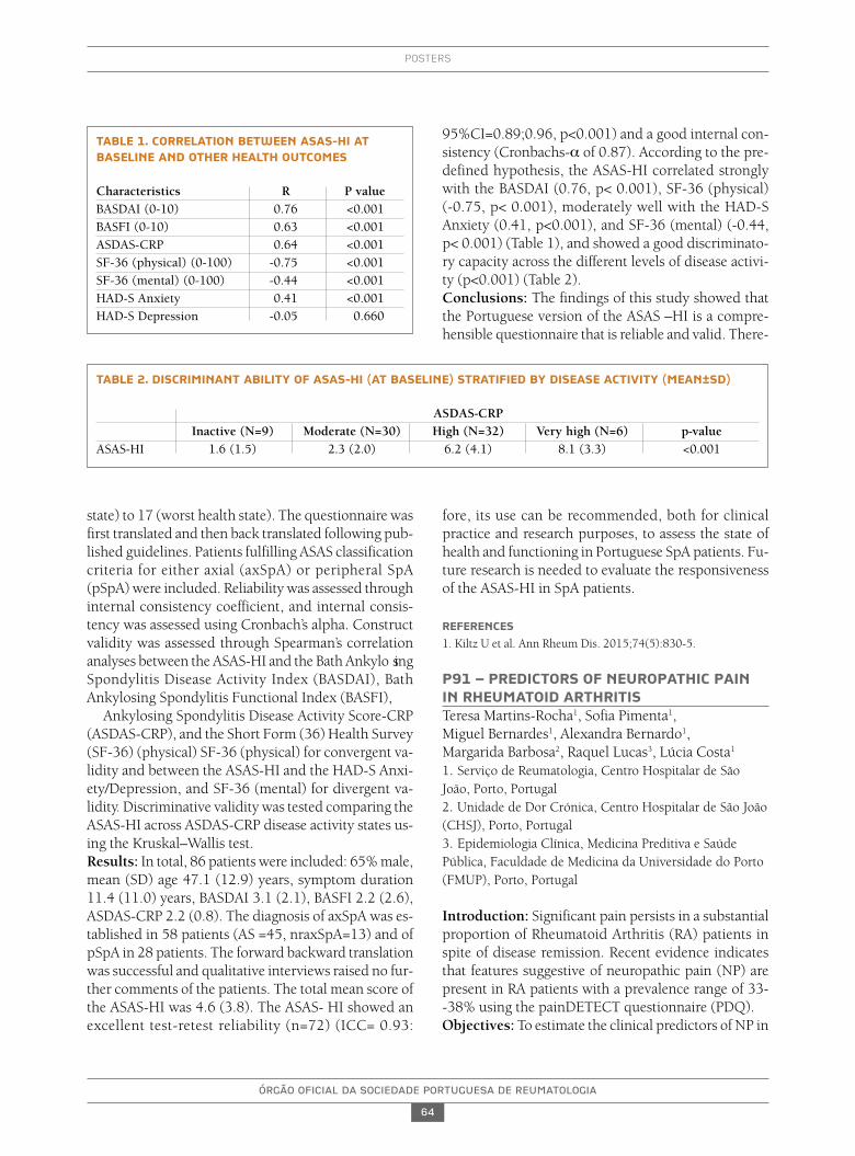
95%CI=0.89;0.96, p<0.001) and a good internal con-sistency (Cronbachs-a of 0.87). According to the pre-defined hypothesis, the ASAS-HI correlated stronglywith the BASDAI (0.76, p< 0.001), SF-36 (physical) (-0.75, p< 0.001), moderately well with the HAD-SAnxiety (0.41, p<0.001), and SF-36 (mental) (-0.44,p< 0.001) (Table 1), and showed a good discriminato-ry capacity across the different levels of disease activi-ty (p<0.001) (Table 2).Conclusions: The findings of this study showed thatthe Portuguese version of the ASAS –HI is a compre-hensible questionnaire that is reliable and valid. There-
fore, its use can be recommended, both for clinicalpractice and research purposes, to assess the state ofhealth and functioning in Portuguese SpA patients. Fu-ture research is needed to evaluate the responsivenessof the ASAS-HI in SpA patients.
REFERENCES
1. Kiltz U et al. Ann Rheum Dis. 2015;74(5):830-5.
p91 – pREDICTORS OF NEuROpATHIC pAININ RHEuMATOID ARTHRITISTeresa Martins-Rocha1, Sofia Pimenta1, Miguel Bernardes1, Alexandra Bernardo1, Margarida Barbosa2, Raquel Lucas3, Lúcia Costa1
1. Serviço de Reumatologia, Centro Hospitalar de SãoJoão, Porto, Portugal2. Unidade de Dor Crónica, Centro Hospitalar de São João(CHSJ), Porto, Portugal3. Epidemiologia Clínica, Medicina Preditiva e SaúdePública, Faculdade de Medicina da Universidade do Porto(FMUP), Porto, Portugal
Introduction: Significant pain persists in a substantialproportion of Rheumatoid Arthritis (RA) patients inspite of disease remission. Recent evidence indicatesthat features suggestive of neuropathic pain (NP) arepresent in RA patients with a prevalence range of 33--38% using the painDETECT questionnaire (PDQ). Objectives: To estimate the clinical predictors of NP in
TABLE 1. CORRELATION BETwEEN ASAS-HI ATBASELINE AND OTHER HEALTH OuTCOMES
Characteristics R P valueBASDAI (0-10) 0.76 <0.001BASFI (0-10) 0.63 <0.001ASDAS-CRP 0.64 <0.001SF-36 (physical) (0-100) -0.75 <0.001SF-36 (mental) (0-100) -0.44 <0.001HAD-S Anxiety 0.41 <0.001HAD-S Depression -0.05 0.660
TABLE 2. DISCRIMINANT ABILITY OF ASAS-HI (AT BASELINE) STRATIFIED BY DISEASE ACTIVITY (MEAN±SD)
ASDAS-CRPInactive (N=9) Moderate (N=30) High (N=32) Very high (N=6) p-value
ASAS-HI 1.6 (1.5) 2.3 (2.0) 6.2 (4.1) 8.1 (3.3) <0.001

ÓRGÃO OfICIAL DA SOCIEDADE PORTUGUESA DE REUMATOLOGIA
65
posters
a cohort of RA patients. Methods: Observational, cross-sectional study wasperformed with RA patients followed at our Rheuma-tology department with unchanged DMARD treatmentduring the last 3 months. Patients with diagnosed neu-ropathy or non-RA risk factors for NP were excluded.Selected patients were eva luated in a medical visit. De-mographic, clinical and labo ratorial data were collect-ed and disease activity and functional measures wereevaluated. Two validated questionnaires were appliedto assess NP: the Leeds Assessment of NeuropathicSymptoms (LANSS) and the PDQ. Univariate and mul-tivariate logistic regression were performed to identifythe predictors of NP. Signi ficance level was set as <0.05. Results: 112 RA patients were included. 86 (77%)were females, with a mean (SD) age of 55.1 (10.8) yearsand median disease duration of 13 years (range: 2-41).84% patients were seropositive for Rheumatoid Factorand/or ACPA. 102 (91%) were treated with DMARDsand 42% with a biologic DMARD, whom 8% inmonotherapy. The mean (SD) DAS28 4V was 3.61(1.01) and 12% were in remission. 45 (40%) patientshad NP by the LANSS (≥12), 28% had a possible/like-ly NP in the PDQ (>12) and 21% were positive in theboth tests. Female sex was predictive of LANSS andPDQ NP (OR: 3.44, p=0.02 and OR: 3.70; p=0.05, res -pectively) and disease duration was a predictor ofLANSS (OR: 0.92 per year, p= 0.004). After adjustingfor these variables, pain VAS, patient global activity andthe tender joint count were positive predictors of NP byboth tests. Swollen joint count, ESR or CRP levels werenot significantly associated with NP. DAS28 CRP andHAQ scores were both significant positive predictors ofPDQ NP (OR: 1.77 and OR: 3.61, p<0.01, respective-ly). DAS28 CRP, dichotomized as remission/non-re-mission, and the HAQ score were also significantlyposi tive predictors of LANSS NP (OR: 5.55, p=0.01(DAS28 CRP≥2.6); OR: 2.15, p=0.03). Positivity forACPA was a significant negative predictor of LANSSNP (OR: 0.23, p=0.009), remaining significant afteradjusting for DAS28 CRP, HAQ and current methotre -xate (MTX). Number of analgesics and current NSAIDstreatment were associated with PDQ NP (OR: 2.72 andOR: 3.40, respectively, p<0.05). Current MTX and pre-vious/current Hydroxychloroquine (HCQ) treatmentwere both negative predictors for PDQ NP (OR: 0.41and OR: 0.20, respectively, p<0.05). No significant as-sociations were found for other therapies or clinical fea-tures. In a group of patients with dosed TSH levels(n=18), there was a positive though non-significant as-
sociation with LANSS and PDQ (OR: 3.12, OR: 13.98,p=0.09). Conclusion: NP was present in sizable proportion ofRA patients. Consistently with previous data, our studysupports the association between NP and disease acti -vity/functional scores but not with objective inflam-matory measures. This study newly points to a possi-ble protective role of ACPA positivity, MTX and HCQtreatment in NP risk. Further studies are needed to con-firm this hypothesis.
REFERENCES1. Koop SM, et al. Arthritis Res Ther. 2015; 17:2372. AW Christensen et al. Scand J Rheumatol 2016;1–9
GRupO 2
p170 – ZOLEDRONATE EFFECTIVENESS AND SAFETY IN ACTIVE pAGET’S DISEASE: LONG-TERM FOLLOw-up AND RETREATMENTIN CLINICAL pRACTICEElsa Vieira-Sousa1, 2, Rodrigues AM1, 2, Joana Caetano-Lopes2, Sílvia Fernandes1, Maria Inês Seixas1, 3, Nádia Martins1, 3, Maria João Gonçalves1, 2, Catarina Tenazinha1, Elisabete Barata Fernandes4, 5, Rita Barros1, José Carlos Romeu1
1. Rheumatology Department, Hospital Santa Maria,CHLN, Lisbon Academic Medical Center, Lisboa, Portugal2. Rheumatology Research Unit, Instituto de MedicinaMolecular, Lisboa, Portugal3. Rheumatology Unit, Centro Hospitalar Tondela-Viseu,Viseu, Portugal4. Unidade de Epidemiologia do Instituto de MedicinaPreventiva e Saúde Pública, Faculdade de Medicina daUniversidade de Lisboa, Lisboa, Portugal5. Centro de Investigação Clinica, Centro Académico deMedicina de Lisboa, Lisboa, Portugal
Background: Bisphosphonates are considered first linetherapy in the treatment of Paget’s Disease (PD). Theobjective of this observational study was to assess long--term effectiveness and safety of zoledronate in thetreatment of Portuguese active PD patients.Methods: Patients with active PD treated with zole-dronate 5 mg were recruited and followed prospec-tively. Clinical parameters and bone turnover markers,calcium phosphorus and parathormone serum levelswere determined before, at 3 and every 6 months aftertreatment. Remission was defined as normalization of

ÓRGÃO OfICIAL DA SOCIEDADE PORTUGUESA DE REUMATOLOGIA
66
posters
alkaline phosphatase (ALP) levels. Retreatment wasconsidered when ALP levels increased more than 25%of the upper limit of normal or of the nadir achieved,in cases of non-normalization of ALP. Adverse eventswere registered. Patients were excluded from the ana -lysis during follow-up if: 1) were retreated with zole-dronate for other indication, 2) were started on oralbisphosphonates or other bone anti-resorption agents3) missed the 3 months, or more than 24 months, la -boratorial evaluations.Results: 75 patients (63% males), with 67±10 yearsand a disease duration of 10±8 years were included.The majority of patients were originated from Alente-jo (58,1%). 67% had polyostotic disease and the meanpercentage of skeletal involvement was of 12±10%. Iliac bone, vertebra and femur were the most frequentlyaffected bones, respectively in 65, 39 and 37% of thepatients. 75% were symptomatic at presentation. 47%had been previously treated with pamidronate (cumu-lative dose 213±259mg). After a single infusion of zole-dronate remission was achieved in 93% and 95% ofpatients, at 3 and 6 months post-treatment, respec-tively. Significant reductions of the mean levels of ALP,bone-specific ALP, procollagen type 1 N-terminal pro -pertied (P1NP), and b-C-terminal telopeptide of typeI collagen (CTX-I) were observed at 3, 6 and 12 monthsafter treatment. The maximum therapeutic effect wasobtained at 12 months with 97% of patients achievingremission. Considering symptomatic patients (bone/joint pain) 70% referred pain improvement after treat-ment. During a mean follow-up of 64±38 months, 10patients (13.3%) relapsed, on average 59 months afterthe 1st zoledronate infusion. From these 10 retreatedpatients, 2 required a 3rd zoledronate infusion, at 48and 68 months after the 2nd infusion. A total of 36 pa-tients were lost to follow-up: 22 due to loss of clini-cal/laboratorial follow-up, 9 due to retreatment of zole-dronate for osteoporosis and 2 due to the start of oralbisphosphonates. Zoledronate infusion with generallywell tolerated: 16 patients referred flu-like symptoms,8 and 16 showed transitory asymptomatic hypocal-caemia and hypophosphatemia, respectively; 1 repor -ted dizziness, 1 bone pain and 2 patients myalgia.Conclusions: This study shows the effectiveness andsafety of zoledronate in a Portuguese population of pa-tients with active PD with long term follow-up. Bio-chemical remission was achieved in 97% of patients at 12months. Furthermore, the beneficial effect of zoledronatewas sustained, with only 13.3% of patients requiring re-treatment during an average follow-up of 64 months.
p22 – METHOTRExATE AND LOw DOSEpREDNISOLONE DOwNREGuLATEOSTEOCLAST FuNCTION IN MONOCYTESFROM EARLY RHEuMATOID ARTHRITISpATIENTSIP Perpétuo1, J Caetano-Lopes1, AM Rodrigues 1, Raquel Campanilho-Marques1,2, Cristina Ponte1,2, Helena Canhão3, M. Ainola4, JE Fonseca1, 2
1. Rheumatology Research Unit, Instituto de MedicinaMolecular, Faculdade de Medicina da Universidade deLisboa, Lisboa, Portugal2. Rheumatology Department, Hospital de Santa Maria(CHLN), Lisbon Medical and Academic Centre, Lisboa,Portugal3. EpiDoC Unit, CEDOC, NOVA Medical School, NOVAUniversity, Lisboa, Portugal4. Institute of Clinical Medicine, University of Helsinki,Helsinki, Finland
Rheumatoid arthritis (RA) is a systemic, immune me-diated inflammatory disease that is associated withbone erosions and joint destruction. Methotrexate(MTX) slows bone damage but the mechanism bywhich it acts is still unknown.
In this study we aimed to assess the effect of MTXand low dose prednisolone (MTX+PDN) on circula tingosteoclast (OC) precursors and OC differentiation inRA patients.Methods: RA patients before and at least 6 months af-ter MTX therapy were analyzed and compared withhealthy donors. A blood sample was collected in orderto assess receptor activator of NF-�B (RANK) ligand(RANKL) surface expression on circulating leukocytesand frequency and phenotype of monocyte subpopu-lations. Serum quantification of bone turnover mar kersand cytokines and in vitro OC differentiation assayswere performed.
The number or RANKL+ neutrophils increased in RA patients when compared to healthy donors(p=0.006) and after treatment with MTX+PDN theircount was reduced to healthy control numbers(p=0.0155). Classical activation markers of monocytessuch as HLA-DR, CD86, CCR2 and CD11b, and alsoRANK were increased in RA patients at baseline, com-paring to control healthy donors. After MTX+PDN ex-posure, expression decreased to healthy control levels.Serum RANKL levels were increased at baseline com-paring to healthy donors (p=0.0164) and normalizedafter therapy. Although the number of OC was not dif-ferent between groups, resorbed area and resorbed

ÓRGÃO OfICIAL DA SOCIEDADE PORTUGUESA DE REUMATOLOGIA
67
posters
area/pit were elevated when compared to controls(p=0.0436 and 0.0249, respectively) and reduced af-ter treatment (p<0.0001).
Our results suggest that MTX+PDN play an impor-tant role in downregulating OC function, which we be-lieve occurs through a decrease in RANK surface ex-pression in monocytes
p86 – A COMpARISON STuDY OFpREVALENCE OF TRADITIONALCARDIOVASCuLAR RISK FACTORS ANDFRAMINGHAM RISK SCORE IN SYSTEMICSCLEROSIS pATIENTS AND MATCHEDCONTROLSJoana Sousa-Neves1, Marcos Cerqueira1, Daniela Santos-Faria1, Joana Leite Silva1, Carmo Afonso1, Filipa Teixeira1
1. Rheumatology Department, Hospital Conde deBertiandos (ULSAM), Ponte de Lima, Portugal
Background: In Systemic Sclerosis (SSc), data onprevalence of traditional cardiovascular (CV) diseaserisk factors is scarce and conflicting (1). Therefore, SScpatients CV risk attributed to traditional CV risk factorsremains an issue of debate.Objectives: To evaluate if patients with SSc have ahigher prevalence of traditional CV disease risk factorsand a higher risk of longterm CV events based on therisk prediction tool of the Framingham risk score (FRS)in comparison with age, race and sex matched controlsubjects.Methods: The study comprised patients diagnosedwith SSc, fulfilling both the 1980 ACR and the 2013ACR/EULAR criteria for the disease, and followed-upat our Rheumatology Department and a group of age,race and sex-matched controls. Inclusion criteria wereage 30 to 74 and no history of CV events in order to cal-culate FRS. In total, 46 out of 62 patients were eligiblefor the study. Traditional CV disease risk factors (dia-betes, arterial hypertension and smoking) were com-pared among the 46 patients with SSc and 51 matchedcontrols. Systolic blood pressure (SBP) values and to-tal and high-density lipoprotein (HDL) cholesterol le -vels were also collected. The 10-year risk for CV eventsaccording to FRS was calculated and means of patientsand controls were compared. Subjects’ distribution into3 categories of risk – low (<10% risk), medium (10--20% risk) and high (>20% risk) was also compared.Parametric and nonparametric tests were used for com-parison between groups. P value <0.05 was defined as
statistically significant.Results: Mean risk for CV events in 10-years assessedby FRS was 10.00%±8.61 for SSc patients and7.76%±8.30 for matched controls. Differences were notstatistically significant (p=0.196). Additionally, preva-lence of diabetes, arterial hypertension and smokingdid not differ significantly between the two groups(p=0.890, p=0.443, p=0.651, respectively). Total andHDL cholesterol levels were also similar betweengroups (p=0.963 and p=0.506, respectively). Only SBPvalues (mmHg) of SSc patients were significantly hi -gher (128.50 mmHg [113.5 to 139.3]) (median [in-terquartile range]) compared with controls (120.00[110 to 130]), p=0.031. Subjects’ distribution into the3 groups of risk defined was similar for both groups(p=0.205).Conclusions: In our study, prevalence of traditionalCV disease risk factors and 10-year risk for CV eventsbased on FRS assessment tool did not differ signifi-cantly between SSc patients and age, sex and racematched controls.
REFERENCES 1. Psarras A, Soulaidopoulos S, Garyfallos A, Kitas G and Dimi-
troulas T. A critical view on cardiovascular risk in systemic scle-rosis. Rheumatol Int. 2017 Jan;37(1):85-95.
p172 – AVALIAÇÃO DA EFICÁCIA ESEGuRANÇA DA FRAGMENTAÇÃO//ASpIRAÇÃO ECOGuIADA DE CALCIFICAÇÃODO OMBRO: RESuLTADOS ÀS 24 SEMANASJoão Moreira1, Daniela Santos-Faria2, Filipa Teixeira2,João Pires3
1. Serviço de Medicina Física e de Reabilitação, CentroHospitalar do Algarve, Faro, Portugal2. Serviço de Reumatologia, Unidade Local de Saúde doAlto Minho, Ponte de Lima, Portugal3. Serviço de Radiologia, Unidade Local de Saúde do AltoMinho, Viana do Castelo, Portugal
Introdução: A tendinopatia calcificante do ombro estáassociada a depósitos de cálcio (predominantementehidroxiapatite) ao nível da coifa dos rotadores. É res-ponsável por 7% dos casos de omalgia, podendo seraltamente incapacitante. O tendão do supraespinhosoé o mais afectado (80% dos casos). A sua etiologia ain-da é desconhecida, apesar de existirem diversas teo-rias. Esta patologia divide-se em 4 fases: fase de pré-cal-cificação, fase de calcificação (que se divide em forma-tiva e reabsortiva) e fase de pós-calcificação. A fasereabsor tiva é a mais dolorosa.

ÓRGÃO OfICIAL DA SOCIEDADE PORTUGUESA DE REUMATOLOGIA
68
posters
Por se tratar de um processo auto-limitado, o trata-mento da tendinopatia calcificante do ombro deve, paraalém de eficaz, ser o menos invasivo possível e livre decomplicações major. Existem uma série de tratamentosque podem ser utilizados nas fases de maior exacerba-ção álgica, no entanto ainda não há um consenso quan-to ao que se mostrou mais útil.
O objectivo deste estudo foi avaliar a eficácia e se-gurança da fragmentação/aspiração ecoguiada da cal-cificação do supraespinhoso, às 24 semanas.Material e Métodos: Estudo prospectivo onde foramavaliados 42 doentes, com omalgia associada a tendi-nopatia calcificante do supraespinhoso e refratária atratamento conservador. Após confirmação ecográficada calcficação este doentes foram submetidos afragmen tação/aspiração ecoguiada da calcificação.
Foram aplicados o Questionário QuickDASH, Esca-la Numérica da Dor (END) e Índice de Dor e Incapaci-dade no Ombro (SPADI), previamente à realização doprocedimento (na baseline) e após 4 semanas e 24 se-manas. Foram registados os efeitos adversos.Resultados: 42 pacientes (31;11) com uma idade mé-dia de 49,8 anos foram incluídos no estudo. Os valo-res médios da END à baseline e após 4 e 24 semanas fo-ram 7,5, 4,3 e 1,7, respectivamente. Os valores médiosdo Questionário QuickDASH à baseline e após 4 e 24semanas foram 64,6, 26,0 e 14,1, respectivamente. Osvalores médios do SPADI à baseline e após 4 e 24 se-manas foram 62,3, 29,8 e 19,7, respectivamente. Em3 casos os doentes não apresentaram qualquer melho-ria após a realização do procedimento, tendo sidoorientados para a realização de outro tipo de trata-mentos. Os únicos efeitos adversos registados foramdesconforto no local das picadas (35,7%) e ligeira reac-ção vagal após a realização do procedimento (4,8%).Discussão e Conclusão: A fragmentação/aspiraçãoecoguiada de calcificação do ombro parece ser uma téc-nica segura e eficaz para alívio sintomático e melhoriafuncional, quando os tratamentos mais conservadoresse mostram ineficazes ou pouco eficazes. Nos casos emque esta técnica se mostra ineficaz, os doentes ser en-caminhados para outros tipos de tratamentos.
p153 – EFFECTIVENESS AND pERSISTENCEOF THE FIRST TuMOR NECROSIS FACTORINHIBITOR IN pORTuGuESE pSORIATICARTHRITIS pATIENTS.Elsa Vieira-Sousa1, Mónica Eusébio2, Pedro Ribeiro1,Nikita Khmelinskii1, Rita C Machado1, Teresa Martins-Rocha3, 4, Miguel Bernardes3, 4,
Daniela Santos-Faria5, Joana Leite Silva5, Helena Santos6, Cláudia Miguel6, Pedro David Carvalho7, Tiago Costa8, Lídia Teixeira9,Tiago Meirinhos10, Patrícia Nero11, Maria José Santos2,9
1. Serviço de Reumatologia e Doenças Ósseas Metabólicasdo Hospital de Santa Maria, Centro Hospitalar Lisboa Norte,Centro Académico de Medicina de Lisboa, Lisboa, Portugal2. Sociedade Portuguesa de Reumatologia, Lisboa, Portugal3. Serviço de Reumatologia, Centro Hospitalar de SãoJoão, Porto, Portugal4. Faculdade de Medicina da Universidade do Porto,Porto, Portugal5. Rheumatology Department, Unidade Local de Saúde doAlto Minho, Ponte de Lima, Portugal6. Rheumatology Department, Instituto Português deReumatologia, Lisboa, Portugal7. Serviço Reumatologia, Centro Hospitalar e Universitáriode Coimbra, Coimbra, Portugal8. Rheumatology Department, Hospital Egas Moniz,CHLO, Lisboa, Portugal9. Rheumatology Department, Hospital Garcia de Orta,Almada, Portugal10. Serviço de Reumatologia, Centro Hospitalar do BaixoVouga, E.P.E., Aveiro, Portugal11. Hospital CUF Descobertas, Lisboa, Portugal
Background: Tumor necrosis factor inhibitors (TNFi)lead to a dramatic improvement in the management ofPsoriatic Arthritis (PsA).Despite their effectiveness, thereis still a significant proportion of patients that do not res -pond and/or are intolerant to TNFis. The objective ofthis work was to assess the effectiveness, measured by re-sponse rates and drug survival, within a period of 4 yearsof the first TNFi treatment, and the main reasons for dis-continuation of the first TNFi, in patients with PsA.Methodology: This was a retrospective non-interven-tional study of adult PsA patients registered at theRheumatic Diseases Portuguese Registry (Reuma.pt),with at least 1 TNFi prescription. Data was analyzed at0, 3, 6, 12, 24, 36 and 48 months after starting a firstTNFi. For qualitative data, absolute and relative fre-quencies are presented. Percentages are based on thetotal number of subjects with non-missing values un-less specified otherwise. In case of quantitative data:mean and standard deviation are presented. Responseto TNFi was measured by composite disease activity(DAS, ACR, PsARC, BASDAI, ASDAS, MDA) and func-tional indices (HAQ). Drug survival was assessed byKaplan-Meier survival analysis. In all analyses signifi-

ÓRGÃO OfICIAL DA SOCIEDADE PORTUGUESA DE REUMATOLOGIA
69
posters
cance level was set at 0.05.Results: A total of 705 PsA patients were included,with a mean age of 52.5 years (±13.3); 50.8% (n=358)female. The mean age at first symptoms was 37.0±12.7and at PsA diagnosis of 40.6±12.5 years. The mostcommon subtype of PsA was symmetric polyarthritis(n=385; 61.5%). The mean time from diagnosis to 1stcsDMARD was 3.3±6.0 and to 1st bDMARD was6.6±6.9 years. 185 patients (26.24%) were treated withadalimumab, 322 (45.67%) etanercept, 100 (14.18%)golimumab and 98 (13.90%) with infliximab as firstTNFi. The average response rates, as measured by com-posite disease activity and functional indices, are shownin Table 1. The average drug persistence was of31.79±17.03 months for TNFi as a group, with 205(29.08%) of discontinuations during a period of 4 yearsof follow-up. The main reasons for discontinuation ofthe first TNFi were: non response/loss of response 111(54.15%), adverse event 48 (23.41%), surgery 6(2.93%), treatment refusal to continue treatment, 4(1.95%), loss to follow-up 3 (1.46%), attempted preg-nancy/pregnancy 4 (1.95%), death 2 (0.98%), remis-sion 2 (0.98%), and others 10 (4.88%).Conclusions: PsA patients receiving a first TNFi willpersist on treatment for an average of 2.6 years withtreatment discontinuations rates of 29.08%. Non res -
ponse/loss of response constitutes the major reason fortreatment discontinuation in this population.
ACKNOwLEDGMENTSTo Fernando Martins. To Ana Roxo, António Vilar, Augusto Fausti-no, Carlos Vaz, Cristina Catita, Filipa Ramos, Filipe Araújo, FilipeBarcelos, Graça Sequeira, Herberto Jesus, Joaquim Polido Pereira,Jorge Garcia, José Alberto Pereira da Silva, José António Costa, JoséAntónio Melo Gomes, José António Pereira da Silva, Luís CunhaMiranda, Mário Viana Queiroz, Margarida Cruz, Margarida Olivei-ra, Maura Couto, Patrícia Pinto, Paula Valente, Raquel Roque, RuiAndré, Teresa Nóvoa. To all Rheumatogists that included patients atReuma.pt. Financial support for statistics and report writing wasprovided by Novartis, Produtos Farmacêuticos S.A.
p174 – CROSS-CuLTuRAL VALIDATION OFTHE EuLAR “RHEuMATOID ARTHRITISIMpACT OF DISEASE” SCORE INTOpORTuGuESE: A CROSS-SECTIONAL STuDYOF 288 RA pATIENTS uSING RASCH ANALYSISRicardo J. O. Ferreira1, 2, Laure Gossec3, 4, Sarah Hewlet5, Cátia Duarte1, 6, Joanna K. Nicklin7,JAP da Silva1, 6, Mwidimi Ndosi5
1. Rheumatology Department, Centro Hospitalar eUniversitário de Coimbra, Coimbra, Portugal2. Health Sciences Research Unit: Nursing (UICiSA:E),Coimbra, Portugal3. Department of rheumatology, AP-HP, Pitié Salpêtrière,
Note: sample size is not constant due to available evaluations.Good EULAR response (DAS28)– 3 months (n=202); 6 months (n=203); 12 months (n=186); 24 months (n=134); 36 months (n=90); 48months (n=81).PsARCresponse – 3 months (n=187); 6 months (n=204); 12 months (n=175); 24 months (n=124); 36 months (n=87); 48 months (n=70).DHAQ-DI – 3 months (n=149); 6 months (n=160); 12 months (n=141); 24 months (n=101); 36 months (n=67); 48 months (n=56).MDA – 3 months (n=129); 6 months (n=113); 12 months (n=103); 24 months (n=69); 36 months (n=56); 48 months (n=37).ASDAS (DASDAS≥1.1) – 3 months (n=103); 6 months (n=110); 12 months (n=89); 24 months (n=65); 36 months (n=52); 48 months (n=45).(DBASDAI≥50% or DBASDAI>2) – 3 months (n=115); 6 months (n=118); 12 months (n=96); 24 months (n=72); 36 months (n=57); 48months (n=50).
TABLE 1. RESpONSE RATES THROuGH FOLLOw-up
3 months 6 months 12 months 24 months 36 months 42 monthsACR20 61 (81.14%) 70 (90.91%) 61 (96.83%) 48 (97.96%) 27 (91.67%) 22 (91.67%)ACR50 47 (67.14%) 52 (67.53%) 48 (76.19%) 39 (79.59%) 23 (79.31%) 19 (79.17%)ACR70 24 (34.29%) 30 (38.96%) 33 (52.38%) 27 (55.10%) 19 (65.52%) 14 (58.33%)Good EULAR response (DAS28) 96 (47.52%) 116 (57.14%) 101 (54.30%) 88 (65.67%) 53 (58.59%) 52 (64.20%)PsARC response 144 (77.01%) 158 (77.45%) 143 (81.71%) 103 (83.06%) 77 (88.51%) 57 (81.43%)DHAQ-DI -0.49±0.54 -0.36±0.52 -0.45±0.67 -0.51±0.54 -0.59±0.54 -0.58±0.64MDA 6 (4.65%) 15 (13.27%) 16 (15.53%) 13 (18.84%) 10 (17.86%) 8 (21.62%)ASDAS (DASDAS≥1.1) 64 (62.14%) 72 (65.45%) 57 (64.04%) 42 (64.62%) 36 (69.23%) 33 (73.33%)(DBASDAI≥50% 66 (57.39%) 68 (57.63%) 60 (62.50%) 44 (61.11%) 38 (66.67%) 34 (68.00%)or DBASDAI>2)

ÓRGÃO OfICIAL DA SOCIEDADE PORTUGUESA DE REUMATOLOGIA
70
posters
Paris, France4. Sorbonne Universités, UPMC Univ Paris 06, Paris, France5. HAS - Nursing and Midwifery, University of the West ofEngland, Bristol, United Kingdom6. Faculdade de Medicina, Universidade de Coimbra,Coimbra, Portugal7. Bristol Royal Infirmary, Bristol, United Kingdom
Background: The Rheumatoid Arthritis Impact of Di -sease (RAID) score1 assesses 7 impact domains of in-
terest for people with rheumatoid arthritis (RA). Its usein research and clinical practice has been growing, andit is already translated into over 70 languages2 but thecross-cultural validity of the Portuguese RAID has notbeen well established.Objectives: To validate the Portuguese RAID for usein Portugal.Methods: This was a single centre, cross-sectional va -lidation study involving 2 phases: (i) cognitive de-briefing with 38 patients to determine comprehension
DF, degrees of freedom; *6 items for cross-cultural comparisons (items 2 "Function" and 5 "Physical well-being" combined).
TABLE 1. RESuLTS OF RASCH ANALYSIS FROM pOOLED DATA
Fit Residual Person N RAID Mean (SD) Chi2 Interaction Separation
Country (n items) Item Person Value (DF) p-value IndexPortugal 288 7 -0.13 (2.53) -0.67 (1.60) 40.50 (35) 0.24 .94UK 205 7 0.22 (1.72) -0.44 (1.37) 40.50 (35) 0.17 .93France 195 7 0.19 (1.99) -0.71 (1.57) 25.69 (21) 0.22 .91
Pooled 6887 -0.06 (3.48) -0.65 (1.55) 94.88 (63) 0.01 .936* -0.34 (3.88) -0.63 (1.44) 66.04 (54) 0.13 .93
Expected values for perfect fit 0 (1) 0 (1) >0.05 >.85
150
125
Freq
uen
cyFr
equ
ency
Persons
Items
100
75
50
25 3.7%
0.0%Location (logits)
0.0%
7.4%
14.3%
21.4%
7.4%
11.1%
14.8%
22.3%
0-4 -3 -2 -1 0 1 2 3 4
Total
Person-Item Threshold Distribution(Grouping Set to Interval Length of 0.5 making 16 Groups)
No.(674)
Mean-0.200
SD1.097
0
5
10
15
FIGuRE 1. Distribution of items and persons along the same scale (logit score) confirming good targeting of the RAID. The x-axis isthe logit score and represents the interval scaling of the items according to the Rasch model, with -4 being no impact and 4 beinghigh impact of disease

ÓRGÃO OfICIAL DA SOCIEDADE PORTUGUESA DE REUMATOLOGIA
71
posters
of the existing2 Portuguese RAID (ii) cross-cultural va -lidation using data from adult patients who were will-ing and able to complete the Portuguese RAID unai -ded. Analyses included fit to the Rasch model (imply-ing construct validity, reliability and statistical suffi-ciency), tests for unidimensionality and invarianceacross different patient subgroups i.e. age, gender,educa tion background, disease duration, function andculture. To test invariance to culture, the Portugaldataset was compared with datasets from France(n=195) and the UK (n=205).3 RUMM2030 softwarewas used in all analyses.Results: Phase I led to minor changes in phrasing 3items to enhance understanding and conceptual equi -valence between the original RAID and the Portugueseversion. In Phase II, 288 patients were included: mean(SD) age=60 (12) years, 82% females, 76% with diseaseduration ≥5 years, 30% on biologics. The PortugueseRAID was shown to have adequate fit to the Raschmodel and high internal consistency (Table 1). Unidi-mensionality and invariance to age, gender, disease du-ration and function were confirmed (data not shown).The scale was well targeted for patients with differentlevels of disease impact (Figure 1). Pooling the datasetsfrom Portugal, France and the UK revealed no cultu ralresponse bias (Table 1). RAID was then calibrated intologit-based scores to enable parametric analyses andbias-free cross-cultural comparisons if desired (data notshown).Conclusions: This study confirms the PortugueseRAID as a robust unidimensional tool for use in Portu-gal. The raw scores of the 7-item RAID can be usedwith confidence in clinical practice. Conversion chartsare available to enable accurate cross-cultural compari -sons across Portugal, France and the UK.
REFERENCES1. Gossec L, et al. Ann Rheum Dis, 2011;70:935-42.2. EULAR RAID and PsAID Questionnaires. Available from
http://www.eular.org/tools_products_.cfm Accessed 25th Jan.2017.
3. Hewlett S, et al. Ann Rheum Dis, 2015;74:Suppl 2:559.
p182 – ApLICAÇÃO ECOGuIADA DE pLASMARICO EM pLAQuETAS – uMA SOLuÇÃO NAABORDAGEM TERApÊuTICA DATENDINOpATIA INSERCIONAL CRÓNICA DOTENDÃO DE AQuILES?Sara Estrela Rego1, João Moreira1, Jonathan Rios1,Victor Milet1, Gisela Leandro1, Alison Reis1, Lucia Gomes1
1. Serviço de Medicina Física e de Reabilitação, CentroHospitalar do Algarve, Faro, Portugal
Introdução: O Tendão de Aquiles é o maior e mais for-te tendão do corpo humano, sendo suscetível a trau-matismos repetidos. A tendinopatia insercional cróni-ca do Tendão de Aquiles (TA) é uma lesão de etiologiamultifatorial, que ocorre predominantemente no gé-nero masculino e em idades compreendidas entre os30 e 50 anos. É frequentemente causa de dor e rigidezna região peritendinosa, condicionando incapacidadefuncional.
Acredita-se que esteja relacionada com o enfraque-cimento progressivo do tendão por lesões de sobreuso,associadas à prática regular de exercício físico. A nívelhistológico, demonstram-se alterações degenerativasdo tendão.
A abordagem terapêutica inicial passa por um pe-ríodo de fisioterapia, associada a repouso e analgesia.No entanto, não raramente, a sintomatologia não é re-solvida após o tratamento conservador.
A aplicação de plasma rico em plaquetas (PRP) sur-ge como uma estratégia alternativa no tratamento dastendinopatias. Consiste em sangue autólogo com ele-vada concentração de plaquetas e fatores de cresci-mento, que recrutam células com capacidade regene-radora dos tecidos. A ecografia coadjuvante permiteuma visualização real das estruturas e a colocação pre-cisa da agulha no local pretendido.
O objetivo deste trabalho passa pela avaliação da efi-cácia e segurança da infiltração de PRP no tratamentoda Tendinopatia Insercional crónica do Tendão deAquiles.Material e Métodos: Estudo retrospetivo e descritivode 6 doentes com diagnóstico de Tendinopatia Inser-cional crónica do Tendão de Aquiles, em que se admi-nistraram, com o apoio de ecografia, 4ml de PRP. Fo-ram avaliados os resultados através de 2 questionários,Western Ontario & McMaster Osteoarthritis Index(WOMAC) e SF-36 (Questionário do Estado de Saúde)e foi aplicada a Escala Numérica da Dor (END). Atra-vés de contacto telefónico, cada paciente respondeuduas vezes a cada questionário, um referente ao perío-do pré-intervenção e outro relativo a uma período de6 semanas após a intervenção. Aferiu-se ainda o graude satisfação geral após o procedimento (pontuandode 0 a 10) e registaram-se os efeitos adversos.Resultados: Foram avaliados 6 doentes do género mas-culino e idade média de 41,3 anos, com o diagnósticode Tendinopatia Insercional crónica do Tendão de

ÓRGÃO OfICIAL DA SOCIEDADE PORTUGUESA DE REUMATOLOGIA
72
posters
Aquiles refratária a tratamento conservador.Comparando o períodos pré-intervenção com a ava-
liação após 6 semanas de procedimento, objetivou-seuma melhoria média na escala WOMAC de 28,73 pon-tos.
No que toca à escala SF-36, os resultados demons-traram uma melhoria de 36,05 pontos nos parâmetrosde função física e de 40,73 pontos na dor após a inter-venção. Quanto à Escala Numérica da Dor, constatou--se uma melhoria média de 6,5 pontos.
No que toca ao grau de satisfação relacionado coma técnica interventiva, três doentes pontuaram 8 e trêspontuaram 9 pontos. Registou-se ainda o aparecimen-to de dor e hematoma no local da punção em 2 doen-tes, tendo revertido espontaneamente após 15 dias.Conclusão: A infiltração ecoguiada de PRP parece seruma uma opção terapêutica eficaz na Tendinopatia In-sercional crónica do Tendão de Aquiles, atuando comoum estímulo regenerativo para a cicatrização tendino-sa. Dada a sua natureza autóloga, demonstra tambémser uma terapêutica segura, sem efeitos adversos im-portantes, podendo ser considerada quando a tera-pêutica conservadora não é eficaz.
Mais estudos científicos deverão ser realizados paraaferir o nível de evidência da utilização de PRP nesta pa-tologia, seja de forma isolada ou como otimizador dotratamento conservador.
p38 – uLTRASOuND-GuIDED SYNOVIALNEEDLE BIOpSY: upDATE OF ExpERIENCEwITH AN EMERGING, MINIMALLY INVASIVETECHNIQuE IN CLINICAL pRACTICE ANDRESEARCHVasco C Romão1, 2, Joaquim Polido Pereira1, 2, Rita Barros1, Elsa Vieira-Sousa1, 2, Rita Luís3, Emília Vitorino3, Fernando Saraiva1, JE Fonseca1, 2
1. Rheumatology Department, Hospital de Santa Maria(CHLN), Lisbon Medical and Academic Centre, Lisboa,Portugal2. Rheumatology Research Unit, Instituto de MedicinaMolecular, Faculdade de Medicina da Universidade deLisboa, Lisboa, Portugal3. Pathology Department, Hospital de Santa Maria (CHLN),Lisbon Academic Medical Centre, Lisboa, Portugal
Background: Synovial biopsy remains an importanttool in clinical practice and research for the study ofsynovitis. Ultrasound-guided needle biopsy (USNB),as described by Kelly et al [1], has recently emerged asa minimally invasive technique, which enables collec-
tion of high quality synovial tissue with very good pa-tient tolerance.Objectives: To update the experience with USNB inour department, since the first report in 2016.Methods: We reviewed the clinical files of all patientswho had an USNB in our department. Degree of USjoint synovitis was evaluated on a semi-quantitativescale (0-3) in terms of synovial thickness (ST) and po -wer Doppler (PD). Since 2015, we assessed patienttole rance and acceptance of the procedure using a stan-dardized questionnaire, which includes visual analoguescales (VAS) of pain, stiffness and swelling of the biop-sied joint. Changes in US and VAS scores were assessedusing the Wilcoxon signed-rank test.Results: Forty-eight patients had 53 USNB, mostly fordiagnostic purposes (79%), performed by 4 differentoperators - Table 1. All types of joints were biopsied,mostly medium sized (26 wrists, 7 ankles), but alsolarge (3 knees, 4 shoulders, 6 elbows, 3 hips) and small(1 sternoclavicular, 1 naviculocuneiform, 1 metacar-pophalangeal and 1 proximal interphalangeal) joints,2 bursae (subacromial) and 1 tendon sheath (1st ex-tensor compartment of the wrist). USNB was repeatedin the same joint (wrist) twice in 3 patients and threetimes in one patient. Procedures were well tolerated,with 67% of patients classifying it as easy or very easy,78% reporting no or only mild discomfort and 77%considering likely/very likely to accept to repeat thebiopsy. An increase in analgesic medication in the daysfollowing the biopsy was reported by 13 out of 44 ques-tioned patients. After a median of 8 days following theprocedure, a significant decrease was observed in VASscores of pain, stiffness and swelling of the biopsiedjoint, although 23%, 23% and 31% of the patients, res -pectively reported small increases in these scores (me-dian 9.5, 11 and 10mm, respectively). There was nosignificant change in US scores pre- and post-biopsy,with only 3 and 2 patients having an increase in ST orPD scores, respectively. Biopsies were overall safe, with6 minor immediate adverse events (11%). There wereno cases of haemartrosis, joint/periarticular infectionor neurovascular damage. Two patients reported tran-sient limitation of the 5th and 1st digit extension fol-lowing a biopsy of the wrist and 1st extensor compart-ment tendon sheath, respectively, with no detectabletendinous ruptures on US; 1 patient had a muscularhematoma of the extensor muscles of the forearm fol-lowing an elbow biopsy.Conclusions: In our center, USNB has proved to be aneffective technique for collection of synovial membrane

ÓRGÃO OfICIAL DA SOCIEDADE PORTUGUESA DE REUMATOLOGIA
73
posters
that can be used for diagnostic and research purposes.The vast majority of the procedures were well tolera ted,without significant worsening of local joint symptomsor synovitis, and safe, without major adverse events.Importantly, patients’ concordance to repeat a USNBwas high.
REFERENCES1. Kelly S, Humby F, Filer A, et al. Ultrasound-guided synovial bio-
psy: a safe, well-tolerated and reliable technique for obtaininghigh-quality synovial tissue from both large and small joints inearly arthritis patients. Ann Rheum Dis 2013;74(3):611–7.
GRupO 3
p28 – puLMONARY EMBOLISM IN SYSTEMICSCLEROSIS – ONE YEAR FOLLOw upAna Catarina Duarte1, Inês Cordeiro2, Melanie Ferreira3, Tiago Judas3, Maria José Loureiro4,Susana Carmona5, Joaquim Santos5, Maria José Santos1, Ana Cordeiro1
1. Rheumatology Department, Hospital Garcia de Orta,Almada, Portugal2. Rheumatology Department, Hospital de Santa Maria(CHLN), Lisbon Medical and Academic Centre, Lisboa,
Portugal3. Internal Medicine, Hospital Garcia de Orta, Almada,Portugal4. Serviço de Cardiologia, Hospital Garcia de Orta,Almada, Portugal5. Nuclear Medicine, Hospital Garcia de Orta, Almada,Portugal
Introduction: The risk of pulmonary embolism (PE) insystemic sclerosis (SSc), has been estimated between2.51-3.47 fold higher when compared to non-SSc pa-tients. Proinflammatory state, vasculopathy and vas-cular injury may contribute to a prothrombotic state inthese patients and an increased risk for venous throm-boembolism.Objectives: Calculate the frequency and identify pos-sible risk factors for PE among SSc patients; analyse theefficacy of long-term anticoagulation in these patients.Methods: We conducted a retrospective analysis of 110patients with SSc followed in our Rheumatology de-partment and selected those who were submitted tolung ventilation/perfusion scintigraphy (V/Q scan) orCT pulmonary angiography due to worsening dysp-nea/fatigue or isolated reduction of the carbon mono -xide diffusing capacity (DLCO). We collected demo-
*p-value significant at <0.05. AE, adverse events; D1, digit 1; D5, digit 5; DAS28, disease activity score 28 joints; disc., discomfort; PsA, psoriatic arthritis; RA, rheumatoid arthritis; SpA, spondyloarthritis; UA, undifferentiated arthritis; US, ultrasound; VAS, visual analoguescale
TABLE 1. pATIENT & BIOpSY CHARACTERISTICS (N=53)
Age/Female 56 ± 18 years / 34 (64%)Diagnosis 21 RA, 13 UA, 8 septic, 5 crystal, 3 PsA, 1SpA, 2 otherDisease duration 5.3 ± 7.1 yearsDAS28 4.3±1.2Clinical indication 42 diagnostic (79%), 11 research (21%)Joint size 16 large, 30 medium, 4 small, 2 bursa, 1 tendon sheathImmediate tolerance 29% very easy, 38% easy, 24% tolerable, 9% difficultDiscomfort during procedure 36% no disc., 42% mild disc., 11% moderate disc., 5.5% mild pain, 5.5% intense painIncrease pain medication 13 (30%) in days following procedureLikelihood to repeat biopsy 41% very likely, 36% likely, 9% maybe, 14% unlikelyImmediate AE 5 minor local bleeding, 1 transitory forearm extensor paralysis due to anesthesiaOther AE 2 transient digit extension limitation (D5, D1), 1 muscular hematoma forearm
Pre-biopsy Post-biopsy p-valueVAS pain biopsied joint (mm) 62±25 46±29 0.001*VAS stiffness biopsied joint (mm) 59±30 37±31 0.004*VAS swelling biopsied joint (mm) 61±26 44±28 0.007*US synovial thickness score 2.5±0.6 2.2±0.8 0.092US Power Doppler score 1.0±1.2 1.0±1.1 0.414

ÓRGÃO OfICIAL DA SOCIEDADE PORTUGUESA DE REUMATOLOGIA
74
posters
graphic features, comorbidities, age at SSc diagnosis,anti-nuclear antibody specificities, dyspnea accordingto New York Heart Association (NYHA) classes and re-sults of cardio-pulmonary exams. PE and related varia -bles were assessed at baseline and after 12 months ofanticoagulation.Results: PE was diagnosed in 12 out of 29 (41.4%) SScpatients that met the inclusion criteria, with the ma-jority presenting bilateral peripheral multisegmentaldefects. Most were females (91.7%), with a mean ageof 59.4 (±12.7) years. Two thirds were diagnosed withlimited SSc with mean disease duration of 13.3 (±12.9)years.
The mean time between SSc diagnosis and PE dia -gnosis was 8.5±8.2 years, although one third of the pa-tients was diagnosed within the first year of SSc diagno -sis. One patient was taking oral contraceptives andnone had thrombophilia, previous surgery or cancer.
Regarding NYHA classes, 33.3% were classified ashaving class≥3, with a mean N-terminal pro-brain na-triuretic peptide (NTproBNP) of 1108pg/mL (19 to8069). Six patients had concomitant interstitial lungdisease (ILD) and 8 had an estimated pulmonary arterysystolic pressure (PASP) ≥35 mmHg (6 of them had
concomitant ILD). From these only 2 had pulmonaryhypertension confirmed by right heart catheterizationand 1 eventually died.
When comparing SSc patients with and without PE,Scl70 positivity was more common in patients with PE(p=0.041). No significant associations were found be-tween PE diagnosis and presence of hypertension, dia -betes, smoking, obesity and dyslipidemia.
From the 12 patients with PE, 10 were on long-termanticoagulation: 5 are anticoagulated with rivaroxaban,4 with warfarin and 1 with apixaban. Data from clini-cal reassessment after 12 months of anticoagulation areshown in table 1.Conclusions: Our results suggest that PE is frequentin SSc and must be considered in the differential diag-nosis of worsening fatigue and dyspnea and/or reduc-tion of DLCO/PASP increase. PE may occur early in di -sease course and more frequently in Scl70 positive pa-tients. A possible explanation for this fact is that vas-culopathy and vascular injury seem to be moreprominent in the early phase of the disease. Althoughthere is no consensus regarding the optimal anticoa -gulant therapy, the disease’s vasculopathy seems to bean important contributor potentially preventing im-
*Symptoms evaluated were fatigue and dyspnea; § V/Q scan defects are too subtle and the risk is considered to outweigh the benefit of anticoagulation; ¤ Cardiac catheterization demonstrated important coronary disease; ¥ In relation with worsening of ILD
TABLE 1. pATIENTS’ REASSESSMENT AFTER 12 MONTHS OF ACO
Patient Anticoagulant Symptoms* NT-pro BNP V/Q scan PASP (mmHg) DLCO1 Warfarin Discrete Reduction Not available Not available Identic
improvement (>100 pg/mL)2 Rivaroxaban Identic Identic Identic Identic Identic3 Non-pplicable§4 Rivaroxaban anticoagulated for < 6 months5 Warfarin Identic Identic Identic Identic Discrete
improvement(29»36)
6 Apixaban Discrete Discrete raise Decrease in Worsening Identicimprovement (±100 pg/mL) perfusion defects (28»40)¤
7 Warfarin Discrete Identic Identic Improvement Not availableimprovement (51»30)
8 Warfarin Identic Marked raise Not available Not available Not available(>5000 pg/mL)¥
9 Non-pplicable§10 Rivaroxaban Identic Raise (±200 pg/mL) Identic Not available Identic11 Rivaroxaban Discrete Identic Identic Not available Not available
improvement12 Rivaroxaban anticoagulated for < 6 months

ÓRGÃO OfICIAL DA SOCIEDADE PORTUGUESA DE REUMATOLOGIA
75
posters
provement in perfusion defects, regardless of the anticoagulant used.
p140 – THE pHYSICIAN GLOBALASSESSMENT CRITERION IN SLE RESpONDERINDEx DOES NOT pRESENT ADDED VALuE TODEFINE RESpONDERS: A pROSpECTIVECOHORT STuDYDiogo Jesus1, Marília Rodrigues1, JAP da Silva1, 2, Inês L1, 2, 3
1. Rheumatology Department, Centro HospitalarUniversitário de Coimbra, Coimbra, Portugal2. Faculdade de Medicina, Universidade de Coimbra,Coimbra, Portugal3. Faculdade de Ciências da Saúde, Universidade da BeiraInterior, Covilhã, Portugal
Background: Measuring SLE disease activity accurate-ly remains a challenging task given the complex mul-ti-system nature of lupus and its variability betweenpatients and within the same patient over time. TheSLE Responder Index (SRI) is a primary efficacy out-come in SLE clinical trials. It defines a responder as apatient whose disease course fulfils a ≥4-point reduc-tion in Systemic Lupus Erythematosus Disease Activi-ty Index (SLEDAI), no new British Isles Lupus Assess-ment Group (BILAG) A or not more than one new BI-LAG B organ domain score, and no worsening frombaseline in the Physician Global Assessment (PGA)≥0.3 points. The main driver of the SRI is the 4-pointor greater reduction in the SLEDAI score and the roleof PGA in SRI seems to be limited.Aim: To evaluate the value of PGA in case definition ofresponders in SLE responder index.Methods: Prospective cohort study of SLE patients fol-lowed at a tertiary care lupus clinic from January 2014to December 2016. Consecutive patients fulfilling theACR’97 and/or the SLICC’12 classification criteria withSLEDAI 2K≥4 were included. At each outpatient visit,disease activity from the last 30 days was scored in thePhysician Global Assessment (PGA) (0-3 cm scale) andSLEDAI 2K. In patients with an improvement definedwith the SRI criterion (DSLEDAI 2K≤ -4 from baseline)we quantified the proportion of cases excluded as res -ponders according to the PGA criterion in SRI.Results: We included 161 patients (87.6% female,mean age at baseline 42.4±1.04 years). At baseline, me-dian PGA and SLEDAI 2K score was 0.4 points (range0.1-2.5) and 4 points (range 4-19), respectively. Du ringfollow-up, eighty-five patients (60.3%) had a redu ction
of SLEDAI 2K≥ 4 points (range 4-13), and with a me-dian reduction of PGA of 0.4 points (range 0.0-2.0).Of these patients, none had an increase in PGA.Conclusion: The PGA criterion in SRI did not presentadded value to define responders, suggesting that theSRI could be simplified to just SLEDAI and BILAG cri-teria.
p211 – CARATERÍSTICAS CLÍNICAS EIMuNOLÓGICAS DA SÍNDROME DE SjÖGRENpRECOCEFilipe Barcelos1, 2, 3, Catarina Martins3, José Vaz Patto1,Joana Cardigos4, Nuno Alves4, Glória Nunes3, Teresa Lopes3, João Amaral5, Jaime C. Branco2,3,6, L Miguel Borrego2, 3
1. Instituto Português de Reumatologia, Lisboa, Portugal2. Hospital CUF Descobertas, Lisboa, Portugal3. CEDOC, Nova Medical School, Lisboa, Portugal4. Oftalmologia, Hospital Santo António dos Capuchos(CHLC), Lisboa, Portugal5. Faculdade de Medicina Dentária, Universidade deLisboa, Lisboa, Portugal, 6Rheumatology Department,Hospital Egas Moniz (CHLO), Lisboa, Portugal
Introdução: A Síndrome de Sjögren primária é habi-tualmente insidiosa, sendo as queixas secas iniciais des-valorizadas e alongando o período de tempo até aodiagnóstico (Dt). Nos casos com envolvimento sisté-mico precoce, as queixas secas são frequentemente es-cassas, dificultando o diagnóstico. Deste modo, é es-casso o conhecimento dos processos imunológicos ini-ciais da doença.Objetivos: Pretente-se avaliar as caraterísticas clínicase laboratoriais, incluindo parâmetros imunológicos, deuma população com SSP de diagnóstico recente.Métodos: Foram incluídos 32 doentes com SSP diag-nosticado há menos de 2 anos (critérios do AECG) e 22controlos saudáveis. Recolheram-se dados demográfi-cos, clínicos e laboratoriais, e realizou-se sialometria,teste de Schirmer I, teste de quebra lacrimal (BUT) eavaliação de queratite. Caraterizaram-se por citometriade fluxo as subpopulações linfocitárias circulantes: cé-lulas T foliculares (Tfh) e reguladoras (Treg), estadosmaturativos das células B, plasmablastos (PB) e célulasB reguladoras (Breg). A análise estatística foi realizadacom GraphPad, sendo a significância p<0,05.Resultados: A idade estimada de início foi de 49,1anos, sendo consideradas manifestações iniciais: quei-xas secas (81,3%), artralgias/artrite (9,4%), exantema(3,1%) e leucopenia (6,3%). Anticorpos anti-SSA esta-

ÓRGÃO OfICIAL DA SOCIEDADE PORTUGUESA DE REUMATOLOGIA
76
posters
vam presentes em 17 doentes (53,1%). Os doentesSSA+ eram mais novos (p=0.0395) e tiveram menor Dt(p=0,0298) em relação aos doentes SSA – (Quadro 1).Doentes SSA+ tinham menor frequência de queratite(p=0,0226), mas maior percentagem de redução doBUT (p=0,0027), apresentando também mais mani-festações extra-glandulares.
Em relação aos controlos, os doentes SSP apresen-taram contagens absolutas mais baixas de linfócitos(p=0,0059), linfócitos T (p=0,0078) e linfócitos T CD4(p=0,0014). Apresentaram ainda menor % de linfóci-tos T CD4 (p=0.0025), maior % de linfócitos T CD8(p=0,0024) e diminuição dos valores absolutos de cé-lulas Tregs (p=0,0094), T CD4 CD25+ ativadas(p=0,0058), Th17 (p=0,0017) e Tfh CXCR5+(p=0,0002). Dentro das últimas, as células Tfh1 evi-denciaram valores mais elevados no grupo SSP(0,0327). No compartimento B, os doentes evidencia-ram diminuição dos valores absolutos de células me-mória (p≤0,0419), bem como % mais baixas de célulasBm1 (p=0,0420) e Bregs CD24hiCD27+ (p=0,0108).
Nos doentes SSA+ identificaram-se % mais elevadasde células Treg (p=0,0191 vs SSA-; p=0,0070 vs Con-trolos), mas menores contagens face aos controlos. Nes-tes doentes também se identificaram contagens maisbaixas de células Tfh CXCR5+ (p=0,0233 vs SSA-;p<0,0001 vs controlos), com a subpopulação Tfh17 di-minuída em comparação com o grupo SSA-(p=0,0260). Observaram-se ainda % mais baixas de cé-lulas Bm1 nos doentes SSA+ em relação a SSA-(p=0,0260) e a controlos (p=0,0079). As contagens ab-solutas de células B de memória e Bregs apresentaramvalores mais baixos nos doentes SSA+ quando compa-rados com os controlos.Discussão e Conclusões: O perfil imunológico da SSPapresenta caraterísticas distintas, com diminuição doslinfócitos B de memória, incluindo as células BregsCD24hiCD27+; diminuição de várias populações decélulas T, mas aumento das Tfh1. Ainda que a peque-na dimensão da amostra possa limitar as nossas con-clusões, nos doentes com componente humoral autoi-mune (SSA+) parecem existir alterações mais pronun-
QuADRO I. CARATERÍSTICAS CLÍNICAS E IMuNOLÓGICAS DA SÍNDROME DE SjÖGREN pRECOCE
SSA + SSA - TotalN 17 15 32Idade (média) 50,6 60,5 55,2Idade de início 45,2 53,5 49,1Tempo até diagnóstico 4,4 5,8 5,1Xerostomia 88,2% 100% 93,8%Xeroftalmia 88,2% 93,3% 90,6%Sialometria NE (ml/min) 0,165 0,161 0,164Sialometria EM (ml/min) 0,998 0,874 0,959Schirmer (mm) 7,7 6,0 6,9BUT diminuído 64,7% 46,7% 56,3%Queratite seca 17,6% 33,3% 25%SSB 53,8% 0% 26,9%ANA ≥ 1/320 76,5% 66,7% 71,9%ANA ≥ 1/160 88,2% 80% 84,4%FR 50% 15,4% 33,3%ESSDAI médio (min-máx) 1,8 (0-7) 0,9 (0-3) 1,4 (0-7)Gamaglobulina ≥ 1,6g/dl 35,3% 6,7% 21,9%Artralgias 29,4% 26,7% 28,1%Artrite 11,8% 0% 6,3%Fenómeno de Raynaud 5,9% 0% 3,1%Alterações Sangue 47,1% 26,7% 37,5%Pele 35,3% 26,7% 31,3%Outras 11,8% 0% 6,3%

ÓRGÃO OfICIAL DA SOCIEDADE PORTUGUESA DE REUMATOLOGIA
77
posters
ciadas no compartimento B de memória, bem comonas células Tfh, importantes reguladores da diferen-ciação B. Para melhor compreensão da dinâmica imu-ne na SSP, será importante avaliar estas populações aolongo da história da doença.
p55 – pREVALENCE OF RHEuMATICDISEASES IN A pOpuLATION OF ANTIDFS70pOSITIVE pATIENTSMiguel Guerra1, Ana Paula Cruz2, Taciana Videira1,Romana Vieira1, Patrícia Pinto1
1. Rheumatology, Centro Hospitalar de Vila Nova deGaia/Espinho, Vila Nova de Gaia, Portugal2. Clinical Pathology, Centro Hospitalar de Vila Nova deGaia/Espinho, Vila Nova de Gaia, Portugal
Introduction: Antinuclear antibodies (ANA) are use-ful in the identification of rheumatic autoimmune di -seases (RAD) such as systemic lupus erythematosusand systemic sclerosis. While some have diagnosticsigni ficance, antibodies to the dense fine speckled70kD antigen (antiDFS70) are more common inhealthy individuals and non-rheumatic diseases (suchas atopic dermatitis); when associated to RAD, otherspecific autoantibodies (AAb) are typically present.Objectives: To assess the prevalence of RAD in patientswith positivity to antiDFS70.Methods: From 3636 ANA screens by indirect im-munofluorescence (IIF) performed during one year ata public health institution, 101 suggested antiDFS70presence (dense-fine-speckled pattern). After confir-mation with an immunoblot assay, a population of 41positive patients was cross-sectionally evaluated. Dataon demographic and clinical characteristics was col-lected, with special consideration to rheumatic diag-noses and positivity for other AAb.Results: Forty-one individuals, 61.0% female (N=25),with a median age of 33 years (ranging from 8 to 84years old) were included. Twelve were under 18 yearsold. Nine patients (22.0%) had a RAD. From these,only 3 had more specific Aab, with the followingdiagno ses: SLE (N=1), rheumatoid arthritis (RA) (N=1)and mixed connective tissue disease (MCTD) (N=1). Other 3, although only antiDFS70 positive, presentedentities not necessarily associated with AAb: reactivearthritis (N=1), juvenile idiopathic arthritis (N=1) andseronegative spondyloarthritis (N=1). Three cases with-out any other AAb were identified: systemic sclerosis(N=1), RA (N=1) and undifferentiated connective tis-sue disease (UCTD) (N=1). Other diseases with possi-
ble but unconfirmed correlation to antiDFS70 werefound, the most common being asthma (N=4), rhini-tis (N=3) and autoimmune thyroiditis (N=3). The mostprevalent diagnosis with autoimmune/auto-inflamma-tory background was type 1 diabetes mellitus (N=5).Considering IIF titration, nearly half (48.8%; N=20)the patients had a value equal or superior to 1/320,highlighting the tendency of antiDFS70 to be presentin medium to high titers.Conclusions: Three patients out of 9 (33.3%) contra-dict the trend to interpret isolated antiDFS70 positivi-ty as a RAD exclusion criteria. However, one must con-sider the small sample size and the heterogeneity ofavailable results between patients. ANA are reliablebiomarkers for RAD, even included in some classifica-tion criteria. In IIF, fine-dense-speckled staining (typi-cal of antiDFS70) can be misread as other patterns, oreven overlap, leading to erroneous interpretations. Alsoof note, it is frequently found in medium to high titers,as in this report. Considering this, IIF testing withoutantiDFS70 screening and outside proper clinical sus-picion detects individuals with no consistent evidenceof RAD.
p65 – ANTI-MÜLLERIAN HORMONE ANDOVARIAN RESERVE IN SYSTEMIC LupuSERYTHEMATOSuSNádia Martins1, 2, Maria Inês Seixas1, 2, Maria João Gonçalves1, 3, Joaquim Polido Pereira1, 3,Maria Manuela Costa1, Maria João Saavedra1, Carla Macieira1, Filipa Oliveira Ramos 1, Helena Canhão1, 3, JE Fonseca1, 3, 4
1. Rheumatology Department, Hospital Santa Maria,CHLN, Lisbon Academic Medical Center, Lisboa,Portugal2. Rheumatology Department, Centro HospitalarTondela-Viseu, Viseu, Portugal3. Rheumatology Research Unit, Instituto de Medicina Molecular, Lisboa, Portugal4. Centro Académico de Medicina de Lisboa, Lisboa,Portugal
Background: Systemic Lupus Erythematosus (SLE) isa chronic immune-mediated inflammatory disease thataffects predominantly females during childbearing age.Fertility in SLE patients is considered to be normal butseveral known factors such as disease activity, renal in-volvement and treatment with cyclophosphamide(CYC) may negatively influence fertility. Immunemechanisms are also thought to be an important cause

ÓRGÃO OfICIAL DA SOCIEDADE PORTUGUESA DE REUMATOLOGIA
78
posters
of premature ovarian senescence, characterized by re-duced ovarian reserve markers such as anti-Müllerianhormone (AMH).Objective: To evaluate the ovarian reserve of womenof reproductive age with SLE, by measuring AMH le -vels, and to compare it to that of non-SLE women. Toanalyze the association of SLE disease characteristics(renal involvement, immunological profile, SLE acti -vity and damage indexes) with AMH levels.Methods: 52 women with SLE between the age of 18and 44, classified according to the 1997 ACR criteriaand 20 non-SLE women were included. Serum con-centrations of AMH were measured using a humanAMH ELISA kit (AMH Gen II ELISA, Beckman Coul-ter Inc., Brea, CA USA). Demographic, clinical and obs -tetric data were obtained for participants with SLE. Lowovarian reserve was considered according to cut-off va -lues provided by the manufacturer. The differences inproportions of low ovarian reserve between SLE andnon-SLE women were tested by the Fisher Exact Testand the association of AMH levels with SLE disease wasexamined by ANOVA, stratified by age. The associa-tion of SLE characteristics with AMH levels was exami -ned with generalized linear univariate and multivariatemodels.Results: The mean age of SLE patients was 33 ± 7 yearsand the mean disease duration was 104 ±63 months.We found that average AMH values in the SLE groupwere significantly lower than in the non-SLE controlgroup after adjusting for age (median 0.7 vs 2,7 ng/ml,p <0.0001). While 38,5% of SLE women had AMHage-specific levels indicative of low ovarian reserve,none non-SLE women had such criteria (p <0.001). InSLE women, lower AMH levels were statistically asso-ciated with age (even adjusted for disease length), di -sease length (but not independent of age), and SLICCdamage index (adjusted to age). No associations werefound between AMH levels and current smoking orbody mass index. There was also no association be-tween low ovarian reserve and renal involvement, di -sease activity (SLEDAI), presence of antiphospholipidantibodies and ds-DNA or complement levels.Conclusions: AMH levels are decreased in SLE wo -men, accounting for a high proportion of women withcriteria for low ovarian reserve. Age and SLE damagewere associated with abnormally lower AMH levels inSLE patients. This could be a sign that SLE itself mayhave a negative influence on the ovarian reserve. Stu -dies with a higher sample size of SLE patients are nee -ded to better examine the role of immunologic and
therapy aspects on the levels of AMH.
p128 – LAVADO ARTICuLAR DO jOELHO –uMA ALTERNATIVA TERApÊuTICA pARA AGONARTROSE E DOENÇA pOR DEpOSIÇÃO DECRISTAIS DE pIROFOSFATO DE CÁLCIOTiago Meirinhos1, Magallares B2
1. Rheumatology Department, Hospital Infante D. Pedro(CHBV), Aveiro, Portugal2. Reumatologia, Hospital de La Santa Creu I Sant Pau,Barcelona, Spain
Introdução: O lavado articular do joelho é uma técni-ca utilizada há várias décadas pelos Reumatologistas, eapresenta-se como uma alternativa terapêutica segurae eficaz para o tratamento da gonartrose e doença pordeposição dos cristais de pirofosfato de cálcio (DDPC)com atingimento do joelho, principalmente em doen-tes com contraindicação cirúrgica. São no entanto es-cassas as referências a esta técnica na literatura.Objetivos: Avaliar a eficácia, segurança e tolerabilida-de do lavado articular do joelho em pacientes com go-nartrose ou DDPC com atingimento do joelho.Materiais e métodos: Estudo prospetivo aberto. Fo-ram incluídos doentes submetidos a lavado articulardo joelho na Consulta de Técnicas do Serviço de Reu-matologia do Hospital de La Santa Creu I Sant Pau (Bar-celona, Espanha) com capacidade para fornecer o con-sentimento informado para o estudo. Todos os doen-tes foram avaliados no momento da realização da téc-nica e 30 dias após a realização da mesma.
Variáveis incluídas: dados clínicos e demográficos,história de infiltração e/ou artrocentese prévia, escalavisual analógica de dor (EVA Dor) e escala WOMAC(Western Ontario and McMaster Universities ArthritisIndex). Foi ainda avaliado o seu nível de recuperaçãoe satisfação subjetiva através da Escala de Likert.
Análise estatística com o Programa SPSS versão 24;foi assumido o valor de p < 0,05 para um intervalo deconfiança de 95%.Resultados: Foram incluídos 18 doentes, 13 do sexofeminino e 5 do sexo masculino, com uma média deidades de 70 anos (mínimo 54, máximo 86). Oitodoentes tinham diagnóstico de DDPC, e os restantesde gonartrose, dos quais 4 no nível III de Kellgren, e 3no nível IV. O tempo médio de duração dos sintomasfoi de 57 meses. A EVA de Dor média inicial foi 7,36.O valor médio inicial da escala WOMAC foi de 37,29.
Dez doentes tinham história de terapêutica local pré-via; quatro tinham sido sujeitos a 2 infiltrações com

ÓRGÃO OfICIAL DA SOCIEDADE PORTUGUESA DE REUMATOLOGIA
79
posters
glucocorticóides (40mg triamcinolona acetonido) e 3tinham recebido 3 infiltrações com o mesmo agente.Quatro doentes tinham sido sujeitos a viscossuple-mentação com ácido hialurónico.
Uma doente não tolerou o lavado por reação vaso-vagal, e outro paciente não compareceu na consulta deavaliação após 1 mês. Nos restantes 16 doentes foi ob-servada uma melhoria da EVA de Dor de 1,8 pontos(média final 5,25), estatisticamente significativa. A pon-tuação de WOMAC no final do estudo foi de 33,82, in-ferior à inicial mas sem significado estatístico. 75% dosdoentes referiram satisfação com o tratamento recebi-do (Escala de Likert).
Não foram registadas outras intercorrências ou com-plicações durante ou após o lavado.
Não foram observadas diferenças entre o grupo dagonartrose e o grupo da DDPC, e também não foramencontradas diferenças relativas ao tempo de evolução,sexo, idade ou terapêutica infiltrativa prévia.Conclusão: Neste estudo foi possível observar umamelhoria das escalas de dor um mês após a realizaçãoda técnica, pelo que concluímos que o lavado articulardo joelho é uma terapêutica eficaz na abordagem dador associada à gonartrose e DDPC do joelho. No en-tanto, uma vez que se trata de uma amostra pequena dedoentes, são necessários estudos adicionais para aferira sua real eficácia a longo prazo, bem como para sele-cionar os melhores doentes candidatos a esta técnica.
p143 – ANÁLISE DESCRITIVA DE uMACONSuLTA REuMA/DERMA: ANÁLISE DOpONTO DE VISTA DA COMpLExIDADEINDIVIDuAL.Tiago Meirinhos1, Sang-Park H2, Laiz A2, Lopez-Ferrer A.3, Puig Sanz L.3
1. Rheumatology Department, Hospital Infante D. Pedro(CHBV), Aveiro, Portugal2. Reumatologia, Hospital de La Santa Creu I Sant Pau,Barcelona, Spain3. Dermatologia, Hospital de La Santa Creu I Sant Pau,Barcelona, Spain
Introdução: A psoríase e a artrite psoriática (AP) sãodoenças sistémicas crónicas inflamatórias com um am-plo espectro de manifestações clinicas, e a sua aborda-gem e controlo efetivo exige uma avaliação multidisci-plinar. Nesse sentido, o Departamento de Reumatolo-gia e Dermatologia do Hospital de La Santa Creu I SantPau (Barcelona, Espanha) criaram, em maio de 2012,uma consulta conjunta denominada PAIDER (Progra-
ma de atenção interdisciplinar Derma-Reuma). O obje -tivo desta consulta é a colaboração no diagnóstico e tra-tamento de doentes com suspeita e/ou diagnóstico deAP. Após avaliação PAIDER os doentes continuam se-guimento no médico assistente; no entanto, verificou--se que existe um número de doentes que devido à suacomplexidade requerem um seguimento regular naconsulta PAIDER.Objetivo: Descrever e caracterizar os doentes avalia-dos de forma regular na consulta PAIDER, denomina-dos doentes complexos.Métodos: Estudo descritivo dos doentes observadosna consulta PAIDER entre maio/2012 e Janeiro/2017.Variáveis incluídas: dados clínicos e demográficos, re-ferenciação, co morbilidades, dados de tratamento enúmero de consultas. Foi definido doente complexoquando cumpre uma das seguintes premissas: compli-cações hepáticas, psiquiátricas, história neoplásica, di-ficuldades de comunicação, reação paradoxal ou efei-to lateral grave a tratamentos prévios. O nível de com-plexidade foi definido de 1 a 6 de acordo com a pre-sença aditiva dessas características.Resultados: Foram avaliados 485 doentes (260 mu-lheres e 225 homens), com uma média de idade 53,63anos. A maioria dos doentes foi referenciada de Reu-matologistas (228 doentes); 197 doentes foram refe-renciados de Dermatologistas e os restantes 60 doen-tes foram do Médico de Família e outras especialidades.
Dos doentes avaliados, 168 (34,6%) foramconsidera dos complexos (90 mulheres e 78 homens);113 doentes (23%) foram classificados de complexi-dade 1, 44 (9%) com complexidade 2, 10 (4,85%) comcomplexidade 3 e apenas um doente com 4 níveis decomplexidade. No total foram observados 49 doentescom disfunção hepática, 49 com patologia psiquiátri-ca, 25 com história neoplásica, 48 com reações para-doxais a tratamentos prévios, 48 com efeitos laterais atratamentos prévios e 16 com problemas de comuni-cação. Dos doentes complexos, 57 foram referenciadosda Dermatologia (34%), 87 da Reumatologia (51,8%)e 24 doentes do Médico de Família e outras especiali-dades.
Neste grupo de doentes, o uso de DMARD�s con-vencionais foi superior após avaliação PAIDER: 30,36%Vs 22,62%. Também o uso de agentes biológicos au-mentou após avaliação PAIDER, 34,5% Vs 26%.
Os doentes complexos tiveram maior número deconsultas, com uma média de 2,6 consultas por pa-ciente (mínimo 1, máximo 12), no entanto esta relaçãonão foi estatisticamente significativa. O subgrupo de

ÓRGÃO OfICIAL DA SOCIEDADE PORTUGUESA DE REUMATOLOGIA
80
posters
doentes com reações paradoxais apresentou maior nú-mero de visitas em relação aos restantes (p = 0,038); osubgrupo de doentes com história neoplásica foi o queapresentou média de visitas mais baixa.Conclusão: Na nossa coorte um terço dos doentes foiconsiderado complexo, e dentro nesse grupo um terçodos doentes apresentava um nível de complexidade su-perior a 2. O número de consultas neste grupo foi su-perior aos restantes, ainda que tenhamos verificadogrande dispersão, com maior número de consultas nosubgrupo de doentes com reações paradoxais. Assim,concluímos que estratificar os doentes por níveis decomplexidade poderá ser útil para a gestão da consul-ta Reuma-Derma, ajudando a subdividir os doentes emgrupos de risco com implicações de follow-up.
GRupO 4
p127 – NÍVEIS DE VITAMINA D, AVALIAÇÃODA FORÇA E MASSA MuSCuLARES ESARCOpÉNIA NOS DOENTES COM FRATuRA DA ExTREMIDADE pROxIMAL DO FÉMuRPaulo Felicissimo1, 2, Carina Lopes3, Margarida Mateus3,Jaime C. Branco2, 3
1. Serviço de Ortopedia B, Hospital Prof. DoutorFernando Fonseca, Amadora, Portugal2. CEDOC, NOVA Medical School. Faculdade de CiênciasMédicas da Universidade NOVA de Lisboa., Lisboa,Portugal3. Rheumatology Department, Hospital Egas Moniz(CHLO), Lisboa, Portugal
Introdução: As doenças do sistema musculoesqueléti-co são um problema major de saúde que afetam os ido-sos nas suas atividades de vida diária e na sua qualida-de de vida. A osteoporose e as fraturas que lhe estão as-sociadas estão entre as patologias musculoesqueléticascom maior impacto a nível mundial. Entre as fraturasosteoporóticas, as da extremidade proximal do femur(FEPF) são as que têm maior impacto clínico. O eleva-do número de fraturas osteoporóticas não pode ser atri-buído em exclusivo à menor resistência óssea associa-da à osteoporose.Objetivos: Pretendeu-se avaliar se os níveis de vitami-na D, força e massa musculares e presença de sarcopé-nia têm um papel nas FEPF.Material e métodos: Estudo prospetivo, observacio-nal, longitudinal, unicêntrico, controlado, onde se pre-
tendeu comparar um grupo de doentes com FEPF comoutro grupo que padecia de osteoartrose (OA) da ancacom indicação para artroplastia. Os doentes incluídosforam submetidos a avaliação clínica, exames labora-toriais e avaliação muscular (força e massa), entre ou-tros parâmetros clínicos e funcionais. A avaliação daforça muscular foi efetuada pela força de preensão, amassa muscular foi aferida por bioimpedância elétrica,utilizando-se os valores de corte definidos por Jassen etal (2004). Definiu-se a concentração de vitamina D[25(OH)D3] como: deficiência grave <10 ng/mL; defi-ciência moderada 10-29 ng/mL e normal 30-100ng/mL. Sarcopénia foi definida de acordo com Euro-pean Working Group on Sarcopenia in Older People(EWGSOP) - existência de uma diminuição da massamuscular associada a uma diminuição da força mus-cular (ou da performance física).Resultados: Obtiveram-se dois grupos: o Grupo A com50 doentes (80% mulheres) com FEPF e o Grupo Bcom 40 doentes (70% mulheres) com OA da anca (gru-po controlo). A média (DP) de idades dos dois gruposfoi 74,96±4,49 e 73,15±4,61 anos, respetivamente,com idade mínima de 65 e máxima de 80 anos paraambos os grupos, sem diferença estatisticamente signi -ficativa para a idade ou para o género. Relativamenteaos valores de preensão, 73,7% do grupo A tem valo-res de preensão reduzidos em comparação com 50% doB (p=0,001). Na análise por bioimpedância, verificou--se redução da massa muscular em 50% da amostra,não se observando diferença estatisticamente significa-tiva entre os grupos. Verificou-se, por outro lado, que50% dos doentes do Grupo A vs 47% do grupo B ti-nham índices de massa muscular reduzida. Foi identi-
50
40
30
20
10
0
F OA
Pree
nsã
o
FIGuRA. Distribuição da força de preensão por grupo. F: FEPF(Grupo A); AO: Osteoartrose da anca (Grupo B)

ÓRGÃO OfICIAL DA SOCIEDADE PORTUGUESA DE REUMATOLOGIA
81
posters
ficada sarcopénia em 40,6% dos doentes do Grupo Ae 13,9% do Grupo B (p=0,015). O défice grave de vi-tamina D foi igualmente superior no grupo A (66% vs32,5%) (p=0,0269) havendo défice ligeiro em 30% dosdo grupo A vs 60% do grupo B. Apenas 5,6% do totalde doentes em ambos os grupos apresentavam valoresnormais de Vitamina D.Conclusões: Tanto quanto é do conhecimento dos au-tores, este é o primeiro estudo que pretende correla-cionar sarcopénia com FEPF com recurso a avaliaçãofuncional dos doentes e exames complementares dediagnóstico. Os doentes com FEPF têm mais sarcopé-nia, menor força de preensão, menores valores séricosde vitamina D, não se encontrando diferenças no quediz respeito à avaliação da massa muscular. Estas alte-rações poderão estar em relação com o aumento do ris-co de fratura nos idosos.
p137 – FATIGuE ASSESSMENT INRHEuMATOID ARTHRITIS: AGREEMENTANALYSIS BETwEEN TwO SCALES (FACIT-F AND RAID-FATIGuE)Cristiana Silva1, Cátia Duarte1, 2, Ricardo Ferreira2, 3,Gisela Eugénio2, JAP da Silva1, 2
1. Faculdade de Medicina, Universidade de Coimbra,Coimbra, Portugal2. Rheumatology Department, Centro HospitalarUniversitário de Coimbra, Coimbra, Portugal3. Health Sciences Research Unit: Nursing (UICiSA:E),Coimbra, Portugal
Introduction: Fatigue is one of the most prevalentsymptoms in people with rheumatoid arthritis (RA),with an important impact on quality of life. Therefore,it should be regularly evaluated in both clinical practi -ce and in research. There are several scales available toevaluate fatigue. Two of the most frequently used arethe Functional Assessment of Chronic Illness Therapy- Fatigue (FACIT-F) and the Rheumatoid ArthritisDisea se Activity Index (RAID-Fatigue). FACIT-F is alonger scale, taking 3 to 5 minutes to complete, andso me of the items could be difficult to understand orre petitive. This study assesses if an easier and quickerinstrument, RAID-Fatigue, could replace it in clinicalpractice.Objectives: To evaluate the correlation and agreementbetween two quantitative measurements of fatigue(FACIT-F and RAID-fatigue).Methodology: This was a cross-sectional, single cen-tre study with consecutive ambulatory RA patients. Fa-
tigue was measured by item 3 of the RAID score,through a numerical rating scale (range 0-10) and FACIT-F a 13-item Likert scale instrument (total range0-52). Spearman’s correlation was used to evaluate thecross-sectional associations. Correlation was catego-rized as strong (r≥0.60), moderate (r=0.40-0.59) andpoor (r<0.40). After z-scores calculations, Bland–Alt-man analysis was used to evaluate the agreement be-tween the two scales.Results: In total, 182 patients were included mostlyfemale (80.8%), with a mean age of 59.7 years old(SD=12.4), a mean disease duration of 11.8 years(SD=9.0) and a mean of disease activity (DAS28 3v) of2.8 (SD=1.0). Biological therapy was used by 16.5%participants.
RAID-fatigue and FACIT-F presented a high and sta-tistically significant correlation (r=0.74; p<0.05). Re-garding Bland-Altman analysis, the mean difference(bias) was 0.02 (SD=0.8) and the 95% confidence in-terval limits of agreement between the two methodsranged from -1.46 to 1.49 (Figure 1).Conclusion: There was a high agreement between the two scales. There was no bias in the evaluation and the two methods consistently provide similar mea-sures. Therefore, the single item RAID-Fatigue seemssuitable for routine use in clinical care of RA patientswith no disadvantages compared to a longer scale(FACIT-F).
-2,00
SLED
A_2
K
2,00
,00
-2,00
-4,00
-1,00 ,00
Average of the two scales
1,00 2,00 3,00
Mean -2 SD
Mean +2 SD
Mean
FIGuRE. Bland–Altman analysis to evaluate the agreement between FACIT-F and RAID-fatigue in rheumatoid arthritis patients.The difference of the two scales is plotted against the mean of thetwo. Horizontal lines are drawn at the mean difference, and at thelimits of agreement, which are defined as the mean difference plusand minus 1.96 times the standard deviation of the differences

ÓRGÃO OfICIAL DA SOCIEDADE PORTUGUESA DE REUMATOLOGIA
82
posters
p138 – STATISTICAL pREDICTORS OFFATIGuE IN pATIENTS wITH RHEuMATOIDARTHRITIS: A CROSS-SECTIONAL STuDYCristiana Silva1, Cátia Duarte1, 2, Ricardo Ferreira2, 3,Gisela Eugénio2, JAP da Silva1, 2
1. Faculdade de Medicina, Universidade de Coimbra,Coimbra, Portugal2. Rheumatology Department, Centro HospitalarUniversitário de Coimbra, Coimbra, Portugal3. Health Sciences Research Unit: Nursing (UICiSA:E),Coimbra, Portugal
Introduction: Rheumatoid Arthritis (RA) is a chronic,inflammatory disease with high impact in patients’quality of life. Fatigue is, frequently, a troublesomesymptom in patients with RA, even among those in re-mission. Nevertheless, this symptom is frequently igno -red by clinicians. The identification of predictors of fa-tigue may improve the understanding and managementof fatigue in patients with RA in clinical practice.Objectives: To evaluate fatigue and its statistical pre-dictors in RA patients.Materials and Methods: This was a cross-sectional,single centre study including consecutive ambulatoryRA patients. Fatigue was measured by RAID fatigue, anumerical rating scale (range 0-10). Other assessedvariables comprised disease activity (DAS28-PCR 3v),patient global assessment (PGA), disease duration,haemoglobin, pain (Visual Analogic Scale), function(Health Assessment Questionnaire, HAQ), anxiety anddepression (Hospital Anxiety and Depression Scale,HADS), happiness (Subjective Happiness Scale, SHS),personality traits (Ten Item Personality Measure, TIPI)and comorbidities. Disease activity categories were de-fined as: 1) remission (ACR/EULAR 2011 boolean-based definition), 2) near-remission (only PGA>1), and
3) non-remission (Ferreira et al., 2016). Univariate(student t-test and Pearson’s correlation) and multi-variable analyses (stepwise, linear regression) wereused to identify predictors of fatigue in RA patients.Correlation was categorized as strong (r≥0.60), mode -rate (r=0.40-0.59) and poor (r<0.40).Results and Discussion: In total, 313 patients were in-cluded, mostly female (81.8%), with a mean age of 59.7years (SD=12.3) a mean disease duration of 11.9 years(SD=9.0) and a mean DAS28 PCR (3v) score of 2.8(SD=1.1). Biological therapy was used by 30.4% par-ticipants. The mean score of fatigue was 5.1 (SD=2.7).In univariate analyses fatigue was statistically (p<0.05)higher in women (W=5.3 vs M=4.5) and was statisti-cally (p<0.001) lower in patients under boolean re-mission (1.8 vs near remission=5.1 vs non-remis-sion=5.7). Furthermore, a strong correlation was ob-served between fatigue and pain and PGA, moderatecorrelation with anxiety and depression and, finally, apoor correlation with age, formal education, comor-bidities, disease activity, haemoglobin, happiness, ex-traversion, emotional stability and openness to expe -rience (Table 1).
In multivariable analyses, fatigue was statistically ex-plained (R2adjusted=0.60, p<0.001) only by PGA(B=0.19), pain (B=0.45), function (B=0.61) and anxie -ty (B=0.10). All the variables in this model presented apositive correlation with fatigue.Conclusion: The variables that best predicted fatiguewere those related to patients’ assessment and symp-toms. The biological component, such as disease ac-tivity and haemoglobin, appeared to play a minor role.This finding may be crucial in shifting the paradigm ofpatient care towards a more holistic view with the acti -ve integration of adjunctive therapies in associationwith measures driven towards disease remission.
TABLE. pEARSON’S CORRELATIONS BETwEEN RAID-F AND AGE, DISEASE DuRATION, FORMAL EDuCATION, COMORBIDITIES, DISEASE ACTIVITY, HAEMOGLOBIN, pAIN, pGA, HAQ, HADS, HAppINESS AND pERSONALITY TRAITS
Disease Formal DAS 28 Age Duration Education Comorbidities PCR (3v) Hb (gr/dL) Pain
RAID-F 0,26* 0.08 -0,29* 0,39* 0,26* -0,15* 0,73*TIPI TIPI
TIPI emotional openness to PGA HAQ HADS-A HADS-D HSS Extraversion stability experience
RAID-F 0,68* 0,60* 0,55* 0,56* -0,27* -0,27* -0,26* -0,22**p<0,05

ÓRGÃO OfICIAL DA SOCIEDADE PORTUGUESA DE REUMATOLOGIA
83
posters
REFERENCESFerreira RJO, Duarte C, Silva C, et al. Patient global assessmentin rheumatoid arthritis conveys a variable blend of disease ac-tivity and disease impact: a cross-sectional study with 311 pa-tients [abstract]. Ann Rheum Dis 2016;75(Suppl 2):409-10.doi: 10.1136/annrheumdis-2016-eular.2186.
p110 – IMpACT OF pSORIATIC ARTHRITIS ON pSORIATIC pATIENT’S QuALITY OF LIFE – SuB-ANALYSIS OF pESSOA, A pATIENTREpORTED OuTCOMES STuDYPaulo Ferreira1, Hugo Oliveira2, Paulo Varela3, Ines Moital4, Sofia Oliveira Martins5, José Cabrita5
1. Dermatologia, Hospital CUF Descobertas, Lisboa,Portugal2. Serviço de Dermatologia, Centro HospitalarUniversitário de Coimbra, Coimbra, Portugal3. Centro de Ambulatório e Serviço de Dermatologia,Centro Hospitalar de Vila Nova de Gaia/Espinho, VilaNova de Gaia, Portugal4. HE&OR, Novartis Farma, Oeiras, Portugal5. Faculdade de Farmácia, Universidade de Lisboa, Lisboa,Portugal
Background & Objective: Psoriasis is a chronic in-flammatory disorder, presenting several types of mani -festations (namely dermic (PsO) and arthritic (PsA))that impact significantly the patient’s quality of life(QoL), with social and economic impact on patientsand Healthcare Systems (HS), frequently undervalued.Globally there is available evidence that characterizeshealthcare resource use from a societal and HS pers -pective but there is scarce information on the patient’sperspective on these diseases.
PeSsoA study was an observational cross-sectionalstudy in adult patients with psoriasis to characterizesociodemographic profile, health behaviors, clinicaland therapeutic profile, psoriasis related social impact,QoL and healthcare resource use through Patient Re-ported Outcomes (PRO).
This analysis aimed to characterize PsO patients withand without PsA, and evaluate the disease impact onpatient QoL.Methodology: Data collection was performed throughan electronic questionnaire fulfilled directly by partici -pating patients, placed on an online platform and disse -minated by three channels: PSOPortugal - by directcontact with associates, through the patients associa-tion website and facebook; 2) Study Physicians/ health-care Professionals with clinical activity in the main Por-tuguese hospitals; 3) National media. The recruitment
period was 2 months (1 June- 31 July 2016). PeSsOastudy included 564 patients from the 631 receivedquestionnaires. This analysis included patients withPsoriasis and associated PsA. Comparisons were per-formed between patients with both diagnosis (PsO andPsA) and patients with PsO and without PsA.Results: PsA was reported by 126 patients (22.4%),63.5% (n=80) females, mean age of 47.2±12.9 years,58.7% (n=74) aged < 50 years old. The majority wereactive workers (65.1%) and 44.4% had high schoolgraduation. The most common type of psoriasis wasplaque psoriasis (67.5%). Time since diagnosis wasmore than 10 years previously to questionnaire appli-cation for 74,6% of included patients, and only 3,2%were diagnosed in the previous year. Based on patientreported Bosy Surface Area - BSA (% of body surfacewith lesions) 67.8% of patients had moderate to severepsoriasis. Comparing to other patients, PsA’s patientspresented a higher prevalence of depression/ anxiety(37.7% vs 21.9%; p=0.002) and hypertension (30.2%vs 16.6%; p<0.001).
In what concerns clinical follow-up of psoriasis themajority of patients were observed by dermatologyphysicians in both groups (54.4% in PsA group; 64.0%in non PsA patients) although more patient in PsAgroup were followed by rheumatologist (23.0% vs0.8%).
Regarding quality of life results, using the EQ-5D--3L, patients with PsA had worse results in the anxie -ty/depression dimension (62,6% reported moderatelyanxious or depressed and 9,3% extremely anxious ordepressed) and pain/discomfort dimension (60,7% re-ported moderate pain or discomfort and 14,9% ex-treme pain or discomfort). PsA patients also had a sig-nificant worse score (59.4 vs 70.6; p<0.001).Conclusions: Results of this sub analysis reinforce theburden of PsA associated with PsO, with impact on pa-tient QoL PsA patients had higher prevalence of co-morbidities, namely depression and hypertension,when compared with PsO patients without PsA diagno -sis. The integration of study results that reflect patients’perspective should increasingly be integrated with es-tablished clinical data, in order to improve diseaseawareness and obtain more real-life outcomes evalua-tion for psoriasis and PsA patients.
p204 – REDuÇÃO DA DOSE Ou AuMENTOINTERVALO DE TERApÊuTICA BIOLÓGICA –ExpERIÊNCIA DE uM SERVIÇOLídia Teixeira1, Filipe Vinagre1,

ÓRGÃO OfICIAL DA SOCIEDADE PORTUGUESA DE REUMATOLOGIA
84
posters
José António Canas da Silva1, Maria José Santos1
1. Serviço de Reumatologia, Hospital Garcia de Orta,Almada, Portugal
Introdução: O paradigma das doenças reumáticascomo a Artrite Reumatóide (AR), as Espondilartrites(Esp) e Artrite Psoriática (AP) mudou com o surgi-mento das terapêuticas biológicas. Estas terapêuticaspodem levar a remissão sustentada em alguns doentes,o que levanta a questão do ajuste da dose do fármacoou mesmo a sua eventual descontinuação1. Alguns tra-balhos demostram que esta estratégia é eficaz e custo-efetiva face a doentes tratados com dose standard2–6.Objectivo: Apresentar a experiência de um serviço deReumatologia com a redução da dose ou aumento do
intervalo das administrações da terapêutica biológicaem doentes com AR, Esp e AP tratados com Adalimu-mab (ADA), Etanercept (ETN), Infliximab (INF) e To-cilizumab (TCZ).Métodos: Análise descritiva da informação recolhidaatravés da consulta de processos clínicos de doentesque reduziram a dose do fármaco biológico ou espaça-ram o intervalo das administrações: dados demográfi-cos; caracterização da doença reumática; terapêuticabiológica; terapêutica concomitante; follow-up após mo-dificação da dose, incluindo número de recidivas e ati-tudes tomadas.Resultados: De 224 doentes com AR, Esp e AP trata-dos com as terapêuticas supracitadas, 30 reduziram te-rapêutica biológica: 10 (9,2%) com AR, 5 (15,6%) com
QuADRO 1. DESCRIÇÃO DOENTES COM Ap, AR E ESp DE ACORDO COM TEMpO DE EVOLuÇÃO DA DOENÇA, TERApÊuTICA BIOLÓGICA E CONCOMITANTE, TEMpO EM BAIxA ATIVIDADE DA DOENÇA ANTES E DuRANTEREDuÇÃO, TEMpO E ESTRATÉGIA EM REDuÇÃO, NÚMERO DE DOENTES COM RECIDIVAS E DESTES OS COMNECESSIDADE DE REAjuSTE DE TERApÊuTICA E DOENTES COM pARAGEM DO FÁRMACO
Artrite Psoriática Artrite Reumatóide Espondilartrites N=5 N=10 N= 15
Duração média da doença (anos) 16,0 (11,7) 14,8 (9,1) 21,9 (7,2)Terapêutica concomitante N (%) 5 (100) 10 (100) 4 (26,7)
MTX/LEF/SLZ 5 (100) 10 (100) 0CS 1 (20) 2 (20) 0AINEs 1 (20) 1 (10) 4 (26,7)
Terapêutica biológica N (%)ADA 2 (40) 2 (20) 4 (26,7)ETN 3 (60) 3 (30) 5 (33,3)INF 0 2 (20) 6 (40)TCZ 0 4 (40) 0
Falência prévia a biológico N (%) 0 2 (20) 3 (20)Tempo total em biológico em anos (dp) 6,1 (2,1) 8,7 (3,8) 10,2 (2,5)Doentes com DAS 28 <3.2/BASDAI 5 (100) 10 (100) 15 (100)<4 antes de redução N (%)Anos em baixa atividade /remissão 3,1 (1,7) 2,3 (1,9) 4,0 (2,7)antes da redução do biológico (dp)Estratégia de redução N (%)
Redução dose 0 2 (20) 4 (26,7)Aumento intervalo 5 (100) 8 (80) 11(7,3)
Follow-up em anos após redução (dp) 2,3 (0,8) 1,9 (1,0) 3,9 (3,9)Anos em baixa atividade da 2,2 (1,1) 1,8 (1,1) 3,8 (3,9)doença/remissão após redução (dp)Número de doentes com recidivas N (%) 1 (40) 4 (40) 1 (6,7)Necessidade de reajuste da terapêutica N (%) 1 (20) 2 (20) 1 (6,7)Paragem fármaco por remissão da doença 1 (20) 2 (20) 1 (6,7)
MTX – Metotrexato, LEF – Leflunomida, SLZ – Sulfassalazina, CS- Corticoides Sistémicos, AINEs –anti-inflamatórios não esteroides

ÓRGÃO OfICIAL DA SOCIEDADE PORTUGUESA DE REUMATOLOGIA
85
posters
AP e 15 (18,1%) com Esp, tendo estes uma idade mé-dia de 52,1 (+/- 13,8) anos e sendo 14 (45,2%) do sexofeminino. Os doentes com Esp tiveram um maior tem-po de follow-up após modificação da dose do biológi-co, enquanto que o maior número de recidivas ocorreuno doentes com AR (Quadro 1).
Seis doentes tiveram 1 ou 2 recidivas durante a es-tratégia de redução (se DAS 28 ≥3.2 ou BASDAI≥4), 4com necessidade de reajuste do biológico. Nos restan-tes, a estratégia tomada foi ajuste da terapêutica con-comitante (metotrexato) e uso de corticoides sistémi-cos. Apenas em 1 caso houve necessidade de mudan-ça para outro biológico, que ocorreu 5 anos após o es-paçamento do intervalo da administração do fármaco.
Os doentes com recidivas tinham maior duração dadoença, menor tempo em baixa atividade da doençaantes de iniciar a redução do biológico e maior per-centagem estava sob terapêutica concomitante comcorticoides sistémicos e/ou AINEs.Discussão/Conclusão: Apesar da dimensão reduzidada amostra, esta estratégia foi bem-sucedida. A maio-ria dos doentes manteve baixa atividade da doença ouremissão sem necessidade de novo reajuste terapêuti-co, tendo 4 doentes parado o biológico por remissão.De notar que, em apenas um caso houve necessidadede switch.
p166 – CAN THE uSE OF NEwTECHNOLOGIES IMpROVE THE uSE OFpATIENT REpORTED OuTCOMES (pROS) ANDpATIENT pARTICIpATION IN A NATIONALREGISTRY?Luis Cunha-Miranda1, Helena Santos1, Filipe Barcelos1, Nathalie Madeira1,
Susana Fernandes1, Joana Borges1, Raquel Campanilho-Marques1, Cândida Silva1, Cláudia Miguel1, Alexandra Cardoso2, Ricardo Trinca3, Augusto Faustino1
1. Rheumatology Department, Instituto Português deReumatologia, Lisboa, Portugal2. Nutrition Department, Instituto Português deReumatologia, Lisboa, Portugal3. Nursery Department, Instituto Português deReumatologia, Lisboa, Portugal
Introduction: PROs are especially useful in the mana -gement of rheumatic diseases in complement to physi-cian evaluation. However they are time consuming andused in a limited manner in the daily clinical practice.
Reuma.pt is the Portuguese national rheumatic dis-eases register and one of the few registries in Europethat allows the patient to do at home the PROs beforethe appointment. In our institute we have comple-mented that with the creation of a paper free day hos-pital with the use of touch screen computers that alsoallows the patient to do the PROs before the clinicalevaluation by the rheumatologist.Objectives: to compare the impact of the use offReuma.pt at home PROs completion platform beforeand after the utilization of touch screen computers inthe day hospital.Methods: We determined the number of patients andappointments with the use at home of the PROs plat-form one year prior to the introduction of the touchscreen computer at our day hospital (October 2014–October 2015) and one year after the paper free dayhospital was installed (November 2015-November2016). To determine any change of pattern of the use
TABLE.
T0 October 2014 to T1 November 2015 to October 2015 November de 2016
Number of appointments with PROs done 93/419/22.1% 216/448/48,2%at home/ total of appointments with completed PROs / percentageNumber of patients with PROs done at home 57 106Age 45.19 ± 10.33 49.82 ± 11.54Sex 40F + 17M 75F + 31MMean school years 11.79 ± 3.96 (N=29) 10.35 ± 4.3 (N=68)Under biologics 53 Appointments (56.99%) 162 Appointments (75%)Diagnosis 43 AS, 11 RA e 3 PsA 61 AS, 33 RA e 12 PsA

ÓRGÃO OfICIAL DA SOCIEDADE PORTUGUESA DE REUMATOLOGIA
86
posters
at home of the platform and the relations between thatand patients characteristics.Results: When we analyse the available variables be-tween the patients that performed the PROs at home wefound for both periods considered that they wereyounger (45,2/49,8 vs 53,4/55,1 p <0.001) they havemore education (11.8/10.35 vs 8.2/ 7,9) no differenceswere found regarding gender. There is a tendency thatwith the continuous use of touchscreen computers atthe day hospital less educated (T0 -11.8, T1-10.35school years) and older patients (T0- 45,2/ T1 -49,8years) are using more at home platform of Reuma.pt.Conclusion: The use of technology could have a con-sider impact on the way we collected data from our pa-tients. With the use of a touchscreen computer we haveimproved not only the overall completion of PROs butalso increased the familiarity of patient to the onlinequestionnaires. Number of appointments with pre -vious at home completion of the questionnaires morethan double. This has a clear impact on patient partici -pation, quality of data in the registry but even more im-pact on time and human resources at a day hospital.
p136 – NEw pROTEIN pANEL FORRHEuMATIC INFLAMMATORY DISEASESDIAGNOSISMariana B. Santos1, Tiago Costa2, Susana Jorge3,Hugo Miguel Santos3, Carlos Lodeiro3, Jaime C. Branco1, 2, Fernando Pimentel-Santos1, 2, Jose L. Capelo3
1. CEDOC, NOVA Medical School. Faculdade de CiênciasMédicas da Universidade NOVA de Lisboa., Lisboa,Portugal2. Rheumatology Department, Hospital Egas Moniz(CHLO), Lisboa, Portugal3. Bioscope Research Group, Faculdade de Ciências eTecnologia - Universidade Nova de Lisboa, Almada, Portugal
Background: Rheumatoid arthritis (RA)1, systemic lu-pus erythematosus (SLE)2 and ankylosing spondylitis(AS)3 are common inflammatory rheumatic diseaseswhere an early diagnosis might influence the overallprognosis. Research for proteomic biomarkers of earlydiagnosis has been intensive but just a few of them havebeen validated up to now.Aims: We have performed a pilot study to find urinebiomarkers or differentially express proteins patterns ofthe above-mentioned diseases. Urine was chosen dueto its invaluable properties as sample, namely, it is ta -ken in a non-invasive way, is readily available and it is
in large quantities.Methods: Patients diagnosed with RA or SLE or AS andhealthy controls (HC) were matched by age and gen-der and they were involved in this study. Urine sampleswere collected for analysis following standard proce-dures. Two-dimensional gel electrophoresis (2-DE) wasused as a tool to interrogate the samples. Differentiallyexpressed spots, DES, were identified using theSameSpot software via Anova test (p≤0.05). DES wereexcised and treated for protein identification by MAL-DI-TOF-MS.Results: 51 urine samples from 24 RA, 12 SLE, 10 ASpatients and 10 HC were analyzed. Preliminary analy-sis of the proteins identified shows that the majority ofthe proteins identified are involved in cellular and re -gulation processes, with functions in blood micropar-ticles, iron transportation, protein depolarization or en-gulfment by phagocytosis. Some of the proteins diffe -rentially expressed are associated with these diseasesfor the first time, as polymeric immunoglobulin re-ceptor, protein AMBP, glutaminyl-peptide cyclotrans-ferase or endosialin.Conclusions: This study has allowed the identifica-tion of a panel of proteins with potential interest fordiag nosis purposes of RA, SLE and AS and it bringsnew light into the physiopathology of these diseases.
REFERENCES:1. Goulielmos, G. N., Zervou, M. I., et al (2016). Genetic data:
The new challenge of personalized medicine, insights for rheu-matoid arthritis patients. Gene, 583(2), 90–101.
2. Meroni, P. L., & Penatti, A. E. (2015). Epigenetics and SystemicLupus Erythematosus: Unmet Needs. Clinical Reviews in Al-lergy & Immunology, (815), 367–376.
3. Fischer, R., Trudgian, D. C., et al (2012) Discovery of candida-te serum proteomic and metabolomic biomarkers in Ankylo-sing Spondylitis. Molecular & Cellular Proteomics, 11(2), 1–11.
p6 – CELASTROL pRESERVES BONESTRuCTuRE AND MECHANICS IN ARTHRITICRATSRita Cascão1, Bruno Vidal1, Finnila M2, 3, Inês Lopes1,Saarakkala S2, 4, 5, Luis Moita6, JE Fonseca1, 7
1. Rheumatology Research Unit, Instituto de MedicinaMolecular, Faculdade de Medicina da Universidade deLisboa, Lisboa, Portugal2. Research Unit of Medical Imaging, Physics andTechnology, Faculty of Medicine, University of Oulu,Oulu, Finland, Oulu, Finland3. Department of Applied Physics, University of EasternFinland, Kuopio, Finland, Oulu, Finland4. Medical Research Center, University of Oulu, Oulu,
ÓRGÃO OfICIAL DA SOCIEDADE PORTUGUESA DE REUMATOLOGIA
87
posters
Finland, Oulu, Finland5. Oulu University Hospital, Oulu, Finland, Oulu, Finland6. Instituto Gulbenkian de Ciência, Oeiras, Portugal7. Rheumatology Department, Hospital de Santa Maria(CHLN), Lisbon Medical and Academic Centre, Lisboa,Portugal
Background: Despite recent progress in rheumatoidarthritis (RA) management, adverse effects, lack of ef-ficacy and economic barriers to treatment access stilllimit therapeutic success. Therefore, safer and less ex-pensive treatments that control inflammation and boneresorption are needed. We have previously shown thatcelastrol is a candidate for RA treatment. We have pre-viously observed that it inhibits both IL-1b and TNF invitro, and that it has significant anti-inflammatoryproperties and ability to decrease synovial CD68+macrophages (biomarker of therapeutic response) invivo. Objective: Herein our goal was to evaluate the effect ofcelastrol in local and systemic bone loss. Methods: Celastrol was administrated intraperitoneal-ly at a dose of 1µg/g/day to female Wistar adjuvant-in-duced arthritis (AIA) rats both in the early phase (4days after disease induction, N=15) and late phase (11days after disease induction, N=15) of disease pro-gression. A group of healthy non-arthritic and untreat-ed arthritic female age-matched Wistar rats were usedas controls in all experiments. Rats were sacrificed af-ter 22 days of disease progression and blood, femurs,tibias and paw samples were collected for bone re-modeling markers quantification, 3-point bending test,micro-computed tomography analysis, nanoindenta-tion and FTIR measurements, and immunohisto-chemical evaluation. Results: We have observed that celastrol preserved ar-ticular structures and decreased the number of osteo-clasts (cathepsin k+ cells) and osteoblasts (osteocalcin+cells lining bone) present in arthritic joints. Moreover,celastrol reduced serum TRACP-5b, P1NP and CTX-IIlevels. Importantly, celastrol prevented bone loss andbone microarchitecture degradation, with improve-ment of both trabecular and cortical parameters. Celas-trol also preserved bone cortical nanoproperties as wellas collagen and mineral content. Additionally, animalstreated with celastrol since the early phase of arthritishave less fragile bones, as depicted by an increase inmaximum load and yield displacement. Conclusions: These results suggest that celastrol re-duces both bone resorption and cartilage degradation,
halted joint destruction, and preserves systemic bonestructure and mechanics, and thus may serve as a use-ful therapeutic agent for the treatment of inflamma-tion-induced bone damage.
GRupO 5
p23 – A RANDOMIZED, DOuBLE-BLIND,pLACEBO-CONTROLLED pHASE 3 STuDYEVALuATING TREATMENT STRATEGIES(CONTINuATION VERSuS wITHDRAwAL) FORMAINTAINING LOw DISEASE ACTIVITY AFTER1 YEAR OF CERTOLIZuMAB pEGOL INDMARD-NAIVE pATIENTS wITH EARLY ANDpROGRESSIVE, ACTIVEPablo Talavera1, Milton E. Weinblatt2, Clifton O. Bingham3, Gerd R. Burmester4, Vivian P. Bykerk5, Daniel E Furst6, Xavier Mariette7,Désirée van der Heijde8, Ronald van Vallenhoven9,Brenda VanLunen10, Cecile Ecoffet11, Christopher Cioffi10, Paul Emery12
1. UCB Pharma, Madrid, Spain2. Brigham and Womens Hospital, Boston, United States3. Johns Hopkins University, Baltimore, United States4. Charité - University Medicine, Berlin, Germany5. Weill Cornell Medical College, New York, United States6. David Geffen School of Medicine at UCLA, Los Angeles,United States7. Université Paris-Sud, Paris, France8. Leiden University Medical Center, Leiden, Netherlands9. Amsterdam Rheumatology and Immunology Center(ARC), Amsterdam, Netherlands10. UCB Pharma, Raleigh, United States11. UCB Pharma, Brussels, Belgium12. University of Leeds, Leeds, United Kingdom
Background/Purpose: There is interest in tapering orstopping biologic DMARD therapy in RA patients (pts)who have achieved sustained disease control.1 We re-port the results from C-EARLY Period 2 (P2), in whichpts continuing on certolizumab pegol (CZP; standardand reduced dose-frequency) were compared with ptsstopping CZP.Methods: Pts from C-EARLY Period 1 (P1;NCT01519791)2 treated with dose-optimized MTXand CZP (200 mg Q2W) or placebo (PBO) whoachieved sustained low disease activity (sLDA;DAS28[ESR] ≤3.2 at Weeks [wks] 40 and 52) enteredP2 (NCT01521923),1 a randomized, double-blind dose

ÓRGÃO OfICIAL DA SOCIEDADE PORTUGUESA DE REUMATOLOGIA
88
posters
withdrawal study. CZP-treated pts were randomi zed2:3:2 to CZP standard dose (200 mg Q2W+MTX), re-duced dose-frequency (200mg Q4W+MTX) or CZPstopped (PBO+MTX). The primary endpoint was thepercentage of pts in maintained (Wks 52–104 withoutflares) LDA. The hierarchical testing scheme comparedCZP standard dose vs CZP stopped; if p<0.05 wasachieved, then CZP reduced dose-frequency vs CZPstopped was compared. Data presented use imputa-tion: NRI for primary endpoint; LOCF for continuousvariables; linear extrapolation for mTSS.Results: The study was powered assuming 455 CZP-treated pts would achieve sLDA in P1 and enter P2;
however, only 293 pts (64%) were eligible and enteredP2. 49% CZP standard and 53% reduced dose-fre-quency pts in sLDA were able to maintain LDA to Wk104 vs 39% CZP stopped pts (p=0.112 and p<0.05,res pectively; the study did not achieve its primary end-point). 44% CZP standard and 43% reduced dose-fre-quency pts were able to maintain REM to Wk 104 vs33% CZP stopped pts. Overall, a higher proportion ofCZP-treated pts (standard and reduced dose-frequen-cy) achieved LDA at Wk 104 vs CZP stopped pts (Fi -gure A). At Wk 104, more pts continuing CZP (stan-dard and reduced dose-frequency) had radiographicnon-progression (change from baseline mTSS ≤0.5) vs
100
90
80
70
60
50
40
30
20
10
00 12 24 36 40 52 64 76 84 92 104
Pati
ents
(%
)
82%79%
64%
Entry toperiod 2
CZP stopped (n=79)CZP reduced dose-frequency (n=126)
CZP standard dose (n=84)
FIGuRE A. Percentage of patients in DAS28(ESR) LDA from baseline to Week 104 (LOCF) 293 patients were randomized: 84, 127, and 82 patients to CZP standard, reduced dose-frequency, and CZP stopped; P2 full analysis set data shown (all patients with a valid post-Week 52 efficacy measurement within P2 for the primary efficacy assessment, DAS28[ESR]).LDA: DAS28(ESR) ≤3.2.
100
80
60
40
20
0
Pati
ents
(%
)
CZP stopped(n=74) [a]
CZP reduceddose-frequency
(n=113)
CZP standarddose (n=72)
MTX alone(n=60)
70
30
78
22
79
21
58
42
Radiographic progression Radiographic-non progression
FIGuRE B. Percentage of patients with radiographic non-progression at Week 104 (linear extrapolation) CZP stopped CZP reduced CZP standard (n=74) [a] dose- dose frequency (n=72) (n=113) Radiographic set (those patients with validradiographs at baseline, Week 52, and Week 104/Withdrawal visit). Radiographic non-progression: change from baseline in mTSS 13.5 basedon linearly extrapolated scores. [a] 1 outlier excluded from analysis in this group. MTX alone data from a post-hoc analysis.

ÓRGÃO OfICIAL DA SOCIEDADE PORTUGUESA DE REUMATOLOGIA
89
posters
CZP stopped and MTX alone pts (Figure B). The safe-ty profiles of all 4 groups were similar, with no newsafety signals for pts continuing CZP treatment up to 2years.Conclusion: The study did not achieve its primaryendpoint of maintained LDA at all visits in CZP trea tedpts (standard and reduced dose-frequency) vs thosewho stopped CZP; however, there was a numerical dif-ference between these groups. One possible reason mayhave been that 36% fewer pts were eligible for P2 thanplanned, based on the entry criteria. A higher propor-tion of CZP-treated pts (standard and reduced dose-frequency) achieved LDA and radiographic stabiliza-tion compared with those who stopped CZP. Addi-tionally, despite clinical improvement, more pts trea tedwith MTX alone experienced radiographic progressionthan pts treated with CZP over 2 years.
REFERENCES1. Emery P. Ann Rheum Dis 2016;75(S2):1432. Emery P. Ann Rheum Dis 2016;doi:10.1136/annrheumdis-
2015-209057
p84 – pATIENT-CENTERED AGING BIOBANKS– A SuRVEY ON puBLIC pERCEpTIONS ANDpATIENT CHOICE AMONG RHEuMATOLOGYOuTpATIENTSJoão V. Cordeiro1, Ana Alexandre Fernandes2, Carina Lopes3, Guilherme Victorino4, Paula Lobato Faria1, M J Cercas4, Jaime C. Branco3, 5, Fernando Pimentel-Santos3, 5
1. Centro de Investigação em Saúde Pública (CISP), EscolaNacional de Saúde Pública, Lisboa, Portugal2. CICS-NOVA - Centro Interdisciplinar de CiênciasSociais, Faculdade de Ciências Sociais e Humanas, NOVAUniversity of Lisbon, Lisboa, Portugal3. Rheumatology Department, Hospital Egas Moniz(CHLO), Lisboa, Portugal4. NOVA IMS, Lisboa, Portugal5. CEDOC, NOVA Medical School. Faculdade de CiênciasMédicas da Universidade NOVA de Lisboa., Lisboa,Portugal
Background: Biobanks for research (BBR)are organizedrepositories of biological materials and associatedhealth information with enormous potential and valuefor scientific research. In consonance with increasingattention to healthy aging research, BBR specificallyorien ted to chronic diseases and aging populationshave gathered heightened attention. Public perceptionsand patient choices are key to design, develop and im-
plement patient-centered BBR. Public awareness, edu-cation and involvement are confidence building andunequivocally lead to higher participation in scientificenterprises.Objectives: To assess patient awareness, perceptionand choices regarding aging biobanking activities.Methods: We developed and applied a standardanonymous questionnaire to rheumatology terciaryoutpatients, aged 50 or older, between March-October2016. Demographic data and perceptions aboutbiobanking were collected. Data analysis was per-formed using Stata 14® software.Results: We obtained a total of 131 valid responses[age (min-max, 50-93), mean (64); sex ratio (M/F)(40/91, 44%), education years (min-max, 0-20), mean(8.5)]. 69% of respondents did not know the specificterm “biobank” but 57% were aware about the possi-bility of donating their biological material for researchpurposes. Furthermore, 77% of respondents indicatedthey were willing to contribute with their biologicalmaterial to BBR, stating they had no particular prefe -rence whether these infrastructures were of private orpublic nature. However, they expressed a clear prefe -rence for these to be based at scientific research insti-tutes (50%), instead of hospitals (23%), universities(16%) or biotechnology companies (7%). Moreover,respondents highlighted diferent requirements for theirparticipation with anonymity (31%) and confidential-ity (27%) ranking as top priorities. Most importantly,a majority of respondents (70%) expressed their agree-ment with a biobank exclusively dedicated to the studyof aging, considering that people of older ages havehigher disease burdens and that such research infras-tructures and practices expressed respect for the par-ticular problems of the elderly (Figure).Conclusions: Our study constitutes a comprehensive
Higher diseaseburden
59%
36%
Respect ageproblems
Other
5%
FIGuRE. Why dedicated aging Biobanks?

ÓRGÃO OfICIAL DA SOCIEDADE PORTUGUESA DE REUMATOLOGIA
90
posters
assessment of public perceptions and patient choicesregarding biobanks for aging research purposes amongrheumatology outpatients. Although awareness is stillsuboptimal, BBR are highly regarded health infrastruc-tures with enormous potential for further patient-cen-tered development.Disclosure of Interest: None declared
p122 – DO ANxIOuS OR DEpRESSIVERHEuMATOID ARTHRITIS pATIENTS ONBIOTECHNOLOGIC THERApY HAVE wORSEDISEASE ACTIVITY, FuNCTION AND QuALITYOF LIFE?Joana Borges1, Nathalie Madeira1, Alexandra Cardoso2, Luís Cunha-Miranda1, Filipe Barcelos1, Cláudia Miguel1, Cândida Silva1, Susana Fernandes1, Ricardo Trinca3, Dina Medeiros1,Raquel Campanilho-Marques1, Helena Santos1, Rui Leitão1, Augusto Faustino1
1. Rheumatology Department, Instituto Português deReumatologia, Lisboa, Portugal2. Nutrition Department, Instituto Português deReumatologia, Lisboa, Portugal3. Nursery Department, Instituto Português deReumatologia, Lisboa, Portugal
Background: Depression, anxiety and fatigue are com-mon symptoms in rheumatoid arthritis (RA) patients,and seem to influence disease activity, pain, quality oflife (QoL) and treatment response.Objectives: To assess disease activity, function and QoLin RA patients with symptoms of anxiety/depression.Methods: Observational, cross-sectional study in-cluding RA patients on bDMARD followed at our cen-tre, registered at Reuma.pt with >/=1 evaluation from2015/11 to 2016/07. Clinical data including DAS28,CDAI, SDAI, TJC, SJC, patients’ and physicians’pain/global assessments (VAS), ESR, CRP, HAQ, EQ5D,HADS score (anxiety and depression domains, cutoff>/=8) and FACIT-F were collected. Data were analyzedusing Mann-Whitney, Qui-Squared and Spearman cor-relation, p<0.05.Results: 182 patients enrolled, 84.6% female, mean:age at 1st bDMARD 53.8±11.1; time since diagnosis16.2±9.3 years; DAS28 3.54±1.3; CDAI 10.2±9.6;SDAI 11.2±10.4; HAQ 0.97±0.6; HADS-Anxiety7.13±4.5; HADS-Depression 6.62±4.54, FACIT-F35.1±9.2, EQ-5D 0.36±0.2. 77 (44.5%) patients scored>/=8 in the HADS-Anxiety domain and 71 (41.0%)scored >/=8 in the HADS-Depression domain. Com-parison of depressive vs non-depressive and anxious vs
TABLE. COMpARISON OF ANxIOuS VS NON-ANxIOuS AND DEpRESSIVE VS NON-DEpRESSIVE pATIENTS
HADS-A <8 HADS-A >/=8 p HADS-D<8 HADS-D >/=8 pN 96 77 102 71Gender (M%) 18.8% 9.1% 0.073 18.6% 8.5% 0.061Age at diagnosis, mean±SD (years) 44.5±13.2 45.4±12.7 0.606 42.3±12.7 48.5±12.5 0.009Age at 1st bMARD, mean±SD (years) 53.6±11.1 54.9±10.0 0.422 52.4±10.3 56.8±10.6 0.008HADS-A, mean±SD 4.99±3.5 10.2±4.04 <0.001HADS-D, mean±SD 4.1±3.6 9.6±3.7 <0.001HAQ, mean±SD 0.75±0.6 1.26±0.6 <0.001 0.7±0.5 1.3±0.6 <0.001DAS28 ESR, mean±SD 3.1±1.1 4.0±1.4 <0.001 3.1±1.0 4.1±1.5 <0.00128 TJC, mean±SD 1.83±3.3 4.95±5.8 <0.001 1.92±3.1 5.08±6.1 <0.00128 SJC, mean±SD 1.02±1.7 1.86±2.7 0.042 0.97±1.5 2.00±2.9 0.020PGA (VAS), mean±SD 28.46±23.2 46.3±24.1 <0.001 28.9±23.03 47.3±24.2 <0.001Patients’ pain assessment (VAS), 28.4±22.5 46.4±26.2 <0.001 28.8±22.4 47.3±26.5 <0.001mean±SDPhGA (VAS), mean±SD 15.9±13.9 23.8±20.6 0.022 14.4±12.7 26.5±20.8 <0.001ESR (mm/H), mean±SD 26.42±19.6 28.43±21.2 0.513 25.3±18.4 30.2±22.6 0.190CRP (mg/L), mean±SD 6.9±10.9 9.3±23.5 0.771 6.1±8.9 10.6±25.3 0.808CDAI, mean±SD 7.3±7.1 13.8±11.2 <0.001 7.2±6.4 14.5±11.7 <0.001SDAI, mean±SD 8.2±7.4 14.96±12.4 <0.001 7.97±6.7 15.8±12.97 <0.001FACIT-F, mean±SD 39.8±7.5 29.3±7.7 <0.001 39.1±7.8 29.4±8.2 <0.001EQ5D, mean±SD 0.43±0.17 0.28±0.20 <0.001 0.44±0.17 0.26±0.19 <0.001

ÓRGÃO OfICIAL DA SOCIEDADE PORTUGUESA DE REUMATOLOGIA
91
posters
non-anxious groups appears on table 1. There was acorrelation of HADS-Anxiety with DAS28 (r=0.391,p<0.001), CDAI (r=0.441, p<0.001), SDAI (r=0.426,p<0.001), HAQ (r=0.509, p<0.001), FACIT-F (r=-0.669, p<0.001) and EQ5D (r=-0.592, p<0.001).There was a correlation of HADS-Depression withDAS28 (r=0.389, p<0.001), CDAI (r=0.455, p<0.001),SDAI (r=0.439, p<0.001), HAQ (0.596, p<0.001),FACIT-F (r=-0.679, p<0.001) and EQ5D (r=-0.659,p<0.001).Conclusions: Anxious or depressive patients showedhigher disease activity, especially in measures withsome subjectivity (such as TJC and PGA) but not re-garding ESR or CRP and worse function and QoL. Thisfact must be taken into account when evaluating thera -peutic efficacy.
p90 – MEDIAN NERVE uLTRASOuNDFINDINGS AND CLINICAL CORRELATIONS INpATIENTS wITH SYSTEMIC SCLEROSIS: ACOMpARATIVE ANALYSIS wITH MATCHEDCONTROL SuBjECTSJoana Sousa-Neves1, Marcos Cerqueira1, Daniela Santos-Faria1, Joana Leite Silva1, Ana Raposo2, Carmo Afonso1, Filipa Teixeira1
1. Rheumatology Department, Hospital Conde deBertiandos (ULSAM), Ponte de Lima, Portugal2. Rheumatology, Centro Hospitalar Trás-os-Montes e AltoDouro, Vila Real, Portugal
Background: Median nerve (MN) entrapment in thecarpal tunnel seems to be common in patients with Sys-temic Sclerosis (SSc). Ultrasound (US) evaluation ofMN in SSc patients was performed in some previousstudies but conclusions were not linear1.Objectives: To compare specific MN US parameters ofpatients with SSc and a group of age and sex matchedcontrols. To understand if specific clinical variables cor-relate with US parameters assessed in the group of SScpatients.Methods: We conducted a cross-sectional study com-paring MN US parameters of SSc patients followed upat our Rheumatology Unit and control subjects. Ex-clusion criteria included body mass index (BMI)>30,previous wrist trauma and known diagnosis of carpaltunnel syndrome. Forty-eight out of 62 SSc patientsand 45 healthy age and sex matched controls were en-rolled. Subjects were consecutively evaluated in ourDepartment. A General Electric LOGIQ S8 US with a15 MHz linear transducer was used for assessment. MN
cross-sectional area (MNA) and perimeter (MNP) ofboth sides of each person were measured at the level ofthe carpal tunnel inlet. For comparative analysis, themean MNA and MNP of combined right and left sidewere used. Patients’ relevant clinical and demogra phicdata were collected. Modified Rodnan skin score(mRSS), hand mobility (HAMIS) and SSc Severity Scale(SScSS) were also assessed. Statistical analysis inclu dedChi-Square test, Mann-Whitney U-test, Kruskal--Wallis and Spearman correlation coefficient. P value<0.05 was defined as statistically significant.Results: A total of 186 MN were assessed by US. Bothgroups had the same proportion of diabetes and histo-ry of tunnel carpal surgery (p=0.803 and p=0.339, res -pectively). Median of MNA and MNP were significant-ly higher in SSc patients (7.5 mm2 [6.6 to 9.5] and13.8 mm [12.4 to 15], respectively) (median [in-terquartile range]) compared with controls (7.0 mm2[6 to 8] and 12.9 mm [11.7 to 14], respectively)(p=0.021 and p=0.018, respectively). Higher mRSScorrelated with higher MNA (Spearman�s rho=0.335,p=0.02) and MNP (rho=0.336, p=0.02). Values ofMNA and MNP did not correlate with age, disease du-ration, HAMIS and SScSS, and were similar accordingto categories of gender and subset of disease (p>0.05).However, median of MNA and MNP were significant-ly different between the 3 phases of skin involvement(p=0.007 and p=0.009, respectively), being higher inpatients in the oedematous phase (median MNA of9.25 mm2 [7.5 to 11.5] and median MNP of 14.5 mm[13.5 to 16.9]).Conclusions: Our study confirmed an increased MNAand MNP in SSc patients in comparison with healthyage and sex matched controls. Patients in the oedema-tous phase of skin involvement and patients with hi -gher skin thickness assessed by mRSS showed higherMNA and MNP values.
REFERENCES1. Bandinelli F et al. Early detection of median nerve syndrome at the
carpal tunnel with high-resolution 18 MHz ultrasonogra phy insystemic sclerosis patients. Clin Exp Rheumatol. 2010; 28:15-8.
p43 – pREDICTORS OF MORTALITY AND RE-FRACTuRE AT 1 AND 3YEARS AFTER HIpFRACTuRELuisa Brites1, Mary Lucy Marques1, Alexandra Daniel1, Mariana Santiago1, Andrea Marques1, JAP da Silva1
1. Rheumatology Department, Centro HospitalarUniversitário de Coimbra, Coimbra, Portugal

ÓRGÃO OfICIAL DA SOCIEDADE PORTUGUESA DE REUMATOLOGIA
92
posters
Background: Osteoporosis is a major health problem,particularly in the elderly, because of fragility fracturesand their consequences. Hip fractures (HF) are themost ominous in terms of morbi-mortality.Objective: The aim of our work was to establish thecurrent mortality and re-fracture rate at 1 and 3 yearsafter HF, as well as their predictors.Material and Methods: The study included all patientsaged >40 years, admitted to Coimbra University Hos-pital between May and October 2013 with the diagno-sis of HF. Demographic and clinical data related to the
fracture episode was collected from medical files. Pa-tients or the caregiver were contacted to assess poten-tial risk factors at baseline and major post-fractureevents at 1 and 3 years after the index HF. The morta -lity and re-fracture rate 1 and 3 years after fracture werecalculated. Possible predictor variables were tested bycox regression analysis: age, gender, physiotherapy,number of re-fractures, BMI, parent hip fracture, cur-rent smoking, corticotherapy, rheumatoid arthritis, sec-ondary osteoporosis, alcohol, history of falls, anti-os-teoporotic treatment, Katz index of independence in
TABLE. MORTALITY AND REFRACTuRE pREDICTORS
Mortality Re-fracturep-value Exp(b) p-value Exp(b)
Gender .106 2.052 .265 3.089Age .002 1.075 .276 .953Katz índex .116 1.154 .752 .918Physiotherapy .020 2.167 .499 .638BMI .812 .991 .142 .891Parent hip fracture .015 .355 .196 .322Current smoking .453 .615 .394 .417Corticotherapy .013 .404 .639 .637Rheumatoid arthritis .071 2.848 .798 1.410Secondary osteoporosis .172 .566 .154 .321Alcohol intake .037 .370 .980 348544.658Charlson índex .000 1.384 .835 .941Number of re-fractures .660 .781Anti-osteoporotic treatment .430 .474
1,0
0,8
0,6
0,4
0,2
0,0
0 10
Male
20 30 40Time to event
Survival (Re-fracture)
Surv
ival
Female
Gender
1,0
0,8
0,6
0,4
0,2
0,0
0 10
Male
20 30 40 50Time to event
Survival (Mortality)
Surv
ival
Female
Gender
FIGuRE. Survival curve - mortality and refrature
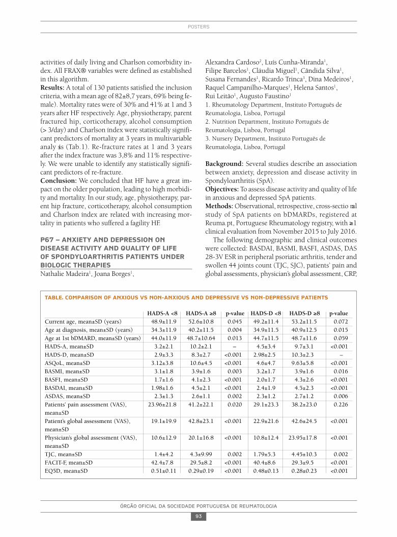
ÓRGÃO OfICIAL DA SOCIEDADE PORTUGUESA DE REUMATOLOGIA
93
posters
activities of daily living and Charlson comorbidity in-dex. All FRAX® variables were defined as establishedin this algorithm.Results: A total of 130 patients satisfied the inclusioncriteria, with a mean age of 82±8,7 years, 69% being fe-male). Mortality rates were of 30% and 41% at 1 and 3years after HF respectively. Age, physiotherapy, parentfractured hip, corticotherapy, alcohol consumption (> 3/day) and Charlson índex were statistically signifi -cant predictors of mortality at 3 years in multivariableanaly sis (Tab.1). Re-fracture rates at 1 and 3 years after the index fracture was 3,8% and 11% respective-ly. We were unable to identify any statistically signifi-cant predictors of re-fracture.Conclusion: We concluded that HF have a great im-pact on the older population, leading to high morbidi -ty and mortality. In our study, age, physiotherapy, par-ent hip fracture, corticotherapy, alcohol consumptionand Charlson índex are related with increasing mor-tality in patients who suffered a fagility HF.
p67 – ANxIETY AND DEpRESSION ONDISEASE ACTIVITY AND QuALITY OF LIFE OF SpONDYLOARTHRITIS pATIENTS uNDERBIOLOGIC THERApIESNathalie Madeira1, Joana Borges1,
Alexandra Cardoso2, Luís Cunha-Miranda1, Filipe Barcelos1, Cláudia Miguel1, Cândida Silva1, Susana Fernandes1, Ricardo Trinca3, Dina Medeiros1,Raquel Campanilho-Marques1, Helena Santos1, Rui Leitão1, Augusto Faustino1
1. Rheumatology Department, Instituto Português deReumatologia, Lisboa, Portugal2. Nutrition Department, Instituto Português deReumatologia, Lisboa, Portugal3. Nursery Department, Instituto Português deReumatologia, Lisboa, Portugal
Background: Several studies describe an associationbetween anxiety, depression and disease activity inSpondyloarthritis (SpA).Objectives: To assess disease activity and quality of lifein anxious and depressed SpA patients.Methods: Observational, retrospective, cross-sectio nalstudy of SpA patients on bDMARDs, registered atReuma.pt, Portuguese Rheumatology registry, with ≥1clinical evaluation from November 2015 to July 2016.
The following demographic and clinical outcomeswere collected: BASDAI, BASMI, BASFI, ASDAS, DAS28-3V ESR in peripheral psoriatic arthritis, tender andswollen 44 joints count (TJC, SJC), patients’ pain andglobal assessments, physician’s global assessment, CRP,
TABLE. COMpARISON OF ANxIOuS VS NON-ANxIOuS AND DEpRESSIVE VS NON-DEpRESSIVE pATIENTS
HADS-A <8 HADS-A ≥8 p-value HADS-D <8 HADS-D ≥8 p-valueCurrent age, mean±SD (years) 48.9±11.9 52.6±10.8 0.045 49.2±11.4 53.2±11.5 0.072Age at diagnosis, mean±SD (years) 34.3±11.9 40.2±11.5 0.004 34.9±11.5 40.9±12.5 0.015Age at 1st bDMARD, mean±SD (years) 44.0±11.9 48.7±10.64 0.013 44.7±11.5 48.7±11.6 0.059HADS-A, mean±SD 3.2±2.1 10.2±2.1 – 4.5±3.4 9.7±3.1 <0.001HADS-D, mean±SD 2.9±3.3 8.3±2.7 <0.001 2.98±2.5 10.3±2.3 –ASQoL, mean±SD 3.12±3.8 10.6±4.5 <0.001 4.6±4.7 9.63±5.8 <0.001BASMI, mean±SD 3.1±1.8 3.9±1.6 0.003 3.2±1.7 3.9±1.6 0.016BASFI, mean±SD 1.7±1.6 4.1±2.3 <0.001 2.0±1.7 4.3±2.6 <0.001BASDAI, mean±SD 1.98±1.6 4.5±2.1 <0.001 2.4±1.9 4.5±2.3 <0.001ASDAS, mean±SD 2.3±1.3 2.6±1.1 0.002 2.3±1.2 2.7±1.2 0.006Patients’ pain assessment (VAS), 23.96±21.8 41.2±22.1 0.020 29.1±23.3 38.2±23.0 0.226mean±SD Patient’s global assessment (VAS), 19.1±19.9 42.8±23.1 <0.001 22.9±21.6 42.6±24.5 <0.001mean±SD Physician’s global assessment (VAS), 10.6±12.9 20.1±16.8 <0.001 10.8±12.4 23.95±17.8 <0.001mean±SD TJC, mean±SD 1.4±4.2 4.3±9.99 0.002 1.79±5.3 4.45±10.3 0.002FACIT-F, mean±SD 42.4±7.8 29.5±8.2 <0.001 40.4±8.6 29.3±9.5 <0.001EQ5D, mean±SD 0.51±0.11 0.29±0.19 <0.001 0.48±0.13 0.28±0.23 <0.001

ÓRGÃO OfICIAL DA SOCIEDADE PORTUGUESA DE REUMATOLOGIA
94
posters
ESR, ASQoL, EQ-5D, FACIT-F for fatigue and HADSscale with 2 domains, HADS-A for anxiety and HADS-Dfor depression (a cutoff of 8 defining these symptoms).Statistics: Mann-Whitney test, p<0.05. SPSS® v.17.Results: 160 patients were included, 41.9% were male,with mean: current age 50.7±11.9, age at diagnosis36.9±11.96, at 1st bDMARD 46.2±11.8, time from di-agnosis 18.5±10.3, years. The mean DAS 28-3V ESRwas 3.2±1.4, BASDAI 2.98±2.2, ASDAS 2.4±1.2, BAS-FI 2.68±2.26, BASMI 3.36±1.7, patient’s global assess-ment 28.8±24.1, physician’s global assessment14.5±15.2, ESR 18.8±18.1 mm/h, CRP 6.7±16.01mg/L, ASQoL 6.2±5.6, FACIT-F 37.2±10.1 and EQ-5D0.417±0.19. The mean HADS-A was 5.96±4.01 andHADS-D was 5.05±4.1 (HADS-A≥8 in 39.5% andHADS-D≥8 in 28.3% patients). Comparison of an xiousvs non-anxious and depressive vs non-depressivegroups appears on table 1.Conclusions: These results suggest that anxious anddepressed patients may have higher disease activity,more functional limitations and worse quality of life.These symptoms should not be underestimated, butinstead, they should be controlled to achieve clinicalimprovement.
p21 – ACpA SEROpOSITIVITY ANDpERIpHERAL NATuRAL KILLER CELLS ASpREDICTIVE MARKERS OF CLINICALRESpONSE TO RITuxIMAB IN RHEuMATOIDARTHRITIS pATIENTSRaquel Miriam Ferreira1, Rita Fonseca1, Diana Rosa-Gonçalves1, Francisca Aguiar1, Teresa Martins-Rocha1, Alexandra Bernardo1, Sofia Pimenta1, Miguel Bernardes1, Lúcia Costa1
1. Serviço de Reumatologia, Centro Hospitalar de SãoJoão, Porto, Portugal
Background: The efficacy of B cell-depletion therapyconfirms the importance of B lymphocytes in rheuma-toid arthritis’s (RA) pathogenesis. Rheumatoid factor(RF) and anti-citrullinated peptide antibodies (ACPA)are prognostic factors for a more severe disease. O thersimmune elements, namely natural killer (NK) cells,seems to influence RA clinical response to rituximab(RTX), but data are lacking.Objectives: To analyze the influence of baseline sta-tus/levels of RF, ACPA and serum immunoglobulin G(IgG) level in RTX treatment. To study the effect of RTXon NK and CD19+ cells in RA patients and their associ-ation with clinical response at 6, 12 and 18 months (M).
Methods: An observational retrospective study wasconducted, including all the consecutive patients withdiagnosis of RA under rituximab, followed at ourRheumatology department. Demographic and clinicaldata were obtained by consulting the national database(Reuma.pt) and the analysis was limited until decem-ber 2016. RF, ACPA and IgG titres were evaluated atbaseline. NK (CD56+CD16+) and B lymphocytes(CD19+) absolute counts were assessed by flow cy-tometry prior to the first RTX cycle and 6 M after. Clini -cal responses were assessed by DAS28 and EULAR cri-teria at 6, 12 and 18 M. Correlations were studied using Spearman coefficient analysis (SPSS 20.0). Signi -ficance level was set as 0.05.Results: We included 63 RA patients (81% of women),with a mean (SD) age of 61(10) years and a mean di -sease duration of 19(10) years, 86% RF-positive and87% ACPA-positive. Bone erosions were present in86% of the patients. At baseline, the mean DAS28 was5.79(1.55). Combination therapy with methotrexateor with others cDMARDs was used in 48% and 30% ofthe patients, respectively; RTX monotherapy in 22% ofour sample. Thirty three patients were previously ex-posed to other biologics. The magnitude of responsewas greater in ACPA-positive vs ACPA-negative patientsin terms of DAS28 variation at 6, 12 and 18M (me diansof 1.09 vs -0.08; 2.03 vs 0.35 and 2.10 vs 0.19;p=0.029, p=0.039 and p=0.004, respectively), withoutsignificant differences between groups in terms of ini-tial DAS28 (5.91(1.60) vs 5.00(0.90), p=0.051). Thepresence of ACPA was also significantly associated withEULAR response at 6, 12 and 18 M (64%, 75% and85% in ACPA-positive patients vs 25%, 16% and 25%in ACPA-negative patients; p=0.034, p=0.010 andp=0.001, respectively). Outcomes did not differ ac-cording FR status. There were no associations betweenthe values of FR, ACPA and IgG at baseline with theclinical response (DAS28 variation). CD19+ cells de-pletion occurred in all patients (mean of 146.4/mm3 atbaseline vs 10.6/mm3 at 6M). An increase of peripher-al NK cells was seen at 6M (mean 231mm/3 at baselinevs 289/mm3 at 6 M). We only have found a positivecorrelation between NK cells number at baseline andDAS28 variation at 6 M (r=0.35, p=0.023). There wereno associations between, neither NK cells, nor CD19cells variations at 6M with clinical response to RTX.Conclusions: Our data suggest that ACPA seroposi-tivity is associated with a better clinical response to RTXin RA patients. NK cells at baseline may be useful toidentify early responders to RTX.
ÓRGÃO OfICIAL DA SOCIEDADE PORTUGUESA DE REUMATOLOGIA
95
posters
p69 – REAL wORLD DATA OF RITuxIMABEFFECTIVENESS IN RHEuMATOID ARTHRITIS:DIFFERENCES BETwEEN BIOLOGIC-NAIVEpATIENTS AND pREVIOuSLY ExpOSED TOBIOLOGICSRaquel Miriam Ferreira1, Rita Fonseca1, Diana Rosa-Gonçalves1, Francisca Aguiar1, Teresa Martins-Rocha1, Alexandra Bernardo1, Sofia Pimenta1, Miguel Bernardes1, Lúcia Costa1
1. Serviço de Reumatologia, Centro Hospitalar de SãoJoão, Porto, Portugal
Background: Rituximab is only approved for rheuma-toid arthritis (RA) treatment in patients with an in-complete response or intolerance to others DMARDs,including TNF alfa inhibitors. It represents a signifi-cant advance in RA biologics arsenal due to its safetyand efficacy profiles.Objectives: To evaluate the effectiveness of rituximabin RA patients and to compare the response betweenfirst-line rituximab patients and those previously ex-posed to other biologics.Methods: An observational retrospective study wasconducted, including all the consecutive patients withdiagnosis of RA under rituximab, followed at ourRheumatology department until December 2016. De-mographic and clinical data were obtained by consul -ting the national database (Reuma.pt). DAS28 varia-tions and EULAR response were measured at 6, 12 and18 months. Parametric and non parametric tests wereused for statistics (SPSS 20.0).Results: We included 63 RA patients (81% of women),with a mean (SD) age of 61(10) years and a mean di -sease duration of 19(10) years; 86% rheumatoid factorpositive and 87% anti- citrullinated peptide antibody(ACPA) positive. Bone erosions and extraarticular ma -ni festations were present in 85,7% and 58,7% of thepatients, respectively. At baseline, the mean DAS28 was5.79 (65% and 29% of patients with severe and mode -rate disease activity, respectively, and 6% in clinical re-mission). Thirty patients were treated with rituximabas first-line therapy and 33 patients were previouslyexposed to other biologics. Combination therapy withmethotrexate (MTX) was observed in 48 % and withothers classic DMARDs in 30%, while 22,3% receivedrituximab monotherapy. First-line rituximab optionwas justified by lung involvement in 21%, past mali -gnancy in 13%, recurrent infections in 5%, congestivecardiac failure in 3%, vascular involvement in 3% anduntreated latent tuberculosis in 3%. In the group pre-
viously exposed to biologics, 13% switched therapydue to ineffectiveness and 87% due to adverse events.No significant differences were found between the 2groups in terms of age, gender, concomitant use ofMTX and baseline DAS28. The group previously ex-posed to biologics had a longer disease duration (mean23 vs 15 years, p=0.001) and fewer patients with ACPAseropositivity (79% vs 97%,p=0.035). There was asigni ficant reduction of DAS28 at 6, 12 and 18 months(p<0.001 for all). Fifty six percent of the patientsachieved a EULAR response at 6 months, 46% at 12months and 59% at 18 months. DAS28 variation at 6months differed significantly between groups, with abetter clinical response in naive biological patientscomparing to those previously exposed to biologics(median 1.173 vs 0.477; p=0.038). There were no diffe -rences in terms of DAS28 variation at 12 and 18months (p=0.642 and p=0.135, respectively) and inEULAR responses at 6, 12 and 18 months between thegroups (p=0.289, p=0.523 and p=1.000,respectively).Conclusions: Our study confirms the effectiveness ofrituximab in RA patients and suggests a higher magni-tude of response in naive biological patients at 6months of RTX therapy. These findings put in pers -pective an extension of rituximab as a first-line biolo -gic for RA treatment.
GRupO 6
p159 – COMpARATIVE EFFECTIVENESS OFSwITCHING TO ALTERNATIVE TuMOuRNECROSIS FACTOR (TNF) ANTAGONISTSVERSuS SwITCHING TO RITuxIMAB INpATIENTS wITH RHEuMATOID ARTHRITISwHO FAILED pREVIOuS TNF INHIBITORSDaniela Santos-Faria1, Joana Leite Silva1, Joana Ramos Rodrigues1, Joana Sousa-Neves1, Marcos Cerqueira1, Daniela Peixoto1, Filipa Teixeira1,Sérgio Alcino 1, Carmo Afonso1, José A. Costa1
1. Rheumatology Department, Unidade Local de Saúde doAlto Minho, Ponte de Lima, Portugal
Background: Tumour necrosis factor inhibititors(TNFi) and B-cell depletion are highly effective treat-ments for active RA. Following an inadequate responseto a prior TNFi, using RTX would be clinically non-in-ferior, cheaper and with a comparable safety profile1.Objectives: To compare the effectiveness and safety ofRTX therapy versus TNFi in RA patients, who failed to

ÓRGÃO OfICIAL DA SOCIEDADE PORTUGUESA DE REUMATOLOGIA
96
posters
respond to a previous TNFi, followed in a rheumatolo -gy unit.Methods: Retrospective study of 33 RA patients non--responsive or intolerant to a TNFi, registered in theRheumatic Diseases Portuguese Register, who haveswitched from a 1st TNFi to a 2nd TNFi or to RTX. Weassessed disease activity at baseline, 6 and 12 months,according to disease activity score-28 joints (DAS28),clinical disease activity index (CDAI) and simplifieddisease activity index (SDAI). Discontinuation rates andrespective reasons were evaluated. Parametric and non-parametric tests were used for comparative analysis. P value <0.05 was defined as statistically significant.Results: 33 patients, 29 females (87.8%), with a meanage of 56.6 (±10.7) years and mean disease duration of14.7 (±3,9) years were evaluated. Patients were divi dedin two groups, whether they switched for TNFi(58.8%) or RTX (42.2%). Disease activity was slightlyhigher in the RTX group, respectively: DAS28 5.37 vs5.79, CDAI 28.58 vs 33.56 and SDAI 30.99 vs 35.67(p>0.05 for all). Similar results were also found abouttender joint count-28 (TJC), swollen joint count-28(SJC), patient and physician global health. In opposi-tion C-reactive protein (CPR) and erythrocyte sedi-mentation rate (ESR) were lower in RTX group, but allof them without significant differences. At 6 months,DAS 28 and SDAI were lower in the TNFi group(p=0.04 and 0.03). However, comparing groups at 12months, there were no significant differences for all pa-rameters. The variation of units in DAS28 betweenbaseline and 6 or 12 months, was 1.65 vs 1.03 and 1.91vs 1.75 units (p>0.05), respectively for TNFi vs RTXgroups. Remission, according to the DAS28 (<2.6),CDAI (≤2.8) and SDAI (≤3.3) was achieved in 15% ofpatients treated with TNFi, at 6 months, but in none ofthe RTX group (p>0.05). However, at 12 months, theremission rates were higher in patients treated withRTX, with a proportion of patients achieving DAS28and CDAI/SDAI remission of 20.01% and 13.03% inTNFi group, while in RTX group it was 23.10% and15.41%, respectively, with p>0.05 for all. At 1 year, thetreatment was discontinued only in the TNFi group,representing 21.05% of patients. The ineffectiveness oftherapy was the main reason, while adverse events andlost to follow up represented a lower proportion.Conclusions: Our findings showed a higher effective-ness for TNFi at 6 months, but not at 12 months, com-paring with RTX treatment which may be related withthe known delay of mechanism of action of the latter.The group of patients treated with RTX had slightly
higher level of disease activity at baseline and it may re-flect a selection bias for a severely resistant populationof patients that respond poorly to biologics. Largerstudies, with more patients and longer follow-up arewarranted.
REFERENCES1. Harrold LR, Reed GW, Magner R, et al. Comparative effectiveness
and safety of rituximab versus subsequent anti–tumor necrosisfactor therapy in patients with rheumatoid arthritis with prior ex-posure to anti–tumor necrosis factor therapies in the United Sta-tes Corrona registry. Arthritis Res Ther. 2015;17(1):256.
p18 – QuALITY OF LIFE IN pATIENTS wITHBEHCET’S DISEASEDaniela Santos-Faria1, Joana Leite Silva1, Marcos Cerqueira1, Joana Ramos Rodrigues1, Joana Sousa-Neves1, Daniela Peixoto1, Filipa Teixeira1, José A. Costa1, Carmo Afonso1, Sérgio Alcino 1
1. Rheumatology Department, Hospital Conde deBertiandos (ULSAM), Ponte de Lima, Portugal
Background: Behcet’s disease (BD) is a chronic andmultisystemic inflammatory disorder affecting the skin,mucosa, joints, eyes, arteries, veins and the nervousand gastrointestinal systems. The symptoms may beseparated by long or short intervals, occur simulta -neously or in sequence, and exhibit a pattern of exa -cerbation and remission. The disease itself or the im-pact of the symptoms affect patients physically, men-tally and socially with a negative effect in the quality oflife (QoL).Objectives: To determine the health status and quali-ty of life in patients with BD followed in a rheumatolo -gy unit and to identify associated demographic and di -sease-related parameters influencing them.Methods: Cross-sectional study of 44 BD patients and39 healthy controls matched for age and sex. All sub-jects completed the Health Assessment Questionnaire(HAQ) to assess impairment in daily activities due toillness, Short Form-36 (SF-36) and EuroQol VisualAnalogic Scale (EQ-VAS) to assess health related quali -ty of life (HRQL). The disease characteristics, inclu dingdisease duration and clinical involvements were col-lected. The Birmingham Vasculitis Activity Score(BVAS) was applied for the evaluation of current diseaseactivity among BD patients. Parametric and nonpara-metric tests were used for comparative analysis. P va -lue <0.05 was defined as statistically significant.Results: Among 44 patients, 35 (79.5%) were female,

ÓRGÃO OfICIAL DA SOCIEDADE PORTUGUESA DE REUMATOLOGIA
97
posters
with a mean age of 40.07 years and mean disease du-ration of 5.93 years. BD patients had significantly high-er HAQ score (p=0.009) and lower levels of SF-36(p<0.001) than the healthy controls. The predominantcontributors to this low SF-36 score were generalhealth, vitality and role-emotional domains. In com-parison with healthy controls, patients with inactivedisease (BVAS=0) also had a higher HAQ, but withoutsignificant differences (p=0.53). The total SF-36 scorealso showed lower levels in patients with inactive disea -se (p=0.04), but when compared the different compo-nents, only half of them maintained significant diffe -rences, namely role-physical (p=0.03), general health(p<0.001), vitality (p<0,001) and social functioning(p=0.05). The controls showed a higher EQ-VAS, witha mean of 88.44 comparing to 68.36 in all BD patients(p<0.001) and 74.12 in the subgroup with inactive di -sease (p=0.003). SF-36 score was negatively correla tedwith HAQ (r=-,553, p<0.001) and positively correla tedwith EQ-VAS (r=.388, p<0.001). Longer disease dura-tion correlated with lower levels of only some SF-36domains, namely physical functioning (r=-.436,p=0.003), role-physical (r=-.533, p<0.001) and role--emotional (r=-.465, p=0.001). There was no correla-tion between disease activity and disease duration(p=0.678) or different scores evaluating QoL(p=0.876). The gender was not associated with statis-tical differences when compared clinical involvements,disease duration, current disease activity, HAQ, EQ-VAS or SF-36 scores. The heterogeneous nature of thedisease expression did not allow the study of the asso-ciation with the levels of health status and QoL.Conclusions: Our findings showed lower levels of QoLand global health status in BD compared to healthycontrols, mainly in active disease, accordingly with pre-vious studies. The disease itself could be a determinantof disability. To recognize these difficulties and coexis -ting conditions is important to better manage these pa-tients.
REFERENCES1. Melikoglu MAM, Melikoglu MAM. What affects the quality of
life in patients with Behcet’s disease? Acta Reumatol Port.2014;39(1):46–53.
p189 – CERTOLIZuMAB pEGOL-LIKEpRODuCT MICE EQuIVALENT REDuCES pAwSINFLAMMATION IN TMTNF TRANSGENIC MICEElsa Vieira-Sousa1, 2, Soraia Caetano-Silva1, Vidal B1,Lopes IP1, Helena Canhão3, JE Fonseca1, 2
1. Rheumatology Research Unit, Instituto de Medicina
Molecular, Lisboa, Portugal2. Rheumatology Department, Hospital Santa Maria,CHLN, Lisbon Academic Medical Center, Lisboa, Portugal3. EpiDoC Unit, CEDOC, NOVA Medical School, NOVAUniversity, Lisboa, Portugal
Background: Transmembrane (tm)TNF (TgA86) miceis a transgenic line that spontaneously develops pe-ripheral arthritis and spondylitis, with 100% pene-trance, mimicking human spondyloarthritis (SpA).Arthritis is characterized by swelling and deformationof the paws and toes with loss of grip, and spondylitisby crinkled tail and hunchback deformation, startingat 4 weeks of age. The aim of this work is to under-stand the effect of TNF blockade in this SpA-like phe-notype mouse strain.Methods: (tm)TNF (TgA86) mice were bred in speci -fic-pathogen-free conditions and according to humanendpoints. (tm)TNF (TgA86) mice at 10 weeks of agewere treated with a certolizumab pegol-like productmice equivalent (Ab501) or with vehicle (phosphatebuffer saline), for 12 weeks. Ab501 was administered100mg/kg twice a week intraperitoneal.
The arthritic paw inflammatory score according tothe European Guidelines for Animal Experimentationwas used. A semi-quantitative score for the severity ofhistologic inflammation was applied to haematoxylinand eosin stained slides (global, infiltration, lining cellsand erosions). Results were analyzed using Mann--Whitney test. p values less than 0.05 were consideredstatistically significant.Results: 14 (tm)TNF (TgA86) mice (6 males and 8 fe-males) at 10 weeks of age were treated with Ab501 and6 (tm)TNF (TgA86) mice with vehicle, for 12 weeks.A significant lower paws inflammatory score was ob-served in Ab501 treated animals as compared to vehi-cle treated animals. Mice weight progressively in-creased through follow-up, showing a trend for high-er gains in the Ab501 treated group in comparison tothe vehicle group. Significant differences betweengroups were observed in the global inflammatory scoreboth in the front (p=0.001) and hind paws (p=0.0086).The analysis of partial scores showed significantly lo -wer inflammatory infiltrate in the front paws (p=0.001)and hind paws (p=0.001), as well as less lining cellslayers (respectively, p=0.0331 and p=0.0051), in theAb501 treated group as compared to the vehicle group.Significant (p=0.0014) lower erosion score at the frontpaws was also observed in the Ab501 treated group,comparing to the vehicle group.
ÓRGÃO OfICIAL DA SOCIEDADE PORTUGUESA DE REUMATOLOGIA
98
posters
Conclusions: The certolizumab pegol like productmice equivalent prevented macroscopic and histolo gicinflammatory infiltrate and lining layers thickening inboth front and hind paws of (tm)TNF (TgA86) mice.Erosive disease was also significantly delayed in thefront paws. These are preliminary results concerningthe effect of certolizumab pegol-like product miceequivalent in peripheral arthritis of (tm) TNF (TgA86)mice.Acknowledgments: To Leonie M van Duivenvoordeand Dominique Baeten (Amsterdam Academic MedicalCenter) for providing (tm)TNF (TgA86) mice forbreeding and colony establishment. George Kolliasfrom Flemming Institute for authorization for the usethis mice line.
This investigator initiated study was supported (fi-nancial & product) by UCB.
p214 – ExISTE DESREGuLAÇÃO IMuNITÁRIANA SÍNDROME SICCA NÃO-SjÖGREN? uMESTuDO DAS SuBpOpuLAÇÕESLINFOCITÁRIAS CIRCuLANTESFilipe Barcelos1, 2, 3, Catarina Martins3, José Vaz Patto1,Glória Nunes3, Teresa Lopes3, Joana Cardigos4, Nuno Alves4, João Amaral5, Jaime C. Branco2, 3, 6, L Miguel Borrego2, 3
1. Instituto Português de Reumatologia, Lisboa, Portugal2. Hospital CUF Descobertas, Lisboa, Portugal3. CEDOC, Nova Medical School, Lisboa, Portugal4. Oftalmologia, Hospital Santo António dos Capuchos(CHLC), Lisboa, Portugal5. Faculdade de Medicina Dentária, Universidade deLisboa, Lisboa, Portugal6. Serviço de Reumatologia, Hospital de Egas Moniz,Lisboa, Portugal
Introdução: Um grande número de doentes com sín-drome sicca não preenche critérios de classificação dasíndrome de Sjögren (SS), mas apresenta manifestaçõesde autoimunidade, como artrite, fenómeno de Ray-naud, alterações cutâneas ou hematológicas. Estesdoentes têm anticorpos anti-nucleares, estando ausen-tes auto-anticorpos mais específicos, e são designadoscomo Doença Indiferenciada do Tecido Conjuntivo(DITC). Alguns poderão progredir para uma doençaespecífica, sendo a SS uma forte candidata. Distúrbiosdas populações linfocitárias podem ser um marcador deprogressão, uma vez que doenças como a SS têm per-fis linfocitários distintos.Objectivos:. Pretendeu-se estudar a distribuição das
populações linfocitárias circulantes em doentes comsíndrome sicca não-Sjögren (n-SS) e compará-los comSS e controlos saudáveis.Métodos: Incluímos 65 doentes n-SS, 53 doentes SS(critérios AECG) e 22 controlos saudáveis. As subpo-pulações linfocitárias foram caraterizadas por citome-tria de fluxo, incluindo as células T foliculares e regu-ladores, as células B naïve, maduras, de memória, plas-mablastos e B reguladoras. A análise estatística foi rea-lizada com GraphPad, com significância para p<0,05.Resultados: Em relação ao grupo controlo, os doentesn-SS tinham contagens de células T mais baixas(p=0,016), com CD4 mais baixos (p=0,0028), emboraessa diferença não seja tão pronunciada como entre SSe controlos. Doentes n-SS tinham percentagens maiselevadas de CD4 (p=0,0005) e mais baixas de CD8(p=0,0009) que os doentes SS. Adicionalmente, cons-tatou-se uma diminuição na contagem absoluta deTregs (p=0,0028) em doentes n-SS comparados com ogrupo controlo, mas que foi menos pronunciada que nacomparação entre doentes SS e controlos (p=0,0008).As células Th17 estavam diminuídas em doentes comSS comparados com controlos (p=0,0005), mas não emdoentes n-SS. Comparando com controlos, os doentesn-SS e SS apresentavam ambos uma contagem absolu-ta diminuída (p=0,0001 and p<0,0001, respetivamen-te) de células Tfh CXCR5+, sem diferenças entre doen-tes n-SS e SS. No entanto, encontraram-se númerosmais elevados de células T IL21+CD4 e células Tfh1nos doentes SS em comparação com os controlos(p=0,0209 and p=0,0092 respectivamente) e doentesn-SS (p=0,0051 and 0,0028 respectivamente).
Doentes n-SS apresentavam contagens absolutas decélulas de memória (com switch e sem switch) em níveisintermédios entre os controlos (com valores significa-tivamente mais elevados) e os doentes SS (com valoressignificativamente mais baixos). Usando a classificaçãoBm1-5, encontrámos níveis mais baixos de células Bm1(p=0,004), eBm5 (Abs, p=0,0273) e células Bm5 (Abs,p=0,0444) em doentes n-SS comparados com contro-los. Embora sem significância estatística, encontrou-seum aumento das células eBm5 (Abs, p=0,063) e Bm5(Abs, p=0,05) em doentes n-SS em comparação comdoentes SS. As células Breg CD24+CD27+ também es-tavam diminuídas em doentes n-SS comparados comcontrolos (p=0,036), mas aumentadas em doentes n-SScomparado com doentes SS (p=0,0007).Conclusões: Os doentes n-SS apresentaram desregu-lação imunitária, representada por alterações nas célu-las B mas também no subgrupo das células Tfh, co-

ÓRGÃO OfICIAL DA SOCIEDADE PORTUGUESA DE REUMATOLOGIA
99
posters
nhecidas por modular a resposta imunitária humural.Apesar de menos pronunciadas, estas alterações asse-melham-se às encontradas em doentes SS. Permanecepor esclarecer se a síndrome sicca n-SS é uma fase naevolução para SS. No entanto, a identificação de umadesregulação imunológica característica em doentes n-SS pode ser útil para o diagnóstico e prognóstico.
p173 – OSTEOARTHRITIS MuLTIDISCIpLINARY CLINIC – 2 YEARS OFExpERIENCEIsabel Sousa Martins1, 2, Marta Gomes1, Ana Santos1,Luís Pinheiro1, Claúdia Meneses1, Patrícia Gomes1,Sandra Abreu1, Fernando Pimentel1, Rodrigues AM1,3,4
1. Hospital de Santo Espírito da Ilha Terceira, Angra doHeroísmo, Portugal2. Unidade de Saúde da Ilha Terceira, Angra do Heroísmo,Portugal3. EpiDoC Unit, CEDOC, NOVA Medical School, NOVAUniversity, Lisboa, Portugal4. Rheumatology Research Unit, Instituto de MedicinaMolecular, Lisboa, Portugal
Background: There is an increasing demand to have acoordinated pharmacological and non-pharmacologi-cal approach in order to boost osteoarthritis (OA) treat-ment effectiveness. The OA multidisciplinary Clinic inHSEIT started in 2014 and aimed to improve pain,function and delay the placement of total joint re-placement in OA patients. The team consists of ele-ments of Rheumatology, Orthopaedics, Physiatrist, Nu-trition and Podology.
This study aims to characterize the populationevalua ted in OA Multidisciplinary Clinic and to evalua -te patient’s level of satisfaction.Methods: Patients that attended the OA multidisci-plinary Clinic in HSEIT between February 2014 andNovember 2016 were evaluated. After the first visit apersonalized multidisciplinary approach was applied toeach individual patient that can include physical thera -py, nutrition program, intra-articular administration ofhyaluronic acid and/or corticosteroid and orthotics.The second visit happens after 6 months of follow-up.Clinical diagnosis, radiographic severity score of kneeand hip OA (Kellgren Lawrence), Knee injury and OAoutcome score (KOOS), pain visual analogic scale(VAS), pharmacological treatment and body mass index(BMI) were retrospectively collected using patient’sclinical process. Patient’s clinical data were assessed atthe first visit and after 6 month of follow-up. Moreover,
an independent researcher through a phone interviewperformed a Patient satisfaction survey in December2016.Results: 64 consultations were identified for a total of45 patients. The majority of the evaluated individualswere female (81.8%) and had a mean age of 60.8±10.8years. About half of the patients had knee OA (52.3%),11.4% ankle OA and 36.4% others. Regarding Knee andhip OA 78.6% had a radiographic severity score of 4.
In the first visit 100% of the patients reported dailyanalgesic therapy intake. After 6 months of follow-upthe proportion of patients with daily use of analgesic in-take decrease to 58.1%. Moreover, a decrease in painVAS was also verified from 8.2±2.7cm at the first visitto 7.0±3.0 cm at the second visit.
At the first visit 100% of patients were obese. Of the17 patients that accepted the nutrition program, 88.2%had lost weight, with decreased in the mean BMI from42.1±7.1 in the first visit to 39.3±5.0.
Considering physical therapy 56.8% were proposedto treatment. Orthopaedic surgery was proposed in56.8% of the patients, with 84%on the waiting list.
In patients with knee OA an improvement in themean score in all subsets of the KOOS questionnairewas observed. The mean Symptoms subset at baselinewas of 33.2±20.8 and at 6 month of follow-up was of50.9±28.4; the mean Pain subset 30.4±17.3 and58.5±25.0 and in Daily Life Activities 30.3±14.1 and56.2±20. These differences verified in the KOOS areconsidered above the minimal Detectable Changes asdefined by the: International Knee DocumentationCommittee.
In the satisfaction survey, on a scale of 0 to 10, pa-tients classified the Multidisciplinary OA treatment ap-proach on average 8.5±2.7 in relation to importanceand 8.9±2.2 in relation to satisfaction. Of the patientssubmitted to surgery (n=4) all rated their satisfactionwith the results on 10/10.Conclusion: The population evaluated in the OA Mul-tidisciplinary Clinic had a severe OA, were obese, verysymptomatic and with severe physical limitation. Afterthe personalized multidisciplinary therapeutic plan,clinical improvement of pain, function and BMI wasobserved. Patients report high degrees of satisfactionwith the consultations and treatments instituted
p146 – ARE RHEuMATOID FACTOR ISOTYpESuSEFuL IN THE DIAGNOSIS OF RHEuMATOIDARTHRITIS?Miguel Guerra1, Romana Vieira1, Ana Paula Cruz2,

ÓRGÃO OfICIAL DA SOCIEDADE PORTUGUESA DE REUMATOLOGIA
100
posters
Taciana Videira1, Patrícia Pinto1
1. Rheumatology, Centro Hospitalar de Vila Nova deGaia/Espinho, Vila Nova de Gaia, Portugal2. Clinical Pathology, Centro Hospitalar de Vila Nova deGaia/Espinho, Vila Nova de Gaia, Portugal
Introduction: Rheumatoid factor (RF) and anti-citrul-linated peptide antibodies (ACPA) are key serologicmarkers in the diagnosis of Rheumatoid Arthritis (RA)included in the 2010 ACR/EULAR diagnostic criteria.Determination by enzyme-linked immunosorbent as-say (ELISA) allows RF isotypes’ quantification (IgG, IgAand IgM), improving diagnostic accuracy.Objectives: To assess the clinical value of RF-IgG/IgA//IgM (ELISA) in the diagnosis of RA, in comparison toRF-IgM (nephelometry).Methods: A population of RA outpatients fulfilling the2010 ACR/EULAR diagnostic criteria was cross-sec-tionally evaluated. Data on demographic and clinicalcharacteristics was collected. RF-IgG/IgA/IgM (ELISA,Orgentec), RF-IgM (nephelometry, Siemens) andACPA-IgG (ELiA, ThermoFisher) were measured. Va -lues three times or more above the upper limit of nor-mal were considered high-positive (in agreement to2010 ACR/EULAR diagnostic criteria).Results: A total of 87 patients (70.1% female) were in-cluded, with a mean (SD) age of 57.3 (12.29) years.Median time of disease evolution was 6 years, rangingfrom 0 to 37 years. Erosions were present in 50.6%(N=44). RF-ELISA was positive (at least one isotype in-creased) in 85.1% (N=74); the most frequent isotypewas IgM (70.1%;N=61) and the most frequent combi-nation was IgG, IgA and IgM positivity (46.0%;N=40)(table1). FR-nephelometry and ACPA were positive in58.6% (N=51) and 47.1% (N=41), respectively.
Comparing the two RF methods, 56.3% (N=49)were both RF-nephelometry and RF-ELISA positive;28.7% (N=25) were RF-ELISA positive and RF-nephe -lometry negative, and only 2.3% (N=2) verified the op-posite (p=0.001). As for RF high-positivity, 4.6% (N=4)of the 87 patients were only RF-nephelometry high--positive, 9.2% (N=8) only RF-ELISA high-positive and34.5% (N=30) both high-positive (p<0.001).
In the RF-nephelometry negative population(N=36), ACPA and RF-ELISA were both positive in11.1% (N=4). Only 8.3% (N=3) were solely ACPA posi -tive and 58.3% (N=21) solely RF-ELISA positive, how-ever without statistical significance.
Considering the ACPA negative population (N=46),32.6% (N=15) were both RF positive; 45.7% (N=21)were RF-ELISA positive and RF-nephelometry nega-tive and 4.3% (N=2) the opposite, without a statisticalsignificance.Conclusions: ELISA is superior to nephelometry de-tecting RF in patients with RA, as also in quantifyinghigh-positive values.
p7 – EFFICACY AND SAFETY OF ORALADMINISTRATION OF puRE CELASTROL INAIA RATSRita Cascão1, Tania Carvalho2, João Gonçalves3, Luis Moita4, JE Fonseca1, 5
1. Rheumatology Research Unit, Instituto de MedicinaMolecular, Faculdade de Medicina da Universidade deLisboa, Lisboa, Portugal2. Laboratório de Histologia e Patologia Comparada,Instituto de Medicina Molecular, Faculdade de Medicinada Universidade de Lisboa, Lisboa, Portugal3. iMed- Research Institute for Medicines, Faculdade deFarmácia da Universidade de Lisboa, Lisboa, Portugal4. Instituto Gulbenkian de Ciência, Oeiras, Portugal5. Rheumatology Department, Hospital de Santa Maria(CHLN), Lisbon Medical and Academic Centre, Lisboa,Portugal
Background: Celastrol, a pentacyclic-triterpene isolat-ed from Tripterigium wilfordii roots, has shown greattherapeutic potential for the treatment of several in-flammatory diseases, including rheumatoid arthritis(RA). We have previously demonstrated that celastrolhas significant anti-inflammatory and bone protectiveeffects in the adjuvant-induced rat model of arthritis(AIA), when administered via intraperitoneal route. Forfurther preclinical evaluation of celastrol as a candidatecompound for RA treatment, an effective and safe oral
TABLE I. FREQuENCIES OF RF-ELISA ISOTYpES'pROFILES
Profile n %IgG-IgA-IgM- 13 14,9IgG+IgA-IgM- 1 1,1IgG+IgA+IgM- 4 4,6IgG+IgA?IgM+ 1 1,1IgG+IgA-IgM+ 12 13,8IgG+IgA+IgM+ 40 46,0IgG-IgA+IgM+ 5 5,7IgG-IgA-IgM+ 3 3,4IgG-IgA+IgM- 8 9,2

ÓRGÃO OfICIAL DA SOCIEDADE PORTUGUESA DE REUMATOLOGIA
101
posters
administration is crucial.Objective: In this work we aimed to study the doserange for both therapeutic and toxic effects for oral admi -nistration of pure celastrol using the AIA rat model.Methods: Celastrol (1, 2.5, 5, 7.5, 12.5 and 25µg/g//day, N=5/group) was administrated orally in femaleAIA rats after 8 days of disease induction (therapeuticmodel) for a period of 14-days. A group of healthy(N=8) and untreated arthritic (vehicle, N=15) genderand age-matched Wistar rats were used as control. Du -ring the period of treatment, the inflammatory score,ankle perimeter and body weight were measured. Atthe end of the treatment, animals were sacrificed, bloodwas collected for clinical pathology, and necropsy wasperformed, with collection of internal organs forhistopathological analysis and of paw samples for di -sease scoring.Results: Oral administration of pure celastrol at 2.5, 5and 7.5µg/g/day reduced the inflammatory score andankle swelling, preserved articular joint structure witha reduction in synovial inflammatory infiltrates andproliferation, halted articular bone destruction, and di-minished the number of synovial CD68+ macrophages(a biomarker of response to anti-arthritic treatment).This compound also reduced the number of osteoclastsand osteoblasts present in joints. Bone resorption andturnover was also reduced at both 5 and 7.5µg/g/day,with a significant decrease in serum levels of TRACP--5b, P1NP and CTX-I. Of note, no significant variationin body weight, evidence of nephro-, hepato- or car-diotoxic effects, nor alterations in blood cell countswere observed at these concentrations. However, thedose of 7.5µg/g/day was already associated with thymicand hepatotoxic changes, and higher doses showedtoxicity signs. The lethal dose (LD) and LD50 were de-fined as 25µg/g/day and 12.5µg/g/day, respectively. Ofnote, oral celastrol at 1µg/g/day had no effect in arthri-tis progression.Conclusion: Our results clearly show that 2.5µg/g/dayis the lowest and 5µg/g/day is the highest effective andsafe oral doses of celastrol in the setting of AIA rat mo -del. These findings suggest that while celastrol is po-tentially very effective to treat RA, it has a narrow the -rapeutic window.
p111 – MuLTIDISCIpLINARY SjÖGREN’SSYNDROME CLINIC: CHARACTERIZATION ANDIMpACT AT THE CLINICAL AND RESEARCHLEVELVasco C Romão1, 2, Ana Silva Martins3, Joana Pinto4,
Ana Inês Gonçalves4, Rita Simão3, Walter Rodrigues4,Francisco Salvado3, Carla Macieira1, JE Fonseca1, 2
1. Rheumatology Department, Hospital de Santa Maria(CHLN), Lisbon Medical and Academic Centre, Lisboa,Portugal2. Rheumatology Research Unit, Instituto de MedicinaMolecular, Faculdade de Medicina da Universidade deLisboa, Lisboa, Portugal3. Stomatology, Hospital de Santa Maria (CHLN), LisbonAcademic Medical Centre, Lisboa, Portugal4. Ophthalmology, Hospital de Santa Maria (CHLN),Lisbon Academic Medical Centre, Lisboa, Portugal
Introduction: Sjögren’s syndrome (SS) is an inflam-matory rheumatic disease that can affect several organsystems, most frequently the musculoskeletal, ocularand oral domains. Multidisciplinary care is thus crucialin the optimal management of SS patients. Recently,the Rheumatology, Stomatology and Ophthalmologydepartments of our center joined efforts to create a mul-tidisciplinary SS (MDSS) clinic aiming to improve pa-tient care and facilitate clinical research.Objectives: Our goal is to report the aims, structureand characteristics of the MDSS clinic. We will alsosummarize the characteristics of the patients evalua tedsince its creation in mid 2016.Methods: We reviewed the procedures conducted atthe MDSS clinic, including clinical evaluation, disease--related questionnaires, specialized oral/ocular assess-ment, biologic samples collection, patient educationand clinical research efforts. We reviewed the clinicalfiles of patients assessed to date for diagnosis, classifi-cation criteria, salivary gland biopsy and ultrasound,tear and salivary flow and ocular staining scores.Results: The main aims of the MDSS clinic are: to pro-vide optimal care to SS patients through a thorough,fast, coordinated, systematic evaluation of the rheuma-tological, oral and ophthalmologic domains; to facili-tate the diagnostic investigation of sicca syndromes orclarification of specific issues in patients with esta -blished diagnosis; to promote clinical research in thefield of primary SS, through the constitution of aprospective cohort with an associated Biobank collec-tion. Patients are referred by other physicians from thethree departments involved in the MDSS clinic and un-dergo full clinical evaluation, with clinical history andcomplementary exams review, classification criteria(AECG 2002 and ACR/SICCA 2012) evaluation, as-sessment of disease activity (ESSDAI, ESSPRI, PRO-FAD-SSI) and damage (SSDI, SSDDI), as well as full

ÓRGÃO OfICIAL DA SOCIEDADE PORTUGUESA DE REUMATOLOGIA
102
posters
oral and ophthalmological evaluation focusing on dry-ness extent, oral/ocular damage, tear/salivary flow andother concomitant eye and mouth conditions affectingthese patients. All patients have an unstimulated sali-vary flow assessment, Schirmer’s I Test and ocular stai -ning for keratoconjunctivitis sicca according to vali-dated scores. Patients are also checked for prior salivarygland biopsy and ultrasound and offered one when notpreviously done. On patient’s agreement, saliva andblood is collected for the SS Biobank-IMM collection,with paired clinical information being registered in theSS module of the Rheumatic Diseases Portuguese Re -gister, Reuma.pt. A final diagnosis is established, the -rapeutic measures are instituted, medication is adjust-ed and follow-up of specific active problems is planned.Patients are referred back to their assisting physiciansand re-evaluated in the MDSS clinic every 6 to 12months. Patients with primary SS are included in theObservational Lisbon Sjögren’s Syndrome ProspectiveObservational (OLISSYPO) Cohort, with paired clini-cal data (Reuma.pt) and biological samples (Biobank--IMM). Since May 2016, 70 patients were evaluated inthe MDSS clinic, with the corresponding data repre-sented in Table 1.Conclusions: The creation of the MDSS clinic hasadded value to the investigation and management ofsicca syndrome patients, most importantly those withprimary SS. Patients were quickly and easily referredfor specialist assessment, thus improving clinical care
and outcomes. Research is facilitated by the MDSS cli -nic, enabling the creation of a thoroughly characteri zedcohort, with matching biological samples.
GRupO 7
p27 – LuNG INVOLVEMENT IN RHEuMATOIDARTHRITIS – A pORTuGuESE REALITYAna Catarina Duarte1, Sandra Sousa1, Ana Cordeiro1,Maria José Santos1, José António Canas da Silva1
1. Rheumatology Department, Hospital Garcia de Orta,Almada, Portugal
Background: Rheumatoid arthritis (RA) is associatedwith a wide range of extra-articular manifestations.Non-cardiac thoracic manifestations occur in approxi -mately 5–20% and can affect the pleura, pulmonaryparenchyma, airways and vasculature. Besides, patientscan also experience drug-induced pulmonary diseaserelated to RA medication.Objectives: To characterize lung involvement and fac-tors associated with lung disease in a cohort of RA pa-tients.Mehods:Retrospective analysis of RA patients followedin our Rheumatology department. Lung involvementwas defined by the presence of imagiological//histopathological alterations described in the spectrumof rheumatoid arthritis-associated lung disease in ei-ther symptomatic or asymptomatic patients. Logisticregression analysis was used to evaluate demographicand clinical features independently associated withlung disease.Results: In total, 532 RA patients were analysed, 400females, mean age of 63.6 (±13.8) years and mean di -sease duration of 11.8 (±9.5) years. Rheumatoid factor(RF) was positive in 69% and anti-cyclic citrullinatedpeptide antibodies (ACPA) in 60%; 8.8% were currentsmokers and 7.5% past smokers. Methotrexate (MTX)was the most prescribed synthetic DMARD (85.9%)and biologics were used in 32.3% of patients.
Lung involvement was documented in 38 patients(7.1%; 95% CI 5.2%-9.7%). The specific types of lungdisease are presented in table 1. The mean interval be-tween articular and pulmonary symptoms was 6.1(±6.4) years, with only 1 patient having lung involve-ment diagnosed prior to joint manifestations. Most pa-tients were female (73.7%), 78.9% RF positive, 68.4%ACPA positive and 29% current/previous smokers. Se -condary Sjögren’s Syndrome was present in 5 patients.
TABLE I. CHARACTERIZATION OF pATIENTS OBSERVED AT THE MDSS CLINIC
Age 56.4±11.8 yearsFemale 69 (98.6%)Primary Sjögren’s Syndrome 45 (64.3%)Secondary Sjögren’s Syndrome 7 (10.0%)Non-Sjögren Sicca Syndrome 18 (25.7%)AECG 2002 criteria (SS only) 38/52 (73.1%)Positive SG biopsy (≥1 focus*/4mm2) 18/50 (36.0%)Positive SG Ultrasound (≥grade 2/4) 17/38 (44.7%)Unstimulated salivary flow 30/70 (42.9%)<0.1ml/minPositive Schirmer’s I Test 28/61 (45.9%)Positive Van Bijsterveld dry eye score 19/60 (31.7%)Positive SICCA Ocular Staining Score 23/46 (50.0%)
*Considered as an aggregate with at least 50 lymphocytes. AECG,American European Consensus Group; SICCA, Sjögren’sInternational Collaborative Clinical Alliance; SG, salivary gland

ÓRGÃO OfICIAL DA SOCIEDADE PORTUGUESA DE REUMATOLOGIA
103
posters
Eighteen (47%) patients were medicated with MTX, 16of them initiated therapy before developing respirato-ry symptoms and 10 (26.5%) with biologics (4 withTNF antagonists, 3 with tocilizumab, 2 with rituximaband 1 with abatacept). Most patients (92.1%) had ab-normal chest x-rays, but only 47.4% were symp-tomatic. Pulmonary function tests (PFT) were abnor-mal in 31.6% of patients and 47.4% had diffusing ca-pacity for carbon monoxide (DLCO) less than 75% pre-dicted (7 had no DLCO estimated). Respiratoryinsufficiency was present in 7 (18.4%) patients.
In multivariate logistic regression analysis, currentMTX use (OR: 2.1[1.02-4.33]), RF positivity (OR:3.48[1.18-10.25]) and older age (OR: 1.03[1.00-1.06)]were independently associated with lung involvement.Conclusions: Lung involvement was present in 7.3%of our cohort and was diagnosed in average 6.1 yearsafter the first joint manifestations. RF positivity, olderage and current MTX use are associated with lungdisea se.
As most patients remain asymptomatic, lung in-volvement is probably underdiagnosed in RA patients.Besides, in clinical practice exams that can detect pre-clinical disease, such as high-resolution chest compu -ted tomography, are usually reserved for symptomaticpatients or with an abnormal chest x-ray.
p32 – SÍNDROMA ANTIFOSFOLIpÍDO:REVISÃO DE 52 CASOS DE uM SERVIÇO DEREuMATOLOGIAFlávio Campos Costa1, João Pedro Freitas1, Carlos Costa1, Pedro David Carvalho1, Marília Rodrigues1, Alexandra Daniel1, Gisela Eugénio1, Mary Lucy Marques1, Diogo Jesus1,
Luisa Brites1, Mariana Luis1, Margarida Coutinho1,Ines L1, Armando Malcata1
1. Serviço Reumatologia, Centro Hospitalar e Universitáriode Coimbra, Coimbra, Portugal
Introdução: O Síndroma Antifosfolipído (SAF) é umdistúrbio adquirido associado a autoanticorpos contraos fosfolipídos e complexos de proteínas de ligação. Édefinido pela presença de dois componentes major: 1)ocorrência de pelo menos uma das seguintes manifes-tações clínicas - trombose (venosa ou arterial) ou mor-bilidade na gravidez. 2) presença no plasma de pelomenos um tipo de autoanticorpo antifosfolipído (Sap-poro et al, 2006)
Esta entidade pode ser classificada como primáriaou secundária sendo, neste ultimo caso, mais frequen-temente associado ao Lúpus Eritematoso Sistémico(LES).
O objetivo deste trabalho foi o de caracterizar de-mográfica, clínica e laboratorialmente o conjunto dedoentes com SAF, seguidos no nosso Serviço de Reu-matologia.Métodos: Avaliação retrospetiva dos processos clíni-cos, cartas de alta e registos do reuma.pt de doentes se-guidos no serviço de Reumatologia entre 2005 e 2017,com o diagnóstico de SAF, segundo os Critérios de Sap-poro.Resultados: Identificaram-se 52 doentes os quais, nasua maioria eram do sexo feminino (88,5%). A idademédia ao diagnóstico foi de 35,7 ± 13,24 anos. Cercade 81% dos casos correspondiam a SAF secundário,sempre associados a LES. 28,8% dos doentes apresen-tavam SAF primário.
Em relação às manifestações clínicas: 80% dos doen-
UIP – usual interstitial pneumonia; NSIP – nonsepcific intersticial pneumonia
TABLE 1. SpECIFIC TYpES OF LuNG INVOLVEMENT AND ITS CHARACTERISTICS
Type of lung Bronchiectasis Follicular Pleural involvement UIP (n=10) NSIP (n=8) (n=16) bronchiolitis (n=1) involvement (n=3)Female 7 6 14 1 0RF positive 8 7 (1 missing) 12 (1 missing) 1 2 (1 missing)Smoking 4 (1 missing) 3 3 0 1PFT 4 normal 5 normal 9 normal 1 normal 3 normal
3 restrictive 1 restrictive 4 restrictive1 obstructive 1 restrictive 2 obstructive
+ obstructiveDLCO < 75% 7 3 (2 missing) 7 (3 missing) 0 2

ÓRGÃO OfICIAL DA SOCIEDADE PORTUGUESA DE REUMATOLOGIA
104
posters
tes apresentaram eventos trombóticos, 7,6% compli-cações na gravidez e 11,5% apresentaram tanto even-tos trombóticos como complicações na gravidez. Deentre os doentes que apresentaram eventos trombóti-cos, 61,9% doentes tiveram uma Trombose Venosa Pro-funda como 1º evento trombótico. 80% das complica-ções na gravidez ocorreram após as 10 semanas de ges-tação.
Sob o ponto de vista laboratorial, o anticoagulantelúpico foi identificado em 75% dos doentes, o anti-car-diolipina IgG em 46,2% e IgM em 13,4% e o anti--beta2-glicoproteina I IgG em 40,3% e IgM: 15,4%.
Os fatores de risco cardiovasculares mais frequente-mente identificados foram a hipertensão arterial(34,6%) e a dislipidémia (38,5%).
A totalidade dos doentes com eventos trombóticosforam anticoagulados.Conclusão: Nesta coorte, a maioria dos doentes era dosexo feminino e apresentava SAF secundário (em to-dos os casos em associação com LES). Os eventos trom-bóticos vasculares foram a manifestação clínica maisfrequente, com destaque para as TVP tanto como pri-meiro evento, como na globalidade dos eventos trom-bóticos. As características clínicas e laboratoriais des-critas nesta coorte vão de encontro às previamente des-critas em outros estudos.
p160 – pERCEpÇÃO DA DOENÇA NuMACONSuLTA DE REuMATOLOGIAJoana Ramos Rodrigues1, Daniela Santos-Faria1, Joana Sousa-Neves1, Joana Leite Silva1, Marcos Cerqueira1, Filipa Teixeira1, Carmo Afonso1,Sérgio Alcino 1, Daniela Peixoto1, José A. Costa1
1. Serviço de Reumatologia, Unidade Local de Saúde doAlto Minho, Ponte de Lima, Portugal
Introdução: A perceção da doença consiste no con-junto de crenças pessoais dos doentes acerca da suadoença. O reconhecimento destas crenças por partedos profissionais de saúde pode ajudar na identificaçãode grupos mais vulneráveis à gestão incorreta da sua pa-tologia e na adequação/otimização das estratégias decomunicação. O Illness Perception Questionnaire – Brief(IPQ-B) é um instrumento que tem por objetivo avaliara perceção da doença nas suas várias vertentes. En-contra-se já validado e adaptado para a população por-tuguesa1.Objectivo: Avaliar o grau de perceção acerca da doen-ça e crenças associadas numa consulta de Reumato -logia.
Metodologia: Colheita de dados demográficos e apli-cação do IPQ-B a todos os doentes observados conse-cutivamente na consulta de Reumatologia num perío-do de quatro semanas, excluindo-se os doentes obser-vados pela primeira vez.Resultados: Foram incluídos 409 doentes. Destes, 273eram mulheres (66,7%) e 136 eram homens (33,3%),com uma média de idades de 55,5 anos (DP ± 14,81).A maioria dos doentes (53,1%) possuía escolaridadeBásica, com 42,1% com formação Secundária ou Su-perior. Vinte doentes, por serem analfabetos, necessi-taram de ajuda na leitura do questionário. A patologiainflamatória foi o grupo nosológico mais representati-vo (74,3%). A duração média de doença foi de 74,3meses (DP ± 60,04). Quando questionados acerca dapatologia pela qual eram vigiados na consulta, 44,3%dos doentes responderam incorretamente ou não res-ponderam. Quanto ao motivo de seguimento na con-sulta, a percentagem de resposta correta foi mais ele-vada entre os doentes mais jovens, do sexo feminino,com patologia inflamatória e com maior grau de dife-renciação académica. Na generalidade, os doentes apre-sentavam-se preocupados com a sua doença, tenden-do a percebê-la como de longa duração, mas acredi-tando no benefício do tratamento. Os indivíduos maisvelhos referiam uma influência mais significativa dadoença na sua vida, apresentavam mais sintomas e es-tavam mais preocupados, apesar de demonstraremmaior perceção de autocontrolo sobre a patologia. In-dependentemente da idade, nos indivíduos com doen-ça inflamatória a perceção da interferência da patolo-gia na sua vida era menor comparativamente aos doen-tes com patologia não inflamatória, referindo maiorcontrolo sobre a sua doença. Estes mesmos doentes ten-diam a acreditar mais no benefício do tratamento, ex-perienciando menos sintomas e um menor impactoemocional.Discussão/Conclusão:No geral, os doentes não apre-sentavam um bom nível de compreensão da sua doen-ça. A percentagem de doentes que desconhecia a pato-logia pela qual se mantinha em vigilância em consultade Reumatologia é bastante significativa e é algo queos profissionais de saúde deverão trabalhar. As crençasdo doente sobre a sua patologia podem ter impacto naadesão ao tratamento e em outros comportamentos desaúde. A desmistificação de conceitos como “cura”,“cronicidade”, entre outros, e a informação adequadapodem influenciar positivamente o estilo de vida dosdoentes e, consequentemente, a evolução das doenças.

ÓRGÃO OfICIAL DA SOCIEDADE PORTUGUESA DE REUMATOLOGIA
105
posters
REFERÊNCIAS1. Trovisqueira, A. (2007). Impacto de uma Intervenção informa-
tiva baseada na Teoria das Cognições de Doença, em Doentes noPós-enfarte do Miocárdio. Tese de Mestrado, Universidade doMinho.
p186 – STATuS DA VITAMINA D EM DOENTESCOM ARTRITE REuMATÓIDE E CORRELAÇÃOCOM A ATIVIDADE DA DOENÇAMaria João1, Pedro Abreu1, 2
1. Faculdade de Ciências da Saúde, Universidade da BeiraInterior, Covilhã, Portugal, 2Rheumatology Department,Unidade Local de Saúde de Castelo Branco, CasteloBranco, Portugal
Introdução: A Artrite Reumatoide (AR) é uma doençainflamatória crónica, caracterizada pela perda de auto--tolerância e ativação imune, particularmente, contra ostecidos sinoviais. A sua patogénese permanece em in-vestigação, mas, suscetibilidade genética, fatores am-bientais e ativação imunológica serão preponderantes.A vitamina D poderá ser um dos fatores ambientais re-lacionado com a AR. Os estudos sobre os seus efeitosimuno-modulatórios e a descoberta de recetores de vi-tamina D nas células do sistema imune poderão eluci-dar melhor esta relação. Um intake reduzido de vita-mina D tem sido associado a um maior risco de desen-volver AR e a sua deficiência parece ser prevalente emindivíduos com AR. Diversas investigações têm aindademonstrado uma correlação inversa entre os níveis sé-ricos de 25-hidroxivitamina D [25(OH)D] e os parâ-metros da atividade da doença.Objetivo: Estimar a prevalência da deficiência de vita-mina D numa população de doentes com AR e avaliara correlação entre o status da vitamina D e o score deatividade da doença.Métodos: Estudo observacional transversal num pe-ríodo de seis meses. Oitenta doentes com AR seguidosna Unidade de Reumatologia da Unidade Local de Saú-de de Castelo Branco foram incluídos neste estudo. Osindivíduos com AR foram selecionados com base nosCritérios de classificação para a AR do American Colle-ge of Rheumatology/European League Against Rheumatism(2010). As características demográficas, clínicas e la-boratoriais dos doentes foram recolhidas através daconsulta dos respetivos processos clínicos. A avaliaçãodo status da vitamina D foi feita através da medição dosníveis séricos de 25(OH)D. Definiu-se suficiência devitamina D como um nível sérico de 25(OH)D ≥ 30ng/ml, a insuficiência como um nível sérico de25(OH)D compreendido entre 20-29 ng/ml e a defi-
ciência como um nível sérico de 25(OH)D < 20 ng/ml.Foi aplicado SPSS para análise estatística dos dados ob-tidos e considerada significância estatística para valorde p < 0,05.Resultados: A população em estudo apresenta 23(28,8%) doentes com suficiência de vitamina D, 23(28,8%) doentes com insuficiência de vitamina D e 34(42,5%) doentes com deficiência de Vitamina D. O va-lor médio do nível sérico de 25(OH)D é 26,86 ± 17,63(4-100) ng/ml. Os níveis de 25(OH)D demonstraram--se significativa e negativamente associados com os pa-râmetros clínicos e laboratoriais da atividade da doen-ça, incluindo o número de articulações tumefatas(p=0,015), o número de articulações dolorosas(p=0,017) e valor da VS (p=0,000). Observou-se ain-da uma correlação negativa e altamente significativaentre os níveis de 25(OH)D e o score de atividade dadoença, DAS28-VS (r=-,415; p<0,00). Os doentes comum score DAS28-VS < 3,2 apresentam valores médiosdo nível sérico de 25(OH)D significativamente supe-riores, (35,45± 20,03), quando comparados com osdoentes com um score DAS28-VS > 5,1, que apresen-tam valores médios do nível sérico de 25(OH)D (13,00± 7,07) (p<0,001).Conclusão: Este estudo demonstra que a deficiência devitamina D é prevalente em doentes com AR e que osseus níveis estão inversamente correlacionados com aatividade da doença. Desta forma, a suplementaçãocom vitamina D poderá melhorar a qualidade de vidados doentes ao reduzir a severidade e proporcionar alí-vio sintomático da doença.
p167 – “IT CAN’T BE ZERO”: A QuALITATIVESTuDY OF pATIENTS’ pERSpECTIVE ONpATIENT GLOBAL ASSESSMENT INRHEuMATOID ARTHRITIS.Marta Henriques1, Cátia Duarte1, 2, Mwidimi Ndosi3,Andrea Marques2, 4, JAP da Silva1, 2, Ricardo J. O. Ferreira2, 4
1. Faculdade de Medicina, Universidade de Coimbra,Coimbra, Portugal2. Rheumatology Department, Centro Hospitalar eUniversitário de Coimbra, Coimbra, Portugal3. HAS - Nursing and Midwifery, University of the West ofEngland, Bristol, United Kingdom4. Health Sciences Research Unit: Nursing (UICiSA:E),Coimbra, Portugal
Background: Patient Global Assessment (PGA) playsan important role in disease activity assessment and

ÓRGÃO OfICIAL DA SOCIEDADE PORTUGUESA DE REUMATOLOGIA
106
posters
treatment decisions in rheumatoid arthritis (RA). How-ever, the meaning of PGA is open to patient interpre-tation and this may affect the validity and reliability ofclinical assessments.Objectives: This study aimed to explore: (i) patients’perspective on PGA and its different formulations (ii)how patients’ perspective may be improved by a briefexplanation from a health care professional (HCP).Methods: This was a qualitative study including con-secutive patients with RA attending a day hospital andan outpatient department of a university hospital inPortugal. Data collection included 4 focus-groups(FGD) and 3 individual interviews to determine pa-tients’ perspectives. To help the discussions, patientscompleted 3 different PGA formulations consecutive-ly and then a HCP explained what information was ex-pected to inform their PGA. The 3 PGA formulationsand their implications were then discussed betweenthe patient and the HCP. Data from the FGDs and theinterviews were transcribed verbatim and inductivecontent analysis was undertaken by two independentresearchers. Data were coded and categorized inthemes, which were agreed upon with patients, HCPand patient research partners.Results: Fourteen patients (12 women) with RA par-ticipated. Their age ranged from 49 to 72 years, di seaseduration 4 to 30 years and 11 were on biologicDMARDs. Four main themes emerged (Figure 1): (1)
The purpose of PGA. Some patients did not knowwhether PGA affects their treatment decisions in thesame way as the objective measures do: “if the answeris not in somehow according to the exams we make(...) obviously they might ignore me”. Some believedthat PGA was only used for research purposes. (2) Themeaning of PGA. Pain was by far the main meaning ofPGA, but also fatigue, function and other dimensionsincluding RA sequelae; (3) Measurement difficulties.Many of these difficulties arose from the presentationof the three different PGA formulations, anchor pointsand their presentations: “I always think that 100 isgreat: you feel 100%”, “Usually the scale is 0 to 10, hereI can see 0 to 100”; “Usually it has the numbers, I answer 2, it’s not like a straight line like this one”; “To-day is different (…) when they ask the last week, wehave to go back in time and the pain isn’t the same any-more”. Also cultural issues and the subjectivity of theconcept were expressed: “We, the patients, can’t reallyassess the intensity of the pain, what could be a 9 forher, for me it might be a 5”; “I can never answer 0, be-cause I always have something that affects me”. (4)Clarification from a HCP as a key factor for global un-derstanding: “Sometimes I just give a random number.(…) now maybe I will think more carefully and try tobe as accurate as possible”.Conclusions: Our results suggest that patients’ inter-pretation of PGA is diverse and may reflect different
The purpose of PGA– To adjust treatment (?)– To “validate” medical
assessment– For research
The meaning of PGA– Pain
– Fatigue– Function
– Psychol. well-being– Leisure
– Stiffness– RA sequelae
Measurement difficulties– “100 is good or bad?”
– “Is it 0-10 the same of 0-100?”– NRS vs VAS
– Today vs Last Week– Arthritis vs Disease vs Health
– “How much is good?”
Patients’perspective of PGA
Clarification from HCP as a key factor
FIGuRE 1. Main themes of patient’s perspective of PGA
ÓRGÃO OfICIAL DA SOCIEDADE PORTUGUESA DE REUMATOLOGIA
107
posters
symptoms such as pain or psychological well-being andcomorbidities. Standardization of PGA is warrantedand dedicated patient debriefing is likely to improvethe reliability of this assessment.
p59 – THE BuRDEN OF SpONDYLOARTHRITIS– pAASpORT A pOpuLATION-BASED STuDYHelena Canhão1, 2, 3, Rodrigues AM1, 2, 3, 4, Mónica Eusébio3, Nélia Gouveia 1, 2, Rute Sousa2, Jaime C. Branco1, 2, 3, 5
1. EpiReumaPt Study Group, Lisboa, Portugal2. EpiDoC Unit, CEDOC, NOVA Medical School, NOVAUniversity, Lisboa, Portugal3. Sociedade Portuguesa de Reumatologia, Lisboa,Portugal4. Rheumatology Research Unit, Instituto de MedicinaMolecular, Faculdade de Medicina da Universidade deLisboa, Lisboa, Portugal5. Rheumatology Department, Hospital Egas Moniz(CHLO), Lisboa, Portugal
Background: Psoriatic arthritis (PsA) and ankylosingspondylitis (AS) are chronic inflammatory disordersthat impact significantly the patient’s quality of life(QoL), health care systems and society; neverthelesslittle information is available on the epidemiology andimpact of PsA and AS in Portugal.
PAASPORT is a sub analysis of EpiReumaPt aimingto estimate the prevalence of PsA and AS in Portugal,characterize Portuguese patients and assess the diseaseimpact on patients QoL.Methods: EpiReumaPt is a cross-sectional study of Por-tuguese population (>18 years old) conducted fromSeptember 2011 to December 2013. This study in-cluded 10,661 subjects screened for rheumatic andmusculoskeletal diseases (RMDs) through a structuredface-to-face questionnaire in participants’ households.All positives screening for at least one RMD were in-vited for a structured evaluation by a rheumatologist atthe local Primary Care Center. Spondyloarthritis (SpA)were evaluated through the new ASAS criteria.Results: The prevalence of SpA (AS, PsA and otherSpA), was of 1.6% (CI 1.2%-2.1%). No differenceswere observed by gender. North Region had lowerprevalence of all types of SpA (1.30% (CI: 0.65%;1.96%). Comparing to other RMDs, SpA patients pre-sented a higher prevalence of cardiovascular risk factorsnamely high blood pressure (BP) (20.79%) and highcholesterol level (30.41%), particularly in the PsA pa-tients (high BP= 35.99%; high cholesterol level
=45.84%). 23.43% of SpA patients reported gastroin-testinal diseases and 13.13% mental disease.
AS patients reported worse QoL when comparedwith other SpA reflected both in EQ5D score(0.71±0.35) and SF36 dimensions scores. Function isalso worse among these patients: HAQ score(0.56±0.95), although the majority of them reportedmild to moderate difficulty (72.00%). Comparing toother RMDs, AS and PsA patients have a significantworse EQ5D score (b=- 0.08; p=0.031); and worseSF36 score among the following dimensions: bodilypain (b=-13.83; p=0.001), general health (b=-12.27;p<0.001), vitality (b=-10.68; p=0.011), social function(b=-13.11; p=0.001), emotional role (b=-19.41;p=0.019) and mental health (b=-10.74; p=0.024). Sig-nificant differences were also found regarding anxietysymptoms which were higher in AS and PsA (OR=2.30;p=0.003). Regarding physical function AS and PsA re-ported higher proportion of moderate (RRR=2.44;p=0.045) and severe disability (RRR=2.97; p=0.008).Patient with AS or PsA also presented higher early re-tirement related to disease (OR=4.95; p=0.007) thanpatients with other RMDs. A significant proportion ofpatients with SpA (13.6%) referred absenteeism in thelast 12 months.Conclusions: Results of PAASPORT / EpiReumaPt em-phasize the burden of SpA in Portugal presenting poorQoL and high early retirement related to disease, par-ticularly for AS patients. These results showed the needto increase SpA awareness and adjust policies associa -ted with resources allocation.
Financial support for statistics and report writingwas provided by Novartis, Produtos Farmacêuticos S.A.
p9 – EFFECTS OF TOFACITINIB IN EARLYARTHRITIS BONE LOSSVidal B1, Rita Cascão1, Finnila M2, 3, Lopes IP1, Saarakkala S2, 4, 5, P. Zioupos6, Helena Canhão7, JE Fonseca1, 8
1. Rheumatology Research Unit, Instituto de MedicinaMolecular, Lisboa, Portugal2. Research Unit of Medical Imaging, Physics andTechnology, Faculty of Medicine, University of Oulu,Oulu, Finland, Oulu, Finland, 3Department of AppliedPhysics, University of Eastern Finland, Kuopio,Finland, 4Medical Research Center, University of Oulu,Oulu, Finland, Oulu, Finland, 5Oulu University Hospital,Oulu, Finland, Oulu, Finland, 6Biomechanics Labs,Cranfield Forensic Institute, Cranfield University, DA ofthe UK, Swindon, United Kingdom, 7EpiDoC Unit,

ÓRGÃO OfICIAL DA SOCIEDADE PORTUGUESA DE REUMATOLOGIA
108
posters
CEDOC, NOVA Medical School, NOVA University, Lisboa,Portugal, 8Rheumatology Department, Hospital de SantaMaria (CHLN), Lisbon Medical and Academic Centre,Lisboa, Portugal
Rheumatoid arthritis (RA) causes immune mediatedlocal and systemic bone damage. Objectives - The maingoal of this work was to analyze, how treatment inter-vention with tofacitinib prevents the early disturbanceson bone structure and mechanics in adjuvant inducedarthritis rat model. This is the first study to access theimpact of tofacitinib on the systemic bone effects of in-flammation. Methods - Fifty Wistar adjuvant-inducedarthritis (AIA) rats were randomly housed in experi-mental groups, as follows: non-arthritic healthy group(N=20), arthritic non-treated (N=20) and 10 animalsunder tofacitinib treatment. Rats were monitored du -ring 22 days after disease induction for the inflamma-tory score, ankle perimeter and body weight. Healthynon-arthritic rats were used as controls for compari-son. After 22 days of disease progression rats were sa -crificed and bone samples were collected for histology,micro-CT, 3-point bending and nanoindentation ana -lysis. Blood samples were also collected for boneturnover markers and systemic cytokine quantification.Results - At tissue level, measured by nanoindentation,tofacitinib increased bone cortical and trabecular hard-ness. However, micro-CT and 3-point bending tests re-vealed that tofacitinib did not revert the effects of arthri-tis on cortical and trabecular bone structure and onmechanical properties. Conclusion - Possible reasonsfor these observations might be related with the mecha -nism of action of tofacitinib, which leads to direct in-teractions with bone metabolism, and/or with kineticsof its bone effects that might need longer exposure.
p53 – CLINICAL OuTCOMES OF AFRICANpATIENTS FROM pORTuGuESE SpEAKINGCOuNTRIES EVACuATED FOR MEDICALASSESSMENT IN pORTuGAL –RHEuMATOLOGY AS A CASE STuDYVítor Teixeira1, Sofia Barreira1, Ana Valido1, JE Fonseca1, Carla Macieira1
1. Rheumatology Department, Hospital de Santa Maria(CHLN), Lisbon Medical and Academic Centre, Lisboa,Portugal
Introduction: Portugal has made an official commit-ment for improving health in Portuguese speakingAfrican countries (PSAC) by allowing an annual quota
of patients evacuated for medical reasons. This pre-tends to compensate difficulties in health care accessand medical resources of these populations. However,the burocratic process that mediates the displacementof patients to Portugal is invariably complex and de-layed, creating barriers to medical assess.Objectives: To report our experience dealing withPSAC rheumatic patients, evaluate their outcomes andthe influence of burocratic, social and economic bar -riers.Methods: Retrospective analysis of patients evacuateddue to medical reasons from PSAC followed-up in ourDepartment for the last 6 years, divided in two groups.The main group corresponded to patients referred dueto rheumatic diseases or potentially related conditions.The second group corresponded to patients that wereevacuated due to non-rheumatic diseases, but eitherdeveloped a rheumatic disease during their stay in Por-tugal or had rheumatic complaints and never soughtmedical support. A sample (n=30) of these patents wereselected randomly to reply to a questionnaire regar dingburocratic and socioeconomic restrains.Results: A total of 76 patients (71.1% female), with anaverage age of 37 (±15) years were included, repre-senting all PSAC (Guinea-Bissau 31, Cape Verde 20,São Tomé and Príncipe 18, Angola 7), except Mozam-bique. Two patients with Lupus were re-evacuated (di -sease flare and lack of mycophenolic acid in its coun-try). Fifty-one patients were included in the maingroup. Of these, only 39.2% were primarily referred toour Department. The majority of patients (58.8%) hadno previous diagnosis and 19.6% had a wrong one(mainly rheumatic fever). After assessment in our De-partment, we ended up with the following diagnoses:Lupus 9; Rheumatoid Arthritis 8; Spondylarthritis 7;Vasculitis 6; Other connective tissue diseases 5; Os-teoarticular infections 3; Other diagnosis 11. At arrivalonly 11.8% of these patients were under conventionalDMARD treatment and after assessment in Portugal thisnumber increased to 60.8% and 11.8% were startedon biologics. Hospitalizations were required in 43.1%of patients. Severe damage, measured by indication fororthopaedic, cardiothoracic or vascular surgery, need ofchronic dialysis or long-term oxygen therapy, was pre-sent in 27.5% patients. Three patients died as conse-quence of their rheumatic diseases. Ultimately 14 pa-tients were discharged but only 9 (11.8%) returned totheir country.
Regarding the second group of patients, the majo rity(32%) were evacuated due to chronic kidney damage

ÓRGÃO OfICIAL DA SOCIEDADE PORTUGUESA DE REUMATOLOGIA
109
posters
requiring dialysis; of these, 71.4% were referred dueto secondary microcrystalline arthropaty.
Regarding infectious comorbidities, 30.2% of totalpatients (76) had current or past history of at least oneof these: active tuberculosis (9), hepatitis B (15), hepa -titis C (2), HIV (1) and HTLV-1 (1). Few patients(13.3%) admitted having surpassed the formal processof evacuation because they “couldn’t wait so long”. Themajority (60%) complained of a total incompliance oftheir countries stipulated obligations regarding finan-cial support for accommodation, medication, food andtransportation. Only 13.3% had complete coverage ofall essential needs.Conclusion: Overall PSAC patients present with long--lasting and severe rheumatic diseases with chronicdamage, due to lack of diagnosis and treatment and fre-quently present with infectious comorbidities and so-cial needs that may interfere with proper treatment.
GRupO 8
p56 – FRAx®pORT, OSTEODENSITOMETRIA EpREVENÇÃO DAS FRATuRAS DEFRAGILIDADE – A REALIDADE DE DuAS uSF’SDA ZONA CENTROAndré Rainho Dias1, Beatriz Meneses2, Ana Lopes da Silva2, Joana Bento1
1. USF Santiago, Leiria, Portugal2. USF Cidade do Lis, Leiria, Portugal
Introdução: A osteoporose é a patologia óssea meta-bólica mais frequente, predispondo à ocorrência de fra-turas de fragilidade, que estão associadas a grande mor-bilidade e mortalidade, tendo um grande impacto a ní-vel social e económico.
O FRAX®Port é a versão validada para a populaçãoportuguesa do algoritmo que permite estimar o riscoindividual de fraturas osteoporóticas nos dez anos sub-sequentes, com base em fatores de risco clínico, po-dendo incluir ou não o resultado da Osteodensitome-tria (DXA).
As recentes recomendações portuguesas sobre odiagnóstico e terapêutica da osteoporose facultamorientações práticas e válidas de forma a otimizar a efi-ciência das intervenções e a minimizar os custos e osriscos. Nesse sentido, destaca-se a utilização da ferra-menta FRAX®Port em todos os utentes com mais de 50anos, como abordagem inicial, devendo a decisão derealizar DXA ou tratamento ser tomada com base no
resultado de risco obtido através do FRAX®Port.O Médico de Família deve avaliar corretamente os
utentes, identificar os com maior probabilidade de fra-tura de fragilidade e realizar medidas de prevenção etratamento adequadas.Objectivo: Avaliar a proporção de utilização da ferra-menta FRAX®Port e realização de DXA nos utentes daUSF Santiago e USF Cidade do Lis, com diagnóstico deOsteoporose (L95).Metodologia: Realizou-se um estudo observacionaltransversal descritivo, com uma componente analíticanas duas unidades de saúde familiar, em fevereiro de2017. Retirou-se do programa MIM@UF a listagem deutentes inscritos nas unidades supracitadas, com odiagnóstico de Osteoporose (L95), relativo a dezem-bro de 2016 (N=584). Foram consultados os processosclínicos (SClinico®) e foram selecionados todos osutentes cuja codificação foi realizada entre 1 de janei-ro de 2015 e 31 de dezembro de 2016. (N=131)Critérios de inclusão: utentes com o diagnóstico de Os-teoporose (L95), avaliados por DXA, cálculo deFRAX®Port ou registo de fratura de fragilidade prévia.(N=110)Critérios de exclusão: utentes com diagnóstico de Os-teoporose (L95) prévio a 2015 e utentes que não têmregisto de intervenção diagnóstica ou terapêutica nes-te âmbito, tendo-se considerado erro de codificação.(N=474)A análise estatística foi realizada através do SPSS 20®.Resultados: Da população de 584 utentes foram in-cluídos 110, com uma média de idade de 68,9 anos,dos quais 99 eram mulheres (90%). Dos 110 selecio-nados, 21 (19.1%) possuíam registo de FRAX®Portcalculado. Desses 21, apenas 6 (28.6%) não tinhamDXA incluída no cálculo, havendo 15 (71.4%) cujoFRAX®Port foi calculado incluindo inicialmente o va-lor da DXA. Em 2 casos o FRAX®Port foi recalculadoapós a realização da DXA. Dos 110 utentes, 99 (90%)tinham DXA realizada, sendo que 72 (72.7%) tinhamo resultado de “Osteoporose” e 27 (27.3%) com “Os-teopenia”. Dos 110 utentes, 94 (85.5%) encontram-sea realizar terapêutica antiosteoporótica. Dos 21 uten-tes com FRAX®Port calculado, o resultado preconizoua realização de terapêutica em 17 utentes, e desses, ape-nas 1 não está medicado.Discussão: Nas duas USF’s, o diagnóstico de Osteo-porose é efetuado sobretudo com base no resultado deDXA, encontrando-se uma pequena percentagem deutentes com FRAX®Port calculado. Poucos Médicosde Família utilizam a ferramenta FRAX®Port para ava-

ÓRGÃO OfICIAL DA SOCIEDADE PORTUGUESA DE REUMATOLOGIA
110
posters
liar a necessidade de prescrição de DXA e instituição te-rapêutica nestes doentes, havendo ainda pouco reco-nhecimento da importância da ferramenta FRAX®Portcomo uma mais valia para uma utilização mais eficazdos recursos de saúde na prevenção das fraturas de fra-gilidade em Portugal.
p142 – pATIENT’S ASSESSMENT OFGOLIMuMAB SELF-INjECTION FORINFLAMMATORY jOINT DISEASESTREATMENT: A QuESTIONNAIRE-BASEDSuRVEYDiogo Jesus1, Marília Rodrigues1, Mary Lucy Marques1, Gisela Eugénio1, Alexandra Daniel1, Flávio Campos Costa1, João Freitas1, Mariana Luis1, Luisa Brites1, Carlos Costa1, Pedro David Carvalho1, Mariana Santiago1, Margarida Coutinho1, Maria João Salvador1, Cátia Duarte1, Armando Malcata1
1. Rheumatology Department, Centro HospitalarUniversitário de Coimbra, Coimbra, Portugal
Background: Anti-tumor necrosis factor (TNF) medi-cation plays an important role in inflammatory jointdiseases treatment. Comparative effectiveness studiesbetween different anti-TNFs in patients with inflam-matory joint diseases have shown no significant diffe -rences regarding clinical and adverse events. Therefore,route of administration, location for receiving the me -dication, intervals between interventions, and patient’sself-adaptation are ever more relevant topics when se-lecting a biologic agent. Despite its importance, onlyfew effectiveness studies reported results from the pa-tient’s preference perspective.Aim: To describe the patient experience regarding sub-cutaneously administered injections of golimumab inpatients with rheumatoid arthritis and predominantlyperipheral spondyloarthritis.Methods: A questionnaire-based cross sectional studywas conducted at Coimbra University Hospital Center,between September and October 2016. Consecutivepatients with rheumatoid arthritis and predominantlyperipheral spondyloarthritis who were receiving subcu -taneous golimumab for at least 3 months were inclu -ded. The sociodemographic characteristics, diseaseacti vity and function were collected from last outpa-tient clinic database. Participants were asked about thereasons to choice subcutaneous therapy, autoinjectordevice satisfaction and injection experience, through asurvey questionnaire developed specifically for this
study. Descriptive analysis of data was performed usingSPSS v20.0.Results: A total of 24 patients (66.7% female, meanage 56±13 years and mean disease duration 15±13years) participated in this study. Fourteen (58.3%) pa-tients had rheumatoid arthritis (DAS28- ESR=2.6 ± 1.0and HAQ = 1.0 ± 0.5) and 10 (41.7%) had predomi-nantly peripheral spondyloarthritis (DAS28-ESR=1.6± 0.2, HAQ = 0.9 ± 0.2 and BASDAI 3.0±1.5). Twen-ty-one (87.5%) patients reminded have been informedabout different anti-TNFs alternatives and all receivedinstructions about subcutaneous self-administration.The main reasons to choose a subcutaneously therapy,were the ability to receive the medication at home(85.7%) and/or the time spent receiving the therapy(76.2%). Seven (29.2%) patients reported some diffi-culties to manipulate and to activate the autoinjectordevice, 4 (16.7%) stated problems to place the deviceagainst the skin in a correct position and 3 (12.5%) in-dicated difficulties to know when the injection is com-plete. When prompted about injection experience, 5(20.8%) reported pain/burning during injection and 6(25%) stated bruising/redness/itching after injection.None reported a local infection.Conclusion: Subcutaneous golimumab appears to ena -ble patients with rheumatoid arthritis and spondy-loarthritis to perform a successful self-injection andmay contribute to improved patients satisfaction andcompliance with therapy. Future research should exa -mine and compare patient experiences regardingavaila ble subcutaneous anti-TNFs agents
p5 – IMMuNOGENICITY OF BIOSIMILARS FOR THE TREATMENT OF INFLAMMATORYRHEuMATIC DISEASES: A REVIEw FROMCONFIRMATORY CLINICAL TRIALSFilipe Araújo1, 2, João Gonçalves3, JE Fonseca4, 5
1. Rheumatology Unit, Hospital Ortopedico de SantAna,SCML, Cascais, Portugal2. Institute of Microbiology, Faculdade de Medicina daUniversidade de Lisboa, Lisboa, Portugal3. iMed- Research Institute for Medicines, Faculdade deFarmácia da Universidade de Lisboa, Lisboa, Portugal4. Rheumatology Department, Hospital de Santa Maria(CHLN), Lisbon Medical and Academic Centre, Lisboa,Portugal5. Rheumatology Research Unit, Instituto de MedicinaMolecular, Lisboa, Portugal
Background: The assessment of immunogenicity is

ÓRGÃO OfICIAL DA SOCIEDADE PORTUGUESA DE REUMATOLOGIA
111
posters
mandatory during the comparability exercise ofbiosimilar candidate drugs, as even small structural dif-ferences can potentially elicit antidrug antibodies(ADA) and affect efficacy and safety.Objective: To review the incidence of ADA and neu-tralizing ADA (nADA) in confirmatory clinical trials ofbiosimilar drugs approved for the treatment of inflam-matory rheumatic conditions in the European andNorth-American markets; to review the type of assaysused for this purpose; to compare the incidence of ADAwith historical data from reference biotechnological
drugs.Methods: We performed a literature search in the Med-line database and hand searched EULAR and ACRmeeting abstracts to identify phase I and III confirma-tory clinical trials of biosimilar drugs for the treatmentof rheumatic conditions approved in the European andNorth-American markets. Open-label extensions in-volving biological switch were not included. All out-comes regarding immunogenicity were extracted(ADA, nADA and type of immunogenicity assays).Results: We screened 255 articles by title and abstract
TABLE. IMMuNOGENICITY OF BIOSIMILARS AppROVED FOR THE TREATMENT OF INFLAMMATORY RHEuMATICDISEASES
Study Reference Reference drug Biosimilar Biosimilar ADA/nADA population drug ADA (%) nADA (%) ADA nADA (%) assay
CT-P13 (infliximab)Yoo 2013 48.2 NA 48.4 NA Electrochemiluminescence (PLANETRA, 30 w) immunoassay/NAYoo 2016 36.0 NA 41.1 NA Electrochemiluminescence (PLANETRA, 54 w) immunoassay/
/flow-through immunoassaySB2 (infliximab) 49.7 NA 55.1 NA ElectrochemiluminescenceChoe 2015 (30 w) immunoassay/competitive
ligand-binding assayChoe 2015 (54 w) 57.5 NA 62.4 NA NASB4 (etanercept)Emery 2015 (24 w) 13.1 3 0.7 0 Electrochemiluminescence
immunoassay/competitiveligand-binding assay
Vencovsky 13.2 NA 1.0 NA NA2015 (52 w)ABP 501 (adalimumab)Cohen 2015 (24 w) 38.2 11.1 38.3 9.1 NACT-P13 (infliximab)Park 2013 22.5 NA 27.4 NA Electrochemiluminescence (PLANETAS, 30 w) immunoassay/NAPark 2016 23.0 100 19.5 100 Electrochemiluminescence (PLANETAS, 54 w) immunoassay/flow-through
immunoassayGP2015 (etanercept)Griffiths 2016 1.9 0 0 0 Electrochemiluminescence (EGALITY, 52 w)* immunoassay/competitive
ligand-binding assay
ADA: antidrug antibody, NA: not available, nADA: neutralizing antidrug antibody * The EGALITY study presented a four-arm design inwhich two arms were continuously treated with either reference etanercept or GP2015 and the other two arms were systematically switched.The results presented in this table concern the groups continuously treated with reference etanercept or GP2015.
Rheumatoidarthritis
Ankylosingspondylitis
Psoriasis

ÓRGÃO OfICIAL DA SOCIEDADE PORTUGUESA DE REUMATOLOGIA
112
posters
and 7 publications fulfilled our inclusion criteria. Threemeeting abstracts were also included. Six studies as-sessed infliximab biosimilars (CT-P13 and SB2), threestudies assessed etanercept biosimilars (SB4 andGP2015) and one study assessed an adalimumabbiosimilar (ABP 501). All but two concerned phase IIItrials and seven were performed on rheumatoid arthri-tis patients. All biosimilars had comparable immuno-genicity profiles in respect to their reference drugs, ex-cept for the etanercept biosimilar SB4, which presen -ted significantly less ADA when compared to referenceetanercept (0.7% vs 13.1% at 24 weeks and 1.0% vs13.2% at 52 weeks, p<0.001 for both). As expected,infliximab had the highest incidence of ADA; the pro-portion of ADA in studies of infliximab and adali-mumab was higher when compared to historical data.Only 4 studies reported nADA, which were highest inthe infliximab biosimilar CT-P13 54-week study inankylosing spondylitis patients. Electrochemilumines-cence immunoassay was the preferred method to mea-sure ADA. Table 1 summarizes the main findings in theincluded studies.Conclusions: Currently approved biosimilars for thetreatment of rheumatic diseases have comparable im-munogenicity profiles in respect to their referencedrugs. The discrepancy in ADA between SB4 and refe -rence etanercept did not correlate with efficacy or safe-ty and did not preclude biosimilarity, according to theregulatory agencies. The higher proportion of ADAcompared to historical data may be explained by thegreater sensitivity of current immunogenicity assays,such as electrochemiluminescence.
p77 – MYCOpHENOLATE MOFETIL AS AN IMMuNOMODuLATORY FOR INFLAMMATORY EYE DISEASE: A CASE-SERIES FROM A TERTIARY CENTREInês Leal1, 2, David Cordeiro Sousa1, 2, Carlos Miranda Rosa3, 4, Elsa Vieira-Sousa3, 4, Vasco C Romão3, 4, Ana Miguel Quintas2, Paulo Guerra2, Walter Rodrigues2, Carlos Marques-Neves1, 2, JE Fonseca3, 4
1. Centro de Estudos das Ciências da Visão, Faculdade de Medicina, Universidade de Lisboa, Lisboa, Portugal2. Ophthalmology, Hospital de Santa Maria, CHLN,Lisbon Academic Medical Center, Lisboa, Portugal3. Rheumatology Department, Hospital Santa Maria,CHLN, Lisbon Academic Medical Center, Lisboa, Portugal
4. Rheumatology Research Unit, Instituto de MedicinaMolecular, Faculdade de Medicina da Universidade deLisboa, Lisboa, Portugal
Purpose: To report short-term (3-6 months) efficacyand safety of mycophenolate mofetil (MMF) in inflam-matory eye disease (IED).Methods: Retrospective case-series. The medicalrecords of patients with IED who started MMF fromJuly-December 2016, at the Ophthalmology Depart-ment of Hospital de Santa Maria, were retrospectivelyreviewed. Demographic data, indications for startingMMF, short-term response to treatment and side effectswere assessed. MMF was given in a dose of 500 mgtwice a day (bid) for 10-15 days and then increased to1g bid after clinical and laboratory work-up confir mingtolerability.Results: 8 patients (3 male) with a mean age of 53 yearsold were studied. IEDs that motivated immunosup-pression with MMF were: a) bilateral undifferentiatedanterior and intermediate uveitis, refractory to oralprednisolone 1 mg/kg/day with structural ocular com-plications with no other additional immunosuppres-sion (active n=2, inactive n=1), b) sight-threatening ci-catricial ocular pemphigoid also under oral pre -dnisolone 1 mg/kg/day and previous methylpre -dnisolone pulses 1000 mg for 3 days (n=1), c) Behçet’sposterior uveitis non controlled under cyclosporine A150 mg bid and refractory to several anti-TNF drugs(n=1) and d) high-risk corneal graft requiring systemicimmunosuppression also under oral prednisolone 1mg/kg/day with progressive taper after corneal graft(n=3). After 3 months, MMF was able to control in-traocular inflammation with no flares in all uveitis pa-tients. Also, no episodes of corneal graft rejection wereremarked in patients taking MMF for this particularpurpose. Adverse effects were: transient gastrointesti-nal disturbances (n=1) and transient and mild elevationof liver enzymes (n=1) with no need to dose reduc-tion/withdrawal from treatment. Stabilization or im-provement of visual acuity was achieved in all eyes.There were no withdraws from treatment due to ad-verse events or treatment failure after 6 months.Conclusions:MMF had beneficial effects on intraocu -lar inflammation and corneal graft rejection pro-phylaxis. Systemic adverse effects were transient andminor in severity. These data support the existing literature on MMF, as an effective corticosteroid-spar-ing agent for IED and with a manageable side effectprofile.

ÓRGÃO OfICIAL DA SOCIEDADE PORTUGUESA DE REUMATOLOGIA
113
posters
p101 – FATIGuE AND DEpRESSION INpATIENTS wITH BEHCET’S DISEASE.Joana Leite Silva1, Daniela Santos-Faria1, Joana Sousa-Neves1, Joana Ramos Rodrigues 1, Marcos Cerqueira1, José A. Costa1, Carmo Afonso1,Daniela Peixoto1
1. Rheumatology Department, Hospital Conde deBertiandos (ULSAM), Ponte de Lima, Portugal
Background: Behcet’s disease (BD) is a systemic vas-culitis with unknown etiology defined by a classic triad consisting of aphtous ulcers of the mouth and gen-ital and inflammation of the eyes. In addition to these,BD may involve cardiovascular, pulmonar, neurological,articular and gastrointestinal systems. Fatigue, anxietyand depression are an important and common prob-lems in chronic inflammatory diseases. There are fewstudies of fatigue in BD patients. Ilhan et al.1 reportedmore fatigue in BD patients than in healthy controls.Objectives: The objective of this study was to investi-gate fatigue and depression in patients with BD and toexamine the relationship between these symptoms anddisease activity and gender.Methods: Forty four patients with BD and thirty ninehealthy controls were included in this study. Age, gen-
der, disease duration and the clinical involvementswere recorded. All subjects completed the Fatigue scale(FACIT-F), Short form-36 (SF-36), Hospital Anxietyand Depression scale (HADS) and Health AssessmentQuestionnaire (HAQ). Disease activity among BD pa-tients was assessed using the Birmingham Vasculitis Ac-tivity Score (BVAS).Results: The mean age was 40.0 years for BD patientsand 38.7 for healthy controls. 77% of BD patients werewomen. The mean disease duration for BD patients was6.1 years. All patients had oral ulcers and genital ul-ceration; 20 patients had pseudofolliculitis; 9 haduveitis, 9 had erythema nodosum, 12 had articular in-volvement, 5 patients had vascular involvement, 4 hadpathergia, 1 had orchitis and one had gastrointestinalinvolvement. Thirty two BD patients were taking me -dication, including colchicine or DMARDs or theircombinations. Seventeen patients had inactive diseaseand twenty seven had BVAS ≥ 1. Both the HADS-de-pression (HADS-D) (4.7 vs 2.5, P=0.004) and HADS-anxiety (HADS-A) (8.3 vs 5.7, P=0.03)) scores were el-evated in BD patients compared to healthy controls.FACIT was higher in healthy controls in comparisonwith BD group (44.1 vs 36.6, P<0.001), revealing lesslevels of fatigue. There were no differences between
UIP – usual interstitial pneumonia; NSIP – nonsepcific intersticial pneumonia
TABLE. DEMOGRApHIC CHARACTERISTICS AND ADVERSE EFFECTS OF INCLuDED SuBjECTS
Clinical reason Concomitant Gender Age to start MMF Dose immunosuppression Adverse effectsM 37 Behçet retinal vasculitis, refractory 500 mg bid Oral CsA 150 mg bid 0
to several anti TNF Æ 1 g bidF 47 High risk corneal graft Topical CsA 0.5% Mild elevation
5 times daily of liver enzymesF 77 Undifferentiated anterior and intermediate None 0
bilateral granulomatous uveitis F 72 Undifferentiated anterior and intermediate None 0
bilateral granulomatous uveitisF 71 Undifferentiated anterior and intermediate None Transient
bilateral granulomatous uveitis (intolerant to MTX) gastrointestinal disturbances
M 55 High risk corneal graft Topical CsA 0.5% 05 times daily
M 38 High risk corneal graft Topical CsA 0.5% 05 times daily
F 23 Cicatricial ocular pemphigoid Oral prednisolone 0(1 mg/kg/day initially)

ÓRGÃO OfICIAL DA SOCIEDADE PORTUGUESA DE REUMATOLOGIA
114
posters
gender on these scores.There were no significant differences in FACIT-F,
HADS-A and HADS-D scores with the disease activity(P= 0.952, P= 0.391 and P= 0.286, respectively).
FACIT-F had a negative correlation with HAQ andHADS and a positive correlation with the different com-ponents of SF-36 and age. FACIT score was not corre-lated with disease duration (Table 1).Conclusion: In our study, fatigue and increased levelsof anxiety and depression were more common in BDpatients. Contrary to the study of Ilhan et al. (1), wefound that fatigue was not higher in patients with ac-tive disease. Similarly, there were no correlation be-tween the assessed scores and gender. Although theseresults, we believe that controlling the symptoms willimprove the quality of life in BD patients.
REFERENCES:1. Ilhan et al. Fatigue in patients with Behcet’s syndrome: rela-
tionship with quality of life, depression, anxiety, diability anddisease activity. International Journal of Rheumatic Diseases.2016.
p124 – TRANSLATION AND CROSS-CuLTuRALADApTATION TO EuROpEAN pORTuGuESELANGuAGE OF OAQOL: OSTEOARTRHITISQuALITY OF LIFEJoão Lagoas Gomes1, 2, Fernando Pimentel-Santos1, 2,Alice Heaney3, Carina Lopes1, Tiago Costa1, José Marona1, Maria Manuela Costa1, Jaime C. Branco1, 2, Stephen McKenna3
1. Rheumatology Department, Hospital Egas Moniz(CHLO), Lisboa, Portugal2. CEDOC, NOVA Medical School. Faculdade de CiênciasMédicas da Universidade NOVA de Lisboa., Lisboa, Portugal3. Galen Research Ltd, Manchester, United Kingdom
Introduction: Osteoarthritis (OA) is the most prevalentrheumatic disease and a major cause of decreased quali -ty of life (QoL) in the elderly population. Patient re-ported outcomes have been used in rheumatic diseases
to evaluate different aspects of disease spectrum, in-cluding QoL. There is a lack of tools in European Por-tuguese language measuring QoL in OA patients.Objective: The aim of this study is to describe the trans-lation and cultural adaptation of OAQoL questionnaireinto European Portuguese and test the conceptualequivalence (clarity, comprehension, cultural relevanceand appropriateness of the items and words used) ofthe translated version in the Portuguese context.Methods: The original UK English version of OAQoLwas translated into European Portuguese using the dualpanel methodology.. Two panels were conducted; abilingual panel (to provide the initial translation intothe target language) and a lay panel (where items are as-sessed by people of average and below average educa-tion levels for comprehension and “naturalness” of lan-guage). Cognitive debriefing interviews with patientsdiagnosed with OA by a Rheumatology specialist wereconducted to test the applicability, relevance and com-prehensiveness of the new instrument. The interviewsinvolved a sample of patients with different gender, age,disease duration, and educational background. The ap-propriate informed consent was obtained.Results: Both the bilingual panel and lay panel con-sisted of 5 individuals with the same moderator foreach group. No major difficulties were experienced inproducing the translations, the lay panel could easilychoose from the options provided by the bilingual pa -nel or they produced a simpler, more natural Por-tuguese translation. Ten cognitive debriefing interviewswere conducted with OA patients (3 males and 7 fe-males), with median age of 64.5 years (IQR: 63-77). Interms of marital status, 5 of them were married, 2 sin-gle, 2 divorced and 1 widow. There were 6 retired pa-tients and 4 full time workers. Self-reported generalhealthy status was good in 4 patients, fair in 3 and poorin other 3 patients. Self-reported OA severity wasmode rate in 4 patients, quite severe in 5 and very se-vere in 1 patient. The Mean time to complete the ques-tionnaire was 5 minutes (Min=3; Max=8,). No changes
TABLE. CORRELATIONS BETwEEN FACIT-F AND HAQ, HADS-A, HADS-D, SF-36, AGE, DISEASE DuRATION IN BEHCET’S DISEASE
HAQ HADS-A HADS-D SF36 Age Disease duration
Correlation coefficient -0.670 -0.569 -0.562 0.857 -0.370 0.053 P-value <0.001 <0.001 <0.001 <0.001 0.013 0.103

ÓRGÃO OfICIAL DA SOCIEDADE PORTUGUESA DE REUMATOLOGIA
115
posters
were made to the Portuguese OAQoL following thecognitive debriefing interviews. Interviewees reportedthe measure to be clear, easy to understand and com-plete and that the content was relevant without mis -sing important issues as well as culturally appropriate.Discussion: The Portuguese version of the OAQoLquestionnaire showed acceptable linguistic and cultur-al validity and can become a useful tool in clinical prac-tice and research related to OA. The evaluation of reli-ability, construct validity and sensitivity are currentlybeing undertaken and are warranted for the full imple-mentation of this tool with Portuguese OA patients
p29 – SERuM AMYLOID A LEVELS AS ApOTENTIAL BIOMARKER TO MONITORpSORIATIC ARTHRITIS pATIENTS ONBIOLOGICS – A RETROSpECTIVEOBSERVATIONAL STuDYTeresa Martins-Rocha1, Rita Fonseca1, Diana Rosa-Gonçalves1, Francisca Aguiar1, Raquel Miriam Ferreira1, Alexandra Bernardo1, Miguel Bernardes1, Lúcia Costa1
1. Serviço de Reumatologia, Centro Hospitalar de SãoJoão, Porto, Portugal
Background: According to previous studies, serumamyloid A (SAA) is involved in the pathophysiology ofseveral conditions including inflammatory arthritis andpsoriasis. Recent evidence suggests its valuable role inmonitoring disease activity in Rheumatoid Arthritis,but its role is yet to be determined in Psoriatic Arthri-tis (PsA).Objective: To study the association between SAA le velsand its variation with other biomarkers and diseaseacti vity/functional parameters in a cohort of PsA pa-tients under biologic therapy.Methods: Observational retrospective study was con-ducted from January 2015 until December 2016 in-cluding patients with PsA (according to CASPAR crite-ria) followed at our Rheumatology department with atleast one measurement of SAA levels. Demographic andclinical data were obtained by consulting the nationaldatabase (Reuma.pt). The disease activity/functionalscores from at least one visit and corresponding mea-surements of SAA, ESR and CRP levels were collected.The difference (D) between 2 evaluations separated bya median time of 6 months [6-18] was calculated for allvariables. Agreement between dichotomized biomar -kers was calculated using kappa coefficients. Correla-tions were studied using Pearson and Spearman coef-
ficient analysis. Significance level was set as 0.05.Results: 53 PsA patients were included. 31 (59%) pa-tients were females with a mean (SD) age of 50 (11.2)years and a median disease duration of 9 years [1-43].28% had axial involvement, 34% peripheral involve-ment and 38% had both types. All patients were underbiologic DMARD. 100 SAA measurements were col-lected. Median SAA and ESR levels were significantlysuperior in female patients (23 vs 6mm/1sth and 8.6 vs4.4mg/L, respectively, p<0.05) and only ESR levels cor-related with age (r=0.20, p=0.05). The three biomar -kers showed a weak association with serum creatininelevels, with greater correlation for SAA (r=0.46,p<0.001). SAA levels had a stronger correlation withCRP (r=0.75, p<0.001) than with ESR levels (r=0.26,p<0.01). SAA and CRP (dichotomized as negative/posi -tive) had a greater level of agreement (�=0.40) com-pared to ESR (�=0.26 and �=0.32, respectively). No sig-nificant correlations were found between the biomar -kers and the tender/swollen joint count or the pain/glo -bal disease activity VAS. SAA levels correlated withASDAS CRP (r=0.43, p<0.001) and weakly with AS-DAS ESR and DAS28 CRP (r= 0.20 and r=0.24, res -pectively, p<0.05). Only ESR had a significant weakcorrelations with BASDAI, MASES and SPARCC scores(r=0.25, r=0.21, r=0.35; p<0.05). All the biomarkershad weak correlations with BASFI and HAQ scores.DSAA levels had a weak correlation with DCRP (r=0.32,p=0.03; n=47) and no significant association was foundwith DESR. DSAA correlated significantly with DAS-DAS CRP and DBASMI (r=0.32, r=0.39; p<0.05).Conclusion: This study showed that SAA levels andits variation had a significant correlation with CRP le -vels and its variation, respectively. Significant associa-tion with ASDAS CRP variations suggests that serialmeasurements of SAA may represent an additionalmarker for monitoring disease activity over time in PsApatients.
REFERENCES 1. Connolly M et al. Arthritis Rheum. (2012);64(4):1035-452. Hwang et al. Arthritis Res Ther (2016) 18:1083. Morizane S. et al. J Invest Dermatol. 2016 Oct 20. pii: S0022-
202X(16)32533-7
p83 – IDENTIFICATION OF SOCIODEMOGRApHIC FACTORS INpERSISTENCE/DISCONTINuATION OFBIOLOGICAL THERApY IN RHEuMATOIDARTHRITIS pATIENTSTiago Costa1, Mónica Eusébio2, Carina Lopes1,

ÓRGÃO OfICIAL DA SOCIEDADE PORTUGUESA DE REUMATOLOGIA
116
posters
João Lagoas Gomes1, Sandra Falcao1, 3, Jaime C. Branco1, 3
1. Rheumatology Department, Hospital Egas Moniz(CHLO), Lisboa, Portugal2. Sociedade Portuguesa Reumatologia, Lisboa, Portugal3. CEDOC, NOVA Medical School. Faculdade de CiênciasMédicas da Universidade NOVA de Lisboa., Lisboa,Portugal
Background: The use of biological DMARDs to treatRheumatoid Arthritis patients has increased conside -rably in recent years, mainly due to their efficacy. Se -veral studies aimed to identify clinical and therapeuticfactors for assessment of persistence/discontinuationof biological therapy in these patients. However, onlyfew studies have recognized sociodemographic factorsin this area.Objectives: To identify sociodemographic factors thataffect persistence or discontinuation of biological thera -py (in relation to the 1st biologic) in RheumatoidArthritis patients.Methods: A retrospective study of Rheumatoid Arthri-tis patients followed at the Rheumatology Service ofHospital Egas Moniz with 1st biologic starting between1/1/2000 and 31/6/2015. Electronic clinical records(Reuma.pt database) were reviewed for all patients thathad been treated with biologic for at least 12 monthspreviously to recruitment. Available sociodemogra phic,clinical and therapeutic data were evaluated using theTest for proportions, t-Test with equal variances, Fi -sher’s exact Test, t-Test with unequal variances. Kaplan--Meier survival analysis and the Wilcoxon Test were
used to test for differences between curves. The twomain comparative groups were patients who persistedbiological therapy and patients who discontinued thatsame therapy.Results: 116 patients with Rheumatoid Arthritis diag-nosis were included. 36 patients maintained biologicaltherapy and 80 patients discontinued that same thera-py. The mean age was 60.7 ± 12.1 years in the firstgroup and 59.2 ± 12.5 years in the second (p=0.5421).Gender distribution was similar between both groups(p=0.4005). A higher prevalence of caucasian patients(p=0.5856), non-Lisbon born (p=0.6007), Lisbon resi -dents (p=0.7611), married (p=0.4182), higher thanprimary education (p=0.6355), pre-illness employ-ment situation be full-time (p=0.4420), current em-ployment situation be not full-time (p=0.8657) wasfound in both groups, though statistical significancewas not reached. In the survival curves analysis, pa-tients who only had primary education (p=0.0485) anddid not work full-time (p=0.0454) discontinued bio-logical therapy later. Etanercept (47%) was the mostdiscontinued biologic agent, followed by Infliximab(33%), and Adalimumab (13%). 48 patients discon-tinued their 1st biologic, mainly due to secondary in-effectiveness (60%).Conclusions: Rheumatoid Arthritis patients with lo -wer education (primary) and who did not work full-time discontinued biological therapy later. It is essen-tial to develop further studies in this area in order torecognize sociodemographic characteristics that maylead to persistence/discontinuation of biological the -rapy in Rheumatoid Arthritis patients.
1,00
0,75
0,50
0,25
0,00
0
Full time
5 10 15analysis time in years
Kaplan-Meier survival estimates
Other situation
p-value=0.0454
1,00
0,75
0,50
0,25
0,00
0
1st Cycle
5 10 15analysis time in years
Kaplan-Meier survival estimates
More than 1st Cycle
p-value=0.0485
FIGuRE. Survival curves analysis

ÓRGÃO OfICIAL DA SOCIEDADE PORTUGUESA DE REUMATOLOGIA
117
posters
GRupO 9
p150 – O pApEL DA BIÓpSIA SINOVIAL NAAVALIAÇÃO DIAGNÓSTICA DE uMAMONOARTRITEDaniela Santos-Faria1, Joana Leite Silva1, Joana Ramos Rodrigues1, Joana Sousa-Neves1, Sérgio Alcino 1, Carmo Afonso1, Filipa Teixeira1
1. Serviço de Reumatologia, Unidade Local de Saúde doAlto Minho, Ponte de Lima, Portugal
Introdução: No diagnóstico diferencial de uma mo-noartrite, em conjunto com os achados clínicos e la-boratoriais, a biópsia da membrana sinovial pode ser degrande utilidade na precisão diagnóstica, em particu-lar no diagnóstico de uma causa infecciosa atípica oude patologias raras da sinovial1. As culturas da sinovialpodem permitir identificar agentes microbianos nãoidentificados em culturas de sangue ou liquido sinoviale a histologia pode mostrar lesões sinoviais típicas deinfeções crónicas, como tuberculose ou infeções fún-gicas, e permitir o diagnóstico de doenças granuloma-tosas e infiltrativas. A identificação de alguns casos desinovite vilonodular pigmentada ou tumores raros dasinovial só será possível com a realização da biópsia.
O tecido sinovial é de fácil acesso utilizando umaagulha de biópsia. A biópsia ecoguiada tem demons-trado muitas vantagens em comparação com o uso daartroscopia ou fluoroscopia2.
O serviço de Reumatologia da ULSAM iniciou re-centemente a realização de biópsias sinoviais ecoguia-das, utilizando agulhas descartáveis semi-automáticasdo tipo guilhotina. Este método tem sido referido comoeficaz para obter uma quantidade de tecido suficiente,bem tolerado e sem complicações significativas.1,2
Objetivo: Avaliar a utilidade, segurança e tolerabilida-de das biópsias sinoviais realizadas em doentes comuma monoartrite.Metodologia: Análise retrospectiva dos casos de doen-tes submetidos a biópsia sinovial entre junho e de-zembro de 2016. Recolheram-se dados demográficos,clínicos, histológicos, assim como intercorrências du-rante e após o procedimento.Resultados: Foram realizadas 5 biópsias, em 3 homense 2 mulheres, com idade média de 42.6 anos.
Num doente de 39 anos com artrite recidivante deum joelho, com diagnóstico prévio de artrite sética porStaphylococcus aureus, mas sem a resposta completa es-perada ao tratamento médico e cirúrgico, foi realizadabiópsia para exclusão de outras causas. No estudo aná-
tomo-patológico foram identificadas estruturas morfo-logicamente suspeitas de esporos fúngicos, que asso-ciadas à positividade para o antigénio Galactomananoapoiaram o diagnóstico de artrite fúngica.
A biópsia da sinovial da tibiotársica de uma jovemde 30 anos com artrite persistente dessa articulação há5 anos, sem resposta às variadas terapêuticas tentadas,mostrou achados compatíveis com o diagnóstico de si-novite nodular pigmentada.
Outro doente de 66 anos apresentava sinovite de umpunho, com 1 ano de evolução, sem resposta aos tra-tamentos instituídos. O resultado da biópsia foi ape-nas de uma sinovite crónica inespecífica.
Em duas doentes de 41 e 36 anos, com sinovite per-sistente do joelho e tibiotársica, respetivamente, semuma causa identificada, a histologia da sinovial evi-denciou alterações inespecíficas.
Esta técnica foi bem tolerada, sem registo de quais-quer complicações de relevo imediatas ou tardias. Ape-nas um doente referiu queixas álgicas na articulaçãoapós o procedimento, que resolveu em 24h sem ne-cessidade de terapêutica.Conclusão: A biópsia sinovial guiada por ecografia,embora seja uma técnica invasiva, é um procedimentoseguro que pode ser de grande relevo no diagnóstico di-ferencial de uma monoartrite.
REFERÊNCIAS1. Sitt JCM, Griffith JF, Lai FM, et al. Ultrasound-guided synovial
Tru-cut biopsy: indications, technique, and outcome in 111 ca-ses. Eur Radiol. 2016:1-9.
2. Lazarou I, D’agostino MA, Naredo E, et al. Ultrasound-guidedsynovial biopsy: A systematic review according to the OME-RACT filter and recommendations for minimal reporting stan-dards in clinical studies. Rheumatol. 2015;54(10):1867-1875.
p180 – RITuxIMAB NA ARTRITEREuMATÓIDE: A ExpERIÊNCIA DE uMSERVIÇO DE REuMATOLOGIADaniela Santos-Faria1, Joana Leite Silva1, Joana RamosRodrigues1, Joana Sousa-Neves1, Marcos Cerqueira1,Daniela Peixoto1, José A. Costa1, Sérgio Alcino 1, Carmo Afonso1, Filipa Teixeira1
1. Serviço de Reumatologia, Unidade Local de Saúde doAlto Minho, Ponte de Lima, Portugal
Introdução: O rituximab (RTX), um anticorpo mono-clonal anti-CD20, é uma terapêutica eficaz no trata-mento da Artrite Reumatóide (AR). Estudos a longoprazo têm demonstrado que a sua eficácia perfil de se-gurança se mantêm estáveis ao longo dos múltiplos ci-

ÓRGÃO OfICIAL DA SOCIEDADE PORTUGUESA DE REUMATOLOGIA
118
posters
clos e ao longo do tempo1,2. Desta forma, além dos be-nefícios terapêuticos, parece poder haver, também, ga-nhos económicos.Objetivos: Avaliar a eficácia e segurança do RTX aolongo do tempo em doentes com AR.Métodos: Estudo retrospetivo incluindo doentes comAR que iniciaram RTX após falência a pelo menos umaterapêutica biotecnológica. Para avaliação da eficácia esegurança ao longo do tempo, recolheram-se os parâ-metros relativos aos índices de atividade da doença (di-sease activity score-28 joints – DAS28, clinical disease ac-tivity index – CDAI e simplified disease activity index -SDAI) no início do tratamento e no momento presen-te e os efeitos adversos registados.Resultados: Iniciaram RTX, desde 2007, 17 doentes,14 do sexo feminino. A idade média era de 53,4 (±11,3)anos e apresentavam uma AR com diagnóstico em mé-dia há 15,2 (±2,6) anos. O RTX foi a 2ª linha de trata-mento em 7 doentes e os restantes já tinham apresen-tado falência a pelo menos 2 biológicos. Um doenteabandonou a consulta 3 anos após ter iniciado RTX e3 doentes passaram a ser seguidas em outro Centro.Assim, nos 13 doentes que mantiveram acompanha-mento regular ao longo do tempo o tratamento comRTX foi iniciado em média há 7,7 anos, com mínimode 4 meses e máximo de 10 anos. No início do trata-mento, os doentes apresentavam em média 12 articu-lações dolorosas (AD), 10 articulações tumefactas (AT)e um DAS28, CDAI e SDAI de 5,7, 32,7 e 34,7, respe-tivamente. Em cada doente foram realizados em média,até ao momento, 3,2 ciclos de RTX (mínimo 1 e máxi-mo 8). Registou-se apenas uma reação infusional, comflushing facial, que reverteu rapidamente não tendo sidonecessária a suspensão do fármaco. Não houve registode outros eventos adversos. Em 2 doentes foi realizadoswitch para outra terapêutica, por ineficácia do RTX. Os11 doentes que se mantêm sob esta terapêutica estão,em média, há 3 anos sem realizar perfusão. Na avalia-ção mais recente, os índices de atividade médios (DAS28, CDAI e SDAI) e o número de AD e AT foram de2,9, 8,9, 9,7, 2,4 e 2,1, respetivamente. Excluindo 2doentes que iniciaram esta terapêutica apenas há umano ou menos, estes valores passam para 2,5, 4,3, 4,8,0,3 e 0,2, respetivamente. O DAS 28 teve uma variaçãomédia de 2,8 unidades desde o início do RTX. Na últi-ma avaliação, 54,5% dos doentes estavam em remis-são de acordo com o DAS 28 (<2,6). Excluindo os 2doentes que iniciaram mais recentemente RTX verifi-cou-se que esta percentagem foi de 66,7%Conclusão: Apesar do número reduzido de doentes,
nesta avaliação verificou-se que a utilização do RTX,após falência a outro biológico, em doentes com AR, foieficaz a longo prazo, com um bom perfil de segurança.A estabilidade clínica verificada ao longo dos anos emvários doentes, sem necessidade de retratamentos re-gulares, parece resultar, face a outras terapêuticas, emganhos económicos.
REFERÊNCIAS1. Van Vollenhoven RF, Fleischmann RM, Furst DE, et al. Longterm
safety of rituximab: Final report of the rheumatoid arthritis glo-bal clinical trial program over 11 years. J Rheumatol.2015;42(10):1761–6.
2. Valleala H, Korpela M, Hienonen-Kempas T, et al. Long-TermReal-Life Experience With Rituximab in Adult Finnish PatientsWith Rheumatoid Arthritis Refractory or With Contraindica-tion to Anti–Tumor Necrosis Factor Drugs. JCR J Clin Rheu-matol. 2015;21(1):323–7.
p207 – MICROARTHROSCOpIC GuIDEDSYNOVIAL BIOpSIES IN RHEuMATOLOGYpRACTICE: BENEFITS FOR THE DIAGNOSISAND TREATMENT OF INFLAMMATORYARTHRITIS pATIENTS.Elsa Vieira-Sousa1, 2, Lurdes Narciso1, Rita Luís3, Emília Vitorino3, Fernando Saraiva1, JE Fonseca1, 2
1. Rheumatology Department, Hospital de Santa Maria(CHLN), Lisbon Medical and Academic Centre, Lisboa,Portugal2. Rheumatology Research Unit, Instituto de MedicinaMolecular, Lisboa, Portugal3. Pathology Department, Hospital de Santa Maria (CHLN),Lisbon Academic Medical Centre, Lisboa, Portugal
Background: Synovial biopsies have been collected byrheumatologists for several decades using arthrosco picguidance with the additional advantages of joint vi -sualization and lavage. We aim to analyze the outcomesof the first 38 knee microarthroscopic guided synovialbiopsies (mAGSB) performed at our centre for diagno-sis and (non-orthopedic) therapeutic purposes.Methods: mAGSB were performed according to stan-dardized procedures. Twenty nine were done fordiagnos tic purposes to patients with knee arthritis ofunknown etiology, after application of a clinical algo-rithm. The remaining 9 patient had a therapeutic indi-cation. A macroscopic scoring for vascularization andproliferation was applied and synovial tissue processedfor routine histology and microbiology assessment.Results: 38 patients were submitted to mAGSB. Se -venteen had a previous diagnosis of inflammatoryarthritis: RA (3), JIA (3), PsA (4), SLE (1), SpA (1), Gout
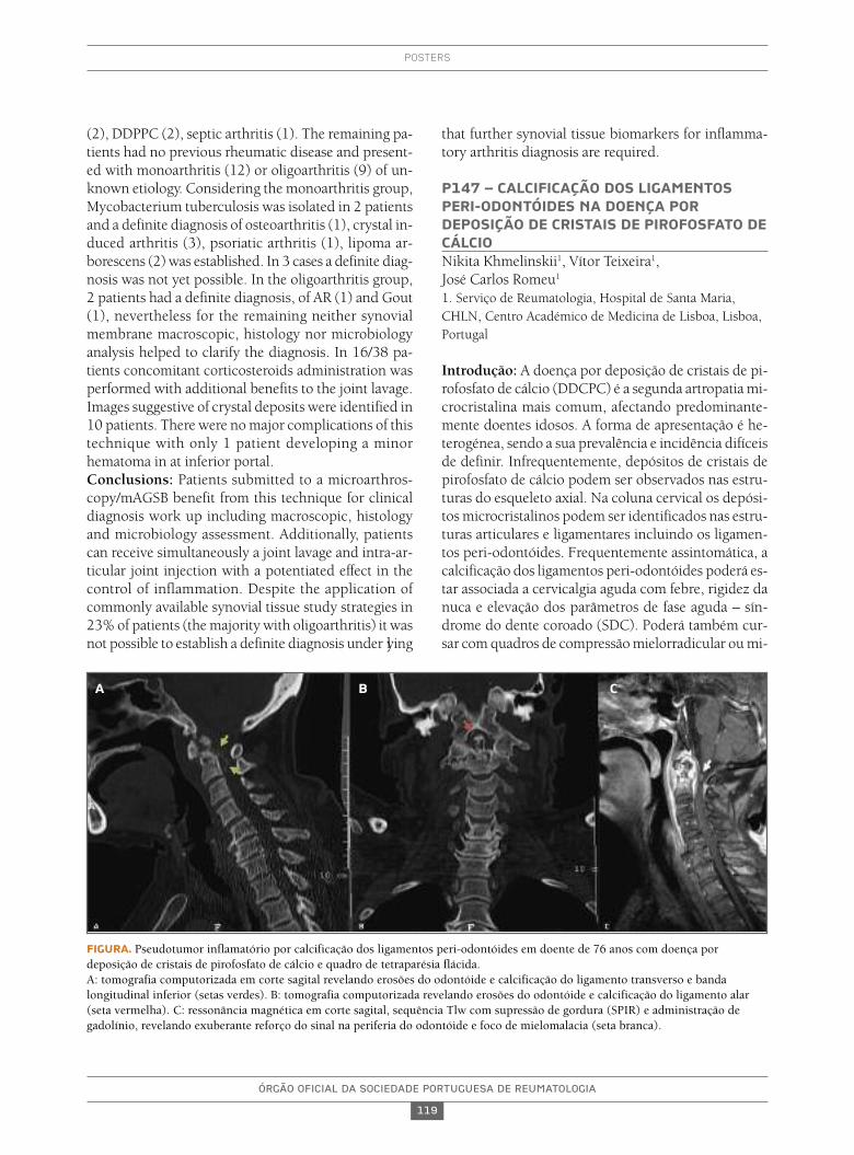
ÓRGÃO OfICIAL DA SOCIEDADE PORTUGUESA DE REUMATOLOGIA
119
posters
(2), DDPPC (2), septic arthritis (1). The remaining pa-tients had no previous rheumatic disease and present-ed with monoarthritis (12) or oligoarthritis (9) of un-known etiology. Considering the monoarthritis group,Mycobacterium tuberculosis was isolated in 2 patientsand a definite diagnosis of osteoarthritis (1), crystal in-duced arthritis (3), psoriatic arthritis (1), lipoma ar-borescens (2) was established. In 3 cases a definite diag -nosis was not yet possible. In the oligoarthritis group,2 patients had a definite diagnosis, of AR (1) and Gout(1), nevertheless for the remaining neither synovialmembrane macroscopic, histology nor microbiologyanalysis helped to clarify the diagnosis. In 16/38 pa-tients concomitant corticosteroids administration wasperformed with additional benefits to the joint lavage.Images suggestive of crystal deposits were identified in10 patients. There were no major complications of thistechnique with only 1 patient developing a minorhematoma in at inferior portal.Conclusions: Patients submitted to a microarthros -copy/mAGSB benefit from this technique for clinicaldiagnosis work up including macroscopic, histologyand microbiology assessment. Additionally, patientscan receive simultaneously a joint lavage and intra-ar-ticular joint injection with a potentiated effect in thecontrol of inflammation. Despite the application ofcommonly available synovial tissue study strategies in23% of patients (the majority with oligoarthritis) it wasnot possible to establish a definite diagnosis under lying
that further synovial tissue biomarkers for inflamma-tory arthritis diagnosis are required.
p147 – CALCIFICAÇÃO DOS LIGAMENTOSpERI-ODONTÓIDES NA DOENÇA pORDEpOSIÇÃO DE CRISTAIS DE pIROFOSFATO DECÁLCIONikita Khmelinskii1, Vítor Teixeira1, José Carlos Romeu1
1. Serviço de Reumatologia, Hospital de Santa Maria,CHLN, Centro Académico de Medicina de Lisboa, Lisboa,Portugal
Introdução: A doença por deposição de cristais de pi-rofosfato de cálcio (DDCPC) é a segunda artropatia mi-crocristalina mais comum, afectando predominante-mente doentes idosos. A forma de apresentação é he-terogénea, sendo a sua prevalência e incidência difíceisde definir. Infrequentemente, depósitos de cristais depirofosfato de cálcio podem ser observados nas estru-turas do esqueleto axial. Na coluna cervical os depósi-tos microcristalinos podem ser identificados nas estru-turas articulares e ligamentares incluindo os ligamen-tos peri-odontóides. Frequentemente assintomática, acalcificação dos ligamentos peri-odontóides poderá es-tar associada a cervicalgia aguda com febre, rigidez danuca e elevação dos parâmetros de fase aguda – sín-drome do dente coroado (SDC). Poderá também cur-sar com quadros de compressão mielorradicular ou mi-
FIGuRA. Pseudotumor inflamatório por calcificação dos ligamentos peri-odontóides em doente de 76 anos com doença pordeposição de cristais de pirofosfato de cálcio e quadro de tetraparésia flácida. A: tomografia computorizada em corte sagital revelando erosões do odontóide e calcificação do ligamento transverso e bandalongitudinal inferior (setas verdes). B: tomografia computorizada revelando erosões do odontóide e calcificação do ligamento alar(seta vermelha). C: ressonância magnética em corte sagital, sequência Tlw com supressão de gordura (SPIR) e administração degadolínio, revelando exuberante reforço do sinal na periferia do odontóide e foco de mielomalacia (seta branca).
A B C

ÓRGÃO OfICIAL DA SOCIEDADE PORTUGUESA DE REUMATOLOGIA
120
posters
metizando outras síndromes (meningismo, tipo-poli-mialgia reumática / arterite de células gigantes, síndro-me febril indeterminada).Objectivos: Caracterizar os doentes com DDCPC comcalcificação dos ligamentos peri-odontóides.Métodos: Análise descritiva, de corte transversal, dosdados demográficos, clínicos, laboratoriais, imagioló-gicos, e de terapêutica, de doentes não sequenciais,cumprindo critérios EULAR para DDCPC definitiva ouprovável, identificados pelo médico reumatologista as-sistente como tendo calcificação dos ligamentos pe -riodontóides em tomografia computorizada (TC) dacoluna cervical ou crânio-encefálica.Resultados: Foram identificados 19 doentes, com mé-dia de idades de 77±9 anos, dos quais 74% eram mu-lheres. As formas de apresentação inicial foram cervi-calgia (n=8), SDC (n=2), tipo-polimialgia reumática(n=1), tetraparesia flácida (n=1), e achado imagiológi-co (n=7). Artrite periférica e febre foram objectivadosem 79% e 37% dos doentes, respectivamente. Verifi-caram-se elevação da velocidade de sedimentação e daproteína C reactiva, tal como condrocalcinose de outrasregiões anatómicas em todos os doentes. As calcifica-ções dos ligamentos peri-odontóides foram mais fre-quentemente identificadas no ligamento transverso(n=17). O estudo por TC e ressonância magnética dadoente com tetraparesia flácida revelou um pseudotu-mor inflamatório e erosões do odontóide com sublu-xação atlantoaxial vertical (imagem). Nenhum dosdoentes foi submetida a punção lombar, objectivando--se rápida resolução da sintomatologia com terapêuti-ca farmacológica com colchicina e/ou anti-inflamatórionão esteróide e/ou prednisolona.Conclusão: O reumatologista deverá considerar as sín-dromes associadas a calcificação dos ligamentos perio -dontóides no diagnóstico diferencial de cervicalgia agu-da com síndrome inflamatório sistémico no doente ido-so. O seu diagnóstico atempado pode evitar a realiza-ção de exames complementares e terapêuticasdesnecessárias.
p199 – IMpACT OF DIFFERENTFORMuLATIONS OF “pATIENT GLOBALASSESSMENT” ON REMISSIONCLASSIFICATION BY DISEASE ACTIVITYINDICES IN RHEuMATOID ARTHRITISGisela Eugénio1, Ricardo J. O. Ferreira1, 2, Cristiana Silva3, Catarina Medeiros3, JAP da Silva1, 3,Cátia Duarte1, 3
1. Rheumatology Department, Centro Hospitalar e
Universitário de Coimbra, Coimbra, Portugal2. Health Sciences Research Unit: Nursing (UICiSA:E),Coimbra, Portugal3. Faculdade de Medicina, Universidade de Coimbra,Coimbra, Portugal
Background: Patient global assessment (PGA) of di -sease activity is included in a large number of com-posite indices of disease activity and definitions of re-mission in Rheumatoid Arthritis (RA). However, theactual question is formulated in a variety of differentways according to the instrument considered.Objectives: To evaluate how 6 different formulationsof PGA affect patient estimates and impact upon diseaseactivity and remission rates as assessed by 4 DiseaseActivity Indices.Methods: Consecutive RA patients followed in aRheumatology outpatient department were includedin this cross-sectional study. Data collection comprised:28 joint counts (tender and swollen), C-reactive pro-tein (CRP) and six different PGA formulations. Thechosen formulations were the ones stated in the: v1)Portuguese National Registry Reuma.pt, the locallyused formulation; v2) ACR/EULAR provisional defini-tion of remission (considered in this study as the “stan-dard”); v3) CDAI and SDAI; v4) Disease Activity Score(DAS28) assessment of general health; v5) DAS28 as-sessment of disease activity (the currently used); v6)one, exploratory, developed by the investigators, in-cluding idiomatic cultural expressions. ACR/EULARBoolean criteria, CDAI, SDAI, and DAS28-CRP(4v)were used to test how these 6 PGA formulations changethe rates of remission. PGA differences were assessed bydescriptive analyses (including patients with PGA ≤10and ≤20mm) and Bland-Altman test.Results: In total, 193 patients were included (82% fe-male, mean (SD) age of 59 (13) years, mean disease du-ration of 12 (9) years and 31% under biologics). Theaverage PGA ranged from 42.3 (25.3) to 48.1(26.7)mm as measured in different formulations. TheACR/EULAR (v2) formulation yielded the largest pro-portion of patients scoring ≤10mm (16.1%), corres -ponding to a difference of up to 4.7 % versus otherPGAs. Similar results were found for the ≤20 cut-off(Table 1).
By using different PGA’s formulations the rates of re-mission calculated with different indices can vary be-tween 4.7% and 6.7% (Table 2).
Bland-Altman chart confirmed the low agreementbetween ACR/EULAR formulation and the other PGA

ÓRGÃO OfICIAL DA SOCIEDADE PORTUGUESA DE REUMATOLOGIA
121
posters
formulations (p<.05), except for the DAS “generalhealth” (p= .054) (Figure 1).Conclusions: The use of different PGA formulationshas clinical implications in disease activity, which, inturn, can influence therapeutic decisions. The esta -blishment of a single standardised formulation seemswarranted.
p19 – IS IMMuNOHISTOCHEMISTRY uSEFuLTO pREDICT RESpONSE TO TREATMENT INNECROTIZING MYOpATHIES?Susana Fernandes1, Begonã Lopez2, Patrick Gordon2,Safa Al-Sarraj3
1. Rheumatology Department, Instituto Português deReumatologia, Lisboa, Portugal2. Rheumatology, Kings College Hospital, London, UnitedKingdom3. Clinical Neuropahology, Kings College Hospital,London, United Kingdom
Background: Muscle biopsy is the gold standard forthe diagnosis of inflammatory myopathies, but the roleof immunohistochemistry in Necrotizing Myopathies(NM) has not been fully characterized yet.Objectives: To determine if MHC-I expression and pat-tern of C5b-9 deposition in capillaries correlate withclinical phenotype and response to treatment in NM.Methods: The Neuropathology Departmental databasewas searched to identify patients with a histologicaldiag nosis of NM and follow up data for at least 6months (30 patients). Electronic patient records werereviewed retrospectively to record demographics, au-toantibodies, treatment, proximal muscle power at 3,6 and 12 months by Manual Muscle Testing (MMT)(2), levels of CK and flares. Patients were classified asresponders when there was improvement of MMT≥20% and non-responders when MMT improvementwas <20% (3). All biopsies were reviewed blindly by anexperienced neuropathologist. MHC-I expression wasclassified as positive only if over expressed in all fibers.The patterns of C5b-9 deposition in endomysial capil-laries were classified as specific (solid), non-specific(granular) or negative.Results: MHC-I positive group (n=16/30) had a hi gherproportion of responders (62,5% vs 7,7%, p=0.002),higher number of patients with total recovery of mus-
,00
Dif
ACR
/EU
LAR
- D
AS2
8 GH
100,00
50,00
,00
-50,00
-100,00
20,00 40,00
Mean ACR/EULAR - DAS28
60,00 80,00 100,00
FIGuRE 1. Bland-Altman plot for agreement betweenACR/EULAR and DAS28 GH formulations. The middle linerepresents the mean of the difference, and the top and bottomlines demarcate 95% limits of agreement between the twoformulations
TABLE 1. IMpACT OF DIFFERENT FORMuLATIONS OF pGA ON pATIENTS' ANSwERS AND ON REMISSION RATESACCORDING TO FOuR COMpOSITE INDICES (N=193)
Descriptive statistics Remission rates, n (%)PGA below PGA below
PGA ≤10 mm ≤20mm ACR/EULAR DAS28Formulation Mean (SD) n (%) n (%) Boolean SDAI CDAI 4vCRPv1 Reuma.pt 47.5 (28.0) 26 (13.5) 43 (22.3) 19 (9.8) 32 (16.8) 33 (17.1) 97 (50.5)v2 ACR/EULAR 43.5 (27.9) 31 (16.1) 48 (24.9) 25 (13.0) 36 (19.1) 39 (20.0) 100 (51.7)v3 CDAI/SDAI 47.2 (25.9) 23 (11.9) 34 (17.6) 16 (8.3) 27 (14.1) 26 (13.5) 91 (47.3)v4 DAS28-GH 42.9 (25.3) 27 (14.0) 42 (21.8) 20 (10.4) 29 (15.2) 33 (17.1) 99 (51.4)v5 DAS28-DA 42.3 (25.3) 28 (14.5) 44 (22.8) 19 (9.8) 29 (15.2) 30 (15.5) 100 (51.7)v6 Investigators 48.1 (26.7) 22 (11.4) 35 (18.1) 17 (8.8) 27 (14.1) 27 (14.0) 97 (50.0)
Maximum difference within definition, n (%):9 (4.7) 9 (4.7) 13 (6.7) 9 (4.7)

ÓRGÃO OfICIAL DA SOCIEDADE PORTUGUESA DE REUMATOLOGIA
122
posters
cle power (66,7 vs. 15,4%) and were more commonlypositive for autoantibodies (75% vs. 35.7%, p=0.030)when compared to the MHC-I negative group (n=14).17 patients were positive for auto-antibodies of which9 were myositis specific antibodies [SRP (n=6), HMG--CoA reductase (n=1), Jo-1 (n=1), P155/140 (n=1)] and4 were myositis associated antibodies [Ro-52 (n=2), Ku(n=1), Pm/Scl (n=1)]. 13/30 patients had C5b-9 depo-sition, with a specific pattern in 5 and non-specific in8. The specific pattern group had a greater reductionof CK after 6 months compared to non-specific and neg-ative respectively (98% vs. 77% vs. 56.8%, p=0.006),greater reduction in CK after 12 months (96.6% vs.68.9% vs. 59.6%, p=0.024) and higher rates of respon-ders (80% vs. 60% vs. 18,8%, p=0.001). Six patientswere on immunosuppressants (azathioprine/hydroxy-chloroquine, n=2), steroids (n=3) or both (n=1) for aminimum of 4 weeks when the bio psy was performed.Differences in age, gender, clinical features or treatmentwere not found to be statistically significant.Conclusions: Upregulation of MHC I and solid stai -ning pattern of C5b-9 in the capillaries of NM patientsappears to be associated with a better outcome.
REFERENCES1. Cong L, Pu CQ, Shi Q, Wang Q, Lu X, Complement membra-
ne attack complex is related with immune-mediated necroti-zing myopathy, Int J Clin Exp Pathol 2014;7(7):4143-4149
2. Rider LG, Koziol D, Giannini EH, Jain MS, Smith MR, Whitney-Mahoney K, Feldman BM, Wright SJ, Lindsley CB, PachmanLM, Villalba ML, Lovell DJ, Bowyer SL, Plotz PH, Miller FW,Hicks JE, Validation of manual muscle testing and a subset ofeight muscles for adult and juvenile idiopathic inflammatorymyopathies, Arthritis Care Res (Hoboken), 2010 Apr62(4):465-72.
3. Vencovsky J, New ACR/EULAR Criteria for Myositis, AnnRheum Dis 2015;74:42-43 doi:10.1136/annrheumdis-2015-eular.6595
p176 – REDuÇÃO DA DOSE DASTERApÊuTICAS ANTI-TNFA EM DOENTESCOM ARTRITE REuMATÓIDE: ESTuDOTRANSVERSAL NA pRÁTICA CLÍNICA.Tiago Meirinhos1, Moya P.2, Diaz-Torné C.2, Castellvi I.2, Milena-Millan A.2, Magallares B.2, Laiz A2, Sang-Park H2, Casademont J.2
1. Rheumatology Department, Hospital Infante D. Pedro(CHBV), Aveiro, Portugal2. Reumatologia, Hospital de La Santa Creu I Sant Pau,Barcelona, Spain
Introdução: A artrite reumatóide (AR) é uma doençareumática inflamatória crónica e sistémica, que quan-
do não tratada de forma adequada pode progredir ori-ginando dano estrutural com limitação funcional con-sequente. As terapêuticas biológicas constituíram umdos avanços mais importantes das últimas décadas notratamento das artropatias crónicas como a AR; com asua introdução, muitos doentes refratários a terapêuti-cas convencionais (DMARD�s clássicos) responderamde forma adequada e entraram em remissão. No en-tanto, na prática clinica não é ainda consensual a ati-tude a tomar em doentes em remissão sustentada. Al-guns estudos observacionais sugerem a possibilidadede reduzir a dose dos tratamentos biológicos em doen-tes em remissão ou baixa atividade sustentada até adose mínima eficaz, maximizando a sua eficácia e mi-nimizando os efeitos laterais.Objetivos: Identificação de variáveis clínicas e analíti-cas associadas à redução da dose das terapêuticas anti--TNFa subcutâneas e sua manutenção sem recaídanuma coorte de doentes com artrite reumatóide.Materiais e métodos: Estudo transversal. Foram in-cluídos doentes com AR sob terapêuticas anti-TNFasubcutâneas (Etanercept, Adalimumab, Golimumab eCertolizumab pegol), seguidos no Serviço de Reuma-tologia do Hospital de La Santa Creu I Sant Pau (Bar-celona, Espanha) e com capacidade para fornecer oconsentimento informado para o estudo.Variáveis analisadas: dados clínicos e demográficosgerais, tempo de seguimento, atividade da doença ava-liada através do DAS28 3v, terapêuticas atuais e passa-das para a AR, efeitos laterais a terapêuticas prévias.Resultados: Foram incluídos 63 doentes com AR sobterapêuticas anti-TNFa subcutâneas; a maioria dosdoentes era do sexo feminino (52 doentes - 82,5%) e amédia de idades foi de 59,4±13,3 anos.
Identificámos 25 doentes (39,7%) que tinham sidosujeitos a uma redução da dose do tratamento biológi-co, em média 25,5 meses após o início do mesmo. Ve-rificámos que neste grupo de doentes a idade média foisuperior (p = 0,024), assim como a percentagem deDMARD�s convencionais no início do tratamento bio-lógico; o uso de glucocorticóides foi inferior. No gru-po de doentes em que foi necessário voltar a aumentara dose do anti-TNFa verificámos que a percentagem depositividade para o FR e anti-CCP era inferior em rela-ção aos doentes que mantiveram doses mais baixas.
Em relação aos efeitos laterais das terapêuticas, nãoforam verificadas diferenças entre os doentes que di-minuíram dose e os que não diminuíram.Conclusão: A redução da dose dos medicamentos bio-lógicos anti-TNFa subcutâneos é comum nesta coorte

ÓRGÃO OfICIAL DA SOCIEDADE PORTUGUESA DE REUMATOLOGIA
123
posters
de doentes com AR, tendo sido realizada em pratica-mente 40% dos doentes. Uma idade superior, assimcomo o uso concomitante de DMARD�s clássicos noinício do tratamento biológico parecem ser fatores quese associam a uma maior taxa de sucesso na redução emanutenção da dose dos medicamentos biológicosanti-TNFa subcutâneos analisados.
p10 – EARLY ARTHRITIS INDuCESDISTuRBANCES AT BONE NANOSTRuCTuRALLEVEL REFLECTED IN DECREASED TISSuEHARDNESS.Vidal B1, Rita Cascão1, Finnila M2, 3, Lopes IP1, Saarakkala S2, 4, 5, P. Zioupos6, Helena Canhão7, JE Fonseca1, 8
1. Rheumatology Research Unit, Instituto de MedicinaMolecular, Lisboa, Portugal2. Research Unit of Medical Imaging, Physics andTechnology, Faculty of Medicine, University of Oulu,Oulu, Finland, Oulu, Finland3. Department of Applied Physics, University of EasternFinland, Kuopio, Finland4. Medical Research Center, University of Oulu, Oulu,Finland, Oulu, Finland5. Oulu University Hospital, Oulu, Finland, Oulu, Finland6. Biomechanics Labs, Cranfield Forensic Institute,Cranfield University, DA of the UK, Swindon, UnitedKingdom7. EpiDoC Unit, CEDOC, NOVA Medical School, NOVAUniversity, Lisboa, Portugal8. Rheumatology Department, Hospital de Santa Maria(CHLN), Lisbon Medical and Academic Centre, Lisboa,Portugal
Introduction: Arthritis induces joint erosions andskeletal bone fragility. Objectives: The main goal of this work was to analyzethe early arthritis induced events at bone tissue level.Methods: Eighty-eight Wistar rats were randomlyhoused in experimental groups, as follows: adjuvant in-duced arthritis (N= 47) and a control healthy group (N=41). Rats were monitored during 22 days for the in-flammatory score, ankle perimeter and body weight andsacrificed at different time points (11 and 22 days postdisease induction). Bone samples were collected for his-tology, micro-CT, 3-point bending, nanoindentationand Fourier transformed infrared spectroscopy (FTIR)analysis. Blood samples were also collected for boneturnover markers and systemic cytokine quantification. Results: At bone tissue level, measured by FTIR ana -
lysis and nanoindentation, there was a reduction of themineral and collagen content and of hardness in thearthritic group, associated with an increase of the ratioof bone concentric to parallel lamellae and of the areaof the osteocyte lacuna. In addition, increased boneturnover and changes in the microstructure and me-chanical properties were observed in arthritic animals,since the early phase of arthritis, when compared withhealthy controls. Conclusion: Arthritis induces veryearly changes at bone tissue level characterized by de-creased tissue hardness and of collagen and mineralcontent. These observations highlight the pertinenceof immediate control of inflammation in the initialstages of arthritis.
GRupO 10
p78 – TRANSLATION AND CROSS-CuLTuRALADApTATION INTO EuROpEAN pORTuGuESELANGuAGE OF THE COpING INSTRuMENT –CORS (OMGAAN MET REuMATOÏDE ARTRITIS)Carina Lopes1, Cruz, E2, Sofia Ramiro3, 4, Alexandre Sepriano1, 3, 4, João Lagoas Gomes1, 4, Tiago Costa1, José Marona1, Margarida Mateus1, Jaime C. Branco1, 4, Fernando Pimentel-Santos1, 4
1. Rheumatology Department, Hospital Egas Moniz(CHLO), Lisboa, Portugal2. Fisioterapia, Escola Superior de Saúde - InstitutoPolitécnico de Setúbal, Setúbal, Portugal3. Rheumatology, Leiden University Medical Center,Leiden, Netherlands4. CEDOC, NOVA Medical School. Faculdade de CiênciasMédicas da Universidade NOVA de Lisboa., Lisboa,Portugal
Background: Inflammatory rheumatic diseases (IRD)are common conditions characterized by periods ofexa cerbations and remissions. The main symptom ofIRD is pain. Patient education programs have becomea complement to traditional medical treatment andhave given people strategies to cope with pain. Om-gaan met Reumatoïde Artritis (CORS), developed inthe Netherlands, evaluates different strategies to copewith pain in patients with Rheumatoid Arthritis (RA).Objective: The aim of this study was to perform thetranslation and cultural adaptation of the CORS ques-tionnaire into European Portuguese; test the conceptualequivalence (clarity, comprehension, cultural relevanceand appropriateness of the items and words used) of

ÓRGÃO OfICIAL DA SOCIEDADE PORTUGUESA DE REUMATOLOGIA
124
posters
the translated version in the Portuguese context.Material and methods: The CORS was firstly trans-lated into European Portuguese and then back-trans-lated into Dutch, following the international guidelinesfor this procedure. After the review of the EuropeanPortuguese version by an expert committee, the pre-fi-nal version of the questionnaire was evaluated by co -gnitive debriefing interviews with an heterogeneoussample of patients with IRD regarding their gender, age,disease duration, and educational background.Results: Translation into Portuguese and then backtranslation into Dutch were well succeeded and ac-cepted by the authors before the cognitive debriefing.A total of 9 patients with IRD [5 female, median [IQR]age 55 [39-62,5] years] participated in the field test.Median of Education level [IQR] was 12 [9-15] years;at the time of the questionnaire 3 were retired, 2 onsick leave, 1 unemployed and 3 active workers. Medi-an [IQR] time to respond the questionnaire was 9:30[7:30, 10:45] minutes. All the participants read the ins -tructions before the questionnaire and verified whetherall issues were met. In general, all questions were equalin terms of time consumption. Cognitive debriefingshowed that all the items were clear, relevant, under-standable, easy to complete and culturally appropria -te. As a result of the cognitive debriefing, minorchanges, like the introduction of a response option “notapplicable”, were adopted for some questions to im-prove their appropriateness.Conclusions: This study presents the first stage of thecultural adaptation process. Before CORS-PT can befully implemented into the clinical and research set-tings in Portugal, its reliability, construct validity andresponsiveness need to be evaluated in patients withinflammatory rheumatic diseases. The CORS may havepotential to measure coping with the disease not onlyin rheumatoid arthritis, but in IRD in general.
p13 – ASSOCIATION OF VITAMIN D STATuSwITH RHEuMATOID ARTHRITIS DISEASEACTIVITY AND uV INDExCarina Lopes1, João Lagoas Gomes1, 2, Tiago Costa1,Teresa Pedrosa1, José Marona1, Margarida Mateus1,Fernando Pimentel-Santos1, 2, Jaime C. Branco1, 2
1. Rheumatology Department, Hospital Egas Moniz(CHLO), Lisboa, Portugal2. EpiDoC Unit, CEDOC, NOVA Medical School, NOVAUniversity, Lisboa, Portugal
Background: Lower serum vitamin D levels have been
shown to be associated with various autoimmune di -sorders, including Rheumatoid Arthritis (RA). VitaminD deficiency is common in RA patients, despite profi -ting from a sunny country.Objectives: The aim of the study is to evaluate (1) -the association between vitamin D serum levels anddisease activity in patients with RA; (2) – seasonal dis-tribution of vitamin D levels.Methods: Patients fulfilling the 2010 EULAR/ACRRheumatoid Arthritis Classification Criteria, which hadserum vitamin D [25(OH)D3] levels measured betweenJanuary 2013 and December of 2016 were included.Demographic data, disease activity scores, includingDAS283v-CRP and DAS283v-ESR, vitamin D supple-mentation with cholecalciferol and other therapeuticapproaches were recorded. Vitamin D insufficiency wasconsidered between 25-75 nmol/L and deficiency if<25 nmol/L. According to the national agency for thestudy of sea and atmosphere, UV Index levels weregrouped into low UV Index 3-6 (October to April) andhigh UV Index 9-10 (May to September). Correlationbetween variables was analyzed using Spearman�s rho.Results: A sample composed by 95 patients, 79 fe-males (83.16%), with an average age (SD) of 68.57(11.92) years within 40-88 years range were included.Average disease duration was 13.46 (11.41) years. Theaverage age at diagnosis was 57.10 (14.25) years. Theaverage vitamin D levels were 78.13 (60.98) nmol/L in
,0
Vit
amin
D le
vels
(n
mol
/L)
400
300
200
100
02,0
DAS283v-CRP4,0 6,0 8,0
y=1,08E2-10,35*x
FIGuRE 1. Bland-Altman plot for agreement betweenACR/EULAR and DAS28 GH formulations. The middle linerepresents the mean of the difference, and the top and bottomlines demarcate 95% limits of agreement between the twoformulations

ÓRGÃO OfICIAL DA SOCIEDADE PORTUGUESA DE REUMATOLOGIA
125
posters
a range between 20-400 nmol/L. Vitamin D levels werenot significantly different in male vs. females patients.The prevalence of vitamin D insufficiency and defi-ciency was 53.68% and 8.42% respectively, despite57.89% of the patients taking supplementation (ave -rage 6141 (4800) UI/week). The univariable analysisshowed that albeit vitamin D levels presented a nega-tive poor correlation with DAS283v-CRP (rho=-0.348,p-value<0.001) and DAS283v-ESR (rho=-0.271, p-va -lue<0.01), there was a direct reduction in dispersion ofthe vitamin D values for increasing values of DAS283v--CRP and ESR. It was observed that vitamin D levels in-crease with patient age and decrease with disease du-ration. Seasonality and supplementation didn’t affectvitamin D levels in our population.Conclusions: Vitamin D insufficiency/deficiency wasfrequent among RA patients (62.1%), independentlyof seasonality or supplementation. An interesting pat-tern behavior was observed in this study, which indi-cates that the likelihood of encountering a very nar-rowband of vitamin D values for patients with high di -sease activity is very high, and thus, the forecast capa-bility of vitamin D values for patients with increasinglyactive disease is quite good. Future research will aim atstrengthening the statistical parameters of relevance,identifying and characterizing the more specific be-haviors of this global pattern.Disclosure of Interest: None declared
p208 – AS VÁRIAS FACES DA SÍNDROMESICCAFilipe Barcelos1, 2, 3, José Vaz Patto1, Catarina Martins3,Joana Cardigos4, Nuno Alves4, João Amaral5, L Miguel Borrego2, 3, Jaime C. Branco2, 3, 6
1. Instituto Português de Reumatologia, Lisboa, Portugal2. Hospital CUF Descobertas, Lisboa, Portugal3. CEDOC, Nova Medical School, Lisboa, Portugal4. Oftalmologia, Hospital Santo António dos Capuchos(CHLC), Lisboa, Portugal5. Faculdade de Medicina Dentária, Universidade deLisboa, Lisboa, Portugal6. Rheumatology Department, Hospital Egas Moniz(CHLO), Lisboa, Portugal
Introdução: A sindrome sicca é referida pelos doentesseguidos na consulta de Síndrome de Sjögren (SSP),independentemente da identificação dos critérios declassificação para SSP. Entre os doentes com Síndromesicca não-Sjögren, aqueles com positividade isoladapara anticorpos anti-nucleares e manifestações de
doença reumática imunomediada, como artrite/artral-gias, fenómeno de Raynaud, fotossensibilidade ou leu-copenia, são habitualmente diagnosticados como co-netivite indiferenciada1 (UCTD). Admite-se que algunsdestes doentes possam evoluir para SSP, enquanto ou-tros manterão o quadro clínico estacionário.Objetivos: Pretendeu-se avaliar as caraterísticas clíni-cas e laboratoriais de uma população seguida numaconsulta de SSP/Síndrome sicca.Métodos: Incluíram-se 119 doentes, dos quais 54 comSSP (critérios AECG) - 22 com >10 anos de diagnósti-co (SSP>10 anos) e 32 com <2 anos de diagnóstico(SSP<2 anos), e 65 doentes com sindrome sicca não--Sjogren - 35 com UCTD (sicca-UCTD) e 30 semUCTD(sicca-nUCTD). Recolheram-se dados demográ-ficos, manifestações clínicas e laboratoriais. Realizou--se sialometria, teste de Schirmer I, teste de quebra la-crimal (BUT, “break-up time”) e avaliação de queratite.Resultados: Os doentes com SSP>10 anos apresenta-vam idade de diagnóstico inferior, menor fluxo salivar,e maior frequência de queratite e redução do BUT, emrelação aos doentes com SSP recente (Quadro 1). A pre-sença de SSA, ANA e FR foi mais prevalente nos SSPevoluídos do que nos precoces. Doentes com SSP>10anos apresentavam também maior ocorrência de ma-nifestações articulares, hematológicas, fenómeno deRaynaud e outras manifestações sistémicas, quandocomparados com doentes com SSP<2 anos.
Ao analisar os doentes sem critérios de SSP, verifi-cou-se que o grupo sicca-UCTD apresentava maior pre-valência de manifestações articulares, cutâneas, hema-tológicas e fenómeno de Raynaud, em comparação como grupo sicca-nUCTD. O estudo funcional não apre-sentava diferenças significativas entre ambos os gru-pos, excepto maior frequência de queratite no gruposicca-nUCTD.Discussão: Nos doentes com SSP, o perfil clínico maisgrave pertencia sobretudo ao grupo SSP>10 anos. Estefato pode dever-se quer ao aparecimento de novas ma-nifestações, relacionadas com a evolução da doença,quer à maior retenção na consulta dos doentes gravese ao abandono gradual do seguimento de doentes li-geiros.
Em relação à caraterização funcional salivar e lacri-mal, era esperado o acentuado compromisso no grupoSSP>10 anos, quando comparado com os restantes gru-pos, que pouco diferem entre si.
Em relação aos doentes com sindrome sicca não-Sjö-gren, a utilização do ANA ≥1/320 como indicador deconetivite traduz-se na divisão desta população em 2

ÓRGÃO OfICIAL DA SOCIEDADE PORTUGUESA DE REUMATOLOGIA
126
posters
grupos: o grupo UCTD e o grupo não-UCTD. O pri-meiro apresentava maior ocorrência de manifestaçõesarticulares, hematológicas, cutâneas e fenómeno deRaynaud. Inclusivé, o grupo sicca-UCTD apresentavamaior ocorrência de artrite/artralgias e fenómeno deRaynaud que o grupo SSP<2 anos, embora com menorocorrência de alterações hematológicas.Conclusões: Parece ser evidente que a aplicação doscritérios atuais de classificação da PSS na prática clíni-ca não é satisfatória, pois excluem muitos doentes commanifestações clínicas relevantes, dos quais alguns te-rão provavelmente SSP. É necessário identificar novosbiomarcadores para auxílio no diagnóstico e decisãoterapêutica de doentes com Síndrome seca, dado quea dicotomia Sjögren versus não-Sjögren, promovida pe-los critérios atuais, parece refletir a divisão artificial deum contínuum clínico e imunológico.
p87 – NEuROpATHIC pAIN: ASSOCIATIONwITH SKIN THICKNESS IN SYSTEMICSCLEROSIS pATIENTSJoana Sousa-Neves1, Marcos Cerqueira1, Daniela Santos-Faria1, Joana Leite Silva1, Carmo Afonso1, Filipa Teixeira1
1. Rheumatology Department, Hospital Conde deBertiandos (ULSAM), Ponte de Lima, Portugal
Background: Systemic Sclerosis (SSc) is a connectivetissue disorder characterized by fibrosis of skin and in-ternal organs, vasculopathy and systemic inflamma-tion. To date, few studies assessed pain in SSc pa-tients,(1) namely the neuropathic component. Data isalso scarce on defining possible neuropathic pain (NP)predictors in these patients.Objectives: To determine if patients with SSc have ahigher prevalence of NP compared with a group of age
and sex matched controls and study possible associa-tions between NP and SSc clinical variables.Methods: The study evaluated patients diagnosed withSSc fulfilling the SSc classification criteria of the Ame -rican College of Rheumatology (ACR), older than 18years old and followed up at our Rheumatology Unit.Forty-eight patients with mean age of 56.98 ± 12.73years and mean disease duration of 9.77 ± 6.12 yearswere included. Comparison group comprised 45 ageand sex matched controls. Patients and controls wereconsecutively evaluated at our Unit and NP in 4 ques-tions questionnaire (DN4) was used for assessing thepresence of NP. In SSc patients, skin involvement wasalso evaluated clinically by the modified Rodnan skinthickness score (mRSS) ranging from 0 to 51. Handmobility (HAMIS) and SSc Severity Scale (SScSS) werealso calculated and relevant clinical variables of the di -sease were collected. Parametric and nonparametrictests were used for comparative analysis. Multivariatelogistic regression was used to identify factors associa -ted to NP. Statistical significance was defined as P < 0.05.Results: In our study, prevalence of NP assessed byDN4 was significantly higher in SSc patients compa -rative to controls (56.2% versus 13.3%, p<0.001). Inaddition to age and sex, presence of diabetes (p=0.541)was also similar in both groups. In SSc group, patientswith and without NP showed some statistically signi -ficant differences (table 1). Multivariate logistic regres-sion revealed that only mean mRSS (odds ratio [OR] =1.90, 95% confidence interval [CI] 1.01 to 3.55,p=0.045) was independently associated with the pre -sence of NP.Conclusions: To the best of our knowledge, this is thefirst study evaluating the prevalence of NP in SSc pa-tients in comparison with age and sex matched con-
TABLE. COMpARATIVE ANALYSIS BETwEEN SSC pATIENTS wITH AND wITHOuT Np
NP positive patients (n=27) No-NP patients (n=21) P valueMedian age, years (IQR) 61 (57-69) 52.38 (42.5-63.5) 0.032Mean disease duration (years) 11.36 ± 6.55 7.73 ± 4.93 0.04Diffuse/limited cutaneous subtype (n) 6/21 0/21 0.024Mean mRSS 22.81 ± 8.74 7.57 ± 3.64 <0.001Mean HAMIS 10.44 ± 6.16 1.52 ± 2.29 <0.001Mean SScSS 10.00 ± 4.32 3.90 ± 2.07 <0.001Digital ulcers n (%) 17 (63.0) 6 (28.6) 0.018Calcinosis n (%) 15 (55.5) 6 (28.6) 0.062

ÓRGÃO OfICIAL DA SOCIEDADE PORTUGUESA DE REUMATOLOGIA
127
posters
trols. NP was significantly more prevalent in patientswith SSc. Skin thickness assessed by mRSS was inde-pendently associated with the presence of NP.
REFERENCES 1. Schiei O et al. Prevalence, severity, and clinical correlates of pain
in patients with systemic sclerosis. Arthritis Care Res (Hobo-ken). 2010 Mar; 62(3):409-17.
p118 – SÍNDROME DE SjÖGREN: CASuÍSTICADE uM SERVIÇO DE REuMATOLOGIA EAVALIAÇÃO DA ATIVIDADE DA DOENÇAApLICANDO O ESSDAIJoão Pedro Freitas1, Flávio Campos Costa1, Mary Lucy Marques1, Alexandra Daniel1, Gisela Eugénio1, Diogo Jesus1, Mariana Luis1, Luisa Brites1, João Rovisco1, Ines L1, Armando Malcata1
1. Rheumatology Department, Centro HospitalarUniversitário de Coimbra, Coimbra, Portugal
Introdução: O Síndrome de Sjögren Primário (SSP) éuma doença inflamatória crónica sistémica caracteri-zada por infiltração linfocítica das glândulas exócrinas.As principais manifestações são xerostomia e xeroftal-mia. No entanto, o espectro clínico estende-se desde oenvolvimento das glândulas até ao envolvimento sisté-mico, com prognóstico variável. Tal como noutrasdoenças do tecido conjuntivo também para o SSP fo-ram desenvolvidos instrumentos de avaliação da ativi-dade da doença. Desde 2009 o ESSDAI, um instru-mento validado, composto por 12 domínios, tem sidoutilizado como Gold Standard para avaliação da ativi-dade da doença nesta patologia. Este trabalho tevecomo objetivo analisar as características demográficas,o espectro clinico, a terapêutica e avaliar a atividade dadoença de doentes com SSP, seguidos num Serviço deReumatologia.Material e Métodos: Foram obtidos dados dos pro-cessos clínicos, cartas de alta e registo do reuma.pt dedoentes seguidos no nosso serviço com SSP, segundo oscritérios de classificação do Consenso Americano-Eu-ropeu (AECG) de 2002. A informação recolhida incluiudados demográficos, manifestações clínicas e serológi-cas, terapêutica imunossupressora e atividade da doen-ça, aplicando o ESSDAI.Resultados: Foram incluídos no estudo 70 doentesque cumpriam os critérios AECG de 2002, dos quais 1era do sexo masculino e 69 do sexo feminino. A idademédia foi de 56,49 anos ± 13,20 DP. As manifestaçõessistémicas mais frequentemente observadas foram lin-
fopenia (37,1%), artrite (35,7%), neutropenia (20,0%),anemia (14,3%), trombocitopenia (10,0%), púrpura(4,5%), livedo reticular (4,5%), doença pulmonar in-tersticial (3,0%), nefrite intersticial (3,0%) e hepatiteauto-imune (2,9%).
Como terapêutica imunossupresora, 22 doentes en-contravam-se sob Hidroxicloquina, 17 com Corticoide,3 com Metotrexato, 2 com Micofenolato de Mofetil, 2com Azatioprina e 1 com Dapsona. A maioria dos doen-tes (55,7%) não apresentava atividade da doença (ESSDAI =0), 32,9% apresentava ligeira atividade (ESSDAI=1-4), 10,0% atividade moderada (ESSDAI=5-13)e 1,4% atividade elevada (ESSDAI14).Conclusões: O SSP é uma doença que pode apresen-tar um envolvimento sistémico grave, podendo atingirvariados órgãos e sistemas com prognóstico variável. Aaplicação de scores de gravidade, como o ESSDAI, naprática clinica diária permite, de forma objetiva e uni-formizada, fazer uma avaliação da atividade da doençae facilita o seguimento destes doentes ao fornecer umconjunto de parâmetros de avaliação, contribuindopara uma conformidade nas atitudes e decisões clíni-cas entre médicos.
p197 – uSTEKINuMAB NO TRATAMENTO DA ARTRITE pSORIÁTICA EM DOENTES COM pSORIASE MODERADA A GRAVE.ExpERIÊNCIA DE uM CENTRO.P Pinto1, Miguel Guerra1, Taciana Videira1, Romana Vieira1
1. Rheumatology, Centro Hospitalar de Vila Nova deGaia/Espinho, Vila Nova de Gaia, Portugal
Introdução: A artrite psoriatica é uma doença reumá-tica crónica que se caracteriza pela presença de sinovi-te, dactilite, entesite e espondilite.
O Ustekinumab é um dos fármacos biológicos usa-do no tratamento da Artrite Psoriatica após a sua apro-vação para o tratamento da Psoríase moderada a grave.É um inibidor da IL12/IL 23p40 que suprime a ativa-ção Th1, Th17 e Th22.Métodos: Estudo retrospectivo e observacional cujoobjectivo principal é identificar as manifestações da Ar-trite psoriatica e a evolução desta patologia numa co-hort de doentes com psoriase grave tratados com uste-kinumab, nos últimos 5 anos, num período mínimo de3 meses. Foram avaliadas e comparadas as populaçõescom psoríase e com artrite psoriatica assim como seidentificou a taxa de retenção com este fármaco e as ra-zões para A SUA descontinuação.

ÓRGÃO OfICIAL DA SOCIEDADE PORTUGUESA DE REUMATOLOGIA
128
posters
Resultados: Foram identificados 42 doentes com pso-ríaiase tratados com ustekinumab e 14 doentes com ar-trite psoriática (apenas 8 doentes tinham o diagnósti-co e eram observados em consulta de Reumatologia).A taxa de retenção no fármaco foi de 58% e a principalcausa de descontinuação foi a falta de eficácia cutânea(4 doentes ) e articular ( 2 doentes). A suspensão porefeitos laterais foi reduzida (4 doentes). Um doente comartrite psoríatica que suspendeu por falta de eficácia játinha realizado 3 fármacos anti TNF alfa.Limitações: O número reduzido de doentes com artritepsoriatica e que descontinuaram.Conclusão:O Ustekinumab demonstrou eficácia e se-gurança no tratamento da Artrite psoríatica e psoríasegrave
p72 – TRATAMENTO pRÉVIO COMBIFOSFONATOS: ALTERA O EFEITO DATERIpARATIDE?Raquel Miriam Ferreira1, Rita Fonseca1, Miguel Bernardes1, Sofia Pimenta1, Lúcia Costa1
1. Serviço de Reumatologia, Centro Hospitalar de SãoJoão, Porto, Portugal
Introdução: A teriparatide é utilizada como tratamen-to na osteoporose. Ela é reservada às formas severas.Recentemente, levantam-se questões sobre o efeito douso prévio de bifosfonatos (BF) no metabolismo ósseoem doentes sob teriparatide.Objetivo: Avaliar a influência do tratamento préviocom BF na efetividade de teriparatide.Métodos: Estudo observacional de 68 doentes trata-dos com teriparatide por osteoporose do nosso serviçode Reumatologia. Os doentes foram classificados em 2grupos: A- doentes previamente tratados com BF porperíodo > 6meses e B- doentes sem exposição prévia aBF ou <6 meses de tratamento. Avaliamos os valoresda densidade mineral óssea (DMO) na coluna verte-bral lombar (CL), cólo femural (CF) e fémur total (FT)aos 0 e 18 meses, e os marcadores de remodelação ós-sea aos 0, 3 e 18 meses. Para análise estatística foramutilizados testes paramétricos e não paramétricos (SPSS20,0). O nível de significância foi fixado em 0,05.Resultados: Nós observamos 42 doentes no grupo A,todos do sexo feminino e 26 doentes no grupo B, 81%eram mulheres. As populações eram comparáveisquanto à idade. O bifosfonato mais utilizado foi o alen-dronato (52,4%), seguido de risedronato (23,8%), pa-midronato (11,9%), ibandronato (7,1%) e acido zole-drónico (4,8%). O período médio de tratamento com
BF foi de 40 ± 35 meses. No grupo A, 28,6% sofriamde osteoporose primária e 71,4% tinham osteoporosesecundária. 57,1% dos doentes apresentavam T-score<-3 e fraturas vertebrais e 14,3% um T-score lombar <-4. No grupo B, 53,8% dos doentes tinham osteopo-rose secundária. 42,3% iniciaram teriparatide devido aT-score <-3 e fraturas vertebrais e 50% por T-score lom-bar <-4. No início do estudo, no grupo A a mediana daDMO na CL, CF e FT foi de 0,848, 0,685 e 0,749 g/cm2e no grupo B foi de 0,708, 0,565 e 0,652 g/cm2, res-petivamente. No fim dos 18 meses, constatou-se umaresposta analítica e densitométrica para as três regiõesestatisticamente significativa no grupo B. No grupo A,houve um aumento significativo dos biomarcadores eda DMO somente na CL. Na comparação entre os doisgrupos, verificaram-se diferenças estatisticamente sig-nificativas na variação na DMO (p<0,001, p=0,016,p=0,028) e no ganho percentual (p<0,001, p=0,010,p=0,016) na CL, CF e FT respetivamente, a favor dogrupo B. Verificou-se um aumento dos marcadores deremodelação óssea durante o tratamento, mas não hou-ve diferenças estatisticamente significativas nas suas va-riações aos 3 e 18 meses entre os dois grupos. No en-tanto, em geral, os valores de Beta Crosslaps (b-CTX) eosteocalcina foram menores no grupo A.Discussão: O grupo B tinha uma DMO inicial inferiorao grupo A e uma percentagem mais elevada de doen-tes com T-score <-4. No entanto, as variações da DMOforam significativamente superiores no grupo B aos 18meses e verificaram-se para as três regiões, em contrastecom o grupo A com uma resposta apenas na CL.Conclusão: Os nossos dados parecem sugerir que ouso prévio de BF reduz o efeito de teriparatide, sobre-tudo no colo femoral e fémur total. Mais estudos são ne-cessários para confirmar estes resultados.
p71 – TERIpARATIDE NA OSTEOpOROSE:EFEITO INDEpENDENTE DA IDADERaquel Miriam Ferreira1, Rita Fonseca1, Miguel Bernardes1, Sofia Pimenta1, Lúcia Costa1
1. Serviço de Reumatologia, Centro Hospitalar de SãoJoão, Porto, Portugal
Introdução: Em países em que o envelhecimento po-pulacional é uma realidade, é expectável que a osteo-porose e o risco de fratura venham a aumentar. A teri-paratide é um agente anabólico disponível para o seutratamento. No entanto, existem poucos estudos sobrea sua eficácia em doentes idosos.Objetivo: demonstrar se a resposta ao tratamento com

ÓRGÃO OfICIAL DA SOCIEDADE PORTUGUESA DE REUMATOLOGIA
129
posters
teriparatide difere com a idade.Métodos: Estudo observacional de doentes tratadoscom teriparatide por osteoporose do nosso serviço deReumatologia. A colheita de dados foi obtida a partirdos registos hospitalares. As características demográfi-cas, clínicas, analíticas e densitométricas foram com-paradas entre 2 grupos atendendo à idade de início detratamento: ≥70 anos (grupo 1) e <70 anos (grupo 2).A resposta ao tratamento foi avaliada pelos valores dadensidade mineral óssea (DMO) na coluna vertebrallombar (CL), fémur total (FT) e cólo femural (CF) noinício e ao fim de 18 meses e pela determinação de BetaCrosslaps (b-CTX), osteocalcina (OC) e fosfatase alca-lina (FA) aos 0, 3 e 18 meses. O teste de chi2, teste deFisher e de Mann-Whitney foram utilizados para a aná-lise estatística (SPSS 20.0). O nível de significância foifixado em 0,05.Resultados: Um total de 74 doentes foram incluídos,23 no grupo 1 e 51 no grupo 2. As populações eramcomparáveis em relação ao sexo, à presença de osteo-porose primária ou secundária e às indicações tera-pêuticas. Na DMO inicial os valores na CL foram simi-lares entre os 2 grupos, mas a nível do FT e CF os doen-tes com ≥ 70anos apresentaram uma mediana inferior.No total, 98,5% das mulheres encontravam-se na pós-menopausa. No grupo 1, a idade média no início dotratamento foi de 75,96 anos (máx:84) e 87% eram mu-lheres. 52,2% deste grupo tinham osteoporose primá-ria e 47,8 % sofriam de osteoporose secundária. Quan-to às indicações de tratamento, 60,9 % tinham um T-score <-3 e fraturas vertebrais, 30,4 % um T-score<-4 e 8,7% iniciaram teriparatide por intolerância oufalência a bifosfonatos. No grupo 2, a idade média noinício do tratamento foi de 60,49 anos (mín:29) e96,1% eram do sexo feminino. Nestes doentes, 70,6%tinham osteoporose secundária. A instituição terapêu-tica justificou-se por T-score <-3 e fraturas vertebraisem 49% dos doentes, T-score <-4 em 21,6% e por in-tolerância ou falência a bifosfonatos nos restantes. Apóso tratamento, a variação mediana da DMO no grupo 1foi de 0,077 g/cm2 na CL, 0,004 g/cm2 no FT e0,035g/cm2 no CF e no grupo 2 de 0,086 g/cm2,0,0125 g/cm2, 0,045g/cm2, respectivamente. Não severificaram diferenças estatisticamente significativasentre os grupos quanto as alterações da DMO para astrês regiões. O aumento dos marcadores ósseos foi si-milar entre os dois grupos.Discussão: Esta análise revelou que o grupo de doen-tes mais idosos apresentou um ganho na DMO e um au-mento dos biomarcadores semelhante ao grupo 2. Mais
estudos são necessários para avaliar a utilidade e perfilde segurança da teriparatide em doentes para além da8ª década de vida.Conclusão: O nosso estudo demonstra que a eficáciada teriparatide não parece depender da idade, poden-do ser considerado em doentes mais velhos.
GRupO 11
p161 – ACTA REuMATOLÓGICApORTuGuESA: THE RHEuMATOLOGISTS’pERSpECTIVES IN 2017João Cavaleiro1, Ana Filipa Mourão2, 3, Rodrigues AM3, 4, António Albino Teixeira5, 6, Fernando Pimentel-Santos2, 3, Filipa Oliveira Ramos 4,
7, Helena Canhão8, Joaquim Polido Pereira4, 7, JE Fonseca4, 7, José Carlos Romeu7, José Melo-Gomes9, Lúcia Costa5, Luís Graça4,Leandro MJ10, 11, Maria José Santos12, PedroMachado13, Sofia Ramiro3, 14, Elsa Vieira-Sousa4, 7
1. Acta Reumatológica Portuguesa, Sociedade Portuguesade Reumatologia, Lisboa, Portugal2. Serviço de Reumatologia, Hospital de Egas Moniz,Lisboa, Portugal3. CEDOC, NOVA Medical School. Faculdade de CiênciasMédicas da Universidade NOVA de Lisboa., Lisboa, Portugal4. Instituto de Medicina Molecular, Faculdade deMedicina da Universidade de Lisboa, Lisboa, Portugal5. Serviço de Reumatologia, Centro Hospitalar de SãoJoão, Porto, Portugal6. Faculdade de Medicina da Universidade do Porto,Porto, Portugal7. Serviço de Reumatologia, Hospital de Santa Maria,CHLN, Centro Académico de Medicina de Lisboa, Lisboa,Portugal8. EpiDoC Unit, CEDOC, NOVA Medical School, NOVAUniversity, Lisboa, Portugal9. Instituto Português de Reumatologia, Lisboa, Portugal10. University College London Hospitals NHS FoundationTrust, London, United Kingdom11. University College London, London, United Kingdom12. Serviço de Reumatologia, Hospital Garcia de Orta,Almada, Portugal13. Centre for Rheumatology Research & MRC Centre forNeuromuscular Diseases, University College London,London, United Kingdom14. Leiden University Medical Center, Leiden, Netherlands
Introduction: Acta Reumatológica Portuguesa (ARP) is

ÓRGÃO OfICIAL DA SOCIEDADE PORTUGUESA DE REUMATOLOGIA
130
posters
the official scientific organ of the Portuguese Society ofRheumatology. In order to understand the interests andperspectives of the Portuguese rheumatology commu-nity regarding the use of ARP journal, namely its struc-ture, divulgation strategies and of ARP website, a dedi -cated study based on an online survey was performed.Methods: A 12-item online questionnaire with ques-tions focusing on how and how often do rheumatolo-gists access ARP, both in its printed and online versions;how much of the these versions are accessed; the mainreasons to access ARP website; what are the most rele-vant types of published articles; how do rheumatolo-gists prefer to receive ARP (printed vs online versions),among others; was developed and sent by email to allPortuguese Rheumatologists registered in the Por-tuguese Society of Rheumatology.Results: A total number of 100 rheumatologists repliedto this survey, representing 45% of all registered mem-bers (220). Considering the actually available printedand online versions of ARP, 45% of responders con-sidered accessing both versions, while 31% exclusive-ly consult the online version. The majority of the res -ponders (77%) read some of the ARP online articles,while 58% reported reading some articles of the printedversion. A higher percentage of rheumatologists ne verconsult the printed version (22%) in comparison withnever consulting the online version (9%). Regardingthe frequency of accessing printed and online ARP ver-sions, 37% and 36% refers doing it occasionally, and27% and 34% monthly, respectively.
In what concerns the most relevant type of articlespublished in ARP the reviews, clinical practice and ima -ges in Rheumatology were those considered being ofhigher interest (3.8, 3.7 and 3.7, respectively in a 1 to5 rating scale) followed by clinical cases (3.5), originalarticles (3.5), editorials (3.4) and letters to the Editor(3.2). Accessing the ARP website is mainly motivatedby the search of a specific article (71%) or an update ofthe published literature (62%), while 43% access theARP website as reviewers and 31% as submitting au-thors. The majority of the rheumatologists expressedtheir preference to receive the digital version of ARP bye-mail (56%), while 13% still prefers the printed ver-sion and 31% would like to receive both.Conclusions: The online version of ARP is being wellaccepted by Portuguese Rheumatologists. Reviews,clinical practice and images in Rheumatology are thearticles considered of higher interest.
Acknowledgments: To all rheumatologists that par-
ticipated in this online survey. To all rheumatologiststhat collaborate with Acta Reumatológica Portuguesa.To the Portuguese Society Rheumatology.
p50 – EFEITO DA TERApÊuTICABIOTECNOLÓGICA NAS MANIFESTAÇÕESExTRA-ARTICuLARES NA ESpONDILITEANQuILOSANTEAna Luisa Pinto1, Cláudia Vaz1, 2, Ines L1, 3
1. Faculdade de Ciências da Saúde, Universidade da BeiraInterior, Covilhã, Portugal2. Serviço de Reumatologia, Unidade Local de Saúde -Hospital Sousa Martins, Guarda, Portugal3. Serviço Reumatologia, Centro Hospitalar e Universitáriode Coimbra, Coimbra, Portugal
Introdução: O tratamento com terapêutica biotecno-lógica (infliximab, etanercept, adalimumab, golimu-mab e certolizumab pegol) na espondilite anquilosan-te (EA) é efetivo. No entanto, a evidência acerca da po-tencial eficácia destes medicamentos anti-TNF nasmani festações extra-articulares da espondilite anquilo-sante, nomeadamente em relação à uveíte (UV), doen-ça intestinal inflamatória (DII) e dactilite é escassa.Objetivos: Analisar qual a potencial eficácia da terapia bio-tecnológica aprovada para a EA na UV, DII e dactilite asso-ciadas a esta patologia em comparação com o placebo.Métodos: Foi realizada uma revisão sistemática da li-teratura usando as bases de dados PubMed e CochraneLibrary. Estudos aleatorizados controlados (EAC),meta-análises e estudos observacionais (EO) acerca datemática em estudo foram pesquisados.Resultados: Cinquenta estudos foram incluídos (de-zassete EAC, seis meta-análises e vinte e sete EO). DosEAC obtivemos os resultados relativamente à UV e DIIapresentados na Tabela 1. Nenhum dos estudos apre-sentou resultados relativamente à dactilite. Das meta--análises incluídas apenas uma apresenta resultados.Demonstrando que incidência de uveíte é menor nospacientes sob terapia com etanercept do que com pla-cebo (incidência de 8,6 e 19,3 por 100 pacientes porano, respetivamente; p value= 0,03). Em EO que com-pararam diferentes fármacos concluímos, num o riscode desenvolver uveíte foi 1,9 vezes maior nos pacien-tes sob etanercept em comparação com aqueles sobadalimumab (p value= 0.0223) e um risco semelhanteao infliximab. Noutro temos uma percentagem maiorde pacientes com eventos de uveíte sob etanercept(8,0%) durante o seu decurso do que com infliximab(4,0%). Nos EO, a percentagem de pacientes com even-

ÓRGÃO OfICIAL DA SOCIEDADE PORTUGUESA DE REUMATOLOGIA
131
posters
tos de UV com infliximab foi entre um intervalo de0,0%-3,1% com um intervalo de tempo de seguimen-to entre 2 anos a 5 anos; com etanercept entre um in-tervalo de 0,9%-29,6% e um intervalo de tempo de se-guimento entre 12 semanas a 7 anos; com adalimumabapenas um estudo com a duração de 2 anos com 3,9%de pacientes com eventos de uveíte. Relativamente àpercentagem de pacientes com eventos de DII em EOsob o fármaco etanercept o intervalo foi de 3,7% a 7,7%
e um tempo de seguimento entre 3,2 anos e 7 anos;com adalimumab apenas um estudo com a duração de2 anos com 0,6% de pacientes com eventos de DII.Dada a escassez de estudos foi impossível obter dadosde todos os fármacos.Conclusões: A eficácia da terapêutica anti-TNF estásub-reportada em EAC. As informações disponíveis sugerem a possível eficácia de infliximab, adalimumabe certolizumab pegol na UV associada à EA, e do
83 estudos excluidosapós leitura dos títulos
62 estudos excluídosapós leitura do abstract
12 estudos excluídos por ausênciade informação relevante
4 estudos incluídosmanualmente
203 estudos identificados
120 estudos potencialmente
58 estudos incluídospara avaliação
50 estudos incluídos para revisão sistemática
FIGuRA. Descrição de método
TABELA I. pERCENTAGEM DE pACIENTES COM EVENTOS DE uVEÍTE Ou DOENÇA INTESTINAL INFLAMATÓRIA EM ESTuDOS ALEATORIZADOS CONTROLADOS
Estudos aleatorizados controlados % pacientes com eventos sob terapia % pacientes com eventos sob placebo
UveíteInfliximab 2,9 8,6 Etanercept 1,1 3,5 Adalimumab Sem resultados Sem resultados Golimumab Sem resultados Sem resultados Certolizumab pegol 0,9 2,8
Doença Intestinal Inflamatória Infliximab Sem resultados Sem resultados Etanercept 1,1 0,7 Adalimumab 1,0 0,0 Golimumab Sem resultados Sem resultados Certolizumab pegol 0,0 0,9

ÓRGÃO OfICIAL DA SOCIEDADE PORTUGUESA DE REUMATOLOGIA
132
posters
certolizumab pegol na DII. Não há evidencia dis-ponível acerca da eficácia da terapia anti-TNF na dac-tilite associada à EA. Estudos futuros com terapia anti-TNF deverão reportar mais evidência acerca dasmanifestações extra-articulares da espondilite anquilo-sante.
p202 – OVERExpRESSION OF A NERVEREGENERATION pROTEIN IMpROVESNOCICEpTIVE BEHAVIOuR IN ExpERIMENTAL OARaquel Alonso1, 2, 3, Diana Nascimento1, 2, 3, José Castro-Lopes1, 2, 3, Jack W Lipton4, Nicholas M Kanaan4, Fani L Neto1, 2, 3, Joana Ferreira-Gomes 1,2,3
1. Departamento de Biomedicina - Unidade de BiologiaExperimental, Faculdade de Medicina do Porto,Universidade do Porto, Porto, Portugal2. Pain Group, Instituto de Biologia Molecular e Celular(IBMC), Porto, Portugal3. Instituto de Investigação e Inovação em Saúde,Universidade do Porto, Porto, Portugal4. Department of Translational Science and MolecularMedicine, Michigan State University, Michigan, UnitedStates
Osteoarthritis (OA) remains a major cause of pain anddisability and its treatment an important clinical con-cern. Its aetiology is far from being completely under-stood, but recent evidence has pointed for the existenceof a possible phenomenon of peripheral nerve injury,at least in a subset of patients, which may be responsi-ble for the ineffectiveness of treatments based on in-flammatory drugs.
Therefore, in this study we aimed at studying the ef-fect of increasing the expression of a regeneration associated protein (the small proline rich protein 1A -SPRR1A) in an experimental model of OA.
For this purpose, a viral vector engineered to expressSPRR1A was injected intra-ganglionically in OA andcontrol animals and the effect of this overexpressionon the nociceptive behaviour was evaluated throughthe von Frey, CatWalk and Knee-Bend tests at severaltime-points.
Preliminary data suggests that increased expressionof this regeneration protein provokes an attenuation ofmechanical allodynia and movement- and loading-in-duced nociception in OA animals.
This observation raises the possibility that overex-pression of SPRR1a could be targeted to facilitate regen-
eration and functional recovery of peripheral nervesdamaged at the joint, and a thus a potential target in OA.
p205 – wHAT IF wE CONSIDER THEpHYSICIAN VISuAL ANALOGuE SCALEINSTEAD OF pATIENT IN CALCuLATING CDAI AND SDAI IN A BIOLOGIC pOOL OF RA pATIENTS?Luis Cunha-Miranda1, Cândida Silva1, Nathalie Madeira1, Cláudia Miguel1, Helena Santos1,Filipe Barcelos1, Raquel Campanilho-Marques1, Alexandra Cardoso2, Joana Borges1, Susana Fernandes1, Ricardo Trinca3, Augusto Faustino1
1. Rheumatology Department, Instituto Português deReumatologia, Lisboa, Portugal2. Nutrition Department, Instituto Português deReumatologia, Lisboa, Portugal3. Nursing department, Instituto Português deReumatologia, Lisboa, Portugal
Introduction: In the last years there is a trend to con-sider CDAI and SDAI as a gold standard (with DASscore) in the follow up of rheumatoid arthritis patients.This two scales are easier to calculate and are current-ly used in clinic as well as in research. However theVAS from patient, as well as physician play an impor-tant role in the overall score. In our clinical practice wehave the clear notion that patient trend to score morethan physicians in the global disease VAS score.Objectives: To determine the difference from SDAI andCDAI if we replace the patient VAS for the Physician;to determine CDAI/SDAI response considering the pa-tient and the physician score.Methods: Patients with RA were selected in our localpool of patients from Reuma.pt on biologics with twoevaluations T0 (beginning of biologic) T12 (one year onbiologic +/- 2 months). We consider the CDAI andSDAI and created a physician version (replacing pa-tient by physician VAS). General clinical and demo-graphic data were obtained.Results: 154 patients were selected, mean age59.9±11.6, mainly female (89.6%) with rheumatoidfactor and/or CCP + (84.8%), most of them on a TNFinhibitor (95,5%).Conclusion: We found that the patient VAS influencesnegatively the response to biologics in comparison withthe use of physician VAS. This may suggest that theself-evaluation from the patient should be trained andexplained to the patient in order to be less different

ÓRGÃO OfICIAL DA SOCIEDADE PORTUGUESA DE REUMATOLOGIA
133
posters
from the physician evaluation.
p125 – pREVALÊNCIA E ASSOCIAÇÃO DEANTICORpOS ESpECÍFICOS DE MIOSITE COMpOLIMIOSITE E DERMATOMIOSITELuisa Brites1, Carlos Costa1, Mariana Luis1, João Pedro Freitas1, Flávio Campos Costa1, Mariana Santiago1, Armando Malcata1
1. Rheumatology Department, Centro HospitalarUniversitário de Coimbra, Coimbra, Portugal
Introdução: As miopatias inflamatórias idiopáticas(MII) constituem um grupo de doenças auto-imunesraras e que incluem a Polimiosite (PM), a Dermato-miosite (DM) e a Miosite por Corpos de Inclusão(MCI). O diagnóstico baseia-se em critérios clínicos,analíticos, eletromiográficos e histológicos. Adicional-mente, são identificados anticorpos específicos de mio-site (MSA) (anti-Jo1, anti- PL7, anti-PL12, anti-Mi-2),os quais estão associados a um subtipo de MII, tornan -do-se úteis no diagnóstico e prognóstico da doença.Objetivo: Determinar a prevalência dos MSA nos doen-tes com PM e DM, bem como a sua associação ao sub-tipo de MII.Métodos: Foram avaliados retrospectivamente osdoentes com PM e DM seguidos no Centro Hospitalare Universitário de Coimbra entre Janeiro 1994 a Ou-tubro 2014, identificando-se aqueles que apresenta-vam anticorpos específicos.Resultados: Foram identificados 13 doentes com PMe 18 com DM numa cohort de 40 doentes com o diag-nóstico de MII (os restantes doentes não foram incluí-dos nesta análise por não apresentarem nenhum des-tes subtipos de MII). Todos os doentes com PM eramdo sexo feminino com idade média de 61,70 anos. Fo-
ram identificados anticorpos específicos de miosite em76,92% (n=10) dos casos, sendo o mais frequente oAnti-Jo1 (46,15%; n=6). Dos doentes com DM,55,55% eram do sexo feminino com idade média de 54anos. Foram identificados anticorpos específicos demiosite em 16,67% (n=3) dos doentes com DM, sen-do mais frequente o Anti-Mi2 (11,11%; n=2). Verifi-cou-se uma relação estatisticamente significativa entrea presença de anticorpos específicos e o diagnóstico dePM (p=0,001).Conclusão: Neste estudo verifica-se uma maior pre-valência de anticorpos específicos de miosite nos doen-tes com PM e uma associação estatisticamente signifi-cativa entre este diagnóstico e a sua presença. Emboraa sua presença não seja necessária para o diagnóstico,a sua determinação é útil pela sua elevada especifici-dade e pela sua maior associação à PM.
p37 – LupuS ANTICOAGuLANT: ANANTIBODY pOSSIBLY ASSOCIATED wITHSENSORINEuRAL HEARING LOSS INpATIENTS wITH SYSTEMIC LupuSERYTHEMATOSuSFilipe Nunes Rodrigues1, Nádia Martins2, Cátia Lameira1, Vera Soares1, Marques dos Santos1
1. ORL, Centro Hospitalar Tondela Viseu, Viseu,Portugal, 2Rheumatology Department, Centro HospitalarTondela-Viseu, Viseu, Portugal
Introduction: Otological disorders have been repor -ted as one of the manifestations of many autoimmunediseases, including systemic lupus erythematosus(SLE). Although the pathogenesis of SNHL in these pa-tients remains unclear, several reports suggest an asso-ciation with antiphospholipid antibodies.
TABELA. CDAI E SDAI: pATIENT VS. pHYSICIAN, IN T0 AND T12; REMISSION
Patient Physician P % of remissionCDAI T0 CDAI T0 Physician <0.0001*
T029.7±12.3 28.7±12.5SDAI T0 SDAI T0 Physician <0.0001*31.9±13.0 30.9±13.3CDAI T12 CDAI T12 Physician <0.0001* Patient: 28/152 (18.4%)
T1211.6±10.1 10.4±9.9 Physician: 39/152 (25.7%)SDAI T12 SDAI T12 Physician <0.0001* Patient: 26/144 (18.1%)12.5±10.4 11.3±10.2 Physician: 37/144 (25.7%)
T-Test pairs and Wilcoxon related samples

ÓRGÃO OfICIAL DA SOCIEDADE PORTUGUESA DE REUMATOLOGIA
134
posters
Objectives: The aim of this study was to determine ifthe prevalence of SNHL is higher in SLE patients whencompared to that of the general population and to eva -luate potential factors contributing to it�s pathogenesis.Methods: Thrirteen patients with SLE (ACR 1997 cri-teria) were evaluated for evidence of hearing abnor-malities. An equal number of healthy, sex and age,matched subjects served as controls. Participants wereasked about the presence of otological symptoms (hea -ring loss, tinnitus, vertigo and aural fullness) and sub-jected to a complete otorhinolaringological examina-tion and audiological evaluation (pure tone and speechaudiometry and tympanometry). Furthermore, the pa-tient�s immunological profile (anti-dsDNA antibodies,anti-cardiolipin antibodies, lupus anticoagulant andanti-beta2 glycoprotein 1 antibodies) was analysed. Di-sease activity was assessed using the SLEDAI index.Results: The audiometric evaluation showed that 62%of SLE patients had SNHL, whereas in the controlgroup only one individual (8%) had mild hearing loss.This difference was found to be statiscally significant(p<0.05).
In the patients group, with regard to the imunologi -cal profile, a tendentially significant association wasnoted between the existence of positivity to lupus an-ticoagulant and elevated pure tone average 4 (p =0.062). None of the other antibodies showed a positiveassociation with SNHL. Conclusions: In our study, patients with SLE presen -ted a significantly higher prevalence of SNHL comparedto that of healthy controls from the same geographicarea, matched by sex and age. This fact should be tak-en into account in the follow up of these patients so thathearing loss can be recognized as soon as possible.
Considering the tendentially significant associationfound between lupus anticoagulant positivity and ele-vated pure tone average, it can be hypothesized thatthis particular antibody could represent a risk factorfor the development of SNHL in patients with LES.
Further studies are needed in order to evaluate thevalidaty of the abovementioned proposition.
p133 – LuMBAR MuLTIFIDuS MYOFASCIALSTIFFNESS IN A HEALTHY YOuNGER ADuLTpOpuLATION: A pILOT STuDY FROM THEMYOSpA pROjECTSantiago Rodrigues-Manica1, 2, João Lagoas Gomes1, 2,Mariana Santos1, Sandro R Freitas3, 4, Sofia Ramiro1, 5,Alexandre Sepriano1, 2, 5, Kalyani Nair6, Alfonse T Masi6, Patricia Gomes-Alves7, Julia Costa8,
Jaime C. Branco1, 2, Fernando Pimentel-Santos1, 2
1. CEDOC, NOVA Medical School. Faculdade de CiênciasMédicas da Universidade NOVA de Lisboa, Lisboa,Portugal2. Rheumatology Department, Hospital Egas Moniz(CHLO), Lisboa, Portugal3. Faculdade de Motricidade Humana, Universidade deLisboa, Lisboa, Portugal4. Benfica Lab, Sport Lisboa e Benfica, Lisboa, Portugal5. Leiden University Medical Center, Leiden, Netherlands6. Bradley University and University of Illinois College ofMedicine at Peoria, Peoria, IL, United States7. Instituto de Biologia Experimental e Tecnológica,Universidade NOVA de Lisboa, Oeiras, Portugal8. Instituto de Tecnologia Química e Biológica,Universidade NOVA de Lisboa, Oeiras, Portugal
Background: The lumbar multifidus is the primary sta-bilizing spinal muscle. Its mechanical properties arehypothesized to be altered in several chronic diseases(e.g. adolescent idiopathic scoliosis and ankylosingspondylitis). Two techniques have been proposed toassess muscle stiffness using an external mechanicalimpulse, the MyotonPRO and ultrasound-based shearwave elastography (SWE).Aims: To investigate in healthy, younger adults: (i) in-tra-observer reliability of the MyotonPRO and SWEmeasurements at the lumbar (L3-L4) level; (ii) correla-tion between MyotonPRO and SWE measurements;and, (iii) correlation between multifidus stiffness (as-sessed by both methods) and demographic variables.Methods: Healthy subjects, 8 male and 9 female,(n=17, 18-50 years old) without previous spine surgeryor disease were included; all of them with right sidedominance. Dynamic stiffness of the multifidus was as-sessed using MyotonPRO (Myoton AS, Tallinn, Estonia)and an ultrasound scanner (Aixplorer, v10, Superso nicImagine, Aix-en-Provence, France). Measurementswere performed bilaterally on subjects, after 10 mi -nutes lying prone on a table, initially and after a tenminutes. At baseline, MyotonPRO measurements wereperformed by one experienced operator (M1) at theL3-L4 spinous processes level and SWE (E1) were per-formed by another experienced operator at the samearea (diameter 10mm and depth of 2cm). Ten minuteslater 2 additional MyotonPRO measurements were per-formed – (M2) assessed at the same point used at base-line and (M3) assessed at another point identified bySWE MyotonPRO impulses induced 10 consecutivesets of muscle oscillations; measurements were per-

ÓRGÃO OfICIAL DA SOCIEDADE PORTUGUESA DE REUMATOLOGIA
135
posters
p103 – NEuROpATHIC pAIN SCREENINGTOOLS IN RHEuMATOID ARTHRITIS: REALwORLD DATATeresa Martins-Rocha1, Sofia Pimenta1, Miguel Bernardes1, Alexandra Bernardo1, Margarida Barbosa2, Raquel Lucas3, Lúcia Costa1
1. Serviço de Reumatologia, Centro Hospitalar de SãoJoão, Porto, Portugal2. Unidade de Dor Crónica, Centro Hospitalar de São João(CHSJ), Porto, Portugal3. Epidemiologia Clínica, Medicina Preditiva e SaúdePública, Faculdade de Medicina da Universidade do Porto(FMUP), Porto, Portugal
Introduction: The Leeds Assessment of NeuropathicSymptoms (LANSS) and the painDETECT questionnaire(PDQ) are two validated screening tools for neuropath-ic pain (NP). Recent evidence reported a low le vel ofagreement between these tests in knee Osteoarthritis pa-tients. Several studies have recently applied the PDQ inRheumatoid Arthritis (RA), sugges ting a NP componentin these patients, although the application and perfor-mance comparison with LANSS is yet to be studied. Objectives: To evaluate PDQ and LANSS performancefor NP classification and investigate its optimal cutoffpoints in a RA cohort. Methods: Observational, cross--sectional study was designed including RA patientsfollowed at our Rheumatology department. Patientswith diagnosed neuropathy or non-RA risk factors forNP were excluded. Selected patients were eva luated ina medical visit where LANSS and PDQ were applied.
TABLE I. INTRA-OBSERVER RELIABILITY OF REpEATED MEASuRES OF LuMBAR MuSCLE pROpERTIES ASSESSED BY MYOTONpRO AND SwE, IN BOTH LEFT AND RIGHT SIDES
Myoton single measurements(M1, M2, M3). ICC (95% CI)
M1: left side 0.98 (0.95;0.99) M1: right side 0.98 (0.94;0.99)M2: left side 0.97 (0.88;0.99) M2: right side 0.91 (0.69;0.98)M3: left side 0.99 (0.96;1.00) M3: right side 0.99 (0.98;1.00)
Myoton average measurements(M1 and M2). ICC (95% CI)
M1M2: left side 0.96 (0.86;0.99)M1M2: right side 0.92 (0.67;0.98)
SWE measurements.ICC (95% CI)
E1: left side 0.94 (0.88;0.98) E1: right side 0.74 (0.39;0.90)
TABLE II. CORRELATION BETwEEN MuSCLE pROpERTIES AS ASSESSED BY MYOTONpRO AND SwE (MEAN VALuES OF BOTH SIDES) AND DEMOGRApHIC FACTORS
Univariable analysis – Myoton Dynamic StiffnessBeta coefficients
Age (years) 0.52 (0.61;9.77) Gender (ref. female) 62.86 (-11.73;137.44) BMI (kg/m2) -14.23 (-31.29;2.84) Physical activity (min/week) 0.06 (-0.13;0.25)Univariate analysis – SWE: Shear Modulus
Beta coefficientsAge (years) -0.01 (-0.25;0.23) Gender (ref. female) 2.54 (-5.86;0.79) BMI (kg/m2) 0.16 (-0.60;0.92) Physical activity (min/week) 0.00008 (-0.009;0.009)
formed twice in each evaluation. SWE shear modulusmeasurements were performed three times. Intra-ob-server reliability for both Myoton and SWE stiffnessmeasurements was determined by intraclass correla-tion coefficient (ICC).
Correlation between the two techniques was deter-mined using the Spearman’s coefficient between the average of the measurements. Association betweeneach technique’s stiffness measurements with demo-graphic variables was assessed by univariate regression.Results: High intra-observer reliability for stiffnessmeasurements was found for MyotonPRO (ICC≥0.91)and for SWE (ICC≥0.74). No important correlationwas found between the MyotonPRO and SWE for stiff-ness assessment. MyotonPRO stiffness values correla -ted positively with age (b =0.52; p=0.03). No associa-tion was observed between SWE data and demographicvariables (Table2). No differences were seen betweenmeasurements made at a blind spot (M2) or identifiedby SWE (M3).Conclusions: MyotonPRO and SWE shear modulusmeasurements of lumbar multifidus myofascia haveshown excellent intra-reader reliability in healthy sub-jects. However, no correlation was found between thetwo techniques for stiffness’ measurements. Furtherstudies are warranted to assess whether these techniquesare measuring different muscle physical pro perties.

ÓRGÃO OfICIAL DA SOCIEDADE PORTUGUESA DE REUMATOLOGIA
136
posters
Agreement between the two tests was evalua ted usingkappa coefficient analysis (using PDQ and LANSS cut--offs of ≥13 and ≥12, respectively). Receiver operatingcharacteristic (ROC) analysis was performed using eachquestionnaire as gold-standard and cutoff points toopti mize agreement were investigated. Non-concor-dant patients were compared with concordant patients using parametric and non-parametric tests. Signifi-cance level was set as <0.05. Results: 112 RA patients were included, 86 (77%)were females, with a mean (SD) age of 55.1 (10.8) yearsand median disease duration of 13 years (range: 2-41).102 (91%) were treated with DMARDs and 42% witha biologic DMARD. Forty five (40%) patients had NPapplying the LANSS (≥12) and 28% had NP in the PDQ(19 possible and 12 likely; no demographic or clinicalsignificant differences were found between these twogroups). 82 (73%) patients had concordant NP classi-fication (59 negative, 23 positive) by the both tests.Concordant group had significantly superior mediandisease duration and inferior LANSS scores comparedto non-concordant group (14 vs 12 years and 8 vs 13,respectively, p<0.05) with no other significant diffe -rences found. A moderate agreement (�= 0.41) and li -near correlation (r=0.58, p<0.001) were observed be-tween the two tests. In the ROC curve analysis, PDQ(≥13) showed an area under the curve (AUC) of 0.80,95% CI [0.72-0.88] with a sensitivity and specificityof 51% and 88%, respectively, using LANSS as goldstandard. LANSS (≥12) had an AUC of 0.80, 95% CI[0.71-0.90] and a sensitivity and specificity of 74% and73%, respectively, using PDQ as gold standard. AfterROC curve analysis, optimal cutoff for PDQ was 10,showing greater sensitivity (69%) but lower specifici-ty (79%) with a slight increase in the agreement be-tween the tests (�= 0.48). For the LANSS score, the opti -mal cutoffs were the previous value or ≥13 (sensitivi ty68% and specificity 78%) with a modest gain in theagreement (�= 0.42). Correction for both cutoffs pointsresulted in a more substantial increase in agreementlevel (�= 0.51). Conclusion: In this study, LANSS and PDQ had a mode -rate level of agreement, possibly because they capturedifferent dimensions of NP. New possible cut-offs werestudied to increase agreement between the tests. Furtherstudies with other conditions and a vali dated gold-stan-dard for NP are needed to confirm this hypothesis.
REFERENCES
1. Moreton B.J. et al Arthritis Care Res. 2015 Apr;67(4):519-28
CASOS CLÍNICOS (CC)
GRupO 12
CC183 – NEM TODA A DOR É FIBROMIALGIAJosé Marona1, Carina Lopes1, João Lagoas Gomes1, 2,Tiago Costa1, Teresa Pedrosa1, Paula Araújo1, Jaime C. Branco1, 2
1. Rheumatology Department, Hospital Egas Moniz(CHLO), Lisboa, Portugal2. CEDOC, NOVA Medical School. Faculdade de CiênciasMédicas da Universidade NOVA de Lisboa., Lisboa, Portugal
Introdução: A fratura de stress ocorre em resultado deum número repetitivo de movimentos numa determi-nada região que pode levar ao desequilíbrio da ativi-dade dos osteoblastos e osteoclastos favorecendo a ro-tura óssea. Esta condição é comumente diagnosticadaao nível da tíbia, metatarsos e rótula. A RessonânciaNuclear Magnética (RNM) é o exame de imagem maissensível e específico para o diagnóstico da fratura porstress, no entanto, a Cintilografia Óssea (tecnécio-99m)apresenta também uma importante sensibilidade (74--100%) para remodelação óssea.Caso clínico: Apresenta-se o caso de uma doente de 54anos, trabalhadora fabril, com antecedentes de Fibro-mialgia e Síndrome do Túnel Cárpico, que recorre àconsulta de Reumatologia por quadro de dor muscu-loesquelética generalizada, ligeiramente mais acentua-da ao nível da face anterior do 1/3 médio da perna di-reita, sem traumatismo associado, com agravamento aocaminhar e subir escadas para a qual se automedicoucom anti-inflamatório local, sem melhoria. Ao exameobjetivo apresentava palpação dolorosa da quase totali-dade dos pontos fibromiálgicos e também da face ante-rior da tíbia direita com ligeiro aumento da temperatu-ra local. As análises laboratoriais não demonstravamqualquer alteração relevante, nomeadamente do meta-bolismo fosfo-cálcio e dos parâmetros de inflamação. Oradiograma da perna esquerda não revelou qualquer al-teração. Perante a hipótese diagnóstica de fratura destress foi pedida Cintilografia Óssea, a qual revelou pa-drão sugestivo compatível com essa suspeita diagnósti-ca. Foi então realizada Ressonância Magnética Nuclearque evidenciou pequena solução de continuidade focalda cortical, na diáfise inferior, anterior e externa da tí-bia direita, igualmente compatível com fratura de stress.Discussão e Conclusão: Trata-se de um caso que, nãosó pela idade como pelos antecedentes da doente (fi-bromialgia) poderia levar o clínico a subvalorizar as quei-

ÓRGÃO OfICIAL DA SOCIEDADE PORTUGUESA DE REUMATOLOGIA
137
posters
xas originais. Tendo em conta as características da dordescrita, particularmente o facto de estar associada a si-nais inflamatórios locais, é essencial excluir outros diag-nósticos diferenciais como é o caso da fratura de stress.Pelo facto de a fibromialgia ser uma síndrome caracteri-zada por hipersensibilidade generalizada à dor que tor-na, muitas vezes, o diagnóstico de outras patologias mús-culo-esqueléticas um verdadeiro desafio, é de extremaimportância que o Reumatologista mantenha especialatenção às alterações no padrão das queixas do doente.
CC60 – THE MANY FACES OF IGG4-RELATEDDISEASE: A CASE REpORT wITH LARGE VESSELINVOLVEMENT AND LITERATuRE REVIEwMariana Luis1, Luisa Brites1, Diogo Jesus1, Tânia Santiago1, Sara Serra1, João Rovisco1, Lina Carvalho2, Armando Malcata1
1. Rheumatology Department, Centro HospitalarUniversitário de Coimbra, Coimbra, Portugal2. Anatomical Pathology Department, Coimbra, Portugal -Centro Hospitalar Universitário de Coimbra, Coimbra, Portugal
Introduction: IgG4-related disease (IgG4-RD), an im-mune-mediated fibro-inflammatory condition, can af-fect various organs. Vascular involvement is a well-re -co gnized feature and large vessel commitment, especial -
ly the aorta, can be the only manifestation of the disea -se. Being a newly recognized disease, its diagnosis andworkup still represents a challenge in clinical practice.Methods: Case report and comprehensive literature review.Results: We present a rare case of IgG4-RD with aor-titis with aneurysm formation, in the absence of mul-tisystemic complaints. A 47 year-old-male with twoaortic aneurysms ruptures, one at abdominal and theother at thoracic level, was conventionally treated atVascular Surgery department. Anatomo-pathologicanalysis of the surgical specimen revealed nonspecificaortitis, and the patient was referred to Rheumatologydepartment for exclusion of large vessel vasculitis.There was no history of systemic complaints, headache,visual impairment or intermittent claudication. Thephysical exam didn’t have any alteration. The bloodpressure was measured in the four limbs with no dif-ferences between them. There was no family history ofvasculitis or heritable connective tissue disorders. Hewas tested for syphilis, HIV, hepatitis B and C and tu-berculosis which came up negative. ANAs and ANCAswere negative. The serum concentration for IgG4 wasnormal. Inflammatory markers were persistently nor-mal. The 18F-FDG Positron Emission Tomography/Computed Tomography Scan revealed an “Aneurysmof the descending thoracic aorta and right common iliac artery with a slight capping in the prosthesis loca-tions. No other functional changes suspected of vas-culitis”. A multidisciplinary team decision was madeto re-evaluate the surgical specimen by the anato-mopathologist to exclude other connective tissue di -seases such as Ehlers-Danlos Syndrome and IgG4-RD.It revealed “areas of necrosis and neutrophilic overlap;lymphoid follicles with reactive germinative centersalong the middle tunica; large plasma cells with im-munopositivity for IgG4 at the periphery of the lym-phoid follicles; adventitial lymphocytic infiltrate alsowith lymphoid follicles”. This result confirmed thediagno sis of IgG4-RD aortitis with aneurysm forma-tion. Since the patient was planned to be submitted toa vascular intervention in order to repair the iliacaneurism, treatment with methotrexate was started,and rituximab will be initiated after vascular surgery.We found 36 cases of IgG4-RD with vascular involve-ment, reported in the literature.1 Of these, only 13 hadaortitis with aneurysm formation.1 According to the li -terature, the primary treatment is with corticosteroids,however rituximab (2 doses of 1 gram, administered in-travenously 2 weeks apart2,3) has proved to have clini -cal and imagiologic improvement1.
FIGuRA. RMN com solução de continuidade focal da cortical na diáfise antero-externa da tíbia direita compatível comfratura de stress

ÓRGÃO OfICIAL DA SOCIEDADE PORTUGUESA DE REUMATOLOGIA
138
posters
Discussion and Conclusion: Few cases of large vesselinvolvement IgG4-RD have been reported in the li -terature. Evidence-based recommendations regardingtreatment and surveillance of large vessel involvementIgG4-RD are lacking. In this particular case, the revi-sion of the biopsy and the histopathologic diagnosis wascrucial. Although serum concentration IgG4 is usuallyrequested as part of the diagnostic approach, it can benormal in up to one third of the patients. This single re-port case, with a review of previously repor ted cases willhelp in characterizing the pathogenesis, and promptrecognition and timely treatment of this rare disorder.
REFERENCES1. Cory A. Perugino, Zachary S. Wallace, Nandini Meyersohn,
George Oliveira, James R. Stone, John H. Stone. Large vessel in-volvement by IgG4-related disease. Medicine (Baltimore). 2016Jul;95(28):e3344
2. Khosroshahi A, Bloch DB, Deshpande V, Stone JH. Rituximabtherapy leads to rapid decline of serum IgG4 levels and promptclinical improvement in IgG4-related systemic disease. Arthri-tis Rheum. 2010 Jun;62(6):1755-62
3. Carruthers MN, Topazian MD, Khosroshahi A, Witzig TE, Wal-lace ZS, Hart PA, Deshpande V, Smyrk TC, Chari S, Stone JH.Rituximab for IgG4-related disease: a prospective, open-labeltrial. Ann Rheum Dis. 2015 Jun;74(6):1171-7
CC198 – MANIFESTAÇÃO CARDÍACA DEDOENÇA DE BEHÇETRenata Aguiar1, Filipa Farinha1, Anabela Gonzaga2,Anabela Barcelos1
1. Serviço de Reumatologia, Centro Hospitalar do BaixoVouga, E.P.E., Aveiro, Portugal, 2Cardiologia, CentroHospitalar do Baixo Vouga, EPE, Aveiro, Portugal
Introdução: A Doença de Behçet é uma vasculite mul-tissistémica cuja apresentação clínica é altamente hete-rogénea, levando, por vezes, ao atraso no diagnóstico.O envolvimento cardíaco, embora raro, é particular-mente grave, exigindo intervenção rápida e agressiva.
Os autores apresentam o caso de um doente cujodiagnóstico, não obstante a presença de manifestaçõesda doença há alguns anos, apenas foi estabelecido pe-rante a existência de um trombo intracardíaco.Caso clínico: Homem, caucasiano, 41 anos, com an-tecedentes de asma e trombose venosa profunda (TVP)do membro inferior direito há 12 anos. Internado porsíndrome febril indeterminada e síndrome constitu-cional (astenia, perda ponderal cerca de 7kg num mês,anorexia), sem queixas focalizadoras ou alterações re-levantes no exame objetivo. Dos exames complemen-tares realizados, salientava-se: analiticamente, anemianormocítica e normocrómica, leucocitose neutrofílica
e elevação marcada da proteína C reativa e velocidadede sedimentação, sem outras alterações relevantes; ras-treio sético negativo; TC do tórax com nódulos pulmo-nares bilaterais, com distribuição vascular, e aumentodos gânglios linfáticos mediastínicos; PET com hiper-metabolismo vascular do membro inferior esquerdo,admitindo-se processo inflamatório venoso ou arterial,e dos hilos pulmonares; ecocardiograma transtorácico(ecoTT) com massa intracardíaca do ventrículo direito,com 31mm de maior diâmetro, de contornos multilo-bulados e com pedículos móveis sugerindo friabilidade.Perante este achado, o doente foi transferido do Servi-ço de Medicina Interna para o Serviço de Cardiologia.
Tinha já cumprido diversos esquemas de antibiote-rapia e tuberculostáticos, sem melhoria.
O caso foi abordado por uma equipa multidisciplinar(Cardiologia, Infeciologia e Reumatologia). Após umaanamnese detalhada, apurou-se, da história pregressa:aftose oral recorrente (frequência pelo menos mensal,ocasionalmente múltipla, desde há cerca de 20 anos);um episódio de aftose genital; pustulose/ furúnculos re-correntes (inicialmente múltiplos, atualmente em me-nor número); episódio de retorragias de etiologia não es-clarecida, contudo cursando com rebate sistémico. Ne-gava olho vermelho doloroso, alterações cutâneas su-gestivas de patergia ou queixas articulares relevantes.
A RM cardíaca confirmou massa no septo interven-tricular sugestiva de trombo subagudo. No ecodopplerdo membro inferior esquerdo, apresentava sinais deTVP recente na veia poplítea e na veia femoral superfi-cial, até ao terço médio da coxa.
Foi assumido o diagnóstico de doença de Behçetcom envolvimento mucocutâneo, vascular e cardíaco.Iniciou-se prednisolona (PDN) 1 mg/kg e ciclofosfa-mida (CYC) 750 mg/m2. Dada a ausência de contrain-dicação, manteve hipocoagulação.
O doente evoluiu favoravelmente, com rápida reso-lução da febre e regularização das alterações analíticas.Após 3 meses, tendo cumprido 2 ciclos de CYC e emdesmame de PDN (30 mg/dia), apresenta-se assintomá-tico; o ecoTT demonstrou resolução total do trombo.Conclusão: O envolvimento cardíaco na doença deBehçet pode apresentar-se de múltiplas formas, sendoa presença de trombos intracardíacos bastante inco-mum. Nestes casos, o prognóstico é particularmentesombrio, em grande parte pela presença frequente deaneurismas, disfunção valvular e trombos pulmonares.
No presente caso, a imunossupressão e a anticoagula-ção permitiram uma rápida regressão do quadro clínico.
Mais uma vez, sublinha-se a importância vital de

ÓRGÃO OfICIAL DA SOCIEDADE PORTUGUESA DE REUMATOLOGIA
139
posters
uma anamnese cuidada, da articulação de todos os da-dos e da intervenção multidisciplinar.
CC200 – ESCLEROMALÁCIA COMOMANIFESTAÇÃO RARA DE GRANuLOMATOSECOM pOLIANGEÍTEAna Valido1, Joana da Silva Dinis1, Vítor Teixeira1,Inês Leal2, Sofia Sousa Mano2, Maria João Saavedra1,JE Fonseca1, 3
1. Rheumatology Department, Hospital de Santa Maria (CHLN),Lisbon Medical and Academic Centre, Lisboa, Portugal2. Ophthalmology Department, Hospital de Santa Maria(CHLN), Lisbon Medical and Academic Centre, Lisboa,Portugal3. Rheumatology Research Unit, Instituto de MedicinaMolecular, Faculdade de Medicina da Universidade deLisboa, Lisboa, Portugal
Introdução: A Granulomatose com Poliangeíte (GP),previamente designada de Granulomatose de Wegner,é uma vasculite necrotizante granulomatosa, idiopáti-ca e rara, que frequentemente envolve o aparelho res-piratório superior e inferior e os rins. Existem formassistémicas e localizadas. O envolvimento ocular podeocorrer em ambas as formas. Relatamos este caso clí-nico com o objetivo de alertar para manifestações rarasdesta patologia e pretendemos salientar a importânciade uma avaliação multidisciplinar precoce para o cor-reto diagnóstico e terapêutica adequada.Caso clínico: Homem de 56 anos, natural da Ucrânia,com antecedente pessoal de amaurose pós traumáticado olho esquerdo em 2009, ex-fumador há 6 anos e se-guido em consulta de Otorrinolaringologia por otitesmédias de repetição. Enviado à consulta de Reumato-logia por poliartralgias migratórias de ritmo inflamató-rio das mãos, ombros, joelhos, tibiotársicas, com 3 me-ses de evolução, de caráter aditivo. As queixas surgiramapós um episódio interpretado como infeção respira-tória baixa sem isolamento de agente, que resolveu comantibioterapia empírica.
Cerca de 2 semanas após observação em consulta, odoente recorreu ao serviço de urgência de Oftalmolo-gia por fotofobia, dor ocular e diminuição da acuidadevisual (AV) do olho direito (OD), tendo sido diagnos-ticado com escleromalácia. Dada a gravidade da situa-ção o doente ficou internado e realizou pulsos de me-tilprednisolona (MPD). Dos exames realizados, desta-ca-se uma ligeira elevação da velocidade de sedimen-tação e proteína C reativa, hipergamaglobulinemia debase larga e anti-PR3 positivo, seronegativo para fator
reumatoide e anti-CCP. Na radiografia do tórax identi-ficou-se um nódulo isolado no lobo superior do pul-mão esquerdo. A TAC dos seios peri-nasais demons-trou inflamação mastoideia. Foi admitida GP como hi-pótese diagnóstica mais provável. O doente iniciouprednisolona (PDN) 60mg/dia e metotrexato (MTX)com aumento gradual da dose até 20mg/semana, commelhoria parcial do quadro. Cerca de 3 meses depois,houve agravamento das queixas com dor e diminuiçãoda AV do OD pelo que foi novamente internado no ser-viço de oftalmologia. Forem realizados novamente pul-sos de MPD e iniciou ciclofosfamida (CFF) 1500 mg,com posterior administração mensal. A TAC-tórax rea-lizada revelou vários nódulos pulmonares compatíveiscom o diagnóstico. Pela gravidade da situação clinicafoi iniciada terapêutica precocemente, sem possibilida-de de realizar biopsia das lesões pulmonares. Um mêsdepois, sob terapêutica com MTX e CFF, o doente apre-senta melhoria significativa da acuidade visual, com de-saparecimento dos nódulos pulmonares na reavaliaçãoradiográfica e normalização dos títulos de anti-PR3.Conclusão: O envolvimento orbitário da GP ocorre en-tre 28% a 60% dos casos, por extensão do trato respi-ratório superior ou por vasculite focal com envolvi-mento de estruturas oculares. As formas de apresenta-ção oculares são múltiplas, mas a esclerite necrotizantee queratite ulcerativa periférica são as mais comuns. Po-dem estar presente na altura do diagnóstico ou ser a ma-nifestação inaugural do quadro clínico em 8% a 16%dos doentes. A otite média de repetição é, neste caso, amanifestação inicial do quadro. Contudo, a manifesta-ções otológicas ainda que presentes em 20% a 61% dos casos, são raramente a manifestação inaugural. Es-pecialmente neste caso, tratando-se de doente com olhoúnico, a avaliação multidisciplinar foi fundamental parao correto diagnóstico e tratamento precoce do doente.
CC66 – AS pOTENCIAIS FACES DASMICOBACTÉRIASNathalie Madeira1, Joana Borges1, Susana Fernandes1,Helena Santos1, Luís Cunha-Miranda1, Miguel Sousa1,Augusto Faustino1
1. Rheumatology Department, Instituto Português deReumatologia, Lisboa, Portugal
Introdução: A epidemiologia da infeção por Mycobac-terium tuberculosis está bem descrita, mas pouco se sabesobre infeções a micobactérias não tuberculosas(MNT). Reportam-se os casos de 3 doentes com infe-ções a MNT.

ÓRGÃO OfICIAL DA SOCIEDADE PORTUGUESA DE REUMATOLOGIA
140
posters
Caso Clínico 1: Mulher de 64 anos, com artrite reu-matóide (AR) há 14 anos. Por tosse não produtiva com5 meses de evolução, efetuou TC torácicas seriadas querevelaram nódulos pulmonares basais, do lobo médioe língula, o maior com 6 mm, estáveis, à data não su-gestivos de infeção ou neoplasia. Sem indicação paraprofilaxia de tuberculose latente (PTL), cumpriu ada-limumab (ADA) durante 5 meses, até internamento porfebre, sudorese noturna e tosse produtiva de expetora-ção hemoptóica. Do estudo efetuado destaca-se: pa-drão reticulo-micronodular, gânglios milimétricos me-diastínicos e hilares em TC torácica e biópsia pulmo-nar com processo inflamatório crónico granulomatoso,necrose focal e células gigantes nucleadas tipo Lan -ghans, sugerindo tuberculose (TB). Cumpriu isoniazi-da (IZN), rifampicina (RFP), pirazinamida e etambutol(ETB) até isolamento de Mycobacterium chelonae emcultura de secreções brônquicas. Ajustou-se antibiote-rapia para levofloxacina, claritromicina (CLT) e sulfa-metoxazol/trimetoprim (8 dias) com melhoria. Reto-mou ADA (suspenso por 3 meses) sem recorrência.Caso clínico 2: Mulher de 38 anos, com AR há 12anos, em quem se documentaram raros gânglios me-diastínicos e axilares, o maior com 22 mm, em TC to-rácica pré-biológico, sem indicação para PTL pelo CDP.Cumpriu etanercept (ETN) durante 6 meses, até agra-vamento dos gânglios, em número e volume. Efetuadapunção ganglionar, com resultados anátomo-patológi-co e bacteriológicos negativos. Iniciou IZN e repetiuTC torácica, sobreponível, retomando ETN (suspensopor 6 meses). Após 20 dias, foi medicada com cefixi-ma por infeção respiratória. Evoluiu com manutençãode expetoração, pelo que se substituiu o antibiótico porciprofloxacina que cumpriu durante 40 dias, com me-lhoria. A cultura de expetoração permitiu o isolamen-to de M. gordonae, sem características patogénicas. En-contrando-se assintomática, optou-se por reiniciarETN. Completou PTL (9 meses) e repetiu TC, com es-tabilidade das adenopatias.Caso clínico 3: Homem de 38 anos, com diagnósticopresuntivo de espondilartrite indiferenciada, que de-senvolveu tenossinovite dos extensores e flexores dosdedos da mão direita, comprovada por ecografia, apóspicada de inseto em país tropical, sem resposta à anti-bioterapia (1 mês). Durante desmame de corticoterapia(CTC) por agudização asmática, verificou-se agrava-mento do quadro com aumento de volume do 2º, 4º e5º dedos da mão direita. Admitiu-se provável dactilite,que melhorou com reintrodução de CTC. Por nova re-corrência após a sua suspensão, introduziu-se sulfas-
salazina e metotrexato e realizou infiltração, com me-lhoria transitória. Iniciou ETN após PTL, com melho-ria inicial e subsequente recorrência, realizando switchpara ADA, sem melhoria. Isolada MNT na biópsia da si-novial (provável M. marinum), medicou-se com RFP,ETB e CLT. Após 1 ano foram excisados granulomas,sem isolamento de MNT.Discussão: As MNT têm sido os agentes bacterianosmais comummente responsáveis por infeções oportu-nistas em doentes sob biológicos. Várias recomenda-ções foram emitidas para prevenção de reativação de TBlatente. Contrariamente à TB, pensa-se que as doençascausadas por MNT nestes doentes surgem principal-mente como infeções de novo. Nos 2 primeiros casosrelatados, as MNT foram causas de infeção ou de colo-nização. O 3º relata a mimetização de manifestação dadoença reumática de base.
CC97 – ARTRITE SÉpTICA: RELATO DE uMCASO CLÍNICO RAROJoana Leite Silva1, Daniela Santos-Faria1, Joana Sousa-Neves1, Marcos Cerqueira1, Joana RamosRodrigues 1, Carmo Afonso1, Daniela Peixoto1, Filipa Teixeira1
1. Rheumatology Department, Hospital Conde deBertiandos (ULSAM), Ponte de Lima, Portugal
Introdução: A artrite séptica é considerada uma ur-gência médica e o atraso no seu tratamento conduz adano articular irreversível, sendo portanto essencial odiagnóstico precoce.
A artrite séptica por fungos é uma condição clínicarara e ocorre maioritariamente em doentes imunode-primidos. Os microrganismos mais frequentemente en-volvidos são espécies de Aspergillus. A artrite fúngicaocorre, principalmente, por disseminação hematogé-nica, por contiguidade de infeção ou por inoculaçãodireta devido a trauma ou um evento iatrogénico.Objetivo: Relato de um caso clínico de monoartrite in-feciosa do joelho por Aspergillus em doente não imu-nodeprimido.Caso clínico: Doente do sexo masculino, de 39 anosde idade, agricultor, com história de traumatismo comobjeto contundente no joelho direito 2 meses antes deser admitido no hospital por febre, dor e tumefação dojoelho direito. Na avaliação constatadas anemia, eleva-ção dos parâmetros inflamatórios, cultura do líquido si-novial e hemoculturas positivas para Staphylococcus au-reus e serologias víricas negativas. Assim, foi estabele-cido o diagnóstico de artrite séptica bacteriana por Sta -

ÓRGÃO OfICIAL DA SOCIEDADE PORTUGUESA DE REUMATOLOGIA
141
posters
phylococcus aureus e, numa 1ª fase, além da medicaçãocom vancomicina, foi apenas submetido a lavagem ar-ticular. No entanto, por não melhorar como esperado,verificando-se, pelo contrário, aumento progressivo datumefacção articular foi submetido a artrotomia e a co-locação de sistema de lavagem e drenagem contínua,mantendo a antibioterapia com vancomicina. O resul-tado também não foi o esperado, embora, nesta altura,a cultura de líquido sinovial se revelasse já amicrobia-na. Por manter o quadro de artrite do joelho apesar dasmedidas instituídas e da antibioterapia já prolongada,foi internado no serviço de Reumatologia para exclu-são de outras causas. Nesta altura, a radiografia sim-ples já mostrava evolução destrutiva com diminuiçãofranca da entrelinha articular e erosões na face lateralda tíbia e côndilo femoral. Voltou a ser realizada artro-centese para estudo do líquido sinovial, incluindo ago-ra o exame bacteriológico, pesquisa de micobactérias eestudo micológico que também foram negativos. Noentanto, no estudo anátomo-patológico da membranasinovial, após biópsia por agulha, foram identificadasestruturas morfologicamente suspeitas de esporos fún-gicos. Estes achados anátomo-patológicos e a positivi-dade para o antigénio Galactomanano apoiaram o diag-nóstico de artrite fúngica, neste caso aspergilose mo-nofocal, e sobre infeção bacteriana. Após seis meses detratamento com voriconazol registou-se melhoria clí-nica significativa com normalização dos valores hema-tológicos e dos parâmetros inflamatórios.Discussão/Conclusão: Com o presente caso preten-deu-se salientar a dificuldade de diagnóstico de umaartrite fúngica e a importância da biópsia sinovial noseu diagnóstico.
A artrite fúngica é uma situação clínica rara. Na li-teratura estão descritos alguns casos de artrite por As-pergillus, mas principalmente em indivíduos imunode-primidos. Neste caso, o doente era imunocompetentemas tinha história prévia de trauma e infeção fúngicamuito provável por inoculação direta, e posteriormen-te sobre infeção bacteriana.
A resposta clínica sustentada à terapêutica antifún-gica reforçou o diagnóstico.
CC121 – xEROSTOMIA DE ETIOLOGIA ATÍpICAJoão Lagoas Gomes1, 2, Tiago Costa1, Carina Lopes1,José Marona1, Teresa Pedrosa1, Walter Castelão1, Maria Manuela Costa1, Jaime C. Branco1, 2
1. Rheumatology Department, Hospital Egas Moniz(CHLO), Lisboa, Portugal2. CEDOC, NOVA Medical School. Faculdade de Ciências
Médicas da Universidade NOVA de Lisboa., Lisboa,Portugal
A amiloidose primária (AL) é caracterizada pelo depó-sito de substância amiloide em diversos tecidos e re-presenta uma causa rara de xerostomia, especialmentequando não é acompanhada de qualquer outro sinalou sintoma de doença sistémica.
O presente caso clínico descreve uma doente de 69anos, caucasiana, com antecedentes de diabetes melli-tus tipo 2, hipertensão arterial essencial, dislipidemiae obesidade, para os quais se encontrava medicada cro-nicamente. Foi observada em consulta externa de reu-matologia após referenciação pelo seu médico de fa-mília por queixas de xerostomia com vários anos deevolução, com necessidade de lubrificação da mucosaoral frequente e dificuldade na mastigação, assim comoparestesias do primeiro e segundo dedos das mãos, ne-gando quaisquer outras queixas de órgão ou sistema.Ao exame objectivo apresentava mucosa oral seca comclara diminuição do lago salivar e teste de Tinnel e Pha-len positivos. A avaliação laboratorial não apresentavaqualquer alteração, nomeadamente do hemograma, VSe PCR, electroforese de proteínas séricas, provas de fun-ção tiroideia, hepática ou renal. O estudo imunológico(ANA, ENA, ds-DNA e complemento C3 e C4) foi todonegativo, com exame de sedimento urinário normal.Dada a suspeita de síndrome de Sjögren realizou-se bió-psia das glândulas salivares minor que revelou lesões desialadenite crónica, sem critérios para doença de Sjö-gren, com presença de depósitos de amilóide do tipoAL. Foi assumido diagnóstico de Amiloidose primárialocalizada, com envolvimento das glândulas salivares esíndrome do canal cárpico bilateral provavelmente nes-te contexto.
Apesar da escassez de casos descritos na literatura in-ternacional, a abordagem do doente com xerostomiadeve considerar a amiloidose primária como diagnós-tico diferencial, mesmo na ausência de sinais ou sinto-mas sistémicos. A biópsia das glândulas salivares minoré deste modo um exame complementar de diagnósti-co de extrema relevância
GRupO 13
CC129 – SARCOIDOSE: DOIS CASOS CLÍNICOSCOM FORMAS DE ApRESENTAÇÃO AMIMETIZAR ESpONDILARTRITESara Ganhão1, Teresa Martins-Rocha1, Fonseca R1,

ÓRGÃO OfICIAL DA SOCIEDADE PORTUGUESA DE REUMATOLOGIA
142
posters
Francisca Aguiar1, Tiago Meirinhos1, Eva Mariz1, Alexandra Bernardo1, Lúcia Costa1
1. Serviço de Reumatologia, Centro Hospitalar de SãoJoão, Porto, Portugal
Introdução: A sarcoidose é uma doença granulomato-sa sistémica que pode envolver vários órgãos e siste-mas e assumir as mais distintas formas de apresentação,mimetizando inclusivé outras patologias inflamatóriasmusculoesqueléticas. Os autores relatam dois casos desarcoidose que tiveram apresentações clínicas iniciaissugestivas de espondilartrite. Caso Clínico 1: Homemde 33 anos internado no serviço de Reumatologia porquadro com um mês de evolução de lombalgia e ar-tralgias inflamatórias envolvendo os joelhos e tornoze-los bilateralmente, talalgias inferiores bilaterais e lesõescutâneas eritematosas e dolorosas nas surperfícies ex-tensoras dos cotovelos e joelhos, sem outra sintomato-logia. Objetivamente apresentava artrite das tibiotársi-cas, fasceíte plantar à esquerda, entesite de Aquiles à di-reita e manobras das sacroilíacas negativas. Analitica-mente com PCR 69,7 mg/L;VS 66 mm/1ªh, serologiasnegativas para VIH,VHB e C; doseamento de ECA nor-mal (49 U/L) e FR, anti-CCP e HLA-B27 negativos. Asradiografias osteoarticulares não apresentavam altera-ções relevantes, nomeadamente sacroileíte; a radiogra-fia de tórax demonstrou reforço hilar acentuado bila-teralmente. Fez TAC de tórax que revelou adenomega-lias hilares e mediastínicas bilaterais compatíveis comsarcoidose grau I. Realizou broncofibroscopia cujo la-vado broncoalveolar demonstrou aumento da relaçãoCD4+/CD8+ e a histologia da biópsia transbrônquicadocumentou discreto infiltrado inflamatório mononu-cleado. Realizada ainda biópsia de lesões cutâneas, cujahistologia revelou granulomas epitelióides sem necro-se, células gigantes multinucleadas e linfócitos adja-centes. Por apresentar alterações electrocardiográfias,fez ainda RMN cardíaca que demonstrou pequenos fo-cos de realce tardio com padrão não isquémico suges-tivos de fibrose, compatíveis com atingimento cardía-co por sarcoidose. Caso Clínico 2: Mulher de 34 anos,alemã, com diagnóstico de artrite psoriática efectuadona Alemanha aos 20 anos de idade e medicada comEtanercept desde há 14 anos, com quadro inicial de oli-goartrite dos joelhos e tibiotársicas, lombalgia infla-matória e lesões psoriasiformes retroauriculares. Sus-pendeu Etanercept por vontade de engravidar e 3 me-ses depois foi internada no serviço de Reumatologiapor febre de predomínio vespertino(38-39,7ºC), su-dorese nocturna, tosse seca e dispneia para médios es-
forços com um mês de evolução. Analiticamente, comanemia normocítica normocrómica (Hb 10,9 g/dl), lin-fopenia (1120/mm3), VS 83 mm/1ªh, PCR 68,0mg/L;colestase G-GT 175 U/L;FA 248 U/L;ECA eleva-da (261 U/L),hemoculturas e serologias negativas paraVIH, VHB e C. A TAC torácica revelou várias formaçõesganglionares mediastínicas milimétricas em localiza-ção paratraqueal direita e múltiplos focos de micronó-dulos com distribuição peri-broncovascular, sugesti-vos de sarcoidose estadio III. Fez broncofibroscopiacom biópsia transbrônquica a demonstrar granulomasepitelióides a nível histológico e exames directo e cul-tural para Mycobacterium tuberculosis negativos. A eco-grafia abdominal demonstrou hepatomegalia hetero-génea e a RMN abdominal revelou granulomas coales-centes periportais. Um ano antes foi colecistectomiza-da por colecistite aguda com histologia a mostrargânglio linfático com reação granulomatosa e célulasdo tipo Langhans. Realizado o diagnóstico de sarcoido -se com envolvimento articular, pulmonar e hepático. Conclusão: A sarcoidose é uma patologia heterogénea,pelo que para o seu diagnóstico precoce é fundamen-tal uma abordagem global do doente com reconheci-mento das mais diversas formas de apresentação.
CC132 – ARTRITE SÉpTICA pORSTREpTOCOCCuS AGALACTIAE EM DOENTECOM LESSara Ganhão1, Teresa Martins-Rocha1, Tiago Meirinhos1, Francisca Aguiar1, Eva Mariz1, Maria do Céu Maia1, Lúcia Costa1
1. Serviço de Reumatologia, Centro Hospitalar de SãoJoão, Porto, Portugal
Introdução: O Lúpus Eritematoso Sistémico (LES) de-termina imunossupressão crónica com aumento de ris-co infeccioso inerente, nomeadamente por microorga-nismos atípicos. Antes de 1980, a artrite séptica porStreptococcus Agalactiae era rara em mulheres não grá-vidas, mas tem havido aumento da incidência desteagente em doentes imunossuprimidos. Os autores re-latam um caso de agudização de LES complicada porvasculite cutânea com úlceras infetadas e artrite sépti-ca por S.Agalactiae. Caso Clínico: Mulher de 31 anos, com diagnóstico deLES há 5 anos com envolvimento articular e hemato-lógico, medicada com hidroxicloroquina 400 mg, de-flazacort 9 mg e celecoxib 200 mg. Internada no Ser-viço de Dermatologia para estudo de lesões cutâneaseritemato-violáceas, pruriginosas, algumas das quais

ÓRGÃO OfICIAL DA SOCIEDADE PORTUGUESA DE REUMATOLOGIA
143
posters
ulceradas, localizadas nas regiões dorsal, nadegueira eombro esquerdo, com 1 mês de evolução. Associada-mente, referia artralgias inflamatórias e tumefacção dopunho e joelho esquerdos. Negava febre, perda pon-deral, adenomegalias, sintomas gastrointestinais ou ge-nitourinários ou contexto epidemiológico de infeção.Ao exame objetivo, apresentava-se apirética, com oli-goartrite (joelho e punho esquerdos) e as lesões cutâ-neas acima descritas. Analiticamente, com anemia nor-mocítica normocrómica (Hb 9,7 g/dL), LDH, hapto-globinas e bilirrubinas normais, com raros esquizóci-tos, trombocitopenia (55 000), linfopenia (170),aumento dos parâmetros inflamatórios (VS 77mm/h;PCR 69,2 mg/L), consumo de complemento,ANA > 1/ 1000 padrão homogéneo, anti-dsDNA > 800,anti-nucleossomas e anti-substância P-ribossómica po-sitivos. Por suspeita de agudização de LES iniciou pre -dnisolona 40 mg/dia e foi transferida para o Serviço deReumatologia. No 2º dia de internamento apresentoufebre (pico máx 39,7ºC) e aumento do calor e rubordo joelho esquerdo, tendo realizado artrocentese comsaída de líquido opalescente e contagem total de 60560 células, das quais 96% de neutrófilos e isolamen-to cultural de S.Agalactiae em três amostras consecuti-vas do liquido articular. Fez artrotomia e lavagem arti-cular, também com o referido isolamento cultural no lí-quido colhido intraoperatoriamente. Para esclareci-mento das lesões cutâneas, foi realizada biópsia, cujahistologia documentou num dos fragmentos sinais devasculite neutrofílica em vasos de pequeno calibre naderme e noutro fragmento lesões de necrose associadasa neutrófilos e vasculite, sugerindo etiologia infeccio-sa, com isolamento bacteriológico do mesmo microor-ganismo no estudo cultural do exsudado da base da úl-cera. Iniciou-se antibioterapia empírica com ceftriaxo-ne e vancomicina e reduziu-se progressivamente a dosede corticoterapia. De acordo com o perfil de sensibili-dade do agente isolado, após 7 dias, descalou-se a an-tibioterapia para penicilina G que cumpriu durante 28dias, substituída posteriormente por ciprofloxacina oralpor boa resposta clínica e analítica, tendo cumpridoum total de 6 semanas de antibioterapia. Apresentouainda melhoria das lesões vasculíticas com a corticote-rapia instituída. Deste modo, foi efetuado o diagnósti-co de flare de LES, com vasculite cutânea associada,complicado com artrite séptica (por agente isolado emúlceras vasculíticas) em relação provável com a imu-nossupressão secundária a corticoterapia e doença debase. Conclusão: O nível de suspeita clínica no que concer-
ne ao diagnóstico diferencial entre agudização da doen-ça ou processo infecioso subjacente deve ser elevado,de modo a permitir uma abordagem precoce e melho-ria do prognóstico funcional e vital.
CC145 – HYpERpHOSpHATEMIC FAMILIALTuMORAL CALCINOSIS – A CASE REpORTMiguel Guerra1, Taciana Videira1, Romana Vieira1,Maria Teresa Costa2, Patrícia Pinto1
1. Rheumatology, Centro Hospitalar de Vila Nova deGaia/Espinho, Vila Nova de Gaia, Portugal2. Haematology Department, Centro Hospitalar de VilaNova de Gaia/Espinho, Vila Nova de Gaia, Portugal
Introduction: Hyperphosphatemic familial tumoralcalcinosis (HFTC) is an autosomal recessive metabolicdisorder where a defect of fibroblast growth factor 23synthesis/action results in increased phosphorus renalreabsorption. The authors report a case of this chal-lenging disease, characterized by accumulation of cal-cium phosphate deposits in soft tissues, especiallyperiarticu lar spaces.Case description: A 36-year-old bipolar woman pre-sented at a rheumatology consultation with right shoul-der and bilateral hip pain for more than five years thatworsened progressively, with the appearance ofswelling. At observation, there were palpable, adherent,hard and painful masses on the right shoulder and late -ral aspect of the hips, compromising active/passivemovements. A similar, smaller, mass was present in themedial region of the dorsum of the right foot. Radio-graphies (images to the left) revealed bulky peri-articu -lar calcifications of the right shoulder and hip and a le-sion in the right foot, all suggestive of tumoral calci-nosis. Serum phosphorus was raised (6.8mg/dl), withnormal serum calcium (9.6 mg/dl), 25-hydroxychole-calciferol of 10 mmol/L, parathyroid hormone of72.6pg/ml, and both ESR and CPR elevated (120mm/hand 15.7mg/dl). Phosphorus and calcium urinary ex-cretion were decreased (0.4g/day and 5mg/day, res -pectively), with increased calculated phosphate tubu-lar resorption (97.9%). Genetic study confirmed re-cessive mutation of the GALTN3 gene (enzyme res -ponsible of FGF23 o-glycosilation) in homozygoty,allowing the definitive diagnosis of HFTC.
Targeted treatment with acetazolamide (to increaserenal phosphorus excretion), sodium aluminium di-hydroxide carbonate (phosphorus binder) and dietaryphosphate restriction was started. After a 6 months’ in-terval, although serum phosphate remained elevated,

ÓRGÃO OfICIAL DA SOCIEDADE PORTUGUESA DE REUMATOLOGIA
144
posters
radiographic revaluation (images to the right) showedreduction of the right shoulder, left hip and right footcalcifications, with marked improvement of patientpain and mobilization.Conclusion: HFTC, besides being an extremely rarecondition, has also marked effects in patient’s daily life.Current available treatment options are scarce and le-sions usually relapse after surgical removal. The casepresented showed radiographic response to medicaland dietary treatment.
CC162 – uMA DOENÇA COM “x”MANIFESTAÇÕES: HISTIOCITOSE DE CÉLuLASDE LANGERHANS (HCL)José Marona1, Tiago Saldanha2, Carina Lopes1, João Lagoas Gomes1, 3, Tiago Costa1, Teresa Pedrosa1,Paula Araújo1, Jaime C. Branco1, 3
1. Rheumatology Department, Hospital Egas Moniz(CHLO), Lisboa, Portugal2. Imagiology Department, Hospital Egas Moniz (CHLO),Lisboa, Portugal\3. CEDOC, NOVA Medical School. Faculdade de CiênciasMédicas da Universidade NOVA de Lisboa., Lisboa,Portugal
Introdução: A Histiocitose de Células de Langerhans(HCL - antigamente conhecida por Histiocitose X) dizrespeito a um conjunto de perturbações raras (com
uma incidência de cerca de 2-5 por milhão) de etiolo-gia desconhecida, em que existe uma proliferação demacrófagos anormais (histiócitos) e eosinófilos. Estacondição é extremamente heterogénea podendo afetardoentes em qualquer faixa etária e atingir qualquer ór-gão, sendo no entanto mais frequente em crianças ecom atingimento particular do osso e do pulmão. Ocomprometimento do esqueleto pode manifestar-se soba forma de áreas solitárias ou múltiplas com padrão dedestruição osteolítica ou de fratura patológica, poden-do também exibir atividade osteoblástica. O diagnós-tico é confirmado por imunohistoquímica e o trata-mento depende da forma de apresentação e evoluçãoda doença baseando-se, sobretudo, em terapêuticaimunomoduladora e anti-neoplásica.Caso clínico: Descreve-se o caso de uma doente dosexo feminino de 30 anos, sem antecedentes pessoaisde relevo, que recorre à consulta de Reumatologia porqueixas persistentes de dor, de ritmo mecânico, ao ní-vel da grelha costal direita, de agravamento progressi-vo com cerca de 4 anos de evolução. Referia ainda, numpassado mais recente, dorsalgia de ritmo incaracterís-tico. Era portadora de exames complementares de ima-gem, nomeadamente de TC Torácica que evidenciavalesões líticas da 10ª costela direita, corpo de D5 e de L1,e Cintigrafia Óssea que documentava hiperactividadeosteoblástica no arco posterior da 10ª costela direita,ilíaco direito, sacroilíaca esquerda e tíbia homolateral.No Exame Objetivo não apresentava exacerbação sig-nificativa da dor à palpação dos arcos costais e apófisesespinhosas, não eram palpáveis quaisquer adenome-gálias e o exame abdominal era, também ele, inocente.Relativamente ao estudo analítico dirigido (especifica-mente hemograma, estudo da coagulação, provas desíntese hepática e metabolismo fosfo-cálcio), não reve-lou, igualmente, alterações relevantes. Após discussãodos achados clínicos e imagiológicos em reunião mul-tidisciplinar de Reumatologia/Imagiologia, foi coloca-da a hipótese diagnóstica de HCL tendo sido sugeridoestudo da coluna dorsal e lombar por RMN. Esta evi-denciou múltiplas alterações ósseas com hipersinal emT2 e hiposinal em T1 em todo o esqueleto axial (C1,D4, D6, D9, L3, L5 e S1). Foi ainda requisitada TC tó-raco-abdomino-pélvica, que comprovou envolvimen-to ósseo multifocal (lesões do tipo lítico) e duas lesõesextra-ósseas (designadamente na transição cervico-to-rácica esquerda e baço). Para esclarecimento dos acha-dos realizou biópsia do ilíaco direito.Conclusão: Trata-se de um caso incomum, não só peladoença em si mas também pelo próprio quadro clíni-
FIGuRE. Radiographic images of pelvis, right shoulder andright foot before (left) and 6 months after treatment (right)

ÓRGÃO OfICIAL DA SOCIEDADE PORTUGUESA DE REUMATOLOGIA
145
posters
co subjacente (tendo em conta que as lesões axiais são,muitas vezes, assintomáticas), que permitiu suspeitare investigar de forma mais orientada a sua origem. Nes-se sentido, e tendo em conta o envolvimento ósseo fre-quente, o reumatologista deve estar atento às várias ma-nifestações possíveis da patologia em questão, facto quepode ser decisivo na conduta terapêutica e no prog-nóstico dos doentes dado o potencial de gravidade quepode estar inerente. A colaboração entre o serviço deReumatologia e de Imagiologia constitui, neste caso,um bom exemplo da interdisciplinaridade que deveexistir no seio do ambiente hospitalar.
CC164 – “SÓ” VASCuLITE, Ou ALGO MAIS?José Marona1, Carina Lopes1, João Lagoas Gomes1, 2,Tiago Costa1, Teresa Pedrosa1, Paula Araújo1, Jaime C. Branco1, 2
1. Rheumatology Department, Hospital Egas Moniz(CHLO), Lisboa, Portugal2. CEDOC, NOVA Medical School. Faculdade de CiênciasMédicas da Universidade NOVA de Lisboa., Lisboa, Portugal
Introdução: Os processos de vasculite são caracteriza-dos por inflamação e necrose da parede dos vasos san-guíneos. Apesar de pouco frequentes, são uma impor-tante causa de disfunção multiorgânica e mortalidadeprecoce. Dependendo do calibre dos vasos atingidos,pode originar manifestações clínicas diversas, o que di-ficulta, por vezes, o seu reconhecimento. Entre os diag-nósticos diferenciais, é de especial importância a ex-
clusão de infeções e neoplasias uma vez que a terapêu-tica imunossupressora pode agravar ou dificultar odiagnóstico destas condições.Caso Clínico: Descreve-se o caso de uma doente dosexo feminino, de 39 anos, seguida em consulta de Reu-matologia por um Síndrome de Sjögren primário (se-rologias positivas para Ac. Anti-Ro/SSA e anti-La/SSB,xerostomia, xeroftalmia e confirmação oftalmológicade queratoconjuntivite sicca) em sobreposição com Es-clerose Sistémica (lesões hemorrágicas punctiformesdigitais, padrão de esclerodermia na capilaroscopia, fe-nómeno de Raynaud e anticorpos anti-Scl-70 positi-vos) medicada com hidroxicloroquina (400mg/dia).Inicia quadro com poucos dias de evolução de lesõeseritematosas punctiformes, purpúricas, indolores e nãopruriginosas, ao nível de ambas as pernas com exten-são à raiz das coxas bilateralmente que, ao exame obje -tivo, não desapareciam com a digitopressão (não erampalpáveis quaisquer adenomegálias). Analiticamentenão apresentava alterações de novo (particularmenteparâmetros inflamatórios ou estudo da coagulação).Após suspeita de processo vasculítico, foi realizada bió-psia cutânea que confirmou vasculite leucocitoclásticaque se assumiu no contexto da patologia de base, peloque iniciou terapêutica com metotrexato (15mg/sema-na). Contudo, e após cerca de um mês de tratamento,registou-se aparecimento de novas lesões na face ante-ro-externa da perna esquerda, de características seme-lhantes às descritas, medindo até 1 cm de diâmetro. Pe-rante o agravamento do quadro de vasculite procedeu--se à alteração da terapêutica imunossupressora, ini-cialmente para rituximab e posteriormente, dada aausência de resposta, para ciclosporina seguida de mi-cofenolato de mofetilo com melhoria clínica das lesõesapós o último switch. Em consulta de reavaliação veri-ficou-se descamação de algumas das referidas lesões(nomeadamente a nível da virilha e perna esquerda) oque levou à suspeição de intercorrência infeciosa deprovável etiologia fúngica. Por conseguinte, foi insti-tuída terapêutica antifúngica com Terbinafina tendo--se verificado regressão das lesões. Assumiu-se assim odiagnóstico de infeção fúngica (provavelmente tineacruris) sobreposta às lesões de vasculite originais.Conclusão: Chama-se a atenção para o facto de, mes-mo após a confirmação do diagnóstico de vasculite, eapesar da dificuldade no seu controlo, existiu sobre-posição de outra condição clínica com característicasmimetizadoras (na fase inicial). Neste caso, a respostaflutuante à terapêutica imunossupressora e a alteraçãodas características das lesões permitiram suspeitar de
FIGuRA. Tomografia Computorizada do tórax evidenciandolesões líticas do corpo de D4 e de L1

ÓRGÃO OfICIAL DA SOCIEDADE PORTUGUESA DE REUMATOLOGIA
146
posters
um diagnóstico alternativo, possibilitando a institui-ção atempada do tratamento mais adequado.
CC175 – COCAINE ASSOCIATED VASCuLITIS –A GROwING ISSuEVítor Teixeira1, Nádia Martins1, 2, Maria João Saavedra1,José Carlos Romeu1
1. Rheumatology Department, Hospital de Santa Maria(CHLN), Lisbon Medical and Academic Centre, Lisboa,Portugal2. Rheumatology Department, Centro Hospitalar Tondela-Viseu, Viseu, Portugal
Introduction: Cocaine toxicity concerns several healthprofessionals, including rheumatologists. Cocaine hasthe propensity to trigger diverse immunological reac-tions, including vasculitis involving multiple organs.Clinical case: 49 years old male with past history ofgout arthritis and ruptured pancreato-duodenal arteryaneurism. He presented with non-thrombocytopenicpalpable, necrotic and confluent purpura involvinglower limbs but with fast progression to abdomen andupper limbs, which had started 3 days earlier. He hada mild rhinopharyngitis two weeks earlier, but deniedarthralgias, abdominal pain, gastrointestinal bleedingand hematuria. Medication included long-term sily-marin and colchicine on demand and no over-the--counter drugs. He was a heavy drinker and cocaineconsumer for 30 years (500 milligrams/day). He ad-mitted having changed his cocaine provider one weekprior to the episode. Physical examination was other-wise normal. Blood tests revealed normal blood count,clotting tests and sedimentation rate, but C-reactiveprotein was mildly elevated (2.6 mg/dL). Antinuclearantibodies and extractable nuclear antibodies panel
(Smith, ribonucleoprotein, Ro, La, Scl-70, Jo-1), dou-ble-stranded DNA, antineutrophil cytoplasmic anti-bodies, antiphospholipid antibodies and cryoglobulinswere not detected and complement normal. Serologyfor HIV, HCV and HBV were negative. There was noleucocytes, erythrocytes or proteinuria in the urineanalysis. Chest X-ray did not show any relevant fea-ture. Skin histopathology was compatible with a leu-cocytoclastic vasculitis with deposits of IgG and IgA inthe dermo-epidermal junction on immunofluorescencestaining. Sinus CT revealed diffuse hyperplasia of theright inferior turbinate but no other alterations. Due tohistory of rhinopharyngitis and IgA deposits on skinbiopsy, we couldn’t exclude an incomplete presenta-tion of adult Henoch-Schönlein Purpura (HSP), but theupper limbs involvement and the chronological rela-tion with cocaine provider change, suggested a cocaineassociated vasculitis (CAV). A sample of the cocainewas analysed at the police laboratory for detection oflevamisole, but it was negative. The patient was suc-cessfully treated with prednisolone, tapered over twomonths. After one-year follow-up, he’s still consumingcocaine but has not gone back to the previous provider.Discussion: Several forms of vasculitis have been asso-ciated with cocaine including leucocytoclastic vasculi-tis, granulomatosis with polyangiitis, cerebral angitis,scrotal vasculitis, urticarial vasculitis, IgA nephropathyand vasculitis associated with spontaneous renal arterybleeding. Cocaine itself can be associated with vasculi-tis, but this is especially true when some adulterantsare present, mainly levamisole, a veterinary anti--helminthic used in the past for treatment of immune-mediated disorders. It is also known that CAV may appear several years after exposure, so despite the nega -tivity for levamisole in our patient’s sample and the
FIGuRA. Lesões (prováveis) de tinea cruris sobrepostas àslesões de vasculite originais
FIGuRE. Púrpura necrotisante dos membros inferiores,abdómen e membros superiores

ÓRGÃO OfICIAL DA SOCIEDADE PORTUGUESA DE REUMATOLOGIA
147
posters
coin cidence of change in the provider, levamisole couldhave been present in previous samples. Another im-portant message is that in adults the presence of IgA indermal vessels is not limited to HSP and is a relativelynonspecific finding, with very different sensitivity andspecificity compared with children. Although we can’tascertain a cause for this patient’s vasculitis, we wish toalert the rheumatological community for the expectedgrowing number of CAV, especially since there is a trendfor increasing percentages of cocaine adulteration withlevamisole.
CC74 – COMpLICAÇÕES pOTENCIALMENTEFATAIS NA DOENÇA STILL DO ADuLTORita Cunha1, Renata Aguiar1, Anabela Barcelos1
1. Serviço de Reumatologia, Centro Hospitalar do BaixoVouga, E.P.E., Aveiro, Portugal
Introdução: A doença de Still do adulto (AOSD) é umadoença sistémica imunomediada cuja complicaçãomais frequente é a síndrome de activação macrofágica(SAM). Contudo, outras complicações menos fre-quentes podem ser potencialmente fatais.
Os autores descrevem o caso de uma doente comAOSD complicada por SAM e hipertensão pulmonar(HTP).Caso clínico: Doente do sexo feminino, 26 anos, semantecedentes patológicos relevantes à data do início dadoença.
Aos 21 anos (durante a sua terceira gravidez), foi es-tabelecido o diagnóstico de AOSD, na presença de fe-bre elevada, mialgias, artrite, rash cutâneo disperso eperíodos de odinofagia, acompanhando-se de leucoci-tose neutrofílica e hiperferritinémia.
Inicialmente tratada com anti-inflamatórios não es-teróides e corticoterapia em dose máxima de 20 mg/dia.Por actividade persistente, depois do parto iniciou me-totrexato em dose crescente até 25mg/semana, semcontrolo das queixas e sem possibilidade de reduçãoda corticoterapia. Foi acrescentada colchicina, que tam-bém não trouxe melhoria. Iniciou anacinra 100mg SCid, por doença refratária e corticodependente. Apesarde ter apresentado boa resposta clínica e analítica, estefármaco teve de ser suspenso devido a exuberantes rea-ções no local das administrações. Por atividade persis-tente da doença, iniciou tocilizumab ev 8 mg/kg; fezquatro infusões mensais, sem qualquer melhoria, peloque foi suspenso. Por agravamento clínico evidente,com ressurgimento do rash e artralgias, síndrome cons-titucional, esplenomegália e adenomegálias, febre e
agravamento da anemia, da hiperferritinémia e eleva-ção da LDH e dos parâmetros inflamatórios, retomouanacinra 100mg sob anti-histamínicos e com alternân-cia dos locais de administração. Cumpriu ainda um ci-clo de imunoglobulina humana endovenosa na dosede 2g/kg e três pulsos de metilprednisolona 500mg,com resposta parcial. Manteve-se medicada com pre -dnisolona 20 mg id e a dose de anacinra foi aumenta-da para 100mg 2id. Na ausência de resposta clínica sa-tisfatória, foi realizado medulograma, que confirmou apresença de SAM. Discutido o caso em painéis inter-nacionais, iniciou canacinumab 150 mg em associaçãoa ciclosporina (6 mg/kg) e reforço da dose de anacinraaté 600mg por dia. Após esta conduta, verificou-se me-lhoria franca do estado geral, mantendo controlo satis-fatório da doença durante cerca de 4 meses. Teve en-tão novo agravamento clínico e analítico, acrescido dedispneia para esforços e dor retroesternal. Realizou eco-cardiograma transtorácico que mostrou insuficiênciamoderada da válvula tricúspide, com PSAP de 80mmHg; morfologia do fluxo pulmonar tipo III, sugesti -vo de HTP severa e derrame pericárdico de grau ligei-ro global com espessamento pericárdico. Foi transferi-da para o Serviço de Cardiologia e, após realização deca teterismo confirmando HTP e exclusão de outrascausas, iniciou macitentano e tadalafil, paralelamenteao reforço da corticoterapia (PDN 1mg/kg) com suspen-são do canacinumab mantendo a restante medicação.
Desde então, com melhoria global marcada, tendo sidopossível reduzir a dose de anacinra até 200mg id e dePDN até 20 mg id, com estabilidade clínica e analítica.Conclusão:Até à data, apenas 11 casos de AOSD com-plicada por HTP estão descritos na literatura e, destes,apenas um em que coexistem os diagnósticos de SAMe HTP. Na maioria dos casos, a HTP foi abordada comsucesso unicamente com imunossupressão, o que tor-na o presente caso singular.
GRupO 14
CC116 – FIBRODISpLASIA ÓSSEA ESÍNDROME DE MCCuNE-ALBRIGHT:DESCRIÇÃO DE uM CASO CLÍNICOJoão Pedro Freitas1, Flávio Campos Costa1, João Rovisco1, Armando Malcata1
1. Rheumatology Department, Centro HospitalarUniversitário de Coimbra, Coimbra, Portugal
Introdução: A fibrodisplasia óssea (FDO) é uma doen-
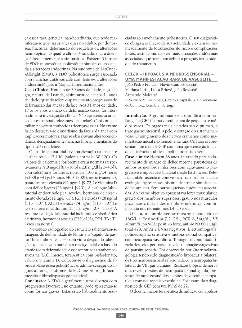
ÓRGÃO OfICIAL DA SOCIEDADE PORTUGUESA DE REUMATOLOGIA
148
posters
ça óssea rara, genética, não-hereditária, que pode ma-nifestar-se quer na criança quer no adulto, por dor ós-sea, fracturas, deformação do esqueleto ou alteraçõesneurológicas. O quadro clínico é variado, mas a doen-ça é frequentemente assintomática. Existem 3 formasde FDO: monostótica, poliostótica simples ou associa-da a alterações endócrinas. Na síndrome de McCune--Albright (SMA), a FDO poliostótica surge associadacom manchas cutâneas café com leite e/ou alteraçõesendocrinológicas múltiplas hiperfuncionantes.Caso Clínico: Homem de 30 anos de idade, raça ne-gra, natural de Luanda, assintomático até aos 14 anosde idade, quando refere o aparecimento progressivo dedeformação das ancas e da face. Aos 31 anos de idade,17 anos após o início da deformação óssea, foi inter-nado para investigação clínica. Não apresentava ante-cedentes pessoais relevantes e em relação à história fa-miliar, não eram conhecidas doenças ósseas. No examefísico destacava-se dimorfismo da face e da anca comimplicações motoras. Não se observavam alterações cu-tâneas, designadamente manchas hiperpigmentadas dotipo «café com leite».
O estudo laboratorial revelou elevação da fosfatasealcalina total 417 UI/L (valores normais: 30-120). Osvalores de calcemia e fosforemia eram normais (respe-tivamente, 9,0 mg/dl [8,8-10,6] e 2,8 mg/dl [2,5-4,5]),com calciúria e fosfatúria normais (100 mg/24 horas[<300] e 441 g/24 horas [400-1300]), respetivamente),paratormorna elevada (92 pg/mL [9-72]) e Vitamina Dcom défice ligeiro (25 ng/mL [>29)]. A avaliação labo-ratorial endocrinológica, revelou hormona de cresci-mento elevada (12 mg/L[<1]), IGF1 elevada (428 ng/ml[115 - 307]), ACTH elevada (74 pg/ml [115 - 307]) etestosterona total diminuída (1.2 ng/ml [2.7 - 11.0]) Arestante avaliação laboratorial incluindo cortisol séricoe urinário, hormonas sexuais (FSH e LH), TSH, T3 e T4livres era normal.
No estudo radiográfico do esqueleto salientavam-seimagens de deformidade de fémur em “cajado de pas-tor” bilateralmente, aspeto em vidro despolido, altera-ções que afetavam também o maciço facial e a base docrânio (com deformidade óssea acentuada) também vi-síveis na TAC. Iniciou terapêutica com bisfosfonato,cálcio e vitamina D. Colocou-se o diagnóstico de fi-brodisplasia óssea poliostótica e, admite-se segundo al-guns autores, síndrome de McCune-Allbrigth (acro-megália e fibrodisplasia poliostótica).Conclusão: A FDO é geralmente uma doença comprognóstico favorável, no entanto, pode apresentar-secomo formas graves. Estas estão habitualmente asso-
ciadas ao envolvimento poliostótico. O seu diagnósti-co obriga à avaliação da sua actividade e extensão, no-meadamente de localizações de risco e complicaçõeslocais, assim como de eventuais alterações endócrinasassociadas, que permitam definir o prognóstico e o ade-quado tratamento.
CC120 – HIpOACuSIA NEuROSSENSORIAL –uMA MANIFESTAÇÃO RARA DE VASCuLITEJoão Pedro Freitas1, Flávio Campos Costa1, Mariana Luis1, Luisa Brites1, João Rovisco1, Armando Malcata1
1. Serviço Reumatologia, Centro Hospitalar e Universitáriode Coimbra, Coimbra, Portugal
Introdução: A granulomatose eosinofílica com po-liangeíte (GEP) é uma vasculite rara de pequenos e mé-dios vasos. Os órgãos mais afetados são o pulmão, otrato gastrointestinal, a pele, o coração e o sistema ner-voso. O atingimento dos nervos cranianos como ma-nifestação inicial é extremamente rara. Os autores apre-sentam um caso de GEP com uma apresentação inicialde deficiência auditiva e polineuropatia severa.Caso clínico: Homem 68 anos, internado para escla-recimento de quadro de défice motor e parestesias deambos os membros inferiores com agravamento pro-gressivo e hipoacusia bilateral desde há 2 meses. Refe-ria também astenia e febre vespertina com 1 semana deevolução. Apresentava história de asma e sinusite des-de há um ano. Sem outras queixas sistémicas associa-das. Ao exame objetivo apresentava força muscular degrau 5 dos membros superiores, grau 3 nos músculosproximais e distais dos membros inferiores, com hi-postesia nos dermátomos L4, L5 e S1.
O estudo complementar mostrou: Leucocitose18G/L e Eosinofilia 7,2 G/L, PCR 8,3mg/dl, VS40mm/h, pANCA: positivo forte, anti-MPO 80 U, IgEtotal 478, ANAs e ENAs negativos. Electromiografia:polineuropatia sensitiva e motora axonal compatívelcom neuropatia vasculítica. Tomografia computadori-zada dos seios peri-nasais revelou alterações sugestivasde pansinusopatia. Foi observado por Otorrinolarin-gologia tendo sido diagnosticado hipoacusia bilateralde tipo neurossensorial relacionada com neuropatia bi-lateral do VIII par craniano. Realizou biópsia de nervoque revelou lesões de neuropatia axonal aguda, pre-sença de raros eosinófilos e lesões de vasculite compa-tíveis com neuropatia vasculítica. Foi assumido o diag-nóstico de GEP com um BVAS de 22.
O doente iniciou terapêutica de indução com pulsos

ÓRGÃO OfICIAL DA SOCIEDADE PORTUGUESA DE REUMATOLOGIA
149
posters
de ciclofosfamida na dose de 15mg/Kg, com boa res-posta clínica e analítica.
À data de alta o doente apresentava-se apirético, semqueixas constitucionais, com normalização da fórmu-la leucocitária (Leucócitos 10,0 G/L Eosinófilos 1,0G/L) e dos parâmetros inflamatórios (PCR: 0,4 mg/dL;VS: 10 mg/dL).Conclusão: Ao apresentar este caso os autores preten-dem ressaltar a grande variabilidade de apresentaçõesclínicas que esta doença multissistémica pode apre-sentar. Conforme a literatura o envolvimento dos ner-vos cranianos é uma manifestação muito rara na gra-nulomatose eosinofílica com poliangeíte.
CC131 – SÍNDROME DE MIKuLICZ COMOFORMA DE ApRESENTAÇÃO DE SARCOIDOSEJoana Ramos Rodrigues1, Daniela Santos-Faria1, Joana Sousa-Neves1, Joana Leite Silva1, Marcos Cerqueira1, Filipa Teixeira1, Carmo Afonso1,Sérgio Alcino 1, José A. Costa1, Daniela Peixoto1
1. Serviço de Reumatologia, Unidade Local de Saúde doAlto Minho, Ponte de Lima, Portugal
Introdução: A Sarcoidose é uma patologia granulo-matosa multissistémica que pode afetar qualquer ór-gão, mais frequentemente, o pulmão e os gânglios lin-fáticos intra-torácicos1. Entre as manifestações extra-pulmonares, contam-se as músculo-esqueléticas comquadros de oligo e poliartrite que podem constituir sin-tomas de grande relevo2 pelo que, numa consulta deReumatologia, no diagnóstico diferencial, esta situaçãoclínica é muitas vezes considerada. Outras formas deapresentação como a de um Síndrome de Mikulicz, im-plicando diagnóstico diferencial com doenças do teci-do conetivo, em particular com o Síndrome de Sjögren,podem também ser observadas.Objetivo: Os autores descrevem um caso de Sarcoido-se de apresentação rara, salientando a importância dasformas atípicas de apresentação de Sarcoidose.Caso Clínico: Doente do sexo feminino, 28 anos deidade, saudável até agosto de 2016, altura em que ini-ciou quadro de edema palpebral bilateral, indolor, in-sidioso, associado a xeroftalmia, mas sem xerostomiaou sintomas constitucionais concomitantes. Posterior-mente, notou tumefação parotídea bilateral, sem cóli-ca salivar ou febre. Foi observada em consulta de Of-talmologia que referenciou para consulta de Reumato-logia, com a suspeita de Síndrome de Sjögren.
Na observação, constatado edema palpebral supe-rior bilateral marcado, tumefação parotídea bilateral e
simétrica, de consistência duro-elástica, não dolorosaà palpação e normalidade do restante exame objetivo.Analiticamente, de salientar normalidade do hemogra-ma, dos parâmetros de bioquímica de função hepáticae renal, dos valores de IgG4, anti-SS-A e anti-SS-B ne-gativos e elevação ligeira da ECA.
A radiografia de tórax evidenciou algumas imagenshipotransparentes, ovaladas e infra-centimétricas, peri-hilares, distribuídas bilateralmente. A TC pulmonar dealta resolução confirmou a presença de adenopatias hi-lares bilaterais e espessamento irregular micronodulardo interstício com predileção pelos terços médios e abiópsia de uma glândula lacrimal, revelou granulomascompatíveis com Sarcoidose.Discussão/Conclusão: A síndrome de Mikulicz, ca-racterizada pela tumefação simétrica e não dolorosa dasglândulas lacrimais e parotídeas3, é uma entidade pou-co frequente que se pode associar a várias situações clí-nicas como tuberculose, sarcoidose e doenças linfo-proliferativas3.
A doença de Mikulicz é atualmente consideradacomo fazendo parte de um espetro mais alargado dadoença relacionada com IgG44 que, além do envolvi-mento das glândulas salivares e lacrimais, pode incluirmuitas outras manifestações5.
Pretendeu-se alertar para uma das formas raras deapresentação da Sarcoidose, implicando diagnóstico di-ferencial com o Síndrome de Sjögren, com a doença re-lacionada com IgG4 e com doenças linfoproliferativas.
REFERÊNCIAS1. Sehgal V, Riyaz N, Chatterjee K, et al. Sarcoidosis as a systemic
disease. Clinics in Dermatology. 2014; 32: 351-363.2. Grainger A, O’Connor P. Arthritis. In: Adam A, Schaefer-Pro-
kop C. Grainger & Allison’s Diagnostic Radiology. 6th edition.London: Elsevier; 2015. p 1186-1210.
3. Coloma-González I, Ruíz-García L, Flores-Preciado J, et al. En-fermedad de Mikulicz. A propósito de un caso. Archivos de laSociedad Española de Oftalmología. 2013; 89: 332-335.
4. Himi T, Takano K, Yamamoto M, et al. A novel concept of Mikulicz’sdisease as IgG4-related disease. Auris Nasus Larynx. 2012; 39: 9-17.
5. West S. Systemic Diseases in Which Arthritis is a Feature. In:Goldman L, Schafer A. Goldman-Cecil Medicine. 25th edition.Philadelphia: Elsevier, 2016. p 1823-1828
CC75 – EFFICACY AND SAFETY OF IMATINIBIN SYSTEMIC SCLEROSIS – A CuRIOuS CASEREpORTJoana da Silva Dinis1, Ana Valido1, Catarina Resende1,Carla Macieira1
1. Rheumatology Department, Hospital de Santa Maria(CHLN), Lisbon Medical and Academic Centre, Lisboa,Portugal

ÓRGÃO OfICIAL DA SOCIEDADE PORTUGUESA DE REUMATOLOGIA
150
posters
Introduction: Preclinical studies suggest that selectivetyrosine kinase inhibitors that target c-Abl, platelet-de-rived growth factor receptor or Src kinases, such asImatinib, might prevent or even induce regression ofestablished fibrosis in Systemic Sclerosis (SSc). Despiteseveral case reports, case series and uncontrolled stu -dies reporting regression of fibrosis and good tolera-bility with Imatinib, subsequent clinical studiesshowed that Imatinib was poorly tolerated, and a largenumber of moderate/severe adverse events seemed torestrict its use in SSc patients.Case Report: A 42 year-old female had diffuse cuta-neous SSc for 12 years. Cutaneous, pulmonary and gas-tric involvement were present without renal manifes-tations. Antinuclear antibodies with a nucleolar pat-tern and a Scl-70 antibody were present in serum.Throughout the years, the disease progressed, mainlywith dermal and pulmonary fibrosis, despite treatmentwith immunosuppressive agents such as methotrexateand cyclophosphamide.
As a result, in 2012, the patient presented with scle-rodactyly, digital tip ulcers and pitting scars, Raynaud’sphenomenon, telangiectasia and abnormal nailfoldcapillaries. At that time, the modified Rodnan SkinScore (mRSS) was 16 out of 51. Gastric involvementwas characterized by esophageal dysmotility and gas-tric-esophageal reflux disease documented by uppertract endoscopy. The thoracic tomography (TT) re-vealed an interstitial lung disease pattern, namely non--specific interstitial pattern, with ground glass opaci-ties. The pulmonary function tests (PFT) documenteda normal Forced Vital Capacity (FVC) and a decreasedcarbon monoxide diffusion capacity (DLCO) - 78%.The transthoracic echocardiogram quantified a pul-monary artery systolic pressure (PASP) of 31 mmHg -slightly elevated, justifying adjustment of drug regi-men. Concomitant a diagnosis of Chronic MyeloidLeukemia (CML) was made and therefore the patientwas started on Imatinib 400 mg/day alone, with a po -sitive response and CML entered a chronic phase.
In the last 4 years, after Imatinib start, SSc progres-sion stabilized without further need for other im-munosuppressive therapy. A subjective improvement ofskin tightness and hands movement was reported bythe patient and the mRSS remains similar at presenttime. FCV and DLCO values on PFT stayed stable. Nofurther progression of pulmonary fibrosis was evidenton TT and PASP documented on transthoracic echocar-diogram suffered no aggravation.
Currently, after 54 months of treatment, Imatinib re-
mains well-tolerated and no adverse events were re -gistered. Neither SSc or CML had need for other drugagents.Conclusion: This report has obvious similarities withthose described in the literature and supports Imatinib’sefficacy in the treatment of SSc. Additionally, a signifi-cant association with CML took place, potentiatingImatinib’s use. No major adverse effects were registeredand the drug was well tolerated, allowing for treatmentof both diseases. Our patient had timely and formal setindication for treatment with Imatinib, contrary to se-lected clinical trial’s population. Also, a less severe formof SSc may explain reported efficacy and safety.
CC193 – BASIC CRYSTAL pHOSpHATEARTHROpATHY DIAGNOSED BY uLTRASOuNDGuIDED BIOpSY?Rui Lourenço Teixeira1, Rita Luís2, Emília Vitorino2,Joaquim Polido Pereira1, Fernando Saraiva1
1. Serviço de Reumatologia e Doenças Ósseas Metabólicasdo Hospital de Santa Maria, Centro Hospitalar Lisboa Norte,Centro Académico de Medicina de Lisboa, Lisboa, Portugal2. Serviço de Anatomia Patológica, Centro HospitalarLisboa Norte, Centro Académico de Medicina de Lisboa,Lisboa, Portugal
Over the last decades, a growing number of publica-tions have been showing the advantages of ultrasoundguide biopsies. This easy to perform technique is use-ful to elucidate less clear diagnoses and to evaluate acti -vity in rheumatic diseases such as rheumatoid arthri-tis1. Concerning our growing experience on this tech-nique, we present the first case of a proximal interpha-langeal joint guided biopsy ever performed in Portugal,and highlight its important application in a difficultdiag nostic setting.
We present the case of a 54 years old woman, work-ing at a shoe store who presented with an history of sixyears of polyarthritis involving proximal interpha-langeal joints (PIP), shoulders, elbows and knees andbiphasic Raynaud phenomenon. ANA, rheumatoid fac-tor and ACPA were negative. She had no radiographicevidence of bone erosion or of chondrocalcinosis.
Although she had normal ESR (19 mm/h) and CRP(0.38 mg/dl), seronegative rheumatoid arthritis diag-nosis was established and she started therapy with non--steroidal anti-inflammatory drugs (NSAIDs), pre -dnisolone and methotrexate. She was intolerant tohigher doses of methotrexate and did not respond totriple therapy nor to leflunomide. Having in considera -

ÓRGÃO OfICIAL DA SOCIEDADE PORTUGUESA DE REUMATOLOGIA
151
posters
tion four years of difficult to treat arthritis the patientwas proposed to biologic therapy with etanercept in2016. She went into latent tuberculosis’ screening, ac-cording to the national guidelines, with the need ofnine months of izoniazide therapy which eventually re-sulted in some hepatic toxicity.
After a 6-month period follow-up, no significant ben-efit was noted with a DAS-28-4V variation of 0.44, from4.68 to 4.24. An ultrasound evaluation was performedand the results are in Table 1. Few osteophytes wereidentified and distal interphalangeal joints were spared.
Since she had seronegative RA, no elevation of in-flammatory markers and refractory disease, an alter-native diagnosis was thought. An ultrasound-guidedsynovial biopsy of the 2nd left PIP was then performed.Microbiologic cultures were negative. Histological eva -luation allowed the visualization of sinovial hyperpla-sia, fibrine exsudate but a scarce chronic inflammato-ry infiltrate, with no lymphoid aggregates or granulo-mas. Extensive deposition of basic calcium phosphate(BCP) over the synovium was identified.
Growing evidence is linking BCP crystal synovial de-positions in osteoarthritis (OA). Although not easy toidentify, they are known to activate inflammatory cellsand to promote a proinflammatory medium which ul-timately leave to matrix degradation and bone des -truction and remodeling2,3.
The acquisition of synovial tissue and its careful his-tologic study have brought light into a misunderstoodclinical case, and help to corroborate the need for acareful and comprehensive/multidisciplinary diagnos-tic approach in Rheumatology.
CC46 – uVEÍTE COMO FORMA DEMANIFESTAÇÃO DE VASCuLITEFlávio Campos Costa1, João Pedro Freitas1, Carlos Costa1, Luisa Brites1, Mariana Luis1, João Rovisco1, Armando Malcata1
1. Serviço Reumatologia, Centro Hospitalar e Universitáriode Coimbra, Coimbra, Portugal
Introdução: A granulomatose com poliangeiite é umadoença sistémica potencialmente letal caracterizada porvasculite necrotizante dos pequenos vasos. Com umpico de incidência entre os 64-75 anos de idade, ca-racteriza-se pelo envolvimento das vias aéreas supe-riores, pulmões e rins. As manifestações oculares dadoença ocorrem em aproximadamente 1/3 dos doen-tes. A uveíte é uma manifestação rara na granulomato-se com poliangeiite, verificando-se apenas em 3% dosdoentes. Usualmente esta ocorre em associação com aescleríte, sendo denominada de esclerouveíte, tendoum pior prognóstico ocular.Caso Clínico: Homem, 43 anos, internado para escla-recimento de olho vermelho (com 2 meses de evolução,tendo sido medicado com antibioticoterapia tópica semmelhoria) e poliartralgias periféricas, aditivas e simé-tricas com 1 mês de evolução, com melhoria discretacom AINE’s. Associadamente referia cansaço fácil, as-tenia, epistáxis e febre vespertina com 1 semana de evo-lução. Sem outras queixas sistémicas.
À entrada destacava-se a presença de olho vermelhoà esquerda e artrite da 2a IFP mão esquerda e 3ª IFDmão direita. Observado por oftalmologia sendo diag-nosticada uveíte anterior e medicado com dexameta-sona tópica em dose de desmame ocorrendo melhoriaprogressiva das queixas oculares.
Do estudo analítico à entrada destacava-se PCR: 5,19mg/dL; VS: 61mm/h; Cr: 6,68 mg/dL; BUN: 59,2mg/dL; Hb: 11,0; sumária de urina: Eritrócitos: 72
FIGuRE. Basic Calcium phosphate deposits highlighted in redby the Alizarin stain (Alizarin red, 100x)
TABLE I. uLTRASOuND FINDINGS IN DISEASE ACTIVITY ASSESSMENT AFTER 6-MONTH pERIODON BIOLOGIC THERApY
Left RightTHUMB Grade 2 Grade 12nd PIP Grade 3 + Doppler 2 Grade 3 + Doppler 23th PIP Grade 2 Grade 14th PIP Grade 2 Grade 15th PIP Grade 1 No sinovitis

ÓRGÃO OfICIAL DA SOCIEDADE PORTUGUESA DE REUMATOLOGIA
152
posters
mg/dL; Proteínas: 1000mg/dL. Pedida colaboração daNefrologia para realização de biopsia renal.
Do restante estudo complementar realizado destaca-se Ac. Anti-cANCA: positivo forte; Ac. Anti-PR3: posi-tivo 108U; ANAs negativos; Anti-DNA ds: negativos,Imunoglobulinas: sem alterações e serologias negati-vas; Cr urina 24h: 58,8 mg/dia; proteínas: 356,5mg/dia; relação proteínas/creatinina: 6063. A biopsiarenal demonstrou: Rim com aspetos de glomerulone-frite necrotizante crescêntica (crescentes em 50% dosglomérulos), pauci-imune, enquadrável em glomeru-lonefrite relacionada com ANCA, sendo assumido odiagnóstico de granulomatose com poliangiite. BVASinicial de 27.
Após resultado da biópsia renal iniciou terapêuticade indução com pulsos de ciclofosfamida na dose de15mg/Kg e prednisolona 1mg/Kg/dia, com boa respostaclínica e analítica.
À data de alta o doente apresentava um score BVASde 12, sem queixas constitucionais, apirético, com me-lhoria da função renal (Cr: 2,58 mg/dL e BUN:48,4mg/dL) e dos parâmetros inflamatórios (PCR: 0,11mg/dL; VS: 8 mg/dL).
Seis meses após iniciada terapêutica o doente man-tinha melhoria clínica (BVAS 7) e analítica com VS:7mg/dL; PCR: 0.17 mg/dL; Cr: 1.5 mg/dL. Iniciou tera-pêutica de manutenção com rituximab.Conclusão: Os autores pretendem alertar para a difi-culdade diagnóstica e necessidade de considerar a gra-nulomatose com poliangiite no diagnóstico diferencialde múltiplas entidades sistémicas ou mesmo de órgão.Neste caso destaca-se a uveíte anterior como manifes-tação clínica inicial de vasculite sistémica.
CC51 – RA CASES OF INpATIENTS wITHSuSpECTED MYOCARDIAL INFARCTIONDIAGNOSISFábio Amaral1, Cláudia Vaz1, 2
1. Serviço de Reumatologia, Unidade Local de Saúde -Hospital Sousa Martins, Guarda, Portugal2. Faculdade de Ciências da Saúde, Universidade da BeiraInterior, Covilhã, Portugal
Background: Inflammation is a corner stone in bothrheumatic disease and heart disease. RA being the mostcommon rheumatic disease is the best-studied con-cerning cardiovascular disease. Some drugs to treat RAare able to control inflammation and reduce CV risk.However, steroids do the opposite due to the adversemetabolic effects, which offset the anti-inflammatory
effects. Takotsubo cardiomyopathy (TTC) is an acutecardiac condition characterized by transient left ven-tricular dysfunction with wall motion abnormalities,usually apical akinesis and ballooning. Patients oftenpresent with chest pain, ST-segment elevation on EKG,and elevated cardiac enzyme levels that increase thesuspicion of a myocardial infarction. Cardiac benignlipomatous lesions include neoplastic, congenital, andreparative phenomena of encapsulated mature adiposecells. The limited evidence available, due to rarity, sup-ports their neoplastic classification.Objectives: To identify rare cardiac causes for RA pa-tient hospitalization.Methods: Analysis of medical records from inpatientsdiagnosed with RA and myocardial infarction.Results: Report of two cases, each with established RAdiagnosis and acute onset of cardiac symptoms. CASE1. A 43-year-old man presented in the ER with the sus-picion of an acute coronary syndrome (ACS), specifi -cally an ST elevation myocardial infarction (STEMI).Ho wever, cardiac catheterization revealed permeablecoronary arteries and an aneurysm involving the ante-rior and apical walls of the left ventricle. 3 Months la -ter, on myocardial perfusion scintigraphy using99mTC-tetrofosmin the patient presented necrosis ofthe anterior wall with peri-necrosis ischemia. CASE 2.A 61-year-old man presented in the ER with chest painof acute onset. EKG was normal, chest X-ray showedaugmen ted cardiothoracic index, and cardiac enzy-mology was elevated. Echocardiography revealed pre-served global systolic function, septum hypertrophy,basal posterior wall dyskinesia, and a large mass in theleft ventricle. Further study supports the diagnosis ofa lipoma for lack of malignant characteristics.Conclusions: Rare cardiac events associated with RAare indeed infrequent. Causality cannot be establishedeven though the correlation may exist, especially be-cause we cannot be certain of which pathology was es-tablished primarily. Our report supports the benefit forprogrammed periodic screening of cardiac health.
GRupO 15
CC148 – LÚpuS ERITEMATOSO SISTÉMICO ELEuCEMIA DE LINFÓCITOS GRANDESGRANuLARESDaniela Santos-Faria1, Ana Raposo2, Joana Leite Silva1, Joana Sousa-Neves1, Joana Ramos Rodrigues1, Carmo Afonso1,

ÓRGÃO OfICIAL DA SOCIEDADE PORTUGUESA DE REUMATOLOGIA
153
posters
Sérgio Alcino 1
1. Serviço de Reumatologia, Unidade Local de Saúde doAlto Minho, Ponte de Lima, Portugal2. Rheumatology, Centro Hospitalar Trás-os-Montes e AltoDouro, Vila Real, Portugal
Introdução: A leucemia de linfócitos grandes granula-res (LLGG) é uma doença linfoproliferativa, geralmen-te indolente, associando-se, na maioria dos casos(74%), a doenças imunomediadas. Estas podem surgirsincronamente com a LLGG ou durante a sua evolu-ção1. A artrite reumatóide é a doença mais frequente-mente descrita em associação a esta situação clínica.Outras patologias como o lúpus eritematoso sistémico(LES), embora mais raramente, também podem estarassociadas1.
Apresenta-se o caso clínico de um doente comLLGG, no qual, ao fim de 5 anos de evolução, surgiuum quadro de LES.Caso Clínico: Doente do sexo masculino, de 44 anosde idade, em vigilância no Instituto Português de On-cologia do Porto com diagnóstico de LLGG desde2011, manifestada por neutropenia, estável na sua evo-lução, não tendo tido indicação para tratamento espe-cífico até ao surgimento do quadro actual.
Em maio de 2016 foi referenciado à consulta de Reu-matologia por quadro de poliartrite simétrica e aditiva,envolvendo médias e pequenas articulações (punhos,mãos e pés), com 2 meses de evolução. Adicionalmen-te referiu apenas episódios de lesões cutâneas eritema-tosas e pruriginosas nos membros superiores, sem fo-tossensíbilidade, que melhoravam com a toma de anti--histamínico. Analiticamente apresentava neutropeniae linfocitose em valores sobreponíveis aos previamen-te conhecidos, velocidade de sedimentação de 33 mme proteína C reativa normal, parâmetros de função he-pática e renal, incluindo sedimento urinário, sem quais-quer alterações, fator reumatóide positivo e anti-CCPnegativo, anticorpos anti-nucleares de 1/160, anti-SSApositivo, mas com Anti-ADN e anti-Sm negativos.Constatada ainda diminuição com complemento C3(79mg/dL) e, embora sem história de eventos trombó-ticos, tinha anticorpos anti-fosfolípideo positivos (an-ticardiolipina e b2glicoproteina-1), em duas avaliações,preenchendo assim critérios de diagnóstico de LES. Ini-ciou terapêutica com prednisolona em baixa dose e,por ter apenas melhoria parcial, iniciou posteriormen-te hidroxicloroquina 400mg/dia e metotrexato (MTX)10mg/semana, com regressão completa das queixas ál-gicas e dos sinais inflamatórios articulares, mantendo-
-se, oito meses depois do início desta terapêutica, cli-nicamente bem, sem intercorrências infeciosas e commelhoria da neutropenia e da linfocitose prévias, su-gerindo benefício do MTX na leucemia de LGG.Discussão/ Conclusão: A associação da LLGG àsdoenças imunomediadas parece estar relacionada comfatores etiopatogénicos comuns, em particular, com asconcentrações de interleucina-181.
Num estudo com objectivo de avaliar neoplasias he-matológicas em doentes com LES, é relatado um casosemelhante ao apresentado tendo sido também estabe-lecido diagnóstico de LES num doente com LLGG com3 anos de evolução.
As terapêuticas imunossupressoras mais utilizadasna LLGG, para controlo da neutropenia, são o MTX embaixas doses (10mg/m2/semana) e ciclofosfamida oral,com resposta em 50-75% dos doentes (1). A respostaobservada neste doente, depois do início de MTX, estátambém de acordo com o descrito.
Assim, nas doenças imunomediadas em que se cons-tate uma neutropenia persistente, um quadro de LLGG,deverá ser considerado.
REFERÊNCIAS1. Bockorny B, Dasanu CA. Autoimmune manifestations in large
granular lymphocyte leukemia. Clin Lymphoma, MyelomaLeuk. 2012;12(6):400–5.
2. Knight JS, Blayney DW, Somers EC. Patients with systemic lu-pus erythematosus and haematological malignancy at a tertia-ry care centre: timing, histopathology and therapy. Lupus SciMed. 2014;1(1):e000051.
CC44 – SMALL VESSEL INVOLVEMENT INTAKAYASu ARTERITIS – wHAT IS THE ExACTROLE FOR BIOLOGICS?Maria Inês Seixas1, 2, Vítor Teixeira1, Filipa Teixeira3,Carla Macieira1
1. Rheumatology Department, Hospital de Santa Maria(CHLN), Lisbon Medical and Academic Centre, Lisboa,Portugal2. Rheumatology Department, Centro Hospitalar Tondela-Viseu, Viseu, Portugal3. Ophthalmology, Hospital de Santa Maria, CHLN,Lisbon Academic Medical Center, Lisboa, Portugal
Introduction: Takayasu arteritis (TA) is a large vesselvasculitis (LVV) affecting predominantly young wo -men, causing inflammation of aorta and its majorbranches, resulting in stenosis/occlusion and aneurysmformation. This rare disorder rarely affects small vessels. Conventional disease modifying agents (cDMARDs) are sometimes insufficient in management

ÓRGÃO OfICIAL DA SOCIEDADE PORTUGUESA DE REUMATOLOGIA
154
posters
but new inflammatory markers for TA have openednew therapeutic possibilities for biologic treatment(BT).Aims: We report two cases of TA with extremely raresmall vessel affection (SVA): cutaneous vasculitis alongwith sensorineural hearing loss (SHL) and retinal vas-culitis, refractory to cDMARDs and requiring BT.Clinical Cases: Case 1: 37-year-old woman diagnosedwith TA in her twenties, presented with intermittentleft arm claudication and high inflammatory markers.Magnetic resonance angiography revealed significantstenosis of left subclavian and both common carotids.Initially started prednisolone 1mg/Kg/day (d) andmethotrexate 25mg/week (w), with no improvement.Monthly pulses of cyclophosphamide and later aza-thioprine were then administered with stable clinicaland serum markers improvement. At age 32, cutaneousulcers appeared on both legs. Biopsy showed a granu-lomatous dermatitis with central necrosis. After tuber-culosis exclusion she was again on prednisolone 1 mg/Kg/d; after 2 years, ulcers persisted and she be-gan infliximab 5 mg/kg 8/8w along with methotrexate10mg/w with complete cutaneous and systemic remis-sion. Recently, 2 years after infliximab, she presentedwith tinnitus and sudden right SHL. Again PDN1mg/Kg/d was started with rapid tapering, achievingremission. Case 2: 19-year-old black woman diagnosedone year earlier with TA, involving ascending and de-scending aorta, aortic arch, brachiocephalic trunk, bothcommon carotids with critic stenosis at the right sub-clavian and carotid arteries requiring elective surgery.The patient had been treated with methotrexate25mg/w and high doses of prednisolone 60mg/d withgradual possible tapering to 10mg/d. Despite treatmentshe was found with a Cushingoid habitus, having per-sistent carotidynia and right superior limb markedclaudication, along with very high inflammation mar -kers. Tocilizumab was indicated in order to achieve di -sease remission, allow prednisolone tapering and ulti-mately improving elective surgery success rate. Whilewaiting for tocilizumab she developed acute ocularphotophobia and blurred vision related to left anterioruveitis and bilateral retinal vasculitis, more extensive inthe left eye.Discussion: Inner ear, cutaneous and retinal vasculi-tis are extremely rare in TA. We found only few reportson SHL and cutaneous granulomatous involvement.Regarding ocular manifestations those are most oftendue to chronic hypoperfusion related to commoncarotid involvement or hypertensive retinopathy. Des -
pite reports of uveitis, scleritis and glaucoma, retinalvasculitis (affecting large retinal vessels) is extremelyrare. We report two cases in which rare manifestationsof SVA in TA were refractory to steroids and cDMARDs,ultimately requiring BT. Case series and observationalstudies support the use of anti-tumor necrosis factortherapy in refractory TA, in particular infliximab. Asmall number of patients refractory to infliximab havebeen successfully treated with tocilizumab and rituxi -mab, however indication for these agents in LVV withSVA is unusual. Despite growing evidence for BT, themajority of data hasn’t assessed whether they should beconsidered and what is the most appropriate agent inSVA of LVV.
CC81 – CRIOGLOBuLINÉMIA ASSOCIADA AOVIH: CASO RARO E DESAFIO TERApÊuTICOTiago Costa1, Carina Lopes1, João Lagoas Gomes1,José Marona1, Sandra Falcao1, 2, Jaime C. Branco1, 2
1. Rheumatology Department, Hospital Egas Moniz(CHLO), Lisboa, Portugal2. CEDOC, NOVA Medical School. Faculdade de CiênciasMédicas da Universidade NOVA de Lisboa, Lisboa,Portugal
Introdução: A crioglobulinémia é considerada umadoença rara, cuja verdadeira prevalência permanecedesconhecida. A crioglobulinémia mista clinicamentesignificativa tem uma prevalência estimada de1:100000, predominando em mulheres, numa razãode 3:1. Até 80-90% dos doentes com crioglobulinémiamista têm infeção crónica pelo vírus da Hepatite C(VHC). As crioglobulinas são frequentemente detetadasno contexto da infeção pelo vírus da ImunodeficiênciaHumana (VIH), mas raramente se associam a vasculi-te crioglobulinémica. O tratamento é complexo e variaconsoante a etiologia e gravidade da doença.Caso clinico: Apresenta-se o caso de um homem de 64anos, com história de hipertensão arterial, diabetes mel-litus tipo 2 e infeção VIH-1 diagnosticada em 1994, me-dicado com terapêutica antirretroviral tripla (emtrici-tabine/tenofovir/raltegravir). Em março de 2016 ini-ciou quadro de lesões cutâneas eritemato-violáceas nosdois pés, artralgias dos joelhos de ritmo inflamatório ecansaço generalizado. A púrpura cutânea evoluiu paraulceração nos calcâneos e polpa digital dos dedos dospés. Analiticamente destacava-se: velocidade de sedi-mentação de 50 mm/h e crioglobulinas positivas (IgA,IgG e IgM). O restante estudo foi normal/negativo, in-clusive vírus Hepatite B e C, complemento C3/C4, fa-

ÓRGÃO OfICIAL DA SOCIEDADE PORTUGUESA DE REUMATOLOGIA
155
posters
tor reumatóide, anticorpos anti-nucleares (ANAs) e an-ticorpos anti-antigenos nucleares extraíveis (ENAs).Desde o início do quadro clínico o doente apresentoucarga viral VIH-1 <20 cópias/ml e linfócitos T CD4+ > 500 células/ul. Realizou-se biópsia de úlcera ao níveldo calcâneo cujo resultado veio confirmar o diagnósti-co de crioglobulinémia mista. O doente não apresen-tou envolvimento hepático ou renal. Iniciou-se corti-coterapia com prednisolona 1mg/kg/dia (60mg/dia,que realizou durante 1 mês) registando-se melhoria clí-nica e boa evolução na cicatrização das úlceras cutâ-neas. Procedeu-se ao desmame progressivo da predni-solona até 10 mg/dia, sem intercorrências.
Segundo a literatura, os corticóides continuam a sera 1ª linha no tratamento da vasculite associada ao VIH.A terapêutica imunossupressora (Rituximab, Ciclofos-famida) e a plasmaferese, utilizados mais frequente-mente no tratamento da crioglobulinémia associada aoVHC, devem ser evitados sempre que possível uma vezque podem permitir o desenvolvimento de infeçõesoportunistas graves, especialmente se a contagem delinfócitos TCD4+ <200 células/ul. Em situações em quea infeção VIH não se encontra controlada o tratamen-to âncora reside na supressão da replicação viral doVIH.Conclusão: Trata-se de um caso raro de um homemcom crioglobulinémia mista grave associada ao VIH.Revela-se um verdadeiro desafio a nível terapêutico,pois tratando-se de uma associação rara não existemrecomendações para o seu tratamento.
CC88 – IS THERE A ROLE FOR ANTI-CD20THERApY IN SpONDYLOARTHRITIS?Maria Inês Seixas1, 2, Carla Macieira1
1. Rheumatology Department, Hospital de Santa Maria(CHLN), Lisbon Medical and Academic Centre, Lisboa,Portugal2. Rheumatology Department, Centro Hospitalar Tondela-Viseu, Viseu, Portugal
Introduction: Rituximab (RTX) is a chimeric mono-clonal anti-CD20 antibody that targets mature B lym-phocytes, approved for the treatment of rheumatoidarthritis (RA) refractory to anti-tumour necrosis factor(TNF). Whether RTX may be effective in spondy-loarthritis (SpA) and psoriatic arthritis (PsA) is present-ly unclear. RA and SpA/PsA have different pathogenicmechanisms and rarely occur together.Aim: A clinical report of a woman with RA and possi-ble coexisting SpA/PsA, refractory to anti-TNF and suc-
cessfully treated with RTX.Clinical Case: A 33-year-old woman presented in herearly twenties, with systemic complains, fever, weightloss, stiffness and symmetric polyarthritis of shoulders,elbows, wrists and knees along with high inflammato-ry markers. As rheumatoid factor (RF) and antibodiesto cyclic citrullinated peptides (anti-CCP) were presentin serum a diagnosis of RA was performed, and she wasstarted on prednisolone and methotrexate (MTX)20mg/week, with partial benefit. One year after(DAS28-PCR=5.1), she began etanercept 50mg/weekwithout significant improvement for 15 months. Shewas then switched for infliximab 3mg/Kg every 8 weeksduring 9 months, afterwards raised to 5mg/Kg every 6weeks, along with MTX 20 mg/week maintaining a di -sease activity score persistently high (DAS28--PCR=4.97). At that time, while she was also takingcelecoxib, severe cholestatic hepatitis developed, andall drugs were stopped. One year later, RA persistedacti ve and RTX on monotherapy was started. Eightweeks after the first RTX treatment she developed sym-metric synovitis of second and third distal interpha-langeal (DIP) hand joints and a right third fingerdactylitis. RTX was extend along 3 cycles, resulting ingood disease response at 10 months (DAS-28--PCR=2.08). DIP joint involvement persisted with ra-diographic structural damage, without any other jointcomplain. Recently, she developed tibialis anteriortenosynovitis and bilateral Achilles enthesitis. Consi -dering the atypical joint involvement and enthesitis, anew hip radiographic was performed, showing rightsacroiliac joint irregularities. Magnetic resonance imag-ing revealed asymptomatic bilateral sacroiliitis with leftbone marrow oedema. A diagnosis of SpA was made,eventually psoriatic arthritis without psoriasis (no pso-riasis noticed in the family), in overlap with RA.Discussion: Coexistence of RA and SpA/PsA is rare.We report a TNF inhibitor failed patient, successfullytreated with RTX that developed a curious clinical--pathological RA/PsA-like expression: large and DIPsymmetric joint involvement with dactylitis andasymptomatic sacroiliitis, without psoriasis along, withRF/anti-CCP present in serum. Safety and efficacy ofRTX in RA has been perfectly shown, but data on SpAand PsA are limited. Case reports and open-label stu -dies have demonstrated modest clinical benefit in somepatients with SpA and PsA. The pathophysiology andclinical features of SpA and PsA are distinct from RA asevidenced by differing clinical features. The efficacy ofRTX, on joint inflammation, in our patient, suggests a

ÓRGÃO OfICIAL DA SOCIEDADE PORTUGUESA DE REUMATOLOGIA
156
posters
role for B cells in SpA/PsA. Some data have highligh tedthe presence of B cell lymphoid aggregates in PsA syn-ovial tissues. Nonetheless, B cells may play a role inantigen presentation in SpA.
Controlled studies will be needed for more definitiveunderstanding of RTX effect in SpA/PsA.Conclusion: This report of a rare association betweenRA and SpA/PsA, treated with RTX, suggests a possiblerole for anti-CD 20 blockage in patients with SpA/PsArefractory to established therapeutic regimens.
CC158 – A MIMETIZAÇÃO DE uMA MIOSITE:uM CASO DE DISFERLINOpATIANathalie Madeira1, Filipe Barcelos1, Luís Cunha-Miranda1, Augusto Faustino1
1. Rheumatology Department, Instituto Português deReumatologia, Lisboa, Portugal
Introdução: Fraqueza muscular proximal e elevaçãodas enzimas musculares são típicas das miopatias in-flamatórias idiopáticas. Porém, outras causas de mio-patias, hereditárias ou adquiridas, podem apresentar--se de modo semelhante.Caso clínico: Mulher de 30 anos, empregada de pa-daria, em aparente estado de saúde até há 3 anos, altu-ra em que inicia mialgias generalizadas e diminuiçãoprogressiva da força muscular de predomínio proxi-mal, inicialmente dos membros inferiores, com pro-gressão para os superiores. Quando se apresentou emconsulta de Reumatologia, estava medicada com pre -dnisolona (PDN) 20 mg/dia pelo médico de família háum mês, referindo alguma melhoria. Referia ainda acro-cianose e queixas esporádicas de fotossensibilidade exeroftalmia. Ao exame objetivo, destacava-se: marchamiopática, incapacidade para se levantar de cócoras,ligeira atrofia muscular da coxa esquerda e discreta di-minuição da força muscular do membro inferior es-querdo de predomínio crural (grau 4/5). Era portado-ra de análises sem elevação de VS e PCR, com elevaçãode TGO, CK, aldolase e LDH (152 U/L, 3396 U/L, 69U/L e 995 U/L, respetivamente) e ANA negativo. In-ternou-se para esclarecimento diagnóstico e despistede manifestações sistémicas. Realizou raio X tórax, TCda coluna lombar, ecografia abdominal, raio X e RMNda bacia, sem alterações de relevo. A capilaroscopia re-velou achados sugestivos de conectivite e a eletromio-grafia era compatível com miopatia, com alteraçõesmais marcadas nos músculos proximais dos membrosinferiores, sendo sugestivas de doença inflamatória domúsculo. Anti-dsDNA, anti-RNP e anti-Jo-1 eram ne-
gativos. Considerando-se a hipótese de polimiosite,ajustou-se terapêutica com azatioprina até 150 mg/diae PDN 15 mg/dia, verificando-se ligeira melhoria clí-nica e analítica, tendo alta. Nos meses seguintes, peloagravamento da fraqueza muscular e a persistência daelevação das enzimas musculares, solicitou-se a bió-psia muscular. Esta não evidenciou infiltrado inflama-tório, mas revelou alterações de perfil predominante-mente distrófico com padrão imuno-histoquímicocompatível com uma disferlinopatia (Limb Girdle Mus-cular Dystrophy, LGMD tipo 2B). Assim excluída a mio-patia inflamatória, suspenderam-se os imunossupres-sores e foi encaminhada para a consulta de doençasneuromusculares em Neurologia.Discussão: Este caso ilustra a importância da biópsiamuscular no diagnóstico diferencial das miopatias. Aperceção inicial do quadro foi prejudicada pela ausên-cia de avaliação clínica e analítica em Reumatologia an-tes do início da corticoterapia e pela subjetiva melho-ria da força muscular com esta terapêutica. A favor dapolimiosite, destacam-se ainda os achados à capilaros-copia e à eletromiografia. Pela refratariedade dos sin-tomas, apesar da optimização terapêutica, decidiu-seinvestigar outras hipóteses, como miosite por corpús-culos de inclusão ou distrofia muscular, através da bió-psia muscular. Esta deve ser analisada em centros es-pecializados, pois a avaliação histológica simples podeser insuficente, porque infiltrados inflamatórios e ne-crose podem existir nas distrofias musculares, mimeti-zando uma polimiosite. O recurso à microscopia elec-trónica, estudos funcionais/metabólicos ou análise mo-lecular permite estabelecer o diagnóstico definitivo. Po-rém, a dificuldade no seu acesso leva a que muitas vezesse coloquem hipóteses de diagnóstico e se inicie trata-mento sem a confirmação. É necessário otimizar a re-lação com centros de referência em miopatias, para umdiagnóstico definitivo em tempo útil, evitando tera-pêuticas desnecessárias.
CC123 – LINFOHISTIOCITOSEHEMOFAGOCÍTICA COMO FORMA DEApRESENTAÇÃO DE LÚpuS INDuZIDO pOR ETANERCEpTTeresa Martins-Rocha1, Raquel Miriam Ferreira1, Tiago Meirinhos2, Alexandra Bernardo1, Helena Baldaia3, Carlos Vaz1, 4, Lúcia Costa1
1. Serviço de Reumatologia, Centro Hospitalar de SãoJoão, Porto, Portugal2. Rheumatology Department, Hospital Infante D. Pedro(CHBV), Aveiro, Portugal

ÓRGÃO OfICIAL DA SOCIEDADE PORTUGUESA DE REUMATOLOGIA
157
posters
3. Serviço de Anatomia Patológica, Centro Hospitalar deSão João, Porto, Portugal, 4Faculdade de Medicina daUniversidade do Porto, Porto, Portugal
Introdução: A Linfohistiocitose Hemofagocítica (LHH)é um síndrome potencialmente fatal, no qual ocorreativação imunitária sistémica resultante da disfunçãode macrófagos e células T citotóxicas. A forma secun-dária resulta de múltiplas etiologias desde infeciosas,neoplásicas, reumáticas sistémicas e farmacológicas, in-cluindo agentes biotecnológicos. Caso clínico: Homem de 43 anos, com diagnóstico deartrite psoriática desde há 12 anos, medicado com eta-nercept há 4 anos, prednisolona 7,5mg/dia e naproxe-no 1000mg/dia. Recorreu ao Serviço de Urgência porfebre diária com 15 dias de evolução, associada a aste-nia, anorexia e perda ponderal de 8kg. Previamente,fora medicado com claritromicina durante 1 semanapor suspeita de traqueobronquite, sem resolução da fe-bre. Sem contexto epidemiológico relevante de infe-ção. Ao exame objetivo, apresentava febre (39ºc), he-patomegalia e poliartrite, sem lesões mucocutâneas,adenopatias ou outras alterações. Analiticamente, apre-sentava anemia microcítica (Hb 11g/dL), linfopenia(840/mm3), elevação da PCR (181mg/L) e VS(98mm/1ªh), haptoglobina normal, citocolestase (AST114 U/L, ALT 165 U/L, GGT 79 U/L, FA 137 U/L, combilirrubinas total/direta normais), elevação dos níveisde desidrogenase láctica (406 U/L), triglicerídeos (192mg/dL) e ferritina (910ng/mL); coagulopatia (apTT eTP aumentados, sem consumo do fibrinogénio). A ra-diografia torácica não apresentava alterações e a eco-grafia abdominal demonstrou hepatoesplenomegaliahomogénea. Foi internado no serviço de Reumatologia,suspendendo terapêutica com etanercept. Do estudorealizado, salienta-se: hemoculturas e urocultura ne-gativas; serologias VIH, VHC/A e IgM CMV, EBV e par-vovírus negativas; anti-HBc positivo com AgHBs nega-tivo e DNA de VHB indetetável; ecocardiograma trans-torácico normal e TC toracoabdominopélvico sem mas-sas, abcessos ou adenopatias. Do estudo imunológicorealça-se: ANA 1/320 padrão homogéneo, anti-histonasborderline, inibidor lúpico, prova de Coombs direta eb2glicoproteína IgG positivos, com anti-dsDNA e an-ticorpos para hepatites auto-imunes (LKM-1, LC-1,SLA/P) negativos e níveis de complemento normais.Após avaliação multidisciplinar e perante a persistên-cia de febre, bicitopenia e citocolestase, foi submetidoa biópsia hepática e da medula óssea. A histologia he-pática evidenciou infiltrado inflamatório polimórfico
nos lóbulos e ductos biliares; a histologia medular re-velou presença de células histiocitárias contendo eri-trócitos, traduzindo hemofagocitose, sem evidência deprocesso neoplásico, com estudos bacteriológico e mi-cobacteriológico negativos. Foram efetuados osdiagnós ticos de Lúpus e LHH induzidos por terapêu-tica anti-TNFa, pelo que manteve suspensão de eta-nercept e iniciou prednisolona 0.5mg/kg e ciclospori-na 2mg/kg, apresentando apirexia sustentada e nor-malização ulterior dos parâmetros analíticos. Conclusão: O Lúpus induzido por anti-TNFa temuma incidência descrita de 0,19-0,76% e a LHH se-cundária a biotecnológicos é uma entidade rara, estan-do apenas descritos cerca de 30 casos na literatura. Odiagnóstico de LHH exige um elevado índice de sus-peição clínica, assim como a exclusão de causas asso-ciadas, particularmente de quadros infeciosos subja-centes, descritos em 67% dos casos.
REFERÊNCIAS1. Brito-Zeron P, et al. Semin Arthritis Rheum. 2016;45(4):391-92. Ramos-Casals M, et al. Autoimmun Rev. 2010;9(3):188-93
CC102 – DOENÇA DE LYME E ARTRITE – pARA ALÉM DA SÍFILIS, uM OuTRO GRANDE IMITADORTeresa Martins-Rocha1, Raquel Miriam Ferreira1, Ana Filipa Rocha Águeda2, Tiago Meirinhos2, Miguel Bernardes1, Alexandra Bernardo1, Margarida Fernandes3, Madalena Pimenta4, Alcina Ferreira5, Lúcia Costa1
1. Serviço de Reumatologia, Centro Hospitalar de SãoJoão, Porto, Portugal2. Rheumatology Department, Hospital Infante D. Pedro(CHBV), Aveiro, Portugal3. Serviço de Anatomia Patológica, Centro Hospitalar deSão João, Porto, Portugal4. Serviço de Radiologia, Centro Hospitalar São João,Porto, Portugal5. Serviço de Doenças Infecciosas, Centro Hospitalar SãoJoão, Porto, Portugal
Introdução: A doença de Lyme é uma infeção causadapela espiroqueta do género Borrelia, transmitida peloartrópode Ixodes. O eritema migrans é uma manifesta-ção patognomónica, mas está ausente em cerca de umterço dos doentes. O envolvimento músculo-esquelé-tico ocorre em cerca de 60% dos casos e carateriza-sepor artrite intermitente de médias/grandes articulações,estando também descrita a ocorrência de tenossinovi-te e dactilite.

ÓRGÃO OfICIAL DA SOCIEDADE PORTUGUESA DE REUMATOLOGIA
158
posters
Caso clínico: Homem de 42 anos, guia turístico, comviagens frequentes em contexto laboral, a última dasquais em Abril de 2016, por zonas rurais no norte deEspanha. Em Maio, uma semana após infeção do tratourinário (sob ciprofloxacina), recorreu ao Serviço deUrgência (SU) por astenia, hiperémia conjuntival e ar-tralgias inflamatórias intermitentes do punho esquer-do, joelhos e tibiotársica direita. Negava lesões cutâ-neas ou picada de carraça, queixas urinárias, exsuda-do uretral ou outra sintomatologia. Por suspeita de ar-trite gonocócica, fez uma toma de ceftriaxone 1g.Regressou ao SU por persistência do quadro, sendo in-ternado no serviço de Medicina Interna. Do estudo efe-tuado, salienta-se: leucocitose (12 330 x 109/L); ele-vação da PCR (141 mg/L) e VS (41mm/1ªhora); PSAnormal; pesquisa de DNA de Neisseria gonorrhoeae eChlamydia trachomatis na urina negativas, uroculturase hemoculturas negativas; serologias de Borrelia (Wes-tern Blot), Brucella (reação de Wright), VIH, VHB eVHC negativas; ecografia abdomino-pélvica e radio-grafia torácica sem alterações relevantes. Por suspeitade artrite reativa, iniciou prednisolona 20mg e poste-riormente sulfassalazina 1g. Em meados de Junho, foiinternado no serviço de Reumatologia por manuten-ção das queixas articulares. Apresentava poliartrite as-simétrica, dactilite e tenossinovite dos tendões flexoresda mão esquerda. Do estudo efetuado, realça-se: repe-tição de serologia para Borrelia IgM positiva, IgG nega-tiva (Western Blot); reação TPHA negativa. Perante odiagnóstico de Doença de Lyme, cumpriu terapêuticacom Doxiciclina 200mg/dia durante 30 dias. Em Se-tembro, foi re-internado por quadro subjetivo de dis-função cognitiva com 1 mês de evolução, sem déficesneurológicos objetivados. Realizou punção lombar,cuja serologia e pesquisa de DNA de Borrelia no líquorforam negativos. Foi também objetivado nódulo axilaresquerdo de 15 mm, cujo estudo ecográfico levantou ahipótese de ser de origem neuronal. Realizou RMN axi-lar que evidenciou achados sugestivos de tumor de bai-nhas nervosas, com sinal heterogéneo e múltiplas áreasquísticas. A pesquisa de DNA de Borrelia Burgdorferipor punção aspirativa nodular foi positiva e a eletro-neuromiografia revelou lesão desmielinizante do ner-vo cubital no cotovelo, sem lesões do plexo braquial.Foi submetido a excisão nodular cuja histologia reve-lou tratar-se de schwannoma com infiltrado linfoplas-mocitário e histiocitário localizado em zona cística. Pe-rante o diagnóstico de neuroborreliose periférica, cum-priu 14 dias de Ceftriaxone 2g. Seis meses após do iní-cio do quadro, mantinha artralgias intermitentes,
porém com resolução da artrite e dos parâmetros ana-líticos inflamatórios. Conclusão: O caso exposto retrata o desafio diagnós-tico e investigação clínica diferencial da Doença deLyme. A produção de anticorpos IgM anti-Borrelia ini-cia-se 2-4 semanas após a infeção, pelo que o seu do-seamento nas primeiras semanas poderá ser falsamen-te negativo. O diagnóstico de neuroborreliose (repor-tado em 15% dos casos) realça igualmente a impor-tância da avaliação global destes doentes.
GRupO 16
CC58 - INTRAVENOuS EpOpROSTENOL – A pROMISING THERApY IN SCLERODERMADIGITAL VASCuLOpATHYAna Catarina Duarte1, Ana Cordeiro1, Maria José Loureiro2, Maria José Santos1
1. Rheumatology Department, Hospital Garcia de Orta,Almada, Portugal2. Serviço de Cardiologia, Hospital Garcia de Orta,Almada, Portugal
Background: Vasculopathy is a key point in the patho-genesis of Systemic Sclerosis (SSc). Raynaud phe-nomenon is the most characteristic manifestation andcan in severe cases lead to ischemic digital ulcers (DU).Pulmonary arterial hypertension (PAH) is also a con-sequence of vascular injury and inflammation and canbe a potentially life-threatening complication.Epoprostenol is a prostacyclin analogue used for thetreatment of severe PAH. More recently this drug hasalso been considered a promising therapeutic optionfor digital vasculopathy.Methods: We report the case of a 64-years-old cau-casian female diagnosed with SSc at the age of 49-years,anticentromere antibody-positive, with significant di -gital vasculopathy and ulcers. She was under treatment,in another institution, with hydroxychloroquine,acetylsalicylic acid, pentoxifylline, nifedipine and al-prostadil every 5 weeks. She was referred to our hos-pital with 2-months complaints of progressive fatigueand dyspnoea with orthopnoea. On physical examina-tion, she had sclerodactyly with digital ulcers in seve -ral fingers, one with signs of infection, and crackles inboth lung bases. Echocardiography revealed an esti-mated pulmonary artery systolic pressure of 108mmHg, dilation of right heart chambers with hyper-trophy of right ventricle walls, good left ventricular sys-

ÓRGÃO OfICIAL DA SOCIEDADE PORTUGUESA DE REUMATOLOGIA
159
posters
tolic function and pericardial effusion. N-terminal pro-brain natriuretic peptide (NTproBNP) was 3952pg/mL. Pulmonary function tests showed forced vitalcapacity of 80% and carbon monoxide diffusing ca-pacity of 36%; pulmonary CT scan demonstratedmode rate pericardial effusion, bilateral pleural effusionand ground-glass opacity in more than 20% of theparenchyma. During 6-minute walk test (6MWT) shehad important desaturation and gasometry at restdemonstrated severe hypoxemia. Pulmonary embolismwas excluded by lung ventilation/perfusion scintigra-phy.Discussion: The patient was submitted to right heartcatheterization that confirmed pulmonary hyperten-sion group 1 (mean pulmonary arterial pressure 41mmHg; pulmonary vascular resistance 9.3 U Wood;pulmonary capillary wedge pressure 9 mmHg). She ini-tiated intravenous epoprostenol at 2 ng/kg/min withprogressive dose titration (target dose 40 ng/kg/min).For serositis and lung involvement prednisolone(0.5mg/kg/day) was added.
She was re-evaluated after 1 month of therapy withalmost all DU healed and without fatigue and dys -pnoea. There was a marked reduction in NTproBNP(592 pg/mL), improvement in 6MWT outcomes andno need for supplementary oxygen.Conclusion: DU are a major concern in patients withSSc and are associated with pain and reduced handfunction. Besides, they can complicate with local in-fection, osteomyelitis, gangrene or amputation.
Prostacyclin analogues are potent vasodilators thatalso inhibit platelet aggregation. Its use in PAH is well--known, but more recently they have been recognizedas a well-succeeded treatment for DU in some specificpatients. Adverse effects are transient and can be mana -ged symptomatically. This clinical case illustrates thepromising value of epoprostenol for the treatment of se-vere digital vasculopathy in SSc patients, particularly inthose with concomitant PAH.
CC82 – A IMpORTÂNCIA DA HISTOLOGIA NASARCOIDOSE: A pROpÓSITO DE uM CASOATÍpICORaquel Miriam Ferreira1, Teresa Martins-Rocha1, Ana Filipa Rocha Águeda2, Miguel Guerra3, Sofia Pimenta1, Lúcia Costa1
1. Serviço de Reumatologia, Centro Hospitalar de SãoJoão, Porto, Portugal2. Rheumatology Department, Hospital Infante D. Pedro(CHBV), Aveiro, Portugal3. Rheumatology, Centro Hospitalar de Vila Nova deGaia/Espinho, Vila Nova de Gaia, Portugal
Introdução: A sarcoidose é uma doença inflamatóriasistémica, caracterizada pela presença de granulomasnão caseosos. Tipicamente manifesta-se na terceira equarta décadas de vida. O seu diagnóstico assenta emaspetos clínicos, radiológicos e histológicos e na ex-clusão de outras condições incluindo infeciosas, neo-plásicas e ambientais.Descrição caso clínico: Homem, de 68 anos, operadorde fundição de alumínio, com antecedentes de diabe-tes mellitus tipo 2, hipertensão arterial, doença renalcrónica, litíase renal e doença pulmonar intersticial nãoespecificada, foi internado no serviço de Reumatologiapor quadro de febre e artralgias de ritmo inflamatóriocom 10 dias de evolução, com envolvimento aditivodos joelhos direito e esquerdo e noção de tumefaçãoarticular. Referia história prévia de fasceíte plantar sub-metida a infiltração local há vários anos, sem novos epi-sódios de talalgias. Desde há cerca de 3 meses, com dis-pneia para médios esforços e perda ponderal significa-tiva, em estudo por Pneumologia. Nesse seguimento,realizou tomografia computorizada torácica com acha-dos de várias adenopatias mediastínicas e espessamen-to difuso de aspeto reticulado do interstício peribron-covascular, interpretados em provável contexto depneumonite de hipersensibilidade. Referia ainda lesõeseritematosas pruriginosas nas superfícies extensorasdos membros inferiores e tronco, com anos de evolu-ção e agravamento recente. Objetivamente, encontra-va-se subfebril, com gonartrite bilateral e lesões cutâ-neas eritematosas periarticulares. Analiticamente, comhemoglobina 12,2g/dL, neutrófilos 9,41x10^9/L, pro-teína C-reativa 179mg/L, velocidade de sedimentação32mm/1ªh, ácido úrico 5,9 mg/dL, cálcio ionizado2,82mEq/L, enzima conversão angiotensina (ECA)69U/L, vitamina D 10ng/ml, paratormona 9,8pg/ml eureia/creatinina 114/3,00mg/dL. O líquido articularapresentava características inflamatórias, com pesqui-FIGuRE. Digital ulcers before and after epoprostenol

ÓRGÃO OfICIAL DA SOCIEDADE PORTUGUESA DE REUMATOLOGIA
160
posters
sa de cristais e exames culturais negativos. Na urina de24horas, uma hiperuricosúria, com quantificação decálcio, fósforo e creatinina normais. As hemoculturas eurocultura foram estéreis. Durante o internamento, adi-tivamente desenvolveu artrite da tibiotársica direita. Desalientar na avaliação radiográfica a evidência de ente-sófito calcaneano bilateral, sem achados de lesões líti-cas ou erosões. Foi medicado com prednisolona 15mgpor dia, com boa resposta analítica e clínica a nível ar-ticular e cutâneo. Prosseguiu-se o estudo complemen-tar com broncofibroscopia que não revelou alteraçõesmorfológicas ou topográficas. O lavado broncoalveolarapresentava uma contagem celular diferencial com lin-focitose intensa e predomínio de CD8+, com uma ra-zão CD4/CD8 de 0.10. As provas funcionais respirató-rias foram normais. Apesar dos resultados dos examesefetuados, realizou ainda criobiópsia transbrônquicapulmonar cujo estudo anatomopatológico revelou pro-cesso inflamatório crónico granulomatoso muito su-gestivo de sarcoidose. A pesquisa de microorganismospor métodos histoquímicos e a citologia para célulasmalignas foram negativas.Discussão:O presente caso realça a importância do es-tudo histológico para o diagnóstico de sarcoidose, so-bretudo perante uma idade de apresentação tardia, comvalores de ECA normais e pela coexistência de exposi-ção profissional a metais e de várias comorbilidadesque por vezes desafiam a interpretação correta do qua-dro clínico.
CC96 – pOEMS SYNDROME: 14 YEARSwITHOuT A DIAGNOSISCarina Lopes1, Tiago Costa1, João Lagoas Gomes1, 2,Teresa Pedrosa1, José Marona1, Margarida Mateus1,Maria Manuela Costa1, Jaime C. Branco1, 2
1. Rheumatology Department, Hospital Egas Moniz(CHLO), Lisboa, Portugal2. CEDOC, NOVA Medical School. Faculdade de CiênciasMédicas da Universidade NOVA de Lisboa, Lisboa,Portugal
Background: Polyneuropathy, organomegaly, en-docrinopathy, M protein, and skin changes (POEMS)syndrome, also known as Crow–Fukase syndrome, os-teosclerotic myeloma and Takatsuki syndrome, is a rareparaneoplastic syndrome. The other importante fea-tures include papilledema, extravascular volume over-load, sclerotic bone lesions, thrombocytosis, elevatedvascular endotelial growth factor (VEGF) and abnormalpulmonary function. The diagnosis is based on having
both the polyradiculoneuropathy and the monoclonalplasma cell disorder (almost always lambda), at least 1of the other 3 major criteria (Castleman disease, scle-rotic bone lesions or elevated VEGF) and at least oneminor criteria.Case report: A 63-year-old woman with personalbackground of asthma, hypothyroidism, arterial hy-pertension and dyslipidemia was admitted to theRheumatology Department. She complained of po -lyarthralgias with no specified rhythm for 14 years, ha -ving subsequently developed fatigue, myalgias associa -ted with distal paresthesias and muscle weakness of theupper and lower limbs with increasing difficulty wal -king. She referred for the last 6 years a progressive in-crease in abdominal volume and inguinal masses. Thepatient was hospitalized for revision of the clinical caseand diagnostic clarification. Physical examination re-vealed progressive paraesthesias, skin hyperpigmenta-tion, hemangiomas, acrocyanosis, multiple peripherallymph nodes and a large splenomegaly. Routine bloodexamination revealed thrombocytosis (739 platelets/mlover 4 years), increased serum creatinine (1.50 mg/dL)and B2-microglobulin (4.56 mg/L) and normal C-re-active protein and erythrocyte sedimentation rate. Hor-monal study showed a primary hypothyroidism andhyperprolactinemia. Serum protein electrophoresis re-vealed abnormal monoclonal immunoglobulinG-lambda (IgG-�) paraprotein for 5 years. She had donemultiple different imagiologic exams which revealedblastic and litic lesions (at the level of the skull, ster-num, dorsal and lumbar spine, sacrum, iliac wings, fe-murs and humerus). She had already been submittedto six myelograms and bone marrow biopsies, all re-vealing plasmacytosis of undetermined significance.Guided biopsy to a subtrocanteric lesion done 5 yearsbefore revealed plasma cells aggregates. Computerizedtomography (CT) scans showed splenomegaly andlymphadenopathies (including the Waldeyer ring, axi -llary and inguinal lymph nodes) for the last 6 years.Needle electromyography confirmed a polyneuropa-thy and measurement of VEGF level was normal. Oncea presumptive diagnosis of POEMS was established,the patient was oriented to a reference center for appropriate treatment.Conclusions: Our patient had almost all the featuresof POEMS syndrome. Surprisingly she had a normalVEGF which is uncommon. POEMS syndrome is achallenging diagnosis due to its low incidence, the di-versity of affected organs and systems, and the varia -bility of its clinical manifestations. In this case the dia -

ÓRGÃO OfICIAL DA SOCIEDADE PORTUGUESA DE REUMATOLOGIA
161
posters
gnosis was delayed 14 years. The disease has a chro niccourse and a better prognossis than multiple mielomawith the patients typically surviving three times longer.
CC95 – MuLTIpLE MuSCuLOSKELETALMANIFESTATIONS IN pOST RENALTRANSpLANT HYpERpARATHYROIDISMCarina Lopes1, Tiago Costa1, João Lagoas Gomes1, 2,José Marona1, Teresa Pedrosa1, Margarida Mateus1,Maria Manuela Costa1, Jaime C. Branco1, 2
1. Rheumatology Department, Hospital Egas Moniz(CHLO), Lisboa, Portugal2. CEDOC, NOVA Medical School. Faculdade de CiênciasMédicas da Universidade NOVA de Lisboa., Lisboa,Portugal
Background: Persistent hyperparathyroidism occursin approximately 15-50% of patients following renaltransplantation. It is thought to be due to chronic struc-tural changes of the parathyroids, such as hyperplasiaand adenoma formation, despite removal of the initialstimuli for hyperparathyroidism. Therefore, renaltransplant recipients are vulnerable to disorders ofmine ral and bone metabolism associated with chronickidney disease (CKD) – renal osteodystrophy (ROD).Furthermore, chondrocalcinosis is more common inprimary hyperparathyroidism, but is also seen in CKDas part of calcium pyrophosphate dehydrate deposi-tion (CPPD).Case report: A 60-year-old female patient was referredto the Rheumatology clinic following evaluation at aNephrology inpatient clinic due to fever, polyarthral-gias and edema at right metacarpophalangeal joints,wrist and elbow with associated increasing laboratoryparameters of inflammation (C-reactive protein 25mg/dL). During a more careful anamnesis she men-tioned incapacitating polyarticular pain of mechanicalrhythm in the last 6 years. She had been diagnosed withAlport syndrome, undergoing hemodialysis between1979 and 2007, before receiving a renal transplanta-tion. During her stay at out Rheumatology Department,laboratory testing revealed high serum calcium (10,8mg/dL) and parathyroid hormone (131,0 pg/mL)which suggested secondary hyperparathyroidism, al-ready treated with cinacalcet every other day and chole-calciferol 2668 UI/day. Radiographic studies showedmultiple characteristic features: osteopaenia; subpe-riosteal resorption of phalanges (figure 1); Rugger-Jer-sey spine - sclerosis of the vertebral body end platesand milimetric hypodense lesions scattered through-
out all segments of the rachis in computed tomography(figure 2); osteosclerosis and multiple lytic lesions andsubchondral cysts. In addition, chondrocalcinosis, car-tilaginous, periarticular and arterial calcifications weredocumented in multiple locations. The patient was notproposed for bone biopsy to establish a specific diag-nosis due to several serious comorbidities. Pain thera-py was adjusted and the patient kept on regular fol-low-up in Rheumatology and kidney transplant con-sultations.Conclusions: Renal transplantation corrects most ofthe metabolic abnormalities that cause ROD. Never-theless, various musculoskeletal manifestations persistin many transplant recipients, remaining a challengingdiagnosis requiring a multidisciplinary approach.
CC185 – MuLTIDISCIpLINARY AppROACH OF DACTYLITIS: A DIAGNOSTIC SuRpRISE!Filipa Pires1, Joana Fonseca Ferreira2, Luis Vaz Rodrigues3, 4, Eduardo Araújo1, 4, Maria de Fátima Cabral4, 5, Cláudia Vaz2, 4
1. Serviço de Ortopedia, Unidade Local de Saúde daGuarda - Hospital Sousa Martins, Guarda, Portugal2. Serviço de Reumatologia, Unidade Local de Saúde daGuarda - Hospital Sousa Martins, Guarda, Portugal3. Serviço de Pneumologia, Unidade Local de Saúde daGuarda - Hospital Sousa Martins, Guarda, Portugal4. Faculdade de Ciências da Saúde, Universidade da BeiraInterior, Covilhã, Portugal5. Serviço de Dermatologia, Unidade Local de Saúde daGuarda - Hospital Sousa Martins, Guarda, Portugal
Introduction: Dactylitis is considered a hallmark fea-ture of psoriatic arthritis, but it is found in other spondy-
FIGuRE. Radiographic characteristic features

ÓRGÃO OfICIAL DA SOCIEDADE PORTUGUESA DE REUMATOLOGIA
162
posters
loarthropathies, especially reactive arthritis, and otherconditions (e.g., sarcoidosis, gout, sickle cell disea se,and a variety of infections). Therefore in an patient pre-senting with a sausage-shaped swelling of the digits acareful history taking, physical examination, and appropriate complementary diagnostic tests are neededto find the cause. The present paper discuss the impor-tance of a multidisciplinary approach to dactylitis.Case Report: The present paper describes a case of a55-year-old woman, cleaning maid, non-smoker, whois referred to Orthopaedics appointment for swelling ofthe second and fourth finger of the right hand. She alsocomplains of changes in the nail of the first toe of theleft foot for the last two years and in the last 6 months,the nail has been destroyed by cracking and a soft pinknodule appear underneath the nail. A Dermatology appointment has scheduled. A biopsy of the nail bed ofthe first toe was done. In the histology, sarcoid granu-lomas, without necrosis of caseification, were observedin the deep reticular dermis associated with a discretelymphocytic infiltrate. No microorganisms wereobserv ed. These findings are suggestive of sarcoidosis.A Rheumatology referral was promptly maid. Besidesthe second and fourth finger swelling and the toe lesionthe patient reported pain in the left shoulder consis-tent with rotator cuff pathology and mucous sputum,but denied other respiratory complaints. With no other systemic complaints. She had controlled highblood pressure with perindopril and amlodipine and anunremarkable family history. Physical examination wasunremarkable, besides the dactylitis and the shoulderinjury.
Blood work show slight elevation of the serum an-giotensin-converting enzyme, without any otherchanges. Chest x-ray show enlargement of the medi-astinum. High resolution thoracic CT was performedand mediastinal and bilateral hilar adenopathies wereconfirmed. Spirometry was within normal values.Therefore she also had asymptomatic pulmonary typeI sarcoidosis.
She was started on prednisone 20 mg/day and afterone month of treatment, swelling of the digits wasmuch better but longer follow-up of the patient is re-quired to assess the efficacy of the treatment.Discussion: Dactylitis is a rare complication of sar-coidosis, been reported to occur in 0.2% of all patientswith sarcoidosis. Generally occur in chronic diseaseand is an unusual features of the disease at presentation.
Most patients with dactylitis have active systemicsarcoidosis with involvement of several organs, and is
more common in younger individuals.This case is an atypical presentation of sarcoidosis
and draws attention to the importance of multidisci-plinary collaboration in the approach to dactylites
CC210 - pREMENOpAuSAL OSTEOpOROSIS:AN uNuSuAL ETIOLOGYRicardo da Silva Ferreira1, Cláudia Vaz2, 3, Joana Fonseca Ferreira2
1. Unidade de Saúde Familiar “A Ribeirinha”, UnidadeLocal de Saúde da Guarda, Guarda, Portugal2. Serviço de Reumatologia, Unidade Local de Saúde daGuarda - Hospital Sousa Martins, Guarda, Portugal3. Faculdade de Ciências da Saúde, Universidade da BeiraInterior, Covilhã, Portugal
Introduction: Multiple fractures in young adult pa-tients are very rare without previous trauma. It shouldlead to investigate a secondary cause of osteoporosis.We report the case of a young female athlete referred forRheumatology consultation after the third fracture.Clinical case: We describe a thirty six-year-old wo -man, physical education teacher and high competitionathlete, referred to a Rheumatology appointment dueto multiple stress fractures. Patient reports a fractureof the left tibia, the left femur and the sacrum. Aftercareful history taking she described low back pain,without any rhythm or morning stiffness and fatigue.She denied any other complaints and have no relevantpersonal history. She had had a healthy baby girl twoyears ago. The contraceptive method chosen after birth,was an intrauterine device with progestin, and is inamenorrhea ever since.
Physical examination revealed no change besidespain on palpation of the sacrum, which had had a re-cent fracture.
An extensive investigation and exclusion of causesfor osteoporosis in premenopausal women was madeand no diagnostic conclusion was achieved. At thisstage we decided to research further causes of osteo-porosis taking into account the socio-professionalbackground of the patient. This patient is a maratho -ner and female athletes of high competition may sufferfrom female athlete triad, an unusual etiology of pre-menopausal osteoporosis.Discussion: The female athlete triad as first describedin 1992 by the American College of Sports Medicine.It consisted of eating disorder, amenorrhea, and osteo-porosis; the definition was updated in 2007 to includea spectrum of dysfunction related to energy availabili-

ÓRGÃO OfICIAL DA SOCIEDADE PORTUGUESA DE REUMATOLOGIA
163
posters
ty, menstrual function, and changes in bone mineraldensity.Our patient fits this definition: she has menstrualchanges and stress fractures. Also this triad is morecommon in athletes involved in sports that lead to lowweight, as endurance running, because low energyavailability, from either dietary restriction or increasedexpenditure, plays a pivotal role in development of thetriad. Treatment is centered on restoring energy avai -lability to reverse adverse metabolic changes.
CC113 – THE IMpORTANCE OF REpEATEDuLTRASOuND-GuIDED SYNOVIAL BIOpSIESAND HISTOLOGICAL ANALYSIS IN A CASE OFSEpTIC/pSORIATIC ARTHRITISVasco C Romão1, 2, Rita Barros1, Joaquim Polido Pereira1, 2, Emília Vitorino3, Rita Luís3, Ana Marcos-Pinto4, Alice Castro1, 2, Cristina Ponte1, 2, Elsa Vieira-Sousa1, 2, Fernando Saraiva1, JE Fonseca1, 2
1. Rheumatology Department, Hospital de Santa Maria(CHLN), Lisbon Medical and Academic Centre, Lisboa,Portugal2. Rheumatology Research Unit, Instituto de MedicinaMolecular, Faculdade de Medicina da Universidade deLisboa, Lisboa, Portugal3. Pathology Department, Hospital de Santa Maria (CHLN),Lisbon Academic Medical Centre, Lisboa, Portugal4. Dermatology Department, Hospital de Santa Maria(CHLN), Lisbon Academic Medical Centre, Lisboa, Portugal
Introduction: Synovial histological analysis plays animportant role in the differential diagnosis of synovitis,namely when considering a septic aetiology. Ultra-sound-guided needle synovial biopsy (USNB) has re-cently emerged as a minimally invasive technique al-lowing for collection of high quality synovial tissuewith very good patient tolerance. We aim to describe acase where repeated USNB and synovial pathologywere crucial in assisting clinical decisions.Clinical case: A 37 year-old man with a history of gut-tate psoriasis since the age of 16 and treated withetaner cept for 10 years was admitted to the Dermato -logy department with severe cellulitis of the left handand wrist. In the last three years he had been experien -cing intermittent inflammatory lumbar pain and morerecently he developed inflammatory cervical, buttockand left wrist pain, suggestive of psoriatic arthritis. Em-piric intravenous (IV) antibiotic (AB) therapy was star -ted with piperacillin-tazobactam, without significant
clinical or laboratory improvement after 10 days. Ob-servation was remarkable for pain, swelling, warmthand marked functional limitation of the left wrist withconfirmed moderately active synovitis on ultrasound(US) (Fig 1A). Joint aspiration revealed turbid synovialfluid, with decreased viscosity and glucose levels andincreased protein content. The direct and cultural bac-terial exams were negative and given the high suspi-cion of septic arthritis an USNB was performed. Mi-crobiological exams were negative and histologicalanalysis showed moderate synovitis with numerousneutrophils suggesting septic aetiology (Fig 1B). Due toclinical worsening and onset of fever vancomycin wasadded to cover methicillin-resistant Staphylococcus au-reus Both AB were suspended after 48h due to a severehypersensitivity skin reaction and a therapeutic win-dow was then started. Five days after the first biopsy,having clinical and US worsening of the arthritis (Fig1C), a second USNB of the left wrist was done, whichwas negative for microbial agents and revealed aggra-vated synovitis with abundant neutrophil infiltrate andfibrin exudate (Fig 1D). Linezolid IV was started withmild clinical and significant laboratory improvement(C-reactive protein [CRP] decrease from 6.4 to0.8mg/dL) and switched to oral route after 14 days. Onday 20 of linezolid, there was clinical worsening ofarthritis, paralleled by an increase in CRP (3.9mg/dL)and worsening of US synovitis (Fig 1E). Due to the pos-sibility of a mycobacterial infection, a third USNB wasperformed. Polymerase chain reaction for Mycobac-terium tuberculosis was negative, as was the directexam (Ziehl-Neelsen). Histological analysis showed amarked improvement of the synovitis, fitting with apsoriatic arthritis pattern (Fig 1F). Considering the pa-tient had been off etanercept for over 3 months, per-sisting synovitis was considered to be related to diseaseactivity and the wrist joint was injected with hexace-tonide triamcinolone 40mg twice, over the course oftwo weeks, with significant clinical and US improve-ment (Fig 1G). The patient was later proposed to switchbiological therapy to golimumab to control both jointand skin disease.Conclusions: In this case, USNB was confirmed as aneffective tool that allowed safe collection of synovialtissue of the same joint for three times in a period of fiveweeks. Pathological analysis was crucial in guidingtreatment decisions, by differentiating active septicarthritis from low-grade inflammatory synovitis in thepresence of a similar clinical, laboratorial and US pic-ture.

ÓRGÃO OfICIAL DA SOCIEDADE PORTUGUESA DE REUMATOLOGIA
164
posters
FIGuRE. A) Ultrasound of the left wrist showing grade 2 synovial thickening (ST) synovitis with grade 2 power Doppler (PD) signalof the radiocarpal and intercarpal joints. B) (H&E, 200x) First synovial biopsy showing moderate synoviocytehyperplasia/hypertrophy, moderate sublining inflammatory infiltrate of lymphocytes, macrophages and numerous neutrophils andrare perivascular lymphocyte aggregates, with no crystals, microorganisms or granulomas. C) Ultrasound of the left wrist at the dayof the second biopsy showing grade 3 ST synovitis with grade 2 PD of the radiocarpal and intercarpal joints. D) (H&E, 200x)Second synovial biopsy consistent with septic arthritis, showing marked subliming neutrophilic infiltrate and intense vascularproliferation/congestion with no crystals, microorganisms or granulomas. E) Ultrasound of the left wrist at the day of the thirdbiopsy showing grade 3 ST synovitis with grade 2 PD of the radiocarpal and intercarpal joints. F) (H&E, 200x) Significantimprovement of chronic synovitis, with oedema, fibrin exudate and mild perivascular lymphocytic infiltrate, without lymphoidaggregates, neutrophils or granulomas. G) Marked improvement of synovitis in the left wrist after two corticosteroid joint injections,with grade 2 ST synovitis in the radiocarpal and intercarpal joints, without PD signal.