Post-SARS-CoV-2 Acute Telogen Effluvium - MDPI
8
Citation: Monari, P.; Gualdi, G.; Bettoni, G.; Costa, R.; Ragni, G.; Zani, F.; Bianchi, G.; Casella, S.; Casella, E.; Crippa, M.; et al. Post-SARS-CoV-2 Acute Telogen Effluvium: An Expected Complication. J. Clin. Med. 2022, 11, 1234. https://doi.org/ 10.3390/jcm11051234 Academic Editor: Masutaka Furue Received: 30 December 2021 Accepted: 13 February 2022 Published: 24 February 2022 Publisher’s Note: MDPI stays neutral with regard to jurisdictional claims in published maps and institutional affil- iations. Copyright: © 2022 by the authors. Licensee MDPI, Basel, Switzerland. This article is an open access article distributed under the terms and conditions of the Creative Commons Attribution (CC BY) license (https:// creativecommons.org/licenses/by/ 4.0/). Journal of Clinical Medicine Article Post-SARS-CoV-2 Acute Telogen Effluvium: An Expected Complication Paola Monari 1,† , Giulio Gualdi 2, * ,† , Giorgio Bettoni 3 , Raffaella Costa 3 , Giorgio Ragni 3 , Francesca Zani 3 , Giovanna Bianchi 3 , Silvia Casella 3 , Elisa Casella 3 , Massimo Crippa 3 , Piergiacomo Calzavara Pinton 1 , Marta Di Nicola 4 , Annamaria Porreca 4 , Paolo Amerio 2, * and Pierangelo Guizzi 5 1 Department of Dermatology, ASST Spedali Civili Brescia, 25123 Brescia, Italy; [email protected] (P.M.); [email protected] (P.C.P.) 2 Dermatologic Clinic, Department of Medicine and Aging Science, Università G. D’Annunzio Chieti-Pescara, 66100 Chieti, Italy 3 Internal Medicine Department, Ospedale di Gardone Val Trompia, 25063 Brescia, Italy; [email protected] (G.B.); [email protected] (R.C.); [email protected] (G.R.); [email protected] (F.Z.); [email protected] (G.B.); [email protected] (S.C.); [email protected] (E.C.); [email protected] (M.C.) 4 Biostatistic Laboratory, Department of Experimental and Clinical Sciences, G. D’Annunzio University, 66100 Chieti, Italy; [email protected] (M.D.N.); [email protected] (A.P.) 5 Department of Orthopedic Surgery, Ospedale di Gardone Val Trompia, 25063 Brescia, Italy; [email protected] * Correspondence: [email protected] (G.G.); [email protected] (P.A.) † These authors contributed equally to this work. Abstract: Post-SARS-CoV-2 telogen effluvium has been described in case reports of COVID-19 patients. We evaluated the prevalence of post-SARS-CoV-2 telogen effluvium in patients from a single medical center, exploring any causal links with the infection. Our hospital-based, cross-sectional study was conducted with patient participants discharged with a diagnosis of SARS-CoV-2 pneumonia from 1 March to 4 April 2020. All patients were evaluated by the same senior dermatologist; a clinical/dermatoscopic evaluation was performed. Alopecia was assessed in 31.3% of patients, with a significant difference in sex (females 73%, males 26.7%). The average time detected from the onset of the first symptoms to alopecia was 68.43 days. Overall, there were no significant associations between alopecia and COVID-19-related features (length of hospitalization, virologic positivity, or duration of fever), treatment characteristics, or laboratory findings. In this paper, we report that post-infection acute telogen effluvium occurs in a significant number of COVID-19 patients. The burden of this condition may impair the quality of life, with a significant impact on individuals. Keywords: SARS-CoV-2; COVID-19; telogen effluvium; alopecia 1. Introduction In December 2019, an outbreak of coronavirus disease (COVID-19)—caused by se- vere acute respiratory syndrome coronavirus 2 (SARS-CoV-2), a positive-sense single- stranded RNA virus—was reported as a public health emergency of international con- cern [1]. COVID-19 rapidly spread from China to the entire world, affecting from January to June 2020, over 22 million people across 215 countries [2]. No specific treatment is yet avail- able for COVID-19, and patient management relies on supportive care [3]. Approximately 31 to 41.8% of hospitalized COVID-19 patients rapidly develop acute respiratory distress syndrome (ARDS), with an increased risk of death [4,5]. Patient deterioration is likely related to a dysregulated systemic inflammation [6], due to the increase in the serum levels of inflammatory cytokines [7]. Patients with ARDS are admitted to Intensive Care Units (ICU) with severe hypoxemia, extrapulmonary organ failures, and a marked inflammatory J. Clin. Med. 2022, 11, 1234. https://doi.org/10.3390/jcm11051234 https://www.mdpi.com/journal/jcm
-
Upload
khangminh22 -
Category
Documents
-
view
4 -
download
0
Transcript of Post-SARS-CoV-2 Acute Telogen Effluvium - MDPI
�����������������
Citation: Monari, P.; Gualdi, G.;
Bettoni, G.; Costa, R.; Ragni, G.; Zani,
F.; Bianchi, G.; Casella, S.; Casella, E.;
Crippa, M.; et al. Post-SARS-CoV-2
Acute Telogen Effluvium: An
Expected Complication. J. Clin. Med.
2022, 11, 1234. https://doi.org/
10.3390/jcm11051234
Academic Editor: Masutaka Furue
Received: 30 December 2021
Accepted: 13 February 2022
Published: 24 February 2022
Publisher’s Note: MDPI stays neutral
with regard to jurisdictional claims in
published maps and institutional affil-
iations.
Copyright: © 2022 by the authors.
Licensee MDPI, Basel, Switzerland.
This article is an open access article
distributed under the terms and
conditions of the Creative Commons
Attribution (CC BY) license (https://
creativecommons.org/licenses/by/
4.0/).
Journal of
Clinical Medicine
Article
Post-SARS-CoV-2 Acute Telogen Effluvium:An Expected ComplicationPaola Monari 1,†, Giulio Gualdi 2,*,†, Giorgio Bettoni 3, Raffaella Costa 3, Giorgio Ragni 3, Francesca Zani 3,Giovanna Bianchi 3, Silvia Casella 3, Elisa Casella 3, Massimo Crippa 3, Piergiacomo Calzavara Pinton 1,Marta Di Nicola 4 , Annamaria Porreca 4 , Paolo Amerio 2,* and Pierangelo Guizzi 5
1 Department of Dermatology, ASST Spedali Civili Brescia, 25123 Brescia, Italy; [email protected] (P.M.);[email protected] (P.C.P.)
2 Dermatologic Clinic, Department of Medicine and Aging Science, Università G. D’Annunzio Chieti-Pescara,66100 Chieti, Italy
3 Internal Medicine Department, Ospedale di Gardone Val Trompia, 25063 Brescia, Italy;[email protected] (G.B.); [email protected] (R.C.);[email protected] (G.R.); [email protected] (F.Z.);[email protected] (G.B.); [email protected] (S.C.);[email protected] (E.C.); [email protected] (M.C.)
4 Biostatistic Laboratory, Department of Experimental and Clinical Sciences, G. D’Annunzio University,66100 Chieti, Italy; [email protected] (M.D.N.); [email protected] (A.P.)
5 Department of Orthopedic Surgery, Ospedale di Gardone Val Trompia, 25063 Brescia, Italy;[email protected]
* Correspondence: [email protected] (G.G.); [email protected] (P.A.)† These authors contributed equally to this work.
Abstract: Post-SARS-CoV-2 telogen effluvium has been described in case reports of COVID-19patients. We evaluated the prevalence of post-SARS-CoV-2 telogen effluvium in patients from a singlemedical center, exploring any causal links with the infection. Our hospital-based, cross-sectional studywas conducted with patient participants discharged with a diagnosis of SARS-CoV-2 pneumoniafrom 1 March to 4 April 2020. All patients were evaluated by the same senior dermatologist; aclinical/dermatoscopic evaluation was performed. Alopecia was assessed in 31.3% of patients, with asignificant difference in sex (females 73%, males 26.7%). The average time detected from the onset ofthe first symptoms to alopecia was 68.43 days. Overall, there were no significant associations betweenalopecia and COVID-19-related features (length of hospitalization, virologic positivity, or duration offever), treatment characteristics, or laboratory findings. In this paper, we report that post-infectionacute telogen effluvium occurs in a significant number of COVID-19 patients. The burden of thiscondition may impair the quality of life, with a significant impact on individuals.
Keywords: SARS-CoV-2; COVID-19; telogen effluvium; alopecia
1. Introduction
In December 2019, an outbreak of coronavirus disease (COVID-19)—caused by se-vere acute respiratory syndrome coronavirus 2 (SARS-CoV-2), a positive-sense single-stranded RNA virus—was reported as a public health emergency of international con-cern [1]. COVID-19 rapidly spread from China to the entire world, affecting from January toJune 2020, over 22 million people across 215 countries [2]. No specific treatment is yet avail-able for COVID-19, and patient management relies on supportive care [3]. Approximately31 to 41.8% of hospitalized COVID-19 patients rapidly develop acute respiratory distresssyndrome (ARDS), with an increased risk of death [4,5]. Patient deterioration is likelyrelated to a dysregulated systemic inflammation [6], due to the increase in the serum levelsof inflammatory cytokines [7]. Patients with ARDS are admitted to Intensive Care Units(ICU) with severe hypoxemia, extrapulmonary organ failures, and a marked inflammatory
J. Clin. Med. 2022, 11, 1234. https://doi.org/10.3390/jcm11051234 https://www.mdpi.com/journal/jcm

J. Clin. Med. 2022, 11, 1234 2 of 8
response. Survivors lose weight and are debilitated, often with cognitive impairments.Metabolic control is often disrupted, and internal organs undergo microscopic damagesduring acute inflammation [8]. Telogen effluvium is a scalp disorder characterized bydiffuse, non-scarring hair shedding [9]. The term “telogen effluvium” (TE) was proposedto differentiate the disorder from the excessive shedding of normal club hair [10]. It affectsboth males and females, with a higher incidence rate in females [9]. Various hypotheseshave been proposed for the pathophysiology of TE, based on abnormalities in the normalhair cycle, triggered by different factors. Headington et al. [11] suggested the existenceof five different functional types of telogen effluvium, based on alternations in particularphases of the follicular cycle [10]. Most of the cases of TE are subclinical; therefore, its trueincidence is not precisely known [11]. Acute TE (ATE), defined as hair shedding lastingfor less than six months, can be taken into consideration when hair shedding exceeds100 hairs every 5 days [11]. Generally, hair loss occurs two to three months after exposureto a trigger or an underlying condition. However, in around 33% of cases, the cause re-mains unknown [12]. ATE usually undergoes remission in around 95% of cases. Usually,the resolution of the effluvium is associated with the appearance of shorter, re-growingfrontal hair which can easily be observed using video dermoscopy [9,13,14]. The mostindicative dermoscopic clue of telogen effluvium is the lack of features typical of otherdiseases [15–17]; common but non-specific findings include the presence of empty hairfollicles, a predominance of follicular units with only one hair, perifollicular discoloration(the peripilar sign), upright regrowing hairs (mainly acute forms) and progressive uniformhair thinning (chronic forms) [15–17]. On trichoscopy, telogen effluvium is a diagnosis ofexclusion, whereas the histopathology of the acute forms is non-specific, and resemblesthat of a normal scalp [10].
During the epidemic, we observed an abnormally high frequency of increased scalphair shedding in post-infected individuals, without scarring or permanent hair loss. Thistype of hair loss occurred early after subject-intensive care (SIC) hospitalization. The SARS-CoV-2 infection showed an increase in proinflammatory cytokines which can initiate theprocess leading to TE by damaging the matrix cells [10]. In particular, the high levels ofinterferons have already been confirmed to be associated with ATE [18]. At the same time,the multi-drug regime of COVID-19 treatment had also been proposed as an alternativeexplanation for COVID-associated ATE [19].
Numerous single case reports or case series have been published, however, none werebased on prospective cohort studies assessing the true incidence of the disease.
Thus, we decided to study the incidence of TE following COVID-19 infection.
2. Materials and Methods
A hospital-based, cross-sectional study was conducted, enrolling participants amongpatients discharged with a diagnosis of SARS-CoV-2 pneumonia from 1 March to 4April 2020.
The study population consists of consecutive adult SARS-CoV-2 patients (pneumoniawith RT-PCR positive for SARS-CoV-2) admitted to SIC. In the course of the study, informedwritten consent was obtained from all participants in accordance with the Declarationof Helsinki.
2.1. Alopecia Assessment
All patients were monthly evaluated by the same senior dermatologist. The onsetof TE was calculated starting from the date of discharge. A clinical and dermoscopicevaluation was performed at each visit, which allowed to differentiate two groups: “cases”vs. “non-cases”. A previous history of alopecia and/or hair problems was also obtained.
2.2. Clinical Covariate Assessment
Routine demographic information was collected from participants. Other informationwas obtained from patient records and included: dates of beginning and end of COVID-

J. Clin. Med. 2022, 11, 1234 3 of 8
19-related fever, swab positivity, inpatient admission and discharge, pharmacologicaltreatment, and oxygen supplementation.
2.3. Laboratory
Data from laboratory tests were also collected, including complete blood count (CBC),blood iron, ferritin, transferrin, and zinc concentration, as well as triiodothyronine andthyroxine blood concentrations. All laboratory tests were performed during hospitalizationand 2 months after discharge.
2.4. Statistics
Continuous variables were presented as mean (±SD), and categorical variables werewritten as numbers (%). We compared proportions for categorical variables betweenthe groups using the Chi-square test. When values were normally distributed, we usedindependent group t-tests to compare means of continuous variables between the groups;otherwise, we used the Mann–Whitney U test. Analyses were performed using the Rsoftware environment for statistical computing (version 3.4.1; http://www.r-project.org/;accessed on 1 November 2020).
3. Results3.1. Characteristics of Participants
Ninety-six patients were consecutively enrolled in the study (Table 1). The meanage of the patients was 59.0 years (54.5–65.0) and the majority were males (n = 62, 64.6%;females n = 34, 35.4%), with no significant difference in mean age. Patients were hospi-talized for an average of 13 days (9.0–16.5). The mean duration of COV-2 positivity was31.0 days [26.0–37.0], while the mean duration of fever was 11.0 days (9.0–13.0). As fortreatment, 83 (86.5%) patients received hydroxychloroquine, 24 (25%) steroids, 59 (61.5%)azithromycin, 31 (32.3%) anticoagulants or antiplatelet agents for pulmonary embolismprophylaxis, and 73 (76%) needed oxygen therapy. Ritonavir was the most used antiviraldrug (94 patients, 97.9%), followed by darunavir, administered to 84 patients (87.5%), whilelopinavir was given to 24 patients (25%).
3.2. Post-COVID Alopecia Characteristics
Alopecia was assessed in 30 of the 96 patients (31.3%) (Figure 1), of whom 22 (73.3%)were females and 8 (26.7%) males, with a significant difference in gender. The average timeelapsed from the onset of the first symptom (fever) to that of alopecia was 68.43 days, witha difference between females (72.36) and males (54.00). Eight patients (26.6%) reportedtrichodynia as the initial symptom of telogen effluvium.
Figure 1. Alopecia frequency in SARS-CoV-2 pneumonia patients.

J. Clin. Med. 2022, 11, 1234 4 of 8
Table 1. General characteristics of patients involved in the study.
Variable OVERALL N = 96 No-Alopecia n = 66 Alopecia n = 30 p-Value
Gender: <0.001F 34 (35.4%) 12 (18.2%) 22 (73.3%)
M 62 (64.6%) 54 (81.8%) 8 (26.7%)
Age (years) 59.0 (54.5-65.0) 59.0 (55.2;64.8) 59.0 (53.2;66.5) 0.880
Swab positivity(days) 31.0 (26.0-37.0) 31.0 (26.0;37.0) 29.5 (25.2;34.5) 0.274
Hospitalization (days) 13.0 (9.0-16.5) 14.0 (8.25;17.0) 11.5 (10.0;15.0) 0.444
Fever (days) 11.0 (9.0-13.0) 10.5 (8.25;13.0) 11.0 (9.25;14.8) 0.225
DRUG
Lopinavir: 0.611No 72 (75.0%) 48 (72.7%) 24 (80.0%)Yes 24 (25.0%) 18 (27.3%) 6 (20.0%)
Darunavir: 0.330No 12 (12.5%) 10 (15.2%) 2 (6.67%)Yes 84 (87.5%) 56 (84.8%) 28 (93.3%)
Ritonavir: 0.847No 2 (2.1%) 2 (3.03%) 0 (0.00%)Yes 94 (97.9%) 64 (97.0%) 30 (100%)
Chloroquine: 0.778No 13 (13.5%) 9 (13.6%) 4 (13.3%)Yes 83 (86.5%) 57 (86.4%) 26 (86.7%)
Azithromycine: 0.977No 37 (38.5%) 25 (37.9 12 (40.0%)Yes 59 (61.5%) 41 (62.1%) 18 (60.0%)
O2: 0.233No 23 (24.0%) 13 (19.7%) 10 (33.3%)Yes 73 (76.0%) 53 (80.3%) 20 (66.7%)
Steroids: 0.611No 72 (75.0%) 48 (72.7%) 24 (80.0%)Yes 24 (25.0%) 18 (27.3%) 6 (20.0%)
Pulmonary Embolismprophylaxis: 0.049
No 65 (67.7%) 40 (60.6%) 25 (83.3%)Yes 31 (32.3%) 26 (39.4%) 5 (16.7%)
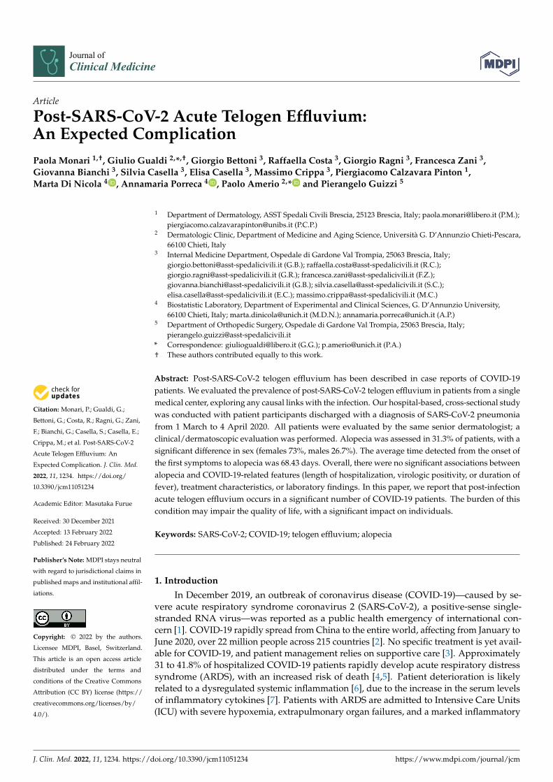
TE involved the whole scalp, with a gradual loss of hair volume in all patients report-ing increased hair loss. (Figure 2). Women, in particular, reported difficulty in bending theirhair. Dermoscopy (trichoscopy) was useful for the purpose of making a good assessment ofthe rarefaction and concentration of vellus hair, in order to differentiate TE from androgenicalopecia (Figure 3). Images of the vertex were collected and compared to those of the occipi-tal and the supra-auricular areas. At trichoscopy, most of the hair had a normal appearance.In addition, follicular openings with only one hair predominated. Dermoscopy showedno significant changes between the vertex area, the parietal zone, and the occipital zone,with no miniaturization, and vellus hair allowed to exclude the diagnosis of androgeneticalopecia in all patients.
J. Clin. Med. 2022, 11, 1234 5 of 8
Figure 2. Macroscopic photo showing diffuse alopecia. The arrows indicate typical areas of alopecia.
Figure 3. The predominance of single-hair follicular units, with perifollicular discoloration andgeneralized tiny hair (red circle). Note the presence of upright regrowing hairs (blue circle).
3.3. COVID-19 Characteristics and Alopecia
Overall, there was no association between COVID-19-related characteristics (days ofhospitalization, days of COV-2 positivity, days with fever, and TE (Table 1). The diseasewas not associated with any COVID-directed therapy (Table 1).
3.4. Laboratory and Alopecia
Overall, there was no association between the laboratory test outcomes of the controlgroup and the patients with TE. In particular, the levels of iron (p = 0.371), ferritin (p = 0.194),transferrin (p = 0.890), and zinc (p = 0.375) were similar in patients with and without TE.

J. Clin. Med. 2022, 11, 1234 6 of 8
4. Discussion
The current study is the first prospective cohort study examining the prevalence of TEfollowing SARS-CoV-2 infection. Previous short reports on a limited number of patientsshowed an increase in the incidence of post-SARS-CoV-2 TE [20], with particular referenceto some ethnic groups [20]. We report a higher incidence of TE in patients after SARS-CoV-2,with about one-third of them presenting with this condition. Due to the TE multifactorialetiology, it’s unclear if this occurrence is more closely related to the stress associated withthe fear of the pandemic or, as we think, if it’s triggered directly by the infection. Indeed,it should be considered that the underlying inflammation may be a triggering factor in asubset of patients [21]. Virus infections have been reported in the literature as having arelationship with the onset of TE. This may be due to direct virus aggression of human hairfollicle dermal papilla cells as demonstrated for Dengue virus infection in Taiwan in 2014and 2015 [22]. Other cases have been described in patients with HIV infection, triggered byinfectious or associated diseases, nutritional deficiencies, or, often by antiviral drugs [23]
The rate of post-SARS-CoV2 TE appeared to be significantly higher in females, al-though in males the onset of alopecia was more premature. This occurrence could be linkedto the evidence that men infected with CoV-2 have an increased risk of severe COVID-19disease compared to women [24]. A large number of other factors have been associatedwith this gender disparity. There is evidence that supports the role of androgens in theCOVID-19 severity [25,26], to the extent that androgenic alopecia has been hypothesized asa predicting factor for more severe SARS-CoV-2 cases [27]. In our study, we report TE notas a causative factor, but as the consequence of the disease. Physiological stress, such aschildbirth, surgical trauma, high fever, chronic systemic illness or infections, and hemor-rhage have been associated with TE. However, the relationship between TE and emotionalstress is uncertain and ambiguous [23]. TE has also been linked to severe protein, fattyacid and zinc deficiency, chronic starvation, and caloric restriction [23]. Numerous drugscan cause TE, starting after Week 12 of dosage [11,28]. Iatrogenic causes of TE include oralcontraceptive pills, androgens, retinoids, beta-blockers, angiotensin-converting enzymeinhibitors, anticonvulsants, antidepressants, and anticoagulants (heparin) [29]. The relationbetween TE and anticoagulant therapy is strongly controversial in the literature [19]; how-ever, in our study, no association was manifest. The onset of hair loss in our sample startedjust over 2 months from the beginning of the fever but, at the same time, it showed no linkwith any significant laboratory abnormalities. In particular, zinc and iron deficiencies werenot related to the onset of alopecia. In the context of these considerations, ATE followingSARS-CoV infection is to be considered as a foreseeable complication directly related tothe disease. Our report on the absence of any association with the severity of infectionand treatments has major implications on the number of patients potentially affected. Theabsence of a nutritional deficiency, combined with trichodynia, may suggest that the in-flammatory condition inherent to COVID-19 may be a principal cause of ATE. Interestingly,the prevalence of trichodynia—a distinctive symptom of TE—is reported in only about 20%of TE patients [30], whereas in our sample, it affected almost one-quarter of subjects.
The onset of this late complication may lead to profound implications, further impair-ing the quality of life of patients, despite them having recovered from their primary illness.The burden of post-infection ATE will impair the quality of life and may have a significantimpact on individuals, employers, and the healthcare systems.
Author Contributions: Conceptualization, P.M. and G.G.; methodology, M.D.N., A.P.; validation,P.A., M.D.N., G.G. and P.C.P.; formal analysis, M.D.N. and A.P.; investigation, P.M., G.B. (GiorgioBettoni), R.C., G.R., F.Z., G.B. (Giovanna Bianchi), S.C., E.C. and M.C.; data curation, A.P. and M.D.N.,M.C. and E.C.; writing—original draft preparation, P.M., G.G., P.A. and P.G.; writing—review andediting, P.A., P.C.P., P.G., S.C., G.B. (Giorgio Bettoni), F.Z., G.R., R.C. and G.B. (Giovanna Bianchi);supervision, P.C.P., P.A. and P.G. All authors have read and agreed to the published version of themanuscript.
Funding: This research received no external funding.
J. Clin. Med. 2022, 11, 1234 7 of 8
Conflicts of Interest: The authors declare no conflict of interest.
References1. Malik, Y.S.; Sircar, S.; Bhat, S.; Vinodhkumar, O.R.; Tiwari, R.; Sah, R.; Rabaan, A.A.; Rodriguez-Morales, A.J.; Dhama, K. Emerging
coronavirus disease (COVID-19), a pandemic public health emergency with animal linkages: Current status update. Preprints2020, 2020030343. [CrossRef]
2. COVID-19 Coronavirus Pandemic, 10 June. Worldometers.Info. Available online: https://www.worldometers.info/coronavirus/?utm_campaign=homeAdvegas1 (accessed on 18 August 2020).
3. Li, L.; Li, R.; Wu, Z.; Yang, X.; Zhao, M.; Liu, J.; Chen, D. Therapeutic strategies for critically ill patients with COVID-19. Ann.Intensive Care 2020, 10, 45. [CrossRef] [PubMed]
4. Wu, C.; Chen, X.; Cai, Y.; Xia, J.; Zhou, X.; Xu, S.; Huang, H.; Zhang, L.; Zhou, X.; Du, C.; et al. Risk Factors Associated With AcuteRespiratory Distress Syndrome and Death in Patients With Coronavirus Disease 2019 Pneumonia in Wuhan, China. JAMA Intern.Med. 2020, 180, 934–943. [CrossRef] [PubMed]
5. Wu, J.T.; Leung, K.; Leung, G.M. Nowcasting and forecasting the potential domestic and international spread of the 2019-nCoVoutbreak originating in Wuhan, China: A modelling study. Lancet 2020, 395, 689–697. [CrossRef]
6. McGonagle, D.; Sharif, K.; O’Regan, A.; Bridgewood, C. The Role of Cytokines including Interleukin-6 in COVID-19 inducedPneumonia and Macrophage Activation Syndrome-Like Disease. Autoimmun. Rev. 2020, 19, 102537. [CrossRef]
7. Zhao, Y.; Zhang, Y.-H.; Denney, L.; Young, D.; Powell, T.J.; Peng, Y.-C.; Li, N.; Yan, H.-P.; Wang, D.-Y.; Shu, Y.-L.; et al. HighLevels of Virus-Specific CD4+T Cells Predict Severe Pandemic Influenza A Virus Infection. Am. J. Respir. Crit. Care Med. 2012, 186,1292–1297. [CrossRef] [PubMed]
8. Stewart, I.J.; Sosnov, J.A.; Howard, J.T.; Orman, J.A.; Fang, R.; Morrow, B.D.; Zonies, D.H.; Bollinger, M.; Tuman, C.; Freedman,B.A.; et al. Retrospective Analysis of Long-Term Outcomes after Combat Injury: A Hidden Cost of War. Circulation 2015, 132,2126–2133. [CrossRef] [PubMed]
9. Grover, C.; Khurana, A. Telogen effluvium. Indian J. Dermatol. Venereol. Leprol. 2013, 79, 591–603. [CrossRef] [PubMed]10. Headington, J.T. Telogen effluvium. New concepts and review. Arch Dermatol. 1993, 129, 356–363. [CrossRef] [PubMed]11. Rebora, A. Proposing a Simpler Classification of Telogen Effluvium. Skin Appendage Disord. 2016, 2, 35–38. [CrossRef]12. Shrivastava, S.B. Diffuse hair loss in an adult female: Approach to diagnosis and management. Indian J Dermatol Venereol Leprol.
2009, 75, 20–27, quiz 27–8. [CrossRef] [PubMed]13. Rudnicka, L.; Olszewska, M.; Rakowska, A.; Slowinska, M. Trichoscopy update 2011. J. Dermatol. Case Rep. 2011, 5, 82–88.
[CrossRef] [PubMed]14. Ross, E.K.; Vincenzi, C.; Tosti, A. Videodermoscopy in the evaluation of hair and scalp disorders. J. Am. Acad. Dermatol. 2006, 55,
799–806. [CrossRef] [PubMed]15. Lacarrubba, F.; Dall’Oglio, F.; Nasca, M.R.; Micali, G. Videodermatoscopy enhances diagnostic capability in some forms of hair
loss. Am. J. Clin. Dermatol. 2004, 5, 205–208. [CrossRef] [PubMed]16. Rakowska, A.; Olszewska, M.; Rudnicka, L. Telogen effluvium. In Atlas of Trichoscopy; Rudnicka, L., Olszewska, M., Rakowska,
A., Eds.; Springer: London, UK, 2012; pp. 237–244.17. Olds, H.; Liu, J.; Luk, K.; Lim, H.W.; Ozog, D.; Rambhatla, P.V. Telogen effluvium associated with COVID-19 infection. Dermatol.
Ther. 2021, 6, e14761. [CrossRef]18. Olsen, E.A.; Rosen, S.T.; Vollmer, R.T.; Variakojis, D.; Roenigk, H.H., Jr.; Diab, N.; Zeffren, J. Interferon alfa-2a in the treatment of
cutaneous T cell lymphoma. J. Am. Acad. Dermatol. 1989, 20, 395–407. [CrossRef]19. Watras, M.M.; Patel, J.P.; Arya, R. Traditional anticoagulants and hair loss: A role for direct oral anticoagulants? A review of the
literature. Drugs Real World Outcomes 2016, 3, 1–6. [CrossRef]20. Cline, A.; Kazemi, A.; Moy, J.; Safai, B.; Marmon, S. A Surge in the Incidence of Telogen Effluvium in Minority Predominant
Communities Heavily Impacted by COVID-19. J. Am. Acad. Dermatol. 2020, 84, 773–775. [CrossRef]21. Puntmann, V.O.; Carerj, M.L.; Wieters, I.; Fahim, M.; Arendt, C.; Hoffmann, J.; Shchendrygina, A.; Escher, F.; Vasa-Nicotera, M.;
Zeiher, A.M.; et al. Outcomes of cardiovascular magnetic resonance imaging in patients recently recovered from coronavirusdisease 2019 (COVID-19). JAMA Cardiol. 2020, 5, 1265–1273. [CrossRef]
22. Wei, K.C.; Huang, M.S.; Chang, T.H. Dengue Virus Infects Primary Human Hair Follicle Dermal Papilla Cells. Front. Cell Infect.Microbiol. 2018, 8, 268. [CrossRef]
23. Almagro, M.; del Pozo, J.; Garcia-Silva, J.; Castro, A.; López-Calvo, S.; Yebra-Pimentel, M.T.; Fonseca, E. Telogen effluvium as aclinical presentation of human immunodeficiency virus infection. Am. J. Med. 2002, 112, 508–509. [CrossRef]
24. Gebhard, C.; Regitz-Zagrosek, V.; Neuhauser, H.K.; Morgan, R.; Klein, S.L. Impact of sex and gender on COVID-19 outcomes inEurope. Biol. Sex Differ. 2020, 11, 29. [CrossRef] [PubMed]
25. Wambier, C.G.; Vaño-Galván, S.; McCoy, J.; Gomez-Zubiaur, A.; Herrera, S.; Hermosa-Gelbard, Á.; Moreno-Arrones, O.M.;Jiménez-Gómez, N.; González-Cantero, A.; Fonda-Pascual, P. Segurado-Miravalles, G. Androgenetic alopecia present in themajority of patients hospitalized with COVID-19: The “Gabrin sign”. J. Am. Acad. Dermatol. 2020, 83, 680–682. [CrossRef][PubMed]

J. Clin. Med. 2022, 11, 1234 8 of 8
26. Montopoli, M.; Zumerle, S.; Vettor, R.; Rugge, M.; Zorzi, M.; Catapano, C.V.; Carbone, G.M.; Cavalli, A.; Pagano, F.;Ragazzi, E.; et al. Androgen-deprivation therapies for prostate cancer and risk of infection by SARS-CoV-2: A population-basedstudy (N = 4532). Ann. Oncol. 2020, 31, 1040–1045. [CrossRef]
27. Asghar, F.; Shamim, N.; Farooque, U.; Sheikh, H.; Aqeel, R. Telogen Effluvium: A Review of the Literature. Cureus 2020, 12, e8320.[CrossRef]
28. Tosti, A.; Pazzaglia, M. Drug reactions affecting hair: Diagnosis. Dermatol. Clin. 2007, 25, 223–231. [CrossRef]29. Harrison, S.; Bergfeld, W. Diffuse hair loss: Its triggers and management. Cleve Clin. J. Med. 2009, 76, 361–367. [CrossRef]30. Sulzberger, M.B.; Witten, V.H.; Kopf, A.W. Diffuse alopecia in women. Its unexplained apparent increase in incidence. Arch
Dermatol. 1960, 81, 556–560. [CrossRef]