Chimeric severe acute respiratory syndrome coronavirus (SARS-CoV) S glycoprotein and influenza...
16
Chimeric severe acute respiratory syndrome coronavirus (SARS-CoV) S glycoprotein and influenza matrix 1 efficiently form virus-like particles (VLPs) that protect mice against challenge with SARS-CoV Ye V. Liu a,* , Michael J. Massare a , Dale L. Barnard b , Thomas Kort a , Margret Nathan a , Lei Wang a , and Gale Smith a a Novavax, Inc. 9920 Belward Campus Drive, Rockville, MD 20850 b Institute for Antiviral Research, Utah State University, Logan, UT 84322 Abstract SARS-CoV was the cause of the global pandemic in 2003 that infected over 8000 people in 8 months. Vaccines against SARS are still not available. We developed a novel method to produce high levels of a recombinant SARS virus-like particles (VLPs) vaccine containing the SARS spike (S) protein and the influenza M1 protein using the baculovirus insect cell expression system. These chimeric SARS VLPs have a similar size and morphology to the wild type SARS-CoV. We tested the immunogenicity and protective efficacy of purified chimeric SARS VLPs and full length SARS S protein vaccines in a mouse lethal challenge model. The SARS VLP vaccine, containing 0.8 μg of SARS S protein, completely protected mice from death when administered intramuscular (IM) or intranasal (IN) routes in the absence of an adjuvant. Likewise, the SARS VLP vaccine, containing 4 μg of S protein without adjuvant, reduced lung virus titer to below detectable level, protected mice from weight loss, and elicited a high level of neutralizing antibodies against SARS-CoV. Sf9 cell-produced full length purified SARS S protein was also an effective vaccine against SARS-CoV but only when co-administered IM with aluminum hydroxide. SARS-CoV VLPs are highly immunogenic and induce neutralizing antibodies and provide protection against lethal challenge. Sf9 cell-based VLP vaccines are a potential tool to provide protection against novel pandemic agents. Keywords severe acute respiratory syndrome; virus like particles; lung virus titer; neutralizing antibody; baculovirus; influenza 1. Introduction Severe Acute Respiratory Syndrome (SARS) is an emerging infectious disease [1]. From November 2002 to July 2003, the SARS global outbreak spread from Guang Dong province © 2011 Elsevier Ltd. All rights reserved. * Corresponding author: Tel: +1 240 268 2069; Fax: +1 240 268 2100; [email protected] . Publisher's Disclaimer: This is a PDF file of an unedited manuscript that has been accepted for publication. As a service to our customers we are providing this early version of the manuscript. The manuscript will undergo copyediting, typesetting, and review of the resulting proof before it is published in its final citable form. Please note that during the production process errors may be discovered which could affect the content, and all legal disclaimers that apply to the journal pertain. NIH Public Access Author Manuscript Vaccine. Author manuscript; available in PMC 2012 September 2. Published in final edited form as: Vaccine. 2011 September 2; 29(38): 6606–6613. doi:10.1016/j.vaccine.2011.06.111. NIH-PA Author Manuscript NIH-PA Author Manuscript NIH-PA Author Manuscript
-
Upload
independent -
Category
Documents
-
view
0 -
download
0
Transcript of Chimeric severe acute respiratory syndrome coronavirus (SARS-CoV) S glycoprotein and influenza...
Chimeric severe acute respiratory syndrome coronavirus(SARS-CoV) S glycoprotein and influenza matrix 1 efficientlyform virus-like particles (VLPs) that protect mice againstchallenge with SARS-CoV
Ye V. Liua,*, Michael J. Massarea, Dale L. Barnardb, Thomas Korta, Margret Nathana, LeiWanga, and Gale Smitha
a Novavax, Inc. 9920 Belward Campus Drive, Rockville, MD 20850b Institute for Antiviral Research, Utah State University, Logan, UT 84322
AbstractSARS-CoV was the cause of the global pandemic in 2003 that infected over 8000 people in 8months. Vaccines against SARS are still not available. We developed a novel method to producehigh levels of a recombinant SARS virus-like particles (VLPs) vaccine containing the SARS spike(S) protein and the influenza M1 protein using the baculovirus insect cell expression system.These chimeric SARS VLPs have a similar size and morphology to the wild type SARS-CoV. Wetested the immunogenicity and protective efficacy of purified chimeric SARS VLPs and fulllength SARS S protein vaccines in a mouse lethal challenge model. The SARS VLP vaccine,containing 0.8 μg of SARS S protein, completely protected mice from death when administeredintramuscular (IM) or intranasal (IN) routes in the absence of an adjuvant. Likewise, the SARSVLP vaccine, containing 4 μg of S protein without adjuvant, reduced lung virus titer to belowdetectable level, protected mice from weight loss, and elicited a high level of neutralizingantibodies against SARS-CoV. Sf9 cell-produced full length purified SARS S protein was also aneffective vaccine against SARS-CoV but only when co-administered IM with aluminumhydroxide. SARS-CoV VLPs are highly immunogenic and induce neutralizing antibodies andprovide protection against lethal challenge. Sf9 cell-based VLP vaccines are a potential tool toprovide protection against novel pandemic agents.
Keywordssevere acute respiratory syndrome; virus like particles; lung virus titer; neutralizing antibody;baculovirus; influenza
1. IntroductionSevere Acute Respiratory Syndrome (SARS) is an emerging infectious disease [1]. FromNovember 2002 to July 2003, the SARS global outbreak spread from Guang Dong province
© 2011 Elsevier Ltd. All rights reserved.* Corresponding author: Tel: +1 240 268 2069; Fax: +1 240 268 2100; [email protected] .Publisher's Disclaimer: This is a PDF file of an unedited manuscript that has been accepted for publication. As a service to ourcustomers we are providing this early version of the manuscript. The manuscript will undergo copyediting, typesetting, and review ofthe resulting proof before it is published in its final citable form. Please note that during the production process errors may bediscovered which could affect the content, and all legal disclaimers that apply to the journal pertain.
NIH Public AccessAuthor ManuscriptVaccine. Author manuscript; available in PMC 2012 September 2.
Published in final edited form as:Vaccine. 2011 September 2; 29(38): 6606–6613. doi:10.1016/j.vaccine.2011.06.111.
NIH
-PA Author Manuscript
NIH
-PA Author Manuscript
NIH
-PA Author Manuscript
of China, to Southeast Asia, and North America. The World Health Organization reported atotal of 8098 SARS cases in 26 countries by July 2003, including 774 deaths and 9.6%mortality rate [2]. This wave of SARS pandemic was finally contained through internationalcoordination and strict quarantine enforcement by local governments. In addition to the lossof life, the affected region suffered from significant social disruptions and economic turmoil[3, 4].
SARS causes flu like symptoms including high fever, cough, difficulty breathing andpneumonia, and can spread from person to person by close contact via respiratory dropletsfrom coughing or sneezing. No specific anti-SARS treatments or vaccines are available [5].SARS is caused by a newly identified virus, SARS-associated coronavirus (SARS-CoV) [6].SARS-CoV is a ~100 nm diameter, enveloped, single strand RNA virus containing a 30 kbgenome with 14 open reading frames. SARS-CoV has four major structural proteins, thespike (S), membrane (M), envelope (E), and nucleocapsid (N) proteins [7]. The S protein isa type I transmembrane glycoprotein responsible for virus binding and virus penetration. Itis the major inducer of neutralizing antibodies and cellular immunity [8, 9]. The S proteinhas been the main target antigen for most SARS vaccines being developed [1, 10-13, 33].
Although we are in an inter-epidemic period with no reported SARS case worldwide, a safeand effective SARS vaccine remains an important public health need. There are naturalreservoirs of SARS-like coronavirus in wild bats and palm civets [14, 15], and humanscould be infected by close contact with other unknown wild animal reservoirs [16]. Also,there have been reported cases of laboratory acquired SARS virus infection of researchers in2004 [17]. SARS-CoV is classified by the National Institute of Allergy and InfectiousDiseases as a biodefense category C priority pathogen, with the potential for use as abiological weapon [18]. There are currently no approved SARS vaccines and only limitedhuman clinical trials with candidate vaccines have been done [22].
Previous SARS vaccine developments fall into several categories: inactivated SARS-CoVbased vaccines [19-21], DNA or other recombinant viruses expressing the S spikeglycoprotein [10, 11, 22, 47], subunit vaccines containing S receptor binding domain orsoluble ectodomain [23, 24, 52], and virus like particles (VLPs) based vaccines. Of these,SARS-CoV VLP vaccine candidates have shown considerable promise [30-32, 48].Recombinant VLP vaccine development has drawn increasing attention because it has thepotential to be safer than inactivated or attenuated viral vaccines and to be moreimmunogenic than subunit or DNA vaccines [25, 49, 50]. Recently, recombinant influenzaVLP vaccines have been reported to be well tolerated and immunogenic in human trials[51]. SARS S proteins have been incorporated into VLPs composed of either M and E, or Mand N structural proteins, but were shown to be produced at low levels in mammalian cells[26, 27]. In a baculovirus insect cell expression system, the SARS S proteins wereincorporated into secreted and intracellular VLPs composed of SARS M and E [28-31], butwith low yields. In an effort to address the low yields of SARS-CoV VLPs, chimeric VLPscontaining the S from SARS-CoV and M, E, N from mouse hepatitis virus were producedand showed increased yields relative to wild type VLPs when expressed in mammalian cells.In addition, immunization with these VLPs reduced lung virus titer in a mouse challengedmodel [32]. Although, SARS VLPs formation and its immunogenicity were shown to bepromising from these studies, the expression levels remained less than what would beneeded for a viable commercial vaccine candidate.
In this report, we described a novel method to produce high levels of SARS VLPs in insectcells infected with a recombinant baculovirus containing a chimeric SARS-CoV S proteincontaining the influenza hemagglutinin (HA) transmembrane and carboxyl terminus, and theavian influenza matrix 1 (M1) protein. These chimeric SARS VLPs are efficiently produced
Liu et al. Page 2
Vaccine. Author manuscript; available in PMC 2012 September 2.
NIH
-PA Author Manuscript
NIH
-PA Author Manuscript
NIH
-PA Author Manuscript
and secreted from infected Sf9 insect cells. Purified chimeric SARS S/M1 VLPs wereshown to completely protect immunized mice against lethal challenge with SARS-CoVwhile immunization with recombinant purified SARS S protein alone produced only partialprotection.
2. Materials and methods2.1. Plasmids, baculovirus and cells
The amino acids sequences of SARS-CoV Urbani strain spike protein (S, #AAP13441),influenza A/Indonesia/5/2005 (H5N1) hemagglutinin (HA, #ABP51969) and matrix protein1 (M1, #ABI36004) were obtained from the NCBI protein database. To construct a chimericSARS S protein, the transmembrane and carboxyl terminus (TM/CT) of the SARS S protein(aa 1196-1255) were removed and replaced with the TM/CT from A/Indonesia/5/2005 HA(aa 531-568). Codon optimized DNA sequences of the SARS S protein, the chimeric Sprotein, and the M1 protein used for high level expression in insect cells were biochemicallysynthesized by Geneart AG (Regensburg, Germany). These coding sequences wereindividually cloned into BamHI and HindIII sites of pFastBac1 baculovirus transfer vectorplasmid (Invitrogen, Carlsbad, CA). pFastBac1-M1 was digested with SnaBI and PvuI. The2571 bp fragment containing M1 coding sequence was gel purified. pFastBac1-S wasdigested with HpaI and PvuI. The 6870 bp fragment containing S coding sequence was gelpurified. The 2571 bp and 6870 bp fragments were ligated together with T4 DNA ligase(Roche, Indianapolis, IN). This process produced a tandem pFastBac1 plasmid thatcontained both the chimeric SARS S gene and the M1 gene with each gene under the controlof its own polyhedrin promoter.
The tandem chimeric S/M1 and the wild type full length SARS S transfer vectors weretransformed into DH10Bac competent cells (Invitrogen) to produce bacmids. The bacmidswere transfected into Spodoptera frugiperda Sf9 insect cells (ATCC CRL-1711) to generaterecombinant baculovirus using a Bac-to-Bac baculovirus expression system (Invitrogen).Sf9 cells were maintained as suspensions in HyQ-SFX insect serum free medium (HyClone,Logan, UT) at 27 ± 2°C.
2.2. SARS S and VLPs expression, purification and characterizationsSf9 cells were infected at 2 × 10⁶ cells/ml cell density for 66 hours with recombinantbaculovirus encoding SARS S or S/M1 chimeric VLP proteins at a multiplicity of infection(MOI) of 1. The SARS S proteins were exacted from infected cell pellet with non-ionicdetergent 0.5% Tergitol NP9 (Sigma-Aldrich, St Louis, MO). The clarified supernatant afterdetergent extraction were purified with a Fractogel TMAE anion exchange capture column(EMD chemicals, Darmstadt, Germany), followed by a Lentil lectin sepharose 4B affinitycolumn (GE healthcare, Piscataway, NJ), and finally polished with a Sephacryl S300 sizeexclusion column (GE healthcare). The chimeric SARS S/M1 VLPs (referred as VLPs inlater text) were purified from infected cell culture medium by tangential filtration, anionexchange, and size exclusion chromatography, the same procedure reported for influenzaVLPs purification [40].
Purified S proteins and chimeric VLPs were analyzed by SDS-PAGE (4-12% Bis-TrisNuPage, Invitrogen), stained with GelCode Blue stain (Pierce, Rockford, IL), and quantifiedby scanning densitometry using OneDscan software (BD Biosciences, Rockville, MD). Foranimal studies, purified S proteins and chimeric VLPs were normalized to have the sameamount of SARS S protein concentration based on total protein by BCA assay (Pierce) and Sprotein content (purity) by densitometry. The identity of the SARS S protein and theinfluenza M1 protein were confirmed by western blot using the following antibodies: rabbit
Liu et al. Page 3
Vaccine. Author manuscript; available in PMC 2012 September 2.
NIH
-PA Author Manuscript
NIH
-PA Author Manuscript
NIH
-PA Author Manuscript
anti-SARS S antibody (Imgnex, San Diego, CA), mouse anti-influenza M1 antibody (AbDSerotec, Oxford, UK), goat anti-mouse and goat anti-rabbit phosphatase labeled secondaryantibody (KPL, Gaithersburg, MD). BCIP/NBT phosphatase substrate (KPL) was used todevelop the western blot. Particle size of the SARS S protein and VLP vaccine weremeasured by dynamic light scattering using ZETASizer Nano (Malvern Instrument,Worcestershire, UK).
2.3. Electron microscopy analysisChimeric SARS VLPs were adsorbed for 2 min by flotation onto a freshly discharged 400mesh carbon parlodion-coated copper grid (Poly-Sciences, Warrington, PA). The grids wererinsed with 20mM Tris, pH 7.4, and 120mM KCl, negatively stained with 1%phosphotungstic acid, then dried by aspiration. VLPs were visualized on a Hitachi H-7600transmission electron microscope (Hitachi High Technologies America, Schaumburg, IL)operating at 80 kV and digitally captured with a CCD camera at 1K×1K resolution(Advanced Microscopy Techniques Corp., Danvers, MA). For immunoelectron microscopy(Immuno EM), rabbit anti-SARS S antibody (Imgnex) was used as primary antibody and 6nm colloidal gold-affinity pure goat anti-rabbit IgG (Jackson Immuno Research, WestGrove, PA) was used as secondary antibody as described previously [46].
2.4. Vaccination and challengeA total of 14 groups of 6-8 weeks old female Balb/c mice, 15 animals per group, were usedin this study. Nine groups were vaccinated intramuscularly (IM) through hind limb withvehicle (PBS), 0.8 μg or 4 μg of SARS S or VLP vaccine, with or without aluminumhydroxide (Brenntag AG, Mülheim, Germany) adjuvant. Five groups were vaccinatedintranasally (IN) with vehicle, 0.8 μg or 4 μg of SARS S or VLP vaccine without adjuvant.The animals were vaccinated on day 0 and day 21. On day 42, mice were intranasallychallenged with 2 lethal dose 50 (LD50) of mouse-adapted SARS-CoV strain v2163 (1 LD50= 103.64 50 TCID50). On Day 45, 3 days post challenge, 5 mice from each group weresacrificed to collect lung tissue, and assayed for SARS-CoV lung titer. For the remaining 10mice per group, body weight, morbidity, and mortality were measured daily from day 42 to63.
In a second animal study to measure SARS-CoV neutralizing antibody titers, a total of 9groups of 6-8 weeks old female Balb/c mice, 5 mice per group, were immunized IM withvehicle, 0.8 μg or 4 μg of SARS S or VLP vaccine, with or without aluminum hydroxideadjuvant, on day 0 and day 21. The animals were bled on day 0, 21 and 42 to measureneutralizing antibody titers. All animals had undetectable level of SARS-CoV neutralizingantibody titers on day 0.
SARS-CoV Urbani strain (200300592) was obtained from the Centers for Disease Controland Prevention (Atlanta, GA) and routinely passed in Vero-76 cells. SARS-CoV v2163 wasa lethal strain derived from the mouse adapted Urbani strain [45].
2.5. Lung virus titer determinationEach mouse lung was weighed and homogenized in MEM (minimum essential medium).Each lung homogenate was centrifuged at 2000 g for 5 min and 10 fold serial dilutions ofthe supernatant was assayed, in triplicate, for infectious virus by plaque assay usingVero-76. Virus titers were measured as CCID50/g tissue and were calculated using the Reed-Muench method. A titer of 0.75 CCID50/g tissue was the minimum detection limit of theassay.
Liu et al. Page 4
Vaccine. Author manuscript; available in PMC 2012 September 2.
NIH
-PA Author Manuscript
NIH
-PA Author Manuscript
NIH
-PA Author Manuscript
2.6. Neutralizing antibody titer determinationA 7 μL aliquot of serum was added to 63 μL of MEM + 50 μg/mL gentamicin, mixed, thenserially diluted 2-fold to achieve a 1/20 to 1/5120 dilutions in 96-well round-bottom plates.SARS-CoV Urbani strain stock was diluted in MEM to 200 CCID50 per 60 μL. 60 μL ofdiluted virus was added to each well, the plates were vibrated for approximately 1 minute,and incubated for 1 hour at 37°C. 100 μL of the virus/serum mixture from each well wastransferred to each well of a 96-well plates containing sub-confluent Vero-76 cells and 100μL of MEM + 4% fetal bovine serum (FBS). Plates were sealed with tape and incubated for5 days at 37°C in 5% CO2, then scored for the presence or absence of virus microscopicallybased on cytopathic effect (CPE). Neutralization titer was calculated as the inverse of thegreatest dilution of serum where no CPE was detected. The assays were performed induplicate and a serum titer of 1:20 was the lowest dilution tested in the assay.
2.7. Ethics regulation of laboratory animalsThe animal studies were conducted in accordance with the approval of the InstitutionalAnimal Care and Use Committee of Utah State University dated 21 September, 2004. Thestudy was conducted in the AAALAC-accredited Laboratory Animal Research Center ofUtah State University. The U. S. Government (National Institutes of Health) approval wasrenewed 27 February, 2002 (Assurance no. A3801-01) in accordance with the NationalInstitutes of Health Guide for the Care and Use of Laboratory Animals (Revision 1996).
3. Results3.1. Expression and characterization of SARS S and VLPs
Full length recombinant SARS S protein was produced in the baculovirus insect cellexpression system (Figure 1B). The SARS S protein was isolated from infected cellssolublized with non-ionic detergent and purified by column chromatography. Purified fulllength SARS S protein had an apparent molecular weight of 160 kDa by SDS-PAGE (Figure1D). Purified SARS S protein formed ~25 nm diameter particles consisting of multiple Sprotein molecules when observed by dynamic light scattering and electron microscopy (datanot shown).
We attempted to produce wild type SARS VLPs with homologous SARS S, M, and Eproteins in insect cells but the process was inefficient and the VLP incorporated proteinscould only be detected by western blot. No S, M or E protein bands were observed in thecoomassie blue stained gel (data not shown). This was probably due to the lack of anefficient matrix core protein needed for VLPs formation. Influenza matrix protein M1,especially the M1 from avian influenza A/Indonesia/5/2005 (H5N1), has been shown tosupport the production of high yield influenza VLPs even with hemagglutinin andneuraminidase from other heterologous influenza strains [34, 35]. As a result of thosestudies we decided to explore the possibility of forming a chimeric VLP containing theSARS S protein and the influenza M1 matrix protein. To maximize the interaction betweenthe SARS S and the influenza M1, we replaced the wild type SARS S protein’stransmembrane and carboxyl terminus (TM/CT) with the A/Indonesia/5/2005 influenzavirus hemagglutinin TM/CT, and constructed a tandem transfer vector for chimeric S/M1VLP production as shown in Figure 1A.
Chimeric S/M1 VLPs were produced at high level from insect cells and were purified fromthe infected culture medium. The purified VLPs produced intense SARS S and influenza M1protein bands on coomassie blue stained gel, at 14% and 51% respective purity as measuredby scanning densitometry (Figure 1C). Two additional protein bands present in the VLPswere identified as baculovirus envelope protein gp64 (#) and insect cell skeleton protein β-
Liu et al. Page 5
Vaccine. Author manuscript; available in PMC 2012 September 2.
NIH
-PA Author Manuscript
NIH
-PA Author Manuscript
NIH
-PA Author Manuscript
tubulin (*) by western blot (data not shown). These two proteins have been detectedpreviously in some of the influenza VLPs we have produced in insect cells. Tubulin is likelyto reside inside of VLPs since it may work together with actin to facilitate the baculovirus orVLPs transition to cell surface [53]. Glycoprotein gp64 is likely to be embedded in the VLPsenvelope. Additional cell culture and purification development may help to reduce the levelof these baculovirus and insect cell proteins in the SARS VLPs.
The average diameter of the chimeric S/M1 VLPs was 160 nm as measured by dynamiclight scattering. The S/M1 VLPs contained spherical enveloped particles with a corona-likespike structure on the outer rim as determined by electron microscopy (EM) (Figure 2A).The size and morphology of the S/M1 VLPs closely resembled the previously publishedSARS-CoV structures [36-38]. Immuno-EM using anti-SARS S protein primary antibodyand 6 nm gold particles labeled secondary antibody showed that multiple S proteins werelocated on the surface of the spherical S/M1 VLPs (Figure 2B).
3.2. Chimeric SARS VLPs protect mice from lethal challenge of SARS-CoVThe immunogenicity and protective efficacy of SARS S protein and S/M1 chimeric VLPvaccines were tested in a mouse lethal challenge model. Mice were intramuscularly (IM) orintranasally (IN) immunized with vehicle, 0.8 μg or 4 μg of SARS S protein or S/M1 VLPs,with or without aluminum hydroxide adjuvant, on day 0 and 21. The animals werechallenged with SARS-CoV lethal strain v2163 on day 42. All mice in the vehicle controlgroup died at 5.0 ± 0.9 days post challenge (Figure 3A). In the IM immunization groups, 0.8μg of purified SARS S protein without alum protected 7 out of 10 mice from death (Figure3B). In all other IM groups, 100% of the mice were protected from death (Figure 3A, 3B).Mice immunized with 0.8 μg or 4 μg of purified SARS S protein, without alum, lost up to15% and 7.5% of body weight respectively before they fully recovered (Figure 3C). AllVLP groups and S + alum groups did not show any sign of weight loss (Figure 3C), and thelung virus titers, at 3 days post challenge, appeared to correlate with the survival data(Figure 3D): without adjuvant, 0.8 μg of the SARS S protein failed to significantly reducelung virus titer, 4 μg of the SARS S protein reduced virus titer 2.5 log, 0.8 μg of the SARSVLPs reduced virus titer 6 logs, and 4 μg of the SARS VLPs reduced virus titer below thedetectable limit. Likewise, in all groups containing SARS S protein with alum, lung virustiters were reduced below the detectable limit. This data indicated that as low as 0.8 μg ofIM administered SARS VLP vaccine, without adjuvant, can fully protect mice from deathand weight loss from a lethal virus challenge. IM administered SARS S protein vaccine canfully protect mice but only when co-administered with an alum adjuvant.
For IN immunization, no adjuvant was used. 0.8 μg or 4 μg of the SARS S protein failed toprotect mice from lethal challenge and resulted in 100% death, same as the vehicle controlgroup. Whereas, all mice immunized with 0.8 μg and 4 μg of the SARS VLPs survived viruschallenge (Figure 4A, 4B). The net weight loss for both of the VLP vaccine groups was ashigh as 3-4 % but the animals completely recovered (Figure 4C). Also, the SARS S proteinfailed to reduce lung virus titer. In the 0.8 μg and 4 μg VLP vaccine groups, lung virus titerwere reduced 1 and 3.5 logs respectively (Figure 4D). The data shows that without aneffective adjuvant, the SARS S protein failed to protect when administered IN. The IN VLPvaccine without adjuvant protected 100% of mice from death, similar to when the vaccine isgiven IM. However, the lung virus titers in the IN VLP vaccine groups were 4 logs higherthan the corresponding IM groups and the IN immunized animals suffered minor weightloss. The data from IM and IN lethal challenge studies indicate that the SARS S proteinvaccine requires an effective adjuvant to be protective. For both vaccines, the IM route ofadministration worked better than IN for reducing lung virus titer. One possible explanationis that local immunity was mainly induced in the upper respiratory tract during INimmunization and not in the lower respiratory tract where the SARS-CoV replicates [41-44]
Liu et al. Page 6
Vaccine. Author manuscript; available in PMC 2012 September 2.
NIH
-PA Author Manuscript
NIH
-PA Author Manuscript
NIH
-PA Author Manuscript
and where immunity was not fully established at the time of challenge. Whereas, after IMimmunization, a broader systemic humoral immunity was developed that offer a betterprotection against SARS-CoV challenge through out the respiratory tract. IM immunizationmay also induce higher neutralizing antibody titers to help reduce lung virus titers.
To measure the humoral neutralizing antibody response, mice were immunized IM withvehicle, 0.8 μg or 4 μg of the SARS S protein or the chimeric SARS S/M1 VLPs, with orwithout aluminum hydroxide, on day 0 and 21. Mice were bled on day 21 and 42, and thesera samples were assayed for neutralizing antibodies against SARS-CoV (Figure 5). Boththe SARS S protein and the SARS VLPs need two vaccinations to elicit any neutralizingantibody titers >250. The SARS S vaccine required alum adjuvant to induce neutralizingantibody titers above the base line, whereas 0.8 μg and 4 μg VLPs vaccine without alum caninduce neutralizing antibody titer up to 878 and 1525, respectively. This result directlycorrelated with the survival rate and post challenge lung virus titer as shown in Figure 3. TheSARS S vaccine with alum and the SARS VLPs vaccine groups had neutralizing antibodygeometry mean titer ranging from 878 to 2550, and they were not statistically different fromeach other. S with alum groups showed a trend of higher neutralizing antibody titer at day 42than VLPs with alum. One explanation may be that S protein can absorb to alum moreefficiently than VLPs, and the slow release of S from alum results a prolonged elevatedantibody response at day 42.
4. DiscussionIn this paper, we report a novel method to produce chimeric SARS VLPs containing thespike protein of SARS and the matrix protein of influenza virus using the baculovirus insectcell expression system. We obtained the level of one milligram (S content) SARS VLPs perliter of Sf9 cell culture in shake flask. Upon further cell culture optimization such as stir tankreactor, high cell density, fed-batch or medium exchange and purification development, suchyield is expected to increase several fold or more. We studied the immunogenicity andprotective efficacy of the SARS S protein vaccine and the SARS VLP vaccine in a micelethal challenge model. The VLP vaccine, without adjuvant, protected 100% of theimmunized mice from death when given IM, reduced lung virus titer to undetectable levels,and elicited serum neutralizing antibody titer up to 1500. On the other hand, the S proteinvaccine required alum adjuvant to effectively protect mice from lethal challenge, reducelung virus titers, and elicit neutralizing antibodies. The data suggest that both the SARS Svaccine with alum and the SARS VLP vaccine are good candidates for protection againstSARS-CoV infections when administered IM.
The recombinant VLP vaccines have been an attractive approach for vaccine developmentfor the past decade. VLPs do not contain the viral genome, therefore can not replicate inhumans or revert to a pathogenic form. These characteristics make VLPs a safer vaccine forthe recipient, and also a safer vaccine to manufacturing than inactivated or live attenuatedviral vaccines, especially for a dangerous virus such as SARS-CoV that can cause highmorbidity and death [25]. The repetitive antigen pattern represented on the surface of VLPsmake them easier to be recognized by antigen presenting cells than subunit vaccines andtherefore are able to induce a stronger and broader humoral and cellular immune response[34, 35, 39]. There are only three VLP vaccines approved for human use, the Hepatitis Bvaccine and the two HPV vaccines. All three are composed of single protein particleswithout a lipid bilayer envelope. In addition, one of the commercial HPV vaccines(Cervarix) is produced in the baculovirus insect cell expression system [25]. The morecomplex version of VLPs, such as influenza VLPs that contain hemagglutinin,neuraminidase, M1 proteins, and a lipid bilayer envelope, are currently being evaluated inphase II human clinical trials by Novavax, Inc. No other VLP vaccine is approved or in the
Liu et al. Page 7
Vaccine. Author manuscript; available in PMC 2012 September 2.
NIH
-PA Author Manuscript
NIH
-PA Author Manuscript
NIH
-PA Author Manuscript
late stage of clinical development, partially because VLPs are usually difficult to produce inhigh level due to the lack of an efficient viral core protein. In this report, we showed thatinfluenza matrix M1 protein has the potential to become a universal core protein to supportVLPs production for other viruses. It is not a coincidence that M1 can form VLPs with theSARS S protein. SARS-CoV and influenza virus share many common features: both have aRNA genome; both are 100-200 nm diameter spherical particles; both have a major surfacespike glycoprotein (S or HA) that form trimers, and both spike glycoproteins contain aglobular head domain and a stem region. The replacement of the SARS S TM/CT with theHA TM/CT may help to maximize the native interaction between core protein and surfaceantigen, and promote VLPs formation and secretion. The use of the influenza M1 as a coreprotein to form VLPs for other viruses is a promising approach that can be explored in thefuture. Besides of the potential yield increase, influenza M1 containing chimeric VLPs canquickly activated M1 specific memory CD4 T cells since most human population wereinfected multiple times with influenza virus during their lifetime. Activated M1 specificCD4 T cells can stimulate SARS S or other antigen specific B cells for differentiation andantibody secretion.
Our SARS chimeric VLP vaccine was manufactured by the same procedures as ourinfluenza VLP vaccine that has been successfully tested in Phase I and Phase II clinicalstudies [51]. These two products share common infrastructure, equipment, supplies(medium, resin, filter, etc) and utilize several identical standard operating procedures. In theunfortunate case of another SARS pandemic, countries or regions with existing influenzaVLP vaccine manufacturing capacity will have the ability to quickly switch to the SARSvaccine production in the same facility, just like switching to another seasonal flu strain. TheSARS VLP vaccine can be produced in an influenza VLP vaccine manufacturing facilitywithin a short, 10-12 weeks period, after the pandemic virus is identified and sequenced orsooner if an Urbani-like strain reoccurs. A multi-use facility for seasonal flu, pandemic flu,and SARS may be desired for many countries because during another SARS pandemic theborders between countries may be closed and restrict vaccines from entering. Thus,pandemic vaccine manufacturing capacity within one’s own borders may be critical toprotecting their own citizens from infection.
AcknowledgmentsWe thank Peter Pushko, Jason Mantich, Attiya Ahmad, Maithili Kale, Yingyun Wu, Malgorzata Wisniewska, JimNorton, Lou Potash, Travis Sadowski, Konnie Taylor, Sarathi Boddapati (Novavax Inc.) for their valuablecontribution to this study. We thank Fredrick Cassels and Heather Greenstone (NIAID, NIH) for their helpfulsuggestions to the animal study. We thank Michael Delannoy for his electron microscopy service. We thank ShinjiMakino (University of Texas) for providing SARS reagent and suggestions. The animal work was supported, inpart, by contract NO1-AI-15345 from the Virology Branch, NIAID, National Institutes of Health. This work wassupported by NIH grant 1U03AI06198801.
References[1]. Enjuanes L, Dediego ML, Alvarez E, Deming D, Sheahan T, Baric R. Vaccines to prevent severe
acute respiratory syndrome coronavirus-induced disease. Virus Res. 2008; 133(1):45–62.[PubMed: 17416434]
[2]. World Health Organization. Summary of probable SARS cases with onset of illness from 1November 2002 to 31 July 2003. http://www.who.int/csr/sars/country/table2004_04_21/en/
[3]. Ahmad A, Krumkamp R, Reintjes R. Controlling SARS: a review on China’s response comparedwith other SARS-affected countries. Trop Med Int Health. 2009; 14(Suppl 1):36–45. [PubMed:19508440]
[4]. Beutels P, Jia N, Zhou QY, Smith R, Cao WC, de Vlas SJ. The economic impact of SARS inBeijing, China. Trop Med Int Health. 2009; 14(Suppl 1):85–91. [PubMed: 19508435]
Liu et al. Page 8
Vaccine. Author manuscript; available in PMC 2012 September 2.
NIH
-PA Author Manuscript
NIH
-PA Author Manuscript
NIH
-PA Author Manuscript
[5]. Chan-Yeung M, Ooi GC, Hui DS, Ho PL, Tsang KW. Severe acute respiratory syndrome. Int JTuberc Lung Dis. 2003; 7(12):1117–30. [PubMed: 14677886]
[6]. Drosten C, Günther S, Preiser W, van der Werf S, Brodt HR, Becker S, et al. Identification of anovel coronavirus in patients with severe acute respiratory syndrome. N Engl J Med. 2003;348(20):1967–76. [PubMed: 12690091]
[7]. Masters PS. The molecular biology of coronaviruses. Adv Virus Res. 2006; 66:193–292.[PubMed: 16877062]
[8]. Li W, Moore MJ, Vasilieva N, Sui J, Wong SK, Berne MA, et al. Angiotensin-converting enzyme2 is a functional receptor for the SARS coronavirus. Nature. 2003; 426(6965):450–4. [PubMed:14647384]
[9]. Qiu M, Shi Y, Guo Z, Chen Z, He R, Chen R, et al. Antibody responses to individual proteins ofSARS coronavirus and their neutralization activities. Microbes Infect. 2005; 7(5-6):882–9.[PubMed: 15878679]
[10]. Bisht H, Roberts A, Vogel L, Bukreyev A, Collins PL, Murphy BR, et al. Severe acuterespiratory syndrome coronavirus spike protein expressed by attenuated vaccinia virusprotectively immunizes mice. Proc Natl Acad Sci U S A. 2004; 101(17):6641–6. [PubMed:15096611]
[11]. Yang ZY, Kong WP, Huang Y, Roberts A, Murphy BR, Subbarao K, et al. A DNA vaccineinduces SARS coronavirus neutralization and protective immunity in mice. Nature. 2004;428(6982):561–4. [PubMed: 15024391]
[12]. Vogel LN, Roberts A, Paddock CD, Genrich GL, Lamirande EW, Kapadia SU, et al. Utility ofthe aged BALB/c mouse model to demonstrate prevention and control strategies for SevereAcute Respiratory Syndrome coronavirus (SARS-CoV). Vaccine. 2007; 25(12):2173–9.[PubMed: 17227689]
[13]. Bai B, Lu X, Meng J, Hu Q, Mao P, Lu B, et al. Vaccination of mice with recombinantbaculovirus expressing spike or nucleocapsid protein of SARS-like coronavirus generateshumoral and cellular immune responses. Molecular Immunology. 2007; 45:868–75. [PubMed:17905435]
[14]. Rihtaric D, Hostnik P, Steyer A, Grom J, Toplak I. Identification of SARS-like coronaviruses inhorseshoe bats (Rhinolophus hipposideros) in Slovenia. Arch Virol. 2010; 155(4):507–14.[PubMed: 20217155]
[15]. Anderson LJ, Tong S. Update on SARS research and other possibly zoonotic coronaviruses. Int JAntimicrob Agents. Nov; 2010 36(Suppl 1):S21–5. Epub 2010 Aug 30. [PubMed: 20801001]
[16]. Guan Y, Zheng BJ, He YQ, Liu XL, Zhuang ZX, Cheung CL, et al. Isolation and characterizationof viruses related to the SARS coronavirus from animals in southern China. Science. 2003;302(5643):276–8. [PubMed: 12958366]
[17]. World Health Organization. China’s latest SARS outbreak has been contained, but biosafetyconcerns remain-Update 7, 18 May 2004. http://www.who.int/csr/don/2004_05_18a/en/
[18]. NIAID Category A, B, and C Priority Pathogens.http://www.niaid.nih.gov/topics/biodefenserelated/biodefense/research/pages/cata.aspx
[19]. Spruth M, Kistner O, Savidis-Dacho H, Hitter E, Crowe B, Gerencer M, et al. A double-inactivated whole virus candidate SARS coronavirus vaccine stimulates neutralising andprotective antibody responses. Vaccine. 2006; 24(5):652–61. [PubMed: 16214268]
[20]. Marshall E, Enserink M. Medicine. Caution urged on SARS vaccines. Science. 2004; 303(5660):944–6. [PubMed: 14963300]
[21]. Qin E, Shi H, Tang L, Wang C, Chang G, Ding Z, et al. Immunogenicity and protective efficacyin monkeys of purified inactivated Vero-cell SARS vaccine. Vaccine. 2006; 24(7):1028–34.[PubMed: 16388880]
[22]. Martin JE, Louder MK, Holman LA, Gordon IJ, Enama ME, Larkin BD, et al. A SARS DNAvaccine induces neutralizing antibody and cellular immune responses in healthy adults in a PhaseI clinical trial. Vaccine. 2008; 26(50):6338–43. [PubMed: 18824060]
[23]. Bisht H, Roberts A, Vogel L, Subbarao K, Moss B. Neutralizing antibody and protectiveimmunity to SARS coronavirus infection of mice induced by a soluble recombinant polypeptide
Liu et al. Page 9
Vaccine. Author manuscript; available in PMC 2012 September 2.
NIH
-PA Author Manuscript
NIH
-PA Author Manuscript
NIH
-PA Author Manuscript
containing an N-terminal segment of the spike glycoprotein. Virology. 2005; 334(2):160–5.[PubMed: 15780866]
[24]. Zhou Z, Post P, Chubet R, Holtz K, McPherson C, Petric M, et al. A recombinant baculovirus-expressed S glycoprotein vaccine elicits high titers of SARS-associated coronavirus (SARS-CoV) neutralizing antibodies in mice. Vaccine. 2006; 24(17):3624–31. [PubMed: 16497416]
[25]. Roy P, Noad R. Virus-like particles as a vaccine delivery system: myths and facts. Hum Vaccin.2008; 4(1):5–12. [PubMed: 18438104]
[26]. Hsieh PK, Chang SC, Huang CC, Lee TT, Hsiao CW, Kou YH, et al. Assembly of severe acuterespiratory syndrome coronavirus RNA packaging signal into virus-like particles is nucleocapsiddependent. J Virol. 2005; 79(22):13848–55. [PubMed: 16254320]
[27]. Huang Y, Yang ZY, Kong WP, Nabel GJ. Generation of synthetic severe acute respiratorysyndrome coronavirus pseudoparticles: implications for assembly and vaccine production. JVirol. 2004; 78(22):12557–65. [PubMed: 15507643]
[28]. Mortola E, Roy P. Efficient assembly and release of SARS coronavirus-like particles by aheterologous expression system. FEBS Letters. 2004; 576:174–178. [PubMed: 15474033]
[29]. Ho Y, Lin PH, Liu CY, Lee SP, Chao YC. Assembly of human severe acute respiratorysyndrome coronavirus-like particles. Biochem Biophys Res Commun. 2004; 318(4):833–8.[PubMed: 15147946]
[30]. Lu X, Chen Y, Bai B, Hu H, Tao L, Yang J. Immune responses against severe acute respiratorysyndrome coronavirus induced by virus-like particles in mice. Immunology. 2007; 122(4):496–502. [PubMed: 17680799]
[31]. Bai B, Hu Q, Hu H, Zhou P, Shi Z, Meng J. Virus-like particles of SARS-like coronavirusformed by membrane proteins from different origins demonstrate stimulating activity in humandendritic cells. PLoS One. 2008; 3(7):e2685. [PubMed: 18628832]
[32]. Lokugamage KG, Yoshikawa-Iwata N, Ito N, Watts DM, Wyde PR, Wang N, et al. Chimericcoronavirus-like particles carrying severe acute respiratory syndrome coronavirus (SCoV) Sprotein protect mice against challenge with SCoV. Vaccine. 2008; 26(6):797–808. [PubMed:18191004]
[33]. Roper RL, Rehm KE. SARS vaccines: where are we? Expert Rev Vaccines. 2009; 8(7):887–98.[PubMed: 19538115]
[34]. Bright RA, Carter DM, Crevar CJ, Toapanta FR, Steckbeck JD, Cole KS, et al. Cross-cladeprotective immune responses to influenza viruses with H5N1 HA and NA elicited by aninfluenza virus-like particle. PLoS One. 2008; 3(1):e1501. [PubMed: 18231588]
[35]. Song H, Wittman V, Byers A, Tapia T, Zhou B, Warren W, et al. In vitro stimulation of humaninfluenza-specific CD8+ T cells by dendritic cells pulsed with an influenza virus-like particle(VLP) vaccine. Vaccine. 2010; 28(34):5524–32. [PubMed: 20600506]
[36]. Almeida J, Tyrell D. The morphology of three previously uncharacterized human respiratoryviruses that grow in organ culture. J Gen Virol. 1967; (2):175–8. [PubMed: 4293939]
[37]. Zhang QF, Cui JM, Huang XJ, Lin W, Tan DY, Xu JW, et al. Morphology and morphogenesis ofsevere acute respiratory syndrome (SARS)-associated virus. Acta Biochimica et BiophysicaSinica. 2003; 35(6):587–591. [PubMed: 12796822]
[38]. Drosten, C.; Lau, A.; Preiser, W.; So, L.; Yam, L. SARS Reference. 3rd edition. Kamps, BS.;Hoffmann, C., editors. Flying Publisher; 2003. p. 41http://www.sarsreference.com
[39]. Bright RA, Carter DM, Daniluk S, Toapanta FR, Ahmad A, Gavrilov V, et al. Influenza virus-like particles elicit broader immune responses than whole virion inactivated influenza virus orrecombinant hemagglutinin. Vaccine. 2007; 25(19):3871–8. [PubMed: 17337102]
[40]. Pushko P, Kort T, Nathan M, Pearce MB, Smith G, Tumpey TM. Recombinant H1N1 virus-likeparticle vaccine elicits protective immunity in ferrets against the 2009 pandemic H1N1 influenzavirus. Vaccine. 2010; 28(30):4771–6. [PubMed: 20470801]
[41]. Hall LJ, Clare S, Dougan G. Probing local innate immune responses after mucosal immunisation.J Immune Based Ther Vaccines. 2010; 8(1):5. [PubMed: 20836885]
[42]. Hamming I, Timens W, Bulthuis ML, Lely AT, Navis GJ, van Goor H. Tissue distribution ofACE2 protein, the functional receptor for SARS coronavirus. A first step in understanding SARSpathogenesis. J Pathol. 2004; 203(2):631–7. [PubMed: 15141377]
Liu et al. Page 10
Vaccine. Author manuscript; available in PMC 2012 September 2.
NIH
-PA Author Manuscript
NIH
-PA Author Manuscript
NIH
-PA Author Manuscript
[43]. Ding Y, He L, Zhang Q, Huang Z, Che X, Hou J, et al. Organ distribution of severe acuterespiratory syndrome (SARS) associated coronavirus (SARS-CoV) in SARS patients:implications for pathogenesis and virus transmission pathways. J Pathol. 2004; 203(2):622–30.[PubMed: 15141376]
[44]. Chen J, Subbarao K. The Immunobiology of SARS. Annu Rev Immunol. 2007; 25:443–72.[PubMed: 17243893]
[45]. Day CW, Baric R, Cai SX, Frieman M, Kumaki Y, Morrey JD, et al. A new mouse-adapted strainof SARS-CoV as a lethal model for evaluating antiviral agents in vitro and in vivo. Virology.2009; 395(2):210–22. [PubMed: 19853271]
[46]. Murti KG, Portner A, Thoughton K, Deshpande K. Localization of proteins on viralnucleocapsids using immunoelectron microscopy. J. Electron Microscopy Tech. 1985; 2:139–146.
[47]. Feng Q, Liu Y, Qu X, Deng H, Ding M, Lau TL, et al. Baculovirus surface display of SARScoronavirus (SARS-CoV) spike protein and immunogenicity of the displayed protein in micemodels. Immunology. 2007; 122(4):496–502. [PubMed: 17680799]
[48]. Lu B, Huang Y, Huang L, Li B, Zheng Z, Chen Z, et al. Effect of mucosal and systemicimmunization with virus-like particles of severe acute respiratory syndrome coronavirus in mice.Immunology. 2010; 130(2):254–61. [PubMed: 20406307]
[49]. Ludwig C, Wagner R. Virus-like particles-universal molecular toolboxes. Curr Opin Biotechnol.2007; 18(6):537–45. [PubMed: 18083549]
[50]. Grgacic EV, Anderson DA. Virus-like particles: passport to immune recognition. Methods. 2006;40(1):60–5. [PubMed: 16997714]
[51]. Allende, M. Preliminary safety and immunogenicity of Novavax’s 2009 A/H1N1 VLP vaccine.WHO 6th Influenza Meeting; February 18-19, 2010; Manuscript submitted
[52]. Hu MC, Jones T, Kenney RT, Barnard DL, Burt DS, Lowell GH. Intranasal Protollin-formulatedrecombinant SARS S-protein elicits respiratory and serum neutralizing antibodies and protectionin mice. Vaccine. 2007; 25(34):6334–40. [PubMed: 17640780]
[53]. Ohkawa T, Volkman LE, Welch MD. Actin-based motility drives baculovirus transit to thenucleus and cell surface. J Cell Biol. 2010; 190(2):187–95. [PubMed: 20660627]
Liu et al. Page 11
Vaccine. Author manuscript; available in PMC 2012 September 2.
NIH
-PA Author Manuscript
NIH
-PA Author Manuscript
NIH
-PA Author Manuscript
Figure 1. Expression and purification of SARS S and S/M1 chimeric VLPA) pFastBac1 baculovirus transfer vector for SARS S/Flu M1 chimeric VLP vaccine: SARSS protein with the influenza A/Indonesia/5/2005 hemagglutinin (HA) transmembrane andcarboxyl terminus (TM/CT) and the M1 influenza matrix protein 1. Each gene is under thecontrol of its own polyhedrin promoter; B) pFastBac1 baculovirus transfer vector for wildtype full length SARS S protein with its native TM/CT; C) Purified SARS VLP vaccine, leftpanel: coomassie blue stain, right panel: western blot using anti-S and anti-M1 antibodies.Positions of the 160 kDa SARS S protein and the 25 kDa M1 protein are labeled. Bandslabeled # and * were identified as baculovirus gp64 protein and insect cell β tubulin proteinrespectively; D) Purified native SARS S protein, left panel: coomassie blue stain, rightpanel: western blot using anti-SARS S antibody. Lane M contains precision plus proteinmolecular weight marker (Bio-Rad, Hercules, CA)
Liu et al. Page 12
Vaccine. Author manuscript; available in PMC 2012 September 2.
NIH
-PA Author Manuscript
NIH
-PA Author Manuscript
NIH
-PA Author Manuscript
Figure 2. Transmission electron microscopy (EM) analysis of SARS VLPsA) Phosphotungstic acid negative stain of EM image of SARS VLPs; B) Immuno EM imageof SARS VLPs using anti-SARS S protein primary antibody and 6 nm colloidal gold labeledsecondary antibody. Black dots indicated gold labeled antibody binding to the SARS Sprotein. Bar: 100 nm.
Liu et al. Page 13
Vaccine. Author manuscript; available in PMC 2012 September 2.
NIH
-PA Author Manuscript
NIH
-PA Author Manuscript
NIH
-PA Author Manuscript
Figure 3. Survival rate and lung virus titer of mice intramuscularly (IM) immunized with SARSS protein vaccine or SARS VLP vaccine after SARS-CoV lethal challengeMice were immunized (IM) with vehicle, 0.8 μg or 4 μg of SARS S protein or SARS VLPs,with or without aluminum hydroxide, on day 0 and 21 (n = 14 or 15). Mice were challengedwith 2 LD50 of SARS-CoV strain v2163 on day 42. Five mice per group were sacrificed onday 45. Lung tissues were collected, homogenized, and assayed for virus titer. Theremaining mice were monitored for survival and body weight from day 42 to day 63. A)Survival percentage curve; B) Survival table with the mean day of death; C) Animal weightchange for 21 days post challenge; D) Lung SARS-CoV titer at day 45, 3 days postchallenge. Virus titer was calculated as Log10 CCID50/gram lung tissue with limit ofdetection as 0.75 (dashed line). Results were expressed as mean titer ± SEM. Statisticalsignificance between the vehicle and vaccine groups was determined by the student t test. **p<0.01, *** p<0.001.
Liu et al. Page 14
Vaccine. Author manuscript; available in PMC 2012 September 2.
NIH
-PA Author Manuscript
NIH
-PA Author Manuscript
NIH
-PA Author Manuscript
Figure 4. Survival rate and lung virus titer of mice intranasally (IN) immunized with SARS Sprotein vaccine or SARS VLP vaccine after SARS-CoV lethal challengeMice were immunized (IN) with vehicle, 0.8 μg or 4 μg of SARS S protein or SARS VLPs,without adjuvant, on day 0 and 21 (n = 14 or 15). Mice were challenged with 2 LD50 ofSARS-CoV strain v2163 on day 42. Five mice per group were sacrificed on day 45. Lungtissues were collected and assayed for virus titer. The remaining mice were monitored forsurvival and body weight from day 42 to day 63. A) Survival percentage curve; B) Survivaltable with the mean day of death; C) Animal weight change for 21 days after challenge; D)Lung SARS-CoV titer at day 45, 3 days post challenge. Virus titer was calculated as Log10CCID50/gram lung tissue. Results were expressed as mean titer ± SEM. Statisticalsignificance between the vehicle and vaccine groups was determined by the student t test.*** p<0.001.
Liu et al. Page 15
Vaccine. Author manuscript; available in PMC 2012 September 2.
NIH
-PA Author Manuscript
NIH
-PA Author Manuscript
NIH
-PA Author Manuscript
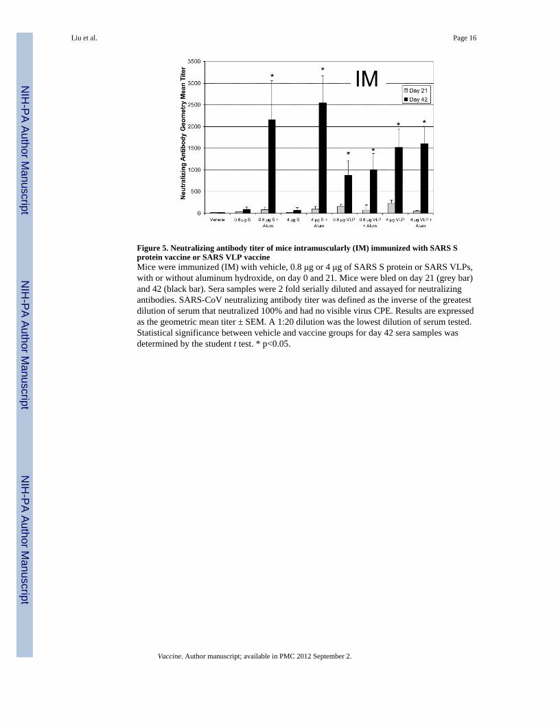
Figure 5. Neutralizing antibody titer of mice intramuscularly (IM) immunized with SARS Sprotein vaccine or SARS VLP vaccineMice were immunized (IM) with vehicle, 0.8 μg or 4 μg of SARS S protein or SARS VLPs,with or without aluminum hydroxide, on day 0 and 21. Mice were bled on day 21 (grey bar)and 42 (black bar). Sera samples were 2 fold serially diluted and assayed for neutralizingantibodies. SARS-CoV neutralizing antibody titer was defined as the inverse of the greatestdilution of serum that neutralized 100% and had no visible virus CPE. Results are expressedas the geometric mean titer ± SEM. A 1:20 dilution was the lowest dilution of serum tested.Statistical significance between vehicle and vaccine groups for day 42 sera samples wasdetermined by the student t test. * p<0.05.
Liu et al. Page 16
Vaccine. Author manuscript; available in PMC 2012 September 2.
NIH
-PA Author Manuscript
NIH
-PA Author Manuscript
NIH
-PA Author Manuscript