Isolation of Virus from a SARS Patient and Genome-wide Analysis of Genetic Mutations Related to...
10
Isolation of Virus from a SARS Patient and Genome-wide Analysis of Genetic Mutations Related to Pathogenesis and Epidemiology from 47 SARS-CoV Isolates YING ZHU, MO LIU, WEIGUANG ZHAO, JIANLIN ZHANG, XUE ZHANG, KE WANG, CHUNFANG GU, KAILANG WU, YAN LI, CONGYI ZHENG, GENGFU XIAO, HUIMIN YAN, JIAMIN ZHANG, DEYIN GUO, PO TIEN & JIANGUO WU* Key Laboratory of Virology, Ministry of Education College of Life Sciences, Wuhan University, Wuhan 430072, China Received June 8, 2004; Accepted July 15, 2004 Abstract. Severe acute respiratory syndrome (SARS) caused by SARS-associated coronavirus (SARS- CoV) is a fatal disease. Prevention of future outbreaks is essential and requires understanding pathogenesis and evolution of the virus. We have isolated a SARS-CoV in China and analyzed 47 SARS-CoV genomes with the aims to reveal the evolution trends of the virus and provide insights into understanding patho- genesis and SARS epidemic. Specimen from a SARS patient was inoculated into cell culture. The presence of SARS-CoV was determined by RT-PCR and confirmed by electron microscopy. Virus was isolated followed by the determination of its genome sequences, which were then analyzed by comparing with other 46 SARS-CoV genomes. Genetic mutations with potential implications to pathogenesis and the epidemic were characterized. This viral genome consists of 29,728 nucleotides with overall organization in agreement with that of published isolates. A total of 348 positions were mutated on 47 viral genomes. Among them 22 had mutations in more than three genomes. Hot spots of nucleotide variations and unique trends of mutations were identified on the viral genomes. Mutation rates were different from gene to gene and were correlated well with periodical or geographic characteristics of the epidemic. Key words: SARS-CoV, isolation, genomic analysis, mutation Introduction In November 2002, first case of a novel infectious disease named severe acute respiratory syndrome (SARS) suddenly appeared in southern China [1]. This illness emerged and rapidly spread to different areas of Asia and then other countries around the world with a high morbidity (about 25% required intensive care) and 9.6% fatality [2]. In March 2003, the World Health Organization (WHO) made an unprecedented international effort by organizing world-leading laboratories to find the causative agent. This effort resulted in the declaration made simultaneously by three research groups that a new SARS-associated coronavirus (SARS-CoV) was the pathogen of this disease [3– 5]. When the outbreak of SARS came to an end in July 2003, it had caused a cumulative total of 8437 cases and 813 deaths worldwide [6]. Since the discovery of SARS-CoV, progresses regarding the studies of this virus have been swift dramatically as the complete viral genome was sequenced [7]. Although the definition of SARS case still largely relied on clinical and epidemio- logical criteria, diagnostic tests based on the detection of viral RNA and proteins have been developed [8], along with the development of vaccines [9]. Results from both phylogenetic analysis and epidemiological studies suggested the *Author for all correspondence: E-mail: [email protected] Virus Genes 30:1, 93–102, 2005 Ó 2005 Springer Science+Business Media, Inc. Manufactured in The Netherlands.
-
Upload
independent -
Category
Documents
-
view
0 -
download
0
Transcript of Isolation of Virus from a SARS Patient and Genome-wide Analysis of Genetic Mutations Related to...

Isolation of Virus from a SARS Patient and Genome-wide Analysis of GeneticMutations Related to Pathogenesis and Epidemiology from 47 SARS-CoV
Isolates
YING ZHU, MO LIU, WEIGUANG ZHAO, JIANLIN ZHANG, XUE ZHANG, KE WANG,CHUNFANG GU, KAILANG WU, YAN LI, CONGYI ZHENG, GENGFU XIAO, HUIMIN YAN,
JIAMIN ZHANG, DEYIN GUO, PO TIEN & JIANGUO WU*Key Laboratory of Virology, Ministry of Education
College of Life Sciences, Wuhan University, Wuhan 430072, China
Received June 8, 2004; Accepted July 15, 2004
Abstract. Severe acute respiratory syndrome (SARS) caused by SARS-associated coronavirus (SARS-CoV) is a fatal disease. Prevention of future outbreaks is essential and requires understanding pathogenesisand evolution of the virus. We have isolated a SARS-CoV in China and analyzed 47 SARS-CoV genomeswith the aims to reveal the evolution trends of the virus and provide insights into understanding patho-genesis and SARS epidemic. Specimen from a SARS patient was inoculated into cell culture. The presenceof SARS-CoV was determined by RT-PCR and confirmed by electron microscopy. Virus was isolatedfollowed by the determination of its genome sequences, which were then analyzed by comparing with other46 SARS-CoV genomes. Genetic mutations with potential implications to pathogenesis and the epidemicwere characterized. This viral genome consists of 29,728 nucleotides with overall organization in agreementwith that of published isolates. A total of 348 positions were mutated on 47 viral genomes. Among them 22had mutations in more than three genomes. Hot spots of nucleotide variations and unique trends ofmutations were identified on the viral genomes. Mutation rates were different from gene to gene and werecorrelated well with periodical or geographic characteristics of the epidemic.
Key words: SARS-CoV, isolation, genomic analysis, mutation
Introduction
In November 2002, first case of a novel infectiousdisease named severe acute respiratory syndrome(SARS) suddenly appeared in southern China [1].This illness emerged and rapidly spread to differentareas of Asia and then other countries around theworld with a high morbidity (about 25% requiredintensive care) and 9.6% fatality [2]. In March2003, the World Health Organization (WHO)made an unprecedented international effort byorganizing world-leading laboratories to find thecausative agent. This effort resulted in the
declaration made simultaneously by three researchgroups that a new SARS-associated coronavirus(SARS-CoV) was the pathogen of this disease [3–5]. When the outbreak of SARS came to an end inJuly 2003, it had caused a cumulative total of 8437cases and 813 deaths worldwide [6].
Since the discovery of SARS-CoV, progressesregarding the studies of this virus have been swiftdramatically as the complete viral genome wassequenced [7]. Although the definition of SARScase still largely relied on clinical and epidemio-logical criteria, diagnostic tests based on thedetection of viral RNA and proteins have beendeveloped [8], along with the development ofvaccines [9]. Results from both phylogeneticanalysis and epidemiological studies suggested the
*Author for all correspondence:
E-mail: [email protected]
Virus Genes 30:1, 93–102, 2005� 2005 Springer Science+Business Media, Inc. Manufactured in The Netherlands.

origin of SARS-CoV was animal-oriented, mostlikely from Himalayan palm civets, ferrets andraccoon dogs [10–13].
As amember of theCoronoviridae family, SARS-CoV is enveloped andpositive-strandedRNAvirus.It harbors 23 coding sequences, including 4 primarystructural proteins (nucleocapsid protein N, spikeprotein S,membrane proteinM, and small envelopeprotein E); 5 non-structural proteins (X1, X2, X3,X4, X5); and 1 polyprotein that compose twoORFs(ORF1a and ORF1b). Polyprotein catalyticallyauto-processes to produce a group of proteinsincluding proteases (PLPpro and 3Clpro), RNA-dependent polymerase (POL), RNA helicase(HEL), and function unknownproteins [4,5,7]. Likeother RNA viruses, whose most striking charac-teristic is the high rate of genetic mutation [11,14–18].
Despite the fact that the SARS-CoV can causean atypical and fatal form of pneumonia, thegenome structure, gene expression pattern, andprotein profiles of the virus are similar to those ofother conventional coronaviruses [17], which areonly responsible for mild respiratory tract infec-tions in a wide range of animals includinghumans, pigs, cows, mice, cats, and birds [10,19].It is possible that distinct patterns of severalgenes and unique variations in the SARS-CoVgenome may contribute to its severe virulence orpathogenesis. The mechanism of SARS-CoVpathogenesis may involve both direct viral cyto-cidal effects on the target cells and immune-mediated mechanisms. Potential mutability of theviral genome may pose problems in the control offuture SARS epidemics.
In this report, we described the isolation of anew SARS-CoV strain (WHU) from a patient inHubei Province, China during the late period ofSARS outbreak. Complete genome sequence ofWHU isolate was determined and compared withthat of 46 other SARS-CoV strains whose com-plete genomic sequences were available at the timeanalyzed. Comparative study of genetic charac-terization and nucleotide variation of all knownSARS-CoV offers insights into understandingfunctions of the viral genes and revealing theevolution trends of the virus. It would also providebasis for clinical diagnosis, future developingpotential drugs and vaccines against SARS-CoVinfections.
Materials and Methods
SARS Patient
The SARS patient was an 18-year-old male fromJiayu County, Hubei Province, China. He workedin Beijing during that time when SARS outbreakwas occurring. He came back to Hubei Provinceand became ill on April 29th, 2003 with fever andatypical pneumonia, and was admitted to hospitalfor isolation and treatment on May 3rd 2003.
Virus Isolation and cDNA Synthesis
VeroE6 cells were inoculated with specimenobtained from the SARS patient. The presence ofthe SARS-associated coronavirus in infected cellcultures was determined by the appearance ofcytopathic effects (CPE) as well as by RT-PCRamplification using primers (Primer-1/Primer-2 andPrimer-3/Primer-4; Table 1) specific to the SARS-CoV. Viral particles were examined under electronmicroscope. Viral RNAwas extracted from infectedVeroE6 cells based on the procedures described bythe manufacture (Invitrogen, Carlsbad, CA).
Table 1. Primers used in this study
Primers Sequences (from 50to 30)
Primer 1 ATGAATTACCAAGTCAATGGTTAC
Primer 2 CATAACCAGTCGGTACAGCTAC
Primer 3 TACACACCTCAGCGTTG
Primer 4 CACGAACGTGACGAAT
Primer 5 ATATTAGGTTTTTACCTACCCAG-
GAAAAGCCAACC
Primer 6 CTCTGCATCGTCCTCTTCTTCCTC
Primer 7 GAGGAAGAAGAGGACGATGCAGAG
Primer 8 GAATGTGACAGATAGCTCTCGTG
Primer 9 CACGAGAGCTATCTGTCACATTC
Primer 10 CATCCATAAGCACATAACGAGTGTC
Primer 11 GACACTCGTTATGTGCTTATGGATG
Primer 12 GCAGTGGCATAAGCGGCATATGATG
Primer 13 CATCATATGCCGCTTATGCCACTGC
Primer 14 CATAGTACTACAGATAGAGACACCAGC
Primer 15 GCTGGTGTCTCTATCTGTAGTACTATG
Primer 16 CAACGCTGAGGTGTGTAGGTGC
Primer 17 GCACCTACACACCTCAGCGTTG
Primer 18 GTCAGCATTCCAAGAATGCTCTG
Primer 19 CAGAGCATTCTTGGAATGCTGAC
Primer 20 CACTAACTAGAGCAGCAGTGTAGGC
Primer 21 GCCTACACTGCTGCTCTAGTTAGTG
Primer 22 CTGTTGTCACTTACTGTACTAGC
Primer 23 GCTAGTACAGTAAGTGACAACAG
Primer 24 TTTGTCATTCTCCTAAGAAGCTAT
94 Zhu et al.

The first strand of the viral cDNA was synthe-sized from extracted viral RNA by reverse tran-scription PCR using random primers provided bythe manufacture (Promega, Madison, WI). Dou-ble-stranded DNA fragments were produced byPCR amplification of the viral cDNA using 10pairs of specific primers (primer 5 to primer 24;Table 1) designed to cover entire viral genomebased on the sequences of SARS-CoV strainHKU-39849 (accession number AY278491). Eachof the PCR products was cloned into vectorpGEM-T, respectively. Random clones were se-lected for DNA sequencing analysis. Sequencesrepresenting the entire viral genome was fullyassembled and edited by DNAsis software pro-grams. Nucleotide sequences of complete genomeof the SARS-CoV isolate (WHU) were depositedto GenBank (accession number AY394850).
Genomic Sequence Analysis
The complete genome sequences of all 47SARS-associated coronaviruses were downloadedfrom GenBank (Table 2). Homology searches for
the DNA sequences were conducted and theirdeduced amino acid sequences were analyzedthrough the public database with the BLASTsearch program provided by the National Centerfor Biotechnology Information (NCBI). Sequencealignment was performed using software ClustalWand further analyzed using software BioEdit.
Nucleotide sequences of the entire genome ofnewly identified WHU strain along with that ofother 46 SARS-CoV isolates released in theGenBank were aligned with the ClustalW softwareprogram. Phylogenetic trees were created for allnucleotide sequences by neighbor-joining andparsimony methods. Sequences were analyzed withreference to the trees to reveal character statesrelevant to phylogenetic branching.
Results
During late period of the SARS outbreak in 2003,three patients were identified as probable SARScases in Hubei Province, a less SARS representa-tive area in China. In order to study theSARS-CoV caused disease, we obtained specimenfrom one of the patients. Seven days after inocu-lation of VeroE6 cells with patient specimens, CPEwas appeared on the infected cells (Fig. 1) indi-cating the presence of an infectious agent. Twospecific amplicons were detected by RT-PCRamplifications using extracted viral RNA as tem-plates when two pairs of SARS-CoV specificprimers were used, respectively (data not shown).These results implicated that exist of a SARS-CoVin the specimen was highly possible. Coronavirus-
Table 2. Accession numbers of genomic sequences of 47 SARS-
associated coronaviruses released in the GenBank
Genome Accession
number
Genome
number
Accession
number
Fra AY310120 SARS NC004718
GD69 AY313906 SoD AY461660
Sino3-11 AY485278 Sino1-11 AY485277
CUHK-AG03 AY345988 CUHK-AG02 AY345987
CHUK-AG01 AY345986 CHUK-Su10 AY282752
PUMC-03 AY357076 PUMC-02 AY357075
PUMC-01 AY350750 GZ50 AY304495
AS AY427439 HSR-1 AY323977
Sin2774 AY283798 WHU AY394850
HKU-39849 AY278491 GD01 AY278489
TWC3 AY362699 TWC2 AY362698
Sin2748 AY283797 Sin2679 AY283796
Sin2677 AY283795 Sin2500 AY283794
Urbani AY278741 TWY AP008581
TWS AP006560 TWK AP006559
TWJ AP006558 TWH AP006557
CUHK-W1 AY278554 Taiwan TC3 AY348314
Taiwan TC2 AY338175 Taiwan TC1 AY338174
TWC AY321118 Frankfurt AY291315
BJ04 AY279354 BJ03 AY278490
BJ02 AY278487 ZJ01 AY297028
TOR2 AY274119 TW1 AY291451
BJO1 AY278488 Shangai QXC1 AY463059
Shangai QXC2 AY463060
Fig. 1. Cytopathic effects (CPE) of SARS-CoV WHU strain on
VeroE6 cells.VeroE6 cells were inoculated with blood samples
obtained from the SARS patient (Fig. 1A) or with PBS
(Fig. 1B). Seven days after inoculation, cytopathic effects (CPE)
of VeroE6 cells were observed under microscope.
Genetic Mutations Analysis of SARS-CoV isolates 95
like particles were observed when we furtherexamined infected cells under electron microscope(data not shown). In addition, SARS-CoV anti-bodies were detected from the patient’s serum. Alltogether, these results provided substantial evi-dence to suggest that this patient was infected bySARS-CoV, named WHU strain.
After identification of the WHU strain, we iso-lated the virus and determined completenucleotide sequences of its genome (accessionnumberAY394850). Since this virus was the onlySARS-CoV that has ever been isolated andsequenced from Hubei Province, we carried outdetailed sequence analysis of its entire genome.Results from sequence analysis indicated that thegenome of WHU strain consisted of 29,728nucleotides with a two-nucleotide deletion atresiduals 27,825 and 27,826.
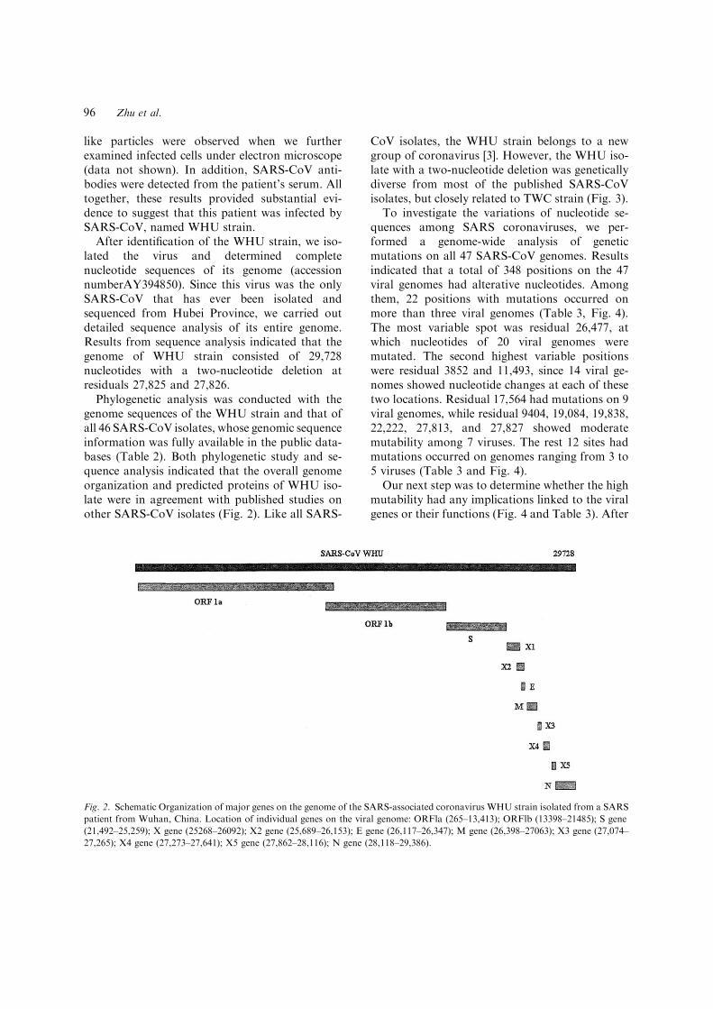
Phylogenetic analysis was conducted with thegenome sequences of the WHU strain and that ofall 46 SARS-CoV isolates, whose genomic sequenceinformation was fully available in the public data-bases (Table 2). Both phylogenetic study and se-quence analysis indicated that the overall genomeorganization and predicted proteins of WHU iso-late were in agreement with published studies onother SARS-CoV isolates (Fig. 2). Like all SARS-
CoV isolates, the WHU strain belongs to a newgroup of coronavirus [3]. However, the WHU iso-late with a two-nucleotide deletion was geneticallydiverse from most of the published SARS-CoVisolates, but closely related to TWC strain (Fig. 3).
To investigate the variations of nucleotide se-quences among SARS coronaviruses, we per-formed a genome-wide analysis of geneticmutations on all 47 SARS-CoV genomes. Resultsindicated that a total of 348 positions on the 47viral genomes had alterative nucleotides. Amongthem, 22 positions with mutations occurred onmore than three viral genomes (Table 3, Fig. 4).The most variable spot was residual 26,477, atwhich nucleotides of 20 viral genomes weremutated. The second highest variable positionswere residual 3852 and 11,493, since 14 viral ge-nomes showed nucleotide changes at each of thesetwo locations. Residual 17,564 had mutations on 9viral genomes, while residual 9404, 19,084, 19,838,22,222, 27,813, and 27,827 showed moderatemutability among 7 viruses. The rest 12 sites hadmutations occurred on genomes ranging from 3 to5 viruses (Table 3 and Fig. 4).
Our next step was to determine whether the highmutability had any implications linked to the viralgenes or their functions (Fig. 4 and Table 3). After
Fig. 2. Schematic Organization of major genes on the genome of the SARS-associated coronavirus WHU strain isolated from a SARS
patient from Wuhan, China. Location of individual genes on the viral genome: ORFla (265–13,413); ORFlb (13398–21485); S gene
(21,492–25,259); X gene (25268–26092); X2 gene (25,689–26,153); E gene (26,117–26,347); M gene (26,398–27063); X3 gene (27,074–
27,265); X4 gene (27,273–27,641); X5 gene (27,862–28,116); N gene (28,118–29,386).
96 Zhu et al.

further comparison and analysis of the viralsequences, we realized that polyprotein gene(ORF1 a and ORF1 b) had the highest variationrate among all genes. This region not only carried11 mutations, but also had the second highestvariable positions (residual 3852 and 11,493).ORF1b gene contains additional two residuals(17,564 and 19,084) at which 7 viruses were mu-tated. We also noticed that the S gene had a highmutability with residual 22222 mutated in 7 viru-ses, residual 21721 in 6, and residual 24933 in 3.Two positions with high mutation rate wereidentified within the M gene. One was located atthe most variable residual 26477, at which 20viruses were mutated. The other one was residual26600, at which 6 viral genomes were changed. Egene and N gene had one mutation spot at residual26203 and 28276, respectively. Among five non-structural genes, X4 had one mutation site atresidual 27243 with mutation rate of 5, while X5gene had two mutation spots at residual 27813 and27827 with mutation rate of 7 (Fig. 4 and Table 3).
Based on the recommendations from WHO [6],all SARS cases can be divided periodically intoearly-period case, mid-period case, and late-periodcase (Table 4). In this study, we proposed all 47known viral isolates into two groups, early-midperiod and mid-late period group (Table 5). Basedon results from sequence analysis, we realized thatthere were some correlations between geneticmutations of the virus and periodical or geo-graphic characteristics of the outbreak. Severalresiduals (9404, 9854, 17564, 19838, 21721, 22222,27243 and 27827) with mutations only occurredduring early-mid period, while nucleotides at somepositions (2557, 11448, 18965, 19084, 24933, 27813and 28276) changed exclusively during mid-lateperiod of the epidemic (Tables 3 5). In addition,some genetic mutations were linked to certaingeographic regions where the viruses isolated. Forinstance, high genetic mutation rate at position3852 was mainly found in viruses isolated fromTaiwan. Mutations at residual 26203 occurred inmost Taiwan isolates (60%), but not found in anyisolates identified from other regions around theworld. Moreover, all three viral strains (FRA, SoDand Frankfurt) isolated from Europe had muta-tions at the same residuals, 2557, 11448 and 24933,while the rest isolates showed no changes in thesepositions (Tables 3 and 5).
Discussion
Although the SARS epidemic ended after6 months spreading, many important questionsremain unclear. What is the natural reservoir ofSARS-CoV; where and how the virus crossed thebarriers between its reservoir and human to initi-ate reservoir–human transmission, and subsequenthuman-to-human infection. It was proposed thatthe natural reservoir of SARS-CoV was animaloriginated [10,11,13], most likely Himalayan palmcivets [12]. This was not a surprise, since manyfatal human viruses including HIV and influenzavirus were originated by transmission from ani-mals. HIV pandemic had happened as a conse-quence of the combination of transmission ofSIVcpz from chimpanzee and common practice of‘‘hunting and field–dressing chimpanzee’’ in WestCentral Africa [20]. Similarly in Southern China,where SARS-CoV initially emerged, people used
Fig. 3. Phylogenetic trees of the full-length nucleotide sequences
of the genome of SARS coronavirus WHU isolate and that of
other 46 SARS-CoV isolates. All sequences were aligned with
the CLUSTAL-W software program. Phylogenetic trees were
created for all nucleotide and predicted amino acid sequences by
neighbor-joining program and parsimony methods.
Genetic Mutations Analysis of SARS-CoV isolates 97

to consume wild animal meat and some of theanimals are now confirmed to carry SARS-likecoronavirus [12].
Another question is whether SARS outbreakwill come back. At the beginning of 2004, threeSARS cases were reported indicating SARS docome back. However, the situation of this yearseems quite different from last year, since trans-mission, infection and severity of SARS-CoV wereclearly weakened. One possible explanation is thatit might be just a preface of SARS epidemics. Likelast year, in the early period of SARS pandemics,the virus did not show strong toxicity. Anotherpossibility is that SARS-CoV might be trulyweakened due to many reasons including genetic
mutations, like the influenza FluA virus which hascaused a disaster outbreak in 1918 and wasweakened after the pandemic that took 20 millionlives [21]. Influenza epidemics throughout theworld occurred periodically between the firstpandemic and present time due to the viral anti-genic drift and shift. These processes also resultedin the appearance of influenza B and C virus withsignificant differences in genetic characterizations[22]. It would be important to find out if SARS-CoV has similar epidemic rules as influenza virusdose, whether SARS-CoV is weakening or willSARS breakout periodically. While these ques-tions remain to be addressed, it is for sure that theSARS-CoV certainly has a high mutation rate on
Table 3. Summary of genetic mutations within genes of 47 SARS-associated coronaviruses
ORF 1a Position 2557 3852 9404 9854 11448 11493
Mutation rate 3 14 7 6 3 14
Nuleotide change G–A T–C T–C C–T C–T C–T
Amino acid change A–T F–S V–A A–V Silent Silent
ORF 1b Position 17,564 18,965 19,064 19,084 19,838
Mutation rate 9 4 5 7 7
Nuleotide change T–G T–A A–G C–T A–G
Amino acid change N–E Silent Silent T–I Silent
S gene Position 21,721 22,222 23,220 24,933
Mutation rate 6 7 4 3
Nuleotide change G–A T–C T–G C–T
Amino acid change G–D I–T S–A L–F
E gene Position 26,203
Mutation rate 6
Nuleotide change C–T
Amino acid change Silent
M gene Position 26,477 26,600
Mutation rate 20 6
Nuleotide change T–G C–T
Amino acid change F–C A–V
X4 gene Position 27,243
Mutuation rate 5
Nuleotide change C–T
Amino acid change T–L
X5 gene Position 27,813 27,827
Mutation rate 7 7
Nuleotide change C–T T–C
Amino acid change S–L C–R
N gene Position 28,276
Mutation rate 4
Nuleotide change C–T
Amino acid change R–W
Positions of nucleotides were based on that of the SARS-CoV Urbani strain.
98 Zhu et al.

Table 4. Classification of geographic locations and periodic divisions of the SARS outbreak from late 2002 and early 2003
Early period case Mid-period case Late-period case
Southern China
(16 November, 2002)
Mainland China (13 February 2003) Mainland China-Taiwan (25 February 2003)
China Hong Kong SAR (15 February 2003) Germany (9 March 2003)
Canada (23 February 2003) Italy (12 March 2003)
Vietnam (23 February 2003) Russian Fedaration (5 May 2003)
Singapore (25 February 2003)
Table 5. Division of 47 SARS-associated coronaviruses based on periods of the SARS outbreak
Early mid-period Mid-late period
GD 69 GZ 50 GD 01 Sino 3-11 Sin 2748 SoD AS HSR-1 WHU Sin 2774
PUM C03 PUM C02 PUM C03 BJ 04 Sin 2679 TW/C3 TW/C2 TW/C Sin 2500 Sin 2677
BJ02 BJ01 HKU-39849 CUHK-
AG03
Urbani TW/Y TWS TWK TWJ TW H
Sino 1-11 CUHK-
AG01
CUHK-W1 CUHK-
AG02
Taiwan TC3 Taiwan TC2 Taiwan C1
BJ03 Frankfurt Shangai QXC1 TW-1 TOR/2
Shangai
XC2
Mutations occurred at residuals 9404, 9854,
17564, 19838, 21721, 22222, 27243 and 27827
Mutations occurred at residuals 2557, 11448, 18695,
19084, 24933, 27813 and 28276
Fig. 4. Positions and mutation rates of 22 highly variable residuals on the viral genomes based on the comparison of genomic
sequences of 47 SARS-CoV isolates published in the databases (GenBank).
Genetic Mutations Analysis of SARS-CoV isolates 99

its genome, which could in turn play significantroles in its pathogenecity and epidemics of thedisease.
Molecular epidemiology and genome-wideanalysis of mutations among SARS-CoV haveprovided insights into our understanding some ofthe questions [11,14–18]. For instance, except thegeographic distribution of potential animal reser-voirs, the high homologies among SARS-CoV ofhuman and SARS-like coronavirus of animalsstrongly supported the hypothesis of animal originof SARS-CoV [12]. It is possible that somemutations on the viral genome were responsiblefor the transmission of SARS-CoV from animalsto human.
In an effort to study the SARS-CoV, we iden-tified and genetically sequenced a new SARS-CoVisolated from a patient with SARS in HubeiProvince. Hubei was a less SARS representativearea in China, because there were only a total ofthree patients confirmed as probable SARS casesand only one viral strain was isolated from thisregion. These facts prompted us to study this virusfurther. Our sequence analysis indicated that al-though the overall genome organization of WHU(Fig. 2) is in agreement with published studies onother isolates, WHU carried a two-nucleotidedeletion at residuals 27825 and 27826 was geneti-cally diverse from most SARS-CoV isolates. Theseresults implicated that mutations occurred duringthe viral transmission from Beijing to Hubei,although we do not know at this point whetherthese mutations have any biological significance. Itis interesting to notice that although the SARS-CoV virus evaded human population only for6 months, its genetic information already alteredin many ways during its short journey of humantransmission.
Individual viral genes displayed distinct patternsof genetic mutations at different time during theSARS outbreak. For instance, mutability of the Sgene was high during early-mid period, but lowduring mid-late period of the epidemic, whichsuggested that mutability of S gene decreased asviral transmission increased. One possible expla-nation for this observation is that during early-midperiod of the epidemic, as the gene encodingprotein for the recognition of receptors of the hostand for the mediation of viral entry into host cells,S gene had to change at a high frequency in order
to quickly fulfill its biological roles. Once the viraladaptation to human cells completed or reachedits equilibrium, genetic changes were less impor-tant or no longer needed. Thus, geneticinformation of S gene became relatively stableduring mid-late period of the outbreak [23].Another example is ORF lab that encodes thepolyprotein of SARS-CoV. Like S gene, ORF labwas also actively involved in genetic mutations.However, in contrast to S gene, mutability of ORFlab was low at the beginning, but high during mid-late period of the epidemics. This observation canbe explained well by the fact that the toxicity ofSARS-CoV was weakened in mid-late period.Other structural genes including E, M, and Ngenes were more conserved at beginning of theoutbreak, but underwent genetic changes at theend of transmission. This pattern of geneticmutation obviously reflects biological roles ofthese structural genes in viral particles assembly,which in turn crucial for the virus to fight withincreasing immune pressures from the hosts.Genetic analysis of non-structural genes showedthat they intended to keep genetic informationconserved throughout the entire process of trans-mission. Therefore, these genes may prove to beideal targets for the diagnosis of SARS-Co.V,screening antiviral drugs, and perhaps developingantiviral vaccines.
Patterns of genetic mutations of certain viralgenes were linked to geographic locations fromwhere the virus isolated. Mutations at residuals3825 and 26203 within the X5 and E genes couldclearly set the Taiwan isolates apart from others.Thus, these two positions may be used as molec-ular signatures in the identification of Taiwanisolates. Similar phenomena were also found inthree viral strains (SoD, FRA, and Frankfurt)isolated from Europe during mid-late period of theoutbreak. These viral strains had mutations at thesame residuals (2557, 11,448 and 24,933), while allisolates from other regions did not show anychanges at these positions. This kind of specificmutation pattern may reflect relatively indepen-dent geographical locations of Taiwan and Europe.We speculated that population in these regionsperhaps developed unique immunity due to theirunique locations, for which the virus had to makespecific genetic mutations in order to invade thesepopulations. In addition, based on genome-wide
100 Zhu et al.

mutation analysis, some viral strains isolated fromBeijing had a close relationship to isolates identifiedfromSouthernChina during early-mid period of theoutbreak. It could be translated to that at least theseSARS-CoV isolates found in Beijing were originallyfrom Southern China.
Much have to be done in order to understandthoroughly the evolution, transmission, origin,and infection of SARS-associated coronavirus. Itis interesting to recognize that genome-widemutation analysis could provide new insights intoour understanding the route of viral transmissionand predication or perhaps prevention of futureSARS epidemics. Our study would provide arational and hypothesis-driven approach to studythese questions, develop rapid diagnostic tests, anddesign measurement to prevent this fatal disease.In addition, fully understand molecular mecha-nism of genetic mutations would provide insightsinto understanding plausible transmission route ofSARS-CoV from animal to humans as well asfrom human to human, and trends of changing inpathogenecity of SARS-CoV during its rout oftransmission and path of evolution.
Acknowledgment
This research was supported by the SARS SpecialGrant of Wuhan University.
References
1. Peng G.W., He J.F., Lin J.Y., Zhou D.H., Yu D.W.,
Liang W.J., Li L.H., Guo R.N., Luo H.M., and Xu
R.H., Chin J Epidemiol 24, 350–352, 2003.
2. WHO. Cumulative number of reported probable cases of
severe acute respiratory syndrome (SARS). http://
www.who.int/csr/sarscountry/2003_07_11/en.
3. Ksiazek T.G., Erdman D., Goldsmith C.S., Zaki S.R., Peret
T., Emery S., Tong S., Urbani C., Comer J.A., Lim W.,
Rollin P.E., Dowell S.F., Ling A.E., Humphrey C.D., Shieh
W.J., Guarner J., Paddock C.D., Rota P., Fields B., DeRisi
J., Yang J.Y., Cox N., Hughes J.M., LeDuc J.W., Bellini
W.J., and Anderson L.J., SARS Working Group, N Engl J
Med 348, 1953–1966, 2003.
4. Drosten C., Gunther S., Preiser W., van der Werf S., Brodt
H.R., Becker S., Rabenau H., Panning M., Kolesnikova L.,
Fouchier R.A., Berger A., Burguiere A.M., Cinatl J., Eick-
mannM., EscriouN.,GrywnaK.,KrammeS.,Manuguerra,
J.C., Muller S., Rickerts V., Sturmer M., Vieth S., Klenk
H.D., Osterhaus A.D., Schmitz H., andDoerrH.W., N Engl
J Med 348, 1967–1976, 2003.
5. Rota P.A., Oberste M.S., Monroe S.S., Nix W.A.,
Campagnoli R., Icenogle J.P., Periaranda S., Bankamp,
B., Maher K., Chen M.H., Tong S., Tamin A., Lowe L.,
Frace M., DeRisi J.L., Chen Q., Wang D., Erdman
D.D., Peret T.C., Burns C., Ksiazek T.G., Rollin P.E.,
Sanchez A., Liffick S., Holloway B., Limor J.,
McCaustland K., Olsen-Rasmussen M., Fouchier R.,
Gunther S., Osterhaus A.D., Drosten C., Pallansch M.A.,
Anderson L.J., and Bellini W.J., Science 300, 1394–1399,
2003.
6. Department of Communicable Disease Surveillance and
Response. WHO Consensus document on the epidemiology
of severe acute respiratory syndrome (SARS). http://
www.who.cds/csr/gar/2003.11.
7. Marra M.A., Jones S.J., Astell C.R., Holt R.A., Brooks-
Wilson A., Butterfield Y.S., Khattra J., Asano, J.K., Barber
S.A., Chan S.Y., Cloutier A., Coughlin S.M., Freeman D.,
GimN., GriffithO.L., Leach S.R.,MayoM.,McDonaldH.,
Montgomery S.B., Pandoh P.K., Petrescu A.S., Robertson
A.G., Schein J.E., Siddiqui A., Smailus D.E., Stott J.M.,
YangG.S., PlummerF.,AndonovA.,ArtsobH., BastienN.,
Bernard K., Booth T.F., Bowness D., Czub M., Drebot M.,
Fernando L., Flick R., Garbutt M., Gray M., Grolla A.,
Jones S., Feldmann H., Meyers A., Kabani A., Li Y., Nor-
mandS., StroherU., TipplesG.A., Tyler S., VogrigR.,Ward
D., Watson B., Brunharm R.C., Krajden M., Petric M.,
Skowronski D.M., Upton C., and Roper R.L., Science 300,
1399–1404, 2003.
8. Poon L.L., Chan K.H., Wong O.K., Yam W.C., Yuen
K.Y., Guan Y., Lo Y.M., and Peiris J.S., J Clin Virol 28(3),
233–238, 2003.
9. Gao W., Tamin A., Soloff A., D’Aiuto L., Nwanegbo E.,
Robbins P.D., Bellini W.J., Barratt-Boyes S., and
Gambotto A., Lancet 362(9399), 1895–1856, 2003.
10. Enserink M., Science 300, 1351, 2003.
11. Eickmann M., Becker S., Klenk H.D., Doerr H.W., Stadler
K., Censini S., Guidotti S., Masignani V., Scarselli M.,
Mora M., Donati C., Han J.H., Song H.C., Abrignani S.,
Covacci A., and Rappuoli R., Science 302(5650), 1504–
1505, 2003.
12. Guan Y., Zheng B.J., He Y.Q., Liu X.L., Zhuang Z.X.,
Cheung C.L., Luo S.W., Li P.H., Zhang L.J., Guan Y.J.,
Butt K.M., Wong K.L., Chan K.W., Lim W., Shortridge
K.F., Yuen K.Y., Peiris J.S., and Poon L.L., Science
302(5643), 276–278, 2003.
13. Ng S.K., Lancet 362(9383), 570–572, 2003.
14. Stavrinides J., Guttman D.S., J. Virol. 78(l), 76–82, 2004.
15. Chim S.S., Tsui S.K., Chan K.C., Au T.C., Hung E.C.,
Tong Y.K., Chiu R.W., Ng E.K., Chan P.Y., Chu C.M.,
Sung J.J., Tam J.S., Fung K.P., Waye M.M., Lee C.Y.,
Yuen K.Y., and Lo Y.M., Lancet 362(9398), 1807–1808,
2003.
16. Ruan Y.J., Wei C.L., Ee A.L., Vega V.B., Thoreau H.,
Su S.T., Chia J.M., Ng P., Chiu K.P., Lim L., Zhang T.,
Peng C.K., Lin E.O., Lee N.M., Yee S.L., Ng L.F., Chee
R.E., Stanton L.W., Long P.M., and Liu E.T., Lancet
361(9371), 1779–1785, 2003.
Genetic Mutations Analysis of SARS-CoV isolates 101

17. Snijder E.J., Bredenbeek P.J., Dobbe J.C., Thiel V., Ziebuhr
J., Poon L.L., Guan Y., Rozanov M., Spaan W.J., and
Gorbalenya A.E., J Mol Biol 331(5), 991–1004, 2003.
18. Tsui S.K., Chim S.S., and Lo Y.M., Chinese University of
Hong Kong Molecular SARS Research Group, N Engl J
Med 349(2), 187–188, 2003.
19. Zhong N.S., Zheng B.J., Li Y.M., Poon, Xie Z.H.,
Chan K.H., Li P.H., Tan S.Y., Chang Q., Xie J.P., Liu
X.Q., Xu J., Li D.X., Yuen K.Y., Peiris, and Guan Y.,
Lancet 362, 1353–1358, 2003.
20. Richter M., Can HIV AIDS Policy Law Rev 8(l), 14–19,
2003.
21. Zambon M.C., J. Antimicrobial Chemother 44, Suppl B, 3–
9, 1999.
22. Bush R.M., Smith C.B., Cox N.J., and Fitch W.M., Proc
Natl Acad Sci USA 97, 6974–6980, 2000.
23. Chinese SARS Molecular Epidemiology Consortium, Sci-
ence 303(5664), 1666–1669, 2004.
102 Zhu et al.