SARS-CoV-2–related MIS-C: A key to the viral and ... - Spiral
24
PERSPECTIVE SARS-CoV-2–related MIS-C: A key to the viral and genetic causes of Kawasaki disease? Vanessa Sancho-Shimizu 1,2 , Petter Brodin 3 , Aur´ elie Cobat 4,5,6 , Catherine M. Biggs 7,8 , Julie Toubiana 9,10 , Carrie L. Lucas 11 , Sarah E. Henrickson 12,13 , Alexandre Belot 14,15 , MIS-C@CHGE 21 , Stuart G. Tangye 16,17 , Joshua D. Milner 18 , Michael Levin 1,2 , Laurent Abel 4,5,6 , Dusan Bogunovic 19 , Jean-Laurent Casanova 4,5,6,20 , and Shen-Ying Zhang 4,5,6 Multisystem inflammatory syndrome in children (MIS-C) emerged in April 2020 in communities with high COVID-19 rates. This new condition is heterogenous but resembles Kawasaki disease (KD), a well-known but poorly understood and clinically heterogenous pediatric inflammatory condition for which weak associations have been found with a myriad of viral illnesses. Epidemiological data clearly indicate that SARS-CoV-2 is the trigger for MIS-C, which typically occurs about 1 mo after infection. These findings support the hypothesis of viral triggers for the various forms of classic KD. We further suggest that rare inborn errors of immunity (IEIs) altering the immune response to SARS-CoV-2 may underlie the pathogenesis of MIS-C in some children. The discovery of monogenic IEIs underlying MIS-C would shed light on its pathogenesis, paving the way for a new genetic approach to classic KD, revisited as a heterogeneous collection of IEIs to viruses. Introduction As the COVID-19 pandemic continues, it is becoming increas- ingly clear that severe clinical manifestations of SARS-CoV-2 infection remain rare in children, accounting for only 1.5% of all COVID-19 hospital admissions (Docherty et al., 2020; G¨ otzinger et al., 2020). However, in the spring of 2020, clusters of children admitted to the hospital with a multisystem hyperinflammatory syndrome, presenting with fever, abdominal pain and/or rash, myocarditis, and other clinical features reminiscent of Kawasaki disease (KD), were reported in communities with high rates of COVID-19, across Europe, and in North and South America (Fig. 1 and Table 1; de Farias et al., 2020; Farias et al., 2020; Jones et al., 2020; Riphagen et al., 2020; Toubiana et al., 2020b; Verdoni et al., 2020; Whittaker et al., 2020). Initial reports described a condition they referred to as an atypical form of KD with an incidence 30 times higher than in previous years (Verdoni et al., 2020). This newly identified syndrome was given the name pediatric inflammatory multisystem syndrome temporally as- sociated with SARS-CoV-2 by the UK Royal College of Pediatrics and Child Health (Whittaker et al., 2020), multisystem inflam- matory syndrome in children (MIS-C) by the US Centers for Disease Control and Prevention (CDC) and the World Health Organization, or COVID-19–associated KD (COVID-KD) by many investigators (Pouletty et al., 2020; Toubiana et al., 2021; Verdoni et al., 2020). MIS-C cases were typically reported 3–6 wk after the peak of SARS-CoV-2 infection in the local popula- tion, suggesting a temporal association with the ongoing pan- demic (Belot and Levy-Bruhl, 2020; Belot et al., 2020; Dufort et al., 2020; Feldstein et al., 2020; Toubiana et al., 2020a). About 84% of MIS-C cases test positive for anti–SARS-CoV-2 ............................................................................................................................................................................. 1 Department of Paediatric Infectious Diseases and Virology, Imperial College London, London, UK; 2 Centre for Paediatrics and Child Health, Faculty of Medicine, Imperial College London, London, UK; 3 Science for Life Laboratory, Department of Women’s and Children’s Health, Karolinska Institutet, Stockholm, Sweden; 4 St. Giles Laboratory of Human Genetics of Infectious Diseases, Rockefeller Branch, The Rockefeller University, New York, NY; 5 Laboratory of Human Genetics of Infectious Diseases, Necker Branch, Institut National de la Sant´ e et de la Recherche M´ edicale, Necker Hospital for Sick Children, Paris, France; 6 University of Paris, Imagine Institute, Paris, France; 7 Department of Pediatrics, University of British Columbia, Vancouver, Canada; 8 British Columbia Children’s Hospital Research Institute, Vancouver, Canada; 9 Department of General Pediatrics and Pediatric Infectious Diseases, Necker Hospital for Sick Children, Assistance Publique - Hˆ opitaux de Paris, University of Paris, Paris, France; 10 Pasteur Institute, Biodiversity and Epidemiology of Bacterial Pathogens, Paris, France; 11 Department of Immunobiology, Yale University School of Medicine, New Haven, CT; 12 Division of Allergy Immunology, Children’s Hospital of Philadelphia, Philadelphia, PA; 13 Department of Microbiology, Perelman School of Medicine, University of Pennsylvania, Philadelphia, PA; 14 Centre International de Recherche en Infectiologie, University of Lyon, Institut National de la Sant´ e et de la Recherche M´ edicale, U1111, Universit´ e Claude Bernard, Lyon 1, Le Centre National de la Recherche Scientifique, UMR5308, Lyon, France; 15 National Reference Center for Rheumatic, Autoimmune and Systemic Diseases in Children (RAISE), Pediatric Nephrology, Rheumatology, Dermatology Unit, Hˆ opital Femme Mère Enfant, Hospices Civils de Lyon, Lyon, France; 16 Garvan Institute of Medical Research, Darlinghurst, Australia; 17 St. Vincent’s Clinical School, Faculty of Medicine, University of New South Wales Sydney, Sydney, Australia; 18 Department of Pediatrics, Columbia University Irving Medical Center, New York, NY; 19 Center for Inborn Errors of Immunity, Precision Immunology Institute, Mindich Child Health and Development Institute, Department of Microbiology, Department of Pediatrics, Icahn School of Medicine at Mount Sinai, New York, NY; 20 Howard Hughes Medical Institute, New York, NY; 21 Multisystem Inflammatory Syndrome in Children, COVID Human Genetic Effort. Members of MIS-C@CHGE are listed at the end of the PDF. All authors contributed equally to this paper; Correspondence to Shen-Ying Zhang: shzh289@ rockefeller.edu; Vanessa Sancho-Shimizu: [email protected]. © 2021 Sancho-Shimizu et al. This article is distributed under the terms of an Attribution–Noncommercial–Share Alike–No Mirror Sites license for the first six months after the publication date (see http://www.rupress.org/terms/). After six months it is available under a Creative Commons License (Attribution–Noncommercial–Share Alike 4.0 International license, as described at https://creativecommons.org/licenses/by-nc-sa/4.0/). Rockefeller University Press https://doi.org/10.1084/jem.20210446 1 of 16 J. Exp. Med. 2021 Vol. 218 No. 6 e20210446 Downloaded from http://rupress.org/jem/article-pdf/218/6/e20210446/1414094/jem_20210446.pdf by Imperial College London Library user on 30 April 2021
-
Upload
khangminh22 -
Category
Documents
-
view
0 -
download
0
Transcript of SARS-CoV-2–related MIS-C: A key to the viral and ... - Spiral

PERSPECTIVE
SARS-CoV-2–related MIS-C: A key to the viral andgenetic causes of Kawasaki disease?Vanessa Sancho-Shimizu1,2, Petter Brodin3, Aurelie Cobat4,5,6, Catherine M. Biggs7,8, Julie Toubiana9,10, Carrie L. Lucas11,Sarah E. Henrickson12,13, Alexandre Belot14,15, MIS-C@CHGE21, Stuart G. Tangye16,17, Joshua D. Milner18, Michael Levin1,2, Laurent Abel4,5,6,Dusan Bogunovic19, Jean-Laurent Casanova4,5,6,20, and Shen-Ying Zhang4,5,6
Multisystem inflammatory syndrome in children (MIS-C) emerged in April 2020 in communities with high COVID-19 rates. Thisnew condition is heterogenous but resembles Kawasaki disease (KD), a well-known but poorly understood and clinicallyheterogenous pediatric inflammatory condition for which weak associations have been found with a myriad of viral illnesses.Epidemiological data clearly indicate that SARS-CoV-2 is the trigger for MIS-C, which typically occurs about 1 mo afterinfection. These findings support the hypothesis of viral triggers for the various forms of classic KD. We further suggest thatrare inborn errors of immunity (IEIs) altering the immune response to SARS-CoV-2 may underlie the pathogenesis of MIS-C insome children. The discovery of monogenic IEIs underlying MIS-C would shed light on its pathogenesis, paving the way for anew genetic approach to classic KD, revisited as a heterogeneous collection of IEIs to viruses.
IntroductionAs the COVID-19 pandemic continues, it is becoming increas-ingly clear that severe clinical manifestations of SARS-CoV-2infection remain rare in children, accounting for only 1.5% of allCOVID-19 hospital admissions (Docherty et al., 2020; Gotzingeret al., 2020). However, in the spring of 2020, clusters of childrenadmitted to the hospital with a multisystem hyperinflammatorysyndrome, presenting with fever, abdominal pain and/or rash,myocarditis, and other clinical features reminiscent of Kawasakidisease (KD), were reported in communities with high rates ofCOVID-19, across Europe, and in North and South America (Fig. 1and Table 1; de Farias et al., 2020; Farias et al., 2020; Jones et al.,2020; Riphagen et al., 2020; Toubiana et al., 2020b; Verdoniet al., 2020; Whittaker et al., 2020). Initial reports described acondition they referred to as an atypical form of KD with an
incidence 30 times higher than in previous years (Verdoni et al.,2020). This newly identified syndrome was given the namepediatric inflammatory multisystem syndrome temporally as-sociated with SARS-CoV-2 by the UK Royal College of Pediatricsand Child Health (Whittaker et al., 2020), multisystem inflam-matory syndrome in children (MIS-C) by the US Centers forDisease Control and Prevention (CDC) and the World HealthOrganization, or COVID-19–associated KD (COVID-KD) by manyinvestigators (Pouletty et al., 2020; Toubiana et al., 2021;Verdoni et al., 2020). MIS-C cases were typically reported 3–6wk after the peak of SARS-CoV-2 infection in the local popula-tion, suggesting a temporal association with the ongoing pan-demic (Belot and Levy-Bruhl, 2020; Belot et al., 2020; Dufortet al., 2020; Feldstein et al., 2020; Toubiana et al., 2020a).About 84% of MIS-C cases test positive for anti–SARS-CoV-2
.............................................................................................................................................................................1Department of Paediatric Infectious Diseases and Virology, Imperial College London, London, UK; 2Centre for Paediatrics and Child Health, Faculty of Medicine, ImperialCollege London, London, UK; 3Science for Life Laboratory, Department of Women’s and Children’s Health, Karolinska Institutet, Stockholm, Sweden; 4St. Giles Laboratoryof Human Genetics of Infectious Diseases, Rockefeller Branch, The Rockefeller University, New York, NY; 5Laboratory of Human Genetics of Infectious Diseases, NeckerBranch, Institut National de la Sante et de la Recherche Medicale, Necker Hospital for Sick Children, Paris, France; 6University of Paris, Imagine Institute, Paris, France;7Department of Pediatrics, University of British Columbia, Vancouver, Canada; 8British Columbia Children’s Hospital Research Institute, Vancouver, Canada; 9Departmentof General Pediatrics and Pediatric Infectious Diseases, Necker Hospital for Sick Children, Assistance Publique - Hopitaux de Paris, University of Paris, Paris, France;10Pasteur Institute, Biodiversity and Epidemiology of Bacterial Pathogens, Paris, France; 11Department of Immunobiology, Yale University School of Medicine, New Haven,CT; 12Division of Allergy Immunology, Children’s Hospital of Philadelphia, Philadelphia, PA; 13Department of Microbiology, Perelman School of Medicine, University ofPennsylvania, Philadelphia, PA; 14Centre International de Recherche en Infectiologie, University of Lyon, Institut National de la Sante et de la Recherche Medicale, U1111,Universite Claude Bernard, Lyon 1, Le Centre National de la Recherche Scientifique, UMR5308, Lyon, France; 15National Reference Center for Rheumatic, Autoimmune andSystemic Diseases in Children (RAISE), Pediatric Nephrology, Rheumatology, Dermatology Unit, Hopital Femme Mère Enfant, Hospices Civils de Lyon, Lyon, France;16Garvan Institute of Medical Research, Darlinghurst, Australia; 17St. Vincent’s Clinical School, Faculty of Medicine, University of New South Wales Sydney, Sydney,Australia; 18Department of Pediatrics, Columbia University Irving Medical Center, New York, NY; 19Center for Inborn Errors of Immunity, Precision Immunology Institute,Mindich Child Health and Development Institute, Department of Microbiology, Department of Pediatrics, Icahn School of Medicine at Mount Sinai, New York, NY; 20HowardHughes Medical Institute, New York, NY; 21Multisystem Inflammatory Syndrome in Children, COVID Human Genetic Effort.
Members of MIS-C@CHGE are listed at the end of the PDF. All authors contributed equally to this paper; Correspondence to Shen-Ying Zhang: [email protected]; Vanessa Sancho-Shimizu: [email protected].
© 2021 Sancho-Shimizu et al. This article is distributed under the terms of an Attribution–Noncommercial–Share Alike–No Mirror Sites license for the first six monthsafter the publication date (see http://www.rupress.org/terms/). After six months it is available under a Creative Commons License (Attribution–Noncommercial–Share Alike4.0 International license, as described at https://creativecommons.org/licenses/by-nc-sa/4.0/).
Rockefeller University Press https://doi.org/10.1084/jem.20210446 1 of 16
J. Exp. Med. 2021 Vol. 218 No. 6 e20210446
Dow
nloaded from http://rupress.org/jem
/article-pdf/218/6/e20210446/1414094/jem_20210446.pdf by Im
perial College London Library user on 30 April 2021

antibodies and/or in viral PCR tests, and all reported caseshave a history of exposure to SARS-CoV-2 (Ahmed et al.,2020). There is, therefore, strong viral and epidemiologicalevidence to suggest that SARS-CoV-2 is the trigger forMIS-C.
The exact incidence ofMIS-C remains unknown, due to a lackof comprehensive SARS-CoV-2 testing data for children. A NewYork–based study reported an incidence of MIS-C of 2/100,000in individuals age <21 yr in a population with an incidence ofconfirmed SARS-CoV-2 infection of 322/100,000 betweenMarch 1 and May 10, 2020 (Ahmed et al., 2020; Dufort et al.,2020). Another study reported a conservative estimate of theincidence of SARS-CoV-2 infection of no more than 5% in chil-dren under the age of 15 yr, with MIS-C detected in <2 in every10,000 infected children (Belot et al., 2020). Unlike children
with acute severe SARS-CoV-2 infection, most children pre-senting with MIS-C display no detectable active viral infectionby PCR assays in the upper respiratory tract at the time ofMIS-Cdiagnosis. Instead, they display signs of prior SARS-CoV-2 in-fection or contact with an infected individual ∼1 m before theonset of MIS-C symptoms (Toubiana et al., 2021). The clinicaland immunological features of MIS-C and pediatric COVID-19pneumonia are different (Gruber et al., 2020; Swann et al.,2020), whereas those of MIS-C and KD, both of which are het-erogenous inflammatory conditions, overlap (Carter et al., 2020;Consiglio et al., 2020; Hoste et al., 2021; Toubiana et al., 2021;Whittaker et al., 2020). The emergence of MIS-C provided thefirst clear evidence to support the notion that KD-like inflam-matory illness can be triggered by a virus (Toubiana et al., 2021).The case definition of MIS-C is evolving with improvements in
Figure 1. Geographic distribution of COVID-19 and MIS-C cases. (A) Choropleth map of cumulative COVID-19 cases, by country, from World HealthOrganization data, as of February 11, 2021. (B) Choropleth map of MIS-C cases, by country, as reported in published studies. Countries that have reported casesbut have not disclosed the number of cases are denoted as “# not reported.” Only MIS-C cases reported in English-language journals are included. A list of thearticles included can be found in Table S1. NA, not applicable.
Sancho-Shimizu et al. Journal of Experimental Medicine 2 of 16
Rare IEIs in MIS-C and Kawasaki disease? https://doi.org/10.1084/jem.20210446
Dow
nloaded from http://rupress.org/jem
/article-pdf/218/6/e20210446/1414094/jem_20210446.pdf by Im
perial College London Library user on 30 April 2021
our understanding of the spectrum of disease. MIS-C may cor-respond to a continuous distribution of different clinical entities.A key question that remains is whether there are common
pathogenic mechanisms underlying MIS-C and classic KD. The start-ing point for addressing this question is a comparison of the epide-miological, clinical, and immunological features of MIS-C and KD.
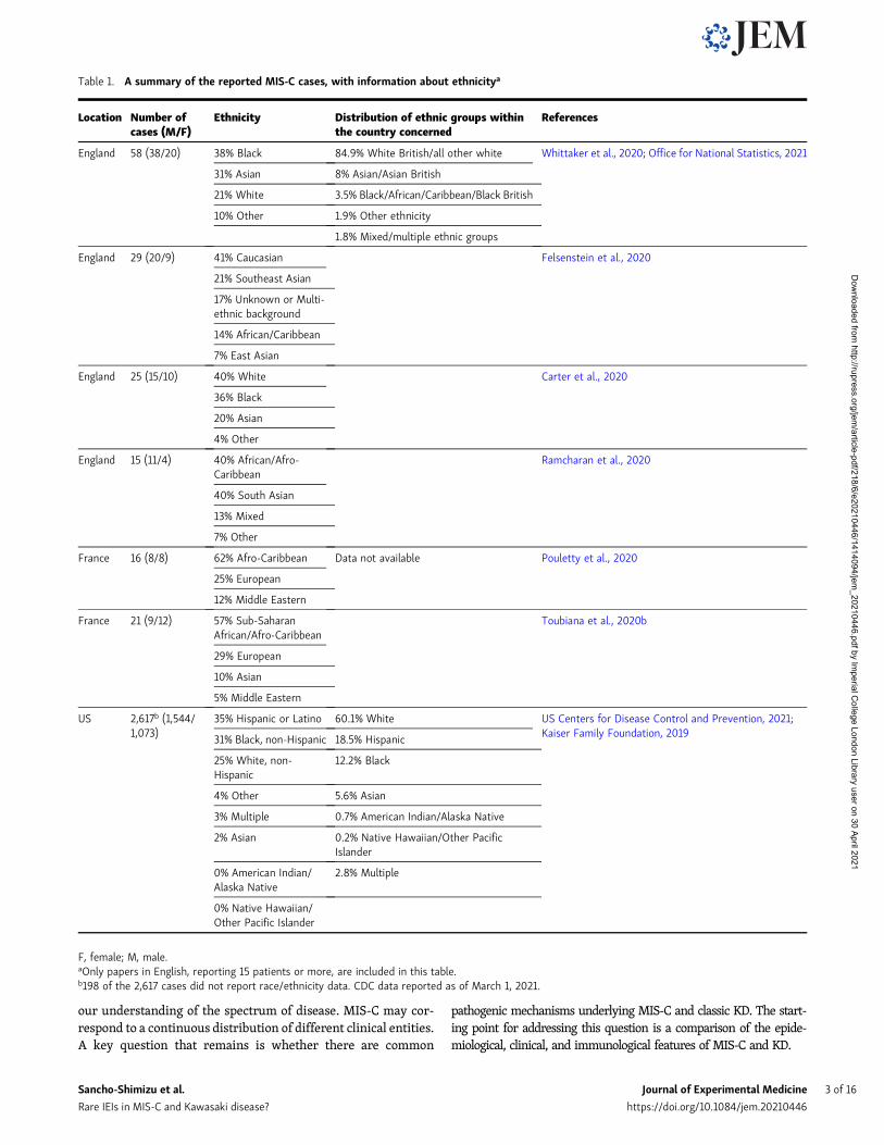
Table 1. A summary of the reported MIS-C cases, with information about ethnicitya
Location Number ofcases (M/F)
Ethnicity Distribution of ethnic groups withinthe country concerned
References
England 58 (38/20) 38% Black 84.9% White British/all other white Whittaker et al., 2020; Office for National Statistics, 2021
31% Asian 8% Asian/Asian British
21% White 3.5% Black/African/Caribbean/Black British
10% Other 1.9% Other ethnicity
1.8% Mixed/multiple ethnic groups
England 29 (20/9) 41% Caucasian Felsenstein et al., 2020
21% Southeast Asian
17% Unknown or Multi-ethnic background
14% African/Caribbean
7% East Asian
England 25 (15/10) 40% White Carter et al., 2020
36% Black
20% Asian
4% Other
England 15 (11/4) 40% African/Afro-Caribbean
Ramcharan et al., 2020
40% South Asian
13% Mixed
7% Other
France 16 (8/8) 62% Afro-Caribbean Data not available Pouletty et al., 2020
25% European
12% Middle Eastern
France 21 (9/12) 57% Sub-SaharanAfrican/Afro-Caribbean
Toubiana et al., 2020b
29% European
10% Asian
5% Middle Eastern
US 2,617b (1,544/1,073)
35% Hispanic or Latino 60.1% White US Centers for Disease Control and Prevention, 2021;Kaiser Family Foundation, 201931% Black, non-Hispanic 18.5% Hispanic
25% White, non-Hispanic
12.2% Black
4% Other 5.6% Asian
3% Multiple 0.7% American Indian/Alaska Native
2% Asian 0.2% Native Hawaiian/Other PacificIslander
0% American Indian/Alaska Native
2.8% Multiple
0% Native Hawaiian/Other Pacific Islander
F, female; M, male.aOnly papers in English, reporting 15 patients or more, are included in this table.b198 of the 2,617 cases did not report race/ethnicity data. CDC data reported as of March 1, 2021.
Sancho-Shimizu et al. Journal of Experimental Medicine 3 of 16
Rare IEIs in MIS-C and Kawasaki disease? https://doi.org/10.1084/jem.20210446
Dow
nloaded from http://rupress.org/jem
/article-pdf/218/6/e20210446/1414094/jem_20210446.pdf by Im
perial College London Library user on 30 April 2021
Epidemiology of MIS-C and KDMIS-C and KD display several notable epidemiological differ-ences. The median age of children with MIS-C is 8–9 yr (Dufortet al., 2020; Feldstein et al., 2020; Godfred-Cato et al., 2020;Swann et al., 2020; Whittaker et al., 2020), whereas most chil-dren with KD are under the age of 5 yr with a median age of 3 yr(Abrams et al., 2020) and a peak of incidence in infants <1 yr old(Ae et al., 2020; Holman et al., 2010). Children who developMIS-C are generally also older than those with severe pediatricCOVID-19, although the lower mean age of patients with severepediatric COVID-19 is largely due to the high proportion of in-fants among these patients (Diorio et al., 2020; Gale et al., 2021;Gotzinger et al., 2020; Swann et al., 2020). In both MIS-C andsevere pediatric COVID-19, most of the patients were previouslyhealthy (Davies et al., 2020; Diorio et al., 2020; Feldstein et al.,2020), although a possible association with obesity in adoles-cents has been suggested (Dufort et al., 2020; Feldstein et al.,2020; Godfred-Cato et al., 2020). KD affects more boys than girls(sex ratio of ∼1.5:1), whereas no clear sex bias has been observedin MIS-C patients (Ahmed et al., 2020). The fatality rate of MIS-C has been estimated at 1–2%, much higher than that reportedfor KD (estimated KD case fatality rate of <0.1% in Japan; Ahmedet al., 2020; McCrindle et al., 2017). Interestingly, KD rates ap-pear to be highest in East Asia, whereas individuals of SouthAsian and Latin American descent appear to be overrepresentedin studies of MIS-C performed in the United States and/orWestern European countries in which ancestry was reported(Table 1; Dufort et al., 2020; Feldstein et al., 2020; Godfred-Catoet al., 2020; Whittaker et al., 2020). There has been a strikingpaucity ofMIS-C cases from East Asia, despite the high incidenceof KD in this geographic area (Kim et al., 2020b; Li et al., 2019).Only two cases of MIS-C have been reported to date in SouthKorea, and no cases have been reported in Japan or China(Toyo Keizai Online COVID-19 Task Team, 2021), contrastingwith the higher numbers of cases from the Americas, Europe,Africa, South Asia, and the Middle East (Almoosa et al., 2020;Dufort et al., 2020; Falah et al., 2020; Feldstein et al., 2020;Jain et al., 2020; Kim et al., 2020a; Kim et al., 2020b; Leeet al., 2021; Mamishi et al., 2020; Ulloa-Gutierrez et al.,2020; Verdoni et al., 2020; Webb et al., 2020). Whether this ismerely due to a lower number of COVID-19 cases in East Asiaremains to be seen, bearing in mind the possibility of as-certainment bias (Fig. 1, A and B). Some areas in Europe sawa marked increase in the incidence of KD-like illnessesduring the first wave of the pandemic, whereas South Koreaobserved no change in KD incidence relative to previousyears (Kim et al., 2020b; Verdoni et al., 2020). Population-related risk factors specific to either condition are thereforeplausible.
Environmental factors may contribute to the geographicdistribution of MIS-C incidence, but social determinants ofhealth disparities also place particular ethnic minority pop-ulations at higher risk of SARS-CoV-2 exposure and associateddisease (Abrams et al., 2020; Godfred-Cato et al., 2020). Thecontributions of viral, host genetic, and other biological factorsto this higher risk remain unclear. It has been suggested thatviral factors have contributed to geographic differences in the
incidence of MIS-C. Following the spread of the COVID-19pandemic from East Asia, new variants of SARS-CoV-2 carryinga D839Y/N/E substitution were identified in Europe and theUnited States. Computational modeling has suggested that thetyrosine substitution at position 839 in the SARS-CoV-2 spikeprotein strengthens the binding of the spike protein to the βvariable (Vβ) region of the host T cell receptor, resulting insuperantigen characteristics potentially capable of driving theuncontrolled immune activation seen in MIS-C (Cheng et al.,2020). However, human genetic factors may also underlie thedifferences in incidence between MIS-C and KD in Westernversus Asian countries. Interestingly, KD studies predatingCOVID-19 suggested differences between ethnic groups forKawasaki shock syndrome, a condition that affects ∼7% of KDpatients (Alsaied et al., 2021). KD shock syndrome has a higherincidence in Western countries than in Asia (Li et al., 2019;Toubiana et al., 2020b), consistent with the observed geo-graphic distribution of MIS-C. These striking epidemiologicalfeatures highlight the need to define the genetic, immuno-logical, and other factors, such as concurrent environmentalexposures (SARS-CoV-2 or other factors), underlying MIS-Cand KD.
Comparison of the clinical features of MIS-C and KDPatients with MIS-C were initially described as having con-junctivitis, cheilitis, rash, and shock (de Farias et al., 2020;Farias et al., 2020; Jones et al., 2020; Riphagen et al., 2020;Toubiana et al., 2020b; Verdoni et al., 2020; Whittaker et al.,2020), immediately inciting comparisons with KD, which hasbeen described as a multisystem inflammatory vasculitis pre-senting with fever, acute mucocutaneous inflammation, and KDshock syndrome (McCrindle et al., 2017; Riphagen et al., 2020).Indeed, all MIS-C patients present with fever, and about halfhave a rash and bilateral bulbar conjunctival injection, as ob-served in KD. The American Heart Association criteria for KD(McCrindle et al., 2017) have not been systematically evaluatedin all studies on MIS-C, but MIS-C cases seem to be more likelyto fulfill the criteria for the incomplete form of KD (Belhadjeret al., 2020b; Belot et al., 2020; Capone et al., 2020; Cheung et al.,2020; Davies et al., 2020; Dufort et al., 2020; Feldstein et al.,2020; Moraleda et al., 2020; Pouletty et al., 2020; Ramcharanet al., 2020; Riphagen et al., 2020; Toubiana et al., 2020b;Verdoni et al., 2020). Some studies comparing cases of MIS-Cwith historical cases of KD have confirmed similarities withclassic KD in terms of clinical features (Pouletty et al., 2020;Toubiana et al., 2021; Verdoni et al., 2020; Whittaker et al.,2020), but all these studies also highlighted specific features ofCOVID-KD cases (Abrams et al., 2020; Toubiana et al., 2021).More than 80% of MIS-C cases present with gastrointestinal (GI)symptoms (Abrams et al., 2020), some even suffering intenseabdominal pain with acute pseudosurgical abdomen, peritonealeffusion, and gallbladder hydrops (Allali et al., 2021; Miller et al.,2020). Neurological symptoms, such as meningitis-like symp-toms or encephalitis, are also more frequently observed in MIS-C than in classic KD (Toubiana et al., 2021). These symptoms areobserved early in the disease and can be misinterpreted, some-times leading to unnecessary treatments or surgery (Allali et al.,
Sancho-Shimizu et al. Journal of Experimental Medicine 4 of 16
Rare IEIs in MIS-C and Kawasaki disease? https://doi.org/10.1084/jem.20210446
Dow
nloaded from http://rupress.org/jem
/article-pdf/218/6/e20210446/1414094/jem_20210446.pdf by Im
perial College London Library user on 30 April 2021
2021). One particular feature specific to MIS-C is the highprevalence of myocarditis associated with cardiogenic-vasoplegic shock. Depending on the definition of myocarditisand the inclusion criteria used (cardiovascular or KD-like ill-ness), cardiac dysfunction has been reported to affect 20–100%of patients (Abrams et al., 2020; Dufort et al., 2020; Friedmanet al., 2020; Matsubara et al., 2020; Theocharis et al., 2020).Recent cardiac magnetic resonance imaging data have shownthese patients to have a myocardial edema, rather than the ne-crosis often observed in common viral myocarditis (Alsaiedet al., 2021; Blondiaux et al., 2020). All these symptoms mayreflect local vasculitis and inflammation of the affected organ,and may account for the rapid, syndrome-resolving response tocorticosteroid treatment (Belhadjer et al., 2020a; Ouldali et al.,2021).
The proportion of myocardial functional defects, includingthe depression of ventricular function (Dufort et al., 2020;Friedman et al., 2020; Theocharis et al., 2020), is higher in MIS-C patients than in KD patients, but the rate of coronary arterydilations is similar to that generally reported for KD, althoughthese abnormalities do not often progress to aneurysm (Davieset al., 2020; Dufort et al., 2020; Feldstein et al., 2020; Godfred-Cato et al., 2020; Matsubara et al., 2020; McCrindle et al., 2017;Nelson et al., 2020; Whittaker et al., 2020). Some MIS-C cohortstudies have reported the occurrence of coronary artery dilation(Dufort et al., 2020; Feldstein et al., 2020; Godfred-Cato et al.,2020; Matsubara et al., 2020; Whittaker et al., 2020) and/oraneurysms (Davies et al., 2020; Dufort et al., 2020; Nelson et al.,2020) in 4–20% of patients, as previously reported for KD(McCrindle et al., 2017). However, unlike the coronary arterydilations and/or aneurysms observed in MIS-C, which are typ-ically transient and have an onset that coincides with that offever, the coronary artery aneurysms seen in KD peak ∼3 wkafter fever onset (Jhaveri et al., 2021; Matsubara et al., 2020;Tsuda and Hashimoto, 2021). Importantly, coronary artery ab-normalities have been observed not only in the youngest MIS-Cpatients meeting the full KD criteria, who would be more likelyto be considered to have “classic” KD, but also in adolescentpatients (Nelson et al., 2020). Physicians should, therefore,consider treating MIS-C patients with low-dose aspirin and in-travenous Ig (IVIG) in addition to appropriate cardiovascularresuscitation and steroids, because IVIG (with or withoutsteroids) is the only treatment that has been shown to reducecoronary complications in KD (Henderson et al., 2020;McCrindle et al., 2017; Oates-Whitehead et al., 2003). MIS-C isalso associated with higher levels of inflammatory markers(including C-reactive protein [CRP], procalcitonin, and tro-ponins) and brain natriuretic peptide (BNP), more profoundlymphopenia, thrombocytopenia rather than the commonthrombocytosis of prolonged KD, and higher ferritin levelsthan are observed in KD (Toubiana et al., 2021; Verdoni et al.,2020; Whittaker et al., 2020). However, all these character-istics are similar to those of the historic KD shock syndrome,the most severe form of KD (Kanegaye et al., 2009). Thus,MIS-C has several specific characteristics, but displays a sig-nificant clinical overlap with KD, and with KD shock syn-drome in particular.
Clinical spectrum of MIS-CWith the progression of the pandemic, the definition of MIS-Chas evolved, although definitions still vary between countries andresearch studies (Diorio et al., 2020; Dufort et al., 2020; Feldsteinet al., 2020; Godfred-Cato et al., 2020; Jain et al., 2020; Mamishiet al., 2020; Riphagen et al., 2020; Verdoni et al., 2020; Whittakeret al., 2020). The CDC defines MIS-C as a persistent fever andinflammation (e.g., increases in the levels of CRP, erythrocytesedimentation rate, D-dimers, procalcitonin, or the serum con-centrations of cytokines, including IL-10 and TNF; Diorio et al.,2020) in patients <21 yr requiring hospitalization for severe illnesswith multiple-organ dysfunction (e.g., GI, cardiovascular, derma-tological, neurological, nephrological, and/or respiratory; Godfred-Cato et al., 2020). Patients must also either have tested positive forSARS-CoV-2 (serologic test and/or PCR) or have clearly been ex-posed to SARS-CoV-2 (Godfred-Cato et al., 2020). Multiple-organdysfunction is a typical clinical feature of MIS-C. The dysfunctionof at least four organ systems has been reported in >70% of pa-tients (Feldstein et al., 2020). As also discussed above, the mostcommon presenting symptoms include multiple days of fever(>5 d in most cases; Feldstein et al., 2020), GI symptoms (e.g.,diarrhea, vomiting, and abdominal pain) and, in some patients,conjunctivitis, neurological symptoms (e.g., headache, confusion,and lethargy) or respiratory symptoms (it is uncommon for theseto be primary, and invasive or noninvasive ventilation is requiredin only a minority of patients; Davies et al., 2020). Patients oftenpresent with shock (Feldstein et al., 2020; Whittaker et al., 2020)and other aspects of impaired cardiovascular function, togetherwith dysfunctions of multiple other systems. This multiple-organdysfunction necessitates admission to intensive care units for64–80% of MIS-C patients (Dufort et al., 2020; Feldstein et al.,2020; Godfred-Cato et al., 2020). At least half these patients re-quire inotropes and/or fluid resuscitation (Belot et al., 2020;Whittaker et al., 2020).
These aspects are common to the general MIS-C patientpopulation, but severity and the nature of individual clinicalpresentations vary widely, and patients can be grouped intodifferent subsets on the basis of different strategies. A recentlypublished consensus management pathway forMIS-C developedin the United Kingdom divides the syndrome into KD-like (basedon American Heart Association criteria) and nonspecific in-flammatory phenotypes (Harwood et al., 2021). A second strat-egy distinguishes between a febrile inflammatory phenotype(lacking shock and without KD criteria), KD-like illness (e.g.,meeting complete or incomplete KD criteria but without shockor multisystem involvement), and shock with hyper-inflammation (Whittaker et al., 2020; PIER Network, 2020). Athird strategy for dividing MIS-C patients into three groups wasdeveloped on the basis of a latent class analysis (Godfred-Catoet al., 2020). Class 1 (∼35% of patients) has the highest frequencyof multiple-organ dysfunction, generally including GI and car-diovascular abnormalities with shock and myocarditis, andlaboratory testing abnormalities, including lymphopenia andhigh CRP, troponin, and BNP levels. Clinically, the features ofthe patients in this class do not overlap with COVID-19, but someoverlap is observed with KD shock syndrome, and there is aresemblance to systemic-onset juvenile idiopathic arthritis
Sancho-Shimizu et al. Journal of Experimental Medicine 5 of 16
Rare IEIs in MIS-C and Kawasaki disease? https://doi.org/10.1084/jem.20210446
Dow
nloaded from http://rupress.org/jem
/article-pdf/218/6/e20210446/1414094/jem_20210446.pdf by Im
perial College London Library user on 30 April 2021
(Binstadt et al., 2005). Class 2 (∼30%) has a larger overlap withtypical COVID-19, with more pulmonary syndromes (e.g., coughand/or acute respiratory distress syndrome), higher rates ofSARS-CoV-2 PCR positivity, and the highest mortality rate (5%).Finally, class 3 (∼35%) is more similar to KD, with a youngermedian age, a higher frequency of rash and mucocutaneous al-terations, and lower levels of inflammatory markers. Given theclinical parallels, treatment attempts initially focused on ap-proaches already shown to alleviate the symptoms of KD, such asIVIG, steroids, and aspirin. IVIG in addition to methylprednis-olone has been shown to be associated with a more favorablecourse of fever and better cardiac recovery in MIS-C thantreatment with IVIG alone (Belhadjer et al., 2020a; Ouldaliet al., 2021). Some teams have also used biological therapies,including antibodies against IL-6 or TNF, and recombinant IL-1 receptor antagonist (IL-1RA; Diorio et al., 2020; Feldsteinet al., 2020; Henderson et al., 2020; Radia et al., 2020;Whittaker et al., 2020). Despite the severe illness presentedby patients, the mean duration of hospital stay is only 6–7 d(Dufort et al., 2020; Feldstein et al., 2020; Godfred-Cato et al.,2020). Most patients receive medium- to long-term clinicalfollow-up at the treating institution to monitor for latecomplications.
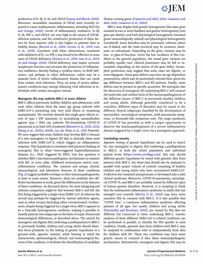
Comparison of the immunological features of MIS-C and KDMIS-C and KD both involve hyperinflammatory responses,presenting clinically with persistent high fever, often accom-panied by a visible rash and conjunctivitis. These two conditionsremain elusive. They appear to be related but different con-ditions, with a clinical and immunological overlap (Fig. 2). Thepathogenesis of KD remains unclear, but the postinfectious na-ture of MIS-C, and possibly also of KD, is consistent with anautoinflammatory and/or autoimmune hyperinflammatoryprocess initially triggered by viral infection (Rowley et al., 1997;Rowley et al., 2020). The immune response in patients with KDis typically characterized by pro-inflammatory signatures, in-cluding increases in IL-1, IL-8, IL-6, and IL-17A levels (Bajolleet al., 2014; Nelson et al., 2020; Oates-Whitehead et al., 2003),marked leukocytosis, eosinophilia, and high levels of monocytes(Kanegaye et al., 2009; Radia et al., 2020), accompanied by highCRP, erythrocyte sedimentation rate, procalcitonin, alanineaminotransferase, and γ-glutamyl transferase levels (Hendersonet al., 2020; Radia et al., 2020). Many of these features arecommon to MIS-C (including high levels of cytokines, such asIL-1, IL-8, IL-6, etc.), but tend to be stronger in MIS-C patients,and this is particularly true for markers of inflammation (such asCRP, procalcitonin, troponin, and ferritin) and BNP levels(Toubiana et al., 2021). KD-specific increases in the number of IgAplasma cells, and tissue-specific changes, mostly concerning thearterial wall, have been described (Marrani et al., 2018; Menikouet al., 2019; Rowley et al., 1997). Finally, it has been suggested thatimmune complexes and autoantibodies (Marrani et al., 2018;Menikou et al., 2019) are involved in the pathogenesis of KD (Jiaet al., 2010), and probably also in MIS-C, but with markedly dif-ferent specificities (Consiglio et al., 2020; Gruber et al., 2020).
In a study comparing MIS-C patients with KD patients thathave been identified before the COVID-19 pandemic, these two
groups of patients were shown to have different cytokine pro-files, both during the hyperinflammatory phase and beforetreatment (Consiglio et al., 2020). The hyperinflammation ob-served in MIS-C also differs from that seen in acute, severeCOVID-19, with lower IL-8 and IL-7 levels in MIS-C (Consiglioet al., 2020). KD patients have high frequencies of T helper type17 cells in the bloodstream, consistent with their high plasma IL-17A concentrations (Jia et al., 2010). This T helper type 17 cellresponse may, therefore, be a key feature distinguishing be-tween KD and MIS-C hyperinflammation in untreated patients(Consiglio et al., 2020), although other studies have reportedhigh plasma IL-17A levels in MIS-C patients too (Esteve-Soleet al., 2021; Gruber et al., 2020; Lee et al., 2020). The autoanti-body profiling of serum from untreated childrenwithMIS-C andKD has revealed many candidate targets, some of which differbetween MIS-C and KD. The presence of autoantibodies againstDEL-1, a potent anti-inflammatory protein that antagonizes theintegrin ICAM-1 and limits vascular inflammation, has beenreported in patients with KD (Shin et al., 2015), but not inMIS-Cpatients (Consiglio et al., 2020). By contrast, one study showedthat MIS-C patients have autoantibodies against proteins of thecasein kinase family (Consiglio et al., 2020). These enzymes areactivated in cells infected with SARS-CoV-2 and may play arole in viral replication (Bouhaddou et al., 2020), and otherprocesses (Consiglio et al., 2020; Gruber et al., 2020). How-ever, it remains unclear whether these autoantibodies playeda role in the disease pathogenesis of KD or MIS-C. It thereforeappears likely that both KD and MIS-C are postinfectious in-flammatory diseases, with some overlapping clinical and im-munological features, but different inflammatory signatures.They are, thus, probably mediated by related but differentinflammatory pathways and, possibly, related but differentpathogenic mechanisms.
Immunological spectrum of MIS-CSome potential clues to the pathophysiological mechanismsunderlying MIS-C have been identified. As discussed above,increases in the levels of inflammatory markers are variable inMIS-C (Diorio et al., 2020; Dufort et al., 2020; Vella et al., 2021).A broad spectrum of serum cytokine concentrations displaysincreases of various magnitudes (e.g., IL-1β, IL-6, IL-8, IL-10, IL-17, IL-18, IFN-γ, and TNF; Carter et al., 2020; Diorio et al., 2020;Gruber et al., 2020; Vella et al., 2021), with specific increases inIL-10 and TNF levels in MIS-C patients relative to patients withsevere COVID-19. There is also evidence of a possible role forcomplement/coagulation (Ramaswamy et al., 2021) in MIS-C,and thrombotic microangiopathy in MIS-C and in some cases ofsevere pediatric COVID-19, based on increases in C5b-9 levelsand on the increase in frequency of Burr cells and schistocyteson peripheral blood smears (Diorio et al., 2020). The immuneprofiling of MIS-C patients by a systems biology approach hasalso revealed other indicators of immune dysfunction (Gruberet al., 2020). The anti–SARS-CoV-2 antibodies in MIS-C patientseffectively neutralize SARS-CoV-2 in vitro, like those from pa-tients convalescing from COVID-19, although antibody levelsdiffer (lower IgM, higher IgA, and anti-spike IgG; Andersonet al., 2020). Analyses of 92 secreted moieties have identified
Sancho-Shimizu et al. Journal of Experimental Medicine 6 of 16
Rare IEIs in MIS-C and Kawasaki disease? https://doi.org/10.1084/jem.20210446
Dow
nloaded from http://rupress.org/jem
/article-pdf/218/6/e20210446/1414094/jem_20210446.pdf by Im
perial College London Library user on 30 April 2021

clear signs of inflammation in the plasma of MIS-C patients.CyTOF-based deep immunophenotyping identified a uniquecytokine signature in MIS-C patients that was not present inhealthy controls or COVID-19 patients and included strong sig-natures of inflammation (IL-18 and IL-6), lymphocytic and my-eloid chemotaxis and activation (CCL3, CCL4, and CDCP1), andmucosal immune dysregulation (IL-17A, CCL20, and CCL28;Gruber et al., 2020). Interestingly, most recent immunologicalanalyses of MIS-C have revealed a spectrum of immunologicalfeatures, including changes in cytokine levels, with similaritiesand differences between subgroups of patients paralleling thosebetween clinical subgroups (Esteve-Sole et al., 2021; Lee et al.,2020).
In addition, the mass cytometry immunophenotyping of pe-ripheral mononuclear cells from MIS-C patients revealed de-creases in the frequencies of nonclassical monocytes, andsubsets of natural killer (NK) and T lymphocytes, suggestingextravasation to peripheral organs (Gruber et al., 2020). There isalso evidence of significant T cell dysregulation in MIS-C, par-ticularly for CD8 T cells, including increases in the populationsof activated T cells (e.g., frequency of CD8 HLA-DR+CD38+ cells)similar to that seen in adults with severe COVID-19, togetherwith an increase in the frequency of PD-1+CD39+CD8+ T cells inMIS-C (Vella et al., 2021). Moreover, HLA-DR levels on γδ andCD4+CCR7+ T cells, and CD64 (FcγRI) levels on neutrophils andmonocytes, are high, indicating myeloid cell activation (Carteret al., 2020). Furthermore, an efflux of immature neutrophilswith low CD10 and CD62L into the periphery has also been
observed (Klocperk et al., 2020). Autoantibodies targeting theprincipal organs affected in MIS-C, including the GI tract, im-mune cells, cardiac and endothelial tissue, and some previouslydescribed autoantigens such as anti-La, have been described(Gruber et al., 2020; Ramaswamy et al., 2021), although it re-mains unclear whether these autoantibodies are specificallyrelevant to the pathogenesis of MIS-C. Finally, a profound ex-pansion of some specific T cell receptor β variable (TRBV) genes,including TRBV11.2 (encoding Vβ 21.3), correlated with MIS-Cseverity and serum cytokine levels, has been observed in MIS-Cpatients and some adult COVID-19 patients with hyper-inflammation as a major presenting phenotype (Cheng et al.,2020; Moreews et al., 2021 Preprint; Porritt et al., 2020;Ramaswamy et al., 2021). This observation is suggestive ofsuperantigen-mediated activation as a key determinant of au-toimmunity and hyperinflammation.
MIS-C may be a new presentation of KD triggered by asingle virusThe pathogenesis of KD has remained enigmatic since its firstdescription in 1967 (Kato et al., 1996; Kawasaki, 1967; Kawasakiet al., 1974; Nakamura, 2018; Newburger et al., 2016; Son andNewburger, 2018), resisting the assaults of both virologists, whotested the hypothesis of a virus-triggered inflammatory disease,and geneticists, who performed genome-wide association stud-ies (GWASs) to test the hypothesis of a polygenic inflammatorydisease. Many studies have provided anecdotal and suggestive,but not conclusive, evidence that KD can be triggered by viruses
Figure 2. A comparison of MIS-C and KD. The common and different clinical and immunological features of MIS-C and KD are shown. The major char-acteristic similarities or differences between the two conditions are highlighted in red. Th17, T helper type 17 cell.
Sancho-Shimizu et al. Journal of Experimental Medicine 7 of 16
Rare IEIs in MIS-C and Kawasaki disease? https://doi.org/10.1084/jem.20210446
Dow
nloaded from http://rupress.org/jem
/article-pdf/218/6/e20210446/1414094/jem_20210446.pdf by Im
perial College London Library user on 30 April 2021
(Catalano-Pons et al., 2005; Jordan-Villegas et al., 2010; Oates-Whitehead et al., 2003). Interestingly, common coronaviruseshave been suggested as a possible trigger for KD, but they ac-counted, at the time, for <10% of the viral infections associatedwith KD each year (Esper et al., 2005), and contradictory find-ings have been reported (Dominguez et al., 2006; Lehmannet al., 2009; Shimizu et al., 2005). Clearly, no virus “causes”KD with sufficient penetrance or in a sufficiently high propor-tion of cases to be identified as the principal trigger (Bajolleet al., 2014; Burns and Glode, 2004; Catalano-Pons et al.,2005). There are two possible interpretations of these findings.Either known viruses do not trigger KD, or a great many knownor unknown viruses trigger KD. One fact is indisputable: KD isnot caused by a single known virus, or even a narrow set ofknown viruses, in the way that fulminant viral hepatitis iscaused by the hepatitis A, B, and E viruses, for example. Inparallel, many studies have also reported modest and suggestive,but not conclusive, genetic associations with KD (Kato et al.,1996; Kawasaki et al., 1974; Nakamura, 2018; Newburger et al.,2016; Son and Newburger, 2018). Again, no particular genotype“causes” KD with sufficient penetrance or in a sufficient pro-portion of cases to be identified as causal (Dietz et al., 2017;Onouchi, 2009). Again, there are two possible interpretations.Either KD is not determined by germline variants, or its geneticcomponent follows a mode of inheritance that has not yet beentested.
The parallel failures of virologists and geneticists, tacklinghypotheses in apparent opposition, are intriguing and possiblyrelated. It could be speculated that KD is not itself a disease, but aspectrum of conditions caused by various viral and geneticfactors. This would be consistent with its clinical and immu-nological heterogeneity. Moreover, the evidence that MIS-C canbe caused by SARS-CoV-2 infection provides proof of principlefor a viral etiology for at least one Kawasaki-like syndrome(Abdel-Mannan et al., 2020; Belot et al., 2020; Cheung et al.,2020; Dufort et al., 2020; Labe et al., 2020; Licciardi et al.,2020; McCrindle and Manlhiot, 2020; Ouldali et al., 2020;Riphagen et al., 2020; Toubiana et al., 2020b; Viner andWhittaker, 2020; Whittaker et al., 2020), suggesting that KDmay also be caused by other viruses. MIS-C is itself clinically andimmunologically heterogenous, suggesting that the greaterheterogeneity of KD may be due, in part, to diverse viral etiol-ogies. Does this discovery exclude the genetic hypothesis? On thecontrary, it probably provides additional support for this hy-pothesis, as various life-threatening viral illnesses of childhoodcan be caused by “lacunar” inborn errors of immunity (IEIs;Casanova and Abel, 2020; Casanova and Abel, 2021). Moreover,it will probably facilitate the discovery of IEIs underlying KD, atask that might be rendered difficult by multiple viral etiologies,“multiplying” genetic heterogeneity, which is already high foreach viral illness studied. By focusing on MIS-C, which may, inmany ways, be considered as COVID-KD, we may be able toidentify IEIs that result in vasculitis upon infection with SARS-CoV-2. Unlike inborn errors of type I IFNs, which result indisseminated COVID-19, with pulmonary and systemic inflam-mation ∼10 d after infection a consequence of viral spread withinsufficient type I IFN immunity in the first few days of
infection (Bastard et al., 2020; Trouillet-Assant et al., 2020;Zhang et al., 2020a; Zhang et al., 2020b), inborn errors under-lying MIS-C may not affect normal viral control, but result inexcessive inflammation of the vessels at about day 30. Both thespatial and temporal aspects of MIS-C are intriguing. Thegenome-wide search for single-gene or digenic rare IEIs shouldclarify this important question and shed light on the mechanismof MIS-C, thereby paving the way for new studies of KD in otherpatients.
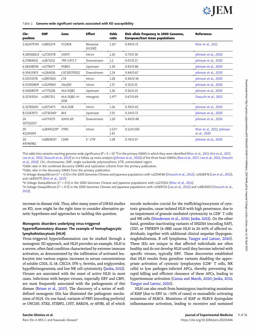
Human genetic studies of KD: GWASFour GWASs on KD have identified several significant genome-wide hits (Table 2). An association of the missense variant(H131R) of FCGR2A with KD was first reported in European andEast Asian cohorts (Khor et al., 2011), and later replicated in aGWAS on independent East Asian populations (Onouchi et al.,2012). The major A allele (encoding histidine) was found to beassociated with a higher risk of KD, with an odds ratio of 1.32.FCGR2A encodes the FcγRIIA protein (CD32a), which belongs to afamily of IgG receptors expressed on many immune cells, in-cluding NK cells, macrophages, and neutrophils. It is involved incell activation and the uptake of immune complexes. The H131Rsubstitution affects binding affinity, and can be used to stratifyindividuals as strong or weak responders to IgG subclasses(Bruhns et al., 2009). ITPKC, reported by Khor et al. (2011) as thesecond locus implicated in KD, was initially identified by linkagedisequilibrium mapping in both Japanese and US children(Onouchi et al., 2008). The associated SNP, rs28493229, wasshown to affect the splicing efficiency of the ITPKC mRNA, andtheCallele reducing splicingwas associatedwith an increase in the riskof KD, with an odds ratio of 1.52. ITPKC encodes inositol-trisphosphate3-kinase, an enzyme responsible for down-regulating the calcium re-sponse in immune cells, thereby decreasing T cell activation (Kumrahet al., 2020; Lo, 2020).
Three subsequent GWASs conducted in Japanese (Onouchiet al., 2012), Taiwanese (Lee et al., 2012), and Korean (Kimet al., 2017) populations reported associations with a cluster ofvariants in the vicinity of the BLK gene. BLK is a src family Blymphoid tyrosine kinase present in B cells that transducessignals downstream from the B cell receptor. Significant asso-ciations were also reported for two noncoding variants in stronglinkage disequilibrium close to CD40, a member of the TNF re-ceptor (TNFR) superfamily, in the Taiwanese and JapaneseGWASs (Lee et al., 2012; Onouchi et al., 2012). Significantgenome-wide associations were also detected for SNPs in HLAclass II genes in the Japanese population (Onouchi et al., 2012),whereas analyses performed with specific HLA class I allelesyielded nonreproducible results (Onouchi, 2009). Finally, a re-cent follow-up study of the three East Asian GWASs identifiedseveral additional significant loci (Johnson et al., 2020; Lee et al.,2012; Onouchi et al., 2012; Table 2). In particular, a higher risk ofKD was found to be associated with the high-expression allelesof SNPs located in the Ig heavy variable gene (IGHV) cluster.Several of these associations with common variants suggest arole for B cells and/or endogenous Igs in KD pathogenesis.However, these epidemiological genetic studies have revealedonly some of the factors involved, resulting in a 1.2- to 1.5-fold
Sancho-Shimizu et al. Journal of Experimental Medicine 8 of 16
Rare IEIs in MIS-C and Kawasaki disease? https://doi.org/10.1084/jem.20210446
Dow
nloaded from http://rupress.org/jem
/article-pdf/218/6/e20210446/1414094/jem_20210446.pdf by Im
perial College London Library user on 30 April 2021
increase in disease risk. Thus, after many years of GWAS studieson KD, now might be the right time to consider alternative ge-netic hypotheses and approaches to tackling this question.
Monogenic disorders underlying virus-triggeredhyperinflammatory disease: The example of hemophagocyticlymphohistiocytosis (HLH)Virus-triggered hyperinflammation can be studied through amonogenic IEI approach, and HLH provides an example. HLH isa severe, often fatal condition characterized by extreme immuneactivation, as demonstrated by the infiltration of activated leu-kocytes into various organs; increases in serum concentrationsof soluble CD25, IL-18, CXCL9, IFN-γ, ferritin, and triglycerides;hypofibrinogenemia; and low NK cell cytotoxicity (Janka, 2012).Viruses are associated with the onset of active HLH in mostcases. Infections with herpes viruses, especially EBV and CMV,are most frequently associated with the pathogenesis of thisdisease (Brisse et al., 2017). The discovery of a series of well-defined monogenic IEIs has illustrated the pathogenic mecha-nism of HLH. On one hand, variants of PRF1 (encoding perforin)or UNC13D, STX11, STXBP2, LYST, RAB27A, or AP3B1, all of which
encode molecules crucial for the trafficking/exocytosis of cyto-toxic granules, cause isolated HLH with high penetrance, due toan impairment of granule-mediated cytotoxicity in CD8+ T cellsand NK cells (Henderson et al., 2020; Janka, 2012). On the otherhand, germline inactivating variants of SH2D1A (encoding SAP),CD27, or TNFRSF9 (4-1BB) cause HLH in 25–60% of affected in-dividuals, together with additional clinical sequelae (hypogam-maglobulinemia, B cell lymphoma; Tangye and Latour, 2020).These IEIs are unique in that affected individuals are oftenhealthy and do not develop HLH until they become infected withspecific viruses, typically EBV. These discoveries establishedthat HLH results from germline variants disabling the appro-priate activation of cytotoxic lymphocytes (CD8+ T cells, NKcells) to lyse pathogen-infected APCs, thereby preventing therapid killing and efficient clearance of these APCs, leading tohyperimmune activation (Canna and Marsh, 2020; Janka, 2012;Tangye and Latour, 2020).
HLH can also result from hemizygous inactivating mutationsof XIAP (due to EBV in ∼50% of cases) or monoallelic activatingmutations of NLRC4. Mutations of XIAP or NLRC4 dysregulateinflammasome activation, leading to excessive and sustained
Table 2. Genome-wide significant variants associated with KD susceptibility
Chr:position
SNP Gene Effect Oddsratio
Risk allele frequency in 1000 Genomes,European/East Asian populations
References
1:161479745 rs1801274 FCGR2A Missense(H131R)
1.32a 0.49/0.72 Khor et al., 2011
4:185568113 rs2720378 CASP3 Intron 1.20 0.73/0.30 Johnson et al., 2020
6:27869631 rs1873212 TRX-CAT1-7 Downstream 1.2 0.07/0.17 Johnson et al., 2020
6:28248594 rs1778477 PGBD1 Upstream 1.30 0.83/0.86 Johnson et al., 2020
6:30411903 rs1264516 LOC105375012 Downstream 1.24 0.44/0.67 Johnson et al., 2020
6:31533378 rs2857602 LTA Intron 1.28 0.40/0.34 Johnson et al., 2020
6:32300809 rs3129960 C6orf10 Intron 1.37 0.15/0.15 Johnson et al., 2020
6:32658079 rs7775228 HLA-DQB1 Upstream 1.26 0.16/0.23 Johnson et al., 2020
6:32763514 rs2857151 HLA-DQB2–H-DOB
Intergenic 1.47b 0.67/0.69 Onouchi et al., 2012
6:32782605 rs2071473 HLA-DOB Intron 1.26 0.39/0.43 Johnson et al., 2020
8:11343973 rs2736340c BLK Upstream 1.55 0.24/0.72 Johnson et al., 2020
14:107152027
rs4774175 IGHV1-69 Downstream 1.20 0.40/0.48 Johnson et al., 2020
19:41224204
rs28493229d ITPKC Intron 1.52a/1.41
0.12/0.065 Khor et al., 2011; Johnsonet al., 2020
20:44746982
rs1883832e CD40 59 UTR 1.28 0.74/0.57 Johnson et al., 2020
This table lists variants reaching genome-wide significance (P < 5 × 10⁻⁸) in the primary GWAS in which they were identified (Khor et al., 2011; Kim et al., 2017;Lee et al., 2012; Onouchi et al., 2012) or in a follow up meta-analysis (Johnson et al., 2020) of the three Asian GWASs (Kim et al., 2017; Lee et al., 2012; Onouchiet al., 2012). Chr, chromosome; SNP, single nucleotide polymorphism; UTR, untranslated region.aOdds ratio in the combined discovery GWAS and replication cohorts from the primary publication.bOdds ratio in the discovery GWAS from the primary publication.cIn linkage disequilibrium (r2 > 0.5) in the 1000 Genomes Chinese and Japanese populations with rs2254546 (Onouchi et al., 2012), rs2618476 (Lee et al., 2012),and rs6993775 (Kim et al., 2017).dIn linkage disequilibrium (r2 > 0.5) in the 1000 Genomes Chinese and Japanese populations with rs2233152 (Khor et al., 2011).eIn linkage disequilibrium (r2 > 0.5) in the 1000 Genomes Chinese and Japanese populations with rs1569723 (Lee et al., 2012) and rs4813003 (Onouchi et al.,2012).
Sancho-Shimizu et al. Journal of Experimental Medicine 9 of 16
Rare IEIs in MIS-C and Kawasaki disease? https://doi.org/10.1084/jem.20210446
Dow
nloaded from http://rupress.org/jem
/article-pdf/218/6/e20210446/1414094/jem_20210446.pdf by Im
perial College London Library user on 30 April 2021
production of IL-1β, IL-18, and CXCL9 (Canna and Marsh, 2020).Moreover, monoallelic mutations of CDC42 were recently re-ported to cause multisystemic inflammation, including HLH (Suand Orange, 2020). Levels of inflammatory mediators, IL-1β,IL-18, IFN-γ, and CXCL9, are very high in the serum of CDC42-deficient patients, and the constitutive production of these fac-tors by bone marrow cells is also higher in patients than inhealthy donors (Bucciol et al., 2020; Gernez et al., 2019; Lamet al., 2019). Consistent with these observations, treatmentwith inhibitors of IL-1 or IFN-γwas found to be effective in somecases of CDC42 deficiency (Gernez et al., 2019; Lam et al., 2019;Su and Orange, 2020). CDC42 deficiency may impair cytotoxiclymphocyte function and promote the production of inflamma-tory mediators, thereby fueling a cytokine storm. In XIAP defi-ciency, and perhaps in other deficiencies, colitis may be aseparate form of severe inflammatory disease that can resultfrom certain viral infections. Thus, an array of severe inflam-matory conditions may emerge following viral infections in in-dividuals with certain monogenic lesions.
Monogenic IEIs may underlie MIS-C in some childrenMIS-C affects previously healthy children and adolescents, withmost other subjects from the same age group infected withSARS-CoV-2 presenting only very mild illness or remainingasymptomatic. We recently showed that single-gene inborn er-rors of type I IFN immunity or neutralizing autoantibodiesagainst type I IFNs can underlie life-threatening COVID-19pneumonia in at least 10% of patients (Bastard et al., 2020, 2021;Zhang et al., 2020a, 2020b; van der Wijst et al., 2021 Preprint).We now suggest that some children may develop MIS-C becauseof a rare monogenic or digenic IEI that is clinically silent untilinfection with SARS-CoV-2, which triggers an inflammatoryresponse. This hypothesis is consistent with previous findings ofmonogenic IEIs in other virus-triggered hyperinflammationdiseases (i.e., HLH triggered by EBV). One key question iswhether MIS-C has immunopathogenic mechanisms in commonwith KD, or even other childhood autoimmune and/or auto-inflammatory conditions. The common and unique clinical,immunological, and laboratory features of these conditions(Fig. 2) suggest probable overlaps in their immunopathogenesis,at least to some extent. However, there are probably also dif-ferent mechanisms at work, given the differences in the featuresof these conditions. As discussed above, the most intriguing andrelevant comparison might be that between MIS-C and KD, thefirst being triggered by a single virus (SARS-CoV-2) whereas thesecond may perhaps be triggered by various infectious agents,such as other viruses (including other coronaviruses). Further-more, despite being triggered by a single virus, MIS-C appears tobe a clinically heterogeneous condition, making it possible toclassify patients into subgroups on the basis of major clinical andimmunological differences, as described above. The search formonogenic and digenic IEIs conferring a predisposition toMIS-Cin previously healthy children and young adults should there-fore focus primarily on the testing of genetic hypotheses in agenome-wide, agnostic manner, while bearing in mind thecharacteristic epidemiological, clinical, and immunological fea-tures of the condition, to facilitate the identification of candidate
disease-causing genes (Casanova and Abel, 2020; Casanova andAbel, 2021; Casanova et al., 2020).
MIS-C may display both genetic homogeneity (the same genemutated in two ormore families) and genetic heterogeneity (onegene per family), and both physiological homogeneity (mutatedgenes immunologically related) and physiological heterogeneity(unrelated). Some disorders may be autosomal, whereas othersare X-linked, and the traits involved may be recessive, domi-nant, or codominant. Depending on the gene, variants may beloss- or gain-of-function. Given the low incidence of this con-dition in the general population, the causal gene variants areprobably equally rare. Clinical penetrance may be full or in-complete, depending on the nature of the gene defect. Incom-plete penetrance may suggest that some cases are digenic, oreven oligogenic. Some gene defects may have an age-dependentpresentation, which may be particularly relevant here, given theage difference between MIS-C and KD patients. Finally, somedefects may be present in specific ancestries. We anticipate thatthe discovery of monogenic IEI underlying MIS-C will unravelthe molecular and cellular basis of the immunopathogenesis ofthe different classes of MIS-C in previously healthy childrenand young adults. Although generally considered to be avasculitis, different types of disorders may be causal in thedifferent clinical subgroups classified by the development ofmyocarditis, neurological symptoms, mild pneumonia symp-toms, or Kawasaki-like symptoms only. The tragic pandemicof COVID-19 has provided us with a unique opportunity todiscover the immunopathogenesis of a severe inflammatorydisease triggered by a single virus via a monogenic approach.
Concluding remarksAgnostic testing of genetic hypotheses can be used to searchfor rare monogenic or digenic IEIs conferring a predispositionto MIS-C, at both the cohort population and individualpatient levels (https://www.covidhge.com). Not only shoulddifferent genetic hypotheses be tested with genomic data frompatients with MIS-C, but these data should also be analyzed inparallel with proper cohorts of controls consisting of healthychildren and young adults who have encountered SARS-CoV-2 infection but remained asymptomatic or developed only a mildclinical syndrome. Moreover, COVID-19 pneumonia, neurologi-cal COVID-19, and MIS-C are probably caused by different typesof human genetic disorders. However, it is tempting to thinkthat the multisystem inflammatory syndrome in adults that hasemerged very recently (Morris et al., 2020) may have somecausative IEIs in common with MIS-C. It is also possible that“COVID toes,” a cutaneous inflammatory syndrome affectingpatients of all ages, but mostly children and young adults(Hernandez and Bruckner, 2020), are caused by IEIs that aredifferent but connected to those underlying MIS-C. Geneticanalyses of these different SARS-CoV-2–related conditions canbe performed in parallel, to identify the IEIs specific to eachcondition. Finally, genomic data from children with MIS-C maybe analyzed in combination with or independently from datafor children with KD. These two conditions may have somegenetic causes in common if they share immunopathogenicmechanisms. Alternatively, monogenic and digenic IEIs may be
Sancho-Shimizu et al. Journal of Experimental Medicine 10 of 16
Rare IEIs in MIS-C and Kawasaki disease? https://doi.org/10.1084/jem.20210446
Dow
nloaded from http://rupress.org/jem
/article-pdf/218/6/e20210446/1414094/jem_20210446.pdf by Im
perial College London Library user on 30 April 2021
detected more readily in genetic and/or physiological homoge-neity approaches, for a condition triggered by a single virus,such as MIS-C. This will provide a basis for genetic diagnosis andcounseling, while guiding the design of preventive and therapeuticinterventions against MIS-C in children and young adults. It willalso pave the way for studies of classic KD, which remains enig-matic, and other childhood autoimmune and/or autoinflammatorypostinfectious conditions (e.g., acute disseminated encephalomy-elitis, rheumatic fever, and Henoch-Schonlein purpura) through arare IEI approach that will become as clinically important forchildren with KD or Kawasaki-like disease as the identification ofthe causal infectious triggers.
Online supplemental materialTable S1 lists reportedMIS-C cases with geographic distribution.
AcknowledgmentsWe thank our colleagues from the COVID Human Genetic Effortconsortium (https://www.covidhge.com) for discussions.
Our studies are funded by the Howard Hughes Medical In-stitute, the Rockefeller University, the St. Giles Foundation, theNational Institutes of Health (R01AI088364 to J.-L. Casanova andS.-Y. Zhang; R01AI148963, R01AI151029, and R01AI150300 to D.Bogunovic; K08AI135091 to S.E. Henrickson; R21AI144315-02S1to C.L. Lucas), the National Center for Advancing TranslationalSciences, National Institutes of Health Clinical and TranslationalScience Award program (UL1 TR001866), the Yale Centerfor Mendelian Genomics and the GSP Coordinating Centerfunded by the National Human Genome Research Institute(UM1HG006504 and U24HG008956), Institut National de laSante et de la Recherche Medicale and Universite de Paris, theFrench National Research Agency (ANR) Resilience-Covid-19grant GenMIS-C, the ANR “Investments for the Future” program(ANR-10-IAHU-01), the Integrative Biology of Emerging Infec-tious Diseases Laboratory of Excellence (ANR-10-LABX-62-IBEID), the ANR project AABIFNCOV (ANR-20-CO11-0001), theFrench Foundation for Medical Research (EQU201903007798),the French Foundation for Medical Research and ANR GENCO-VID project, the ANRSCOV05 project, the Square Foundation,Grandir - Fonds de solidarite pour l’enfance, the Fondationdu Souffle, the SCOR Corporate Foundation for Science, UKResearch and Innovation Future Leader’s Fellowship (MR/S032304/1), the NIHR Imperial Biomedical Research Centre atImperial College Healthcare NHS Trust (70931), the BurroughsWellcome Fund Career Awards for Medical Scientists, theClinical Immunology Society, the American Academy of AllergyAsthma and Immunology, the Michael Smith Foundation forHealth Research, the National Health and Medical ResearchCouncil of Australia, and the University of New South WalesSydney COVID Rapid Response Initiative (to S.G. Tangye).
Author contributions: V. Sancho-Shimizu, J.-L. Casanova, andS.-Y. Zhang conceptualized the study. V. Sancho-Shimizu, P.Brodin, A. Cobat, C.M. Biggs, J. Toubiana, C.L. Lucas, S.E. Hen-rickson, A. Belot, members of MIS-C@CHGE, S.G. Tangye, J.D.Milner, M. Levin, L. Abel, D. Bogunovic, J.-L. Casanova, and S.-Y.Zhang wrote and edited the manuscript.
Disclosures: The authors declare no competing interests exist.
Submitted: 22 February 2021Revised: 24 March 2021Accepted: 7 April 2021
ReferencesAbdel-Mannan, O., M. Eyre, U. Lobel, A. Bamford, C. Eltze, B. Hameed, C.
Hemingway, and Y. Hacohen. 2020. Neurologic and RadiographicFindings Associated With COVID-19 Infection in Children. JAMA Neurol.77:1–6. https://doi.org/10.1001/jamaneurol.2020.2687
Abrams, J.Y., S.E. Godfred-Cato, M.E. Oster, E.J. Chow, E.H. Koumans, B.Bryant, J.W. Leung, and E.D. Belay. 2020. Multisystem InflammatorySyndrome in Children Associated with Severe Acute Respiratory Syn-drome Coronavirus 2: A Systematic Review. J. Pediatr. 226:45–54.e1.https://doi.org/10.1016/j.jpeds.2020.08.003
Ae, R., N. Makino, K. Kosami, M. Kuwabara, Y. Matsubara, and Y. Nakamura.2020. Epidemiology, Treatments, and Cardiac Complications in Patientswith Kawasaki Disease: The Nationwide Survey in Japan, 2017-2018.J. Pediatr. 225:23–29.e2. https://doi.org/10.1016/j.jpeds.2020.05.034
Ahmed, M., S. Advani, A. Moreira, S. Zoretic, J. Martinez, K. Chorath, S. Acosta,R. Naqvi, F. Burmeister-Morton, F. Burmeister, et al. 2020. Multisysteminflammatory syndrome in children: A systematic review. EClinicalMedi-cine. 26:100527. https://doi.org/10.1016/j.eclinm.2020.100527
Allali, S., J.F. Cohen, J. Brice, D. Khraiche, and J. Toubiana. 2021. Gastroin-testinal Symptoms Followed by Shock in a Febrile 7-Year-Old Childduring the COVID-19 Pandemic. Clin. Chem. 67:54–58. https://doi.org/10.1093/clinchem/hvaa279
Al Lawati, Z., H. Al Rawahi, and L.S.F. Al Yazidi. 2021. ACUTE APPENDICITISMIMICKING MULTISYSTEM INFLAMMATORY SYNDROME INCHILDREN: CASE REPORT AND REVIEW OF THE LITERATURE.J. Paediatr. Child Health. 57:461–462. https://doi.org/10.1111/jpc.15398
Almoosa, Z.A., H.H. Al Ameer, S.M. AlKadhem, F. Busaleh, F.A. AlMuhanna,and O. Kattih. 2020. Multisystem Inflammatory Syndrome in Children,the Real Disease of COVID-19 in Pediatrics - A Multicenter Case SeriesFrom Al-Ahsa, Saudi Arabia. Cureus. 12:e11064.
Alsaied, T., A.H. Tremoulet, J.C. Burns, A. Saidi, A. Dionne, S.M. Lang, J.W.Newburger, S. de Ferranti, and K.G. Friedman. 2021. Review of CardiacInvolvement in Multisystem Inflammatory Syndrome in Children.Circulation. 143:78–88. https://doi.org/10.1161/CIRCULATIONAHA.120.049836
Anderson, E.M., C. Diorio, E.C. Goodwin, K.O. McNerney, M.E. Weirick, S.Gouma, M.J. Bolton, C.P. Arevalo, J. Chase, P. Hicks, et al. 2020. SARS-CoV-2 antibody responses in children with MIS-C and mild and severeCOVID-19. J. Pediatric Infect. Dis. Soc.:piaa161. https://doi.org/10.1093/jpids/piaa161
Aydın, F., E. Çelikel, Z. Ekici Tekin, S. Coskun, M. Sezer, C. Karagol, M.M.Kaplan, N. Tekgoz, T. Kurt, S. Ozcan, et al. 2021. Comparison of baselinelaboratory findings of macrophage activation syndrome complicatingsystemic juvenile idiopathic arthritis and multisystem inflammatorysyndrome in children. Int. J. Rheum. Dis. 24:542–547. https://doi.org/10.1111/1756-185X.14078
Bahrami, A., M. Vafapour, B. Moazzami, and N. Rezaei. 2020. Hyper-inflammatory shock related to COVID-19 in a patient presenting withmultisystem inflammatory syndrome in children: First case from Iran.J. Paediatr. Child Health.:jpc.15048. https://doi.org/10.1111/jpc.15048
Bajolle, F., J.F. Meritet, F. Rozenberg, M. Chalumeau, D. Bonnet, D. Gendrel,and P. Lebon. 2014. Markers of a recent bocavirus infection in childrenwith Kawasaki disease: “a year prospective study”. Pathol. Biol. (Paris).62:365–368. https://doi.org/10.1016/j.patbio.2014.06.002
Bastard, P., L.B. Rosen, Q. Zhang, E. Michailidis, H.H. Hoffmann, Y. Zhang, K.Dorgham, Q. Philippot, J. Rosain, V. Beziat, et al. COVID Human GeneticEffort. 2020. Autoantibodies against type I IFNs in patients with life-threatening COVID-19. Science. 370:eabd4585. https://doi.org/10.1126/science.abd4585
Bastard, P., E. Michailidis, H.H. Hoffmann, M. Chbihi, T. Le Voyer, J. Rosain,Q. Philippot, Y. Seeleuthner, A. Gervais, M. Materna, et al. 2021. Auto-antibodies to type I IFNs can underlie adverse reactions to yellow feverlive attenuated vaccine. J. Exp. Med. 218:e20202486. https://doi.org/10.1084/jem.20202486
Sancho-Shimizu et al. Journal of Experimental Medicine 11 of 16
Rare IEIs in MIS-C and Kawasaki disease? https://doi.org/10.1084/jem.20210446
Dow
nloaded from http://rupress.org/jem
/article-pdf/218/6/e20210446/1414094/jem_20210446.pdf by Im
perial College London Library user on 30 April 2021
Bautista-Rodriguez, C., J. Sanchez-de-Toledo, B.C. Clark, J. Herberg, F. Bajolle,P.C. Randanne, D. Salas-Mera, S. Foldvari, D. Chowdhury, R. Munoz,et al. 2021. Multisystem Inflammatory Syndrome in Children: An In-ternational Survey. Pediatrics. 147:e2020024554. https://doi.org/10.1542/peds.2020-024554
Belhadjer, Z., J. Auriau, M. Meot, M. Oualha, S. Renolleau, L. Houyel, and D.Bonnet. 2020a. Addition of Corticosteroids to Immunoglobulins Is As-sociated With Recovery of Cardiac Function in Multi-InflammatorySyndrome in Children. Circulation. 142:2282–2284. https://doi.org/10.1161/CIRCULATIONAHA.120.050147
Belhadjer, Z., M. Meot, F. Bajolle, D. Khraiche, A. Legendre, S. Abakka, J.Auriau, M. Grimaud, M. Oualha, M. Beghetti, et al. 2020b. Acute HeartFailure in Multisystem Inflammatory Syndrome in Children in theContext of Global SARS-CoV-2 Pandemic. Circulation. 142:429–436.https://doi.org/10.1161/CIRCULATIONAHA.120.048360
Belot, A., D. Antona, S. Renolleau, E. Javouhey, V. Hentgen, F. Angoulvant, C.Delacourt, X. Iriart, C. Ovaert, B. Bader-Meunier, et al. 2020. SARS-CoV-2-related paediatric inflammatory multisystem syndrome, an ep-idemiological study, France, 1 March to 17 May 2020. Euro Surveill. 25:2001010. https://doi.org/10.2807/1560-7917.ES.2020.25.22.2001010
Belot, A., and D. Levy-Bruhl. French Covid-19 Pediatric Inflammation Con-sortium. 2020. Multisystem Inflammatory Syndrome in Children in theUnited States. N. Engl. J. Med. 383:1793–1794. https://doi.org/10.1056/NEJMc2026136
Binstadt, B.A., J.C. Levine, P.A. Nigrovic, K. Gauvreau, F. Dedeoglu, R.C.Fuhlbrigge, S.N. Weindling, J.W. Newburger, and R.P. Sundel. 2005.Coronary artery dilation among patients presenting with systemic-onset juvenile idiopathic arthritis. Pediatrics. 116:e89–e93. https://doi.org/10.1542/peds.2004-2190
Blondiaux, E., P. Parisot, A. Redheuil, L. Tzaroukian, Y. Levy, C. Sileo, A.Schnuriger, M. Lorrot, R. Guedj, and H. Ducou le Pointe. 2020. CardiacMRI in Children with Multisystem Inflammatory Syndrome Associatedwith COVID-19. Radiology. 297:E283–E288. https://doi.org/10.1148/radiol.2020202288
Bouhaddou, M., D. Memon, B. Meyer, K.M. White, V.V. Rezelj, M. CorreaMarrero, B.J. Polacco, J.E. Melnyk, S. Ulferts, R.M. Kaake, et al. 2020.The Global Phosphorylation Landscape of SARS-CoV-2 Infection. Cell.182:685–712.e19. https://doi.org/10.1016/j.cell.2020.06.034
Brisse, E., C.H. Wouters, G. Andrei, and P. Matthys. 2017. How VirusesContribute to the Pathogenesis of Hemophagocytic Lymphohistiocyto-sis. Front. Immunol. 8:1102. https://doi.org/10.3389/fimmu.2017.01102
Bruhns, P., B. Iannascoli, P. England, D.A. Mancardi, N. Fernandez, S. Jorieux,and M. Daeron. 2009. Specificity and affinity of human Fcgamma re-ceptors and their polymorphic variants for human IgG subclasses.Blood. 113:3716–3725. https://doi.org/10.1182/blood-2008-09-179754
Bucciol, G., B. Pillay, J. Casas-Martin, S. Delafontaine, M. Proesmans, N.Lorent, J. Coolen, T. Tousseyn, X. Bossuyt, C.S. Ma, et al. 2020. SystemicInflammation and Myelofibrosis in a Patient with Takenouchi-KosakiSyndrome due to CDC42 Tyr64Cys Mutation. J. Clin. Immunol. 40:567–570. https://doi.org/10.1007/s10875-020-00742-5
Buonsenso, D., G. Di Sante, and M. Sali. CURE COVID-19 Study Group.2020. Cytokine Profile in an Adolescent With Pediatric Multisys-tem Inflammatory Syndrome Temporally Related to COVID-19.Pediatr. Infect. Dis. J. 39:e213–e215. https://doi.org/10.1097/INF.0000000000002802
Burns, J.C., and M.P. Glode. 2004. Kawasaki syndrome. Lancet. 364:533–544.https://doi.org/10.1016/S0140-6736(04)16814-1
Canna, S.W., and R.A. Marsh. 2020. Pediatric hemophagocytic lymphohistio-cytosis. Blood. 135:1332–1343. https://doi.org/10.1182/blood.2019000936
Capone, C.A., A. Subramony, T. Sweberg, J. Schneider, S. Shah, L. Rubin, C.Schleien, S. Epstein, J.C. Johnson, A. Kessel, et al. Northwell HealthCOVID-19 Research Consortium. 2020. Characteristics, Cardiac In-volvement, and Outcomes of Multisystem Inflammatory Syndrome ofChildhood Associated with severe acute respiratory syndrome corona-virus 2 Infection. J. Pediatr. 224:141–145. https://doi.org/10.1016/j.jpeds.2020.06.044
Carter, M.J., M. Fish, A. Jennings, K.J. Doores, P.Wellman, J. Seow, S. Acors, C.Graham, E. Timms, J. Kenny, et al. 2020. Peripheral im-munophenotypes in children with multisystem inflammatory syn-drome associated with SARS-CoV-2 infection. Nat. Med. 26:1701–1707.https://doi.org/10.1038/s41591-020-1054-6
Casanova, J.L., and L. Abel. 2020. The human genetic determinism of life-threatening infectious diseases: genetic heterogeneity and physiologicalhomogeneity? Hum. Genet. 139:681–694. https://doi.org/10.1007/s00439-020-02184-w
Casanova, J.L., and L. Abel. 2021. Lethal Infectious Diseases as Inborn Errorsof Immunity: Toward a Synthesis of the Germ and Genetic Theories.Annu. Rev. Pathol. 16:23–50. https://doi.org/10.1146/annurev-pathol-031920-101429
Casanova, J.L., and H.C. Su. COVID Human Genetic Effort. 2020. A GlobalEffort to Define the Human Genetics of Protective Immunity to SARS-CoV-2 Infection. Cell. 181:1194–1199. https://doi.org/10.1016/j.cell.2020.05.016
Catalano-Pons, C., P. Quartier, M. Leruez-Ville, F. Kaguelidou, D. Gendrel, G.Lenoir, J.L. Casanova, and D. Bonnet. 2005. Primary cytomegalovirusinfection, atypical Kawasaki disease, and coronary aneurysms in 2 in-fants. Clin. Infect. Dis. 41:e53–e56. https://doi.org/10.1086/432578
Cheng, M.H., S. Zhang, R.A. Porritt, M. Noval Rivas, L. Paschold, E. Willscher,M. Binder, M. Arditi, and I. Bahar. 2020. Superantigenic character of aninsert unique to SARS-CoV-2 spike supported by skewed TCR reper-toire in patients with hyperinflammation. Proc. Natl. Acad. Sci. USA. 117:25254–25262. https://doi.org/10.1073/pnas.2010722117
Cheung, E.W., P. Zachariah, M. Gorelik, A. Boneparth, S.G. Kernie, J.S. Or-ange, and J.D. Milner. 2020. Multisystem Inflammatory SyndromeRelated to COVID-19 in Previously Healthy Children and Adolescents inNew York City. JAMA. 324:294–296. https://doi.org/10.1001/jama.2020.10374
Clark, B.C., J. Sanchez-de-Toledo, C. Bautista-Rodriguez, N. Choueiter, D.Lara, H. Kang, S. Mohsin, A. Fraisse, S. Cesar, A. Sattar Shaikh, et al.2020. Cardiac Abnormalities Seen in Pediatric Patients During theSARS-CoV2 Pandemic: An International Experience. J. Am. Heart Assoc.9:e018007. https://doi.org/10.1161/JAHA.120.018007
Cogan, E., P. Foulon, O. Cappeliez, N. Dolle, G. Vanfraechem, and D. DeBacker. 2020. Multisystem Inflammatory Syndrome With CompleteKawasaki Disease Features Associated With SARS-CoV-2 Infection in aYoung Adult. A Case Report. Front. Med. (Lausanne). 7:428. https://doi.org/10.3389/fmed.2020.00428
Consiglio, C.R., N. Cotugno, F. Sardh, C. Pou, D. Amodio, L. Rodriguez, Z. Tan,S. Zicari, A. Ruggiero, G.R. Pascucci, et al. CACTUS Study Team. 2020.The Immunology of Multisystem Inflammatory Syndrome in Childrenwith COVID-19. Cell. 183:968–981.e7. https://doi.org/10.1016/j.cell.2020.09.016
Davies, P., C. Evans, H.K. Kanthimathinathan, J. Lillie, J. Brierley, G. Waters,M. Johnson, B. Griffiths, P. du Pre, Z. Mohammad, et al. 2020. Intensivecare admissions of children with paediatric inflammatory multisystemsyndrome temporally associated with SARS-CoV-2 (PIMS-TS) in theUK: a multicentre observational study. Lancet Child Adolesc. Health. 4:669–677. https://doi.org/10.1016/S2352-4642(20)30215-7
de Farias, E.C.F., J. Pedro Piva, M.L.F.M.F. de Mello, L.M.P.P. do Nascimento,C.C. Costa, M.M.M. Machado, T.D.S. Rodrigues, R.D.F.P. Carvalho,M.C.B. Alves, L.F.Q. Aires, et al. 2020. Multisystem InflammatorySyndrome Associated With Coronavirus Disease in Children: A Multi-centered Study in Belem, Para, Brazil. Pediatr. Infect. Dis. J. 39:e374–e376. https://doi.org/10.1097/INF.0000000000002865
Dhanalakshmi, K., A. Venkataraman, S. Balasubramanian, M. Madhusudan,S. Amperayani, S. Putilibai, K. Sadasivam, B. Ramachandran, and A.V.Ramanan. 2020. Epidemiological and Clinical Profile of Pediatric In-flammatory Multisystem Syndrome - Temporally Associated withSARS-CoV-2 (PIMS-TS) in Indian Children. Indian Pediatr. 57:1010–1014. https://doi.org/10.1007/s13312-020-2025-1
Dietz, S.M., D. van Stijn, D. Burgner,M. Levin, I.M. Kuipers, B.A. Hutten, and T.W.Kuijpers. 2017. Dissecting Kawasaki disease: a state-of-the-art review. Eur.J. Pediatr. 176:995–1009. https://doi.org/10.1007/s00431-017-2937-5
Diorio, C., S.E. Henrickson, L.A. Vella, K.O. McNerney, J. Chase, C. Bur-udpakdee, J.H. Lee, C. Jasen, F. Balamuth, D.M. Barrett, et al. 2020.Multisystem inflammatory syndrome in children and COVID-19 aredistinct presentations of SARS-CoV-2. J. Clin. Invest. 130:5967–5975.https://doi.org/10.1172/JCI140970
Docherty, A.B., E.M. Harrison, C.A. Green, H.E. Hardwick, R. Pius, L. Nor-man, K.A. Holden, J.M. Read, F. Dondelinger, G. Carson, et al. ISARIC4Cinvestigators. 2020. Features of 20 133 UK patients in hospital withcovid-19 using the ISARIC WHO Clinical Characterisation Protocol:prospective observational cohort study. BMJ. 369:m1985. https://doi.org/10.1136/bmj.m1985
Dolhnikoff, M., J. Ferreira Ferranti, R.A. de Almeida Monteiro, A.N. Duarte-Neto, M. Soares Gomes-Gouvea, N. Viu Degaspare, A. Figueiredo Del-gado, C. Montanari Fiorita, G. Nunes Leal, R.M. Rodrigues, et al. 2020.SARS-CoV-2 in cardiac tissue of a child with COVID-19-related multi-system inflammatory syndrome. Lancet Child Adolesc. Health. 4:790–794.https://doi.org/10.1016/S2352-4642(20)30257-1
Sancho-Shimizu et al. Journal of Experimental Medicine 12 of 16
Rare IEIs in MIS-C and Kawasaki disease? https://doi.org/10.1084/jem.20210446
Dow
nloaded from http://rupress.org/jem
/article-pdf/218/6/e20210446/1414094/jem_20210446.pdf by Im
perial College London Library user on 30 April 2021
Dominguez, S.R., M.S. Anderson, M.P. Glode, C.C. Robinson, and K.V.Holmes. 2006. Blinded case-control study of the relationship betweenhuman coronavirus NL63 and Kawasaki syndrome. J. Infect. Dis. 194:1697–1701. https://doi.org/10.1086/509509
Dufort, E.M., E.H. Koumans, E.J. Chow, E.M. Rosenthal, A. Muse, J. Rowlands,M.A. Barranco, A.M.Maxted, E.S. Rosenberg, D. Easton, et al. New YorkState and Centers for Disease Control and Prevention Multisystem In-flammatory Syndrome in Children Investigation Team. 2020. Multi-system Inflammatory Syndrome in Children in New York State.N. Engl.J. Med. 383:347–358. https://doi.org/10.1056/NEJMoa2021756
Esper, F., E.D. Shapiro, C. Weibel, D. Ferguson, M.L. Landry, and J.S. Kahn.2005. Association between a novel human coronavirus and Kawasakidisease. J. Infect. Dis. 191:499–502. https://doi.org/10.1086/428291
Esteve-Sole, A., J. Anton, R.M. Pino-Ramirez, J. Sanchez-Manubens, V. Fu-mado, C. Fortuny, M. Rios-Barnes, J. Sanchez-de-Toledo, M. Girona-Alarcon, J.M. Mosquera, et al. 2021. Similarities and differences be-tween the immunopathogenesis of COVID-19-related pediatric inflam-matory multisystem syndrome and Kawasaki disease. J. Clin. Invest. 131:e144554. https://doi.org/10.1172/JCI144554
Falah, N.U., S. Hashmi, Z. Ahmed, A. Jaan, A. Akhtar, F. Khalid, U. Farooque,M.T. Shera, S. Ali, and A. Javed. 2020. Kawasaki Disease-Like Featuresin 10 Pediatric COVID-19 Cases: A Retrospective Study. Cureus. 12:e11035.
Farias, E.C.F., M.C.A. Justino, and M.L.F.M.F. Mello. 2020. Multisys-tem Inflammatory Syndrome in a Child Associated with Coro-navirus Disease 19 in the Brazilian Amazon: Fatal Outcome in anInfant. Rev. Paul. Pediatr. 38:e2020165. https://doi.org/10.1590/1984-0462/2020/38/2020165
Feldstein, L.R., E.B. Rose, S.M. Horwitz, J.P. Collins, M.M. Newhams, M.B.F.Son, J.W. Newburger, L.C. Kleinman, S.M. Heidemann, A.A. Martin,et al. CDC COVID-19 Response Team. 2020. Multisystem InflammatorySyndrome in U.S. Children and Adolescents. N. Engl. J. Med. 383:334–346. https://doi.org/10.1056/NEJMoa2021680
Felsenstein, S., E. Willis, H. Lythgoe, L. McCann, A. Cleary, K. Mahmood,D. Porter, J. Jones, J. McDonagh, A. Chieng, et al. 2020. Presentation,Treatment Response and Short-Term Outcomes in Paediatric Mul-tisystem Inflammatory Syndrome Temporally Associated withSARS-CoV-2 (PIMS-TS). J. Clin. Med. 9:3293. https://doi.org/10.3390/jcm9103293
Fouriki, A., Y. Fougère, C. De Camaret, G. Blanchard Rohner, S. Grazioli, N.Wagner, C. Relly, J. Pachlopnik Schmid, J. Trück, L. Kottanatu, et al.2021. Case Report: Case Series of Children With Multisystem Inflam-matory Syndrome Following SARS-CoV-2 Infection in Switzerland.Front Pediatr. 8:594127. https://doi.org/10.3389/fped.2020.594127
Friedman, K.G., D.M. Harrild, and J.W. Newburger. 2020. Cardiac Dysfunc-tion in Multisystem Inflammatory Syndrome in Children: A Call toAction. J. Am. Coll. Cardiol. 76:1962–1964. https://doi.org/10.1016/j.jacc.2020.09.002
Gale, C., M.A. Quigley, A. Placzek, M. Knight, S. Ladhani, E.S. Draper, D.Sharkey, C. Doherty, H. Mactier, and J.J. Kurinczuk. 2021. Char-acteristics and outcomes of neonatal SARS-CoV-2 infection in theUK: a prospective national cohort study using active surveillance.Lancet Child Adolesc. Health. 5:113–121. https://doi.org/10.1016/S2352-4642(20)30342-4
Gernez, Y., A.A. de Jesus, H. Alsaleem, C. Macaubas, A. Roy, D. Lovell, K.A.Jagadeesh, S. Alehashemi, L. Erdman, M. Grimley, et al. 2019. Severeautoinflammation in 4 patients with C-terminal variants in cell divisioncontrol protein 42 homolog (CDC42) successfully treated with IL-1β in-hibition. J. Allergy Clin. Immunol. 144:1122–1125.e6. https://doi.org/10.1016/j.jaci.2019.06.017
Ghatasheh, G., H. Al Dhanhani, A. Goyal, M.B. Noureddin, D. Al Awaad, and Z.Peerwani. 2021. COVID-19-Related Giant Coronary Aneurysms in anInfant with Multisystem Inflammatory Disorder in Children: The FirstCase Report from the United Arab Emirates and the Arab Region. CaseRep. Infect. Dis. 2021:8872412. https://doi.org/10.1155/2021/8872412
Godfred-Cato, S., B. Bryant, J. Leung, M.E. Oster, L. Conklin, J. Abrams, K.Roguski, B. Wallace, E. Prezzato, E.H. Koumans, et al. California MIS-CResponse Team. 2020. COVID-19-Associated Multisystem Inflamma-tory Syndrome in Children - United States, March-July 2020. MMWRMorb. Mortal. Wkly. Rep. 69:1074–1080. https://doi.org/10.15585/mmwr.mm6932e2
Gotzinger, F., B. Santiago-Garcıa, A. Noguera-Julian, M. Lanaspa, L. Lancella,F.I. Calo Carducci, N. Gabrovska, S. Velizarova, P. Prunk, V. Osterman,et al. ptbnet COVID-19 Study Group. 2020. COVID-19 in children andadolescents in Europe: a multinational, multicentre cohort study. Lancet
Child Adolesc. Health. 4:653–661. https://doi.org/10.1016/S2352-4642(20)30177-2
Grazioli, S., F. Tavaglione, G. Torriani, N.Wagner, M. Rohr, A.G. L’Huillier, C.Leclercq, A. Perrin, A. Bordessoule, M. Beghetti, et al. 2020. Immuno-logical assessment of pediatric multisystem inflammatory syndromerelated to COVID-19. J. Pediatric Infect. Dis. Soc.:piaa142. https://doi.org/10.1093/jpids/piaa142
Gruber, C.N., R.S. Patel, R. Trachtman, L. Lepow, F. Amanat, F. Krammer,K.M. Wilson, K. Onel, D. Geanon, K. Tuballes, et al. 2020. MappingSystemic Inflammation and Antibody Responses in Multisystem In-flammatory Syndrome in Children (MIS-C). Cell. 183:982–995.e14.https://doi.org/10.1016/j.cell.2020.09.034
Harwood, R., B. Allin, C.E. Jones, E. Whittaker, P. Ramnarayan, A.V. Ram-anan, M. Kaleem, R. Tulloh, M.J. Peters, S. Almond, et al. PIMS-TSNational Consensus Management Study Group. 2021. A national con-sensus management pathway for paediatric inflammatory multisystemsyndrome temporally associated with COVID-19 (PIMS-TS): results of anational Delphi process. Lancet Child Adolesc. Health. 5:133–141. https://doi.org/10.1016/S2352-4642(20)30304-7
Henderson, L.A., S.W. Canna, K.G. Friedman, M. Gorelik, S.K. Lapidus, H.Bassiri, E.M. Behrens, A. Ferris, K.F. Kernan, G.S. Schulert, et al. 2020.American College of Rheumatology Clinical Guidance for Pediatric Pa-tients with Multisystem Inflammatory Syndrome in Children (MIS-C)Associated with SARS-CoV-2 and Hyperinflammation in COVID-19.Version 2. Arthritis Rheumatol. https://doi.org/10.1002/art.41454
Hernandez, C., and A.L. Bruckner. 2020. Focus on “COVID Toes”. JAMADermatol. 156:1003. https://doi.org/10.1001/jamadermatol.2020.2062
Holman, R.C., E.D. Belay, K.Y. Christensen, A.M. Folkema, C.A. Steiner, andL.B. Schonberger. 2010. Hospitalizations for Kawasaki syndromeamong children in the United States, 1997-2007. Pediatr. Infect. Dis. J. 29:483–488. https://doi.org/10.1097/INF.0b013e3181cf8705
Hoste, L., R. Van Paemel, and F. Haerynck. 2021. Multisystem inflammatorysyndrome in children related to COVID-19: a systematic review. Eur.J. Pediatr. 18:1–16.
Jain, S., S. Sen, S. Lakshmivenkateshiah, P. Bobhate, S. Venkatesh, S. Udani,L. Shobhavat, P. Andankar, T. Karande, and S. Kulkarni. 2020. Multi-system Inflammatory Syndrome in Children With COVID-19 in Mum-bai, India. Indian Pediatr. 57:1015–1019. https://doi.org/10.1007/s13312-020-2026-0
Jain, M.K., S.K. Sahu, J.R. Behera, and S. Patnaik. 2021. Multisystem In-flammatory Syndrome in Children Associated with COVID 19 Treatedwith Oral Steroid. Indian J. Pediatr. 88:106. https://doi.org/10.1007/s12098-020-03497-4
Janka, G.E. 2012. Familial and acquired hemophagocytic lymphohistiocytosis.Annu. Rev. Med. 63:233–246. https://doi.org/10.1146/annurev-med-041610-134208
Jhaveri, S., N. Ahluwalia, S. Kaushik, R. Trachtman, S. Kowalsky, S. Aydin,and K. Stern. 2021. Longitudinal Echocardiographic Assessment ofCoronary Arteries and Left Ventricular Function followingMultisystemInflammatory Syndrome in Children. J. Pediatr. 228:290–293.e1. https://doi.org/10.1016/j.jpeds.2020.08.002
Jia, S., C. Li, G.Wang, J. Yang, and Y. Zu. 2010. The T helper type 17/regulatoryT cell imbalance in patients with acute Kawasaki disease. Clin. Exp.Immunol. 162:131–137. https://doi.org/10.1111/j.1365-2249.2010.04236.x
Johnson, T.A., Y. Mashimo, J.Y. Wu, D. Yoon, A. Hata, M. Kubo, A. Takahashi,T. Tsunoda, K. Ozaki, T. Tanaka, et al. Korean Kawasaki Disease Ge-netics Consortium, Taiwan Kawasaki Disease Genetics Consortium,Taiwan Pediatric ID Alliance, Japan Kawasaki Disease Genome Con-sortium. 2020. Association of an IGHV3-66 gene variant with Kawasakidisease. J. Hum. Genet. 26:1–15. https://doi.org/10.1038/s10038-020-00864-z
Jones, V.G., M. Mills, D. Suarez, C.A. Hogan, D. Yeh, J.B. Segal, E.L. Nguyen,G.R. Barsh, S. Maskatia, and R. Mathew. 2020. COVID-19 and KawasakiDisease: Novel Virus and Novel Case. Hosp. Pediatr. 10:537–540. https://doi.org/10.1542/hpeds.2020-0123
Jordan-Villegas, A., M.L. Chang, O. Ramilo, and A. Mejıas. 2010. Concomitantrespiratory viral infections in children with Kawasaki disease. Pediatr.Infect. Dis. J. 29:770–772. https://doi.org/10.1097/INF.0b013e3181dba70b
Kaiser Family Foundation. 2019. Population Distribution by Race/Ethnicity.https://www.kff.org/other/state-indicator/distribution-by-raceethnicity(accessed December 27, 2020)
Kanegaye, J.T., M.S. Wilder, D. Molkara, J.R. Frazer, J. Pancheri, A.H.Tremoulet, V.E. Watson, B.M. Best, and J.C. Burns. 2009. Recognition ofa Kawasaki disease shock syndrome. Pediatrics. 123:e783–e789. https://doi.org/10.1542/peds.2008-1871
Sancho-Shimizu et al. Journal of Experimental Medicine 13 of 16
Rare IEIs in MIS-C and Kawasaki disease? https://doi.org/10.1084/jem.20210446
Dow
nloaded from http://rupress.org/jem
/article-pdf/218/6/e20210446/1414094/jem_20210446.pdf by Im
perial College London Library user on 30 April 2021
Kato, H., T. Sugimura, T. Akagi, N. Sato, K. Hashino, Y. Maeno, T. Kazue, G.Eto, and R. Yamakawa. 1996. Long-term consequences of Kawasakidisease. A 10- to 21-year follow-up study of 594 patients. Circulation. 94:1379–1385. https://doi.org/10.1161/01.CIR.94.6.1379
Kawasaki, T. 1967. [Acute febrile mucocutaneous syndrome with lymphoidinvolvement with specific desquamation of the fingers and toes inchildren]. Arerugi. 16:178–222.
Kawasaki, T., F. Kosaki, S. Okawa, I. Shigematsu, and H. Yanagawa. 1974. Anew infantile acute febrile mucocutaneous lymph node syndrome(MLNS) prevailing in Japan. Pediatrics. 54:271–276.
Khor, C.C., S. Davila, W.B. Breunis, Y.C. Lee, C. Shimizu, V.J. Wright, R.S.Yeung, D.E. Tan, K.S. Sim, J.J. Wang, et al. Blue Mountains Eye Study.2011. Genome-wide association study identifies FCGR2A as a suscepti-bility locus for Kawasaki disease. Nat. Genet. 43:1241–1246. https://doi.org/10.1038/ng.981
Kim, J.J., S.W. Yun, J.J. Yu, K.L. Yoon, K.Y. Lee, H.R. Kil, G.B. Kim, M.K. Han,M.S. Song, H.D. Lee, et al. Korean Kawasaki Disease Genetics Consor-tium. 2017. A genome-wide association analysis identifies NMNAT2 andHCP5 as susceptibility loci for Kawasaki disease. J. Hum. Genet. 62:1023–1029. https://doi.org/10.1038/jhg.2017.87
Kim, H., J.Y. Shim, J.H. Ko, A. Yang, J.W. Shim, D.S. Kim, H.L. Jung, J.H. Kwak,and I.S. Sol. 2020a. Multisystem Inflammatory Syndrome in ChildrenRelated to COVID-19: the First Case in Korea. J. Korean Med. Sci. 35:e391.https://doi.org/10.3346/jkms.2020.35.e391
Kim, Y.J., H. Park, Y.Y. Choi, Y.K. Kim, Y. Yoon, K.R. Kim, and E.H. Choi.2020b. Defining Association between COVID-19 and the MultisystemInflammatory Syndrome in Children through the Pandemic. J. KoreanMed. Sci. 35:e204. https://doi.org/10.3346/jkms.2020.35.e204
Klocperk, A., Z. Parackova, J. Dissou, H. Malcova, P. Pavlicek, T. Vymazal, P.Dolezalova, and A. Sediva. 2020. Case Report: Systemic InflammatoryResponse and Fast Recovery in a Pediatric Patient With COVID-19.Front. Immunol. 11:1665. https://doi.org/10.3389/fimmu.2020.01665
Kumrah, R., P. Vignesh, A. Rawat, and S. Singh. 2020. Immunogenetics ofKawasaki disease. Clin. Rev. Allergy Immunol. 59:122–139. https://doi.org/10.1007/s12016-020-08783-9
Labe, P., A. Ly, C. Sin, M. Nasser, E. Chapelon-Fromont, P. Ben Saıd, and E.Mahe. 2020. Erythema multiforme and Kawasaki disease associatedwith COVID-19 infection in children. J. Eur. Acad. Dermatol. Venereol. 34:e539–e541. https://doi.org/10.1111/jdv.16666
Lad, S.S., S.P. Kait, P.B. Suryawanshi, J. Mujawar, P. Lad, R. Khetre, L.M.Jadhav, A. Bhor, P. Balte, P. Kataria, et al. 2021. Neurological Manifes-tations in Pediatric Inflammatory Multisystem Syndrome TemporallyAssociated with SARS-CoV-2 (PIMS-TS). Indian J. Pediatr. 88:294–295.https://doi.org/10.1007/s12098-020-03530-6
Lam, M.T., S. Coppola, O.H.F. Krumbach, G. Prencipe, A. Insalaco, C. Cifaldi, I.Brigida, E. Zara, S. Scala, S. Di Cesare, et al. 2019. A novel disorder in-volving dyshematopoiesis, inflammation, and HLH due to aberrant CDC42function. J. Exp. Med. 216:2778–2799. https://doi.org/10.1084/jem.20190147
Lee, Y.C., H.C. Kuo, J.S. Chang, L.Y. Chang, L.M. Huang, M.R. Chen, C.D.Liang, H. Chi, F.Y. Huang, M.L. Lee, et al. Taiwan Pediatric ID Alliance.2012. Two new susceptibility loci for Kawasaki disease identifiedthrough genome-wide association analysis. Nat. Genet. 44:522–525.https://doi.org/10.1038/ng.2227
Lee, P.Y., M. Day-Lewis, L.A. Henderson, K.G. Friedman, J. Lo, J.E. Roberts,M.S. Lo, C.D. Platt, J. Chou, K.J. Hoyt, et al. 2020. Distinct clinical andimmunological features of SARS-CoV-2-induced multisystem inflam-matory syndrome in children. J. Clin. Invest. 130:5942–5950. https://doi.org/10.1172/JCI141113
Lee, J.H., H.S. Han, and J.K. Lee. 2021. The Importance of Early Recognition,Timely Management, and the Role of Healthcare Providers in Multi-system Inflammatory Syndrome in Children. J. Korean Med. Sci. 36:e17.https://doi.org/10.3346/jkms.2021.36.e17
Lehmann, C., R. Klar, J. Lindner, P. Lindner, H. Wolf, and S. Gerling. 2009.Kawasaki disease lacks association with human coronavirus NL63 andhuman bocavirus. Pediatr. Infect. Dis. J. 28:553–554. https://doi.org/10.1097/INF.0b013e31819f41b6
Li, Y., Q. Zheng, L. Zou, J. Wu, L. Guo, L. Teng, R. Zheng, L.K.L. Jung, and M.Lu. 2019. Kawasaki disease shock syndrome: clinical characteristics andpossible use of IL-6, IL-10 and IFN-γ as biomarkers for early recogni-tion. Pediatr. Rheumatol. Online J. 17:1. https://doi.org/10.1186/s12969-018-0303-4
Licciardi, F., G. Pruccoli, M. Denina, E. Parodi, M. Taglietto, S. Rosati, and D.Montin. 2020. SARS-CoV-2-Induced Kawasaki-Like HyperinflammatorySyndrome: A Novel COVID Phenotype in Children. Pediatrics. 146:e20201711. https://doi.org/10.1542/peds.2020-1711
Lima-Setta, F., M.C. Magalhães-Barbosa, G. Rodrigues-Santos, E.A.D.N. Fig-ueiredo, M.L. Jacques, R.S. Zeitel, R. Sapolnik, C.T.D.S. Borges, V.S.Lanziotti, R.E.V. Castro, et al. Brazilian Research Network in PediatricIntensive Care (BRnet-PIC). 2020. Multisystem inflammatory syn-drome in children (MIS-C) during SARS-CoV-2 pandemic in Brazil: amulticenter, prospective cohort study. J. Pediatr. (Rio J.).:S0021-7557(20)30225-4. https://doi.org/10.1016/j.jped.2020.10.008
Lishman, J., C. Kohler, C. de Vos, M.M. van der Zalm, J. Itana, A. Redfern, L.Smit, and H. Rabie. 2020. Acute Appendicitis in Multisystem Inflam-matory Syndrome in Children With COVID-19. Pediatr. Infect. Dis. J. 39:e472–e473. https://doi.org/10.1097/INF.0000000000002900
Lo, M.S. 2020. A framework for understanding Kawasaki disease pathogenesis.Clin. Immunol. 214:108385. https://doi.org/10.1016/j.clim.2020.108385
Mamishi, S., Z. Movahedi, M. Mohammadi, V. Ziaee, M. Khodabandeh, M.R.Abdolsalehi, A. Navaeian, H. Heydari, S. Mahmoudi, and B. Pourakbari.2020.Multisystem inflammatory syndrome associated with SARS-CoV-2 infection in 45 children: a first report from Iran. Epidemiol. Infect. 148:e196. https://doi.org/10.1017/S095026882000196X
Marrani, E., J.C. Burns, and R. Cimaz. 2018. How Should We Classify Kawa-saki Disease? Front. Immunol. 9:2974. https://doi.org/10.3389/fimmu.2018.02974
Matsubara, D., H.L. Kauffman, Y. Wang, R. Calderon-Anyosa, S. Nadaraj,M.D. Elias, T.J. White, D.L. Torowicz, P. Yubbu, T.M. Giglia, et al. 2020.Echocardiographic Findings in Pediatric Multisystem InflammatorySyndrome Associated With COVID-19 in the United States. J. Am. Coll.Cardiol. 76:1947–1961. https://doi.org/10.1016/j.jacc.2020.08.056
McCrindle, B.W., and C. Manlhiot. 2020. SARS-CoV-2-Related InflammatoryMultisystem Syndrome in Children: Different or Shared Etiology andPathophysiology as Kawasaki Disease? JAMA. 324:246–248. https://doi.org/10.1001/jama.2020.10370
McCrindle, B.W., A.H. Rowley, J.W. Newburger, J.C. Burns, A.F. Bolger, M.Gewitz, A.L. Baker, M.A. Jackson, M. Takahashi, P.B. Shah, et al.American Heart Association Rheumatic Fever, Endocarditis, and Ka-wasaki Disease Committee of the Council on Cardiovascular Disease inthe Young; Council on Cardiovascular and Stroke Nursing; Council onCardiovascular Surgery and Anesthesia; and Council on Epidemiologyand Prevention. 2017. Diagnosis, Treatment, and Long-Term Manage-ment of Kawasaki Disease: A Scientific Statement for Health Pro-fessionals From the American Heart Association. Circulation. 135:e927–e999. https://doi.org/10.1161/CIR.0000000000000484
Meena, P., D. Pallavi, D. Mishra, U. Jhamb, and M. Aggarwal. 2021. Low-DoseDexamethasone Following IVIG in Pediatric Inflammatory MultisystemSyndrome in Temporal Association with COVID-19 (PIMS-TC). IndianJ. Pediatr. 88:301–302. https://doi.org/10.1007/s12098-020-03509-3
Menikou, S., P.R. Langford, and M. Levin. 2019. Kawasaki Disease: The Roleof Immune Complexes Revisited. Front. Immunol. 10:1156. https://doi.org/10.3389/fimmu.2019.01156
Miller, J., A. Cantor, P. Zachariah, D. Ahn, M. Martinez, and K.G. Margolis.2020. Gastrointestinal Symptoms as a Major Presentation Componentof a Novel Multisystem Inflammatory Syndrome in Children That IsRelated to Coronavirus Disease 2019: A Single Center Experience of 44Cases. Gastroenterology. 159:1571–1574.e2. https://doi.org/10.1053/j.gastro.2020.05.079
Moraleda, C., M. Serna-Pascual, A. Soriano-Arandes, S. Simó, C. Epalza, M.Santos, C. Grasa, M. Rodrıguez, B. Soto, N. Gallego, et al. 2020. Multi-Inflammatory Syndrome in Children related to SARS-CoV-2 in Spain.Clin. Infect. Dis.:ciaa1042. https://doi.org/10.1093/cid/ciaa1042
Moreews, M., K. Le Gouge, A. Bellomo, C. Malcus, R. Pescarmona, S. Khaldi-Plassart, S. Djebali, A.L. Mathieu, M. Perret, M. Villard, et al. 2021.Superantigenic TCR Vbeta 21.3 signature in Multisystem InflammatorySyndrome in Children. medRxiv. (Preprint posted February 12, 2021)https://doi.org/10.1101/2021.02.11.21251166
Morris, S.B., N.G. Schwartz, P. Patel, L. Abbo, L. Beauchamps, S. Balan, E.H.Lee, R. Paneth-Pollak, A. Geevarughese, M.K. Lash, et al. 2020. CaseSeries of Multisystem Inflammatory Syndrome in Adults Associatedwith SARS-CoV-2 Infection - United Kingdom and United States,March-August 2020. MMWR Morb. Mortal. Wkly. Rep. 69:1450–1456.https://doi.org/10.15585/mmwr.mm6940e1
Nakamura, Y. 2018. Kawasaki disease: epidemiology and the lessons from it.Int. J. Rheum. Dis. 21:16–19. https://doi.org/10.1111/1756-185X.13211
Nelson, C., P. Ishimine, S.R. Hayden, M. Correia, and G. Wardi. 2020. Mul-tisystem Inflammatory Syndrome in Children (MIS-C) in an Adolescentthat Developed Coronary Aneurysms: A Case Report and Review of theLiterature. J. Emerg. Med. 59:699–704. https://doi.org/10.1016/j.jemermed.2020.09.008
Sancho-Shimizu et al. Journal of Experimental Medicine 14 of 16
Rare IEIs in MIS-C and Kawasaki disease? https://doi.org/10.1084/jem.20210446
Dow
nloaded from http://rupress.org/jem
/article-pdf/218/6/e20210446/1414094/jem_20210446.pdf by Im
perial College London Library user on 30 April 2021

Newburger, J.W., M. Takahashi, and J.C. Burns. 2016. Kawasaki Disease.J. Am. Coll. Cardiol. 67:1738–1749. https://doi.org/10.1016/j.jacc.2015.12.073
Ng, K.F., T. Kothari, S. Bandi, P.W. Bird, K. Goyal, M. Zoha, V. Rai, and J.W.Tang. 2020. COVID-19 multisystem inflammatory syndrome in threeteenagers with confirmed SARS-CoV-2 infection. J. Med. Virol. 92:2880–2886. https://doi.org/10.1002/jmv.26206
Oates-Whitehead, R.M., J.H. Baumer, L. Haines, S. Love, I.K. Maconochie, A.Gupta, K. Roman, J.S. Dua, and I. Flynn. 2003. Intravenous immuno-globulin for the treatment of Kawasaki disease in children. CochraneDatabase Syst. Rev. 4:CD004000. https://doi.org/10.1002/14651858.CD004000
Office for National Statistics. 2021. Office for National Statistics. https://www.ons.gov.uk (accessed December 27, 2020)
Toyo Keizai Online COVID-19 Task Team. 2021. https://toyokeizai.net/sp/visual/tko/covid19/en.html (accessed April 2, 2021)
Onouchi, Y. 2009. Molecular genetics of Kawasaki disease. Pediatr. Res. 65:46R–54R. https://doi.org/10.1203/PDR.0b013e31819dba60
Onouchi, Y., T. Gunji, J.C. Burns, C. Shimizu, J.W. Newburger, M. Yashiro, Y.Nakamura, H. Yanagawa, K. Wakui, Y. Fukushima, et al. 2008. ITPKCfunctional polymorphism associated with Kawasaki disease suscepti-bility and formation of coronary artery aneurysms. Nat. Genet. 40:35–42. https://doi.org/10.1038/ng.2007.59
Onouchi, Y., K. Ozaki, J.C. Burns, C. Shimizu, M. Terai, H. Hamada, T. Honda,H. Suzuki, T. Suenaga, T. Takeuchi, et al. US Kawasaki Disease GeneticsConsortium. 2012. A genome-wide association study identifies threenew risk loci for Kawasaki disease. Nat. Genet. 44:517–521. https://doi.org/10.1038/ng.2220
Ouldali, N., M. Pouletty, P. Mariani, C. Beyler, A. Blachier, S. Bonacorsi, K.Danis, M. Chomton, L. Maurice, F. Le Bourgeois, et al. 2020. Emergenceof Kawasaki disease related to SARS-CoV-2 infection in an epicentre ofthe French COVID-19 epidemic: a time-series analysis. Lancet ChildAdolesc. Health. 4:662–668. https://doi.org/10.1016/S2352-4642(20)30175-9
Ouldali, N., J. Toubiana, D. Antona, E. Javouhey, F. Madhi, M. Lorrot, P.L.Leger, C. Galeotti, C. Claude, A. Wiedemann, et al. French Covid-19Paediatric Inflammation Consortium. 2021. Association of IntravenousImmunoglobulins Plus Methylprednisolone vs Immunoglobulins AloneWith Course of Fever in Multisystem Inflammatory Syndrome inChildren. JAMA. 325:855–864. https://doi.org/10.1001/jama.2021.0694
Ozsurekci, Y., S. Gürlevik, S. Kesici, U.K. Akca, P.D. Oygar, K. Aykac, D.Karacanoglu, O. Sarıtas Nakip, S. Ilbay, B. Katlan, et al. 2021. Multi-system inflammatory syndrome in children during the COVID-19 pan-demic in Turkey: first report from the Eastern Mediterranean. Clin.Rheumatol. https://doi.org/10.1007/s10067-021-05631-9
PIER Network. 2020. Paediatric Inflammatory Multisystem Syndrome -Temporarily Associated with SARS-CoV2 (PIMS-TS). https://www.piernetwork.org/pims-ts.html (accessed February 9, 2021)
Porritt, R.A., L. Paschold, M.N. Rivas, M.H. Cheng, L.M. Yonker, H. Chand-nani,M. Lopez, D. Simnica, C. Schultheiss, C. Santiskulvong, et al. 2020.Identification of a unique TCR repertoire, consistent with a superan-tigen selection process in Children with Multi-system InflammatorySyndrome. bioRxiv 2020.11.09.372169. https://doi.org/10.1101/2020.11.09.372169
Pouletty, M., C. Borocco, N. Ouldali, M. Caseris, R. Basmaci, N. Lachaume,P. Bensaid, S. Pichard, H. Kouider, G. Morelle, et al. 2020. Paediatricmultisystem inflammatory syndrome temporally associated withSARS-CoV-2 mimicking Kawasaki disease (Kawa-COVID-19): amulticentre cohort. Ann. Rheum. Dis. 79:999–1006. https://doi.org/10.1136/annrheumdis-2020-217960
Radia, T., N. Williams, P. Agrawal, K. Harman, J. Weale, J. Cook, and A. Gupta.2020. Multi-system inflammatory syndrome in children & adolescents(MIS-C): A systematic review of clinical features and presentation.Paediatr. Respir. Rev.:S1526-0542(20)30117-2. https://doi.org/10.1016/j.prrv.2020.08.001
Ramaswamy, A., N.N. Brodsky, T.S. Sumida, M. Comi, H. Asashima, K.B.Hoehn, N. Li, Y. Liu, A. Shah, N.G. Ravindra, et al. 2021. Immunedysregulation and autoreactivity correlate with disease severity inSARS-CoV-2-associated multisystem inflammatory syndrome in chil-dren. Immunity. https://doi.org/10.1016/j.immuni.2021.04.0031074-76131074-7613
Ramcharan, T., O. Nolan, C.Y. Lai, N. Prabhu, R. Krishnamurthy, A.G. Richter,D. Jyothish, H.K. Kanthimathinathan, S.B.Welch, S. Hackett, et al. 2020.Paediatric Inflammatory Multisystem Syndrome: Temporally Associ-ated with SARS-CoV-2 (PIMS-TS): Cardiac Features, Management and
Short-Term Outcomes at a UK Tertiary Paediatric Hospital. Pediatr.Cardiol. 41:1391–1401. https://doi.org/10.1007/s00246-020-02391-2
Regev, T., M. Antebi, D. Eytan, Y. Shachor-Meyouhas, A. Ilivitzki, Y.B. Aviel,and J. Ben-Ari. 2020. Pediatric Inflammatory Multisystem SyndromeWith Central Nervous System Involvement and HypocomplementemiaFollowing SARS-COV-2 Infection. Pediatr. Infect. Dis. J. 39:e206–e207.https://doi.org/10.1097/INF.0000000000002804
Remppis, J., T. Ganzenmueller, M. Kohns Vasconcelos, O. Heinzel, R. Hand-gretinger, and H. Renk. 2021. A case series of children and young peopleadmitted to a tertiary care hospital in Germany with COVID-19. BMCInfect. Dis. 21:133. https://doi.org/10.1186/s12879-021-05791-8
Riphagen, S., X. Gomez, C. Gonzalez-Martinez, N. Wilkinson, and P.Theocharis. 2020. Hyperinflammatory shock in children duringCOVID-19 pandemic. Lancet. 395:1607–1608. https://doi.org/10.1016/S0140-6736(20)31094-1
Rowley, A.H., C.A. Eckerley, H.M. Jack, S.T. Shulman, and S.C. Baker. 1997.IgA plasma cells in vascular tissue of patients with Kawasaki syndrome.J. Immunol. 159:5946–5955.
Rowley, A.H., S.C. Baker, D. Arrollo, L.J. Gruen, T. Bodnar, N. Innocentini, M.Hackbart, Y.E. Cruz-Pulido, K.M. Wylie, K.A. Kim, and S.T. Shulman.2020. A Protein Epitope Targeted by the Antibody Response to Kawa-saki Disease. J. Infect. Dis. 222:158–168. https://doi.org/10.1093/infdis/jiaa066
Sadiq, M., O.A. Aziz, U. Kazmi, N. Hyder, M. Sarwar, N. Sultana, A. Bari, andJ. Rashid. 2020. Multisystem inflammatory syndrome associated withCOVID-19 in children in Pakistan. Lancet Child Adolesc. Health. 4:e36–e37. https://doi.org/10.1016/S2352-4642(20)30256-X
Shahbaznejad, L., M.R. Navaeifar, A. Abbaskhanian, F. Hosseinzadeh, G.Rahimzadeh, and M.S. Rezai. 2020. Clinical characteristics of 10 chil-dren with a pediatric inflammatory multisystem syndrome associatedwith COVID-19 in Iran. BMC Pediatr. 20:513. https://doi.org/10.1186/s12887-020-02415-z
Shimizu, C., H. Shike, S.C. Baker, F. Garcia, L. van der Hoek, T.W. Kuijpers,S.L. Reed, A.H. Rowley, S.T. Shulman, H.K. Talbot, et al. 2005. Humancoronavirus NL63 is not detected in the respiratory tracts of childrenwith acute Kawasaki disease. J. Infect. Dis. 192:1767–1771. https://doi.org/10.1086/497170
Shin, J., T. Maekawa, T. Abe, E. Hajishengallis, K. Hosur, K. Pyaram, I.Mitroulis, T. Chavakis, and G. Hajishengallis. 2015. DEL-1 restrainsosteoclastogenesis and inhibits inflammatory bone loss in nonhu-man primates. Sci. Transl. Med. 7:307ra155. https://doi.org/10.1126/scitranslmed.aac5380
Somasetia, D.H., T.T. Malahayati, F.M. Andriyani, D. Setiabudi, and H.M.Nataprawira. 2020. A fatal course of multiple inflammatory syndromein children coinfection with dengue. A case report from Indonesia.IDCases. 22:e01002. https://doi.org/10.1016/j.idcr.2020.e01002
Son, M.B.F., and J.W. Newburger. 2018. Kawasaki Disease. Pediatr. Rev. 39:78–90. https://doi.org/10.1542/pir.2016-0182
Su, H.C., and J.S. Orange. 2020. The Growing Spectrum of Human DiseasesCaused by Inherited CDC42 Mutations. J. Clin. Immunol. 40:551–553.https://doi.org/10.1007/s10875-020-00785-8
Swann, O.V., K.A. Holden, L. Turtle, L. Pollock, C.J. Fairfield, T.M. Drake, S. Seth,C. Egan, H.E. Hardwick, S. Halpin, et al. ISARIC4C Investigators. 2020.Clinical characteristics of children and young people admitted to hospitalwith covid-19 in United Kingdom: prospective multicentre observationalcohort study. BMJ. 370:m3249. https://doi.org/10.1136/bmj.m3249
Taffarel, P., F. Jorro Barón, A.P. Rodrıguez, J. Widmer, and C. Meregallia.2021. Multisystem inflammatory syndrome in children related toCOVID-19: An update regarding the presentation of two critically illpatients. Arch. Argent. Pediatr. 119:e26–e35. https://doi.org/10.5546/aap.2021.eng.e26
Tam, H., T. El Tal, E. Go, and R.S.M. Yeung. 2020. Pediatric inflammatorymultisystem syndrome temporally associated with COVID-19: a spec-trum of diseases with many names. CMAJ. 192:E1093–E1096. https://doi.org/10.1503/cmaj.201600
Tangye, S.G., and S. Latour. 2020. Primary immunodeficiencies reveal themolecular requirements for effective host defense against EBV infec-tion. Blood. 135:644–655. https://doi.org/10.1182/blood.2019000928
Theocharis, P., J. Wong, K. Pushparajah, S.K. Mathur, J.M. Simpson, E. Pas-call, A. Cleary, K. Stewart, K. Adhvaryu, A. Savis, et al. 2020. Multi-modality cardiac evaluation in children and young adults withmultisystem inflammation associated with COVID-19. Eur. HeartJ. Cardiovasc. Imaging.:jeaa212. https://doi.org/10.1093/ehjci/jeaa212
Torres, J.P., G. Izquierdo, M. Acuña, D. Pavez, F. Reyes, A. Fritis, R. Gonzalez,C. Rivacoba, V. Contardo, and L.I. Tapia. 2020. Multisystem
Sancho-Shimizu et al. Journal of Experimental Medicine 15 of 16
Rare IEIs in MIS-C and Kawasaki disease? https://doi.org/10.1084/jem.20210446
Dow
nloaded from http://rupress.org/jem
/article-pdf/218/6/e20210446/1414094/jem_20210446.pdf by Im
perial College London Library user on 30 April 2021

inflammatory syndrome in children (MIS-C): Report of the clinical andepidemiological characteristics of cases in Santiago de Chile during theSARS-CoV-2 pandemic. Int. J. Infect. Dis. 100:75–81. https://doi.org/10.1016/j.ijid.2020.08.062
Toubiana, J., C. Levy, S. Allali, C. Jung, M. Leruez-Ville, E. Varon, F. Bajolle, N.Ouldali, J. Chareyre, S. Bechet, et al. 2020a. Association between SARS-CoV-2 infection and Kawasaki-like multisystem inflammatory syn-drome: a retrospective matched case-control study, Paris, France, Aprilto May 2020. Euro Surveill. 25:2001813. https://doi.org/10.2807/1560-7917.es.2020.25.48.2001813
Toubiana, J., C. Poirault, A. Corsia, F. Bajolle, J. Fourgeaud, F. Angoulvant, A.Debray, R. Basmaci, E. Salvador, S. Biscardi, et al. 2020b. Kawasaki-likemultisystem inflammatory syndrome in children during the covid-19pandemic in Paris, France: prospective observational study. BMJ. 369:m2094. https://doi.org/10.1136/bmj.m2094
Toubiana, J., J.F. Cohen, J. Brice, C. Poirault, F. Bajolle, W. Curtis, F. Moulin, S.Matczak, M. Leruez, J.L. Casanova, et al. 2021. Distinctive Features ofKawasaki Disease Following SARS-CoV-2 Infection: a Controlled Studyin Paris, France. J. Clin. Immunol. 41:526–535. https://doi.org/10.1007/s10875-020-00941-0
Tracewski, P., K.M. Ludwikowska, L. Szenborn, and J. Kusa. 2020. The firstcase of pediatric inflammatory multisystem syndrome temporally as-sociated with SARS-CoV-2 infection (PIMS-TS) in Poland, complicatedby giant coronary artery aneurysms. Kardiol. Pol. 78:1064–1065.
Trouillet-Assant, S., S. Viel, A. Gaymard, S. Pons, J.C. Richard, M. Perret, M.Villard, K. Brengel-Pesce, B. Lina, M. Mezidi, et al. COVID HCL Studygroup. 2020. Type I IFN immunoprofiling in COVID-19 patients.J. Allergy Clin. Immunol. 146:206–208.e2. https://doi.org/10.1016/j.jaci.2020.04.029
Tsuda, E., and S. Hashimoto. 2021. Time Course of Coronary Artery Aneur-ysms in Kawasaki Disease. J. Pediatr. 230:133–139.e2. https://doi.org/10.1016/j.jpeds.2020.12.004
Ulloa-Gutierrez, R., G. Ivankovich-Escoto, A. Yock-Corrales, and A.H.Tremoulet. 2020. Multisystem Inflammatory Syndrome Surveillanceand COVID-19 in Latin America. Pediatr. Infect. Dis. J. 39:e473–e474.https://doi.org/10.1097/INF.0000000000002901
US Centers for Disease Control and Prevention. 2021. Health Department-Reported Cases of Multisystem Inflammatory Syndrome in Children(MIS-C) in the United States. https://www.cdc.gov/mis-c/cases/index.html (accessed March 1, 2021)
Valverde, I., Y. Singh, J. Sanchez-de-Toledo, P. Theocharis, A. Chikermane, S.Di Filippo, B. Kuciñska, S. Mannarino, A. Tamariz-Martel, F. Gutierrez-Larraya, et al. AEPC COVID-19 Rapid Response Team*. 2021. AcuteCardiovascular Manifestations in 286 Children With Multisystem In-flammatory Syndrome Associated With COVID-19 Infection in Europe.Circulation. 143:21–32. https://doi.org/10.1161/CIRCULATIONAHA.120.050065
van der Wijst, M.G.P., S.E. Vazquez, G.C. Hartoularos, P. Bastard, T. Grant, R.Bueno, D.S. Lee, J.R. Greenland, Y. Sun, R. Perez, et al. 2021. Longitu-dinal single-cell epitope and RNA-sequencing reveals the immunolog-ical impact of type 1 interferon autoantibodies in critical COVID-19.bioRxiv (Preprint posted March 10, 2021)https://doi.org/10.1101/2021.03.09.434529
Vella, L.A., J.R. Giles, A.E. Baxter, D.A. Oldridge, C. Diorio, L. Kuri-Cervantes,C. Alanio, M.B. Pampena, J.E. Wu, Z. Chen, et al. UPenn COVID Pro-cessing Unit. 2021. Deep immune profiling of MIS-C demonstratesmarked but transient immune activation compared to adult and pedi-atric COVID-19. Sci. Immunol. 6:eabf7570. https://doi.org/10.1126/sciimmunol.abf7570
Verdoni, L., A. Mazza, A. Gervasoni, L. Martelli, M. Ruggeri, M. Ciuffreda, E.Bonanomi, and L. D’Antiga. 2020. An outbreak of severe Kawasaki-likedisease at the Italian epicentre of the SARS-CoV-2 epidemic: an ob-servational cohort study. Lancet. 395:1771–1778. https://doi.org/10.1016/S0140-6736(20)31103-X
Viner, R.M., and E. Whittaker. 2020. Kawasaki-like disease: emerging com-plication during the COVID-19 pandemic. Lancet. 395:1741–1743. https://doi.org/10.1016/S0140-6736(20)31129-6
Webb, K., D.R. Abraham, A. Faleye, M. McCulloch, H. Rabie, and C. Scott.Cape TownMISC-Team. 2020. Multisystem inflammatory syndrome inchildren in South Africa. Lancet Child Adolesc. Health. 4:e38. https://doi.org/10.1016/S2352-4642(20)30272-8
Whittaker, E., A. Bamford, J. Kenny, M. Kaforou, C.E. Jones, P. Shah, P.Ramnarayan, A. Fraisse, O. Miller, P. Davies, et al. PIMS-TS StudyGroup and EUCLIDS and PERFORM Consortia. 2020. Clinical Charac-teristics of 58 Children With a Pediatric Inflammatory MultisystemSyndrome Temporally Associated With SARS-CoV-2. JAMA. 324:259–269. https://doi.org/10.1001/jama.2020.10369
Yañez, J.A., A. Alvarez-Risco, and J. Delgado-Zegarra. 2020. Covid-19 in Peru:from supervised walks for children to the first case of Kawasaki-likesyndrome. BMJ. 369:m2418. https://doi.org/10.1136/bmj.m2418
Yozgat, C.Y., S. Uzuner, B. Bursal Duramaz, Y. Yozgat, U. Erenberk, A. Iscan, andO. Turel. 2020. Dermatological manifestation of pediatrics multisysteminflammatory syndrome associated with COVID-19 in a 3-year-old girl.Dermatol. Ther. (Heidelb.). 33:e13770. https://doi.org/10.1111/dth.13770
Zhang, Q., P. Bastard, A. Bolze, E. Jouanguy, S.Y. Zhang, A. Cobat, L.D. No-tarangelo, H.C. Su, L. Abel, and J.L. Casanova. COVID Human GeneticEffort. 2020a. Life-Threatening COVID-19: Defective Interferons Un-leash Excessive Inflammation. Med (N Y). 1:14–20. https://doi.org/10.1016/j.medj.2020.12.001
Zhang, Q., P. Bastard, Z. Liu, J. Le Pen, M. Moncada-Velez, J. Chen, M. Ogishi,I.K.D. Sabli, S. Hodeib, C. Korol, et al. NIAID-USUHS/TAGC COVIDImmunity Group. 2020b. Inborn errors of type I IFN immunity in pa-tients with life-threatening COVID-19. Science. 370:eabd4570. https://doi.org/10.1126/science.abd4570
Sancho-Shimizu et al. Journal of Experimental Medicine 16 of 16
Rare IEIs in MIS-C and Kawasaki disease? https://doi.org/10.1084/jem.20210446
Dow
nloaded from http://rupress.org/jem
/article-pdf/218/6/e20210446/1414094/jem_20210446.pdf by Im
perial College London Library user on 30 April 2021
MIS-C@CHGE:
Elie Haddad, Department of pediatrics, department of microbiology, immunology and infectious diseases, University of Montreal, CHU Sainte-Justine, Montreal, QC, Canada. Kathie Beland, Department of pediatrics, department of microbiology, immunology and infectious diseases, University of Montreal, CHU Sainte-Justine, Montreal, QC, Canada. Aurora Pujol, Neurometabolic Diseases Laboratory, IDIBELL–Hospital Duran I Reynals; and Catalan Institution for Research and Advanced Studies (ICREA), Barcelona, Catalonia, Spain and CIBERER U759, ISCiii Madrid Spain. Agatha Schlüter, Neurometabolic Diseases Laboratory, IDIBELL–Hospital Duran I Reynals; Barcelona, Catalonia, Spain. Laura Planas-Serra, Neurometabolic Diseases Laboratory, IDIBELL–Hospital Duran I Reynals; Barcelona, Catalonia, Spain. Sergio Aguilera-Albesa, Navarra Health Service Hospital, Pamplona, Spain.
Juan Valencia-Ramos, University Hospital of Burgos, Burgos, Spain. Agustí Rodríguez-Palmero, University Hospital Germans Trias i Pujol, Badalona, Barcelona, Spain. Marta Gut, CNAG-CRG, Centre for Genomic Regulation (CRG), Barcelona Institute of Science and Technology (BIST), Carrer Baldiri i Reixac 4, 08028, Barcelona, Spain. Jacques G. Rivière, Pediatric Infectious Diseases and Immunodeficiencies Unit, Vall d’Hebron Research Institute, Vall d’Hebron Barcelona Hospital Campus, Universitat Autònoma de Barcelona, Barcelona, Catalonia, Spain. Roger Colobran, Immunology Division, Genetics Department, Vall d’Hebron Research Institute, Vall d’Hebron Barcelona Hospital Campus, Universitat Autònoma de Barcelona, Barcelona, Catalonia, Spain. Pere Soler-Palacin, Pediatric Infectious Diseases and Immunodeficiencies Unit, Vall d’Hebron Research Institute, Vall d’Hebron Barcelona Hospital Campus, Universitat Autònoma de Barcelona, Barcelona, Catalonia, Spain. Carlos Rodriguez-Gallego, Department of Immunology, Hospital Universitario de Gran Canaria Dr. Negrín, Las Palmas de Gran Canaria, Spain; Department of Clinical Sciences, University Fernando Pessoa Canarias, Las Palmas de Gran Canaria, Spain. Rebeca Perez De Diego, Laboratory of Immunogenetics of Human Diseases, Innate Immunity Group, IdiPAZ Institute for Health Research, La Paz Hospital, Madrid, Spain; Interdepartmental Group of Immunodeficiencies, Madrid, Spain. Carlos Flores, Research Unit, Hospital Universitario Nuestra Señora de la Candelaria, Santa Cruz de Tenerife, Spain; Genomics Division, Instituto Tecnológico y de Energías Renovables (ITER), Granadilla de Abona, Spain; CIBER de Enfermedades Respiratorias, Instituto de Salud Carlos III, Madrid, Spain; Instituto de Tecnologías Biomédicas (ITB), Universidad de La Laguna, Santa Cruz de Tenerife, Spain.
Dow
nloaded from http://rupress.org/jem
/article-pdf/218/6/e20210446/1414094/jem_20210446.pdf by Im
perial College London Library user on 30 April 2021
Laia Alsina. Unit of Clinical Immunology and Primary Immunodeficiencies, Hospital Sant Joan de Déu, Barcelona, Spain. Daniel Blazquez-Gamero. Pediatric Infectious Diseases Unit, Instituto de Investigación 12 de Octubre imas12, and Hospital Universitario 12 de Octubre, Madrid, Spain. Iolanda Jordan, Pediatric Intensive Care Unit, Hospital Sant Joan de Déu, Kids Corona Platfform, Barcelona, Spain.
Sevgi Keles, Necmettin Erbakan University, Konya, Turkey. Melike Emiroglu, Division of Pediatric Infectious Diseases, Department of Pediatrics, Selcuk University Faculty of Medicine, Konya, Turkey. Ozge Metin Akcan, Necmettin Erbakan University, Konya, Turkey.
Gulsum Alkan, Selcuk University, Konya, Turkey. Selma Erol Aytekin, Necmettin Erbakan University, Konya, Turkey.
Yahya Gul, Necmettin Erbakan University, Konya, Turkey. Şadiye Kübra Tüter Öz, Selcuk University,Konya, Turkey.
Sefika Elmas Bozdemir, Bursa City Hospital, Bursa, Turkey. Gulsum Iclal Bayhan, Yildirim Beyazit University, Ankara City Hospital, Ankara, Turkey.
Saliha Kanık Yüksek, Ankara City Hospital, Ankara, Turkey. Aslınur Özkaya Parlakay, Yildirim Beyazit University, Ankara City Hospital, Ankara, Turkey.
Belgin Gülhan, Ankara City Hospital, Ankara, Turkey. Aysun Yahşi, Ankara City Hospital, Ankara, Turkey.
Ahmet Osman Kilic, Necmettin Erbakan University, Konya, Turkey. Adem Karbuz, Prof.Dr.Cemil Taşcıoglu City Hospital, Istanbul, Turkey.
Emine Hafize Erdeniz, Ondokuz Mayis University, Samsun, Turkey. Esra Akyüz Özkan, Ondokuz Mayis University, Samsun, Turkey.
Zerrin Orbak, Ataturk University, Erzurum Turkey. Şehnaz Aydemir, Dr Ali Kemal Belviranli State Hospital, Konya Turkey.
Jale Bengi Celik, Selcuk University,Konya, Turkey. Bahar Kandemir, Necmettin Erbakan University, Konya, Turkey.
Gökhan Aytekin, Konya City, Hospital, Konya, Turkey. Hasan Kapakli, Balikesir City Hospital, Balikesir, Turkey.
Volkan Yarar, Balikesir City Hospital, Balikesir, Turkey. Alper Yosunkaya, Necmettin Erbakan University, Konya, Turkey.
Hulya Vatansev, Necmettin Erbakan University, Konya, Turkey.
Dow
nloaded from http://rupress.org/jem
/article-pdf/218/6/e20210446/1414094/jem_20210446.pdf by Im
perial College London Library user on 30 April 2021

Caner Aytekin, Department of Pediatric Immunology, Dr. Sami Ulus Maternity and Children's Health and Diseases Training and Research Hospital, Ankara, Turkey. Selda Hancerli Torun, Department of Pediatric Infectious Disease, Faculty of Medicine, Istanbul University, Istanbul, Turkey. Serdar Nepesov, Pediatric allergy and immunology Unit, İstanbul Medipol Üniversity, Istanbul, Turkey. Taner Coskuner, Division of Pediatric Rheumatology, Umraniye Training and Research Hospital, University of Health Sciences, Istanbul, Turkey. Betül Sözeri, Division of Pediatric Rheumatology, Umraniye Training and Research Hospital, University of Health Sciences, Istanbul, Turkey. Yasemin Kendir Demirkol, Department of Pediatric Genetics, Health Sciences University, Umraniye Education and Research Hospital, İstanbul, Turkey. Ozgur Kasapcopur, Department of Pediatric Rheumatology, Cerrahpasa Medical School, Istanbul University-Cerrahpasa, Turkey. Mehmet Yıldız, Department of Pediatric Rheumatology, Cerrahpasa Medical School, Istanbul University-Cerrahpasa, Turkey. Esra Sevketoglu, Pediatric Intensive Care Unit, Bakirkoy Dr. Sadi Konuk Training and Research Hospital, University of Health Sciences, Istanbul, Turkey. Nevin Hatipoğlu, Pediatric Infectious Diseases Unit, Bakirkoy Dr. Sadi Konuk Training and Research Hospital, University of Health Sciences, Istanbul, Turkey. Tayfun Özçelik, Department of Molecular Biology and Genetics, Bilkent University, Ankara, Turkey. Osman Yesilbas, Department of Pediatrics, Division of Pediatric Critical Care Medicine, Faculty of Medicine, Karadeniz Technical University, Trabzon, Turkey. Zeynep Gökçe Gayretli Aydin, Department of Pediatrics, Division of Pediatric Infectious Disease, Faculty of Medicine, Karadeniz Technical University, Trabzon, Turkey. Anna Sediva, Department of Immunology, 2nd Faculty of Medicine, Charles University and Motol University Hospital, Prague, Czech Republic. Adam Klocperk, Department of Immunology, 2nd Faculty of Medicine, Charles University and Motol University Hospital, Prague, Czech Republic. Marketa Bloomfield, Department of Immunology, 2nd Faculty of Medicine, Charles University and Motol University Hospital, Prague, Czech Republic; Department of Pediatrics, First Faculty of Medicine, Charles University and Thomayer's Hospital, Prague, Czech Republic. Isabelle Meyts, Department of Pediatrics, University Hospitals Leuven, Laboratory for Inborn Errors of Immunity, KU Leuven, Leuven, Belgium. Selket Delafontaine, Department of Pediatrics, University Hospitals Leuven, Laboratory for Inborn Errors of Immunity, KU Leuven, Leuven, Belgium. Filomeen Haerynck, Primary Immunodeficiency Research Lab, Center for Primary Immunodeficiency Ghent, Ghent University Hospital, Ghent, Belgium.
Dow
nloaded from http://rupress.org/jem
/article-pdf/218/6/e20210446/1414094/jem_20210446.pdf by Im
perial College London Library user on 30 April 2021
Levi Hoste, Primary Immunodeficiency Research Lab, Center for Primary Immunodeficiency Ghent, Ghent University Hospital, Ghent, Belgium. Mohammad Shahrooei, Department of Microbiology, Immunology and Transplantation, KU Leuven. Leuven, Belgium. Laura Marque, Immunodeficiencies snd infectous diseases Unit. Pediatric Department, Centro Materno-Infantil do Norte, Centro Hospitalar Universitario do Porto, Porto, Portugal. João Farela Neves, Primary Immunodeficiencies Unit, Hospital Dona Estefânia, CHULC, EPE; CEDOC, Center for Chronic Diseases, Portugal. Giuseppe Novelli, Department of Biomedicine and Prevention, Tor Vergata University of Rome, Italy.
Antonio Novelli, UOC Genetica Medica, Ospedale Pediatrico, Bambino Gesù, Rome, Italy. Alessandro Aiuti, Pediatric Immunohematology, San Raffaele Hospital, Salute San Raffaele University, Italy. Giorgio Casari, Pediatric Immunohematology, San Raffaele Hospital, Salute San Raffaele University, Italy. Amed Aziz Bousfiha, Clinical Immunology Unit, Casablanca Children's Hospital, Ibn Rochd Medical School, King Hassan II University, Casablanca, Morocco. Saleh Zaid Almuhsen, Prince Naif Center for Immunology Research, King Saud University, Riyadh, Saudi Arabia. Ali Sobh, Department of Pediatrics, Mansoura University Children's Hospital, Faculty of Medicine, Mansoura University, Mansoura, Egypt. Alenka Gagro, Department of Paediatrics, University of Zagreb School of Medicine, Children's Hospital Zagreb, Zagreb, Josip Juraj Strossmayer University of Osijek, Medical Faculty Osijek, Osijek, Croatia. Fanny Bajolle, Department of Pediatric Cardiology, Necker Hospital for Sick Children, AP-HP, University of Paris, Paris, France, EU. Damien Bonnet, Department of Pediatric Cardiology, Necker Hospital for Sick Children, AP-HP, University of Paris, Paris, France, EU.
Pierre Lebon, Medical School, University of Paris, Paris, France, EU. Weite Lei, St. Giles Laboratory of Human Genetics of Infectious Diseases, Rockefeller Branch, The Rockefeller University, New York, NY, USA. Danyel Lee, St. Giles Laboratory of Human Genetics of Infectious Diseases, Rockefeller Branch, The Rockefeller University, New York, NY, USA. Yoann Seeleuthner, Laboratory of Human Genetics of Infectious Diseases, Necker Branch, INSERM U.1163, Necker Hospital for Sick Children; Paris Descartes University, Imagine Institute, France, EU. Peng Zhang, St. Giles Laboratory of Human Genetics of Infectious Diseases, Rockefeller Branch, The Rockefeller University, New York, NY, USA.
Dow
nloaded from http://rupress.org/jem
/article-pdf/218/6/e20210446/1414094/jem_20210446.pdf by Im
perial College London Library user on 30 April 2021
Majistor Maglorius, Laboratory of Human Genetics of Infectious Diseases, Necker Branch, INSERM U.1163, Necker Hospital for Sick Children; Paris Descartes University, Imagine Institute, France, EU. Quentin Philippot, Laboratory of Human Genetics of Infectious Diseases, Necker Branch, INSERM U.1163, Necker Hospital for Sick Children; Paris Descartes University, Imagine Institute, France, EU. Simon Pelham, St. Giles Laboratory of Human Genetics of Infectious Diseases, Rockefeller Branch, The Rockefeller University, New York, NY, USA. Paul Bastard, Laboratory of Human Genetics of Infectious Diseases, Necker Branch, INSERM U.1163, Necker Hospital for Sick Children; Paris Descartes University, Imagine Institute, France, EU. Qian Zhang, St. Giles Laboratory of Human Genetics of Infectious Diseases, Rockefeller Branch, The Rockefeller University, New York, NY, USA. Emmanuelle Jouanguy, Laboratory of Human Genetics of Infectious Diseases, Necker Branch, INSERM U.1163, Necker Hospital for Sick Children; Paris Descartes University, Imagine Institute, France, EU. Anne Puel, Laboratory of Human Genetics of Infectious Diseases, Necker Branch, INSERM U.1163, Necker Hospital for Sick Children; Paris Descartes University, Imagine Institute, France, EU. Jethro Herberg, Dept of Paediatric Infectious Diseases & Virology, Imperial College London, London, UK. Taco W Kuijpers, Sanquin Research and Landsteiner Laboratory, Amsterdam University Medical Center, University of Amsterdam, The Netherlands; Emma Children's Hospital, Department of Pediatric Immunology, Rheumatology and Infectious Disease, Amsterdam University Medical Center, University of Amsterdam, Amsterdam, The Netherlands. Evangelos Bellos, Dept of Paediatric Infectious Diseases & Virology, Imperial College London, London, UK. Myrsini Kaforou, Dept of Paediatric Infectious Diseases & Virology, Imperial College London, London, UK. Stephanie Menikou, Dept of Paediatric Infectious Diseases & Virology, Imperial College London, London, UK. Qiang Pan-Hammarström, Department of Biosciences and Nutrition, Karolinska Institutet, SE14183, Huddinge, Sweden. Lennart Hammarström, Division of Clinical Immunology, Department of Laboratory Medicine, Karolinska University Hospital Huddinge, SE14186, Stockholm, Sweden. Hassan Abolhassani, Department of Biosciences and Nutrition, Karolinska Institutet, SE14183, Huddinge, Sweden. Yenan Bryceson, Center for Hematology and Regenerative Medicine, Depatment of Medicine, Karolinska Institute, Stockholm, Sweden.
Dow
nloaded from http://rupress.org/jem
/article-pdf/218/6/e20210446/1414094/jem_20210446.pdf by Im
perial College London Library user on 30 April 2021
Antonio Condino-Neto, Department of Immunology, Institute of Biomedical Sciences, University of Sao Paulo, Sao Paulo, SP. Brazil. Carolina Prando, Faculdades Pequeno Príncipe, Instituto de Pesquisa Pelé Pequeno Príncipe, Curitiba, Brazil. Silvia Yumi Bando, Laboratory of Pediatric Genomics, Faculty of Medicine, University of Sao Paulo, Sao Paulo, SP, Brazil. Andre Cavalcanti, Department of Pediatrics, Clinical Hospital of Federal University of Pemambuco, Recife, Brazil. Jacques Fellay, School of Life Sciences, EPFL, Lausanne, Switzerland; Precision Medicine Unit, Lausanne University Hospital and University of Lausanne, Lausanne, Switzerland. Giradine Blanchard-Hohner, Unit of Immunology and Vaccinology, Division of General Pediatrics, Department of Pediatrics, Gynecology and Obstetrics, Geneva University Hospitals, University of Geneva, Geneva, Switzerland. Davood Mansouri, Department of Clinical Immunology and Infectious Diseases, National Research Institute of Tuberculosis and Lung Diseases, Shahid Beheshti University of Medical Sciences , Tehran, Iran. Shima Mahmoudi, Pediatric infectious disease research center, Tehran University of Medical Sciences, Tehran, Iran. Oksana Boyarchuk, Department of Children's Diseases and Pediatric Surgery, I.Horbachevsky Ternopil National Medical University, Ukraine. Alla Volokha, Department of Pediatric Infectious Diseases and Pediatric Immunology, Shupyk National Medical Academy of Postgraduate Education, Kyiv, Ukraine. Anastasiia Bondarenko, Pediatric Infectious Disease and Pediatric Immunology Department, Shupyk National Medical Academy for Postgraduate Education, Kiev, Ukrane. Yuriy Stepanovskiy, Pediatric Infectious Disease and Pediatric Immunology Department, Shupyk National Medical Academy for Postgraduate Education, Kiev, Ukrane. Trine Mogensen, Department of Infectious Diseases, Aarhus University Hospital, Aarhus N, Denmark, EU.
Diederik van de Beek, Amsterdam University Medical Center UMC. Amsterdam, Netherlands. Evangelos Andreakos, Department of Immunobiology, Center for Clinical, Experimental Surgery and Clinical Research, Biomedical Research Foundation of the Academy of Athens, Greece. Maria Papadaki, Department of Immunobiology, Center for Clinical, Experimental Surgery and Clinical Research, Biomedical Research Foundation of the Academy of Athens, Greece. Ahmad Abou Tayoun, Al Jalila Genomics Center, Al Jalila Children’s Specialty Hospital, Dubai, UAE; Genetics Department, Mohammed Bin Rashid University of Medicine and Health Sciences, Dubai, UAE.
Rabih Halwani, College of Medicine, University of Shajah, Shajah, UAE. Fahd Al-Mulla, Dasman Diabetes Institute, Center of Genomic Medicine, Kuwait University, Kuwait.
Dow
nloaded from http://rupress.org/jem
/article-pdf/218/6/e20210446/1414094/jem_20210446.pdf by Im
perial College London Library user on 30 April 2021
José Luis Franco, Department of Microbiology & Parasitology, School of Medicine, University of Antioquia, Medellin, Colombia. Yu-Lung Lau, Department of Paediatrics & Adolescent Medicine, LKS Faculty of Medicine, the University of Hong Kong, HK.
Mike Kwan, Princess Margaret Hospital, HK. Kohsuke Imai, Department of Community Pediatrics, Perinatal and Maternal Medicine, Tokyo Medical and Dental University, Japan. Satoshi Okada, Department of Pediatrics, Hiroshima University Graduate School of Biomedical and Health Sciences, Japan. Alexandre Bolze, Helix, San Mateo, CA, USA Manish J. Butte, Department of Pediatrics at, Division of Immunology, Allergy, and Rheumatology University of California, Los Angeles, CA, USA. Elena Hsieh, Department of Pediatrics, Section of Allergy and Immunology, Department of Immunology and Microbiology, University of Colorado Anschutz Medical Campus, Children's Hospital Colorado, Colorado, USA. Beth A Drolet, University of Wisconsin School of Medicine, Madison, WI, USA.
Lisa Arkin, University of Wisconsin School of Medicine, Madison, WI, USA. Yuval Itan, Icahn School of Medicine at Mount Sinai, New York, NY, USA.
Tom Maniatis, New York Genomic Center, New York, NY, USA. Moshe Arditi, Cedars Sinai Medical Center, UCLA School of Medicine, University of California, Los Angeles, CA, USA. Megan Cooper, Department of Pediatrics, Division of Rheumatology/Immunology, Washington University in St. Louis, St. Louis, MO, USA. Erica Schmitt, Department of Pediatrics, Division of Rheumatology/Immunology, Washington University in St. Louis, St. Louis, MO, USA. Samya Chakravorty, Emory University Department of Pediatrics, Emory + Children’s Center forFOR Neurosciences Research (CCNR), Atlanta, GA, USA. Mark S. Anderson, Diabetes Center, University of California, San Francisco, CA, USA. Helen C. Su, Laboratory of Clinical Immunology and Microbiology, Division of Intramural Research, National Institute of Allergy and Infectious Diseases, National Institutes of Health, Bethesda, MD, USA. Luigi D. Notarangelo, Laboratory of Clinical Immunology and Microbiology, Division of Intramural Research, National Institute of Allergy and Infectious Diseases, National Institutes of Health, Bethesda, MD, USA.
Acknowledgement Our studes are also funded by Instituto de Salud Carlos III (COV20_01333 and COV20_01334), the Spanish Ministry of Science and Innovation (RTC-2017-6471-1; AEI/FEDER, UE), and
Dow
nloaded from http://rupress.org/jem
/article-pdf/218/6/e20210446/1414094/jem_20210446.pdf by Im
perial College London Library user on 30 April 2021
Cabildo Insular de Tenerife (CGIEU0000219140 and “Apuestas científicas del ITER para colaborar en la lucha contra la COVID-19”). Work at the Neurometabolic Diseases laboratory is supported by the European Union’s Horizon 2020 research and innovation programme under grant agreement No 824110. H. C. Su and L. D. Notarangelo as part of COVID-HGE are supported by the Intramural Research Program of the NIAID, National Institutes of Health. IM is a senior clinical researcher at the FWO Vlaanderen, SD is a FWO predoctoral mandate recipient.
Dow
nloaded from http://rupress.org/jem
/article-pdf/218/6/e20210446/1414094/jem_20210446.pdf by Im
perial College London Library user on 30 April 2021