Long-Term Comparison of 7 SARS-CoV-2 Antibody Assays in ...
16
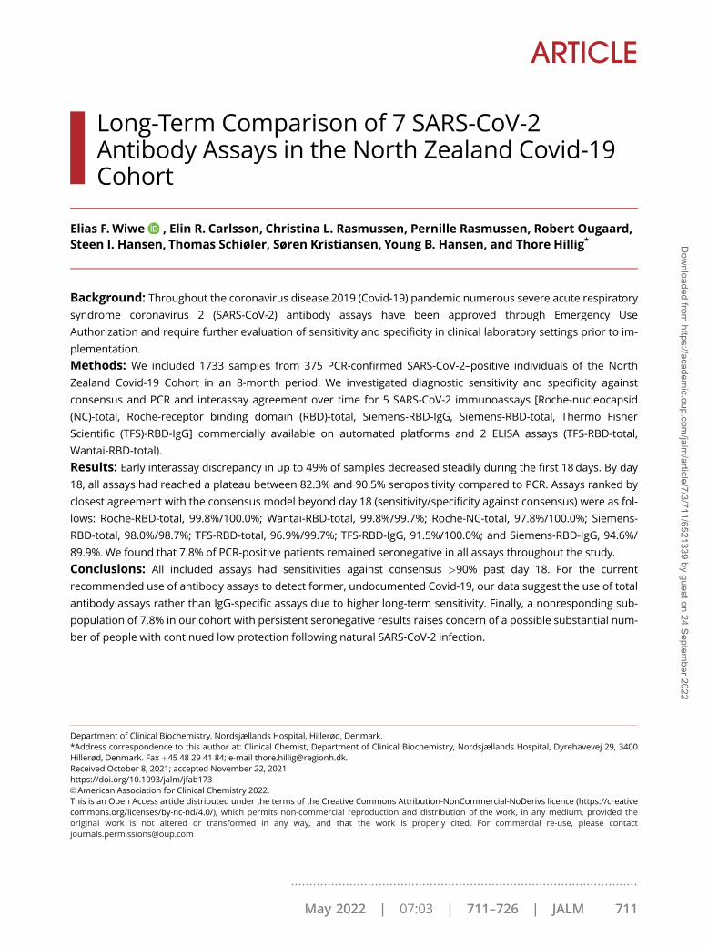
Long-Term Comparison of 7 SARS-CoV-2 Antibody Assays in the North Zealand Covid-19 Cohort Elias F. Wiwe , Elin R. Carlsson, Christina L. Rasmussen, Pernille Rasmussen, Robert Ougaard, Steen I. Hansen, Thomas Schiøler, Søren Kristiansen, Young B. Hansen, and Thore Hillig * Background: Throughout the coronavirus disease 2019 (Covid-19) pandemic numerous severe acute respiratory syndrome coronavirus 2 (SARS-CoV-2) antibody assays have been approved through Emergency Use Authorization and require further evaluation of sensitivity and specificity in clinical laboratory settings prior to im- plementation. Methods: We included 1733 samples from 375 PCR-confirmed SARS-CoV-2–positive individuals of the North Zealand Covid-19 Cohort in an 8-month period. We investigated diagnostic sensitivity and specificity against consensus and PCR and interassay agreement over time for 5 SARS-CoV-2 immunoassays [Roche-nucleocapsid (NC)-total, Roche-receptor binding domain (RBD)-total, Siemens-RBD-IgG, Siemens-RBD-total, Thermo Fisher Scientific (TFS)-RBD-IgG] commercially available on automated platforms and 2 ELISA assays (TFS-RBD-total, Wantai-RBD-total). Results: Early interassay discrepancy in up to 49% of samples decreased steadily during the first 18 days. By day 18, all assays had reached a plateau between 82.3% and 90.5% seropositivity compared to PCR. Assays ranked by closest agreement with the consensus model beyond day 18 (sensitivity/specificity against consensus) were as fol- lows: Roche-RBD-total, 99.8%/100.0%; Wantai-RBD-total, 99.8%/99.7%; Roche-NC-total, 97.8%/100.0%; Siemens- RBD-total, 98.0%/98.7%; TFS-RBD-total, 96.9%/99.7%; TFS-RBD-IgG, 91.5%/100.0%; and Siemens-RBD-IgG, 94.6%/ 89.9%. We found that 7.8% of PCR-positive patients remained seronegative in all assays throughout the study. Conclusions: All included assays had sensitivities against consensus >90% past day 18. For the current recommended use of antibody assays to detect former, undocumented Covid-19, our data suggest the use of total antibody assays rather than IgG-specific assays due to higher long-term sensitivity. Finally, a nonresponding sub- population of 7.8% in our cohort with persistent seronegative results raises concern of a possible substantial num- ber of people with continued low protection following natural SARS-CoV-2 infection. Department of Clinical Biochemistry, Nordsjællands Hospital, Hillerød, Denmark. *Address correspondence to this author at: Clinical Chemist, Department of Clinical Biochemistry, Nordsjællands Hospital, Dyrehavevej 29, 3400 Hillerød, Denmark. Fax þ45 48 29 41 84; e-mail [email protected]. Received October 8, 2021; accepted November 22, 2021. https://doi.org/10.1093/jalm/jfab173 V C American Association for Clinical Chemistry 2022. This is an Open Access article distributed under the terms of the Creative Commons Attribution-NonCommercial-NoDerivs licence (https://creative commons.org/licenses/by-nc-nd/4.0/), which permits non-commercial reproduction and distribution of the work, in any medium, provided the original work is not altered or transformed in any way, and that the work is properly cited. For commercial re-use, please contact [email protected] ............................................................................................... May 2022 | 07:03 | 711–726 | JALM 711 ARTICLE Downloaded from https://academic.oup.com/jalm/article/7/3/711/6521339 by guest on 24 September 2022
-
Upload
khangminh22 -
Category
Documents
-
view
0 -
download
0
Transcript of Long-Term Comparison of 7 SARS-CoV-2 Antibody Assays in ...
Long-Term Comparison of 7 SARS-CoV-2Antibody Assays in the North Zealand Covid-19Cohort
Elias F.Wiwe , Elin R. Carlsson, Christina L. Rasmussen, Pernille Rasmussen, Robert Ougaard,Steen I. Hansen, Thomas Schiøler, Søren Kristiansen, Young B. Hansen, and Thore Hillig*
Background: Throughout the coronavirus disease 2019 (Covid-19) pandemic numerous severe acute respiratory
syndrome coronavirus 2 (SARS-CoV-2) antibody assays have been approved through Emergency Use
Authorization and require further evaluation of sensitivity and specificity in clinical laboratory settings prior to im-
plementation.
Methods: We included 1733 samples from 375 PCR-confirmed SARS-CoV-2–positive individuals of the North
Zealand Covid-19 Cohort in an 8-month period. We investigated diagnostic sensitivity and specificity against
consensus and PCR and interassay agreement over time for 5 SARS-CoV-2 immunoassays [Roche-nucleocapsid
(NC)-total, Roche-receptor binding domain (RBD)-total, Siemens-RBD-IgG, Siemens-RBD-total, Thermo Fisher
Scientific (TFS)-RBD-IgG] commercially available on automated platforms and 2 ELISA assays (TFS-RBD-total,
Wantai-RBD-total).
Results: Early interassay discrepancy in up to 49% of samples decreased steadily during the first 18days. By day
18, all assays had reached a plateau between 82.3% and 90.5% seropositivity compared to PCR. Assays ranked by
closest agreement with the consensus model beyond day 18 (sensitivity/specificity against consensus) were as fol-
lows: Roche-RBD-total, 99.8%/100.0%; Wantai-RBD-total, 99.8%/99.7%; Roche-NC-total, 97.8%/100.0%; Siemens-
RBD-total, 98.0%/98.7%; TFS-RBD-total, 96.9%/99.7%; TFS-RBD-IgG, 91.5%/100.0%; and Siemens-RBD-IgG, 94.6%/
89.9%.We found that 7.8% of PCR-positive patients remained seronegative in all assays throughout the study.
Conclusions: All included assays had sensitivities against consensus >90% past day 18. For the current
recommended use of antibody assays to detect former, undocumented Covid-19, our data suggest the use of total
antibody assays rather than IgG-specific assays due to higher long-term sensitivity. Finally, a nonresponding sub-
population of 7.8% in our cohort with persistent seronegative results raises concern of a possible substantial num-
ber of people with continued low protection following natural SARS-CoV-2 infection.
Department of Clinical Biochemistry, Nordsjællands Hospital, Hillerød, Denmark.*Address correspondence to this author at: Clinical Chemist, Department of Clinical Biochemistry, Nordsjællands Hospital, Dyrehavevej 29, 3400Hillerød, Denmark. Fax þ45 48 29 41 84; e-mail [email protected] October 8, 2021; accepted November 22, 2021.https://doi.org/10.1093/jalm/jfab173VC American Association for Clinical Chemistry 2022.This is an Open Access article distributed under the terms of the Creative Commons Attribution-NonCommercial-NoDerivs licence (https://creativecommons.org/licenses/by-nc-nd/4.0/), which permits non-commercial reproduction and distribution of the work, in any medium, provided theoriginal work is not altered or transformed in any way, and that the work is properly cited. For commercial re-use, please [email protected]
...............................................................................................
May 2022 | 07:03 | 711–726 | JALM 711
ARTICLED
ownloaded from
https://academic.oup.com
/jalm/article/7/3/711/6521339 by guest on 24 Septem
ber 2022

INTRODUCTION
Throughout the coronavirus disease 2019(Covid-19) pandemic, several applications have
been suggested for antibody testing against se-vere acute respiratory syndrome coronavirus 2
(SARS-CoV-2) (1). PCR testing of viral SARS-CoV-2RNA is the preferred diagnostic test and has be-
come widely available for diagnostic purposes.Thus, serology testing is almost exclusively limitedto epidemiologic surveillance or supporting diag-
nosis of late complications to an otherwise undoc-umented SARS-CoV-2 infection (2, 3). Antibody
assays cannot currently confirm or disprove im-munity as the link to neutralizing activity is not suf-
ficiently established, and it is unclear to whatdegree the cellular immunity contributes and for
what duration (1, 4, 5).A multitude of SARS-CoV-2 antibody assays
have been developed and launched, often veryfast using Emergency Use Authorization. Such
assays require further evaluation of sensitivity andspecificity in a clinical laboratory setting prior to
implementation in routine use, preferably cover-ing long-term antibody development.In this study, we aimed to investigate sensitivity,
specificity, and interassay agreement over time of7 SARS-CoV-2 antibody assays in the North
Zealand Covid-19 Cohort.
MATERIALS AND METHODS
The study was performed at the Department of
Clinical Biochemistry, Nordsjællands Hospital,
Denmark, which handles all blood samples col-
lected in the primary and secondary healthcare
institutions from 8 municipalities with approxi-
mately 325 000 inhabitants (North Zealand).
Study Population
PCR-positive cohort. From March 21 to
November 6, 2020, 1286 individuals in the geo-
graphical uptake area of Nordsjællands Hospital
were identified SARS-CoV-2–positive by PCR test-
ing at the local Department of Clinical
Microbiology. Of these, 39% (503/1286) later had
blood drawn for routine biochemical analysis.
Residual plasma was systematically collected.
Some samples were lost to inclusion due to, for
example, shortage of staff, delayed reporting of
positive PCR results, etc. We successfully collected
61% (1748/2860) of all possible samples from
75% (375/503) of all relevant PCR-positive
patients. Fifteen of 1748 samples were excluded;
4 samples were drawn prior to PCR testing, and
11 samples were lost or yielded no valid results
(see Supplemental Fig. 1 in the online Data
IMPACT STATEMENT
This study adds to the current knowledge of severe acute respiratory syndrome coronavirus 2 (SARS-
CoV-2) antibody assay performance, particularly the long-term sensitivity of commercially available assays.
The study serves as a decision support for laboratories regarding what assay(s) to implement in routine
practice. For the indication of SARS-CoV-2 antibody analysis to identify patients with earlier, undocumented
coronavirus disease 2019, the data suggest that total antibody assays are more sensitive long-term than
IgG-specific assays. Finally, a subpopulation of 7.8% remained seronegative throughout the study, highlight-
ing the presence of a considerable subgroup with missing immunity following natural SARS-CoV-2 infection,
which must be considered when interpreting the results.
ARTICLE Comparison of 7 SARS-CoV-2 Antibody Assays
....................................................................................................
712 JALM | 711–726 | 07:03 | May 2022
Dow
nloaded from https://academ
ic.oup.com/jalm
/article/7/3/711/6521339 by guest on 24 September 2022
Supplement). Thus, 1733 samples were included
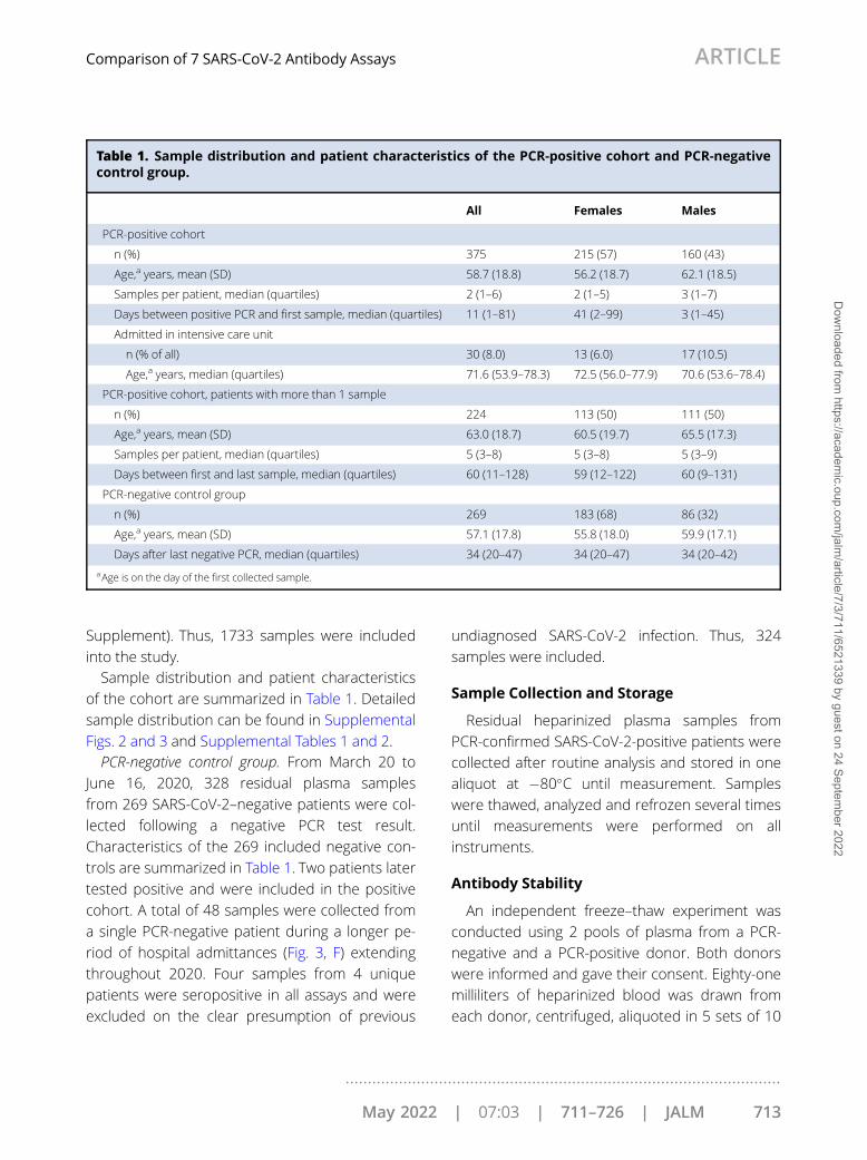
into the study.Sample distribution and patient characteristics
of the cohort are summarized in Table 1. Detailed
sample distribution can be found in Supplemental
Figs. 2 and 3 and Supplemental Tables 1 and 2.PCR-negative control group. From March 20 to
June 16, 2020, 328 residual plasma samples
from 269 SARS-CoV-2–negative patients were col-
lected following a negative PCR test result.
Characteristics of the 269 included negative con-
trols are summarized in Table 1. Two patients later
tested positive and were included in the positive
cohort. A total of 48 samples were collected from
a single PCR-negative patient during a longer pe-
riod of hospital admittances (Fig. 3, F) extending
throughout 2020. Four samples from 4 unique
patients were seropositive in all assays and were
excluded on the clear presumption of previous
undiagnosed SARS-CoV-2 infection. Thus, 324
samples were included.
Sample Collection and Storage
Residual heparinized plasma samples from
PCR-confirmed SARS-CoV-2-positive patients were
collected after routine analysis and stored in one
aliquot at �80�C until measurement. Samples
were thawed, analyzed and refrozen several times
until measurements were performed on all
instruments.
Antibody Stability
An independent freeze–thaw experiment was
conducted using 2 pools of plasma from a PCR-
negative and a PCR-positive donor. Both donors
were informed and gave their consent. Eighty-one
milliliters of heparinized blood was drawn from
each donor, centrifuged, aliquoted in 5 sets of 10
Table 1. Sample distribution and patient characteristics of the PCR-positive cohort and PCR-negativecontrol group.
All Females Males
PCR-positive cohort
n (%) 375 215 (57) 160 (43)
Age,a years, mean (SD) 58.7 (18.8) 56.2 (18.7) 62.1 (18.5)
Samples per patient, median (quartiles) 2 (1–6) 2 (1–5) 3 (1–7)
Days between positive PCR and first sample, median (quartiles) 11 (1–81) 41 (2–99) 3 (1–45)
Admitted in intensive care unit
n (% of all) 30 (8.0) 13 (6.0) 17 (10.5)
Age,a years, median (quartiles) 71.6 (53.9–78.3) 72.5 (56.0–77.9) 70.6 (53.6–78.4)
PCR-positive cohort, patients with more than 1 sample
n (%) 224 113 (50) 111 (50)
Age,a years, mean (SD) 63.0 (18.7) 60.5 (19.7) 65.5 (17.3)
Samples per patient, median (quartiles) 5 (3–8) 5 (3–8) 5 (3–9)
Days between first and last sample, median (quartiles) 60 (11–128) 59 (12–122) 60 (9–131)
PCR-negative control group
n (%) 269 183 (68) 86 (32)
Age,a years, mean (SD) 57.1 (17.8) 55.8 (18.0) 59.9 (17.1)
Days after last negative PCR, median (quartiles) 34 (20–47) 34 (20–47) 34 (20–42)
aAge is on the day of the first collected sample.
Comparison of 7 SARS-CoV-2 Antibody Assays ARTICLE
..................................................................................................
May 2022 | 07:03 | 711–726 | JALM 713
Dow
nloaded from https://academ
ic.oup.com/jalm
/article/7/3/711/6521339 by guest on 24 September 2022

aliquots (1–10) and frozen at �80�C. Aliquots 2 to
10 were thawed at 4�C and refrozen, followed by
aliquots 3 to 10 and so on. After 10 freeze–thaw
cycles, all aliquots were analyzed on each immu-
noassay. Aliquot 1 was additionally left at room
temperature without cap and reanalyzed after
24 h.
Antibody Assays
The SARS-CoV-2 antibody assays compared in
this study are listed in Table 2, including short
names used throughout the article. Analysis was
performed by experienced medical laboratory
technicians following the manufacturer’s instruc-
tions including internal quality control. A single
proficiency test was performed for all assays using
sample material from the UKNEQAS quality assur-
ance program (6). The manufacturers’ recom-
mended cutoff values were used in interpretation
of the results. Borderline results [Thermo Fisher
Scientific EliATM SARS-CoV-2-Sp1 IgG (Thermo
Fisher Scientific [TFS]-receptor binding domain
[RBD]-IgG) 7–10 EliA U/mL (n¼ 37) and WANTAI
SARS-CoV-2 Ab ELISA (Wantai-RBD-total) 0.9–1.1
absorbance/calculated cutoff (A/CO) (n¼6)] were
not interpreted as either positive or negative but
included as “no valid result.”The TFS-RBD-IgG assay received CE approval
halfway through the study and changed output
unit. The results in mg/L were converted to EliA U/
mL using a lot-specific correction factor supplied
by the manufacturer.
Data Analysis and Statistics
Data were extracted from the respective instru-
ments. Sampling date, requesting unit, patient
age, sex, and SARS-CoV-2 PCR test results were re-
trieved from the local laboratory information man-
agement system.Complete data (valid results from all 7 assays)
could be obtained for 57.7% (1000/1733) and
52.5% (170/324) of samples in the cohort and
negative control group, respectively. Incomplete
data were due to factors such as insufficient vol-
ume or clotting of the plasma. Both complete and
incomplete data were included in the analysis.All cohort samples were stratified into time
intervals based on days from the date of the initial
positive PCR result. The length and number of the
created time intervals were based on the inclusion
of a minimum of 15 samples per interval. Only the
earliest sample was included per time interval
from each single patient. Thus, 352 samples
(20.3%) were excluded to avoid duplicates, result-
ing in a total of 1381 samples (79.7%) included
into the time intervals.In the negative control group, only 1 sample
from each individual patient was included in the
statistical analysis, excluding 55 (17.0%) samples
as duplicates. Differences related to sex, age, and
requester type were evaluated using Fisher’s exact
test, the Mann–Whitney U test, and the Kruskal–
Wallis test, respectively. The 95% CIs of percen-
tages (p) were calculated as p 6 1.96 ��(p�(1 � p)/
n). Any 2 results were considered significantly dif-
ferent from each other if there was no overlap be-
tween the 95% CIs. The outputs of qualitative
assays were treated as semiquantitative. For opti-
mal visual presentation the specific cutoffs in each
of the different assays were adjusted to 1.0. This
enabled plotting of the multiple longitudinal
curves into the same logarithmic graph.All statistical analysis was performed in IBMVR
SPSSVR Statistics version 25 using a P value of
�0.05.
Consensus Model
All assays were evaluated in terms of agreement
with a consensus model designed as follows.Samples with positive results from 3 assays or
with 2 positive and no negative results were
coded as positive. Any sample with at least 2 nega-
tive results and no positive results was coded as
negative. In addition, samples with 1 positive
ARTICLE Comparison of 7 SARS-CoV-2 Antibody Assays
....................................................................................................
714 JALM | 711–726 | 07:03 | May 2022
Dow
nloaded from https://academ
ic.oup.com/jalm
/article/7/3/711/6521339 by guest on 24 September 2022
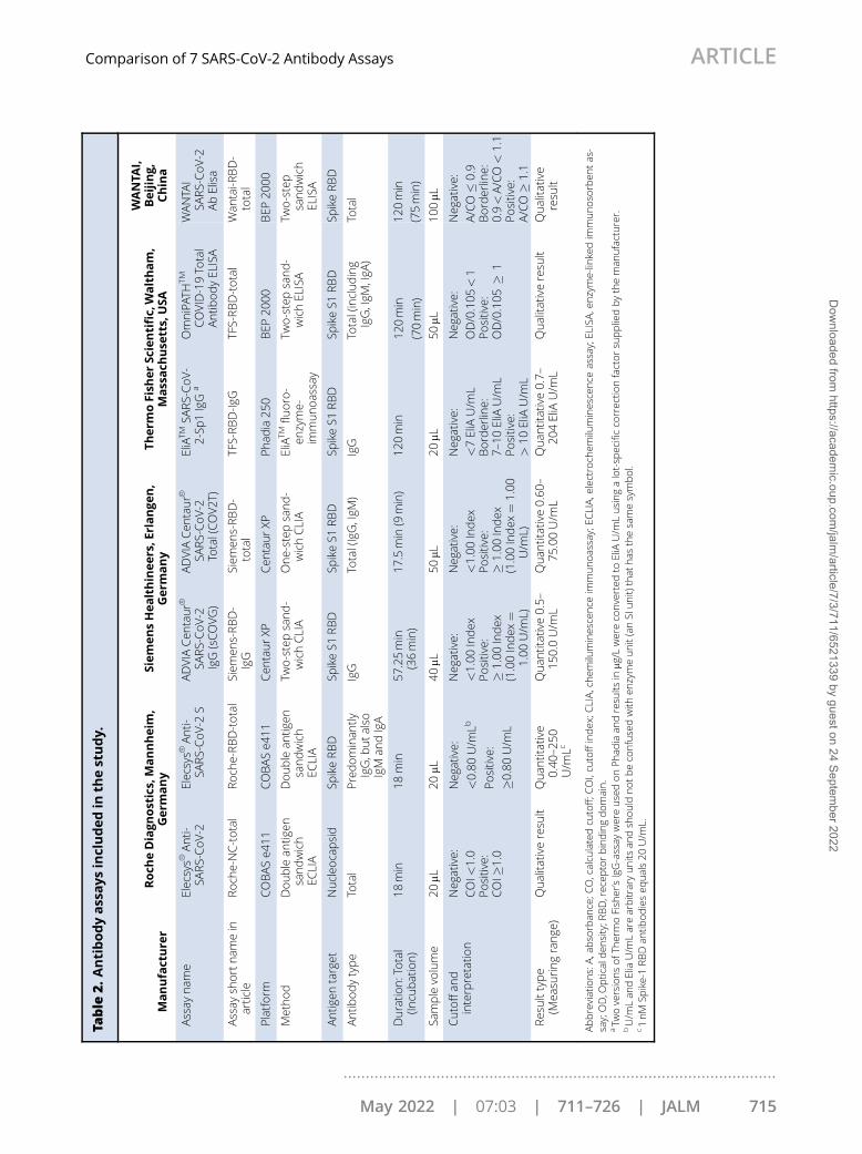
Table
2.Antibodyassaysincludedin
thestudy.
Manufacture
rRoch
eDiagn
ostics,
Mannheim
,Germ
any
SiemensHealthineers,E
rlangen,
Germ
any
Therm
oFish
erSc
ientific,Waltham,
Massach
use
tts,
USA
WANTA
I,Beijing,
China
Assay
nam
eElec
sysVR
Anti-
SARS-CoV-2
Elec
sysVR
Anti-
SARS-CoV-2
SADVIA
Cen
taurVR
SARS-CoV-2
IgG(sCOVG)
ADVIA
Cen
taurVR
SARS-CoV-2
Total(COV2T
)
EliA
TMSA
RS-CoV-
2-Sp
1IgG
aOmniPAT
HTM
COVID-19To
tal
Antib
odyEL
ISA
WANTA
ISA
RS-CoV-2
AbElisa
Assay
shortnam
ein
artic
leRoch
e-NC-total
Roch
e-RBD-total
Siem
ens-RBD-
IgG
Siem
ens-RBD-
total
TFS-RBD-Ig
GTF
S-RBD-total
Wan
tai-R
BD-
total
Platform
COBASe4
11COBASe4
11Cen
taurXP
Cen
taurXP
Phad
ia25
0BEP
2000
BEP
2000
Method
Double
antig
ensandwich
ECLIA
Double
antig
ensandwich
ECLIA
Two-stepsand-
wichCLIA
One-step
sand-
wichCLIA
EliA
TMflu
oro-
enzyme-
immunoassay
Two-stepsand-
wichEL
ISA
Two-step
sandwich
ELISA
Antig
entarget
Nucleo
capsid
SpikeRBD
SpikeS1
RBD
SpikeS1
RBD
SpikeS1
RBD
SpikeS1
RBD
SpikeRBD
Antib
odytype
Total
Predominan
tlyIgG,b
utalso
IgM
andIgA
IgG
Total(IgG,IgM
)IgG
Total(including
IgG,IgM
,IgA
)To
tal
Duratio
n:Total
(Incu
bation)
18min
18min
57.25min
(36min)
17.5min
(9min)
120min
120min
(70min)
120min
(75min)
Samplevo
lume
20mL
20mL
40mL
50mL
20mL
50mL
100mL
Cutoffan
dinterpretatio
nNeg
ative:
COI<
1.0
Positiv
e:COI�
1.0
Neg
ative:
<0.80
U/m
Lb
Positiv
e:
�0.80
U/m
L
Neg
ative:
<1.00
Index
Positiv
e:�
1.00
Index
(1.00Index¼
1.00
U/m
L)
Neg
ative:
<1.00
Index
Positiv
e:�
1.00
Index
(1.00Index¼
1.00
U/m
L)
Neg
ative:
<7EliA
U/m
LBorderlin
e:7–
10EliA
U/m
LPositiv
e:>
10EliA
U/m
L
Neg
ative:
OD/0.105<1
Positiv
e:OD/0.105�
1
Neg
ative:
A/CO�
0.9
Borderlin
e:0.9<A/CO<
1.1
Positiv
e:A/CO�
1.1
Resulttype
(Mea
suringrange
)Qualita
tiveresu
ltQuan
titative
0.40
–250
U/m
Lc
Quan
titative0.5–
150.0U/m
LQuan
titative0.60
–75
.00U/m
LQuan
titative0.7–
204EliA
U/m
LQualita
tiveresu
ltQualita
tive
resu
lt
Abbreviations:A,a
bso
rban
ce;C
O,calcu
latedcu
toff;C
OI,cu
toffindex
;CLIA,chem
iluminesce
nce
immunoassay;EC
LIA,e
lectroch
emilu
minesce
nce
assay;EL
ISA,e
nzyme-lin
kedim
munoso
rben
tas-
say;OD,O
ptic
alden
sity;R
BD,rec
eptorbindingdomain.
aTw
oversionsofT
hermoFish
er’sIgG-assay
wereusedonPhad
iaan
dresu
ltsin
mg/L
wereco
nverted
toEliA
U/m
Lusingalot-sp
ecificco
rrec
tionfactorsu
ppliedbytheman
ufacturer.
bU/m
Lan
dEliaU/m
Larearbitraryunits
andsh
ould
notbeco
nfusedwith
enzymeunit(anSI
unit)
that
has
thesamesymbol.
c1nM
Spike-1RBDan
tibodieseq
uals20
U/m
L.
Comparison of 7 SARS-CoV-2 Antibody Assays ARTICLE
..................................................................................................
May 2022 | 07:03 | 711–726 | JALM 715
Dow
nloaded from https://academ
ic.oup.com/jalm
/article/7/3/711/6521339 by guest on 24 September 2022

result opposed by at least 4 negative results were
coded as negative.Differences in distribution between assays and
between assays and the consensus model were
evaluated using McNemar’s test. Agreement, sen-
sitivity, and specificity were calculated for all
assays compared to the consensus model, to the
gold standard assays identified, and to the PCR
result.
Ethics
The study was performed in accordance to the
Helsinki Declaration. The project received approval
from the Danish Data Protection Agency (March
20, 2020, “Corona Serologi” #P-2020-279) for the
collection of residual plasma following routine
measurement.None of the patients included in the study was
registered in the Tissue Utilization Register
(Vævsanvendelsesregistret), where citizens may
register that the collected blood may solely be
used in relation to their own medical treatment.
RESULTS
All assays passed the external control assur-
ance program. The concentration of plasma anti-
bodies was demonstrated to be stable through 10
freeze–thaw cycles and after 24 h at room temper-
ature (Supplemental Fig. 4).
The PCR-Positive Cohort
The distribution of samples from the PCR-
positive cohort in time intervals is shown in Fig. 1,
A. Initially, following the PCR test, all samples came
from hospitalized patients, including patients re-
quiring intensive care. This distribution gradually
shifted over time with predominance of outpatient
and primary care samples from week 6 and on-
ward (Fig. 1, A).Overall, in the included samples of the cohort,
58.6% (809/1381) of samples gave uniformly
positive results, 17.8% (246/1381) gave uniformly
negative results, and 23.6% (326/1381) showed
discrepancy between assays. Figure 1, B shows
the distribution of uniformity and discrepancy be-
tween assay results over time. Discrepancy is fur-
ther stratified by the result of the consensus
model. Initial interassay discrepancy of 49% at day
0 decreased steadily in the first 18 days (Fig. 1, B).As shown in Fig. 2, A, the seropositivity percent-
age for all assays rose until day 18, whereafter an
assay-wide plateau occurred with a mean sero-
positive percentage of 82.3% to 90.5%, depending
on assay. The cohort samples were split into day
0–17 with 781 samples from 198 individuals and
day 18þ with 600 samples from 282 individuals
(Supplemental Fig. 1). Limited to day 18þ, 79.7%(478/600) of samples were uniformly positive,
8.5% (51/600) were uniformly negative, and 11.8%
(71/600) showed discrepant results. The distribu-
tion of discrepancy between sexes between day 0
to 17 was similar (33.6% discrepancy in males vs
31.4% in females; P¼0.537), while discrepant
samples were significantly more frequent in
females at day 18þ (16.0% vs 7.4%; P¼0.002).
The age distribution was similar between the dis-
crepant and uniform results at both day 0–17
(P¼0.468) and day 18þ (P¼0.189).
Nonresponders of the Cohort
A subgroup of the PCR-positive cohort
remained seronegative throughout the study.
Thirty-seven samples from 22 unique patients
(7.8% of all patients with at least 1 sample beyond
day 18) were uniformly negative in all assays. The
22 nonresponding patients collectively contrib-
uted 42 samples (5 from day 0–17) which
accounted for 0.0%, 1.4%, 5.2%, and 8.8% of sam-
ples from intensive care, other hospital wards,
outpatient clinics, and primary care centers, re-
spectively. This distribution was significantly differ-
ent from the remaining samples (P< 0.001). On an
individual level, these 22 patients were similar to
ARTICLE Comparison of 7 SARS-CoV-2 Antibody Assays
....................................................................................................
716 JALM | 711–726 | 07:03 | May 2022
Dow
nloaded from https://academ
ic.oup.com/jalm
/article/7/3/711/6521339 by guest on 24 September 2022

the remaining patients according to age
(P¼0.150) and sex (P¼0.377). Although none of
the 22 patients had any samples taken at the in-
tensive care unit, this difference was not statisti-
cally significant (P¼0.239).
The PCR-Negative Control Group
In the negative control group, 90.0% (242/269)
of samples were uniformly negative, and 10.0%
(27/269 samples) showed discrepancy between
assays. The discrepant group was similar in
Fig. 1. Distribution of cohort samples in intervals of time since positive PCR test. Each patient is repre-sented maximally once per time interval. (A) Distribution according to requester type. (B) Percentagedistribution according to uniformity or discrepancy between all assay results. Discrepancy is furtherstratified by the result of the consensus model.
Comparison of 7 SARS-CoV-2 Antibody Assays ARTICLE
..................................................................................................
May 2022 | 07:03 | 711–726 | JALM 717
Dow
nloaded from https://academ
ic.oup.com/jalm
/article/7/3/711/6521339 by guest on 24 September 2022

Fig. 2. Longitudinal development of seropositivity and signal strength for 7 SARS-CoV-2 antibodyassays. (A) Percentage of seropositive individuals over time. (B) Medians of assay signal strengths overtime, presented on a logarithmic scale. Note that all assays have been adjusted to a positive cutoff at1.0 (dotted line) and that titers are not directly comparable between assays.
ARTICLE Comparison of 7 SARS-CoV-2 Antibody Assays
....................................................................................................
718 JALM | 711–726 | 07:03 | May 2022
Dow
nloaded from https://academ
ic.oup.com/jalm
/article/7/3/711/6521339 by guest on 24 September 2022

Fig. 3. Illustrative longitudinal patient courses by 7 SARS-CoV-2 antibody assays. All assays are adjustedto a positive cutoff of 1.0. (A–C) Patient courses with strong and persistent antibody levels. (D) Weakand declining response. (E) Delayed response. Note incidences of false negatives in (A1 E), clusteredfalse positives in (E1F) and weak, declining signal strength of IgG assays in (A1D).
Comparison of 7 SARS-CoV-2 Antibody Assays ARTICLE
..................................................................................................
May 2022 | 07:03 | 711–726 | JALM 719
Dow
nloaded from https://academ
ic.oup.com/jalm
/article/7/3/711/6521339 by guest on 24 September 2022
distribution of age (P¼0.312), sex (P¼ 0.384), and
sampling location (P¼0.781) compared to the
uniform group.The discrepancy between assays was in 81.5%
(22/27) of the discrepant samples (8.2% of all in-
cluded control group samples) solely due to posi-
tive results of the Siemens ADVIA CentaurVR SARS-
CoV-2 IgG (Siemens-RBD-IgG) assay, interpreted
as false positives. As illustration, the entire
patient antibody course of patient 1485 (PCR-neg-
ative) with 48 consecutive samples is presented in
Fig. 3, F.
Results of the Consensus Model
The consensus model was applied on the co-
hort samples and controls as described in the
Materials and Methods section. In the cohort sam-
ples, the model resulted in 71.8% (991/1381) con-
sensus positive samples, 22.7% (314/1381)
consensus negative samples, and 5.5% (76/1381)
undetermined samples. Limited to samples from
day 18þ, 89.7% (538/600) of samples were coded
consensus positive, 9.0% (54/600) consensus neg-
ative, and 1.3% (8/600) were undetermined.
Further limited to 435 samples of day 18þ with
complete data from all 7 assays, 90.3% (393/435)
of samples were consensus positive, 9.4% (41/
435) consensus negative, and 0.2% (1/435)
undetermined.In the unique negative controls, 98.1% (264/
269) of samples were consensus negative, 0.4%
(1/269) consensus positive, and 1.5% (4/269)
inconclusive.
Assay Agreement with Consensus Model
Roche ElecsysVR Anti-SARS-CoV-2 S (Roche-RBD-
total) and Wantai-RBD-total had the highest per-
centage agreement with the consensus model of
99.9 (95% CI 99.6–100) and 99.8 (95% CI 99.4–
100), respectively (Table 3). Neither was signifi-
cantly different from the consensus model (PRoche-
RBD-total ¼ 1.000 and PWantai-RBD-total ¼ 1.000) or
each other (P¼ 0.125). The remaining total assays
had high agreement: Roche ElecsysVR Anti-SARS-
CoV-2 (nucleocapsid) (Roche-NC-total; 98.6%
agreement, 95% CI 97.7–99.4), Siemens ADVIA
CentaurVR SARS-CoV-2 total (Siemens-RBD-total;
98.2% agreement, 95% CI 97.3–99.2), and Thermo
Fisher Scientific OmnipathTM COVID-19 total anti-
body ELISA (TFS-RBD-total); 97.9% agreement,
95% CI 97.0–98.9). The 2 selective IgG assays, TFS-
RBD-IgG (94.6% agreement, 95% CI 93.1–96.2)
and Siemens-RBD-IgG (93.1% agreement, 95% CI
91.2–94.9), showed lower agreement. Agreement,
sensitivity, and specificity of all assays against the
consensus model, the Roche-RBD-total and
Wantai-RBD-total assays as well as the PCR test re-
sult are presented in Table 3.Illustrative patient antibody courses are pre-
sented in Fig. 3 to visualize different types and
observations of antibody development over time.
DISCUSSION
The Cohort
We have introduced the North Zealand Covid-
19 Cohort, a unique collection of serial plasma
samples representing 75% of all PCR-confirmed
SARS-CoV-2-positive individuals from North
Zealand having blood samples taken during the
initial (wild-type) Covid-19 wave in Denmark in
2020. The cohort’s diversity is a direct reflection of
the patient composition of our clinical laboratory’s
sample flow, including samples from primary care
centers. This is to our knowledge one of the
largest comparative studies of SARS-CoV-2 anti-
body assays in respect to the large number of
patients with several serial samples (Supplemental
Table 2).
Early Serology
This stage was defined as day 0-17 after the
positive PCR test and was characterized by an ini-
tial low seropositivity and a high degree of
ARTICLE Comparison of 7 SARS-CoV-2 Antibody Assays
....................................................................................................
720 JALM | 711–726 | 07:03 | May 2022
Dow
nloaded from https://academ
ic.oup.com/jalm
/article/7/3/711/6521339 by guest on 24 September 2022

discrepancy between assays, which supports cur-
rent recommendations not to rely on antibody
measurements in the first 3 weeks following infec-
tion (2).In the first 2 days after the positive PCR test, the
2 total antibody ELISA assays (Wantai-RBD-total
and TFS-RBD-total) showed significantly higher
rates of seroconversion compared to the 5 auto-
mated immunoassays (Fig. 2, A and example in
Fig. 3, B). The subsequent drop in seropositive
percentage reflects a shift in patient composition
in the following time intervals. The ELISA assays
also reached approximately 90% seropositivity
(the plateau level) by day 10, whereas the assay-
wide plateau was not reached until day 18. These
results contradict early reports of an overall lower
sensitivity of ELISA assays compared to chemilu-
minescence immunoassays (7), but are in line with
later reports on specific ELISA assays, including
Wantai-RBD-total (8, 9).
Late Serology
Past day 18, where the plateau had been
reached for all assays, the median signal strength
of all total antibody assays was steady, capped at
maximum value, or slightly increasing throughout
the study period (Fig. 2, B). In contrast, the IgG
assays had a decreasing signal strength after 4
weeks, most attenuated in the TFS-RBD-IgG assay
(Fig. 2, B and examples in Fig. 3, A, D). A similar de-
crease in signal strength of the TFS-RBD-IgG assay
has been described by Favresse et al. in a 10-
month follow-up study (10). Yet, the same study in-
cluded 2 additional IgG-specific assays (Ortho and
DiaSorin), which did not decrease to the same ex-
tent. This could indicate that the decreasing signal
strength observed for TFS-RBD-IgG is an assay-
specific phenomenon rather than a general trait
of IgG-specific assays. With regards to the TFS-
RBD-IgG assay, we suspect an inappropriately
high cutoff for the TFS-RBD-IgG assay, which
results in a sensitivity below its potential. Based
on our data, a positive cutoff at 3.4 (currently at
10 and borderline results at 7–10) would result in
96.6% sensitivity and 99.7% specificity against
consensus.A declining signal strength of nucleocapsid (NC)
antibodies over time was reported by Masia et al.
(11). In their 12-month followup period, 56% of
patients with moderate/severe disease reverted
to seronegativity according to the Euroimmun-NC-
IgG assay. In our 8-month study and in the 10-
month study of Favresse et al. (10), the signal
strength of Roche-NC-total was very persistent
over time. Thus, the NC signal loss observed by
Masia et al. seems likely to be assay-specific rather
than a general trait of NC antibody development.We found interassay discrepancy to be signifi-
cantly more frequent in females than in males at
day 18þ (16.0% vs 7.3%; P¼0.002). This is likely a
result of differences between sexes in the back-
ground demographics of our cohort. We observed
an overrepresentation of women having their first
blood sample drawn months after diagnosis (i.e.,
following mild or asymptomatic Covid-19 without
need of health care services) (Supplemental Fig.
3). Such mild cases more often yielded long-term
samples with discrepancy due to low signal
strength of TFS-RBD-IgG.
Comparison of Assay Performance
We introduced a consensus model and ex-
cluded samples of day 0–17 to achieve the opti-
mal comparability between assays despite
differences in IgG targets—IgG or total. The con-
sensus model was successfully applied to 98.7%
of samples with comparable distribution of results
between complete and incomplete data. Of the 7
assays tested (Table 2), Roche-RBD-total and
Wantai-RBD-total had the closest agreement with
the consensus model of 99.9% and 99.8%, respec-
tively. The Wantai-RBD-total ELISA assay has
proved superior to competing assays in earlier
studies (8, 9, 12–14). The Roche-RBD-total assay is
Comparison of 7 SARS-CoV-2 Antibody Assays ARTICLE
..................................................................................................
May 2022 | 07:03 | 711–726 | JALM 721
Dow
nloaded from https://academ
ic.oup.com/jalm
/article/7/3/711/6521339 by guest on 24 September 2022

Table
3.Agre
ement,se
nsitivity,andsp
ecificity
ofall7assaysagainst
theco
nse
nsu
smodel,th
eRoch
e-RBD-tota
lassay,
theWanta
i-RBD-tota
lassay,
andth
ePCRte
stre
sult.
Assay
Compare
dto
the
conse
nsu
smodel
Compare
dto
Roch
e-RBD-tota
lasgold
standard
Compare
dto
Wanta
i-RBD-tota
lasgold
standard
Compare
dto
PCRte
stre
sult
Agre
ement%
(95%
CI)
Sensi-
tivity%
Specifi-
city
%Agre
ement%
(95%
CI)
Sensi-
tivity%
Specifi-
city
%Agre
ement%
(95%
CI)
Sensi-
tivity%
Specifi-
city
%Agre
ement%
(95%
CI)
Sensiti-
vity%
Specifi-
city
%
Roch
e-NC-total
98.6
(97.7–
99.4)
97.8
100.0
98.4
(97.5–
99.3)
97.8
99.6
97.9
(96.9–
99.0)
97.0
99.6
91.9
(90.0–9
3.9)
88.8
99.6
Roch
e-RBD-total
99.9
(99.6–
100)
99.8
100.0
––
–99
.4(98.8–
100)
99.1
100.0
93.1
(91.2–9
5.0)
90.5
99.5
Siem
ens-RBD-IgG
93.1
(91.2–
94.9)
94.6
89.9
93.1
(91.2–
95.1)
94.8
89.6
92.4
(90.4–
94.4)
94.0
89.0
85.8
(83.3–8
8.3)
85.5
86.7
Siem
ens-RBD-total
98.2
(97.3–
99.2)
98.0
98.7
98.1
(97.1–
99.2)
98.0
98.5
97.5
(96.3–
98.6)
97.3
97.8
91.4
(89.4–9
3.4)
89.1
98.9
TFS-RBD-Ig
G94
.6(93.1–
96.2)
91.5
100.0
94.5
(92.8–
96.2)
91.6
100.0
94.1
(92.4–
95.8)
90.6
99.7
87.6
(85.4–8
9.9)
82.3
100.0
TFS-RBD-total
97.9
(97.0–
98.9)
96.9
99.7
97.4
(96.2–
98.6)
96.6
98.8
97.8
(96.8–
98.8)
96.9
99.3
92.3
(90.5–9
4.1)
88.8
100.0
Wan
tai-R
BD-total
99.8
(99.4–
100)
99.8
99.7
99.4
(98.8–
100)
100.0
98.4
––
–92.9
(91.1–9
4.7)
90.1
98.9
Calcu
latio
nsarebased
onallcohortsamplesincluded
intim
eintervalspastday
18an
dallu
niqueneg
ativeco
ntrols.
ARTICLE Comparison of 7 SARS-CoV-2 Antibody Assays
....................................................................................................
722 JALM | 711–726 | 07:03 | May 2022
Dow
nloaded from https://academ
ic.oup.com/jalm
/article/7/3/711/6521339 by guest on 24 September 2022

newer but has emerging reports of a very accept-
able performance (10, 15). The remaining total
assays performed acceptably in comparison to
consensus (97.9–98.6% agreement) (Table 3). The
2 selective IgG assays displayed lower agreement
with consensus; the TFS-RBD-IgG (94.6% agree-
ment) primarily due to low sensitivity, especially
long-term (91.5% sensitivity compared to consen-
sus) and Siemens-RBD-IgG (93.1% agreement) pri-
marily due to false-positive results and a resulting
low specificity (89.9% specificity compared to con-
sensus). Earlier reports on the Siemens-RBD-IgG
assay did not report false-positive results, in con-
trast they reported a specificity of 99.4% (16). We
observed several incidences with timely clustering
of false-positive results for Siemens-RBD-IgG
(Fig. 3, E and F), which raises the suspicion that a
specific condition or treatment might interfere
with this specific assay.For the current use of antibody assays to detect
former, undocumented Covid-19, our data sug-
gest the use of total antibody assays rather than
IgG-specific assays due to a more persistent long-
term sensitivity. This is further supported by
reports of more homologous results across total
antibody assays compared to IgG-specific anti-
body assays (17).
Low Overall Seroconversion Rate
We observed a low overall seropositivity past
day 18 of 82% to 91% (depending on assay).
Interestingly, for 7.8% of all PCR-positive patients
in our cohort with samples beyond day 18, we
saw no seroconversion at all; that is, all their sam-
ples were uniformly negative. This group (nonres-
ponders) was not significantly different from the
remaining patients regarding age or sex. Yet, on a
sample level, the nonresponders had a signifi-
cantly different distribution of requester type with
no intensive care samples and an overrepresenta-
tion of samples from primary care, which may be
reflective of a subpopulation with lower need for
hospital care (i.e., with lower disease severity). This
would be in line with previous observations of a
stronger antibody response in patients with se-
vere illness (8, 11, 18, 19).Our findings of low assay sensitivities compared
to PCR of Wantai-RBD-total, Roche-NC-total, and
Siemens-RBD-total between 89% and 90% are
comparable to reports of Herroelen et al. (20)
(Wantai-RBD-total 92.1%, Roche-NC-total 88.2%,
day 20þ after symptom onset, 52.6% severe ill-
ness) and Oved et al. (21) (Siemens-RBD-total
85.9%, Roche-NC-total 89.0%, day 14þ after PCR,
4.9% severe illness and 22.6% unknown severity).
We initially hypothesized this shared limit at ap-
proximately 90% seropositivity compared to PCR
to be due to similar inclusion of mild and asymp-
tomatic cases, unlike many earlier studies primar-
ily concerning hospitalized patients (7). Yet, there
are conflicting reports of significantly higher sensi-
tivity between 95% and 100% for Wantai-RBD-
total (9), Roche-RBD-total (10), Roche-NC-total (9,
10, 22, 23), and Siemens-RBD-total (22, 23) in pop-
ulations with hospitalization percentages ranging
from 0.0% to 21.8%. The freeze–thaw experiment
and consistency of seronegativity in several pa-
tient courses suggests our sample collection prac-
tice is unlikely to explain low seroconversion. We
hypothesize that the low seropositivity reflects the
cohort composition and by extension our labora-
tory sample flow and background population.
Implications of Missing Seroconversion
Of all PCR-positive patients in our cohort with
samples beyond day 18, 7.8% remained seronega-
tive throughout the study (nonresponders). If
interpreted as convalescent Covid-19 patients
with impaired or missing immunity, this subpopu-
lation is of concern. Immunocompromised
patients without any measurable antibody re-
sponse have been observed (24). Yet, cellular
Comparison of 7 SARS-CoV-2 Antibody Assays ARTICLE
..................................................................................................
May 2022 | 07:03 | 711–726 | JALM 723
Dow
nloaded from https://academ
ic.oup.com/jalm
/article/7/3/711/6521339 by guest on 24 September 2022
immunity may be present in the absence of circu-
lating antibodies (25). Meanwhile, correlation be-
tween virus neutralization tests and protective
immunity is increasingly documented (26, 27). But
the correlation between virus neutralization tests
and nonneutralizing antibody assays is ongoingly
debated with conflicting reports (23, 28–31). Such
correlations and the immunity status of the non-
responders requires further clarification, espe-
cially if antibody titers in the future will guide
booster vaccination strategies as earlier sug-
gested (1).Compared to PCR, the assay sensitivities ranged
from 82.3% to 90.5%. Thus, depending on assay
choice, up to 18% of patients suffering from long-
term sequelae following undocumented Covid-19
may remain undiagnosed in the absence of de-
tectable antibodies. If using any of the many lat-
eral flow assays available, false-negative rates may
be even higher (7, 32). Such risk of false negatives
should be considered when choosing an assay
and when interpreting antibody measurements.
Limitations
The focus of this study was to compare the quali-
tative assay results alone (positive vs negative).
Although early in the pandemic, inclusion of the neg-
ative control group through negative PCR results
alone introduces a small risk of false negatives.
Finally, since there is no agreed upon international
gold standard assay, we evaluated all assays against
consensus, which was our closest approximation of
an objective truth.
CONCLUSIONS
In summary, all assays had sensitivities against
consensus of >90% past day 18. For the currently
recommended use of antibody assays to detect
former Covid-19, our data suggest the use of total
antibody assays rather than IgG-specific assays
due to higher long-term sensitivities. Finally, a
nonresponding subpopulation of 7.8% in our co-
hort with persistent seronegative results raises
concern of a possible substantial number of peo-
ple with continued low protection following natu-
ral SARS-CoV-2 infection.
SUPPLEMENTAL MATERIAL
Supplemental material is available at The Journal
of Applied Laboratory Medicine online.
Nonstandard Abbreviations: Covid-19, coronavirus disease 2019; SARS-CoV-2, severe acute respiratory syndrome coronavirus2; RBD, receptor binding domain (of the spike glycoprotein); TFS-RBD-IgG, Thermo Fisher Scientific EliATM SARS-CoV-2-Sp1 IgG;Wantai-RBD-total: WANTAI SARS-CoV-2 Ab ELISA; A/CO, absorbance/calculated cutoff (Wantai-RBD-total output); Roche-RBD-total, Roche ElecsysVR Anti-SARS-CoV-2 S; Roche-NC-total, Roche ElecsysVR Anti-SARS-CoV-2 (nucleocapsid); TFS-RBD-total: ThermoFisher Scientific OmnipathTM COVID-19 total antibody ELISA; Siemens-RBD-IgG, Siemens ADVIA CentaurVR SARS-CoV-2 IgG;Siemens-RBD-total: Siemens ADVIA CentaurVR SARS-CoV-2 total.
Author Contributions: All authors confirmed they have contributed to the intellectual content of this paper and have met the follow-ing 4 requirements: (a) significant contributions to the conception and design, acquisition of data, or analysis and interpretation ofdata; (b) drafting or revising the article for intellectual content; (c) final approval of the published article; and (d) agreement to be ac-countable for all aspects of the article thus ensuring that questions related to the accuracy or integrity of any part of the article are ap-propriately investigated and resolved.
Authors’ Disclosures or Potential Conflicts of Interest: Upon manuscript submission, all authors completed the author disclo-sure form. Disclosures and/or potential conflicts of interest: Employment or Leadership: None declared. Consultant or AdvisoryRole: None declared. Stock Ownership: None declared. Honoraria: None declared. Research Funding: Aase og EjnarDanielsens Fond (#20-10-0336), Familien Hede Nielsens Fond (#2020-1160), Lizzi og Mogens Staal Fonden (#2020-0178), Marieog Børge Kroghs Fond. Thermo Fisher Scientific, Siemens, and Roche provided COVID antibody assay reagents. ExpertTestimony: None declared. Patents: None declared.
ARTICLE Comparison of 7 SARS-CoV-2 Antibody Assays
....................................................................................................
724 JALM | 711–726 | 07:03 | May 2022
Dow
nloaded from https://academ
ic.oup.com/jalm
/article/7/3/711/6521339 by guest on 24 September 2022

Role of Sponsor: The funding organizations played no role in the design of study, choice of enrolled patients, review and inter-pretation of data, preparation of manuscript, or final approval of manuscript.
Acknowledgments: We thank Claus Juel Jensen, Tina Skov Thomsen, and Dorthe Kjeldgaard Hansen (Nordsjællands Hospital)and Tina Profft Larsen (Clinical Microbiology, Herlev Hospital). We thank Siemens, Thermo Fisher, and Roche for sponsoring. Wegratefully thank the foundations “Familien Hede Nielsens Fond,” “Lizzi og Mogens Staal Fonden,” “Marie og Børge Kroghs Fond,”and “Aase og Ejnar Danielsens Fond.”
REFERENCES
1. West R, Kobokovich A, Connell N, Gronvall GK. COVID-19antibody tests: a valuable public health tool with limitedrelevance to individuals. Trends Microbiol 2021;29:214–23.
2. Center for Disease Control and Prevention. Interimguidelines for COVID-19 antibody testing. https://www.cdc.gov/coronavirus/2019-ncov/lab/resources/antibody-tests-guidelines.html (Accessed July 2021).
3. Danish Health Authority. Retningslinjer for handtering afCOVID-19 i sundhedsvæsenet. https://www.sst.dk/da/Udgivelser/2021/Retningslinjer-for-haandtering-af-COVID-19 (Accessed July 2021).
4. Infectious Diseases Society of America. IDSA Guidelineson the Diagnosis of COVID-19: Serologic Testing. www.idsociety.org/COVID19guidelines/serology (Accessed July2021).
5. Rodda LB, Netland J, Shehata L, Campbell DJ, Rawlings DJ,Pepper M, et al. Functional SARS-CoV-2-specific immunememory persists after mild COVID-19. Cell 2021;184:169–83.e17.
6. UK NEQAS. COVID-19 (coronavirus) external qualityassessment. https://ukneqas.org.uk/news/Covid-19-coronavirus-external-quality-assessment (Accessed June2021).
7. Deeks JJ, Dinnes J, Takwoingi Y, Davenport C, Spijker R,Taylor-Phillips S, et al. Antibody tests for identification ofcurrent and past infection with SARS-CoV-2. CochraneDatabase Syst Rev 2020;2020.
8. Trabaud MA, Icard V, Milon MP, Bal A, Lina B, Escuret V.Comparison of eight commercial, high-throughput,automated or ELISA assays detecting SARS-CoV-2 IgG ortotal antibody. J Clin Virol 2020;132:104613.
9. Harritshøj LH, Gybel-Brask M, Afzal S, Kamstrup PR,Jørgensen CS, Thomsen MK, et al. Comparison of 16serological SARS-CoV-2 immunoassays in 16 clinicallaboratories. J Clin Microbiol 2021;59.
10. Favresse J, Eucher C, Elsen M, Gillot C, Van Eeckhoudt S,Dogn�e JM, et al. Persistence of anti-Sars-Cov-2 antibodiesdepends on the analytical kit: a report for up to 10months after infection. Microorganisms 2021;9:556–13.
11. Masia M, Fernandez-Gonzalez M, Telenti G, Agullo V,Garc�ıa JA, Padilla S, et al. Durable antibody response oneyear after hospitalization for COVID-19: a longitudinalcohort study. J Autoimmun 2021;123:102703. doi:10.1016/j.jaut.2021.102703.
12. Brochot E, Demey B, Handala L, Francois C, Duverlie G,Castelain S. Comparison of different serological assaysfor SARS-CoV-2 in real life. J Clin Virol 2020;130:104569.
13. Weidner L, Gansdorfer S, Unterweger S, Weseslindtner L,
Drexler C, Farcet M, et al. Quantification of SARS-CoV-2antibodies with eight commercially availableimmunoassays. J Clin Virol 2020;129:104540.
14. Nicholson S, Karapanagiotidis T, Khvorov A, Douros C,Mordant F, Bond K, et al. Evaluation of 6 commercialSARS-CoV-2 serology assays detecting differentantibodies for clinical testing and serosurveillance. OpenForum Infect Dis 2021;8(7):ofab239.
15. Theel ES, Johnson PW, Kunze KL, Wu L, Gorsh AP, GrangerD, et al. SARS-CoV-2 serologic assays dependent on dual-antigen binding demonstrate diverging kinetics relativeto other antibody detection methods. J Clin Microbiol2021;59:1–9.
16. Irsara C, Egger AE, Prokop W, Nairz M, Loacker L, SahanicS, et al. Clinical validation of the Siemens quantitativeSARS-CoV-2 spike IgG assay (sCOVG) reveals improvedsensitivity and a good correlation with virusneutralization titers. Clin Chem Lab Med 2021;59:1453–62.
17. Tacker DH, Bashleben C, Long TC, Theel ES, Knight V,Kadkhoda K, et al. Interlaboratory agreement of anti–severe acute respiratory syndrome coronavirus 2 (SARS-CoV-2) serologic assays in the expedited College ofAmerican Pathologists proficiency testing program. ArchPathol Lab Med 2021;145:536–42.
18. Zhao J, Yuan Q, Wang H, Liu W, Liao X, Su Y, et al.Antibody responses to SARS-CoV-2 in patients with novelcoronavirus disease 2019. Clin Infect Dis 2020;71:2027–34.
19. Gudbjartsson DF, Norddahl GL, Melsted P, GunnarsdottirK, Holm H, Eythorsson E, et al. Humoral immuneresponse to SARS-CoV-2 in Iceland. N Engl J Med 2020;383:1724–34.
20. Herroelen PH, Martens GA, De Smet D, Swaerts K,Decavele AS. Humoral immune response to SARS-CoV-2.Am J Clin Pathol 2020;154:610–9.
21. Oved K, Olmer L, Shemer-Avni Y, Wolf T, Supino-Rosin L,Prajgrod G, et al. Multi-center nationwide comparison ofseven serology assays reveals a SARS-CoV-2 non-responding seronegative subpopulation.EClinicalMedicine 2020;29–30:100651.
22. Ainsworth M, Andersson M, Auckland K, Baillie JK, BarnesE, Beer S, et al. Performance characteristics of fiveimmunoassays for SARS-CoV-2: a head-to-headbenchmark comparison. Lancet Infect Dis 2020;20:1390–400.
23. Muecksch F, Wise H, Batchelor B, Squires M, Semple E,Richardson C, et al. Longitudinal serological analysis andneutralizing antibody levels in coronavirus disease 2019
Comparison of 7 SARS-CoV-2 Antibody Assays ARTICLE
..................................................................................................
May 2022 | 07:03 | 711–726 | JALM 725
Dow
nloaded from https://academ
ic.oup.com/jalm
/article/7/3/711/6521339 by guest on 24 September 2022
convalescent patients. J Infect Dis 2021;223:389–98.24. Harley K, Gunsolus IL. Comparison of the clinical
performances of the Abbott Alinity IgG, Abbott architectIgM, and Roche Elecsys total SARS-CoV-2 antibodyassays. J Clin Microbiol 2021;59:1–8.
25. Dan JM, Mateus J, Kato Y, Hastie KM, Yu ED, Faliti CE, et al.Immunological memory to SARS-CoV-2 assessed for upto 8 months after infection. Science 2021;371.
26. Khoury DS, Cromer D, Reynaldi A, Schlub TE, WheatleyAK, Juno JA, et al. Neutralizing antibody levels are highlypredictive of immune protection from symptomaticSARS-CoV-2 infection. Nat Med 2021;27:1205–11.
27. Lau EHY, Tsang OTY, Hui DSC, Kwan MYW, Chan W, HungChiu SS, et al. Neutralizing antibody titres in SARS-CoV-2infections. Nat Commun 2021;12:7.
28. Tang MS, Case JB, Franks CE, Chen RE, Anderson NW,Henderson JP, et al. Association between SARS-CoV-2neutralizing antibodies and commercial serological
assays. Clin Chem 2020;66:1538–47.29. Jahrsdorfer B, Kroschel J, Ludwig C, Corman VM, Schwarz
T, Korper S, et al. Independent side-by-side validationand comparison of 4 serological platforms for SARS-CoV-2 antibody testing. J Infect Dis 2021;223:796–801.
30. Suhandynata RT, Hoffman MA, Huang D, Tran JT, KelnerMJ, Reed SL, et al. Commercial serology assays predictneutralization activity against SARS-CoV-2. Clin Chem2021;67:404–14.
31. Walker GJ, Naing Z, Stella AO, Yeang M, Caguicla J,Ramachandran V, et al. Sars coronavirus-2microneutralisation and commercial serological assayscorrelated closely for some but not all enzymeimmunoassays. Viruses 2021;13:247.
32. Coste AT, Jaton K, Papadimitriou-Olivgeris M, Greub G,Croxatto A. Comparison of SARS-CoV-2 serological testswith different antigen targets. J Clin Virol 2021;134:104690.
ARTICLE Comparison of 7 SARS-CoV-2 Antibody Assays
....................................................................................................
726 JALM | 711–726 | 07:03 | May 2022
Dow
nloaded from https://academ
ic.oup.com/jalm
/article/7/3/711/6521339 by guest on 24 September 2022