SARS-CoV-2/COVID-19 and advances in developing potential ...
37
Rabaan et al. Ann Clin Microbiol Antimicrob (2020) 19:40 https://doi.org/10.1186/s12941-020-00384-w REVIEW SARS-CoV-2/COVID-19 and advances in developing potential therapeutics and vaccines to counter this emerging pandemic Ali A. Rabaan 1 , Shamsah H. Al‑Ahmed 2 , Ranjit Sah 3 , Ruchi Tiwari 4 , Mohd. Iqbal Yatoo 5 , Shailesh Kumar Patel 6 , Mamta Pathak 6 , Yashpal Singh Malik 7 , Kuldeep Dhama 6* , Karam Pal Singh 6 , D. Katterine Bonilla‑Aldana 8,9 , Shafiul Haque 10 , Dayron F. Martinez‑Pulgarin 9 , Alfonso J. Rodriguez‑Morales 9,11,12* and Hakan Leblebicioglu 13 Abstract A novel coronavirus (SARS‑CoV‑2), causing an emerging coronavirus disease (COVID‑19), first detected in Wuhan City, Hubei Province, China, which has taken a catastrophic turn with high toll rates in China and subsequently spread‑ ing across the globe. The rapid spread of this virus to more than 210 countries while affecting more than 25 million people and causing more than 843,000 human deaths, it has resulted in a pandemic situation in the world. The SARS‑CoV‑2 virus belongs to the genus Betacoronavirus, like MERS‑CoV and SARS‑CoV, all of which originated in bats. It is highly contagious, causing symptoms like fever, dyspnea, asthenia and pneumonia, thrombocytopenia, and the severely infected patients succumb to the disease. Coronaviruses (CoVs) among all known RNA viruses have the larg‑ est genomes ranging from 26 to 32 kb in length. Extensive research has been conducted to understand the molecular basis of the SARS‑CoV‑2 infection and evolution, develop effective therapeutics, antiviral drugs, and vaccines, and to design rapid and confirmatory viral diagnostics as well as adopt appropriate prevention and control strategies. To date, August 30, 2020, no effective, proven therapeutic antibodies or specific drugs, and vaccines have turned up. In this review article, we describe the underlying molecular organization and phylogenetic analysis of the coronaviruses, including the SARS‑CoV‑2, and recent advances in diagnosis and vaccine development in brief and focusing mainly on developing potential therapeutic options that can be explored to manage this pandemic virus infection, which would help in valid countering of COVID‑19. Keywords: SARS‑CoV‑2, COVID‑19, Immunotherapeutics, Therapeutics, Vaccines © The Author(s) 2020. This article is licensed under a Creative Commons Attribution 4.0 International License, which permits use, sharing, adaptation, distribution and reproduction in any medium or format, as long as you give appropriate credit to the original author(s) and the source, provide a link to the Creative Commons licence, and indicate if changes were made. The images or other third party material in this article are included in the article’s Creative Commons licence, unless indicated otherwise in a credit line to the material. If material is not included in the article’s Creative Commons licence and your intended use is not permitted by statutory regulation or exceeds the permitted use, you will need to obtain permission directly from the copyright holder. To view a copy of this licence, visit http://creativeco mmons.org/licenses/by/4.0/. The Creative Commons Public Domain Dedication waiver (http://creativecommons.org/publicdomain/ zero/1.0/) applies to the data made available in this article, unless otherwise stated in a credit line to the data. Introduction Since December 2019, a pneumonia-like Coronavirus emerged in Wuhan, Hubei province of China. Within a few weeks, a novel coronavirus was given a nomenclature as 2019 novel coronavirus (2019-nCoV) by the World Health Organization (WHO) and severe acute respira- tory syndrome coronavirus-2 (SARS-CoV-2) by the Inter- national Committee on Taxonomy of Viruses (ICTV) [1–4], while the disease termed as an emerging corona- virus disease 2019 (COVID-19). Since then, this virus has spread to every populous continent and infected millions around the world [5]. After severely affecting few coun- tries particularly such as China, Italy and Iran, the vast majority of confirmed cases, and fatalities presently fall Open Access Annals of Clinical Microbiology and Antimicrobials *Correspondence: kdhama@rediffmail.com; [email protected] 6 Division of Pathology, ICAR‑Indian Veterinary Research Institute, Izatnagar, Bareilly, Uttar Pradesh 243 122, India 9 Public Health and Infection Research Group, Faculty of Health Sciences, Universidad Tecnologica de Pereira, Pereira, Colombia Full list of author information is available at the end of the article
-
Upload
khangminh22 -
Category
Documents
-
view
0 -
download
0
Transcript of SARS-CoV-2/COVID-19 and advances in developing potential ...

Rabaan et al. Ann Clin Microbiol Antimicrob (2020) 19:40 https://doi.org/10.1186/s12941-020-00384-w
REVIEW
SARS-CoV-2/COVID-19 and advances in developing potential therapeutics and vaccines to counter this emerging pandemicAli A. Rabaan1, Shamsah H. Al‑Ahmed2, Ranjit Sah3, Ruchi Tiwari4, Mohd. Iqbal Yatoo5, Shailesh Kumar Patel6, Mamta Pathak6, Yashpal Singh Malik7, Kuldeep Dhama6*, Karam Pal Singh6, D. Katterine Bonilla‑Aldana8,9, Shafiul Haque10, Dayron F. Martinez‑Pulgarin9, Alfonso J. Rodriguez‑Morales9,11,12* and Hakan Leblebicioglu13
Abstract
A novel coronavirus (SARS‑CoV‑2), causing an emerging coronavirus disease (COVID‑19), first detected in Wuhan City, Hubei Province, China, which has taken a catastrophic turn with high toll rates in China and subsequently spread‑ing across the globe. The rapid spread of this virus to more than 210 countries while affecting more than 25 million people and causing more than 843,000 human deaths, it has resulted in a pandemic situation in the world. The SARS‑CoV‑2 virus belongs to the genus Betacoronavirus, like MERS‑CoV and SARS‑CoV, all of which originated in bats. It is highly contagious, causing symptoms like fever, dyspnea, asthenia and pneumonia, thrombocytopenia, and the severely infected patients succumb to the disease. Coronaviruses (CoVs) among all known RNA viruses have the larg‑est genomes ranging from 26 to 32 kb in length. Extensive research has been conducted to understand the molecular basis of the SARS‑CoV‑2 infection and evolution, develop effective therapeutics, antiviral drugs, and vaccines, and to design rapid and confirmatory viral diagnostics as well as adopt appropriate prevention and control strategies. To date, August 30, 2020, no effective, proven therapeutic antibodies or specific drugs, and vaccines have turned up. In this review article, we describe the underlying molecular organization and phylogenetic analysis of the coronaviruses, including the SARS‑CoV‑2, and recent advances in diagnosis and vaccine development in brief and focusing mainly on developing potential therapeutic options that can be explored to manage this pandemic virus infection, which would help in valid countering of COVID‑19.
Keywords: SARS‑CoV‑2, COVID‑19, Immunotherapeutics, Therapeutics, Vaccines
© The Author(s) 2020. This article is licensed under a Creative Commons Attribution 4.0 International License, which permits use, sharing, adaptation, distribution and reproduction in any medium or format, as long as you give appropriate credit to the original author(s) and the source, provide a link to the Creative Commons licence, and indicate if changes were made. The images or other third party material in this article are included in the article’s Creative Commons licence, unless indicated otherwise in a credit line to the material. If material is not included in the article’s Creative Commons licence and your intended use is not permitted by statutory regulation or exceeds the permitted use, you will need to obtain permission directly from the copyright holder. To view a copy of this licence, visit http://creat iveco mmons .org/licen ses/by/4.0/. The Creative Commons Public Domain Dedication waiver (http://creat iveco mmons .org/publi cdoma in/zero/1.0/) applies to the data made available in this article, unless otherwise stated in a credit line to the data.
IntroductionSince December 2019, a pneumonia-like Coronavirus emerged in Wuhan, Hubei province of China. Within a few weeks, a novel coronavirus was given a nomenclature
as 2019 novel coronavirus (2019-nCoV) by the World Health Organization (WHO) and severe acute respira-tory syndrome coronavirus-2 (SARS-CoV-2) by the Inter-national Committee on Taxonomy of Viruses (ICTV) [1–4], while the disease termed as an emerging corona-virus disease 2019 (COVID-19). Since then, this virus has spread to every populous continent and infected millions around the world [5]. After severely affecting few coun-tries particularly such as China, Italy and Iran, the vast majority of confirmed cases, and fatalities presently fall
Open Access
Annals of Clinical Microbiologyand Antimicrobials
*Correspondence: [email protected]; [email protected] Division of Pathology, ICAR‑Indian Veterinary Research Institute, Izatnagar, Bareilly, Uttar Pradesh 243 122, India9 Public Health and Infection Research Group, Faculty of Health Sciences, Universidad Tecnologica de Pereira, Pereira, ColombiaFull list of author information is available at the end of the article

Page 2 of 37Rabaan et al. Ann Clin Microbiol Antimicrob (2020) 19:40
within USA, Brazil, India, Russia, South Africa, Peru, Mexico, Colombia, Chile, Spain, UK, France, Germany [6, 7], and multiple countries in the globe. COVID-19 has now spread to more than 210 countries, with more than 25 million confirmed cases and nearly 843,000 human deaths (on August 30, 2020). WHO declared it a pandemic situation worldwide [8, 9]. Unlike other coro-navirus epidemics like SARS and MERS (Table 1), the transmission rate of COVID-19 is much higher, alarming infection to an average of two to three individuals getting affected from one infected individual [10–13].
Travellers have been mainly implicated in giving wings to SARS-CoV-2 for its worldwide spread [5, 14, 15]. COVID-19 is spreading rapidly and crossing borders too. It has been stated that most of the affected patients in China worked at or lived in the Huanan seafood whole-sale market, where live animals like snakes, bats, birds, marmots, and other wildlife farm animals were sold [16, 17]. Wuhan was considered a hotspot for emerg-ing infectious diseases. The news of numerous cases of pneumonia was reported from December 31, 2019, with unknown aetiology in Wuhan, China [18], which was thought to be SARS-CoV (an epidemic which originated
in China as well in 2002). Finally, it was confirmed that the SARS-CoV-2 virus, too, belongs to the fam-ily of viruses that include the common cold, and viruses of SARS and MERS [19–21]. The available yet limited epi-demiological and clinical data for SARS-CoV-2 suggest that the disease spectrum and transmission efficiency of this virus differ from those reported for SARS-CoV [22, 23]. Apart from implications of SARS-CoV-2 to have its origin from bats and pangolins and then documented to affect humans leading to mainly respiratory infection and also to cause multiple system infection, the virus infection has also been reported from few animals such as cats, dogs, tiger, lion and mink [24]. Infected patients develop symptoms of high fever, dyspnea, pneumonia, chest pain, dry cough, myalgia and diarrhoea as clinical signs of the disease [25, 26] and few other non-respira-tory illness symptoms too [27–30].
Structures of many individual proteins of SARS, MERS, and related coronaviruses, as well as their biological interactions with other viral and host proteins, have been explored along with the experimental testing of anti-viral properties of small compounds [31–33]. To date, there are no proclaimed clinically proven therapeutic antibod-ies, drugs, and vaccines specific for coronaviruses, which makes it tougher to tackle SARS-CoV-2. This article, in brief, describes the molecular organization and phyloge-netic analysis of the coronaviruses, including the SARS-CoV-2 highlights few advances in diagnosis and vaccine development [34]. It elaborately emphasizes on the differ-ent potential therapeutic options that could be pursued for therapy despite limited knowledge of the biology of SARS-CoV-2 such as neutralizing antibodies, oligonucle-otides, passive antibody transfer, and drug repurposing, anti-viral proteases, blocking Coronavirus receptors like an angiotensin-converting enzyme (ACE2), combination therapy, which can bring a revolutionary change to curb the SARS-CoV-2/COVID-19 in the coming future [35, 36].
Phylogenetic analysis and genomic organizationRecent phylogenetic analysis has revealed that the new virus which commenced its spreading from China is a version of SARS-CoV[37, 38]. There are two significant similarities between SARS-CoV-2 (nCoV-2019) and SARS-CoV: they share nearly 80% of their genetic codes, and both were originated in bats [39, 40]. The first study to analyze the viral genome analysis was conducted by the Wuhan Institute of Virology (Table 2). Samples from seven patients initially complaining of severe pneumonia were used in the report. It was found that the genetic of SARS-CoV-2 was 79.5%, similar to SARS-CoV [41].
The genome of CoVs consists, a single-stranded, posi-tive-sense RNA of around 29.8 kb nucleotides in length
Table 1 Classification of Coronavirus [260]
Classification Viruses
Alphacoronavirus 1. Alphacoronavirus 12. Porcine epidemic diarrhoea virus (PEDV)3. Bat coronavirus 14. BtCoV 5125. BtCoV‑HKU86. BtCoV‑HKU27. HCoV‑NL638. HCoV‑229E
Betacoronaviru (4 Lineages including A, B, C, D)
Lineage A: 1. HCoV‑OC43 2. HCoV‑HKU1, 3. Betacoronavirus 1 (more commonly
known as bovine coronavirus, BCoV) 4. Murine coronavirus (MHV)Lineage B: 1. Severe acute respiratory syndrome‑
related SARS‑CoVLineage C: 1. Tylonycteris bat coronavirus HKU4
(BtCoV‑HKU4) 2. Pipistrellus bat coronavirus HKU5 (BtCoV‑
HKU5) 3. Middle East Respiratory Syndrome
MERS‑CoVLineage D: Rousettus bat coronavirus HKU9 (BtCoV‑
HKU9)
Gammacoronavirus 1. Avian coronavirus2. Whale coronavirus SW1
Deltacoronavirus 1. HKU112. HKU123. HKU13

Page 3 of 37Rabaan et al. Ann Clin Microbiol Antimicrob (2020) 19:40
with a 5′-cap structure and 3′-polyA tail. The polyprotein 1a/1ab (pp1a/pp1ab) is directly translated using genomic RNA as a template, which encodes nonstructural proteins (nsps) and forms the replication-transcription complex (RTC). Also, a nested set of subgenomic RNAs (sgRNAs) are produced by the replication-transcription complex in a discontinuous manner of transcription. It has com-mon 5′-leader sequences and 3′-terminal sequences. Transcription termination and acquisition of leader RNA happens at transcription regulatory sequences, which are located between open reading frames (ORFs). These sgR-NAs (minus-strand) acts as the templates for the synthe-sis of subgenomic mRNAs[18, 42, 43].
It has also been established that the genomes of SARS-CoV-2, SARS-CoV, and MERS-CoV strains are almost identical and possess only six nucleotide differences in the genome. A typical CoV’s genome and subgenome contain at least six ORFs. The first ORF (ORF1a/b) con-tains two-third of the whole length genome and encodes 16 nsps (nsp1-16). A-1 frameshift between ORF1b and ORF1a, leads to the synthesis of two polypeptides: pp1ab and pp1a. These are processed by chymotrypsin-like pro-tease or two papain-like protease or main protease and one into 16 nsps. The rest of the ORFs encode four major structural proteins: membrane (M), spike (S), nucleocap-sid (N), and envelope (E) proteins. Other than these pro-teins, different CoVs encode specific accessory proteins and structural proteins, such as 3a/b protein, HE protein, and 4a/b protein [18, 43].
The S protein (~ 150 kDa) mediates attachment of the virus to the host cell surface receptors resulting in the fusion and subsequent viral entry. The S protein con-sists of two functional subunits; S1 for binding with the receptor of the host cell and S2 for the fusion of viral and cellular membranes. Different coronaviruses use dif-ferent domains present within the S1 subunit helps the virus to recognize and bind to specific receptors [18]. In
some CoVs, the S protein also mediates cell–cell fusion between infected and adjacent, uninfected cells result-ing in the formation of multinucleated giant cells. This strategy allows direct viral spread between cells while avoiding virus-neutralizing antibodies. The S protein uti-lizes an N-terminal signal sequence to gain access to the endoplasmic reticulum (ER) and is heavily N-linked gly-cosylated. Homotrimers of the virus-encoded S protein make up the distinctive spike-like structure [44, 45].
The M protein (~ 25–30 kDa) with three transmem-brane domains is the most abundant structural protein and defines the shape of the viral envelope[46, 47]. Inter-action of S with M protein is necessary for retention of S in the ER-Golgi intermediate compartment (ERGIC)/Golgi complex and its incorporation into new virions. However, it is not required for the assembly process[48].
The E protein (~ 8–12 kDa) is the smallest of the major structural proteins. This transmembrane protein has an N-terminal ectodomain and a C-terminal endodomain with ion channel activity. During the replication cycle, E is abundantly expressed inside the infected cell, but only a small portion is incorporated into the virus envelope[49].
The N protein is the only one that binds to the RNA genome (DST and Health Commission China, 2020). The protein is composed of two separate domains, an N-ter-minal domain (NTD) and a C-terminal domain (CTD). It has been suggested that optimal RNA binding requires a contribution from both these domains[50]. It is also involved in viral assembly and budding [51], resulting in complete virion formation.
The genomic sequence alignment among different CoVs shows 58% identity on the non-structural protein-coding region, 43% identity on the structural protein-coding region, with 54% at the whole genome level which suggests the non-structural proteins are more conserved when compared to the structural proteins and structural proteins are more diverse when in need of adaptation for new hosts. However, the CoV genome is extensive, with ~ 30 kb in length, and is considered as the larg-est known RNA viruses (Table 2). Such a large genome of CoVs is maintained by the unique features of the CoV RTC, which encodes for many RNA processing enzymes. The 3′–5′ exoribonuclease is one such kind of RNA pro-cessing enzyme and is unique in CoVs among different RNA viruses, also believed in providing a proofreading function to the RTC. Sequence analysis depicts that the SARS-CoV-2 has a typical genome structure the same as of CoV and mainly belongs to the Betacoronaviruses cluster, which includes Bat-SL ZXC21, Bat-SARS-like (SL)-ZC45, SARS-CoV, and MERS-CoV [18, 42]. The gly-coprotein spike surface plays an essential role in recep-tor binding and determines host tropism [52]. These proteins of SARS-CoV and MERS-CoV bind via different
Table 2 Comparative genome size of SARS-CoV-2 and other viruses
Virus Genome (nt) Proteins
Picornavirus (Polio) 7500 15
Retrovirus (HIV) 9700 15
Flavivirus (Dengue) 10,700 10
Togavirus (Chikungunya) 11,200 9
Rhabdovirus (Rabies) 13,500 11
Paramyxovirus (Mumps) 15,000 9
Reovirus (Rotavirus) 18,500 12
Filovirus (Ebola) 19,000 8
Coronavirus (SARS-CoV-2) 29,900 26
Page 4 of 37Rabaan et al. Ann Clin Microbiol Antimicrob (2020) 19:40
receptor-binding domains (RBDs) to various host recep-tors. SARS-CoV utilizes angiotensin-converting enzyme 2 (ACE2) as the primary receptor, and MERS-CoV uses dipeptidyl peptidase 4 (DPP4, also known as CD26) as the primary receptor. Initial analysis depicted that SARS-CoV-2 has a quite close phylogenetic association with SARS-like bat coronaviruses [39, 53].
An observation of the amino acid substitutions in dif-ferent proteins occurs among them, which sheds light on how SARS-CoV-2 differs from SARS-CoVs structur-ally and functionally. In total, 380 amino acid substitu-tions were there between the SARS-CoV-2 amino acid sequences and the SARS and SARS-like viruses. How-ever, no amino acid substitutions were found in non-structural protein 7 (nsp7), envelope, nsp13, matrix, or accessory proteins 8b and p6. Nevertheless, due to mini-mal knowledge about this novel virus, it is quite hard to give reasonable explanations for the amino acid substitu-tions between the SARS-CoV-2 and SARS or SARS-like CoVs and also whether these differences could affect the
host transmission property of the SARS-CoV-2 when compared to SARS-CoV needs future investigation [54].
SARS-CoV-2 became the seventh member of the human coronaviruses (HCoVs) identified as HCoV-229E, HCoV-OC43, HCoV-NL63, HCoV-HKU1, SARS-CoV (which causes severe acute respiratory syndrome), MERS-CoV (Middle East respiratory syndrome), and now SARS-CoV-2 [55].
Currently, the Global Initiative on Sharing All Influ-enza Data (GISAID) (http://www.gisai d.org), have ana-lyzed over 75,853 full genomes of SARS-CoV-2 up to August 30, 2020, with the predominance of the GR clade (23,502), followed by the G clade (17,176), and the GH clade (16,416), among others (Fig. 1).
The emergence of Coronaviruses (SARS-CoV, MERS CoV, and SARS-CoV-2)The Coronaviruses were known to cause only mild res-piratory disease in humans, with severe infections in the immunocompromised cases and young population
Fig. 1 Genome sequence phylogenetic analyses of GISAID (http://www.gisai d.org)

Page 5 of 37Rabaan et al. Ann Clin Microbiol Antimicrob (2020) 19:40
mostly acting as an asymptomatic carrier because dis-ease symptoms are not much prominent in young ones [56]. Coronaviruses have the highest known frequency of recombination of any positive-strand RNA virus [55]. Earlier reports suggested the emergence of SARS-CoV by recombination between bat SARS related coronaviruses (SARSr-CoVs) followed by mutations in civets before its spillover[57, 58]. Similarly, the MERS-CoV also cir-culated and attained mutations for around 30 years in camels before the MERS pandemic happened [59, 60], suggesting the adaptation of the viruses to the environ-ment and different host before their spillover to humans [61]. Coronaviruses also have an essential replication process, which involves a 2-step replication mechanism. Many RNA virus genomes contain a single open read-ing frame (ORF), but coronaviruses can contain up to 10 separate ORFs except for SARS-CoV-2, which has single intact ORF [55]. That can lead to the emergence of CoV at times.
SARS‑CoVSARS-CoV emerged first in the Guangdong Province of China; and spread internationally to 37 different coun-tries. Noteworthy outbreak sites included China, Hong Kong, Taiwan, Singapore, and Vietnam. However, out-side the Southeast Asia area, Canada was also signifi-cantly affected. The WHO reported 8,437 SARS cases and 813 deaths between the period of November 2002 to July 2003. Transmission is presumed to be from Chi-nese horseshoe bats harboring SARS-like viruses directly to humans or via intermediate hosts, i.e., palm civets or raccoon dogs in the live-animal markets of China. The employment of control measures inclusive of quaran-tine and air travel blockade[62], ultimately resulted in the disappearance of SARS-CoV in 2003, with no human infections reported ever since [21, 63]. The structural and genomic similarities between the two viruses have been discussed earlier.
MERS‑CoVMERS-CoV was first identified in the Kingdom of Saudi Arabia in 2012. Cases have been geographically restricted to the Arabian Peninsula. However, a smaller number of cases were also found in Europe, i.e., in people who had travelled to the Arabian Peninsula or had been in contact with infected individuals. Individual cases and small clus-ters continue to be reported in that region. MERS-CoV is thought to be transmitted from camels to humans, with the possibility that at some point bats infected cam-els. MERS-CoV is thought to have emerged from bats or other small animals, and have infected humans via drom-edary camels acting as intermediate hosts [20, 64].
SARS‑CoV‑2A novel (new) coronavirus (SARS-CoV-2) that was first detected in Wuhan City, Hubei Province, China, is pos-ing a significant threat now by creating a pandemic situa-tion of the COVID-19 [9, 65]. The first case was detected in December 2019, and presently above 22 million con-firmed cases have been identified [8]. The disease terror-ized over 210 countries/territories/areas with 785,000 deaths, which is probably not the last figure to be known [8]. In early March 2020, the pandemic started to subside in China [66] but began to haunt Europe and the United States, becoming Italy the epicentre of Europe and later New York city and state. In this context, other than China, the major blow was felt, in the first months of the pandemic, then with a significant impact at the UK, Italy, France, and Spain, in Europe. However, the increasing of cases and mortality was later higher in the US, where now there is the highest number of cases and deaths. Emerging situations, such as the significant increase in Russia, or the exponential growth in Brazil and other countries in Latin America, as well in India could not be overlooked [67].
Few countries are left beyond the reach of this unfor-tunate pandemic. However, their possibility of remaining untouched by this virus is meagre because, within a short period, the virus emerged in many new countries, includ-ing India, where the statistics are uprising[68]. Now the number of confirmed cases and deaths is shooting day by day with no hope of sudden halt, leaving the global population in a stage of physiological, psychological, and socioeconomic stress. In a study, the mental stress of the general public, along with nurses involved in the treatment of COVID-19 patients was evaluated based on vicarious traumatization scores and revealed that the scores were found significantly higher for the general public than nurses [69–71].
The origin of SARS-CoV-2 was postulated to be from bats, considering them the natural reservoirs of the virus [72]. The search for probable intermediate hosts is going on, keeping the main focus on animal species available for sale in the Huanan seafood market. Recently, a study suggested the pangolins to serve as a natural reservoir host of SARS-CoV-2-like coronaviruses (Pangolin-CoV) based on 91.02% sequence similarities at the whole-genome level between pangolin-CoV and SARS-CoV-2. Additionally, the S1 protein of pangolin coronavirus was reported to be closer to SARS-CoV-2 than bat corona-virus (RaTG13)[73]. Initially, the disease was thought to be spreading via seafood consumption, but later contact transmission of SARS-CoV-2 among humans was con-firmed. In nearby places like clinics and hospitals, CoV can be transmitted through the air if people remain there
Page 6 of 37Rabaan et al. Ann Clin Microbiol Antimicrob (2020) 19:40
for a long duration of time, suggesting the risk of aero-sol transmission [74].
The emergence of SARS-CoV in 2002 established CoVs as capable of causing severe disease in humans. This abil-ity was once again demonstrated by the emergence of a second severe CoV a decade later, the MERS-CoV in 2012. The MERS-CoV and SARS-CoV emergence are believed to be the result of spill-over of bat-adapted CoVs into an intermediate host. They are transmitted from person to person through respiratory droplets and close contact (https ://www.cdc.gov/coron aviru s/types .html). The virus is believed to be transmitted to patients living in or working at areas in proximity to wholesale seafood markets and places where live animals like snakes, bats, birds, marmots, and other wild or farm animals were sold [16]. Rarely animal coronaviruses infect humans severely except for SARS-CoV, MERS-CoV, and now SARS-CoV-2. Seldom coronaviruses cause lower respiratory infection or pneumonia but SARS-CoV-2 causes. Coro-naviruses have seasonal pattern usually affecting in win-ter, however, can infect out of season also [55].
Clinical pathology of SARS-CoV-2The coronavirus claimed the first life on January 10, 2020, and since then, the death toll has climbed at an alarm-ing and accelerating rate. The virus seems lethal, causing severe acute respiratory symptoms, including fever, dysp-nea, asthenia and pneumonia, thrombocytopenia, and increased C-reactive protein and lactate dehydrogenase levels among people in Wuhan, China [40, 75]. As per reports, mild COVID-19 cases revealed higher levels of pro-inflammatory cytokines and chemokines like IFN-α, IL-1β, MCP-1, and IP-10 whereas individuals with severe COVID-19 had upregulation of IP-10, IL-8, IL-10, TNF-α, G-CSF, MCP-1 and MIP-1A [25, 76] resulting into cytokine storm syndrome followed by severe pulmonary damage and death due to respiratory failure. Additionally, all the blood cells except neutrophils were reported to be decreased with fall in lymphocytes subsets like T cells, B cells, and NK cells in severe COVID-19 cases [76]. Chest radiographs show invasive lesions in both lungs, with flaws of a variable degree in the lungs. Additionally, bilat-eral multilobular subsegmental consolidation, ground-glass opacity with many mottling was also reported in the COVID-19 patients [25, 26]. Recently, myalgia and fatigue are found associated with rhabdomyolysis in a COVID-19 patient in Wuhan, China suggesting the need for rapid clinical diagnosis followed by favourable hydra-tion treatment to reduce the risk of severe outcomes as a result of rhabdomyolysis [70, 77].
Additionally, COVID-19 patients may also mani-fest neurological signs such as headache [29], nausea, and sometimes vomiting and diarrhoea [30], but even
dysgeusia and anosmia [27, 78]. Moreover, SARS-CoV infection has been reported in the nervous tissue of experimental animals and patients with heavy involve-ment of the brainstem. In this context, acute respiratory failure among COVID-19 patients suggests the probable invasion of the brain by SARS-CoV-2 [79]. Recently, a study supported the neurotropic potential of the SARS-CoV-2 virus, as 36.4% of involved COVID-19 patients manifested neurological signs [80]. Those who devel-oped severe pneumonia, pulmonary oedema, hypoxemic respiratory failure, gastrointestinal infection, multiple system failure, or Acute Respiratory Distress Syndrome (ARDS) succumb to the disease. The threat is still loom-ing high in the event of this deadly virus created a pan-demic situation [8, 9, 81–83]. Besides, there have been reports on renal failure, hepatic and pancreatic damage by SARS-CoV-2 [84, 85]. Destruction of microvascula-ture in various organs is believed to be one of the patho-genic mechanisms of the COVID-19 [84, 86].
COVID-19 diagnosticsSeveral molecular diagnostics tests such as Real Time-PCR for genes encoding the internal RNA-dependent RNA polymerase and Spike’s receptor binding domain, full genome analysis by next-generation sequencing (NGS), isothermal loop-mediated amplification of coro-navirus (i-LACO), multiplex nucleic acid amplification, and microarray-based assays are in use currently for the laboratory confirmation of coronavirus infection [87–89]. Full genome sequencing is the ultimate tool to study the origin and evolution of this novel virus, which has struc-tural similarities to the earlier coronaviruses. That can also be helpful in guidance to the therapeutic outcome and genotyping.
Diagnostic tests for SARS-CoV-2/ COVID-19 cases include viral genome sequencing, RT-PCR, real-time RT-PCR (rRT-qPCR), POCT/bedside testing, reverse transcriptional loop-mediated isothermal amplification (RT-LAMP), serological assays (enzyme-linked immu-noassay, ELISA), computed tomography technique (CT) imaging, X-ray [25, 88–92]. A reverse real-time PCR assay (rRT-PCR) is required for useful and timely screen-ing of COVID-19 patients, which can be carried out in clinical samples like fibre bronchoscope brush biop-sies, bronchoalveolar lavage, nasal swabs, pharyngeal swabs, sputum, blood and faeces [93]. An interactive web-based dashboard for monitoring COVID-19 in real-time mode has also been reported [94]. A fluorescence-based quantitative PCR assay based on SARS-CoV-2 N and ORF1ab regions has been developed [41]. Moreover, testing for COVID-19 requires travelling to a clinical set-ting that could lead to increased risk of disease transmis-sion; hence a rapid, cheap, user-friendly, and sensitive
Page 7 of 37Rabaan et al. Ann Clin Microbiol Antimicrob (2020) 19:40
diagnostic tool must be developed for use by ordinary people in their homes [95].In this context, a potential RNA-based POCT diagnostic device which combines a LAMP assay technology and a paper-based POCT was described as a home-based highly accessible and sensitive COVID-19 diagnostic tool with the additional advan-tage of smartphone integration enabling the individuals to record and share the results with healthcare workers and subsequent clinical care [95]. Currently, ELISA kits are being developed using the NGS data) to analyze the antigen presence. Recently a new ELISA kit was approved (Roche®). There are several quick tests for detection of IgM and IgG, but they were not extensively validated, and results are not reliable.
COVID-19 immunotherapeutics and therapeuticsSARS-CoV-2 is now growing as a potential emerging pandemic [8, 9]. The lessons learned from earlier SARS-CoV-2 and MERS-CoV threats, recent and past epidem-ics and pandemics situations of Ebola, Zika, Nipah, swine flu, avian/bird flu led to make tremendous advances in science and research for developing suitable vac-cines. Therapeutics/drugs, which along with the current research advances on COVID-19, are warranted to be explored optimally [19, 48, 91, 96–105]. The novel coro-navirus is spreading rapidly from human to human with a wide range of clinical symptoms like fever, cough, myal-gia, and fatigue with pneumonia [25, 40, 87]. To over-come the symptoms of novel coronavirus pneumonia, as per the severity of the patient, sedatives, analgesics, oxygen therapy, and ventilator facility should be provided [36, 106].
The current situation resembles the situation that hap-pened with SARS in the 2002–2003 outbreak and the Ebola outbreak in 2014–2015 [107]. During this out-break, special quarantine rules and protocols were set up to limit and identify the patient’s contact risk. There were no unique antiviral treatments available for SARS and Ebola at the time of outbreaks as situations were beyond control, similar to the SARS-CoV-2 outbreak. As the structure of the virus is known, so to prevent the virus entry and replication within the body of the host, various inhibitors producing hurdles at different steps are explored and tested in cell-based systems [108, 109]. They involve spike (S) protein inhibitors, S cleavage inhibitors, helicase and protease inhibitors, fusion core blockers, mAbs against host cell receptor, anti-viral peptide target-ing S2, RBD–ACE2 blockers, antiviral peptides, siRNAs, neutralizing antibodies, convalescent-phase plasma, repurposed drugs, among others [47, 51, 110–112]. Dur-ing the current outbreak and pandemic situation, patients require immediate treatment. The below sections will emphasize on different potential treatment options that
could be pursued for therapy despite limited knowledge of the biology of SARS-CoV-2. Food and Drug Develop-ment Agency (FDA) has started Coronavirus Treatment Acceleration Program (CTAP), and presently there are more than 570 drug development programmes in plan-ning stages, 270 plus trials reviewed, two treatments currently authorized for emergency use however no treatment is yet approved by FDA [113]. Of these treat-ments under investigation, more than 20 are based on antivirals, cell and gene therapies, 90 plus are based on immunomodulators, 30 plus on neutralizing antibodies, more than 70 include other treatments when the 30 plus are of combinations [113]. More than 210 have reached late stages when the 60 plus are at early stages of drug trials [113]. The use of chloroquine/hydroxychloroquine has also been suggested [114], though with some risk of harm. Besides, the evidence to support the use of lopina-vir/ritonavir and remdesivir have also come up for treat-ing COVID-19 patients [115].
Developing neutralizing antibodiesIn general, the infection of coronavirus starts with the entry of S protein, which binds to the surface of the cells. This S protein fuses with the cell membrane and helps the syncytial formation and delivering of viral nucleocap-sids into the cell for further replication [116] and as per reports, neutralizing antibodies against receptor-bind-ing domain (RBD) of S protein of SARS-CoV [117] and MERS-CoV [45, 118, 119] successfully neutralized those infections. In this context, neutralizing antibodies may prove highly useful in treating the COVID-19 as long as the SARS-CoV-2 shares the sequences in the RBD with SARS-CoV and MERS-COV [120].
For the time being, immunoglobulin G has been administered in COVID-19 critical patients as therapy [121, 122]. FcR has a role in pulmonary inflammation; hence blocking of FcR activation can reduce inflam-matory damage in COVID-19. Thus intravenous use of immunoglobulins can prove helpful in the therapy of SARS-CoV-2 induced pulmonary inflammation [123].
S protein was targeted for developing a neutralizing antibody therapy to combat the new coronavirus disease [124]. Methods such as phage or yeast display libraries which express antibody fragments could be used effi-ciently to identify the candidate neutralizing antibody. Traditional methods of screening, such as mice or rab-bits for neutralizing antibodies, would be too late dur-ing outbreaks. The only challenge is that neutralizing antibodies should be rigorously tested in animal and cell culture models to confirm that they can neutralize the SARS-CoV-2 disease infection [47, 125, 126]. The alter-nate strategy of generating the neutralizing antibodies against S protein is to immunize large animals like sheep,
Page 8 of 37Rabaan et al. Ann Clin Microbiol Antimicrob (2020) 19:40
goat, cow, and horse and purify the polyclonal antibod-ies from these animals. Monoclonal antibodies can be used as potent bio-therapeutics in the form of pas-sive immunotherapy to neutralize the SARS-CoV-2 and to control the harmful outcomes of COVID-19 [127]. These strategies may prove to be beneficial in the con-dition of an outbreak since they have many advantages, such as simplifying production and manufacturing. For a shorter treatment strategy, this could quickly help in the SARS-CoV-2 outbreak. Though antibody-based therapy is effective and immediate in use, it is short-lived. It has limitations of infection transmission, abnormal reactions, and the possibility of other severe risks like induction of severe acute lung injury or antibody-dependent enhance-ment [35, 127].
Oligonucleotides targeting SARS‑CoV‑2 RNA genomeApart from targeting S-protein of the nCoV-2019 virus using neutralizing antibodies, targeting of viral genomes could be another option to reduce the infectivity by degrading its genome. Recently, the RNA genome of the nCoV-2019 virus has been published (Gen Bank: MN908947.3). GS-5734, a nucleotide prodrug, showed broad-spectrum anti-coronavirus activity against bat CoV, pre-pandemic bat CoV, and existing human CoV in vitro and over primary human lung epithelial cell cul-tures and was found promising for treating epidemic and zoonotic coronaviruses of the near future [128]. The siRNA or antisense oligonucleotides (ASO) can be used to combat the virus by targeting its genome [129].
Nevertheless, there are a few challenges associated with these methods, such as conserved RNA sequence in the genome of coronaviruses is still not known. Since the conserved sequences are essential in siRNA targeting to avoid viral escape from the oligonucleotides targeting strategy. Second, the delivery of oligonucleotides (siRNA and ASO) would be very challenging. Lipid nanoparticle technology can mediate the delivery of these oligonucle-otides into the lungs [130]. Even though they were suc-cessful in the preclinical studies in animal models [131, 132], siRNA candidates in viral infections like Ebola have failed in clinical trials [133]. Oligonucleoside analogues like remdesivir, which is a broad-spectrum antiviral drug, is proposed to have beneficial effects in COVID-19 ther-apy [111, 134]. Remdesivir and chloroquine have been reported to inhibit SARS-CoV-2 in vitro [35] effectively. They were able to block virus infection at low concentra-tions (micromolar) and showed a high selectivity index [135, 136].
However, these drugs have not proved enough effi-cacy in clinical trials. In the case of chloroquine and hydroxychloroquine, although initially was thought to be useful [114], clinical trials have shown no significant
benefits, but significant adverse events associated [137–139]. There is insufficient evidence to support the effec-tiveness or safety of hydroxychloroquine or chloroquine for the treatment of COVID-19 in hospitalized patients as a systematic living review reported [140, 141]. In the case of remdesivir, appears to decrease the time to recovery by 2.5 days, but its clinical utility remains to be confirmed [142–144]. Remdesivir, chloroquine and hydroxychloro-quine though being used in COVID-19 patients under the common emergencies but need to be appropriately evaluated for clinical applications in COVID-19 patients and side effects need to be explored for the safety pur-poses [145–148]. For dexamethasone, considered a boon for COVID-19 patients [149, 150] but a trial showed that it reduces mortality only in patients that require oxygen [151–153].
Further it has immunosuppressive effects also. Hence repurposing of these drugs with the proper formulation is required to improve their safety and effectiveness for the treatment of COVID-19 [154]. Alpha-lipoic acid, baloxavir, colchicine, interferon, lopinavir/ritonavir, favi-piravir, ribavirin, ruxolitinib, among others, do not have RCTs, then no conclusions yet can be made [155]. Riba-virin and favipiravir are RNA polymerase inhibitors and have been used alone or in combination with IFN-α in COVID-19 patients [156]. Tocilizumab, ivermectin does not have RCTs yet, although they have preclinical data suggesting usefulness in COVID-19 [157, 158]. In any case, it should be considered that most of the drugs with biological plausibility and favourable preclinical stud-ies ultimately did not have enough clinical benefits to allow their approval for commercialization. This indicates that medicines with biological plausibility and preclini-cal studies will often only cause harm and can even be fatal. Then, evidence-based decisions should be carefully assessed [159]. LAM-002A 9 (apilimod), a drug used to treat follicular lymphoma and autoimmune diseases have proven effective against SAR-CoV-2. Yale University and AI Therapeutics firm has started a phase II trial for LAM-002A 9 to assess its effectiveness for inhibiting SARS-CoV-2 (https ://www.medic alnew stoda y.com/artic les/list-of-promi sing-drugs -again st-covid -19-leads -to-new-treat ment-trial ).
Passive antibody transferOne of the most effective and traditional tools used in most of the infectious outbreaks is the use of serum of patients who just recovered from the active viral infec-tion to treat patients who contract in the future [160]. Patients recovered from active viral infections develop a polyclonal immune response to different antigens of SARS-CoV-2 as they neutralize active viral infec-tions. Hence, convalescent-phase plasma can be used

Page 9 of 37Rabaan et al. Ann Clin Microbiol Antimicrob (2020) 19:40
as a therapeutic alternative [110, 161]. Passive immu-notherapy in the form of convalescent serum tested in MERS-CoV infected mice either as prophylactic or in the therapeutic way both showed good results and sup-ported the use of dromedary immune serum in prevent-ing MERS-CoV infection [162]. Patients who have 100% recovery from the novel coronavirus infection, can sim-ply donate their plasma to treat the infected patients [160, 163]. The same strategy of convalescent serum was used during the Ebola virus outbreak in 2014–2015 [164]. Plasma-derived from the patients who recovered from the disease has also been used as therapy [165]. It is the earliest and available means of treatment and provides antibodies that help in neutralizing SARS-CoV-2 how-ever in addition to scarcity; this therapy may be non-spe-cific and short-lived [35, 166] latter can be overcome by developing monoclonal antibodies [35, 127]. α-interferon atomization inhalation has been weakly recommended at a dose of 5 million U per time for adults in sterile injec-tion water, twice a day [165]. Similarly, interferon therapy has also been used; however, it aggravated pathology [135].
Drug repurposing using available antiviralsDrug repurposing is a promising, fast, and cost-effec-tive method that can overcome traditional de novo drug discovery and development challenges of targeting various diseases and disorders. Drug repurposing, the process of identifying new uses for the existing or can-didate drugs, is an effective strategy for drug discovery in multiple diseases, including infectious viral diseases. In combating the nCoV-2019 viral outbreak, the use of already approved small drug molecules could inhibit the biological aspects of the viral life cycle like replica-tion, transcriptions, host protein interaction, boosting immunity, among others. Viral polymerase and protease inhibitors of HIV and hepatitis C virus are two potential antiviral regimens that can be repurposed against 2019-nCoV [167, 168]. During the SARS outbreak, HIV pro-tease inhibitors like lopinavir and ritonavir had positive efficacy [169]. In vitro and in vivo (over rhesus macaque) experiments suggested that combined therapeutic regi-men of lopinavir/ritonavir alone or in combination with recombinant interferon-β1b (IFN-β1b) have been found useful in treating MERS-CoV infection [110, 170, 171]. Hence, these are being explored as repurposed drugs against SARS-CoV-2 to address COVID-19 [172]. The study demonstrated that a combination of remdesivir (RDV) and interferon beta (IFN β) had better anti-viral efficacy as compared to the combined formula of lopina-vir and ritonavir in treating MERS-CoV infections [173].
Since the novel coronavirus belongs to the same cat-egory of SARS, repurposing of HIV drugs against novel
coronavirus could give positive efficacy. Lopinavir may have some prospects in COVID-19 therapy as has been in the SARS and MERS treatment; however, an exten-sive evaluation is required [174]. Efficacy of remdesivir against SARS-CoV-2 is under testing by Gilead Sciences (NASDAQ-GILD) pharmaceutical company (https ://www.fool.com/inves ting/2020/03/04/is-gilea d-scien ces-the-best-buy-in-the-coron aviru s.aspx). National Health Commission of the People’s Republic of China has advo-cated the inclusion of chloroquine phosphate for the cure of COVID-19 patients, in its revised guidelines for the prevention, diagnosis, and treatment of pneumonia developed due to COVID-19 infection in the vast popu-lous country [175, 176]. Future research could continue to screen currently clinically available small molecular antiviral drugs in tissue culture models to identify can-didate drugs to combat the novel coronavirus infection.
The pangolin CoV and SARS-CoV-2 are more than 92% similar at amino acid levels. Thus, using pangolin coro-navirus as a model, three drugs, namely, cepharanthine (CEP), selamectin, and mefloquine hydrochloride, were observed to possess anti-SARS-CoV-2 activity with com-plete blocking of cell cytopathic effects [177].
Chen et al. [18] have used oseltamivir (75 mg), lopi-navir (500 mg), ritonavir (500 mg) per os twice daily fol-lowed by ganciclovir (0.25 g) intravenously for 3–14 days in COVID-19 infected patients and they have shown safety hence can be considered as treatment options [18]. EIDD-2801 compound has proven effective against influ-enza viruses and can be evaluated against SARS-CoV-2 as well [178].
Anti‑viral proteasesAnti-coronavirus protease activity was exhibited by the lopinavir (LPV), and it is proposed as a treatment option for ongoing COVID-19 infection [174]. Further, a novel vinylsulfone protease inhibitor suggested treating patients suffering from the 2019-nCoV. That would help in the development of broad-spectrum anti-coronaviral agents for future epidemics [179]. Recently, a break-through in search of antivirals came with the elucidation of the SARS-CoV-2 main protease (Mpro) structure. The same could be exploited globally to design some novel drug candidates. Lately, a Deep Docking (DD) platform was used for structure-based virtual screening of nearly 1.3 billion molecules with the potential of 1,000 putative ligands for SARS-CoV-2 Mpro protein [180].
Blocking Coronavirus receptors like ACE2It is already known that ACE2 is a crucial player in the coronavirus infection by promoting cell entry [181]. The metallopeptidase, ACE2, has been identified as a func-tional receptor for SARS-CoV [182]and a potent receptor

Page 10 of 37Rabaan et al. Ann Clin Microbiol Antimicrob (2020) 19:40
for SARS-CoV-2 [53]. The B domain of S1 in SARS-CoV-2 engages human ACE2 (hACE2) with comparable affinity found in SARS-CoV SB from viral isolates associated with the 2002–2003 epidemic (i.e., binding with high affinity to hACE2). The tight binding to hACE2 could partially explain the efficient transmission of SARS-CoV-2 in humans, as was the case for SARS-CoV [183]. ACE2 is an important drug target for the treatment of cardiovascu-lar and kidney diseases [85, 184]. Recently, Lei et al. [185] demonstrated the potential of ACE2 based therapeutics against SARS-CoV-2, which could further be exploited alone or in combination, and they elucidate the molec-ular mechanisms of their potent and broad neutralizing activity. These ACE2 fusion proteins could be used for diagnosis and as research reagents in the development of vaccines and inhibitors. ACE2 and AT1R (angioten-sin receptor one blocker) molecules such as losartan as inhibitors of the renin-angiotensin system (RAS) could be a useful therapeutic option in reducing the lung inflam-mation and treating pneumonia in COVID-19 patients [186, 187]. The virus attachment through spike glycopro-tein (S) to ACE2 receptors and subsequent priming of S protein by the host cell serine protease TMPRSS2 has been exploited as therapeutic targets. In this context, the role of TMPRSS2 protease in SARS-CoV-2 replication has been reported, further supporting their probable role in the development of an active therapeutic agent [188]. The TMPRSS2 inhibitor proved useful in blocking the virus entry and could work as a therapeutic option [181].
Combination therapyRothan and Byrareddy[135]have recommended the application of broad-spectrum antivirals like lopina-vir/ritonavir, neuraminidase inhibitors, peptide (EK1), RNA synthesis inhibitors for the time being till spe-cific antivirals are evaluated. Chen et al. [18] have used oseltamivir, lopinavir, ritonavir, ganciclovir in COVID-19 infected patients with excellent results; however, the combination of antiviral drugs is believed to be contro-versial [165]. Other medications used are antibiotics (cephalosporins, quinolones, carbapenems, tigecycline against methicillin-resistant Staphylococcus aureus, linezolid, moxifloxacin or levofloxacin, azithromycin or amoxicillin, and antifungal drugs), corticosteroids (prednisolone, dexamethasone), antipyretics (ibupro-fen), anticoagulants (heparin) [18, 135, 165, 189, 190]. Huang et al. [25] used antibiotics, methylprednisolone corticosteroid (40–120 mg per day), and oseltamivir (orally 75 mg twice daily) in COVID-19 patients along with oxygen support. However, the use of antibiot-ics should be taken care of as patients may not always develop bacterial complications [50]. Other trials involved the use of lopinavir–ritonavir and interferon-α
2b in COVID-19 patients [191, 192]. National Health Commission of China recommended a combination of ribavirin and interferon-α as a treatment regimen for COVID-19 in its fifth edition; however, the efficacy of remdisivir and ritonavir/lopinavir needs to be deter-mined by randomized controlled trial [50]. Rigorous preclinical and clinical both kinds of trials are required before the commencement of the commercialization of combination therapy against COVID-19 [193].
Arbidol is a broad-spectrum antiviral drug and blocks the fusion of the virus with the host cell [156], It is an effective antiviral against SARS-CoV in combination with antibiotics (moxifloxacin or levofloxacin, nemonoxacin, linezolid, azithromycin or amoxicillin), corticosteroids and oxygen therapy has been used in COVID-19 treat-ment [189]. Corticosteroids have been routinely used for the treatment of Th1 and Th2 induced lung injury reported in COVID-19 [25]. Conventional Chinese drugs like ShuFengJieDu and Lianhuaqingwen were also used in the treatment of COVID-19, but their efficacy needs to be determined [111]. In contrast to this, WHO has indicated that currently no effective treatment for SARS-CoV-2 is known and use of different antibiotics, antiviral drugs, traditional Chinese drugs, corticoster-oids like glucocorticoid and their combinations are not recommended before clinical trials as their efficacy is not known and might be detrimental to COVID-19 patients [65, 194]. Besides, to manage the hypoxia in the COVID-19 patients’ ventilation and salvage therapy are reported beneficial. An α-glucan-based mushroom extract, i.e., AHCC has been reported to be used as an immunostim-ulant for animals and humans affected by viral infections like the influenza virus, West Nile virus, hepatitis virus, herpes virus, papillomavirus, and HIV. In this context, AHCC may be used in the prevention of COVID-19 after evaluation of its efficacy against SARS-CoV-2 [49]. For safe and successful treatment of severe respiratory illness in infants and children, the probability of atelectasis due to invasive or non-invasive ventilation support and risks of oxygen toxicity must be taken into account [195]. So far, until August 12, 2020, no effective therapy has been demonstrated in any study, including clinical trials. It seems that the most promising candidates, including as mentioned combination therapies, including remdesi-vir, and ivermectin [115]. Many newer drugs or candi-dates are being evaluated for application in COVID-19 patients. Teicoplanin, a glycopeptide antibiotic against staphylococci [196], has been recommended in compli-cated infections of COVID-19 and Staphylococcus aureus [197, 198]. It has proven effective in treating clinical con-ditions of patients. Danoprevir, a viral protease inhibi-tor, has also been used in a clinical trial on COVID-19 patients and showed considerable improvement in the

Page 11 of 37Rabaan et al. Ann Clin Microbiol Antimicrob (2020) 19:40
situation of patients [199]. An overview of developing COVID-19 therapeutics and drugs is illustrated in Fig. 2.
COVID-19 vaccinesCurrently, there are no specific vaccines available against COVID-19, but there are many candidates under devel-opment [192, 200–202]. Attempts are being made for the development of safe and effective prophylactic strat-egies [201, 203]. The earliest possibility is convalescent sera from the persons who recovered from the COVID-19 attack, which can be used as an immediate therapy (Fig. 3) [204].
In search of suitable vaccine against SARS-CoV-2, mRNA-based vaccine (mRNA-1273) by Moderna (NASDAQ: MRNA), INO-4800 by Inovio pharmaceu-ticals (NASDAQ-INO) and vaccines by other company such as CanSino Biologics, Sinovac Biotech Ltd., John-son & Johnson, among others, are being developed and they have reached final stages of clinical trials [47, 205]. Moreover, a joint effort is made by Oxford University and Rocky Mountain Laboratories to develop a chimpanzee adenovirus vectored vaccine (ChadOx1)[206].
There are 29 vaccine candidates under clinical evalu-ation and 138 under preclinical evaluation as on August 13, 2020 [207, 208]. The ChAdOx1 encoded SARS-CoV-2
antigens vaccine with a non-replicating virus may trig-ger a robust immune response against the virus with the added advantage of safe immunization of geriatric peo-ple, children and individuals with comorbidities [209]. The ChAdOx1-S vaccine has shown that this adenovi-rus-vectored vaccine is immunogenic in mice, elicit-ing a robust humoral and cell-mediated response. This response is predominantly Th1, as demonstrated by IgG subclass and cytokine expression profiling. Vaccination with ChAdOx1 nCoV-19 (prime-only and prime-boost regimen) induces a balanced Th1/Th2 humoral and cel-lular immune response in rhesus macaques. Data showed that a significantly reduced viral load in bronchoalveo-lar lavage fluid and lower respiratory tract tissue of vac-cinated rhesus macaques challenged with SARS-CoV-2 compared with control animals, and no pneumonia was observed in vaccinated animals. However, there was no difference in nasal shedding between vaccinated and control animals. Importantly, no evidence of immune-enhanced disease following viral challenge in vacci-nated animals was observed. Safety, immunogenicity and efficacy of ChAdOx1 nCoV-19 against symptomatic PCR-positive COVID-19 illness will now be assessed in randomised controlled human clinical trials [210]. A pre-liminary report of a phase 1/2, single-blind, randomised
Fig. 2 An overview of COVID‑19 therapeutics and drugs

Page 12 of 37Rabaan et al. Ann Clin Microbiol Antimicrob (2020) 19:40
controlled trial to evaluate the safety and immunogenicity of the ChAdOx1 nCoV-19 vaccine against SARS-CoV-2 have shown an acceptable safety profile, and homologous boosting increased antibody responses. These results, together with the induction of both humoral and cellu-lar immune responses, support large-scale evaluation of this candidate vaccine in an ongoing phase 3 programme (ISRCTN89951424) [211].
A vaccine manufactured by Moderna Therapeutics in collaboration with NIAID has also completed clinical trials [212]. Additionally, a vaccine candidate express-ing S protein of SARS-CoV-2 using mRNA vaccine
platform technology is also in final stages [206]. Target-ing spike proteins of CoV may have a role in vaccines and therapeutics as they induce highly potent neu-tralizing antibodies and are involved in host receptor binding and pathogenesis [213, 214]. Targeting spike glycoprotein (S glycoprotein) in SARS-CoV-2 can be useful [192]. However, multiple efforts to develop vac-cines against SARS-CoV-2 have been made but not reach a commercial level and could excel only to the pre-clinical levels until the preparation of the manu-script. Recently, WHO released a list of 138 vaccine candidates under pre-clinical level grouped into five major groups like live attenuated virus vaccines, DNA
Fig. 3 An overview of designing and developing COVID‑19 vaccines
Page 13 of 37Rabaan et al. Ann Clin Microbiol Antimicrob (2020) 19:40
vaccines, non-replicating viral vector vaccines, replicat-ing viral vector vaccines, and subunit vaccines [215].
A DNA plasmid-based vaccine targeting S protein of the SARS-CoV-2 (INO-4800) has been developed by INOVIO Pharmaceuticals and reported to induce immune cells activation via intradermal inoculation [209, 216]. Moreover, the generation of cell-mediated and humoral immune responses in mice and guinea pigs were reported post-immunization with INO-4800 during pre-clinical studies along with the induction of specific neu-tralizing antibodies [216]. The phase 1 clinical trial of the INO-4800 reported its safety and well-tolerability in all volunteers along with stimulation of immune response in 94% of the volunteers against the SARS-CoV-2 after com-pletion of phase 1 clinical trial [217].
The other modes of vaccine development are the uti-lization of either the virus itself or its part for develop-ing whole organism-based vaccines or subunit vaccines. These include attenuated or inactivated vaccines using cultured SARS-CoV-2 that can be mitigated by passag-ing or inactivated by physical and chemical methods such as UV light, formaldehyde, and β-propiolactone [201]. However, these may have limitations of infectivity, reversion to pathogenicity, and disease-causing potential [218].
Exploration of vaccine candidates of SARS-CoV-2 is essential for the development of specific vaccines [192, 201], and the research and development are already being initiated [215]. A set of epitopes of SARS-CoV-2 have been screened, immune targeting of these epitopes can protect against this novel coronavirus and hence can provide experimental platforms for the develop-ment of vaccines [200]. Identification of putative protec-tive antigen/peptide from SARS-CoV-2 is essential for the development of subunit vaccines [201]. The timely revealing of genome sequences is proving beneficial for subunit vaccine development [72, 219]. Structural pro-teins of SARS-CoV-2, including envelope (E), membrane (M), nucleocapsid (N), and spike (S), are being explored as antigens for subunit vaccine development [72, 201]. Ahmed et al. [200] have examined a set of T and B cell epitopes derived from Spike (S) and nucleocapsid (N) proteins mapping identically to SARS-CoV-2, and no mutations have been noted in these epitopes in 120 genome sequences hence can serve as vaccine candidates for the development of subunit vaccines.
Recently, antigenicity, along with structure and func-tion of spike glycoprotein, especially of linear epitope S2 subunit of SARS-CoV-2 has been evaluated [183]. Neutralizing antibodies have been raised against the S2 subunit of SARS-CoV-2 that cross-react and neutral-ize both SARS-CoV-2 and SARS-CoV [183] hence, can be explored as candidates for subunit vaccines. Subunit
vaccines have limitations of low immunity, the require-ment of adjuvants, and sometimes inefficient protective immunity. In contrast, DNA and mRNA based vaccines are more accessible and quick to clinical trials, recombi-nant vaccines [201]. Viral-vector based vaccines can be constructed and used without adjuvant. These vaccines are possible only when the antigens having neutralizing epitopes are explored [201]. Mutated SARS-CoV-2, espe-cially with altered E protein, can be exploited as a recom-binant vaccine as N protein is conserved across CoVs hence not a suitable vaccine candidate [81, 201].
The vaccine based on the S2 protein subunit of the spike glycoprotein (S) that helps in receptor binding and entry can have broad-spectrum antiviral effects as it is conserved in SARS-CoV-2 [72, 81, 201]. Target-ing S1 protein of SARS-CoV-2 can prevent virus entry and hence a strategy for controlling viral infection [201]. Targeting S protein can both develop both cellular and humoral immunity by inducing neutralizing antibodies and by developing protective cellular immunity [201]. Full-length S protein [220], receptor-binding domain (RBD) [221] of SARS-CoV have shown vaccine potential and can be explored in SARS-CoV-2 [201]. Similarly, the S1 subunit of spike protein in SARS-CoV-2 can be stud-ied for antibody production hence prophylactic and ther-apeutic target [72]. Both aerosol or oral routes need to be explored as possible modes of administration [201].
DNA vaccines, chimeric viral vaccines, and membrane vesicle-vaccines are valuable options [201]. An mRNA-based vaccine by Moderna® is believed to develop anti-bodies against spike proteins of SARS-CoV-2, and a batch of vaccine has been delivered to the National Insti-tute of Allergy and Infectious Diseases. That was devel-oped 42 days after the DNA sequence of SARS-CoV-2 was disclosed. Eli Lilly® and AbCellera® are evaluating Antibody-based treatment. GlaxoSmithKline® has pro-duced a pandemic vaccine adjuvant platform that it is partnering with other institutes and companies for use in COVID-19 vaccine candidates. Concerning this, recom-binant DNA vaccines using GSK’s adjuvant are being developed by Sanofi® in collaboration with GlaxoSmith-Kline (GSK) and expected to start phase 1/2 clinical study in September 2020 [222, 223].
The speed of COVID-19 vaccine development under the influence of pandemic is remarkable as only in six months from the first release of SARS-CoV-2 sequences the vaccines enter clinical trials. In contrast, a typical of 3 to 9 years are required for the same [224]. In context to this, the rapid pace is mainly attributed to the prior knowledge of S protein and its role in humoral immunity and coronavirus pathogenesis [225, 226]the evolution of multiple vaccine platforms and advanced activities in the process of vaccine development [224]. However, the

Page 14 of 37Rabaan et al. Ann Clin Microbiol Antimicrob (2020) 19:40
rapid pace should not interfere with the quality of vac-cines, and a substandard vaccine must be avoided to be commercialized even under the extreme global demand and pressure. An overview of designing and developing COVID-19 vaccines is depicted in Fig. 3.
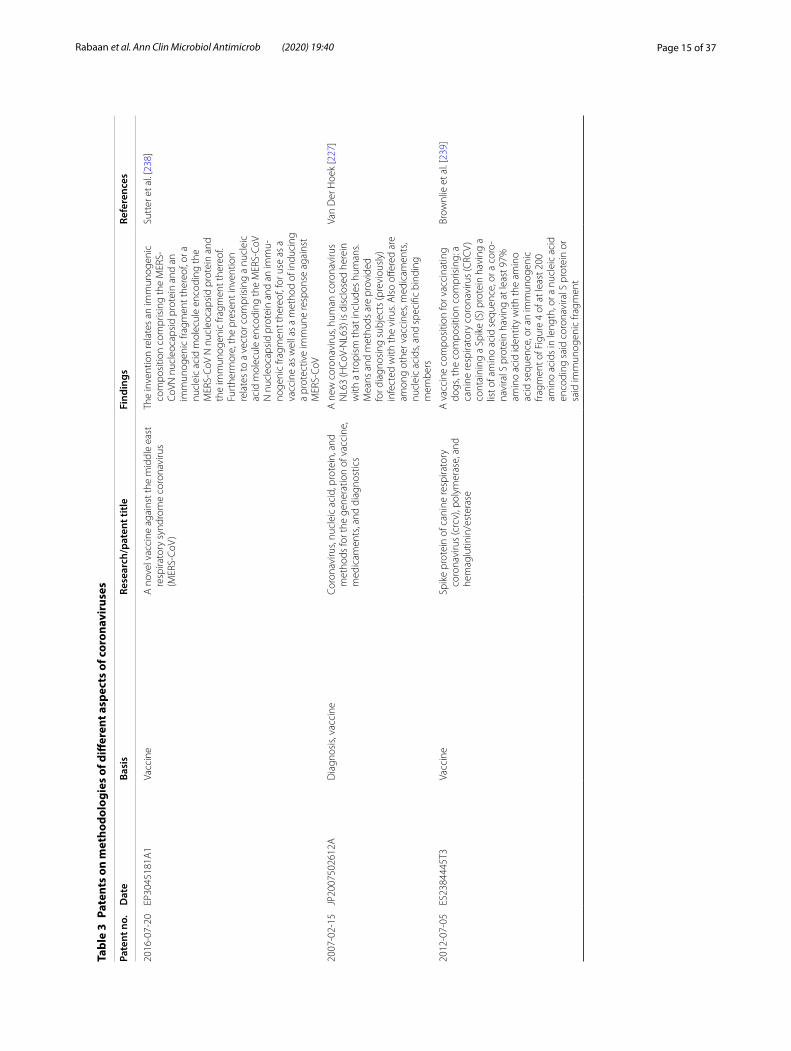
Patents on successful methodologies on various aspects of coronavirusesGlobal research focuses on coronavirus infections, espe-cially the recently happened outbreaks of SARS, MERS, and the most newly COVID-19. That involves the explo-ration of diagnostics, prophylactics, and therapeutics. Accordingly, the patenting of successful methodologies has gained immense importance, safeguarding the inter-ests of scientists and institutions. Various patents are being filed, and many approved on diagnostic, prophy-lactic, and therapeutic aspects of coronaviruses and their diseases, especially SARS-CoV caused by SARS, MERS-CoV caused by MERS and the most newly SARS-CoV-2 caused COVID-19. As per one study, around 80% of pat-ents are related to therapeutics, 35% for vaccines, and 28% for diagnostics agents or methods [121]. Similarly, on MERS, more than 100 patents are published on thera-peutics, and more than 50 on diagnostics and prophylac-tics [121]. These patents cover a range of research areas.
Table 3 provides details about the various fields and research areas for which patents have been applied and granted. These areas include developing novel, rapid, and specific diagnostics that are cheap and readily avail-able [121, 227–229]. For this purpose, the exploration of novel methods or techniques is being elucidated that can be transformed into diagnostic technology. Further, identifying diagnostic markers are vital. Similarly, for the treatment purpose, novel drugs or therapeutic agents are being explored [121]. These need to be effective against coronaviruses, including SARS-CoV, MERS-CoV, and SARS-CoV-2 [121, 230, 231]. These can be viral protease inhibitors [231], enzyme inhibitors [232], antivirals [228, 233], immunomodulatory [234], and treatment adjuvants [235]. That is the primary class that has attracted most of the patents [121]. Either a drug target is located, or a direct antiviral compound, molecule or agent is estab-lished, or an indirect immunomodulator that raises the immune response against the virus is identified as shown in Table 3. Vaccines, which are the main backbone for the prevention strategies and are mostly lacking, especially for novel coronaviruses, have shown immense research prospects and are attracting a sufficient number of pat-ents as shown in Table 3 [121]. Exploring novel vaccine candidates, targeting appropriate antigens, evaluating immune responses, and transforming into a safe, effec-tive, and potent vaccine are some of the areas on which patents have been published [236–238]. They can be
univalent targeting a single pathogen (species/strain) [238, 239] or multivalent targeting many at a time [237]. Presently no specific therapeutics or vaccines are avail-able against SARS-CoV-2. Since there is some degree of genomic and structural identity hence therapeutics active against SARS-CoV or MERS-CoV or other broad-spectrum antivirals (e.g., remdesivir, chloroquine) are being explored for SARS-CoV-2. Identification of specific therapeutic targets and vaccines candidates will enable the developing of concrete and potential drugs or vac-cines that can prove useful in the prevention and control of SARS-CoV-2 [121].
Specifically, with regards to COVID-19, three drugs are in the race for patents. These include remdesivir of Gilead Sciences®, hydroxychloroquine of Sanofi® and Kaletra (Lopinavir+Ritonavir) of Abbvie®. They are pat-ented for use against viruses (Ebola), malaria and HIV/AIDS, respectively, however, have shown potential for application in COVID-19 hence being evaluated in clini-cal trials (https ://ttcon sulta nts.com/blog/poten tial-covid -19-drugs -and-their -paten ts-in-major -juris dicti ons/). Wuhan Institute of Virology in China has filed a pat-ent for the application of remdesivir in COVID-19 [240] and Suzhou-based BrightGene Bio-Medical Technol-ogy for the synthesis of its active ingredient [241, 242]. Chinese were first to evaluate the effectiveness of rem-desivir, chloroquine and Kaletra in treating COVID-19 patients [25, 136]. However, simultaneous intentions for patent applications raised global concerns [242]. This led to a sort of conflict between Chinese institutes and the pharmaceutical companies having original patents for the drugs [242]. Thus, avoiding such differences and focusing on improving the health of people in times of pandemic need to be the priority rather than personnel monetary gains [242].
Similarly, in the case of vaccines which are in final stages of development, inferences from related corona-viruses like SARS and MERS are being taken. There are around 500 patents on SARS and 50 on MERS vaccines; however, it is unknown how many will be granted [35, 121]. Nevertheless, these can provide the basis for vac-cine development for COVID-19 [35, 121].
Conclusion and future perspectivesCurrently, the SARS-CoV-2 outbreak has taken a disas-trous turn with high toll rates alone in China itself, most likely. The infection is spreading across the globe. There are no licensed vaccines or therapeutic agents (i.e., anti-virals and monoclonal antibodies) indicated for this coro-navirus prevention or treatment. However, researchers are working to develop countermeasures. Several vac-cine candidates for both SARS-CoV-2 are in early clini-cal trials. This review is an accumulative hub of the latest
Page 15 of 37Rabaan et al. Ann Clin Microbiol Antimicrob (2020) 19:40
Tabl
e 3
Pate
nts
on m
etho
dolo
gies
of d
iffer
ent a
spec
ts o
f cor
onav
irus
es
Pate
nt n
o.D
ate
Basi
sRe
sear
ch/p
aten
t titl
eFi
ndin
gsRe
fere
nces
2016
‑07‑
20EP
3045
181A
1Va
ccin
eA
nov
el v
acci
ne a
gain
st th
e m
iddl
e ea
st
resp
irato
ry s
yndr
ome
coro
navi
rus
(MER
S‑Co
V)
The
inve
ntio
n re
late
s an
imm
unog
enic
co
mpo
sitio
n co
mpr
isin
g th
e M
ERS‑
CoVN
nuc
leoc
apsi
d pr
otei
n an
d an
im
mun
ogen
ic fr
agm
ent t
here
of, o
r a
nucl
eic
acid
mol
ecul
e en
codi
ng th
e M
ERS‑
CoV
N n
ucle
ocap
sid
prot
ein
and
the
imm
unog
enic
frag
men
t the
reof
. Fu
rthe
rmor
e, th
e pr
esen
t inv
entio
n re
late
s to
a v
ecto
r com
pris
ing
a nu
clei
c ac
id m
olec
ule
enco
ding
the
MER
S‑Co
V N
nuc
leoc
apsi
d pr
otei
n an
d an
imm
u‑no
geni
c fra
gmen
t the
reof
, for
use
as
a va
ccin
e as
wel
l as
a m
etho
d of
indu
cing
a
prot
ectiv
e im
mun
e re
spon
se a
gain
st
MER
S‑Co
V
Sutt
er e
t al.
[238
]
2007
‑02‑
15JP
2007
5026
12A
Dia
gnos
is, v
acci
neCo
rona
viru
s, nu
clei
c ac
id, p
rote
in, a
nd
met
hods
for t
he g
ener
atio
n of
vac
cine
, m
edic
amen
ts, a
nd d
iagn
ostic
s
A n
ew c
oron
aviru
s, hu
man
cor
onav
irus
NL6
3 (H
CoV‑
NL6
3) is
dis
clos
ed h
erei
n w
ith a
trop
ism
that
incl
udes
hum
ans.
Mea
ns a
nd m
etho
ds a
re p
rovi
ded
for d
iagn
osin
g su
bjec
ts (p
revi
ousl
y)
infe
cted
with
the
viru
s. A
lso
offer
ed a
re
amon
g ot
her v
acci
nes,
med
icam
ents
, nu
clei
c ac
ids,
and
spec
ific
bind
ing
mem
bers
Van
Der
Hoe
k [2
27]
2012
‑07‑
05ES
2384
445T
3Va
ccin
eSp
ike
prot
ein
of c
anin
e re
spira
tory
co
rona
viru
s (c
rcv)
, pol
ymer
ase,
and
he
mag
lutin
in/e
ster
ase
A v
acci
ne c
ompo
sitio
n fo
r vac
cina
ting
dogs
, the
com
posi
tion
com
pris
ing:
a
cani
ne re
spira
tory
cor
onav
irus
(CRC
V)
cont
aini
ng a
Spi
ke (S
) pro
tein
hav
ing
a lis
t of a
min
o ac
id s
eque
nce,
or a
cor
o‑na
vira
l S p
rote
in h
avin
g at
leas
t 97%
am
ino
acid
iden
tity
with
the
amin
o ac
id s
eque
nce,
or a
n im
mun
ogen
ic
fragm
ent o
f Fig
ure
4 of
at l
east
200
am
ino
acid
s in
leng
th, o
r a n
ucle
ic a
cid
enco
ding
sai
d co
rona
vira
l S p
rote
in o
r sa
id im
mun
ogen
ic fr
agm
ent
Brow
nlie
et a
l. [2
39]

Page 16 of 37Rabaan et al. Ann Clin Microbiol Antimicrob (2020) 19:40
Tabl
e 3
(con
tinu
ed)
Pate
nt n
o.D
ate
Basi
sRe
sear
ch/p
aten
t titl
eFi
ndin
gsRe
fere
nces
2017
‑10‑
12W
O20
1717
6596
A1
Vacc
ine
Mul
tival
ent v
acci
nes
for r
abie
s vi
rus
and
coro
navi
ruse
sTh
e pr
esen
t dis
clos
ure
prov
ides
met
hods
an
d co
mpo
sitio
ns fo
r ind
ucin
g an
im
mun
e re
spon
se th
at c
onfe
rs d
ual
prot
ectio
n ag
ains
t inf
ectio
ns b
y ei
ther
or
bot
h of
a ra
bies
viru
s an
d a
coro
navi
‑ru
s, an
d w
hich
can
be
used
ther
apeu
ti‑ca
lly fo
r an
exis
ting
infe
ctio
n w
ith th
e ra
bies
viru
s an
d a
coro
navi
rus
to tr
eat
at le
ast o
ne s
ympt
om th
ereo
f and
to
neut
raliz
e or
cle
ar th
e in
fect
ing
agen
ts.
In p
artic
ular
, the
pre
sent
dis
clos
ure
pro‑
vide
s a
reco
mbi
nant
rabi
es v
irus
vect
or
com
pris
ing
a nu
cleo
tide
sequ
ence
en
codi
ng a
t lea
st o
ne c
oron
aviru
s im
mun
ogen
ic g
lyco
prot
ein
fragm
ent,
as w
ell a
s ph
arm
aceu
tical
com
posi
tions
co
mpr
isin
g th
e va
ccin
e ve
ctor
s
John
son
et a
l. [2
37]
2017
‑12‑
28W
O20
1722
2935
A1
Trea
tmen
t and
con
trol
Smal
l mol
ecul
e th
erap
eutic
inhi
bito
rs
agai
nst p
icor
navi
ruse
s, ca
liciv
iruse
s, an
d co
rona
viru
ses
Ant
ivira
l pro
teas
e in
hibi
tors
are
dis
clos
ed,
alon
g w
ith re
late
d an
tivira
l dip
eptid
yl
com
poun
ds, m
acro
cycl
ic d
eriv
ativ
es
ther
eof,
and
met
hods
of u
sing
the
sam
e to
trea
t or p
reve
nt v
iral i
nfec
tion
and
dise
ase
from
cor
onav
iruse
s, ca
liciv
i‑ru
ses,
and
pico
rnav
iruse
s
Cha
ng e
t al.
[261
]
2018
‑05‑
22S9
9758
85B2
Trea
tmen
t and
con
trol
Broa
d‑sp
ectr
um n
on‑c
oval
ent c
oron
avi‑
rus
prot
ease
inhi
bito
rsTh
is in
vent
ion
pert
ains
to m
ater
ials
and
m
etho
ds fo
r the
trea
tmen
t of p
atie
nts
with
cor
onav
irus
infe
ctio
n an
d th
e co
ntro
l of z
oono
tic d
isea
se o
utbr
eaks
us
ing
broa
d‑sp
ectr
um n
on‑c
oval
ent
coro
navi
rus
prot
ease
inhi
bito
rs
Emm
a et
al.
[231
]
2017
‑08‑
03W
O20
1712
9975
A1
Vacc
ine
Att
enua
ted
infe
ctio
us b
ronc
hitis
viru
sTh
e pr
esen
t inv
entio
n pr
ovid
es a
live
, at
tenu
ated
cor
onav
irus
com
pris
ing
a m
utat
ion
in n
on‑s
truc
tura
l pro
tein
ns
p‑3
and
dele
tion
of a
cces
sory
pro
‑te
ins
3a a
nd 3
b. T
he c
oron
aviru
s m
ay
be u
sed
as a
vac
cine
for t
reat
ing
and
prev
entin
g di
seas
e, s
uch
as in
fect
ious
br
onch
itis,
in a
sub
ject
Bick
erto
n et
al.
[262
]

Page 17 of 37Rabaan et al. Ann Clin Microbiol Antimicrob (2020) 19:40
Tabl
e 3
(con
tinu
ed)
Pate
nt n
o.D
ate
Basi
sRe
sear
ch/p
aten
t titl
eFi
ndin
gsRe
fere
nces
2019
‑12‑
05JP
2019
2065
73A
Imm
unity
boo
ster
sIm
mun
omod
ulat
ion
met
hods
and
co
mpo
sitio
nsM
etho
ds a
nd c
ompo
sitio
ns fo
r im
mu‑
nom
odul
atio
n ar
e pr
ovid
ed. C
ells
co
ntai
ning
exo
geno
us a
ntig
ens
and
uses
ther
eof a
re p
rodu
ced.
The
pre
sent
in
vent
ion
incl
udes
, for
exa
mpl
e, a
way
of
indu
cing
imm
une
tole
ranc
e, c
om‑
pris
ing
enuc
leat
ed h
emat
opoi
etic
cel
ls
that
exp
ress
an
exog
enou
s an
tigen
to a
hu
man
sub
ject
suff
erin
g fro
m o
r at r
isk
of d
evel
opin
g an
aut
oim
mun
e di
seas
e,
diso
rder
or c
ondi
tion
Adm
inis
terin
g a
phar
mac
eutic
al c
ompo
sitio
n, w
here
in
the
phar
mac
eutic
al c
ompo
sitio
n is
ad
min
iste
red
in a
n am
ount
effe
ctiv
e to
indu
ce im
mun
e to
lera
nce
in th
e su
bjec
t aga
inst
the
antig
en th
at m
edi‑
ates
the
auto
imm
une
dise
ase,
dis
orde
r or
con
ditio
n
Jord
i et a
l. [2
34]
2020
‑01‑
02U
S202
0000
2370
A1
Trea
tmen
t, ad
juva
nts
Nov
el c
ompo
unds
The
inve
ntio
n al
so re
late
s to
the
use
of s
aid
com
poun
ds, c
ombi
natio
ns,
com
posi
tions
and
med
icam
ents
, in
the
trea
tmen
t of d
isea
ses
in w
hich
mod
ula‑
tion
of S
TIN
G (S
timul
ator
of I
nter
fero
n G
enes
) is
bene
ficia
l, fo
r exa
mpl
e,
infla
mm
atio
n, a
llerg
ic a
nd a
utoi
mm
une
dise
ases
, inf
ectio
us d
isea
ses,
canc
er,
pre‑
canc
erou
s sy
ndro
mes
and
as
vac‑
cine
adj
uvan
ts
Ada
ms
and
Lian
[235
]
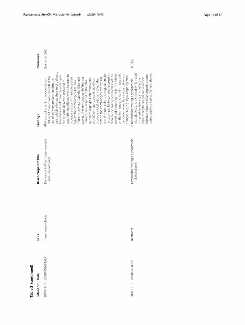
Page 18 of 37Rabaan et al. Ann Clin Microbiol Antimicrob (2020) 19:40
Tabl
e 3
(con
tinu
ed)
Pate
nt n
o.D
ate
Basi
sRe
sear
ch/p
aten
t titl
eFi
ndin
gsRe
fere
nces
2019
‑11‑
14U
S201
9034
3862
A1
Imm
unom
odul
ator
sD
eliv
ery
of R
NA
to tr
igge
r mul
tiple
im
mun
e pa
thw
ays
RNA
enc
odin
g an
imm
unog
en is
co‑
deliv
ered
to n
on‑im
mun
e ce
lls a
t the
si
te o
f del
iver
y an
d al
so to
imm
une
cells
, whi
ch in
filtr
ate
the
site
of d
eliv
ery.
Th
e re
spon
ses
of th
ese
two
cell
type
s to
the
sam
e de
liver
ed R
NA
lead
to
two
diffe
rent
effe
cts,
whi
ch in
tera
ct to
pr
oduc
e a
robu
st im
mun
e re
spon
se
agai
nst t
he im
mun
ogen
. The
non
‑im
mun
e ce
lls tr
ansl
ate
the
RNA
and
ex
pres
s th
e im
mun
ogen
. Infi
ltrat
ing
imm
une
cells
resp
ond
to th
e RN
A
by e
xpre
ssin
g ty
pe I
inte
rfer
ons
and
pro‑
infla
mm
ator
y cy
toki
nes,
whi
ch
prod
uce
a lo
cal a
djuv
ant e
ffect
that
ac
ts o
n th
e im
mun
ogen
‑exp
ress
ing
non‑
imm
une
cells
to u
preg
ulat
e m
ajor
hi
stoc
ompa
tibili
ty c
ompl
ex e
xpre
ssio
n,
ther
eby
incr
easi
ng p
rese
ntat
ion
of th
e tr
ansl
ated
pro
tein
to T
cel
ls. T
he e
ffect
s on
the
imm
une
and
non‑
imm
une
cells
ca
n be
ach
ieve
d by
a s
ingl
e de
liver
y of
a
sing
le R
NA
, e.g
., by
a s
ingl
e in
ject
ion
Gea
ll et
al.
[263
]
2018
‑12‑
18U
S101
5498
5B2
Trea
tmen
tM
etho
d fo
r tre
atin
g a
glyc
opro
tein
‑re
late
d di
seas
eA
met
hod
for t
reat
ing
a gl
ycop
rote
in‑
rela
ted
dise
ase
is d
iscl
osed
, whi
ch c
om‑
pris
es: a
dmin
iste
ring
a fir
st e
ffect
ive
amou
nt o
f phe
nol r
ed a
nd a
sec
ond
effec
tive
amou
nt o
f an
orga
nic
arse
nic
com
poun
d to
a s
ubje
ct in
nee
d th
ereo
Lu [2
64]

Page 19 of 37Rabaan et al. Ann Clin Microbiol Antimicrob (2020) 19:40
Tabl
e 3
(con
tinu
ed)
Pate
nt n
o.D
ate
Basi
sRe
sear
ch/p
aten
t titl
eFi
ndin
gsRe
fere
nces
2016
‑11‑
24U
S201
6033
9097
A1
Reco
mbi
nant
vac
cine
pro
tein
sCo
rona
viru
s pr
otei
ns a
nd a
ntig
ens
Dis
clos
ed h
erei
n ar
e em
bodi
men
ts o
f a
met
hod
for c
olle
ctin
g, e
xtra
ctin
g, o
r el
utin
g pr
otei
ns a
nd a
ntig
ens
from
cel
ls
infe
cted
with
the
coro
navi
rus.
The
coro
‑na
viru
s m
aybe
a p
orci
ne c
oron
aviru
s, su
ch a
s po
rcin
e ep
idem
ic d
iarr
hoea
vi
rus
(PED
V) o
r por
cine
del
ta c
oron
avi‑
rus
(PD
CoV
). A
lso
disc
lose
d ar
e em
bodi
‑m
ents
of a
com
posi
tion
com
pris
ing
the
coro
navi
rus
prot
eins
and
ant
igen
s, an
d em
bodi
men
ts o
f a m
etho
d of
usi
ng
such
a c
ompo
sitio
n. A
pplic
atio
ns fo
r th
e co
mpo
sitio
n in
clud
e, b
ut a
re n
ot
limite
d to
, use
in th
e pr
epar
atio
n of
an
tibod
ies
agai
nst t
he p
rote
ins
and
antig
ens,
use
as re
fere
nce
mar
kers
for
coro
navi
rus
prot
eins
, and
use
in a
n im
mun
ogen
ic c
ompo
sitio
n, s
uch
as in
a
vacc
ine
com
posi
tion
Kim
[265
]
2017
‑01‑
19W
O20
1701
0835
A1
Dru
g/tr
eatm
ent
Use
of r
adot
inib
for p
reve
ntio
n or
trea
t‑m
ent o
f vira
l res
pira
tory
dis
ease
The
pres
ent i
nven
tion
rela
tes
to a
new
us
e of
a c
ompo
und
of C
hem
ical
Fo
rmul
a 1
(rado
tinib
) in
the
prev
en‑
tion
or tr
eatm
ent o
f vira
l res
pira
tory
di
seas
e. A
ccor
ding
to th
e pr
esen
t in
vent
ion,
a c
ompo
und
of C
hem
i‑ca
l For
mul
a 1
or a
pha
rmac
eutic
ally
ac
cept
able
sal
t the
reof
can
be
used
for
the
prev
entio
n, a
llevi
atio
n, o
r tre
atm
ent
of c
oron
aviru
s in
fect
ion.
Spe
cific
ally
, th
e pr
esen
t inv
entio
n ca
n be
use
d as
a
usef
ul a
ntiv
iral a
gent
for t
he p
reve
n‑tio
n or
trea
tmen
t of d
isea
se c
ause
d by
in
fect
ion
of th
e M
iddl
e Ea
st re
spira
tory
sy
ndro
me‑
coro
navi
rus
(MER
S‑Co
V).
Kim
et a
l. [2
32]
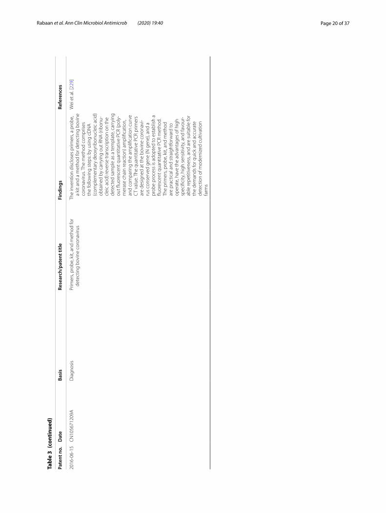
Page 20 of 37Rabaan et al. Ann Clin Microbiol Antimicrob (2020) 19:40
Tabl
e 3
(con
tinu
ed)
Pate
nt n
o.D
ate
Basi
sRe
sear
ch/p
aten
t titl
eFi
ndin
gsRe
fere
nces
2016
‑06‑
15C
N10
5671
209A
Dia
gnos
isPr
imer
s, pr
obe,
kit,
and
met
hod
for
dete
ctin
g bo
vine
cor
onav
irus
The
inve
ntio
n di
sclo
ses
prim
ers,
a pr
obe,
a
kit a
nd a
met
hod
for d
etec
ting
bovi
ne
coro
navi
rus.
The
met
hod
com
pris
es
the
follo
win
g st
eps:
by u
sing
cD
NA
(c
ompl
emen
tary
deo
xyrib
onuc
leic
aci
d)
obta
ined
by
carr
ying
out
RN
A (r
ibon
u‑cl
eic
acid
) rev
erse
tran
scrip
tion
on th
e de
tect
ed s
ampl
e as
a te
mpl
ate,
car
ryin
g ou
t fluo
resc
ent q
uant
itativ
e PC
R (p
oly‑
mer
ase
chai
n re
actio
n) a
mpl
ifica
tion,
an
d co
mpa
ring
the
ampl
ifica
tion
curv
e C
T va
lue.
The
qua
ntita
tive
PCR
prim
ers
are
desi
gned
at t
he b
ovin
e co
rona
vi‑
rus
cons
erve
d ge
ne (N
gen
e), a
nd a
pr
obe
proc
ess
is a
dopt
ed to
est
ablis
h a
fluor
esce
nt q
uant
itativ
e PC
R m
etho
d.
The
prim
ers,
prob
e, k
it, a
nd m
etho
d ar
e pr
actic
al a
nd s
trai
ghtfo
rwar
d to
op
erat
e, h
ave
the
adva
ntag
es o
f hig
h sp
ecifi
city
, hig
h se
nsiti
vity
, and
favo
ur‑
able
repe
titiv
enes
s, an
d ar
e su
itabl
e fo
r th
e de
man
ds fo
r qui
ck a
nd a
ccur
ate
dete
ctio
n of
mod
erni
zed
culti
vatio
n fa
rms
Wei
et a
l. [2
29]

Page 21 of 37Rabaan et al. Ann Clin Microbiol Antimicrob (2020) 19:40
Tabl
e 3
(con
tinu
ed)
Pate
nt n
o.D
ate
Basi
sRe
sear
ch/p
aten
t titl
eFi
ndin
gsRe
fere
nces
2016
‑06‑
1C
N10
5671
006A
Dia
gnos
is a
nd tr
eatm
ent
Hig
h‑effi
cien
cy ra
nilla
luci
fera
se g
ene
expr
essi
on re
com
bina
nt H
CoV‑
OC
43
(hum
an c
oron
aviru
s O
C43
) viru
s an
d ap
plic
atio
n th
ereo
f
The
inve
ntio
n di
sclo
ses
a hi
gh‑e
ffici
ency
ra
nilla
luci
fera
se g
ene
expr
essi
on
reco
mbi
nant
hum
an c
oron
aviru
s O
C43
viru
s an
d ap
plic
atio
n th
ereo
f to
a s
cree
ning
of a
ntiv
iral m
edic
ines
. By
an
over
lap
PCR
(pol
ymer
ase
chai
n re
actio
n) m
etho
d, a
rani
lla lu
cife
rase
ge
ne is
repl
aced
or i
nser
ted
into
ac
cess
ory
gene
s (n
s2 a
nd n
s12.
9) to
be
clon
ed in
to h
uman
cor
onav
irus
OC
43
full‑
leng
th in
fect
ious
clo
ne p
BAC
‑O
C43
FL, a
nd fo
ur ra
nilla
luci
fera
se g
ene
expr
essi
on re
com
bina
nt v
iruse
s, in
clud
‑in
g rO
C43
‑ns2
Del
Rluc
, rO
C43
‑ns2
Fu‑
sion
Rluc
, rO
C43
‑ns1
2.9S
topR
luc,
and
rO
C43
‑ns1
2.9F
usio
nRlu
c, a
re o
btai
ned
resp
ectiv
ely.
The
viru
s rO
C43
‑ns2
Del
R‑lu
c is
effi
cien
t in
rani
lla lu
cife
rase
gen
e ex
pres
sion
and
sim
ilar t
o a
pare
nt v
irus
HCo
V‑O
C43
‑WT
in th
e vi
rus
grow
th
curv
e, th
e in
sert
ed re
port
er g
ene
is
stab
le in
a s
eria
l pas
sage
pro
cess
, and
th
e vi
rus
rOC
43‑n
s2D
elRl
uc c
an b
e su
c‑ce
ssfu
lly a
pplie
d to
ant
ivira
l med
icin
e sc
reen
ing
expe
rimen
ts a
nd h
as a
n ex
tens
ive
appl
icat
ion
pros
pect
in h
igh‑
thro
ughp
ut s
cree
ning
of a
nti‑c
oron
avi‑
rus
med
icin
es a
nd h
ost a
ntiv
iral g
enes
Shen
liang
, [22
8]
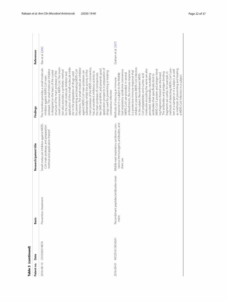
Page 22 of 37Rabaan et al. Ann Clin Microbiol Antimicrob (2020) 19:40
Tabl
e 3
(con
tinu
ed)
Pate
nt n
o.D
ate
Basi
sRe
sear
ch/p
aten
t titl
eFi
ndin
gsRe
fere
nces
2016
‑08‑
10C
N10
5837
487A
Prev
entio
n /t
reat
men
tSm
all‑m
olec
ule
inhi
bito
r aga
inst
MER
S‑Co
V m
ain
prot
ease
, and
pre
para
tion
met
hod
and
appl
icat
ion
ther
eof
The
inve
ntio
n pr
ovid
es a
sm
all‑m
olec
ule
inhi
bito
r aga
inst
MER
S‑Co
V m
ain
prot
ease
. The
sm
all‑m
olec
ule
inhi
bito
r is
des
igne
d on
the
basi
s of
the
crys
tal
stru
ctur
e of
the
mai
n pr
otea
se o
f the
no
vel c
oron
aviru
s M
ERS‑
CoV.
The
inve
n‑tio
n al
so p
rovi
des
a sy
nthe
tic m
etho
d fo
r the
sm
all‑m
olec
ule
inhi
bito
r and
ap
plic
atio
n of
the
smal
l‑mol
ecul
e in
hib‑
itor i
n th
e pr
epar
atio
n of
dru
gs u
sed
for p
reve
ntin
g an
d tr
eatin
g M
ERS‑
CoV
infe
ctio
ns. T
he s
mal
l‑mol
ecul
e in
hibi
tor
agai
nst M
ERS‑
CoV
mai
n pr
otea
se c
an
subs
tant
ially
inhi
bit t
he a
ctiv
ity o
f the
m
ain
prot
ease
of t
he M
ERS
coro
navi
rus,
has
an e
xcel
lent
inhi
bito
ry a
ctiv
ity to
th
e m
ain
prot
ease
of c
oron
aviru
ses
like
SARS
and
MH
V, a
nd p
rese
nts
good
ap
plic
atio
n pr
ospe
cts
in p
repa
ratio
n of
dr
ugs
used
for p
reve
ntin
g or
trea
ting
coro
navi
rus
infe
ctio
n
Rao
et a
l. [2
66]
2016
‑09‑
01W
O20
1613
8160
A1
Reco
mbi
nant
pep
tides
/ant
ibod
ies
trea
t‑m
ent
Mid
dle
east
resp
irato
ry s
yndr
ome
coro
‑na
viru
s im
mun
ogen
s, an
tibod
ies,
and
thei
r use
Met
hods
of i
nduc
ing
an im
mun
e re
spon
se in
a s
ubje
ct to
the
Mid
dle
East
resp
irato
ry s
yndr
ome
coro
navi
rus
(MER
S‑Co
V) a
re p
rovi
ded.
In s
ever
al
embo
dim
ents
, the
imm
une
resp
onse
is
a p
rote
ctiv
e im
mun
e re
spon
se th
at
inhi
bits
or p
reve
nts
MER
S‑Co
V in
fect
ion
in th
e su
bjec
t. Re
com
bina
nt M
ERS‑
CoV
poly
pept
ides
and
nuc
leic
aci
d m
olec
ules
enc
odin
g th
e sa
me
are
also
pr
ovid
ed. A
dditi
onal
ly, n
eutr
aliz
ing
antib
odie
s th
at s
peci
fical
ly b
ind
to
MER
S‑Co
V S
prot
ein
and
antig
en‑b
ind‑
ing
fragm
ents
ther
eof a
re d
iscl
osed
. Th
e an
tibod
ies
and
antig
en‑b
indi
ng
fragm
ents
are
use
ful,
for e
xam
ple,
in
met
hods
of d
etec
ting
MER
S‑Co
V S
pro‑
tein
in a
sam
ple
or in
a s
ubje
ct, a
s w
ell
as m
etho
ds o
f pre
vent
ing
and
trea
ting
a M
ERS‑
CoV
infe
ctio
n in
a s
ubje
ct
Gra
ham
et a
l. [2
67]

Page 23 of 37Rabaan et al. Ann Clin Microbiol Antimicrob (2020) 19:40
Tabl
e 3
(con
tinu
ed)
Pate
nt n
o.D
ate
Basi
sRe
sear
ch/p
aten
t titl
eFi
ndin
gsRe
fere
nces
2016
‑06‑
01C
N10
5624
122A
Mut
ant v
irus
as a
vac
cine
Avi
an in
fect
ious
bro
nchi
tis v
irus
natu
ral
mut
ant
The
inve
ntio
n re
late
s to
the
field
of v
irol‑
ogy.
It a
ims
to p
rovi
de a
n av
ian
infe
c‑tio
us b
ronc
hitis
viru
s na
tura
l mut
ant,
kept
in C
hina
Gen
eral
Mic
robi
olog
ical
Cu
lture
Col
lect
ion
Cent
er, n
amed
infe
c‑tio
us b
ronc
hitis
viru
s, an
d co
llect
ed
unde
r CG
MCC
No.
114
91. A
gen
ome
of th
is m
utan
t is
27, 5
41 lb
p in
leng
th,
a co
mpl
ete
geno
me
sequ
ence
of t
he
mut
ant i
s as
sho
wn
in S
EQ ID
NO
: 1,
with
str
uctu
ral c
hara
cter
istic
s of
a ty
pi‑
cal I
BV g
enom
e co
ding
str
uctu
re. T
his
mut
ant h
avin
g sp
ecia
l gen
es in
sert
ed
in a
nd p
rovi
ded
by th
e in
vent
ion
can
brea
k th
e im
mun
ity p
rote
ctio
n of
va
ccin
es to
bre
ed in
an
orga
nism
, thi
s m
utan
t can
pro
lifer
ate
in s
usce
ptib
le
chic
kens
with
out c
ausi
ng s
igni
fican
t pa
thog
enic
ity to
sen
sitiv
e ch
icke
ns,
and
thus
this
mut
ant c
an s
erve
as
the
base
mat
eria
l for
the
furt
her s
tudy
on
mut
atio
n m
echa
nism
of a
n at
tenu
ated
va
ccin
e or
IBV
viru
s st
rain
Liao
et a
l. 20
16[2
68]

Page 24 of 37Rabaan et al. Ann Clin Microbiol Antimicrob (2020) 19:40
Tabl
e 3
(con
tinu
ed)
Pate
nt n
o.D
ate
Basi
sRe
sear
ch/p
aten
t titl
eFi
ndin
gsRe
fere
nces
2017
‑08‑
08C
N10
7022
008A
Prev
entio
n an
d tr
eatm
ent
Supp
ress
pol
ypep
tide
and
its a
pplic
atio
n of
hum
an c
oron
aviru
s’s in
fect
ion
broa
d sp
ectr
um
The
inve
ntio
n be
long
s to
the
bio‑
med
icin
e fie
ld. I
t is
rela
ted
to th
e po
lype
ptid
e fo
r sup
pres
sing
hum
an
coro
navi
rus
infe
ctio
n, a
nd in
par
ticul
ar,
to p
olyp
eptid
e an
d its
app
licat
ion
of
hum
an c
oron
ary
viru
s’s d
isea
se c
an b
e co
ntai
ned
a br
oad
spec
trum
. The
S2
regi
ons
base
d on
cor
onav
irus
S pr
otei
n of
the
inve
ntio
n ar
e m
ore
cons
erva
‑tiv
e, a
nd fe
atur
es o
f sim
ilar s
yncr
etiz
‑in
g m
echa
nism
s pr
ovid
e ca
n be
to
the
poly
pept
ide
of th
e in
fect
ion
with
br
oad‑
spec
trum
inhi
bito
ry a
ctio
n of
tw
o or
mor
e hu
man
cor
onar
y vi
rus.
The
pres
ent i
nven
tion
is th
e re
sults
sho
wed
ob
tain
“gen
eral
cha
ract
er” o
f the
hum
an
coro
navi
rus,
the
iden
tical
syn
cret
izin
g m
echa
nism
in, i
.e.,
sim
ilar H
R re
gion
s an
d its
med
iatio
n, a
nd p
rovi
de th
e se
rial p
olyp
eptid
es o
f HCo
V‑EK
as
the
poin
t of p
enet
ratio
n, th
e po
lype
ptid
e no
t onl
y ha
s pr
efer
able
inhi
bitio
n to
so
me
curr
ently
a p
opul
ar h
uman
cor
o‑na
viru
ses,
and
equa
lly h
as a
n ex
celle
nt
inhi
bito
ry a
ctiv
ity to
the
clas
s SA
RS v
irus
(RsS
HC
014‑
CoV
or R
sW1V
1‑Co
V) f
or
bein
g po
ssib
le to
infe
ct th
e hu
man
kind
. Th
e pr
esen
t inv
entio
n ca
n pr
ovid
e th
e dr
ug c
andi
date
for p
reve
ntio
n an
d tr
eatm
ent f
or th
e no
vel h
uman
co
rona
viru
s fo
r bei
ng s
till p
ossi
ble
to
outb
urst
in p
opul
ar h
uman
cor
onav
irus
and
futu
re a
t pre
sent
Shib
o et
al.
[269
]
2018
‑02‑
13U
S988
9194
BIm
mun
ogen
ic p
rote
ins
Imm
unog
enic
com
posi
tion
for M
ERS
coro
navi
rus
infe
ctio
nD
escr
ibed
her
ein
are
imm
unog
enic
co
mpo
sitio
ns fo
r pre
vent
ing
infe
ctio
n w
ith M
iddl
e Ea
st re
spira
tory
syn
drom
e co
rona
viru
s (M
ERS‑
CoV
) whe
rein
the
imm
unog
enic
com
posi
tions
com
pris
e at
leas
t a p
ortio
n of
the
MER
S‑Co
V S
prot
ein
and
an im
mun
opot
entia
tor
Jiang
et a
l. [2
70]
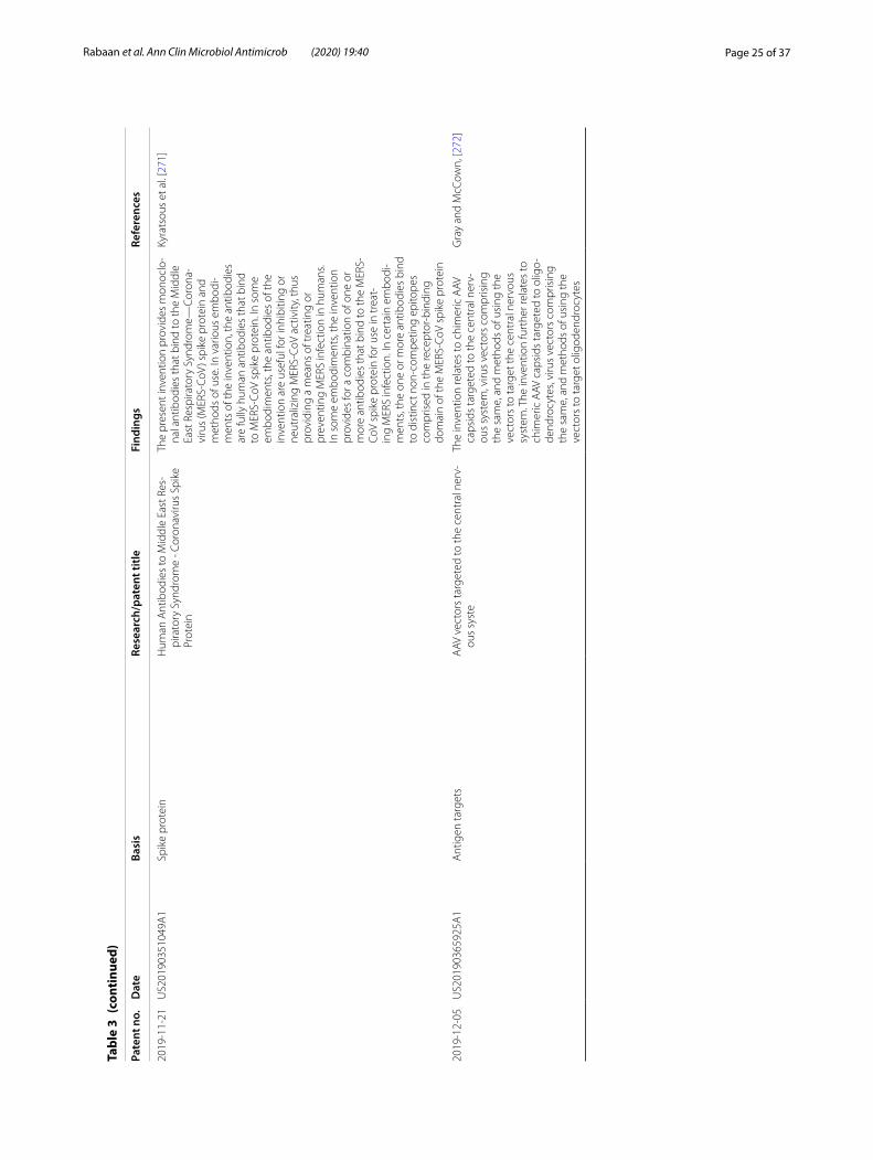
Page 25 of 37Rabaan et al. Ann Clin Microbiol Antimicrob (2020) 19:40
Tabl
e 3
(con
tinu
ed)
Pate
nt n
o.D
ate
Basi
sRe
sear
ch/p
aten
t titl
eFi
ndin
gsRe
fere
nces
2019
‑11‑
21U
S201
9035
1049
A1
Spik
e pr
otei
nH
uman
Ant
ibod
ies
to M
iddl
e Ea
st R
es‑
pira
tory
Syn
drom
e ‑ C
oron
aviru
s Sp
ike
Prot
ein
The
pres
ent i
nven
tion
prov
ides
mon
oclo
‑na
l ant
ibod
ies
that
bin
d to
the
Mid
dle
East
Res
pira
tory
Syn
drom
e—Co
rona
‑vi
rus
(MER
S‑Co
V) s
pike
pro
tein
and
m
etho
ds o
f use
. In
vario
us e
mbo
di‑
men
ts o
f the
inve
ntio
n, th
e an
tibod
ies
are
fully
hum
an a
ntib
odie
s th
at b
ind
to M
ERS‑
CoV
spik
e pr
otei
n. In
som
e em
bodi
men
ts, t
he a
ntib
odie
s of
the
inve
ntio
n ar
e us
eful
for i
nhib
iting
or
neut
raliz
ing
MER
S‑Co
V ac
tivity
, thu
s pr
ovid
ing
a m
eans
of t
reat
ing
or
prev
entin
g M
ERS
infe
ctio
n in
hum
ans.
In s
ome
embo
dim
ents
, the
inve
ntio
n pr
ovid
es fo
r a c
ombi
natio
n of
one
or
mor
e an
tibod
ies
that
bin
d to
the
MER
S‑Co
V sp
ike
prot
ein
for u
se in
trea
t‑in
g M
ERS
infe
ctio
n. In
cer
tain
em
bodi
‑m
ents
, the
one
or m
ore
antib
odie
s bi
nd
to d
istin
ct n
on‑c
ompe
ting
epito
pes
com
pris
ed in
the
rece
ptor
‑bin
ding
do
mai
n of
the
MER
S‑Co
V sp
ike
prot
ein
Kyra
tsou
s et
al.
[271
]
2019
‑12‑
05U
S201
9036
5925
A1
Ant
igen
targ
ets
AAV
vec
tors
targ
eted
to th
e ce
ntra
l ner
v‑ou
s sy
ste
The
inve
ntio
n re
late
s to
chi
mer
ic A
AV
caps
ids
targ
eted
to th
e ce
ntra
l ner
v‑ou
s sy
stem
, viru
s ve
ctor
s co
mpr
isin
g th
e sa
me,
and
met
hods
of u
sing
the
vect
ors
to ta
rget
the
cent
ral n
ervo
us
syst
em. T
he in
vent
ion
furt
her r
elat
es to
ch
imer
ic A
AV c
apsi
ds ta
rget
ed to
olig
o‑de
ndro
cyte
s, vi
rus
vect
ors
com
pris
ing
the
sam
e, a
nd m
etho
ds o
f usi
ng th
e ve
ctor
s to
targ
et o
ligod
endr
ocyt
es
Gra
y an
d M
cCow
n, [2
72]

Page 26 of 37Rabaan et al. Ann Clin Microbiol Antimicrob (2020) 19:40
Tabl
e 3
(con
tinu
ed)
Pate
nt n
o.D
ate
Basi
sRe
sear
ch/p
aten
t titl
eFi
ndin
gsRe
fere
nces
2019
‑09‑
05JP
2019
1465
88A
Vacc
ine
Prim
ing
of a
n im
mun
e re
spon
seTh
e pr
esen
t inv
entio
n pr
ovid
es a
met
hod
for e
xpan
ding
and
impr
ovin
g im
mun
e re
spon
se a
nd, a
t the
sam
e tim
e, in
crea
s‑in
g th
e sp
eed
of re
spon
se a
gain
st a
pa
thog
enic
ant
igen
or a
can
cer a
nti‑
gen.
A tw
o‑st
ep p
rime‑
boos
t met
hod
whe
reby
the
imm
une
syst
em is
firs
t pr
imed
with
a n
ucle
ic a
cid
cons
truc
t co
mpr
isin
g an
inva
riant
cha
in o
r a v
ari‑
ant t
here
of, f
ollo
wed
by
a bo
oste
r vac
‑ci
ne u
sing
any
type
of s
uita
ble
vacc
ine
Adm
inis
trat
ion)
to s
uffici
ently
stim
ulat
e th
e im
mun
e re
spon
se g
ener
ated
by
vacc
ine
adm
inis
trat
ion
Thom
sen
et a
l. [2
73]
2019
‑10‑
17U
S201
9031
4497
A1
Trea
tmen
tCo
mpo
sitio
ns a
nd m
etho
ds fo
r tre
atin
g vi
ral i
nfec
tions
thro
ugh
stim
ulat
ed
inna
te im
mun
ity in
com
bina
tion
with
an
tivira
l com
poun
ds
Embo
dim
ents
are
dire
cted
to c
ompo
si‑
tions
and
met
hods
for t
reat
ing
vira
l in
fect
ions
Dic
key
et a
l. [2
74]
2019
‑03‑
12U
S102
2643
4B2
Trea
tmen
tD
esig
n, s
ynth
esis
, and
met
hods
of u
se o
f ac
yclic
flex
imer
nuc
leos
ide
anal
ogue
s ha
ving
ant
i‑cor
onav
irus
activ
ity
The
pres
ent i
nven
tion
is d
irect
ed to
co
mpo
unds
, tec
hniq
ues,
and
com
posi
‑tio
ns fo
r tre
atin
g or
pre
vent
ing
vira
l in
fect
ions
usi
ng n
ucle
osid
es a
nalo
gues
. Sp
ecifi
cally
, the
pre
sent
inve
ntio
n pr
ovid
es fo
r the
des
ign
and
synt
hesi
s of
ac
yclic
flex
imer
nuc
leos
ide
anal
ogue
s, ha
ving
incr
ease
d fle
xibi
lity
and
abili
ty
to a
lter t
heir
conf
orm
atio
n st
ruct
ures
to
pro
vide
incr
ease
d an
tivira
l act
ivity
po
tent
ial w
ith th
e re
sult
of in
hibi
ting
seve
ral c
oron
aviru
ses
Radt
ke e
t al.
[275
]
2020
‑02‑
04U
S105
4897
1B2
Vacc
ine
MER
S‑Co
V va
ccin
eD
iscl
osed
her
ein
is a
vac
cine
com
pris
ing
a M
iddl
e Ea
st R
espi
rato
ry S
yndr
ome
coro
navi
rus
(MER
S‑Co
V) a
ntig
en. T
he
antig
en c
an b
e a
cons
ensu
s an
tigen
. Th
e co
nsen
sus
antig
en c
an b
e a
cons
ensu
s sp
ike
antig
en. A
lso
disc
lose
d he
rein
is a
met
hod
of tr
eatin
g a
subj
ect
in n
eed
ther
eof b
y ad
min
iste
ring
the
vacc
ine
to th
e to
pic
Wei
nere
t al.
2020
[276
]

Page 27 of 37Rabaan et al. Ann Clin Microbiol Antimicrob (2020) 19:40
Tabl
e 3
(con
tinu
ed)
Pate
nt n
o.D
ate
Basi
sRe
sear
ch/p
aten
t titl
eFi
ndin
gsRe
fere
nces
2019
‑05‑
08Pu
blic
atio
n of
JP65
0860
5B2
Vacc
ine
Mod
ular
DN
A b
indi
ng d
omai
n an
d m
etho
d of
use
The
pres
ent i
nven
tion
prov
ides
a m
etho
d of
sel
ectiv
ely
reco
gniz
ing
base
pai
rs in
a
targ
et D
NA
seq
uenc
e by
a p
olyp
eptid
e,
a m
odifi
ed p
olyp
eptid
e th
at re
cogn
izes
ex
plic
itly
one
or m
ore
base
pai
rs w
ithin
a
targ
et D
NA
seq
uenc
e, a
nd a
DN
A th
at
is m
odifi
ed s
o th
at it
can
be
reco
gniz
ed
expl
icitl
y by
the
poly
pept
ide,
and
spe
‑ci
fic u
se o
f pol
ypep
tides
and
DN
A in
NA
ta
rget
ing
and
met
hods
of m
odul
atin
g th
e ex
pres
sion
of t
arge
t gen
es in
cel
ls
Bona
s et
al.
[277
]
2019
‑09‑
10U
S104
0622
9B2
Vacc
ine
Met
hods
and
com
posi
tions
rela
ted
to
inhi
bitio
n of
vira
l ent
ryD
iscl
osed
her
ein
are
com
posi
tions
and
ar
rang
emen
ts fo
r inh
ibiti
ng v
iral e
ntry
Fran
cis
et a
l. [2
78]
2018
‑12‑
20AU
2015
3401
34B2
Ant
igen
ic g
ene
iden
tifica
tion
Mic
roflu
idic
dev
ice
for d
etec
ting
targ
et
gene
, the
met
hod
for m
anuf
actu
ring
sam
e, a
nd m
etho
d fo
r det
ectin
g us
ing
sam
e
The
pres
ent i
nven
tion
prov
ides
a ta
rget
ge
ne c
apab
le o
f bei
ng d
iffer
entia
ted
by
the
nake
d ey
e by
am
plify
ing
the
targ
et
gene
to s
elec
tivel
y bl
ock
the
fluid
pat
h an
d, s
peci
fical
ly, a
mic
roflu
idic
dev
ice
for d
etec
ting
path
ogen
gen
es, a
nd
a de
tect
ion
met
hod
usin
g th
e sa
me.
Th
eref
ore,
the
pres
ent i
nven
tion
can
conv
enie
ntly
iden
tify
a si
ngle
targ
et
gene
, suc
h as
a s
ingl
e pa
thog
en, o
r at
the
sam
e tim
e, s
ever
al ta
rget
gen
es,
such
as
seve
ral p
atho
gens
, with
out
com
plic
ated
mec
hani
cal d
evic
es
Jung
et a
l. [2
79]
2018
‑01‑
30U
S987
8024
B2En
hanc
ed im
mun
e re
spon
seM
etho
ds a
nd c
ompo
sitio
ns u
sing
Lis
teria
fo
r enh
anci
ng im
mun
ogen
icity
by
prim
e‑bo
ost
Prov
ided
her
ein
are
prim
e‑bo
ost
regi
men
s an
d m
ater
ials
use
d th
erei
n.
The
prim
e‑bo
ost r
egim
ens
enha
nce
the
imm
une
resp
onse
to a
targ
et
antig
en. T
he v
acci
nes
used
for b
oost
ar
e co
mpr
ised
of r
ecom
bina
nt a
tten
u‑at
ed m
etab
olic
ally
act
ive
List
eria
that
en
code
s an
exp
ress
ible
ant
igen
that
is
cros
s‑re
activ
e w
ith th
e ta
rget
ant
igen
. In
som
e ex
ampl
es, t
he im
mun
e re
spon
se is
a c
ellu
lar i
mm
une
resp
onse
Dub
ensk
y et
al.
[280
]

Page 28 of 37Rabaan et al. Ann Clin Microbiol Antimicrob (2020) 19:40
Tabl
e 3
(con
tinu
ed)
Pate
nt n
o.D
ate
Basi
sRe
sear
ch/p
aten
t titl
eFi
ndin
gsRe
fere
nces
2017
‑08‑
0U
S971
9107
B2Re
com
bina
nt g
ene
and
prot
ein
expr
es‑
sion
Cons
truc
tion
of fu
lly‑d
elet
ed a
deno
viru
s‑ba
sed
gene
del
iver
y ve
ctor
s an
d us
es
ther
eof
The
embo
dim
ents
dis
clos
ed h
erei
n re
late
to
the
cons
truc
tion
of fu
lly‑d
elet
ed
Ade
novi
rus‑
base
d ge
ne d
eliv
ery
vect
ors
pack
aged
with
out h
elpe
r A
deno
viru
s, an
d m
ore
part
icul
arly
to
thei
r use
in g
ene
ther
apy
for g
ene
and
prot
ein
expr
essi
on, v
acci
ne d
evel
op‑
men
t, an
d im
mun
osup
pres
sive
ther
apy
for a
lloge
neic
tran
spla
ntat
ion.
In a
n em
bodi
men
t, a
met
hod
for p
ropa
gat‑
ing
an a
deno
vira
l vec
tor i
nclu
des
(a)
prov
idin
g an
Ade
novi
rus
pack
agin
g ce
ll lin
e; (b
) tra
nsfe
ctin
g a
fully
‑del
eted
A
deno
vira
l vec
tor c
onst
ruct
into
the
cell
line;
and
opt
iona
lly (c
) tra
nsfe
ctin
g a
pack
agin
g co
nstr
uct i
nto
the
cell
line,
w
here
in th
e fu
lly‑d
elet
ed A
deno
vira
l ve
ctor
con
stru
ct a
nd o
ptio
nally
the
pack
agin
g co
nstr
uct c
an tr
ansf
ect t
he
Ade
novi
rus
pack
agin
g ce
ll lin
e re
sulti
ng
in th
e en
caps
idat
ion
of a
fully
‑del
eted
A
deno
vira
l vec
tor i
ndep
ende
nt o
f he
lper
Ade
novi
rus.
In a
n em
bodi
men
t, a
targ
et c
ell i
s tr
ansd
uced
with
the
enca
psid
ated
fully
‑del
eted
Ade
novi
ral
vect
or fo
r tre
atin
g a
cond
ition
, dis
ease
, or
dis
orde
r
Bren
nan
et a
l. [2
81]
2019
‑06‑
04U
S103
0743
9B2
Dru
g tr
eatm
ent
Subs
titut
ed n
ucle
osid
es, n
ucle
otid
es a
nd
anal
ogue
s th
ereo
fD
iscl
osed
her
ein
are
nucl
eosi
des,
nucl
eotid
es a
nd n
ucle
otid
e an
alog
ues,
met
hods
of s
ynth
esiz
ing
the
sam
e an
d m
etho
ds o
f tre
atin
g di
seas
es a
nd c
on‑
ditio
ns s
uch
as a
Cor
onav
irida
e vi
rus,
a To
gavi
ridae
viru
s, a
Hep
eviri
dae
viru
s an
d a
Buny
aviri
dae
viru
s in
fect
ion
with
on
e or
mor
e nu
cleo
side
s, nu
cleo
tides
an
d nu
cleo
tide
anal
ogue
Blat
t et a
l. [2
82]

Page 29 of 37Rabaan et al. Ann Clin Microbiol Antimicrob (2020) 19:40
Tabl
e 3
(con
tinu
ed)
Pate
nt n
o.D
ate
Basi
sRe
sear
ch/p
aten
t titl
eFi
ndin
gsRe
fere
nces
2008
‑05‑
22W
O20
0706
2656
A3
Vacc
ine
A n
ucle
otid
e va
ccin
eTh
e pr
esen
t inv
entio
n re
late
s to
a v
acci
ne
com
pris
ing
a nu
clei
c ac
id c
onst
ruct
su
ch a
s a
DN
A c
onst
ruct
, prim
arily
a
nucl
eic
acid
con
stru
ct c
onta
inin
g se
quen
ces
enco
ding
inva
riant
cha
in
oper
ativ
ely
linke
d to
ant
igen
ic p
rote
in
or p
eptid
e en
codi
ng s
eque
nces
. Th
e va
ccin
e st
imul
ates
an
imm
une
resp
onse
, esp
ecia
lly a
n im
mun
e re
spon
se in
an
MH
C‑I
depe
nden
t, bu
t C
D4+
T‑c
ell i
ndep
ende
nt m
anne
r
Hol
st e
t al.
[283
]
2018
‑01‑
23U
S987
2900
BVa
ccin
eN
ucle
ic a
cid
vacc
ines
The
inve
ntio
n re
late
s to
com
posi
tions
an
d m
etho
ds fo
r the
pre
para
tion,
m
anuf
actu
re, a
nd th
erap
eutic
use
of
ribo
nucl
eic
acid
vac
cine
s (N
AVs)
co
mpr
isin
g po
lynu
cleo
tide
mol
ecul
es
enco
ding
one
or m
ore
antig
ens
Cia
ram
ella
et a
l. [2
84]
2019
‑05‑
14U
S102
8606
5B2
Trea
tmen
t and
imm
une
mod
ulat
ors
Com
posi
tions
and
met
hods
for t
reat
ing
vira
l inf
ectio
ns th
roug
h st
imul
ated
in
nate
imm
unity
in c
ombi
natio
n w
ith
antiv
iral c
ompo
unds
Embo
dim
ents
are
dire
cted
to c
ompo
si‑
tions
and
met
hods
for t
reat
ing
vira
l in
fect
ions
Dic
key
et a
l. [2
85]
2019
‑10‑
09EP
3194
423B
1Tr
eatm
ent a
ntiv
iral
Met
hod
for p
repa
ring
a va
ccin
e an
tigen
, re
sulti
ng in
a va
ccin
e an
tigen
and
use
sTh
e pr
esen
t inv
entio
n re
late
s to
a
met
hod
for m
akin
g a
vacc
ine
antig
en
com
pris
ing
a m
embr
ane
prot
ein,
as
wel
l as
a va
ccin
e an
tigen
and
a v
acci
ne,
and
uses
ther
eof
Rosa
‑Cal
atra
va e
t al.
[286
]
2018
‑11‑
20U
S101
3070
1B2
Trea
tmen
t/va
ccin
eCo
rona
viru
The
pres
ent i
nven
tion
prov
ides
a li
ve,
atte
nuat
ed c
oron
aviru
s co
mpr
isin
g a
varia
nt re
plic
ase
gene
enc
odin
g po
lypr
otei
ns c
onsi
stin
g of
a m
uta‑
tion
in o
ne o
r mor
e of
non
‑str
uctu
ral
prot
ein(
s) (n
sp)‑1
0, n
sp‑1
4, n
sp‑1
5 or
ns
p‑16
. The
cor
onav
irus
may
be
used
as
a v
acci
ne fo
r tre
atin
g an
d pr
even
ting
dise
ase,
suc
h as
infe
ctio
us b
ronc
hitis
, in
a su
bjec
t
Bick
erto
n et
al.
[287
]
Page 30 of 37Rabaan et al. Ann Clin Microbiol Antimicrob (2020) 19:40
knowledge and out comings of all novel approaches to tackle this deadly disease. Validated and clinically proven therapeutic measures are in great need at this hour of cri-sis to ensure global safety and on stopping this pandemic before it leads to distressing global outbreaks [243].
Under the current COVID-19 pandemic scenario, the global threats are increasing, more countries are being predisposed, and new outbreaks are being reported, increasing the number of infected cases and risks to non-infected persons—this necessitates timely intervention for appropriate management of affected ones and preven-tion of threats to healthy ones. Non-availability of spe-cific antivirals against COVID-19 is causing heavy tolls in infected persons. Utilization of conventional therapies as a supportive treatment, though, helps in managing sever-ity but is proving ineffective on a long-term basis as the overall mortality is changing significantly daily. Non-spe-cific antiviral therapy by oseltamivir, ganciclovir, antibac-terial therapy by moxifloxacin, ceftriaxone, azithromycin; and glucocorticoid therapy is being improvised by add-ing broad-spectrum antivirals such as remdesivir, chlo-roquine, or lopinavir/ritonavir. Passive treatment by α-interferon atomization inhalation, immunoglobulin G therapy, or plasma therapy is proving beneficial, but require confirmation in clinical trials in order to be rec-ommended [244–246].
However, there is a dire need for development and evaluation of specific antivirals against COVID-19. There have been attempts to explore the applicability of already existing antivirals, potential antivirals, or a combina-tion thereof along with supportive medicines; however, the focus should be on designing and adopting safe and effective modalities against SARS-CoV-2. These include developing neutralizing antibodies, oligonucleotides targeting SARS-CoV-2 RNA genome, passive antibody transfer, drug repurposing using available antivirals, anti-viral proteases, blocking coronavirus receptors like ACE2, targeting spike proteins, and combination thera-pies [247–253].
Alternative measures like neutralizing antibodies, oli-gonucleotides, passive antibody transfer, and drug repur-posing can bring a revolutionary change until the core researchers are busy finding the specific target to curb the SARS-CoV-2 which can be time taking and still at large [254–259].
AcknowledgementNone.
Authors’ contributionsAll the authors substantially contributed to the conception, design, analysis, and interpretation of data, checking and approving the final version of the manuscript, and agree to be accountable for its contents. AAR, SHA, RS, and SH initiated this review compilation; RT, SKP, MP, MIY, DKB, YSM, DKBA, DM, HL, and KD updated various sections. SKP and MP developed Figure and MIY developed Table of Patents. KD and AJRM reviewed advances in therapeutics
and vaccines, overviewed and edited final. All authors read and approved the final manuscript.
FundingThis compilation is a review article written, analyzed, and designed by its authors and required no substantial funding to be stated.
Competing interestsAll authors declare that there exist no commercial or financial relationships that could, in any way, lead to a potential conflict of interest.
Author details1 Molecular Diagnostic Laboratory, Johns Hopkins Aramco Healthcare, Dhahran, Saudi Arabia. 2 Specialty Paediatric Medicine, Qatif Central Hospital, Qatif, Saudi Arabia. 3 Department of Microbiology, Tribhuvan University Teaching Hospital, Institute of Medicine, Kathmandu, Nepal. 4 Department of Veterinary Microbiology and Immunology, College of Veterinary Sciences, UP Pandit Deen Dayal Upadhayay Pashu Chikitsa Vigyan Vishwavidyalay Evum Go‑Anusandhan Sansthan (DUVASU), Mathura 281001, India. 5 Sher‑E‑Kashmir University of Agricultural Sciences and Technology of Kashmir, Shalimar, Srinagar, Jammu and Kashmir 190025, India. 6 Division of Pathology, ICAR‑Indian Veterinary Research Institute, Izatnagar, Bareilly, Uttar Pradesh 243 122, India. 7 Division of Biological Standardization, ICAR‑Indian Veterinary Research Institute, Izatnagar, Bareilly, Uttar Pradesh 243 122, India. 8 Semillero de Investigación en Zoonosis (SIZOO), Grupo de Investigación BIOECOS, Fun‑dación Universitaria Autónoma de las Américas, Sede Pereira, Pereira, Risaralda, Colombia. 9 Public Health and Infection Research Group, Faculty of Health Sciences, Universidad Tecnologica de Pereira, Pereira, Colombia. 10 Research and Scientific Studies Unit, College of Nursing & Allied Health Sciences, Jazan University, Jazan, Saudi Arabia. 11 Grupo de Investigación Biomedicina, Faculty of Medicine, Fundación Universitaria Autónoma de las Americas, Pereira, Risaralda, Colombia. 12 School of Medicine, Universidad Privada Franz Tamayo (UNIFRANZ), Cochabamba, Bolivia. 13 Department of Infectious Diseases, Samsun VM Medicalpark Hospital, Samsun, Turkey.
Received: 4 June 2020 Accepted: 27 August 2020
References 1. Gorbalenya AE BS, Baric RS, de Groot RJ, Drosten C, Gulyaeva AA,
Haagmans BL, Lauber C, Leontovich AM, Neuman BW, Penzar D: Severe acute respiratory syndrome‑related coronavirus: the species and its viruses–a statement of the Coronavirus Study Group. bioRxiv 2020. 2020.2002.2007.937862.
2. Bonilla‑Aldana DK, Dhama K, Rodriguez‑Morales AJ. Revisiting the one health approach in the context of COVID‑19: a look into the ecology of This emerging disease. Adv Anim Vet Sci. 2020;8(3):234–7.
3. Bonilla‑Aldana DK, Holguin‑Rivera Y, Cortes‑Bonilla I, Cardona‑Trujillo MC, Garcia‑Barco A, Bedoya‑Arias HA, Rabaan AA, Sah R, Rodriguez‑Morales AJ. Coronavirus infections reported by ProMED, February 2000–January 2020. Travel Med Infect Dis. 2020;98:101575.
4. Bonilla‑Aldana DK, Quintero‑Rada K, Montoya‑Posada JP, Ramirez‑Ocampo S, Paniz‑Mondolfi A, Rabaan AA, Sah R, Rodriguez‑Morales AJ. SARS‑CoV, MERS‑CoV and now the 2019‑novel CoV: Have we investi‑gated enough about coronaviruses?—A bibliometric analysis. Travel Med Infect Dis. 2020;33:101566.
5. Rodriguez‑Morales AJ, MacGregor K, Kanagarajah S, Patel D, Schlagen‑hauf P. Going global—travel and the 2019 novel coronavirus. Travel Med Infect Dis. 2020;33:101578.
6. Rodriguez‑Morales AJ, Gallego V, Escalera‑Antezana JP, Mendez CA, Zambrano LI, Franco‑Paredes C, Suarez JA, Rodriguez‑Enciso HD, Balbin‑Ramon GJ, Savio‑Larriera E, et al. COVID‑19 in Latin America: the implications of the first confirmed case in Brazil. Travel Med Infect Dis. 2020;35:101613.
7. Biscayart C, Angeleri P, Lloveras S, Chaves T, Schlagenhauf P, Rodriguez‑Morales AJ. The next big threat to global health? 2019 novel corona‑virus (2019‑nCoV): What advice can we give to travellers?—Interim recommendations January 2020, from the Latin‑American society for Travel Medicine (SLAMVI). Travel Med Infect Dis. 2020;33:101567.

Page 31 of 37Rabaan et al. Ann Clin Microbiol Antimicrob (2020) 19:40
8. World Health Organization. Coronavirus disease 2019 (COVID‑19): situa‑tion report, 55. Geneva: World Health Organization; 2020.
9. Yang Y, Peng F, Wang R, Guan K, Jiang T, Xu G, Sun J, Chang C. The deadly coronaviruses: The 2003 SARS pandemic and the 2020 novel coronavirus epidemic in China. J Autoimmun. 2020;109:102434–102434.
10. Gates B. Responding to Covid‑19—a once‑in‑a‑century pandemic? N Engl J Med. 2020;382(18):1677–9.
11. Bonilla‑Aldana DK, Villamil‑Gómez WE, Rabaan AA, Rodriguez‑Morales AJ. Una nueva zoonosis viral de preocupación global: COVID‑19, enfermedad por coronavirus 2019. Iatreia. 2020;33(2):107–10.
12. Al‑Tawfiq JA, Gautret P. Asymptomatic Middle East Respiratory Syndrome Coronavirus (MERS‑CoV) infection: extent and implica‑tions for infection control: A systematic review. Travel Med Infect Dis. 2019;27:27–32.
13. Baharoon S, Memish ZA. MERS‑CoV as an emerging respiratory illness: a review of prevention methods. Travel Med Infect Dis. 2019;32:101520.
14. Wilson ME, Chen LH. Travellers give wings to novel coronavirus (2019‑nCoV). J Travel Med. 2020;27(2):015.
15. Bastola A, Sah R, Rodriguez‑Morales AJ, Lal BK, Jha R, Ojha HC, Shrestha B, Chu DKW, Poon LLM, Costello A, et al. The first 2019 novel coronavi‑rus case in Nepal. Lancet Infect Dis. 2020;20(3):279–80.
16. Ji WWW, Zhao X, Zai J, Li X. Homologous recombination within the spike glycoprotein of the newly identified coronavirus may boost cross‑species transmission from snake to human. J Med Virol. 2020;92(4):433–40.
17. Bonilla‑Aldana DK, Jimenez‑Diaz SD, Arango‑Duque JS, Aguirre‑Florez M, Balbin‑Ramon GJ, Paniz‑Mondolfi A, Suárez JA, Pachar MR, Perez‑Gar‑cia LA, Delgado‑Noguera LA, et al. Bats in ecosystems and their wide spectrum of viral infectious threats: SARS‑CoV‑2 and other emerging viruses. Int J Infect Dis. 2020. https ://doi.org/10.1016/j.ijid.2020.08.050.
18. Chen Y, Liu Q, Guo D. Emerging coronaviruses: Genome structure, replication, and pathogenesis. J Med Virol. 2020;92(4):418–23.
19. Peeri NC, Shrestha N, Rahman MS, Zaki R, Tan Z, Bibi S, Baghbanzadeh M, Aghamohammadi N, Zhang W, Haque U. The SARS, MERS and novel coronavirus (COVID‑19) epidemics, the newest and biggest global health threats: what lessons have we learned? Int J Epidemiol. 2020;49(3):717–26.
20. Ramadan N, Shaib H. Middle East respiratory syndrome coronavirus (MERS‑CoV): a review. Germs. 2019;9(1):35–42.
21. Vijayanand P, Wilkins E, Woodhead M. Severe acute respiratory syn‑drome (SARS): a review. Clin Med (Lond). 2004;4(2):152–60.
22. Liu J, Zheng X, Tong Q, Li W, Wang B, Sutter K, Trilling M, Lu M, Dittmer U, Yang D. Overlapping and discrete aspects of the pathology and pathogenesis of the emerging human pathogenic coronaviruses SARS‑CoV, MERS‑CoV, and 2019‑nCoV. J Med Virol. 2020;92(5):491–4.
23. Li Q, Guan X, Wu P, Wang X, Zhou L, Tong Y, Ren R, Leung KSM, Lau EHY, Wong JY, et al. Early transmission dynamics in Wuhan, China, of novel Coronavirus‑infected pneumonia. N Engl J Med. 2020;382(13):1199–207.
24. Dhama K, Khan S, Tiwari R, Sircar S, Bhat S, Malik YS, Singh KP, Chai‑cumpa W, Bonilla‑Aldana DK, Rodriguez‑Morales AJ. Coronavirus disease 2019‑COVID‑19. Clin Microbiol Rev. 2020;33(4):e00028‑20.
25. Huang C, Wang Y, Li X, Ren L, Zhao J, Hu Y, Zhang L, Fan G, Xu J, Gu X, et al. Clinical features of patients infected with 2019 novel coronavirus in Wuhan, China. Lancet. 2020;395(10223):497–506.
26. Bassetti M, Vena A, Giacobbe DR. The novel Chinese coronavirus (2019‑nCoV) infections: challenges for fighting the storm. Eur J Clin Invest. 2020;50(3):e13209–e13209.
27. Ollarves‑Carrero MF, Rodriguez‑Morales AG, Bonilla‑Aldana DK, Rodriguez‑Morales AJ. Anosmia in a healthcare worker with COVID‑19 in Madrid, Spain. Travel Med Infect Dis. 2020;23:101666.
28. Rodriguez‑Morales AJ, Cardona‑Ospina JA, Murillo‑Munoz MM. Gastro‑enterologists, hepatologists, COVID‑19 and the use of acetaminophen. Clin Gastroenterol Hepatol. 2020;18(9):2142–3.
29. Rodriguez‑Morales AJ, Rodriguez‑Morales AG, Mendez CA, Hernandez‑Botero S. Tracing new clinical manifestations in patients with COVID‑19 in Chile and its potential relationship with the SARS‑CoV‑2 divergence. Curr Trop Med Rep. 2020;7(3):75–8.
30. Villamizar‑Pena R, Gutierrez‑Ocampo E, Rodriguez‑Morales AJ. Pooled prevalence of diarrhea among COVID‑19 Patients. Clin Gastroenterol Hepatol. 2020;18(10):2385–7.
31. Gui M, Song W, Zhou H, Xu J, Chen S, Xiang Y, Wang X. Cryo‑electron microscopy structures of the SARS‑CoV spike glycoprotein reveal a prerequisite conformational state for receptor binding. Cell Res. 2017;27(1):119–29.
32. Jia Z, Yan L, Ren Z, Wu L, Wang J, Guo J, Zheng L, Ming Z, Zhang L, Lou Z, et al. Delicate structural coordination of the Severe Acute Respiratory Syndrome coronavirus Nsp13 upon ATP hydrolysis. Nucleic Acids Res. 2019;47(12):6538–50.
33. Galasiti Kankanamalage AC, Kim Y, Damalanka VC, Rathnayake AD, Fehr AR, Mehzabeen N, Battaile KP, Lovell S, Lushington GH, Perlman S, et al. Structure‑guided design of potent and permeable inhibitors of MERS coronavirus 3CL protease that utilize a piperidine moiety as a novel design element. Eur J Med Chem. 2018;150:334–46.
34. Pathak M, Patel SK, Jigyasa R, Tiwari R, Dhama K, Sah R, Rabaan AA, Bonilla‑Aldana DK, Rodriguez‑Morales AJ. Global threat of SARS‑CoV‑2/COVID‑19 and the need for more and better diagnostic tools. Arch Med Res. 2020;51(5):450–2.
35. Iqbal Yatoo M, Hamid Z, Parray OR, Wani AH, Ul Haq A, Saxena A, Patel SK, Pathak M, Tiwari R, Malik YS, et al. COVID‑19 ‑ Recent advancements in identifying novel vaccine candidates and current status of upcom‑ing SARS‑CoV‑2 vaccines. Hum Vaccin Immunother. 2020. https ://doi.org/10.1080/21645 515.2020.17883 10.
36. Amawi H, Abu Deiab GI. Tambuwala MM: COVID‑19 pandemic: an overview of epidemiology, pathogenesis, diagnostics and potential vaccines and therapeutics. Ther Deliv. 2020;11(4):245–68.
37. Rodriguez‑Morales AJ, Balbin‑Ramon GJ, Rabaan AA, Sah R, Dhama K, Paniz‑Mondolfi A, Pagliano P. Genomic Epidemiology and its importance in the study of the COVID‑19 pandemic. Infez Med. 2020;28(2):139–42.
38. Rodriguez‑Morales AJ, Bonilla‑Aldana DK, Balbin‑Ramon GJ, Paniz‑Mondolfi A, Rabaan A, Sah R, Pagliano P, Esposito S. History is repeating itself, a probable zoonotic spillover as a cause of an epidemic: the case of 2019 novel Coronavirus. Infez Med. 2020;28(1):3–5.
39. Malik YS, Sircar S, Bhat S, Sharun K, Dhama K, Dadar M, Tiwari R, Chai‑cumpa W. Emerging novel coronavirus (2019‑nCoV)‑current scenario, evolutionary perspective based on genome analysis and recent devel‑opments. Vet Q. 2020;40(1):68–76.
40. Zhu N, Zhang D, Wang W, Li X, Yang B, Song J, Zhao X, Huang B, Shi W, Lu R, et al. A Novel Coronavirus from patients with pneumonia in China, 2019. N Engl J Med. 2020;382(8):727–33.
41. Wang M, Wu Q, Xu W, Qiao B, Wang J, Zheng H, Jiang S, Mei J, Wu Z, Deng Y, et al. Clinical diagnosis of 8274 samples with 2019‑novel coro‑navirus in Wuhan. medRxiv. 2020:2020.2002.2012.20022327.
42. Panduro‑Correa V, Arteaga‑Livias K, Rodríguez‑Morales AJ. Coronavi‑rus disease 2019 (COVID‑19) and surgical recommendations in Latin America. Am Surg. 2020;86(6):596–8.
43. Perlman S, Netland J. Coronaviruses post‑SARS: update on replication and pathogenesis. Nat Rev Microbiol. 2009;7(6):439–50.
44. Corman VM, Eckerle I, Bleicker T, Zaki A, Landt O, Eschbach‑Bludau M, van Boheemen S, Gopal R, Ballhause M, Bestebroer TM, et al. Detection of a novel human coronavirus by real‑time reverse‑transcription poly‑merase chain reaction. Eurosurveillance. 2012;17(39):20285.
45. Corti D, Zhao J, Pedotti M, Simonelli L, Agnihothram S, Fett C, Fernan‑dez‑Rodriguez B, Foglierini M, Agatic G, Vanzetta F, et al. Prophylactic and postexposure efficacy of a potent human monoclonal antibody against MERS coronavirus. Proc Natl Acad Sci. 2015;112(33):10473–8.
46. de Haan CAM, Rottier PJM. Molecular interactions in the assembly of coronaviruses. Adv Virus Res. 2005;64:165–230.
47. Dhama K, Sharun K, Tiwari R, Dadar M, Malik YS, Singh KP, Chaicumpa W. COVID‑19, an emerging coronavirus infection: advances and prospects in designing and developing vaccines, immunotherapeutics, and therapeutics. Hum Vacc Immunother. 2020:1–7.
48. Dhama K, Verma AK, Rajagunalan S, Deb R, Karthik K, Kapoor S. Swine flu is back again: a review. Pak J Biol Sci. 2012;15(21):1001–9.
49. Di Pierro F, Bertuccioli A, Cavecchia I. Possible therapeutic role of a highly standardized mixture of active compounds derived from cultured Lentinula edodes mycelia (AHCC) in patients infected with
Page 32 of 37Rabaan et al. Ann Clin Microbiol Antimicrob (2020) 19:40
2019 novel coronavirus. Minerva Gastroenterologica e Dietologica. 2020;66(2):172–6.
50. Du B, Qiu HB, Zhan X, Wang YS, Kang HYJ, Li XY, Wang F, Sun B, Tong ZH. Pharmacotherapeutics for the new coronavirus pneumonia. Zhonghua Jie He He Hu Xi Za Zhi. 2020;43(3):173–6.
51. Du L, He Y, Zhou Y, Liu S, Zheng B‑J, Jiang S. The spike protein of SARS‑CoV — a target for vaccine and therapeutic development. Nat Rev Microbiol. 2009;7(3):226–36.
52. Zhu Z, Zhang Z, Chen W, Cai Z, Ge X, Zhu H, Jiang T, Tan W, Peng Y. Pre‑dicting the receptor‑binding domain usage of the coronavirus based on kmer frequency on spike protein. Infect Genet Evol. 2018;61:183–4.
53. Zhou P, Yang X‑L, Wang X‑G, Hu B, Zhang L, Zhang W, Si H‑R, Zhu Y, Li B, Huang C‑L, et al. Discovery of a novel coronavirus associated with the recent pneumonia outbreak in humans and its potential bat origin. bioRxiv. 2020:2020.2001.2022.914952.
54. Wu A, Peng Y, Huang B, Ding X, Wang X, Niu P, Meng J, Zhu Z, Zhang Z, Wang J, et al. Genome Composition and Divergence of the Novel Coronavirus (2019‑nCoV) Originating in China. Cell Host Microbe. 2020;27(3):325–8.
55. Redfield R. Novel Coronavirus (2019‑nCoV) Update: Uncoating the Virus. In: American Society for Microbiology; 2020.
56. Shen K, Yang Y, Wang T, Zhao D, Jiang Y, Jin R, Zheng Y, Xu B, Xie Z, Lin L, et al. Diagnosis, treatment, and prevention of 2019 novel coronavirus infection in children: experts’ consensus statement. World J Pediatr. 2020.
57. Dhama K, Patel SK, Sharun K, Pathak M, Tiwari R, Yatoo MI, Malik YS, Sah R, Rabaan AA, Panwar PK, et al. SARS‑CoV‑2 jumping the species barrier: zoonotic lessons from SARS, MERS and recent advances to combat this pandemic virus. Travel Med Infect Dis. 2020;8:101830.
58. Bonilla‑Aldana DK, Holguin‑Rivera Y, Perez‑Vargas S, Trejos‑Mendoza AE, Balbin‑Ramon GJ, Dhama K, Barato P, Lujan‑Vega C, Rodriguez‑Morales AJ. Importance of the One Health approach to study the SARS‑CoV‑2 in Latin America. One Health. 2020;10:100147.
59. Cui J, Li F, Shi Z‑L. Origin and evolution of pathogenic coronaviruses. Nat Rev Microbiol. 2019;17(3):181–92.
60. Müller MA, Corman VM, Jores J, Meyer B, Younan M, Liljander A, Bosch B‑J, Lattwein E, Hilali M, Musa BE, et al. MERS coronavirus neutralizing antibodies in camels, Eastern Africa, 1983–1997. Emerg Infect Dis. 2014;20(12):2093–5.
61. Ellwanger JH, Chies JAB. Zoonotic spillover and emerging viral diseases time to intensify zoonoses surveillance in Brazil. Braz J Infect Dis. 2018;22:76–8.
62. Diaz‑Quijano FA, Rodriguez‑Morales AJ, Waldman EA. Translating trans‑missibility measures into recommendations for coronavirus prevention. Rev Saude Publica. 2020;54:43.
63. World Health O. Cumulative Number of Reported Probable Cases of SARS. In. Geneva: World Health, Organization; 2020.
64. World Health Organization. Middle East respiratory syndrome coronavi‑rus (MERS‑CoV). Geneva: World Health Organization; 2020.
65. World Health O: Clinical management of severe acute respiratory infec‑tion when novel coronavirus; infection is suspected: interim guidance, 28 January 2020. Geneva: World Health Organization; 2020. vol. WHO/nCoV/Clinical/2020.3].
66. Flahault A. Has China faced only a herald wave of SARS‑CoV‑2? Lancet. 2020;395(10228):947.
67. Cimerman S, Chebabo A, Cunha CAd, Rodríguez‑Morales AJ. Deep impact of COVID‑19 in the healthcare of Latin America: the case of Brazil. Braz J Infect Dis. 2020;24(2):93–5.
68. Malik YS, Kumar N, Sircar S, Kaushik R, Bhat S, Dhama K, Gupta P, Goyal K, Singh MP, Ghoshal U, et al. Coronavirus Disease Pandemic (COVID‑19): challenges and a global perspective. Pathogens. 2020;9:7.
69. Li Z, Ge J, Yang M, Feng J, Qiao M, Jiang R, Bi J, Zhan G, Xu X, Wang L, et al.. Vicarious traumatization in the general public, members, and non‑members of medical teams aiding in COVID‑19 control. Brain Behav Immunity. 2020.
70. Gutiérrez AB, Rodríguez‑Morales AJ, Narváez Mejía ÁJ, García Peña ÁA, Giraldo Montoya ÁM, Cortes Muñoz AJ, García AL, Ospina Serrano AV, Escobar BP, Acevedo Medina CA, et al. Colombian consensus recom‑mendations for diagnosis, management and treatment of the infection by SARS‑COV‑2/ COVID‑19 in health care facilities ‑ Recommendations
from expert´s group based and informed on evidence. Infection. 2020;24:1–102.
71. Millan‑Oñate J, Rodríguez‑Morales AJ, Camacho‑Moreno G, Mendoza‑Ramírez H, Rodríguez‑Sabogal IA, Álvarez‑Moreno C. A new emerging zoonotic virus of concern: the 2019 novel Coronavirus (COVID‑19). Infection. 2020;24(3):187–92.
72. Chan JF‑W, Yuan S, Kok K‑H, To KK‑W, Chu H, Yang J, Xing F, Liu J, Yip CC‑Y, Poon RW‑S et al: A familial cluster of pneumonia associated with the 2019 novel coronavirus indicating person‑to‑person transmission: a study of a family cluster. Lancet 2020, 395(10223):514‑523.
73. Zhang T, Wu Q, Zhang Z. Probable Pangolin Origin of SARS‑CoV‑2 Associated with the COVID‑19 Outbreak. Curr Biol. 2020;30(7):1346–51.
74. Features, Evaluation and Treatment Coronavirus (COVID‑19). https ://www.ncbi.nlm.nih.gov/books /NBK55 4776/.
75. Lu H, Stratton CW, Tang Y‑W. Outbreak of pneumonia of unknown etiology in Wuhan, China: The mystery and the miracle. J Med Virol. 2020;92(4):401–2.
76. Qin C, Zhou L, Hu Z, Zhang S, Yang S, Tao Y, Xie C, Ma K, Shang K, Wang W, et al. Dysregulation of immune response in patients with COVID‑19 in Wuhan, China. Clin Infect Dis. 2020;12:248.
77. Min J, Qiaoxia T. Rhabdomyolysis as potential late complication associ‑ated with COVID‑19. Emerg Infect Dis J. 2020;26:7.
78. Vargas‑Gandica J, Winter D, Schnippe R, Rodriguez‑Morales AG, Mon‑dragon J, Escalera‑Antezana JP, Trelles‑Thorne MDP, Bonilla‑Aldana DK, Rodriguez‑Morales AJ, Paniz‑Mondolfi A. Ageusia and anosmia, a com‑mon sign of COVID‑19? A case series from four countries. J Neurovirol. 2020;. https ://doi.org/10.1007/s1336 5‑020‑00875 ‑8.
79. Holshue ML, DeBolt C, Lindquist S, Lofy KH, Wiesman J, Bruce H, Spitters C, Ericson K, Wilkerson S, Tural A, et al. First Case of 2019 Novel Corona‑virus in the United States. N Engl J Med. 2020;382(10):929–36.
80. Mao L, Wang M, Chen S, He Q, Chang J, Hong C, Zhou Y, Wang D, Li Y, Jin H, et al. Neurological Manifestations of Hospitalized Patients with COVID‑19 in Wuhan, China: a retrospective case series study. medRxiv. 2020:2020.2002.2022.20026500.
81. Gralinski LE, Menachery VD. Return of the Coronavirus: 2019‑nCoV. Viruses. 2020;12(2):135.
82. Greenland JR, Michelow MD, Wang L, London MJ: COVID‑19 Infection: Implications for Perioperative and Critical Care Physicians. Anesthesiol‑ogy. 2020; 132(6):1346‑1361.
83. Guo Y‑R, Cao Q‑D, Hong Z‑S, Tan Y‑Y, Chen S‑D, Jin H‑J, Tan K‑S, Wang D‑Y, Yan Y. The origin, transmission and clinical therapies on coronavirus disease 2019 (COVID‑19) outbreak ‑ an update on the status. Mil Med Res. 2020;7(1):11–11.
84. Dhama K, Patel SK, Pathak M, Yatoo MI, Tiwari R, Malik YS, Singh R, Sah R, Rabaan AA, Bonilla‑Aldana DK, et al. An update on SARS‑CoV‑2/COVID‑19 with particular reference to its clinical pathology, pathogenesis, immunopathology and mitigation strategies. Travel Med Infect Dis. 2020;12:101755.
85. Patel SK, Singh R, Rana J, Tiwari R, Natesan S, Harapan H, Arteaga‑Livias K, Bonilla‑Aldana DK, Rodríguez‑Morales AJ, Dhama K. The kidney and COVID‑19 patients ‑ Important considerations. Travel Med Infect Dis. 2020;37:101831.
86. Yao XH, Li TY, He ZC, Ping YF, Liu HW, Yu SC, Mou HM, Wang LH, Zhang HR, Fu WJ, et al. A pathological report of three COVID‑19 cases by mini‑mal invasive autopsies. Zhonghua Bing Li Xue Za Zhi. 2020;49(5):411–7.
87. Chan JFW, Kok KH, Zhu Z, Chu H, To KKW, Yuan S, Yuen KY. Genomic characterization of the2019 novel human‑pathogenic coronavirus isolated from a patient with atypical pneumonia after visiting Wuhan. Emerg Microbes Infect. 2020;9(1):221–36.
88. Yu L, Wu S, Hao X, Li X, Liu X, Ye S, Han H, Dong X, Li X, Li J et al. Rapid colorimetric detection of COVID‑19 coronavirus using a reverse tran‑scriptional loop‑mediated isothermal amplification (RT‑LAMP) diagnostic plat‑form: iLACO. medRxiv 2020:2020.2002.2020.20025874.
89. Zhang N, Wang L, Deng X, Liang R, Su M, He C, Hu L, Su Y, Ren J, Yu F, et al. Recent advances in the detection of respiratory virus infection in humans. J Med Virol. 2020;92(4):408–17.
90. Corman V, Eckerle I, Bleicker T, Zaki A, Landt O, Eschbach‑Bludau M, van Boheemen S, Gopal R, Ballhause M, Bestebroer T. Detection of a novel human coronavirus by real‑time reverse‑transcription polymerase chain reaction. Eurosurveillance. 2012;17:39.

Page 33 of 37Rabaan et al. Ann Clin Microbiol Antimicrob (2020) 19:40
91. Dhama K, Khan S, Tiwari R, Dadar M, Malik Y, Singh K, Chaicumpa W. COVID‑19, an emerging coronavirus infection: Advances and prospects in designing and developing vaccines, immunotherapeutics and thera‑peutics‑ A Mini‑Review. Hum Vacc Immunother. 2020;16:8.
92. Xu Y‑H, Dong J‑H, An W‑M, Lv X‑Y, Yin X‑P, Zhang J‑Z, Dong L, Ma X, Zhang H‑J, Gao B‑L. Clinical and computed tomographic imaging fea‑tures of novel coronavirus pneumonia caused by SARS‑CoV‑2. J Infect. 2020.
93. Wang W, Xu Y, Gao R, Lu R, Han K, Wu G, Tan W. Detection of SARS‑CoV‑2 in different types of clinical specimens. JAMA. 2020;323(18):1843–4.
94. Dong E, Du H, Gardner L. An interactive web‑based dashboard to track COVID‑19 in real time. Lancet Infect Dis. 2020.
95. Yang T, Wang Y‑C, Shen C‑F, Cheng C‑M. Point‑of‑Care RNA‑Based Diagnostic Device for COVID‑19. Diagnostics (Basel). 2020;10(3):165.
96. Dhama K. Avian/Bird Flu virus: poultry pathogen having. J Med Sci. 2013;13(5):301–15.
97. Dhama K, Karthik K, Khandia R, Chakraborty S, Munjal A, Latheef SK, Kumar D, Ramakrishnan MA, Malik YS, Singh R. Advances in designing and developing vaccines, drugs, and therapies to counter Ebola virus. Front Immunol. 1803;2018:9.
98. Munjal A, Khandia R, Dhama K, Sachan S, Karthik K, Tiwari R, Malik YS, Kumar D, Singh RK, Iqbal H. Advances in developing therapies to combat Zika virus: current knowledge and future perspectives. Front Microbiol. 2017;8:1469.
99. Singh RK, Dhama K, Chakraborty S, Tiwari R, Natesan S, Khandia R, Mun‑jal A, Vora KS, Latheef SK, Karthik K. Nipah virus: epidemiology, pathol‑ogy, immunobiology and advances in diagnosis, vaccine designing and control strategies–a comprehensive review. Vet Q. 2019;39(1):26–55.
100. Malik YS, Sircar S, Bhat S, Sharun K, Dhama K, Dadar M, Tiwari R, Chai‑cumpa W. Emerging novel Coronavirus (2019‑nCoV)‑Current scenario, evolutionary perspective based on genome analysis and recent devel‑opments. Vet Q. 2020;40(1):1–12.
101. Health Commission China. Expert consensus on chloroquine phos‑phate for the treatment of novel coronavirus pneumonia. Chin J Tuberculosis Respir Dis. 2020;43(3):185–8.
102. Li H, Wang YM, Xu JY, Cao B. Potential antiviral therapeutics for 2019 Novel Coronavirus. Chin J Tuberculosis Respir Dis. 2020;43(3):170–2.
103. Di Pierro F, Bertuccioli A, Cavecchia I. Possible therapeutic role of a highly standardized mixture of active compounds derived from cultured Lentinula edodes mycelia (AHCC) in patients infected with 2019 novel coronavirus. Minerva Gastroenterol Dietol. 2020. https ://doi.org/10.23736 /s2112 1‑23421 x.23720 .02697 ‑23735 .
104. Nishiura H, Mizumoto K, Villamil‑Gomez WE, Rodriguez‑Morales AJ. Preliminary estimation of the basic reproduction number of Zika virus infection during Colombia epidemic, 2015‑2016. Travel Med Infect Dis. 2016;14(3):274–6.
105. Rodriguez‑Morales AJ. Zika and microcephaly in Latin America: An emerging threat for pregnant travelers? Travel Med Infect Dis. 2016;14(1):5–6.
106. Qiu HB, Li XY, Du B, Kang HYJ, Wang YS, Wang F, Sun B, Tong ZH. The keypoints in treatment of the critical coronavirus disease 2019 patient(1). Zhonghua Jie He He Hu Xi Za Zhi. 2020;43(4):273–7.
107. Singh RK, Dhama K, Malik YS, Ramakrishnan MA, Karthik K, Khandia R, Tiwari R, Munjal A, Saminathan M, Sachan S, et al. Ebola virus – epide‑miology, diagnosis, and control: threat to humans, lessons learnt, and preparedness plans – an update on its 40 year’s journey. Vet Quart. 2017;37(1):98–135.
108. Kilianski A, Baker SC. Cell‑based antiviral screening against coronavi‑ruses: developing virus‑specific and broad‑spectrum inhibitors. Antiviral Res. 2014;101:105–12.
109. Pillaiyar T, Meenakshisundaram S, Manickam M. Recent discovery and development of inhibitors targeting coronaviruses. Drug Discov Today. 2020;25(4):668–88.
110. Chan JFW, Yao Y, Yeung ML, Deng W, Bao L, Jia L, Li F, Xiao C, Gao H, Yu P, et al. Treatment With Lopinavir/Ritonavir or Interferon‑β1b Improves Outcome of MERS‑CoV Infection in a Nonhuman Primate Model of Common Marmoset. J Infect Dis. 2015;212(12):1904–13.
111. Lu H. Drug treatment options for the 2019‑new coronavirus (2019‑nCoV). BioScience Trends. 2020;14(1):69–71.
112. Zumla A, Chan JFW, Azhar EI, Hui DSC, Yuen K‑Y. Coronaviruses ‑ drug discovery and therapeutic options. Nat Rev Drug Discov. 2016;15(5):327–47.
113. Coronavirus Treatment Acceleration Program (CTAP). https ://www.fda.gov/drugs /coron aviru s‑covid ‑19‑drugs /coron aviru s‑treat ment‑accel erati on‑progr am‑ctap. Accessed 13 Aug 2020.
114. Millan‑Onate J, Millan W, Mendoza LA, Sanchez CG, Fernandez‑Suarez H, Bonilla‑Aldana DK, Rodriguez‑Morales AJ. Successful recovery of COVID‑19 pneumonia in a patient from Colombia after receiv‑ing chloroquine and clarithromycin. Ann Clin Microbiol Antimicrob. 2020;19(1):16.
115. Sanders JM, Monogue ML, Jodlowski TZ, Cutrell JB. Pharmacologic Treatments for Coronavirus Disease 2019 (COVID‑19): A Review. JAMA. 2020;323(18):1824–36.
116. Tortorici MA, Veesler D. Structural insights into coronavirus entry. Adv Virus Res. 2019;105:93–116.
117. Traggiai E, Becker S, Subbarao K, Kolesnikova L, Uematsu Y, Gismondo MR, Murphy BR, Rappuoli R, Lanzavecchia A. An efficient method to make human monoclonal antibodies from memory B cells: potent neutralization of SARS coronavirus. Nat Med. 2004;10(8):871–5.
118. Chen Z, Bao L, Chen C, Zou T, Xue Y, Li F, Lv Q, Gu S, Gao X, Cui S, et al. Human neutralizing monoclonal antibody inhibition of middle east res‑piratory syndrome coronavirus replication in the common marmoset. J Infect Dis. 2017;215(12):1807–15.
119. Wang L, Shi W, Chappell JD, Joyce MG, Zhang Y, Kanekiyo M, Becker MM, van Doremalen N, Fischer R, Wang N, et al. Importance of neutral‑izing monoclonal antibodies targeting multiple antigenic sites on the middle east respiratory syndrome coronavirus spike glycoprotein to avoid neutralization escape. J Virol. 2018;92(10):e02002–17.
120. Yu F, Du L, Ojcius DM, Pan C, Jiang S. Measures for diagnosing and treating infections by a novel coronavirus responsible for a pneumonia outbreak originating in Wuhan, China. Microbes Infect. 2020;22(2):74–9.
121. Liu C, Zhou Q, Li Y, Garner LV, Watkins SP, Carter LJ, Smoot J, Gregg AC, Daniels AD, Jervey S, et al. Research and development on therapeutic agents and vaccines for COVID‑19 and related human Coronavirus diseases. ACS Cent Sci. 2020;6(3):315–31.
122. Liu K, Fang Y‑Y, Deng Y, Liu W, Wang M‑F, Ma J‑P, Xiao W, Wang Y‑N, Zhong M‑H, Li C‑H, et al. Clinical characteristics of novel coronavi‑rus cases in tertiary hospitals in Hubei Province. Chin Med J (Engl). 2020;133(9):1025–31.
123. Fu Y, Cheng Y, Wu Y. Understanding SARS‑CoV‑2‑mediated inflamma‑tory responses: from mechanisms to potential therapeutic tools. Virol Sin. 2020;8:1–6.
124. Casadevall A. The Ebola epidemic crystallizes the potential of passive antibody therapy for infectious diseases. PLoS Pathog. 2015;11(4):e1004717.
125. Keck Z‑Y, Wang Y, Lau P, Foung SKH. Isolation of HCV neutralizing anti‑bodies by yeast display. Methods Mol Biol. 2019;1911:395–419.
126. Shin YW, Chang K‑H, Hong G‑W, Yeo S‑G, Jee Y, Kim J‑H, Oh M‑D, Cho D‑H, Kim S‑H. Selection of vaccinia virus‑neutralizing antibody from a phage‑display human‑antibody library. J Microbiol Biotechnol. 2019;29(4):651–7.
127. Shanmugaraj B, Siriwattananon K, Wangkanont K, Phoolcharoen W. Perspectives on monoclonal antibody therapy as potential therapeutic intervention for Coronavirus disease‑19 (COVID‑19). Asian Pac J Allergy Immunol. 2020;38(1):10–8.
128. Sheahan TP, Sims AC, Graham RL, Menachery VD, Gralinski LE, Case JB, Leist SR, Pyrc K, Feng JY, Trantcheva I, et al. Broad‑spectrum antiviral GS‑5734 inhibits both epidemic and zoonotic coronaviruses. Sci Transl Med. 2017;9(396):eaal3653.
129. Qureshi A, Tantray VG, Kirmani AR, Ahangar AG. A review on current status of antiviral siRNA. Rev Med Virol. 2018;28(4):e1976–.
130. Youngren‑Ortiz SR, Gandhi NS, España‑Serrano L, Chougule MB. Aerosol Delivery of siRNA to the Lungs Part 1: Rationale for Gene Delivery Systems. Kona. 2016;33:63–85.
131. Thi EP, Lee ACH, Geisbert JB, Ursic‑Bedoya R, Agans KN, Robbins M, Deer DJ, Fenton KA, Kondratowicz AS, MacLachlan I, et al. Rescue of non‑human primates from advanced Sudan ebolavirus infection with lipid encapsulated siRNA. Nat Microbiol. 2016;1(10):16142–16142.
132. Thi EP, Mire CE, Lee ACH, Geisbert JB, Zhou JZ, Agans KN, Snead NM, Deer DJ, Barnard TR, Fenton KA, et al. Lipid nanoparticle siRNA
https://www.fda.gov/drugs/coronavirus-covid-19-drugs/coronavirus-treatment-acceleration-program-ctap

Page 34 of 37Rabaan et al. Ann Clin Microbiol Antimicrob (2020) 19:40
treatment of Ebola‑virus‑Makona‑infected nonhuman primates. Nature. 2015;521(7552):362–5.
133. Dunning J, Sahr F, Rojek A, Gannon F, Carson G, Idriss B, Massaquoi T, Gandi R, Joseph S, Osman HK, et al. Experimental treatment of ebola virus disease with TKM‑130803: A Single‑Arm Phase 2 Clinical Trial. PLoS Med. 2016;13(4):e1001997–e1001997.
134. Al‑Tawfiq JA, Al‑Homoud AH, Memish ZA. Remdesivir as a pos‑sible therapeutic option for the COVID‑19. Travel Med Infect Dis. 2020;34:101615–101615.
135. Rothan HA, Byrareddy SN. The epidemiology and pathogenesis of coro‑navirus disease (COVID‑19) outbreak. J Autoimmun. 2020;109:102433.
136. Wang M, Cao R, Zhang L, Yang X, Liu J, Xu M, Shi Z, Hu Z, Zhong W, Xiao G. Remdesivir and chloroquine effectively inhibit the recently emerged novel coronavirus (2019‑nCoV) in vitro. Cell Res. 2020;30(3):269–71.
137. Tang W, Cao Z, Han M, Wang Z, Chen J, Sun W, Wu Y, Xiao W, Liu S, Chen E, et al. Hydroxychloroquine in patients with mainly mild to moderate coronavirus disease 2019: open label, randomised controlled trial. BMJ. 2020;369:m1849.
138. Borba MGS, Val FFA, Sampaio VS, Alexandre MAA, Melo GC, Brito M, Mourao MPG, Brito‑Sousa JD, Baia‑da‑Silva D, Guerra MVF, et al. Effect of High vs Low Doses of Chloroquine Diphosphate as Adjunctive Therapy for Patients Hospitalized With Severe Acute Respiratory Syndrome Coronavirus 2 (SARS‑CoV‑2) Infection: A Randomized Clinical Trial. JAMA Netw Open. 2020;3(4):e208857.
139. Liu X, Chen H, Shang Y, Zhu H, Chen G, Chen Y, Liu S, Zhou Y, Huang M, Hong Z, et al. Efficacy of chloroquine versus lopinavir/ritonavir in mild/general COVID‑19 infection: a prospective, open‑label, multicenter, randomized controlled clinical study. Trials. 2020;21(1):622.
140. Hernandez AV, Roman YM, Pasupuleti V, Barboza JJ, White CM. Update Alert: Hydroxychloroquine or Chloroquine for the Treatment or Prophy‑laxis of COVID‑19. Ann Intern Med. 2020.
141. Hernandez AV, Roman YM, Pasupuleti V, Barboza JJ, White CM. Hydroxy‑chloroquine or Chloroquine for Treatment or Prophylaxis of COVID‑19: A Living Systematic Review. Ann Intern Med. 2020.
142. Siemieniuk RA, Bartoszko JJ, Ge L, Zeraatkar D, Izcovich A, Pardo‑Her‑nandez H, Rochwerg B, Lamontagne F, Han MA, Kum E, et al. Drug treat‑ments for covid‑19: living systematic review and network meta‑analysis. BMJ. 2020;370:m2980.
143. Torequl Islam M, Nasiruddin M, Khan IN, Mishra SK, Kudrat EZM, Alam Riaz T, Ali ES, Rahman MS, Mubarak MS, Martorell M, et al. A perspec‑tive on emerging therapeutic interventions for COVID‑19. Front Public Health. 2020;8:281.
144. Goldman JD, Lye DCB, Hui DS, Marks KM, Bruno R, Montejano R, Spinner CD, Galli M, Ahn MY, Nahass RG et al: Remdesivir for 5 or 10 Days in Patients with Severe Covid‑19. N Engl J Med. 2020.
145. Beigel JH, Tomashek KM, Dodd LE, Mehta AK, Zingman BS, Kalil AC, Hohmann E, Chu HY, Luetkemeyer A, Kline S et al: Remdesivir for the Treatment of Covid‑19 — Preliminary Report. N Engl J Med. 2020.
146. Gevers S, Kwa MSG, Wijnans E, van Nieuwkoop C. Safety considerations for chloroquine and hydroxychloroquine in the treatment of COVID‑19. Clin Microbiol Infect. 2020;26(9):1276–7.
147. Li L, Wang X, Wang R, Hu Y, Jiang S, Lu X. Antiviral Agent Therapy Opti‑mization in Special Populations of COVID‑19 Patients. Drug Des Devel Ther. 2020;14:3001–13.
148. Mehta M, Shyh G: A Review of Remdesivir for COVID‑19: Data to Date. Cardiology. 2020.
149. Ledford H. Coronavirus breakthrough: dexamethasone is first drug shown to save lives. Nature. 2020;582(7813):469.
150. Patel SK, Saikumar G, Rana J, Dhama J, Yatoo MI, Tiwari R, Rodríguez‑Morales AJ, Dhama K. Dexamethasone: A boon for critically ill COVID‑19 patients? Travel Med Infect Dis. 2020;37:101844–101844.
151. Mahase E. Covid‑19: Demand for dexamethasone surges as RECOVERY trial publishes preprint. BMJ. 2020;369:m2512.
152. Group RC, Horby P, Lim WS, Emberson JR, Mafham M, Bell JL, Linsell L, Staplin N, Brightling C, Ustianowski A, et al. Dexamethasone in Hospital‑ized Patients with Covid‑19 ‑ Preliminary Report. N Engl J Med. 2020.
153. Patel SK, Saikumar G, Rana J, Dhama J, Yatoo MI, Tiwari R, Rodríguez‑Morales AJ, Dhama K. Dexamethasone: A boon for critically ill COVID‑19 patients? Travel Med Infect Dis. 2020;222:101844.
154. Kumar S, Zhi K, Mukherji A, Gerth K. Repurposing antiviral protease inhibitors using extracellular vesicles for potential therapy of COVID‑19. Viruses. 2020;12:5.
155. Takahashi H, Iwasaki Y, Watanabe T, Ichinose N, Okada Y, Oiwa A, Kobayashi T, Moriya M, Oda T: Case studies of SARS‑CoV‑2 treated with favipiravir among patients in critical or severe condition. Int J Infect Dis. 2020.
156. Pan X, Dong L, Yang L, Chen D, Peng C. Potential drugs for the treat‑ment of the novel coronavirus pneumonia (COVID‑19) in China. Virus Res. 2020;286:198057.
157. Sharun K, Dhama K, Patel SK, Pathak M, Tiwari R, Singh BR, Sah R, Bonilla‑Aldana DK, Rodriguez‑Morales AJ, Leblebicioglu H. Ivermectin, a new candidate therapeutic against SARS‑CoV‑2/COVID‑19. Ann Clin Microbiol Antimicrob. 2020;19(1):23.
158. Ortiz‑Martinez Y. Tocilizumab: A new opportunity in the pos‑sible therapeutic arsenal against COVID‑19. Travel Med Infect Dis. 2020;123:101678.
159. Martinez‑Rivera RN, Taype‑Rondan A. Overmedication in COVID‑19 context: a report from Peru. J Clin Pharmacol. 2020.
160. Mire CE, Geisbert JB, Agans KN, Thi EP, Lee ACH, Fenton KA, Geisbert TW. Passive immunotherapy: assessment of convalescent serum against ebola virus makona infection in nonhuman primates. J Infect Dis. 2016;214(suppl 3):S367–74.
161. Focosi D, Anderson AO, Tang JW, Tuccori M. Convalescent plasma therapy for COVID‑19: state of the art. Clin Microbiol Rev. 2020;33(4):e00072–80.
162. Zhao J, Perera RAPM, Kayali G, Meyerholz D, Perlman S, Peiris M. Passive immunotherapy with dromedary immune serum in an experimental animal model for Middle East respiratory syndrome coronavirus infec‑tion. J Virol. 2015;89(11):6117–20.
163. Marano G, Vaglio S, Pupella S, Facco G, Catalano L, Liumbruno GM, Grazzini G. Convalescent plasma: new evidence for an old therapeutic tool? Blood Transfus. 2016;14(2):152–7.
164. Kraft CS, Hewlett AL, Koepsell S, Winkler AM, Kratochvil CJ, Larson L, Varkey JB, Mehta AK, Lyon GM 3rd, Friedman‑Moraco RJ, et al. The Use of TKM‑100802 and convalescent plasma in 2 patients with ebola virus disease in the United States. Clin Infect Dis. 2015;61(4):496–502.
165. Jin Y‑H, Cai L, Cheng Z‑S, Cheng H, Deng T, Fan Y‑P, Fang C, Huang D, Huang L‑Q, Huang Q, et al. A rapid advice guideline for the diagnosis and treatment of 2019 novel coronavirus (2019‑nCoV) infected pneu‑monia (standard version). Mil Med Res. 2020;7(1):4–4.
166. Roback JD, Guarner J. Convalescent plasma to treat COVID‑19: possibili‑ties and challenges. JAMA. 2020;323(16):1561–2.
167. Anderson J, Schiffer C, Lee S‑K, Swanstrom R. Viral protease inhibitors. Handb Exp Pharmacol. 2009;189(189):85–110.
168. Ching‑Hsiu T, Pei‑Yu L, Victor S, Mei‑Ling L. Antiviral therapy targeting viral polymerase. Curr Pharm Des. 2006;12(11):1339–55.
169. Chu CM, Cheng VCC, Hung IFN, Wong MML, Chan KH, Chan KS, Kao RYT, Poon LLM, Wong CLP, Guan Y, et al. Role of lopinavir/ritonavir in the treatment of SARS: initial virological and clinical findings. Thorax. 2004;59(3):252–6.
170. Arabi YM, Alothman A, Balkhy HH, Al‑Dawood A, AlJohani S, Al Harbi S, Kojan S, Al Jeraisy M, Deeb AM, Assiri AM, et al. Treatment of Middle East Respiratory Syndrome with a combination of lopinavir‑ritonavir and interferon‑β1b (MIRACLE trial): study protocol for a randomized controlled trial. Trials. 2018;19(1):81–81.
171. Falzarano D, de Wit E, Rasmussen AL, Feldmann F, Okumura A, Scott DP, Brining D, Bushmaker T, Martellaro C, Baseler L, et al. Treatment with interferon‑α2b and ribavirin improves outcome in MERS‑CoV‑infected rhesus macaques. Nat Med. 2013;19(10):1313–7.
172. Wang Z, Chen X, Lu Y, Chen F, Zhang W. Clinical characteristics and therapeutic procedure for four cases with 2019 novel coronavirus pneumonia receiving combined Chinese and Western medicine treat‑ment. BioScience Trends. 2020;14(1):64–8.
173. Sheahan TP, Sims AC, Leist SR, Schäfer A, Won J, Brown AJ, Montgomery SA, Hogg A, Babusis D, Clarke MO, et al. Comparative therapeutic effi‑cacy of remdesivir and combination lopinavir, ritonavir, and interferon beta against MERS‑CoV. Nat Commun. 2020;11(1):222.
174. Yao T‑T, Qian J‑D, Zhu W‑Y, Wang Y, Wang G‑Q. A systematic review of lopinavir therapy for SARS coronavirus and MERS coronavirus‑A

Page 35 of 37Rabaan et al. Ann Clin Microbiol Antimicrob (2020) 19:40
possible reference for coronavirus disease‑19 treatment option. J Med Virol. 2020. https ://doi.org/10.1002/jmv.25729 .
175. Gao J, Tian Z, Yang X. Breakthrough: Chloroquine phosphate has shown apparent efficacy in treatment of COVID‑19 associated pneu‑monia in clinical studies. BioScience Trends. 2020;14(1):72–3.
176. Lin L, Li TS. Interpretation of “Guidelines for the Diagnosis and Treat‑ment of Novel Coronavirus (2019‑nCoV) Infection by the National Health Commission (Trial Version 5)”. Zhonghua yi xue za zhi. 2020;100(11):805–7.
177. Fan H‑H, Wang L‑Q, Liu W‑L, An X‑P, Liu Z‑D, He X‑Q, Song L‑H, Tong Y‑G. Repurposing of clinically approved drugs for treatment of coro‑navirus disease 2019 in a 2019‑novel coronavirus‑related coronavirus model. Chin Med J (Engl). 2020;133(9):1051–6.
178. Toots M, Yoon J‑J, Cox RM, Hart M, Sticher ZM, Makhsous N, Plesker R, Barrena AH, Reddy PG, Mitchell DG, et al. Characterization of orally efficacious influenza drug with high resistance barrier in ferrets and human airway epithelia. Sci Transl Med. 2019;11(515):eaax5866.
179. Morse JS, Lalonde T, Xu S, Liu WR. Learning from the past: possible urgent prevention and treatment options for severe acute respiratory infections caused by 2019‑nCoV. ChemBioChem. 2020;21(5):730–8.
180. Ton A‑T, Gentile F, Hsing M, Ban F, Cherkasov A. Rapid identification of potential inhibitors of SARS‑CoV‑2 main protease by deep docking of 13 billion compounds. Mol Inform. 2020. https ://doi.org/10.1002/minf.20200 0028.
181. Hoffmann M, Kleine‑Weber H, Schroeder S, Krüger N, Herrler T, Erichsen S, Schiergens TS, Herrler G, Wu N‑H, Nitsche A, et al. SARS‑CoV‑2 Cell entry depends on ACE2 and TMPRSS2 and is blocked by a clinically proven protease inhibitor. Cell. 2020;181(2):271–80.
182. Li W, Moore MJ, Vasilieva N, Sui J, Wong SK, Berne MA, Somasundaran M, Sullivan JL, Luzuriaga K, Greenough TC, et al. Angiotensin‑con‑verting enzyme 2 is a functional receptor for the SARS coronavirus. Nature. 2003;426(6965):450–4.
183. Walls AC, Park Y‑J, Tortorici MA, Wall A, McGuire AT, Veesler D. Struc‑ture, function, and antigenicity of the SARS‑CoV‑2 spike glycoprotein. Cell. 2020;181(2):281–92.
184. Vasquez‑Bonilla WO, Orozco R, Argueta V, Sierra M, Zambrano LI, Munoz‑Lara F, Lopez‑Molina DS, Arteaga‑Livias K, Grimes Z, Bryce C, et al. A review of the main histopathological findings in the Coronavi‑rus Disease 2019 (COVID‑19). Hum Pathol. 2020.
185. Lei C, Fu W, Qian K, Li T, Zhang S, Ding M, Hu S: Potent neutraliza‑tion of 2019 novel coronavirus by recombinant ACE2‑Ig. bioRxiv 2020:2020.2002.2001.929976.
186. Gurwitz D: Angiotensin receptor blockers as tentative SARS‑CoV‑2 therapeutics. Drug Develop Res. 2020.
187. Sun ML, Yang JM, Sun YP, Su GH. Inhibitors of RAS might be a good choice for the therapy of COVID‑19 pneumonia. Zhonghua Jie He He Hu Xi Za Zhi. 2020;43:E014.
188. Matsuyama S, Nao N, Shirato K, Kawase M, Saito S, Takayama I, Nagata N, Sekizuka T, Katoh H, Kato F, et al. Enhanced isolation of SARS‑CoV‑2 by TMPRSS2‑expressing cells. Proc Natl Acad Sci. 2020;117(13):7001.
189. Zhang J, Zhou L, Yang Y, Peng W, Wang W, Chen X. Therapeutic and triage strategies for 2019 novel coronavirus disease in fever clinics. Lancet Respir Med. 2020;8(3):e11–2.
190. Macauley P, Martin A, Epelbaum O: CORTICOSTEROIDS in the treat‑ment of severe covid‑19 lung disease: the pulmonology perspective from the first United States epicenter. Int J Infect Dis. 2020.
191. Cao B, Wang Y, Wen D, Liu W, Wang J, Fan G, Ruan L, Song B, Cai Y, Wei M, et al. A trial of lopinavir‑ritonavir in adults hospitalized with severe Covid‑19. N Engl J Med. 2020;382(19):1787–99.
192. Habibzadeh P, Stoneman EK. The novel Coronavirus: a bird’s eye view. Int J Occup Environ Med. 2020;11(2):65–71.
193. Li H, Zhou Y, Zhang M, Wang H, Zhao Q, Liu J. Updated Approaches against SARS‑CoV‑2. Antimicrob Agents Chemother. 2020;64(6):e00483–90.
194. Xiao Y, Torok ME. Taking the right measures to control COVID‑19. Lancet Infect Dis. 2020;20(5):523–4.
195. Marraro GA, Spada C. Consideration of the respiratory support strat‑egy of severe acute respiratory failure caused by SARS‑CoV‑2 infec‑tion in children. Zhongguo Dang Dai Er Ke Za Zhi. 2020;22(3):183–94.
196. Baron SA, Devaux C, Colson P, Raoult D, Rolain J‑M. Teicoplanin: an alternative drug for the treatment of coronavirus COVID‑19. Int J Antimicrob Agents. 2020;105:944.
197. Ceccarelli G, Alessandri F, d’Ettorre G, Borrazzo C, Spagnolello O, Oliva A, Ruberto F, Mastroianni CM, Pugliese F, Venditti M, et al. Is teicoplanin a complementary treatment option for COVID‑19? The question remains. Int J Antimicrob Agents. 2020;56(2):106029–106029.
198. Grasselli G, Zangrillo A, Zanella A, Antonelli M, Cabrini L, Castelli A, Cereda D, Coluccello A, Foti G, Fumagalli R, et al. Baseline characteristics and outcomes of 1591 patients infected with SARS‑CoV‑2 admitted to ICUs of the Lombardy Region, Italy. JAMA. 2020;323(16):1574–81.
199. Chen H, Zhang Z, Wang L, Huang Z, Gong F, Li X, Chen Y, Wu JJ: First clinical study using HCV protease inhibitor danoprevir to treat naive and experienced COVID‑19 patients. medRxiv 2020:2020.2003.2022.20034041.
200. Ahmed SF, Quadeer AA, McKay MR: Preliminary identification of poten‑tial vaccine targets for 2019‑nCoV based on SARS‑CoV immunological studies. bioRxiv 2020:2020.2002.2003.933226.
201. Shang W, Yang Y, Rao Y, Rao X. The outbreak of SARS‑CoV‑2 pneumonia calls for viral vaccines. Vaccines. 2020;5(1):18.
202. Patel SK, Pathak M, Tiwari R, Yatoo MI, Malik YS, Sah R, Rabaan AA, Sharun K, Dhama K, Bonilla‑Aldana DK, et al. A vaccine is not too far for COVID‑19. J Infect Dev Ctries. 2020;14(5):450–3.
203. Prevention & Treatment. https ://www.cdc.gov/coron aviru s/2019‑ncov/preve nt‑getti ng‑sick/preve ntion .html.
204. Casadevall A. Pirofski L‑a: The convalescent sera option for containing COVID‑19. J Clin Investig. 2020;130(4):1545–8.
205. Coronavirus Vaccine Candidates Could Enter Clinical Trials in April. https ://www.fool.com/inves ting/2020/03/12/coron aviru s‑vacci ne‑candi dates ‑could ‑enter ‑the‑cli.aspx.
206. Developing Therapeutics and Vaccines for Coronaviruses. https ://www.niaid .nih.gov/disea ses‑condi tions /coron aviru ses‑thera peuti cs‑vacci nes.
207. DRAFT landscape of COVID‑19 candidate vaccines. https ://www.who.int/publi catio ns/m/item/draft ‑lands cape‑of‑covid ‑19‑candi date‑vacci nes.
208. WHO Coronavirus Disease (COVID‑19) Dashboard. https ://covid 19.who.int/.
209. Kim YC, Dema B, Reyes‑Sandoval A. COVID‑19 vaccines: breaking record times to first‑in‑human trials. Vaccines. 2020;5(1):34.
210. van Doremalen N, Lambe T, Spencer A, Belij‑Rammerstorfer S, Purush‑otham JN, Port JR, Avanzato VA, Bushmaker T, Flaxman A, Ulaszewska M et al. ChAdOx1 nCoV‑19 vaccine prevents SARS‑CoV‑2 pneumonia in rhesus macaques. Nature. 2020.
211. Folegatti PM, Ewer KJ, Aley PK, Angus B, Becker S, Belij‑Rammerstorfer S, Bellamy D, Bibi S, Bittaye M, Clutterbuck EA, et al. Safety and immu‑nogenicity of the ChAdOx1 nCoV‑19 vaccine against SARS‑CoV‑2: a preliminary report of a phase 1/2, single‑blind, randomised controlled trial. Lancet 2020.
212. First coronavirus vaccine trial in the US is recruiting volunteers. https ://www.lives cienc e.com/us‑coron aviru s‑vacci ne‑trial ‑recru iting .html.
213. He Y, Zhou Y, Liu S, Kou Z, Li W, Farzan M, Jiang S. Receptor‑binding domain of SARS‑CoV spike protein induces highly potent neutral‑izing antibodies: implication for developing subunit vaccine. Biochem Biophys Res Commun. 2004;324(2):773–81.
214. Wang Q, Wong G, Lu G, Yan J, Gao GF. MERS‑CoV spike protein: targets for vaccines and therapeutics. Antiviral Res. 2016;133:165–77.
215. World Health Organization. DRAFT landscape of COVID‑19 candidate vaccines–20 April 2020. In. Geneva: World Health Organization; 2020.
216. Smith TRF, Patel A, Ramos S, Elwood D, Zhu X, Yan J, Gary EN, Walker SN, Schultheis K, Purwar M, et al. Immunogenicity of a DNA vaccine candidate for COVID‑19. Nat Commun. 2020;11(1):2601.
217. Inovio’s COVID‑19 vaccine shows encouraging early results, joins Trump’s ‘Warp Speed’. https ://www.times nowne ws.com/healt h/artic le/inovi os‑covid ‑19‑vacci ne‑shows ‑encou ragin g‑early ‑resul ts‑joins ‑trump s‑warp‑speed /61448 7.
218. Roper RL, Rehm KE. SARS vaccines: where are we? Expert Rev Vaccines. 2009;8(7):887–98.
219. Wu F, Zhao S, Yu B, Chen Y‑M, Wang W, Song Z‑G, Hu Y, Tao Z‑W, Tian J‑H, Pei Y‑Y, et al. A new coronavirus associated with human respiratory disease in China. Nature. 2020;579(7798):265–9.

Page 36 of 37Rabaan et al. Ann Clin Microbiol Antimicrob (2020) 19:40
220. Bukreyev A, Lamirande EW, Buchholz UJ, Vogel LN, Elkins WR, Claire MS, Murphy BR, Subbarao K, Collins PL. Mucosal immunisation of African green monkeys (Cercopithecus aethiops) with an attenuated parain‑fluenza virus expressing the SARS coronavirus spike protein for the prevention of SARS. Lancet. 2004;363(9427):2122–7.
221. Song Z, Xu Y, Bao L, Zhang L, Yu P, Qu Y, Zhu H, Zhao W, Han Y, Qin C. From SARS to MERS, thrusting Coronaviruses into the spotlight. Viruses. 2019;11(1):59.
222. Coronavirus Drug Update: The Latest Info On Pharmaceutical Treatments And Vaccines. https ://www.forbe s.com/sites /alexk napp/2020/03/13/coron aviru s‑drug‑updat e‑the‑lates t‑info‑on‑pharm aceut ical‑treat ments ‑and‑vacci nes/.
223. Sanofi Reports Faster Progress for COVID‑19 Vaccine Candidate. https ://www.fdane ws.com/artic les/19776 5‑sanofi ‑repor ts‑faste r‑progr ess‑for‑covid ‑19‑vacci ne‑candi date.
224. Heaton PM. The Covid‑19 vaccine‑development multiverse. N Engl J Med. 2020.
225. Folegatti PM, Bittaye M, Flaxman A, Lopez FR, Bellamy D, Kupke A, Mair C, Makinson R, Sheridan J, Rohde C, et al. Safety and immunogenicity of a candidate Middle East respiratory syndrome coronavirus viral‑vectored vaccine: a dose‑escalation, open‑label, non‑randomised, uncontrolled, phase 1 trial. Lancet Infect Dis. 2020;20(7):816–26.
226. Martin JE, Louder MK, Holman LA, Gordon IJ, Enama ME, Larkin BD, Andrews CA, Vogel L, Koup RA, Roederer M, et al. A SARS DNA vaccine induces neutralizing antibody and cellular immune responses in healthy adults in a Phase I clinical trial. Vaccine. 2008;26(50):6338–43.
227. VDH. Coronavirus, nucleic acid, protein, and methods for the genera‑tion of vaccine, medicaments and diagnostics. 2007.
228. TW S. High‑efficiency ranilla luciferase gene expression recombinant HCoV‑OC43 (human coronavirus OC43) virus and application thereof. 2016.
229. Wei L CL, Yanhua S, Yongfei P, Shuang S, Dongdong W. Primers, probe, kit and method for detecting bovine coronavirus. 2016.
230. Kyeong‑Ok Chang YK, William C. Groutas. Small molecule therapeutic inhibitors against picornaviruses, caliciviruses, and coronaviruses. 2017.
231. St. John SE. Broad‑spectrum non‑covalent coronavirus protease inhibi‑tors. vol. S9975885B2; 2018.
232. Kim DY PM, Cho DJ, Lee GY, Kim HY, Lee HU, Ahn CA, Bang HY, Song SE, Park JS. Use of radotinib for prevention or treatment of viral respiratory disease. 2017.
233. Radtke KL PH, Neyts J, Jochmans D, Snijder EJ. Design, synthesis and methods of use of acyclic fleximer nucleoside analogues having anti‑coronavirus activity. 2019.
234. Jordi MF, Nover BA, Abac C. Immunomodulation methods and compo‑sitions. 2019.
235. Adams JLY. Novel Compounds. 2020. 236. Bickerton EKS, Britton P. Coronavirus. 2018. 237. Johnson RF SM, Hensley LE, Wirblich C, Coleman CM, Frieman MB.
Multivalent vaccines for rabies virus and coronaviruses. 2017 238. Sutter G FR, Volz A, Song F, Becker S, Eickmann M. A novel vaccine
against the middle east respiratory syndrome coronavirus (MERS‑CoV). 2016.
239. Brownlie J CV, Erles K. Spike protein of canine respiratory coronavirus (crcv), polymerase and hemaglutinin / esterase. 2012.
240. Wuhan’s Much‑Maligned Virology Institute Seeks Patent on US Drug. http://www.sixth tone.com/news/10051 69/wuhan s‑much‑malig ned‑virol ogy‑insti tute‑seeks ‑paten t‑on‑us‑drug.
241. Chinese firm copies Gilead’s remdesivir, the most promising drug against the new coronavirus. https ://www.fierc ephar ma.com/pharm a‑asia/chine se‑firm‑copie s‑gilea d‑s‑remde sivir ‑most‑promi sing‑drug‑again st‑new‑coron aviru s.
242. Bonadio E, Baldini A. COVID‑19, patents and the never‑ending tension between proprietary rights and the protection of public health. Eur J Risk Regul. 2020;1:6.
243. Petersen E, Zumla A. Advancing COVID‑19 vaccines – avoiding different regulatory standards for different vaccines and need for open and transparent data sharing. Int J Infect Dis. 2020.
244. Frederiksen LSF, Zhang Y, Foged C, Thakur A. The long road toward COVID‑19 herd immunity: vaccine platform technologies and mass immunization strategies. Front Immunol. 1817;2020:11.
245. Kaur SP, Gupta V. COVID‑19 Vaccine: A comprehensive status report. Virus Res. 2020;21:198114.
246. Al‑Kassmy J, Pedersen J, Kobinger G. Vaccine candidates against Coro‑navirus infections. Where Does COVID‑19 stand? Viruses. 2020;12:8.
247. Matusik É, Ayadi M, Picard N: Covid‑19, prise en charge, pistes thérapeu‑tiques et vaccinales. Actualités Pharm. 2020.
248. Hou L, Li M, Guo K, Wang W, Li B, Li J, Yuan Y. First successful treatment of a COVID‑19 pregnant woman with severe ARDS by combining early mechanical ventilation and ECMO. Heart Lung. 2020.
249. Owji H, Negahdaripour M, Hajighahramani N. Immunotherapeutic approaches to curtail COVID‑19. Int Immunopharm. 2020;23:106924.
250. Ibrahim NK: Epidemiologic surveillance for controlling Covid‑19 pan‑demic: challenges and implications. J Infect Public Health. 2020.
251. Liu Q, Xu K, Wang X, Wang W: From SARS to COVID‑19: What lessons have we learned? J Infect Public Health. 2020.
252. Prajapat M, Shekhar N, Sarma P, Avti P, Singh S, Kaur H, Bhattacharyya A, Kumar S, Sharma S, Prakash A, et al. Virtual screening and molecular dynamics study of approved drugs as inhibitors of spike protein S1 domain and ACE2 interaction in SARS‑CoV‑2. J Mol Graphics Model. 2020;213:107716.
253. Kuo S, Ou H‑T, Wang CJ. Managing medication supply chains: lessons learned from Taiwan during the COVID‑19 pandemic and preparedness planning for the future. J Am Pharm Assoc. 2020.
254. Dellapiana G, Naqvi M, Leggett C, Tholemeier L, Burwick RM. Preferen‑tial use of dexamethasone for fetal lung maturation in severe COVID‑19. Am J Obst Gynecol MFM. 2020;891:100218.
255. Elkahloun AG, Saavedra JM. Candesartan could ameliorate the COVID‑19 cytokine storm. Biomed Pharmacother. 2020;2:110653.
256. Cheng X, Liu Y‑M, Li H, Zhang X, Lei F, Qin J‑J, Chen Z, Deng K‑Q, Lin L, Chen M‑M et al: Metformin use is associated with increased incidence of acidosis but not mortality in individuals with COVID‑19 and pre‑existing type 2 diabetes. Cell Metab. 2020.
257. Yadav M, Dhagat S, Eswari JS. Emerging strategies on in silico drug development against COVID‑19: challenges and opportunities. Eur J Pharm Sci. 2020;105:522.
258. DomBourian MG, Annen K, Huey L, Andersen G, Merkel PA, Jung S, Dominguez SR, Knight V. Analysis of COVID‑19 convalescent plasma for SARS‑CoV‑2 IgG using two commercial immunoassays. J Immunol Methods. 2020. https ://doi.org/10.1016/j.jim.2020.11283 7.
259. Manigandan S, Wu M‑T, Ponnusamy VK, Raghavendra VB, Pugazhendhi A, Brindhadevi K. A systematic review on recent trends in transmis‑sion, diagnosis, prevention and imaging features of COVID‑19. Process Biochem. 2020.
260. Yang D, Leibowitz JL. The structure and functions of coronavirus genomic 3’ and 5’ ends. Virus Res. 2015;206:120–33.
261. Chang KYK, Groutas WC. Small molecule therapeutic inhibitors against picornaviruses, caliciviruses, and coronaviruses. 2017.
262. Bickerton EKS, Britton P. Attenuated infectious bronchitis virus. 2017. 263. Geall RK, Otten G, Mand CW. Delivery of RNA to trigger multiple
immune pathways. 2019. 264. MC L. Method for treating a glycoprotein‑related disease. 2018. 265. BK K. Recombinant vaccine proteins. 2016. 266. Rao Z SF, Yang H, Cheng Y, Yan C, Yan W, He L, Shuang L, Yang X. Small‑
molecule inhibitor against MERS‑CoV main protease, and preparation method and application thereof. 2016.
267. Graham BKW, Modjarrad K, Wang L, Shi W, Joyce MG, Kanekiyo M, Mas‑cola J. : Middle East Respiratory Syndrome coronavirus immunogens, antibodies, and their use. 2016.
268. Liao ZJ, Li J, Kaikun LM. Avian infectious bronchitis virus natural mutant. 2016.
269. Shibo JXL, Qian W, Fei Y: Suppress polypeptide and its application of human coronary virus’s infection wide spectrum. 2017.
270. Jiang S DL, Zhou Y, Zhao G: Immunogenic composition for MERS coro‑navirus infection. 2018.
271. Kyratsous C SN, Sivapalasingam S: Human antibodies to middle east respiratory syndrome ‑ coronavirus spike protein. 2019.
272. Gray S. AAV vectors targeted to the central nervous system. 2019. 273. Thomsen AR, Grujic M. Priming of an immune response. 2019. 274. Dickey B, Gilbert B, Markesich D, Scott B, Tuvim M. Compositions and
methods for treating viral infections through stimulated innate immu‑nity in combination with antiviral compounds. 2019.

Page 37 of 37Rabaan et al. Ann Clin Microbiol Antimicrob (2020) 19:40
• fast, convenient online submission
•
thorough peer review by experienced researchers in your field
• rapid publication on acceptance
• support for research data, including large and complex data types
•
gold Open Access which fosters wider collaboration and increased citations
maximum visibility for your research: over 100M website views per year •
At BMC, research is always in progress.
Learn more biomedcentral.com/submissions
Ready to submit your research ? Choose BMC and benefit from:
275. Radtke KL, Neyts J, Jochmans D, Snijder EJ. Design, synthesis and methods of use of acyclic fleximer nucleoside analogues having anti‑coronavirus activity. 2019.
276. Weiner D MK, Sardesai NY. MERS‑CoV vaccine. 2020. 277. Bonas U BJ, Schornack S, Lahaie T. Modular DNA binding domain and
method of use. 2019. 278. Francis JN, Kay MS. Methods and compositions related to inhibition of
viral entry. 2019. 279. Jung IY, Lee HY, Lee H. Microfluidic device for detecting target gene,
method for manufacturing same, and method for detecting using same. 2018.
280. Dubensky TW BJ, Leong MLL, Bahjat KS. Methods and compositions using Listeria for enhancing immunogenicity by prime boost. 2018.
281. Brennan MB, Staerz UD, Wall C, Wheeler J, Maslanik WJ, Zhang X. Con‑struction of fully‑deleted adenovirus‑based gene delivery vectors and uses thereof. 2017.
282. Blatt LM, Symons JA, Smith DB. Substituted nucleosides, nucleotides and analogs thereof. 2019.
283. Holst PJ, Christensen JP. A nucleotide vaccine. 2008. 284. Ciaramella GBA, Huang EYC.: Nucleic acid vaccines. 2018. 285. Dickey B, Markesich D, Scott B, Tuvim M: Compositions and methods
for treating viral infections through stimulated innate immunity in combination with antiviral compounds. 2019.
286. Rosa‑Calatrava M TA, Desuzinges‑Mandon E, Dejean E: Method for preparing a vaccine antigen, resulting vaccine antigen and uses. 2019.
287. Bickerton EKS, Britton P: Coronavirus. 2018.
Publisher’s NoteSpringer Nature remains neutral with regard to jurisdictional claims in pub‑lished maps and institutional affiliations.