Potential repurposed SARS-CoV-2 (COVID-19) infection drugs
22
Potential repurposed SARS-CoV-2 (COVID-19) infection drugs Gamal El-Din A. Abuo-Rahma, * a Mamdouh F. A. Mohamed, b Tarek S. Ibrahim, cd Mai E. Shoman, a Ebtihal Samir e and Rehab M. Abd El-Baky fg The global outbreak of COVID-19 viral infection is associated with the absence of specific drug(s) for fighting this viral infection. About 10 million people are already infected, about 500 000 deaths all over the world to date. Great efforts have been made to find solutions for this viral infection, either vaccines, monoclonal antibodies, or small molecule drugs; this can stop the spread of infection to avoid the expected human, economic and social catastrophe associated with this infection. In the literature and during clinical trials in hospitals, several FDA approved drugs for different diseases have the potential to treat or reduce the severity of COVID-19. Repurposing of these drugs as potential agents to treat COVID-19 reduces the time and cost to find effective COVID-19 agents. This review article summarizes the present situation of transmission, pathogenesis and statistics of COVID-19 in the world. Moreover, it includes chemistry, mechanism of action at the molecular level of the possible drug molecules which are liable for redirection as potential COVID-19 therapeutic agents. This includes polymerase inhibitors, protease inhibitors, malaria drugs, lipid lowering statins, rheumatoid arthritis drugs and some miscellaneous agents. We offer research data and knowledge about the chemistry and biology of potential COVID-19 drugs for the research community in this field. 1 Introduction This work aims to nd a suitable vaccine or drug to save the global population promptly. One of the most promising strategies is drug repurposing, known as repositioning, reproling or redirecting strategies. 1 This review article is focused on the chemistry and biology of drug molecules or drug groups that target diseases other than COVID-19 and may be useful to design possible drugs or drug combinations for treatment of COVID-19 viral infection. 1.1 Background Infectious diseases are those which are caused by microorganisms. Their importance comes from the degree and the extent of damage caused to the host cells and organs. Damage caused by microor- ganisms is due to their growth, multiplication or replication, their metabolic biproducts, distribution and their interference with functions of host systems. 2 By the failure of immune system to eliminate the pathogenic organism, the infection can emerge. 3 Emerging infectious disease usually appears in a certain time, in a given population of certain environment causing epidemics. Sometimes the infectious agent is highly contagious; spread rapidly resulting in the transfer of infection from one population to another and from one country to other causing pandemics. It was noticed in the last decades that many emerging infectious diseases which was controlled in the past, begins to reappear again. These reemerged infectious diseases found to be caused by the previously controlled pathogens aer their acquisition of new properties due to gene mutation, genetic recombination, reassortment or the passage of the organism by many stages of adaptation to new environment and hosts. 4 Many emerging infectious diseases were found to threaten the global public health causing pandemics such as SARS-COV (severe acute respiratory syndrome), MERS-COV (Middle East respiratory syndrome), hemorrhagic fever, Ebola, inuenza and Zika virus infections. The impact of epidemics caused by the previous diseases depends on the number of the infected individuals in a certain time, mode of transmission, severity of cases and mortality rate. 5 On 12 December 2019, A Chinese patient (from Wuhan, Hubei province, china) suffering from severe respiratory disease was hospitalized followed by the admission of 1975 cases with a Department of Medicinal Chemistry, Faculty of Pharmacy, Minia University, 61519 Minia, Egypt. E-mail: [email protected]; Tel: +20 1003069431 b Department of Pharmaceutical Chemistry, Faculty of Pharmacy, Sohag University, 82524 Sohag, Egypt c Department of Pharmaceutical Chemistry, Faculty of Pharmacy, King Abdulaziz University, Jeddah, 21589, Saudi Arabia d Department of Pharmaceutical Organic Chemistry, Faculty of Pharmacy, Zagazig University, Zagazig, 44519, Egypt e Physical Chemistry, Department of Analytical Chemistry, Faculty of Pharmacy, Deraya University, Minia 11566, Egypt f Department of Microbiology and Immunology, Faculty of Pharmacy, Minia University, 61519 Minia, Egypt g Department of Microbiology and Immunology, Faculty of Pharmacy, Deraya University, Minia 11566, Egypt Cite this: RSC Adv. , 2020, 10, 26895 Received 3rd July 2020 Accepted 8th July 2020 DOI: 10.1039/d0ra05821a rsc.li/rsc-advances This journal is © The Royal Society of Chemistry 2020 RSC Adv. , 2020, 10, 26895–26916 | 26895 RSC Advances REVIEW Open Access Article. Published on 17 July 2020. Downloaded on 5/29/2022 6:22:16 PM. This article is licensed under a Creative Commons Attribution-NonCommercial 3.0 Unported Licence. View Article Online View Journal | View Issue
-
Upload
khangminh22 -
Category
Documents
-
view
4 -
download
0
Transcript of Potential repurposed SARS-CoV-2 (COVID-19) infection drugs
RSC Advances
REVIEW
Ope
n A
cces
s A
rtic
le. P
ublis
hed
on 1
7 Ju
ly 2
020.
Dow
nloa
ded
on 5
/29/
2022
6:2
2:16
PM
. T
his
artic
le is
lice
nsed
und
er a
Cre
ativ
e C
omm
ons
Attr
ibut
ion-
Non
Com
mer
cial
3.0
Unp
orte
d L
icen
ce.
View Article OnlineView Journal | View Issue
Potential repurpo
aDepartment of Medicinal Chemistry, Facul
Minia, Egypt. E-mail: gamal.aborahama@mbDepartment of Pharmaceutical Chemistry,
82524 Sohag, EgyptcDepartment of Pharmaceutical Chemistry
University, Jeddah, 21589, Saudi ArabiadDepartment of Pharmaceutical Organic C
University, Zagazig, 44519, EgyptePhysical Chemistry, Department of Analytica
University, Minia 11566, EgyptfDepartment of Microbiology and Immunolog
61519 Minia, EgyptgDepartment of Microbiology and Immu
University, Minia 11566, Egypt
Cite this: RSC Adv., 2020, 10, 26895
Received 3rd July 2020Accepted 8th July 2020
DOI: 10.1039/d0ra05821a
rsc.li/rsc-advances
This journal is © The Royal Society o
sed SARS-CoV-2 (COVID-19)infection drugs
Gamal El-Din A. Abuo-Rahma, *a Mamdouh F. A. Mohamed, b Tarek S. Ibrahim,cd
Mai E. Shoman,a Ebtihal Samire and Rehab M. Abd El-Bakyfg
The global outbreak of COVID-19 viral infection is associated with the absence of specific drug(s) for
fighting this viral infection. About 10 million people are already infected, about 500 000 deaths all over
the world to date. Great efforts have been made to find solutions for this viral infection, either vaccines,
monoclonal antibodies, or small molecule drugs; this can stop the spread of infection to avoid the
expected human, economic and social catastrophe associated with this infection. In the literature and
during clinical trials in hospitals, several FDA approved drugs for different diseases have the potential to
treat or reduce the severity of COVID-19. Repurposing of these drugs as potential agents to treat
COVID-19 reduces the time and cost to find effective COVID-19 agents. This review article summarizes
the present situation of transmission, pathogenesis and statistics of COVID-19 in the world. Moreover, it
includes chemistry, mechanism of action at the molecular level of the possible drug molecules which
are liable for redirection as potential COVID-19 therapeutic agents. This includes polymerase inhibitors,
protease inhibitors, malaria drugs, lipid lowering statins, rheumatoid arthritis drugs and some
miscellaneous agents. We offer research data and knowledge about the chemistry and biology of
potential COVID-19 drugs for the research community in this field.
1 Introduction
This work aims to nd a suitable vaccine or drug to save the globalpopulation promptly. One of the most promising strategies is drugrepurposing, known as repositioning, reproling or redirectingstrategies.1 This review article is focused on the chemistry andbiology of drugmolecules or drug groups that target diseases otherthan COVID-19 andmay be useful to design possible drugs or drugcombinations for treatment of COVID-19 viral infection.
1.1 Background
Infectious diseases are those which are caused bymicroorganisms.Their importance comes from the degree and the extent of damage
ty of Pharmacy, Minia University, 61519
u.edu.eg; Tel: +20 1003069431
Faculty of Pharmacy, Sohag University,
, Faculty of Pharmacy, King Abdulaziz
hemistry, Faculty of Pharmacy, Zagazig
l Chemistry, Faculty of Pharmacy, Deraya
y, Faculty of Pharmacy, Minia University,
nology, Faculty of Pharmacy, Deraya
f Chemistry 2020
caused to the host cells and organs. Damage caused by microor-ganisms is due to their growth, multiplication or replication, theirmetabolic biproducts, distribution and their interference withfunctions of host systems.2 By the failure of immune system toeliminate the pathogenic organism, the infection can emerge.3
Emerging infectious disease usually appears in a certain time, ina given population of certain environment causing epidemics.Sometimes the infectious agent is highly contagious; spreadrapidly resulting in the transfer of infection fromone population toanother and from one country to other causing pandemics. It wasnoticed in the last decades that many emerging infectious diseaseswhich was controlled in the past, begins to reappear again. Thesereemerged infectious diseases found to be caused by the previouslycontrolled pathogens aer their acquisition of new properties dueto gene mutation, genetic recombination, reassortment or thepassage of the organism by many stages of adaptation to newenvironment and hosts.4 Many emerging infectious diseases werefound to threaten the global public health causing pandemicssuch as SARS-COV (severe acute respiratory syndrome), MERS-COV(Middle East respiratory syndrome), hemorrhagic fever, Ebola,inuenza and Zika virus infections. The impact of epidemicscaused by the previous diseases depends on the number of theinfected individuals in a certain time, mode of transmission,severity of cases and mortality rate.5
On 12 December 2019, A Chinese patient (from Wuhan,Hubei province, china) suffering from severe respiratory diseasewas hospitalized followed by the admission of 1975 cases with
RSC Adv., 2020, 10, 26895–26916 | 26895
RSC Advances Review
Ope
n A
cces
s A
rtic
le. P
ublis
hed
on 1
7 Ju
ly 2
020.
Dow
nloa
ded
on 5
/29/
2022
6:2
2:16
PM
. T
his
artic
le is
lice
nsed
und
er a
Cre
ativ
e C
omm
ons
Attr
ibut
ion-
Non
Com
mer
cial
3.0
Unp
orte
d L
icen
ce.
View Article Online
the same symptoms until 25 January, 2020. By the increase inthe number of cases with the same symptoms (fever, dry cough,fatigue and sore throat), samples were obtained from thebronchoalveolar lavage uid from a patient (a worker at a sea-food market) admitted to the central hospital of Wuhan, Chinafor isolating the causative agent. Metagenomic RNA sequencingfor the extracted RNA showed a new RNA virus belongs tocoronavirdae that was named as 2019-nCOV on 7th January2020. Then, it was renamed as SARS-COV-2 aer testing itsnucleotides' similarity to SARS-COV and MERS-COV thatshowed 89.1% of nucleotide identity between SARS-COV (waspreviously found in bats causing an outbreak in China, 2003)and the new virus.6
On 30 January 2020, the new coronavirus outbreak wasdeclared as a public health emergency of international concern(PHEIC) which means that the new disease is an extraordinaryevent and is considered as a public health risk to other coun-tries by the international spread of disease and require a coor-dinated international response.7 On 11 February 2020, WorldHealth Organization (WHO) announced the official name forthe new virus as coronavirus disease 2019 (COVID-19).8 Thisname was announced according to the guidelines of WHO set in2015, which stated that naming new human disease should notrefer to certain geographical area, animal, group of people toavoid the negative effects on travelling, tourism, trade, animalwelfare, any cultural or national or professional or ethnicgroups.9 On 11 March 2020, WHO officially changed the clas-sication of COVID-19 from PHEIC to a pandemic diseasewhich was the rst pandemic since H1N1 pandemic in 2009.10
1.2 Epidemiology
As of 21 June 2020, WHO has reported that there are 8 894 711conrmed cases of COVID-19 infection globally distributed in 6different regions in 213 countries and territories. Out of thetotal number of the conrmed cases, active cases were 3 704 142(364 966 mild conditions (98.5%) and 54 492 serious cases(1.4%)) and closed cases were 5 190 569 from which 4 724 625(91.1%) were recovered and 495 944 (9.5%) deaths,11 Fig. 1. Also,number of deaths among males were found to be more thanthat observed among females which may be attributed tofemales sex steroid concentrations and X chromosome diploidythat results in strong immune response observed by females,12
Fig. 2.
1.3 Origin and transmission
COVID-19 or SARS-CoV-2 is a member of b-coronavirus,subgenus Sarbecovirus-Orthocoronavirinae subfamily.14,15 b-Coronaviruses were known to cause epidemics of severe acuterespiratory illness such as epidemics caused by SARS-CoV in2003 and MERS-CoV in 2012.16 Metagenomic RNA sequencingof the isolated strain, revealed 96.2% similarity to bat-CoV RaTG13 and 89.1% similarity to SARS-CoV which suspected that itis a zoonotic disease and bat is the natural host of the virus.Also, previous ndings suggested that COVID-19 usesangiotensin-converting enzyme-2 receptor (ACE2) similar toSARS-CoV. On the other side, the rst isolated virus was isolated
26896 | RSC Adv., 2020, 10, 26895–26916
from a worker on a seafood market which did not contain batsthat indicated the probability of the presence of some otherintermediate hosts.17 Lui et al., reported that protein sequencesalignment and genome sequencing showed that many speciesshared similar residues of receptors such as turtles, pangolinsand snakes.18 Another study done by Wrapp et al.19 supportedthe probability of the presence of more than one intermediatehost. As they revealed that binding affinity of SARS-CoV-2 Sprotein to ACE2 is much stronger than that observed by SARS-CoV. Human to human transmission was rstly reported on30 January 2020 when a husband of a conrmed COVID-19 casetested positive for SARS-CoV-2 with no history of travellingoutside USA.20 Many studies reported that 85% of human tohuman transfer occurs within social events and among familymembers. In addition, transmission of SARS-CoV-2 infectionamong health care team was found to be 3.8% of cases which islower than that reported in SARS-COV in 2003 while majority ofcases were infected from their families due to the long period ofcontact with them.6,10,21,22
1.4 COVID-19 (SARS-CoV-2) structure
SARS-CoV-2 viruses are enveloped positive sense RNA viruseswith helical symmetrical nucleocapsid. Coronaviruses arecharacterized by their specic features of having club-shapedspike projections on their surfaces. Coronavirus particles have4 main structural proteins which are: Spike (S) protein (trimetricS glycoprotein) is a class I fusion protein which is activated byhuman proteases and cleaved at S1/S2 containing receptorbinding domain (RBD) and at S20 portion responsible for virusfusion with cell membrane.23 M protein (�25–30 kDa) gives thevirus its shape and can adapt for 2 different conformationsallowing it to promote membrane curvature to bind the nucle-ocapsid. E protein (�8–12 kDa) is a transmembrane protein. Itwas found that viruses without E protein have no lethal action.It has a role in the assembly and the release of viruses. N proteinis the protein forming nucleocapsid with high affinity for viralRNA. It has a role in the packaging of encapsidated genome toviral particles.24–26
In addition, the virus has hemagglutination-esterase (HE)dimer in their structure which binds to sialic acid and showsesterase activity to facilitate viral S protein cell entry and viralspread.27
1.5 SARS-CoV-2 life cycle and pathogenesis
Viral attachment was initiated by binding S protein to ACE2receptor. S protein is cleaved into S1, containing receptorbinding site, which binds to peptidase domain of ACE2 receptorand S2 which is responsible for membrane fusion. Aer thebinding of S protein with ACE2 receptor, they undergo confor-mational changes by pH-dependent cysteine proteasecathepsin L. followed by fusion of viral envelope with the wall ofendosome.28 Another way of entry depends on direct proteolyticcleavage of transmembrane protease 2 (TMPRSS2) to ACE2receptor and the activation of S protein followed by the fusion ofviral envelope with the host cell membrane and the passage of
This journal is © The Royal Society of Chemistry 2020

Fig. 1 Global total deaths of COVID-19 starting from 23 January to 21 June 2020 (data from Worldometer).13
Review RSC Advances
Ope
n A
cces
s A
rtic
le. P
ublis
hed
on 1
7 Ju
ly 2
020.
Dow
nloa
ded
on 5
/29/
2022
6:2
2:16
PM
. T
his
artic
le is
lice
nsed
und
er a
Cre
ativ
e C
omm
ons
Attr
ibut
ion-
Non
Com
mer
cial
3.0
Unp
orte
d L
icen
ce.
View Article Online
nucleocapsid into the cytoplasm and the release of viralgenome.29
Viral genomes act as mRNA. Translation was employed to thetwo third of the genome containing (open reading frame)ORF1a and ORF1b into polyproteins pp1a and pp1ab. Poly-proteins with their proteases (PLpro and 3CLpro) were cleavedinto 16 non-structural proteins forming replicase–transcriptasecomplex (RTC). The main protein of RTC is RNA-dependentRNA polymerase (RdRp) which mediates synthesis of negativesense subgenomic RNA from positive sense mRNA and thetranscription of negative sense subgenomic RNA into positivesense mRNA and the replication of the positive mRNA tobecome the genome of the viral particles.30
The remaining part of the genome following ORF is trans-lated into the structural proteins (S, E, M and N proteins) in theendoplasmic reticulum. Structural proteins move to Golgiintermediate compartment where M protein direct protein–
Fig. 2 Global death rate among all cases and confirmed cases accordin
This journal is © The Royal Society of Chemistry 2020
protein interaction for protein assembly forming viral particles.Viral particles transferred by exocytosis using secretory vesiclesfor release.30
The peak viral load of patients with COVID-19 occurs duringthe rst week of illness and gradually decreases over the secondweek which explained why SARS-CoV-2 is highly infectious andshowed high transmissibility during the rst week of gettinginfection. It was found that the severity of symptoms is corre-lated with age. Older adults showed more severe symptoms dueto their lower immunity, presence of other diseases such ascardiovascular diseases, diabetes, pulmonary diseases, canceror other diseases affecting their overall immunity31 and highexpression of ACE2 receptors,32 Fig. 3.
1.6 Response to COVID-19 infection
Response to SARS-CoV-2 infection was found to have 3 stagesdepending on the overall immunity of patients. The rst is
g to gender.13
RSC Adv., 2020, 10, 26895–26916 | 26897
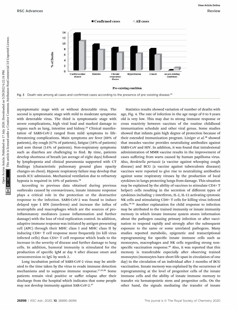
Fig. 3 Death rate among all cases and confirmed cases according to the presence of pre-existing disease.13
RSC Advances Review
Ope
n A
cces
s A
rtic
le. P
ublis
hed
on 1
7 Ju
ly 2
020.
Dow
nloa
ded
on 5
/29/
2022
6:2
2:16
PM
. T
his
artic
le is
lice
nsed
und
er a
Cre
ativ
e C
omm
ons
Attr
ibut
ion-
Non
Com
mer
cial
3.0
Unp
orte
d L
icen
ce.
View Article Online
asymptomatic stage with or without detectable virus. Thesecond is symptomatic stage with mild to moderate symptomswith detectable virus. The third is symptomatic stage withsevere complications, high viral load and marked damage toorgans such as lung, intestine and kidney.33 Clinical manifes-tation of SARS-CoV-2 ranged from mild symptoms to life-threatening complications. Main symptoms are fever (88% ofpatients), dry cough (67% of patients), fatigue (38% of patients)and sore throat (14% of patients). Non-respiratory symptomssuch as diarrhea are challenging to nd. By time, patientsdevelop shortness of breath (an average of eight days) followedby lymphopenia and clinical pneumonia supported with CTndings (characteristic pulmonary ground glass opacitychanges on chest). Hypoxic respiratory failure may develop thatneeds ICU admission. Mechanical ventilation due to refractoryhypoxia is required in 10% of patients.34
According to previous data obtained during previousoutbreaks caused by coronaviruses, innate immune responseplays a critical role in the protection or the destructiveresponse to the infection. SARS-CoV-2 was found to inducedelayed type I IFN (interferon) and increase the inux ofneutrophils and macrophages which are the sources of pro-inammatory mediators (cause inammation and furtherdamage) with the loss of viral replication control. In addition,adaptive immune response was initiated by antigen presentingcell (APC) through their MHC class I and MHC class II byinducing CD8+ T cell response more frequently (to kill virusinfected cells) than CD4+ T cell response which leads to theincrease in the severity of disease and further damage to lungcells. In addition, humoral immunity is stimulated for theproduction of specic IgM at day 9 aer disease onset andseroconversion to IgG by week 2.
Long incubation period of SARS-CoV-2 virus may be attrib-uted to the time taken by the virus to evade immune detectionmechanisms and to suppress immune response.17,35,36 Somepatients remain viral positive or suffer relapse aer theirdischarge from the hospital which indicates that some peoplemay not develop immunity against SAR-CoV-2.37
26898 | RSC Adv., 2020, 10, 26895–26916
Statistics results showed variation of number of deaths withage, Fig. 4. The rate of infection in the age range of 0 to 9 yearsold is very low. This may due to strong immune response orcross reactivity between vaccines of the routine childhoodimmunization schedule and other viral genus. Some studiesshowed that infants gain high degree of protection because oftheir extended immunization program. Liniger et al.38 showedthat measles vaccine provides neutralizing antibodies againstSARS-CoV and HIV. In addition, it was found that intralesionaladministration of MMR vaccine results in the improvement ofcases suffering from warts caused by human papilloma virus.Also, Bordetella pertussis (a vaccine against whooping coughdisease) and BCG (a vaccine against tuberculosis diseases)vaccines were reported to give rise to neutralizing antibodiesagainst some respiratory viruses by the production of localcytokines in lungs protecting lungs from damage. This situationmay be explained by the ability of vaccines to stimulate CD4+ Thelper1 cells resulting in the secretion of different types ofcytokines including g-interferon, IL-2, IL-12 activating cytotoxicNK cells and stimulating CD8+ T cells for killing virus infectedcells.39–41 Another explanation for child response to infectionmay be attributed to the trained immunity or innate immunitymemory in which innate immune system stores informationabout the pathogen causing primary infection or aer vacci-nation to respond rapidly and strongly aer the subsequentexposure to the same or some unrelated pathogens. Manystudies reported metabolic, epigenetic and transcriptionalreprogramming for specic innate immune cells such asmonocytes, macrophages and NK cells regarding strong non-specic vaccination response.42 Also, it was reported that thismemory is transferable especially aer observing trainedmonocytes (monocytes have short-life span in circulation of oneday) in the circulation of an individual aer 3 months of BCGvaccination. Innate memory was explained by the occurrence ofreprogramming at the level of progenitor cells of the innateimmune cells and the ability of innate immune memory totransfer via hematopoietic stem and progenitor cells. On theother hand, the signals mediating the transfer of innate
This journal is © The Royal Society of Chemistry 2020
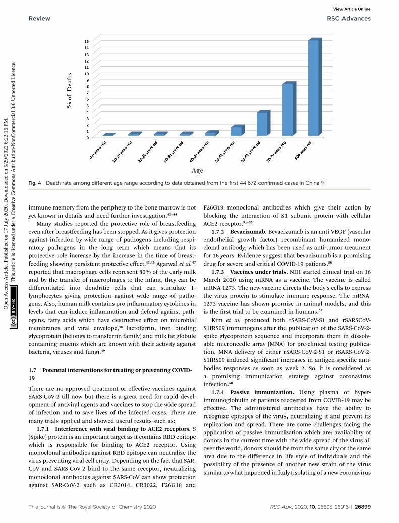
Fig. 4 Death rate among different age range according to data obtained from the first 44 672 confirmed cases in China.50
Review RSC Advances
Ope
n A
cces
s A
rtic
le. P
ublis
hed
on 1
7 Ju
ly 2
020.
Dow
nloa
ded
on 5
/29/
2022
6:2
2:16
PM
. T
his
artic
le is
lice
nsed
und
er a
Cre
ativ
e C
omm
ons
Attr
ibut
ion-
Non
Com
mer
cial
3.0
Unp
orte
d L
icen
ce.
View Article Online
immune memory from the periphery to the bone marrow is notyet known in details and need further investigation.42–44
Many studies reported the protective role of breastfeedingeven aer breastfeeding has been stopped. As it gives protectionagainst infection by wide range of pathogens including respi-ratory pathogens in the long term which means that itsprotective role increase by the increase in the time of breast-feeding showing persistent protective effect.45,46 Agarwal et al.47
reported that macrophage cells represent 80% of the early milkand by the transfer of macrophages to the infant, they can bedifferentiated into dendritic cells that can stimulate T-lymphocytes giving protection against wide range of patho-gens. Also, human milk contains pro-inammatory cytokines inlevels that can induce inammation and defend against path-ogens, fatty acids which have destructive effect on microbialmembranes and viral envelope,48 lactoferrin, iron bindingglycoprotein (belongs to transferrin family) andmilk fat globulecontaining mucins which are known with their activity againstbacteria, viruses and fungi.49
1.7 Potential interventions for treating or preventing COVID-19
There are no approved treatment or effective vaccines againstSARS-CoV-2 till now but there is a great need for rapid devel-opment of antiviral agents and vaccines to stop the wide spreadof infection and to save lives of the infected cases. There aremany trials applied and showed useful results such as:
1.7.1 Interference with viral binding to ACE2 receptors. S(Spike) protein is an important target as it contains RBD epitopewhich is responsible for binding to ACE2 receptor. Usingmonoclonal antibodies against RBD epitope can neutralize thevirus preventing viral cell entry. Depending on the fact that SAR-CoV and SARS-CoV-2 bind to the same receptor, neutralizingmonoclonal antibodies against SARS-CoV can show protectionagainst SAR-CoV-2 such as CR3014, CR3022, F26G18 and
This journal is © The Royal Society of Chemistry 2020
F26G19 monoclonal antibodies which give their action byblocking the interaction of S1 subunit protein with cellularACE2 receptor.51–55
1.7.2 Bevacizumab. Bevacizumab is an anti-VEGF (vascularendothelial growth factor) recombinant humanized mono-clonal antibody, which has been used as anti-tumor treatmentfor 16 years. Evidence suggest that bevacizumab is a promisingdrug for severe and critical COVID-19 patients.56
1.7.3 Vaccines under trials. NIH started clinical trial on 16March 2020 using mRNA as a vaccine. The vaccine is calledmRNA-1273. The new vaccine directs the body's cells to expressthe virus protein to stimulate immune response. The mRNA-1273 vaccine has shown promise in animal models, and thisis the rst trial to be examined in humans.57
Kim et al. produced both rSARS-CoV-S1 and rSARSCoV-S1fRS09 immunogens aer the publication of the SARS-CoV-2-spike glycoprotein sequence and incorporate them in dissolv-able microneedle array (MNA) for pre-clinical testing publica-tion. MNA delivery of either rSARS-CoV-2-S1 or rSARS-CoV-2-S1fRS09 induced signicant increases in antigen-specic anti-bodies responses as soon as week 2. So, it is considered asa promising immunization strategy against coronavirusinfection.58
1.7.4 Passive immunization. Using plasma or hyper-immunoglobulin of patients recovered from COVID-19 may beeffective. The administered antibodies have the ability torecognize epitopes of the virus, neutralizing it and prevent itsreplication and spread. There are some challenges facing theapplication of passive immunization which are: availability ofdonors in the current time with the wide spread of the virus allover the world, donors should be from the same city or the samearea due to the difference in life style of individuals and thepossibility of the presence of another new strain of the virussimilar to what happened in Italy (isolating of a new coronavirus
RSC Adv., 2020, 10, 26895–26916 | 26899

RSC Advances Review
Ope
n A
cces
s A
rtic
le. P
ublis
hed
on 1
7 Ju
ly 2
020.
Dow
nloa
ded
on 5
/29/
2022
6:2
2:16
PM
. T
his
artic
le is
lice
nsed
und
er a
Cre
ativ
e C
omm
ons
Attr
ibut
ion-
Non
Com
mer
cial
3.0
Unp
orte
d L
icen
ce.
View Article Online
from an Italian patient that differs than that isolated fromChina).59
2 Repofiling of known antiviral agentsin COVID-viruses2.1 Polymerase inhibitors
Polymerase inhibitors, in general, can be classied into twocategories: nucleotide/nucleoside analogs and non-nucleosideinhibitors (pyrophosphate derivatives). Nucleotide and nucleo-side analogue inhibitors are chemically synthesized analoguesof purines and pyrimidines in which the heterocyclic ring orsugar moiety has been altered and include 5-substituted 20-deoxyuridine analogs (idoxuridine, triuridine, and brivudine)Fig. 5 and Table 1. They include arabinosyl nucleoside analogs(vidarabine) and acyclic guanosine analogs (acyclovir, ganci-clovir, penciclovir, famciclovir, valaciclovir, and valganciclovir)(Fig. 5). Related to these drugs, cidofovir is a broad-spectrumnucleotide analog (acyclic nucleoside phosphonate), approvedfor treatment of HCMV retinitis in AIDS patients and used totreat many other DNA virus infections (e.g., HSV, adeno-, pox-,polyoma-, and papillomavirus infections) (Fig. 5).60,61
Nucleoside analogs are prodrugs that mimic natural nucle-osides and are used, in their triphosphate forms, as substratesfor the synthesis of viral nucleic acid. This means that nucleo-side analogs must not be only substrates for viral polymerases,they also need to be recognized and phosphorylated by host
Fig. 5 Structure of some nucleoside, nucleotide and pyrophosphate an
26900 | RSC Adv., 2020, 10, 26895–26916
nucleo(s/t)ide kinases.60,61 Nucleotide and nucleoside analogueinhibitors (NIs) currently used to treat both acute and chronicviral infections. NIs exhibit a relatively high barrier to resistanceemergence because the structural conservation of the poly-merase targets binding site is high among virus families, andresistance mutations generally incur a tness cost for theenzyme and the virus.62
For CoVs, amino acid conservation of the viral RdRp rangesfrom 70 to near 100% and is maintained across genera, sug-gesting NIs could potentially serve as broad-spectrum inhibitorsof CoV infection.63 However, proofreading activity of nsp14-ExoN activity protects CoVs from many NIs effective againstother RNA viruses.64,65 For effective inhibition of CoVs, an NIneeds to either evade recognition by ExoN or undergo uptakeinto the elongating strand at a rate exceeding ExoN excisionkinetics.
2.1.1 Efficacy of nucleotide and nucleoside analogueinhibitors (NIs) against CoVs
2.1.1.1 Favipiravir. Favipiravir, also known as T-705, pyr-azinecarboxamide derivative, a guanine nucleoside analogue(Fig. 6 and Table 1) was originally developed by ToyamaChemical of Japan.66 In March 2015, the US Food and DrugAdministration completed a phase III clinical trial studying thesafety and efficacy of favipiravir in the treatment of inuenza.67
Moreover, on March 15, 2020 the drug was approved in Chinafor the treatment of inuenza.68 Recently, favipiravir approvedfor use in clinical trials for treating coronavirus disease 2019
alogs.
This journal is © The Royal Society of Chemistry 2020

Table 1 Existing drugs with therapeutic potentials for COVID-19 (drug repurposing) targeting RdRp95
Antiviral agents Infectious diseases Reported mechanism of action Status
Favipiravir 2019-nCoV; inuenza Inhibits RdRp � Approved for inuenza in Japan� Randomized trial for 2019-nCoV(ChiCTR2000029544,ChiCTR2000029600)
Ribavirin 2019-nCoV, MERS-CoV, SARS-CoV,RSV, HCV
Inhibits viral RNA synthesis andmRNA capping
� Approved for HCV and RSV� Randomized trial for 2019-nCoVin combination a pegylatedinterferon (ChiCTR2000029387)� Randomized trial for SARS(NCT00578825)
Penciclovir 2019-nCoV Inhibits RdRp Approved for HSVRemdesivir (GS-5734) 2019-nCoV, MERS-CoV, SARS-CoV Terminates the non-obligate chain � Phase III for 2019-nCoV
(NCT04252664, NCT04257656)� Phase I for Ebola (NCT03719586)
Galidesivir (BCX4430) Broad-spectrum (e.g. SARS-CoV,MERS-CoV, IAV)
Inhibits viral RNA polymerasefunction by terminating non-obligate RNA chain
� Phase I for yellow fever(NCT03891420)� Phase I for Marburg virus(NCT03800173)
60-Fluorinated-aristeromycinanalogues
Broad-spectrum (e.g. CoV, ZIKV,CHIKV)
Inhibits the activity of RdRp andhost cell S-adenosyl-L-homocysteinehydrolase
Preclinical
Acyclovir eximer analogues HCoV-NL63, MERS-CoV Doubly exible nucleosideanalogues inhibit RdRp
Preclinical
Review RSC Advances
Ope
n A
cces
s A
rtic
le. P
ublis
hed
on 1
7 Ju
ly 2
020.
Dow
nloa
ded
on 5
/29/
2022
6:2
2:16
PM
. T
his
artic
le is
lice
nsed
und
er a
Cre
ativ
e C
omm
ons
Attr
ibut
ion-
Non
Com
mer
cial
3.0
Unp
orte
d L
icen
ce.
View Article Online
pneumonia.68 Its mechanism of actions is thought to be relatedto the selective inhibition of viral RNA-dependent RNA poly-merase.69 Other research study suggests that favipiravir induceslethal RNA transversion mutations, producing a nonviable viralphenotype.70 Favipiravir is a prodrug that is metabolized to itsribofuranosyl-50-triphosphate (favipiravir-RTP) form which actas purine mimetic binding to ATP- and GTP-binding sites onpolymerase in a competitive manner.71–73 Notably, favipiravir is
Fig. 6 Structure of some polymerase inhibitors used in treatment of CO
This journal is © The Royal Society of Chemistry 2020
selective to viral cells, it does not inhibit RNA or DNA synthesisin mammalian cells therefor it is not toxic to them.74 Favipiravircan effectively inhibit the RNA-dependent RNA polymerase ofRNA viruses such as inuenza, Ebola, yellow fever, chikungu-nya, norovirus and enterovirus.75
In February 2020, favipiravir has been approved for a clinicaltrial as a drug to treat COVID-19.76,77 On March 17, Chineseofficials suggested the drug had been effective in treating
VID-19.
RSC Adv., 2020, 10, 26895–26916 | 26901
RSC Advances Review
Ope
n A
cces
s A
rtic
le. P
ublis
hed
on 1
7 Ju
ly 2
020.
Dow
nloa
ded
on 5
/29/
2022
6:2
2:16
PM
. T
his
artic
le is
lice
nsed
und
er a
Cre
ativ
e C
omm
ons
Attr
ibut
ion-
Non
Com
mer
cial
3.0
Unp
orte
d L
icen
ce.
View Article Online
COVID-19 in Wuhan and Shenzhen. Recently, a study reportedits activity against 2019-nCoV (EC50 ¼ 61.88 mM in Vero E6cells).78 Patients with 2019-nCoV are being recruited inrandomized trials to evaluate the efficacy of favipiravir plusinterferon-a (ChiCTR2000029600) and favipiravir plus baloxavirmarboxil (an approved inuenza inhibitor targeting the cap-dependent endonuclease) (ChiCTR2000029544). Moreover, Astudy on 80 patients comparing it to lopinavir/ritonavir foundthat it signicantly reduced viral clearance time to 4 days,compared to 11 for the control group, and that 91.43% ofpatients had improved CT scans with few side effects.79,80 As of23 March 2020, it seems that Japan and China have issued anexport ban on the substance. Japan and China are the onlycountries in which favipiravir is produced and approved asa medical compound. Some Chinese pharmaceutical compa-nies assure that export rights are still granted via internationaldiplomatic means by the Chinese Ministry of Industry andInformation Technology.
2.1.1.2 Ribavirin. Ribavirin (Fig. 6) is a synthetic nucleosideguanosine analogue and has broad-spectrum antiviral activityagainst multiple RNA viruses. It is approved for treating respi-ratory syncytial virus, hepatitis C and E virus, Lassa virus, andhantavirus infections. Although the coronavirus is an RNAvirus, in theory favipiravir and ribavirin have some anti-coronavirus activity. However, coronaviruses express exonu-clease (nsp14-ExoN) in nonstructural protein 14, and arecommon throughout the coronavirus family. The currentresearch results show that nsp14-ExoN has an RNA proongfunction.81 So, it is speculated that the coronavirus is resistantto nucleoside analogs. The results of in vitro experiments haveshown that ribavirin has little antiviral effect on coronavirus.NIs effective against other RNA viruses.64 Synergistic activityagainst MERS-CoV of ribavirin combined with IFNa2b wasobserved in vitro and in rhesus macaques, suggesting that IFNincreases the potency of ribavirin at lower, more tolerableconcentrations.82,83 However, ve critically ill MERS-CoV-positive patients who were treated with a combination of riba-virin and IFNa2b showed no clinical improvement.84 Treatmentof 20MERS patients with a combination of ribavirin and IFNa2ashowed signicantly improved survival at 14 days but not at 28days,85 whereas treatment of MERS patients with a combinationof IFNa2a or IFNb1a and ribavirin yielded no survival benet inanother study.86 Thus, although ribavirin shows some efficacy invitro, it does not provide clinical benet to humans with SARS-CoV or MERS-CoV infections. Ribavirin approved for treatingHCV and respiratory syncytial virus (RSV) that has been evalu-ated in patients with SARS andMERS, but its side effects such asanaemia may be severe at high doses87 and whether it offerssufficient potency against 2019-nCoV is uncertain. However,a xed dose of the anti-HIV combination, lopinavir–ritonavir, iscurrently in clinical trials with umifenovir or ribavirin.88
2.1.1.3 Remdesivir (GS-5734). Remdesivir (Fig. 6), nucleo-side analogue, is a phosphoramidate prodrug of the adenosineNI GS-441524 which have multiple mechanisms of action,including lethal mutagenesis, obligate or non-obligate chaintermination, and perturbation of natural nucleotide triphos-phate pools via inhibition of nucleotide biosynthesis.89
26902 | RSC Adv., 2020, 10, 26895–26916
Remdesivir has demonstrated antiviral activity in vitro againstseveral viral families of emerging infectious diseases includingpneumoviridae, loviridae, pneumoviridae, paramyxoviridae,and coronaviridae.63,90,91 Remdesivir has broad-spectrum activ-ities against a diverse panel of RNA viruses such as Ebola virus(EBOV), Marburg, MERS-CoV, SARS-CoV, respiratory syncytialvirus (RSV), Nipah virus (NiV), and Hendra virus under clinicaldevelopment. The mechanism of remdesivir's anti-MERS-CoVactivity is likely through premature termination of viral RNAtranscription.92,93 A recent study reported that remdesivirinhibited 2019-nCoV (EC50 ¼ 0.77 mM in Vero E6 cells),78 anda US patient with 2019-nCoV recovered aer receiving intrave-nous remdesivir in January.94 The drug is currently beingstudied in phase III clinical trials in both China and USA. Twophase III trials were initiated in early February to evaluateintravenous remdesivir (200 mg on day 1 and 100 mg once dailyfor 9 days) in patients with 2019-nCoV (NCT04252664 andNCT04257656), with estimated completion dates in April 2020.95
Studies probing the interactions between remdesivir and theCoV replication machinery will likely yield crucial insights intohow this NI circumvents or overcomes CoV proofreadingactivity, which can in turn be applied to modeling the devel-opment of new NIs and enhancing potency of existing NIs.Currently, remdesivir clinical trials are enrolling and are sup-ported by the National Institutes of Health (NIH),96 USA andAIFA, Italy.97 The use of remdesivir in COVID-19 patients ina single-arm trial gave positive preliminary outcomes,98 and thisin contrast to the result published by another group.99 However,the debate over remdesivir efficacy is still open, according to thereported preliminary results in the ACTT NIH clinical trial,96 theFDA has given remdesivir an emergency use authorizationrestricted to patients affected by sever COVID-19.97
2.1.1.4 Sofosbuvir. Sofosbuvir (Fig. 6), a nucleotide analoguehepatitis C virus NS5B polymerase inhibitor, is used to treatchronic hepatitis C as a component of a combination antiviralregimen. In addition, the European Medicines Agency'sCommittee for Medicinal Products for Human Use has recom-mended the approval of sofosbuvir for the treatment of chronichepatitis C.100,101 The most important druggable targets ofsofosbuvir is the RNA-dependent RNA polymerase (RdRp),where it is incorporated into RNA, and due to modications atthe 20 position, inhibits further RNA chain extension and haltsRNA replication, and sofosbuvir can inhibit RdRp of the hepa-titis C virus, it acts as an RNA polymerase inhibitor bycompeting with natural ribonucleotides. As the hepatitis C virusand the coronavirus use a similar viral genome replicationmechanism, sofosbuvir may also inhibit coronaviruses,including 2019-nCoV.102
2.1.1.5 Beta-D-N4-hydroxycytidine (NHC). NHC (Fig. 6) isa cytidine analogue with demonstrated potent, broad-spectrumantiviral activity against many viruses such as Venezuelanequine encephalitis virus (VEEV), inuenza A virus (IAV),respiratory syncytial virus (RSV), chikungunya virus (CHIKV),inuenza B virus (IBV), and CoVs. NHC exerts its antiviral effectprimarily through mutagenesis of viral RNA serial passaging inthe presence of NHC led to low level resistance for VEEV but notRSV, IAV, and bovine viral diarrhea virus, thus indicating a high
This journal is © The Royal Society of Chemistry 2020

Review RSC Advances
Ope
n A
cces
s A
rtic
le. P
ublis
hed
on 1
7 Ju
ly 2
020.
Dow
nloa
ded
on 5
/29/
2022
6:2
2:16
PM
. T
his
artic
le is
lice
nsed
und
er a
Cre
ativ
e C
omm
ons
Attr
ibut
ion-
Non
Com
mer
cial
3.0
Unp
orte
d L
icen
ce.
View Article Online
resistance barrier.103–107 Potent anti-CoV activity of NHC wasdemonstrated for SARS-CoV and HCoV-NL63.106,108 Although themechanism of CoV inhibition has not been determined,micromolar-range EC50s suggests that NHC like remdesivir mayalso have a novel way of interacting with the CoV replication. Itspotent, broad-spectrum antiviral activity warrants furtherinvestigation for the treatment of CoV infections, either alone orin combination with other DAAs and immunomodulators.Moreover, the isopropyl ester prodrug of the ribonucleotideanalogue, N4-hydroxycytidine (Fig. 6) with improved bioavail-ability was found to inhibit replication of human and bat SARS-Cov-2. This occurs in the airway epithelial cells in bothmice andhuman. Clinical trials for the effectiveness of this drug are inprogress.109
2.1.1.6 Other drugs. Galidesivir (BCX4430), Fig. 6, is anadenosine analogue that was originally developed for HCV, andhas broad spectrum activity positive and negative sense RNAviruses. Galidesivir is currently in early-stage clinical studiesevaluating its safety in healthy subjects and its efficacy againstyellow fever, and has shown antiviral activities in preclinicalstudies against many RNA viruses, including SARS andMERS.87,110
Gemcitabine hydrochloride, Fig. 6, another NIs deoxy-cytidine analogue, is a chemotherapy drug that inhibits SARS-CoV and MERS-CoV, in addition to the uridine analogue 6-azauridine (Fig. 6) with activity against HCoV-NL63;106 andnally, mizoribine (Fig. 6), the immunosuppressant imidazolenucleoside, which inhibits SARS-CoV.87
2.2 Proteases inhibitors
2.2.1 SARS-Cov-2 main protease inhibitors. Nominated themolecule of the month in February 2020,111 the main protease(Mpro) of the new coronavirus SARS-CoV-2 is a heart shapeddimer that is responsible for different cuttings in the virallipoproteins112 into essential functional pieces forming viralspike, membrane, envelop, nucleoprotein, replicase, and poly-merase.113,114 These cutting products are essential for viralmaturation, survival and replication.114 Thus since the proteaseenzyme is crucial for viral proteolytic activities in early stage ofthe SARS-CoV life cycle, it is considered as a promising target forattacking the viral activity.115,116 This fact was supported by thesuccess of inhibiting viral proteases in the ght against previousviral infections such as human immunodeciency virus (HIV)117
and hepatitis C.118
The use of protease inhibitors for preventing coronavirusreplication started with repurposing anti-HIV protease inhibi-tors. While the main residue in HIV protease is an aspartateresidue,119 coronaviruses protease uses a cysteine residue fornucleophilic attack.120,121 The approach of using HIV proteaseinhibitors for older type of coronavirus SARS-CoV proteasestarted with rational screening for old medicines includingantivirals to block the proteolytic activity of SARS-CoV.120 Sincethe Mpro sequence of both types of coronavirus is conserved by96% and homology studies showed they also share a verysimilar 3D structure, the use of drugs that block Mpro of SARS-CoV as potential SARS-CoV-2 protease inhibitors is rational.112
This journal is © The Royal Society of Chemistry 2020
Actually, previously approved anti-HIV protease inhibitors;lopinavir and ritonavir (Fig. 7); were found to be promisingcandidates for tackling the older type of coronavirus, SARS-CoV,active site.122–124 Theoretically both lopinavir and ritonavir wasbound efficiently to the same pocket in active sites of Mpro ofboth SARS-CoV and SARS-CoV-2.115 Unfortunately, a random-ized clinical study performed on 199 hospitalized patients inChina and USA did not prove that use of the anti-HIV drugs ofclinical signicance over the standard care introduced for SARS-CoV-2 affected patients.124,125 A similar contradiction betweencell culture testing and in vivo screening results was reportedearlier also with using alisporivir; is a cyclophilin inhibitor;against emerged coronaviruses SARS and MERS. Alisporivirinhibited both viruses in EC50 of about 3.6 mM but it failed toshow any improvements of SARS infection in mouse model.126
Another anti-HIV protease inhibitor, nelnavir, was repurposedfor SARS infection. Nelnavir showed a strong inhibitoryactivity on SARS-CoV replication127 and though it gave strongreduction in viral pathogenicity and viral antigen expression forSARS-CoV but no reports tested on SARS-CoV-2 yet.
Based on the separated crystal structure of SARS-CoV-2,a German Laboratory112 was able to design a-ketoamide inhib-itor (compound 2, Fig. 7) that successively inhibited SARS-CoV-2viral replication in infected human Calu3 cells with an EC50 of4–5 mM. The design is based on a previously designed SARS-CoVprotease inhibitor introduced earlier this year (compound 1,Fig. 2). The ability of these a-ketoamides to form a bond on theCys145 residue in the active site of SARS-CoV series forminga thiohemiketal offered an advantage over HIV protease inhib-itors used lacking thiol reactivity.128 An additional advantage isthe ability of these compounds to form two hydrogen bonds atthe catalytic active sit of SARS-CoV-2 with Gly143, Cys129,145 butstill the lack of animal testing raising a big question on theavailability of use of such inhibitors for controlling COVID-19.
Additionally, a theoretical study using the crystal structureof the Mpro for SARS-CoV2, scientists tried to repurposecommercially available medicines to t into the enzymepocket. Surprisingly, antibiotics and chemotherapeutic agentsnamed colistin and valrubicin (Fig. 7) showed tight bindingwith 9 and 7 hydrogen bonds formed with essential amino acidresidues in Mpro active site including THR24, THR25, andTHR26.115 Other theoretical studies investigating millions ofsmall molecules as possible candidates for Mpro inhibitionsare coming out lipoproteins112,130 but the lack of biologicaltesting hinders the use of these molecules as potential Mpro
inhibitors and a need for more evidence to support to supporttheir use is still required.
2.2.2 Serine protease inhibitors. Interestingly, proteaseinhibitors have been employed in the ght against coronavi-ruses for purposes more that inhibiting the proteolytic activityrequired for virus life. Reports announced the possible use ofserine protease inhibitors for blocking virus penetration intothe host cell.131 The coronaviral entry into the host cell startswith the viral spike (S) protein. This S protein is composed oftwo subunits, S1 and S2. S1 is the subunit responsible forbinding to receptors on the host cell surface (mostly angio-tensin converting enzyme 2, ACE 2), while S2 is responsible for
RSC Adv., 2020, 10, 26895–26916 | 26903
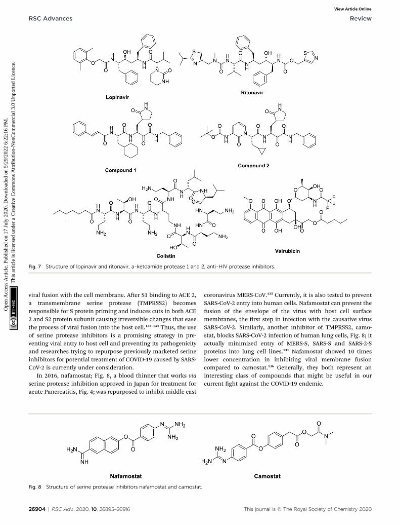
Fig. 7 Structure of lopinavir and ritonavir, a-ketoamide protease 1 and 2, anti-HIV protease inhibitors.
RSC Advances Review
Ope
n A
cces
s A
rtic
le. P
ublis
hed
on 1
7 Ju
ly 2
020.
Dow
nloa
ded
on 5
/29/
2022
6:2
2:16
PM
. T
his
artic
le is
lice
nsed
und
er a
Cre
ativ
e C
omm
ons
Attr
ibut
ion-
Non
Com
mer
cial
3.0
Unp
orte
d L
icen
ce.
View Article Online
viral fusion with the cell membrane. Aer S1 binding to ACE 2,a transmembrane serine protease (TMPRSS2) becomesresponsible for S protein priming and induces cuts in both ACE2 and S2 protein subunit causing irreversible changes that easethe process of viral fusion into the host cell.132–134 Thus, the useof serine protease inhibitors is a promising strategy in pre-venting viral entry to host cell and preventing its pathogenicityand researches trying to repurpose previously marketed serineinhibitors for potential treatment of COVID-19 caused by SARS-CoV-2 is currently under consideration.
In 2016, nafamostat; Fig. 8, a blood thinner that works viaserine protease inhibition approved in Japan for treatment foracute Pancreatitis, Fig. 4; was repurposed to inhibit middle east
Fig. 8 Structure of serine protease inhibitors nafamostat and camostat.
26904 | RSC Adv., 2020, 10, 26895–26916
coronavirus MERS-CoV.135 Currently, it is also tested to preventSARS-CoV-2 entry into human cells. Nafamostat can prevent thefusion of the envelope of the virus with host cell surfacemembranes, the rst step in infection with the causative virusSARS-CoV-2. Similarly, another inhibitor of TMPRSS2, camo-stat, blocks SARS-CoV-2 infection of human lung cells, Fig. 8; itactually minimized entry of MERS-S, SARS-S and SARS-2-Sproteins into lung cell lines.131 Nafamostat showed 10 timeslower concentration in inhibiting viral membrane fusioncompared to camostat.136 Generally, they both represent aninteresting class of compounds that might be useful in ourcurrent ght against the COVID-19 endemic.
This journal is © The Royal Society of Chemistry 2020

Review RSC Advances
Ope
n A
cces
s A
rtic
le. P
ublis
hed
on 1
7 Ju
ly 2
020.
Dow
nloa
ded
on 5
/29/
2022
6:2
2:16
PM
. T
his
artic
le is
lice
nsed
und
er a
Cre
ativ
e C
omm
ons
Attr
ibut
ion-
Non
Com
mer
cial
3.0
Unp
orte
d L
icen
ce.
View Article Online
2.3 Malaria drugs in COVID-19
Chloroquine, hydroxychloroquine and the structurally relatedatovaquone and meoquine, Fig. 9, are old known therapeuti-cally used antimalarial drugs. Hydroxychloroquine, thehydroxyl analogue of chloroquine being synthesized in the midof 20th century was introduced as a less toxic derivative than theparent chloroquine in animal studies. Moreover, these twodrugs are known to have immunomodulatory effects; hencethey have been used in rheumatoid arthritis or in lupuserythematous diseases.137
A collective review about the possible in vivo and in vitroantiviral activity of malaria drugs concluded that utilization ofantimalarial drugs against viral infection may be effectiveespecially in cases of viral resistance and emergences.138 An invitro study showed that The IC50 of chloroquine against SARS-CoV was 8.8 � 1.2 mM which is less than the cytostaticactivity; CC50 (261.3 � 14.5 mM) with selectivity index 30. TheIC50 of chloroquine for inhibition of SARS-CoV in vitro approx-imates the plasma concentrations of chloroquine reachedduring treatment of acute malaria. Addition of chloroquine toinfected cultures could be delayed for up to 5 h post infection,without a signicant drop-in antiviral activity. Authorsconcluded that chloroquine, an old antimalarial drug, may beconsidered for prompt use in the prevention and treatment ofSARS-CoV infections.139 Also, another in vitro study resulted inimportant results where chloroquine can inhibit the replicationand spread of CoV, and prevents CoV infection in newbornmice; this is considered a promising potential therapy for thisresistant virus.140 Recent reports about chloroquine andhydroxychloroquine recommend them as SARS-CoV potentialagents. In vitro research studies results showed that hydroxy-chloroquine is three times more potent than chloroquine in itseffect on SARS-CoV-2 infected cells (EC50 ¼ 0.72 and 5.47 mM).This proposal depends upon the immunomodulatory effect ofchloroquine and hydroxychloroquine which may be useful incontrolling the cytokines stress that occurs in patients withSARS-CoV-2.141 Vincent et al., (Vincent et al.)142 concluded thatchloroquine is effective in preventing the spread of SARS-CoV in
Fig. 9 Structure of malaria drugs effective in CoV.
This journal is © The Royal Society of Chemistry 2020
cell culture. This effect was found to occur in cells either treatedwith chloroquine prior or aer SARS-CoV infection. This studyindicated that chloroquine could elevate endosomal pH or caninterfere with terminal glycosylation of the cellular receptor,angiotensin converting enzyme-2. These actions can affect virusreceptor binding and hence repeal viral infection. A most recentstudy revealed that hydroxylchloroquine may contribute effi-ciently in inhibiting SARS-CoV-2 in vitro. It may attenuateinammatory response associated with COVID-19 and hence itcan compete with the infection if the toxicity prole has beenestablished by clinical studies.18
It is noteworthy worth that the N-cinnamoyl analogues ofchloroquine 3a, b, Fig. 10, are effective agents in vitro inrelieving pneumocystis pneumonia associated with somepulmonary viral, bacterial, mycobacterial or parasitic infec-tions. Compounds 3a and 3b will be further assessed in in vivoassays as potential potent anti-pneumocystis pneumonia.143
The mechanism of broad-spectrum antiviral of chloroquineand hydroxychloroquine is associated with suppressive effect onthe production of the inammatory mediators TNF-a and IL6.Authors in this study suggest hydroxychloroquine andanalogues to have useful clinical applications in treatment ofviral infections associated with inammation or immune-hyperactivation. Moreover, a series of chloroquine analogues4, Fig. 10 (ref. 144) can inhibit tumor susceptibility gene(TSG101) and also seems to inhibit viral replication by blockinglate-stage viral activity, possibly aer completion of viral proteinsynthesis. Inhibition of this gene could retard the movement ofthe virus to the cell surface and subsequently affect budding.145
Moreover, the antimalarial antiparasitic agent atovaquone(Fig. 9) acts via retardation of pyrimidine biosynthesis andconsequently inhibits viral replication. In vitro study usinghuman placental model showed that atovaquone can limit Zikavirus infection and suggesting that it can serve as broad spec-trum antiviral effect.146 Theoretically, computer-based designand screening proposed that chloroquine may act as SARS-CoV-2 entry blocker with S-score of 7.2639 kcal mol�1.147 Atovaquoneshowed a potential binding affinity with S-score of
RSC Adv., 2020, 10, 26895–26916 | 26905

Fig. 10 Structure of potential antiviral chloroquine analogues.
RSC Advances Review
Ope
n A
cces
s A
rtic
le. P
ublis
hed
on 1
7 Ju
ly 2
020.
Dow
nloa
ded
on 5
/29/
2022
6:2
2:16
PM
. T
his
artic
le is
lice
nsed
und
er a
Cre
ativ
e C
omm
ons
Attr
ibut
ion-
Non
Com
mer
cial
3.0
Unp
orte
d L
icen
ce.
View Article Online
�0.8.449 kcal mol�1 This study states that atovaquone has thesame scaffold features like chloroquine to elevate endosomalpH and interfere with ACE-2 glycosylation.147 In very recentresearch study, a model was used for 2019-nCoV research,results showed that the antimalarial meoquine HCl, and theantiparasitic selamectin are potential agents for treatingCOVID-19 infection.148
Due to the geographical overlaps between malaria and viral-related diseases, antimalarial drugs represent additional modesand mechanisms of action as antiviral agents. The lack of neweffective antiviral drugs has strengthened interest in thepotential antiviral activity of antimalarial drugs.149 It is obviousthat mechanism of action of chloroquine is not certain, it mayact by elevation of endosomal pH or interference with terminalglycosylation of the cellular receptor, angiotensin convertingenzyme-2.150 Chloroquine inhibits quinone reductase, a struc-tural neighbor of UDP-N-acetylglucosamine-2-epimerase that isinvolved in the biosynthesis of sialic acid. Viruses can use sialicacid moiety as receptor.151,152
Chloroquine and hydroxylchloroquine are consideredpromising in treatment of COVID-19. Their previous effects onMERS-COV, the analogue of COVID-19 in addition to the recentstudies about their effects153 recommend them as potentialurgent solution to treat this viral infection on some inamma-tory mediators involved in pneumonia associated with COVID-19 infections recommend them as potential urgent solution totreat this viral infection aer completing the clinical studies.Also, medicinal chemists are invited to study the moleculartarget mechanism of chloroquine and its analogues as prom-ising therapy for COVID-19 that enables development of newerbetter targeted derivatives with less side effects.
Many clinical trials and studies, in addition to in vitro and invivo, are currently ongoing to validate the efficacy of chloro-quine and hydroxychloroquine as effective treatment for SARS-COV-2. They are testing chloroquine if able to shorten SARS-CoV-2 disease course, mitigate inammatory responses toinfection, inhibit the exacerbation of pneumonia, improve lungimaging ndings, and promote a virus negative conver-sion.154–158 On the 13th March 2020, the FDA approved chloro-quine as a treatment for COVI-19 infection with specicprecautions. While early studies suggested benecial effects ofchloroquine and hydroxylchloroquine, recent studies could notconrm this, but instead highlighted potential meaningful
26906 | RSC Adv., 2020, 10, 26895–26916
adverse events of chloroquine and hydroxylchloroquine in thetreatment of COVID-19; this led to a temporary pause from May23 to June 3 of the chloroquine and hydroxylchloroquine armwithin the large, international Solidarity trial.159 Aer a reas-sessment of the evolving data the FDA revoked the emergencyuse authorization for chloroquine and hydroxylchloroquine onJune 15.160 Therefore, we must pay the attention chloroquineand hydroxylchloroquine need to be administered with cautionwhen treating COVID-19 infection to prevent the possiblecardiovascular problems.160
2.4 The lipid lowering statins in COVID-viruses
Rosuvastatin (RSV), Fig. 11, is a statin FDA-approved to be usedas lipid lowering agent, it acts through inhibition of HMG-CoAreductase enzyme consequently it lowers or cholesterol level.This molecule has the advantages of being inexpensive, safe andeasily obtained.161 A recent study showed that rosuvastatinimproves lung pathological changes by decreasing T helper cellsTh2 and Th17-mediated cytokines where this action is notrelated to its lipid-decreasing activity.162
Another study by Farag et al., in a structure-based drugdesign approach aiming at targeting COVID-19 virus revealedthat rosuvastatin on docking along with COVID-19 virus Mpro
substrate-binding pocket (PDB ID: 6LU7). Rosuvastatin showedoutstanding binding affinity regarding free energy with S scoreof �12.3096 kcal mol�1. Concerning binding mode, it experi-enced hydrophobic interactions and hydrogen bonding withGly143 and Glu166 amino acids.147 Although the study wastotally computer-based, and need more validation studiesbefore any clinical application, the study gives a starting pointto consider the re-evaluation of statin against COVID-19.
Indeed, some research studies indicate that statins therapyis associated with a reduction in cardiovascular problems and
Fig. 11 Structure of rosuvastatin.
This journal is © The Royal Society of Chemistry 2020

Review RSC Advances
Ope
n A
cces
s A
rtic
le. P
ublis
hed
on 1
7 Ju
ly 2
020.
Dow
nloa
ded
on 5
/29/
2022
6:2
2:16
PM
. T
his
artic
le is
lice
nsed
und
er a
Cre
ativ
e C
omm
ons
Attr
ibut
ion-
Non
Com
mer
cial
3.0
Unp
orte
d L
icen
ce.
View Article Online
mortality rates in patients of inuenza and/or pneumonia.147
Totura et al.163 proposed that toll-like receptor 3 (TLR3)signaling has a protective role in innate immune response incases of severe SARS-CoV infection. The FDA approved statinsare considered TLR-MYD88 agonists, they keep or retains TLR-MYD88 level during hypoxia.164 Synchronously, an early andhigh dose of a statin might be an idea for treatment of MERS-CoV infections. However, statins may not be very effective forlate-stage patients. Timely administration of statins may becrucial to surviving MERS-CoV infection.165 Importantly,a recent research study showed that angiotensin receptorblockers and statins upregulate ACE2, the tissue receptor forCOVID-19; these effects participated in protection against acuterespiratory disease syndrome and can decrease mortality rate.166
Statins are believed to reduce the risk of mortality in cases ofinuenza viral infection due to their anti-inammatory andimmunomodulatory effect.167 Moreover, rosuvastatin reducedcytokines TNF-a, IFN-g and Th-1 immune response during 72 h,it exerts rapid immunomodulatory effects.168 It is important tonote that in all studies, there was no harm associated with statintherapy. Therefore, it is conceivable that patients admitted166
with viral respiratory illnesses including COVID-19 could derivea benecial effect from the continuation of their statin therapy.Statins especially rosuvastatin are advised to be given to acuteCOVID-19 patients; they have a role in decreasing cardiovas-cular problems like the fatal myocardial infarction associatedwith COVID-19 infection.169
2.5 Rheumatoid arthritis drugs
Disease-Modifying Antirheumatic Drugs (DMARDs), Fig. 12 area diverse collection of drugs, grouped according their use andconvention, for treatment of rheumatoid arthritis (RA) andother related inammatory diseases. DMARDs are immuno-suppressive and immunomodulatory agents that have beenfound to improve symptoms, decrease joint damage, different
Fig. 12 Structure of methotrexate, leflunomide, teriflunomide, baricitini
This journal is © The Royal Society of Chemistry 2020
types of pains and inammations that affect, heart and bloodvessels.170
They are classied as either conventional DMARDs or bio-logic DMARDs. Commonly used conventional DMARDs thera-peutic agents have fast onset with respect to their anti-inammatory and analgesic effects but they neither removethe underlying cause of the disease nor protect against thefunctional disability.171 They include methotrexate (MTX),leunomide, hydroxychloroquine, and sulfasalazine. BiologicDMARDs were introduced in the early 1990s and are usuallyprescribed aer the failure of conventional DMARD therapywith advantages of more efficacy and fewer side effects.172 Bio-logic DMARDs are highly specic and target a specic pathwayof the immune system against inammation like inhibition oftumor necrosis factor, suppression of IL-1 and TNF-a, inductionof apoptosis of inammatory cells, by increasing chemotacticfactors, inhibition of purine synthesis, pyrimidine metabolismor purine metabolism. They include TNF-alpha inhibitors (cer-tolizumab, iniximab and etanercept), modied antibody(Abatacept), modied human interleukin 1 receptor antagonistprotein (Anakinra), Janus associated kinase (JAK) inhibitors(baricitinib and tofacitinib) and monoclonal antibodies asinterleukin-6 receptor blockers (e.g. tocilizumab, sarilumab,and rituximab).173
A number of classic DMARDs have demonstrated not onlyantitumor but unexpected antiviral activities such as metho-trexate (MTX, Fig. 12) against mosquito-borne aviviruses, suchas dengue virus (DENV) and Zika virus (ZIKV) through dihy-drofolate reductase (DHFR) inhibition mechanism leading todecreased viral replication.174 Other examples include leuno-mide, Fig. 12, an isoxazole derivative, is nucleotide biosynthesisinhibitor, that is rapidly metabolized to its active form, andteriunomide, which is a potent inhibitor of mitochondrialdihydroorotate dehydrogenase (DHODH), a key enzyme in thede novo biosynthesis of pyrimidine nucleoside triphosphates.175
Both leunomide has demonstrated antiviral activity toward
b and ruxolitinib as examples of DMARDs.
RSC Adv., 2020, 10, 26895–26916 | 26907

Fig. 13 Structure of ivermectin B1b.
RSC Advances Review
Ope
n A
cces
s A
rtic
le. P
ublis
hed
on 1
7 Ju
ly 2
020.
Dow
nloa
ded
on 5
/29/
2022
6:2
2:16
PM
. T
his
artic
le is
lice
nsed
und
er a
Cre
ativ
e C
omm
ons
Attr
ibut
ion-
Non
Com
mer
cial
3.0
Unp
orte
d L
icen
ce.
View Article Online
diverse DNA and RNA viruses, such as herpes simplex virus(HSV),176 human cytomegalovirus (HCMV),177 polyoma BK virus(BKV),178,179 human immunodeciency virus (HIV),180 respiratorysyncytial virus (RSV),181 and arenaviruses.182 Currently, they havenot been tested against SARS-COV-2.
Currently, there are no specic antivirals or vaccines to treatSARS-CoV-2 infection. High-throughput screening (HTS) ofvarious compound libraries against SARS-CoV-2 have beenstarted and several biologic DMARDs were identied as possibletargets in controlling SARS-COV-2 complications such as toci-lizumab, sarilumab and baricitinib, Fig. 12.
Interleukin 6 (IL-6), a glycoprotein composed of 212 aminoacids in humans, has pleiotropic effects on many cells,including B cells, T cells, hematopoietic stem cells, hepatocytes,megakaryocytes, osteoclasts, synoviocytes, keratinocytes, andpossibly chondrocytes, and thus has a wide range of biologicalactivity, including regulation of immune response, support ofhematopoiesis, generation of acute phase reactions, andinduction of inammation and oncogenesis. Therapiesinvolving blockade of IL-6 functions have constituted a newtherapeutic strategy for some inammatory and autoimmunediseases.183
Unlike conventional DMARDs, interleukin-6 antagonists (e.g.sarilumab and tocilizumab) have no direct or indirect antiviralactivities; they block IL-6 a key cytokine that plays an importantrole in immune responses as proinammatory cytokine thatinduces strong inammatory responses. So, they may havea role in the severe deterioration of lung function and COVID-19pneumonia.
Tocilizumab (TCZ), has been developed by Osaka Universityfor the treatment of inammatory and autoimmunedisorders.184,185
TCZ was the rst humanized IgG1 monoclonal antibodiesacting as interleukin-6 (IL-6) receptor antagonist approved inJapan in 2008, in Europe in 2009, and FDA-approved in 2010 for
26908 | RSC Adv., 2020, 10, 26895–26916
the treatment of adult patients with moderately to severelyactive rheumatoid arthritis (RA) that didn't provide enoughprogress with other agents.186,187
TCZ can be administered intravenously or subcutaneously(SC) with similar efficacy, but convenience of SC formulationpermits once-weekly self-administration.187 Extensive clinicalexperience has rmly established the short- and long-termefficacy and safety of tocilizumab (monotherapy or in combi-nation with conventional DMARDs) in adults with early-stageand longer-duration established RA. In the clinical trial andreal-world settings, tocilizumab monotherapy or combinationtherapy provided rapid and sustained improvements in clinicaland radiographic outcomes and health-related quality of life.The safety prole of tocilizumab is consistent over time and, ingeneral, is consistent with that of other immunomodulatoryagents. It exhibits low immunogenicity.188
Tocilizumab has anticancer potency against non-small celllung cancer cells via apoptosis induction as an agonistic IL-6Rregulator and it is currently under evaluation in a multicenterclinical trial for large-cell lung carcinoma.189,190 In addition,tocilizumab completed phase II clinical trials for PulmonaryArterial Hypertension (PAH) treatment.191
At present, this excellent safety and efficacy of tocilizumabencourages China's National Health Commission to include itsuse 8mg kg�1/12 h (maximum of 800mg per dose) in guidelinesto treat coronavirus (COVID-19) patients either alone or incombination with favipiravir, a broad spectrum anti-viral agent.They report that the drug has been involved with a Chineseclinical trial involving 20 severe COVID-19 cases. 19 of themwere discharged from the hospital within two weeks. Moreover,a 150-patient trial assessing tocilizumab is led by Hong Zhao ofPeking University First Hospital and a 188-patient trial assess-ing tocilizumab alone is led by Dongsheng Wang of The FirstAffiliated Hospital of University of Science and Technology ofChina (Anhui Provincial Hospital).192
This journal is © The Royal Society of Chemistry 2020

Review RSC Advances
Ope
n A
cces
s A
rtic
le. P
ublis
hed
on 1
7 Ju
ly 2
020.
Dow
nloa
ded
on 5
/29/
2022
6:2
2:16
PM
. T
his
artic
le is
lice
nsed
und
er a
Cre
ativ
e C
omm
ons
Attr
ibut
ion-
Non
Com
mer
cial
3.0
Unp
orte
d L
icen
ce.
View Article Online
On 11 March 2020, in Italy, tocilizumab was the subject of anup-to-30-patient phase II trial designed to study the drug asa single 8 mg kg�1 dose in patients affected by severe pneu-monia correlated to SARS-CoV2. The study by researchers atUniversita Politecnica delle Marche and Azienda OspedalieraOspedali Riuniti Marche Nord has two primary outcomemeasures: TCZ arrest in deterioration of pulmonary function,and improvement in pulmonary function. These signs ofimprovement prompting the Italian Pharmacological Agency(AIFA) to expand testing in 5 other hospitals.193
TCZ prevents overreaction of the immune system that hasled to organ failure especially the lung and death in coronaviruspatients. Roche and theWHO are each launching separate trialsfor its use in severe COVID-19 cases. Finally, Genentech (Roche)announced that the FDA has approved the initiation ofa double-blind, randomized phase III clinical trial of theoncology supportive care drug tocilizumab (Actemra) for use incombination with standard care for the treatment of hospital-ized adult patients with severe COVID-19 pneumonia.194
2.5.1 Sarilumab. Sarilumab, is one of two IL-6 receptorantagonists being studied as potential COVID-19 treatments.The logic behind using sarilumab is similar to that of TCZ. It isapproved by the FDA in 2017 to treat adults with moderately toseverely active rheumatoid arthritis.195 However, On March 16th,2020, A research group announced that they launched a phaseII/III clinical program of up to 400 patients assessing the drug insevere COVID-19 coronavirus infection in collaboration with theFDA and the Biomedical Advanced Research and DevelopmentAuthority (BARDA). The phase III portion will evaluate longer-term outcomes such as reducing the need for hospitalizationand mechanical ventilation, as well as mortality.196
2.5.2 Baricitinib. Baricitinib (Fig. 12) was FDA-approved inJune 2018 for the treatment of moderately to severely activerheumatoid arthritis (RA). Baricitinib joins tofacitinib as thesecond oral once-daily medication in the Janus Kinase (JAK)inhibitor class for RA. Baricitinib exerts its effects by selectiveinhibiting JAK1 and JAK2 enzymes, targeting cytokine andgrowth factor receptor stimulation, thus reducing downstreamimmune cell function.197
On February 15th, 2020, a month aer the rst reports ofa novel coronavirus spreading in China. The articial intelli-gence (AI) research groups and others have used AI soware tond an already-approved drug that might limit the virus's abilityto infect people.198 They used the rst genomic sequence of thevirus published in mid-January, and by January 29th, 2020scientists suggested that the virus might enter human cells bybinding to a cell-surface molecule called ACE2.199
The soware pointed at the enzyme adaptor-associatedprotein kinase 1 (AAK1) as a possible target for the disease.AAK1 regulates endocytosis, the process that brings materialinto cells, which also is a common mode of viral infection. AIresults select baricitinib, based on its affinity for the kinase andits toxicity amongst more than 378 known AAK1 inhibitors.Janus-associated kinase (JAK) inhibitor baricitinib is predictedto reduce the ability of the virus to infect lung cells by inhibitionof ACE2-mediated endocytosis.198
This journal is © The Royal Society of Chemistry 2020
Later, the researchers propose testing baricitinib in clinicaltrials against the virus as a dual antiviral and anti-inammatoryagent aer conrming in vitro activities. Currently, ruxolitinib,Fig. 1, a drug that works by a similar mechanism, showsa promise in sever COVID-19-associated cytokine storm, wherelevels of cytokines were found to be signicantly reduced. Also,It was found to be well-tolerated with low toxicities. It is sub-jected for phase III clinical trials for COVID-19.200
2.6 Miscellaneous drugs
Some other drug molecules or agents have a potential positiveimpact in management of COVID-infections. Research reportssuggest that the use of antioxidants like ascorbic acid, N-ace-tylcysteine in combination with antiviral drugs can synergisti-cally decrease the lethal effect of inuenza viral infection.201
Another research study showed that combination of the inu-enza drug oseltamivir with N-acetylcysteine can increase hostdefense mechanism and decrease mortality rates. This occursthrough reduction of oxidative stress associated with viralinfection.202 Dietary supplements such as resveratrol, curcuminand sulphorafane were found to have cytoprotective effect onvital organs. This approach could be extrapolated to the lungs ofinfected patients with coronaviruses infections. A recentresearch study revealed that combination thalidomide and theselective COX-2 inhibitor, celecoxib, can improve the severeCOVID-19 pneumonia through modulation of activated NF-KBthat promote severe lung injury.203 The FDA-approved antipar-asitic drug ivermectin (Fig. 13) is known in the literature by itsbroad-spectrum antiviral activity. Surprisingly, recent reportsshowed that this drug is a potent inhibitor of SARS-Cov-2.Ivermectin causes 5000� reduction in viral RNA during twodays, it requires future clinical investigation in humans.204
3 Conclusion
The war between scientists and viral infection is a continuouswar. Finding specic potent drug with high efficacy andacceptable toxicity prole is a dream. It is the rst time in theworld to join scientists, politicians, diplomats, businessmenand the normal people all over the world to have the sametarget. Although the FDA approved chloroquine and hydroxy-chloroquine to be used in the emergency clinical managementprotocols used in CoV, this approval has some restrictions. Theinformation about the wide use of these drugs is not enough.Some of the polymerase nucleoside/nucleotide inhibitors areconsidered promising agents. Favipiravir has selectivity on viralcells as it is viral RNA-dependent RNA polymerase inhibitorwithout effects on human cell lines; it has no effect on RNA orDNA in human cells. It is approved to be tested in clinical trialsin cases of COVID-19 infections. Moreover, the combinationbetween lopinavir and ritonavir is under investigation for clin-ical studies. The broad-spectrum antiviral effect of remdesiviragainst different RNA viruses in addition to its interferenceeffect on CoV replication machinery recommend this drug forintensive studies to be COVID-19 drug.
RSC Adv., 2020, 10, 26895–26916 | 26909

RSC Advances Review
Ope
n A
cces
s A
rtic
le. P
ublis
hed
on 1
7 Ju
ly 2
020.
Dow
nloa
ded
on 5
/29/
2022
6:2
2:16
PM
. T
his
artic
le is
lice
nsed
und
er a
Cre
ativ
e C
omm
ons
Attr
ibut
ion-
Non
Com
mer
cial
3.0
Unp
orte
d L
icen
ce.
View Article Online
On the other hand, the ability of protease inhibitors to ghtCOVID-19 is under investigation. Inhibition of this enzyme is crucialfor viral proteolytic and other essential vital activities. Lopinavir,ritonavir and nelnavir are effective agents in COVID-19 infectionbut still need additional studies. Similarly, the serine proteaseinhibitors such as nafamostat and camostat are promising strategyin COVID-19 therapy. The important relatively save and potent lipidlowering statin, rosovastatin, has a good impact on lung patholog-ical changes through decreasing Th2 and Th17mediated cytokines.Statins are recommended to be useful in reducing the mortalityrates in cases of inuenza infections due to their anti-inammatoryand immunomodulatory effects. Some rheumatoid arthritis drugsmay be benecial in management of COVID-19 cases such as TNF-alpha inhibitors, interleukin 1 receptor antagonists; Janus associ-ated kinases (JAK) in addition to monoclonal antibodies whichblock interleukin 6 receptors. Tocilizumab is under clinical inves-tigation to treat hospitalized severe COVID-19 associated pneu-monia. Also, baricitinib and ruxolitinib are suggested to induceACE-2 mediated endocytosis that reduces ability of the virus toinfect viral cells with their additional anti-inammatory actions.
In the near future, scientists are invited to collect theirefforts and share knowledge in redirecting the mentioned drugsin this review to have suitable, cheap drugs with the lowestpossible toxicity prole.
Abbreviations
COVID
26910 | RSC
Coronavirus disease
SARS-CoV-2Severe acute respiratory syndrome coronavirus 2
MERS
Middle East respiratory syndrome PHEIC Public health emergency of international concern WHO World Health Organization HE Hemagglutination-esterase RBD Receptor binding domain ACE2 Angiotensin converting enzyme 2 TMPRSS2 Transmembrane serine protease 2 ORF1a Open reading frame 1a ORF1b Open reading frame 1b pp Polyprotein PLpro Papain-like protease 3CLpro Chymotrypsin-like protease RTC Replicase–transcriptase complex RdRp RNA-dependent RNA polymerase IFN Interferon APC Antigen presenting cell MHC Major histocompatibility complex CD8 Cluster of differentiation T cell ¼TcCytotoxic lymphocyte
IgM
Immunoglobulin M IgG Immunoglobulin G TMPRSS2 Transmembrane protease, serine 2 ICU Intensive care unit NK cells Natural killer cell RBD Receptor-binding domain VEGF Vascular endothelial growth factorAdv., 2020, 10, 26895–26916
HCMV
Human cytomegalovirus HSV Herpes simplex virus BK Polyomavirus NIs Nucleotide and nucleoside analogue inhibitors FDA Food and Drug Administration TRP Ribofuranosyl-50-triphosphate ATP Adenosine triphosphate GTP Guanosine-50-triphosphate CT Computerized tomography IFNa2b Interferon alpha-2b nsp14 Nonstructural protein 14 HCV Hepatitis C virus RSV Respiratory syncytial virus NiV Nipah virus EBOV Ebola virus VEEV Venezuelan equine encephalitis virus IAV Inuenza A virus CHIKV Chikungunya virus IBV Inuenza B virus HCoV-NL63EC50: the concentration of a drug that gives half-maximal response
DAAs
Direct acting antivirals ZIKV Virus family Flaviviridae TSG101 Tumor susceptibility gene RSV Rosuvastatin HMG-CoA 3-Hydroxy-3-methyl-glutaryl-coenzyme A UDP Uridine diphosphate TLR3 Toll-like receptor 3 MYD88 Myeloid differentiation primary response 88 Th1 T helper cell DMARD Disease-modifying antirheumatic drugs IL-1 Interleukin-1 family JAK Janus associated kinase DENV Dengue virus HCMV Human cytomegalovirus BKV Polyoma BK virus HTS High-throughput screening SC Subcutaneously RA Rheumatoid arthritis PAHBARDAPulmonary arterial hypertension; biomedicaladvanced research and development authority
AI
Articial intelligence AAK1 Adaptor-associated protein kinase 1 COX-2 Cyclooxygenase 2 NF-KB Nuclear factor kappa-light-chain-enhancer ofactivated B cells
GIT Gastrointestinal tractConflicts of interest
Authors declare no conict of interest.
References
1 C. C.-U. F. R. E. U. A. f. C. H. and U. S. F. D. A., https://www.fda.gov/news-events/pressannouncements/coronavirus-covid-19-update-fda-revokes-emergency-
This journal is © The Royal Society of Chemistry 2020
Review RSC Advances
Ope
n A
cces
s A
rtic
le. P
ublis
hed
on 1
7 Ju
ly 2
020.
Dow
nloa
ded
on 5
/29/
2022
6:2
2:16
PM
. T
his
artic
le is
lice
nsed
und
er a
Cre
ativ
e C
omm
ons
Attr
ibut
ion-
Non
Com
mer
cial
3.0
Unp
orte
d L
icen
ce.
View Article Online
useauthorization-chloroquine-and., published 2020,accessed June 15, 2020, 2020.
2 N. I. Nii-Trebi, BioMed Res. Int., 2017, 2017, 5245021.3 M. J. Walker, T. C. Barnett, J. D. McArthur, J. N. Cole,C. M. Gillen, A. Henningham, K. Sriprakash,M. L. Sanderson-Smith and V. Nizet, Clin. Microbiol. Rev.,2014, 27, 264–301.
4 V. R. Racaniello, J. Clin. Invest., 2004, 113, 796–798.5 C. Dye, Philos. Trans. R. Soc., B, 2014, 369, 20130426.6 F. Wu, S. Zhao, B. Yu, Y.-M. Chen, W. Wang, Z.-G. Song,Y. Hu, Z.-W. Tao, J.-H. Tian, Y.-Y. Pei, M.-L. Yuan,Y.-L. Zhang, F.-H. Dai, Y. Liu, Q.-M. Wang, J.-J. Zheng,L. Xu, E. C. Holmes and Y.-Z. Zhang, Nature, 2020, 579,265–269.
7 World Health Organization, International health regulations(2005), World Health Organization, 2008.
8 World Health Organization, Naming the coronavirus disease(COVID-19) and the virus that causes it, 2020.
9 World Health Organization, World Health Organization bestpractices for the naming of new human infectious diseases,World Health Organization, 2015.
10 World Health Organization, https://www.who.int/emergencies/diseases/novel-coronavirus-2019/situation-reports, 2020.
11 World Health Organization, Coronavirus disease (COVID-19)Situation Report – 153, June, 2020, p. 21.
12 S. L. Klein and K. L. Flanagan, Nat. Rev. Immunol., 2016, 16,626–638.
13 Worldometer, Coronavirus Update (Live). 2020.14 N. Zhu, D. Zhang, W. Wang, X. Li, B. Yang, J. Song, X. Zhao,
B. Huang, W. Shi, R. Lu, P. Niu, F. Zhan, X. Ma, D. Wang,W. Xu, G. Wu, G. F. Gao and W. Tan, N. Engl. J. Med.,2020, 382, 727–733.
15 T. Pillaiyar, S. Meenakshisundaram and M. Manickam,Drug Discovery Today, 2020, 25, 668–688.
16 Y. Yin and R. G. Wunderink, Respirology, 2018, 23, 130–137.17 P. Zhou, X. L. Yang, X. G. Wang, B. Hu, L. Zhang, W. Zhang,
H. R. Si, Y. Zhu, B. Li, C. L. Huang, H. D. Chen, J. Chen,Y. Luo, H. Guo, R. D. Jiang, M. Q. Liu, Y. Chen,X. R. Shen, X. Wang, X. S. Zheng, K. Zhao, Q. J. Chen,F. Deng, L. L. Liu, B. Yan, F. X. Zhan, Y. Y. Wang,G. F. Xiao and Z. L. Shi, Nature, 2020, 579, 270–273.
18 Z. Liu, X. Xiao, X. Wei, J. Li, J. Yang, H. Tan, J. Zhu, Q. Zhangand J. Wu, J. Med. Virol., 2020, 92, 595–601.
19 D. Wrapp, N. Wang, K. S. Corbett, J. A. Goldsmith,C.-L. Hsieh, O. Abiona, B. S. Graham and J. S. McLellan,Science, 2020, 367, 1260–1263.
20 I. Ghinai, T. D. McPherson, J. C. Hunter, H. L. Kirking,D. Christiansen, K. Joshi, R. Rubin, S. Morales-Estrada,S. R. Black, M. Pacilli, M. J. Fricchione, R. K. Chugh,K. A. Walblay, N. S. Ahmed, W. C. Stoecker, N. F. Hasan,D. P. Burdsall, H. E. Reese, M. Wallace, C. Wang,D. Moeller, J. Korpics, S. A. Novosad, I. Benowitz,M. W. Jacobs, V. S. Dasari, M. T. Patel, J. Kauerauf,E. M. Charles, N. O. Ezike, V. Chu, C. M. Midgley,M. A. Rolfes, S. I. Gerber, X. Lu, S. Lindstrom, J. R. Veraniand J. E. Layden, Lancet, 2020, 395, 1137–1144.
This journal is © The Royal Society of Chemistry 2020
21 C. Rothe, M. Schunk, P. Sothmann, G. Bretzel, G. Froeschl,C. Wallrauch, T. Zimmer, V. Thiel, C. Janke, W. Guggemos,M. Seilmaier, C. Drosten, P. Vollmar, K. Zwirglmaier,S. Zange, R. Wolfel and M. Hoelscher, N. Engl. J. Med.,2020, 382, 970–971.
22 L. Zou, F. Ruan, M. Huang, L. Liang, H. Huang, Z. Hong,J. Yu, M. Kang, Y. Song, J. Xia, Q. Guo, T. Song, J. He,H. L. Yen, M. Peiris and J. Wu, N. Engl. J. Med., 2020, 382,1177–1179.
23 B. J. Bosch, R. van der Zee, C. A. de Haan and P. J. Rottier, J.Virol., 2003, 77, 8801–8811.
24 J. Armstrong, H. Niemann, S. Smeekens, P. Rottier andG. Warren, Nature, 1984, 308, 751–752.
25 B. Nal, C. Chan, F. Kien, L. Siu, J. Tse, K. Chu, J. Kam,I. Staropoli, B. Crescenzo-Chaigne, N. Escriou, S. van derWerf, K. Y. Yuen and R. Altmeyer, J. Gen. Virol., 2005, 86,1423–1434.
26 P. Venkatagopalan, S. M. Daskalova, L. A. Lopez,K. A. Dolezal and B. G. Hogue, Virology, 2015, 478, 75–85.
27 A. Klausegger, B. Strobl, G. Regl, A. Kaser, W. Luytjes andR. Vlasak, J. Virol., 1999, 73, 3737–3743.
28 G. Simmons, P. Zmora, S. Gierer, A. Heurich andS. Pohlmann, Antiviral Res., 2013, 100, 605–614.
29 A. Heurich, H. Hofmann-Winkler, S. Gierer, T. Liepold,O. Jahn and S. Pohlmann, J. Virol., 2014, 88, 1293–1307.
30 A. R. Fehr and S. Perlman,Methods Mol. Biol., 2015, 1282, 1–23.
31 F. Zhou, T. Yu, R. Du, G. Fan, Y. Liu, Z. Liu, J. Xiang,Y. Wang, B. Song, X. Gu, L. Guan, Y. Wei, H. Li, X. Wu,J. Xu, S. Tu, Y. Zhang, H. Chen and B. Cao, Lancet, 2020,395, 1054–1062.
32 Y. Chen, K. Shan and W. Qian, Asians and Other RacesExpress Similar Levels of and Share the Same GeneticPolymorphisms of the SARS-CoV-2 Cell-Entry Receptor,Preprints, 2020, 2020020258, DOI: 10.20944/preprints202002.0258.v1.
33 D. Wang, B. Hu, C. Hu, F. Zhu, X. Liu, J. Zhang, B. Wang,H. Xiang, Z. Cheng, Y. Xiong, Y. Zhao, Y. Li, X. Wang andZ. Peng, JAMA, J. Am. Med. Assoc., 2020, 323, 1061–1069.
34 F. A. Rabi and M. S. Al Zoubi, Pathogens, 2020, 9, 231.35 J. Lessler, N. G. Reich, R. Brookmeyer, T. M. Perl,
K. E. Nelson and D. A. Cummings, Lancet Infect. Dis.,2009, 9, 291–300.
36 E. Prompetchara, C. Ketloy and T. Palaga, Asian Pac. J.Allergy Immunol., 2020, 38, 1–9.
37 Y. Shi, Y. Wang, C. Shao, J. Huang, J. Gan, X. Huang,E. Bucci, M. Piacentini, G. Ippolito and G. Melino, CellDeath Differ., 2020, 27, 1451–1454.
38 M. Liniger, A. Zuniga, A. Tamin, T. N. Azzouz-Morin,M. Knuchel, R. R. Marty, M. Wiegand, S. Weibel,D. Kelvin and P. A. Rota, Vaccine, 2008, 26, 2164–2174.
39 S. Salman and M. L. Salem, Med. Hypotheses, 2020, 140,109689.
40 P. Baskar, G. Collins, B. Dorsey-Cooper, R. Pyle, J. Nagel,D. Dwyer, G. Dunston, C. Johnson, N. Kendig andE. Israel, Clin. Exp. Immunol., 1998, 111, 251.
RSC Adv., 2020, 10, 26895–26916 | 26911

RSC Advances Review
Ope
n A
cces
s A
rtic
le. P
ublis
hed
on 1
7 Ju
ly 2
020.
Dow
nloa
ded
on 5
/29/
2022
6:2
2:16
PM
. T
his
artic
le is
lice
nsed
und
er a
Cre
ativ
e C
omm
ons
Attr
ibut
ion-
Non
Com
mer
cial
3.0
Unp
orte
d L
icen
ce.
View Article Online
41 S. Salman, M. S. Ahmed, A. M. Ibrahim, O. M. Mattar, H. El-Shirbiny, S. Sarsik, A. M. A, R. M. Anis, N. A. Y. Agha andA. I. Abushouk, J. Am. Acad. Dermatol., 2019, 80, 922–930.
42 B. Gourbal, S. Pinaud, G. J. M. Beckers, J. W. M. Van DerMeer, U. Conrath and M. G. Netea, Immunol. Rev., 2018,283, 21–40.
43 S. Yona, K.-W. Kim, Y. Wolf, A. Mildner, D. Varol, M. Breker,D. Strauss-Ayali, S. Viukov, M. Guilliams and A. Misharin,Immunity, 2013, 38, 79–91.
44 J. Kleinnijenhuis, J. Quintin, F. Preijers, L. A. Joosten,D. C. Ifrim, S. Saeed, C. Jacobs, J. van Loenhout, D. deJong and H. G. Stunnenberg, Proc. Natl. Acad. Sci. U. S. A.,2012, 109, 17537–17542.
45 D.-L. Dixon, Children, 2015, 2, 289–304.46 E. Pandol, F. Gesualdo, C. Rizzo, E. Carloni, A. Villani,
C. Concato, G. Linardos, L. Russo, B. Ferretti,I. Campagna and A. Tozzi, Frontiers in Pediatrics, 2019, 7,152.
47 S. Agarwal, W. Karmaus, S. Davis and V. Gangur, J. Hum.Lactation, 2011, 27, 171–186.
48 K. M. Hunt, J. E. Williams, B. Shai, M. K. Hunt, R. Behre,R. Ting, M. K. McGuire andM. A. McGuire, Breastfeed. Med.,2013, 8, 105–110.
49 O. Ballard and A. L. Morrow, Pediatr. Clin. North Am., 2013,60, 49–74.
50 V. Surveillances, China CDC Weekly, 2020, 2, 113–122.51 J. D. Berry, K. Hay, J. M. Rini, M. Yu, L. Wang,
F. A. Plummer, C. R. Corbett and A. Andonov, mAbs, 2010,2, 53–66.
52 E. N. van den Brink, J. ter Meulen, F. Cox,M. A. Jongeneelen, A. Thijsse, M. Throsby,W. E. Marissen, P. M. Rood, A. B. Bakker andH. R. Gelderblom, J. Virol., 2005, 79, 1635–1644.
53 J. ter Meulen, E. N. van den Brink, L. L. Poon,W. E. Marissen, C. S. Leung, F. Cox, C. Y. Cheung,A. Q. Bakker, J. A. Bogaards, E. van Deventer, W. Preiser,H. W. Doerr, V. T. Chow, J. de Kruif, J. S. Peiris andJ. Goudsmit, PLoS Med., 2006, 3, e237.
54 J. ter Meulen, A. B. Bakker, E. N. van den Brink,G. J. Weverling, B. E. Martina, B. L. Haagmans, T. Kuiken,J. de Kruif, W. Preiser and W. Spaan, Lancet, 2004, 363,2139–2141.
55 Z. Zhu, S. Chakraborti, Y. He, A. Roberts, T. Sheahan,X. Xiao, L. E. Hensley, P. Prabakaran, B. Rockx andI. A. Sidorov, Proc. Natl. Acad. Sci. U. S. A., 2007, 104,12123–12128.
56 C. Yihai and C. Yuguo, Bevacizumab in Severe or CriticalPatients With COVID-19 Pneumonia, 2020, https://www.smartpatients.com/trials/NCT04275414.
57 National Institute of Allergy and Infectious Diseases, NIHClinical Trial of Investigational Vaccine for COVID-19 Begins,2020, https://www.niaid.nih.gov/news-events/nih-clinical-trial-investigational-vaccine-covid-19-begins.
58 E. Kim, G. Erdos, S. Huang, T. W. Kenniston, S. C. Balmert,C. D. Carey, V. S. Raj, M. W. Epperly, W. B. Klimstra,B. L. Haagmans, E. Korkmaz, L. D. Falo Jr andA. Gambotto, EBioMedicine, 2020, 55, 102743.
26912 | RSC Adv., 2020, 10, 26895–26916
59 S. Jawhara, Int. J. Mol. Sci., 2020, 21, 2272.60 E. De Clercq and G. Li, Clin. Microbiol. Rev., 2016, 29, 695–
747.61 A. A. Johnson, A. S. Ray, J. Hanes, Z. Suo, J. M. Colacino,
K. S. Anderson and K. A. Johnson, J. Biol. Chem., 2001,276, 40847–40857.
62 P. C. Jordan, S. K. Stevens and J. Deval, Antiviral Chem.Chemother., 2018, 26, 1–19.
63 T. P. Sheahan, A. C. Sims, R. L. Graham, V. D. Menachery,L. E. Gralinski, J. B. Case, S. R. Leist, K. Pyrc, J. Y. Fengand I. Trantcheva, Sci. Transl. Med., 2017, 9, eaal3653.
64 E. C. Smith, H. Blanc, M. C. Surdel, M. Vignuzzi andM. R. Denison, PLoS Pathog., 2013, 9, e1003565.
65 M. Saijo, S. Morikawa, S. Fukushi, T. Mizutani,H. Hasegawa, N. Nagata, N. Iwata and I. Kurane, AntiviralRes., 2005, 66, 159–163.
66 E. J. Mifsud, F. G. Hayden and A. C. Hurt, Antiviral Res.,2019, 169, 104545.
67 L. MDVI and ClinicalTrials. gov, Phase 3 Efficacy and SafetyStudy of Favipiravir for Treatment of Uncomplicated Inuenzain Adults - T705US316, National Library of Medicine (US),Bethesda, MD, 2015.
68 http://www.chinadaily.com.cn, 2020.69 Z. Jin, L. K. Smith, V. K. Rajwanshi, B. Kim and J. Deval,
PLoS One, 2013, 8, e68347.70 T. Baranovich, S.-S. Wong, J. Armstrong, H. Marjuki,
R. J. Webby, R. G. Webster and E. A. Govorkova, J. Virol.,2013, 87, 3741–3751.
71 S. P. Gupta, Viral Polymerases: Structures, Functions andRoles as Antiviral Drug Targets, Academic Press, 2018.
72 J. G. Julander, D. F. Smee, J. D. Morrey and Y. Furuta,Antiviral Res., 2009, 82, 169–171.
73 L. Delang, N. Segura Guerrero, A. Tas, G. Querat,B. Pastorino, M. Froeyen, K. Dallmeier, D. Jochmans,P. Herdewijn and F. Bello, J. Antimicrob. Chemother., 2014,69, 2770–2784.
74 Y. Furuta, K. Takahashi, K. Shiraki, K. Sakamoto,D. F. Smee, D. L. Barnard, B. B. Gowen, J. G. Julander andJ. D. Morrey, Antiviral Res., 2009, 82, 95–102.
75 E. De Clercq, Chem.–Asian J., 2019, 14, 3962–3968.76 D. Sissoko, C. Laouenan, E. Folkesson, A. B. M'Lebing,
A. H. Beavogui, S. Baize, A. M. Camara, P. Maes,S. Shepherd, C. Danel, S. Carazo, M. N. Conde, J. L. Gala,G. Colin, H. Savini, J. A. Bore, F. Le Marcis,F. R. Koundouno, F. Petitjean, M. C. Lamah, S. Diederich,A. Tounkara, G. Poelart, E. Berbain, J. M. Dindart,S. Duraffour, A. Lefevre, T. Leno, O. Peyrouset, L. Irenge,N. Bangoura, R. Palich, J. Hinzmann, A. Kraus,T. S. Barry, S. Berette, A. Bongono, M. S. Camara,V. Chanfreau Munoz, L. Doumbouya, H. Souley,P. M. Kighoma, F. R. Koundouno, L. Rene, C. M. Loua,V. Massala, K. Moumouni, C. Provost, N. Samake,C. Sekou, A. Soumah, I. Arnould, M. S. Komano,L. Gustin, C. Berutto, D. Camara, F. S. Camara,J. Colpaert, L. Delamou, L. Jansson, E. Kourouma,M. Loua, K. Malme, E. Manfrin, A. Maomou,A. Milinouno, S. Ombelet, A. Y. Sidiboun, I. Verreckt,
This journal is © The Royal Society of Chemistry 2020

Review RSC Advances
Ope
n A
cces
s A
rtic
le. P
ublis
hed
on 1
7 Ju
ly 2
020.
Dow
nloa
ded
on 5
/29/
2022
6:2
2:16
PM
. T
his
artic
le is
lice
nsed
und
er a
Cre
ativ
e C
omm
ons
Attr
ibut
ion-
Non
Com
mer
cial
3.0
Unp
orte
d L
icen
ce.
View Article Online
P. Yombouno, A. Bocquin, C. Carbonnelle, T. Carmoi,P. Frange, S. Mely, V. K. Nguyen, D. Pannetier,A. M. Taburet, J. M. Treluyer, J. Kolie, R. Moh,M. C. Gonzalez, E. Kuisma, B. Liedigk, D. Ngabo,M. Rudolf, R. Thom, R. Kerber, M. Gabriel, A. Di Caro,R. Wolfel, J. Badir, M. Bentahir, Y. Deccache, C. Dumont,J. F. Durant, K. El Bakkouri, M. Gasasira Uwamahoro,B. Smits, N. Touk, S. Van Cauwenberghe, K. Ezzedine,E. D'Ortenzio, L. Pizarro, A. Etienne, J. Guedj, A. Fizet,E. Barte de Sainte Fare, B. Murgue, T. Tran-Minh,C. Rapp, P. Piguet, M. Poncin, B. Draguez, T. AllafordDuverger, S. Barbe, G. Baret, I. Defourny, M. Carroll,H. Raoul, A. Augier, S. P. Eholie, Y. Yazdanpanah, C. Levy-Marchal, A. Antierrens, M. Van Herp, S. Gunther, X. deLamballerie, S. Keıta, F. Mentre, X. Anglaret and D. Malvy,PLoS Med., 2016, 13, e1001967.
77 A. Maxmen, Nature, 2020, 578, 347–348.78 M. Wang, R. Cao, L. Zhang, X. Yang, J. Liu, M. Xu, Z. Shi,
Z. Hu, W. Zhong and G. Xiao, Cell Res., 2020, 30, 269–271.79 Q. Cai, M. Yang, D. Liu, J. Chen, D. Shu, J. Xia, X. Liao,
Y. Gu, Q. Cai and Y. Yang, Engineering, 2020, DOI:10.1016/j.eng.2020.03.007.
80 L. Dong, S. Hu and J. Gao, Drug Discoveries Ther., 2020, 14,58–60.
81 E. Minskaia, T. Hertzig, A. E. Gorbalenya, V. Campanacci,C. Cambillau, B. Canard and J. Ziebuhr, Proc. Natl. Acad.Sci. U. S. A., 2006, 103, 5108–5113.
82 D. Falzarano, E. De Wit, C. Martellaro, J. Callison,V. J. Munster and H. Feldmann, Sci. Rep., 2013, 3, 1686.
83 D. Falzarano, E. De Wit, A. L. Rasmussen, F. Feldmann,A. Okumura, D. P. Scott, D. Brining, T. Bushmaker,C. Martellaro and L. Baseler, Nat. Med., 2013, 19, 1313–1317.
84 J. A. Al-Tawq, H. Momattin, J. Dib and Z. A. Memish, Int. J.Infect. Dis., 2014, 20, 42–46.
85 A. S. Omrani, M. M. Saad, K. Baig, A. Bahloul, M. Abdul-Matin, A. Y. Alaidaroos, G. A. Almakhla, M. M. Albarrak,Z. A. Memish and A. M. Albarrak, Lancet Infect. Dis., 2014,14, 1090–1095.
86 S. Shalhoub, F. Farahat, A. Al-Jiffri, R. Simhairi,O. Shamma, N. Siddiqi and A. Mushtaq, J. Antimicrob.Chemother., 2015, 70, 2129–2132.
87 A. Zumla, J. F. Chan, E. I. Azhar, D. S. Hui and K. Y. Yuen,Nat. Rev. Drug Discovery, 2016, 15, 327–347.
88 L. Zhang, B. Cao, Y. Wang, D. Wen, W. Liu, J. Wang, G. Fan,L. Ruan, B. Song, Y. Cai, M. Wei, X. Li, J. Xia, N. Chen,J. Xiang, T. Yu, T. Bai, X. Xie, C. Li, Y. Yuan, H. Chen,H. Li, H. Huang, S. Tu, F. Gong, Y. Liu, Y. Wei, C. Dong,F. Zhou, X. Gu, J. Xu, Z. Liu, Y. Zhang, H. Li, L. Shang,K. Wang, K. Li, X. Zhou, X. Dong, Z. Qu, S. Lu, X. Hu,S. Ruan, S. Luo, J. Wu, L. Peng, F. Cheng, L. Pan, J. Zou,C. Jia, J. Wang, X. Liu, S. Wang, X. Wu, Q. Ge, J. He,H. Zhan, F. Qiu, L. Guo, C. Huang, T. Jaki, F. G. Hayden,P. W. Horby, D. Zhang and C. Wang, N. Engl. J. Med.,2020, 382, 1787–1799.
This journal is © The Royal Society of Chemistry 2020
89 M. L. Agostini, E. L. Andres, A. C. Sims, R. L. Graham,T. P. Sheahan, X. Lu, E. C. Smith, J. B. Case, J. Y. Fengand R. Jordan, mBio, 2018, 9, e00221-0021–8.
90 T. K. Warren, R. Jordan, M. K. Lo, A. S. Ray, R. L. Mackman,V. Soloveva, D. Siegel, M. Perron, R. Bannister andH. C. Hui, Nature, 2016, 531, 381–385.
91 M. K. Lo, R. Jordan, A. Arvey, J. Sudhamsu, P. Shrivastava-Ranjan, A. L. Hotard, M. Flint, L. K. McMullan, D. Siegeland M. O. Clarke, Sci. Rep., 2017, 7, 43395.
92 E. P. Tchesnokov, J. Y. Feng, D. P. Porter and M. Gotte,Viruses, 2019, 11, 326.
93 T. P. Sheahan, A. C. Sims, S. R. Leist, A. Schafer, J. Won,A. J. Brown, S. A. Montgomery, A. Hogg, D. Babusis andM. O. Clarke, Nat. Commun., 2020, 11, 1–14.
94 M. L. Holshue, C. DeBolt, S. Lindquist, K. H. Lofy,J. Wiesman, H. Bruce, C. Spitters, K. Ericson,S. Wilkerson, A. Tural, G. Diaz, A. Cohn, L. Fox, A. Patel,S. I. Gerber, L. Kim, S. Tong, X. Lu, S. Lindstrom,M. A. Pallansch, W. C. Weldon, H. M. Biggs, T. M. Uyekiand S. K. Pillai, N. Engl. J. Med., 2020, 382, 929–936.
95 G. Li and E. De Clercq, Nat. Rev. Drug Discovery, 2020, 19,149–150.
96 G. Griffiths, R. Fitzgerald, T. Jaki, A. Corkhill, E. Marwood,H. Reynolds, L. Stanton, S. Ewings, S. Condie, E. Wrixon,A. Norton, M. Radford, S. Yeats, J. Robertson, R. Darby-Dowman, L. Walker, S. Khoo and U. N. Community,Trials, 2020, 21, 544.
97 G. Ciliberto, R. Mancini andM. G. Paggi, J. Exp. Clin. CancerRes., 2020, 3939, 8686.
98 J. Grein, N. Ohmagari, D. Shin, G. Diaz, E. Asperges,A. Castagna, T. Feldt, G. Green, M. L. Green, F. X. Lescureand E. Nicastri, N. Engl. J. Med., 2020, 382, 2327–2336.
99 Y. Wang, D. Zhang, G. Du, R. Du, J. Zhao, Y. Jin, S. Fu,L. Gao, Z. Cheng, Q. Lu, Y. Hu, G. Luo, K. Wang, Y. Lu,H. Li, S. Wang, S. Ruan, C. Yang, C. Mei, Y. Wang,D. Ding, F. Wu, X. Tang, X. Ye, Y. Ye, B. Liu, J. Yang,W. Yin, A. Wang, G. Fan, F. Zhou, Z. Liu, X. Gu, J. Xu,L. Shang, Y. Zhang, L. Cao, T. Guo, Y. Wan, H. Qin,Y. Jiang, T. Jaki, F. G. Hayden, P. W. Horby, B. Cao andC. Wang, Lancet, 2020, 395, 1569–1578.
100 G. M. Keating and A. Vaidya, Drugs, 2014, 74, 273–282.101 A. A. Elky, Med. Chem., 2019, 15, 130–137.102 D. Mani, A. Wadhwani and P. T. Krishnamurthy, J. Young
Pharm., 2019, 11, 117.103 N. Urakova, V. Kuznetsova, D. K. Crossman, A. Sokratian,
D. B. Guthrie, A. A. Kolykhalov, M. A. Lockwood,M. G. Natchus, M. R. Crowley and G. R. Painter, J. Virol.,2018, 92, e01965-01917.
104 M. Ehteshami, S. Tao, K. Zandi, H.-M. Hsiao, Y. Jiang,E. Hammond, F. Amblard, O. O. Russell, A. Merits andR. F. Schinazi, Antimicrob. Agents Chemother., 2017, 61,e02395-02316.
105 J.-J. Yoon, M. Toots, S. Lee, M.-E. Lee, B. Ludeke,J. M. Luczo, K. Ganti, R. M. Cox, Z. M. Sticher andV. Edpuganti, Antimicrob. Agents Chemother., 2018, 62,e00766-00718.
RSC Adv., 2020, 10, 26895–26916 | 26913

RSC Advances Review
Ope
n A
cces
s A
rtic
le. P
ublis
hed
on 1
7 Ju
ly 2
020.
Dow
nloa
ded
on 5
/29/
2022
6:2
2:16
PM
. T
his
artic
le is
lice
nsed
und
er a
Cre
ativ
e C
omm
ons
Attr
ibut
ion-
Non
Com
mer
cial
3.0
Unp
orte
d L
icen
ce.
View Article Online
106 K. Pyrc, B. J. Bosch, B. Berkhout, M. F. Jebbink, R. Dijkman,P. Rottier and L. van der Hoek, Antimicrob. AgentsChemother., 2006, 50, 2000–2008.
107 L. J. Stuyver, T. Whitaker, T. R. McBrayer, B. I. Hernandez-Santiago, S. Lostia, P. M. Tharnish, M. Ramesh, C. K. Chu,R. Jordan and J. Shi, Antimicrob. Agents Chemother., 2003,47, 244–254.
108 D. L. Barnard, V. D. Hubbard, J. Burton, D. F. Smee,J. D. Morrey, M. J. Otto and R. W. Sidwell, Antiviral Chem.Chemother., 2004, 15, 15–22.
109 B. Hannah, Drug Target Review, https://www.drugtargetreview.com/news/59567/eidd-2801-shows-efficacy-against-covid-19-in-human-cells-and-mice/, 2020.
110 R. Taylor, P. Kotian, T. Warren, R. Panchal, S. Bavari,J. Julander, S. Dobo, A. Rose, Y. El-Kattan,B. Taubenheim, Y. Babu and W. P. Sheridan, Journal ofInfection and Public Health, 2016, 9, 220–226.
111 M. Prajapat, P. Sarma, N. Shekhar, P. Avti, S. Sinha,H. Kaur, S. Kumar, A. Bhattacharyya, H. Kumar, S. Bansaland B. Medhi, Indian J. Pharmacol., 2020, 52, 56–65.
112 D. Lin, X. Sun, U. Curth, C. Drosten, L. Sauerhering,S. Becker, K. Rox and R. Hilgenfeld, Science, 2020a, 368,409–412.
113 J. Jacobs, V. Grum-Tokars, Y. Zhou, M. Turlington,S. A. Saldanha, P. Chase, A. Eggler, E. S. Dawson,Y. M. Baez-Santos and S. Tomar, J. Med. Chem., 2013, 56,534–546.
114 M. R. Dayer, S. Taleb-Gassabi and M. S. Dayer, Arch. Clin.Infect. Dis., 2017, 12(4), 13823.
115 X. Liu and X. J. Wang, J. Genet. Genomics, 2020, 47, 119–121.116 T. R. Tong, Infect. Disord.: Drug Targets, 2009, 9, 223–245.117 A. K. Ghosh, H. L. Osswald and G. Prato, J. Med. Chem.,
2016, 59, 5172–5208.118 P. de Leuw and C. Stephan, GMS Infectious Diseases, 2017, 5,
DOI: 10.3205/id000034.119 S. Todd, C.-G. Anderson, D. J. Jolly and C. S. Craik, Biochim.
Biophys. Acta, Protein Struct. Mol. Enzymol., 2000, 1477, 168–188.
120 J. Cinatl Jr, M. Michaelis, G. Hoever, W. Preiser andH. W. Doerr, Antiviral Res., 2005, 66, 81–97.
121 U. Bacha, J. Barrila, A. Velazquez-Campoy, S. A. Leavitt andE. Freire, Biochemistry, 2004, 43, 4906–4912.
122 V. Nukoolkarn, V. S. Lee, M. Malaisree, O. Aruksakulwongand S. Hannongbua, J. Theor. Biol., 2008, 254, 861–867.
123 C. Chu, V. Cheng, I. Hung, M. Wong, K. Chan, K. Chan,R. Kao, L. Poon, C. Wong and Y. Guan, Thorax, 2004, 59,252–256.
124 B. Cao, Y. Wang, D. Wen, W. Liu, J. Wang, G. Fan, L. Ruan,B. Song, Y. Cai, M.Wei, X. Li, J. Xia, N. Chen, J. Xiang, T. Yu,T. Bai, X. Xie, L. Zhang, C. Li, Y. Yuan, H. Chen, H. Li,H. Huang, S. Tu, F. Gong, Y. Liu, Y. Wei, C. Dong,F. Zhou, X. Gu, J. Xu, Z. Liu, Y. Zhang, H. Li, L. Shang,K. Wang, K. Li, X. Zhou, X. Dong, Z. Qu, S. Lu, X. Hu,S. Ruan, S. Luo, J. Wu, L. Peng, F. Cheng, L. Pan, J. Zou,C. Jia, J. Wang, X. Liu, S. Wang, X. Wu, Q. Ge, J. He,H. Zhan, F. Qiu, L. Guo, C. Huang, T. Jaki, F. G. Hayden,
26914 | RSC Adv., 2020, 10, 26895–26916
P. W. Horby, D. Zhang and C. Wang, N. Engl. J. Med.,2020, 382, 1787–1799.
125 S. Chavez, B. Long, A. Koyfman and S. Y. Liang, Am. J.Emerg. Med., 2020, DOI: 10.1016/j.ajem.2020.03.036.
126 A. H. De Wilde, D. Falzarano, J. C. Zevenhoven-Dobbe,C. Beugeling, C. Fett, C. Martellaro, C. C. Posthuma,H. Feldmann, S. Perlman and E. J. Snijder, Virus Res.,2017, 228, 7–13.
127 N. Yamamoto, R. Yang, Y. Yoshinaka, S. Amari, T. Nakano,J. Cinatl, H. Rabenau, H. W. Doerr, G. Hunsmann andA. Otaka, Biochem. Biophys. Res. Commun., 2004, 318, 719–725.
128 H. Zhang, K. M. Saravanan, Y. Yang, M. T. Hossain, J. Li,X. Ren, Y. Pan and Y. Wei, Interdiscip. Sci.: Comput. LifeSci., 2020, 1–9.
129 L. Zhang, D. Lin, Y. Kusov, Y. Nian, Q. Ma, J. Wang, A. vonBrunn, P. Leyssen, K. Lanko, J. Neyts, A. de Wilde,E. J. Snijder and H. Liu, J. Med. Chem., 2020, 63, 4562–4578.
130 A. Fischer and M. Sellner, Int. J. Mol. Sci., 2020, 21, 3626.131 M. Hoffmann, H. Kleine-Weber, S. Schroeder, N. Kruger,
T. Herrler, S. Erichsen, T. S. Schiergens, G. Herrler,N. H. Wu, A. Nitsche, M. A. Muller, C. Drosten andS. Pohlmann, Cell, 2020, 181, 271–280.
132 A. B. Patel and A. Verma, JAMA, J. Am. Med. Assoc., 2020,323(23), 2386–2387.
133 I. Glowacka, S. Bertram, M. A. Muller, P. Allen, E. Soilleux,S. Pfefferle, I. Steffen, T. S. Tsegaye, Y. He and K. Gnirss, J.Virol., 2011, 85, 4122–4134.
134 A. Shulla, T. Heald-Sargent, G. Subramanya, J. Zhao,S. Perlman and T. Gallagher, J. Virol., 2011, 85, 873–882.
135 M. Yamamoto, S. Matsuyama, X. Li, M. Takeda,Y. Kawaguchi, J.-i. Inoue and Z. Matsuda, Antimicrob.Agents Chemother., 2016, 60, 6532–6539.
136 Y. Kawaoka, M. Yamamoto, M. Kiso, Y. Sakai-Tagawa,K. Iwatsuki-Horimoto, M. Imai, M. Takeda, N. Kinoshita,N. Ohmagari, J. Gohda, K. Semba, Z. Matsuda,Y. Kawaguchi and J. Inoue, Viruses, 2020, 12, 629.
137 M. A. A. Al-Bari, J. Antimicrob. Chemother., 2015, 70, 1608–1621.
138 S. D'Alessandro, D. Scaccabarozzi, L. Signorini, F. Perego,D. P. Ilboudo, P. Ferrante and S. Delbue, Microorganisms,2020, 8, 85.
139 E. Keyaerts, L. Vijgen, P. Maes, J. Neyts and M. Van Ranst,Biochem. Biophys. Res. Commun., 2004, 323, 264–268.
140 E. Keyaerts, S. Li, L. Vijgen, E. Rysman, J. Verbeeck, M. VanRanst and P. Maes, Antimicrob. Agents Chemother., 2009, 53,3416–3421.
141 X. Yao, F. Ye, M. Zhang, C. Cui, B. Huang, P. Niu, X. Liu,L. Zhao, E. Dong, C. Song, S. Zhan, R. Lu, H. Li, W. Tanand D. Liu, Clin. Infect. Dis., 2020, ciaa237.
142 M. J. Vincent, E. Bergeron, S. Benjannet, B. R. Erickson,P. E. Rollin, T. G. Ksiazek, N. G. Seidah and S. T. Nichol,Virol. J., 2005, 2, 69.
143 A. Gomes, R. Ferraz, L. Ficker, M. S. Collins, C. Prudencio,M. T. Cushion, C. Teixeira and P. Gomes, Antimicrob. AgentsChemother., 2018, 62, e00983-00918.
This journal is © The Royal Society of Chemistry 2020

Review RSC Advances
Ope
n A
cces
s A
rtic
le. P
ublis
hed
on 1
7 Ju
ly 2
020.
Dow
nloa
ded
on 5
/29/
2022
6:2
2:16
PM
. T
his
artic
le is
lice
nsed
und
er a
Cre
ativ
e C
omm
ons
Attr
ibut
ion-
Non
Com
mer
cial
3.0
Unp
orte
d L
icen
ce.
View Article Online
144 P. M. Njaria, J. Okombo, N. M. Njuguna and K. Chibale,Expert Opin. Ther. Pat., 2015, 25, 1003–1024.
145 OBI and E. Justice, Compositions and methods for treatingwarts associated with viral infections, Google Patents, WO2014/008248 A2, 2014.
146 A. C. Kottkamp, E. De Jesus, R. Grande, J. A. Brown,A. R. Jacobs, J. K. Lim and K. A. Stapleford, J. Virol., 2019,93, e00389-00319.
147 A. Farag, P. Wang, M. Ahmed and H. Sadek, ChemRxiv.,2020, DOI: 10.26434/chemrxiv.12003930.v1.
148 H.-H. Fan, L.-Q. Wang, W.-L. Liu, X.-P. An, Z.-D. Liu,X.-Q. He, L.-H. Song and Y.-G. Tong, Chin. Med. J., 2020,133, 1051–1056.
149 N. Salam, S. Mustafa, A. Haz, A. A. Chaudhary, F. Deebaand S. Parveen, BMC Public Health, 2018, 18, 710.
150 J. Liu, R. Cao, M. Xu, X. Wang, H. Zhang, H. Hu, Y. Li, Z. Hu,W. Zhong and M. Wang, Cell Discovery, 2020, 6, 1–4.
151 P. A. Gilormini, C. Lion, D. Vicogne, Y. Guerardel,F. Foulquier and C. Biot, J. Inherited Metab. Dis., 2018, 41,515–523.
152 A. Savarino, L. Di Trani, I. Donatelli, R. Cauda andA. Cassone, Lancet Infect. Dis., 2006, 6, 67–69.
153 J. Gao, Z. Tian and X. Yang, BioSci. Trends, 2020, 14, 72–73.154 A. Cortegiani, G. Ingoglia, M. Ippolito, A. Giarratano and
S. Einav, J. Crit. Care, 2020, 57, 279–283.155 F. Touret and X. de Lamballerie, Antiviral Res., 2020, 177,
104762.156 P. Gautret, J.-C. Lagier, P. Parola, L. Meddeb, M. Mailhe,
B. Doudier, J. Courjon, V. Giordanengo, V. E. Vieira andH. T. Dupont, Int. J. Antimicrob. Agents, 2020, 105949.
157 Z. Wu and J. M. McGoogan, JAMA, J. Am. Med. Assoc., 2020,323(13), 1239–1242.
158 X. Yao, F. Ye, M. Zhang, C. Cui, B. Huang, P. Niu, X. Liu,L. Zhao, E. Dong and C. Song, Clin. Infect. Dis., 2020,ciaa237.
159 M. R. Mehra, S. S. Desai, F. Ruschitzka and A. N. Patel,Lancet, 2020, 395, 102.
160 J. Zhang, B. Xie and K. Hashimoto, Brain, Behav., Immun.,2020, 87, 59–73.
161 P. H. Jones, M. H. Davidson, E. A. Stein, H. E. Bays,J. M. McKenney, E. Miller, V. A. Cain, J. W. Blasetto andS. S. Group, Am. J. Cardiol., 2003, 92, 152–160.
162 S. Saadat, N. M. Roshan, M. R. Aslani andM. H. Boskabady,Cytokine, 2020, 128, 154993.
163 A. L. Totura, A. Whitmore, S. Agnihothram, A. Schafer,M. G. Katze, M. T. Heise and R. S. Baric, mBio, 2015, 6,e00638-00615.
164 X. Yuan, Y. Deng, X. Guo, J. Shang, D. Zhu and H. Liu,Biochem. Biophys. Res. Commun., 2014, 446, 292–297.
165 S. Yuan, mBio, 2015, 6, e01120-01115.166 D. S. Fedson, S. M. Opal and O. M. Rordam, mBio, 2020, 11,
e00398-00320.167 M. L. Vandermeer, A. R. Thomas, L. Kamimoto,
A. Reingold, K. Gershman, J. Meek, M. M. Farley, P. Ryan,R. Lyneld and J. Baumbach, J. Infect. Dis., 2012, 205, 13–19.
This journal is © The Royal Society of Chemistry 2020
168 A. Link, T. Ayadhi, M. Bohm and G. Nickenig, Eur. Heart J.,2006, 27, 2945–2955.
169 C. S. Kow and S. S. Hasan, An International Journal ofMedicine, 2020, 1–2.
170 J. Hamilton and V. Saravanan, Expert Opin. Pharmacother.,2002, 3, 845–856.
171 M. G. Feely and J. R. O'Dell, Curr. Opin. Rheumatol., 2010,22, 316–320.
172 P. Joshi and S. S. Dhaneshwar, Inammation Allergy: DrugTargets, 2014, 13, 249–261.
173 M. Abbasi, M. J. Mousavi, S. Jamalzehi, R. Alimohammadi,M. H. Bezvan, H. Mohammadi and S. Aslani, J. Cell.Physiol., 2019, 234, 10018–10031.
174 S. Beck, Z. Zhu, M. F. Oliveira, D. M. Smith, J. N. Rich,J. A. Bernatchez and J. L. Siqueira-Neto, Viruses, 2019, 11,338.
175 J. P. Davis, G. A. Cain, W. J. Pitts, R. L. Magolda andR. A. Copeland, Biochemistry, 1996, 35, 1270–1273.
176 D. A. Knight, A. Q. Hejmanowski, J. E. Dierksheide,J. W. Williams, A. S. Chong and W. J. Waldman,Transplantation, 2001, 71, 170–174.
177 D. L. Evers, X. Wang, S.-M. Huong, K. A. Andreoni andE.-S. Huang, Antiviral Res., 2005, 65, 1–12.
178 E. Bernhoff, G. D. Tylden, L. J. Kjerpeseth, T. J. Gutteberg,H. H. Hirsch and C. H. Rinaldo, J. Virol., 2010, 84, 2150–2156.
179 L. K. Jeffers-Francis, R. Burger-Calderon and J. Webster-Cyriaque, Antiviral Res., 2015, 118, 46–55.
180 E. Schlapfer, M. Fischer, P. Ott and R. F. Speck, AIDS, 2003,17, 1613–1620.
181 M. C. C. Dunn, D. A. Knight and W. J. Waldman, AntiviralTher., 2011, 16, 309.
182 C. S. Sepulveda, C. C. Garcıa and E. B. Damonte, J. Med.Virol., 2018, 90, 819–827.
183 K. Sato, M. Tsuchiya, J. Saldanha, Y. Koishihara, Y. Ohsugi,T. Kishimoto and M. M. Bendig, Cancer Res., 1993, 53, 851–856.
184 T. Kishimoto, Clin. Rev. Allergy Immunol., 2005, 28, 177–185.185 C. Ding and G. Jones, Rev. Recent Clin. Trials, 2006, 1, 193–
200.186 N. Nishimoto and T. Kishimoto, Handbook of experimental
pharmacology, 2008, pp. 151–160.187 A. Ogata, T. Hirano, Y. Hishitani and T. Tanaka, Clin. Med.
Insights: Arthritis Musculoskeletal Disord., 2012, 5,CMAMD.S7371.
188 L. J. Scott, Drugs, 2017, 77, 1865–1879.189 N. H. Kim, S. K. Kim, D. S. Kim, D. Zhang, J. A. Park, H. Yi,
J. S. Kim and H. C. Shin, Oncol. Lett., 2015, 9, 2283–2288.190 K. V. Korneev, K.-S. N. Atretkhany, M. S. Drutskaya,
S. I. Grivennikov, D. V. Kuprash and S. A. Nedospasov,Cytokine, 2017, 89, 127–135.
191 K. W. Prins, T. Thenappan, E. K. Weir, R. Kalra, M. Pritzkerand S. L. Archer, J. Am. Heart Assoc., 2019, 8, e011343.
192 X. Xu, M. Han, T. Li, W. Sun and D. B. Wang, Proc. Natl.Acad. Sci. U. S. A., 2020, 5117(20), 10970–10975.
RSC Adv., 2020, 10, 26895–26916 | 26915

RSC Advances Review
Ope
n A
cces
s A
rtic
le. P
ublis
hed
on 1
7 Ju
ly 2
020.
Dow
nloa
ded
on 5
/29/
2022
6:2
2:16
PM
. T
his
artic
le is
lice
nsed
und
er a
Cre
ativ
e C
omm
ons
Attr
ibut
ion-
Non
Com
mer
cial
3.0
Unp
orte
d L
icen
ce.
View Article Online
193 X. Xu, M. Han, T. Li, W. Sun, D. Wang, B. Fu, Y. Zhou,X. Zheng, Y. Yang, X. Li, X. Zhang, A. Pan and H. Wei,Proc. Natl. Acad. Sci. U. S. A., 2020, 117, 10970–10975.
194 G. Guaraldi, M. Meschiari, A. Cozzi-Lepri and J. Milic,Lancet Rheumatol, 2020, DOI: 10.1016/S2665-9913(20)30210-1.
195 E. G. Boyce, E. L. Rogan, D. Vyas, N. Prasad and Y. Mai, Ann.Pharmacother., 2018, 52, 780–791.
196 D. J Cennimo, 2020, https://www.medscape.com/answers/2500114-197456/what-is-the-role-of-the-il-6-inhibitor-sarilumab-kevzara-in-the-treatment-of-coronavirus-disease-2019-covid-19.
197 A. Mogul, K. Corsi and L. McAuliffe, Ann. Pharmacother.,2019, 53, 947–953.
198 P. Richardson, I. Griffin, C. Tucker, D. Smith, O. Oechsle,A. Phelan and J. Stebbing, Lancet, 2020, 395, e30–e31.
26916 | RSC Adv., 2020, 10, 26895–26916
199 Y. Zhao, Z. Zhao, Y. Wang, Y. Zhou, Y. Ma and W. Zuo,BioRxiv, 2020, DOI: 10.1101/2020.01.26.919985.
200 S. Yeleswaram, P. Smith, T. Burn, M. Covington, A. Juvekar,Y. Li, P. Squier and P. Langmuir, Clin. Immunol., 2020, 218,108517.
201 N. Uchide and H. Toyoda, Molecules, 2011, 16, 2032–2052.202 A. Garozzo, G. Tempera, D. Ungheri, R. Timpanaro and
A. Castro, Int. J. Immunopathol. Pharmacol., 2007, 20, 349–354.
203 M. Hada, Chemotherapeutic Strategy with Synbiotics,Thalidomide and Celecoxib for severe COVID-19Pneumonia. Association between microbiota, chronicinammation and pneumonia, 2020, DOI: 10.13140/RG.2.2.26979.91689.
204 L. Caly, J. D. Druce, M. G. Catton, D. A. Jans andK. M. Wagstaff, Antiviral Res., 2020, 178, 104787.
This journal is © The Royal Society of Chemistry 2020