Polarized traffic of LRP1 involves AP1B and SNX17 operating upon Y- dependent sorting motifs in...
63
Polarized traffic of LRP1 involves AP1B and SNX17 operating upon Y- dependent sorting motifs in different pathways Maribel Donoso * , Jorge Cancino * , Jiyeon Lee † , Peter van Kerkhof ‡ , Claudio Retamal * , Guojun Bu † , Alfonso Gonzalez *,§ , Alfredo Cáceres ║ and María-Paz Marzolo* * Centro de Regulación Celular y Patología (CRCP), Departamento de Biología Celular y Molecular, Facultad de Ciencias Biológicas, Pontificia Universidad Católica de Chile and MIFAB, Santiago, Chile, † Departments of Pediatrics, and Cell Biology and Physiology, Washington University School of Medicine, St. Louis, MO 63110, USA, ‡ Department of Cell Biology and Institute of Biomembranes, University Medical Center, Utrech 3584 CX, The Netherlands, § Departamento de Inmunología Clínica y Reumatología, Facultad de Medicina, Pontificia Universidad Católica de Chile. ║ Instituto de Investigación Médica Mercedes y Martín Ferreyra (INIMEC-CONICET), Córdoba, Argentina. • Running Title: LRP1 sorting in polarized cells Key words: Basolateral, apical, axonal, somatodendritic, sorting nexin 17, µ1B. Address Correspondence: María-Paz Marzolo ([email protected] ). Departamento de Biología Celular y Molecular, Facultad de Ciencias Biológicas, Pontificia Universidad Católica de Chile, Santiago 6513492, Chile. Telephone: 56-2-6862112; Fax 56-2-2229995. http://www.molbiolcell.org/content/suppl/2008/11/12/E08-08-0805.DC1.html Supplemental Material can be found at:
-
Upload
independent -
Category
Documents
-
view
1 -
download
0
Transcript of Polarized traffic of LRP1 involves AP1B and SNX17 operating upon Y- dependent sorting motifs in...

Polarized traffic of LRP1 involves AP1B and SNX17 operating upon Y-
dependent sorting motifs in different pathways
Maribel Donoso*, Jorge Cancino*, Jiyeon Lee†, Peter van Kerkhof‡, Claudio
Retamal*, Guojun Bu†, Alfonso Gonzalez*,§, Alfredo Cáceres║ and María-Paz Marzolo*
* Centro de Regulación Celular y Patología (CRCP), Departamento de Biología Celular y
Molecular, Facultad de Ciencias Biológicas, Pontificia Universidad Católica de Chile and MIFAB, Santiago,
Chile, † Departments of Pediatrics, and Cell Biology and Physiology, Washington University School of
Medicine, St. Louis, MO 63110, USA, ‡ Department of Cell Biology and Institute of Biomembranes,
University Medical Center, Utrech 3584 CX, The Netherlands, § Departamento de Inmunología Clínica y
Reumatología, Facultad de Medicina, Pontificia Universidad Católica de Chile. ║ Instituto de Investigación
Médica Mercedes y Martín Ferreyra (INIMEC-CONICET), Córdoba, Argentina.
• Running Title: LRP1 sorting in polarized cells
Key words: Basolateral, apical, axonal, somatodendritic, sorting nexin 17, µ1B.
Address Correspondence: María-Paz Marzolo ([email protected]). Departamento de Biología Celular
y Molecular, Facultad de Ciencias Biológicas, Pontificia Universidad Católica de Chile, Santiago 6513492,
Chile. Telephone: 56-2-6862112; Fax 56-2-2229995.
http://www.molbiolcell.org/content/suppl/2008/11/12/E08-08-0805.DC1.htmlSupplemental Material can be found at:

2
ABSTRACT
Low-Density Lipoprotein Receptor-related protein 1 (LRP1) is an endocytic recycling
receptor with two cytoplasmic tyrosine-based basolateral sorting signals. Here we show that
during biosynthetic trafficking LRP1 uses AP1B adaptor complex to move from a post-TGN
recycling endosome (RE) to the basolateral membrane. Then it recycles basolaterally from the
basolateral sorting endosome (BSE) involving recognition by sorting nexin 17 (SNX17). In the
biosynthetic pathway, Y29 but not N26 from a proximal NPXY directs LRP1 basolateral sorting
from the TGN. A N26A mutant revealed that this NPXY motif recognized by SNX17 is required
for the receptor’s exit from BSE. An endocytic Y63ATL66 motif also functions in basolateral
recycling, in concert with an additional endocytic motif (LL86,87), by preventing LRP1 entry into
the transcytotic apical pathway. All this sorting information operates similarly in hippocampal
neurons to mediate LRP1 somatodendritic distribution regardless of the absence of AP1B in
neurons. LRP1 basolateral distribution results then from spatially and temporally segregation
steps mediated by recognition of distinct tyrosine-based motifs. We also demonstrate a novel
function of SNX17 in basolateral/somatodendritic recycling from a different compartment than
AP1B-endosomes.

3
INTRODUCTION
Epithelial cells posses functional, morphological and biochemically distinct apical and
basolateral cell surface domains and maintain this polarized phenotype addressing specific
plasma membrane proteins into each domain (Yeaman et al., 1999; Mostov, 2003; Rodriguez-
Boulan et al., 2005). Apical and basolateral proteins are sorted in the biosynthetic route at the
level of the trans-Golgi network (TGN) (Rindler et al., 1984; Fuller et al., 1985; Griffiths and
Simons, 1986) and those proteins that undergo endocytosis can be additionally sorted in
recycling endosomes (RE) (Matter and Mellman, 1994; Mostov and Cardone, 1995; Odorizzi
and Trowbridge, 1997). Evidence accumulate over a decade and consolidated in the most
recent studies (Ang et al., 2004; Lock and Stow, 2005; Cancino et al., 2007; Cresawn et al.,
2007; Gravotta et al., 2007), have shown that the biosynthetic route of at least some proteins
includes a post-TGN transit through RE. Under this scenery, it is now important to define the
relative contribution of the TGN and RE in the polarized sorting mechanisms of different cargo,
and in different kind of polarized cells. Neurons, for instance, have to direct distinct proteins to
somato-dendritic or axonal plasma membrane domains (Rodriguez-Boulan and Powell, 1992;
Winckler and Mellman, 1999), yet their protein sorting mechanisms remain less known than in
epithelial cells. A comparative analysis in epithelial cells and neurons could indeed help to
understand the underlying mechanisms of the polarized phenotype.
Studies in MDCK cells, the most currently used model of cell polarity, settled the basics
of apical and basolateral protein sorting (Rodriguez-Boulan et al., 2005). Apical membrane
proteins possess sorting information located in their extracellular, transmembrane, or cytosolic
regions (Rodriguez-Boulan and Gonzalez, 1999; Marzolo et al., 2003), and their polarized
sorting has been mainly linked to lipid raft association (Fullekrug and Simons, 2004) and
glycosylation (Fiedler and Simons, 1995). Glycosylation-independent apical pathways have
been also reported (Marzolo et al., 1997; Rodriguez-Boulan and Gonzalez, 1999; Bravo-
Zehnder et al., 2000; Marmorstein et al., 2000; Marzolo et al., 2003). In contrast, basolateral

4
transmembrane proteins hold discrete sorting signals exclusively in their cytoplasmic domains
and frequently based on tyrosine (NPxY, Yxxφ) or dihydrophobic (LL; IL) residues. Non-canonic
basolateral motifs lacking any consensus sequence have been also described (Casanova et al.,
1991; Aroeti and Mostov, 1994; Le Gall et al., 1997; Odorizzi and Trowbridge, 1997; Deora et
al., 2004). In addition, many basolateral proteins possess recessive apical sorting information
that becomes apparent after abrogation of their basolateral motifs (Rodriguez-Boulan et al.,
2005). The frequent finding that Y-dependent basolateral motifs are collinear with endocytic
determinants has for a long time suggested that the basolateral and the endocytic sorting
machineries share some common elements (Hunziker and Fumey, 1994; Matter and Mellman,
1994; Matter et al., 1994; Rodriguez-Boulan et al., 2005). Studies involving clathrin-adaptors
(Folsch et al., 1999; Ohno et al., 1999; Simmen et al., 2002) and, most recently, clathrin itself
(Deborde et al., 2008) in basolateral sorting, support this notion.
Because the TGN and endosomal compartments cooperate in the process of polarized
protein sorting (Rodriguez-Boulan et al., 2005), it is important to define where and how the
variety of sorting signals become decoded. In MDCK cells, newly synthesized apical and
basolateral membrane proteins segregate first at the TGN (Rodriguez-Boulan et al., 2005).
Then, membrane proteins leaving the Golgi apparatus may traverse RE compartments before
arrival to the cell surface. This pathway has been better documented for basolateral proteins
(Ang et al., 2004; Lock and Stow, 2005; Cancino et al., 2007; Gravotta et al., 2007). At least for
some basolateral proteins, such as the transferrin receptor (TfR) and VSVG protein, but not the
low-density lipoprotein receptor (LDLR), biosynthetic trafficking through RE seems to be an
obligate station (Cancino et al., 2007). Some apical proteins may also pass through endosomal
intermediates (Cresawn et al., 2007). Once at the plasma membrane, proteins internalized from
each cell surface domain can be recycled back to the same domain or transported by
transcytosis to the opposite pole (Matter et al., 1993; Aroeti and Mostov, 1994; Matter and
Mellman, 1994; Mostov and Cardone, 1995; Odorizzi and Trowbridge, 1997). Again, this view

5
mainly derives from observations in basolateral proteins, as many of them are also endocytic
proteins that recycle several times without losing polarity (Rodriguez-Boulan et al., 2005),
indicating that they are sorted first during their biosynthetic trafficking and then several times
during recycling (Matter et al., 1993; Gan et al., 2002; Marzolo et al., 2003; Cancino et al., 2007;
Gravotta et al., 2007). Studies on the LDLR (Matter et al., 1993) and polymeric immunoglobulin
receptor (pIgR) (Aroeti and Mostov, 1994) led to the concept that the same sorting motifs are
employed both at the TGN and recycling endosomes. However, the TfR seems to use distinct
motifs at these locations (Odorizzi and Trowbridge, 1997). The sorting machinery of the TGN
can discriminate between different basolateral sorting signals in vitro (Müsch et al., 1996) and
in-vivo (Soza et al., 2004) and could in principle also discriminate between otherwise similar
basolateral and recycling motifs. So far, the proteins involved in decoding basolateral sorting
signals have been the widely expressed AP4 complex (Simmen et al., 2002) and the clathrin
adaptor complexes AP1B complex specifically expressed by certain epithelial cells (Folsch et
al., 1999; Ohno et al., 1999; Gan et al., 2002), both acting predominantly upon Y-dependent
motifs. AP1B has been localized in TfR-containing recycling endosomes as part of the sorting
machinery operating in post-TGN biosynthetic and recycling pathways (Cancino et al., 2007;
Gravotta et al., 2007). It is necessary to extend the analysis of the sorting signals that could
operate at the TGN and/or endosomes, and to search for additional decoding elements that
might be involved in distinct endocytic compartments.
All these aspects are less known in neurons, even though they are considered to share
elements of the sorting machinery with epithelial cells (Horton and Ehlers, 2003; Silverman et
al., 2005). There are several examples of apical and basolateral proteins handled, respectively,
as axonal and somatodendritic proteins in neurons, and vice-versa in epithelial cells (Dotti and
Simons, 1990; Dotti et al., 1991; Pietrini et al., 1994; Bradke and Dotti, 1998). However, there
are also examples that do not fit into this pattern. For example, there is evidence suggesting
that neurons cannot interpret dihydrophobic sorting signals as epithelial cells do (Silverman et

6
al., 2005). Interestingly, even while neurons do not express AP1B (Ohno et al., 1999), they still
direct proteins to dendrites, such as the TfR (Bradke and Dotti, 1998), LDLR (Jareb and Banker,
1998) and low-density lipoprotein receptor-related protein 1 (LRP1) (Brown et al., 1997), whose
basolateral distribution in epithelial cells is AP1B-dependent (Folsch et al., 1999; Gan et al.,
2002; Marzolo et al., 2003).
The variety of functions played by LRP1 and what we known about its trafficking
behavior makes it an interesting model protein to explore how epithelial cells and neurons
organize their protein sorting machineries. This receptor is essential for early embryonic
development (Herz et al., 1992; Herz et al., 1993) and also plays roles in blood coagulation, cell
adhesion and migration, neuronal process outgrowth, and the pathogenesis of Alzheimer’s
disease (Hussain, 2001; Herz and Bock, 2002). LRP1 ligands include proteinases, proteinase-
inhibitor complexes, lipoprotein particles, amyloid precursor protein, and extracellular matrix
proteins (Bu et al., 1992; Godyna et al., 1995; Hussain, 2001; Salicioni et al., 2002; Brandan et
al., 2006). Upon binding, LRP1 mediates catabolism of these ligands and/or their signal
transduction effects (Goretzki and Mueller, 1998; Bacskai et al., 2000; Zhuo et al., 2000). LRP1
is expressed broadly, being basolateral in epithelial cells and somato-dendritic in neurons
(Brown et al., 1997; Marzolo et al., 2003). We previously described that basolateral sorting of
LRP1 depends on two critical tyrosine residues (Y29 and Y63, after the first amino acid residue of
the cytoplasmic domain after the transmembrane domain) and the adaptor complex AP1B
(Marzolo et al., 2003). Studies in non-polarized cells have shown that Y63, within the YATL motif,
contributes to the very fast internalization of LRP1 (Li et al., 2000). It is important to define
whether Y63 operates as a basolateral signal within the context of NPxY63 or Y63xxφ motifs that
share this residue (Marzolo et al., 2003). In addition, our studies in non-polarized cells
demonstrated that Y29 belongs to a recycling motif N26PxY29 that provides a binding site for
sorting nexin 17 (SNX17). These studies also showed that SNX17 is required for LRP1
recycling (van Kerkhof et al., 2005 3518). SNX17 belongs to a family of proteins involved in

7
sorting processes (Worby and Dixon, 2002) and binds to the cytosolic domain of several LDLR
family members (Stockinger et al., 2002). However, the role of SNX17 and its binding motif
N26PxY29 has not been explored in polarized cells, neither in epithelial cells nor in neurons.
Actually, no sorting signal has been identified as somatodendritic determinant in LRP1 (Brown
et al., 1997). Thus, LRP1 provides unique opportunities to study the relative contribution of
different motifs in basolateral sorting processes taking place at the TGN and at post-TGN
pathways, including AP1B- and SNX17-containing endocytic compartments. It is also an
interesting model protein to assess the role of SNX17 in the sorting machinery of epithelial cells
and neurons.

8
MATERIALS AND METHODS
Materials
All tissue culture media, serum, and plastic ware were from Life Technologies, Inc
(Rockville, MI, USA). Tissue culture-treated Transwell polycarbonate filters were from Costar
(Costar, Cambridge, MA, USA). Complete protease inhibitor tablets were from Roche
(Indianapolis, IN). Rabbit polyclonal anti-human LRP1 (RRR) and the monoclonal anti-HA
antibody have been described before (Obermoeller et al., 1998). Monoclonal anti-HA (12CA5)
was from Babco (Richmond, CA). Polyclonal anti-HA antibody was from Upstate (Lake Placid,
NY, USA) and a rabbit polyclonal anti-HA (HA, Y-11) was from Santa Cruz Biotechnology.
Monoclonal anti-myc (9E10) was purchased from Roche Diagnostics. Mouse anti-E-cadherin
monoclonal antibody was from BD Biosciences (San Jose, CA USA). Rat anti-uvomorulin/E-
cadherin and mouse anti-acetylated tubulin (clone 6-11B-1) monoclonal antibodies were from
Sigma (Sigma Chemical, St Louis MO, USA). Polyclonal anti-human megalin was generated
against a recombinant megalin tail obtained as described previously (Marzolo et al., 2003).
Human serum with reactivity against EEA1 was obtained from a patient with systemic lupus
erythematosus controlled in our Rheumatology laboratory. Rabbit anti-human SNX17 was made
against the peptide “HGNFAFEGIGDEDL” present in the carboxyl terminal region. This
sequence is completely conserved between human and mouse SNX17, and does not have
homology to other proteins. µ1B-specific blocking-function antibody was previously described
(Cancino et al., 2007) and recognized the human as well as the rat adaptor subunit. The
secondary antibodies sheep anti-mouse and goat anti-rabbit-conjugated to horseradish
peroxidase were from Chemicon (Temecula, CA). The monoclonal antibody against MAP2

9
(clone AP14, mouse IgG) was described previously (Caceres et al., 1992). Cy-3 conjugated
goat anti-mouse IgG was from Chemicon. Alexa488- and Alexa594-conjugated goat anti-rabbit
antibodies, goat-anti-chickenAlexa594 and goat anti-mouseAlexa350, were obtained from
Molecular Probes (Leiden, The Netherlands). TRITC-conjugated goat-anti-human IgG was from
Sigma (Sigma Chemical, St Louis MO, USA). EZ-link Sulfo-NHS-LC-biotin, EZ-link Sulfo-NHS-
SS-biotin, and Immunopure Streptavidin-agarose were from Pierce (Rockford, IL, USA).
ProteinA-agarose was from Repligen (Waltham, MA, USA). Immobilon-P transfer membrane
was from Millipore (Billerica, MA, USA). Kaleidoscope molecular weight markers were from Bio-
Rad (Hercules, CA, USA). The ECL system was from Amersham Pharmacia Biotech
(Piscataway, NJ, USA).
Construction of LRP1 minireceptors
All the constructs have the HA epitope in their amino termini and are composed of the fourth
ligand binding, transmembrane and cytoplasmic domains of LRP1 (mLRP4). The construction of
wild-type and mutant forms of the minireceptor (N26A, Y29A, LL43,44AA , N60A, Y63A, L66A, S76A,
LL86,87AA, Y63A/LL86,87AA), have already been described (Li et al., 2000; Li et al., 2001a; Li et al.,
2001b). The new mutant mLRP4 DD38,39AA was made using the QuickChange site-directed
mutagenesis kit was from Stratagene, according to the manufacturer’s instructions. The
oligonucleotides synthesized at the Washington University School of Medicine Protein
Chemistry Laboratory. All the constructs were verified by DNA sequencing.
Plasmids for the expression µ1B-HA, myc-SNX17 and of a dominant negative form of
eps15
The plasmid encoding µ1B with an internal HA tag was kindly provided by Dr. Ira Mellman
(Folsch et al., 2001). The plasmid encoding the myc-tagged SNX17 was obtained as described
(van Kerkhof et al., 2005). The expression plasmid for GFP-EPS15 DD (E∆95/295), a dominant

10
form of eps15 (D/N-eps15) (Benmerah et al., 1999) was kindly provided by Dr. Alexander
Benmerah (Institut Cochin-U567 INSERM/UMR8104 CNRS-Paris France).
Cell Culture Conditions and Transfection
MDCK cells (strain II) were maintained in DMEM (Life Technologies) supplemented with 7.5%
fetal bovine serum (FBS) (Life Technologies) containing 100 U/ml penicillin and 100 mg/ml
streptomycin sulphate. Fisher rat thyroid (FRT) cells were grown in Coon’s modified F12
medium with 10% FBS and antibiotics as described (Marzolo et al., 1997). Clonal cell lines were
derived from MDCK and FRT cells stably transfected with 2 µg DNA by using Lipofectamine
Plus transfection reagent (Life Technologies) according to the supplier’s protocol, followed by
10-14 days of selection with 0.8 mg/ml of G418 (Life Technologies). Cells were screened and
analyzed by western blot and indirect immunofluorescence as described below. Selected clones
were maintained in the same medium plus 0.4 mg/ml of G418.
Hippocampal neurons were cultured as described (Banker and Cowan, 1977).
Dissociated cells were plated on glass coverslips coated with 1 mg/ml poly-L-lysine in medium
containing 10% horse serum (Gibco-BRL). After 3 hr, the medium was supplemented with N2
(Gibco-BRL) (Bottenstein and Sato, 1979). Cells were transiently transfected at 7 d using
LipofectAMINE 2000 (Invitrogen, Carlsbad, CA) (Paglini et al., 1998). Cells were then analyzed
at different post-transfection intervals ranging from 12 to 18 h.
For most of the experiments related to epithelial polarity analysis, cells were plated at
high density onto 12- or 24-mm Transwell polycarbonate filter units (0.4-µm pore size). Cells
were grown until the transepithelial resistance reached 200-350 ohm/cm2 for MDCK cells, 5000-
8000 ohm/cm2 for FRT cells, measured with an EVOM electrometer (World Precision
Instruments, Sarasota, FL, USA). The formation of a properly polarized monolayer of MDCK
cells was determined by the analysis of the lateral expression of E-cadherin either by indirect
immunofluorescence or by biotinylation, as indicated.

11
Immunofluorescence Microscopy
MDCK and FRT cells were plated at 1×10⁵ on glass coverslips in 24-well dishes and grown for
three days. Cells were fixed in 2% paraformaldehyde (PFA) in PBSc (0.1 mM Ca, 1mM Mg) and
then permeabilized or not with 0.2% Triton X-100 in PBSc. Before being incubated with the
primary antibody, cells were blocked with 0.2% gelatin in PBS. Successive incubations with the
first antibody and Cy3 or Alexa 488 secondary antibody were carried out. After washing with
0.2% gelatin in PBS and PBS, the coverslips were mounted with Mowiol (Calbiochem, San
Diego, CA, USA).
Hippocampal neurons were fixed with 4% PFA and 4% sucrose for 20 min at 37ºC, and
processed for immunofluorescence as described previously (Rosso et al., 2004). For cell
surface staining, incubation with primary or secondary antibodies was performed before
permeabilization with detergents. Stained cells were observed and analyzed with an inverted
microscope (Carl Zeiss Axiovert 35M) equipped with epifluorescence and photographed using a
63x objective (Carl Zeiss) using Axiovision (version 3.0.6), or a Zeiss laser scanning confocal
microscope. X-Y sections, in steps of 0.45 µm were collected sequentially at 1024×1024
resolution.
Cell Surface Biotinylation and Flow Cytometry in Epithelial Cell Lines
The cell surface distribution of the receptors was assessed by cell surface biotinylation,
immunofluorescence and flow cytometry. Cell surface biotinylation was performed at 4°C, as
described (Le Bivic et al., 1989; Marzolo et al., 2003). Briefly, the cell monolayers grown on
filters were washed in PBSc (PBS containing 1.3 mM calcium and 1 mM magnesium) and then
biotinylated with sulfo-NHS-LC-biotin from either the apical (0.7 ml) or basolateral (1.5 ml)
chamber compartment. The chamber not receiving biotin was incubated with PBSc. Filters were
incubated twice for 30 min. After biotinylation steps, biotin was quenched by incubation with 50
mM NH4Cl in PBSc for 10 min. Cells were lysed in ice-cold lysis buffer (150 mM NaCl, 20 mM

12
Tris, pH 8.0, 5 mM EDTA, 1% Triton X-100, 0.2% BSA, and protease inhibitors). Biotinylated
cell surface proteins were then adsorbed to Streptavidin-agarose beads for 2 h. Beads were
washed and the biotinylated proteins analyzed by SDS-PAGE followed by immunoblotting (see
below). For analysis of the cell surface receptor by flow cytometry, MDCK cells expressing the
different minireceptors were grown on 100 mm dishes until 80% confluent. The cells were
detached by incubation with PBS/5mM EDTA and trypsin. Detection of cell surface minireceptor
was performed by using monoclonal anti-HA antibody, followed by incubation with goat anti-
mouse R-Phycoerythrin (RPE) antibody (Dako, Denmark). The total amount of receptor
expressed en each cell was assessed in cells previously permeabilized with PBS 0.1% Saponin.
Background fluorescence intensity was assessed in the absence of primary antibody and
subtracted. Mean fluorescence values were obtained in triplicate with a FACScalibur (BD
Biosciences-Pharmingen, Sweden), and data were analyzed with Cell Quest software (BD
Biosciences-Pharmingen, Sweden).
Immunoblotting and Coimmunoprecipitation assays
For Western blotting, cells were lysed with lysis buffer (phosphate-buffered saline containing 1%
Triton X-100, 1mM PMSF, 1 µg/ml each of pepstatin, antipain, leupeptin, aprotinin) for 1 h at
4ºC. Lysates were resolved by SDS-PAGE under reducing conditions, transferred to
polyvinylidene difluoride (PVDF) membrane, and incubated with the corresponding primary
antibody (mouse anti-HA 1:200 for the 12CA5 monoclonal antibody and 1:500 for the Babco
antibody, anti-human LRP1 1:500, anti-E-cadherin 1:1000, mouse anti-myc 1: 500) overnight at
4ºC. Membranes were washed, incubated with species-specific secondary antibodies-
conjugated to horseradish peroxidase (anti-mouse HRP 1:5000 and anti-rabbit HRP 1:10000)
for 1 h at room temperature and immunoreactive proteins were detected using the ECL system.
For the coimmunoprecipitation, cells were grown to subconfluence on 10-cm tissue
culture dishes. Cells were rinsed twice with cold PBS and lysed in 800 µL of buffer lysis (150
mM NaCl, 50 mM Hepes, pH 7.5, 10% glycerol, 1 mM EGTA, 1% Triton X-100, 1.5 mM MgCl2,

13
100 mM NaF, 10 mM sodium pyrophosphate, 500 µM orthovanadate, 1 mM PMSF, 1 mM
aprotinin, 1 mM leupeptin, 1 mM pepstatin). Lysates were cleared by centrifugation at 10,000
rpm at 4ºC, incubated with 10 µg of anti-LRP1 or anti-MegT antibody (Marzolo et al., 2003) for
2 h at 4°C, and subsequently mixed with 40 µl of protein A-Sepharose for 1 h at 4°C.
Sepharose beads were collected by centrifugation and washed 3 times with lysis buffer.
Immunoprecipitates were boiled 3 min and resolved by SDS-PAGE and immunoblotting.
Cell surface fluorescence quenching recycling assay
This assay was basically performed as described (van Kerkhof et al., 2005). In our assay we
also included non-transfected cells to substract the non-specific fluorescence. MDCK cells, wild
type and stably expressing minireceptors were incubated with Alexa488-labeled anti-HA
antibodies for 20 min at 37°C. Following removal of fluorescent antibody in the medium, the
cells were incubated for the indicated time periods (0-10 min) in the absence or presence of 24
µg/ml anti-Alexa488 IgG (Molecular Probes) to quench the fluorescence, and then rapidly
chilled, detached, and analyzed by flow cytometry for fluorescence intensity. Background
fluorescence intensity assessed in the absence of primary antibody was subtracted. Mean
fluorescence values were obtained in triplicate with a FACScalibur (BD Biosciences-
Pharmingen, Sweden), and data were analyzed with Cell Quest software (BD Biosciences-
Pharmingen, Sweden). The percentage of initial fluorescence (pulse) remaining at each time
point was calculated as the difference between non-chased (time 0) and chased cell
fluorescence, and then normalized to the non-chased value in order to calculate the % of
recycling efficiency.
Microinjection experiments
A) Minireceptor´s trafficking assays: The microinjection approach to express and accumulate
exogenous cargo at the TGN, and then assess its subsequent trafficking to the plasma
membrane, was performed essentially as described (Kreitzer et al., 2000; Cancino et al., 2007).

14
MDCK and FRT cells were plated at subconfluent levels on glass coverslips and microinjected
after 3 days of reaching confluence. The expression plasmids (25 µg/ml) and antibodies (40
µg/ml) were dissolved in HKCl microinjection buffer (10 mM Hepes, 140 mM potassium chloride,
pH 7.4). Microinjections were performed in the cell nucleus (plasmids) and cytosol (antibodies)
using back-loaded glass capillaries and an Eppendorf Transjector 5246 system mounted on a
Zeiss Axiovert S100 inverted microscope, keeping the cells in bicarbonate-free DMEM
supplemented with 5% FBS, 20 mM Hepes pH 7.4. The microinjected cells were incubated for 1
h at 37°C and newly synthesized protein was accumulated in the TGN at 20ºC for 2 h in the
presence of 100 µg/ ml of cycloheximide. After the 20ºC block the cells were incubated for
different times at 37ºC to resume TGN-to-cell-surface protein traffic and then fixed with 4% PFA
in PBS-CM buffer for 30 min. The cells were then incubated with rabbit polyclonal anti-HA
antibody before permeabilization for 30 min. Cells were washed twice 5 min with PBS and re-
fixed in 4 % PFA for 15 min and then permeabilized with 0.2% Triton X-100 for 10 min. The cells
were then incubated with chicken anti HA-antibody for 30 min. The apical localization of mini-
receptors was detected with secondary goat-anti-rabbit-Alexa488 or goat-anti-rabbit-Alexa555
antibodies. Intracellular/basolateral localization of mini-receptors was detected with goat-anti-
chickenAlexa-594 antibody. Confocal microscopy was performed using a laser scanning LSM
510 Zeiss microscope, 63x oil immersion lens, at 22°C. Images were processed using
MetaMorph software version 6.0r1. B) Colocalization analysis: MDCK cells were microinjected
as above with plasmids for the expression of RAP, SNX17-myc and HA-tagged minireceptors.
The microinjected cells were incubated for 2 h at 37 °C, newly synthesized proteins were left 2 h
in the presence of 100 µg/ ml of cycloheximide, and then cells were fixed, permeabilized and
stained with rabbit anti-HA, mouse anti-myc and human anti-EEA1 antibodies. The secondary
antibodies used were goat-anti-mouse-Alexa350, goat-anti-rabbit-Alexa488 and goat-anti-
human TRITC. Images were captured in a Zeiss Axiophot microscope using 100x lens and
Axiocam camera. Images were processed using MetaMorph software version 6.0r1. For

15
quantification, all images from a single experiment, five for each coverslip, were acquired under
identical settings (14 bits; 1300x1030 pixels, and the same exposure times, avoiding signal
saturation), and their integrated fluorescence intensities (equivalent to the sum of all grayscale
values for every pixel in the region) analysed after 2D deconvolution and threshold adjustment
to select positive staining for each fluorescent probe. The percentage of co-localization,
measured as integrated pixel intensity in the regions of overlap, of mLRP4 and mLRP4N26A
with EEA-1 and SNX17 was calculated for each individual cell (n=30). Objects with saturated
pixels were omitted from quantifications.
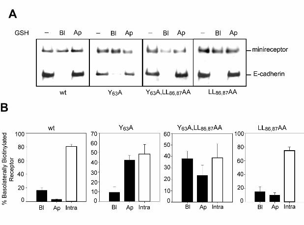
Transcytosis assay
To assess basolateral-to-apical transcytosis we applied a described strategy that uses sulfo-
NHS-SS-biotin and glutathione stripping (Burgos et al., 2004) with modifications. Cells were
grown in transwell filters until confluent. On the day of the experiment, cells were washed two
times with ice-cold PBSc for 5 min and the basolateral domain was biotinylated with 1 mM of the
cleavable sulfo-NHS-SS-biotin for 20 min at 4°C. The reaction was then quenched twice with 50
mM NH4Cl for 10 min at 4°C. After three washes with PBSc for 5 min each, the cells were
incubated with DMEM/Hepes for 45 min at 37°C and then chilled and washed again 3 times with
ice-cold PBS. The biotin of proteins appearing at the apical cell surface was stripped with 50
mM GSH, added twice to the apical compartment for 30 min at 4°C. Reduction was also
performed at the basolateral domain in different filters as control. The reaction was stopped by
washing with PBS and incubating with 5 mg/ml iodoacetamide in PBS, containing 10% BSA, for
30 min at 4°C. Cells were washed with PBS three times and then were lysed in ice-cold lysis
buffer containing 2.5 mM iodoacetamide for 1 h at 4°C. Lysates were centrifuged at 14,000 rpm
for 10 min at 4°C and incubated with streptoavidin beads as in the biotinylation protocol.
Precipitated proteins were identified by western blot with anti-HA to detect the minireceptors and
anti-E-cadherin to control loading and domain specific reduction. All the experiments were
repeated three times, with 1-2 filters per point, depending on the expression level of the

16
minireceptor analyzed. The band corresponding to the minireceptor in each condition was
quantified by densitometry. The transcytosis efficiency was determined after 45 min of
basolateral-to-apical trafficking at 37°C after the basolateral biotinylation, as follows:
Basolateral to Apical transcytosed minireceptor = Total Basolaterally Biotinylated
minireceptor – Biotinylated minireceptor remaining after reduction at the apical surface.
Basolaterally Recycled receptor = Total Basolaterally Biotinylated receptor - Biotinylated
minireceptor remaining after reduction at the basolateral surface.

17
RESULTS
LRP1 is transported to the basolateral surface in MDCK and FRT cells
To assess the sorting behavior of LRP1 we used HA-tagged minireceptors containing the fourth
ligand binding domain, the transmembrane domain, and the cytoplasmic tail of the receptor
(mLRP4) (Marzolo et al., 2003). We first examined whether two different epithelial cell lines,
MDCK and FRT, of kidney and thyroid origin respectively, express and sort endogenous LRP1
similarly. The LRP1 antibody against the human protein readily localized the endogenous LRP1
in the basolateral domain of MDCK cells, as we previously described (Marzolo et al., 2003)
(Figure 1A). In contrast, FRT cells showed much lower expression levels (not shown). However,
both cell lines distributed the transfected mLRP4 predominantly in the basolateral domain
(Figure 1B), indicating similar decoding of the LRP1 basolateral sorting information.
AP1B regulates LRP1 trafficking at a post-Golgi recycling endosome
We have previously shown that LLC-PK1 cells, which lack the adaptor subunit µ1B of the AP1B
complex that recognizes tyrosine-based basolateral sorting motifs (Ohno et al., 1999),
distributes LRP1 in a non-polarized manner unless exogenous µ1B is expressed by transfection
(Marzolo et al., 2003). To determine whether LRP1 interacts with AP1B, we transiently
transfected MDCK cells with HA-µ1B and then LRP1 was immunoprecipitated from the cell
lysates. The adaptor complex subunits, HA-µ1B and γ-adaptin, were found in the
immunoprecipitates (Figure 2A), suggesting that LRP1 does interact with AP1B.
We recently reported that an anti-µ1B antibody is able to block the biosynthetic
trafficking of VSVG and TfR at a µ1B-containing RE that most likely represents the common
recycling endosome (CRE) in FRT cells, 5-10 min after exiting the TGN (Cancino et al., 2007).
The antibody also blocked the post-endocytic trafficking of LDLR at µ1B-RE. Here we

18
performed similar experiments to define which µ1B-dependent route is used by LRP1. FRT cells
were microinjected with mLRP4 and RAP plasmids with or without anti µ1B antibody. After 1 h
of synthesis at 37 °C, 2 h of TGN accumulation at 20 °C and 1 h of releasing the TGN block at
37°C, the mLRP4 achieved a basolateral distribution that contrasted with the perinuclear
retention caused by the µ1B antibody (Figure 2B). This experiment revealed a µ1B-dependence
of LRP1 trafficking at µ1B-RE but did not allow to define whether this occurs immediately after
exiting the TGN or after endocytosis. A time course experiment resolved this question (Figure
2C, D). Before releasing from the 20°C TGN block, the receptor does not colocalize with µ1B.
After 5 min of TGN block release, more than 50% of mLRP4 colocalizes with µ1B in a
perinuclear compartment and reaches up to a 75 % of colocalization at 10 min, maintaining this
distribution even after 1 h at 37 °C. These results indicate that after exiting the TGN the LRP1 is
first transported to the µ1B-RE while en route to the basolateral cell surface, thus sharing with
VSVG and TfR this indirect basolateral pathway (Cancino et al., 2007).
Identification of sorting motifs in the LRP cytoplasmic domain
To define sorting motifs within the LRP1 cytoplasmic domain, as well as the context sequence
of the basolateral information conveyed by the Y29 and Y63 residues (Marzolo et al., 2003), we
analyzed new site-directed mutants of LRP1 minireceptors, as depicted in Figure 3, The wild-
type minireceptor mLRP4 has a cytoplasmic domain composed of 100 amino acids containing
several putative sorting motifs (Figure 3). These include two NPxY motifs (N26PTY29 and
N60PVY63), one YxxΦ (Y63ATL66) motif that shares the tyrosine with the N60PVY63 motif, two
dileucine motifs (LL43,44 and LL86,87), and a pair of negatively charged amino acids (DD38,39),
similar to those involved in LDLR basolateral sorting (Matter et al., 1992). There is also a protein
kinase A (PKA) phosphorylation site involving the serine in position 76 (S76) that has a role in
LRP endocytosis (Li et al., 2001b).

19
We generated stable MDCK cells lines and assessed the steady state distribution and
trafficking properties of the different minireceptor constructs by analyzing 2 to 3 MDCK clones
for each construct. The results of domain specific biotinylation assays are presented from the
proximal to distal location of the analyzed motifs (Figure 4). As described (Marzolo et al., 2003),
the mutation in the first NPxY motif (NPTY29A) completely redistributes LRP1 to the apical
domain. Neither the DD38,39AA mutant nor the LL43,44AA mutant affected the LRP1 basolateral
distribution. However, a truncated LRP1 construct lacking the last 41 amino acids revealed a
potential basolateral sorting function of DD38,39 residues (data not shown). The already
described mutation of the Y63 residue leads to a non-polarized distribution of the receptor
(Marzolo et al., 2003). This tyrosine is shared by the distal N60PVY63 motif and the endocytic
motif Y63ATL66 (Li et al., 2000). Here, we determined that its basolateral sorting function is
exerted independently of N60, since the N60A mutant distributed predominantly basolateral.
Instead, the L66A mutation mistargeted the receptor to the apical domain by more than 50%.
Thus, Y63 constitutes a YxxΦ and not a NPxY basolateral sorting motif.
In non-polarized cells, the Y63ATL66 and LL86,87 motifs contribute independently to the
rate of LRP1 endocytosis (Li et al., 2000). We found here that LL86,87 is also required for LRP1
basolateral distribution. The mutant LL86,87AA, similarly to the Y63A mutant, showed a non-
polarized distribution. Furthermore, the double mutant Y63A/LL86,87AA also distributed in a non-
polarized manner. Neither of these motifs seem to compensate for the loss of function of the
other. These signals seem to act in concert, as if forming part of a single complex sorting signal.
Finally, the unique PKA phosphorylation site S76 was not required for LRP1 basolateral sorting.
These results define the context of the previously described Y63-dependent basolateral
sorting signal and revealed novel sorting requirements involving L66 and LL86,87 residues. The
contrasting apical segregation of Y29A and non-polarized distribution of Y63A and LL86,87AA
mutants prompts to further analyzing the proximal NPxY motif and the sorting pathways where
these motifs exert their function.

20
The basolateral information conveyed by Y29 operates at the TGN
The different phenotypes produced by alanine replacement in either the Y29 or the Y63 and
LL86,87 residues suggest that sorting signals associate with temporally-spatially
compartmentalized functions. An interesting possibility is that the Y29-dependent motif operates
during biosynthetic trafficking at the TGN level, whereas Y63 and LL86,87 operates
biosynthetically in a post-TGN station and/or at a basolateral recycling route after internalization.
In this hypothetic model, mutant receptors lacking the function of Y63 or LL86,87 residues would
be targeted basolaterally from TGN due to the Y29-dependent sorting, but would enter into
basolateral-to-apical transcytotic pathways and lose polarity during post-endocytic recycling.
To test the hypothesis that the Y29-dependent motif acts at the biosynthetic route we
performed cDNA microinjection experiments in MDCK cells grown on glass coverslips and
microinjected after 3 days of reaching confluence. Under these conditions the cells are properly
polarized, as is shown by the basolateral distribution of E-cadherin and the presence of
detectable primary cilium, which is labeled with a monoclonal antibody recognizing acetylated
tubulin (Supplemental Figure 1). We used an established approach that assesses transport from
the TGN to the cell surface (Kreitzer et al., 2000). The wild-type or mutant receptor was
expressed together with the chaperone RAP, which is required for correct folding and
subsequent transport of LRP1 out of the endoplasmic reticulum (Bu and Marzolo, 2000). In
addition, we co-expressed a dominant negative form of the accessory protein eps15 (D/N-
eps15), known to interfere with the clathrin-mediated endocytosis of the TfR, as a fusion protein
with GFP (Benmerah et al., 1999) and also previously used to block the endocytosis of ApoER2,
another member of the LDLR family (Cuitino et al., 2005). We checked that this D/N-eps15
effectively interrupts the internalization of mLRP4 wt in transient transfection conditions
(Supplemental Figure 2A,B ) and also under the microinjection conditions used, in this case for
the mLRP4 Y29A (Supplemental Figure 2C). In this way, we attempted to inhibit the rapid

21
endocytosis of mLRP4 from the basolateral membrane and thus increase the probability of its
detection at the cell surface. In addition, this strategy allow us to determine whether the apical
distribution of the Y29A receptor mutant derives from a sorting defect at the TGN resulting in its
direct segregation to the apical domain, or at an endosomal compartment, which would
determine basolateral to apical transcytosis instead of basolateral recycling.
The cells microinjected with the corresponding plasmids were first incubated for 1 h at
37°C to allow expression and then for 2 h at 20ºC to accumulate the newly synthesized proteins
at the TGN. After releasing the TGN block for 1 h at 37ºC, both the wild-type mLRP4 and the
Y29A minireceptor showed a predominant intracellular vesicular pattern, with some apical
distribution of Y29A (Supplemental Figure 3). Then, in the presence of D/N-eps15, mLRP4
distributed basolaterally, indicating that its endocytosis was effectively reduced (Figure 5).
Instead, in these conditions the Y29A mutant showed an apical cell surface distribution, albeit
with some intracellular staining. Supplemental Figure 4 shows higher magnification images
corresponding to the same confocal plane, for mLRP4Y29A, mLRP4 wt and E-cadherin, which
clearly illustrate the basolateral distribution of wt minireceptor (coincident with E-cadherin
distribution), whereas the mutant was mostly intracellular. These results are representative of
the analysis of several fields, quantifying the distribution of minireceptors in 200 microinjected
cells (D/N-eps15-GFP positive cells) per condition (Supplemental Figure 5A). The wild type
receptor distributed basolaterally in around 70 % of microinjected cells. In 25 % of the cells we
could detect apical staining but representing just 7.4% of the total fluorescence (Supplemental
Figure 5B), suggesting missorting by overexpression. In contrast, the Y29A mutant distributed
apically in 70% of cells accounting for 50% of the total fluorescence (Supplemental Figure 5B),
with the remaining fluorescence being only intracellular.
These results discard two possibilities: Firstly, that the Y29A mutant is sorted without
polarity to both cell surfaces and upon endocytosis from the basolateral domain becomes
intracellularly arrested at endosomal compartments and subsequently disappears from this

22
domain. Secondly, that the Y29A mutant reaches the apical pole by transcytosis after rapid
endocytosis from the basolateral domain. In both cases, the endocytic inhibition caused by co-
expressing D/N-eps15 would have increased the basolateral location of the mutant. The Y29A
mutation most likely released otherwise recessive apical sorting information present in the
ectodomain (Marzolo et al., 2003).
Experiments in FRT cells showed that after releasing the TGN block in the presence of
the function-blocking anti-µ1B antibody the Y29A mutant did not accumulate in the post-Golgi RE
(Figure 6), contrasting with the wild-type receptor (See Figure 2). Under these conditions, the
Y29A mutant distributed in peripheral vesicles and was not detected at any moment in µ1B-RE,
indicating that it was very likely missorted directly from the TGN. Therefore, the Y29 seems to be
part of a dominant basolateral sorting signal that operates in the TGN during biosynthetic
trafficking. The results cannot exclude the possibility that the same sorting signal also operates
at RE during post-TGN trafficking or that additional biosynthetic basolateral sorting signals
decoded by AP1B direct LRP1 to the basolateral plasma membrane.
The N26PTY29 motif is involved in basolateral recycling and in recruiting SNX17 to
basolateral sorting endosomes (BSE)
It is currently believed that the functional integrity of NPxY motifs depend equally on both the N
and the Y residues (Bansal and Gierasch, 1981). Mutational analysis has revealed that the Y
residue can be engaged in functions different from those of the NPxY motif. This was first
demonstrated in the LDLR, in which the NPxY motif also acts as an endocytic signal but the Y
residue participates independently of the N residue in basolateral sorting (Matter et al., 1992).
In LRP1, the proximal NPxY motif has no role in endocytosis (Li et al., 2000) but
regulates receptor’s recycling (van Kerkhof et al., 2005). Because the Y29A mutation caused
missorting to the apical domain we decided to study the effect an N26 A mutation. Surprisingly, in
FRT cells, most of the mLRP4N26A mutant displayed an intracellular location instead of an

23
apical distribution (Figure 7A). MDCK cells showed the same effect (not shown). Biotinylation
assays could not detect mLRP4N26A mutant at the cell surface of MDCK cells, while a rather
small amount appeared basolaterally in FRT cells, relative to its total level of expression (Figure
7B). By detecting E-cadherin and Na+K+ ATPase at the basolateral domain of MDCK and FRT
cells respectively, we ensured that the cells were correctly polarized and that the biotin had
access to the basolateral cell surface.
In a complementary approach, we used flow cytometry to compare the expression level
of the wild type and mutant receptors at the cell surface of MDCK cells. Only 4 % of the total
mLRP4N26A was at the cell surface, compared with about 33 % of the wild type minireceptor,
indicating the mutant minireceptor is able to get the cell surface but with an almost 10 fold less
efficiency than mLRP4 wild type (Figure 7C). Thus, our results suggest that after rapid
internalization the N26A mutant stays longer in rate-limiting recycling compartments. If this is so,
one might expect that inhibiting the LRP1 endocytosis would increase the detection of the
mutant at the basolateral cell surface. The microinjection protocol, detailed in Figure 5, to co-
express D/N-eps15 allowed detecting the N26A mutant at the basolateral membrane, albeit in a
few cells (Figure 8A). As shown for the wild type and Y29A mutant minireceptors, we quantified
the % of microinjected cells expressing the N26A mutant at the cell surface, finding that no more
than 5% of the cells had surface staining, being almost all basolateral (Supplemental Figure 5).
To test directly whether recycling of the N26A mutant is defective in polarized epithelial
cells, as was previously shown in non-polarized cells (van Kerkhof et al., 2005), we used a
described cell surface fluorescence quenching recycling assay (van Kerkhof et al., 2005). In this
procedure, the receptor that becomes exposed to the cell surface binds Alexa488-labeled anti-
HA and after 20 minutes of internalization, the fluorescent antibody is quenched by an anti-
Alexa488 antibody; the magnitude of the quenching being proportional to the receptor’s
recycling rate. Accordingly to the difference in the cell surface expression of the minireceptors
(Figure 7C and Supplemental Figure 5), cells expressing the wild type protein show a 5 fold

24
increase in the total fluorescence intensity compared to the N26A mutant after 20 min of anti-HA
internalization (not shown). At 5 minutes of antibody-quenching, the measured recycling
efficiency of cells expressing the wild type mLRP4 was around 27 %, compared to less than 2 %
for cells expressing the N26A mutant. After 20 minutes of quenching, the efficiency of recycling
for the mutant was still very low (7% compared to 26 % of the control).
Taken together, all these results demonstrate that recycling of the mutant N26A receptor
is severely impaired (Figure 8B) and more dramatically than in non-polarized cells (van Kerkhof
et al., 2005). Thus the lack of surface localization of this mutant at the steady state is most likely
due to abrogation of a motif that promotes recycling.
To determine the sorting compartment where the N26A mutant minireceptor is
intracellularly arrested and its relationship with SNX17, a cytosolic protein involved in LRP1
recycling (van Kerkhof et al., 2005), we performed quantitative analysis of immunofluorescent
colocalization with the EEA1, an early endosomal marker. We found a significantly increased
co-localization of the N26A mutant minireceptor with EEA1 (62 ± 2.7 % of the N26A mutant
versus 54 ± 2.8 % of the mLRP4 wild-type; p < 0.05, unpaired t-test) (Figure 9). Because the
proximal NPxY is the motif which binds SNX17 and mediates LRP1’s recycling (van Kerkhof et
al., 2005), we assessed the distribution of SNX17 in cells expressing either the wild-type or the
N26A mutant receptor. The two available antibodies recognizing the human protein (van Kerkhof
et al., 2005) detect the endogenous SNX17 in MDCK cells in immunoblot (not shown), but not
by immunofluorescence. Therefore we performed microinjection experiments to express myc-
tagged human SNX17 together with the minireceptors and RAP.
Interestingly, SNX17 displayed a better endosomal distribution when co-expressed with
the wild type minireceptor than with the N26A mutant receptor. In cells expressing the mLRP4wt
37 ± 3.3 % of EEA1 endosomes contained SNX17, versus 5.8 ± 1.8 % in cells expressing the
N26A mutant. The wild type minireceptor colocalized 33 ± 3 % with vesicular SNX17, versus 7 ±
2.1 % by the N26A mutant receptor. These results show that mLRP4 promotes SNX17

25
recruitment to EEA1 early endosomes and provide further evidence that N26 is part of a sorting
signal that mediates LRP1 basolateral recycling. As judged by the immunofluorescence
patterns, the rate limiting-step of LRP1 recycling could be EEA1/SNX17 containing
compartments.
All these results indicate that N26 and Y29 residues conform distinct sorting determinants
that act at different steps of LRP1 trafficking. The Y29 constitutes a biosynthetically basolateral
sorting signal that functions at the TGN, and acts independently of the NPxY motif, while the N26
and the Y29 residues within the proximal NPxY motif regulate an additional sorting event
occurring at the recycling route of the receptor. This recycling step most likely involves SNX17
recruitment to the EEA1 endosomes that would represent an earlier compartment than µ1B-
containing endosomes.
The endocytic motifs, Y63ATL and LL86,87 also contribute to receptor basolateral recycling
The non-polarized cell surface distribution of LRP1 mutants Y63A and LL86,87A resemble LRP1 in
the absence of AP1B (Marzolo et al., 2003), thus suggesting that these motifs are involved in
basolateral sorting from a µ1B recycling compartment. Indeed, µ1B could recognize tyrosine-
based motifs of the kind of Y63ATL66. In addition, both Y63A and LL86,87A motifs have been
reported to decrease, but not completely abrogate, the endocytic rate of LRP1 (Li et al., 2000).
Therefore, these motifs could also contribute to the polarized recycling, maybe at a different
step than that mediated by N26PxY29 motif. To test this hypothesis we performed transcytosis
assays in MDCK cells grown on filters, using glutathione stripping of cleavable sulfo-NHS-SS-
biotin (Figure 10). After basolateral biotinylation the cells were incubated for 45 min at 37ºC.
The majority (80 ± 5 %) of the mLRP4 wild-type becomes insensitive to glutathione stripping
from the apical or basolateral cell surfaces, indicating internalization and steady-state
distribution at intracellular compartments, with almost undetectable transcytosis (3.3 ± 0.3 %).
On the contrary, a percentage of the basolaterally biotinylated mutants, 42.4 ± 4.6 % of the

26
Y63A, 24 ± 6 % of double mutant Y63A/LL86,87AA, and 11 ± 3 % of LL86,87AA mutants, became
sensitive to glutathione stripping from the apical cell surface, reflecting an increased basolateral-
to-apical transcytosis. These results indicate that Y63 and the LL86,87 promote basolateral
recycling, in addition to internalization, of LRP1. Disruption of these residues causes non-
polarized recycling, a fraction to the basolateral surface and a fraction to the opposite domain by
transcytosis.
The lack of colocalization of SNX17 and µ1B in polarized MDCK cells (Supplemental Figure 6)
strongly suggests that LRP1 experiences two post-endocytic recycling steps spatially and
temporally separated. The dominant recycling step would depend on the proximal NPxY motif
and SNX17 operating at the BSE, while the few receptors that escape this sorting event would
enter into the µ1B-RE and rescued to a basolateral sorting.
Neurons decode LRP1 sorting motifs with mechanisms similar to those in epithelial cells
LRP1 has been localized in the somatodendritic domain (Brown et al., 1997), but the sorting
elements that determine this distribution remain unknown. Having defined the effect of our
LRP1 mutants in polarized epithelial cells and the role of µ1B and SNX17 in LRP1 basolateral
sorting, we went on to assess their sorting behavior in neurons which are known to lack AP1B
(Ohno et al., 1999).
We transfected primary cultured hippocampal neurons with each of the mLRP4
constructs and, by indirect immunofluorescence with anti-HA monoclonal antibodies, assessed
the cell surface distribution of the minireceptor in non-permeabilized cells. Then, we
permeabilized the cells to define the somatodendritic domain by MAP2 staining (Caceres et al.,
1984). As expected for a somatodendritic protein, the wild-type minireceptor mLRP4 completely
colocalized with MAP2 (Figure 11). Minireceptors with Y63A and LL86,87AA mutations mimicked
epithelial sorting behavior when expressed in neurons. Both distributed without polarity, evenly

27
in somatodendritic and axonal domains. Strikingly, the Y29A mutant distributed exclusively at the
distal part of the axon.
In permeabilized neurons, the wild-type minireceptor exhibits an exclusively somato-
dendritic vesicular distribution (Figure 12), resembling the TfR that is also excluded from the
axon (Cameron et al., 1991). The intracellular vesicles carrying the Y29A mutant, however,
distributed both in axons and dendrites. The mechanistic relationship between such intracellular
non-polarized distribution and the distal axonal sorting that this mutant displays at the cell
surface remains unknown.
The sorting behavior of the N26A mutant was similar to that as seen in MDCK cells. We
could not detect the minireceptor mLRP4N26A at the cell surface of non-permeabilized neurons,
whereas permeabilized cells show intense immunofluorescence staining, indicating intracellular
accumulation (Figure 13A). These results suggest that in hippocampal neurons, LRP1 also
recycles through a mechanism that depends on the N26PxY29 motif. Supporting this possibility,
SNX17 is shown to be endogenously expressed in cultured hippocampal and cortical neurons
(Figure 13B).
All these results suggest that the same sorting motifs that address LRP1 basolaterally in
epithelial cells operate to somatodendritically target the receptor in neurons, despite the
differences in the expression of AP1B. In this system the recycling step would be only mediated
by SNX17.

28
DISCUSSION
This work shows novel features of the sorting mechanisms involved in the polarized
distribution of LRP1, by defining the trafficking behavior of sorting mutants and the participation
of AP1B and SNX17. The biosynthetic basolateral pathway of LRP1 includes a TGN sorting
event, mediated by Y29, and a post-TGN trafficking through µ1B-containing RE, which is blocked
by an µ1B antibody (Cancino et al., 2007). Y29A mutants were apically missorted without
entering this µ1B-RE indicating that Y29 directs LRP1 from TGN to RE. We also detected an
interaction of LRP1 with AP1B by coimmunoprecipitation, in agreement with our previous
observation of LRP1 missorting in cells lacking µ1B (Marzolo et al., 2003). We also found that a
different Y-dependent sorting signal mediates LRP1 basolateral recycling, implicating distinct
sorting elements operating during biosynthetic and recycling trafficking. These sorting elements
include, for the first time, SNX17 recruitment to BSE, which are distinct from the AP1B-
endosomes. Finally, sorting signals of LRP1 seem to be similarly decoded in neurons. We found
that neurons, which lack AP1B, express SNX17 and the LRP1 mutant with a defective SNX17
binding site distributes intracellularly as expected for a recycling defect. This suggests that
SNX17 constitutes a conserved element of the polarized protein sorting machinery between
neurons and epithelial cells.
Mutational analysis revealed multiple motifs contributing to basolateral distribution of
LRP1, including Y29, N26, Y63 and LL85,86 residues. The individual silencing of these residues
produces distinct LRP1 distribution patterns, indicating that they are not functionally redundant
and cannot compensate the lack of each other. The different phenotypes could be explained by
temporal-spatial differences in the sorting events that these motifs mediate.
In non-polarized cells the proximal NPxY motif of LRP1 constitutes both a binding site for
SNX17 and a sorting motif for SNX17-mediated receptor recycling (van Kerkhof et al., 2005).

29
The mutant NPxA29 showed lower recycling efficiency, and SNX17 knockdown by siRNA
inhibited LRP1 recycling (van Kerkhof et al., 2005). Although these studies only analyzed the
recycling effect of the Y29A mutant, they show that both Y29 and N29 residues are required for
SNX17 binding in vitro. Therefore, one might expect that both N26A and Y29A mutant receptors
display similar sorting defects unless they follow different pathways. Our current results clearly
demonstrate that N26 and Y29 residues exert separable functions at different sorting places in
polarized cells.
In polarized cells, the N26A mutation dramatically decreased the cell surface expression
of LRP1. The receptor became intracellularly accumulated in endosomes that contain EEA1,
thus corresponding to described BSE (Wilson et al., 2000). LRP1 has a very high internalization
rate, about 10 times higher than LDLR’s (Li et al., 2001a), thus at steady-state, it distributes
mainly at intracellular endocytic pools, but also basolaterally in small yet detectable levels
(Marzolo et al., 2003). In MDCK cells, we performed microinjection experiments to follow the
sorting of LRP1 minireceptors from the TGN to the cell surface. Co-expression of D/N-eps15 to
inhibit LRP1 endocytosis increased the basolateral distribution of the wild-type minireceptors.
However, the distribution of the N26A mutants was much less affected, as expected for a defect
in recycling, which was demonstrated using a sensitive recycling assay and cytometry. The
results showed that recycling efficiency of N26A mutant decreased by about 90%.
The only proteins so far described to mediate Y-dependent basolateral sorting have
been the clathrin adaptors AP4 (Simmen et al., 2002) and AP1B (Folsch et al., 1999; Sugimoto
et al., 2002). In non-polarized cells, LRP1 recycling involves SNX17 (van Kerkhof et al., 2005).
Strikingly, we found here that the wild type minireceptor, but not the N26A mutant, recruits
SNX17 to EEA1-containing BSE, the same compartments in which the mutant accumulates.
This previously unnoticed correspondence between NPxY-mediated SNX17 endosomal
recruitment and LRP1 recycling indicates that SNX17 constitutes a novel element of the
basolateral recycling machinery.

30
When the Y29A mutant was co-expressed with D/N-eps15 and after having exited the
TGN, it appeared at the apical cell surface, without displaying any basolateral location.
Therefore, in epithelial cells the Y29A mutant does not follow the same pathway as the N26A
mutant, otherwise a recycling arrest would have led to its intracellular accumulation, with very
little or undetectable cell surface expression. Experiments performed in FRT cells, which first
accumulated the Y29A in the TGN by the 20ºC block and then followed its exit from this
compartment, did not detect a transit through the µ1B-RE, in contrast to the wild-type receptor.
Thus, the data fit better with a scenario in which the Y29A mutant, due to recessive apical
information possibly contained in its ectodomain (Marzolo et al., 2003), reaches the apical
surface directly from the TGN rather than from the post-Golgi endosomal compartment or from
the basolateral cell surface by transcytosis (Figure 14). Thus, LRP1 contains a Y29-dependent,
but N26-independent, sorting signal for biosynthetic basolateral sorting at the TGN, while the
N26PxY29 motif directs post-endocytic exit from BSE, through a mechanism involving membrane
recruitment of SNX17 (Figure 14).
Exhaustive studies on LDLR have led to the prevalent notion that the same Y-dependent
basolateral information is decoded during biosynthetic and recycling trafficking (Matter et al.,
1993). In this sense, our results constitute the first indication that the biosynthetic and recycling
sorting machineries can distinguish between Y-based sorting motifs. Furthermore, we provide
evidence that such distinction could be due to temporally and spatially separated sorting events
accomplished by yet unknown adaptors at the TGN, AP1B in RE and SNX17 in BSE. In FRT
cells, where our anti-µ1B antibody is suitable for immunofluorescent staining of endogenous
µ1B, we found that the SNX17 endosomes contain EEA1, which is considered a marker of BSE
(Wilson et al., 2000), but do not contain µ1B. AP1B distributes poorly in early endosomes
labeled with rab 5 (Gan et al., 2002) and distributes in a compartment with typical characteristics
of CRE (Cancino et al., 2007). Because CRE is downstream BSE, all these observations mean
that most recycling of LRP1 is SNX17-mediated at BSE before reaching CRE.

31
Our results reinforce the notion that NPxY motifs can have distinct sorting functions and
even dual functions in a same protein depending on the kind of cell. The trafficking role of NPxY
motifs of other proteins seems to be mainly endocytic (Chen et al., 1990; Hsu et al., 1994;
Cuitino et al., 2005). However, neither of the two cytosolic NPxY motifs of LRP1 play roles in
internalization (Li et al., 2000). In the LDLR, the proximal NPxY motif is an endocytic motif in
which the tyrosine residue, independent of the N and P residues and together with downstream
acidic residues, constitutes a mild basolateral sorting signal, as it is easily overcome by receptor
overexpression and becomes apparent only in truncated receptors (Matter et al., 1992; Matter
et al., 1994). The strongest basolateral sorting signal of the LDLR depends on a more distal
tyrosine and also includes distal acidic residues (Matter et al., 1994). In contrast, the proximal
Y29-dependent basolateral sorting signal of LRP1 is rather independent of downstream acidic
residues, and exerts basolateral destination even upon receptor overexpression and in the
context of the entire cytosolic tail, thus acting as a strong basolateral determinant. This Y29
residue also belongs to an NPxY motif, but it is only crucial for basolateral recycling. These
results show for the first time that trafficking information embedded within a single NPxY motif
can assign distinct temporal-spatial sorting functions to the Y residue.
We previously showed that Y63 also exerts a basolateral sorting function (Marzolo et al.,
2003). Y63 is shared by a distal N60PxY63 motif and by the previously identified endocytic motif
Y63ATL (Li et al., 2000). Here, we demonstrate that the Y63ATL is a basolateral sorting motif
which seems to function in concert with the other LRP1 endocytic signal LL86,87 (Li et al., 2000).
Disruption of any of these motifs separately or together in a double mutant Y63A/LL86,87AA,
results in non-polarized receptor distribution, suggesting that both act as a single complex
sorting signal. Furthermore, the sorting information conveyed by Y63ATL and LL86,87 does not
compensate for the Y29A mutation thus ruling out their participation at the TGN.
Because Y63A mutant has been shown to decrease but not completely abrogate the
endocytic rate of LRP1 (Li et al., 2000), the loss of polarity of this mutant could be achieved

32
during its recycling traffic through a compartment different from BSE, in which the
N26PxY29/SNX17 system mediates basolateral recycling. Our biotinylation assays demonstrated
that these mutants undergo basolateral-to-apical transcytosis. Taken together, the overall
results suggest that Y63A mutants are first addressed basolaterally by Y29-dependent sorting at
the TGN and then become missorted to both plasma membrane domains when they reach the
CRE compartment, presumably after several rounds of recycling from the SNX17-containing
BSE. Because the non-polarized distribution of the endocytosis mutants is similar to the
distribution of LRP1 in cells lacking µ1B (Marzolo et al., 2003), the most plausible model is that
Y63ATL and LL86,87 motifs contribute to AP1B-dependent basolateral sorting of LRP1 in CRE
compartment, both during post-TGN biosynthetic trafficking and post-SNX17 recycling
trafficking. This model implies that AP1B and SNX17 sort LRP1 at different stations of its
endocytic recycling itinerary recognizing distinct sorting motifs (see proposed model in Figure
14).
A matter still under scrutiny and debate is the proposed correspondence between
basolateral/apical sorting in epithelial cells and somatodendritic/axonal sorting in neurons (Dotti
and Simons, 1990; Silverman et al., 2005). It could be dependent on the specific type of sorting
signals possessed by individual proteins. For instance, mutants of the LDLR and TfR lacking
basolateral sorting signals are directed apically in MDCK cells but without polarity in neurons
(Jareb and Banker, 1998). This suggests that neurons cannot handle recessive apical
information contained in these proteins. There is also evidence suggesting that neurons do not
recognize dihydrophobic-based basolateral sorting signals (Silverman et al., 2005). However,
the sorting behavior of wild-type and all our mutant receptors expressed in hippocampal
neurons show striking congruency with the sorting correspondence hypothesis. Even the apical
Y29A mutant segregate exclusively into the distal part of the axon, whereas the Y63A and
LL86,87AA mutants segregate without polarity. This indicates that neurons can effectively decode
recessive apical information as well as dihydrophobic somatodendritic sorting signals in

33
recycling proteins. Interestingly, both the axon and dendrites contained intracellular vesicles
carrying the Y29A mutant, suggesting a complex mechanism of distal axonal regionalization that,
as described for other axonal proteins such as NgCAM (Horton and Ehlers, 2003), might involve
selective fusion/retention processes. Neurons do not express AP1B (Ohno et al., 1999), but we
found that they do express SNX17. Furthermore, neurons also intracellularly distributed the
N26A mutant lacking the SNX17 binding motif. In principle, AP4, which has been involved in
somatodendritic addressing of the glutamate-receptor δ2 (Yap et al., 2003), could sort LRP1 at
the TGN, while SNX17 could play a predominant role in LRP1 somatodendritic recycling. The
missorting similarities of the LRP1 mutants suggest conservation of the compartmentalized use
of sorting signals at the TGN and recycling compartments in epithelial cells and neurons.
LRP1 is required for development and exerts crucial physiological functions in tissues
such as brain, liver and different epithelia, which rely on polarized cells (Herz et al., 1992; Herz
et al., 1993). We could anticipate, in accordance with recent data (Roebroek et al., 2006), that
natural mutants affecting those critical sorting motifs that we have defined, especially the
proximal NPxY, would be relatively unviable.

34
ACKNOWLEDGMENTS
We want to thank Dr. Alexander Benmerah (Institut Cochin -U567 INSERM/UMR8104
CNRS- Paris France) for giving us the plasmid encoding the dominant negative forms of eps15
with GFP and Dr. Ira Mellman (Genetech, Inc South San Francisco CA 94080) for providing us
the cDNA of µ1B-HA tagged. From the Facultad de Ciencias Biológicas, PUC, we thank Dr.
Alejandra Alvarez for providing us some of the hippocampal neurons used in this work, and Dr.
Alexis Kalergis and Leandro Carreño for providing access to the FACS equipment. We also
thank Dr. María Isabel Yuseff for the characterization of the human anti-EEA1 antibodies in our
laboratory and Olivia Ying for reviewing this manuscript. This work was supported by FIRCA
grant TW006456 to G.B and MPM, NIH grant R01 AG027924 to G.B, an Alzheimer’s Disease
Research grant from the American Health Assistance Foundation (AHAF) to G.B, Grant
1020746 from the Fondo Nacional de Investigación Científica y Tecnológica (FONDECYT)
(MPM), Fondo de Investigación Avanzada en Areas Prioritarias (FONDAP) Grant 13980001 and
the Millenium Institute for Fundamental and Applied Biology (MIFAB) to MPM and AG. It was
also supported by grants from CONICET
and ANPCyT (Argentina) to AC. G.B. is an Established Investigator of the American Heart
Association.

35
REFERENCES
Ang, A.L., Taguchi, T., Francis, S., Folsch, H., Murrells, L.J., Pypaert, M., Warren, G., and Mellman, I. (2004). Recycling endosomes can serve as intermediates during transport from the Golgi to the plasma membrane of MDCK cells. J Cell Biol 167, 531-543. Aroeti, B., and Mostov, K.E. (1994). Polarized sorting of the polymeric immunoglobulin receptor in the exocytotic and endocytotic pathways is controlled by the same amino acids. Embo J 13, 2297-2304. Bacskai, B.J., Xia, M.Q., Strickland, D.K., Rebeck, G.W., and Hyman, B.T. (2000). The endocytic receptor protein LRP also mediates neuronal calcium signaling via N-methyl-D-aspartate receptors. Proc Natl Acad Sci U S A 97, 11551-11556. Banker, G.A., and Cowan, W.M. (1977). Rat hippocampal neurons in dispersed cell culture. Brain Res 126, 397-342. Bansal, A., and Gierasch, L.M. (1981). The NPXY internalization signal of the LDL receptor adopts a reverse-turn conformation. Cell 67, 1195-1201. Benmerah, A., Bayrou, M., Cerf-Bensussan, N., and Dautry-Varsat, A. (1999). Inhibition of clathrin-coated pit assembly by an Eps15 mutant. J Cell Sci 112, 1303-1311. Bottenstein, J.E., and Sato, G.H. (1979). Growth of a rat neuroblastoma cell line in serum-free supplemented medium. Proc Natl Acad Sci U S A 76, 514-517. Bradke, F., and Dotti, C.G. (1998). Membrane traffic in polarized neurons. Biochim Biophys Acta 1404, 245-258. Brandan, E., Retamal, C., Cabello-Verrugio, C., and Marzolo, M.P. (2006). The low density lipoprotein receptor-related protein functions as an endocytic receptor for decorin. J Biol Chem 281, 31562-31571. Bravo-Zehnder, M., Orio, P., Norambuena, A., Wallner, M., Meera, P., Toro, L., Latorre, R., and Gonzalez, A. (2000). Apical sorting of a voltage- and Ca2+-activated K+ channel alpha -subunit in Madin-Darby canine kidney cells is independent of N-glycosylation. Proc Natl Acad Sci U S A 97, 13114-13119. Brown, M.D., Banker, G.A., Hussaini, I.M., Gonias, S.L., and Vandenberg, S.R. (1997). Low Density Lipoprotein Receptor-Related Protein Is Expressed Early and Becomes Restricted to a Somatodendritic Domain During Neuronal Differentiation In Culture. Brain Research 747, 313-317. Bu, G., and Marzolo, M.P. (2000). Role of rap in the biogenesis of lipoprotein receptors. Trends Cardiovasc Med 10, 148-155.

36
Bu, G., Williams, S., Strickland, D.K., and Schwartz, A.L. (1992). Low density lipoprotein receptor-related protein/alpha 2-macroglobulin receptor is an hepatic receptor for tissue-type plasminogen activator. Proceedings of the National Academy of Sciences of the United States of America 89, 7427-7431. Burgos, P.V., Klattenhoff, C., de la Fuente, E., Rigotti, A., and Gonzalez, A. (2004). Cholesterol depletion induces PKA-mediated basolateral-to-apical transcytosis of the scavenger receptor class B type I in MDCK cells. Proc Natl Acad Sci U S A 101, 3845-3850. Caceres, A., Banker, G., Steward, O., Binder, L., and Payne, M. (1984). MAP2 is localized to the dendrites of hippocampal neurons which develop in culture. Brain Res 315, 314-318. Caceres, A., Mautino, J., and Kosik, K.S. (1992). Suppression of MAP2 in cultured cerebellar macroneurons inhibits minor neurite formation. Neuron 9, 607-618. Cameron, P.L., Südhof, T.C., Jahn, R., and Camilli, P.d. (1991). Colocalization of synaptophysin with transferrin receptors: implications of synaptic vesicle biogenesis. J. Cell Biol 1991, 151-164. Cancino, J., Torrealba, C., Soza, A., Yuseff, M.I., Gravotta, D., Henklein, P., Rodriguez-Boulan, E., and Gonzalez, A. (2007). Antibody to AP1B adaptor blocks biosynthetic and recycling routes of basolateral proteins at recycling endosomes. Mol Biol Cell 18, 4872-4884. Casanova, J.E., Apodaca, G., and Mostov, K.E. (1991). An autonomous signal for basolateral sorting in the cytoplasmic domain of the polymeric immunoglobulin receptor. Cell 66, 65-75. Chen, W.J., Goldstein, J.L., and Brown, M.S. (1990). NPXY, a sequence often found in cytoplasmic tails, is required for coated pit-mediated internalization of the low density lipoprotein receptor. J Biol Chem 265, 3116-3123. Cresawn, K.O., Potter, B.A., Oztan, A., Guerriero, C.J., Ihrke, G., Goldenring, J.R., Apodaca, G., and Weisz, O.A. (2007). Differential involvement of endocytic compartments in the biosynthetic traffic of apical proteins. Embo J 26, 3737-3748. Cuitino, L., Matute, R., Retamal, C., Bu, G., Inestrosa, N.C., and Marzolo, M.-P. (2005). ApoER2 is endocytosed by a clathrin-mediated process involving the adaptor protein Dab2 independent of its rafts association. Traffic 6, 820-838. Deborde, S., Perret, E., Gravotta, D., Deora, A., Salvarezza, S., Schreiner, R., and Rodriguez-Boulan, E. (2008). Clathrin is a key regulator of basolateral polarity. Nature 452, 719-723. Deora, A.A., Gravotta, D., Kreitzer, G., Hu, J., Bok, D., and Rodriguez-Boulan, E. (2004). The basolateral targeting signal of CD147 (EMMPRIN) consists of a single leucine and is not recognized by retinal pigment epithelium. Mol Biol Cell 15, 4148-4165. Dotti, C.G., Parton, R.G., and Simons, K. (1991). Polarized sorting of glypiated proteins in hippocampal neurons. Nature 349, 158-161. Dotti, C.G., and Simons, K. (1990). Polarized sorting of viral glycoproteins to the axon and dendrites of hippocampal neurons in culture. Cell 62, 63-72.

37
Fiedler, K., and Simons, K. (1995). The role of N-glycans in the secretory pathway. Cell 81, 309-312. Folsch, H., Ohno, H., Bonifacino, J.S., and Mellman, I. (1999). A novel clathrin adaptor complex mediates basolateral targeting in polarized epithelial cells. Cell 99, 189-198. Folsch, H., Pypaert, M., Schu, P., and Mellman, I. (2001). Distribution and function of AP-1 clathrin adaptor complexes in polarized epithelial cells. J. cell Biol. 152, 595-606. Fullekrug, J., and Simons, K. (2004). Lipid rafts and apical membrane traffic. Ann N Y Acad Sci 1014, 164-169. Fuller, S.D., Bravo, R., and Simons, K. (1985). An enzymatic assay reveals that proteins destined for the apical and basolateral domains of an epithelial cell line share the same late Golgi compartments. EMBO J. 4, 297-307. Gan, Y., McGraw, T.E., and Rodriguez-Boulan, E. (2002). The epithelial-specific adaptor AP1B mediates post-endocytic recycling to the basolateral membrane. Nat Cell Biol 4, 605-609. Godyna, S., Liau, G., Popa, I., Stefansson, S., and Argraves, W.S. (1995). Identification of the low density lipoprotein receptor-related protein (LRP) as an endocytic receptor for thrombospondin-1. J Cell Biol 129, 1403-1410. Goretzki, L., and Mueller, B.M. (1998). Low-density-lipoprotein-receptor-related protein (LRP) interacts with a GTP-binding protein. Biochem J 336, 381-386. Gravotta, D., Deora, A., Perret, E., Oyanadel, C., Soza, A., Schreiner, R., Gonzalez, A., and Rodriguez-Boulan, E. (2007). AP1B sorts basolateral proteins in recycling and biosynthetic routes of MDCK cells. Proc Natl Acad Sci U S A 104, 1564-1569. Griffiths, G., and Simons, K. (1986). The trans-Golgi network: sorting at the exit site of the Golgi complex. Science (Wash. DC) 234, 4381-4443. Herz, J., and Bock, H.H. (2002). Lipoprotein receptors in the nervous system. Annu Rev Biochem 71, 405-434. Herz, J., Clouthier, D.E., and Hammer, R.E. (1992). LDL receptor-related protein internalizes and degrades uPA-PAI-1 complexes and is essential for embryo implantation [published erratum appears in Cell 1993 May 7;73(3):428]. Cell 71, 411-421. Herz, J., Couthier, D.E., and Hammer, R.E. (1993). Correction: LDL receptor-related protein internalizes and degrades uPA-PAI-1 complexes and is essential for embryo implantation [letter]. Cell 73, 428. Horton, A.C., and Ehlers, M.D. (2003). Neuronal polarity and trafficking. Neuron 40, 277-295. Hsu, D., Knudson, P.E., Zapf, A., Rolband, G.C., and Olefsky, J.M. (1994). NPXY motif in the insulin-like growth factor-I receptor is required for efficient ligand-mediated receptor internalization and biological signaling. Endocrinology 134, 744-750.

38
Hunziker, W., and Fumey, C. (1994). A di-leucine motif mediates endocytosis and basolateral sorting of macrophage IgG Fc receptors in MDCK cells. Embo J 13, 2963-2967. Hussain, M.M. (2001). Structural, biochemical and signaling properties of the low-density lipoprotein receptor gene family. Front Biosci 6, D417-428. Jareb, M., and Banker, G. (1998). The polarized sorting of membrane proteins expressed in cultured hippocampal neurons using viral vectors. Neuron 20, 855-867. Kreitzer, G., Marmorstein, A., Okamoto, P., Vallee, R., and Rodriguez-Boulan, E. (2000). Kinesin and dynamin are required for post-Golgi transport of a plasma-membrane protein. Nat Cell Biol 2, 125-127. Le Bivic, A., Real, F.X., and Rodriguez-Boulan, E. (1989). Vectorial targeting of apical and basolateral plasma membrane proteins in a human adenocarcinoma epithelial cell line. Proc Natl Acad Sci U S A 86, 9313-9317. Le Gall, A.H., Powell, S.K., Yeaman, C.A., and Rodriguez-Boulan, E. (1997). The neural cell adhesion molecule expresses a tyrosine-independent basolateral sorting signal. J Biol Chem 272, 4559-4567. Li, Y., Lu, W., Marzolo, M.P., and Bu, G. (2001a). Differential Functions of Members of the Low Density Lipoprotein Receptor Family Suggested by Their Distinct Endocytosis Rates. J Biol Chem 276, 18000-18006. Li, Y., Marzolo, M.P., Kerkhof, P., Strous, G.J., and Bu, G. (2000). The YXXL motif, but not the two NPXY motifs, serves as the dominant endocytosis signal For LDL receptor-related protein (LRP). J Biol Chem 275, 17187-17194. Li, Y., van Kerkhof, P., Marzolo, M.P., Strous, G.J., and Bu, G. (2001b). Identification of a major cyclic AMP-dependent protein kinase A phosphorylation site within the cytoplasmic tail of the low-density lipoprotein receptor-related protein: implication for receptor-mediated endocytosis. Mol Cell Biol 21, 1185-1195. Lock, J.G., and Stow, J.L. (2005). Rab11 in Recycling Endosomes Regulates the Sorting and Basolateral Transport of E-Cadherin. Mol Biol Cell 16, 1744-1755. Marmorstein, A.D., Csaky, K.G., Baffi, J., Lam, L., Rahaal, F., and Rodriguez-Boulan, E. (2000). Saturation of, and competition for entry into, the apical secretory pathway. Proc Natl Acad Sci U S A 97, 3248-3253. Marzolo, M.P., Bull, P., and Gonzalez, A. (1997). Apical sorting of hepatitis B surface antigen (HBsAg) is independent of N-glycosylation and glycosylphosphatidylinositol-anchored protein segregation. Proc Natl Acad Sci U S A 94, 1834-1839. Marzolo, M.P., Yuseff, M.I., Retamal, C., Donoso, M., Ezquer, F., Farfan, P., Li, Y., and Bu, G. (2003). Differential distribution of low-density lipoprotein-receptor-related protein (LRP) and megalin in polarized epithelial cells is determined by their cytoplasmic domains. Traffic 4, 273-288.

39
Matter, K., Hunziker, W., and Mellman, I. (1992). Basolateral sorting of LDL receptor in MDCK cells: the cytoplasmic domain contains two tyrosine-dependent targeting determinants. Cell 71, 741-753. Matter, K., and Mellman, I. (1994). Mechanisms of cell polarity: sorting and transport in epithelial cells. Curr. Opin. Cell Biol. 6, 545-554. Matter, K., Whitney, J.A., Yamamoto, E.M., and Mellman, I. (1993). Common signals control low density lipoprotein receptor sorting in endosomes and the Golgi complex of MDCK cells. Cell 74, 1053-1064. Matter, K., Yamamoto, E.M., and Mellman, I. (1994). Structural requirements and sequence motifs for polarized sorting and endocytosis of LDL and Fc receptors in MDCK cells. J Cell Biol 126, 991-1004. Mostov, K.E. (2003). Epithelial polarity and morphogenesis. Methods 30, 189-190. Mostov, K.E., and Cardone, M.H. (1995). Regulation of protein traffic in polarized epithelial cells. Bioessays 17, 129-138. Müsch, A., Xu, H., Schields, D., and Rodriguez-Boulan, E. (1996). Transport of vesicular stomatitis virus G protein to the cells surface is signal mediated in polarized and nonpolarized cells. J. Cell Biol. 133, 543-558. Obermoeller, L.M., Chen, Z., Schwartz, A.L., and Bu, G. (1998). Ca2+ and receptor-associated protein are independently required for proper folding and disulfide bond formation of the low density lipoprotein receptor-related protein. J Biol Chem 273, 22374-22381. Odorizzi, G., and Trowbridge, I.S. (1997). Structural requirements for basolateral sorting of the human transferrin receptor in the biosynthetic and endocytic pathways of Madin-Darby canine kidney cells. J Cell Biol 137, 1255-1264. Ohno, H., Tomemori, T., Nakatsu, F., Okazaki, Y., Aguilar, R.C., Foelsch, H., Mellman, I., Saito, T., Shirasawa, T., and Bonifacino, J.S. (1999). Mu1B, a novel adaptor medium chain expressed in polarized epithelial cells. FEBS Lett 449, 215-220. Paglini, G., Pigino, G., Kunda, P., Morfini, G., Maccioni, R., Quiroga, S., Ferreira, A., and Caceres, A. (1998). Evidence for the participation of the neuron-specific CDK5 activator P35 during laminin-enhanced axonal growth. J Neurosci 18, 9858-9869. Pietrini, G., Suh, Y.J., Edelmann, L., Rudnick, G., and Caplan, M.J. (1994). The axonal gamma-aminobutyric acid transporter GAT-1 is sorted to the apical membranes of polarized epithelial cells. J Biol Chem 269, 4668-4674. Rindler, M.J., Ivanov, I.E., Plesken, H., Rodriguez-Boulan, E., and Sabatini, D.D. (1984). Viral glycoproteins destined for apical or basolateral plasma membrane domains traverse the same Golgi apparatus during their intracellular transport in doubly infected Madin-Darby canine kidney cells. J Cell Biol 98, 1304-1319.

40
Rodriguez-Boulan, E., and Gonzalez, A. (1999). Glycans in post-Golgi apical targeting: sorting signals or structural props? Trends Cell Biol 9, 291-294. Rodriguez-Boulan, E., Kreitze, G., and Musch, A. (2005). Organization of vesicular trafficking in epithelia. Nat Rev Mol Cell Biol 6, 233-247. Rodriguez-Boulan, E., and Powell, S.K. (1992). Polarity of epithelial and neuronal cells. Ann Rev Cell Biol 8, 395-427. Roebroek, A.J., Reekmans, S., Lauwers, A., Feyaerts, N., Smeijers, L., and Hartmann, D. (2006). Mutant Lrp1 knock-in mice generated by recombinase-mediated cassette exchange reveal differential importance of the NPXY motifs in the intracellular domain of LRP1 for normal fetal development. Mol Cell Biol 26, 605-616. Rosso, S., Bollati, F., Bisbal, M., Peretti, D., Sumi, T., Nakamura, T., Quiroga, S., Ferreira, A., and Caceres, A. (2004). LIMK1 regulates Golgi dynamics, traffic of Golgi-derived vesicles, and process extension in primary cultured neurons. Mol Biol Cell 15, 3433-3449. Salicioni, A.M., Mizelle, K.S., Loukinova, E., Mikhailenko, I., Strickland, D.K., and Gonias, S.L. (2002). The low density lipoprotein receptor-related protein mediates fibronectin catabolism and inhibits fibronectin accumulation on cell surfaces. J Biol Chem 277, 16160-16166. Silverman, M.A., Peck, R., Glover, G., He, C., Carlin, C., and Banker, G. (2005). Motifs that mediate dendritic targeting in hippocampal neurons: a comparison with basolateral targeting signals. Mol Cell Neurosci 29, 173-180. Simmen, T., Honing, S., Icking, A., Tikkanen, R., and Hunziker, W. (2002). AP-4 binds basolateral signals and participates in basolateral sorting in epithelial MDCK cells. Nat Cell Biol 4, 154-159. Soza, A., Norambuena, A., Cancino, J., De La Fuente, E., Henklein, P., and Gonzalez, A. (2004). Sorting competition with membrane-permeable peptides in intact epithelial cells revealed discrimination of transmembrane proteins not only at the trans-golgi network but also at pre-golgi stages. J Biol Chem 279, 17376-17383. Stockinger, W., Sailler, B., Strasser, V., Recheis, B., Fasching, D., Kahr, L., Schneider, W.J., and Nimpf, J. (2002). The PX-domain protein SNX17 interacts with members of the LDL receptor family and modulates endocytosis of the LDL receptor. Embo J 21, 4259-4267. Sugimoto, H., Sugahara, M., Folsch, H., Koide, Y., Nakatsu, F., Tanaka, N., Nishimura, T., Furukawa, M., Mullins, C., Nakamura, N., Mellman, I., and Ohno, H. (2002). Differential recognition of tyrosine-based basolateral signals by AP-1B subunit mu1B in polarized epithelial cells. Mol Biol Cell 13, 2374-2382. van Kerkhof, P., Lee, J., McCormick, L., Tetrault, E., Lu, W., Schoenfish, M., Oorschot, V., Strous, G.J., Klumperman, J., and Bu, G. (2005). Sorting nexin 17 facilitates LRP recycling in the early endosome. Embo J 24, 2851-2861.

41
Wilson, J.M., de Hoop, M., Zorzi, N., Toh, B.H., Dotti, C.G., and Parton, R.G. (2000). EEA1, a tethering protein of the early sorting endosome, shows a polarized distribution in hippocampal neurons, epithelial cells, and fibroblasts [In Process Citation]. Mol Biol Cell 11, 2657-2671. Winckler, B., and Mellman, I. (1999). Neuronal polarity: controlling the sorting and diffusion of membrane components. Neuron 23, 637-640. Worby, C.A., and Dixon, J.E. (2002). Sorting out the cellular functions of sorting nexins. Nat Rev Mol Cell Biol 3, 919-931. Yap, C.C., Murate, M., Kishigami, S., Muto, Y., Kishida, H., Hashikawa, T., and Yano, R. (2003). Adaptor protein complex-4 (AP-4) is expressed in the central nervous system neurons and interacts with glutamate receptor delta2. Mol Cell Neurosci 24, 283-295. Yeaman, C., Grindstaff, K.K., and Nelson, W.J. (1999). New perspectives on mechanisms involved in generating epithelial cell polarity. Physiol Rev 79, 73-98. Zhuo, M., Holtzman, D.M., Li, Y., Osaka, H., DeMaro, J., Jacquin, M., and Bu, G. (2000). Role of tissue plasminogen activator receptor LRP in hippocampal long-term potentiation. J Neurosci 20, 542-549.

42
FIGURE LEGENDS
Figure 1. Basolateral expression of LRP1 and its minireceptors in polarized epithelial cells. (A)
MDCK cells biotinylated either at the apical (Ap) or basolateral (Bl) domain at 4°C were lysed
and the surface biotinylated proteins were precipitated with Streptavidin-agarose and resolved
in 5% SDS-PAGE followed by immunoblot with anti-human LRP1. Endogenous LRP1
distributes at the basolateral domain. (B) MDCK and FRT cells stably expressing the mLRP4
minireceptor were grown until confluent on coverslips and the distribution of the minireceptor
was visualized by indirect immunofluorescence with monoclonal anti-HA. The minireceptor was
detected only after cell permeabilization at the basolateral cell borders and in intracellular
vesicles. Scale bars, 10 µm.
Figure 2. Physical and functional interaction between LRP1 and AP-1B. (A) MDCK cells were
transiently transfected with a plasmid encoding µ1B-HA. Cells were lysed and the lysates were
immunoprecipitated with anti-LRP1 or anti-megalin antibodies. The immunoprecipitates were
resolved on 4-15% SDS-PAGE. The presence of LRP1 was detected by immunobloting with
anti-LRP antibody and the complex AP-1B was evidenced by the presence of γ-adaptin and µ1-
B. (B) FRT cells were microinjected with mLRP4 minireceptor plus RAP cDNAs with or without
blocking-function anti µ1B antibody. Cells were maintained for 1 h at 37°C, followed by 2 h at
20°C to accumulate the receptor at the TGN. After 1 h of exit at 37°C cells were fixed and
processed for immunofluorescence with anti HA to detect the minireceptor. After 60 minutes
release from TGN block mLRP4 was located on surface in control cells. In contrast, in µ1B
microinjected cells, mLRP4 was located mainly in a perinuclear localization, most likely RE,
indicating that AP1B adaptor complex is involved in the intracellular trafficking of the receptor.

43
(C-D), Kinetic analyses of mLRP4/µ1B colocalization of newly synthetized mLRP4
demonstrates that perinuclear accumulation was achieved during biosynthetic trafficking. Scale
bar 10 µm.
Figure 3. Schematic representation of mLRPs with the potential cytoplasmic basolateral sorting
signals and their mutations. The sequence of the wild-type tail (mLRP4) is depicted with the
already known recycling and endocytic sorting motifs described in non-polarized cells ((Li et al.,
2000; van Kerkhof et al., 2005) and the two tyrosines (Y29 and Y63, underlined) involved in
basolateral distribution (Marzolo et al., 2003). All the mutants made are highlighted in bold
where the critical residues were replaced by alanine.
Figure 4. Changes in the steady-state distribution of mLRP4 mutants uncover the critical
basolateral sorting motifs within the LRP1 tail. MDCK cells stably expressing the different
mLRP4 constructs were grown on filters to determine the membrane distribution of the
receptors by domain specific biotinylation. Biotinylated minireceptors were precipitated with
Streptavidin-agarose and resolved in 6% SDS-PAGE followed by immunoblot with monoclonal
anti-HA. The receptor’s relative expression level in each membrane domain was estimated by
densitometry of the resulting bands. The critical basolateral determinants include the Y29 of the
proximal NPxY, the Y63ATL66 and the distal LL86,87 motifs. Similar results were obtained from 2-3
clones of each construct.
Figure 5. The mLRP4Y29A mutant is directly addressed to the apical domain in MDCK cells.
Three days after reaching confluency, MDCK cells plated on glass coverslips were
microinjected in the nucleus with the expression plasmids mLRP4 or Y29A together with pcDNA-
RAP and a plasmid encoding the dominant negative form of eps15 (D/N-eps15), GFP- Eps15
DD (E∆95/295). After incubating the cells for 1 h at 37°C, the newly synthesized receptors were
accumulated in the TGN at 20ºC for 2 h in the presence of cycloheximide. TGN-to-cell-surface

44
protein traffic was then allowed to proceed at 37ºC for 60 min and the cells were treated for
indirect immunofluorescence. The apical or intracellular/basolateral surface distributions of the
receptors were assessed in non-permeabilized and permeabilized cells, respectively.
Expression of the D/N-eps15 increased mLRP4 wt detection at the cell surface. The cells were
analyzed by confocal microscopy and representative x-y plane and x-z and y-z sections are
shown (red). D/N-eps15 expression is shown in the x-y plane, medial to x-z sectioning (green).
The mLRP4 wt appears in intracellular vesicles and in the basolateral membrane, whereas the
Y29A mutant was present only in intracellular vesicles and at the apical surface, indicating direct
TGN-to-apical addressing. Scale bar: 20 µm. Arrowhead indicates selected x-y plane of x-z
sectioning.
Figure 6. The mutant Y29A does not traffic through post-Golgi compartment to get the apical
plasma membrane. FRT cells were microinjected with mLRP4-Y29A minireceptor plus the
chaperone RAP cDNAs and the blocking-function anti µ1B antibody. Cells were maintained for
1 h at 37°C, followed by 2 h at 20°C to accumulate the receptor at the TGN, then the cells were
fixed at the indicated time and processed for immunofluorescence with anti-HA to detect the
minireceptor. Kinetic analyses of mLRP4-Y29A/µ1B colocalization of newly synthetized mLRP4-
Y29A show that µ1B-compartment is not involved in biosynthetic trafficking of this mutant. Scale
bar, 10 µm.
Figure 7. Mutations within the proximal NPxY motif of the LRP1 tail result in different receptor
distributions. (A) FRT cells stably transfected with either mLRP4Y29A or mLRP4N26A were
grown to confluence on coverslips and treated for indirect immunofluorescence with anti-HA
under non-permeabilized and permeabilized conditions, as indicated. Y29A distributed both at
the apical cell surface (right panel) and intracellular vesicles (left panel), whereas N26A show

45
only an intracellular localization, not being detectable at the cell surface. MDCK cells gave
similar results (not shown). Scale bars, 10 µm. (B) Domain specific cell surface biotinylation of
MDCK and FRT cells expressing mLRP4N26A. In MDCK cells the protein was not detected at
the cell surface. Controls confirmed the receptor expression (immunoblot of total lysate) and
proper basolateral expression of E-Cadherin. In FRT cells, however, a low amount of
minireceptor was detected at the basolateral cell surface. Na+K+ ATPase was used as an
endogenous basolateral marker. (C) MDCK cells expressing either mLRP4wt or the N26A
mutant were consecutively incubated, either intact or after permeabilization with 0.1% saponin,
with an anti-HA and anti-mouse RPE-conjugated antibody and analyzed by flow cytometry. The
ratio of expression levels observed in intact versus permeabilized cells show 30% of the wild-
type receptor versus no more than 4% of the N26A mutant at the cell surface.
Figure 8. The mutant N26A reaches the basolateral membrane but its recycling efficiency is
severely impaired. (A) MDCK cells were microinjected 3 days after reaching confluence with the
plasmid encoding the D/N-eps15, GFP- EPS15 DD (E∆95/295) as described and analyzed in
Figure 5. Confocal microscopy and representative x-y plane and x-z and y-z sections are shown
(red). D/N-eps15 expression is shown as x-y plane medial of x-z sectioning (green). The
distribution of the N26A mutant was mainly intracellular, but was also detectable at the
basolateral membrane (arrows). Scale bar, 20 µm. Arrowhead indicates selected x-y plane of x-
z sectioning. (B) The recycling efficiency of the minireceptors was evaluated in MDCK cell lines
by a fluorescence quenching assay. The mLRP4wt or N26A mutant were labeled with Alexa488-
conjugated anti-HA antibody at 37°C for 20 minutes and then chased for the indicated time
periods in the presence of quenching anti-Alexa488 IgG. Cells were processed for flow
cytometry and the recycling efficiency calculated. The data plotted and showed in the graph
correspond to two experiments measured in duplicate (the averages ± s.e.m values are given
as error bars).

46
Figure 9. Colocalization of minireceptors with endosomal markers EEA1 and SNX17. MDCK
cells were microinjected with the plasmids encoding for RAP, SNX17-myc, and either the
mLRP4 wt or N26A minireceptors. Cells were processed for immunofluorescence to detect the
HA-tagged minireceptor (green), the early endosome marker EEA1 (red), and the myc-tagged
SNX17 (blue). In the amplified merged images, co-localization of the wild type minireceptor with
EEA1 in endosome-like structures was clearly visible; some of these structures also contain
SNX17 (white dots). The N26A minireceptors practically did not colocalize with SNX17 and
exhibited an increased colocalization with EEA1 (amplified merge image, yellow dots). Scale
bars, 10 µm.
Figure 10. Basolateral to apical trancytosis of mLRPs in MDCK cells. MDCK cells stably
expressing one of the following constructs: mLRP4wt, mLRP4LL86,87AA, mLRPY63A or the
double mutant minireceptor, were grown on filters to confluence. Cells were biotinylated with
reducible biotin at 4ºC, shifted to 37ºC 45 min and reduced with glutathione either at the apical
or basolateral surface. Cells were lysed, and biotinylated proteins were precipitated with
streptavidin beads and the complexes resolved by reducing 6% SDS-PAGE and western blot.
(A) Minireceptor detection with anti-HA and E-cadherin blot. The absence of apical reduction of
biotinylated E-cadherin indicates that the observed reduction of the minireceptor was due to its
transcytosis from the basolateral to the apical surface. (B) Bands were quantified and the total
biotinylated minireceptor was taken as 100% (see Material and Methods section). Minireceptor
present in the basolateral surface, apical surface and within the cells was determined as a
percentage of total. The determinations were performed in triplicate for each construct.
Figure 11. The somatodendritic distribution of mLRP in hippocampal neurons depends on the
same motifs required for its basolateral distribution in epithelial cells. Primary cultured
hippocampal neurons were transfected as described in Materials and Methods section. The

47
distribution of the transfected mLRP was determined by confocal microscopy, using anti-HA to
detect the receptor at the cell surface (green) and, after cell permeabilization, using anti-MAP2
(red) to determine the somatodendritic domain. The restricted cell surface somatodendritic
distribution of mLRP (arrowheads) was lost when the critical basolateral determinants, based on
tyrosines and dileucines, were replaced by alanines. This was visualized by the axonal staining
of the receptor in MAP2 negative structures (arrows). The most striking distribution was the
mLRPY29A mutant, for which only the distal region of the axon was positively stained with anti-
HA. Scale bars, 20 µm (wt, Y63A and LL86,87AA) and 100 µm (Y29A).
Figure 12. Intraneuronal distribution and unrestricted transport of the mLRP4Y29A mutant to the
axons. Upper Panel: Confocal micrographs showing the distribution of tubulin (red) and HA-
tagged mLRP4 wt (green) in 7 DIV permeabilized cultured hippocampal neurons. HA-tagged
mLRP4 localized to short, dendritic-like processes. Note that a thin long axon-like neurite
(arrows) that emerges from a dendritic shaft (arrowhead) and is positive for tubulin, does not
contain HA-tagged mLRP4. The insert on the right panel (merge color) shows a high
magnification view of the region where the axon-like neurite originates. Lower Panel: Confocal
micrographs showing the distribution of the somatodendritic marker MAP2 (red) and the mutant
HA-tagged mLRP4Y29A (green) in 7 DIV cultured hippocampal neurons. Note that the ectopic
mutant variant of mLRP4 not only localized to dendritic-like processes, but also to MAP2 (-),
axon-like neurites (short arrows in the merge panel). The long arrow in the merge panel shows
the dendritic site from which the axon merges. For these experiments cells were fixed and
permeabilized 18 hours after transfection. Scale bar, 10 µm.
Figure 13. Intraneuronal distribution and absence of cell surface expression of the mLRP4N26A
mutant in hippocampal neurons. (A) Differential cell surface expression of mLRP4 wt (upper
panel) and N26A (lower panel) in hippocampal neurons in primary culture was evidenced by
immunofluorescence detection of the surface minireceptor using a polyclonal anti-HA (red),

48
followed by permeabilization and detection of the intracellular minireceptor using a monoclonal
anti-HA (green). The wild-type mLRP4 was expressed in the somato-dendritic surface, but the
N26A mutant was only intracellular and somatodendritic. Scale bars, 10 µm. (B) 7 DIV cultured
hippocampal and cortical neurons were lysed, 100 µg of proteins were resolved in reducing
SDS-PAGE, and the presence of endogenous SNX17 and LRP1 was determined by western
blot.
Figure 14. Working model for LRP1 intracellular trafficking in epithelial cells. The data obtained
in this work and others previously published (Marzolo et al., 2003; Rodriguez-Boulan et al.,
2005; van Kerkhof et al., 2005; Cancino et al., 2007) allow us to derive a working model for
LRP1 trafficking. During the biosynthetic pathway, LRP1 is primarily segregated into basolateral
destined vesicles through the recognition of Y29, likely without an NPxY motif. This recognition
step occurs at the TGN and likely requires a not yet known adaptor protein(s) that recognizes
the critical tyrosine residue (1a). Most of LRP1 should be then segregated by AP1B in the RE
(1b), probably recognized through the distal tyrosine based signal YATL. When the sorting
signal is mutated or the corresponding sorting machinery is functionally absent, the receptor
fails to be correctly segregated and is directed to the apical membrane or to both domains
(punctuate arrow). Once LRP1 arrives at the basolateral membrane, it is actively endocytosed
and has to be post-endocytically segregated from the basolateral sorting endosome (BSE) in a
manner that depends on the proximal NPxY motif and SNX17 (van Kerkhof et al., 2005)(2a). A
failure in this step causes LRP1 to be trapped in this compartment. After BSE (2b), a secondary
recycling step may occur at the common RE and depend on the Y63ATL66/ LL86,87 motifs, and
probably on the AP1B adaptor complex as well (Marzolo et al., 2003) (3).