POISE-2 ASA suppl1
26
Supplementary Appendix This appendix has been provided by the authors to give readers additional information about their work. Supplement to: Devereaux PJ, Mrkobrada M, Sessler DI, et al. Aspirin in patients undergoing noncardiac surgery. N Engl J Med. DOI: 10.1056/NEJMoa1401105
-
Upload
independent -
Category
Documents
-
view
2 -
download
0
Transcript of POISE-2 ASA suppl1
Supplementary Appendix
This appendix has been provided by the authors to give readers additional information about their work.
Supplement to: Devereaux PJ, Mrkobrada M, Sessler DI, et al. Aspirin in patients undergoing noncardiac surgery. N Engl J Med. DOI: 10.1056/NEJMoa1401105
1
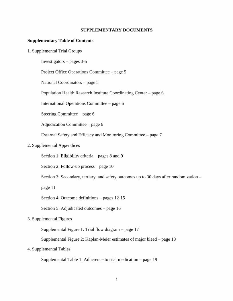
SUPPLEMENTARY DOCUMENTS
Supplementary Table of Contents
1. Supplemental Trial Groups
Investigators – pages 3-5
Project Office Operations Committee – page 5
National Coordinators – page 5
Population Health Research Institute Coordinating Center – page 6
International Operations Committee – page 6
Steering Committee – page 6
Adjudication Committee – page 6
External Safety and Efficacy and Monitoring Committee – page 7
2. Supplemental Appendices
Section 1: Eligibility criteria – pages 8 and 9
Section 2: Follow-up process – page 10
Section 3: Secondary, tertiary, and safety outcomes up to 30 days after randomization –
page 11
Section 4: Outcome definitions – pages 12-15
Section 5: Adjudicated outcomes – page 16
3. Supplemental Figures
Supplemental Figure 1: Trial flow diagram – page 17
Supplemental Figure 2: Kaplan-Meier estimates of major bleed – page 18
4. Supplemental Tables
Supplemental Table 1: Adherence to trial medication – page 19
2
Supplemental Table 2: Effects of Aspirin on the 30-day outcomes in the Initiation
Stratum – page 20 and 21
Supplemental Table 3: Effects of Aspirin on the 30-day outcomes in the Continuation
Stratum – page 22 and 23
Supplemental Table 4: Strata subgroup analyses for acute kidney injury with receipt of
dialysis, stroke, and bleeding – page 24
Supplemental Table 5: Independent predictors of myocardial infarction – page 25
3
SUPPLEMENTAL TRIAL GROUPS
Investigators - Investigators who recruited at least 20 patients; number of patients enrolled in
each country and site are in parentheses.
Canada (3498)- Hamilton Health Sciences (1112) J. DeBeer, R. Mizera, A. Patel, T. VanHelder;
St Joseph's Healthcare Hamilton (552) D. Cook, W. Dechert, P. Jackson, M. Tiboni; Queen's
University (424) R. Allard, D. Dumerton-Shore, J. McCourt, J.L. Parlow; University of Western
Ontario (406) P.M. Jones, R. Lavi, S. Lavi, R. Moor; London Health Sciences Centre (193) G.K.
Dresser, A.X. Garg, M.L. Gros, V.C. Schumann; Grey Nuns Hospital (179) M. Baur, C.
MacDonald, B. Wirzba; Health Sciences Centre, Winnipeg (109) O. Regalado, S.K. Srinathan;
University of Saskatchewan (95) D.D. Ong, A. Todd; University of Toronto (172) S. Abbas,
W.S. Beattie, V.W.S. Chan, K.J. Chin, D.N. Wijeysundera; University of Alberta Hospital (93)
M.M. Graham, M. Irwin, M. Jacka; Mississauga Hospital (53) H. El Beheiry; Queen Elizabeth II
Health Sciences Centre (24) S.M. McMullen; Cape Breton Regional Hospital (22) P.
MacDonald. USA (1760) - Cleveland Clinic (991) Z. Akhtar, S. Ayad, M. Buttar, A. Deroee, Y.
Eshraghi, A. Fergany, P. Finnigan, A. Fu, M. Grady, S. Helper, B Hesler, H. Honar, M.
Hutcherson, V. Krebs, A. Kurz, J. Lee, M. Malik, A. Podolyak, V. Salmasi, D.I. Sessler;
University of North Carolina (302) H. Arora, R.F. Coombs, P.A. Kumar, S.M. Martinelli; The
Ohio State University (126) S.D. Bergese, S.B. Melibary, A.A. Uribe; Wake Forest School of
Medicine (63) M. Jordan, S.A. Miller; MD Anderson Cancer Center (60) J.P. Cata; University of
Virginia (58) E.C. Nemergut; University of Miami (49) K.A. Candiotti; Weill Cornell Medical
College (39) S.G. Memtsoudis; University of California San Francisco (33) R.E. McKay.
Colombia (649) - Fundación Cardioinfantil Instituto de Cardiologia (250) F.R. Montes;
Fundación Oftalmológica de Santander (246) G.A. Parra, M.F. Rojas; Clínica Chicamocha (125)
4
R. Plata, S.M. Vásquez; Fundación Clínica Shaio (28) T. Sarquis. India (595) - Rahate Surgical
Hospital & ICU (219) Z. Haider, N.B. Jane, P.P. Lanjewar, P.V. Rahate; Mahatma Gandhi
Institute of Medical Sciences (92) B.R. Mehra, B. Premendaran; Christian Medical College,
Ludhiana (78) V. Abraham, P. George, P. Kumar; Dr. Gaikwad's Critical Care Center (25) S.B.
Gaikwad; CARE Hospitals (24) N.V.S. Mohan; Sidhu Hospital Pvt. Ltd. (21) G. Sidhu. Spain
(538) - Biomedical Research Institute (IJB – SANT PAU) (255) J. Alvarez, R. Gonzalez, M.
Maestre, E. Popova, G. Urrutia; Hospital Universitari Vall d'Hebron (139) M. de Nadal, S.
González-Suárez, A. González-Tallada; Hospital Donostia (35) P. Plou; Hospital Universitario
La Princesa (27) E. Mata Mena; Hospital General Universitario Gregorio Marañón de Madrid
(23) C. Fernandez Riveira; Hospital Universitario Fundación Hospital Alcorcon (22) S. Garcia
del Valle; Hospital Clinic Barcelona (22) B. Tena. Australia (470)- Royal Adelaide Hospital
(143) S.A. Lang, G.L. Ludbrook, T.W. Painter; Royal Hobart Hospital (55) N.C. Terblanche;
Geelong Hospital (52) C. Osborne; Peninsula Health Frankston Hospital (43) J.R. Mahood;
Royal Melbourne Hospital (32) K. Leslie; Monash University (31) P.S. Myles; Princess
Alexandra Hospital (30) P. Sivalingam; Peter MacCallum Cancer Centre (26) B. Riedel; Queen
Elizabeth Hospital (23) I. Elhalawani. South Africa (353) - University of Kwazulu-Natal (247)
B.M. Biccard, L. Drummond, A. Mugabi, P. Naidoo; University of Cape Town (59) A.L.
Myburgh, O.S. Porrill; University of the Free State (40) B.J.S. Diedericks, E.W. Turton.
Denmark (321) - Herlev Hospital (234) M. Bøgeskov, R.M. Dahl, M.V. Madsen, E.S.
Søndergaard; Vejle Hospital (87) N.E. Bauer, K.R. Martinsen. Hong Kong (276) - The Chinese
University of Hong Kong (276) M.T.V. Chan, G.Y.S. Choi, T. Gin, S.S.M. Ng. Belgium (184) -
Free University of Brussels (101) S.J. Bidgoli, P.J. Van der Linden; Université catholique de
Louvain (75) M. De Kock, P. Forget. Austria (176) - University of Vienna (176) E. Fleischmann,
5
B. Kabon, F. Luf, M. Radonic. Pakistan (175) - Shifa International Hospital (175) M. Amir, O.
Ishtiaq, J. Safdar. Peru (172) - Hospital Nacional Cayetano Heredia (83) A. Acuna-Villaorduna,
P. Barrionuevo, A. Castaneda-Guarderas; Hospital Regional Docente de Trujillo (50) J.A.
Caballero, V.E. Lau; Hospital Nacional Arzobispo Loayza (39) M.R. Aphang-Lam. Italy (154) -
San Raffaele Scientific Institute (96) R. Lembo, L. Pasin; "Sapienza" University of Rome (47)
B. Gossetti. Chile (153) - Clinica Santa Maria (153) X. Jara, P. Leon, D. Torres. Malaysia (120)
- University of Malaya, Kuala Lumpur (63) G.S.Y. Ong, C.Y. Wang; Sultanah Aminah Hospital
Johor Bahru (25) H.S. Lee. Switzerland (96) - University Hospital Basel (96) E.E. Seeberger,
M.D. Seeberger. France (89) - Hôpital Cochin (25) P. Alfonsi; Hospitalier Pitié Salpétrière (24)
P. Coriat; Centre Hospitalier Lyon Sud (21) V. Piriou. UK (86) - Chelsea & Westminster
Hospital (52) M.P. Vizcaychipi. Brazil (57) - Hospital Universitário Mãe de Deus Canoa (22)
R.L. Rech; Hospital Maternidade e Pronto Socorro Santa Lúcia (21) R.R. Bergo. New Zealand
(46) - Middlemore Hospital, Manukau Surgery Centre (30) S. Walker.
Project Office Operations Committee: P.J. Devereaux (Principal Investigator), D.I. Sessler, A.
Kurz, M. Mrkobrada, G. Lurati Buse, F. Botto, R. Rodseth, A. Robinson, G. Guyatt, Y.
LeManach, J. Pogue, S. Pettit, S. Chrolavicius, S. Yusuf (Chair).
National Coordinators: Argentina - F. Botto and R. Díaz; Australia/New Zealand - K. Leslie;
Austria - E. Fleischmann; Belgium - P. Forget; Brazil - O. Berwanger; Canada - P.J. Devereaux,
M. Mrkobrada; Chile - D. Torres; Colombia - J.C. Villar, O.L.Cortés; Denmark – C.S. Meyhoff ,
J. Wetterslev; France - P. Alfonsi; Germany - A. Hoeft, M. Wittmann; Hong Kong - M. Chan;
India - A. Sigamani, D. Xavier; Italy - G. Landoni; Malaysia - C.Y. Wang; Pakistan - O. Ishtiaq;
Peru - G. Malaga; South Africa - B.M. Biccard; Spain - P. Alonso-Coello; Switzerland - D.
Conen; United Kingdom - P. Balaji; United States - A. Kurz.
6
Population Health Research Institute Coordinating Center: A. Robinson (Trial Coordinator),
T. Sovereign, L. Blake, J. Sephton, A. Serra, C. Agrippa, M. Lawrence, P. Gao, S. Pettit
(Associate Project Manager), S. Chrolavicius (Project Manager).
International Operations Committee: K. Leslie, P. Alonso-Coello, B. Biccard, M. Chan, C.S.
Meyhoff, J.C. Villar, D. Xavier, C.Y. Wang, G. Landoni, O. Berwanger, M. Jacka, G. Malaga,
C. Chow, C. Gluud, C. Baigent, G. Karthikeyan, and members of the Project Office Operations
Committee.
Steering Committee: P.J. Devereaux, P. Alfonsi, P. Alonso-Coello, A. Auerbach, C. Baigent, P.
Balaji, S. Beattie, O. Berwanger, B.M. Biccard, F. Botto, N. Buckley, M. Chan, C. Chow, S.
Chrolavicius, D. Conen, D. Cook, J. Douketis, J. Eikelboom, P. Forget, A.X. Garg, H. Gerstein,
W. Ghali, M. Graham, G. Guyatt, R. Hart, M. Hill, A. Hoeft, M. Jacka, G. Karthikeyan, C.
Kearon, A. Kurz, A. Lamy, G. Landoni, Y. LeManach, K. Leslie, G. Lurati Buse, G. Malaga, F.
McAlister, D. McAuley, C.S. Meyhoff, S. Miller, M. Mrkobrada, M. O’Donnell, P. Pais, J.
Parlow, J. Pogue, A. Robinson, R. Rodseth, T. Schricker, D. Sessler, M. Simunovic, S.
Srinathan, K. Teoh, D. Torres, G. Urrutia, J.C. Villar, M. Walsh, C.Y. Wang, J. Wetterslev, R.
Whitlock, D. Wijeysundera, D. Xavier, H. Yang, S. Yusuf.
Adjudication Committee: G. Guyatt (co-chair), F. Botto (co-chair), P. Alonso-Coello, S.
Alshalash, J. Alvarez, A. Bessissow, J. Douketis, E. Duceppe, E. Fleischmann, R. Hart, M. Hill,
Z. Khalid, J. Khan, A. Kurz, M. Lauw, Y. Le Manach, G. Lurati Buse, K. Martinsen, C.S.
Meyhoff, M. Mrkobrada, J. Neary, W. Oczkowski, M. O’Donnell, P. Paniagua, M. Papina, L.
Pasin, E. Popova, R. Rodseth, M. Seeberger, V. Tandon, S. Thomas, M. Tiboni, D. Torres, J.
Wetterslev.

7
External Safety and Efficacy and Monitoring Committee: L. Friedman (Chair), D. Cheng, D.
Johnstone, E. Lowenstein, R. Roberts.
8
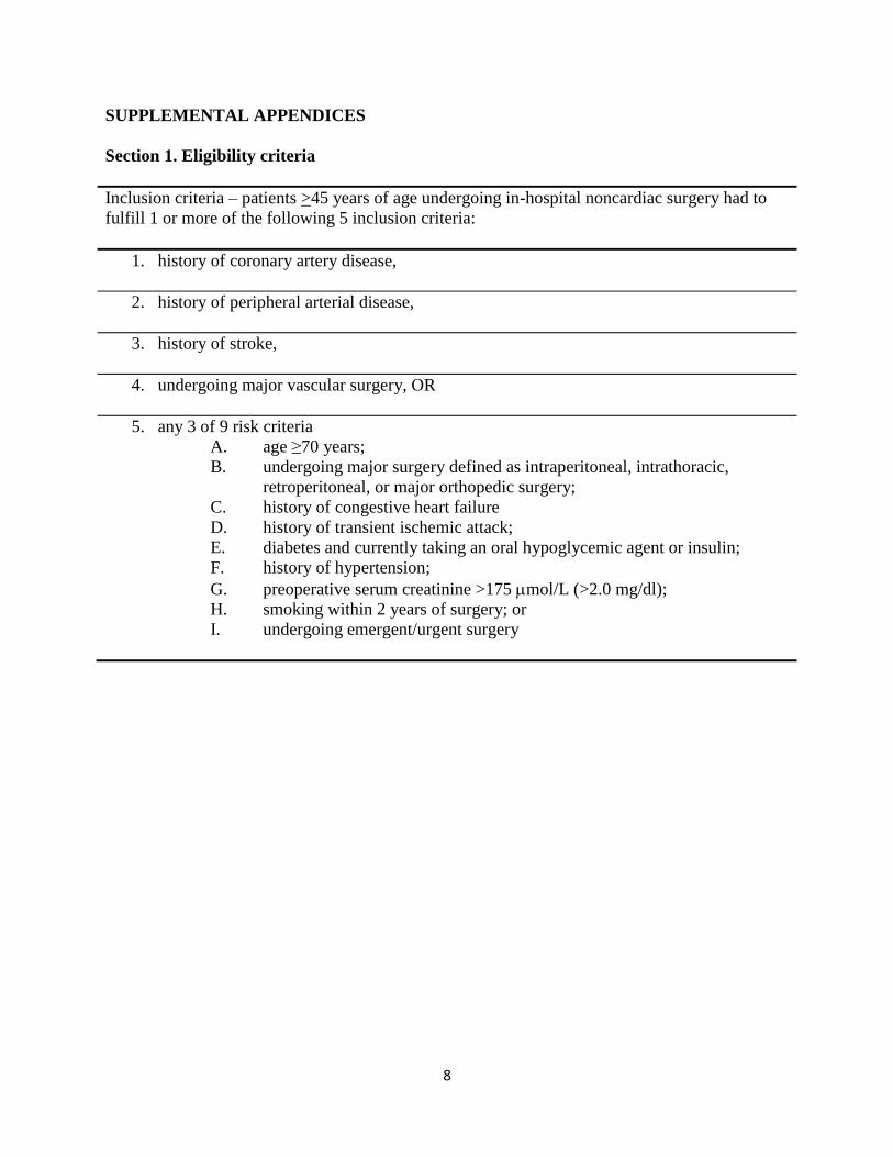
SUPPLEMENTAL APPENDICES
Section 1. Eligibility criteria
Inclusion criteria – patients >45 years of age undergoing in-hospital noncardiac surgery had to
fulfill 1 or more of the following 5 inclusion criteria:
1. history of coronary artery disease,
2. history of peripheral arterial disease,
3. history of stroke,
4. undergoing major vascular surgery, OR
5. any 3 of 9 risk criteria
A. age ≥70 years;
B. undergoing major surgery defined as intraperitoneal, intrathoracic,
retroperitoneal, or major orthopedic surgery;
C. history of congestive heart failure
D. history of transient ischemic attack;
E. diabetes and currently taking an oral hypoglycemic agent or insulin;
F. history of hypertension;
G. preoperative serum creatinine >175 mol/L (>2.0 mg/dl);
H. smoking within 2 years of surgery; or
I. undergoing emergent/urgent surgery
9
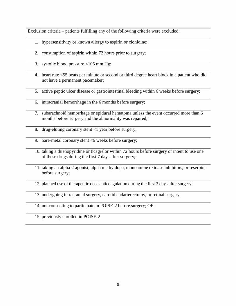
Exclusion criteria – patients fulfilling any of the following criteria were excluded:
1. hypersensitivity or known allergy to aspirin or clonidine;
2. consumption of aspirin within 72 hours prior to surgery;
3. systolic blood pressure <105 mm Hg;
4. heart rate <55 beats per minute or second or third degree heart block in a patient who did
not have a permanent pacemaker;
5. active peptic ulcer disease or gastrointestinal bleeding within 6 weeks before surgery;
6. intracranial hemorrhage in the 6 months before surgery;
7. subarachnoid hemorrhage or epidural hematoma unless the event occurred more than 6
months before surgery and the abnormality was repaired;
8. drug-eluting coronary stent <1 year before surgery;
9. bare-metal coronary stent <6 weeks before surgery;
10. taking a thienopyridine or ticagrelor within 72 hours before surgery or intent to use one
of these drugs during the first 7 days after surgery;
11. taking an alpha-2 agonist, alpha methyldopa, monoamine oxidase inhibitors, or reserpine
before surgery;
12. planned use of therapeutic dose anticoagulation during the first 3 days after surgery;
13. undergoing intracranial surgery, carotid endarterectomy, or retinal surgery;
14. not consenting to participate in POISE-2 before surgery; OR
15. previously enrolled in POISE-2
10
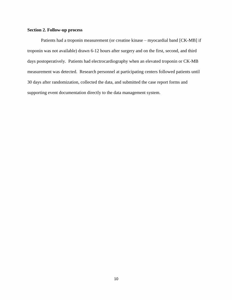
Section 2. Follow-up process
Patients had a troponin measurement (or creatine kinase – myocardial band [CK-MB] if
troponin was not available) drawn 6-12 hours after surgery and on the first, second, and third
days postoperatively. Patients had electrocardiography when an elevated troponin or CK-MB
measurement was detected. Research personnel at participating centers followed patients until
30 days after randomization, collected the data, and submitted the case report forms and
supporting event documentation directly to the data management system.
11
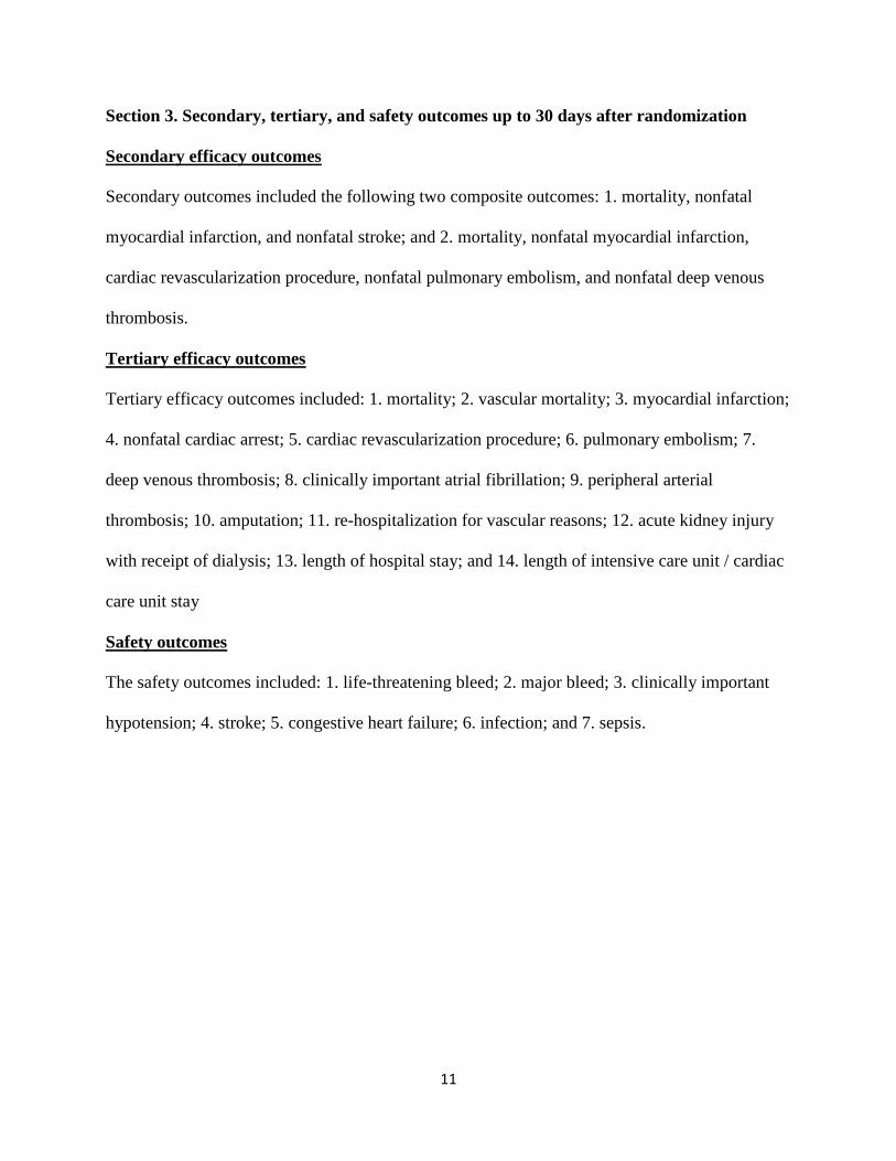
Section 3. Secondary, tertiary, and safety outcomes up to 30 days after randomization
Secondary efficacy outcomes
Secondary outcomes included the following two composite outcomes: 1. mortality, nonfatal
myocardial infarction, and nonfatal stroke; and 2. mortality, nonfatal myocardial infarction,
cardiac revascularization procedure, nonfatal pulmonary embolism, and nonfatal deep venous
thrombosis.
Tertiary efficacy outcomes
Tertiary efficacy outcomes included: 1. mortality; 2. vascular mortality; 3. myocardial infarction;
4. nonfatal cardiac arrest; 5. cardiac revascularization procedure; 6. pulmonary embolism; 7.
deep venous thrombosis; 8. clinically important atrial fibrillation; 9. peripheral arterial
thrombosis; 10. amputation; 11. re-hospitalization for vascular reasons; 12. acute kidney injury
with receipt of dialysis; 13. length of hospital stay; and 14. length of intensive care unit / cardiac
care unit stay
Safety outcomes
The safety outcomes included: 1. life-threatening bleed; 2. major bleed; 3. clinically important
hypotension; 4. stroke; 5. congestive heart failure; 6. infection; and 7. sepsis.
12
Section 4. Outcome definitions
Outcome Definition
Sub classification of death Vascular death was defined as any death with a vascular cause and included those deaths following
a myocardial infarction, cardiac arrest, stroke, cardiac revascularization procedure (i.e.,
percutaneous coronary intervention [PCI] or coronary artery bypass graft [CABG] surgery),
pulmonary embolism, hemorrhage, or deaths due to an unknown cause. Non-vascular death was
defined as any death due to a clearly documented non-vascular cause (e.g. trauma, infection,
malignancy).
Myocardial infarction The diagnosis of myocardial infarction required any one of the following criterion:
1. A typical rise of troponin or a typical fall of an elevated troponin detected at its peak post surgery
in a patient without a documented alternative explanation for an elevated troponin (e.g., pulmonary
embolism) OR a rapid rise and fall of CK-MB. This criterion also required that 1 of the following
was also present:
A. ischemic signs or symptoms;
B. development of pathologic Q waves;
C. electrocardiography (ECG) changes indicative of ischemia;
D. coronary artery intervention (i.e., PCI or CABG surgery); or
E. new or presumed new cardiac wall motion abnormality on echocardiography or new or
presumed new fixed defect on radionuclide imaging;
2. Pathologic findings of an acute or healing myocardial infarction; or
3. Development of new pathological Q waves on an ECG if troponin levels were not obtained or
were obtained at times that could have missed the clinical event.
Nonfatal cardiac arrest Nonfatal cardiac arrest was defined as successful resuscitation from either documented or presumed
ventricular fibrillation, sustained ventricular tachycardia, asystole, or pulseless electrical activity
requiring cardiopulmonary resuscitation, pharmacological therapy, or cardiac defibrillation.
Cardiac revascularization
procedure
Cardiac revascularization procedure was defined as PCI or CABG surgery.
13
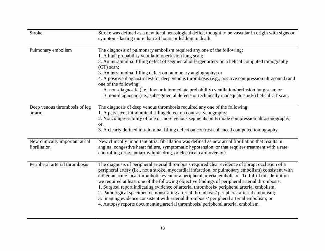
Stroke Stroke was defined as a new focal neurological deficit thought to be vascular in origin with signs or
symptoms lasting more than 24 hours or leading to death.
Pulmonary embolism The diagnosis of pulmonary embolism required any one of the following:
1. A high probability ventilation/perfusion lung scan;
2. An intraluminal filling defect of segmental or larger artery on a helical computed tomography
(CT) scan;
3. An intraluminal filling defect on pulmonary angiography; or
4. A positive diagnostic test for deep venous thrombosis (e.g., positive compression ultrasound) and
one of the following:
A. non-diagnostic (i.e., low or intermediate probability) ventilation/perfusion lung scan; or
B. non-diagnostic (i.e., subsegmental defects or technically inadequate study) helical CT scan.
Deep venous thrombosis of leg
or arm
The diagnosis of deep venous thrombosis required any one of the following:
1. A persistent intraluminal filling defect on contrast venography;
2. Noncompressibility of one or more venous segments on B mode compression ultrasonography;
or
3. A clearly defined intraluminal filling defect on contrast enhanced computed tomography.
New clinically important atrial
fibrillation
New clinically important atrial fibrillation was defined as new atrial fibrillation that results in
angina, congestive heart failure, symptomatic hypotension, or that requires treatment with a rate
controlling drug, antiarrhythmic drug, or electrical cardioversion.
Peripheral arterial thrombosis
The diagnosis of peripheral arterial thrombosis required clear evidence of abrupt occlusion of a
peripheral artery (i.e., not a stroke, myocardial infarction, or pulmonary embolism) consistent with
either an acute local thrombotic event or a peripheral arterial embolism. To fulfill this definition
we required at least one of the following objective findings of peripheral arterial thrombosis:
1. Surgical report indicating evidence of arterial thrombosis/ peripheral arterial embolism;
2. Pathological specimen demonstrating arterial thrombosis/ peripheral arterial embolism;
3. Imaging evidence consistent with arterial thrombosis/ peripheral arterial embolism; or
4. Autopsy reports documenting arterial thrombosis/ peripheral arterial embolism.
14
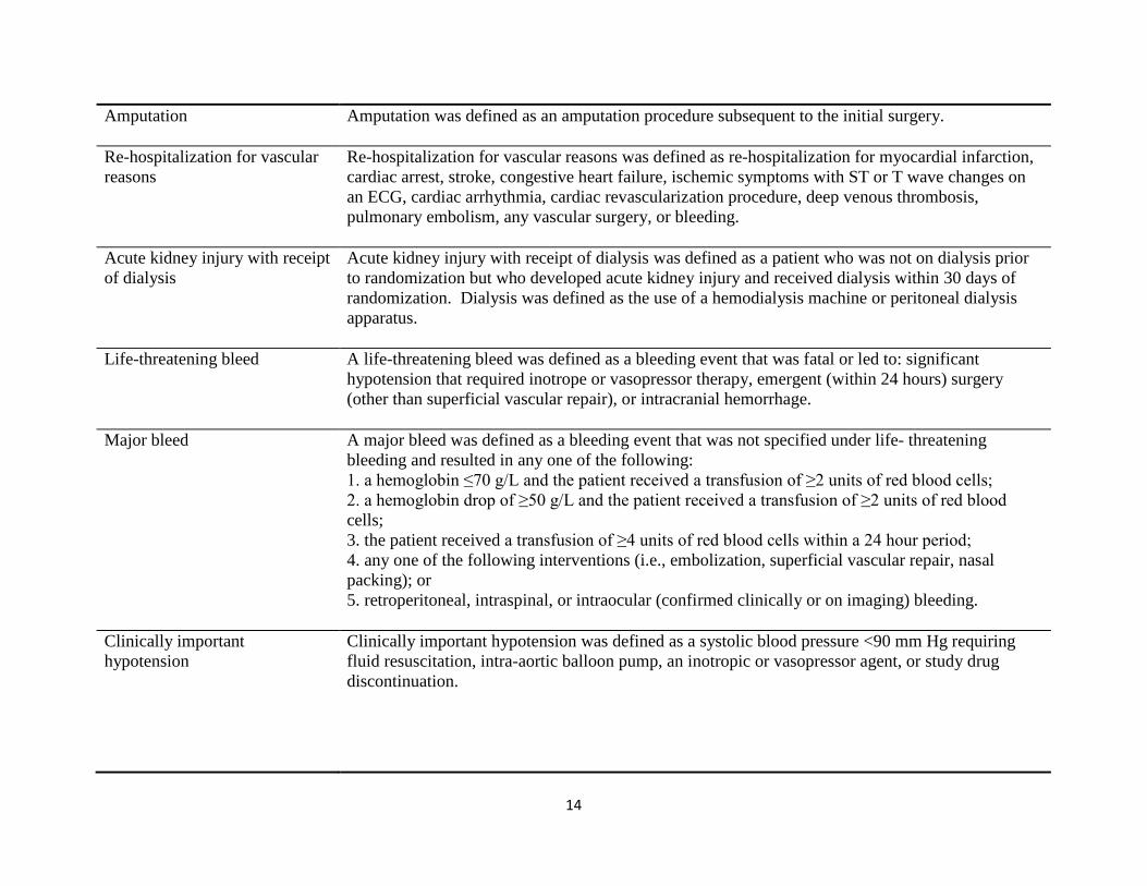
Amputation
Amputation was defined as an amputation procedure subsequent to the initial surgery.
Re-hospitalization for vascular
reasons
Re-hospitalization for vascular reasons was defined as re-hospitalization for myocardial infarction,
cardiac arrest, stroke, congestive heart failure, ischemic symptoms with ST or T wave changes on
an ECG, cardiac arrhythmia, cardiac revascularization procedure, deep venous thrombosis,
pulmonary embolism, any vascular surgery, or bleeding.
Acute kidney injury with receipt
of dialysis
Acute kidney injury with receipt of dialysis was defined as a patient who was not on dialysis prior
to randomization but who developed acute kidney injury and received dialysis within 30 days of
randomization. Dialysis was defined as the use of a hemodialysis machine or peritoneal dialysis
apparatus.
Life-threatening bleed A life-threatening bleed was defined as a bleeding event that was fatal or led to: significant
hypotension that required inotrope or vasopressor therapy, emergent (within 24 hours) surgery
(other than superficial vascular repair), or intracranial hemorrhage.
Major bleed
A major bleed was defined as a bleeding event that was not specified under life- threatening
bleeding and resulted in any one of the following:
1. a hemoglobin ≤70 g/L and the patient received a transfusion of ≥2 units of red blood cells;
2. a hemoglobin drop of ≥50 g/L and the patient received a transfusion of ≥2 units of red blood
cells;
3. the patient received a transfusion of ≥4 units of red blood cells within a 24 hour period;
4. any one of the following interventions (i.e., embolization, superficial vascular repair, nasal
packing); or
5. retroperitoneal, intraspinal, or intraocular (confirmed clinically or on imaging) bleeding.
Clinically important
hypotension
Clinically important hypotension was defined as a systolic blood pressure <90 mm Hg requiring
fluid resuscitation, intra-aortic balloon pump, an inotropic or vasopressor agent, or study drug
discontinuation.
15
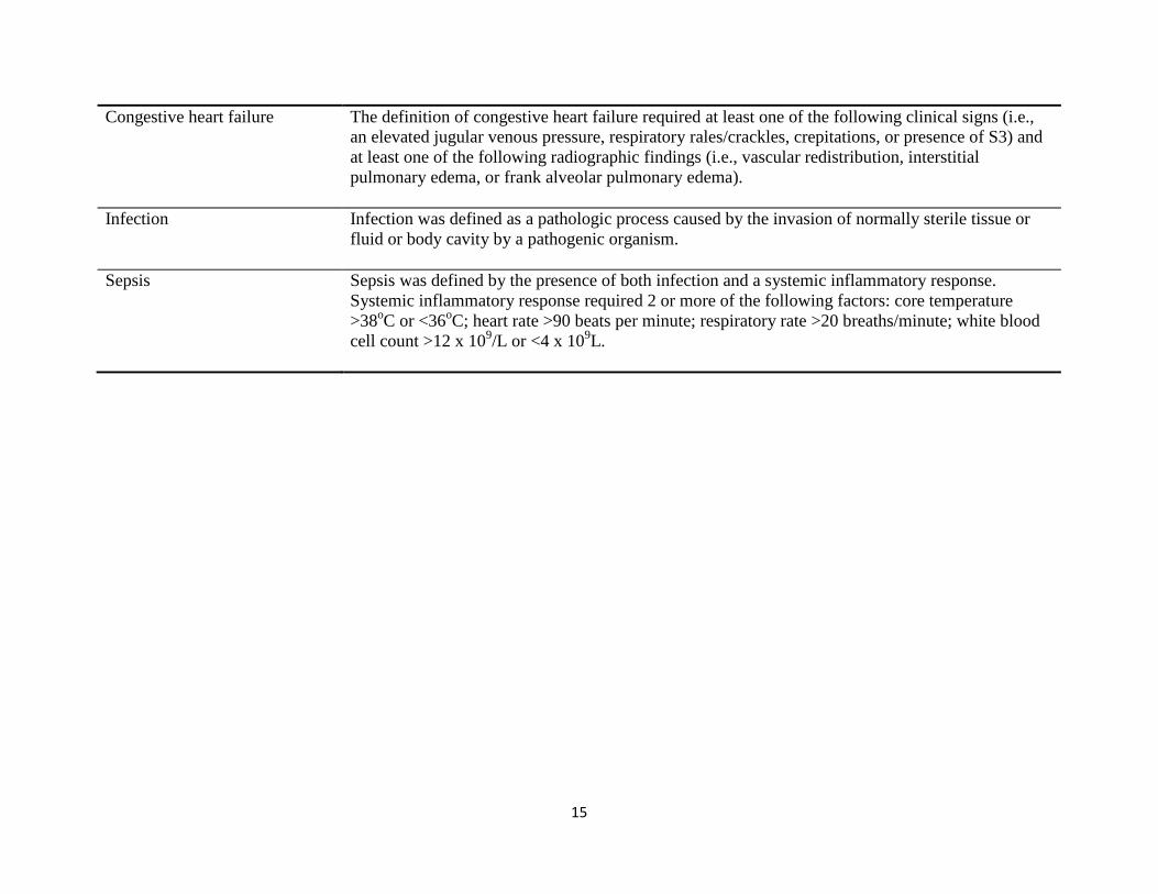
Congestive heart failure
The definition of congestive heart failure required at least one of the following clinical signs (i.e.,
an elevated jugular venous pressure, respiratory rales/crackles, crepitations, or presence of S3) and
at least one of the following radiographic findings (i.e., vascular redistribution, interstitial
pulmonary edema, or frank alveolar pulmonary edema).
Infection Infection was defined as a pathologic process caused by the invasion of normally sterile tissue or
fluid or body cavity by a pathogenic organism.
Sepsis
Sepsis was defined by the presence of both infection and a systemic inflammatory response.
Systemic inflammatory response required 2 or more of the following factors: core temperature
>38oC or <36
oC; heart rate >90 beats per minute; respiratory rate >20 breaths/minute; white blood
cell count >12 x 109/L or <4 x 10
9L.

16
Section 5: Adjudicated outcomes
Outcome adjudicators determined whether a death was vascular or non-vascular, and whether a
patient had a myocardial infarction, nonfatal cardiac arrest, pulmonary embolism, deep venous
thrombosis, stroke, peripheral arterial thrombosis, life-threatening bleed, or major bleed.
17
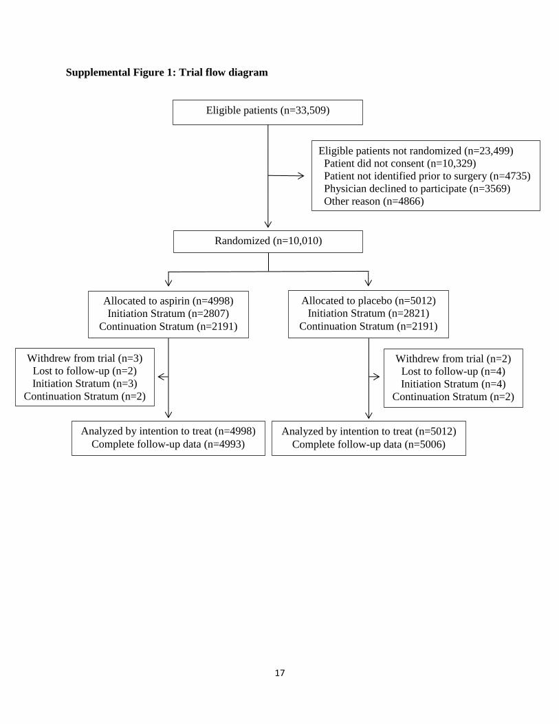
Supplemental Figure 1: Trial flow diagram
Eligible patients (n=33,509)
Eligible patients not randomized (n=23,499)
Patient did not consent (n=10,329)
Patient not identified prior to surgery (n=4735)
Physician declined to participate (n=3569)
Other reason (n=4866)
Randomized (n=10,010)
Allocated to aspirin (n=4998)
Initiation Stratum (n=2807)
Continuation Stratum (n=2191)
Allocated to placebo (n=5012)
Initiation Stratum (n=2821)
Continuation Stratum (n=2191)
Withdrew from trial (n=3)
Lost to follow-up (n=2)
Initiation Stratum (n=3)
Continuation Stratum (n=2)
Analyzed by intention to treat (n=4998)
Complete follow-up data (n=4993)
Analyzed by intention to treat (n=5012)
Complete follow-up data (n=5006)
Withdrew from trial (n=2)
Lost to follow-up (n=4)
Initiation Stratum (n=4)
Continuation Stratum (n=2)
18
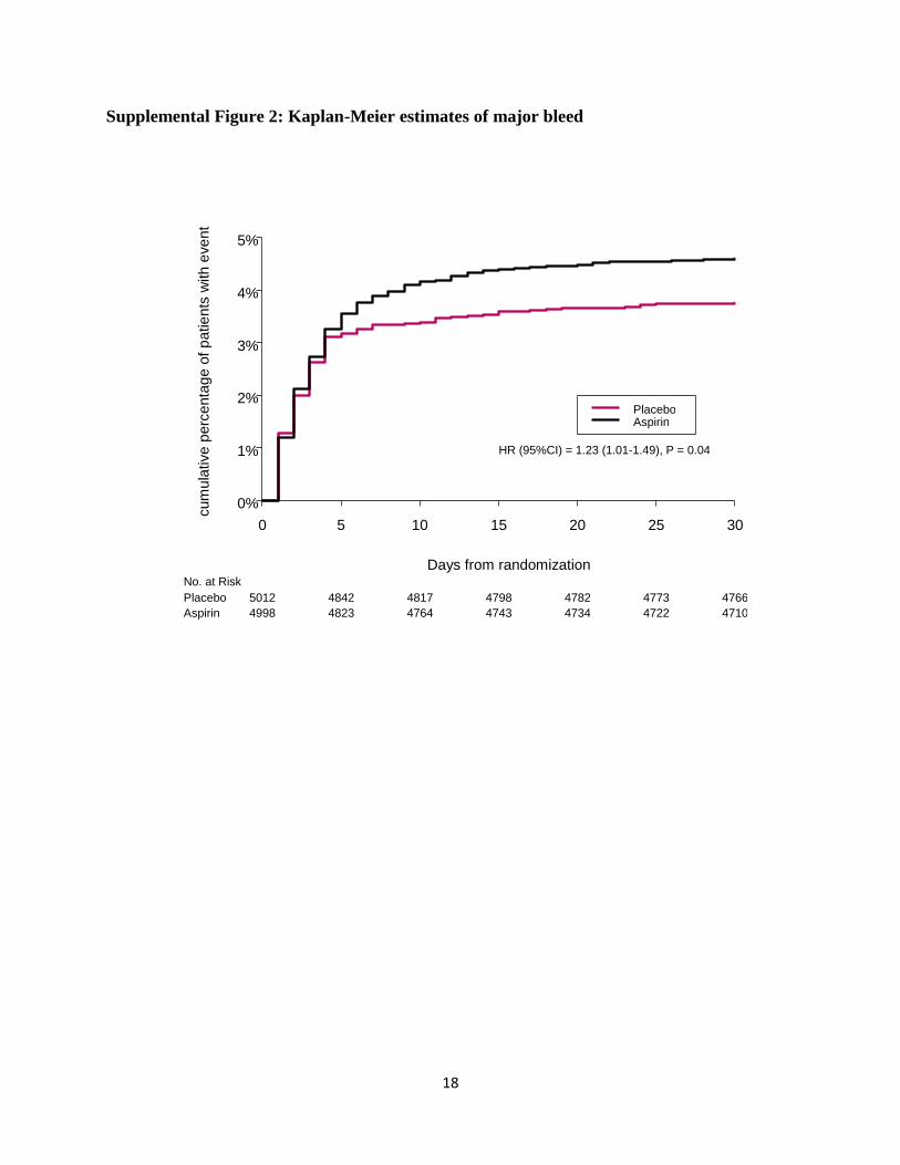
Supplemental Figure 2: Kaplan-Meier estimates of major bleed
Days from randomization
cum
ula
tive
perc
enta
ge o
f p
atie
nts
with e
ve
nt
0 5 10 15 20 25 30
0%
1%
2%
3%
4%
5%
Placebo Aspirin
No. at Risk Placebo Aspirin
HR (95%CI) = 1.23 (1.01-1.49), P = 0.04
5012 4842 4817 4798 4782 4773 4766 4998 4823 4764 4743 4734 4722 4710
19
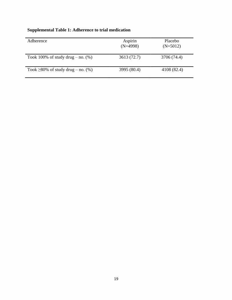
Supplemental Table 1: Adherence to trial medication
Adherence Aspirin
(N=4998)
Placebo
(N=5012)
Took 100% of study drug – no. (%)
3613 (72.7) 3706 (74.4)
Took ≥80% of study drug – no. (%)
3995 (80.4) 4108 (82.4)
20
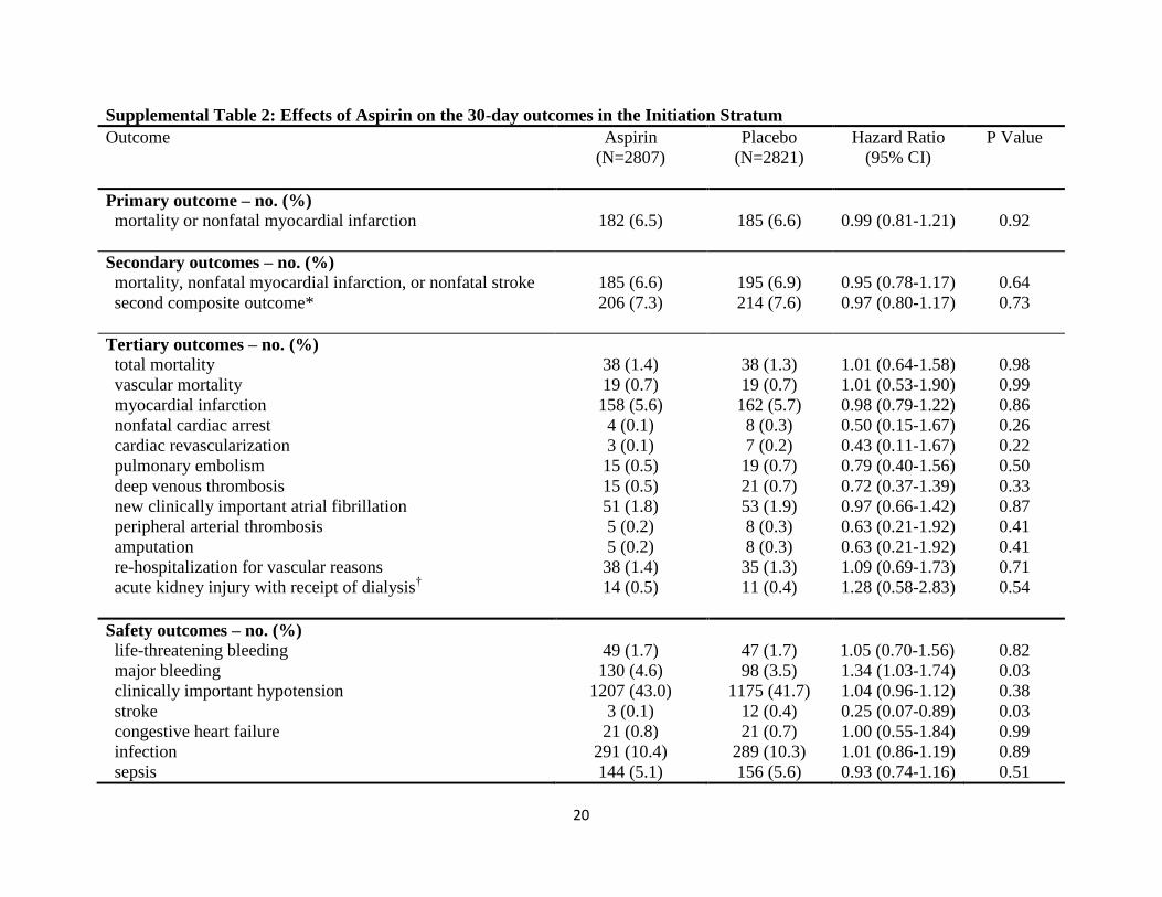
Supplemental Table 2: Effects of Aspirin on the 30-day outcomes in the Initiation Stratum
Outcome Aspirin
(N=2807)
Placebo
(N=2821)
Hazard Ratio
(95% CI)
P Value
Primary outcome – no. (%) mortality or nonfatal myocardial infarction
182 (6.5)
185 (6.6)
0.99 (0.81-1.21)
0.92
Secondary outcomes – no. (%)
mortality, nonfatal myocardial infarction, or nonfatal stroke
second composite outcome*
185 (6.6)
206 (7.3)
195 (6.9)
214 (7.6)
0.95 (0.78-1.17)
0.97 (0.80-1.17)
0.64
0.73
Tertiary outcomes – no. (%)
total mortality
vascular mortality
myocardial infarction
nonfatal cardiac arrest
cardiac revascularization
pulmonary embolism
deep venous thrombosis
new clinically important atrial fibrillation
peripheral arterial thrombosis
amputation
re-hospitalization for vascular reasons
acute kidney injury with receipt of dialysis†
38 (1.4)
19 (0.7)
158 (5.6)
4 (0.1)
3 (0.1)
15 (0.5)
15 (0.5)
51 (1.8)
5 (0.2)
5 (0.2)
38 (1.4)
14 (0.5)
38 (1.3)
19 (0.7)
162 (5.7)
8 (0.3)
7 (0.2)
19 (0.7)
21 (0.7)
53 (1.9)
8 (0.3)
8 (0.3)
35 (1.3)
11 (0.4)
1.01 (0.64-1.58)
1.01 (0.53-1.90)
0.98 (0.79-1.22)
0.50 (0.15-1.67)
0.43 (0.11-1.67)
0.79 (0.40-1.56)
0.72 (0.37-1.39)
0.97 (0.66-1.42)
0.63 (0.21-1.92)
0.63 (0.21-1.92)
1.09 (0.69-1.73)
1.28 (0.58-2.83)
0.98
0.99
0.86
0.26
0.22
0.50
0.33
0.87
0.41
0.41
0.71
0.54
Safety outcomes – no. (%)
life-threatening bleeding
major bleeding
clinically important hypotension
stroke
congestive heart failure
infection
sepsis
49 (1.7)
130 (4.6)
1207 (43.0)
3 (0.1)
21 (0.8)
291 (10.4)
144 (5.1)
47 (1.7)
98 (3.5)
1175 (41.7)
12 (0.4)
21 (0.7)
289 (10.3)
156 (5.6)
1.05 (0.70-1.56)
1.34 (1.03-1.74)
1.04 (0.96-1.12)
0.25 (0.07-0.89)
1.00 (0.55-1.84)
1.01 (0.86-1.19)
0.93 (0.74-1.16)
0.82
0.03
0.38
0.03
0.99
0.89
0.51

21
* the second composite outcome was a composite of mortality, nonfatal myocardial infarction, cardiac revascularization procedure,
nonfatal pulmonary embolism, or nonfatal deep venous thrombosis.
† for this outcome we report the odds ratio instead of the hazard ratio, because we did not collect the actual date patients first started
dialysis.
22
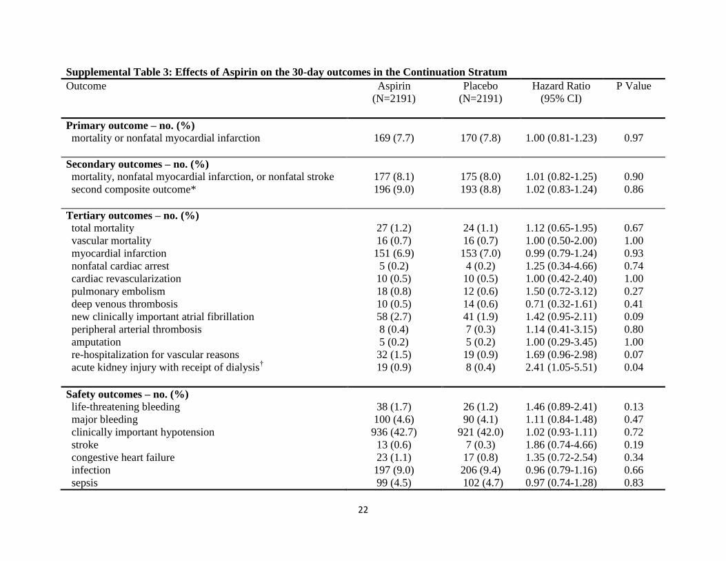
Supplemental Table 3: Effects of Aspirin on the 30-day outcomes in the Continuation Stratum
Outcome Aspirin
(N=2191)
Placebo
(N=2191)
Hazard Ratio
(95% CI)
P Value
Primary outcome – no. (%) mortality or nonfatal myocardial infarction
169 (7.7)
170 (7.8)
1.00 (0.81-1.23)
0.97
Secondary outcomes – no. (%)
mortality, nonfatal myocardial infarction, or nonfatal stroke
second composite outcome*
177 (8.1)
196 (9.0)
175 (8.0)
193 (8.8)
1.01 (0.82-1.25)
1.02 (0.83-1.24)
0.90
0.86
Tertiary outcomes – no. (%)
total mortality
vascular mortality
myocardial infarction
nonfatal cardiac arrest
cardiac revascularization
pulmonary embolism
deep venous thrombosis
new clinically important atrial fibrillation
peripheral arterial thrombosis
amputation
re-hospitalization for vascular reasons
acute kidney injury with receipt of dialysis†
27 (1.2)
16 (0.7)
151 (6.9)
5 (0.2)
10 (0.5)
18 (0.8)
10 (0.5)
58 (2.7)
8 (0.4)
5 (0.2)
32 (1.5)
19 (0.9)
24 (1.1)
16 (0.7)
153 (7.0)
4 (0.2)
10 (0.5)
12 (0.6)
14 (0.6)
41 (1.9)
7 (0.3)
5 (0.2)
19 (0.9)
8 (0.4)
1.12 (0.65-1.95)
1.00 (0.50-2.00)
0.99 (0.79-1.24)
1.25 (0.34-4.66)
1.00 (0.42-2.40)
1.50 (0.72-3.12)
0.71 (0.32-1.61)
1.42 (0.95-2.11)
1.14 (0.41-3.15)
1.00 (0.29-3.45)
1.69 (0.96-2.98)
2.41 (1.05-5.51)
0.67
1.00
0.93
0.74
1.00
0.27
0.41
0.09
0.80
1.00
0.07
0.04
Safety outcomes – no. (%)
life-threatening bleeding
major bleeding
clinically important hypotension
stroke
congestive heart failure
infection
sepsis
38 (1.7)
100 (4.6)
936 (42.7)
13 (0.6)
23 (1.1)
197 (9.0)
99 (4.5)
26 (1.2)
90 (4.1)
921 (42.0)
7 (0.3)
17 (0.8)
206 (9.4)
102 (4.7)
1.46 (0.89-2.41)
1.11 (0.84-1.48)
1.02 (0.93-1.11)
1.86 (0.74-4.66)
1.35 (0.72-2.54)
0.96 (0.79-1.16)
0.97 (0.74-1.28)
0.13
0.47
0.72
0.19
0.34
0.66
0.83
23
* the second composite outcome was a composite of mortality, nonfatal myocardial infarction, cardiac revascularization procedure,
nonfatal pulmonary embolism, or nonfatal deep venous thrombosis.
† for this outcome we report the odds ratio instead of the hazard ratio, because we did not collect the actual date patients first started
dialysis.
24
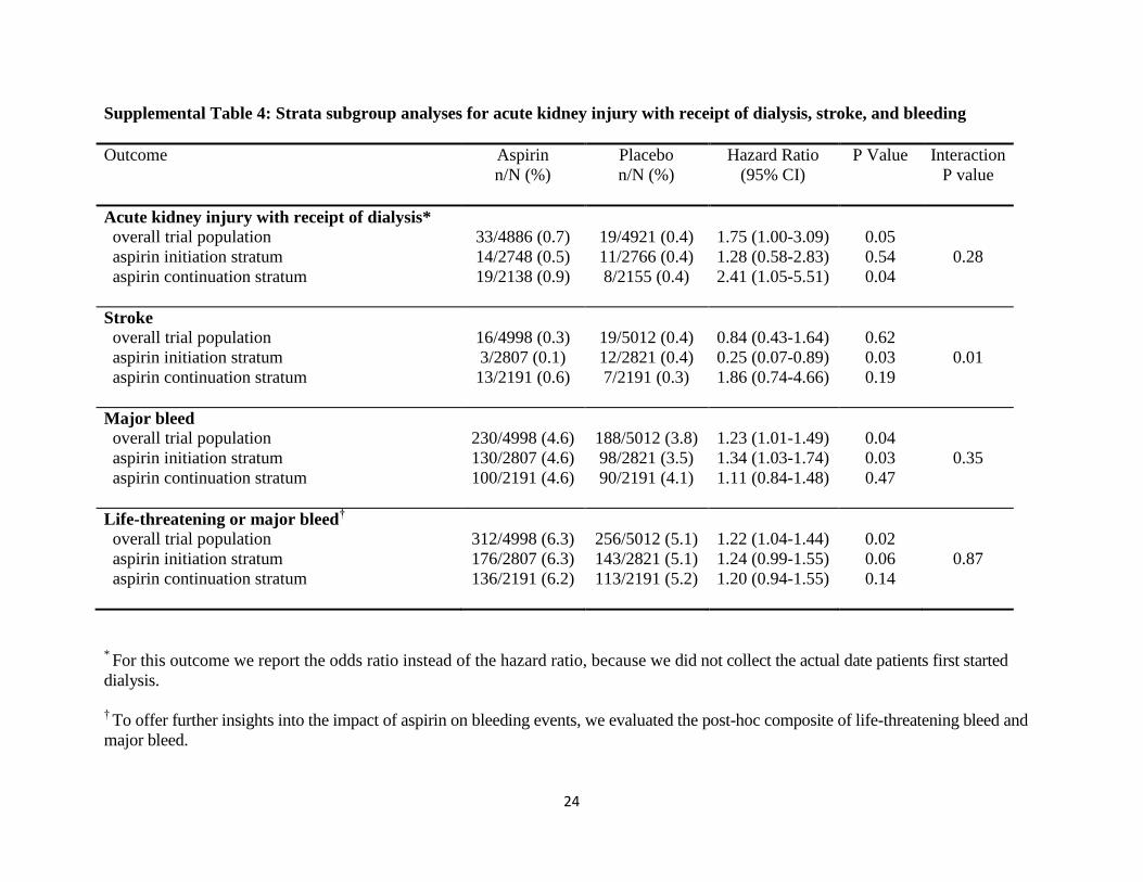
Supplemental Table 4: Strata subgroup analyses for acute kidney injury with receipt of dialysis, stroke, and bleeding
Outcome Aspirin
n/N (%)
Placebo
n/N (%)
Hazard Ratio
(95% CI)
P Value Interaction
P value
Acute kidney injury with receipt of dialysis*
overall trial population
aspirin initiation stratum
aspirin continuation stratum
33/4886 (0.7)
14/2748 (0.5)
19/2138 (0.9)
19/4921 (0.4)
11/2766 (0.4)
8/2155 (0.4)
1.75 (1.00-3.09)
1.28 (0.58-2.83)
2.41 (1.05-5.51)
0.05
0.54
0.04
0.28
Stroke
overall trial population
aspirin initiation stratum
aspirin continuation stratum
16/4998 (0.3)
3/2807 (0.1)
13/2191 (0.6)
19/5012 (0.4)
12/2821 (0.4)
7/2191 (0.3)
0.84 (0.43-1.64)
0.25 (0.07-0.89)
1.86 (0.74-4.66)
0.62
0.03
0.19
0.01
Major bleed
overall trial population
aspirin initiation stratum
aspirin continuation stratum
230/4998 (4.6)
130/2807 (4.6)
100/2191 (4.6)
188/5012 (3.8)
98/2821 (3.5)
90/2191 (4.1)
1.23 (1.01-1.49)
1.34 (1.03-1.74)
1.11 (0.84-1.48)
0.04
0.03
0.47
0.35
Life-threatening or major bleed†
overall trial population
aspirin initiation stratum
aspirin continuation stratum
312/4998 (6.3)
176/2807 (6.3)
136/2191 (6.2)
256/5012 (5.1)
143/2821 (5.1)
113/2191 (5.2)
1.22 (1.04-1.44)
1.24 (0.99-1.55)
1.20 (0.94-1.55)
0.02
0.06
0.14
0.87
* For this outcome we report the odds ratio instead of the hazard ratio, because we did not collect the actual date patients first started
dialysis.
† To offer further insights into the impact of aspirin on bleeding events, we evaluated the post-hoc composite of life-threatening bleed and
major bleed.
25
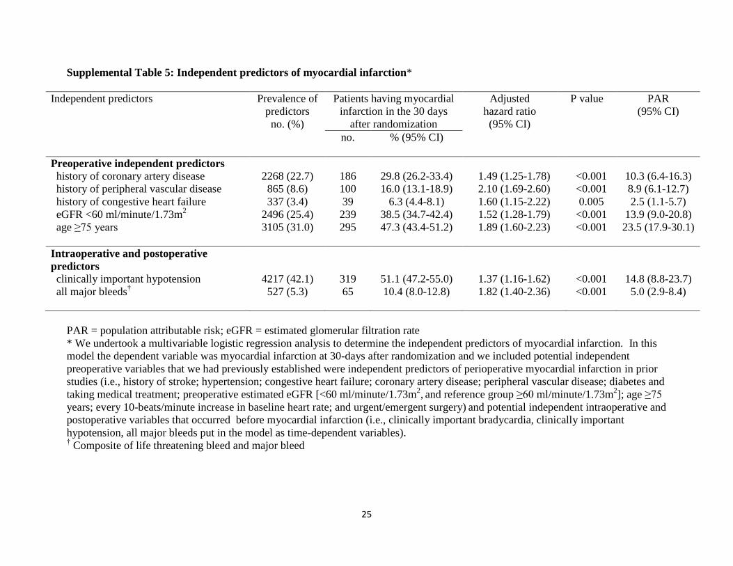
Supplemental Table 5: Independent predictors of myocardial infarction*
Independent predictors Prevalence of
predictors
no. (%)
Patients having myocardial
infarction in the 30 days
after randomization
Adjusted
hazard ratio
(95% CI)
P value PAR
(95% CI)
no. % (95% CI)
Preoperative independent predictors
history of coronary artery disease
history of peripheral vascular disease
history of congestive heart failure
eGFR <60 ml/minute/1.73m2
age ≥75 years
2268 (22.7)
865 (8.6)
337 (3.4)
2496 (25.4)
3105 (31.0)
186
100
39
239
295
29.8 (26.2-33.4)
16.0 (13.1-18.9)
6.3 (4.4-8.1)
38.5 (34.7-42.4)
47.3 (43.4-51.2)
1.49 (1.25-1.78)
2.10 (1.69-2.60)
1.60 (1.15-2.22)
1.52 (1.28-1.79)
1.89 (1.60-2.23)
<0.001
<0.001
0.005
<0.001
<0.001
10.3 (6.4-16.3)
8.9 (6.1-12.7)
2.5 (1.1-5.7)
13.9 (9.0-20.8)
23.5 (17.9-30.1)
Intraoperative and postoperative
predictors
clinically important hypotension
all major bleeds†
4217 (42.1)
527 (5.3)
319
65
51.1 (47.2-55.0)
10.4 (8.0-12.8)
1.37 (1.16-1.62)
1.82 (1.40-2.36)
<0.001
<0.001
14.8 (8.8-23.7)
5.0 (2.9-8.4)
PAR = population attributable risk; eGFR = estimated glomerular filtration rate
* We undertook a multivariable logistic regression analysis to determine the independent predictors of myocardial infarction. In this
model the dependent variable was myocardial infarction at 30-days after randomization and we included potential independent
preoperative variables that we had previously established were independent predictors of perioperative myocardial infarction in prior
studies (i.e., history of stroke; hypertension; congestive heart failure; coronary artery disease; peripheral vascular disease; diabetes and
taking medical treatment; preoperative estimated eGFR [<60 ml/minute/1.73m2, and reference group ≥60 ml/minute/1.73m
2]; age ≥75
years; every 10-beats/minute increase in baseline heart rate; and urgent/emergent surgery) and potential independent intraoperative and
postoperative variables that occurred before myocardial infarction (i.e., clinically important bradycardia, clinically important
hypotension, all major bleeds put in the model as time-dependent variables). † Composite of life threatening bleed and major bleed