geophysical investigation of faults and fractures in kiamunyi
Upload
independentCategory
view
3download
0
Podoplanin Immunopositive Lymphatic Vessels at theImplant Interface in a Rat Model of OsteoporoticFracturesKatrin Susanne Lips1*, Vivien Kauschke1, Sonja Hartmann1, Ulrich Thormann2, Seemun Ray1, MarianKampschulte3, Alexander Langheinrich4, Matthias Schumacher5, Michael Gelinsky5, Sascha Heinemann6,Thomas Hanke6, Armin R. Kautz7, Matthias Schnabelrauch7, Reinhard Schnettler1,2, Christian Heiss2,Volker Alt2, Olaf Kilian1,8
1 Laboratory of Experimental Trauma Surgery, Justus-Liebig University, Gießen, Germany, 2 Department of Trauma Surgery Gießen, University Hospital ofGießen, Marburg, Justus-Liebig University, Gießen, Germany, 3 Department of Radiology, Justus-Liebig University, Gießen, Germany, 4 Department ofDiagnostic and Interventional Radiology, BG Trauma Hospital, Frankfurt/Main, Germany, 5 Centre for Translational Bone, Joint, and Soft Tissue Research,Medical Faculty and University Hospital, Technische Universität, Dresden, Germany, 6 Max-Bergmann-Center of Biomaterials and Institute of Material Science,Technische Universität, Dresden, Germany, 7 Biomaterials Department, INNOVENT e.V., Jena, Germany, 8 Department of Orthopedics and Trauma,Zentralklinik, Bad Berka, Germany
Abstract
Insertion of bone substitution materials accelerates healing of osteoporotic fractures. Biodegradable materials arepreferred for application in osteoporotic patients to avoid a second surgery for implant replacement. Degradedimplant fragments are often absorbed by macrophages that are removed from the fracture side via passage throughveins or lymphatic vessels. We investigated if lymphatic vessels occur in osteoporotic bone defects and whether theyare regulated by the use of different materials. To address this issue osteoporosis was induced in rats using theclassical method of bilateral ovariectomy and additional calcium and vitamin deficient diet. In addition, wedge-shapeddefects of 3, 4, or 5 mm were generated in the distal metaphyseal area of femur via osteotomy. The 4 mm defectswere subsequently used for implantation studies where bone substitution materials of calcium phosphate cement,composites of collagen and silica, and iron foams with interconnecting pores were inserted. Different materials werepartly additionally functionalized by strontium or bisphosphonate whose positive effects in osteoporosis treatment arewell known. The lymphatic vessels were identified by immunohistochemistry using an antibody against podoplanin.Podoplanin immunopositive lymphatic vessels were detected in the granulation tissue filling the fracture gap,surrounding the implant and growing into the iron foam through its interconnected pores. Significant more lymphaticcapillaries were counted at the implant interface of composite, strontium and bisphosphonate functionalized ironfoam. A significant increase was also observed in the number of lymphatics situated in the pores of strontium coatediron foam. In conclusion, our results indicate the occurrence of lymphatic vessels in osteoporotic bone. Our resultsshow that lymphatic vessels are localized at the implant interface and in the fracture gap where they might beinvolved in the removal of lymphocytes, macrophages, debris and the implants degradation products. Therefore thelymphatic vessels are involved in implant integration and fracture healing.
Citation: Lips KS, Kauschke V, Hartmann S, Thormann U, Ray S, et al. (2013) Podoplanin Immunopositive Lymphatic Vessels at the Implant Interface in aRat Model of Osteoporotic Fractures. PLoS ONE 8(10): e77259. doi:10.1371/journal.pone.0077259
Editor: Stephen E Alway, West Virginia University School of Medicine, United States of America
Received April 29, 2013; Accepted August 31, 2013; Published October 9, 2013
Copyright: © 2013 Lips et al. This is an open-access article distributed under the terms of the Creative Commons Attribution License, which permitsunrestricted use, distribution, and reproduction in any medium, provided the original author and source are credited.
Funding: The study was funded by the Deutsche Forschungsgesellschaft DFG (SFB/TRR 79 projects B7, T1, T2 M2, M3 and Z3). The funders had no rolein study design, data collection and analysis, decision to publish, or preparation of the manuscript.
Competing interests: The authors have declared that no competing interests exist.
* E-mail: [email protected]
Introduction
The insertion of bone substitution materials has been provento accelerate healing of osteoporotic fractures. Osteoporosis isa systemic disease with a characteristic decrease of bonestrength and therefore an increase in fracture risk [1]. Usually,
postmenopausal women and aged men contract this disease.Because of the demographic trend elderly people will increasein the population which is why in future more people will sufferfrom osteoporosis and osteoporotic fractures [1]. Thecommercially available bone substitution materials are notetiology-adapted and therefore not appropriate for the
PLOS ONE | www.plosone.org 1 October 2013 | Volume 8 | Issue 10 | e77259
application in osteoporotic damaged bone. In the last years ithas been worked out that modification with strontium [2], [3],bisphosphonate [4] as well as enhanced resorption capacityand changes from solid material to interconnected porousmaterial [5] improves the materials. High resorption capacitygoes along with the disposal of the degraded fragments ofteninserted in cell organelles of macrophages. Macrophagescontaining biomaterials in intracellular vesicles are able todifferentiate into osteoclasts which are responsible for boneresorption [6]. Macrophages are usually removed via veins andlymphatic vessels [7]. The lymphatic network begins with thelymphatic capillaries in the peripheral tissues that transportlymph to the venous system via passage through severalregional and collective lymph nodes. Lymph consists ofinterstitial fluid, lymphocytes, and macrophages. Lymphaticcapillaries differ from vascular capillaries in their largerdiameter, irregular outline, thinner walls, incomplete basalmembrane, overlapping endothelial cells, and molecularcomposition of their endothelial cells [7]. In the last yearsseveral markers have been identified, that are qualified foridentification of lymphatic vessels [8]. The aim of the presentstudy was to identify the lymphatic vessels in a rat model ofosteoporotic fractures which were stabilized by newlyestablished bone substitution materials. Therefore, we recentlyestablished a fracture defect model in the distal metaphysealarea of the femur of osteoporotic rats [9-11]. We decided to usesmall animals in this first approach because of the shorterbreeding cycles, easier and less expensive animal housing thatallowed us to investigate several different bone substitutionmaterials. A follow up study in a large animal model (e.g. ovinemodel) where only the most suitable biomaterials from this firststudy will be integrated would be very helpful because of thecloser mimic of large animals to the situation in humans. Ratlymphatic vessels were detected by immunohistochemicalstaining with a monoclonal antibody against podoplanin.Podoplanin is an integral membrane glycoprotein [12] that wasfirst identified in the murine osteoblast cell line MC3T3 [13].Later it was clarified that podoplanin occurs in matureosteoblasts and newly formed osteocytes [14]. The namepodoplanin is explained by its occurrence in podocytes of thekidney [15], [16]. Besides it is also expressed in tumors (e.g.[17], [18] [19], [20],), reticular and epithelial cells of lymphatictissues [21], alveolar type 1 cells of the lung [22], [23] [24],,fibroblasts and premalignant keratinocytes of skin [19], [25],stromal cells of placenta [26], choroid plexus, ependymal andneuronal cells of brain [27], [28], and fibroblast-likesynoviocytes in rheumatoid arthritis [29]. Because of thiswidespread distribution the protein is also well known under thesynonyms OTS-8 [13], E11 antigen [14], gp38 [21], T1α [22],PA2.26 [19], RANDAM-2 [28], RTI40 [23], and Aggrus [17].Lymphatic vessels were amongst others found in skeletalmuscle [30], heart [31], and during pathological conditions ofeye [32], skin [19], [25], and bone [7], [33]. Lymphatic vesselswere absent in healthy cancellous and cortical bone butcommonly exist in the periosteum and connective tissue [34],[7]. Lymphatic vessels only occur in bone after lesions,fractures, or degenerations [7], [33]. It is supposed that thelympahtics are spreading from the periosteum and the
surrounding connective tissue into the damaged bone area.Lymphangiogenesis is stimulated by inflammatory cytokinesbecause of its function in transport of immune cells andclearance of antigen [35]. Pro-inflammatory mediators alsopromote the formation of hematoma as one of the first steps offracture healing [36]. The hematoma is infiltrated by immunecompetent cells (e.g. lymphocytes, granulocytes) andmacrophages [36]. The duty of these cells is the removal ofdebris and the release of cytokines and growth factors thatpromote fracture healing. Neutrophil granulocytes are usuallyrecruited by lymphocytes that afterwards leave the hematomathrough lymphatic vessels. Thus lymphatic vessels are usuallyappearing during the subsequent stages of fracture healingwhere lymphocytes, neutrophils, and macrophages have to beremoved after they fulfilled their duty. However, there might bea relation between lymphangiogenesis and fracture healing.Implantation of bone substitution materials leads to animproved fracture healing. If bio-degradable material is usedmacrophages are involved in the resorption and removal ofdegradation products. An increase in macrophages did notresult in a higher number of lymphatic vessels [7]. Also theimplantation of plain hydroxyl apatite or its modification withplatelet factors did not result in significant differences in thelymphangiogenesis [7]. Nevertheless, lymphatic vesselsoccurred in the granulations tissue after implantation of bothmaterials. On this background the aim of the present study wasto investigate if lymphatic vessels occurred in systematicallydiseased bone as well as in fracture healing and implantinsertion in osteoporotic bone. Furthermore, we asked whetherthe choice of implant material influences lymphangiogenesis.The obtained results might help for further improvement ofimplant materials.
Materials and Methods
Ethics StatementAll animal procedures were carried out in accordance to the
Guide for Care and Use of Laboratory Animals of the NationalInstitutes of Health. The study was approved by the local ethiccommittee (Regierungspräsidium Gießen, Permit Number: V54 - 19c20-15 (1) GI 20/28, Nr. 92/2009). All efforts were madeto minimize suffering.
Animal modelWe recently established a fracture defect model in the distal
femoral metaphysis of osteoporotic rats [9-11]. In brief, 14week old female Spargue-Dawley rats (Charles River, Sulzfeld,Germany) underwent laparotomy with subsequent removal ofovaries under deep anaesthesia with i. p. injections of ketamine(62.5 mg/kg bodyweight, Hostaket®, Hoechst) and xylazine (7.5mg/kg bodyweight, Rompun®, Bayer). The animals were keptunder a 12 h light-dark cycle with free access to water andchow that were specifically produced for deficiency diet ofcalcium, vitamin C/D2/D3, and phosphate (Altromin-C1034,Altromin-Spezialfutter GmbH, Lage, Germany) [10]. Againunder deep anesthesia a wedge-shaped methaphyseal fractureof 3, 4, and 5 mm in diameter was performed at the left distalfemur 12 weeks of ovariectomy and the 4 mm defects were
Podoplanin in Osteoporotic Fractures
PLOS ONE | www.plosone.org 2 October 2013 | Volume 8 | Issue 10 | e77259
filled with one of the implants as described in detail earlier [11],[9]. In brief, a skin incision was made, a 7-hole T-shapedminiplate (Leibinger® XS-miniplate, Stryker®, Schönkirchen,Germany) was fixed with 1.7 mm screws on the lateral femur,and the defect was created, rinsed with sterile water, filled withthe implant, and closed in multilayers and by prolene 4/0stitches. Always 7 animals got the same implant (groups) andwere sacrificed 6 weeks after surgery. Via the abdominal aortaa lead containing radio-opaque polymer (Microfil® MV-122,Flow Tech, Carver, MA, USA) was infused rinsing the vesselswith heparinized saline up to blood freeness (10 ml of 0.9 %sodium chloride with 1000 LU heparin) [11]. In the histologicalstaining the polymerized Microfil® could be found in the lumenof blood vessels.
Bone substitution materialsAs implant materials calcium phosphate bone cement (CPC)
[37] that was composed of 58 wt% of α-tricalcium phosphate[Ca3(PO4)2], 24 wt% dicalcium phosphate (CaHPO4), 8.5 wt%precipitated hydroxyapatite [Ca10(PO4)6(OH)2], and 8.5 wt%calcium carbonate (CaCO3) was used plain and with strontiummodification (CPC-S). CPC-S was generated by replacementof CaCO3 with strontium carbonate (SrCO3) in the cementprecursor powder (Sr/Ca = 0.123) [38]. Implantation itself wasconducted with a paste formulation of the cement that wascreated by mixing with an aqueous solution of 4 % Na2HPO4.
Furthermore, composites consisting of silica and fibrillarbovine collagen were used for implantation as monolithicxerogels (B30, 70 wt% silica, 30 wt% collagen) or as porousscaffold (pB30, xerogel particles B30, size < 250 µm,embedded in a collagen matrix with xerogel/matrix weight ratioof 1.0) [39], [11], [40]. A modification of the scaffolds wasprepared by using xerogel particles consisting of 50 wt% silica,30 wt% fibrillar bovine collagen, and 20 wt% strontiumcarbonate (pB30S20). Moreover, iron foam with interconnectedpores (Fe) was applied. The iron-foam was basically created ofcarbonyl iron, Fe3-powder, and a polyvinyl alcohol binder bypowder-metallurgical replication, subsequent sintering at1150°C, and shaping by wire eroding. The iron foam wascoated with a) SrCO3 H3PO4 (Fe-S) under vacuum for 4 hours(h) and rinsed afterwards with ethanol, and b) with zolendronicacid (Fe-BP) that is a member of the bisphosphonate family.Fe-BP was created by precipitation of zolendronic acid,stearate, and iron. Then, the complex was carefully washed,dried, grinded and finally resulted in a coating of 35 µgzolendronic acid on the basic iron foam. These 8 differentformulations of bone substitution materials were implanted inthe current animal model.
ImmunohistochemistryLeft femora were fixed in 4% phosphate-buffered
paraformaldehyde (Merck, Darmstadt, Germany), dehydratedthrough a series of ethanol gradient, embedded inmethylmethacrylate (Technovit 9100, Heraeus Kulzer, Hanau,Germany), and cut into slices with a thickness of 5 µm using arotations microtome (Leica RM2155, Wetzlar, Germany).Sections were deplasticized with 3-Methoxyethylacetat (MEA,Merck, 3 x 20 min) and rehydrated through acetone and Tris-
NaCl buffer with 0.025 % Triton-X-100 (wash buffer, pH 7.4).Endogenous peroxidase was blocked with 3 % H2O2 in washbuffer for 5 min. Sections were incubated with primary mousemonoclonal anti podoplanin antibody (antibodies-online,Atlanta, GA, USA) and rabbit anti CD31 antibody (Abbiotec,San Diego, CA, USA) (dilution 1:8000 for podoplanin and 1:350for CD31; dilution buffer was from Dako, Glostrup, Denmark) at4°C for 16 hours. After rinsing in wash buffer sections wereincubated with biotinlated horse-anti-mouse secondaryantiserum (dilution: 1:500) and goat anti-rabbit secondaryantiserum (dilution: 1:500) in Tris-NaCl with 1 % bovine serumalbumin (Sigma, Taufkirchen, Deutschland) and 12.5 % ratserum (PAA, Pasching, Austria) for 30 min. Afterwards sectionswere incubated for 30 min in the ABC complex/horseradishperoxidase labeled avidin (Vector laboratories, Burlingame,California, USA) with subsequent development of the stainingvia incubation in Nova Red (Vector laboratories).Counterstaining of nuclei was performed with hematoxylin (1:3dilution; Shandon Scientific Ltd, Cheshire, UK) andcoverslipped with DePex (Serva, Heidelberg, Deutschland).
Quantification of lymphatic vesselsOne investigator counted the lymphatic vessels manually
using an ocular micrometer. Additionally, a double blind studywas conducted where a second investigator confirmed theacquired data. The lymphatic vessels were quantified in thewhole fracture gap in sections with empty defects or withoutresidues of the implants. An area of 250 µm at the implantinterface was analyzed if the implants remained or at least aresidual existed. In a separate set of examinations lymphaticvessels penetrating the iron foam were counted. A manualdetermination of the lymphatic vessels was necessary becausethe lymphatic vessels were not uniformly distributed. Theevaluation was conducted with a 20 x objective at the lightmicroscope (Zeiss, Jena, Germany).
Real-time RT-PCRTissue from the implant interface and the fracture gap was
collected in RNAlater immediately after scarification of animals.Total RNA was isolated with the Lipid Tissue mini Kit (Qiagen,Hilden, Germany) according to manufacturer’s protocol. Inbrief, 100 mg of the sample was homogenized in 1 ml QIAzollysis reagents with a ultra-turrax (IKA, Staufen, Germany), 200µl chloroform were added and centrifuged for 15 min at 4°C.The RNA containing upper fraction was collected and washedwith 70 % ethanol and Kit buffer RW1. The dried RNA wasresuspended with RNase-free water and reverse transcribedwith the Quantitect Kit (Qiagen). Contaminations of genomicDNA were removed using 2 µl DNA Wipeout buffer (componentof the Kit) for 2 min at 42°C. Afterwards 1 µg RNA wasincubated with 1 µl Quantiscript reverse transcriptase, 4 µlbuffer and 1 µl primer mix containing random-primers andOligo(dT)s at 42° for 30 min. The reverse transcriptase wasinactivated at 95°C for 3 min. Real-time RT-PCR was carriedout with the Light-Cycler Fast Start DNA Master plus SYBRGreen Kit (Roche Mannheim, Germany) in a Light Cycler 2.0(Roche). 2 µl of cDNA, 2 µl Roche reagent (Kit component), 6.8µl RNase-free water and 0.2 µl forward and reverse primer
Podoplanin in Osteoporotic Fractures
PLOS ONE | www.plosone.org 3 October 2013 | Volume 8 | Issue 10 | e77259
(Table 1, MWG Biotech, Ebersberg, Germany) were added andincubated for 10 min at 95°C, following by 40 cycles of 5seconds (s) heating at 95°C, annealing for 5 s at 58°C andelongation for 5 s at 72°C. The PCR product was controlled bymelting curve and gel electrophoresis. As negative controls a)reverse transcription was carried out without adding theenzyme and b) water was used instead of template forconducting PCR. Calculation of Cp-values was performed byLight Cycler software. For quantification Cp-values ofpodoplanin were normalized with the reference gene β-actin.
Statistical analysisStatistical analysis was performed using the SPSS software
(version 21.0; SPSS Institute Inc, Cicago, USA), non-parametric Kruskal-Wallis and Mann-Whitney tests. Confidencelevel of p ≤ 0.05 was considered to be significant.
Results
Osteoporotic fracture modelFractures without implanted bone substitution material
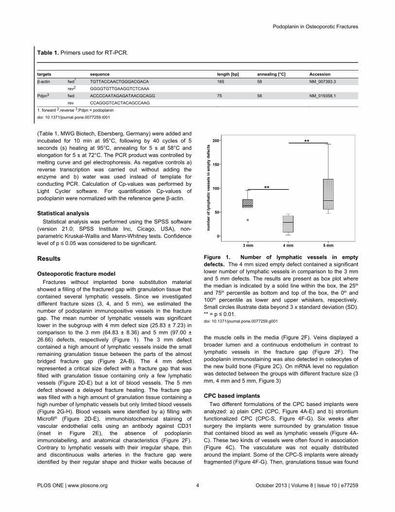
showed a filling of the fractured gap with granulation tissue thatcontained several lymphatic vessels. Since we investigateddifferent fracture sizes (3, 4, and 5 mm), we estimated thenumber of podoplanin immunopositive vessels in the fracturegap. The mean number of lymphatic vessels was significantlower in the subgroup with 4 mm defect size (25.83 ± 7.23) incomparison to the 3 mm (64.83 ± 8.36) and 5 mm (97.00 ±26.66) defects, respectively (Figure 1). The 3 mm defectcontained a high amount of lymphatic vessels inside the smallremaining granulation tissue between the parts of the almostbridged fracture gap (Figure 2A-B). The 4 mm defectrepresented a critical size defect with a fracture gap that wasfilled with granulation tissue containing only a few lymphaticvessels (Figure 2D-E) but a lot of blood vessels. The 5 mmdefect showed a delayed fracture healing. The fracture gapwas filled with a high amount of granulation tissue containing ahigh number of lymphatic vessels but only limited blood vessels(Figure 2G-H). Blood vessels were identified by a) filling withMicrofil® (Figure 2D-E), immunohistochemical staining ofvascular endothelial cells using an antibody against CD31(inset in Figure 2E), the absence of podoplaninimmunolabelling, and anatomical characteristics (Figure 2F).Contrary to lymphatic vessels with their irregular shape, thinand discontinuous walls arteries in the fracture gap wereidentified by their regular shape and thicker walls because of
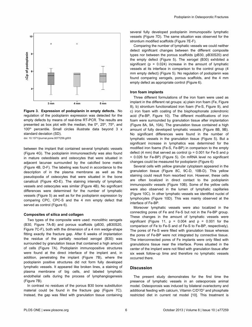
the muscle cells in the media (Figure 2F). Veins displayed abroader lumen and a continuous endothelium in contrast tolymphatic vessels in the fracture gap (Figure 2F). Thepodoplanin immunostaining was also detected in osteocytes ofthe new build bone (Figure 2C). On mRNA level no regulationwas detected between the groups with different fracture size (3mm, 4 mm and 5 mm, Figure 3)
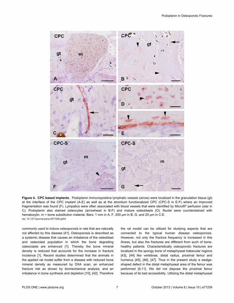
CPC based implantsTwo different formulations of the CPC based implants were
analyzed: a) plain CPC (CPC, Figure 4A-E) and b) strontiumfunctionalized CPC (CPC-S, Figure 4F-G). Six weeks aftersurgery the implants were surrounded by granulation tissuethat contained blood as well as lymphatic vessels (Figure 4A-C). These two kinds of vessels were often found in association(Figure 4C). The vasculature was not equally distributedaround the implant. Some of the CPC-S implants were alreadyfragmented (Figure 4F-G). Then, granulations tissue was found
Figure 1. Number of lymphatic vessels in emptydefects. The 4 mm sized empty defect contained a significantlower number of lymphatic vessels in comparison to the 3 mmand 5 mm defects. The results are present as box plot wherethe median is indicated by a solid line within the box, the 25th
and 75th percentile as bottom and top of the box, the 0th and100th percentile as lower and upper whiskers, respectively.Small circles illustrate data beyond 3 x standard deviation (SD).** = p ≤ 0.01.doi: 10.1371/journal.pone.0077259.g001
Table 1. Primers used for RT-PCR.
targets sequence length [bp] annealing [°C] Accessionβ-actin fwd1 TGTTACCAACTGGGACGACA 165 58 NM_007393.3 rev2 GGGGTGTTGAAGGTCTCAAA Pdpn3 fwd ACCCCAATAGAGATAACGCAGG 75 58 NM_019358.1 rev CCAGGGTCACTACAGCCAAG
1. forward 2,reverse 3,Pdpn = podoplanindoi: 10.1371/journal.pone.0077259.t001
Podoplanin in Osteoporotic Fractures
PLOS ONE | www.plosone.org 4 October 2013 | Volume 8 | Issue 10 | e77259
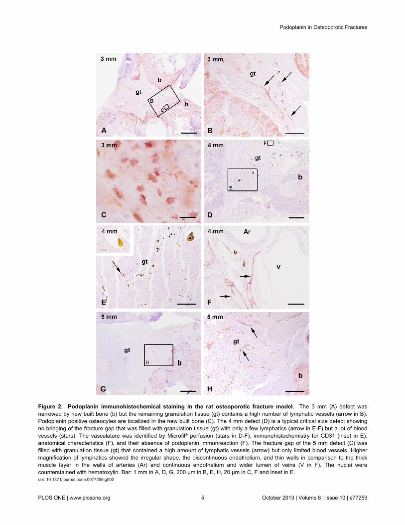
Figure 2. Podoplanin immunohistochemical staining in the rat osteoporotic fracture model. The 3 mm (A) defect wasnarrowed by new built bone (b) but the remaining granulation tissue (gt) contains a high number of lymphatic vessels (arrow in B).Podoplanin positive osteocytes are localized in the new built bone (C). The 4 mm defect (D) is a typical critical size defect showingno bridging of the fracture gap that was filled with granulation tissue (gt) with only a few lymphatics (arrow in E-F) but a lot of bloodvessels (stars). The vasculature was identified by Microfil® perfusion (stars in D-F), immunohistochemistry for CD31 (inset in E),anatomical characteristics (F), and their absence of podoplanin immunreaction (F). The fracture gap of the 5 mm defect (C) wasfilled with granulation tissue (gt) that contained a high amount of lymphatic vessels (arrow) but only limited blood vessels. Highermagnification of lymphatics showed the irregular shape, the discontinuous endothelium, and thin walls in comparison to the thickmuscle layer in the walls of arteries (Ar) and continuous endothelium and wider lumen of veins (V in F). The nuclei werecounterstained with hematoxylin. Bar: 1 mm in A, D, G, 200 µm in B, E, H, 20 µm in C, F and inset in E.doi: 10.1371/journal.pone.0077259.g002
Podoplanin in Osteoporotic Fractures
PLOS ONE | www.plosone.org 5 October 2013 | Volume 8 | Issue 10 | e77259
between the implant that contained several lymphatic vessels(Figure 4G). The podoplanin immunoreactivity was also foundin mature osteoblasts and osteocytes that were situated inadjacent lacunae surrounded by the calcified bone matrix(Figure 4B, D-F). The labeling was found in accordance to thedescription of in the plasma membrane as well as thepseudopodia of osteocytes that were situated in the bonecanaliculi (Figure 4D-E). The staining intensity of lymphaticvessels and osteocytes was similar (Figure 4B). No significantdifferences were determined for the number of lymphaticvessels (Figure 5) as well as for the podoplanin expression bycomparing CPC, CPC-S and the 4 mm empty defect thatserved as control (Figure 6).
Composites of silica and collagenTwo types of the composite were used: monolithic xerogels
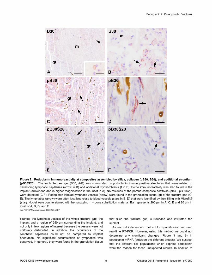
(B30, Figure 7A-B) and porous scaffolds (pB30, pB30S20,Figure 7C-F), both with the dimension of a 4 mm wedge-shapefitting exactly the fracture gap. After 6 weeks of implantationthe residue of the partially resorbed xerogel (B30) wassurrounded by granulation tissue that contained a high amountof cells (Figure 7A). Podoplanin immunopositive structureswere found at the direct interface of the implant and, inaddition, penetrating the implant (Figure 7B), where thepodoplanin positive structures did not form fully developedlymphatic vessels. It appeared like broken lines, a staining ofplasma membrane of big cells, and labeled lymphaticendothelial cells during the process of lymphangiogenesis(Figure 7B).
In contrast no residues of the porous B30 bone substitutionmaterial could be found in the fracture gap (Figure 7C).Instead, the gap was filled with granulation tissue containing
Figure 3. Expression of podoplanin in empty defects. Noregulation of the podoplanin expression was detected for theempty defects by means of real-time RT-PCR. The results arepresented as box plot with the median, the 0th, 25th, 75th, and100th percentile. Small circles illustrate data beyond 3 xstandard deviation (SD).doi: 10.1371/journal.pone.0077259.g003
several fully developed podoplanin immunopositiv lymphaticvessels (Figure 7D). The same situation was observed for thestrontium modified scaffolds (Figure 7E-F).
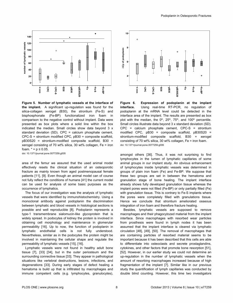
Comparing the number of lymphatic vessels we could neitherdetect significant changes between the different compositetypes nor between the porous scaffolds (pB30, pB30S20) andthe empty defect (Figure 5). The xerogel (B30) exhibited asignificant (p = 0.024) increase in the amount of lymphaticvessels at its interface in comparison to the control group (4mm empty defect) (Figure 5). No regulation of podoplanin wasfound comparing xerogels, porous scaffolds, and the 4 mmempty defect as appropriate control (Figure 6).
Iron foam implantsThree different formulations of the iron foam were used as
implant in the different rat groups: a) plain iron foam (Fe, Figure8), b) strontium functionalized iron foam (Fe-S, Figure 9), andc) iron foam with coating of the bisphosphonate zolendronicacid (Fe-BP, Figure 10). The different modifications of ironfoam were surrounded by granulation tissue after implantation(Figure 8A, 9A, 10A). The granulation tissue contained a highamount of fully developed lymphatic vessels (Figure 8B, 9B).No significant differences were found in the number oflymphatic vessels in the granulation tissue (Figure 5). But asignificant increase in lymphatics was determined for themodified iron foams (Fe-S, Fe-BP) in comparison to the emptydefect (4 mm) that served as control (p = 0.001 for Fe-S and p= 0.026 for Fe-BP) (Figure 5). On mRNA level no significantchanges could be measured for podoplanin (Figure 6)
Several cells with yellow granular cytoplasm appeared in thegranulation tissue (Figure 8C, 9C-D, 10B-D). This yellowstaining could result from resorbed iron. However, these cellsare often localized in direct contact to the podoplaninimmunopositiv vessels (Figure 10B). Some of the yellow cellswere also observed in the lumen of lymphatic capillaries(Figure 10C). In other lymphatic vessels we identified nuclei oflymphocytes (Figure 10D). This was mainly observed at theinterface of Fe-BP.
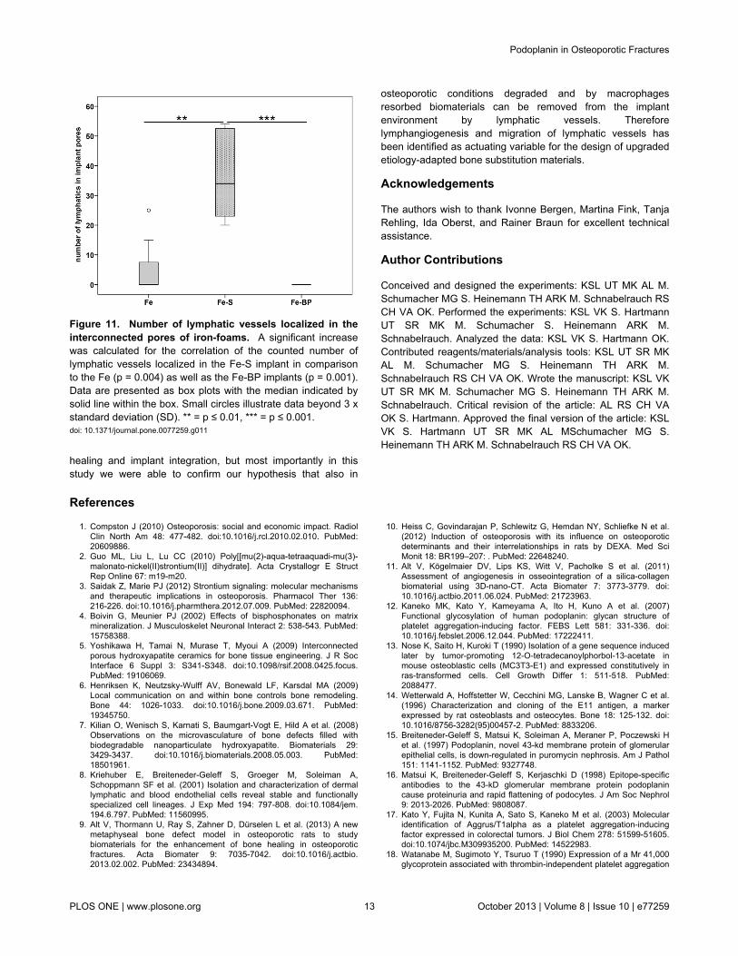
Moreover lymphatic vessels were also localized in theconnecting pores of Fe and Fe-S but not in the Fe-BP group.These changes in the amount of lymphatic vessels weresignificant (Figure 11, p = 0.004 and p = 0.001 for thecomparison of Fe to Fe-S and of Fe-S to Fe-BP, respectively).The pores of Fe-S were filled with granulation tissue whereasthe pores of Fe-BP were not integrated by connective tissue.The interconnected pores of Fe implants were only filled withgranulations tissue near the interface. Pores situated in thecenter of the implant were not filled with granulation tissue aftersix week follow-up time and therefore no lymphatic vesselsoccurred there.
Discussion
The present study demonstrates for the first time thepresence of lymphatic vessels in an osteoporosis animalmodel. Osteoporosis was induced by bilateral ovariectomy andadditional feeding with calcium, Vitamin C/D2/D3 and phosphaterestricted diet in current rat model [10]. This treatment is
Podoplanin in Osteoporotic Fractures
PLOS ONE | www.plosone.org 6 October 2013 | Volume 8 | Issue 10 | e77259
commonly used to induce osteoporosis in rats that are naturallynot affected by this disease [41]. Osteoporosis is described asa systemic disease that causes an imbalance of the osteoblastand osteoclast population in which the bone degradingosteoclasts are enhanced [1]. Thereby the bone mineraldensity is reduced that accounts for the increase in fractureincidence [1]. Recent studies determined that the animals inthe applied rat model suffer from a disease with reduced bonemineral density as measured by DXA scan, an enhancedfracture risk as shown by biomechanical analysis, and animbalance in bone synthesis and depletion [10], [42]. Therefore
the rat model can be utilized for studying aspects that areconnected to the typical human disease osteoporosis.However, not only the fracture frequency is increased in thisillness, but also the fractures are different from such of bone-healthy patients. Characteristically osteoporotic fractures arelocalized in the spongy bone of metaphyseal trabecular regions[43], [44] like vertebrae, distal radius, proximal femur andhumerus [45], [46], [47]. Thus in the present study a wedge-shaped defect in the distal metaphyseal area of the femur wasperformed [9,11]. We did not dispose the proximal femurbecause of its bad accessibility. Utilizing the distal metaphyseal
Figure 4. CPC based implants. Podoplanin immunopositive lymphatic vessels (arrow) were localized in the granulation tissue (gt)at the interface of the CPC implant (A-E) as well as at the strontium functionalized CPC (CPC-S in E-F) where an improvedfragmentation was found (F). Lympatics were often associated with blood vessels that were identified by Microfil® perfusion (star inC). Podoplanin also stained osteocytes (arrowhead in B-F) and mature osteoblasts (O). Nuclei were counterstained withhematoxylin. m = bone substitution material. Bars: 1 mm in A, F, 200 µm in B, G, and 20 µm in C-E.doi: 10.1371/journal.pone.0077259.g004
Podoplanin in Osteoporotic Fractures
PLOS ONE | www.plosone.org 7 October 2013 | Volume 8 | Issue 10 | e77259
area of the femur we assured that the used animal modeleffectively resets the clinical situation of an osteoporoticfracture as mainly known from aged postmenopausal femalepatients [11], [9]. Even though an animal model can of coursenot fully reflect the conditions of humans [41] the current modelcan be used for analysis of some basic purposes as theoccurrence of lymphatics.
The focus of our investigation was the analysis of lymphaticvessels that were identified by immunohistochemistry. Using amonoclonal antibody against podoplanin the discriminationbetween lymphatic and blood vessels in histological sections ispossible and well reproducible [8]. Podoplanin represents atype-1 transmembrane sialomucin-like glycoprotein that iswidely spread. In podocytes of kidney the protein is involved inobtaining cell morphology and maintenance in glomerularpermeability [16]. Up to now, the function of podoplanin inlymphatic endothelial cells is not fully understood.Nevertheless, similar as in the podocytes the protein might beinvolved in maintaining the cellular shape and regulate thepermeability of lymphatic vessels [15], [16].
Lymphatic vessels were not found in healthy adult bonetissue [7], [33] [34], but in the outer periosteum and thesurrounding connective tissue [33]. They appear in pathologicalsituations like vertebral destructions, lesions, infections, anddegenerations [33]. During early stages of defect healing ahematoma is build up that is infiltrated by macrophages andimmune competent cells (e.g. lymphocytes, granulocytes),
Figure 5. Number of lymphatic vessels at the interface ofthe implant. A significant up-regulation was found for thesilica-collagen xerogel (B30), the strontium (Fe-S) andbisphosphonate (Fe-BP) functionalized iron foam incomparison to the negative control without implant. Data werepresented as box plots where a solid line within the boxindicated the median. Small circles show data beyond 3 xstandard deviation (SD). CPC = calcium phosphate cement,CPC-S = strontium modified CPC, pB30 = composite scaffold,pB30S20 = strontium-modified composite scaffold, B30 =xerogel consisting of 70 wt% silica, 30 wt% collagen, Fe = ironfoam. * = p ≤ 0.05.doi: 10.1371/journal.pone.0077259.g005 amongst others [36]. Thus, it was not surprising to find
lymphocytes in the lumen of lymphatic capillaries of someanimal groups in our implant study. An obvious enhancementof lymphocytes inside lymphatic vessels was determined ingroups of plain iron foam (Fe) and Fe-BP. We suppose thatthese two groups are set in between the hematoma andgranulation stage of bone healing. The implant interfacealready shows fully developed granulation tissue whereas theimplant pores were not filled (Fe-BP) or only partially filled (Fe)with granulation tissue. This is contrary to Fe-S implants wherethe pores were completely filled with granulations tissue.Hence we conclude that strontium ameliorated osseousintegration of iron foam and therefore fracture healing.
Besides, lymphatic vessels are supposed to removemacrophages and their phagocytozed material from the implantinterface. Since macrophages with resorbed wear particlesfrom prosthesis were found in lymph nodes it has beenassumed that the implant interface is cleared via lymphaticcirculation [48], [49], [50]. The removal of macrophages thatare containing particles of resorbed material seems to beimportant because it has been described that this cells are ableto differentiate into osteoclasts and secrete prostaglandins,cytokines, and other factors that promote bone resorption [51],[52]. However, in our earlier study we could not determine anup-regulation in the number of lymphatic vessels when theamount of resorbing macrophages increased because of highfragmentation of the implant [7]. Similar like in our previousstudy the quantification of lymph capillaries was conducted bydouble blind counting. However, this time two investigators
Figure 6. Expression of podoplanin at the implantinterface. Using real-time RT-PCR, no regulation ofpodoplanin at the mRNA level could be detected in theinterface area of the implant. The results are presented as boxplot with the median, the 0th, 25th, 75th, and 100th percentile.Small circles illustrate data beyond 3 x standard deviation (SD).CPC = calcium phosphate cement, CPC-S = strontiummodified CPC, pB30 = composite scaffold, pB30S20 =strontium-modified composite scaffold, B30 = xerogelconsisting of 70 wt% silica, 30 wt% collagen, Fe = iron foam.doi: 10.1371/journal.pone.0077259.g006
Podoplanin in Osteoporotic Fractures
PLOS ONE | www.plosone.org 8 October 2013 | Volume 8 | Issue 10 | e77259
counted the lymphatic vessels of the whole fracture gap, theimplant and a region of 250 µm surrounding the implant, andnot only in few regions of interest because the vessels were notuniformly distributed. In addition, the occurrence of thelymphatic capillaries could not be compared to implantorientation. No significant accumulation of lymphatics wasobserved. In general, they were found in the granulation tissue
that filled the fracture gap, surrounded and infiltrated theimplant.
As second independent method for quantification we usedreal-time RT-PCR. However, using this method we could notdetermine any significant changes (Figure 3 and 6) inpodoplanin mRNA (between the different groups). We suspectthat the different cell populations which express podoplaninwere the reason for these unexpected results. In addition to
Figure 7. Podoplanin immunoreactivity at composites assembled by silica, collagen (pB30, B30), and additional strontium(pB30S20). The implanted xerogel (B30, A-B) was surrounded by podoplanin immunopositive structures that were related todeveloping lymphatic capillaries (arrow in B) and additional myofibroblasts (f in B). Some immunoreactivity was also found in theimplant (arrowhead and in higher magnification in the inset in A). No residues of the porous composite scaffolds (pB30, pB30S20)were detected (C-F). Podoplanin labeled lymphatic vessels (arrow) were found in the granulation tissue (gt) of the fracture gap (C,E). The lymphatics (arrow) were often localized close to blood vessels (stars in B, D) that were identified by their filling with Microfil®(star). Nuclei were counterstained with hematoxylin. m = bone substitution material. Bar represents 200 µm in A, C, E and 20 µm ininset of A, B, D, and F.doi: 10.1371/journal.pone.0077259.g007
Podoplanin in Osteoporotic Fractures
PLOS ONE | www.plosone.org 9 October 2013 | Volume 8 | Issue 10 | e77259
lymphatic endothelial cells podoplanin is found in differentiatedosteoblasts and newly developed osteocytes [14]. Thus, inreal-time RT-PCR analysis for RNA isolated out of the wholesample it is not possible to distinguish between lymphaticvessels and the osteoblast lineage. Therefore, we concludethat this second method of quantification was in our case notsuitable for the assignation of the number of lymphatic vesselswhereas it was well implemented in other studies where nobone was found in the surrounding area [53].
In the first part of our study we performed wedge-shapedmetaphysal fractures of 3 different sizes. The 3 mm defect waspartially bridged by bone containing osteocyts that werelocalized in adjacent lacunae surrounded by calcified bonematrix. Newly formed osteocytes as well as mature osteoblastsare podoplanin immunopositive [14]. Regardless of the bonybridge some granulation tissue remained and contained a highnumber of lymphatic vessels inside a small area. The 4 and 5mm wedge-shaped fractures represent critical size defects [9].In the fracture gap of the 5 mm defect only new build
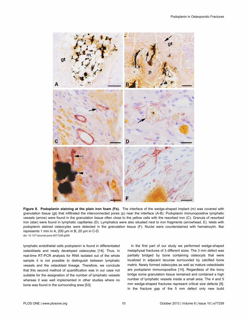
Figure 8. Podoplanin staining at the plain iron foam (Fe). The interface of the wedge-shaped implant (m) was covered withgranulation tissue (gt) that infiltrated the interconnected pores (p) near the interface (A-B). Podoplanin immunopositive lymphaticvessels (arrow) were found in the granulation tissue often close to the yellow cells with the resorbed iron (C). Granula of resorbediron (star) were found in lymphatic capillaries (D). Lymphatics were also situated next to iron fragments (arrowhead, E). Islets withpodoplanin stained osteocytes were detected in the granulation tissue (F). Nuclei were counterstained with hematoxylin. Barrepresents 1 mm in A, 200 µm in B, 20 µm in C-D.doi: 10.1371/journal.pone.0077259.g008
Podoplanin in Osteoporotic Fractures
PLOS ONE | www.plosone.org 10 October 2013 | Volume 8 | Issue 10 | e77259
granulation tissue with a lot of lymphatic capillaries was found.The 4 mm fracture was narrowed by insular regions of newlybuilt bone and the granulation tissue contained a high numberof blood vessels. The bony islands were surrounded bygranulation tissue that included only few lymphatic capillaries.Also only few lymphatic vessels were found in association withthe blood vasculature in the empty defect. Thus we counted forthe 4 mm defect a significant smaller number of lymphaticvessels in comparison to the 3 and 5 mm fractures. Our resultsshowed that examination of lymphatic vessels were providinginformation about bone healing. However, less lymphaticvessels did not reflect best fracture healing as might beassumed because of the absence of lympathic vessels inundamaged bone.
For the second part of our study the 4 mm defects were usedto test the occurrence of lymphatic vessels at the interface ofbone substitution materials. Applying biomaterials into thefracture gap has been proven to accelerate bone healing [54].Since up to now none of the commercial available biomaterialsis appropriate for the usage in osteoporotic bone weinvestigated the podoplainin immunolabeling after implantationof several biomaterials. CPC is already well approved for theadministration in non-systemically diseased bone tissue [37].
To adapt CPC to the required conditions in osteoporosis it wasmodified with strontium. Because of the systemic nature ofosteoporosis it is not possible that local acting biomaterialsthemselves influence the illness but modifying them with drugs,growth factors, organic and inorganic active components mightpromise success. Recently it has been described that strontiumadministration was successful for the treatment of osteoporosis[2], [3]. Thus one animal subgroup received an implant with astrontium modification of the CPC. At the implant interface ofboth CPC groups only a few lymphatic vessels were found inclose proximity of blood vessels. No significant differences inthe number of lymphatic vessels were determined between thetwo CPC groups themselves, nor in comparison to the controlgroup (4 mm defect). In some animals from the CPC-Ssubgroup the fragmentation of the implant was upgraded andsubsequent granulation tissue including lymphatic vesselspenetrated the implant. Thus, in some animals of the CPC-Sgroup we found a higher amount of lymphatic vessels.Nonetheless, no significances were calculated most likelybecause of the high variance between the individual animals. Inanother subset of experiments composites consisting of silicaand collagen were implanted as monolithic xerogels or asporous scaffolds containing xerogel particles with or without
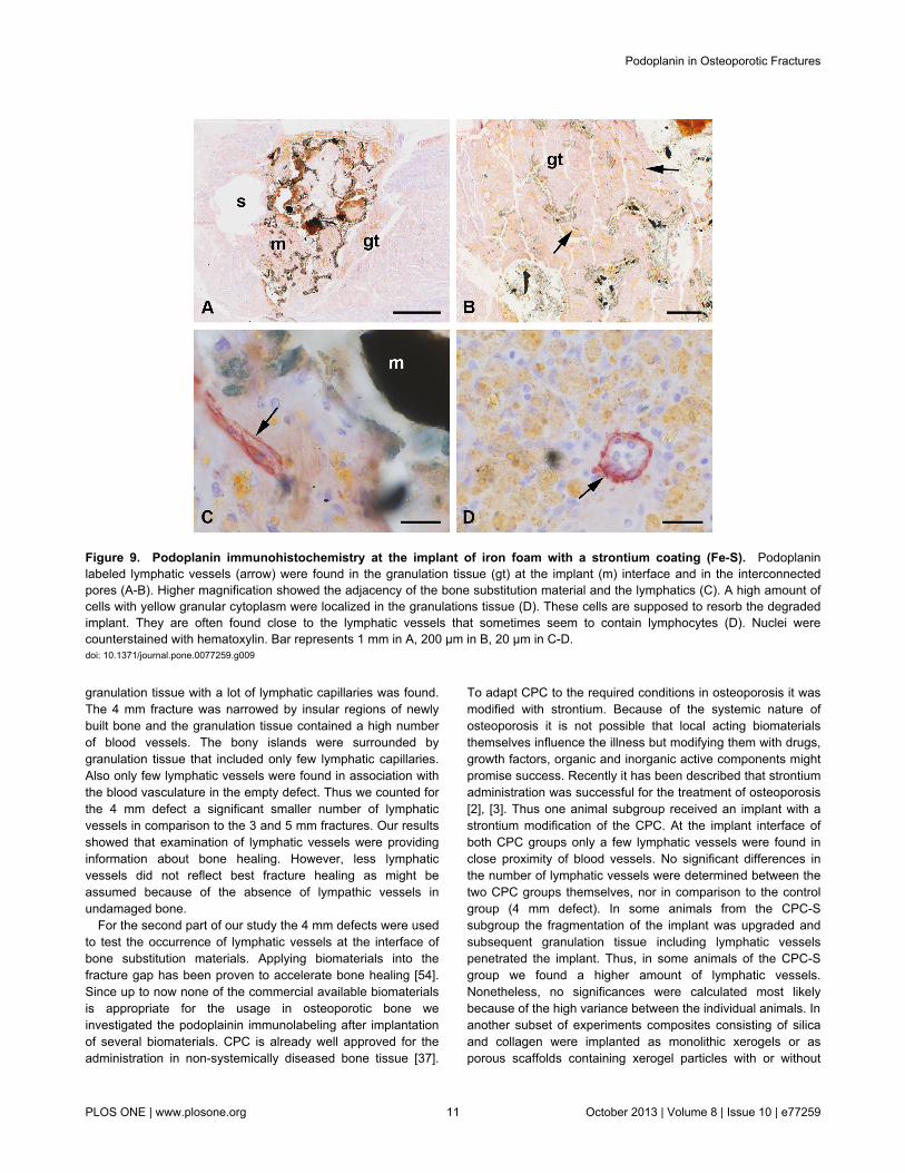
Figure 9. Podoplanin immunohistochemistry at the implant of iron foam with a strontium coating (Fe-S). Podoplaninlabeled lymphatic vessels (arrow) were found in the granulation tissue (gt) at the implant (m) interface and in the interconnectedpores (A-B). Higher magnification showed the adjacency of the bone substitution material and the lymphatics (C). A high amount ofcells with yellow granular cytoplasm were localized in the granulations tissue (D). These cells are supposed to resorb the degradedimplant. They are often found close to the lymphatic vessels that sometimes seem to contain lymphocytes (D). Nuclei werecounterstained with hematoxylin. Bar represents 1 mm in A, 200 µm in B, 20 µm in C-D.doi: 10.1371/journal.pone.0077259.g009
Podoplanin in Osteoporotic Fractures
PLOS ONE | www.plosone.org 11 October 2013 | Volume 8 | Issue 10 | e77259
strontium. A recent study demonstrated the composition,structure, surface to have significant impact on the mechanicalproperties degradation, the homeostasis of calcium, andthereby the local cell and tissue reaction at the interface [55].Interestingly, we could not discover the porous implant in thehistological sections but granulation tissue in the fracture gapthat contained lymphatic capillaries. Accordingly, no significantdifferences in comparison to the control group (4 mm defect)were calculated. However, significant changes were detectedfor the monolithic xerogels composed of the same ingredients.The number of lymphatics found in this animal subgroup wassignificant up-regulated in comparison to the control group (4mm defect). The podoplainin staining in sections of thissubgroup had an altered image. The labeled cells werelocalized directly at the interface. It appeared like an individuallabeling of the plasma membrane of a specific cell type. Sinceit is well known that myofibroblasts are stained by podoplanin[20], [29] and the presence of myofibroblasts at implantinterfaces has already been described [56], [57] wehypothesize that at the interface of the xerogel myofibroblastsare accumulated. This interesting theory should further beanalyzed studying the osseous integration and degradation ofthe xerogel in future studies. However, some of the labeling
was found in cells invading the implant. An infiltration of theimplant was also observed for the Fe-S group. All implants ofthe Fe group include interconnected pores. Recently,Yoshikawa et al. 2009 [5] described that bone substitutionmaterials with interconnected pores improve bony integrationby allowing the immigration of mesenchymal stem cells,granulation tissue, and vasculature into the pores. Interestingly,the pores of the Fe-BP implant were not filled with granulationtissue and therefore no lymphatic vessels were found there.Sections of the plain Fe exhibited a filling of the pores withgranulation tissue near the circumference of the implant. Intothis area also incorporated lymphatic capillaries were found.The highest number of infiltrating lymphatic vessels was foundin the Fe-S group showing the sophisticated implantintegration. Besides all Fe implants were surrounded bygranulation tissue including lymphatic capillaries where nosignificant differences were detected.
In conclusion, our results showed that lymphatic vessels alsooccur in damaged bone of animals where osteoporosis wasinduced. In addition, we found lymphatic capillaries in thefracture gap and at the interface of several implanted bonesubstitution materials. After quantification of the lymphaticcapillaries we could speculate about the progress of bone
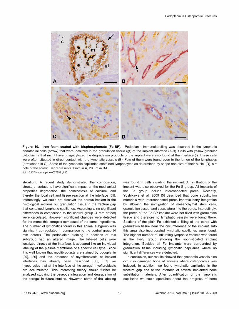
Figure 10. Iron foam coated with bisphosphonate (Fe-BP). Podoplanin immunolabelling was observed in the lymphaticendothelial cells (arrow) that were localized in the granulation tissue (gt) at the implant interface (A-B). Cells with yellow granularcytoplasma that might have phagocytozed the degradation products of the implant were also found at the interface (i). These cellswere often situated in direct contact with the lymphatic vessels (B). Few of them were found even in the lumen of the lymphatics(arrowhead in C). Some of the lymphatic capillaries contained lymphocytes as determined by shape and size of their nuclei (D). s =hole of the screw. Bar represents 1 mm in A, 20 µm in B-D.doi: 10.1371/journal.pone.0077259.g010
Podoplanin in Osteoporotic Fractures
PLOS ONE | www.plosone.org 12 October 2013 | Volume 8 | Issue 10 | e77259
healing and implant integration, but most importantly in thisstudy we were able to confirm our hypothesis that also in
Figure 11. Number of lymphatic vessels localized in theinterconnected pores of iron-foams. A significant increasewas calculated for the correlation of the counted number oflymphatic vessels localized in the Fe-S implant in comparisonto the Fe (p = 0.004) as well as the Fe-BP implants (p = 0.001).Data are presented as box plots with the median indicated bysolid line within the box. Small circles illustrate data beyond 3 xstandard deviation (SD). ** = p ≤ 0.01, *** = p ≤ 0.001.doi: 10.1371/journal.pone.0077259.g011
osteoporotic conditions degraded and by macrophagesresorbed biomaterials can be removed from the implantenvironment by lymphatic vessels. Thereforelymphangiogenesis and migration of lymphatic vessels hasbeen identified as actuating variable for the design of upgradedetiology-adapted bone substitution materials.
Acknowledgements
The authors wish to thank Ivonne Bergen, Martina Fink, TanjaRehling, Ida Oberst, and Rainer Braun for excellent technicalassistance.
Author Contributions
Conceived and designed the experiments: KSL UT MK AL M.Schumacher MG S. Heinemann TH ARK M. Schnabelrauch RSCH VA OK. Performed the experiments: KSL VK S. HartmannUT SR MK M. Schumacher S. Heinemann ARK M.Schnabelrauch. Analyzed the data: KSL VK S. Hartmann OK.Contributed reagents/materials/analysis tools: KSL UT SR MKAL M. Schumacher MG S. Heinemann TH ARK M.Schnabelrauch RS CH VA OK. Wrote the manuscript: KSL VKUT SR MK M. Schumacher MG S. Heinemann TH ARK M.Schnabelrauch. Critical revision of the article: AL RS CH VAOK S. Hartmann. Approved the final version of the article: KSLVK S. Hartmann UT SR MK AL MSchumacher MG S.Heinemann TH ARK M. Schnabelrauch RS CH VA OK.
References
1. Compston J (2010) Osteoporosis: social and economic impact. RadiolClin North Am 48: 477-482. doi:10.1016/j.rcl.2010.02.010. PubMed:20609886.
2. Guo ML, Liu L, Lu CC (2010) Poly[[mu(2)-aqua-tetraaquadi-mu(3)-malonato-nickel(II)strontium(II)] dihydrate]. Acta Crystallogr E StructRep Online 67: m19-m20.
3. Saidak Z, Marie PJ (2012) Strontium signaling: molecular mechanismsand therapeutic implications in osteoporosis. Pharmacol Ther 136:216-226. doi:10.1016/j.pharmthera.2012.07.009. PubMed: 22820094.
4. Boivin G, Meunier PJ (2002) Effects of bisphosphonates on matrixmineralization. J Musculoskelet Neuronal Interact 2: 538-543. PubMed:15758388.
5. Yoshikawa H, Tamai N, Murase T, Myoui A (2009) Interconnectedporous hydroxyapatite ceramics for bone tissue engineering. J R SocInterface 6 Suppl 3: S341-S348. doi:10.1098/rsif.2008.0425.focus.PubMed: 19106069.
6. Henriksen K, Neutzsky-Wulff AV, Bonewald LF, Karsdal MA (2009)Local communication on and within bone controls bone remodeling.Bone 44: 1026-1033. doi:10.1016/j.bone.2009.03.671. PubMed:19345750.
7. Kilian O, Wenisch S, Karnati S, Baumgart-Vogt E, Hild A et al. (2008)Observations on the microvasculature of bone defects filled withbiodegradable nanoparticulate hydroxyapatite. Biomaterials 29:3429-3437. doi:10.1016/j.biomaterials.2008.05.003. PubMed:18501961.
8. Kriehuber E, Breiteneder-Geleff S, Groeger M, Soleiman A,Schoppmann SF et al. (2001) Isolation and characterization of dermallymphatic and blood endothelial cells reveal stable and functionallyspecialized cell lineages. J Exp Med 194: 797-808. doi:10.1084/jem.194.6.797. PubMed: 11560995.
9. Alt V, Thormann U, Ray S, Zahner D, Dürselen L et al. (2013) A newmetaphyseal bone defect model in osteoporotic rats to studybiomaterials for the enhancement of bone healing in osteoporoticfractures. Acta Biomater 9: 7035-7042. doi:10.1016/j.actbio.2013.02.002. PubMed: 23434894.
10. Heiss C, Govindarajan P, Schlewitz G, Hemdan NY, Schliefke N et al.(2012) Induction of osteoporosis with its influence on osteoporoticdeterminants and their interrelationships in rats by DEXA. Med SciMonit 18: BR199–207: . PubMed: 22648240.
11. Alt V, Kögelmaier DV, Lips KS, Witt V, Pacholke S et al. (2011)Assessment of angiogenesis in osseointegration of a silica-collagenbiomaterial using 3D-nano-CT. Acta Biomater 7: 3773-3779. doi:10.1016/j.actbio.2011.06.024. PubMed: 21723963.
12. Kaneko MK, Kato Y, Kameyama A, Ito H, Kuno A et al. (2007)Functional glycosylation of human podoplanin: glycan structure ofplatelet aggregation-inducing factor. FEBS Lett 581: 331-336. doi:10.1016/j.febslet.2006.12.044. PubMed: 17222411.
13. Nose K, Saito H, Kuroki T (1990) Isolation of a gene sequence inducedlater by tumor-promoting 12-O-tetradecanoylphorbol-13-acetate inmouse osteoblastic cells (MC3T3-E1) and expressed constitutively inras-transformed cells. Cell Growth Differ 1: 511-518. PubMed:2088477.
14. Wetterwald A, Hoffstetter W, Cecchini MG, Lanske B, Wagner C et al.(1996) Characterization and cloning of the E11 antigen, a markerexpressed by rat osteoblasts and osteocytes. Bone 18: 125-132. doi:10.1016/8756-3282(95)00457-2. PubMed: 8833206.
15. Breiteneder-Geleff S, Matsui K, Soleiman A, Meraner P, Poczewski Het al. (1997) Podoplanin, novel 43-kd membrane protein of glomerularepithelial cells, is down-regulated in puromycin nephrosis. Am J Pathol151: 1141-1152. PubMed: 9327748.
16. Matsui K, Breiteneder-Geleff S, Kerjaschki D (1998) Epitope-specificantibodies to the 43-kD glomerular membrane protein podoplanincause proteinuria and rapid flattening of podocytes. J Am Soc Nephrol9: 2013-2026. PubMed: 9808087.
17. Kato Y, Fujita N, Kunita A, Sato S, Kaneko M et al. (2003) Molecularidentification of Aggrus/T1alpha as a platelet aggregation-inducingfactor expressed in colorectal tumors. J Biol Chem 278: 51599-51605.doi:10.1074/jbc.M309935200. PubMed: 14522983.
18. Watanabe M, Sugimoto Y, Tsuruo T (1990) Expression of a Mr 41,000glycoprotein associated with thrombin-independent platelet aggregation
Podoplanin in Osteoporotic Fractures
PLOS ONE | www.plosone.org 13 October 2013 | Volume 8 | Issue 10 | e77259
in high metastatic variants of murine B16 melanoma. Cancer Res 50:6657-6662. PubMed: 2208129.
19. Gandarillas A, Scholl FG, Benito N, Gamallo C, Quintanilla M (1997)Induction of PA2.26, a cell-surface antigen expressed by activefibroblasts, in mouse epidermal keratinocytes during carcinogenesis.Mol Carcinog 20: 10-18. doi:10.1002/(SICI)1098-2744(199709)20:1.PubMed: 9328432.
20. Liang P, Hong JW, Ubukata H, Liu G, Katano M et al. (2005)Myofibroblasts correlate with lymphatic microvessel density and lymphnode metastasis in early-stage invasive colorectal carcinoma.Anticancer Res 25: 2705-2712. PubMed: 16080515.
21. Farr AG, Berry ML, Kim A, Nelson AJ, Welch MP et al. (1992)Characterization and cloning of a novel glycoprotein expressed bystromal cells in T-dependent areas of peripheral lymphoid tissues. JExp Med 176: 1477-1482. doi:10.1084/jem.176.5.1477. PubMed:1402691.
22. Rishi AK, Joyce-Brady M, Fisher J, Dobbs LG, Floros J et al. (1995)Cloning, characterization, and development expression of a rat lungalveolar type I cell gene in embryonic endodermal and neuralderivatives. Dev Biol 167: 294-306. doi:10.1006/dbio.1995.1024.PubMed: 7851650.
23. Vanderbilt JN, Dobbs LG (1998) Characterization of the gene andpromoter for RTI40, a differentiation marker of type I alveolar epithelialcells. Am J Respir Cell Mol Biol 19: 662-671. doi:10.1165/ajrcmb.19.4.3121. PubMed: 9761764.
24. Williams MC, Cao Y, Hinds A, Rishi AK, Wetterwald A (1996) T1 alphaprotein is developmentally regulated and expressed by alveolar type Icells, choroid plexus, and ciliary epithelia of adult rats. Am J Respir CellMol Biol 14: 577-585. doi:10.1165/ajrcmb.14.6.8652186. PubMed:8652186.
25. Scholl FG, Gamallo C, Quintanilla M (2000) Ectopic expression ofPA2.26 antigen in epidermal keratinocytes leads to destabilization ofadherens junctions and malignant progression. Lab Invest 80:1749-1759. doi:10.1038/labinvest.3780185. PubMed: 11092535.
26. Bellini C, Rutigliani M, Boccardo F, Campisi C, Bellini T et al. (2012)Are there lymphatic vessels in the placenta? Lymphology 45: 34-36.PubMed: 22768471.
27. Kaji C, Tomooka M, Kato Y, Kojima H, Sawa Y (2012) The expressionof podoplanin and classic cadherins in the mouse brain. J Anat 220:435-446. doi:10.1111/j.1469-7580.2012.01484.x. PubMed: 22352427.
28. Kotani M, Osanai T, Tajima Y, Kato H, Imada M et al. (2002)Identification of neuronal cell lineage-specific molecules in the neuronaldifferentiation of P19 EC cells and mouse central nervous system. JNeurosci Res 67: 595-606. doi:10.1002/jnr.10150. PubMed: 11891772.
29. Ekwall AK, Eisler T, Anderberg C, Jin C, Karlsson N et al. (2011) Thetumour-associated glycoprotein podoplanin is expressed in fibroblast-like synoviocytes of the hyperplastic synovial lining layer in rheumatoidarthritis. Arthritis Res Ther 13: R40. doi:10.1186/ar3274. PubMed:21385358.
30. Gehlert S, Theis C, Weber S, Schiffer T, Hellmich M et al. (2010)Exercise-induced decline in the density of LYVE-1-positive lymphaticvessels in human skeletal muscle. Lymph Res Biol 8: 165-173. doi:10.1089/lrb.2009.0035. PubMed: 20863269.
31. Kholová I, Dragneva G, Cermáková P, Laidinen S, Kaskenpää N et al.(2011) Lymphatic vasculature is increased in heart valves, ischaemicand inflamed hearts and in cholesterol-rich and calcified atheroscleroticlesions. Eur J Clin Invest 41: 487-497. doi:10.1111/j.1365-2362.2010.02431.x. PubMed: 21128936.
32. Wessel JM, Hofmann-Rummelt C, Kruse FE, Cursiefen C, Heindl LM(2012) Invasion of lymphatic vessels into the eye after open globeinjuries. Invest Ophthalmol Vis Sci 53: 3717-3725. doi:10.1167/iovs.12-9507. PubMed: 22589431.
33. Kashima TG, Dongre A, Athanasou NA (2011) Lymphatic involvementin vertebral and disc pathology. Spine (Phila Pa 1976) 36 : 899-904.PubMed: 21343852.
34. Edwards JR, Williams K, Kindblom LG, Meis-Kindblom JM,Hogendoorn PC et al. (2008) Lymphatics and bone. Hum Pathol 39:49-55. doi:10.1016/j.humpath.2007.04.022. PubMed: 17904616.
35. Mkonyi LE, Bakken V, Søvik JB, Mauland EK, Fristad I et al. (2012)Lymphangiogenesis is induced during development of periodontaldisease. J Dent Res 91: 71-77. doi:10.1177/0022034511424747.PubMed: 21979132.
36. Claes L, Recknagel S, Ignatius A (2012) Fracture healing under healthyand inflammatory conditions. Nat Rev Rheumatol 8: 133-143. doi:10.1038/nrrheum.2012.1. PubMed: 22293759.
37. Khairoun I, Boltong MG, Driessens FC, Planell JA (1997) Effect ofcalcium carbonate on the compliance of an apatitic calcium phosphatebone cement. Biomaterials 18: 1535-1539. doi:10.1016/S0142-9612(97)80005-1. PubMed: 9430336.
38. Schumacher M, Henss A, Rohnke M, Gelinsky M (2013) A novel andeasy-to-prepare strontium(II) modified calcium phosphate bone cementwith enhanced mechanical properties. Acta Biomater 9: 7536-7544. doi:10.1016/j.actbio.2013.03.014. PubMed: 23523939.
39. Heinemann S, Heinemann C, Bernhardt R, Reinstorf A, Nies B et al.(2009) Bioactive silica-collagen composite xerogels modified bycalcium phosphate phases with adjustable mechanical properties forbone replacement. Acta Biomater 5: 1979-1990. doi:10.1016/j.actbio.2009.02.029. PubMed: 19345651.
40. Heinemann S, Heinemann C, Jäger M, Neunzehn J, Wiesmann HP etal. (2011) Effect of silica and hydroxyapatite mineralization on themechanical properties and the biocompatibility of nanocompositecollagen scaffolds. ACS Appl Mater Interfaces 3: 4323-4331. doi:10.1021/am200993q. PubMed: 21942510.
41. Egermann M, Goldhahn J, Schneider E (2005) Animal models forfracture treatment in osteoporosis. Osteoporos Int 16 Suppl 2: S129-S138. doi:10.1007/s00198-005-1859-7. PubMed: 15750681.
42. Govindarajan P, Schlewitz G, Schliefke N, Weisweiler D, Alt V et al.(2013) Implications of combined ovariectomy/multi-deficiency diet onrat bone with age-related variation in bone parameters and bone loss atmultiple skeletal sites by DEXA. Med Sci Monit Basic Res 19: 76-86.doi:10.12659/MSMBR.883815. PubMed: 23446183.
43. Peng Z, Tuukkanen J, Zhang H, Jämsä T, Väänänen HK (1994) Themechanical strength of bone in different rat models of experimentalosteoporosis. Bone 15: 523-532. doi:10.1016/8756-3282(94)90276-3.PubMed: 7980963.
44. Wronski TJ, Lowry PL, Walsh CC, Ignaszewski LA (1985) Skeletalalterations in ovariectomized rats. Calcif Tissue Int 37: 324-328. doi:10.1007/BF02554882. PubMed: 3926284.
45. Cooper C, Atkinson EJ, O'Fallon WM, Melton LJ 3rd (1992) Incidenceof clinically diagnosed vertebral fractures: a population-based study inRochester, Minnesota, 1985-1989. J Bone Miner Res 7: 221-227.PubMed: 1570766.
46. Cooper C, Campion G, Melton LJ 3rd (1992) Hip fractures in theelderly: a world-wide projection. Osteoporos Int 2: 285-289. doi:10.1007/BF01623184. PubMed: 1421796.
47. Cooper C, Melton LJ 3rd (1992) Epidemiology of osteoporosis. TrendsEndocrinol Metab 3: 224-229. doi:10.1016/1043-2760(92)90032-V.PubMed: 18407104.
48. Edwards J, Schulze E, Sabokbar A, Gordon-Andrews H, Jackson D etal. (2008) Absence of lymphatics at the bone-implant interface:implications for periprosthetic osteolysis. Acta Orthop 79: 289-294. doi:10.1080/17453670710015175. PubMed: 18484257.
49. Bos I, Johannisson R, Löhrs U, Lindner B, Seydel U (1990)Comparative investigations of regional lymph nodes andpseudocapsules after implantation of joint endoprostheses. Pathol ResPract 186: 707-716. doi:10.1016/S0344-0338(11)80260-8. PubMed:2084636.
50. Urban RM, Jacobs JJ, Tomlinson MJ, Gavrilovic J, Black J et al. (2000)Dissemination of wear particles to the liver, spleen, and abdominallymph nodes of patients with hip or knee replacement. J Bone JointSurg Am 82: 457-476. doi:10.1302/0301-620X.82B3.9310. PubMed:10761937.
51. Sabokbar A, Fujikawa Y, Brett J, Murray DW, Athanasou NA (1996)Increased osteoclastic differentiation by PMMA particle-associatedmacrophages. Inhibitory effect by interleukin 4 and leukemia inhibitoryfactor. Acta Orthop Scand 67: 593-598. doi:10.3109/17453679608997763. PubMed: 9065074.
52. Sabokbar A, Fujikawa Y, Neale S, Murray DW, Athanasou NA (1997)Human arthroplasty derived macrophages differentiate into osteoclasticbone resorbing cells. Ann Rheum Dis 56: 414-420. doi:10.1136/ard.56.7.414. PubMed: 9486003.
53. Xu Y, Ogose A, Kawashima H, Hotta T, Ariizumi T et al. (2011) High-level expression of podoplanin in benign and malignant soft tissuetumors: immunohistochemical and quantitative real-time RT-PCRanalysis. Oncol Rep 25: 599-607. PubMed: 21234520.
54. Goldhahn J, Scheele WH, Mitlak BH, Abadie E, Aspenberg P et al.(2008) Clinical evaluation of medicinal products for acceleration offracture healing in patients with osteoporosis. Bone 43: 343-347. doi:10.1016/j.bone.2008.04.017. PubMed: 18544475.
55. Heinemann S, Heinemann C, Wenisch S, Alt V, Worch H et al. (2013)Calcium phosphate phases integrated in silica/collagen nanocompositexerogels enhance the bioactivity and ultimately manipulate theosteoblast/osteoclast ratio in a human co-culture model. Acta Biomater9: 4878-4888. doi:10.1016/j.actbio.2012.10.010. PubMed: 23072829.
56. Thevenot PT, Baker DW, Weng H, Sun MW, Tang L (2011) The pivotalrole of fibrocytes and mast cells in mediating fibrotic reactions tobiomaterials. Biomaterials 32: 8394-8403. doi:10.1016/j.biomaterials.2011.07.084. PubMed: 21864899.
Podoplanin in Osteoporotic Fractures
PLOS ONE | www.plosone.org 14 October 2013 | Volume 8 | Issue 10 | e77259

57. Cheriyan T, Ready JE, Brick GW, Martin SD, Martin TL et al. (2013)Lubricin and smooth muscle alpha-actin-containing myofibroblasts inthe pseudomembranes around loose hip and knee prostheses. Acta
Biomater 9: 5751-5758. doi:10.1016/j.actbio.2012.11.010. PubMed:23174700.
Podoplanin in Osteoporotic Fractures
PLOS ONE | www.plosone.org 15 October 2013 | Volume 8 | Issue 10 | e77259
Copyright © 2022 FDOKUMEN