


Pocket Medicine Sabatine 6th Edition
280
-
Upload
khangminh22 -
Category
Documents
-
view
0 -
download
0
Transcript of Pocket Medicine Sabatine 6th Edition

P O C K E TN O T E B O O K
Po$ lE D I C I N ESixth Edition
E dited by
Marc S. Sabatine, MD, MPHProfessor of Medicine
Harvard Medical School
Ihe Massachusetts General Hospital
Handbook o j Internal Medicine
(ikWolters KluwerPhiladelphia • Baltimore • New York • London Buenos Aires • Hong Kong • Sydney • Tokyo

CONTENTS rContributing Authors v iForeword ix
Preface X
CARDIOLOGYNino Mihatov,John D. SerfasJ. Sowalla Guseh,William J. Hucker,
Marc S. Sabatine, Michelle L. O’DonoghueElectrocardiography 1-1Chest Pain 1-3Noninvasive Evaluation of CAD 1-4Coronary Angiography and Revascularization 1-5Acute Coronary Syndromes 1-6PA Catheter and Tailored Therapy 1-12Heart Failure 1-14Cardiomyopathies 1-17Valvular Heart Disease 1-20Pericardial Disease 1-25Hypertension 1-28Aortic Aneurysms 1-30Acute Aortic Syndromes 1-31Arrhythmias 1-32Atrial Fibrillation 1-35Syncope 1-37Cardiac Rhythm Management Devices 1-39Cardiac Risk Assessment for Noncardiac Surgery 1-40Peripheral Artery Disease 1-41
PU LM O N AR YAlyssa Sclafani, Elias N. Baedorf Kassis, Walter J. O’DonnellDyspnea 2-1Pulmonary Function Tests 2-1Asthma 2-2Anaphylaxis 2-4Chronic Obstructive Pulmonary Disease 2-5Hemoptysis 2-7Bronchiectasis 2-7Solitary Pulmonary Nodule 2-8Sleep Apnea 2-8Interstitial Lung Disease 2-9Pleural Effusion 2-11Venous Thromboembolism 2-13Pulmonary Hypertension 2-16Respiratory Failure 2-18Mechanical Ventilation 2-19Acute Respiratory Distress Syndrome 2-22Sepsis and Shock 2-23Toxicology 2-24Lung Transplant 2-24
G ASTR O EN TER O LO G YVanessa Mitsialis, Nneka N. Ufere, Lawrence S. FriedmanEsophageal and Gastric Disorders 3-1Gastrointestinal Bleeding 3-3Diarrhea 3-5Dysmotility & Nutrition 3-8Disorders of the Colon 3-9

Inflammatory Bowel Disease 3-10Intestinal Ischemia 3-12Pancreatitis 3-13Abnormal Liver Tests 3-15Hepatitis 3-17Acute Liver Failure 3-20Cirrhosis 3-21Hepatic Vascular Disease 3-25Ascites 3-26Biliary Tract Disease 3-27
NEPHROLOGYJacob Stevens, Andrew S.AIIegretti, Hasan BazariAcid-Base Disturbances 4-1Sodium and Water Homeostasis 4-6Potassium Homeostasis 4-10Renal Failure 4-12Glomerular Disease 4-17Urinalysis 4-19Nephrolithiasis 4-20
HEMATOLOGY-ONCOLOGYEdmond M. Chan, Tanya £. Keenan, Andrew M. Brunner, Sheheryar K. Kabraji,
Jean M. Connors, Daniel J. DeAngelo, David P. Ryan
Anemia 5-1Disorders of Hemostasis 5-6Platelet Disorders 5-7Coagulopathies 5-10Hypercoagulable States 5-11Disorders of Leukocytes 5-12Transfusion Therapy 5-13Myelodysplastic Syndromes 5-14Myeloproliferative Neoplasms 5-15Leukemia 5-17Lymphoma 5-21Plasma Cell Dyscrasias 5-24Hematopoietic Stem Cell Transplantation 5-26Lung Cancer 5-28Breast Cancer 5-30Prostate Cancer 5-32Colorectal Cancer 5-33Chemotherapy Side Effects 5-34Pancreatic Tumors 5-35Oncologic Emergencies 5-36Cancer of Unknown Primary Site 5-37
INFECTIOUS DISEASES Michael S.Abers,Ana A. Weil, Nesli Basgoz
Pneumonia 6-1Fungal Infections 6-3Infxns in Immunosuppressed Hosts 6-4Urinary Tract Infections 6-5Soft Tissue and Bone Infections 6-6Infections of the Nervous System 6-9Bacterial Endocarditis 6-12Tuberculosis 6-15HIV/AIDS 6-17

Tick-Borne Diseases 6-20Fever Syndromes 6-22
ENDOCRINOLOGYTaher Modarressi, Kelly Lauter Roszko, Michael Mannstadt
Pituitary Disorders 7-1Thyroid Disorders 7-3Adrenal Disorders 7-7Calcium Disorders 7-11Diabetes Mellitus 7-13Lipid Disorders 7-16
RHEUMATOLOGYSarah Keller, Zachary S. Wallace, Robert P. Friday
Approach to Rheumatic Disease 8-1Rheumatoid Arthritis 8-3Adult-Onset Still’s Disease & Relapsing Polychondritis 8-4Crystal Deposition Arthritides 8-5Seronegative Spondyloarthritis 8-7Infectious Arthritis & Bursitis 8-9Connective Tissue Diseases 8-11Systemic Lupus Erythematosus 8-15Vasculitis 8-17lgG4-Related Disease 8-20Cryoglobulinemia 8-21Amyloidosis 8-22
NEUROLOGYJessica M. Baker, Michael G. Erkkinen, Mark R. Etherton,
Khaled Moussawi, Tracey A. Cho
Change in Mental Status 9-1Seizures 9-3Alcohol Withdrawal 9-5Stroke 9-6Weakness & Neuromuscular Dysfunction 9-8Headache 9-10Back and Spinal Cord Disease 9-11
CONSULTSSarah J. Carlson, Jennifer F. Tseng, Katherine T. Chen, Stella K. Kim
Surgical Issues 10-1Ob/Gyn Issues 10-3Ophthalmic Issues 10-4
APPENDIXICU Medications & Treatment of Hypotension/Shock 11-1Antibiotics 11-3Formulae and Quick Reference 11-4
ABBREVIATIONS 12-1
INDEX 1-1PHOTO INSERTSRadiology P-1Echocardiography & Coronary Angiography P-9Peripheral Blood Smears & Leukemias P-13Urinalysis P-15
ACLS ACLS-1

CONTRIBUTING AUTHORS iMichael S.Abers, MDInternal Medicine Resident, Massachusetts General Hospital
Andrew S. Allegretti, MD, MScNephrology Fellow, BW H/MGH Joint Nephrology Fellowship Program
Elias N. Baedorf Kassis, MDPulmonary Fellow, Massachusetts General Hospital
Jessica M. Baker, MDNeurology Resident, Partners Neurology Residency
Nesli Basgoz, MDAssociate Chief and Clinical Director, Infectious Disease Division,
Massachusetts General Hospital Associate Professor o f Medicine, Harvard Medical School
Hasan Bazari, MDProgram D irector Emeritus, Internal Medicine Residency, Massachusetts
General HospitalAttending Physician, Nephrology Unit, Massachusetts General Hospital Associate Professor of Medicine, Harvard Medical School
Andrew M. Brunner, MDHematology-Oncology Fellow, Dana-Farber/Partners CancerCare Hematology/Oncology Program
Sarah J. Carlson, MDSurgical Resident, Beth Israel Deaconess Medical Center
Edmond M. Chan, MDInternal Medicine Resident, Massachusetts General Hospital
KatherineT. Chen, MD, MPHProfessor o f Obstetrics, Gynecology, and Reproductive Science Professor o f Medical EducationVice-Chair of Ob/Gyn Education, Career Development, and Mentorship Icahn School o f Medicine at Mount Sinai, New York
Tracey A. Cho, MDAssociate Program Director, Partners-Harvard Neurology Residency Assistant Neurologist, Massachusetts General Hospital Assistant Professor o f Neurology, Harvard Medical School
Jean M. Connors, MDMedical D irecto r, Anticoagulation Management Services Hematology D ivision, Brigham and W omen’s Hospital & Dana-Farber
Cancer Institu te Assistant Professor o f Medicine, Harvard Medical School
Daniel J. DeAngelo, MD, PhDD irector o f Clinical and Translational Research, Adu lt Leukemia Program Dana-Farber Cancer Institute and Brigham and W omen’s Hospital Associate Professor o f Medicine, Harvard Medical School

Michael G. Erkkinen, MDNeurology Resident, Partners Neurology Residency
Mark R. Etherton, MD, PhDNeurology Resident, Partners Neurology Residency
Robert P. Friday, MD, PhDChief o f Rheumatology, Newton-Wellesley Hospital Affiliate Physician,
Rheumatology Unit, Massachusetts General Hospital Instructor in Medicine, Harvard Medical School
Lawrence S. Friedman, MDAnton R. Fried, MD, Chair, Department o f Medicine, Newton-Wellesley
HospitalAssistant Chief o f Medicine, Massachusetts General Hospital Professor o f Medicine, Harvard Medical School Professor of Medicine,Tufts University School of Medicine
). Sawalla Guseh, II, MDCardiology Fellow, Massachusetts General Hospital
William J. Hucker, MD, PhDCardiology Fellow, Massachusetts General Hospital
Sheheryar K. Kabraji, BM, BChHematology-Oncology Fellow, Dana-Farber/Partners CancerCare Hematology/Oncology Program
Sarah Keller, MDInternal Medicine Resident, Massachusetts General Hospital
Tanya E. Keenan, MD, MPHInternal Medicine Resident, Massachusetts General Hospital
Stella K. Kim, MDJoe M. Green Jr. Professor o f Clinical Ophthalmology Ruiz Department o f Ophthalmology and Visual Sciences Robert Cizik Eye ClinicUniversity of Texas McGovern School o f Medicine
Michael Mannstadt, MDChief, Endocrine Unit, Massachusetts General Hospital Assistant Professor o f Medicine, Harvard Medical School
Nino Mihatov, MDInternal Medicine Resident, Massachusetts General Hospital
Vanessa Mitsialis, MDInternal Medicine Resident, Massachusetts General Hospital
Taher Modarressi, MDInternal Medicine Resident, Massachusetts General Hospital
Khaled Moussawi, MD, PhDNeurology Resident, Partners Neurology Residency

W alter J. O ’Donnell, MDClinical D irector, Pulmonary/Critical Care Unit, Massachusetts General
HospitalAssistant Professor o f Medicine, Harvard Medical School
Michelle L. O ’Donoghue, MD, MPH Investigator,TIMI Study GroupAssociate Physician, Cardiovascular Division, Brigham and W om en’s
HospitalAffiliate Physician, Cardiology Division, Massachusetts General Hospital Assistant Professor o f Medicine, Harvard Medical School
Kelly Lauter Roszko, MD, PhDEndocrinology Fellow, Massachusetts General Hospital
David P. Ryan, MDClinical Director, Massachusetts General Hospital Cancer Center Chief o f Hematology/Oncology, Massachusetts General Hospital Professor of Medicine, Harvard Medical School
Marc S. Sabatine, MD, MPH Chairman,TIMI Study GroupLewis Dexter, MD, Distinguished Chair in Cardiovascular Medicine,
Brigham and W om en’s Hospital Affiliate Physician, Cardiology Division, Massachusetts General Hospital Professor of Medicine, Harvard Medical School
Alyssa Sclafani, MDInternal Medicine Resident, Massachusetts General Hospital
John D. Serfas, MDInternal Medicine Resident, Massachusetts General Hospital
Jacob Stevens, MDInternal Medicine Resident, Massachusetts General Hospital
Jennifer F.Tseng, MD, MPHChief, Division o f Surgical Oncology, Beth Israel Deaconess Medical
CenterAssociate Professor o f Surgery, Harvard Medical School
Nneka N. Ufere, MDGastroenterology Fellow, Massachusetts General Hospital
Zachary S. Wallace, MDRheumatology Fellow, Massachusetts General Hospital
Ana A. Weil, MD, MPHInfectious Disease Fellow, Massachusetts General Hospital

FOREWORD nTo the 1st Edition
It is w ith the greatest enthusiasm that I introduce Pocket Medicine. In an era o f
in form ation glut, it w ill logically be asked,“ W hy another manual fo r medical house
officers?” Yet, despite enormous inform ation readily available in any number o f
textbooks, o r at the push o f a key on a computer, it is often tha t the harried house
officer is less helped by the description o f differential diagnosis and therapies than
one would wish.
Pocket Medicine is the jo in t venture between house staff and faculty expert in
a number o f medical specialties.This collaboration is designed to provide a rapid
but thoughtful initial approach to medical problems seen by house officers w ith
great frequency. Questions tha t frequently come from faculty to the house staff on
rounds, many hours after the initial in teraction between patient and doctor, have
been anticipated and im portan t pathways fo r arriving at diagnoses and initiating
therapies are presented.This approach w ill facilitate the evidence-based medicine
discussion tha t w ill fo llow the w orkup o f the patient. This well-conceived hand
book should enhance the ability o f every medical house officer to properly evalu
ate a patient in a tim ely fashion and to be stimulated to th ink o f the evidence
supporting the diagnosis and the likely outcom e o f therapeutic in tervention. Pocket Medicine w ill prove to be a w o rthy addition to medical education and to the care
o f ou r patients.
D ennis A. A usiello, M D
Physician-in-Chief Massachusetts General Hospital Jackson Professor of Clinical Medicine, Harvard Medical School

PREFACETo my parents, Matthew and Lee Sabatine, to their namesake
grandchildren Matteo and Natalie, and to my wife Jennifer
Written by residents, fellows, and attendings, the mandate for Pocket Medicine was to provide, in a concise a manner as possible, the key information a clinician needs for the initial approach to and management of the most common inpatient medical problems.
The tremendous response to the previous editions suggests we were able to help fill an important need for clinicians. With this sixth edition come several major improvements. We have updated every topic thoroughly. In particular, we have included the latest pharmacotherapy for acute coronary syndromes, heart failure, pulmonary hypertension, hepatitis C, HIV, and diabetes, as well as the latest device- based treatments for valvular heart disease, atrial fibrillation, and stroke. Recent paradigm shifts in the guidelines for hypertension and cholesterol have been distilled and incorporated. We have expanded coverage of the molecular classification of malignancies and the corresponding biologic therapies.We have added new sections on mechanical circulatory support, angioedema, non-invasive ventilation, toxicology, lung transplantation, Gl motility disorders, and the cardiorenal syndrome, just to name a few. We have also updated the section on Consults in which non-internal medicine specialists provide expert guidance in terms of establishing a differential diagnosis for common presenting symptoms and initiating an evaluation in anticipation of calling a consult. As always, we have incorporated key references to the most recent high-tier reviews and important studies published right up to the time Pocket Medicine went to press. We welcome any suggestions for further improvement.
Of course medicine is far too vast a field to ever summarize in a textbook of any size. Long monographs have been devoted to many of the topics discussed herein. Pocket Medicine is meant only as a starting point to guide one during the initial phases of diagnosis and management until one has time to consult more definitive resources. Although the recommendations herein are as evidence-based as possible, medicine is both a science and an art. As always, sound clinical judgement must be applied to every scenario.
I am grateful for the support of the house officers, fellows, and attendings at the Massachusetts General Hospital. It is a privilege to work with such a knowledgeable, dedicated, and compassionate group of physicians. I always look back on my time there as Chief Resident as one of the best experiences I have ever had. I am grateful to several outstanding clinical mentors, including Hasan Bazari, Larry Friedman, Nesli Basgoz, Eric Isselbacher, Bill Dec, Mike Fifer, and Roman DeSanctis, as well as the late Charlie McCabe, Mort Swartz, and Peter Yurchak.
This edition would not have been possible without the help of Melinda Cuerda, my academic coordinator. She shepherded every aspect of the project from start to finish, with an incredible eye to detail to ensure that each page of this book was the very best it could be.
Lastly, special thanks to my parents for their perpetual encouragement and love and, of course, to my wife, Jennifer Tseng, who, despite being a surgeon, is my closest advisor, my best friend, and the love of my life.
I hope that you find Pocket Medicine useful throughout the arduous but incredibly rewarding journey of practicing medicine.
Marc S. Sabatine, MD, MPH

ELECTROCARDIOGRAPHYApproach (a systematic approach is vital)• Rate (? tachy o r brady) and rhythm (? P waves, regularity, P & QRS relationship)• Intervals (PR, QRS, QT) and axis (? LAD or RAD)• Cham ber abnorm ality (? LAA and/or RAA, > LVH and/or RVH)• Q RST changes (? Q waves, poor R-wave progression V i-V 6, ST V i orT-wave As)
Left axis deviation (L A D )• Definition: axis beyond -30° (S > R in lead II)• Etiologies: LVH, LBBB, inferior Ml, W PW• Left anterior fascicular block (LAFB): LAD
(-45 to -90°) and qR in aVL and QRS <120 msec and no other cause of LAD (eg, IMI)
Right axis deviation (R A D )• Definition: axis beyond +90° (S > R in lead I)• Etiologies: RVH, PE, COPD (usually not > +110°),
septal defects, lateral Ml, W PW• Left posterior fascicular block (LPFB): RAD (90-
180°) and rS in I & aVL and qR in III & aVF and QRS <120 msec and no other cause of RAD
Figure 1-1 QRS axis
Bundle Branch Blocks (Grc 2009:119:e235)
NormalInitial depol. left-to-right across septum (r in Vi & q in V6i nb, absent in LBBB) followed by LV & RV free wall, with LV dominating (nb, RV depol. later and visible in RBBB).
RBBB1 (Kr
1. QRS >120 msec (110-119 = IVCD or “ incomplete” )2. rSR' in R precordial leads (Vi,V2)3. Wide S wave in 1 and V*4. ± ST! orTW I in R precordial leads
LBBB
1. QRS >120 msec (110-119 = IVCD or "incomplete")2. Broad, slurred, monophasic R in 1, aVL,Vs-V& (± RS in
V$-V6 if cardiomegaly)3. Absence of Q in l,Vs andV6 (may have narrow q in aVL)4. Displacement of ST &Tw opposite major QRS deflection5. ± PRWP, LAD, Qws in inferior leads
Bifascicular block: RBBB + LAFB/LPFB. “ Trifascicular block": bifascicular block + 1° AVB.
Prolonged Q T interval (NE/M 2008.3S8:169; www.torsades.org)
• QT measured from beginning of QRS complex to end of T wave (measure longest QT)• QT varies w/ HR -> corrected w/ Bazett formula: Q Tc = Q T /J r R (RR in sec), overcorrects
at high HR, undercorrects at low HR (nl QTc <440 msec d,<460 msec ?)• Fridericia’s formula preferred at very high or low HR: Q Tc = Q J /IIr R• QT prolongation a/w T riskTdP (espec >500 msec); establish baseline QT and monitor if
using QT prolonging meds, no estab guidelines for stopping Rx if QT prolongs• Etiologies:
Antiarrhythmics: class la (procainamide, disopyramide), class III (amio, sotalol, dofet) Psych drugs: antipsychotics (phenothiazines, haloperidol, atypicals), Li, ? SSRI,TCA Antimicrobials: macrolides, quinolones, azoles, pentamidine, atovaquone, atazanavir Other: antiemetics (droperidol, 5-HTj antagonists), alfuzosin, methadone, ranolazine Electrolyte disturbances: hypoCa (nb, hyperCa a/w I QT), ± hypoK, ? hypoMg Autonom ic dysfxn: ICH (deep TWI),Takotsubo, stroke, CEA, neck dissection Congenital (long QT syndrome): K, Na, & Ca channelopathies (G>c 2013:127:126)
Misc: CAD, CMP, bradycardia, high-grade AVB, hypothyroidism, hypothermia, BBB
ECGP-waveCriteria
Left Atrial Abnormality (LAA)>120 ms >40 ms
- t i— i
Right Atrial Abnormality (RAA)
A TI \ >2.5 mm AH J \_ 1 or
Left ventricular hypertrophy (LVH) (Ore 2009;Ii9 :e 2 5 l)
• Etiologies: HTN, AS/AI, HCM, coarctation of aorta• Criteria (all w / Se <50%, Sp >85%; accuracy affected by age, sex, race, BMI)
Romhilt-Estes point-score system (4 points = probable; 5 points = diagnostic): T volt: limb lead R or S >20 mm or S in Vi orV2 >30 mm or R in Vs orV4 >30 mm (3 pts)

ECG
1-
2ST displacement opposite to QRS deflection: w/o dig (3 pts); w / dig (1 pt)LAA (3 pts); LAD (2 pts); QRS duration >90 msec (1 pt)Intrinsicoid deflection (QRS onset to peak of R) in Vs o rV 6 >50 msec (1 pt)
Sokolow-Lyon: S in Vi + R in Vs orV* >35 mm or R in aVL >11 mm (X Se w/ T BMI) Cornell: R in aVL + S inV j >28 mm in men or >20 mm in women If LAFB present: S in III + max (R+S) in any lead >30 mm in men or >28 mm in women
Right ven tricu la r hypertrophy (RVH) (Oc 2009;Il9:e25l;)ACC 2014:63:672)• Etiologies: cor pulmonale, congenital (tetralogy,TGA, PS,ASD,VSD), MS.TR• Criteria [all insensitive, but specific (except in COPD); all w/ poor PPV in general population]
R > S in Vi, R in V i > 6 mm, S in Vs >10 mm, S in V6 >3 mm, R in aVR >4 mm RAD >110° (LVH + RAD or prominent S in Vs orV6 -> consider biventricular hypertrophy)
Ddx o f dom inant R wave in V i orV2• Ventricular enlargement: RVH (RAD, RAA, deep S waves in l,Vs,V6); HCM• Myocardial injury: posterior Ml (anterior R wave = posterior Q wave; often with IMI)• Abnormal depolarization: RBBB (QRS >120 msec, rSR');WPW ( i PR, 5 wave, T QRS)• Other: dextroversion; counterclockwise rotation; Duchenne’s; lead misplacement; nl variant
Poor R wave progression (PRW P) (Am Heart J 2004:148:80)• Definition: loss of anterior forces w/o frank Q waves (V1-V3); R wave in V3 <3 mm• Possible etiologies (nonspecific):
old anteroseptal Ml (usually w/ R waveVa <1.5 mm,± persistent ST T o rT W IV 2 &V 3) LVH (delayed RWP w/ T left precordial voltage), RVH, COPD (may also have RAA,
RAD, limb lead QRS amplitude <5,SiSiiSm w/ R/S ratio <1 in those leads) LBBB;WPW; clockwise rotation of the heart; lead misplacement; CMP; PTX
Pathologic Q waves• Definition: >30 msec (>20 msecVj-Vj) or >25% height of R wave in that QRS complex• Small (septal) q waves in l,aVL,Vs &V 6 are nl, as can be isolated Qw in III, aVR.Vi• “ Pseudoinfarct” pattern may be seen in LBBB, infiltrative dis., HCM, COPD, PTX.WPW
ST elevation (STE) (NEJM 2003:349:2128; Ore 2009;119:e241 & e262)• Acute Ml: upward convexity STE (ie, a “ frown*’) ±TW I (or prior Ml w/ persistent STE)• Coronary spasm: Prinzmetal’s angina; transient STE in a coronary distribution• Pericarditis: diffuse, upward concavity STE (ie, a “ smile” ); a/w PR X;Tw usually upright• HCM .Takotsubo CMP, ventricular aneurysm, cardiac contusion• Pulmonary embolism: occ. STEV1-V 3; classically a/wTW I V 1-V 4, RAD, RBBB, S1Q 3T 3
• Repolarization abnormalities:LBBB ( t QRS duration, STE discordant from QRS complex; see “ACS" for dx Ml in LBBB) LVH (T QRS amplitude); Brugada syndrome (rSR', downsloping STE V 1-V 2); pacing Hyperkalemia (T QRS duration, tall Ts, no Ps)
• aVR: STE >1 mm a/w T mortality in STEMI; STE aVR > V i a/w left main disease• Early repolarization: most often seen inV 2-Vs in young adults (JACC 2015:66:470)
1-4 mm elev of peak of notch or start of slurred downstroke of R wave (ie,J point); ± up concavity of ST & large Tw (.*. ratio of STE/T wave <25%; may disappear w/ exercise)
? early repol in inf leads may be a/w T risk ofVF (NEJM 2009:361:2529; Circ 2011:124:2208)
ST depression (STD)• Myocardial ischemia (±Tw abnl)• Acute true posterior Ml: posterior STE appearing as anterior STD (± T R wave) inV i-V 3
/ posterior ECG leads; manage as a STEMI with rapid reperfusion (see “ACS”)• Digitalis effect (downsloping ST ±Tw abnl, does not correlate w / dig levels)• Hypokalemia (± U wave)• Repolarization abnl a/w LBBB or LVH (usually in leads Vs.V*, I, aVL)
T wave inversion (T W I; generally >1 m m ; deep if >5 m m ) (Ore 2009:119:e24i)• Ischemia or infarct; Wellens'sign (deep, symm precordial TWI) -> critical prox LAD lesion• Myopericarditis; CMP (Takotsubo.ARVC, apical HCM); MVP; PE (espec if TW I V 1-V 4)• Repolarization abnl in a/w LVH/RVH ("strain pattern"), BBB• Posttachycardia or postpacing (“ memory” T waves)• Electrolyte, digoxin, PaC>2, PaCC>2, pH or core temperature disturbances• Intracranial bleed (“ cerebral T waves,” usually w / T QT)• Normal variant in children (V1-V 4) and leads in which QRS complex predominantly ©
Low voltage• QRS amplitude (R + S) <5 mm in all limb leads & <10 mm in all precordial leads• Etiol: COPD, pericard./pleural effusion, myxedema, T BMI, amyloid, diffuse CAD
E lectro lyte abnorm alities• t K: tented Tw, i QT, T PR, AVB, wide QRS, STE; i K: flattened Tw, U waves, T QT• t Ca: i QT, flattened Tw & Pw,J point elevation; i Ca: T QT;Tw As

A CHEST PAINDisorder Typical Characteristics & Diagnostic Studies
Cardiac CausesACS(15-25% of chest pain in ED)
Substernal "pressure” (© LR 1.3) -> neck, jaw, arm (© LR 1.3-2.6)Sharp, pleuritic, positional, or reprod. w/ palp all w/ © LR <0.35 Diaphoresis (© LR 1.4), dyspnea (© LR 1.2),a/w exertion (© LR 1.5-1.8) ~ prior Ml (© LR 2.2); 4 w/ NTG/rest (but not reliable; Annok E/M 2005;45:58l)
± ECG As: STE, STD.TWI, Qw. ± T Troponin.Pericarditis & myo- pericarditis
Sharp pain -> trapezius, T w/ respiration, 4 w/ sitting forward. ± Pericardial friction rub. ECG As (diffuse STE & PR 4, opposite in aVR) ± pericardial effusion. If myocarditis, same as above + TTn and ± s/s HF and 4 EF.
Aorticdissection
Sudden severe tearing pain (absence 0 LR 0.3).±Asymm (>20 mmHg)BP or pulse (© LR 5.7), focal neuro deficit (® LR >6), Al, widened mediast. on CXR (absence 0 LR 0.3); false lumen on imaging. (JAMA 2002,287:2262)
Pulmonary CausesPneumonia Pleuritic; dyspnea, fever, cough, sputum. T RR, crackles. CXR infiltrate.Pleuritis Sharp, pleuritic pain.± Pleuritic friction rub.PTX Sudden onset, sharp pleuritic pain. Hyperresonance, 4 BS. PTX on CXR.PE Sudden onset pleuritic pain. T RR & HR, 4 Sa02, ECG As (sinus tach,
RAD, RBBB, SiQmTm.TWI occ STE V^V j), © CTA or V/Q, ± T TnPulm HTN Exertional pressure, DOE. 4 SjOj, loud P2, RV heave, right S3 and/or S4.
Gl Causes
Esophagealreflux
Substernal burning, acid taste in mouth, water brash. T by meals, recumbency; 4 by antacids. EGD, manometry, pH monitoring.
Esoph spasm Intense substernal pain. T by swallowing, 4 by NTG/CCB. Manometry.Mallory-Weiss Esoph tear precipitated by vomiting. ± Hematemesis. Dx w/ EGD.Boerhaave Esoph rupture. Severe pain, T w/ swallow. Mediastinal air palpable & on CT.PUD Epigastric pain, relieved by antacids. ± GIB. EGD,± H. pylori test.Biliary dis. RUQ pain, N/V. T by fatty foods. RUQ U/S; T LFTs.Pancreatitis Epigastric/back discomfort. T amylase & lipase; abd CT.
Musculoskeletal and Miscellaneous CausesCostochond Localized sharp pain. T w/ movement. Reproduced by palpation.Zoster Intense unilateral pain. Pain may precede dermatomal rash.Anxiety “Tightness,” dyspnea, palpitations, other somatic symptoms
(Braunwolds Heart Disease, 10th ed, 2014; JAMA 2015:314:1955)
In itia l app ro ach• Focused history: quality, severity, location, radiation; provoking/palliating factors; intensity
at onset; duration, freq & pattern; setting; assoc sx; cardiac hx & risk factors• Targeted exam: VS (incl. BP in both arms); gallops, murmurs, rubs; signs of vascular dis.
(carotid/femoral bruits, 4 pulses) or CHF; lung & abd. exam; chest wall for reproducibility• 12-lead ECG: obtain w/in 10 min; c/w priors & obtain serial ECGs; consider posterior leads
(V7-V 9) to / for posterior STEMI if hx c/w ACS but stnd ECG unrevealing or ST 4 Vi-AA (ant ischemia vs. post STEMI) and angina that is hard to relieve or R/S >1 in V1-V 2
• CXR; other imaging (echo, PE CTA, etc.) as indicated based on H&P and initial testing• Troponin: / at baseline & 3-6 h after sx onset; repeat 6 h later if clinical o r ECG As;
level >99th %ile w / rise & fall in appropriate setting is dx of Ml; >95% Se, 90% Sp detectable 1-6 h after injury, peaks 24 h, may be elevated for 7-14 d in STEMI high-sens, assays (not yet available in U.S.) offer NPV >99% at 1 h (toncet 2015:386:2481) Causes for TTn other than plaque rupture (="type 1 Ml” ): (1) Supply-demand mismatch
not due to A in CAD (=“ type 2 Ml"; eg, TT HR, shock, HTN crisis, spasm, severe AS), (2) non-ischemic injury (myocarditis/toxic CMP, cardiac contusion) or (3) multifactorial (PE, sepsis, severe HF, renal failure,Takotsubo, infilt dis.) (Ore 2012:126:2020)
• CK-MB: less Se & Sp than Tn (other sources: skel. muscle, intestine, etc); CK-MB/CKratio >2.5 -> cardiac source. Useful for dx of post-PCI/CABG Ml o r (re)MI if Tn already high.
E arly non invasive im a g in g *• If low prob of ACS (eg,© ECG &Tn) & stable -> outPt or inPt noninvasive fxnal or imaging
test (qv). CCTA w/ high NPV but low PPV; 4 LOS c/w fxnal testing (NE/M 2012:366:1393).• "Triple r/o ” CT angiogram sometimes performed to r/o CAD, PE, AoD if dx unclear

NON IN VASIVE EVALUATION OF CAD kStress testing (Circ 2007:115:1464:^0: 2012:60:1828)• Indications: dx CAD, evaluate A in clinical status in Pt w/ known CAD, risk stratify after
ACS, evaluate exercise tolerance, localize ischemia (imaging required)• Contraindications (Ore 2002; 106:1883; & 2012;126:2465)
Absolute: AMI w/in 48 h, high-risk UA, acute PE, severe sx AS, uncontrolled HF, uncontrolled arrhythmias, myopericarditis, acute aortic dissection
Relative (discuss with stress lab): left main CAD, mod valvular stenosis, severe HTN, HCMP, high-degree AVB, severe electrolyte abnl
Exercise tolerance test (w / ECG alone)• Generally preferred if Pt can meaningfully exercise: ECG As w/ Se -65%, Sp -80%• Typically via treadmill w/ Bruce protocol (modified Bruce or submax if decond. or recent Ml)• Hold anti-isch. meds (eg, nitrates, (}B) if dx'ing CAD but give to assess adequacy of meds Pharm acologic stress test (nb, requires imaging as ECG not interpretable)• Use if unable to exercise, low exercise tolerance, o r recent Ml. Se & Sp = exercise.• Preferred if LBBB or V-paced, as higher prob of false © imaging with exercise• Coronary vasodilator: diffuse vasodilation relative “ coronary steal” from vessels w/ fixed
epicardial disease. Reveals CAD, but not if Pt ischemic w/ exercise. Regadenoson, dipyridamole, adenosine. Side effects: flushing, I HR & AVB, dyspnea & bronchospasm.
• Cbronotropes/znotropes (dobuta): more physiologic, but longer test; may precip arrhythmia Imaging fo r stress test• Use if uninterpretable ECG (V-paced, LBBB, resting ST I >1 mm, digoxin, LVH.WPW),
after indeterminate ECG test, or if pharmacologic test• Use when need to localize ischemia (often used if prior coronary revasc)• Radionuclide myocardial perfusion imaging w/ images obtained at rest & w/ stress
SPECT (eg, 99"Tc-sestamibi): Se - 85%, Sp -80%PET (rubidium-82): Se - 90%, Sp -85%; requires pharmacologic stress not exercise ECG-gated imaging allows assessment of regional LV fxn (sign of ischemia/infarction)
• Echo (exercise o r dobuta): Se - 85%, Sp -85%; no radiation; operator-dependent• Cardiac MRI (w/ pharmacologic stress) another option with excellent Se & SpTest results• HR (must achieve >85% of max pred HR [220-age] for exer. test to be dx), BP response,
peak double p roduc t (HR x BP; nl >20k), HR recovery (HRpeaic - HRi miniawrl nl >12)• Max exercise capacity achieved (METS o r min); occurrence of symptoms• ECG As: downsloping or horizontal ST 1 (>1 mm) 60-80 ms after QRS predictive of CAD
(but does not localize ischemic territory); however, STE highly predictive & localizes• Duke treadmill score = exercise min - (5 x max ST dev) - (4 x angina index) [0 none, 1
nonlimiting, 2 limiting]; score >5 -> <1% 1-y m ort;-10 to + 4 -> 2-3%; < -1 1 -> >5%• Imaging: radionuclide defects or echocardiographic regional wall motion abnormalities
reversible defect = ischemia; fixed defect = infarct; transient isch dilation ? severe 3VD false ©: breast -> ant defect; diaphragm -> inf defect. False ©: balanced (3VD) ischemia.
High-risk test results (PPV -50% for LM or 3VD, /. consider coronary angio)• ECG: ST i >2 mm or >1 mm in stage 1 or in >5 leads or >5 min in recovery; ST T;VT• Physiologic: I o r fail to T BP, <4 METS, angina during exercise, Duke score <-11; 1 EF• Radionuclide: >1 Ig or >2 mod. reversible defects, transient LV cavity dilation, T lung uptake
Myocardial v iab ility (Ore 2008; 117:103; Ear Heart J 2011:31:2984 & 2011:32:810)• Goal: identify hibernating myocardium that could regain fxn after revascularization• Options: MRI (Se -85%, Sp -75%), PET (Se -90%, Sp -65%), dobutam ine stress
echo (Se -80%, Sp -80%); SPECT/rest-redistribution (Se -85%, Sp -60%)In Pts w/ LV dysfxn, viabil. doesn’t predict T CABG benefit vs. med Rx (NEJM 2011:364:1617)
C oronary CT/MR angio (NEJM 2008;359:2324; Circ 2010;121:2509; Lcncct 2012:379:453)• In Pts w / CP, CCTA 100% Se, 54% Sp for A C S . N P V 100%, PPV 17% (JACC 2009;53:
1642). i LOS, but T cath/PCI, radiation vs. fxnal study (nejm 2012:367:299:yACC 2013:61:8B0).• In sx outPt, CCTA vs. fxnal testing -> T radiation, cath/PCI,« outcomes (NEJM 2015:372:1291)• Unlike CCTA, MR does not require iodinated contrast, HR control o r radiation. Can assess
LV fxn, enhancement (early = microvasc obstr.; late = Ml). Grossly ~ Se/Sp to CCTA.
C oronary a rte ry calcium score (CACS; NEjM 2012:366:294; JAMA 2012:308:788)• Quantifies extent of calcium; thus estimates plaque burden (but not % coronary stenosis)• CAC sensitive (91%) but not specific (49%) for presence of CAD; high NPV to r/o CAD• May provide incremental value to clinical scores for risk stratification {JAMA 2004:291:210).
ACC/AHA guidelines note CAC assessment is reasonable in asx Pts w/ intermed risk (10-20% 10-y Framingham risk; ? value if 6-10% 10-y risk) (0>c20l0;l22-.e584).

CORONARY ANGIOGRAPHY AND REVASCULARIZATION kIndications for coronary angiography in stable C A D or asx Pts• CCS class lll-IV angina despite med Rx, angina + systolic dysfxn, or unexplained low EF• High-risk stress test findings (qv) or uncertain dx after noninv testing (& info will A mgmt)• Occupational need for definitive dx (eg, pilot) o r inability to undergo noninvasive testing• Survivor of SCD, polymorphic VT, sustained monomorphic VT• Suspected spasm or nonatherosclerotic cause of ischemia (eg, anomalous coronary)
Precath checklist & periprocedural pharmacotherapy• Document peripheral arterial exam (radial, femoral, DP, PT pulses; bruits). For radial
access, / palmar arch intact (eg, w / pulse oximetry & plethysmography). Ensure can lie flat for several hrs. NPO >6 h. Ensure blood bank sample.
• / CBC, PT, & Cr; IVF (? N aH C 03), ± acetylcysteine (see “ CIAKI” ), hold ACEI/ARB• ASA 325 mg x 1.Timing of P2Yi2 inhib debated. ASAP for STEMI. ? preRx NSTEACS if
clopi (JAMA 2012:308:2507) or ticag (PLATO), not prasugrel. Cangrelor (IV P2Yn inhib) i peri- PCI events vs. clopi w/o preload (NEJM 2013:368:1303). ? statin preRx (Ore 2011:123:1622).
Coronary revascularization in stable C A D (Circ 20ll;l24:eS74;N EjA i 2016:374:1167)
• Optimal med Rx (O M T) should be initial focus if stable, w/o critical anatomy, & w/o i EF• PCI: i angina more quickly c/w OMT; does not I D/MI (NEJM 2007:356:1503 & 2015:373:1204);
i f £ l s tenos is w / FFR (qv) <0.8, ! urg revasc & > D/MI c /w OMT (NEJM 2014:371:1208);
? n o n in f t o CABG in u n p ro t LM dz. (NEJM 2011:364:1718)
• CABG (NEJM 2016:374:1954): in older studies, i m ort c/w OMT if 3VD, LM, 2VD w/ crit. proxLAD, esp. if 4. EF; re c e n tly c o n f irm e d if m u ltive sse l dis. & EF <35% (NEJM 2016:374:1511);
in d ia b e tics w / >2VD, i D/MI, b u t T s t ro k e c /w PCI (NEJM 2012:367:2375)
• If revasc deemed necessary, PC/ if limited # of discrete lesions, nl EF, no DM, pooroperative candidate; CABG if extensive or diffuse disease, i EF, DM or valvular disease; if 3VD/LM: CABG I D/MI & revasc but trend toward t stroke c/w PCI (Lancet 2013:381:629);
SYNTAX score II helps identify Pts who benefit most from CABG (Lancet 2013:381:639)
PCI and peri-PCI interventions• Access: radial vs femoral, w/ former -> I bleeding and MACE (JACC fntv20i6;9:i4i9)• Fractional flow reserve (FFR): ratio of max flow (induced by IV o r 1C adenosine)
distal vs. prox to stenosis; help ID lesions that are truly hemodyn. significant• Balloon angioplasty by itself rare b/c elastic recoil; reserved for lesions too narrow to stent• Bare m etal stents (BMS): 1 restenosis & repeat revasc c/w angioplasty alone• Drug-eluting stents (DES): i neointimal hyperplasia -» -75% i restenosis,-50% I repeat
revasc (<5% by 1 y), ? T la te s te n t th ro m b o s is .n o A D/MI c /w BMS (NEJM 2013:368:254);
la te s t gen. DES w / v e ry lo w ra te s o f res te n o s is , re p e a t revasc & s te n t th ro m b o s is
• Bioresorbable stent: re so rb s o v e r y rs , b u t ? T MACE & s te n t th ro m b . (NEJM 2015:373:1905)
• Duration of DAPT: ASA (81 mg) lifelong. I f S/HD, P2Yi2 inhib x 4 wk (BMS) o r >6 mo(DES). If ACS, P2Yn >12 mo -> -20% i MACE, T bleeding and -15% I CV death (NEJM 2014:371:2155 & 2015:372:1791). If need oral anticoag, consider clopi + NOAC ± ASA.
Post-PCI complications• Postprocedure / vascular access site, distal pulses, ECG, CBC, Cr• Bleeding
hematoma/overt bleeding: manual compression, reverse/stop anticoag retroperitoneal bleed: may p/w i H a ± back pain; T HR & i BP late; Dx w/ abd/pelvic CT
(l~); Rx: reverse/stop anticoag (d/w interventionalist), IVF/PRBC/pIts as required if bleeding uncontrolled, consult performing interventionalist o r surgery
• Vascular damage (-1% of dx angio, -5% of transfemoral PCI; Ore 2007:115:2666)
pseudoaneurysm: triad of pain, expansile mass, systolic bruit; Dx: U/S; Rx (if pain or >2 cm): manual or U/S-directed compression, thrombin injection or surgical repair
AV fistula: continuous bruit; Dx: U/S; Rx: surgical repair if large or sx LE ischemia (emboli, dissection, clot): cool, mottled extremity, i distal pulses; Dx: pulse
volume recording (PVR), angio; Rx: percutaneous o r surgical repair• Peri-PCI Ml: >5x ULN ofTn/CK-MB + either sx o r ECG/angio As;Qw Ml in <1%• Contrast-induced acute kidney injury: manifests w/in 48 h, peaks 3-5 d (see “ CIAKI” )• Cholesterol emboli syndrome (typically in middle-aged & elderly and w/ Ao atheroma)
renal failure (late and progressive,! eos in urine); mesenteric ischemia (abd pain, LGIB, pancreatitis); intact distal pulses but livedo pattern and toe necrosis
• Stent thrombosis: mins to yrs after PCI, typically p/w AMI. Due to mech prob. (stentunderexpans ion o r unrecogn ized d issection, typ ica lly presents early) o r d/c of antiplt Rx (espec if d /c b o th ASA & P2Yn inh ib ; JAMA 2005:293:2126).
• In-stent restenosis: mos after PCI, typically p/w gradual T angina (10% p/w ACS).Due to combination of elastic recoil and neointimal hyperplasia; i w / DES vs. BMS.
An
gio/P
CI
1-5

ACS
1-6
i ACUTE CORONARY SYNDROMES ik
Spectrum of Acute Coronary SyndromesDx UA NSTEMI STEMICoronary thrombosis Subtotal occlusion Total occlusionHistory angina that is new-onset, crescendo
or at rest; usually <30 minangina at rest
ECG ± ST depression and/or TWI
X XST elevationsJx
Troponin/CK-MB © ] © © 0
Ddx (causes of myocardial ischemia/infarction other than atherosclerotic plaque rupture)• N o n a th e ro s c le ro tic c o ro n a ry a r te r y d isease
Spasm: Prinzmetal’s variant, cocaine-induced (6% of chest pain + cocaine use r/i for Ml) Dissection: spontaneous (vasculitis, CTD, pregnancy), aortic dissection with retrograde
extension (usually involving RCA -» IMI) or mechanical (PCI, surgery, trauma) Embolism (G/c 2015:132:241):AF, thrombus/myxoma, endocard., prosth valve thrombosis Vasculitis: Kawasaki syndrome,Takayasu arteritis, PAN, Churg-Strauss, SLE, RA Congenital: anomalous origin from aorta or PA, myocardial bridge (intramural segment)
• Ischem ia w /o p laque ru p tu re (“ type 2” Ml): T demand (eg, T H R ),i supply (eg, HoTN)• D ire c t m yocardial injury: myocarditis;Takotsubo/stres$ CMP; toxic CMP, cardiac contusion C l in ic a l m a n i f e s ta t io n s (JAMA 2015:314:1955)• Typ ical angina: retrosternal pressure/pain/tightness ± radiation to neck, jaw, arms;
precip. by exertion, relieved by rest/ NTG. In ACS: new-onset, crescendo or at rest.• A sso c ia ted s y m p to m s: dyspnea, diaphoresis, N/V, palpitations o r light-headedness• Many Mis (-20% in older series) are initially unrecognized b/c silent or atypical sx• Atypical sxs (incl N/V & epig pain) I more common in 9 , elderly, diabetes, inferior ischemia Physical exam• Signs of ischemia: S-t, new MR murmur 2° pap. muscle dysfxn, paradoxical Sj, diaphoresis• Signs of heart failure: T JVP, crackles in lung fields,© S3, HoTN, cool extremities• Signs of other vascular disease: asymmetric BP, carotid or femoral bruits, I distal pulsesDiagnostic studies• ECG: ST i/t,T W I, new LBBB, hyperacuteTw; Qw/PRWP may suggest prior Ml & CAD
/ ECG w/in 10 min of presentation, with any A in sx & at 6-12 h; compare w/ baseline STEMI dx if old LBBB: >1 mm STE concordant w/ QRS (Se 73%, Sp 92%), STD >1 mm
V1-V3 (Se 25%, Sp 96%), STE >5 mm discordant w/ QRS (Se 31%, Sp 92%)
Localization of MlAnatomic area ECG leads w/ STE Coronary artery
Septal V i-V 2 ± aVR Proximal LADAnterior V3-V 4 LAD
Apical Vs-V 6 Distal LAD, LCx, or RCALateral l.aVL LCxInferior II, lll.aVF RCA ( 85%), LCx (-15%)
RV V1-V 2 &V4R (most Se) Proximal RCAPosterior ST depressionVi-V3 (= S TE V rX
posterior leads, / if clinical suspicion)RCA or LCx
If ECG non-dx & suspicion high, / leads V7-V 9 to assess distal LCx/RCA territory. / R-sided precordial leads in IMI to help detect RV involvement (STE in V<R most Se). STE in III > STE in II and lack o f STE in I o r aVL suggest RCA rather than LCx culprit in IMI. STE in aVR suggests LM or prox LAD occlusion o r diffuse ischemia.
• Cardiac biomarkers: / Tn (prefen-ed over CK-MB) at presentation & 3-6 h after sx onset;repeat 6 h later if clinical or ECG As; rise to >99th %ile in appropriate clinical setting dx of Ml (see “ Chest Pain” ); rise inTn in CKD still portends poor prognosis (NEJM 2002:346:2047)
• If low prob, stress test, CT angio to r/o CAD; new wall motion abnl on TTE suggests ACS• Coronary angio gold standard for CAD P rinzm etal’s (variant) angina• Coronary spasm -* transient STE usually w /o Ml (but MI,AVB,VT can occur)• Pts usually young, smokers, ± other vasospastic disorders (eg, migraines, Raynaud’s)• Angiography: nonobstructive CAD (spasm can be provoked during cath but rarely done)• Treatment: high-dose CCB & standing nitrates (+SL prn), ? a-blockers/statins; d/c smoking;
avoid high-dose ASA (can inhibit prostacyclin and worsen spasm), nonselect (3B, triptans• Cocaine-induced vasospasm: CCB, nitrates, ASA; ? avoid [3B, but labetalol appears safe

Likelihood of ACS (Ore 2007; 116:e 148)
Feature High (any of below) Intermediate (no high features, any of below)
Low (no high/inter. features, may have below)
History Chest or L arm pain like prior angina, h/o CAD (incl Ml)
Chest or arm pain, age >70 y, male, diabetes
Atypical sx (eg, pleuritic, sharp or positional pain)
Exam HoTN, diaphoresis, HF, transient MR
PAD or cerebrovascular disease
Pain reproduced on palp.
ECG New STD (>1 mm) TWI in mult leads
Old Qw, STD (0.5-0.9 mm),TWI (>1 mm)
TWF/TWI (<1 mm) in leads w/ dominant R wave
Biomarkers ©Tn or CK-MB Normal Normal
A pp roach to tr iage* If hx and initial ECG &Tn non-dx, repeat ECG q15-30min x 1 h &Tn 3-6 h after sx onset* If remain nl and low likelihood of ACS, search for alternative causes of chest pain* If remain nl, have ruled out Ml, but if suspicion for ACS based on hx, then still need to r/o
UA w/ stress test to assess for inducible ischemia (or CTA to r/o CAD); if low risk (eg, age <70; 0 prior CAD, CVD, PAD; 0 rest angina) can do before d/c
from ED or as O U tP t w/in 72 h (0% mortality, <0.5% Ml; Ann Emerg Med 2006:47:427)
if not low risk, admit and initiate Rx for possible ACS and consider stress test or cath
Acute Anti-Ischemic and Analgesic TreatmentNitrates (SL or IV)0.3-0.4 mg SL q5min x 3, then consider IV if still sx
Use for relief of sx, Rx for HTN or HF. No clear I in mortality. Caution if preload-sensitive (eg, HoTN, AS, sx RV infarct);
contraindicated if recent PDE5 inhibitor use.(3-blockerseg, metop 25-50 mg PO q6h titrate slowly to HR 50-60 IV only if HTN and no HF
i ischemia & progression of UA to Ml </ama 1988:260:2259) STEMI: I arrhythmic death & reMI, but T cardiogenic shock
early (espec if Signs Of HF) (Lancet 2005:366:1622). IV pB prior to 1° PCI i infarct size and T EF (Ore 2013:128:1495).
Contraindic. PR >0.24 sec, HR <60,2°/3° AVB, severe bron- chospasm, s/s HF or low output, risk factors for shock (eg, >70 y, HR >110, SBP <120, late presentation STEMI)
CCB (nondihydropyridines) If cannot tolerate pB b/c bronchospasmMorphine Relieves pain/anxiety; venodilation 1 preload. Do not mask
refractory sx. May delay antipit effects of P2Yi2 inhib.Oxygen Use prn for resp distress or to keep SaC>2 >90%
? T infarct size in STEMI w/o hypoxia (Cue 2015:131:2143)
O th er early adjunctive therapy* High-intensity statin therapy (eg, a to rv a 80 m g qd; p ro v e - it tim i 22 nejm 2004;350:149s)
I ischemic events w / benefit emerging w/in wks (JAMA 2001:285:1711 &JACC 2005:46:1405)
i peri-PCI Ml (/ACC 2010:56:1099); i contrast-induced nephropathy (/ACC 2014:63:71)
* ACEI/ARB: start once hemodynamics and renal function stableStrong indication for ACEI if heart failure, EF <40%, HTN, DM, CKD; -10% i mortality,
greatest benefit in ant. STEM I o r prior Ml (toncet 1994:343:1115 & 1995:345:669)
ARB a p p e a r = ACEI (NEJM 2003:349:20); g ive if c o n t ra in d ic t o ACEI* Ezetimibe, aldosterone blockade, and ranolazine discussed later (long-term Rx)* IABP: can be used for refractory angina when PCI not available
NSTE-ACS (Cite 2014:130:e344)
Key issues are antithrombotic regimen and invasive vs. conservative strategy
Antiplatelet TherapyAspirin162-325 mg x 1,then 81 mg qd (non-enteric-coated, chewable)
50-70% 1 D/MI (NEJM 1988:319:1105)Low dose (-81 mg) pref long term (NE/A1 2010:363:930) If allergy, use clopi and/or desensitize to ASA
P2Yi2 (ADP receptor) inhibitor (choose one of the following in addition to ASA). Timing remains controversial. European guidelines recommend P2Yi2 inhibitor as soon as possible (except prasugrel; £H/ 2011:32:2999). See below for specific recommendations.• Ticagrelor (preferred over clopi)
180 mg x 1 -»90 mg bid Reversible, but wait 3-5 d prior
to surgUse only with ASA <100 mg qd
More rapid and potent pit inhib c/w clopi16% i CVD/MI/stroke & 21% I CV death c/w clopi;
t non-CABG bleeding (NEJM 2009:361:1045)Given upstream or at time of PCIDyspnea (but SjCh & PFTs nl) & ventricular pauses

• Prasugrel (preferred over clopi) 60 mg x 1 at PCI -> 10 mg qd (consider 5 mg/d if <60 kg)Wait 7 d prior to surgery
More rapid and potent pit inhib c/w clopi 19% i CVD/MI/stroke in ACS w/ planned PCI vs. clopi,
but T bleeding (NEJM 2007:359:2001), incl fatal bleeds Not sup to clopi if med mgmt w/o PCI (NE/M 2012:367:1297) In NSTE-ACS, should be given at time of PCI and not
upstream due to t bleeding (NE/M 2013:369:999) Contraindic. if h/o TIA/CVA; I avoid if >75 y
• Clopidogrel*300-600 mg x 1 -> 75 mg qd Requires -6 h to steady state
ASA+clopi 20% i CVD/MI/stroke vs. ASA alone T benefit if given hrs prior to PCI (jama 2012:308:2507),
but if require CABG, need to wait >5 d after d/c clopi• Cangrelor
Only IV P2Yi2 inhibitor Rapid onset/offset; tVi 3-5 min
22% i CV events (mostly peri-PCI Ml and stent thrombosis) vs. clopi 300 mg at time of PCI; no significant T bleeding (NEJM 2013:368:1303)
Unclear benefit if upstream clopi administered (NEJM 2009:361:2318) and no data vs. prasugrel or ticagrelor
GP llb/llla inhibitors (GPI)abciximab; eptifibatide; tirofiban Infusions given <24 h peri & post
PCI; shorter (~2 h) as effective w/ i bleeding {/ACC 2009:53:837)
No clear benefit for routinely starting prior to PCI and T bleeding (NE/M 2009:360:2176)
Consider if refractory ischemia despite optimal Rx while awaiting angio or in high-risk Pts (eg, large clot burden) at time of PCI, espec if using clopi and no preRx.
*~30% pop has I fxn CYP2C19 -» t CV events if PCI on clopi (NE/M 2009;360:354)
Anticoagulant Therapy (choose one)UFH: 60 U/kg IVB (max 4000 U) then 12 U/kg/h (max 1000 U/h initially) x 48 h or until end of PCI
24% i D/MI (JAMA 1996:276:811)Titrate to aPTT 1.5-2x control (-50-70 sec) Hold until INR <2 if already on warfarin
Enoxaparin (low-molec-wt heparin)1 mg/kg SC bid (± 30 mg IVB) (qd if CrCI <30) x 2-8 d or until PCI
-10% i D/MI VS. UFH (JAMA 2004:292:45,89). Can perform PCI on enox (Ore 2001:103:658), but T bleeding if switch b/w enox and UFH.
Bivalirudin (direct thrombin inhibitor) 0.75 mg/kg IVB at PCI 1.75 mg/kg/h
I bleeding (espec vs. UFH + GPI),± T early Ml {Loncet 2014:384.599). Use instead of UFH if HIT.
Fondaparinux (Xa inhibitor) 2.5 mg SC qd x 2-8 d
C/w enox, 17% i death & 38% 1 bleeding (NE/M 2006:354:1464). However, t risk of catheter throm bosis;must supplement w/ UFH if PCI.
Coronary angiography (Ore 20l4;l30:e344)
» Immediate/urgent coronary angiography (w/in 2 h) if refractory/recurrent angina or hemodynamic o r electrical instability
• Invasive ( IN V ) strategy = routine angiography w/in 72 hEarly (w/in 24 h) if: © Tn, ST A, GRACE risk score (www.outcomes-
umassmed.org/grace) >140 (NEJM 2009;360:2165)Delayed (ie, w/in 12 h) acceptable ifw/o above features but wl: diabetes, EF <40%, GFR <60,
post-MI angina, TRS >3, GRACE score 109-140, PCI w/in 6 mo, prior CABG 32% i rehosp for ACS, nonsignif 16% i Ml, no A in mortality c/w cons. (JAMA 2008:300:71) T peri-PCI Ml counterbalanced by i i in spont. Mlmortality benefit seen in some studies, likely only if cons, strategy w/ low rate of angio
• Conservative (C O N S ) strategy = selective angio. Med Rx w/ pre-d/c stress test; angioonly if recurrent ischemia or strongly © ETT. Indicated for: low TIM I Risk Score, Pt or physician pref in absence of high-risk features, o r low-risk women (JAMA 2008:300:71).
TIM I Risk Score (TRS) for UA/NSTEMI (jama 2000:284835)Calculation of Risk Score Application of Risk Score
Characteristic Point Score D/M I/UR by 14 dHistorical 0-1 5%
Age >65 y 1 2 8%>3 Risk factors for CAD 1 3 13%Known CAD (stenosis >50%) 1 4 20%ASA use in past 7 d 1 5 26%
Presentation 6-7 41%Severe angina (>2 episodes w/in 24 h) 1 Higher risk Pts (TRS >3) deriveST deviation >0.5 mm 1 T benefit from LMWH, GP llb/llla
inhibitors and early angiographyiiArr innivdi ftOQi© cardiac marker (troponin, CK-MB) 1
RISK SCORE = Total points (0-7)V'

Figure 1-2 Approach to UAyNSTEMI
STEMIRequisite STE (a tj point)• >2 contiguous leads w/ >1 mm (except forV2-V3: >2 mm in 6 and >1.5 mm in 9), or• New or presumed new LBBB w/ compelling H&P. or• True posterior Ml: ST depression V1-V3 ± tall Rw w/ STE on posterior leads (V7-V9)
R eperfusion ("time is muscle")• Immediate reperfusion (ie, opening occluded culprit coronary artery) is critical• In PCI-capable hospital, goal should be prim ary PCI w/in 90 min of 1st medical contact• In non-PCI-capable hospital, consider transfer to PCI-capable hospital (see below), o/w
fib rino ly tic therapy w/in 30 min of hospital presentation• Do not let decision regarding method of reperfusion delay t/me to reperfusion
P rim a ry PCI (NEJM 2007;3S6:47;JACC 2013;61:e78 & 2016:67:1235)• Definition: immediate PCI upon arrival to hospital or transfer for immediate PCI• Indie: STE + sx onset w/in <12 h; ongoing ischemia 12-24 h after sx onset: shock• Superior to lysis: 27% i death, 65% I reMI, 54% I stroke, 95% 1 ICH (Lancet 2003:361:13)• Transfer to center for 1° PCI superior to lysis (N£JM 2003:349:733), see below• Routine thrombus aspiration: no benefit,? stroke (Loncci2015:387:127;2015:372:1389)• Complete revasc: i MACE vs. culprit artery alone (NEJM 2013; 369:1115,JACC 2015:65:963);
alternatively, assess ischemia due to residual lesions w/ imaging stress (G>c20ll;l24:e574)
Fibrinolysis vs. Hospital Transfer for Primary PCI: Assess Time and Risk
1. Time required for transport to skilled PCI lab: door-to-balloon <120 min & [door-to- balloon]-[door-to-needle] <1 h favors transfer for PCI
2. Risk from STEMI: high-risk Pts (eg, shock) fare better with mechanical reperfusion3. Time to presentation: efficacy of lytics i w/ ? time from sx onset, espec >3 h
4. Risk of fibrinolysis: if high risk of ICH o r bleeding, PCI safer op tion
Adapted from ACC/AHA 2013 STEMI Guidelines (Circ 2013:127:529)
F ibrino lys is• Indie: STE/LBBB + sx <12 h {& >120 min before PCI can be done); benefit if sx >12 h less
clear; reasonable if persist sx & STE or hemodynamic instability or large territory at risk• Mortality 4- - 20% in anterior Ml or LBBB and -10% in IMI c/w 0 reperfusion Rx• Prehospital lysis (ie, ambulance): further 17% i in mortality {JAMA 2000:283:2686)• -1% risk of ICH; high risk incl elderly (-2% if >75 y), 9, low wt. PCI more attractive
C ontra ind ications to Fibrinolysis
A b s o lu te c o n t r a in d ic a t io n s R e la t iv e c o n t r a in d ic a t io n s
• Any prior ICH• Intracranial neoplasm, aneurysm,AVM• Ischemic stroke or dosed head trauma
w/in 3 mo; head/spinal surg. w/ir 2 mo• Active internal bleeding or known
bleeding diathesis• Suspected aortic dissection• Severe uncontrollable HTN• For SK, SK Rx w/in 6 mo
• H/o severe HTN, SBP >180 or DBP >110 on presentation (> absolute if low-risk Ml)
• Ischemic stroke >3 mo prior• CPR >10 min; trauma/major surg. w/in 3 wk• Internal bleed w/in 2-4 wk; active PUD• Noncompressible vascular punctures• Pregnancy• Current use of anticoagulants• For SK, prior SK exposure

ACS
1-10
N o n p rim a ry PCI• Rescue PCI if shock, unstable, failed reperfusion or persistent sx (NE]M 2005:353:2758)
• Routine angio ± PCI w/in 24 h of successful lysis: i D/MI/revasc (Loncet 2004:364:1045) andw/in 6 h i reMI, recurrent ischemia, & HF compared to w/in 2 wk (nejm 2009:360:2705):
i f lysed at non-PCI-capable hosp., consider transfer to PCI-capable hosp.ASAP espec i f hi-risk (eg, ant. Ml, IMI wl I EF or RV infarct, extensive STE/LBBB, HF, i BP or t HR)
• Late PCI (median day 8) of occluded infarct-related artery: no benefit (nejm 2006:355:2395)
Antiplatelet Therapy
Aspirin 162-325 mgx 1 (crushed/chewed) then 81 mg qd
23% I in death (Loncet 1988;ii:349)Should not be stopped if CABG required
P2Yn inhibitorGive ASAP (do not wait for angio) b/c
onset inhib delayed in STEMI pts Ticagrelor or prasugrel (if PCI) as
detailed above Clopidogrel: 600 mg pre-PCI; 300 mg if
lysis (no LD if >75 y) -+ 75 mg qd
Lysis: clopidogrel 41% T in patency, 7% i mort, no A major bleed or ICH (NEJM 2005:352:1179; Lancet 2005:366:1607); no data for pras or ticag w/ lytic
PCI: prasugrel and ticagrelor i CV events c/w clopi (Lancet 2009;373:723 & Ore 2010:122:2131)
Prehospital ticagrelor may be safe & ? i rate of stent thrombosis (NEJM 2014:371:1016)
GP llb/llla inhibitorsabeiximab, eptifibatide, tirofiban
Lysis: no indication (Loncet 2001:357:1905) Peri-PCI: 60% i D/MI/UR (NEJM 2001:344:1895)
Adapted from AC C /A H A 2013 STEMI Guidelines Update (Ore 2013:127:529); Loncct 2013:382:633
Anticoagulant Therapy (choose one)
UFH60 U/kg IVB (max 4000 U)12 U/kg/h (max 1000 U/h initially)
No demonstrated mortality benefit T patency with fibrin-specific lytics Titrate to aPTT 1.5-2x control (-50-70 sec)
EnoxaparinLysis: 30 mg IVB 1 mg/kg SC bid
(adjust for age >75 & CrCI)PCI: 0.5 mg/kg IVB
Lysis: 17% i D/MI w / ENOX x 7 d vs. UFH x 2 d
(NEJM 2006:354:1477)
PCI: i D/MI/revasc and « bleeding vs. UFH (Loncct
2011:378:693)
Bivalirudin0.75 mg/kg IVB -4 1.75 mg/kg/hr IV
PCI: i bleeding (espec vs. UFH + GP llb/llla inhib),± T Ml, T Stent thromb, ? 1 mortality (Loncet 2014:384:599;
JAMA 2015:313:1336; NEJM 2015:373:997)
Fondaparinux can be used (if CrCI >30 mL/min) in setting of lysis, where superior to UFH w/ less bleeding {JAMA 2006:295:1519). Adapted from ACC/AHA 2013 STEMI Guidelines (Grc 2013:127:529; Lancet 2013:382:633)
In traao rtic Balloon Pump (IABP) Counterpulsation• Roi/t/ne use in high-risk STEMI -» T stroke/bleeds w/o A in survival {jama 2011:306:1329)
• In cardiogenic shock, no survival benefit w / IABP if early revasc {NEJM 2012:367:1287);
18% I death in Pts w/ cardiogenic shock treated with lytic (EHJ 2009:30:459)
LV failure (-25%)• Diurese to achieve PCWP ~14 i pulmonary edema, I myocardial O 2 demand• i Afterload -> T stroke volume & CO, 1 myocardial O 2 demand
can use IV NTG o r nitroprusside (risk of coronary steal) -» short-acting ACEI• Inotropes if HF despite diuresis & i afterload; use dopamine, dobutamine, or milrinone• Cardiogenic shock (~7%) = MAP <60 mmHg, Cl <2 L/min/m2, PCWP >18 mmHg;
inotropes, mech circulatory support to keep Cl >2; pressors to keep MAP >60; if not done already, coronary revasc (NEJM 1999:341:625)
IMI com plications (Grc 1990;81:401 ;NEJM 1994:330:1211;yACC 2003:41:1273)• H eart block: -20%, occurs in part because RCA typically supplies AV node
40% on present., 20% w/in 24 h, rest by 72 h; high-grade AVB can develop abruptly Rx: atropine, epi, aminophylline (100 mg/min x 2.5 min), temp pacing wire
• RV infarct (proximal RCA occlusion -> compromised flow to RV marginal branch)Angiographically present in 30-50%, but only Vi of those clinically signif.HoTN; T JVP, © Kussmaul’s; >1 mm STE in V4R; RA/PCWP >0.8; RV dysfxn on TTE Rx: optimize preload (RA goal 10-14; BHJ 1990:63:98); T contractility (dobutamine);
maintain AV synchrony (pacing as necessary); reperfusion (NEJM 1998:338:933);
mechanical support (IABP or RVAD); pulmonary vasodilators (eg, inhaled NO)
Mechanical com plications (incid. <1 % for each; typically occur a few days post-MI)• Free wall rupture: T risk w/ lysis, large Ml, T age, 9, HTN; p/w PEA or hypoTN,
pericardial sx, tamponade; Rx: volume resusc., ? pericardiocentesis, inotropes, surgery• VSD: large Ml in elderly; AMI apical VSD, IMI -> basal septum; 90% w/ harsh murmur ±
thrill (NEJM 2002:347:1426); Rx: diuretics, vasodil., inotropes, IABP, surgery, perc. closure• Papillary muscle rupture: more common after IMI (PM pap m. supplied by PDA a lone)
than AMI (AL supplied by OMs & diags); 50% w/ new murmur; T v wave in PCWP tracing;

asymmetric pulmonary edema on CXR. Rx: diuretics, vasodilators, IABR surgery.
Arrhythm ias post-MI (treat all per ACLS protocols if unstable o r symptomatic)• AF (10-16% incidence): (iB or amio, ± digoxin (particularly if HF), heparin• VT/VF: lido or amio x 6-24 h, then reassess; T [3B as tol., replete K & Mg, r/o ischemia;
monomorphic VT <48 h post-MI does not worsen prognosis; >48 h, consider ICD (? wearable; see below)
• Accelerated idioventricular rhythm (AIVR): slowVT (<100 bpm), often seen aftersuccessful reperfusion; typically asx, self-terminates, and does not require treatment
• May consider backup transcutaneous pacing (TP) if: 2° AVB type I, BBB• Backup TP or initiate transvenous pacing if: 2° AVB type II; BBB + AVB• Transvenous pacing (TV) if: 3° AVB; new BBB + 2° AVB type II; alternating LBBB/
RBBB (can bridge w/TP until TV, which is best accomplished with fluoroscopic guidance)
Other Post-MI ComplicationsComplication Clinical features TreatmentLV thrombus -30% incid. (espec Ig antero-apical Ml) Anticoagulate x 3-6 moVentricularaneurysm
Noncontractile outpouching of LV; 8-15% incid. (espec ant); persist STE
Surgery or perc repair if HF, thromboemboli, arrhythmia
Ventricularpseudoaneurysm
Rupture (narrow neck) -> scaled by thrombus and pericardium (esp in inf).
Urgent surgery (or percutaneous repair)
Pericarditis 10-20% incid.; 1^1 d post-MI © pericardial rub; ECG As rare
High-dose ASA, colchicine, narcotics; minimize anticoag
Dressler’ssyndrome
<4% incid.; 2-10 wk post-MI fever, pericarditis, pleuritis
High-dose aspirin, NSAIDs
Prognosis• In registries, in-hospital mortality is 6% w/ reperfusion Rx (lytic o r PCI) and -20% w/o• TIMI Risk Score for STEMI (includes age, time to Rx, anterior Ml o r LBBB, Killip class,
tachycardia, HoTN) defines 30-d mortality after STEMI (JAfAA 2001:286:1356) * •
Checklist and Long-Term Post-ACS Management Risk s tra tifica tion• Stress test if anatomy undefined; consider stress if signif residual CAD post-PCI of culprit• Assess LVEF prior to d/c; EF T ~6% in STEMI over 6 mo (/acc 2007:50:149)
Medications (barring contraindications)• Aspirin: 81 mg daily (no clear benefit to higher doses)• P2Y 12 inhib (ticagrelor or prasugrel preferred over clopi): treat for at least 12 mo
Prolonged Rx >12 mo i MACE & CV death, T in bleeding, but no T ICH. Beyond 1“ 12 mo.ticag 60 bid preferred to 90, as better tolerability (NEJM 2015:372:1791: EHJ 2016:37:390).
PPIs i Gl complic; some PPIs i antipit effect, but no clear T in CV risk (NEJM 2010:363:1909)• (3-blocker: 23% I mortality after Ml• Statin: high-intensity lipid-lowering (eg, atorva 80 mg, prove-ittimi 22,NEJM 2004;350:1495)• Ezetimibe: i CV events when added to statin (IMPROVE-IT, nejm 2015:372:1500)• ACEI: lifelong if HF, I EF, HTN, DM; 4-6 wk or at least until hosp. d/c in all STEMI
? long-term benefit in CAD w/o HF (NEJM 2000:342:145 & 2004;351:2058; Lancet 2003:362:782)• Aldosterone antag: 15% i mort. if EF <40% & either sIs of HF or DM (nejm 2003:348:1309)• Nitrates: standing if symptomatic; SL NTG prn for all• Ranolazine: I recurrent ischemia, no impact on CVD/MI (JAMA 2007;297:177S)• Oral anticoag: if needed (eg.AF or LV thrombus), warfarin w/ target INR 2-2.5 or NOAC.
Clopi (not ticag or pras) and ? stop ASA if at high bleeding risk (Loncet 2013:381:1107). Not FDA approved: low-dose rivaroxaban (2.5 mg bid) in addition to ASA & clopi in patients without an indication for anticoag 16% i D/MI/stroke and 32% i all-cause death, but t major bleeding and ICH (NEJM 2012:366:9).
ICD (NEJM 2008:359:2245; Circ 2014;130:94)• If sust. VT/VF >2 d post-MI not due to reversible ischemia; consider wearable defib• Indicated in 1° prevention of SCD if post-MI w/ EF <30-40% (NYHA ll-lll) or <30-35%
(NYHA I); need to wait >40 d after Ml (NEJM 2004:351:2481 & 2009:361:1427)
Risk factors and lifestyle m odifications (Ore 20l4;i29(Suppi 2):S1 &S76)• Low chol. (<200 mg/d) & fat (<7% saturated) diet; ? L2-3 FA• Traditional LDL-C goal <70 mg/dL; current recs w/o target; given IMPROVE-IT. ? mid 50s• BP <140/90 & ? 120-130/80 mmHg (HTN 2015:65:1372:NEJM 2015:373:2103); quit smoking• If diabetic, tailor HbAlc goal based on Pt (avoidTZDs if HF); in Pts w/ CAD, empagliflozin
(NEJM 2015:373:2117) and liraglutide (nejm 2016:375:311) I cardiovascular events• Exercise (30-60 min 5-7x/wk); cardiac rehab; BMI goal 18.5-24.9 kg/m1• Influenza & S. pneumo vaccines (Ore 2006:114:1549;JAMA 2013:310:1711); / for depression
ACS 1-11

PA C A T H E T E R AN D T A I L O R E D T H E R A P YR a t io n a le• Cardiac output (CO) = SV x HR; optimize SV (and thereby CO) by manipulating preload/
LVEDV (w/ IVF, diuretics), contractility (w/ inotropes), & afterload (w/ vasodilators)• Balloon at catheter tip inflated -> floats into "wedge” position. Column of blood extends from
tip of catheter, through pulm venous circulation to a point just prox to LA. Under conditions of no flow, PCWP ~ LA pressure = LVEDP, which is proportional to LVEDV.
• Situations in which these basic assumptions fail:(1) Catheter tip not in West lung zone 3 (and PCWP = alveolar pressure * LA
pressure); clues include lack of a & v waves and if PA diastolic pressure < PCWP(2) PCWP > LA pressure (eg, mediastinal fibrosis, pulmonary VOD, PV stenosis)(3) Mean LA pressure > LVEDP (eg, MR, MS)(4) A LVEDP-LVEDV relationship (ie, abnl com pliance,"n l” LVEDP may not be optimal)
I n d ic a t io n s (Ore 2009; 119:e391; /VE/M 2013;369;e35)• D ia g n o s is a n d e v a lu a t io n
Ddx of shock (cardiogenic vs. distributive; espec if trial of IVF failed or is high risk) and of pulmonary edema (cardiogenic vs. not; espec if trial of diuretic failed or is high risk)
Evaluation of CO, intracardiac shunt, pulm HTN, MR, tamponade, cardiorenal syndrome Evaluation of unexplained dyspnea (PAC during provocation w / exercise, vasodilator)
• T h e r a p e u t ic s (Grc 2006:113:1020)Tailored therapy to optimize PCWP, SV, Smv02, RAP, PVR in heart failure or shock Guide to vasodilator therapy (eg, inhaled NO, nifedipine) in PHT, RV infarction Guide periop mgmt in some high-risk Pts, candidacy for mech circ support & transplant
• C o n t r a in d ic a t io n sA b s o lu te : right-sided endocarditis, thrombus/mass or mechanical valve; proximal PE R e la t iv e : coagulopathy (reverse), recent PPM or ICD (place under fluoroscopy). LBBB
(~5% risk of RBBB CHB, place under fluoro), bioprosthetic R-sided valve
E f f ic a c y c o n c e r n s (NEJM 2006:354:2213;JAMA 2005:294:1664)• No benefit to routine PAC use in high-risk surgery, sepsis, ARDS• No benefit in decompensated HF (jama 2005:294:1625); untested in cardiogenic shock• But: ~ / i of clinical CO & PCWP estimates incorrect; CVP & PCWP not well correl.; use
PAC to (a) answer hemodynamic ? and then remove, or (b) manage cardiogenic shock
P la c e m e n t (NEJM 2013;369:e3S)• Insertion site: R in te r n a l ju g u la r or L s u b c la v ia n v e in s for "anatomic” flotation into PA• I n f la t e balloon (max 1.5 mL) when a d v a n c in g and to m e a s u r e P C W P• Use resistance to inflation and pressure tracing to avoid overinflation & risk of PA rupture• D e f la t e the balloon when w i t h d r a w in g and at all other times• CXR should be obtained after placement to assess for catheter position and PTX• If catheter cannot be floated (i.e., severe TR, RV dilatation), consider fluoroscopic guidance
C o m p l ic a t io n s• C e n t r a l v e n o u s a c c e s s : pneumo/hemothorax (-1 % ), arterial puncture (if inadvertent
cannulation w/ dilation surgical/endovasc eval), air embolism, thoracic duct injury• A d v a n c e m e n t : atrial o r ventricular arrhythmias (3%VT; 20% NSVT and >50% PVC),
RBBB (5%), catheter knotting, cardiac perforation/tamponade, PA rupture• M a in te n a n c e : infection (espec if catheter >3 d old), thrombus, pulm infarction (<1%),
valve/chordae damage, PA rupture/pseudoaneurysm (espec w/ PHT), balloon rupture
I n t r a c a r d ia c p re s s u re s• Transmural pressure (~ preload) = measured intracardiac pressure - intrathoracic pressure• Intrathoracic pressure (usually slightly 0 ) is transmitted to vessels and heart• A lw a y s m e a s u re in tra c a rd ia c p re s s u re a t e n d -e x p ira t io n , when intrathoracic pressure
closest to 0 ("high point” in spont breathing Pts;“ low point” in Pts on 0 pressure vent)• If T intrathoracic pressure (eg, PEEP), measured PCWP overestimates true transmural
pressures. Can approx by subtracting ~ / i PEEP (x 3A to convert cm H2O to mmHg).• PCWP: LV preload best estimated at 0 wave; risk of pulmonary edema from avg PCWP
C a r d ia c o u t p u t• T h e r m o d i lu t io n : saline in jec ted in RA o r p ro x th e rm a l filam en t. A in te m p o v e r t im e
measured at thermistor (in PA) used to calc CO. Inaccurate if I CO, sevTR,or shunt.• F ic k m e th o d : O 2 consumption (V02)(l_/min) = CO (L/min) x A arteriovenous O 2 content
C O = V 0 2 / C ( a - v ) 0 2 VO2 ideally measured (esp. if T metab demands), but freq estimated (125 mL/min/m2) C(a-v)02 = [10 x 1.36 mL Oilg of Hb x Hb g/dL x ($,02 - SmvC>2)]. Smv02 is key variable that As. If Smv02 >80%, consider if the PAC is "wedged” (ie, pulm vein sat), L-»R shunt,
impaired O 2 utilization (severe sepsis, cyanide, carbon monoxide), TT F i02.

/
LocationDistanceNormalPressure(mrnHg)
Waves
Comment
RA-20 cm mean <6
PA Catheter WaveformsRV PA
-30 cm -40 cmsyst 15-30 diast 1-8
f
f \
a = atrial contraction, occurs in PR interval
c = bulging of TV back into RA at start of systole
x = atrial relaxation and descent of base of heart
v = blood entering RA, occurs mid T wave
y = blood exiting RA after TV opens at start of diastole
syst 15-30 mean 9-18 diast 6-12
RVEDP occurs right before upstroke and > mean RA pressure unless there is TS orTR
PCW P~50 cm
mean <12
A aWaveform should contain notch (closure of pulmonic valve). Peak duringT wave.PA systolic = RV systolic unless there is a gradient (eg, PS).PA diastolic =* PCWP unless I trans-pulm gradient (eg. T PVR).
Similar to RA except dampened and delayed, a wave after QRS,± distinct c wave, v wave after! (helps distinguish PCWP w/ large v waves 2° MR from PA).
PCWP waveform abnormalities: large a wave -> ? m itral stenosis; large v wave -» ? m itral regurgitation; blunted y descent —> ? tamponade; steep x & y descents -> ? constriction.
Hemodynamic Profiles ofVarious Forms of Shock (NEJM 2013:369:1726)Type of shock RA PCW P CO SVRHypovolemic i i i T
Cardiogenic nl or T T 4 TRV infarct/massive PE T nl or -I 4 tTamponade T T 4 TDistributive variable variable usually T (can be i in sepsis) 4
Surrogates: RA ~ JVP (1 mrnHg = 1.36 cm H 2O); pulmonary edema on CXR implies T PCWP; UOP °c CO (barring AKI): delayed capillary refill (ieP>2-3 sec) implies T SVR
Tailored therapy in cardiogenic shock (Cue 2009;ii9 :e39 i)• Goals: optimize both MAP and CO while 4 risk of pulmonary edema
MAP = CO x SVR; CO = HR x SV (which depends on preload, afterload and contractility) pulmonary edema when PCWP >20-25 (T levels may be tolerated in chronic HF) hepatic and renal congestion when CVP/RAP >15 mrnHg
• O ptim ize preload = LVEDV - LVEDP = LAP = PCWP (nejm 1973:289:1263)
goal P C W P -1 4 -1 8 in acute M l, <14 in acute decompensated HF optimize in individual Pt by measuring SV w/ different PCWP to create Starling curve T by giving NS (albumin w/o clinical benefit over NS; PRBC if significant anemia)4 by diuresis (qv), ultrafiltration or dialysis if refractory to diuretics
• Optim ize afterload = wall stress during LV ejection = [(-SBP x radius) / (2 x wall thick.)]and oc MAP and SVR = (MAP - CVP / CO);goals: MAP >60, SVR 800-1200 MAP >60 & SVR T: vasodilators (eg, nitroprusside, NTG,ACEI,hydral.) or wean pressors MAP <60 & SVR T (& CO 4): temporize w/ pressors until can T CO (see below) MAP <60 & SVR low/nl (& ,\ inappropriate vasoplegia): vasopressors (eg, norepineph
rine [of, P], dopamine [D, a, p], phenylephrine [a] or vasopressin [V i] if refractory); better outcomes w/ norepi than dopa even in cardiogenic shock (ne /m 2010:362:779)
• Optimize contractility « CO for given preload & afterload; goal Cl = (CO / BSA) >2.2if too low despite optimal preload & vasodilators (as MAP permits):©/notropes: eg, dobutamine (mod inotrope & mild vasodilator) or milrinone (strong
inotrope & vasodilator, ind pulm), both proarrhythmic, or epi (strong inotrope & pressor) mec/i circulatory support (Umin): IABP (0.5), Impella (2-5),TandemHeart (5),VAD
(L-sided, R-sided o r both; temp or perm; 10) or ECMO (6) (/ACC 20lS;65:c7 & 2542)
PAC 1-13

HEART FAILURED efin itions (Braunwald's Heart Disease, 10th ed., 2014)• Failure of heart to pump blood forward at rate sufficient to meet metabolic demands of
peripheral tissues, or ability to do so only at abnormally high cardiac filling pressures• Low output ( i cardiac output) vs. high output (T stroke volume ± T cardiac output)• Left-sided (pulmonary edema) vs. right-sided (T JVP, hepatomegaly, peripheral edema)• Backward (T filling pressures, congestion) vs. forward (impaired systemic perfusion)• Systolic (inability to expel sufficient blood) vs. diastolic (failure to relax and fill normally)• Reduced (HFrEF, EF <40%), mid-range (HFmrEF, EF 40-49%), & preserved (HFpEF, EF
>50%); combination of systolic and diastolic dysfxn may occur regardless of EF
Figure 1-3 Approach to left-sided heart failure
Le ft-s ided Heart Failure
hT LVEDP
T LV E D V */ \
T ESV T SV
r/o mitral valve diseaseconsider pulm vein stenosis (eg, after PVI),myxoma, pulmonary VOD
normal LVEDV
S ys lo licD ys fu n c tio n
H igh SV Failure
D ia s to licD ys fu n c tio n
P erica rd ia lD isease
/ I I 1Contractility I Afterload High Output
Ischemia/MI DCMP
Chronic AI/MFt
AS, HCMP HTN crisis
Coarctation
AV fistula Paget’s Sepsis
Beriberi Anemia
Thyrotoxicosis
or
I Forward Flow MR, Al, VSD, PDA
LVH 1° HCMP
2° to HTN. AS
or
Ischemia
or
RCMPMyocardial
Endomyocardial
(usuallyright-sided
failure)
TamponadeConstriction
H isto ry• Low output: fatigue, weakness, exercise intolerance, A MS, anorexia• Congestive: left-sided -> dyspnea, orthopnea, paroxysmal nocturnal dyspnea
right-sided -> peripheral edema, RUQ discomfort, bloating, satietyFunctional classification (New York Heart Association class)• Class I: no sx w/ ordinary activity; class II: sx w/ ordinary activity;
class III: sx w/ minimal activity; class IV: sx at restPhysical exam (“ 2-minute” hemodynamic profile;)AMA 1996:275:630 & 2002:287:628)
• Congestion (“ dry” vs. “w e t” ): T JVP ( -80% of the time JVP >10 -» PCWP >22)© hepatojugular reflux: >4 cm t in JVP for >15 sec w/ abdominal pressure
Se/Sp 73/87% for RA >8 and Se/Sp 55/83% for PCWP >15 (AJC 1990:66:1002)Abnl Valsalva response: square wave (T SBP w/ strain), no overshoot (no T BP after strain) Sj (in Pts w/ HF -> -40% T risk of HF hosp. or pump failure death; NEJM 2001:345:574) rales, dullness at base 2° pleural effus. (often absent in chronic HF due to lymphatic
compensation) ± hepatomegaly, ascites and jaundice, peripheral edema• Perfusion ( “w arm ” vs, “ cold” )
narrow pulse pressure (<25% of SBP) -» Cl <2.2 (91% Se, 83% Sp;jAMA 1989:261:884); soft Si ( ! dP/dt), pulsus alternans, cool & pale extremities, i UOP, muscle atrophy
• ± Other: Cheyne-Stokes resp., abnl PMI (diffuse, sustained or lifting depending on cause ofHF), S« (diast. dysfxn), murmur (valvular dis.,T MV annulus, displaced papillary muscles)
Evaluation fo r the presence o f heart fa ilu re• CXR (see Radiology insert): pulm edema, pleural effusions ± cardiomegaly, cephalization,
Kerley B-lines; lung U/S better than CXR (PPV & NPV 92% vs. 77%; Chest 2015:148:202)• BNP/NT-proBNP can help exclude HF; levels T w/ age, renal dysfxn,AF; i w/ obesity
Se £95%, Sp: -50%, PPV -65%, NPV > 94% for HF in Pts p/w SOB (BMJ 20lS:350:h9i0)• Evidence of i organ perfusion: T Cr, i Na, abnl LFTs• Echo (see inserts): 1 EF & t chamber size -> systolic dysfxn; hypertrophy, abnl MV inflow,
abnl tissue Doppler -> ? diastolic dysfxn; abnl valves or pericardium; t estimated RVSP• PA catheterization: t PCWP, i CO, and T SVR (in low-output failure)

Evaluation for the potential causes of heart failure• ECG: evidence for CAD, LVH, LAE, heart block or low voltage (? infiltrative CMP/DCMP)• TTE: LV & RV size & fxn, valve abnl (cause o r consequence?), infiltrative or pericardial dis.• Cardiac MRI: distinguishes ischemic vs. nonischemic and can help determine etiol. of latter• Coronary angio (or noninvasive imaging, eg, CT angio); if no CAD, w/u fo r NICM
Precipitants of acute heart failure• D ietary indiscretion or medical nonadherence (-40% of cases)• Myocardial ischemia or infarction (-10-15% of cases); myocarditis• Renal failure (acute, progression of CKD, o r insufficient dialysis) t preload• Hypertensive crisis (incl. from RAS), worsening AS -» T left-sided afterload• Drugs (pB, CCB, NSAIDs,TZDs), chemo (anthracyclines, trastuzumab), or toxins (EtOH)• Arrhythmias; acute valvular dysfxn (eg, endocarditis), espec mitral or aortic regurgitation• COPD/PE -» T right-sided afterload; extreme stress, anemia, systemic infxn, thyroid dis.
Treatm ent of acute decompensated heart failure• Assess degree of congestion & adequacy of perfusion• For congestion:“ L M N O P ” <>: o
Lasix IV; total daily dose 2.5x usual daily .2 ZPO dose -> T UOP, but transient T in Cr 3
vs. l x usual dose; 0 clear diff between fc wcontin. gtt vs. q 12h (NEJM 2011:364:797) J >
Morphine (4 sx, venodilator, 4 afterload) oNitrates (venodilator)Oxygen ± noninvasive vent (4 sx, T Pa0 2;no A mortality; see "Mechanical Ventilation") Position (sitting up & legs dangling over side of bed -» 4 preload)
• For low perfusion, see below• Adjustment of oral meds
ACEI/ARB: hold if HoTN, consider A to hydralazine & nitrates if renal decompensation pB: reduce dose by at least Vz if mod HF, d/c if severe HF and/or need inotropes
C o n g e s t i o n ?No Yes
Warm & DryOutPt Rx
Warm & WetDiuresis
Cold & Dryinotropes
(CCU)
Cold & WetDiuresis,
inotropes and/or vasodil
(CCU)
Treatment of acute advanced heart failure (Grc 2009:119:e39i)• Consider PAC if not resp to Rx, unsure re; vol status, HoTN, T Cr, need inotropes• Tailored Rx w/ PAC (qv); goals of MAP >60, Cl >2.2 (M V02 >60%), SVR <800, PCWP <18• IV vasodilators: NTG, nitroprusside (risk of coronary steal if CAD; prolonged use -»
cyanide/thiocyanate toxicity); nesiritide (rBNP) not rec for routine use (NEJM 20il;365:32)
• Inotropes (properties in addition to T inotropy listed below)dobutamine: vasodilation at doses <5 fig/kg/min; mild 4 PVR; desensitization over time dopamine; splanchnic vasodil. -> T GFR & natriuresis; vasoconstrictor at >5 pg/kg/min milrinone: prominent systemic & pulmonary vasodilation; 4 dose by 50% in renal failure
• Ultrafiltration: similar w t loss to aggressive diuresis, but T renal failure (NEJM 2012:367:2296)
• Mechanical circulatory support (also see "Tailored Therapy;” JACC 20l5;65:e7 & 2542)
Temporary: bridge to recovery, transplant, or durable MCS; periprocedural support Intra-aortic balloon pump (IABP): inflates in diastole & deflates in systole to 4 impedance
to LV ejection, 4 myocardial 0 2 demand & T coronary perfusion.+0.5 L/min CO Axial flow pumps (eg, Impella): Archimedes screw principle in LV;+2.5-5 L/min Extracorporeal centrifugal pumps:TandemHeart (+5 L/min, percutaneous) &
CentriMag (10 L/min, surgical).Extracorporeal membrane oxygenation (ECMO): 6 L/min (Grc 2015;131:676)
Durable: surgically placed LVAD ± RVAD as bridge t o recovery (NEJM 2006:355:1873) or transplant (HeartMate II or HeartWare LVAD or Total Artificial Heart if BiV failure), or as destination Rx (>50% 4 1-y mortvs. med Rx; NE/M 2001:345:1435 & 2009:361:2241).
• Cardiac transplantation: -2500/yr in U.S. 10% mort. in 1st y, median survival -10 y
R ecom m ended C hronic Therapy by H F Stage (Grc2009;ll9:e39l)
Stage (not NYHA Class) Therapy
AAt risk for HF (eg HTN, FHx CMP); but asx & w/o struct, heart dis.
Rx HTN, lipids, DM.Stop smoking, EtOH. T exercise. ACEI/ARB if HTN/DM/CAD/PAD
B© Struct, heart dis.(eg CMP, LVH), but asx
As per stage A + ACEI/ARB & pB if MI/CAD or 4 EF. ? ICD.
C © Struct, heart dis. ©Any h/o Sx of HF
As per stage A + diuretics, 4 Na. If 4 EF: ACEI,ARB or ARNI; pB; aldo antag; ICD; ? CRT; nitrate/hydral; dig.
DRefractory HF requiring specialized interventions
All measures for stages A-C. Consider IV inotropes, VAD, transplant, end-of-life care (4-y mortality >50%)

• U tility of BNP-guided Rx remains debated (Eur Heart J 2014;35:16)• Implantable PA pressure sensor in NYHA III -> -33% i risk of hosp (Loncet 2016;387:453)
Treatment of Chronic Heart Failure with Reduced Ejection FractionDiet, exercise Na <2 g/d, fluid restriction, exercise training in ambulatory PtsAC El 1 mortality; 40% in NYHA IV, 16% in NYHA 11/111,20-30% in asx but
1 EF (NEJM 1992:327:685 & Loncet 2000:355:1575)High-dose more effic. than low.Watch for t Cr, T K (ameliorate by
low-K diet, diuretics, K binders), cough, angioedema.ATM receptor blockers (ARBs)
Consider as alternative if cannot tolerate ACEI (eg, b/c cough) Noninferior to ACEI (Lancet 2000:355:1582 & 2003:362:772)As with ACEI, higher doses more efficacious (Lancet 2009:374:1840) Adding to ACEI T risk of T K and T Cr (BMJ 2013;346:f360)
ARNi (ARB + neprilysin inhib)(do not use wl ACEI, allow 36-h washout)
Alternative to ACEI/ARB, espec if sx despite ACEI/ARB. Neutral endopeptidase (NERaka neprilysin) degrades natriuretic peptides, bradykinin & angiotensins.Valsartan + sacubitril (NEPi) i CV mort & HF hosp c/wACEi; t HoTN.AKI, ? angioedema (NEJM 2014:371:993).
Hydralazine + nitrates
Consider if cannot tolerate ACEI/ARB or in blacks wl class lll/IV 25% i mort. (NEJM 1986:314:1547); infer, to ACEI (NEJM 1991:325:303) 40% i mort. in blacks on standard Rx (A-HEFT, NEJM 2004:351:2049)
(i-blocker(dam for carvedilol, metoprolol, bisoprolol)
EF will transiently i, then T. Contraindic. in decompensated HF.35% I mort. & 40% i rehosp, in NYHA ll-IV (jama 2002:287:883) Carvedilol superior to low-dose metop in 1 trial (Loncet 2003:362:7), but
meta-analysis suggests no diff between pB (BMJ 20l3;346:fS5).Aldosteroneantagonists
Consider if adeq. renal fxn and w/o hyperkalemia; watch for T K 25-30% I mort. in NYHA ll-IV & EF <35% (NEJM 2011:364:11) 15% i mort. in HF poSt-MI, EF <40% (EPHESUS, NEJM 2003:348:1309)
Cardiacresynch therapy (CRT, qv)
Consider ifEF <35%, LBBB (QRS >130 ms) and symptomatic HF 36% I mort. & T EF in NYHA lll-IV (CARE-HF,NEJM 2005:352:1539) 41% i mort. if EF <30%, LBBB and NYHA l/ll (NEJM 2014:370:1694)
ICD (see “Cardiac Rhythm Mgmt Devices’’)
For 1° prevention i f EF <30-35% or 2° prevention; not i f NYHA IV ! mort. in ischemic & non-isch CMP; no A mort. early post-MI (NEJM 2004:351:2481 & 2009:361:1427), wait >40 d
Diuretics Loop ± thiazides diuretics (sx relief; no mortality benefit)Digoxin 23% i HF hosp., no A mort (NEJM 1997:336:525); ? T mort w/ 1 levels
(NEJM 2002:347:1403); optimal 0.5-0.8 ng/mL (JAMA 2003:289:871)Ivabradine (ly blocker w/o © ino)
Consider ifEF <35%, NYHA II or III, HR >70, NSR on max fJB. 18% I CV mort or HF hosp (Lancet 2010:376:875)
Ironsupplementation
? i f NYHA ll/lll, EF <40%, Fe-deftc (ferritin <100 or ferritin 100-200 & TSAT <20%). i Sx, T 6MWD, independent of Hct (NEJM 2009:361:2436).
Anticoagulation lfAF,VTE, LV t/irombus, ± if large akinetic LV segmentsIn SR w/ EF <35%, i isch stroke, but T bleed (NEJM 2012:366:1859)
Heart rhythm Catheter ablation of AF -» T in EF, i sx (NEJM 2004:351:2373)No mortality benefit to AF rhythm vs. rate cntl (NEJM 2008:358:2667) Pulm vein isolation i sx c/w AVN ablation & CRT (NEJM 2008:359:1778)
Meds to avoid NSAIDs, nondihydropyridine CCB.TZDsExperimental Serelaxin ± 1 dyspnea & ? -t mortality (Loncet 2013:381:29)
Empagliflozin (SGLT2i) 1 death/HF hosp in DM (NEJM 2015:373:2117)
Interatrial shunting I PCWP & sx (Lancet 2016:387:1290)
(Cfrc 2013;128:e240 & 2016 ACC/AHA/HFSA Update; EHJ 2016;37:2129)
H eart failure with preserved EF (H FpE F;“ Diastolic H F ” ) (Ore 2011;l24:e540)• Epidemiology: ~yh of Pts w/ HF have normal or only min. impaired systolic fxn (EF >40%);
risk factors for HFpEF ind t age, 9, DM,AF. Mortality ~ to those w/ systolic dysfxn.• Etiologies (impaired relaxation and/or t passive stiffness): ischemia, prior Ml, LVH,
HCMP, infiltrative CMP, RCMP, aging, hypothyroidism• Precipitants of pulmonary edema: volume overload (poor compliance of LV -> sensitive
to even modest T in volume); ischemia ( i relaxation); tachycardia ( i filling time in diastole), AF (loss of atrial boost to LV filling); HTN (T afterload -► i stroke volume)
• Dx w/ clinical s/s of HF w/ preserved systolic fxn. Dx supported by evidence of diast dysfxn:(1) echo:abnl MV inflow (E/A reversal and As in E wave deceleration time) &
i myocardial relax. (T isovol relax, time & I early diastole tissue Doppler vel)(2) exercise-induced T PCWP (± i response chronotropic & vasodilator reserve)
• Treatment: diuresis for vol overload, BP control, prevention of tachycardia and ischemia;no benefit to:ACEI/ARB (NEJM 2008:359:2456) or PDE5 inhib (JAMA 2013:309:1268) spironolactone > 1 CV death & HF hosp (at least in Americas) (NEJM 2014:370:1383) ARNi (Loncet 2012:380:1387) and serelaxin (Lancet 2013:381:29) under study

CARDIOMYOPATHIESDiseases with mechanical and/or electrical dysfunction o f the myocardium
D il a t e d C a r d io m y o p a t h y (D C M )
Definition and epidemiology (Ore 2013;128:e240;yACC 2013;62:2046)
• Ventricular dilatation and i contractility ± i wall thickness in the absence o f myocardialdisease caused by ischemia/infarct, valvular disease or hypertension
• Incidence: 5-8/100,000/y; prevalence: 1/2500. Most common reason for heart transplant.
Etiologies (JACC 2011:57:1641; Ore Res 2012; 111:131)
• Familial (-35%): Pt & >2 closely related family members w/ otherwise unexplained DCM;-30 genes identified to date, encoding structural & nuclear proteins
• Idiopathic (<20%): ? undiagnosed infectious, alcoholic o r genetic cause• Infectious myocarditis (10-15%; Lancet 2012;379:738;JACC 2012:59:779)
Viruses (parvoB19 & HHV6 > Coxsackie, adeno, echo, CMV, HCV): from subacute (dilated LV, mild-mod dysfxn) to fulminant (nondil., thick, edematous LV, sev dysfxn)
Bacterial, fungal, rickettsial,TB, Lyme (mild myocarditis, often with AVB)HIV: -8% of asx HIV @;due to HIV, other virus or antiretrovirals; also premature CAD Chagas: apical aneurysm ± thrombus, RBBB, megaesophagus/colon (NEJM 2015:373:456)
• Toxic: alcohol (-20%) typ. 7-8 drinks/d x >5 y, but variable; cocaine; XRT (usu RCMP);anthracyclines (risk T >550 mg/m2, may manifest late), cyclophosphamide, trastuzumab
• Infiltrative (5%): often mix of DCMP + RCMP (qv) with thickened wallamyloidosis, sarcoidosis, hemochromatosis, tumor
• Autoim m une: collagen vase, dis, (3%): PM, SLE, scleroderma, PAN, RA,Wegener’s;peripartum (last month 5 mo postpartum; EH] 2015:36:1090): -1:3000 preg. T risk w/
multi parity, t age.Afr Am;stnd HF Rx (if preg.noACEi or spironolact.); > bromocriptine to I prolactin; 72% normalize EF (/ACC 2015:66:905); -30% recur w/ next preg
Idiopathic giant cell myocarditis (GCM):avg age 42, fulminant, AVB/VT {GrcHF 2013:6:15) Eosinophilic (variable peripheral eos): hypersensitivity (mild HF but at risk for SCD) or
acute necrotizing eosinophilic myocarditis (ANEM; STE, effusion, severe HF)• Stress-induced (Takotsubo = apical ballooning):Typically postmenopausal 9; mimics Ml
(chest pain,± STE & TTn; deepTWI & T QT); mid/apex dyskinesis; ? Rx w/ pB,ACEl; usu. improves over wks (jama 2011:306:277). In-hosp morb/mort similar to ACS (NEJM 2015:373:929).
• Arrhythmogenic right ventricular cardiomyopathy (ACM/ARVC): fibrofatty replacementof RV -> dilation (dx w/ MRI); ECG: ± RBBB.TWI V1-V3, e wave; riskVT (Lancet 2009:373:1289)
• Tachycardia: likelihood °c rate/duration; often resolves w/ rate cntl (Ore 2005:112:1092)• LV noncompaction (JACC 2015:66:578): prominent trabeculae, arrhythmias, cardioemboli• M etab/other: hypothyroid, acromegaly, pheo, OSA.Vit Bi, selenium or carnitine defic.
Clinical manifestations• H eart failure: both congestive & poor forward flow sx; signs of L- & R-sided HF
diffuse, laterally displaced PMI, S3, ± MR orTR (annular dilat, displaced pap. muscle)• Embolic events (-10%), supraventricular/ventricular arrhythmias, & palpitations• Chest pain can be seen w/ some etiologies (eg, myocarditis)
Diagnostic studies and workup (JACC 2016:67:2996)
• CXR: moderate to marked cardiomegaly, ± pulmonary edema & pleural effusions• ECG: may see PRWP, Q waves or BBB; low-voltage; AF (20%); may be normal• Echocardiogram: LV dilatation, i EF, regional or global LV HK ± RV HK, ± mural thrombi• Cardiac MRI: up to 76% Se, 96% Sp for myocarditis or infiltrative dis. (JACC imaging
2014;7:254); extent of midwall fibrosis correlated w / mortality in NICMP (JAMA 2013:309:896)• Labs:TFTs, Fe panel, HIV, SPEP, ANA; viral sero not recommended; others per suspicion• Family hx (20-35% w/ familial dis.), genetic counseling ± genetic testing (jama 2009:302:2471)• Stress test: useful to r/o ischemia (low false 0 rate), high false ® rate, even w/ imaging• Coronary angiography to r/o CAD if risk factors, h/o angina, Qw Ml on ECG, equivocal
ETT; consider CT angiography (JACC 2007:49:2044)• ? Endomyocardial biopsy (JACC 2007;50:1914): yield 10%; of these, 75% myocarditis (for
which no proven Rx) & 25% systemic disease; 40% false © rate (patchy dis.) & false © (necrosis -* inflammation); biopsy if: acute & hemodyn compromise (r/o GCM,ANEM); arrhythmia or RCMP features (r/o infiltrative); or suspect toxic, allergic, tumor
Treatm ent (see “Heart Failure" for standard HF Rx)• Possibility of reversibility of CMP may temper implantation of devices• Immunosuppression: for giant cell myocarditis (prednisone + AZA), collagen vascular
disease, peripartum (? IVIg), & eosinophilic; no proven benefit for viral myocarditis• Prognosis differs by etiology (NEJM 2000:342:1077); postpartum (best), ischemic/GCM (worst)
CM
P 1-17

CM
P
1-18
Hypertrophic Cardiomyopathy (HCM)D e fin it io n and e p id e m io lo g y• LV (usually £15 mm) and/or RV hypertrophy disproportionate to hemodynamic load• Prevalence: 1/500; 50% sporadic, 50% familial, most asymptomatic• Ddx: LVH 2° to HTN,AS, elite athletes (wall usually <13 mm & symmetric and nl/t rates of
tissue Doppler diastolic relaxation; Ore 2011;123:2723), Fabry dis. (T Cr, skin findings)
P ath o lo g y• Autosomal dominant mutations in cardiac sarcomere genes (eg, (3-myosin heavy chain)• Myocardial fiber disarray with hypertrophy, which creates arrhythmogenic substrate• Morphologic hypertrophy variants: asymmetric septal; concentric; midcavity; apical
Pathophysio logy• Subaortic outflow obstruction: narrowed tract 2° hypertrophied septum + systolic
anterior motion (SAM) of ant. MV leaflet (may be fixed, variable or nonexistent) and papillary muscle displacement. Gradient (V) worse w/ T contractility (digoxin, (3- agonists, exercise, PVCs), I preload (eg,Valsalva maneuver) or i afterload.
• Mitral regurgitation: due to SAM (mid-to-late, post.-directed regurg. jet) and/or abnlmitral leaflets and papillary muscles (pansystolic,ant.-directed regurg. jet)
• Diastolic dysfunction: T chamber stiffness + impaired relaxation• Ischemia: small vessel dis., perforating artery compression (bridging), i coronary perfusion• Syncope: As in load-dependent CO, arrhythmias
C lin ica l m an ifes ta tions (70% are asymptomatic at dx)• Dyspnea (90%): due to T LVEDP, MR, and diastolic dysfunction• A ng ina (25%) even w/o epicardial CAD; microvasc. dysfxn {NEJM 2003:349:1027)• A rrh y th m ia s (AF in 20-25%;VT/VF): palpitations, syncope, sudden cardiac death
Physical e x a m• Sustained PMI, S2 paradoxically split if severe outflow obstruction, ® S4 (occ. palpable)• S y s to lic m u r m u r : crescendo-decrescendo; LLSB; T w /V a ls a lv a & standing ( I preload)• ± mid-to-late o r holosystolic murmur of MR at apex• Bifid carotid pulse (brisk rise, decline, then 2nd rise); JVP w/ prominent a wave• Contrast to AS, which has murmur that i w/Valsalva and 1 carotid pulses
Diagnostic studies (£HJ 2014 ;35.2733)
• CXR: cardiomegaly (LV and LA)• ECG: LVH, anterolateral TWI and inferior pseudo-Qw,± apical giant TWI (apical variant)• Echo: any LV wall segment >15 mm (or ? even >13 if® HFx), often but not necessarily
involving septum; other findings include dynamic outflow obstruction, SAM, MR• MRI: hypertrophy + patchy delayed enhancement (useful for dx & prog) (Ore 2015:132:292)• Cardiac cath: subaortic pressure V; Brockenbrough sign = i pulse pressure post-PVC (in
contrast to AS, in which pulse pressure T post-PVC)• ? Genotyping for family screening, but pathogenic mutation ID'd in <Vi (Ore 2 0 1 1;124:2761)
Treatm ent (Ore 2011;124:e783 & 2012;125:1432; Lancet 2013:381:242)• Heart failure
G inotropes/chronotropes: (3-blockers, CCB (verapamil), disopyramide Careful use of diuretics, as may further i preload.Vasodilators only if systolic dysfxn.
Avoid digoxin.If sx refractory to drug Rx + obstructive physiology (V >50 mmHg):(a) Surgical myectomy: long-term 1 symptoms in 90% (Ore 2014; 130:1617)(b) Alcohol septal ablation C/cthf 2015:3:896): gradient I by - 80%, only 5-20% remain w /
NYHA lll-IV sx; 14% require repeat ablation or myectomy. Good alternative for older Pts, multiple comorbidities. Complic: transient (& occ. delayed) 3° AVB w / 10-20% req. PPM;VT due to scar formation.
No clear benefit of dual-chamber pacing (/ACC 1997.29:435; Crrc 1999:99:2927)
If refractory to drug therapy and there is nonobs(ructiVe pathophysiology: transplant• Acute HF: can be precip. by dehydration or tachycardia; Rx w/ fluids, (3B, phenylephrine• AF: rate control w/ (3B. maintain SR w/ disopyramide or amio; low threshold to anticoag• SCD: ICD (JACC 2003:42:1687). Risk factors; h/o VT/VF, ® FHx SCD, unexplained syncope,
NSVT, I SBP or rel HoTN (T SBP <20 mmHg) w/ exercise, LV wall >30 mm, ? extensive MRI delayed enhancement. EPS not useful. Risk 4%/y if high-risk (JAMA 2007:298:405).
• Counsel to avoid dehydration, extreme exertion• Endocarditis prophylaxis not recommended (Circ 2007:16:1736)
• 1 “ -degree relatives: periodic screening w/ echo, ECG (as timing of HCMP onset variable).Genetic testing if known mutation.

Res tr ic tiv e C a r d io m y o p a t h y (RCM)D efin ition (Cite 2006:113:1807)• Impaired ventricular filling with I compliance in nonhypertrophied, nondilated ventricles;
normal or 4- diastolic volumes, normal o r near-normal EF; must r/o pericardial disease
Etiology (/ACC 2010;55:1769)• Myocardial processes
Autoimmune (scleroderma, polymyositis-dermatomyositis)Infiltrative diseases (see primary entries for extracardiac manifestations, Dx, Rx)
Amyloidosis (Grc 2011 ; l24:1079): age at presentation -60 y; d :9 = 3:2 AL (eg, MM, etc.); familial (transthyretin, ATTR);AA/senile (dep. ofTTR.ANP) ECG: I QRS amplitude (50%), pseudoinfarction pattern (Qw),AVB (10-20%),
hemiblock (20%), BBB (5-20%)Echo; biventricular wall thickening (yet w/ low voltage on ECG), granular
sparkling texture (30%), biatrial enlargement (40%), thickened atrial septum, valve thickening (65%), diastolic dysfxn, small effusions
Nl voltage/septal thickness has NPV -90%Labs: / SPEP/UPEP, serum free light chain ratio (<0.25 or >1.65 K-to-X. ratio) MRI: distinct late gadolinium enhancement pattern (/ACC 2008:51:1022)
Sarcoidosis (can also be DCM): presents at age -30 y; T’d in blacks, N. Europe, 9 5% w/ systemic sarcoid have overt cardiac involvement; cardiac w/o systemic in 10% ECG: AVB (75%), RBBB (20-60%),VT; PET: T FDG uptake in affected area Echo: regional W M A (particularly basal septum) w/ thinning o r mild hypertrophy Gallium or FDG uptake at areas of inflam.; sestaMIBI w/ non-cor. perfusion defects Cardiac MRI:T2 early gad (edema); fibrosis/scar in basal septum; LGE prognostic Cardiac bx low yield b/c patchy
Hemochromatosis: in middle-aged men (espec N. European); 15% p/w cardiac sx Diabetes; storage diseases: Gaucher’s, Fabry, Hurler’s, glycogen storage diseases
• Endomyocardial processesChronic eosinophilic: Loffler’s endocarditis (temperate climates; T eos; mural thrombi
that embolize); endomyocardial fibrosis (tropical climates; var. eos; mural thrombi) Toxins: radiation (also p/w constrictive pericarditis, valvular dis, ostial CAD),anthracyclines Serotonin: carcinoid, serotonin agonists, ergot alkaloids. Metastatic cancer.
Pathology & pathophysiology• Path: normal or T wall thickness ± infiltration or abnormal deposition• I myocardial compliance -> nl EDV but T EDP -* T systemic & pulm. venous pressures• -i ventricular cavity size I SV and I CO
Clinical manifestations (Circ 2000; 101:2490)
• Right-sided > left-sided heart failure with peripheral edema > pulmonary edema• D iuretic “ refractoriness” ; throm boem bolic events• Poorly tolerated tachyarrhythmias;VT -* syncope/sudden cardiac death
Physical exam• T JVP, ± Kussmaul’s sign (JVP not i w/ inspir., classically seen in constrict, pericarditis)• Cardiac: ± S3 and S4, ± murmurs of MR and TR• Congestive hepatomegaly, ± ascites and jaundice, peripheral edema
Diagnostic studies• CXR: normal ventricular chamber size, enlarged atria, ! pulmonary congestion• ECG: low voltage, pseudoinfarction pattern (Qw), i arrhythmias• Echo: ± symmetric wall thickening, biatrial enlarge.,! mural throm bi,! cavity oblit.
w/ diast dysfxn: T early diast (E) and I late atrial (A) filling, T E/A ratio, I decel. time• Cardiac MRI/PET: may reveal inflammation or evidence of infiltration (but nonspecific)• Cardiac catheterization
Atria: M ’s or W ’s (prominent x and y descents)Ventricles: dip & plateau (rapid I pressure at onset of diastole, rapid T to early plateau) Concordance of LV & RV pressure peaks during respiratory cycle (vs. discordance in
constrictive pericarditis; Circ 1996:93:2007)
• Endomyocardial biopsy if suspect infiltrative process; fat pad bx for amyloid• Restrictive cardiomyopathy vs. constrictive pericarditis: see “ Pericardial Disease"
T rea tm ent (in addition to Rx’ing underlying disease)• Gentle diuresis. May not tolerate CCB or other vasodilators.• Control HR (but can i CO); maintain SR (helps filling). Digoxin T arrhythmias in amyloid.• Anticoagulation (particularly with AF or low CO)• Transplantation for refractory cases
CM
P 1-19

Va
lve
s 1-
20VALVULAR HEART DISEASE
A o r t ic St e n o s is (A S )
Etiology• Calcific: predominant cause in Pts >70 y; risk factors include HTN, T chol., ESRD• Congenital (ie, bicuspid AoV w/ premature calcification): cause in 50% of Pts <70 y• Rheumatic heart disease (AS usually accompanied by AR and MV disease)• AS mimickers: subvalvular (HCMP, subAo membrane) or supravalvular stenosis
Clinical manifestations (usually indicates AVA <1 cm2 or concomitant CAD)• Angina: T O 2 demand (hypertrophy) + -I O 2 supply (1 cor perfusion pressure) ± CAD• Syncope (exert/onal): peripheral vasodil. w/ fixed CO -> i MAP -* i cerebral perfusion• Heart failure: outflow obstruct + diastolic dysfxn -> pulm. edema, esp. if T HR/AF (1 LV fill.)• Acquired vWF disease (-20% of sev. AS): destruction of vWF; Gl angiodysplasia• Natural hx: usually slowly progressive (AVA i -0.1 cm2/y, but varies: Ore 1997:95:2262), until
sx develop; mean survival based on sx: angina = 5 y; syncope = 3 y; CHF = 2 y
Physical exam• Midsystolic crescendo-decrescendo murmur at RUSB,
harsh, high-pitched, radiates to carotids, apex (holosystolic =Gallavardin effect), t w / passive leg raise, I w / standing &Valsalva. Dynamic outflow obstruction (HCM) is the reverse.
• Ejection click after Si sometimes heard with bicuspid AoV• Signs of severity: late-peaking murmur, paradoxically split S2 o r
inaudible A 2, small and delayed carotid pulse (“ pu/sus parvus et tardus” ), LV heave, © S4 (occasionally palpable)
Diagnostic studies• ECG: may see LVH, LAE, LBBB, AF (in late disease)• CXR: cardiomegaly, AoV calcification, poststenotic dilation of
ascending Ao, pulmonary congestion• Echo: valve morphology, jet velocity, estim pressure gradient (V) & calculate AVA, LVEF• Cardiac cath: usually to r/o CAD (in ~ h of calcific AS); for hemodyn. if disparity between
exam & echo: / pressure gradient (V) across AoV. calc AVA (underestim. if mod/sev AR)• Dobutamine challenge (echo or cath): if low EF and mean V <30, use to differentiate:
afterload mismatch: 20% T SV & V, no A AVA (implies contractile reserve, T EF post-AVR) pseudostenosis: 20% T SV, no A in V, T AVA (implies low AVA artifact of LV dysfxn) limited contractile reserve: no A SV, V or AVA (implies EF prob. will not improve w/AVR)
Pa&Dpfys Heart Dis., 6'1' ed., 2015, for th li et al.
Classification of Aortic Stenosis (Circ 2014; I29:e52l)
Stage Sx Severity Max JetVel (m/,)
Mean Grad (mmHg)
AVA(cm2)*
LVEF
n/a N Normal 1 ' 0 3-4 nlA N At risk <2 <10 3-4 nl
B NMild 2-2.9 <20 >1.5 nlModerate 3-3.9 20-39 1-1.5 nl
C lSevere >4 >40 <1.0 nl
N Very severe >5 >60 <0.8 nlC2
■Severe + i EF >4 >40 <1.0 4
D1. _ _ _
Severe >4 >40 <1.0 nlD2 Y Severe + low flow/V + 4 EFb <4 <40 <1.0 4D3 Severe + low flow/V + nl EFC <4 <40 <1.0 nl
JAVA indexed to BSA <0.6 cm2/m3 also severe; fcDSE -> max jet vel >4 & AVA <1.0 ;'small LV w / i stroke vol.
Treatm ent {Circ 2014;129:e521;NEJM 2014:371:744; Lancet 2016:387:1312)• Based on symptoms: once they develop, AVR needed. If asx, HTN can be cautiously Rx’d.• AVR: indicated in sx (stage D1); asx severe + EF <50% (stage C2); or asx severe (stage
C l) and undergoing other cardiac surgery Reasonable if:
Asx severe (stage C l) but either sx o r I BP w/ exercise (can carefully exercise asx AS to uncover sx, do not exercise sx AS) or very severe.
Sx severe w / low flow/V w/ low EF & response to dobuta (stage D2) or normal EF but AS felt to be cause of sx (stage D3)
Asx moderate AS (stage B) and undergoing cardiac surgery• Transcatheter AoV replacement (TAVR, see below) indicated if surgical risk prohibitive or
as reasonable alternative to surgery if medium (STS predicted 30-d mortality -4-8%) or high (mortality 8-15%) operative risk

• Medical (if not AVR candidate or to temporize): careful diuresis prn, control HTN, maintainSR;digoxin if 4 EF & HF or if AF; avoid venodilators (nitrates) & G inotropes (pB/CCB) if severe AS; avoid vigorous physical exertion once AS mod-severe;? nitroprusside in HF w/ sev.AS, EF <35%, Cl <2.2, & MAP >60 (NEJM 2003:348:1756) or if
low flow w/ 4 EF and HTN (Circ 2013:128:1349)
• IABP: stabilization, bridge to surgery• Balloon AoV valvotomy (BAV): 50% t AVA & 4 peak V, but 50% restenosis by 6-12 mo &
T risk of peri-PAV stroke/AR (NEJM 1988:319:12S), bridge to AVR or palliation
TAVR (transcatheter AoV replacement)• Valves: balloon-expandable (Edwards SAPIEN) or self-expanding (Medtronic CorcValve)• Approaches: most commonly retrograde via perc. transfemoral access; also retrograde via
axillary art. or ascend. Ao (via small sternotomy & aortotorny). Alternatively antegrade transapical via small thoracotomy & LV puncture (if narrow iliofem art. or calcified Ao).
• Peri- & postprocedural complic.: low CO; annular rupture or coronary occlusion (both rare);local vascular; paravalvular leaks; CHB.
• Lifelong ASA + ? clopidogrel (or OAC) x 6 mo; > subclinical valve thrombus in -20%, 4 w/anticoag (NEJM 2015:373:2015)
• Outcomes w/TAVR. In nonoperative Pts (ie, vs. med Rx): 44% 4 mortality but still -20%annual mortality in TAVR group (NEJM 2012:366:1 696-jacc 2014:63:1972).
In high-risk Pts vs. surg AVR (NEJM 2012:366:1686 & 2014:370:1790): mortality = (balloon-expand) or 26% 4 (self-expand); t vase complic; t early risk of stroke/TIA w/ balloon- expand; PPM required for CHB in -20% w/ self-expand; paravalvular leaks in - 7%.
In medium-risk Pts (NEJM 2016:374:1609): death/stroke T vase complic but 4 bleeding, AKI, AF. If transfemoral 21%, 4 death/stroke, whereas tended to be 21% T if transapical.
A o r t ic R e g u r g it a t io n (A R )
E t io lo g y (Circ 2006:114:422)• Valve disease (43%): rheumatic heart disease (usually mixed AS/AR + MV disease);
bicuspid AoV (natural hx: normal, V3 AS, V$ -»AR, V* endocarditis -»AR);infective endocarditis; valvulitis (RA, SLE, certain anorectics & serotonergics, XRT)
• Root disease (57%): H T N , aortic aneurysm/dissection,annuloaortic ectasia (ie, Marfan),aortic inflammation (GCA.Takayasu’s, ankylosing spond., reactive arthritis, syphilis)
Clinical manifestations• Acute: sudden 4 forward SV and T LVEDP (noncompliant ventricle) pulmonary edema
± hypotension and cardiogenic shock• Chronic: clinically silent while LV dilates (to T compliance to keep LVEDP low) more than it
hypertrophies -> chronic volume overload -> LV decompensation -> CHF• Natural hx: variable progression (unlike AS, can be fast or slow); once decompensation
begins, prognosis poor w/o AVR (mortality -10%/y)Physical exam• Early diastolic decrescendo m urm ur at LUSB (RUSB il
dilated Ao root); T w/ sitting forward, expir, handgrip; severity of AR <* duration of murmur (except in acute and severe late); Austin Flint murmur: mid-to-late diastolic rumble at apex (AR jet interfering w/ mitral inflow)
• W id e pulse pressure due to t stroke volume, hyperdynamic pulse; pulse pressure narrows in late AR with 4 LV fxn; bisferiens (twice-beating) arterial pulse
• PMI diffuse and laterally displaced; soft Si (early closure ofMV); ± S3 (* 4 EF but rather just volume overload in AR)
Classic Eponymous Signs in Chronic AR (South MedJ 1981:74:459)
S ig n D e s c r ip t i o n
Corrigan’s pulse “water hammer" pulse (ie, rapid rise/fall or distention/collapse)Hill’s sign (popliteal SBP - brachial SBP) >60 mmHgDuroziez’s sign to-and-fro murmur heard over femoral artery w/ light compressionPistol shot sounds pistol shot sound heard over femoral arteryTraube’s sound double sound heard over femoral artery when compressed distall/de Musset’s sign head-bobbing with each heartbeat (low Se)Muller’s sign systolic pulsations of the uvulaQuincke’s pulses subungual capillary pulsations (low Sp)
D iagnostic studies• ECG: can see LVH, LAD, abnl repol; CXR: cardiomegaly ± ascending Ao dilatation
Va
lves 1-21
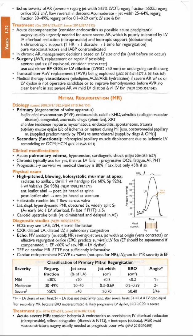
Valv
es
1-22
• Echo: severity of AR (severe = regurg jet width >65% LVOT, regurg fraction >50%, regurgorifice >0.3 cm2, flow reversal in descend. Ao; moderate - jet width 25-64%, regurg fraction 30-49%, regurg orifice 0.1-0.29 cm2); LV size & fxn
Treatm ent (Circ 2014;129:e521 ;Loncet 2016:387:1312)
• Acute decompensation (consider endocarditis as possible acute precipitant):surgery usually urgently needed for acute severe AR, which is poorly tolerated by LV IV afterload reduction (nitroprusside) and inotropic support (dobutamine)± chronotropic support (T HR -»1 diastole -» i time for regurgitation) pure vasoconstrictors and IABP contraindicated
• In chronic AR, management decisions based on LV size and fxn (and before sx occur)• Surgery (AVR, replacement or repair if possible);
severe and sx (if equivocal, consider stress test)asx and either EF £50% or LV dila tion (LVESD >50 mm) or undergoing cardiac surg
• Transcatheter AoV replacement (TAVR) being explored (JACC 2013:61:1577 & 2015:66:169)• Medical therapy: vasodilators (nifedipine, ACEI/ARB, hydralazine) if severe AR w/ sx or
LV dysfxn & not operative candidate or to improve hemodynamics before AVR; no clear benefit in asx severe AR w/ mild LV dilation & nl LV fxn (NEJM 2005:353:1342)
M it r a l R e g u r g it a t io n (M R )
E tio logy (Lancet 2009:373:1382; NEJM 2010:363:156)• P r im a ry (degeneration of valve apparatus)
leaflet abnl: myxomatous (MVP), endocarditis, calcific RHD, valvulitis (collagen-vascular disease), congenital, anorectic drugs (phen-fen), XRT
chordae tendineae rupture: myxomatous, endocarditis, spontaneous, trauma papillary muscle dysfxn b/c of ischemia o r rupture during Ml [usu. posteromedial papillary
m. (supplied predominantly by PDA) vs. anterolateral (suppl. by diags & OMs)]• Secondary (functiona l): inferoapical papillary muscle displacement due to ischemic LV
remodeling or DCM; HCM (JACC 2015:65:1231)
C lin ica l m an ifes ta tions• Acute: p u lm o n a ry edem a, hypotension, cardiogenic shock (NEJM 2004:351:1627)
• Chronic: typically asx for yrs, then as LV fails -+ progressive DOE, fatigue, AF, PHT• Prognosis: 5-y survival w/ medical therapy is 80% if asx, but only 45% if sx
Physical exam• High-pitched, blowing, holosystolic m urm ur at apex;
radiates to axilla; ± thrill; T w / handgrip (Se 68%, Sp 92%), lw / Valsalva (Se 93%) (NEJM 1988:318:1572)
ant. leaflet abnl -♦ post, jet heard at spine post, leaflet abnl -» ant. jet heard at sternum
• ± diastolic rumble b/c T flow across valve• Lat. displ. hyperdynamic PMI, obscured Si, widely split S2
(A2 early b/c i LV afterload, P2 late if PHT); ± S3• Carotid upstroke brisk (vs. diminished and delayed in AS)Diagnostic studies (NEJM 2005:352:875)• ECG: may see LAE, LVH, ± atrial fibrillation• CXR: dilated LA, dilated LV, ± pulmonary congestion• Echo: MV anatomy (ie, etiol); MR severity: jet area, jet width at origin (vena contracta) or
effective regurgitant orifice (ERO; predicts survival); LV fxn (EF should be supronormal if compensated, EF <60% w/ sev. MR = LV dysfxn)
• TEE or cardiac MR ifTTE not sufficiently informative• Cardiac cath: prominent PCWP c-v waves (not spec, for MR), LVgram for MR severity & EF
Heartsounds:
S2
Classification o f P rim ary M itra l Regurgitation
Severity Regurg.fraction
Jet area(% of LA)
Jet width(cm)
ERO(cm2)
Angio*
Mild <30% <20 <0.3 <0.2 1 +Moderate 30-49% 20-40 0.3-0.69 0.2-0.39 2+Severe' >50% >40 >0.70 >0.40 3/4+
*1+ = LA clears w/ each beat; 2+ = LA does not dear,faintly opac. after several beats; 3+ = LA & LV opac. equal.
'For secondary MR, because ERO underestimated & likely progressive LV dysfxn, ERO £0.20 is severe
T re a tm e n t (Grc 2014;129:eS21; Lancet 2016:387:1324)
* A cu te severe MR: consider ischemia & endocarditis as precipitants; IV afterload reduction (nitroprusside), relieve congestion (diuresis & NTG),± inotropes (dobuta), IABP, avoid vasoconstrictors; surgery usually needed as prognosis poor w/o (JAMA 2013:310:609)

• Chronic severe prim ary MR: surgery (repair [preferred if feasible] vs. replacement)indicated if sx & EF >30% or if asx & either EF 30-60% or LV sys. diam. >40 m mMV repair reasonable if asx & either EF >60% + LVESD <40 mm or new AF or PHT if AF, concomitant surgical ablation LAF recurrence, 0 A stroke; consider for sx cntl or if
planning no anticoag {NEJM 2015:372:1399)
• Severe secondary MR; consider surgery if NYHA lll-IV; replacement results in moredurable correction & i HF & 4 CV admissions than repair (NEJM 2016:374:344)
• In Pts undergoing CABG w/ moderate fxnal MR, annuloplasty I MR but longer surgery,t neurologic events, & no impact on fxnal status or mortality (NE/m 2016:374:1932)
• Percut MV repair (Grc 2014:130:1712); edge-to-edge clip less effective than surgery but considerfor sev. sx nonoperative Pt (NEJM 2011:364:1395); perc valve under study (/ACC 2014:64:1814)
• If sx & EF<60% but not operative candidate; HF Rx (PB,ACEI,± aldo antag);i preload w /diuretics, NTG (espec. if ischemic MR) for sx relief ± i ERO; maintain SR
• Asymptomatic: 0 proven benefit of medical therapy; [5B T LV fxn (/ACC 2012:60:833).
M it r a l V a lve P r o la pse (M V P )
Definition and Etiology• Billowing of MV leaflet >2 mm above mitral annulus in parasternal long axis echo view• Primary: sporadic or familial myxomatous proliferation of spongiosa of MV apparatus• Secondary: trauma, endocarditis, congenital, CTD (eg, Marfan’s, Ol, Ehlers-Danlos)
Clinical manifestations (usually asymptomatic)• MR (MVP most common cause), endocarditis, embolic events, arrhythmias (rarely SCD)• High-pitched, midsystolic click (earlier w / i preload) ± mid-to-late systolic murmur• No Rx per se [endocarditis Ppx no longer rec. (Grc 2007,116:1736)]; Rx MR as above
M it r a l St e n o s is (M S )
Etiology (Lancet 2012:379:953)• Rheumatic heart disease (RHD): fusion o f commissures -» "fish-mouth” valve
from autoimmune rxn to p strep infxn; seen largely in developing world today• M itral annular calcification: encroachment upon leaflets -> fxnal MS; espec in ESRD• Congenital, infectious endocarditis w/ large lesion, myxoma near MV, thrombus• Valvulitis (eg, SLE, amyloid, carcinoid) o r infiltration (eg, mucopolysaccharidoses)
Clinical manifestations (Lancet 2009:374:1271)
• Dyspnea and pulm onary edem a (if due to RHD, sx usually begin in 30s)precipitants: exercise, fever, anemia, volume overload (incl. pregnancy), tachycardia,AF
• A trial fibrillation: onset often precipitates heart failure in Pts w / MS• Embolic events: commonly cerebral, espec in AF or endocarditis• Pulmonary: hemoptysis, frequent bronchitis (due to congestion), PHT, RV failure• O rtner’s syndrome: hoarseness from LA compression of recurrent laryngeal nerve
Physical exam• Low-pitched mid-diastolic rum ble at apex w/
presystolic accentuation (if not in AF); best heard in L lat decubitus position during expiration, T w/ exercise; severity proportional to duration (not intensity) of murmur; loud Si
• Opening snap (high-pitched early diastolicsound at apex) from fused leaflet tips;MVA proportional to S2-OS interval (tighter valve -» T LA pressure shorter interval)
• Loud Si (unless MV calcified and immobile)
Diagnostic studies• ECG: LAE ("P mitrale”), ± AF, ± RVH• CXR: dilated LA (flat L heart border, R double density, displaced L mainstem bronchus)• Echo: estimate pressure gradient (V), RVSP, valve area, valve echo score (0-16, based
on leaflet mobility & thick, subvalvular thick., Ca+f); exer.TTE (to assess A RVSP and V) if sx & severity of MS at rest discrepant;TEE to assess for LA thrombus before PMBC
• Cardiac cath: V, calculated MVA; LA tall a wave & blunted y descent; T PA pressures
Classification of Mitral StenosisStage Mean V (mmHg) Pressure V2 time MVA (cm2) PA sys (mmHg)Normal 0 4-6 <25Mild-Mod <5 100-149 1.6-2 <30Severe 5-9 150-219 1.1-1.5 30-50Very severe >10 >220 <1 V 1/
10
hmiiC'jnds

YTreatm ent (Circ 2014;129:e521;Lrwcet 2016:387:1324)• Medical: Na restriction, cautious diuresis, f)B, AF control, sx-limited physical stress• Antibiotic Ppx recommended if h/o RHD w / valvular disease for 10 y o r until age 40• Anticoag: AF; prior embolism; LA clot; ? LA >55 mm or Large LA w / spont contrast• Mechanical intervention indicated if h e a r t fa i lu r e sx w / M V A £ 1 .5 ; reasonable if asx but
very severe (MVA <1) and morphology favorable for PMBC; may consider PMBC if MVA >1.5 but hemodyn signif w/ exercise, or if asx but MVA <1.5 and new-onsetAF
• Percutaneous m itral balloon commissurotomy (PMBC): preferred Rx if RHD; MVAdoubles, V i by 50%; ~ MVR i f valve score < 8 ,0 if mod-severe MR or LA clot
• Surgical (MV repair if possible, o/w replacement): consider in sx Pts w / MVA <1.5if PMBC unavailable/failed/contraindicated or valve morphology unsuitable
• Pregnancy: if NYHA class lll/IV -4 PMBC, o/w medical Rx w/ low-dose diuretic & (3B
T r ic u s p i d R e g u r g i t a t i o n
• 10 etiol: rheumatic, CTD, XRT, IE, Ebstein’s, carcinoid, tumors, pacemaker leads• Fxnl etiol (most common): RV and/or PHT (may be 2° to L-sided dis.), RV dilation ± Ml• Holosystolic murmur, 3rd/4th ICS, T w/ insp (Carvallo’s sign); S3; prominent cv wave in JVP• Consider repair, annuloplasty or replacement for sx and severe TR (eg, ERO >0.40 cm2);
transcatheter system (provides surface for coaptation) under study (JACC 2015:66:2475)
P r o s t h e t i c H e a r t V a l v e s
Mechanical (60%)• Bileaflet (eg, St. Jude Medical); tilting disk; caged-ball• Very durable (20-30 y), but thrombogenic and /. require anticoagulation
consider if age < -60 y or if anticoagulation already indicated (/ACC 2010:55:2413)Bioprosthetic (40%)• Bovine pericardial o r porcine heterograft (eg, Carpentier-Edwards), homograft• Less durable, but min. thrombogenic; consider if >~70 y, lifespan <20 y, or 0 anticoag• If 50-69 y, 2x reop but Vi bleeding o r stroke vs. mech ( jA tA A 2014;312:1323 & 2015:313:1435)
Physical exam• C ris p sounds ± soft murmur during forward flow (normal to have small V) Anticoagulation & antiplatelet therapy (Ore 2014;l29e52l)• High-risk features: prior thromboembolism, AF, EF <30-35%, hypercoagulable• W a rfa rin (0 N O A C s ): mech MVR or high-risk mech AVR: INR 2.5-3.5. Low-risk mech
AVR or high-risk bio MVR/AVR: INR 2-3. Consider in low-risk bio MVR/AVR for 1st 3 mo.• + A S A (<100 mg): all prosth. valves unless h/o GIB, uncontrolled HTN, erratic INR,
o r >80 y• If thrombosis, T intensity (eg, INR 2-3 -4 2.5-3.5; 2.5-3.5 -4 3.5-4.5; add ASA if not on)
Periprocedural “ Bridging” of Anticoagulation in Pts with Mechanical Valve(s)
AVR w/o risk factors d/c warfarin 2-4 d before surg; restart 12-24 h after surgMVR or AVR w/ risk factors
Preop: d/c warfarin, start UFH (preferred to LMWH) when INR <2 4-6 h preop: d/c UFH; postop: restart UFH & warfarin ASAP
Procedures include noncardiac surgery, invasive procedures, and major dental work
C o r r e c t io n o f o v e r a n t ic o a g u la t io n (Circ 2014;I29.e52i)
• Risk from major bleeding must be weighed against risk of valve thrombosis• N o t bleeding: if INR 5-10, withhold warfarin; if INR >10 also give vit K 1-2.5 mg PO• Bleeding: FFP or PCC + low-dose (1 mg) vit K IV
E n d o c a rd itis prophylaxis: for all prosthetic valves (see “ Endocarditis” )
C o m p lic a tio n s• Structural failure (r/o endocarditis); mechanical valves: rare except for Bjork-Shiley;
bioprosth: up to 30% rate w/in 10-15 y, mitral > aortic; consider TAVR {JAMA 2014:312:162)
• Paravalvular leak (r/o endocarditis); small central jet of regurg is normal in mech. valves• Obstruction from thrombosis (JACC 2013:62:1731) or pannus: / TTE.TEE, CTA, or fluoro
significantly symptomatic pannus ingrowth: remove w/ surgery thrombosis: surgery if L-sided valve & either severe sx o r Ig (> >0.8 cm) thrombus;
lytic successful in -70% of L-sided thrombosis, but w/ 14% risk of stroke; consider UFH ± lytic (? low-dose tPA via slow infusion; JACC CV Imaging 2013:6:206) if mild sx & small clot burden or poor surg candidate; lytic reasonable for R-sided
• Infective endocarditis ± valvular abscess and conduction system dis. (see “ Endocarditis” )• Embolization (r/o endocarditis); risk highest 1st 90 d, ~1%/y w/ warfarin (vs. 2% w/ASA, or
4% w/o meds); mech MVR 2x risk of embolic events vs. mech AVR (Ore 1994:89635)
• Bleeding (from anticoag), hemolysis (espec w / caged-ball valves o r paravalvular leak)

PERICARDIAL DISEASE kG e n e r a l P r in c ip l e s
Anatom y• Tissue sac surrounding heart & proximal great vessels; 2 layers (parietal & visceral)
Disease states• Inflammation (w/ o r w/o fluid accumulation) pericarditis• Fluid accumulation -> effusion ± tamponade• Decrease in compliance (sequela of inflammation) -> constrictive pericarditis• Tamponade and constriction characterized by increased ventricular interdependence
P e r ic a r d it is a n d P e r ic a r d ia l E f f u s io n
Etiologies of Acute Pericarditis (jama 2015:314:1498)
Idiopathic (>80%) Most presumed to be undiagnosed viral etiologiesInfectious(<5% can beconfirmedinfectious)
Viral: Coxsackie, echo, adeno, EBV.VZV, HIV, influenza Bacterial (from endocarditis, pneumonia or s/p cardiac surgery):
S. pneumococcus, N. meningii/dis, S. oureus, 8orrelia (Lyme);TB Fungal: Histo, Coccidio, Candida; Parasitic: Entamoeba, Echino
Neoplastic(<10%)
Common: metastatic (lung, breast, lymphoma, leukemia, RCC) Rare: primary cardiac & serosal tumors (mesothelioma)
Autoimmune Connective tissue diseases: SLE, RA, scleroderma, Sjogren’s Vasculitides: PAN, eosin GPA (Churg-Strauss), GPA (Wegener’s) Drug-induced: procainamide, hydralazine, INH, CsA
Uremia -5-13% of Pts prior to HD;-20% occurrence in chronic HD PtsCardiovascular STEMI, late post-MI (Dressler’s syndrome); ascending AoD; chest
trauma; postpericardiotomy; procedural complic. (ie, PCI, PPM)Radiation >40 Gy to mediastinum; acute or delayed; may be transudativeEffusion w/o pericarditis
CHF, cirrhosis, nephrotic syndrome, hypothyroidism, amyloidosis. Transudative.
Clinical manifestations (NEJM 2014:371:2410)• Pericarditis: retrosternal CP, pleuritic, positional (often i by sitting forward), -> trapezius;
may be absent in TB, neoplastic, XRT, or uremic; ± fever; ± sis of systemic etiologies• Effusion: present in —2/3 of Pts w/ pericarditis; ranges from asx to tamponadePhysical exam• Pericarditis: multiphasic friction rub best heard at USB w/ diaphragm of stethoscope.
Notoriously variable and evanescent leathery sound w/ up to 3 components: atrial contraction, ventricular contraction, ventricular relaxation (nejm 20l2;367:e20).
• Effusion: distant heart sounds, dullness over left posterior lung field due to compressiveatelectasis from pericardial effusion (Ewart’s sign)
Diagnostic studies (JAMA 2015:314:1498; EHJ 2015:36:2921)• Need >2 of the following: chest pain (as noted above), friction rub, ECG findings, effusion• ECG: may show diffuse STE (concave up) & PR depression (except in aVR: ST I & PR T),
TWI; classically and in contrast to STEMI.TWI do not occur until STs normalize Stages: (I) STE & PR i ; (II) ST & PR normalize; (III) diffuse TW I; (IV) Tw normalize ECG may show evidence of large effusion w/ low voltage & electrical alternans
(beat-to-beat A in QRS amplitude and/or axis due to swinging heart)• CXR: if Ig effusion (>250 mL) -> T cardiac silhouette w/"water-bottle” heart & epicardial halo• Echocardiogram: presence, size, & location of effus/on; presence of tamponade
physiology; pericarditis itself w/o spec, abnl (.*. echo can be nl), although can see pericardial stranding (fibrin o r tumor); can also detect LV/RV dysfxn (myocarditis?)
• CT: will reveal pericardial effusions, but they often appear larger by CT than by echo.• MRI: may reveal pericardial thickening/inflammation, as well as myocardial involvement• CK-MB or troponin (© in -30%;JACC 2003:42:2144) if myopericarditis. Consider CRP/ESR.W orkup for effusion• r/o infxn: usually apparent from Hx & CXR; ? value of / acute and convalescent serologies• r/o noninfectious etiologies: BUN, Cr.ANA, RF, HIV, relevant malignancy evaluation• Pericardiocentesis if suspect infxn o r malignancy o r large effusion (>2 cm) or recurrent
/ cell counts,TP, LDH,glc, Gram stain & Cx,AFB, cytology ADA, PCR for MTb, and specific tum or markers as indicated by clinical suspicion “ exudate” :TP >3 g/dL,TPeff/TPserum >0.5, LDHc(r/LDHsemm >0.6 or glc <60 mg/dL;
high Se (- 90%) but very low Sp (-20%); overall low utility (Chest 1997:111:1213)• Pericardial bx if suspicion remains for malignancy orTB
Pe
rica
rdia
l 1-25

Treatm ent of pericarditis (JAMA 2015:314:1498; EHJ 2015:36:2921)
• High-dose NSAID (eg, ibuprofen 600-800 mg tid) or ASA (eg, 650-1000 mg tid) x 7-14 dthen taper over wks;ASA preferred over NSAID in acute Ml; consider PPI to I risk of GIB
» Add colchicine 0.5 mg bid (qd if <70 kg) x 3 mo; I risk of refractory or recurrent pericarditis by 50% (NEJM 2013:369:1522)
• Avoid steroids except for systemic autoimmune disorder, uremic, preg., NSAIDs contraindicated, or refractory idiopathic dis. Appear to T rate of pericarditis recurrence (Grc
2008:118:667). If due toTB, steroids 4 risk of constriction (NEjM 2014:371:1121).• Avoid anticoagulants (although no convincing data that T risk of hemorrhage/tamponade)• Infectious effusion -» pericardial drainage (preferably surgically) + systemic antibiotics• Acute idiopathic pericarditis self-limited in 70-90% of cases• Recurrent pericarditis (Grc 2007:115:2739)
risk factors: subacute, Ig effusion/tamponade.T >38r’C,lack of NSAID response after 7 d treatment: colchicine 0.5 mg bid x 6 mo (AwiaJs 2011;15S:409 & Lancet 2014:383:2232)
■ Recurrent effusions: consider pericardial window (percutaneous vs. surgical) * •
P e r ic a r d ia l T a m p o n a d e
E t io lo g y• Any cause of pericarditis but espec, malignancy, infectious, uremia, ascending AoD,
myocardial rupture, periprocedural complication, trauma, post-cardiotomy• Rapidly accumulating effusions most likely to cause tamponade as no time for pericardium
to stretch (eg, to T compliance) and accommodate t intrapericardial fluid volume
P a th o p h y s io lo g y (NEJM 2003:349:684)• T intrapericardial pressure, compression of heart chambers, 4 venous return -»4 CO• Diastolic pressures T & equalize in all cardiac chambers -» minimal flow of blood from RA
to RV when TV opens -> blunted y descent• T ventricular interdependence -> pulsus paradoxus (pathologic exaggeration of nl physio)
Inspiration -» 4 intrapericardial & RA pressures -> T venous return -4 T RV size -> septal shift to left. Also, T pulmonary vascular compliance -> 4 pulm venous return.Result is 4 LV filling 4 LV stroke volume & blood pressure & pulse pressure.
C l in ic a l m a n i f e s ta t io n s• Cardiogenic shock (hypotension, fatigue) w ithout pulm onary edem a• Dyspnea (seen in -85%) may be due to T respiratory drive to augment venous return
P h y s ic a l e x a m {ehj 2014:35:2279)• Beck's triad (present in minority of cases): distant heart sounds, T JVP, hypotension• t JVP (76%) w/ blunted y descent• Reflex tachycardia (77%), hypotension (26%; occasionally hypertensive), cool extremities• Pulsus paradoxus (Se 82%, Sp 70%) = 4 SBP >10 mmHg during inspiration
© LR 3.3 (5.9 if pulsus >12), © LR 0.03Ddx = PE. hypovolemia, severe COPD, constriction (-V 3), RV infarct ? absent if pre-existing T LVEDP, irregular rhythm, severe AI.ASD, regional tamponade
• Distant heart sounds (28%), ± pericardial friction rub (30%)• Tachypnea and orthopnea but clear lungs
D ia g n o s t ic s tu d ie s• ECG: T HR, 4 voltage (seen in 42%), electrical alternans (20%), ± signs of pericarditis• CXR: T cardiac silhouette (89%)• Echocardiogram: © effusion, IVC plethora, septal shift with inspiration
diastolic collapse of RA (Se 85%, Sp 80%) and/or RV (Se <80%, Sp 90%) respirophasic A’s in transvalvular velocities (T across TV & 4 across MV w/ inspir.) postsurgical tamponade may be localized and not easily visible
• Cardiac cath (right heart and pericardial): elevation (15-30 mmHg) and equalization ofintrapericardial and diastolic pressures (RA, RV, PCWP), blunted y descent in RA T in stroke volume postpericardiocentesis = ultimate proof of tamponade if RA pressure remains elevated after drainage, may have effusive-constrictive disease
(constriction from visceral pericardium; NEJM 2004:350:469) o r myocardial dysfxn (eg, from concomitant myocarditis)
T r e a t m e n t (EHJ 2014:35:2279)• Volume (but be careful as overfilling can worsen tamponade) and © inotropes (avoid (3B)• Avoid vasoconstrictors as will 4 stroke volume & potentially 4 HR• Avoid positive pressure ventilation as it can further impair cardiac filling (Ore 2006:113:1622)• Pericardiocentesis (except if due to aortic/myocardial rupture for which emergent surgery
treatment of choice; if too unstable, consider small pericardiocentesis to prevent PEA)• Surgical drainage considered if fluid rapidly reaccumulates, loculated, or hemorrhagic

C o n s t r ic t iv e P e r ic a r d it is
Etiology (Circ 2011;124:1270)
• Any cause of pericarditis (-1-2% incidence overall after acute pericarditis)• Highest risk w/ TB , bacte ria l, neoplastic, XRT, connective tissue, postcardiac surgery• V ira l/id iopa th ic , as most common cause of pericarditis, also account for signif proportion
Pathophysiology• Adhesion of visceral and parietal pericardial layers -» rigid pericardium that limits diastolic
filling of ventricles -» T systemic venous pressures• Venous return is limited only after early rapid filling phase;.-. rapid i in RA pressure with
atrial relaxation and opening of tricuspid valve and prominent x and y descents• Kussmaul sign.JVP does not decrease with inspiration (T venous return with inspiration,
but negative intrathoracic pressure not transmitted to heart because of rigid pericardium)
Clinical manifestations (NEJM 2011;364:1350)
• Right-sided > left-sided heart failure (systemic congestion > pulmonary congestion)
Physical exam• T JVP with p ro m in e n t y descent, © Kussmaul sign [Ddx: tricuspid stenosis,acute cor
pulmonale, RV dysfxn (CMP, RV Ml), SVC syndrome]• Hepatosplenomegaly, ascites, peripheral edema. Consider in Ddx of idiopathic cirrhosis.• PMI usually not palpable, pe rica rd ia l knock, usually no pulsus paradoxus
Diagnostic studies• ECG; nonspecific, AF common (up to 33%) in advanced cases• CXR: calcification (MTb most common), espec in lateral view (although not specific)• Echocardiogram;! thickened pericardiunV'septal bounce” = abrupt displacement of
septum during rapid filling in early diastole• Cardiac catheterization: atria w / Ms or W s (prominent x and y descents)
ventricles: dip-and-plateau or square-root sign (rapid I pressure at onset of diastole, rapid t to early plateau)
discordance between LV & RV pressure peaks during respiratory cycle (Ore 1996:93:2007)
• CT or MRI: thickened pericardium (>4 mm;Se -80%) w/ tethering (Circ 20iT.i23-.e4i8)
Treatm ent• Diuresis if intravascular volume overload; surgical pcricardiectomy if infectious or advanced
Constrictive Pericarditis vs Restrictive CardiomyopathyEvaluation Constrictive pericarditis Restrictive cardiomyopathy
Physical exam© Kussmaul sign Absent PMI © Pericardial knock
± Kussmaul sign Powerful PMI, ± S3 and S4 ± Murmurs of MR, TR
ECG ± Low voltage Low voltage if infiltrative myopathy ± Conduction abnormalities
Echocardiogram
Respirophasic variation (25-40%): inspir. -> T flow across TV and I flow across MV
E' (tissue velocity) nl/T (>12 cm/sec)
Expir. hepatic vein flow reversal Septal bounce in early diastoleNormal wall thickness
<10% respirophasic variation Slower peak filling rate Longer time to peak filling rate E' I (<8 cm/sec) (Se 95%, Sp
96%; HF Re/ 2013:18:255)Inspir, hepatic vein flow reversal Biatrial enlargement ± t wall thickness
CT/MRI Usually w/ thickened pericardium Normal pericardiumNT-proBNP Variable Typically T/TT (/ACC 2005:45:1900)
Prominent x and y descents (more so in constriction)Dip-and-plateau sign (mo e so in constriction)
Cardiaccatheterization
LVEDP = RVEDPRVSP <55 mmHg (Se 90%, Sp 29%) RVEDP >V3 RVSP (Se 93%, Sp 46%) Discordance of LV & RV pressure
peaks during respiratory cycle Systolic area index (ratio of RV to
LV pressure-time area in inspir vs. expir) >1.1 (Se 97%, Sp 100%)
LVEDP > RVEDP (esp. w/ vol.) RVSP >55 mmHg RVEDP <’/3 RVSP Concordance of LV & RV
pressure peaks during respiratory cycle
Systolic area index <1.1 (JACC 2008:51:315)
Endomyocardialbiopsy
Usually normal ± Specific etiology of RCMP(fibrosis, infiltration, hypertrophy)
Pe
ric
ar
dia
l 1
-27

Hy
pe
rt
en
sio
n
1-28
HYPERTENSIONJNC 8 Classification
Category Systolic DiastolicNormal <120 <80Pre-HTN 120-139 80-89Stage 1 HTN 140-159 90-99Stage 2 HTN >160 >100
Ambulatory ThresholdsSetting Systolic Diastolic24-hr avg 135 85Day (awake) 140 90Night (asleep) 125 75
(Clrc 2005:111:697)
BP in mmHg. Average >2 measurements > 1 -2 min apart. Confirm stage 1 w/in 1 -4 wk; can Rx stage 2 immediately (J Clin HTN 2014:16:14)
Epidemiology (JAMA 2014:311:1424; Ore 201 S;131 :e29)
• Prevalence -30% in U.S. adults, >44% in African-Americans; M = F• O f those with HTN, -3/4 were treated, -1/2 achieve target BP, -1 /6 were unaware of dxEtiologies• Essential (95%): onset 25-55 y;© FHx. Unclear mechanism but ? additive microvasc
renal injury over time w/ contribution of hyperactive sympathetics (NEJM 2002:346:913). T Age -> i art compliance HTN. Genetics + environment involved (Noture 2011:478:103).
• Secondary: Consider if Pt <30 y o r if sudden onset, severe, refractory HTN
Secondary Causes of HypertensionDiseases Suggestive findings Initial workup
_iRenal parenchymal(2-3%)
h/o DM, polycystic kidney disease, glomerulonephritis
CrCI, albuminuria See “ Renal Failure”
<ZHI0c
Renovascular (1-2%) Athero (90%)FMD (10%, young women) PAN, scleroderma
ARF induced by ACEI/ARB Recurrent flash pulm edema Renal bruit; hypokalemia (NEyM 2009:361:1972)
MRA (>90% Se & Sp, less for FMD),CTA, duplex U/S, angio, plasma renin (low Sp)
OHyperaldo or Cushing’s (1-5%)
Hypokalemia Metabolic alkalosis See “Adrenal
Disorders”QZLU
Pheochromocytoma (<1%) Paroxysmal HTN, H/A, palp.Myxedema (<1%) See “Thyroid Disorders” TFTsHypercalcemia (<1%) Polyuria, dehydration, A MS iCa
CC Obstructive sleep apnea (qv); alcoholLUI Medications: OCP, steroids, licorice; NSAIDs (espec COX-2); Epo; cyclosporine
Aortic coarctation: 1 LE pulses, systolic murmur, radial-femoral delay; abnl TTE, CXRu
Polycythemia vera: T Hct
S ta n d a r d w o r k u p• Goals: (1) identify CV risk factors; (2) seek 2° causes; (3) assess for target-organ damage• History: CAD, HF,TIA/CVA, PAD, DM, renal insufficiency, sleep apnea, preeclampsia;
© FHx for HTN; diet, Na intake, smoking, alcohol, prescription and OTC meds, OCP• Physical exam: / BP in b o th arms; funduscopic exam, BMI, cardiac (LVH, murmurs),
vascular (bruits, radial-femoral delay), abdominal (masses or bruits), neuro exam• Testing: K, BUN, Cr, Ca, glc, Hct, U/A, lipids,TSH, urinary albumin:creatinine (if t Cr, DM,
peripheral edema), ? renin, ECG (for LVH), CXR.TTE (eval for valve abnl, LVH)• Ambulatory BP monitoring (ABPM): consider for episodic, resistant, or white coat HTN C o m p l ic a t io n s o f H T N• Neurologic: T IA /C V A , ruptured aneurysms, vascular dementia• Retinopathy: stage I = arteriolar narrowing; II = copper-wiring, AV nicking;
III = hemorrhages and exudates; IV = papilledema• Cardiac: C AD , LVH, H F ,A F• Vascular: aortic dissection, aortic aneurysm (HTN = key risk factor for aneurysms)• Renal: proteinuria, renal failureT r e a t m e n t (JAMA 2014.311:507;y Cifn HTN 2014:16:14: HTN 2015:65:1372:JACC 2015:65:1998)• Every i 10 mmHg -> 20% i MACE, 28% -i HF, 13% 4. mort. (Lancet 2016:387:957)• Traditional goal: <140/90; i f prior M llstroke: reasonable to consider <130/80
If high-risk (CV dis., 10-y risk of CV dis.>15%, CKD.or >75 y;non-DM and no h/o stroke) SBP -120 vs.-135 (via unattended automated cuff) -» i MACE 25%, i death 27%, i HF 38%, but T HoTN.AKI, syncope, electrolyte abnl (NEJM 2015:373:2103)
If DM: optimal goal disputed b/c lack of clear benefit in one study (NEJM 2010:362:1575); may consider <130/80 for renal protection if CKD & albuminuria (ASH/ISH)
If intermed risk (RF for but w/o CV dis.), benefit only if SBP >-140 (NEJM 2016:374:2009) In elderly: ? more lenient targets, but benefit to Rx’ing stage 2 HTN in low-risk (NEJM 2008;
358:1887) and targeting SBP -120 in high-risk ( i MACE & mortality;jama 2016:315:2673)

Lifestyle modifications (each may i SBP ~5 mmHg) weight loss: goal BMI 18.5-24.9; aerobic exercise: >30 min exercise/d, £5 d/wk diet: rich in fruits & vegetables, low in saturated & total fat (DASH, nejm 2001,344:3) limit Na: <2.4 g/d (ideally <1.5 g/d); maintain K intake (NEJM 2007:356:1966 & 2010:362:2102)
limit alcohol: <2 drinks/d in men;<1 drink/d in women & lighter-wt Pts; avoid NSAIDs Pharmacologic options
P re -H T N : ARB prevents onset of HTN, no i in clinical events (NEJM 2006:354:1685)
H T N : choice o f therapy controversial, concomitant disease and stage may help guide Rx uncomplicated: CCB,ARB/ACEI,or thiazide (chlorthalidone preferred) are 1£t line (NE|M
2009:361:2153). [3B not 1st line (Loncet 2005:366:1545).
For non-black Pts <60 y: reasonable to start w/ARB or ACEl.then add CCB or thiazide if needed, and then add remaining class if still needed
For black, elderly, and ? obese Pts (all o f whom more likely to be salt sensitive): reasonable to start with CCB or thiazide, then add either the other 1st choice class or ARB or ACEI if needed, and then all 3 classes if still needed
+C A D (Ore 2015:131 :e435): ACEI o r ARB (NEJM 2008:358:1547); ACEI+CCB superior to ACEI+thiazide (NEJM 2008:359:2417) or pB+diuretic (Lancet 2005:366:895); may require pB and/or nitrates for anginal relief; if h/o Ml, pB ± ACEI/ARB ± aldo antag (see "ACS” )
+HF: ACEI/ARB/ARNi, PB, diuretics, aldosterone antagonist (see "Heart Failure” ) +2° stroke prevention: ACEI ± thiazide (Loncet 2001:358:1033)
+diabetes mellitus: consider ACEI or ARB; can also consider thiazide or CCB -fchronic kidney disease: ACEI o r ARB (NEJM 1993:329:1456 & 2001:345:851 & 861)
Tailoring therapy: if stage 1, start w/ monoRx; if stage 2, consider starting w/ combo (eg.ACEI + CCB; NEJM 2008:359:2417); start at Vi max dose; after 2-3 wk, uptitrate o r add drug
Pregnancy: methyldopa, labetalol, & nifed pref. Hydral OK; avoid diuretics; 0 ACEI/ARB. Targeting DBP 85 vs. 105 safe and i severe HTN (nejm 2015:372:407).
Resistant H T N (BP > goal on >3 drugs incl diuretic;JAMA 2014:311:2216)• Exclude: 2° causes (see table) and pseudoresistance: inaccurate measure (cuff size), diet
noncomp ( t Na), poor Rx compliance/dosing, white coat HTN ( / ABPM)• Ensure effective diuresis dosing (chlorthalidone > HCTZ, loop > thiazide if eGFR <30)• Can add aldosterone antagonist (Lancet 2015:386:2059), p-blocker (particularly vasodilators like
labetalol, carvedilol, or nebivolol), a-blocker, or direct vasodilator
H y p e r t e n s iv e C rises
• H ype rtens ive em ergency: T BP -> acute target-organ ischemia and damageneurologic damage: encephalopathy, hemorrhagic or ischemic stroke, papilledema cardiac damage: ACS, HF/pulmonary edema, aortic dissection renal damage: proteinuria, hematuria, acute renal failure; scleroderma renal crisis microangiopathic hemolytic anemia; preeclampsia-eclampsia
• Hypertensive urgency: SBP >180 or DBP >120 (?110) w/ min. o r 0 target-organ damage
Precipitants• Progression of essential HTN ± medical noncompliance (espec clonidine) o r A in diet• Progression of renovascular disease; acute glomerulonephritis; scleroderma; preeclampsia• Endocrine: pheochromocytoma, Cushing’s• Sympathomimetics: cocaine, amphetamines, MAO inhibitors + foods rich in tyramineTreatm ent (Chest 2007:131:1949)• Tailor goals to clinical context Treat Ao dissection aggressively. Do not treat HTN in acute
ischemic stroke unless lysis planned or extreme BP (>220/120).• Emergency: i MAP by -25% in m/ns to 2 h w/ IV agents (may need arterial line for
monitoring); goal DBP <110 w/in 2-6 h,as tolerated• Urgency: I BP to <160/100 in hrs using PO agents; goal normal BP in -1 -2 d• Watch UOP, Cr, mental status: may indicate a lower BP is not tolerated
D r u g s f o r H y p e r t e n s iv e C r i s e s
Nitroprusside* 0.25-10 pg/kg/min Nitroglycerin 5-1000 pg/min
IV
Labetalol 20-80 mg IVB qlOmin or 0.5- 2 mg/min. Preferred in pregnancy.
Esmolol 0.5 mg/kg load -* 0.05-0.2 mg/kg/min
Fenoldopam 0.1-1.6 jjg/kg/min Hydralazine 10-20 mg q20-30minNicardipine 5-15 mg/h Clevidipine 1-16 mg/h
Phentolamine 5-15 mg bolus q5-15min Enalaprilat 1.25 mg
POCaptopril 12.5-100 mg q8h Labetalol 200-800 mg, repeat after 2-3 h
Clonidine 0.2 mg load -* 0.1 mg qh Hydralazine 10-75 mg qid
^Metabolized to cyanide ->A MS, lactic acidosis, death. Limit use of very high doses (8-10 pg/kg/min) to <10 min. Monitor thiocyanate levels. Hydroxocobalamin o r sodium thiosulfate infusion for treatment of cyanide toxicity.
Hy
pe
rt
en
sio
n
1-29

Ao
rta
1-
30AORTIC ANEURYSMS I
D e f i n i t i o n s• True aneurysm (>50% dilation of all 3 layers of aorta) vs. false (rupture within adventitia)• Location: root (annuloaortic ectasia), thoracic aortic aneurysm (TAA), thoracoabdominal
aortic aneurysm (TAAA), abdominal aortic aneurysm (AAA)• Type: fusiform (circumferential dilation) vs. saccular (localized dilation of aortic wall)
E p id e m io lo g y (Grc 2010;121:e266,2011:124:2020; Nat Rev Cardiol 2011;8:92)
• TAA: <?:9 2:1; —60% root/ascending; 40% desc.• AA A : -4-8% prev in those >60y; 5x more common in d; mostly infrarenal
P a t h o p h y s io lo g y & r i s k f a c t o r s (NEJM 2009:361:1114; Nat Med 2009:15:649)
• Medial degen and/or T wall stress; wall stress [(AP x r) / (wall thickness)] (Laplace’s law)• TAA: medial degeneration (muscle apoptosis, elastin fiber weakening); a/w CTD, aortitis• AAA: long-standing HTN + athero/inflammation medial weakening• Classic clinical risk factors: HTN, atherosclerosis, smoking, age, 6• C T D (Marfan, Ehlers-Danlos type IV, Loeys-Dietz); congenital (bicuspid AoV,Turner’s)
aortitis (Takayasu’s GCA, spondyloarthritis, lgG4, syphilis); trauma
S c r e e n in g {Ore 2010:121:e266 & 2011;124:2020; Annals 2014;161:281;JAMA 2015:313:1156)• TAA: if bicuspid AoV or 1° relative w/: (a) TAA o r bicuspid AoV, (b) CTD as above• AAA: / for pulsatile abd mass; U/S 6 >60 y w/ FHx of AAA & d 65-75 y w/ prior tobacco
D ia g n o s t ic s t u d ie s (Grc 2010;121:e266 & 2011;124:2020)
■ Contrast CT: quick, noninvasive, high Se & Sp for all aortic aneurysms• TTE/TEE: TTE most useful for root and proximal Ao;TEE can visualize other sites ofTAA• MRI: favored over CT for AoRoot imaging; useful in AAA but time-consuming; noncontrast
“ black blood” MR to assess aortic wall• Abdominal U/S: screening/surveillance test of choice for infrarenal AAA
T r e a t m e n t {Ore 2006:113:e463; 2008:117:1883; 2010:121 :e266; NEJM 2014:371:2101)
• Goal is to prevent rupture (50% mortality prior to hospital) by modifying risk factors• Risk factor modification: smoking cessation; LDL-C <70 mg/dL• BP control: pB ( i dP/dt) i aneurysm growth (NEJM 1994:330:1335); ACEI a/w i rupture risk
(toncet 2006:368 659); ARB m ay i rate o f aortic root growth in Marfan (NE/M 2008:358:2787)
• Mod CV exercise OK, no burst activity requiring Valsalva maneuvers (eg, heavy lifting)• Indications for surgery (individualized based on FHx, body size, gender, anatomy)
TAA: sxs, ascending Ao >5.5 cm (4-5 cm if Marfan, bi-AoV, L-D, vascular EDS); size may not predict repair benefit; descending Ao >6 cm; >4.5 cm and planned AoV surgery
A A A : sx; infrarenal >5.5 cm; consider >5.0 cm in 9;T >0.5 cm/y; inflam/infxn
E n d o v a s c u la r r e p a i r (EVAR) {NEJM 2008:358:494; Crrc 2011:124:2020 & 2015:131:1291)
• Requires favorable aortic anatomy• TEVAR (thoracic EVAR) for descending TAA >5.5 cm may 1 periop morbidity and possibly
m o r ta li ty {Grc 2010:121:2780;JACC 2010:55:986;/ Viorae CV Surg 2010,140:1001 & 2012:144:604)
• AAA: guidelines support open repair or EVAR for infrarenal AAA in good surg candidatesi short-term mort, bleeding, LOS; but long-term graft complic. (3-4%/y; endoleak, need for
reintervention, rupture) necessitate periodic surveillance, with no proven A in overall mortality in trials, except ? in those <70 y (NEJM 2010:362:1863,1881 & 2012:367:1988)
In observ. data, EVAR a/w T early survival but T long-term rupture (NEJM 2015:373:328)
In Pts unfit for surgery or high periop risks: i aneurysm-related mortality but no A in overall mortality over med Rx (NE/M 2010:362:1872). EVAR noninferior (? superior) to open repair in ruptured AAA w/ favorable anatomy (Ann Surg 2009;250:818).
C o m p l i c a t i o n s (Grc 2010;121:e266; Nat Rev Cardiol 2011:8:92)
• Pain: gnawing chest, back or abdominal pain; new or worse pain may signal rupture• Rupture: risk T w/ diameter, 9, current smoking, HTN
TAA: -2.5%/y if <6 cm vs. 7%/y if >6 cmA A A : ~1%/y if <5 cm vs. 6.5%/y if 5-5.9 cm ;-80% mortality at 24 h
• A ortic insufficiency (T A A ), CHF, acute aortic syndromes (qv)• Throm boem bolic ischemic events (eg, to CNS, viscera, extremities)• Compression of adjacent structures (eg, SVC, trachea, esophagus, laryngeal nerve)
F o l l o w - u p (Grc 2010;121:e266; Not Rev Cordrof 2011;8:92;JAMA 2013:309:806)
• Expansion rate - 0.1 cm/y for TAA, -0.3-0.4 cm/y fo r AAA• AAA: <4 cm q2-3 y; 4-5.4 cm q6-12 mos; more often if rate of expansion >0.5 cm in 6 mo• TAA: 6 m o a f te r d x t o e n s u re s ta b le , a n d i f s ta b le , th e n a n n u a lly (Ore 2005:111:816)
• Screen for CAD, PAD and aneurysms elsewhere, espec popliteal.About 25% of Pts w/TAAwill also have AAA, and 25% of AAA Pts will have a TAA: consider pan-Ao imaging.

ACUTE AORTIC SYNDROMES IDefinitions (Ore 2010;121:e266; Ear Heart) 2012;33:26)• Aortic dissection: intimal tear-* blood extravasates into Ao media (creates false lumen)• Intramural hematoma (IMH): vasa vasorum rupture -» medial hemorrhage that does not
communicate with aortic lumen; 6% of aortic syndromes; clinically managed as AoD• Penetrating ulcer: atherosclerotic plaque penetrates elastic lamina -> medial hemorrhage
Classification (proximal twice as common as distal)• Proximal: involves ascending Ao, regardless of origin (= Stanford A, DeBakey I & II)• Distal: involves descending Ao only, distal to L subclavian art. (= Stanford B, DeBakey III)
Risk factors (Loncel 2015.385:800)• Classic (in older Pts): H T N (h/o HTN in >70% of dissections); age (60s-70s), sex
(-65% d); smoking; t lipids. Acute T BP: cocaine,Valsalva (eg, weightlifting).• Genetic or acquired predisposition: CTD (Marfan, Loeys-Dietz, Ehlers-Danlos type IV);
congenital anomaly (bicuspid AoV, coarct [eg,Tuner’s syndrome], PCKD);aortitis (Takayasu’s, GCA, Behcet’s, syphilis,TB); pregnancy (typically 3rd trimester)
• Trauma: blunt, decel. injury (eg, MVA); IABP, cardiac o r aortic surgery, cardiac cath
Clinical Manifestations and Physical Exam* (/am a 2000:283:897)
Feature Proximal Distal‘‘Aortic” pain (abrupt, severe, tearing or ripping quality, maximal at onset [vs. crescendo for ACS])
94%(chest, back)
98%(back, chest, abd)
Syncope (often due to tamponade) 13% 4%HF (usually due to acute Al) 9% 3%CVA 6% 2%
HTN 36% 70%H oTN or shock (tamponade,Al, Ml, rupture) 25% 4%Pulse deficit (if involves carotid, subclavian, fern) 19% 9%Al murmur 44% 12%
*S/S correlate w / affected branch vessels & distal organs; may A as dissection progresses
I n i t ia l e v a lu a t io n & d ia g n o s t ic s tu d ie s (Grc2Q1Q;121:e266;iACCCV/mg2014;7:406)• H&P, inch bilat BP & radial pulses for symmetry; ECG w/ STE if propogates to cor• CXR:abnl in 60-90% [T mediast. (absence © LR 0.3), L pi effusion] but cannot r/o AoD• C T : quick and available, Se >93%, Sp 98%; facilitates “ triple rule-out" ACS vs. PE vs. AoD• MRI: Se & Sp >98%, but time-consuming test & not readily available• TEE: Se >95% prox, 80% for distal; can assess cors/peric/AI;“ blind spot" behind trachea• © Initial imaging but high clinical suspicion -* further studies (V3 w/ AoD have >2 studies)• D-dimer: Se/NPV -97%, Sp -47%; ? <500 ng/mL to r/o dissec (Ore 2009;119:2702) but not in
Pts at high clinical risk {Annofs EM 2015:66:368); does not r/o IMH
T r e a t m e n t (Ore 2010;121:1544;JACC 2013:61:1661; toncet 2015:385:800)• ! dP/dt targeting HR <60 & central BP <120 (or lowest that preserves perfusion; r/o
pseudohypotension, eg, arm BP J, due to subclavian dissection; use highest BP reading)• First IV pB (eg,esmolol, labetalol) to blunt reflex t HR & inotropy in response to vaso
dilators; verap/di It if pB contraindic; then I SBP w l IV vasodilators (eg, nitroprusside)• If H oTN : urgent surgical consult, IVF to achieve euvolemia, pressors to keep (MAP
70 mmHg); r/o complication (eg, tamponade, contained rupture, severe Al)• Proximal: surgery considered in all acute and in chronic if d b progression, Al o r aneurysm• Distal: med Rx unless complication (see below), however pre-emptive endovascular
intervention may i late complications, m ort (JACC 2013:61:1661; Ore Cardiovose Int 2013:6:407)
C o m p l ic a t io n s (occur in -20%; Ore 2010:121 :e266;L<wicet 2015:385:800)• Freq assess (sx, BP, UOP), pulses, labs (Cr; Hb, lactic acid), imaging (~7 d or sooner i f As)• Uncontrolled BP or persistent pain may indicate complication/extension• Progression: propagation of dissection, t aneurysm size, T false lumen size• Rupture: pericardial sac -> tamponade (avoid pericardiocentesis unless PEA); blood in
pleural space, mediast., retroperitoneum; T in hematoma on imaging portends rupture• Malperfusion (partial or complete obstruction o f branch artery)
coronary -» Ml (usually RCA -> IMI, since dissection often along outer Ao curvature); innominate/carotid -* CVA, Horner; ;ntercosto///umbor -* spinal cord ischemia/paraplegia; innominate/subclavian -> upper extremity ischemia; iliac -> lower extremity ischemia; celiac/mesenteric -> bowel ischemia; renal -> AKI or gradually t Cr, refractory HTN
• A l: due to annular dilatation o r disruption or displacement of leaflet by false lumen• Mortality: historically ~1%/h x 48 h fo r acute prox AoD w/ 10-35% at 30 d• Long-term serial imaging (CT or MRI; latter may be preferred due to lower cumulative
radiation exposure) at 1,3, and 6 mo, and then annually (18 mo, 30 mo, etc.)
Ao
rta
1-31
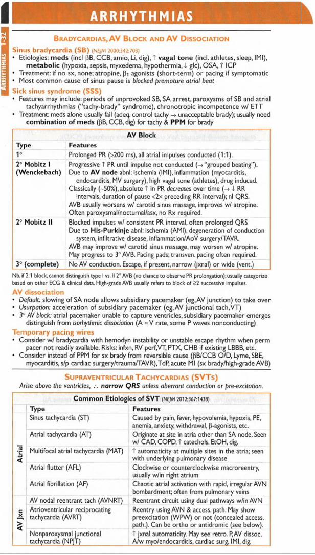
ARRHYTHMIASB r a d y c a r d i a s , AV B l o c k a n d AV D i s s o c i a t i o n
Sinus bradycardia (SB) (NEJM 2000:342:703)• Etiologies: meds (incl [}B, CCB, amio, Li, dig), T vagal tone (incl. athletes, sleep, IMI),
metabolic (hypoxia, sepsis, myxedema, hypothermia, 4 glc), OSA, T ICP• Treatment: if no sx, none; atropine, |3i agonists (short-term) or pacing if symptomatic• Most common cause of sinus pause is blocked premature atrial beat
Sick sinus syndrome (SSS)• Features may include: periods of unprovoked SB, SA arrest, paroxysms o f SB and atrial
tachyarrhythmias (“ tachy-brady” syndrome), chronotropic incompetence w/ ETT• Treatment: meds alone usually fail (adeq. control tachy -» unacceptable brady); usually need
combination of meds (|3B, CCB, dig) for tachy & PPM for brady
AV BlockType Features1° Prolonged PR (>200 ms), all atrial impulses conducted (1:1).2° Mobitz 1 (Wenckebach)
Progressive t PR until impulse not conducted (-> “grouped beating”). Due to AV node abnl: ischemia (IMI), inflammation (myocarditis,
endocarditis, MV surgery), high vagal tone (athletes), drug induced. Classically (-50%), absolute T in PR decreases over time (-> 4 RR
intervals, duration of pause <2x preceding RR interval); nl QRS. AVB usually worsens w/ carotid sinus massage, improves w/ atropine. Often paroxysmal/nocturnal/asx, no Rx required.
2° Mobitz II Blocked impulses w/ consistent PR interval, often prolonged QRS Due to His-Purkinje abnl: ischemia (AMI), degeneration of conduction
system, infiltrative disease, inflammation/AoV surgery/TAVR.AVB may improve w/ carotid sinus massage, may worsen w/ atropine. May progress to 3° AVB. Pacing pads; transven. pacing often required.
3° (complete) No AV conduction. Escape, if present, narrow (jxnal) or wide (vent,)
Nb.if 2:1 block, cannot distinguish type I vs. II 23 AVB (no chance to observe PR prolongation); usually categorize based on other ECG & clinical data. High-grade AVB usually refers to block of >2 successive impulses.
AV dissociation• Default: slowing of SA node allows subsidiary pacemaker (eg,AV junction) to take over• Usurpation: acceleration o f subsidiary pacemaker (eg,AV junctional tach,VT)• 3° AV block: atrial pacemaker unable to capture ventricles, subsidiary pacemaker emerges
distinguish from isorhythmic dissociation (A =*V rate, some P waves nonconducting)Tem porary pacing wires• Consider w/ bradycardia with hemodyn instability or unstable escape rhythm when perm
pacer not readily available. Risks: infxn, RV perf,VT, PTX, CHB if existing LBBB, etc.• Consider instead of PPM for sx brady from reversible cause ((3B/CCB O/D, Lyme, SBE,
myocarditis, s/p cardiac surgery/trauma/TAVR),TdP, acute Ml (sx brady/high-grade AVB)
S u p r a v e n t r i c u l a r T a c h y c a r d i a s ( S V T s )Ar/se above the ventricles, narrow QRS unless aberrant conduction or pre-excitation.
Common Etiologies of SVT (N EJM 2012:367:1438)
Type FeaturesSinus tachycardia (ST) Caused by pain, fever, hypovolemia, hypoxia, PE,
anemia, anxiety, withdrawal, [3-agonists, etc.
Atr
ial
Atrial tachycardia (AT) Originate at site in atria other than SA node. Seen w/ CAD, COPD, t catechols, EtOH, dig.
Multifocal atrial tachycardia (MAT) T automaticity at multiple sites in the atria; seen with underlying pulmonary disease
Atrial flutter (AFL) Clockwise or counterclockwise macroreentry, usually w/in right atrium
Atrial fibrillation (AF) Chaotic atrial activation with rapid, irregular AVN bombardment; often from pulmonary veins
AV nodal reentrant tach (AVNRT) Reentrant circuit using dual pathways w/in AVNcX
1Atrioventricular reciprocating tachycardia (AVRT)
Reentry using AVN & access, path. May show preexcitation (WPW) or not (concealed access, path.). Can be ortho or antidromic (see below).
Nonparoxysmal junctional tachycardia (NPJT)
T jxnal automaticity. May see retro. RAV dissoc. A/w myo/endocarditis, cardiac surg, IMI, dig.

✓
Diagnosis of SVT Type (NE/M 2012:367:1438)
Onset Abrupt on/off argues against sinus tachycardiaRate Not dx as most can range from 140-250 bpm, but: ST usually <150; AFL
often conducts 2:1 vent, rate 150; AVNRT & AVRT usually >150Rhythm Irregular ~> AF, AFL w/ variable block, or MATP wave morphology
Before QRS -> ST,AT (P different from sinus), MAT (>3 morphologies) After QRS & inverted in inf. leads retrograde atrial activation via AVN
AVNRT: buried in or distort terminal portion of QRS (pseudo RSR' in Vi) AVRT: slightly after QRS (RP interval >100 ms favors AVRT vs. AVNRT) NPJT: either no P wave or retrograde P wave similar to AVNRT
Fibrillation or no P waves -»AFSaw-toothed T ” waves (best seen in inferior leads &Vi) -»AFL
Response to vagal stim. or adenosine
Slowing of HR often seen with ST,AF,AFL,AT, whereas re-entrant rhythms (AVNRT, AVRT) may abruptly terminate (classically w/ P wave after last QRS) or no response. Occ AT may terminate.In AFL & AF, T AV block may unmask ” F” waves or fibrillation
Figure 1-4 Approach to SVT (adapted from NEJM 2012:367:1438)
none or fibrillation
SVT ■
Regular Irregular
s P wave morphology v' P wave morphology
r r 1 Aupright none or retrograde * sinus F waves none or morph.a QRS distorts QRS p QRS a QRS @ 300 bpm fibrillation a QRS
* * 1 1 1 1ST AVNRT AVNRT AT AFL AF MAT
1 AVRT A 1Gradual onset often Response to vagal maneuvers or adenosine
t t _____» M_____tfurther dx testing Terminates Does not terminate
Treatment of SVT (Circ 2016;133:e506)
Rhythm Acute treatm ent Long-term treatm entUnstable Cardioversion per ACLS n/aST Treat underlying stressor(s) n/aAT JiB, CCB or adenosine;
? amiodaroneradiofrequency ablation (RFA); pB or CCB, ± class O i l antiarrhythmics
AVNRT or AVRT
Vagal maneuvers Adenosine (caution in AVRT ) CCB or (3B, DCCV if other Rx fail
for AVNRT (see next section for AVRT): RFA. CCB, pB,or dig (chronic or prn) ± Class IC/III antiarrhythmics (if nl heart)
NPJT CCB, p>B, amiodarone Rx underlying dis. (eg, dig tox, ischemia)AF [}B, CCB, digoxin, AAD See “Atrial Fibrillation”AFL pB, CCB, digoxin, AAD RFA; |3B or CCB ± class III antiarrhyth.MAT CCB or [5B if tolerated Treat underlying disease. CCB or 0B.
AVN ablation + PPM if refractory to meds
*Avoid adenosine & nodal agents if accessory pathway + pre-excited tachycardia, see below (jACC 2003:42:1493)
• Catheter oblation: high overall success rate (AFL/AVNRT ~95%,AVRT -90%, AF -80%) complications: stroke, Ml, bleeding, perforation, conduction block (jama 2007;290:2768)
A c c e s s o r y P a t h w a y s ( W o l f f -P a r k in s o n - W h it e )
Definitions• Accessory pathway (bypass tract) of conducting myocardium connect- Swave
ing atria & ventricles, allowing impulses to bypass normal AVN delay N * *• Preexcitation (W P W ) pattern: 1 PR interval, T QRS width w/ 8 wave
(slurred onset, can be subtle). ST &Tw abnl (can mimic old IMI). only seen wl pathv/ays that conduct antegrade (if pathway only conducts retrograde, then
ECG will be normal during SR; “ concealed” bypass tract)PAC can exaggerate pre-excitation if AV node conduction slowed
• W P W syndrome: W PW accessory pathway + paroxysmal tachycardia
Ar
rh
ythm
ias
1-33

iRRH
YTHM
IAS
1-34
7 Classic tachycardias of W P W accessory pathways• O rthodrom ic AVRT: narrow-complex SVT (typically), conducting IAVN & T accessory
pathway; requires retrograde conduction and can occur w/ concealed bypass tracts• Antidromic AVRT (rare): wide-complex SVT, conducting I accessory pathway & T AVN;
requires antegrade conduction and ,\ should see pre-excitation pattern during SR• AF w/ rapid conduction down accessory pathway; wide-complex irregular SVT; requires
antegrade conduction; /. should see pre-excitation in SR. Rarely can degenerate intoVF.
Treatm ent (Heart Rhythm 2012:9:1006; Circ 2016; 133:eS06)
• AVRT (orthodromic): vagal, [5B, CCB; care w/ adenosine (can precip AF); have defib ready• AF/AFL w/ conduction down accessory pathway: need to Rx arrhythmia and t pathway
refractoriness. Use procainamide, ibutilide, or DCCV; avoid CCB, pB, amio, dig, &adenosine, as can i refractoriness of pathway -* T vent rate -»VF (Ore 2016;133:e506).
• Long term : RFA if sx; if not candidate for RFA, then antiarrhythmics (IA, III) or CCB/pB. consider RFA if asx but AVRT or AF inducible on EPS (NEJM 2003:349:1803) or if rapid
conduction possible ( / w / EPS if pre-excitation persists during exercise testing) risk of SCD related to how short RR interval is in AF (eg, <250ms) and if SVT inducible
W ide-Complex T achycardias (WCTs)
Etiologies (Lancet 2012;380:1520)♦ Ventricu la r tachycardia (V T): accounts for 80% ofW CT in unselected population• SVT conducted w ith aberrancy: either fixed BBB, rate-dependent BBB (usually
RBBB), conduction via an accessory pathway or atrially triggered ventricular pacing
M onom orph ic ven tricu la r tachycardia (M M VT)• All beats look similar; predominantly upward in Vi = RBBB-type vs. downward = LBBB-type• In structurally abnormal heart: p r io r Ml (scar); CMP; m yocarditis; v.
arrhythm ogenic RV CMP (ARVC): incomplete RBBB, e wave (terminal notch in QRS) &TW I inVi-V3 on resting ECG, LBBB-type VT, d x w/ MRI (Loncet 2009:373:1289)
In structurally normal heart (w/ normal resting ECG):RVOTVT: LBBB-type VT w/ inferior axis; typically ablateid iopath ic LVVT: RBBB-type VT w/ superior axis; responds to verapamil
Epsilon
Polymorphic ventricular tachycardia (P M V T )• QRS morphology changes from beat to beat• Etiologies: ischemia; CMP; catecholaminergic;
torsades de pointes (TdP= “twisting of the points,” PMVT + T QT);T QT acquired (meds, lytes,stroke, see” ECG”) w/ risk t w / l HR.freq PVCs (pause dependent) or congenital (K/Na channelopathies) w/ resting Tw abnl &TdP triggered by sympathetic stimulation (eg, exercise, emotion, sudden loud noises) (tancet 2008:372:750).
Brugada syndrome (Na channelopathy): 6 > 9; pseudo-RBBB w/ STE in V i-V j (provoked w/ class IA o r 1C) on resting ECG
Diagnostic clues that favor V T (assume until proven o/w)• Prior M l, C H F or LV dysfunction best predictors that W C T is V T (Amj Med 1998:84:53)
• Hemodynamics and rate do not reliably distinguish VT from SVT• MMVT is regular, but initially it may be slightly irregular, mimicking AF w/ aberrancy;
grossly irregularly irregular rhythm suggests AF w / aberrancy or pre-excitation• ECG fea tu res th a t fa vo r VT (Ore 1991:83:1649)
AV dissociation (independent P waves, capture o r fusion beats) proves VT very wide QRS (>140 ms in RBBB-type or >160 in LBBB-type); extreme ax/s deviation QRS morphology atypical for BBB
RBBB-type: absence of tall R' (or presence of monophasic R) in Vi, r/S ratio <1 inV$ LBBB-type: onset to nadir >6CH100 ms inV i.q wave inV6
concordance (QRS in all precordial leads w/ same pattern/direction)
Long-term m anagem ent (JACC 2006:48:1064; EH] 2015:36:2793; Ore 2016:133:1715)
• Workup: echo to / LV fxn, cath or stress test to r/o ischemia, ? MRI and/or RV bx tolook for infiltrative CMP o r ARVC, I EP study to assess inducibility
• ICD: 2° prevention after documentedVT/VF arrest (unless due to reversible cause). 1° prev.if high risk, eg, EF <30-35%, ARVC, Brugada, certain LQTS, severe HCMP. See “ Cardiac Rhythm Mgmt Devices." Wearable vest if reversible etiology or waiting for ICD? Antitachycardia pacing (ATP = burst pacing faster than VT) can terminate VT w/o shock
• Meds: pB, verapamil if idiopathic LVVT, o rA A D (eg, amio, mexiletine) to suppress VT• If med a/wTdP QT >500 ±VPBs: d/c med, replete K, give Mg, ± pacing (JACC 2010:55:934)
• RFA if isolated VT focus or if recurrent VT triggering ICD firing (4VT storm by 34%; NEJM2016:375:i l l ) ; a b la tio n b e fo re ICD im p la n ta tio n i- d ischa rge ra te by 40% (Lancet 2010:375:31)

ATRIAL FIBRILLATIONC lassifica tion (Circ 2014:130:e199)
• Paroxysmal (self-terminating, usually <48 h, often triggered in pulm veins) vs. persistent(>7 d) vs. long-standing persistent (>1 y) vs. pe rm anen t (no plan for SR)
• Nonvalvular (AF absent rheumatic MS, prosthetic valve, or mitral valve repair) vs. valvular
E p idem io logy and e tio log ies (Circ 2011:124:1982)
• 1-2% of pop. has AF (10% of those age >80); M > F; lifetime risk -25%• Acute (up to 50% w/o identifiable cause)
Cardiac; HF, new CMP, myo/pericarditis, ischemia/MI, HTN crisis, valve dis., cardiac surg P u lm onary: acute pulmonary disease or hypoxemia (eg. COPD flare, PNA), PE, OSA M etabolic: high catecholamine states (stress, infection, postop, pheo), thyrotoxicosis Drugs: alcohol (“ holiday heart” ), cocaine, amphetamines, theophylline, caffeine, smoking N eurogen ic : subarachnoid hemorrhage, ischemic stroke
• Chronic: T age, HTN, ischemia, valve dis. (MV,TV, AoV), CMP, hyperthyroidism, obesity
E va luation• H&P, ECG, CXR.TTE (LA size, thrombus, valves, LV fxn, pericardium), K, Mg, Cr, FOBT
before anticoag.TFTs; r/o Ml not necessary unless other ischemic sx
Figure 1-5 Approach to acute AF {Adapted from Crc 2014;130:e199)
New or Recent-Onset AF —unstable-► Urgent cardioversionI
stableISpont. cardioversion
(occurs w/in 24 hr in 50-67% acute AF)
1. Rate Control
PB or CCB (see Table)
2. Anticoagulate
IV UFH il admitting & cardioverting
o/w oraI anticoag
AF <48 hr & low risk of stroke
Cardioversion (electric and/or pharmacologic)
Consider anticoag x 4 wk if risk factors
AF >48 hr (or unknown) or high risk of stroke
TEE re /s
© LA_ thromb.
©LAthromb.
Empiric oral anticoag x >3 wk
Cardioversion -* oral anticoag x £4 wk
Rate C ontro l (if sx, goal HR <80; if asx & EF >40%, goal HR <110; Lancet 2016:388:818)Agent Acute (IV ) Maint. (PO) Com m ents
VerapamilCQ
5-10 mg over 2' may repeat in 30'
120-360 mg/d in divided doses
1 BP (Rx w/ Ca glue) Can worsen HF
uu
Diltiazem0.25 mg/kg over 2' may repeat after 15' 5-15 mg/h infusion
120-360 mg/d in divided doses
Preferred if severe COPD Can T dig levels
Metoprolol2.5-5 mg over 2' may repeat q5' x 3
25-100 mg bid or tid
i BP (Rx w/ glucagon) Preferred if CAD Risks: HF & bronchospas.
Digoxin*(onset >30 min)
0.25 mg q2h up to 1.5 mg/24 h
0.125-0.375 mg qd (adj for CrCI)
Consider in HF or low BP Poor exertional HR Ctrl
Am iodarone 300 mg over 1 h then 10-50 mg/h x 24 h
Circ 2014;13Q:e199. IV pB, CCB & dig contraindicated if evidence o fW P W (ie, pre-excitation o r W C T) since may facilitate conduction down accessory pathway leading to VF; / . use procainamide, ibutilide o r amio. *Many meds incl. amio, verapamil, quinidine, propafenone, macrolides & azole antifungals T digoxin levels.
C ard iovers ion• Consider if: 1” AF, sx, tachycardia-mediated CMP, or difficult to rate control
if AF >48 h 2-5% risk stroke w/ cardioversion (pharmacologic or electric)either TEE to r/o thrombus or ensure therapeutic anti coagulation for >3 wk prior
if needs to cardiovert urgently, often anticoagulate acutely (eg, IV UFH)• Likelihood of success <* AF duration & atrial size; control precipitants (eg, vol status, thyroid)• Before electrical cardiovert, consider pre-Rx w/ AAD (eg, ibutilide), esp. if 1 ” cardiovert failed• For pharmacologic cardioversion, class III and 1C drugs have best proven efficacy• If SR returns (spont. or w / Rx), atria may be mech. stunned; also, high risk of recurrent AF
over next 3 mo. .\ Anticoag postcardioversion £4 w k (> unless AF <48 h and low risk).
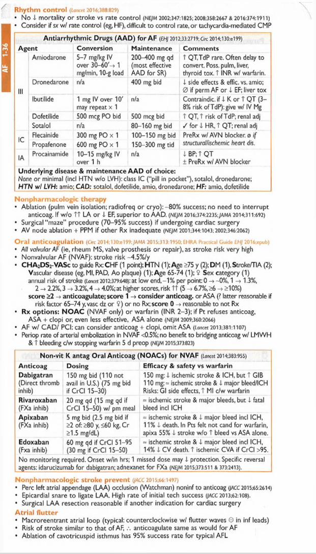
AF
1-36
^ Rhythm control (Lancer 2016;388:829)
• No i mortality or stroke vs rate control (NEJM 2002;347:1825; 2008:358:2667 & 2016:374:1911)• Consider if sx w/ rate control (eg, HF), difficult to control rate, o r tachycardia-mediated CMP
Antiarrhythmic Drugs (AAD) for AF (EHJ 2012:33:2719; c/rc 2014;130:ei99)
Agent Conversion Maintenance CommentsAmiodarone 5-7 mg/kg IV
over 30-60' ■» 1 mg/min, 10-g load
200^400 mg qd (most effective AAD for SR)
T QT.TdP rare. Often delay to convert. Poss. pulm, liver, thyroid tox. t INR w/ warfarin.
IIIDronedarone n/a 400 mg bid 1 side effects & effic. vs. amio;
0 if perm AF or I EF; liver toxIbutilide 1 mg IV over 10'
may repeat x 1n/a Contraindic. if 1 K or T QT (3-
8% risk ofTdP):give w/ IV MgDofetilide 500 meg PO bid 500 meg bid T QT, T risk of TdP; renal adjSotalol n/a 80-160 mg bid / for 1 HR, t QT; renal adj
1CFlecainidePropafenone
300 mg PO x 1 600 mg PO x 1
100-150 mg bid 150-300 mg tid
PreRx w/ AVN blocker, o i f structuralfischemic heart dis.
IAProcainamide 10-15 mg/kg IV
over 1 hn/a 1 BP; t QT
± PreRx w/ AVN blockerUnderlying disease & maintenance AAD of choice:None or minimal (ind HTN w/o LVH): class 1C ("pill in pocket” ), sotalol, dronedarone;HTN wl LVH: amio; CAD: sotalol, dofetilide, amio, dronedarone; HF: amio, dofetilide
Nonpharmacologic therapy• Ablation (pulm vein isolation; radiofreq o r cryo): - 80% success; no need to interrupt
anticoag. If w/o TT LA or i EF, superior to AAD. (NEJM 2016;374:2235;./M1A 2014:311:692)• Surgical "maze” procedure (70-95% success) if undergoing cardiac surgery• AV node ablation + PPM if other Rx inadequate {NE)M 2001:344:1043:2002:346:2062)
O ral anticoagulation (Ore 2014:130:e199;/M1A 2015:313:1950; EHRA Practical Guide EH] 2016;epub)• All valvular AF (ie, rheum MS, valve prosthesis or repair), as stroke risk very high• Nonvalvular AF (NVAF): stroke risk -4.5%/y• C H A 2DS2-VASc to guide Rx: CHF (1 point); HTN (1); Age >75 y (2); DM (1), Stroke/TIA (2);
Vascular disease (eg, Ml, PAD, Ao plaque) (1); Age 65-74 (1); 9 Sex category (1) annual risk of stroke (Lancet 2012:379:648); at low end,-1% per point: 0 ~0%, 1 -»1.3%,
2 -> 2.2%, 3 -> 3.2%, 4 4.0%; at higher scores, risk t t (5 -> 6.7%, >6 >10%)score i>2 -> anti coagulate; score 1 consider anticoag. or ASA (> latter reasonable if
risk factor 65-74 y, vase dz or 9) or no Rx; score 0 -» reasonable to not Rx• Rx options: N O A C (NVAF only) or warfarin (INR 2-3); if Pt refuses anticoag,
ASA + clopi or, even less effective, ASA alone (NEJM 2009:360:2066)• AF w/ CAD/ PCI: can consider anticoag + clopi, om it ASA {Lancet 2013:381:1107)• Periop rate of arterial embolization in NVAF <0.5%; no benefit to bridging anticoag w/ LMWH
& T bleeding c/w stopping warfarin 5 d preop {NEJM 2015:373823)
Non-vit K antag Oral Anticoag (NOACs) for NVAF (Loncet 2014:383:9SS)
Anticoag Dosing Efficacy & safety vs warfarinDabigatran(Direct thro mb inhib)
150 mg bid (110 not avail in U.S.) (75 mg bid if CrCI 15-30)
150 mg: i ischemic stroke & ICH, but t GIB 110 mg: = ischemic stroke & I major bleed/ICH Risks: Gl side effects,! Ml c/w warfarin
Rivaroxaban(FXa inhib)
20 mg qd (15 mg qd if CrCI 15-50) w/ pm meal
= ischemic stroke & major bleeds, but I fatal bleed ind ICH
Apixaban(FXa inhib)
5 mg bid (2.5 mg bid if >2 of: >80 y, <60 kg, Cr >1.5 mg/dL)
= ischemic stroke & ! major bleed incl ICH,11% i death. In Pts felt not cand for warfarin, apixa 55% i stroke w/o t bleed vs ASA alone.
Edoxaban(Fxa inhib)
60 mg qd if CrCI 51-95 (30 mg if CrCI 15-50)
» ischemic stroke & i major bleed incl ICH, 14% i CV death. T ischemic CVA if CrCI >95.
No monitoring required. Onset w/in hrs; 1 missed dose may I protection. Specific reversal agents: idarucizumab for dabigatran; adnexanet for FXa (NEJM 2015:373:511 & 373:2413).
Nonpharmacologic stroke prevent (JACC 2015;66:1497)• Perc left atrial appendage (LAA) occlusion (Watchman) noninf to anticoag (JACC 2015:65:2614)• Epicardial snare to ligate LAA. High rate of initial tech success (JACC 2013:62:108).• Surgical LAA resection reasonable if another indication for cardiac surgeryA trial flu tter• Macroreentrant atrial loop (typical: counterclockwise w/ flu tter waves © in inf leads)• Risk of stroke similar to that of AF, anticoagulate same as would for AF• Ablation of cavotricuspid isthmus has 95% success rate for typical AFL

SYNCOPEDefinition• Symptom of sudden transient loss of consciousness due to global cerebral hypoperfusion• If CPR or cardioversion required, then SCD and not syncope (different prognosis)• Presyncope = prodrome of light-headedness w ithout LOC
E t io lo g ie s (NEJM 2002:347:878; JACC 2006;47:473; Eur Heart J 2009:30:2631)• Neurocardiogenic (a.k.a. vasovagal, -25%; NEJM 2005,352:1004): T sympathetic tone ->
vigorous contraction of LV -> mechanoreceptors in LV trigger T vagal tone (hyperactive Bezold-Jarisch reflex) -> 4- HR (cardioinhibitory) and/or 4- BP (vasodepressor). Cough, deglutition, defecation, & micturition -> T vagal tone and thus can be precipitants. Carotid sinus hypersensitivity (exag vagal resp to carotid massage) is related disorder.
• O rthostatic hypotension (-10%)hypovolemia/diuretics, deconditioning; vasodilat (esp. if combined w/ © chronotropes) autonomic neuropathy [1° = Parkinson’s, MSA/Shy-Drager, Lewy body dementia, POTS
(dysautonomia in the young); 2° = DM, EtOH, amyloidosis, CKD] {NEJM 2008:358:615)
• Cardiovascular (-20%, more likely in men than women)Arrhythmia (15%): challenging to dx as often transient
Bradyarrhythmias: SSS, high-grade AV block,© chronotropes, PPM malfunction Tachyarrhythmias:VT, SVT (syncope rare unless structural heart disease orW P W )
Mechanical (5%)Endocardial/Valvular: AS, MS, PS, prosthetic valve thrombosis, myxoma Myocardial: pump dysfxn from Ml or outflow obstruction from HCMP (but usually VT) Pericardial: tamponade;Vascular: PE, PHT, AoD, ruptured AAA, subclavian steal
• Neurologic (-10%); vertebrobasil insuff, cerebrovasc dissection, SAH,TIA/CVA, migraine• Misc. causes of LOC (but not syncope): seizure, i glc, hypoxia, narcolepsy, psychogenic
W orkup (etiology cannot be determined in -40% of cases)• H&P incl. orthostaticVS have highest yield and most cost effective (Archives 2009:169:1299)
• H is to ry (from Pt and witnesses if available)activity and posture before the incidentprecipitating factors: exertion (AS, HCMP, PHT), positional A (orthostatic hypotension),
stressors such as sight of blood, pain, emotional distress, fatigue, prolonged standing, warm environment, N/V, cough/micturition/defecation/swallowing (neurocardiogenic), head turning o r shaving (carotid sinus hypersens.); arm exercise (subclavian steal)
prodrome (eg, diaphoresis, nausea, blurry vision): cardiac <~5 sec, vasovagal >~5 sec associated sx: chest pain, palp., neurologic, postictal, bowel o r bladder incontinence
(convulsive activity for <10 sec may occur w/ transient cerebral HoTN & mimic seizure)• PMH: prior syncope, previous cardiac or neurologic dis.;no CV disease at baseline -> 5%
cardiac, 25% vasovagal; CV disease 20% cardiac, 10% vasovagal (NEJM 2002:347:878)
• Medications that may act as precipitantsvasodilators: a-blockers, nitrates, ACEI/ARB, CCB, hydralazine, phenothiazines, antidep. diuretics;© chronotropes (eg, |3B and CCB)proarrhythmic or Q T prolonging: class IA, 1C or III antiarrhythmics (see “ ECG” ) psychoactive drugs: antipsychotics.TCA, barbiturates, benzodiazepines, EtOH
• Family history: CMP, SCD, syncope (vasovagal may have genetic component)• Physical exam
VS including orthostatics (© if supine -> standing results in >20 mmHg i SBP, >10 mmHg i DBP, or >10-20 bpm T HR), BP in both arms
cardiac: HF (t JVP, displ. PMI, S3), murmurs, LVH (S4, LV heave), PHT (RV heave, t P2) vascular: / for asymmetric pu/ses, carotid/vert/subclavian bruits; carotid sinus massage to
/ for carotid hypersens (if no bruits):© if asystole >3 sec or I SBP >50 mmHg neurologic exam: focal findings, evidence of tongue biting; FOBT
• ECG (abnormal in -50%, but only definitively identifies cause of syncope in <10%)Conduction: SB, sinus pauses/sinus arrhythmia,AVB, BBB/IVCDArrhythmia: ectopy, T or I QT, preexcitation (WPW), Brugada, e wave (ARVC), SVT/VTIschemic changes (new or old): atrial o r ventricular hypertrophy
• Lab: glc, Hb, preg test (child-bearing age 9), ? D-dimer, ? troponin (low yield w/o other sis)
O th e r d iagnostic studies (consider based on results of H&P and ECG)• Ambulatory ECG monitoring: if suspect arrhythmogenic syncope
Holter monitoring (continuous ECG 24^48 h): useful if frequent events arrhythmia + sx (4%);asx but signif. arrhythmia (13%); sx but no arrhythmia (17%)
Event recorder (activated by Pt to record rhythm strip): limited role in syncope as only useful if established prodrome (because must be Pt activated)
Syn
co
pe 1-37

External loop recorders (continuously saves rhythm, can be activated after an event): useful for episodes (including w/o prodrome) likely to occur w/in 1 mo; can be coupled w/ mobile cardiac telemetry than can be auto-triggered for specific rhythms
Implantable loop recorders (inserted SC; can record >1 y): useful if episodes <1/mo;dx in 55% of cases (Gk 2001:104:46); recommended for recurrent syncope w/o prodrome
• Echo: consider to r/o structural heart disease (eg, CMP [incl HCMP & ARVC], valvulardisease [incl AS, MS, MVP], myxoma, amyloid, PHT, ± anomalous coronaries)
• ETT/CCTA/Cath: esp. w/ exertional syncope; r/o ischemia or catechol-induced arrhythmia• Electrophysiologic studies (EPS); consider in high-risk Pts in whom tachy or brady etiology is
strongly suspected, but cannot be confirmed;50% abnl (inducible VT, conduction abnormalities) if heart disease, but ? significance 3-20% abnl if abnl ECG; <1% abnl if normal heart and normal ECG (Annofs 1997;127:76)
• I Tilt table testing: debated utility due to poor Se/Sp/reproducibility; consider only if vasovagaldx suspected but cannot be confirmed by hx
• Cardiac MRI: helpful to dx ARVC if suggestive ECG, echo (RV dysfxn) o r © FHx of SCD• Neurologic studies (cerebrovascular studies, CT, MRI, EEG): if H&P suggestive; low yield
Figure 1-6 Approach to syncope
Syncope
IHistory, Physical Exam (incl. orthostatics), 12-lead ECG
d i a g n o s t i c s u g g e s t i v e n e g a t i v e
Orthostatichypotension © o r t h o s t a t i c s
Arrhythmia a b n l r h y t h m
o n E C G
(s i t u a t i o n a l o r
© p r o d r o m e
a b n l h e a r t
o r E C G ,
e x e r t i o n a l o ir
© p r o d r o m e
echo ± ETT(± CTA to no PE or AoD)
a b n l n e u r o
e x a m r 1n o r m a l
Iambul. ECG
if recurs
if recurs CT/MRI EP study
(Adapted from JACC 2006:47:473)
High -risk features (usually admit w/ telemetry & testing;,/Emerg Med 2012:42:345)• Age >60 y, h/o CAD, HF/CMP, valvular o r congenital heart dis., arrhythmias, FHx SCD• Syncope c/w cardiac cause (lack of prodrome, exertional, resultant trauma) or recurrent• Complaint of chest pain or dyspnea; abnl VS, cardiac, pulm, o r neuro exam• ECG suggesting conduction abnormality, arrhythmia, or ischemia; Pt w / PPM/ICD
Treatm ent• Arrhythmia, cardiac mechanical or neurologic syncope: treat underlying disorder• Vasovagal syncope: ? benefit of fludrocortisone, midodrine or SSRI (Jnt) Cardiol 2013:167:1906;
]ACC 2016:68:1); no proven benefit for disopyramide or fiB (Grc 2006:113:1164)? 16 oz of H2O before at-risk situations (Grc 2003:108:2660)? benefit w/ PPM if >3 episodes/2y & loop recorder w/ asystole >3 sec (Grc 2012:125:2566);
PPM likely ineffective if positive tilt-test and no arrhythmia (EHJ 2014:35:2211)• If orthostatic: vol replete (eg, 500 mL PO q am ); if chronic -> rise from supine to standing
slowly, stockings, midodrine, ? atomoxetine (HTN 2014:64:1235), fludrocort, T Na diet
Prognosis (Ann EnrngMed 1997:29:459;NEJM 2002;347:878)• 22% overall recurrence rate if idiopathic, else 3% recurrence• Cardiac syncope: 2-fold T in mort., 20-40% 1-y SCD rate, median survival - 6 y• Unexplained syncope w/ 1.3-fold T in m ort, but noncardiac or unexplained syncope w/ nl
ECG, no h/o VT, no HF, age <45 -> low recurrence rate and <5% 1-y SCD rate• Vasovagal syncope: Pts not at increased risk for death, Ml, o r stroke• / state driving laws and MD reporting requirements. Consider appropriateness of Pt
involvement in exercise/sport, operating machinery, high-risk occupation (eg, pilot).

CARDIAC RHYTHM MANAGEMENT DEVICESPacemaker Code
A, atrial;V, vent; O, none; 1st letter 2nd letter 3rd letter 4th letter1, inhibition; D, dual; Chamber Chamber Response to Program
R, rate-adaptive paced sensed sensed beat features
Common Pacing Modes
VVI Ventricular pacing on demand w/ single lead in RV. Sensed ventricular beat inhibits V pacing. Used in chronic AF with symptomatic bradycardia.
DDD A & V sensing/pacing (RA & RV leads). Native A beat inhib A pacing & triggers V pacing -> tracking of intrinsic atrial activity. Maintains AV synchrony, i AF.
ModeSwitch
In atrial tachyarrhythmia (eg.AF), PPM As from DDD to nontracking mode (eg, Wl). Prevents PPM from pacing at maxV rate in response to rapid atrial rate.
Magnetover generator
PPM: fixed rate pacing (VOO/DOO). ICD: no shock, pacing preserved.Indie: / capture; surgery; inapprop PPM inhib/ICD shock, PM-mediated tachy
Leadless intracardiac PPM with emerging indications (NE/ai 2015,373:1125 & 2016:374:53)
Indications for Permanent Pacing (Crrc 2008:117:350 & 2012:126:1784)AV block 3° or type II 2° AVB a/w sx or w/ either HR <40 or asystole £3 sec (£5 if
in AF) while awake; ? asx 3° or type II 2° AVB; bifasc or alter. L & R BBBSinus node SB, pauses (SSS), chronotrop incompet a/w sx or ? if sx w/o clear assocTachyarrhythmia
AF w/ SSS; sx recurrent SVT term, by pacing after failing drugs/ablation; Sustained pause-dependent VT; > high-risk congenital long QT
Syncope Carotid sinus hypersensitivity with asystole >3 sec ? Neurocardiogenic syncope w/ prominent cardioinhib. response > Syncope with bi- or trifascicular block and not likely 2° to other causes
Pacemaker ComplicationsIssue Manifestation Description & etiologiesPerforation Effusion/tamp/pain Typically acute, consider if HoTNFailure to pace Bradycardia i Battery, lead fx/dislodgment, T pacing threshold
due to tissue rxn/injury; oversense -» inapprop. inhibFailure to sense Inapprop. pacing Lead dislodgment or sensing threshold too highPM-mediatedtachycardia
WCT at device Seen w/ DDD.V A retrograde conduction; upper rate sensed by A lead -> triggers V pacing -> etc.
PM syndrome Palpit, HF Seen w /W l, due to loss of AV synchrony
C a r d ia c r e s y n c h t h e r a p y ( C R T ) / B iv e n t r ic u la r ( B iV ) p a c in g (/ACC 2013:61 :e6)• 3-lead pacemaker (RA, RV, coronary sinus to LV); R > S in Vi suggests approp LV capture• Synchronize & enhance LV fxn ( t CO, -I adverse remodeling)• Indications: LVEF <35% + NYHA ll-IV despite med Rx + SR + LBBB >150 (? ^120) ms;
mort. benefit w / CRT-D only if LBBB {& QRS >130ms) (NEJM 2014:370:1694)? benefit in NYHA l- lll, EF <50% w/ PPM indication for AVB (NE/M 2013:368:1585) consider in AF: need rate control o r AVN ablation; more pacing -» greater CRT effect
• Benefits: I HF SX, I HF hosp., t survival (NEJM 2005:352:1539 & 2010:363:2385)
Im p la n t a b le c a r d ia c d e f i b r i l l a t o r ( I C D ) (/ACC 2013;6l:e6;Grc 2015:132:1613)• RV lead: defib & pacing (± anti tachycardia pacing [ATP] = burst pacing >VT rate to stopVT);
± RA lead for dual chamber PPM.Wearable defib & subcut-ICD, but 0 pace/ATR• Pt selection (NEJM 2004:350:2151 & 351:2481:2005:352:225; 2009:361:1427: Circ 2012:126:1784)
2° prevention: survivors ofVF arrest, unstable VT w/o reversible cause;structural heart disease & spontaneous sustained VT (even if asx)
1° prevention: LVEF <30% & post-MI or LVEF <35% & NYHA ll-lll (wait £40 d if post-MI, >90 d for NICMP) or LVEF <40% & inducible VT/VF; life expectancy must be >7 y; consider if unexplained syncope + DCM .or if HCM.ARVC, Brugada, sarcoid, LQTS, Chagas or congenital heart disease if at risk for SCD; ? wearable vest as bridge to ICD
• Risks: inapprop shock in -15-20% at 3 y (commonly d/t misclassified SVT); infxn; lead fx• ICD discharge: / device to see if approp; r/o ischemia; 6-mo driving ban ( / state law);
if recurrent VT, ? drug Rx (eg, amio + pB;^AAIA 2006:295:165) orV T ablation (NEJM 2007:357:2657); ablation at time of ICD i risk ofVT by 40% (toncet 2010:375:31)
D e v ic e in fe c t io n (Ore 2010:121:458;JAMA 2012:307:1727: NE/M 2012:367:842)• Presents as pocket infection (warmth, erythema, tenderness) and/or sepsis w/ bacteremia• Incidence ~2% over 5 y; if S. aureus bacteremia, infxn in £35%• TTE/TEE used to help visualize complic. (eg, vegetation), but even GTEE does not r/o• Rx: abx; system removal if pocket infxn or GPC bacteremia; 0 routine abx prior to inv. proc.
PP
M/IC
D
1-39
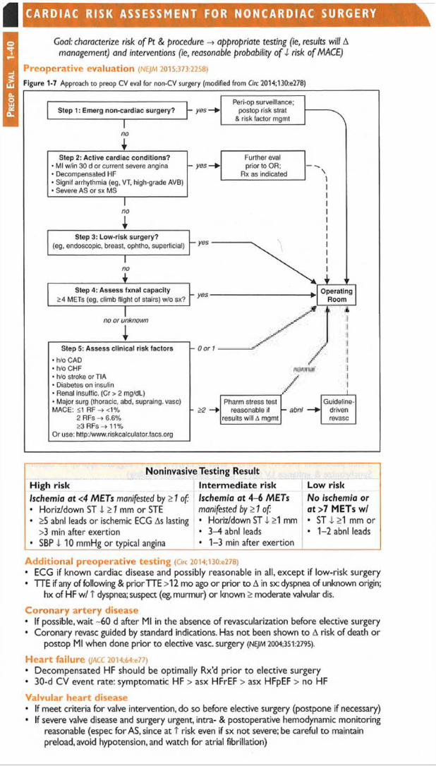
Pr
eop
Eva
l 1-
40I C A R D I A C R I S K A S S E S S M E N T FOR N O N C A R D I A C S U R G E R Y
Goal: characterize risk o f Pt & procedure -♦ appropriate testing (ie, results will A management) and interventions (ie, reasonable probability o f I risk of MACE)
Preoperative evaluation (NEJM 2015:373:2258)
Figure 1-7 Approach to preop CV eval for non-CV surgery (modified from Circ 2014;130:e278)
Step 1: Emerg non-cardiac surgery? - yesPeri-op surveillance;
postop risk strat & risk factor mgmt
4Step 2: Active cardiac conditions? Further eval
• Ml w/in 30 d or current severe angina - yes—► prior to OR;• Decompensated HF Rx as indicated• Signif arrhythmia (eg, VT, high-grade AVB)• Severe AS or sx MS
no
Step 3: Low-risk surgery?— y e s ------------------
1(eg, endoscopic, breast, ophtho, superficial) 1
11
no \ 11
rStep 4: Assess fxnal capacity
>4 METs (eg, climb flight of stairs) w/o sx?- yes ---------------------------------------------- ► Operating
Room
no or unkno wn
1Step 5: Assess clinical risk factors
»hro CAD• tVo CHF• h/o stroke orTIA• Diabetes on insulin• Renal insulfic. (Cr > 2 mg/dl)• Major surg (thoracic, abd, supraing. vase) MACE: <1 RF-><1%
2 RFs -> 6.6%53 RFs -»11%
Or use: http:,1‘www.riskcalculator.facs.org
- 0 or 1
- >2Pharm stress test Guideline-
reasonable it - abnt — ► drivenresults will A mgmt revasc
Noninvasive Testing ResultHigh risk Intermediate risk Low riskIschemia at <4 METs manifested by >1 of:• Horiz/down ST I >1 mm or STE• >5 abnl leads or ischemic ECG As lasting
>3 min after exertion• SBP I 10 mmHg or typical angina
Ischemia at 4-6 METsmanifested by>1 of:• Horiz/down ST i >1 mm• 3-4 abnl leads• 1-3 min after exertion
No ischemia or at >7 METs w/• ST i >1 mm or• 1-2 abnl leads
Additional preoperative testing (Ore 2014;I30:e278)• ECG if known cardiac disease and possibly reasonable in all, except if low-risk surgery• TTE if any of following & prior TTE >12 mo ago or prior to A in sx: dyspnea of unknown origin;
hx of HF w/ T dyspnea; suspect (eg, murmur) or known > moderate valvular dis.
Coronary artery disease• If possible, wait - 60 d after Ml in the absence of revascularization before elective surgery• Coronary revasc guided by standard indications. Has not been shown to A risk of death or
postop Ml when done prior to elective vase, surgery {NEJM 20O4;3S 1:2795).
H eart failure (JACC 2014;64:e77)• Decompensated HF should be optimally Rx’d prior to elective surgery• 30-d CV event rate: symptomatic HF > asx HFrEF > asx HFpEF > no HF
Valvular heart disease• If meet criteria for valve intervention, do so before elective surgery (postpone if necessary)• If severe valve disease and surgery urgent, intra- & postoperative hemodynamic monitoring
reasonable (espec for AS, since at T risk even if sx not severe; be careful to maintain preload, avoid hypotension, and watch for atrial fibrillation)

If severe symptomatic AS and surgAVR not an option, balloon aortic valvuloplasty (BAV) or transcatheter aortic valve replacement (TAVR) can be considered (/ACC 20l4;64:e77)
Cardiac im plantable electronic devices• Discuss w/ surgical team need for device (eg, complete heart block) & consequences if
interference w/ fxn, and likelihood of electromagnetic interference• Consider reprogramming, magnet use, etc. as needed
Pre- & perioperative pharmacologic m anagem ent• ASA: continue in Pts w/ existing indication. Initiation prior to surgery does not I 30-d
ischemic events and T bleeding (NEJM 2014:370:1494), but Pts w / recent stents excluded.• D ual a n tip la te le t the rapy: delay elective surg 14 d after balloon angioplasty, 30 d
after BMS and ideally 6 mos (min 3 mos) after DES implantation (2016 ACC/AHA Update) unless risk of bleeding > risk of stent thrombosis or ACS. If must discontinue P2Yi2 inhibitor, continue ASA and restart P2Yi2 inhibitor ASAP.
• P-blockers (C/rc 2009; 120:2123; JAMA 2010:303:551; Am} Med 2012:125:953)
Continue [IB in Pts on them chronically. Do not stop [3B abruptly postop (may cause reflex sympathetic activation). Use IV if Pt unable to take PO.
In terms of initiating, conflicting evidence; may depend on how done. Some studies show 1 death/MI, another i Ml, but T death & stroke and T brady/HoTN (Lancet 2008;371; 1839).
Reasonable to initiate if interned- or high-risk © stress test, o r RCRI >3, espec if vase surgery. Initiate >1 wk prior to surgery (not day of), use low-dose, short-acting (3B, and titrate to achieve HR and BP goal (? HR -55-65). Avoid bradycardia and HoTN.
• S ta tins: i ischemia & CV events in Pts undergoing vascular surg (NEJM 2009;361:980).
Consider if risk factors & non-low-risk surgery and in all Pts undergoing vascular surgery.• ACEI/ARB: may cause HoTN perioperatively. If held before surgery, restart ASAP.• Amiodarone: i incidence o f postop AF i f started prior to surgery (NE/M 1997;337:1785)
Postoperative m onitoring* ECG if known CAD o r high-risk surgery. Consider if >1 risk factor for CAD.* Routine troponin prognostic (/AMA 2012:307:2295) but / only if sx/ECG As suggestive of ACS
PERIPHERAL ARTERY DISEASE (PAD)Clinical features (NEJM 2016,374:861)• Prev. t w / age; <1% if <40 y, -15% if >70 y; risk factors incl. smoking, DM , HTN, chol• Claudication (dull ache, often in calves) precip by walking and relieved by stopping (vs.
spinal stenosis, qv); Leriche synd = claudication, i or 0 femoral pulses, & erectile dysfxn• Critical limb ischemia (C L I): rest pain (T w / elevation b/c i perfusion), ulcer
(typically at pressure foci, often dry; in contrast, venous ulcers are more often at medial malleolus, wet, and with hemosiderin deposition) o r gangrene, due to PAD, and >2-wk duration (implies chronicity vs. acute limb ischemia; see below)
Diagnosis• i peripheral pulses; other signs of chronic PAD: hair loss, skin atrophy, nail hypertrophy• Ankleibrachial index (ABI): nl 1-1.4; borderline 0.91-0.99; abnl <0.90; if >1.4, non-dx possibly
due to calcified noncompressible vessel / PVR. If ABI abnl -> segmental ABI w/ PVR to localize disease. If © sx but nl ABI, / for i lower extrem BP after exercise.
• Duplex arterial U/S; CTA w/ distal run-off; MRA or angio if dx in }. o r possible intervention
Treatm ent (/ACC 2013;61:1S55;JAMA 2013:309:453 & 2015:314:1936)
• Risk factor modification. Screen for CAD. Structured exercise program (/a m a 2013:310:57).
• If sx, ASA or clopi to -I D/MI/stroke. More intensive Rx (eg, adding ticagrelor or vorapaxar)i both MACE and limb ischemic events (Crrc 2013:112:679 &JACC 2016:67:2719).
• Cilostazol (if no HF) & ? ACEI & statins to i sx (Ore 2003:108:1481)
• Endovascular (angioplasty vs. stent) o r surgical revasc if limiting/refractory sx o r CLI
Acute limb ischemia (A L I)• Sudden decrement in limb perfusion that threatens viability;
viable (no immed threat of tissue loss): audible a rt Doppler signals, sensory, & motor OK threatened (salvage requires prompt Rx): loss of arterial Doppler signal, sensory, or motor
• Etiologies: embolism > acute thrombosis (eg,athero,APS, HITT), trauma to artery• Clinical manifestations (6 Ps): pain (distal to proximal, T in severity), poikilothermia,
pallor, pulselessness, paresthesias, paralysis• Testing: thorough pulse & neuro exam; arterial Doppler; angiography, either CT w/
bilateral run-off through feet o r arteriography• Urgent consultation w/ vascular medicine and/or vascular surgery• Treatment: immediate anticoagulation ± intra-arterial lytic; angioplasty or surgery
PAD 1-41

DYSPNEAPathophysiology EtiologiesAirway obstruction(T resistance to airflow)
Asthma, COPD, bronchiectasis, cystic fibrosis, tumor, foreign body, anaphylaxis
Alveolar / Parenchymal disease
Pulmonary edema: cardiogenic or noncardiogenic ILD; pneumonia; atelectasis
Vascular(V/Q mismatch)
Large vessel: PE, tumor emboliSmall vessel: PHT, vasculitis, ILD, emphysema, PNA
Chest wall(T resistance to expansion; weakness of respir. muscles)
Pleural disease: large effusion, fibrosis; pneumothorax Chest wall/diaphragm: kyphoscoliosis, T abd girth Neuromuscular disorders (ALS, GBS, MG) Hyperinflation (COPD, asthma)
Stimulation of receptors Chemoreceptors: hypoxemia, metabolic acidosis Mechanoreceptors: ILD, pulmonary edema, PHT, PE
i O 2 carrying cap. (but nl Pa0 2 ) Anemia, methemoglobinemia, CO poisoningPsychological Anxiety, panic attack, depression, somatization
Evaluation• History: quality of sensation, tempo, positional dependence, exac./allev. factors, exertion• Cardiopulmonary exam, SaC>2, CXR (see Appendix & Radiology inserts), ECG, ABG, U/S
predictors of CHF: h/o CHF, PND, S3, CXR w / venous congestion, AF (JAMA 2005:294:1944) dyspnea w / nl CXR -> CAD, asthma, PE, PHT, early ILD, anemia, acidosis, NM disease
• Based on results of initial evaluation: PFT, chest CT.TTE, cardiopulmonary testing• BNP & N T p ro B N P T in CHF (also t in AF, RV strain from PE.COPD flare, PHT, ARDS)
BNP <100 pg/mL to r/o CHF (90% Se),>400 to r/i (NE)M 2002:347:161)NT-proBNP <300 pg/mL to r/o CHF (99% Se); age-related cut points to r/i:
>450 pg/mL (<50 y),>900 (50-75 y), >1800 (>75 y) (EHJ 2006:27:330) in chronic heart failure, need to compare to known “ dry BNP”
PULMONARY FUNCTION TESTS (PFTs)• Spirom etry: evaluate for obstructive disease
Flow-volume loops: diagnose and/or localize obstructionBronchodilator: indicated if obstruction at baseline or asthma clinically suspected Methacholine challenge: helps dx asthma if spirometry nl,>20% 1 FEVi asthma
• Lung volumes: evaluate for hyperinflation or restrictive disease including NM causes• D lCO: evaluates functional surface area for gas exchange; helps differentiate causes of
obstructive and restrictive diseases and screens for vascular disease & early ILD
Figure 2-1 Approach to abnormal PFTs
Approach to abnormal PFTs
FEV, / FVC <0.75 TLC <80% predicted
O BS TR U C TIVE PATTERNJ-------- 1-----------------------
1r RESTR IC TIVE PATTERN
>12% and 200 cc T
+Asthma or asthmatic bronchitis
Chronicbronchitis
minimalI
i Dl (V
TCOPD
*PE, PHT
/ 1 Vasculitis^ I dlco Early ILD
Anemia▼
Emphysema
normal spirometry 1 DLCO / \
T d lc o
\Asthma ILD
Hemorrhage CHF Polycythemia
T Pulm blood vol.(eg, obesity, mild CHR L-»R shunts)
nl Dt CO*\i Pimax
T RV
Pleural NM disease? disease
Chest wall disorders
*D lC O can be diminished due to secondary atelectasis
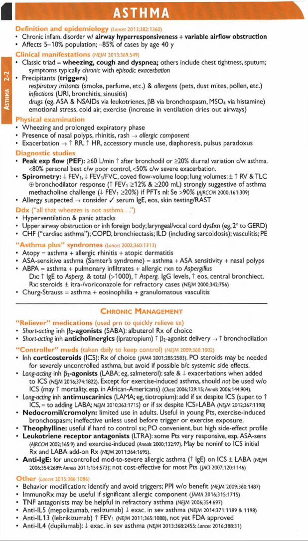
As
thm
a
2-2
ASTHMA iIDefinition and epidemiology (Loncet 2013:382:1360)• Chronic inflam, disorder w/ airway hyperresponsiveness + variable airflow obstruction• Affects 5-10% population; - 85% of cases by age 40 y
Clinical manifestations (NEJM 2013:369:549)• Classic triad = wheezing, cough and dyspnea; others include chest tightness, sputum;
symptoms typically chronic with episodic exacerbation• Precipitants (triggers)
respiratory irritants (smoke, perfume, etc.) & allergens (pets, dust mites, pollen, etc.) infections (URI, bronchitis, sinusitis)drugs (eg, ASA & NSAIDs via leukotrienes, f5B via bronchospasm, MS04 via histamine) emotional stress, cold air, exercise (increase in ventilation dries out airways)
Physical examination• Wheezing and prolonged expiratory phase• Presence of nasal polyps, rhinitis, rash -> allergic component• Exacerbation -» T RR, t HR, accessory muscle use, diaphoresis, pulsus paradoxusDiagnostic studies• Peak exp flow (PEF): >60 L/min T after bronchodil or >20% diurnal variation c/w asthma.
<80% personal best c/w poor control, <50% c/w severe exacerbation.• S p iro m e try : i FEW i FEVi/FVC, coved flow-volume loop; lung volumes: ± T RV & TLC
© bronchodilator response (T FEVi >12% & >200 ml.) strongly suggestive of asthma methacholine challenge ( i FEVi >20%) if PFTs nl: Se >90% (AJRCCM 2000:161:309)
• Allergy suspected -» consider / serum IgE, eos, skin testing/RAST
Ddx (“all that wheezes is not asthma...”)• Hyperventilation & panic attacks• Upper airway obstruction or inh foreign body; laryngeal/vocal cord dysfxn (eg, 2° to GERD)• CHF (“ cardiac asthma” ); COPD, bronchiectasis; ILD (including sarcoidosis); vasculitis; PE
“Asthm a plus” syndromes (Lancet 2002:360:1313)• Atopy = asthma + allergic rhinitis + atopic dermatitis• ASA-sensitive asthma (Samter's syndrome) = asthma + ASA sensitivity + nasal polyps• ABPA = asthma + pulmonary infiltrates + allergic rxn to Aspergillus
Dx: t IgE to Asperg. & total (>1000), T Asperg. IgG levels, T eos, central bronchiect. Rx: steroids ± itra-/voriconazole for refractory cases (NEJM 2000:342:756)
• Churg-Strauss = asthma + eosinophilia + granulomatous vasculitis
C hronic Management
“ Reliever” m edications (used prn to quickly relieve sx)• Short-acting inh p2 -agonists (SABA): albuterol Rx of choice• Short-act/ng inh anticho linerg ics (ipratropium) t P2-agonist delivery T bronchodilation
“ C o n tro lle r” meds (taken daily to keep control) (NEJM 2009;360:1002)• Inh co rticos te ro ids (ICS): Rx of choice (jama 2001:285:2583). PO steroids may be needed
for severely uncontrolled asthma, but avoid if possible b/c systemic side effects.• Long-acting inh (^-agonists (LABA; eg, salmeterol): safe & i exacerbations when added
to ICS (NEJM 2016:374:1822). Except for exercise-induced asthma, should not be used w/o ICS (may t mortality, esp. in African-Americans) (Chest 2006; 129:15; Annals 2006:144:904).
• Long-acting inh antim uscarinics (LAMA; eg, tiotropium): add if sx despite ICS (super, to TICS, = to adding LABA; nejm 2010:363:1715) or if sx despite ICS+LABA (NEJM 2012:367:1198)
• N e d o c ro m il/c ro m o lyn : limited use in adults. Useful in young Pts, exercise-inducedbronchospasm; ineffective unless used before trigger or exercise exposure.
• Theophy lline : useful if hard to control sx; PO convenient, but high side-effect profile• Leuko triene re ce p to r antagonists (LTRA): some Pts very responsive, esp. ASA-sens
(AjRCCM 2002:165:9) and exercise-induced (Annals 2000:132:97). May be noninf to ICS initial Rx and LABA add-on Rx (NEJM 2011:364:1695).
• A n ti- lg E : for uncontrolled mod-to-severe allergic asthma (T IgE) on ICS ± LABA (NEJM2006:354:2689; Annals 2011:154:573); not COSt-effective for most Pts (JACI 2007:120:1146)
O the r (Lancet 2015:386:1086)• Behavior modification: identify and avoid triggers; PPI w/o benefit (nejm 2009:360:1487)• ImmunoRx may be useful if significant allergic component (JAMA 2016:315:1715)
• TNF antagonists may be helpful in refractory asthma (NEJM 2006:354:697)• Anti-IL5 (mepolizumab, reslizumab) 1 exac. in sev asthma (NEJM 2014:371:1189 & 1198)
• Anti-ILI 3 (lebrikizumab) T FEVi (NEJM 2011;365:1088), not yet FDA approved• Anti-IL4 (dupilumab): i exac. in sev asthma (NEJM 2013:368:2455; Lancet 2016:388:31)

• Bronchial thermoplasty (exp’tal): radiofrequency destruction of airway smooth muscle no Ain FEVi, but I in sx and # of exacerbations (nejm 2007:356:1327)
Principles of trea tm en t• Education and avoidance of environmental triggers (Lancet 2015:386:1075); yearly flu shot• Use quick-relief rescue medication as needed for all Pts• Goal to achieve c o m p le te c o n t r o l = daily sx <2/wk, 0 nocturnal sx or limitation of
activity, reliever med <2/wk, nl PEF or FEVi; partly controlled = 1-2 of the above present in a wk; uncontrolled = £3 of the above present in a wk
• Step up treatment as needed to gain control, step down as tolerated• If PEF i 15% x 2 d or 1 30%, 4x ICS dose I need for PO steroids (A)RCCM 2009:180:598)
A sthm a Stepwise Therapy (Adapted from Global Initiative for Asthma [G INA] 2015 update)
Step 1 Step 2 Step 3 Step 4 Step 5
Rapid-acting p2-agoniscs 5rnSe/ect one Select one Do one or more Add one or both
&_-SJ c/1 — c
Low-dose ICS Low-dose ICS + LABA
T ICS dose (w/ LABA)
Oral steroids (lowest dose)
2 oC LTRA Low-dose ICS + LAMA Add LAMA Anti-lgE Rx
u ° Med-dose ICS Add LTRALow-dose ICS + LTRA Add TheoLow-dose ICS +Theo
E x a c e r b a t io nEvaluation• History: baseline PEF, steroid requirement, ED visits, hospital admissions, prior intubation
Current exacerbation: duration, severity, potential precipitants, meds used Risk factors for life-threatening: prior intubation, h/o near-fatal asthma, ED visit/hosp for
asthma w/in 1 y, current/recent PO steroids, not using ICS, overdependent on SABA, ML h/o noncompliance
• Physical exam:VS, pulm, accessory muscle use, pulsus paradoxus, abdominal paradoxAssess for barotrauma: asymmetric breath sounds, tracheal deviation, subcutaneous
air -» pneumothorax, precordial (Hamman’s) crunch -* pneumomediastinum• Diagnostic studies: PEF (used to follow clinical course); Sa0 2; C XR to r/o PNA or PTX
ABG if severe: low PaCC>2 initially; nl or high PaC02 may signify tiring
Severity of Asthma ExacerbationFeature Mild Moderate Severe
Breathless w /... Walking Talking At restTalking in ... Sentences Phrases WordsMental status ± Agitated Agitated Agitated
RR T T >30Accessory muscles 0 0 ©
Wheeze Moderate, end-expir Loud Usually loudHR <100 100-120 >120
Pulsus paradoxus Normal (<10) 10-25 >25PEF >80% 60-80% <60%Sa0 2 >95% 91-95% <90%P,02 Normal V O-' O <60
PaC 0 2 <45 <45 >45
Resp arrest imminent: drowsy, abdominal paradox, wheezes inaudible (b/c 0 air movement), bradycardia, loss of abdominal paradox (respiratory muscle fatigue). Presence o f several parameters (not necessarily all) indicates classification (adapted from Chest 2003:123:1018; G INA 2015 update).
Initial trea tm en t (NEJM 2010:363:755)• O xygen to keep Sa02 >90%• Inhaled S A B A (eg, albuterol) by MDI (4—8 puffs) or nebulizer (2.5-5 mg) q20min• Corticosteroids: prednisone 0.5-1 mg/kg PO; IV if impending resp arrest• Ipratropium MDI (4-6 puffs) or nebulizer (0.5 mg) q20min if severe (Chest 2002:121:1977)• Epinephrine (0.3-0.5 mL SC of 1:1000 dilution) no advantage over inh SABA• Montelukast IV T FEVi but did not A rate of hosp (JAllergy Clin immunol 2010:125:374)• Reassess after 60-90 min o f Rx
Mild-mod exacerbation: cont SABA q lhSevere exacerbation: SABA & ipratropium q lh or continuously;± Mg 2 g IV over 20 min
(Lancet 2003:361:2114); ± heliox (60-80%)• Decide disposition within 4 h o f presentation and after 1-3 h o f Rx

An
ap
hy
la
xis
2
-4/
Figure 2-2 Disposition of patients after initial treatment of asthma exacerbation
Reassessment after 1 -3 h of Rx / 1 \
Good responseT ^
Incomplete response Poor responsePEF >70%; Sa0 2 >90% PEF 40-69% PEF <40%
no distress; normal exam mild/mod s/s P0O2 <60 or PaC 02 242response sustained 60' after Rx x severe sx; AMS
|
I RFs for near-fatal asthma i• Discharge home Noncompliant Admit to ICU
Inh SABA Inh SABA + anticholOral steroid taper Admit to hospital ward Steroids IV
? Start ICS Inh SABA ± antichol ± intubationClose follow-up Steroids PO/IV
Reassess periodically at intervals Incomplete after 6-12 h
or poor response
ICU-level care• High-dose steroids: methylprednisolone 125 mg IV q6h (Archives 1983:143:1324)• In v a s iv e v e n t i la t io n :
large ET tube, Ppiat <30 cm H^O (predicts barotrauma better than PIP), max exp time PEEP individualized to Pt physiologyparalysis, inhalational anesthetics, bronchoalveolar lavage w / mucolytic, heliox
(60-80% helium) and ECHO have been used with success• NPPV likely improves obstruction (Chest 2003:123:1018), but controversial and rarely used
ANAPHYLAXISDefinition and pathophysiology (Ann Emerg Med 2006:47:373)• Severe, rapid-onset (mins to hrs), potentially life-threatening systemic allergic response• IgE-mediated mast cell degranulation with release of histamine, tryptase andTNF• Precipitates systemic reactions (bronchospasm, tissue swelling, fluid shifts, vasodilation)• Common triggers: penicillins, cephalosporins, shellfish, nuts, insect stings, IV contrast (not
truly an IgE-mediated mechanism, but clinically similar)
Diagnosis: any of the three following criteria1) Acute illness with skin ± mucosal involvement (rash, flushing, hives),AND at least one of:
• Respiratory compromise (wheeze, stridor, dyspnea, hypoxemia)• Hypotension o r hypoperfusion (syncope, incontinence)
2) Two or more of the following after exposure to a likely allergen: skin/mucosal involvement,respiratory compromise, i BP or hypoperfusion, Gl symptoms
3) Hypotension after exposure to know n allergen for that Pt
Treatm ent• Epi: IM/SC 0.3-0.5 mL of 1:1000 dilution q5-20min; if HoTN 1 mg IVB q5min ± gtt• A irw ay : suppl O 2 ± intubation or cricothyroidotomy (if laryngeal edema); [^-agonists• Fluid resuscitation w/ Ig volume of crystalloid (may extravasate up to 35% of blood volume)• Antihistamines relieve hives & itching, no effect on airv/ay or hemodynamics
H1RA (diphenhydramine 50 mg IV/IM) ± H2RA (eg, ranitidine 50 mg IV)• Corticosteroids have no immediate effect but may help prevent relapse:
methylprednisolone 125 mg IV q6h if severe or prednisone 50 mg PO• Avoid unopposed a.-adrenergic vasopressors
Disposition• Mild rxn limited to urticaria or mild bronchospasm can be observed for >6 h; admit all others• Watch for biphasic reac tion ; occurs in 23%, typically w/in 8-10 h but up to 72 h• A t time of d/c: education re: allergen avoidance, instruction and Rx for EpiPen, allergist f/u
Angioedem a (Ann Allergy Asthma Immunol 2000:85:52'!; J Allerg-/ Clin Immunol 2013:131:1491)• Localized swelling of skin/mucosa; involves face, lips, tongue, uvula, larynx, and bowels• Etiologies: mast cell-mediated (eg, NSAIDs); bradykinin-mediated (eg. AC E i, ARNi,
hereditary angioedema, acquired C l inhibitor deficiency); idiopathic• Diagnosis: C4 and C l inhibitor level, tryptase (if suspect anaphylaxis)• Rx: intubation if risk of airway compromise; allergic angioedema: H 1 /H2 antihist, steroids
if 2° ACEi: d/c ACEi, antihist., ica tib a n t (bradykinin-receptor antag; NEJM 2015:372:418) Hereditary angioedema: synthetic C l inhibitor cinryze (NEJM 2010:363:513)

CHRONIC OBSTRUCTIVE PULMONARY DISEASED e fin itio n and ep idem io logy (Lancet 2014:385:1778)• Progressive airflow limitation caused by airway and parenchymal inflammation
Emphysema vs. Chronic Bronchitis
Emphysema Chronic B ronchitisDefin ition Dilation/destruction of
parenchyma (path definition)Productive cough >3 mo/y x >2 y (clinical definition)
Pathophysiology Tissue destructionV/Q: T dead space fraction ->hypercarbia, but only mildhypoxemia
Small airways affected V/Q: T shunt fraction -» severe hypoxemia, hypercapnia PHT, cor pulmonale
Clinicalmanifestations
Severe, constant dyspnea Mild cough
Intermittent dyspnea Copious sputum production
Physical exam "Pink puffer”Tachypneic, noncyanotic, thin Diminished breath sounds
"Blue bloater”Cyanotic, obese, edematous Rhonchi & wheezes
Pathogenesis (Loncet 2003:362:1053)• C igarette smoke (centrilobular emphysema, affects 15-20% of smokers)• Recurrent airway infections• oti-antitrypsin defic.: early-onset panacinar emphysema, 1-3% of COPD cases. Suspect
if age <45, lower lungs affected, extrathoracic manifestations (liver disease [not if MZ subtype], FMD, pancreatitis). / serum AAT level (nb, acute phase reactant).
• Low FEVi in early adulthood important in genesis of COPD (N£yM 2015:373:111)• Misc: biomass (eg, cooking fuels in enclosed space), chronic asthma (Lancet 2009:374:733)
Clinical manifestations• Chronic cough, sputum production, dyspnea; later stages -> freq exac., a.m. HA, w t loss• Exacerbation triggers: infxn, other cardiopulmonary disease, incl. PE {Annals 2006:144:390)
Infxn: overt tracheobronchitis/pneumonia from viruses, S. pneumoniae, H. influenzae, M. catarrhalis or triggered by changes in strain of colonizers (NEJM 2008:359:2355)
• Physical exam: T AP diameter of chest ("barrel-chest” ), hyperresonance, I diaphragmaticexcursion, X breath sounds, T expiratory phase, rhonchi, wheezesduring exacerbation: tachypnea, accessory muscle use, pulsus paradoxus, cyanosis
• Asthma-COPD overlap syndrome (ACOS; nejm 2015:373:1241): features of both present.For example: reversibility of airway obstruction w/ bronchodilator in COPD; neutrophilic inflammation in asthma (more classic in COPD);eos in COPD.
Diagnostic studies• CXR (see Radiology inserts): hyperinflation,flat diaphragms,! interstitial markings & bullae• PFTs: obstruction: XX FEVi, X FVC, FEVi/FVC <0.7 (no sig A post bronchodilator),
expiratory scooping of flow-volume loop; hyperinflation: TT RV T TLC, T RV/TLC; abnormal gas exchange: X DlCO (in emphysema)
• ABG: X Pa02,± T PaC02 (in chronic bronchitis, usually only if FEVi <1.5 L) and X pH• ECG: PRWP, S1S2S3, R-sided strain, RVH, T P waves in lead II ("P pulmonale")Chronic trea tm en t (Loncet 2015:385:1789)• Bronchodilators (first-line therapy): anticholinergics, ^-agonists (BA), theophylline
Long-acting (LA) antimuscarinic (LAMA; eg, tiotropium): X exac., X admit, X resp failure (NEJM 2008:359:1543), better than ipratropium or LABA as mono Rx (NEJM 2011:364:1093)
LABA: —11% ! in exacerbations, no T in CV events (Lancet 2016:387:1817)LABA + inh steroid: ? X mort. vs. either alone (nejm 2007:356:775)LAMA + LABA: t FEVi, J. sx vs. either alone (Chest 2014:145:981) and superior to LABA +
inh steroid (NE/M 2016:374:2222)• Corticosteroids (inhaled, ICS): -11% X in exacerb & slow X FEVi; no A in risk of PNA or
in mortality (Loncct2016;387:1817)• Roflumilast (PDE-4 inhibitor): T FEVi & X exacerbations when added to bronchodilator
(Lancet 2009:374:685,695 & 2015:385:857)• Antibiotics: daily azithro J, exacerb, but not yet routine (jAfAA 2014:311:2225)• Mucolytics: no A FEVi, but ? 1 exacerbation rate (Loncet 2008:371:2013)• Oxygen: if PaC>2 <55 mmHg or SaC>2 <89% (during rest, exercise, or sleep) to prevent cor
pulmonale; only Rx proven to X mortality (Anno/s 1980:93:391; Loncet 1981;i:681)• Prevention: Flu/Pneumovax; smoking cessation (eg, varenicline, bupropion) 50% I
in lung function decline (AJRCCM 2002:166:675) and X long-term mortality (Annois 2005:142:223)• Rehabilitation: X dyspnea and fatigue, T exercise tolerance, X QoL (NEJM 2009:360:1329)
CO
PD
2-5
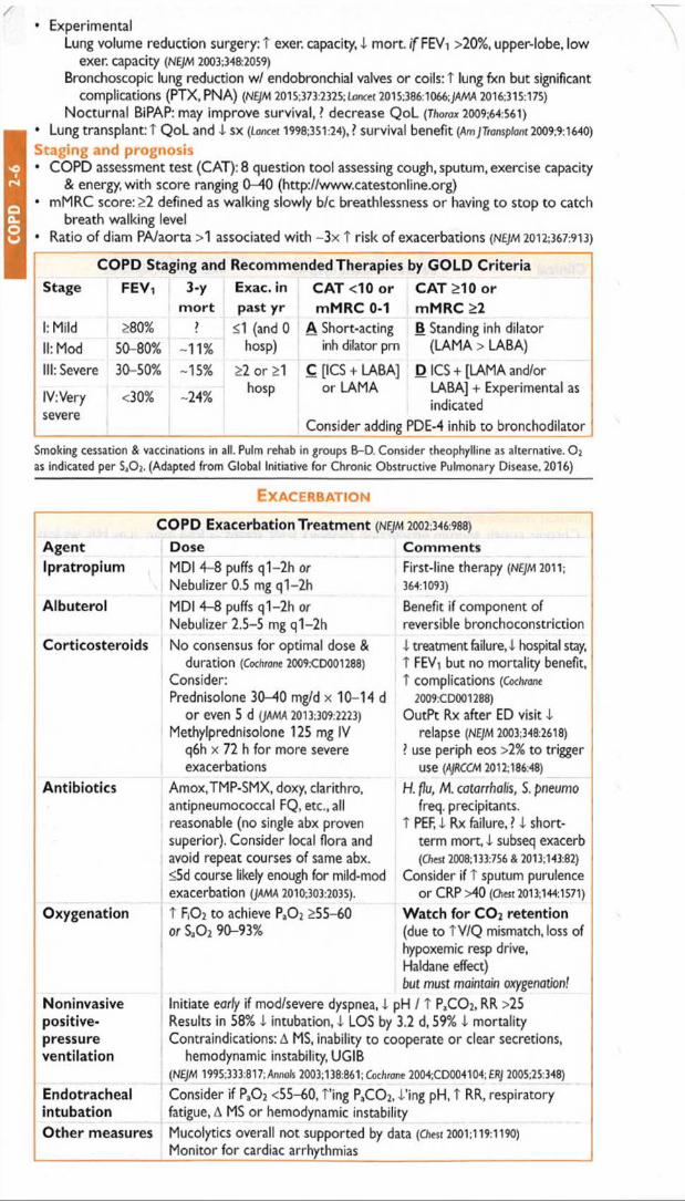
CO
PD
2-
6• Experimental
Lung volume reduction surgery: T exer. capacity, 4 mort. i f FEVi >20%, upper-lobe, low exer. capacity (NEJM 2003:348:2059)
Bronchoscopic lung reduction w/ endobronchial valves or coils: T lung fxn but significant complications (PTX,PNA) (NEJM 2015:373:2325; Lancet 2015:386:1066;JAMA 2016:315:175)
Nocturnal BiPAP: may improve survival, ? decrease QoL (Thorax 2009:64:561)
• Lung transplant: T QoL and 4 SX (Eoncet 1998:351:24), ? survival benefit (Am JTransplanl 2009;9:1640)
Staging and prognosis• COPD assessment test (CAT): 8 question tool assessing cough, sputum, exercise capacity
& energy, with score ranging 0-40 (http://www.catestonline.org)• mMRC score: >2 defined as walking slowly b/c breathlessness or having to stop to catch
breath walking level• Ratio of diam PA/aorta >1 associated with -3 x t risk of exacerbations (NEJM 2012:367:913)
COPD Staging and Recommended Therapies by GOLD C rite riaStage FEVi 3-y
m ortExac. in past yr
CAT <10 or mMRC 0-1
CAT >10 or mMRC >2
1: Mild >80% £1 (and 0 A Short-acting B Standing inh dilatorII: Mod 50 80% -11% hosp) inh dilator pm (LAMA > LABA)
III: Severe 30-50% -15% >2 or >1 hosp
C [ICS + LABA] or LAMA
Consider adding
D ICS + [LAMA and/or LABA] + Experimental as indicated
3DE-4 inhib to bronchodilator
IV: Very severe
<30% -24%
Smoking cessation & vaccinations in all. Pulm rehab in groups B-D. Consider theophylline as alternative. O 2 as indicated per Sa02. (Adapted from Global Initiative fo r Chronic Obstructive Pulmonary Disease, 2016)
E x a c e r b a t i o n
COPD Exacerbation Treatment (NEJM 2002:346:988)Agent Dose CommentsIpratropium MDI 4-8 puffs q1-2h o r
Nebulizer 0.5 mg q1-2hFirst-line therapy (NEJM 2011; 364:1093)
Albuterol MDI 4-8 puffs q1-2h o r
Nebulizer 2.5-5 mg q1-2hBenefit if component of reversible bronchoconstriction
Corticosteroids No consensus for optimal dose & duration (Cochrane 2009:CD001288)
Consider:Prednisolone 30-40 mg/d x 10-14 d
or even 5 d [JAMA 2013:309:2223) Methylprednisolone 125 mg IV
q6h x 72 h for more severe exacerbations
4 treatment failure, 4 hospital stay, T FEVi but no mortality benefit, T complications [Cochrane
2009:CD001288)OutPt Rx after ED visit 4
relapse (NEJM 2003:348:2618)? use periph eos >2% to trigger
use (AJRCOA 2012:186:48)Antibiotics Amox,TMP-SMX, doxy, darithro,
antipneumococcal FQ, etc., all reasonable (no single abx proven superior). Consider local flora and avoid repeat courses of same abx. <5d course likely enough for mild-mod exacerbation (JAMA 2010:303:2035).
H . flu , M. ca ta rrh a lis , S. p n e u m o
freq. precipitants.T PEF, 4 Rx failure, ? 4 short
term mort, 4 subseq exacerb (C/iest 2008:133:756 & 2013:143:82)
Consider if T sputum purulence or CRP >40 (Chest 2013:144:1571)
Oxygenation T RO2 to achieve Pa0 2 >55-60 or $a0 2 90-93%
Watch for C 02 retention(due to TV/Q mismatch, loss of hypoxemic resp drive,Haldane effect)but must maintain oxygenation!
Noninvasivepositive-pressureventilation
Initiate early if mod/severe dyspnea, 4 pH / T PaC 0 2, RR >25 Results in 58% 4 intubation, 4 LOS by 3.2 d, 59% 4 mortality Contraindications: A MS, inability to cooperate or dear secretions,
hemodynamic instability, UGIB(NEJM 1995:333:817; Anno/s 2003:138:861; Cochrane 2004;CD004104; ERJ 2005:25:348)
Endotrachealintubation
Consider if Pa0 2 <55-60, Ting PaC 0 2,4’ing pH, T RR, respiratory fatigue, A MS or hemodynamic instability
Other measures Mucolytics overall not supported by data (Chest 2001:119:1190)
Monitor for cardiac arrhythmias

HEMOPTYSISD e fin itio n and pathophys io logy• Expectoration of blood o r blood-streaked sputum• Massive hemoptysis: ~>600 mL/24-48 h; gas exchange more important than blood loss• Massive hemoptysis usually from tortuous or invaded b ronch ia l a rte rie s
Etiologies (Crit Care Med 2000:28:1642)
Infection/Inflam m ation
Bronchitis (most common cause of trivial hemoptysis) Bronchiectasis ind. CF (common cause of massive hemoptysis) TB or aspergilloma (can be massive); pneumonia or lung abscess
Neoplasm Usually primary lung cancer, sometimes metastasis (can be massive)Cardiovasc. PE (can be massive), pulmonary artery rupture (2° to instrumentation),
CHF, mitral stenosis, trauma/foreign body, bronchovascular fistulaO ther Vase (GPA, Goodpasture’s, Behcet’s; can be massive),AVM, anticoag
(w/ underlying lung dis), coagulopathy, cocaine, pulm hemosiderosis
D iagnostic w o rku p■ Localize bleeding site (r/o Gl or ENT source by H&P ± endo); determine whether unilateral
or bilateral, localized or diffuse, parenchymal or airway by CXR/ chest CT ± bronch• PT, PTT, CBC to rule out coagulopathy• Sputum culture/stain for bacteria, fungi and AFB; cytology to r/o malignancy• ANCA, anti-GBM, urinalysis to / for vasculitis or pulmonary-renal syndromeTreatm ent• Mechanism of death is asphyxiation not exsanguination; maintain gas exchange, reverse
coagulation and treat underlying condition; cough supp. may T risk of asphyxiation• Massive hemoptysis: put bleeding side dependent; selectively intubate nl lung if needed
Angiography: Dx & Rx (vascular occlusion balloons or selective embol of bronchial art) Rigid bronch: allows more options (electrocautery, laser) than flex. Surgical resection.
BRONCHIECTASISD e fin itio n and ep idem io lo gy (NEJM 2002:346:1383)• Obstructive airways disease of bronchi and bronchioles, chronic transmural inflamm w/
airway dilatation and thickening, collapsibility, mucus plugging w / impaired clearanceIn itia l w o rku p• H&P: cough, dyspnea, copious sputum production, ± hemoptysis, inspiratory “ squeaks”• CXR: scattered or focal; rings o f bronchial cuffing;“ tram track" of dilated, thick airways• PFTs: obstructive; chest CT: airway dilation & thickening ± cystic As, infiltrates, adenopathy
Etiology O the r Features Diagnostic Testing
Chronic infxns (eg, MTb.ABPA)
Chronic cough, freq/persist infiltrate, refract asthma (ABPA)
Sputum cx (ind myobact, fungal), ± bronch/BAL, IgE & eos (ABPA)
1° ciliary dyskin Sinusitis, infertility, otitis Dynein mutationsImmunodefic. Recurrent infxns often as child IgA, IgG, IgM, IgG subclassesRA, SLE Resp sx may precede joint sx RF.ANAIBD Not relieved by bowel resection Colonoscopy, biopsya-i-AT deficiency Lower lobe emphysema ai-AT levelAnatomic R middle lobe synd. from sharp
takeoff, foreign body aspir.Bronchoscopy
T r e a t m e n t• Treat underlying condition; mucolytics & bronchodilators• Prophylactic azithro shown to i exacerb. in non-CF bronchiectasis (JAMA 2013:1251)• Antibiotics: at time of acute exacerbation directed against suspected or prior pathogensC y s t ic f ib r o s is (NEJM 2015;372:3S1)• Autosomal recessive genetic disorder due to mutations in chloride channel (CFTR gene)• t mucus thickness, I mucociliary clearance, T infections -> bronchiectasis• Clinical: recurrent PNA, weight loss, sinus infxns, infertility, pancreatic insuffic (fatty stools)• Rx: airway clearance (chest PT, inh hypertonic saline, DNAse), abx for exacerb. for drug-
resistant org. (eg. Pseudomonas, Burkholderia), gene targeted with CFTR potentiator (ivakaftor) & corrector (lumakaftor) (NEJM 2011:365:1663 & 2015:373:220), lung transplant
N o n - t u b e r c u lo u s m y c o b a c t e r iu m (N T M ; u b iq u ito u s h y d ro p h ilic bac te ria )• Chronic cough, I wt; Lady Windermere synd.: R middle lobe bronchiectasis in elderly 9
who suppress expectoration; in HIV ® disseminated disease (see HIV/AIDS)• Dx: CT scan (tree-in-bud, nodules, cavities, bronchiect), sputum x3 or BAL, AFB stain + cx• Treatment: [darithro or azithro] + rifamycin & ethambutol for >12 mo (Chest 2004:126:566)
He
mo
p/Br
on
ch

SOLITARY PULMONARY NODULEPrincip les• Definition: single, <3 cm, surrounded by normal lung, no LAN or pleural effusion• Often “ incidentalomas,’’ esp with t CT use, but may still be early, curable malignancy
EtiologiesMalignant (30%)Bronchogenic carcinoma (75%):
adeno & large cell (peripheral) squamous & small cell (central)
Metastatic (20%): breast, head & neck, colon, testicular, renal, sarcoma, melanoma
Lipoma, fibroma, amyloidoma, pneumonitis Carcinoid, primary sarcoma
In itia l eva luation• History: h/o cancer, smoking, age {<30 y = 2% malignant,+15% each decade >30)• CT: size/shape, Ca2+, LAN, effusions, bony destruction, compare w / old studies
0 Ca -> T likelihood malignant; laminated -4 granuloma;,‘popcorn’, -> hamartoma• High-risk features for malig: size (eg,>2.3 cm diameter), spiculated, upper lobe, 9,>60 yo,
>1 ppd c u r re n t sm oke r, no p r io r s m o k in g ce ssa tion (NEJM 2003:348:2535 & 2013:369:910)
D iagnostic studies• PET: detects metab. activity of tumors, 97% Se & 78% Sp for malig. (esp. if >8 mm)
also useful for surgical staging b/c may detect unsuspected mets (JAMA 2001:285:914)
useful in deciding which lesions to bx vs. follow w/ serial CT (jThor Oncol 2006:1:71)• Transthoracic needle biopsy (TTNB): if tech, feasible, 97% will obtain definitive tissue dx
(AJR 2005:185:1294); if noninformative or malignant -* resect• Video-assisted thoracoscopic surgery (VATS): for percutaneously inaccessible lesions;
highly sensitive and allows resection; has replaced thoracotomy• Transbronchial bx (TBB): most lesions too small to reliably sample w/o endobronchial
U/S (Chest 2003:123:604); bronch w/ brushings low-yield unless invading bronchus; navigational bronchoscopy w / 70% yield, t sens w / larger nodules (Chest 2012:142:385)
• PPD, fungal serologies, ANCAM anagem ent (for solid SPN >8 mm; if <8 mm, serial CT) (Chest 2013:143:840)• Low risk (<5%, see ref): serial CT (freq depending on risk); shared decision w/ Pt re: bx• Intermediate risk (5-60%): PET, if © -4 follow low-risk protocol; if © -4 high-risk protocol• High risk (and surgical candidate):TBB,TTNB, or VATS -» lobectomy if malignant• Ground-glass nodules: longer f/u b/c if malignant can be slow-growing & PET 0
Benign (70%)Granuloma (80%):TB, histo, coccidio Ham artom a (10%)Bronchogenic cyst.AVM, pulm infarct Echinococcosis, ascariasis, aspergilloma Wegener’s, rheumatoid nodule
SLEEP APNEAD e f in i t io n a n d p a th o p h y s io lo g y• Obstructive: pharyngeal collapse -> apnea (>10 s) or hypopnea ( i airflow) ± desaturation;
risk factors: obesity (present in 70%), large neck, male sex, 1 muscle tone, T age, alcohol• Central: i neurologic feedback w/ oscillating drive. Apneas w/o resp effort ± subsequent
T resp rate. Associated with CHF & atrial fibrillation; worsened by sedatives.• Complex: obstructive + central (nb, untreated obstructive -> complex)• Proposed mech: Apnea/arousals -4 sympathetic nervous system activation, negative
intrathoracic pressure -4 T preload, t afterload. Consequently -* HTN, pulm HTN.
C l in ic a l m a n if e s ta t io n s (Lancet 2002:360:237; Lancet Resp Med 2013:1:61)
• Snoring, witnessed apneas/gasping, daytime sleepiness• Cardiovascular: HTN (jama 2012:307:2169); a/w T risk of stroke and death (NE)M
2005:353:2034) & possibly CAD & endothelial dysfxn (AJRCCM 2001;163:19: Circ 2008:117:2270)
• Neurocognitive: i cognitive performance, i QoL, T motor vehicle and work accidents(NEJM 1999:340:847; AJRCCM 2001:164:2031)
D ia g n o s is a n d t r e a t m e n t (JAMA 2013:310:731 & Loncet 2014:383:736)
• Polysomnography (sleep study); can do home-testing• Obstructive: CPAP U apnea/hypopnea, i BP (JAMA 2013:310:2407 & NEJM 2014:370:2276),
i sleepiness, t performance (ajrccm 2012:186:677), T EF in Pts with CHF (NEJM 2003:348:1233),
1 metab syndrome (NEJM 2011:365:2277), I mortality after stroke (AjRCCfA 2009:180:36)
• Oral appliances if refusing CPAP; upper-airway stimulator under study (NEJM 2014:370:139)
• Central: adaptive servoventilation (ASV) i f w/o CHF (nb,T mortality if CHF; NEJM 2015:373:1095)
• Avoid alcohol and sedatives• Surgery (eg, uvulopalatopharyngoplasty, UPPP) of limited benefit (Chest 1997:111:265)
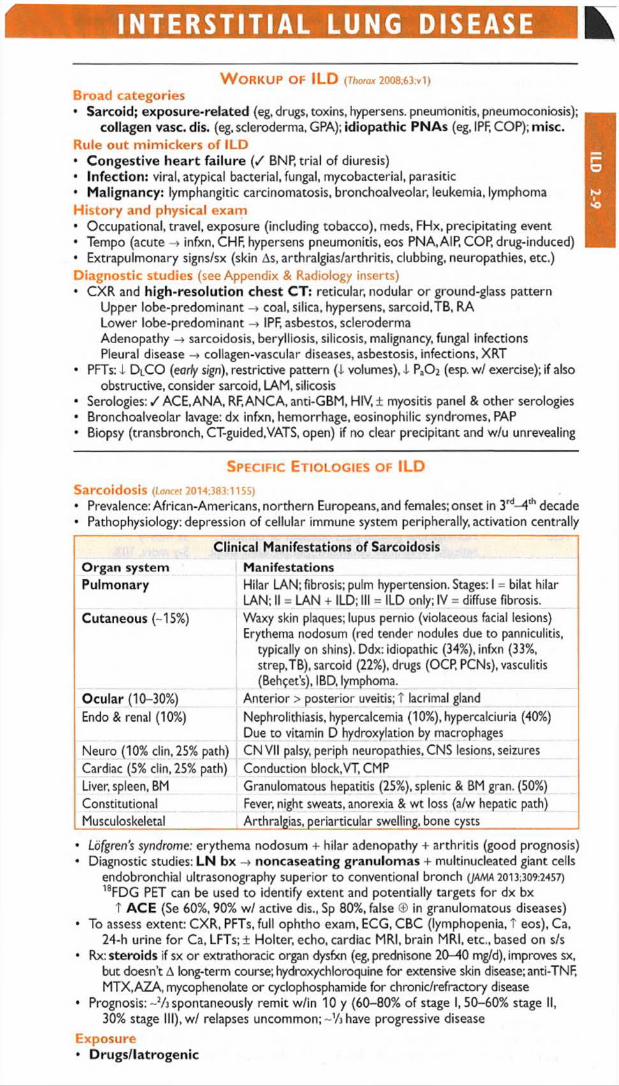
INTERSTITIAL LUNG DISEASE
W o r k u p o f IL D (Thorax 2008;63:vi)Broad categories• Sarcoid; exposure-related (eg, drugs, toxins, hypersens, pneumonitis, pneumoconiosis);
collagen vase. dis. (eg, scleroderma, GPA); idiopathic PNAs (eg, IPF, COP); misc.Rule out m imickers of ILD• Congestive heart failure ( / BNP, trial of diuresis)• Infection: viral, atypical bacterial, fungal, mycobacterial, parasitic• Malignancy: lymphangitic carcinomatosis, bronchoalveolar, leukemia, lymphomaHistory and physical exam• Occupational, travel, exposure (including tobacco), meds, FHx, precipitating event• Tempo (acute -> infxn, CHF, hypersens pneumonitis, eos PNA,AIP, COP, drug-induced)• Extrapulmonary signs/sx (skin As, arthralgias/arthritis, clubbing, neuropathies, etc,)Diagnostic studies (see Appendix & Radiology inserts)• CXR and high-resolution chest CT: reticular, nodular o r ground-glass pattern
Upper lobe-predominant -> coal, silica, hypersens, sarcoid.TB, RA Lower lobe-predominant -* IPF, asbestos, scleroderma Adenopathy -> sarcoidosis, berylliosis, silicosis, malignancy, fungal infections Pleural disease -» collagen-vascular diseases, asbestosis, infections, XRT
• PFTs: I D lCO (early sign), restrictive pattern ( I volumes), i Pa02 (esp. w / exercise); if alsoobstructive, consider sarcoid, LAM, silicosis
• Serologies; / ACE, ANA, RF, ANCA, anti-GBM, HIV, ± myositis panel & other serologies• Bronchoalveolar lavage: dx infxn, hemorrhage, eosinophilic syndromes, PAP• Biopsy (transbronch, CT-guided,VATS, open) if no clear precipitant and w/u unrevealing
S p e c if ic Et io l o g ie s o f IL D
Sarcoidosis (Loncet 2014:383:1155)
• Prevalence: African-Americans, northern Europeans, and females; onset in 3rd-4 th decade• Pathophysiology: depression of cellular immune system peripherally, activation centrally
Clinical Manifestations o f SarcoidosisOrgan system ManifestationsPulmonary Hilar LAN; fibrosis; pulm hypertension. Stages: 1 = bilat hilar
LAN; II = LAN + ILD; III = ILD only; IV = diffuse fibrosis.Cutaneous (-15%) Waxy skin plaques; lupus pernio (violaceous facial lesions)
Erythema nodosum (red tender nodules due to panniculitis, typically on shins). Ddx: idiopathic (34%), infxn (33%, strep,TB), sarcoid (22%), drugs (OCP, PCNs), vasculitis (Behcet’s), IBD, lymphoma.
Ocular (10-30%) Anterior > posterior uveitis; T lacrimal glandEndo & renal (10%) Nephrolithiasis, hypercalcemia (10%), hypercalciuria (40%)
Due to vitamin D hydroxylation by macrophagesNeuro (10% clin, 25% path) CNVII palsy, periph neuropathies, CNS lesions, seizuresCardiac (5% clin, 25% path) Conduction block,VT, CMPLiver, spleen, BM Granulomatous hepatitis (25%), splenic & BM gran. (50%)Constitutional Fever, night sweats, anorexia & wt loss (a/w hepatic path)Musculoskeletal Arthralgias, periarticular swelling, bone cysts
• Lofgren's syndrome: erythema nodosum + hilar adenopathy + arthritis (good prognosis)• Diagnostic studies: LN bx -h> noncaseating granulomas + multinucleated giant cells
endobronchial ultrasonography superior to conventional branch (JAMA 2013:309:2457) 18FDG PET can be used to identify extent and potentially targets for dx bx
t AC E (Se 60%, 90% w / active dis., Sp 80%, false © in granulomatous diseases)• To assess extent: CXR, PFTs, full ophtho exam, ECG, CBC (lymphopenia, T eos), Ca,
24-h urine fo r Ca, LFTs;± Holter, echo, cardiac MRI, brain MRI, etc., based on s/s• Rx: steroids if sx o r extrathoracic organ dysfxn (eg, prednisone 20-40 mg/d), improves sx,
but doesn’t A long-term course; hydroxychloroquine for extensive skin disease; anti-TNF, MTX.AZA, mycophenolate or cyclophosphamide for chronic/refractory disease
• Prognosis: ~2/j spontaneously remit w/in 10 y (60-80% of stage I, 50-60% stage II,30% stage III), w / relapses uncommon; ~Vj have progressive disease
Exposure• Drugs/latrogenic

ILD
2-
10Am io (dose & duration depend.): chronic interstitial PNA <-> ARDS; Rx: d/c amio; steroids O ther drugs: nitrofurantoin, sulfonamides, thiazides, INH, hydralazine, gold Chemo: bleomycin (triggered by hyperoxia), busulfan, cyclophosphamide, MTX, etc. XRT: COP/BOOP w/ sharply linear, nonanatomic boundaries; DAH
• Pneumoconioses (inorganic dusts) (NEJM 2000:342:406; C/m Chest Med 2004:467)
Coal worker’s: upper lobe coal macules; may progress to massive fibrosis Silicosis: upper lobe opacities ± eggshell calcification of lymph nodes; T risk of TB Asbestosis: lower lobe fibrosis, calcified pleural plaques, DOE, dry cough, rales on
exam. Asbestos exposure also -» pleural plaques, benign pleural effusion, diffuse pleural thickening, rounded atelectasis, mesothelioma, lung Ca (esp. in smokers).
Berylliosis: multisystemic granulomatous disease that mimics sarcoidosis « Hypersensitivity pneumonitides (organic dusts): loose, noncaseating granulomas
Antigens: farmer’s lung (spores of thermophilic actinomyces); pigeon fancier’s lung (proteins from feathers and excreta of birds); humidifier lung (thermophilic bacteria)
Collagen vascular diseases (Chest 2013:143:814)
• Rheumatologic diseaseScleroderma: fibrosis in -67%; PHT seen in -10% of CREST PtsPM-DM: ILD & weakness o f respiratory muscles; MCTD: PHT & fibrosisSLE & RA: pleuritis and pleural effusions more often than ILD; SLE can cause DAH
• Vasculitis (can p/w DAH)GPA (Wegener’s granulomatosis) (© c-ANCA) w/ necrotizing granulomas EGPA (Churg-Strauss) (© c- o r p-ANCA) w / eosinophilia & necrotizing granulomas Microscopic polyangiitis (® p-ANCA) w/o granulomas
• Goodpasture’s syndrome = DAH + RPGN; typically in smokers;® anti-GBM in 90%• Lymphangioleiomyomatosis (LAM): c y s t ic ,T in $ ,R x w / s iro lim u s (NEJM 2011:364 1595) •
Id io p a th ic in te rs t it ia l pneum on ias (IIPs) (AJRCCM 2013:188:733; nejm 2014;370:1820)
• Definition: ILD o f unknown cause; dx by radiographic, histologic, and clinical features
IIPsType 1 maging/Histology ClinicalUIP/IPF Reticular opacities, honeycombing, traction
bronchiectasis; periph, subpl., & basalSx >12 mo 5-y mort. -80%
NSIP Homogenous ground-glass opacities or consolid., reticular irreg lines; symmetric, periph, basal, subpl. Mimics CTD ILD. Cellular and fibrotic subtypes, latter similar to UIP but homogenous.
Sx mos-y 5-y mort. 10% (fibrotic = UIP)
COP/BOOP Patchy bilat consolid., nodules; subpl. & peribronchial. Prolif of granulation tissue in small bronchioles & inflam of surrounding alveoli.
Can be post-infxn, HSCT, XRT, rxn to drugs. 5-y mort <5%.
AIP Diffuse ground-glass opacities, consolid. w/ lobular sparing. Path similar to DAD.
Sx <3 wk 6-mo mort. 60%
DIP Diffuse ground-glass opacities, reticular lines; lower zones, periph. M<|> in alveoli.
30-50 yo smokers Sx wks-mos
RB-ILD Bronchial thickening, centrilobular nodules, patchy ground-glass opacities. M<{> in alveoli.
Death rare
UIP, usual interstitial PNA (IP); IPF, idiopathic pulm fibrosis ((.oncet 2011:378:1949); NSIP, nonspecific IP; COP, cryptogenic organizing PNA; BOOP, bronchiolitis obliterans w / organizing PNA; AIP, acute IP (Hamman-Rich syndrome): DIP, desquamative IP; RB-ILD, resp bronchiolitis-assoc ILD.• Rx fo r UIP/IPF: suppl O 2, pulm rehab, Rx for GERD, lung transplant referral
Pirfenidone (antifibrotic) or nintedanib (tyrosine kin. inhib mediating fibrogenic growth factors) i rate of FVC decline (NE/M 2014:370:2071 & 2083;AJRCCM 2015:192:3)
High-dose steroids may be used for acute exacerbations• Steroids for other IIPs: NSIP (esp. cellular type) and COP (AJRCCM 2000:162:571); ?
benefit for AIP and DIP/RB-ILD (for which Pts should stop smoking)Pulmonary infiltrates w / eosinophilia (P IE ) = eos on BAL ± periph. blood• Allergic bronchopulmonary aspergillosis (ABPA)• Loffler’s syndrome: parasites/drugs -> transient pulm infilt + cough, fever, dyspnea, eos• Acute eosinophilic PNA (AEP): acute hypox febrile illness; Rx: steroids, tobacco cessation• Chronic eosinophilic pneumonia (CEP):“ photonegative” of CHF, typically in women
Miscellaneous• Pulm alveolar proteinosis (PAP): accum of surfactant-like phospholipids; 6 smokers; white
& gummy sputum; BAL milky fluid (NEJM 2003:349:2527); Rx w/ lung lavage & GMCSF• Langerhans cell granulomatosis (LCG): young 6 smokers; apical cysts; PTX (25%)• Lymphocytic interstitial PNA: polyclonal B-cell infiltration (> lymphoma); Rx: steroids

PLEURAL EFFUSIONPathophysiology• Systemic factors (eg, T PCWP,! oncotic pressure) -> transudat/ve effusion• Local factors (ie, A pleural surface permeability) exudative effusion
Transudates• Congestive heart failure (40%): 80% bilateral, ± cardiomegaly on CXR
occasionally exudative (esp. after aggressive diuresis o r if chronic), but -75% of exudative effusions in CHF Pts found to have non-CHF cause (Chest 2002;122:1518)
• Constrictive pericarditis (knock on exam, calcification or thickening on imaging)• Cirrhosis (“ hepatic hydrothorax” ): diaphragmatic pores allow passage of ascitic fluid
often right-sided (Vj) & massive (even w/o marked ascites)• Nephrotic syndrome: usually small, bilateral, asymptomatic (r/o PE b/c hypercoag)• Other: PE (usually exudate), malignancy (lymphatic obstruction), myxedema, CAPD
Exudates• Lung parenchymal infection (25%)
bacterial (parapneumonic): can evolve along spectrum of exudative (but sterile) -> fibropurulent (infected fluid) -> organization (fibrosis & formation of rigid pleural peel). Common causes: Strep pneumo, Staph aureus, Strep miileri, Klebsiella, Pseudomonas, Haemophilus, Bacteroides, Peptostreptococcus, mixed flora in aspiration pneumonia,
mycobacterial: >50% lymphs 80% of the time, ADA >40, pleural bx -70% Se fungal, viral (usually small), parasitic (eg, amebiasis, echinococcosis, paragonimiasis)
• Malignancy (15%): primary lung cancer most common, metastases (esp. breast,lymphoma, etc.), mesothelioma ( / serum osteopontin levels; NEJM 2005:353:15)
• Pulmonary embolism (10%): effusions in -40% of PEs; exudate (75%) > transudate(25%); hemorrhagic— must have high suspicion b/c presentation highly variable
• Collagen vascular disease: RA (large), SLE (small),Wegener’s, Churg-Strauss• Gastrointestinal diseases: pancreatitis, esophageal rupture, abdominal abscess• Hemothorax (HcWHctbiood >50%): trauma, PE, malignancy, coagulopathy, leaking aortic
aneurysm, aortic dissection, pulmonary vascular malformation• Chylothorax (triglycerides >110): thoracic duct damage due to trauma, malignancy, LAM• Other:
post-CABG: left-sided; initially bloody, clears after several wksDressler’s syndrome (pericarditis & pleuritis post-MI), uremia, postradiation therapyAsbestos exposure: benign;© eosinophilsDrug-induced (eg, nitrofurantoin, methysergide, bromocriptine, amiodarone): © eos Uremia; post-XRT; sarcoidosisMeigs’ syndrome = benign ovarian tum or -» ascites & pleural effusion Yellow-nail syndrome: yellow nails, lymphedema, pleural effusion, bronchiectasis
Diagnostic studies• Thoracentesis (NEJM 2006;3S5:e16)
Indications: all effusions >1 cm in decubitus view if suspect due to CHF, can diurese and see if effusions resolve (75% do so in 48 h)
asymmetry, fever, chest pain or failure to resolve -> thoracentesis parapneumonics should be tapped ASAP (cannot exclude infxn clinically)
Diagnostic studies: / total protein, LDH, glucose, cell count w / differential, Gram stain & culture, pH; remaining fluid for additional studies as dictated by clinical scenario
Complications: PTX (5-10%), hemothorax (~1%), re-expansion pulm edema (if >1.5 L removed), spleen/liver lac.; post-tap CXR not routinely needed (Annals 1996:124:816)
■I PTX w/ U/S and experienced supervisor; even with INR ~1.9, risk of bleed low w/ U/S & experienced operator (Chest 2009:135:1315 & 2013:144:456; Ardwes 2010:170:332)
• Transudate vs. exudate (jama 2014:311:2422)
Light’s criteria: exudate = TPeff/TP5erum >0.5 or LDHeff/LDHsenjm >0.6 or LDHcff >2A ULN of LDHserum; 97% Se, 85% Sp; best Se of all methods; however, will misidentify 25% of transudates as exudates; if clinically suspect transudate but meets criterion for exudate, confirm w/ test w / higher Sp
exudative criteria w / better Sp: choU >55 mg/dL (95-99% Sp); choU >45 mg/dL and LDHeff >200 (98% Sp); choU/cholienjm >0.3 (94% Sp); serum-effusion alb gradient <1.2 (92% Sp); serum-effusion TP gradient <3.1 (91% Sp)
CHF effusions: TP may T with diuresis or chronicity -> “ pseudoexudate” ; alb gradient <1.2, choleff >60 mg/dL (Se 54%, Sp 92%) or clin judgment to distinguish (Chest 2002:122:1524)
• Com plicated vs. uncomplicated parapneumonic (Chest 1995:108:299)
complicated = © Gram stain or culture or pH <7.2 or glucose <60complicated parapneumonic effusions usually require drainage to achieve resolutionempyema = frank pus, also needs drainage to achieve resolution

Ple
ur
al
Eff
2-12
/• Additional pleural fluid studies (NEJM 2002,346:1971)
NT-proBNP >1500 pg/mL has 91% Se & 93% Sp for CHF (Am j Med 2004;l 16:417)
WBC & diff.: exudates tend to have T WBC vs. transudates but nonspecific neutrophils -» parapneumonic, PE, pancreatitis lymphocytes (>50%) -> cancer,TB, rheumatologiceos (>10%) -> blood, air, drug rxn, asbestos, paragonimiasis, Churg-Strauss, PE
RBC: Hctcff 1-20% -> cancer, PE, trauma; HcWHctbiood >50% -> hemothorax AFB: yield inTB 0-10% w / stain, 11-50% w/ culture,-70% w/ pleural bx adenosine deaminase (ADA): seen w/ granulomas, >70 suggests TB, <40 excludes TB cytology: ideally >150 mL and at least 60 ml_ should be obtained (Chest 2010:137:68)
glucose: <60 mg/dl_ -» malignancy, infection, RAamylase: seen in pancreatic disease and esophageal rupture (salivary amylase) rheumatoid factor, ChSO, ANA: limited utility in dx collagen vascular disease triglycerides: >110 -» chylothorax, 50-110 -> / lipoprotein analysis for chylomicrons cholesterol: >60; seen in chronic effusions (eg, CHF, RA, old TB) creatinine: effusion/serum ratio >1 -> urinothoraxfibulin-3:T plasma and/or effusion levels -> mesothelioma (NEJM 2012:367:1417)
• Chest CT; pleural biopsy;VATS• Undiagnosed persistent pleural effusions (Clin Chest Med 2006:27:309)
Transudative: most commonly CHF or hepatic hydrothorax. / s/s CHF or cirrhosis, NT-proBNPeff; consider intraperitoneal injection of technetium-99m sulfur colloid
Exudative (ensure using Sp test listed above): most commonly malig, empyema,TB, PE. / s/s malig, chest CT (P),ADA or IFN-y release assay; consider thoracoscopy.
Characteristics o f Pleural Fluid (not diagnostic crite ria )
E tio logy Appear W B C d iff RBC pH Glc C om m entsCHF clear, straw <1000
lymphs<5000 normal = serum bilateral,
cardiomegalyCirrhosis clear, straw <1000 <5000 normal = serum right-sidedUncomplicatedparapneumonic
turbid 5-40,000polys
<5000 normal to 4
= serum(>40)
Complicatedparapneumonic
turbid to purulent
5-40,000po/ys
<5000 44 44 (<40) need drainage
Empyema purulent 25-100,000polys
<5000 I ' l ’i U need drainage
Tuberculosis serosang. 5-10,000lymphs
<10,000 normal to 4
normal to 4
©AFB©ADA
Malignancy turbid to bloody
1-100,000lymphs
<100,000 normal to 4
normal to 4
© cytology
Pulmonaryembolism
sometimesbloody
1-50,000polys
<100,000 normal ~ serum no infarct -» transudate
Rheumatoidarthritis/SLE
turbid 1-20,000variable
<1000 4 RA -4 4 4 SLE nl
T RF, 4 Ch50 T imm. complex
Pancreatitis serosang. to turbid
1-50,000polys
<10,000 normal = serum left-sided, T amylase
Esophagealrupture
turbid to purulent
<5000>50,000
<10,000 •l-'L'l' 44 left-sided, T amylase
T re a tm e n t• Symptomatic effusion: therapeutic thoracentesis, treat underlying disease process• Parapneumonic effusion (Chest 2000:118:1158)
uncomplicated antibiotics for pneumonia>V2 h e m ith o ra x o r com p lica te d o r em pyem a -> tu b e th o ra co s to m y
(otherwise risk of organization and subsequent need for surgical decortication) loculated-> tube thoracostomy or VATS; intrapleural t-PA + DNase 4- need for surgical
referral (NEJM 2011:365:518)
• Malignant effusion: serial thoracenteses vs. tube thoracostomy + pleurodesis (success rate-80-90%) vs. indwelling pleural catheter (ja m a 2012:307:2383); systemic steroids & pH <7.2 a/w T pleurodesis failure rate
• TB effusions: effusion will often resolve spontaneously; however, treat Pt for active TB• Hepatic hydrothorax
Rx: A pressure gradient (ie, 4 ascitic fluid volume, NIPPV)avoid chest tubes; pm thoracenteses, pleurodesis,TIPS or VATS closure of diaphragmatic
defects if medical Rx fails; NIPPV for acute short-term management spontaneous bacterial empyema (SBEM) can occur (even w/o SBP being present), /.
thoracentesis if suspect infection transplant is definitive treatment and workup should begin immediately
\

VENOUS THROMBOEMBOLISM (VTE) | 1Definitions• Superficial thrombophlebitis: pain, tenderness, erythema along superficial vein• Deep venous thrombosis (DVT): Proximal = thrombosis of iliac, femoral, or popliteal veins
(nb,“ superficial” femoral vein part of deep venous system). Distal = calf veins below knee; lower risk of PE/death than proximal (Thmmb Hcrem 2009; 102:493).
• Pulmonary embolism (PE): thrombosis originating in venous system and embolizing topulmonary arterial circulation; 1 case/1000 person y; 250,000/y (Archives 2003;163:1711)
Risk fac to rs• Virchow’s triad for thrombogenesis
stasis: b e d re s t, in a c t iv ity , CHF, CVA w / in 3 m o , a ir t ra v e l > 6 h {NEJM 2001779) injury to endothelium: t ra u m a , s u rg e ry , p r io r DVT, in f la m m ., c e n tra l c a th e te r
thrombophilia: g e n e tic d is o rd e rs (q v ) , HIT, OCP, HRT, ta m o x ife n , ra lo x ife n e• Malignancy (12% of "idiopathic” DVT/PE; Ore 2013:128:2614)• History of thrombosis (greater risk of recurrent VTE than genetic thrombophilia)• Obesity, smoking, acute infection, postpartum (JAMA 1997:277:642; Circ 2012:125:2092)
Thromboprophylaxis (Chest 2012;141:e195S,227S,278S)
Patient & situation ProphylaxisLow-risk med; same-day surg & <40 y Early, aggressive ambulationMinor surgery in mobile Pt Mechanical PpxHigh-risk med (immobile, h/o VTE, thrombophilia or cancer) & most surgery Pts
UFH 5000 U SC bid or tid.or LMWH, or fonda (if HIT ©), or mech Ppx (esp. if high bleed risk);? extended Ppx w/ NOAC {NEJM 2016375:534)
High-risk surg (trauma, stroke, spinal cord injury, h/o VTE/thrombophilia)
[LMWH or UFH SC] + mech Ppx
Orthopedic surgery LMWH [or fonda, direct oral anticoag (qv) or warfarin (INR 2-3)] + mech Ppx
NOACs overall appear favorable vs LMWH
For enoxaparin, 30 mg bid for highest risk or 40 mg qd fo r moderate risk o r spinal/epidural anesth. Dose adjust: qd in CrCI <30 mL/min, T 30% if BMI >40 (Ann Phormocother 2009:43:1064).
Clinical manifestations— D V T• Calf pain, swelling (>3 cm c/w unaffected side), venous distention, erythema, warmth,
tenderness, palpable cord,© Homan’s sign (calf pain on dorsiflexion, seen in <5%)• Phlegmasia cerulea dolens: massive prox DVT w/ edema, cyonosis, pain, compart, synd.• 50% of Pts with sx DVT have asx PE• Popliteal (Baker’s) cyst: may lead to DVT due to compression of popliteal vein
“Simplified Wells” Pretest Probability Scoring of DVT (JAMA 2006:295:199)
+1 point each for: active cancer (Rx ongoing or w/in 6 mo or palliative); paralysis, paresis, or recent immobilization of lower extremities; recently bedridden for £3 d or major surgery w/in 12 wk; localized tenderness along distribution of deep venous system; entire leg swelling; calf >3 cm larger than asx calf (at 10 cm below tibial tuberosity); pitting edema confined to sx leg; collateral superficial veins (nonvaricose); previous DVT
-2 points if alternative dx at least as likely as DVTPretest Probability Assessment (useful i f outPt, less so if inPt; jama fM20lS;175:l 112)
Score <0 Score 1 or 2 Score >3Low probability (5%) Moderate probability (17%) High probability (53%) * •
• For UE DVT, +1 point each for venous cath, local pain, & unilateral edema,-1 if alternativedx. <1 = un like ly ; >2 = likely. U/S if like ly o r i f u n like ly b u t abn l D-dimer (Annds 2014:160:451)
Diagnostic studies— D VT• D-dimer: <500 helps r/o; ? use 1000 as threshold if low risk (Annols 2013:158:93)• Compression U/S >95% Se & Sp for sx DVT (lower if asx); survey whole leg if £mod prob
Figure 2-3 Approach to suspected DVT (Chest 2012;141:e351S)
VTE 2-13

Clinical manifestations— PE• Dyspnea (-50%), pleuritic chest pain (-40%), cough (-23%), hemoptysis (-8%)• T RR (>70%), crackles (51%), T HR (30%), fever, cyanosis, pleural friction rub, loud P2
• Moss/ve: syncope, HoTN, PEA; t JVP, R-sided S3. Graham Steell (PR) murmur
Simplified Wells Pretest Probability Scoring for PE (Annofs 2011:154:709)
• Prior PE or DVT • Clinical signs of DVT• Active cancer • HR>100bpm• Immobilization (bed rest >3 d) or surgery w/in 4 wk • Hemoptysis• Alternative dx less likely than PE
Dichotomized Wells Probability Assessment
<1 Variable = “ Unlikely” (13% probability) £2 Variables = "Likely” (39% probability)
Diagnostic S tu d ie s — PE (EHJ 2014:35:3033)• CXR (limited Se & Sp): 12% nl, atelectasis, effusion, T hemidiaphragm, Hampton hump
(wedge-shaped density abutting pleura); Westermark sign (avascularity distal to PE)• ECG (limited Se & Sp): sinus tachycardia, AF; signs of RV strain -> RAD, P pulmonale,
RBBB, S1Q 111THI & T W IV 1—V^ (McGinn-White pattern; Chest 1997:111:537)• ABG: hypoxemia, hypocapnia, respiratory alkalosis, t A-a gradient (Chest 1996:10978)
18% w / room air Pa02 85-105 mmHg. 6% w / nl A-a gradient (Chest 1991:100:598)• D-dimer (ja m a 201S;313:1668); high Se, poor Sp (-25%); © ELISA has >99% NPV
/. use to r/o PE if “ unlikely” pretest prob. (ja m a 2006795:172)consider age-specific cut point: 500 if <50 y, lOx age if £50 y (JAMA 2014:311:1117)
• Echocardiography: useful for risk stratification (RV dysfxn), but not dx (Se <50%)• V/Q scan: high Se (-98%), low Sp (-10%). Sp improves to 97% for high prob VQ.
Use if pretest prob of PE high and CT not available or contraindicated. Can also exclude PE if low pretest prob, low probVQ, but 4% false © (JAMA 1990:263:2753).
• C T angiography (CTA; see Radiology inserts;jam a 2015:31474): Se -90% & Sp -95%;PPV & NPV >95% if imaging concordant w/ clinical suspicion, <80% if discordant (.-. need to consider both);-1/4 of single & subseg may be false @;CT may also provide other dx
• Lower extremity compression U/S shows DVT in -9%, sparing CTA, but when addedto CTA, does not A outcomes (Lancet 2008:371:1343)
Figure 2-4 Approach to suspected PE (Anno/s 2015:163701)
W orkup for idiopathic VTE (NEJM 2015:373:697)
• Thrombophilia workup: / if © FH, may be helpful but consider timing as thrombus,heparin and warfarin A results. N ot helpful for Pt if will not A management (eg, plan for long-term anticoagulation regardless), although could be of use to relatives.
• Malignancy workup: 12% Pts w /“ idiopathic” DVT/PE will have malignancy; age-appropriate screening adequate; avoid extensive w/u
Risk stratification for Pts w ith PE• Clinical: hypotension and/or tachycardia (-30% mortality), hypoxemia• CTA: RV/LV d im e n s io n r a t io >0.9 (Ore 2004:110:3276)
• Biomarkers: t troponin & T BNP a/w T mortality; w/ ©Tn, decomp extremely unlikely (Ore2002:106:1263 & 2003:107:1576; Chest 2015:147:685)
• Echocardiogram: RV dysfxn (even if normal troponin) (Chest 2013.144:1539)
W h o m to trea t (Lancet 2012:379:1835: Chest 2012;141:e419S)
• Superficial venous thrombosis: elevate extremity, warm compresses, compressionstockings, NSAIDs for sx. Ant/coag if high risk for DVT (eg, >5 cm, proximity to deep vein <5 cm, other risk factors) for 4 wk as -10% have VTE w/in 3 mo (Annals 2010:152:218)
• LE DVT: proximal -» anticoag. If distal: anticoag if severe sx, o/w consider serial imagingover 2 wk and anticoag if extends (although if bleeding risk low, many would anticoag).

UE DVT: anticoagulate (same guidelines as LE;NEJM 2011;364:861). If catheter-associated, need not remove if catheter functional and ongoing need for catheter.
PE: anticoagulate
Anticoagulation options (Chest 2012;141:e4l9$ & 2016; 149:315;JAMA 2014;311:717)• Initiate parenteral Rx immediately i f high or intermed suspicion while dx testing underway• Non-vitam in K antag oral anticoag (NEJM 2010:363:2499; 2012:366:1287; 2013:369:799 & 1406)
Preferred b/c as good/better than warfarin in preventing recurrentVTE w/ less bleeding Can give as sole anticoag w/ initial loading dose (riva or apixa) or initiate after >5 d
of parenteral anticoag (edox or dabi; T c dose when d/c IV UFH or w/in 2 h before when next LMWH dose would have been due)
• L M W H (eg, enoxaparin 1 mg/kg SC bid or dalteparin 200 lU/kg SC qd)Preferred over UFH (espec in cancer) except; renal failure (CrCI <25), extreme obesity,
hemodynamic instability or bleed risk (Cochrane 2004;CD00l 100)
Can use as outpatient bridge to long-term oral anticoagulation• If cancer, LMWH i recurrence and mortality c/w UFH & warfarin (nejm 2003:349:146; Lancet
Oncoi 2008;9:577); / head CT for brain mets if melanoma, renal cell, thyroid, chorioCA• Fondaparinux: 5-10 mg SC qd (NEJM 2003:349:1695); use if HIT ©; avoid if renal failure• IV UFH : 80 U/kg bolus -* 18 U/kg/h -> titrate to PTT 1.5-2.3 x cntl (eg, 60-85 sec);
preferred option when contemplating thrombolysis o r catheter-based Rx (qv)• IV Direct thrombin inhibitors (eg, argatroban, lepirudin) used in HIT © Pts• W arfarin (goal INR 2-3): start same day as parenteral anticoag unless instability and
? need for lytic, catheter-based Rx or surgery; overlap >5 d w/ parenteral anticoag & until INR >2 x >24 h
Systemic thrombolysis (Chest 2012:141:c419S & 2016:149:315)• Typically TPA 100 mg over 2 h or wt-adjusted TNK bolus; risk of ICH -1.5%, T w/ age• Massive PE (hemodynamic compromise): i death and recurrent PE each by - 50%
(/AMA 2014:311:2414; EHJ 2015:36:605) & lower PVR long te r m (JACC 1990:15:65)
• Submassive PE (hemodyn. stable but RV dysfxn on echo or enlargement on CTA, or ?marked dyspnea or severe hypoxemia): i hemodyn. decompensation, T ICH, i mortality; consider i f <75 y and/or lo w bleed risk (NEJM2014;370:1402;JAMA2014;311;2414). Some centers prefer catheter-directed therapy.
• Moderate PE w I large clot burden (>2 lobar arteries or main artery on CT or high-probVQ w/ >2 lobes w/ mismatch): low-dose lytic (50 mg if >50 kg or 0.5 mg/kg if <50 kg; for both 10-mg bolus -> remainder over 2 h) i pulm HTN w/ - bleeding vs. heparin alone
• D VT: consider if (a) acute (<14 d) & extensive (eg, iliofemoral), (b) severe sx swellingor ischemia, (c) catheter-directed Rx not available, and (d) low bleed risk
Mechanical intervention• Catheter^directed (fibrinolytic & thrombus fragmentation/aspiration;Circ 2012:126:1917)
Consider if extensive DVT (see above) and to i postthrombotic synd (Lancet 2012:379:31)
Consider if PE w/ hemodyn. compromise or high risk & not candidate for systemic lysis or surgical thrombectomy (Circ 2011:124:2139). Preferred to systemic lytic by some centers.
U/S-assisted improves hemodynamics & RV fxn vs. anticoag alone (EHJ 2015:36:597)
• Throm bectom y: if large, proximal PE + hemodynamic compromise + contra, to lysis;consider in experienced ctr if large prox. PE + RV dysfxn (JThome CV Surg 2005:129:1018)
• IV C f i l te r : use instead of anticoagulation if latter contraindicatedNo benefit to adding to anticoag (including in submassive) (JAMA 2015:313:1627)
Consider removable filter for temporary indicationsComplications: migration, acute DVT, t risk of recurrent DVT & IVC obstruction (5-18%)
Duration of full-intensity anticoagulation• Superficial venous thrombosis: 4 wk• 15t prox DVT or PE 2° reversible/time-limited risk factor or distal DVT: 3-6 mo• 1st unprovoked prox DVT/PE: £3 mo, then reassess; benefit to prolonged Rx
Consider clot, bleed risk, Pt preference, and intensity of Rx when crafting strategy: full-dose NOAC: 80-90% I recurrentVTE, 2-5x bleeding, but no signif excess in major
bleeding (NEJM 2010:363:2499; 2013:368:699 & 709)
Vi dose apixa (2.5 mg bid): 80% i recur.VTE, w/o signif T bleeding (NEJM 2013:368:699)
warfarin, either regular (JAMA 2015:314:31) or low-intensity (NEJM 2003:348:1425)
aspirin: 32% I recurrentVTE (nejm 2012:366:1959 & 367:1979)
• 2nd VTE e v e n t o r c a n c e r: in d e f in ite ( o r u n t il ca n c e r c u re d ) (NEJM 2003:348:1425)
C om plications & prognosis• Postthrombotic syndrome (23-60%): pain, edema, venous ulcers• RecurrentVTE: 1%/y (after 1stVTE) to 5%/y (after recurrentVTE)♦ Chronic thromboembolic PHT after acute PE -3.8%, consider thromboendarterectomy* Mortality: -10% fo r DVT and -10-15% for PE at 3-6 mo (Circ 2008:117:1711)
VTE 2-15

PH
T 2-
16PULMONARY HYPERTENSION (PHT
PHT defined as PA mean pressure >25 mmHg at rest PA mean = CO x PVR + PA wedge pressure. Trans pulm gradient = PA mean - PA wedge.
Etiologies (Revised W H O Classification) (Circ 2009; 119:2250)
Primary Pulmonary arterial H TN (PAH)(group 1)Precapillary PHT PCWP <15 mmHg T transpulm grad T PVR
• Idiopathic (IPAH): yearly incidence 1-2 per million; mean age ofonset 36 y (d older than 9); d:9 = —2:1, usually mild T in PAP
• Familial (FPAH)• Associated conditions (APAH)
Connective tissue dis.: CREST, SLE, MCTD, RA, PM, Sjogren Congenital L->R shunts: ASD.VSD, PDA Portopulmonary HTN (? 2° vasoactive substances not filtered in
ESLD; * hepatopulmonary syndrome)HIV; drugs & toxins: anorexic agents, L-tryptophan
• Pulmonary veno-oedusive disease: ? 2° chemo, BMT; orthopnea, pieff, CHF, nl PCWP; art vasodil. worsen CHF (AjRCCM 2000:162:1964)
• Pulmonary capillary hemangiomatosisLeft heart disease(group 2). T PCWP
♦ Left atrial or ventricular (diastolic or systolic) dysfunction♦ Left-sided valvular heart disease (eg, MS/MR)
Lung diseases and/ or chronic hypoxemia (group 3) Chronic thromboembolic dis (group 4)
• COPD • Alveolar hypoventilation (eg, NM disease)• ILD • Chronic hypoxemia (eg, high altitude)• Sleep apnea • Developmental abnormalities• Prox or distal PEs;-V i w/o clinical h/o PE (NE/M 2011:364:351)
• Nonthrombotic emboli (tumor, foreign body, parasites)Miscellaneous(group 5)
• Sarcoidosis, histiocytosis X, LAM, schistosomiasis, ESRD• Compression of pulm vessels (adenopathy, tumor, fibrosing
mediastinitis, histoplasmosis, XRT)• Other: thyroid dis., glycogen storage dis., Gaucher dis., HHT,
sickle cell etc, chronic myeloprolif d/o, splenectomyC l in i c a l m a n i f e s t a t i o n s
• Dyspnea, exertional syncope (hypoxia, I CO), exertional chest pain (RV ischemia)• Symptoms of R-sided CHF (eg, peripheral edema, RUQ fullness, abdominal distention)• W H O class: I = asx w/ ordinary activity; 11= sx w/ ord. activ; III = sx w/ min activ.; IV = sx at rest
P h y s ic a l e x a m
• PHT: prominent P2, R-sided S4, RV heave, PA tap & flow murmur, PR (Graham Steell),TR• ± RV failure: T JVP, hepatomegaly, peripheral edema Diagnostic studies & workup (JACC 2013;62:D40;C«-c2014;130:1820)
• High-res chest CT: dil. & pruning of pulm arteries, T RA & RV; r/o parenchymal lung dis.• ECG: RAD, RBBB, RAE (“ P pulmonale” ), RVH (Se 55%, Sp 70%)• PFTs: disproportionate i D lc o , mild restrictive pattern; r/o obstructive & restrict, lung dis.• ABG & polysomnography: i Pa0 2 and Sa0 2 (espec w/ exertion), i PaC 0 2 , t A-a gradient;
r/o hypoventilation and OSA• TTE: T RVSP (but estimate over/under by >10 mmHg in Vi of PHT Pts; Chest 201 i ; i39:988)
T RA, RV,& PA; t pressure -> interventricular septum systolic flattening (“ D” shape)I RV systolic fxn (TAPSE <1.6 cm);TR, PR; r/o LV dysfxn, MV.AoV, congenital disease
• RHC: T RA, RV, & PA pressures; / L-sided pressures and for shuntif PAH: nl PCWP, T transpulm gradient (mean PAP-PCWP >12-15), T PVR,± I CO if 2° to L-heart disease: PCWP (or LVEDP) >15; if PVR nl -* “ passive PHT” ; PVR >240
suggests mixed picture: if i PCWP -» i PVR, then “ reactive” PHT; if no A, then “ fixed”• CTA (large/med vessel),V/Q scan (small vessel to r/o CTEPH),± pulm angio if still concern• Vasculitis labs: A N A ( -40% 0 in PAH), RF, anti-Scl-70, anticentromere, ESR• LFTs & HIV: r/o portopulmonary and HIV-associated PAH• 6 -min walk test (6 MW T) or cardiopulmonary exercise testing to establish fxnl capacityT r e a t m e n t (\ACC 2013:62:255 & 2015:65:1976; EHJ 2016:37:67)
• Principles: 1) prevent & reverse vasoactive substance imbalance and vascular remodeling2) prevent RV failure: i wall stress ( i PVR, PAP, RV diam); ensure adeq. systemic DBP
• SupportiveOxygen: maintain Sa0 2 >90-92% (reduces vasoconstriction)Diuretics: 1 RV wall stress and relieve RHF sx;gent/e b/c RV is preload dependent Digoxin: control AF, ? counteract neg. inotropic effects CCB Dobutamine and inhaled NO or prostacyclin fo r decompensated PHT Anticoag: not routinely used; i VTE risk of RHF; I prevention of in situ microthrombi;
? mort. benefit even if in NSR, no RCTs (Circ 1984:70:580; Chest 2006:130:545)Supervised exercise training; aggressive apnea/hypoventilatory Rx w/ CPAP/BiPAP

Vasodilators (ideally right heart catheterization prior to initiation; NEJM 2004:351:1425)
acute vasoreactivity test use inh NO, adenosine or prostacyclin to identify Pts likely to have long-term response to CCB (© response = ! PAP >10 mmHg to <40 mmHg w/ T or stable CO);-10% Pts acute responders; no response -> still candidate for other vasodil.
Vasoactive agents Comments (data primarily in Group 1; little evidence in 2° PHT)PDE-5 InhibitorSildenafil, tadalafil, vardenafil
T cGMP vasodilation,! smooth muscle proliferation,! sx,T 6MWT, no data on clinical outcomes. Often first-line b/c minimal side-effect profile: HA, vision A's, sinus congestion (NEJM 2009:361:1864).
Endothelin receptor antagonists (ERAs)Bosentan, ambrisentan, macicentan
! Smooth muscle remodeling,vasodilation,! fibrosis,! sx,T 6MWT,! worsening PAH or need for prostanoids w/ trend for ! PAH more (w/ macitentan). Side effects: t LFTs, HA, anemia, edema, teratogen (NEJM 2002:346:896; Grc 2008; 117:3010; NEJM 2013:369:809).
IV ProstacyclinEpoprostenol (Flolan)
Vasodilation,! pit agg,! smooth muscle proliferation; benefits T w/
time (? vascular remodeling). T 6MWT, T QoL,! mortality. Side effects: HA, flushing, jaw/leg pain, abd cramps, nausea, diarrhea, catheter infxn (NEJA1 1996:334:296 & 1998.338:273; Annofs 2000; 132:425).
Prostacyclin analogues [lloprost (inh) Treprostinil (IV, inh, SC)] & receptor agonist selexipag (PO)
Same mechanism as prostacyclin IV but easier to take,! side effects, and w/o risk of catheter infxn, ! sx, T 6MWT; trend to ! clinical events w / iloprost but not treprostinil. Inh Rx with improved V/Q matching. Selexipag ! disease prog & hosp by - 40% (NEJfA 2015:373:2522).
Soluble guanylate cyclase (sGC) stim.Riociguat
NO-independent T cGMP -> vasodilation,! smooth muscle proliferation,! sx, T 6MWT in PAH;! sx,! PVR, T 6MWT in CTEPH (NE/M 2013:369:319 & 330)
Oral CCBNifedipine, diltiazem
Consider if © acute vasoreactive response; <V2 long-term responder (NYHA l/ll & near-nl hemodynamics) & have ! mortality. Not 1st line b/c side effects: HoTN, lower limb edema (Gvt 2005;iii:3i05).
• Upfront combination Rx (tadalafil 4- ambrisentan vs. monotherapy): ! sx, ! NT-BNP,T 6MWT, ! h o s p ita l iz a tio n s (NEJM 2015:373:834)
• Treat underlying causes of 2° PHT; can use vasodilators, although little evidence• CTEPH: Rx as above. Pulm endarterectomy potentially curative (AJRCCM 2011:183:1605).
• Refractory PHT:balloon atrial septostomy: R-»L shunt causes T CO, ! Sa02, net T tissue O 2 delivery lung transplant (single or bilateral); heart-lung needed if Eisenmenger physiology
Figure 2-5 Treatment of PAH (modified from )ACC 2013;62:D60 & EHJ 2016:37:67)
M anagem ent of IC U patient• Avoid tachyarrhythmias & overly aggressive volume resuscitation• Caution w/ vasodilators if any L-sided dysfxn. Intubation can cause hemodynamic collapse.• May benefit from inotropes/chronotropes• Consider fibrinolysis if acute, refractory decompensation (eg.TPA 100 mg over 2 h)
Prognosis• Median survival after dx ~2.8 y; PAH (all etiologies): 2-y 66% , 5-y 48% (C/iest 2004;126:78-S)
• Poor prognostic factors: clinical evidence of RV failure, rapidly progressive sx.W HO(modified NYHA) class IV, 6MW T <300 m, peakV02 <10.4 mL/kg/min, T RA o r RV o r RV d y s fx n , RA >20 o r Cl <2.0, T BNP (Chest 2006:129:1313)
• Lung transplant: 1-y survival 66-75%; 5-y survival 45-55% (Chest 2004;126:63-S)
PH
T 2-17

Res
p Fa
ilu
re
2-18
RESPIRATORY FAILUREH ypoxem ia -> PA0 2 = F ,0 2 x (760 - 47) - w
R• A -a g ra d ie n t = Pa02 - Pa02: normal (on room air) = “ 4 + age/4” o r “ 2.5 + (0.2 x age)"• Hypoxemia + nl A-a gradient: problem is i P i02/FiC>2 or T PaCOj (ie, hypoventilation)• Hypoxemia + T A-a gradient: problem is either
R -» L shunt, anatomic (congen. heart dis.) or severe pathophys. (alveoli filled w/ fluid; eg, PNA, pulm edema); cannot overcome w/ 100% O 2 b/c of sigmoidal Hb-02 curve
V/Q mismatch where “ shunt-like" areas ( !V & nl Q) cause unoxygenated blood to mix with oxygenated blood; can be overcome w/ T O 2 delivery
Diffusion limitation: generally seen with exercise/TCO Figure 2-6 Workup of acute hypoxemia
• Cyanosis: seen when >4 g/dL of reduced Hb in blood vessels of skin/mucous membranes central: i Sa02 (pulm disease, shunt); abnl Hb [metHb, sulfHb, COHb (not true cyanosis)] peripheral: i blood flow -> T O 2 extraction (eg, I CO, cold, arterial or venous obstruction)
Chemical Causes o f Cellu lar HypoxiaCondition Causes Classic features P»02 Pulse
Ox satc o -
Ox satTreatment (+100% 0 2)
Carbonmonoxide
Fires, portable heaters, auto exhaust
Cherry-red skin (COHb color)
nl nl i Hyperbaric O2
Methemoglobinemia
Nitrates, sulfonamide, benzocaine, dapsone
Chocolate brown blood
nl mild i i Methyleneblue
Cyanide Nitroprusside, fires, industrial
Bitter almond odor; pink skin
nl nl(T Sv0 2)
nl Hydroxy-cobalamin
CO binds to Hb more avidly than does O 2. Pulse oximeter (Ox) misreads COHb as Hb02 -> falsely nl sat. Oxidizing drugs A Hb (ferrous) to MetHb (ferric), which cannot carry O 2. Pulse ox misreads MetHb as Hb02. Cyanide inhibits mitochondrial O 2 use -» cellular hypoxia but pink skin and T venous O 2 sat.
Hypercapnia -> PaC O ?
Etiologies o f High T PaC02“ Won’t Breathe" “ Can’t Breathe”
i RR iV T TVd and/or i Vt
Respiratory Drive NM System CW/Pleura Lung/AirwaysVoluntary hypervent. Nl PImax & A-a grad.
i Plmax i PEmax
Abnl PEx Abnl CT
Abnl PFTs i End Tidal C 0 2
Metabolic alkalosis 1° neurologic: brainstem
stroke, tumor, 1° alveolar hypovent
2° neurologic:sedatives, CNS infxn, hypothyroidism
Neuropathies: cervical spine, phrenic nerve, GBS, ALS, polio
NMJ: MG, LE Myopathies: diaphragm
PM/DM; J, PO4 muse dystrophies
Chest wall:obesity,kyphosis,scoliosis
Pleura:fibrosiseffusion
Lung parench.:emphysema, ILD/fibrosis, CHF, PNA
Airways: asthma, CO PD, OSA, bronchiect., CF
T VCO2 typically transient cause of T P,COj; Ddx: exercise, fever, hyperthyroidism, T work of breathing, t carbs.

MECHANICAL VENTILATIONIndications• Improve gas exchange: T oxygenation, t alveolar vent, and/or reverse acute resp. acidosis• Relieve respiratory distress: I work of breathing (can account for up to 50% of total O 2
consumption), X respiratory muscle fatigue• Apnea, airway protection, pulmonary toilet
Supportive Strategies Prior to Intub. or A fter Extub .Oxygen Delivery Systems (Lancet 2016:387:1867)
System or Device O 2 Flow* F1O 2 range & CommentsLow-flow nasal cannula 1-6 24-40%, 1L adds approx 3% FiCbStandard face mask 5-10 35-50%, minimum 5 L/minPartial rebreather mask >10 40-70%Nonrebreather mask >10 60-80% (not 100% b/c air leaks)Air-entrainment mask (Venturi orVenti mask)
10-15b 24-50%, Fj02 stays constant
High-flow nasal O 2
(NEJM 2015:372:2185 JAMA 2015;313:2331 & 2016:315:1354)
<40 21-100%. In nonhypercapnic acute hypoxemic resp failure,! X intub. (espec if PaC>2/ F,C>2
<200) & X 90-d mort vs. stnd O2 or NPPV. Routine use after extub. 4- need for reintub.
aL/min. fcTotal airflow >60L/min. (Adapted from Marino P. The ICU Bock, 4th ed, Philadelphia: LWW, 2014:431)
Noninvasive Positive Pressure Ventilation (NPPV) (NEJM 20l5;372:e30)
IndicationsLancet 2009:374:250
Clinical: mod-severe dyspnea, RR >24-30, signs of T work of breathing, accessory muscle use.abd paradox
Gas exchange: PaCC>2 >45 mmHg (& significantly worse than baseline), hypoxemia, Pa0 2 /Fi0 2 <200
ContraindicationsCrit Care Med 2007:35:2402
Claustrophobia, poor mask fit, AMS, vomiting, cannot protect airway, extrapulm organ failure, HD instab, sev UGIB, T secretions
Continuous positive airway pressure (CPAP)
- PEEP. Pt breathes spont. at own rate while vent maintains constant positive airway pressure throughout respiratory cycle.
No limit on O2 delivered (ie, can give hi-flow -♦ F:C>2 «1.0)Used if primary problem hypoxemia (eg, CHF)
Bilevel positive airway pressure (BiPAP)
= PSV + PEEP. Able to set both inspiratory (usually 8-10 cm H2O) and expiratory pressures (usually <5 cm H2O).
Used if primary problem hypoventilation; F,02 delivery limitedMask ventilation(? helmet better;JAMA 2016:315:2435)
Tight-fitting mask connecting patient to a standard ventilator Can receive PS -20-30 cm H2O, PEEP -10 cm H2O, FjC>2 -1.0 Used for short-term support (<24 h) for a reversible process
Conditions w/ strong evidence
Lancet 2000:355:1931 AJRCCM 2006:173:164
JAMA 2016:315:1345 NEJM 2001:344:481
Cardiogenic pulmonary edema: may X intub. & mortality (JAMA 2005:294:3124: Lancet 2006:367:1155) although recent trial (w/ high crossover) did not show any mortality benefit (NEJM 2008:359:142)
COPD exac. w/ T PXO2: X intub. & mort, but if pH <7.3 -> intubate High-risk extub. (age >65, CHF,APACHE II >12): NPPV x 24 h
directly after extub. -» X reintub. and, if PaC02 >45 mmHg during SBT, X mortality
Hypoxemic resp failure after abdominal surgery: X reintubation Immunosupp. w / infiltrates: X complications & mortality
V entilator Management
Ventilator Modes and Principles (NEJM 2001:344:1986, CHEST 2015:148 340-355)
Cont. mandatory ventilation (CMV), aka Assist control (AC)
Vent delivers a minimum number of supported breaths Additional Pt-initiated breaths trigger f u l ly a s s is te d vent breaths /. Vent-triggered breaths identical to Pt-triggered breaths Tachypnea -»? resp. alkalosis, breath-stacking, & auto-PEEP May be pressure targeted or volume targeted (qv)
Pressure support vent(PSV)
Support Pt-initiated breaths w/ a set inspiratory pressure & PEEP A mode of p a r t i a l vent support because no set rate
Other Synch intermittent mand. vent: deliver min. # supported breaths;Vj of additional Pt-initiated breaths determined by Pts effort
Proportional assist ventilation (PAV): delivers variable pressure to achieve targeted % of work of breathing
nO

Mec
h V
ent
2-20
Volume or Pressure TargetedVolumetargeted
Vent delivers a setVj; pressures depend on airway resist. & lung/CW compl. Benefit: T control over ventilation (ideal initial ventilator setting); benefit in
ALI/ARDS; easy to measure mechanics (PIP, Ppi„, airway resist., compl.) Volume control (VC) ©: vent delivers variable pressure (depending on real
time lung compliance) to achieve setVj.Pressuretargeted
Vent delivers a fixed inspiratory pressure regardless ofV j V j depends on airway resistance and lung/chest wall compliance Benefit: May t patient comfort (PSV) requiring less sedation
Generalprinciples
Institutional/practitioner preference and patient comfort usually dictate ventilator strategy; no strategy has proven superior
Alarms can be set for T volumes and T airway pressures in pressure- targeted and volume-targeted strategies, respectively
Risks: volutrauma (ie, overdistention, if set volume too high; NEJM
2013:369:2126), barotrauma [can happen w/ relatively high set volumes (espec if stiff lungs) or if pressure target set too high; key is to monitor transpulmonary pressure (difference between Ppia, and esophageal = intrapleural), not just airway pressure]; can result in PTX, pneumomediastinum
Hypo-/hyperventilation: need to / minute vent & pH/PaC02
Variables on the Ventilator
F i0 2 Fraction of inspired air that is oxygenV j (tidal vol) Volume of breath delivered; Lung protective ventilation: goal <6 cc/kg IBWf (resp. rate) Rate set by ventilator, f may be lower than RR if Pt triggering breaths.
Adjust to achieve desired PaC 0 2.Positiveend-expiratorypressure(PEEP)
Positive pressure applied during exhalation via resistor in exhalation port Benefits: prevents alveolar collapse, i shunt, t 0 2 via alveolar recruitment
and improved compliance, allows severely obstructed Pt to initiate breath Cardiac effects: 1 preload by T intrathoracic pressure - » 1 venous return;
i afterload by i cardiac transmural pressure; may T or i CO and may T or 1 oxygen delivery based on the above
Auto-PEEP or intrinsic PEEP: inadequate exhalation time lungs unable to completely empty before the next breath (ie,“ breath stacking’’); if flow at end-expiration, there must be pressure = auto-PEEP Will 1 preload and may i CO, espec if hypovolemicWill t work of breathing as must be overcome by Pt to trigger breaths;
can prevent Pt from triggering ventilator, extrinsic PEEP helps Can be detected if end-expiratory flow * 0 before next breath Can measure by occluding expiratory port of vent at end-expiration Can 1 by: T exp time, I RR, i Vt, Rx bronchospasm and secretions
Inspiratorytime
Normally l:E ratio is ~1:2; however, can alter 1 time (and consequently flow rate, see later); use in pressure-control mode
Inspiratory flow rates
T flow rate i 1 time -> T E time -» may improve ventilation in obstructive disease, but may affect resp rate and bronchodilation/constriction
Peak inspiratory pressure (PIP)
Dynamic measurement during inspiration; set in pressure-targeted mode Determined by airway resistance and lung/chest wall compliance T PIP w/o T Ppiat -» T aii-way resist (eg, bronchospasm, plugging) i PIP -» 1 airway resistance or air leak in the system
Plateaupressure(Ppiat)
Static measurement at the end of inspiration when there is no flow Determined by resp system compliance (resist, not a factor since 0 flow) T Ppia - » I lung or chest wall compliance (eg, PTX, pulmonary edema,
pneumonia, atelectasis), t PEEP or auto-PEEP Ppiat <30 cm H20 I barotrauma (4-Vj, i PEEP or T compl [eg, by diuresis])
Tailoring the ventilator settings • To improve oxygenation: options include T F i0 2 , T PEEP
Sa0 2 88-92% acceptable (A ]R C C M 2016:193:43)First, T F i 0 2. If >0.6 and oxygenation remains suboptimal, then try T PEEP:If T Pa0 2/F,02 and Ppiat stable, suggests recruitable lung (ie, atelectasis). Continue to t
PEEP until either can I Fj02 to <0.6 or Ppiat £30 cm H20 . If PEEP 20 & F,02 1.0 and oxygenation remains suboptimal, consider rescue/expt strategies (see “ARDS").

If T PEEP yields no A or i P^OilhOi or T PaCC>2, suggests additional lung not recruitable and instead overdistending lung T shunt & dead space; .*. 4- PEEP
• To improve ventilation: T V t or inspiratory pressure, T RR (may need to I I time). Nb,tolerate T PaC02 (permissive hypercapnia) in ALI/ARDS (qv) as long as pH >7.15.
Acute ventilatory deterioration (usually T PIP)• Response to t PIP: disconnect Pt from vent., bag, auscultate, suction, / CXR & ABG
Figure 2-7 Approach to acute ventilatory deterioration
rair leak
i airway resistance
d e c re a s e d — P IP ------n o rm a lI
in c re a s e d (eg , > 4 0 )
IJpiat V
PEextrathoracic process
normal
t airway resistance = p ip -p pI.i Ini <io]bronchospasmsecretionsaspiration(ventilator tubing problem)
VT [nl >60 ]
in c re a s e d (eg, > 3 0 )
I lung compliance =asynch breathing p^ - peep auto-PEEPpneumothorax, R-mainstem intub atelectasis, PNA, pulmonary edema PE
(Adapted from Marino PL. The ICU Rook, 3rd ed.. Philadelphia: Lippincott Williams & Wilkins, 2007:467)
W eaning fro m the ventilator (NEJM 2012;367:2233; Lancet 2016;387:18S6)
• Perform daily assessment of readiness for spontaneous breathing trial (SBT)• Clinical screening criteria:VS stable, minimal secretions, adequate cough, cause of
respiratory failure or previously failed SBT reversed• Vent parameters: Pa02/F|02 >200, PEEP <5, f/V j <105, Ve <12 L/min,VC >10 mL/kg; rapid
s h a llo w b re a th in g in d e x (f/V y ) >105 p re d ic ts fa ilu re , NPV 0.95 (NEJM i 991:324:144 5)
• Daily awakening trial (d/c all sedation; Loncet 2008:371:126); open eyes & w/o: agitation, RR>35, Sa0 2 <88%, resp distress or arrhythmias (if fail, restart sedation at Vj prior dose).
• SBT = CPAP o rT piece x 30-120 minfailure if: deteriorating ABGs, T RR, T o r I HR, T or i BP, diaphoresis, anxiety
• Tolerate SBT extubation. Fail SBT -» ? cause -» work to correct -> retry SBT qd• ? acetazolamide in Pts w / COPD & metabolic alkalosis (JAMA 2016:315:480)
Complications• Oxygen toxicity (theoretical); proportional to duration + degree of T oxygen (F|02 >0.6)• Ventilator-induced lung injury (see "ARDS” )• Ventilator-associated pneumonia (~1%/day, mortality rate -30%)
typical pathogens: MRSA, Pseudomonas, Acinetobacter and Enterobacter species preventive strategies (AJRCCM 2005;171:388): wash hands, HOB elevated, non-nasal intub.,
enteral nutrition rather thanTPN, routine suction of subglottic secretions, avoid unnecessary abx & transfusions, routine oral antiseptic, stress-ulcer prophylaxis w/ ? sucralfate (I VAR T GIB) vs. H2RA/PPI, ? silver-coated tubes (JAMA 2008:300:805)
• Laryngealedema: for Pts vent >36 h; ? predicted by © cuff leak test Methylprednisolone 20 mg IV
q4h starting 12 h pre-extub. -» i i edema and 50% i in reintubation (Loncet 2007:369:1003).
ulceration: consider tracheostomy for patients in whom expect >14 d of mech vent -» i duration mech vent i # ICU days (BMJ 2005:330:1243); no benefit to performing at -1 wk vs. waiting until -2 wk (JAMA 2010:303:1483)
• Malnutrition (for all critically ill Pts): enteral nutrition initiated early is safe but not necessary(JAMA 2012:307:795), but bolus may T risk ofVAP & C diff. (JPEN 2002:26:174); no clear benefit to /in g gastric residuals (JAMA 2013:309:249); permissive enteral underfeeding ( -Y 2
of calculated caloric req) & standard enteral feeding w/ similar outcomes (NEJM 2015:372:2398); parenteral nutrition should be delayed until after day 8 to i risk of infections, cholestasis, RRT, ventilator days (NEJM 2011:365:506)
• Oversedation/delirium: BDZs and polypharmacy are risk factorspropofol: HoTN in -25%; propofol infusion syndrome (PRIS) ? espec w/ high (>5 mg/kg/h)
& prolonged (>48 h) infusions & concom vasopressors -* T AG, cardiac dysfxn, rhabdomyolysis, t triglycerides, & renal failure (Crrt Care 2009;13:R169)
dexmedetomidine: T vent-free days, but brady & HoTN c/w BDZ (jama 2 0 i2 ;3 0 7 :iis i
& 2016:315:1460)

AR
DS
2-22
ACUTE RESPIRATORY DISTRESS SYNDROMEk
Berlin definition (jama 2012:307:2526)• Acute onset within 1 wk of clinical insult or worsening respiratory status• B ilateral infiltrates without alternative explanation (eg, effusion, atelectasis, nodules)• Edema not fully explained by fluid overload o r congestive heart failure• Hypoxemia: Pa0 2/Fj0 2 determined with 5 cm H 20 of PEEP
Pa0 2/Fj0 2 200-300 = mild ARDS (may be on N PPV), 100-200 = mod, <100 = severe• Chest CT: heterogeneous lung with densities greater in dependent areas• Lung bx: diffuse alveolar damage (DAD); 0 req, may give useful dx info (Chest 2004; 125:197)
Pathophysiology• T intrapulmonary shunt -> hypoxemia (.*. Rx w / PEEP to prevent derecruitment)• T increased dead space fraction (see Appendix), predicts t mort. (NEJM 2002,346:1281)
• ! compliance:Vr/(PPiat - PEEP) <50 mL/cm H20
Etiologies
D irec t in ju ry Ind irect in ju ry• Pneumonia (-40%) • Inhalation injury• Aspiration (-15%) • Lung contusion• Near drowning
• Sepsis (-25%) • Pancreatitis• Shock • Trauma/multiple fractures• DIC • Transfusion (TRALI)
T re a tm e n t (p r im a r ily su p p o rtive ) (Lancet 2007;369:1553\NEjM 2007.357:1113)* Goal is to maintain gas exchange, sustain life, & avoid ventilator-induced lung injury (VILI)
Mechanisms o fV IL I Ventila tor Strategies (seeARDSnet.org)
Barotraum a/volutraum a:alveolar dist mech damage
Vy <6 mL/kg, Ppiat <30 cm H 20 , tolerate T PaC 02 (but keep pH >7.15),! mortality (NEJM 2000:342:1301)
Biotraum a -» SIRS Low V t, open lung strategy w/ high PEEPAtelectraum a: repetitive alveoli recruit & decruit
T itra te PEEP to prevent tidal alveolar collapseSee below for options
Hyperoxia: ? injury; worsened V/Q matching
T PEEP rather than F,02 (keep <0.60) Ch-induced injury only theoretical in humans
PEEP titration methods (best method unclear)• No benefit at given VT if titrated to Pa0 2 alone (NEJM 2004:351:327;JAMA 2008:299:637)• Best PEEP trial: incremental PEEP titration using compliance, 0 2, hemodynamics
If able to T PEEP w/o T Ppia„ suggests “ recruitability",*.T PEEP if -» T Sa0 2 (target >88-90%) & Ppiat <30 cm H20 -» ! time on vent, bet
ter lung mechanics (JAMA 2008:299:646), ? ! mortality (]AMA 2010:303:865)
• ARDSnet"high” PEEP table fo r optimal F,02/PEEP combo for goal Sa0 2 (ARDSnet.org)
• Esophageal balloon: used to determine true transpulmonary pressure, adjust PEEPaccording to esoph pressure (=pleural pressure) to maintain positive transpulm pressure and optimal PEEP; improves oxygenation and lung compliance but no effect on mortality (NEJM 2008:359:2095); helpful in obese Pts o r w/ T abdominal pressure
O th er trea tm en t considerations• Fluid balance: target CVP 4-6 cm H20 (if nonoliguric & normotensive) -> t vent/ICU-free
days, but no A mortality (NEJM 2006:354:2564); PA catheter unproven (NEJM 2006:354:2213); consider using BNP >200 to trigger diuresis (UOP goal 4.5-9 mL/kg/h x 3 h)
• S teroids: debate continues. Adverse effects include neuromuscular weakness, poor glccontrol, ! infection. Benefit may vary by time since ARDS onset:<72 h: older studies w/o benefit (NEJM 1987:317:1565); ? ! mortality, t vent/ICU-free days in
more recent, controversial study (Chest 2007:131:954)7-13 d: > benefit -> T vent/ICU-free days, no mortality difference (NEJM 2006:354:1671)
>14 d: T mortality (NEJM 2006:354:1671)
• Paralysis: if Pa0 2/Fi02 <150,cisatracurium x 48 h ! mortality by 32% (NEJM 2010;363:1107)• P roning: if Pa0 2/F,02 <150, prone-positioning £16 h ! mort. by -50% (NEJM 2013:368:2159)• E xp e rim e n ta l (jama 2010:304 2521)
Inhaled NO or prostacyclins: T Pa0 2/Fi02, no ! mort. or vent-free days (BMJ 2007:334:779) Lung recruitment apply CPAP 40-45 cm H20 x 2 min to recruit lung and then t PEEP to
maintain; sicker Pts had T recruitable lung (NEJM 2006:354:1775 & 1839)Driving pressure (AP = Ppisteau-PEEP); i AP a/w T survival; target <15 (NEJM 2015:372:747) V-V ECMO: may be useful in refractory ARDS, but no good trial data (NEJM 201 l;365:l 905)
Prognosis (JAMA 2016:315:788)• Mortality -40% overall in clinical trials; 9-15% resp. causes, 85-91% extrapulm (MODS)• Survivors: PFTs -normal, ! DLCO, muscle wasting, weakness persists (NEJM 2003:348:683),
! exercise to lerance,! QoL, t psych morbidity (NEJM 2011:364:1293)

SEPSIS AND SHOCKDefinitions (JAMA 2016:315:801)
Systemic inflammatory response synd. (SIRS)
>2 of the following: (1) Temp >38 or <36°C; (2) HR >90; (3) RR >20 or PaC 0 2 <32; (4) WBC >12k or <4k or >10% bands
Sepsis Life-threatening organ dysfxn (SOFA A >2) due to infxn qSOFA >2 useful in triage of potentially septic pts
Septic shock Sepsis-induced circulatory abnl: pressor required for MAP >65 and lactate >2 despite adequate fluid resuscitation
Sequential [Sepsis-Related] Organ Failure Assessment (SOFA, 0-24 points)Points 0 1 2 3 4
Resp: Pa0 2/F i0 2 >400 <400 <300 <2003 <1003Coag: pit (103/pL) >150 <150 <100 <50 <20Liver: bili (mg/dL) <1.2 1.2-1.9 2.0-5.9 6.0-11.9 >12CV: MAP*5 >70 <70 dopa <5 or
any DBAdopa 5.1-15 or norepi/epi <0.1
dopa >15 or norepi/epi >0.1
Neuro: GCG 15 13-14 10-12 6-9 <6Renal: Cr or
UOP<1.2 1.2-1.9 2.0-3,4 3.5^4.9 <500 >5
<200Quick SOFA (qSOFA):>2 of following: RR >22, AMS, SBPjJIIO mmHg
V / respiratory support; bcatehcols (in pg/kg/min) fo r >1 h (JAMA 2016:315:762:775; & 801)
Shock (see "PA Catheter & Tailored Therapy” for subtypes; NEJM 2013:369:1726)• Tissue hypoxia due to i tissue perfusion and hence i tissue O 2 delivery and/or
T 0 2 consumption or inadequate O 2 utilization• Typical signs include HoTN (SBP <90 mmHg or drop in SBP >40 mmHg), tachycardia,
oliguria (UOP <0.5 cc/kg/h), A mentation, metabolic acidosis ± T lactate• Hard to dx as T SVR can maintain SBP, but tissue perfusion poor; shock index (HR/SBP)
>0.9 and pulse pressure [(SBP - DBP)/SBP] <25% clues to significant shock Fluids & Early Goal-D irected Therapy in Septic Shock (Jama 2 0 1 5:314:708)• EGDT uses IVF & pressors to target MAP 565 mmHg, CVP 8-12 mmHg and UOP >0.5
mL/kg/h, and inotropes & PRBCs to achieve Scv02 >70% in first 6 h (nejm 2001 :34S:l368)• Did not i mortality c/w usual care in recent trials (NEJM 2014:371:1496,2014:370:1683. &
2015:372:1301); however Pts had already rcvd >2 L fluid & abx, underscoring importance of these interventions (see below), and avg Scv02 was >70%, .* *. no need for inotropes
• Lactate clearance (>20%/2 h) as effective as Scv0 2 to guide resusc. (JAMA 2010:303:739)• Crystalloid as good as colloid for resuscitation (JAMA 2013:310:1809; NEJM 2014:370:1412)• Predictors of fluid responsiveness: pulse pressure variation >13% w / respiration (Cfiest
2008:133:252); res p. variation in I VC diam, & passive leg raise. Static CVP poor surrogate.• Hb goal >7 g/dL as good as >9, except perhaps if coronary insuffic. (NEJM 2014:371:1381)• After early resuscitation, if ALI/ARDS, target CVP 4-6 mmHg as additional fluids may be
harmful -> T ventilator/ICU days (NEJM 2006;3S4:2S64; Chest 2008:133:252)
Pressors (also see "ICU Medications”)• MAP target 65-70 mmHg as good as 80-85 and i AF (NEJM 2014:370:1583)• Norepinephrine: i arrhythmia & mortality c /w dopamine (NEJM 2010:362:779; Crit Care Med
2012;40:725) a n d / . is p re s s o r o f c h o ic e in s e p t ic s h o c k• Vasopressin: added to low-dose norepi not superior to high-dose norepi but ? benefit in
less severe shock (norepi 5-14) (NEJM 2008:358:877); consider if HoTN catechol refractory Antibiotics• Start empiric IV abx w/in 1 h of recognition of severe sepsis or septic shock; every hour
delay in abx a d m in a /w 8% T in mortality (Cm Core Med 2006:34:1589)• If possible, obtain 2 sets of BCx before urgently starting abx (but do not delay abx)• Broad gram-positive (incl MRSA) & gram-neg (incl highly resistant) coverage, ± anaerobes Steroids (NEJM 2003;348:727 & 2008:358:111;JAMA 2000:283:1038 & 2009.301:2362)• Cortisol secretion helps predict mortality, but treatment of adrenal insufficiency is unproven• Possible mortality benefit w/in 8 h of severe septic shock (SBP <90 for >1 h despite fluids &
p re sso rs ) if p o s t ACTH s tim c o r t is o l A < 9 pg/dL (JAMA 2002:288:862)• No mortality benefit to early (<72 h) empiric corticosteroids in all Pts w/ septic shock,
rega rd less o f ACTH s tim ; fa s te r re s o lu t io n o f sh o ck , m o re s u p e r in fx n (NEJM 2008:358:111)
• Hydrocortisone 50-100 q6-8h ± fludrocortisone 50 pg daily in septic shock refractory tofluids & pressors, regardless of ACTH stim (Git Can Med 2008:36:296)
Intensive glycemic control (nejm 2010:363:2540)• No clear benefit; reasonable to keep glc <150 mg/dL using validated protocol
Sepsis
2-23
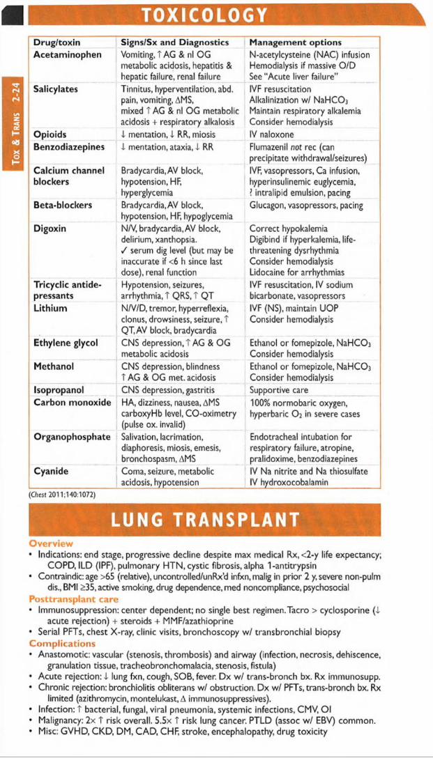
Tox
& T
rans
2-
24TOXICOLOGY
Drug/toxinAcetaminophen
Signs/Sx and DiagnosticsVomiting, t AG & nl OG metabolic acidosis, hepatitis & hepatic failure, renal failure
Management optionsN-acetylcysteine (NAC) infusion Hemodialysis if massive O/D See "Acute liver failure”
Salicylates Tinnitus, hyperventilation, abd. pain, vomiting, AMS, mixed T AG & nl OG metabolic acidosis + respiratory alkalosis
IVF resuscitation Alkalinization w/ NaHCOj Maintain respiratory alkalemia Consider hemodialysis
Opioids 4 mentation, 4 RR. miosis IV naloxoneBenzodiazepines 4 mentation, ataxia, 4 RR Flumazenil not rec (can
precipitate withdrawal/seizures)Calcium channel blockers
Bradycardia,AV block, hypotension, HF, hyperglycemia
IVF, vasopressors, Ca infusion, hyperinsulinemic euglycemia,? intralipid emulsion, pacing
Beta-blockers Bradycardia, AV block, hypotension, HF, hypoglycemia
Glucagon, vasopressors, pacing
Digoxin N/V, bradycardia, AV block, delirium, xanthopsia./ serum dig level (but may be inaccurate if <6 h since last dose), renal function
Correct hypokalemia Digibind if hyperkalemia, life- threatening dysrhythmia Consider hemodialysis Lidocaine for arrhythmias
Tricyclic antidepressants
Hypotension, seizures, arrhythmia, T QRS, T QT
IVF resuscitation, IV sodium bicarbonate, vasopressors
Lithium N/V/D, tremor, hyperreflexia, clonus, drowsiness, seizure, T QT, AV block, bradycardia
IVF (NS), maintain UOP Consider hemodialysis
Ethylene glycol CNS depression, T AG & OG metabolic acidosis
Ethanol or fomepizole, NaHCOi Consider hemodialysis
Methanol CNS depression, blindness T AG & OG met. acidosis
Ethanol or fomepizole, NaHC0 3
Consider hemodialysisIsopropanol CNS depression, gastritis Supportive careCarbon monoxide HA, dizziness, nausea, AMS
carboxyHb level, CO-oximetry (pulse ox. invalid)
100% normobaric oxygen, hyperbaric O2 in severe cases
Organophosphate Salivation, lacrimation, diaphoresis, miosis, emesis, bronchospasm, AMS
Endotracheal intubation for respiratory failure, atropine, pralidoxime, benzodiazepines
Cyanide Coma, seizure, metabolic acidosis, hypotension
IV Na nitrite and Na thiosulfate IV hydroxocobalamin
(Chest 2011:140:1072)
LUNG TRANSPLANTOverview• Indications: end stage, progressive decline despite max medical Rx, <2-y life expectancy;
COPD, ILD (IPF), pulmonary HTN, cystic fibrosis, alpha 1-antitrypsin• Contraindic: age >65 (relative), uncontrolled/unRx’d infxn, malig in prior 2 y, severe non-pulm
dis., BMI >35, active smoking, drug dependence, med noncompliance, psychosocialPosttransplant care• Immunosuppression: center dependent; no single best regimen.Tacro > cyclosporine (4
acute rejection) + steroids + MMF/azathioprine• Serial PFTs, chest X-ray, clinic visits, bronchoscopy w/ transbronchial biopsyComplications• Anastomotic: vascular (stenosis, thrombosis) and airway (infection, necrosis, dehiscence,
granulation tissue, tracheobronchomalacia, stenosis, fistula)• Acute rejection: 4 lung fxn, cough, SOB, fever. Dx w / trans-bronch bx. Rx immunosupp.• Chronic rejection: bronchiolitis obliterans w/ obstruction. Dx w/ PFTs, trans-bronch bx. Rx
l im ite d (a z ith ro m y c in , m o n te lu k a s t, A im m u n o su p p re ss ive s ).
• Infection: T bacterial, fungal, viral pneumonia, systemic infections, CMV, Ol• Malignancy: 2x T risk overall. 5.5x t risk lung cancer. PTLD (assoc w/ EBV) common.• Misc: GVHD, CKD, DM, CAD, CHF, stroke, encephalopathy, drug toxicity
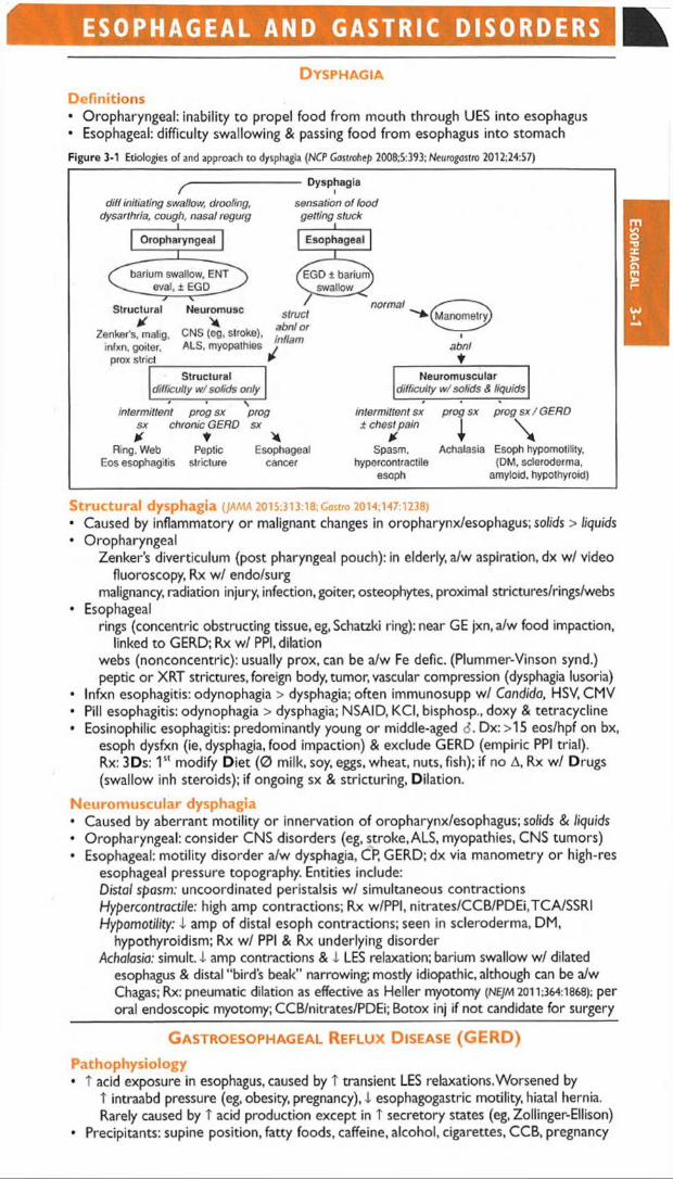
E S O P H A G E A L A N D GASTRI C D I S O R D E R S
D y s p h a g i a
Definitions• Oropharyngeal; inability to propel food from mouth through UES into esophagus• Esophageal; difficulty swallowing & passing food from esophagus into stomach
Figure 3-1 Etiologies of and approach to dysphagia (NCP Gostrohep 2008:5:393; Nct/rogostro 2012:24:57)
/ ---------------diff initiating swallow, drooling,
dysarthria, cough, nasal regurg
Dysphagia
sensation of food getting stuck
intermittent prog sx prog sx chronic GERD sx X t X
Ring, Web Peptic EsophagealEos esophagilis stricture cancer
intermittent sx ± chest pain
* Spasm,
hypcrcontractile esoph
prog sx prog sx / GERD
i \Achalasia Esoph hypomotility,
(DM, scleroderma, amyloid, hypothyroid)
Structural dysphagia (JAMA 2015:313:18; Castro 2014:147:1238)
• Caused by inflammatory or malignant changes in oropharynx/esophagus; solids > liquids• Oropharyngeal
Zenker’s diverticulum (post pharyngeal pouch): in elderly, a/w aspiration, dx w/ video fluoroscopy, Rx w / endo/surg
malignancy, radiation injury, infection, goiter, osteophytes, proximal strictures/rings/webs• Esophageal
rings (concentric obstructing tissue, eg, Schatzki ring): near GE jxn,a/w food impaction, linked to GERD; Rx w/ PPI, dilation
webs (nonconcentric): usually prox, can be a/w Fe defic. (Plummer-Vinson synd.) peptic o r XRT strictures, foreign body, tumor, vascular compression (dysphagia lusoria)
• Infxn esophagitis: odynophagia > dysphagia; often immunosupp w/ Condido, HSV, CMV• Pill esophagitis: odynophagia > dysphagia; NSAID, KCI, bisphosp., doxy & tetracycline• Eosinophilic esophagitis: predominantly young or middle-aged 6. Dx: >15 eos/hpf on bx,
esoph dysfxn (ie, dysphagia, food impaction) & exclude GERD (empiric PPI trial).Rx: 3Ds: I s' modify D iet (0 milk, soy, eggs, wheat, nuts, fish); if no A, Rx w/ Drugs (swallow inh steroids); if ongoing sx & stricturing, Dilation.
Neurom uscular dysphagia• Caused by aberrant motility o r innervation of oropharynx/esophagus; solids & liquids• Oropharyngeal: consider CNS disorders (eg, stroke, ALS, myopathies, CNS tumors)• Esophageal: motility disorder a/w dysphagia, CP, GERD; dx via manometry o r high-res
esophageal pressure topography. Entities include:Distal spasm: uncoordinated peristalsis w / simultaneous contractions Hypercontractile; high amp contractions; Rx w/PPI, nitrates/CCB/PDEi,TCA/SSRI Hypomotility: I amp of distal esoph contractions; seen in scleroderma, DM,
hypothyroidism; Rx w/ PPI & Rx underlying disorder Achalasia: simult.4- amp contractions & 1 LES relaxation; barium swallow w/ dilated
esophagus & distal "bird s beak” narrowing; mostly idiopathic, although can be a/w Chagas; Rx: pneumatic dilation as effective as Heller myotomy (NE/M 2011:364:1868); per oral endoscopic myotomy; CCB/nitrates/PDEi; Botox inj if not candidate for surgery * •
G a s t r o e s o p h a g e a l R e f l u x D is e a s e (GERD)P a th o p h y s io lo g y♦ T acid exposure in esophagus, caused by T transient LES relaxations. Worsened by
T intraabd pressure (eg, obesity, pregnancy), I esophagogastric motility, hiatal hernia. Rarely caused by T acid production except in T secretory states (eg, Zollinger-Ellison)
• Precipitants: supine position, fatty foods, caffeine, alcohol, cigarettes, CCB, pregnancy
Eso
pha
gea
l

Ga
st
ric
3-
2Clinical manifestations• Esophageal: h e a rtb u rn , atypical chest pain, regurgitation, water brash, dysphagia• Extraesophageal: cough, asthma (often poorly controlled), laryngitis, dental erosions
Diagnosis (AnnoJs 201S;163:ITC1)
• Clinical diagnosis based on sx and response to empiric trial of PPI (“ PPI test” )• EGD: if (1) 0 response to PPI; or if (2) alarm features: dysphagia, vomiting, i wt, anemia• If dx uncertain & EGD nl -> esoph manometry w/ 24-h esoph pH monitoring ± impedance
Treatm ent (Loncet 2013;381:1933)
• Lifestyle: avoid precipitants, lose weight, avoid large & late meals, elevate head o f bed• Medical: PPI achieve relief in 80-90% of Pts; H2 blockers for interm ittent sx• Refractory: confirm w/ pH testing (on PPI to assess need for T Rx, or off PPI to verify dx).
If acidic or sx correlate w/ reflux episodes: surgical fundoplication (emerging Rx: LES sphincter augmentation w/ radiofrequency, implantable magnetic or electrical devices)
If nl pH or no sx correlation: Dx esoph hypersensitivity. Rx w /TCA, SSRI or baclofen.
Complications (NEJM 2014;371:836;Gastro 2011; 140:1084.e 18 & 2015:149:1599)
• Reflux esophagitis (erosions/ulcers above GE jxn), strictures (caused by chronic inflamm)• Barrett’s esophagus (BE; NEJM 2014:371:836): metaplastic columnar mucosa above GE jxn
replaces squam esoph epithelium (dx via EGD/bx)Screen if GERD w/ >1 risk factor for esophageal adeno.: >50 y, d, white, hiatal hernia,
central adiposity, smoking. 0.1 -0.3%/y risk of esoph adenocarcinoma, T if T dysplasia.Management: PPI. W /o dysplasia: surveillance EGD no sooner than q3-5y (limited data
on utility of EGD screening in nondysplastic BE). Low-grade dysplasia: EGD q6-12mo; potential benefit of endoscopic eradication, eg RFA (JAMA 2014:311:1209).High-grade dysplasia: endoscopic eradication (resection or ablation treatment). * •
P e p t ic U lc er D isea se ( P U D )
D efin ition & etiologies (Lonctt 2009:374:1449)• Ulcers (break in mucosal lining >5 mm) & erosions (<5 mm) in stomach and duodenum• Principal risk factors: H. pylori infection > NSAID/ASA use• H. pylori infection: causes -80% of duodenal ulcers (DU) & - 60% of gastric ulcers (GU).
-50% of world colonized w/ H. pylori, but only 5-10% will develop PUD.• ASA & NSAIDs: damage to mucosa caused by i prostaglandin synthesis. Cause majority
of non-H. py/or/'-related DU & GU. Regular use a/w 5-6x t odds of GIB.• Other: smoking, stress, excessive EtOH, gastric cancer/lymphoma, Crohn’s, viral infxn (eg,
CMV/HSV in immunosupp), bisphosphonates, steroids (in combo w/ NSAIDs, but not risk factor alone); rarely gastrinoma (Zollinger-Ellison synd.), mastocytosis, idiopathic
• Stress ulcer: risk factors = ICU & coagulopathic, mech vent, h/o GIB, steroid use; Rx w/ PPI
Clinical manifestations• Epigastric abdom inal pain: relieved with food (DU) o r worsened by food (GU)• Complications: UGIB, perforation & penetration, gastric outlet obstruction
Diagnostic studies• Testing for H. pylori: stool antigen or EGD + rapid urease test False 9 if on abx, bismuth,
PPI, so stop prior to testing if possible. Serology: J. utility, useful only to exclude infection in low prevalence areas (most of U.S.).
• EGD (definitive dx): if fail empiric Rx or alarm features (see above); bx GU to r/o malig & H.pylori; relook in 6-12 wk if >2 cm, malig features, risk factors for gastric cancer (ie,® FHx, © H. pylori, atrophic gastritis, dysplasia/ metaplasia on bx, > 50 y.o.), or sx persist
Trea tm en t (NEJM 2010:362:1597; Cut 2012:61:646; EM) 2013;347:f4587)• I f H. pylori ©, eradicate (“ test and treat” ) (Gastro 2016:151:51):
Triple Rx: clarith + [amox, MNZ or levoflox] + PPI bid x 10-14 d (if clarith resist rate <20%) Quadruple Rx: MNZ +TCN + bismuth + PPI (if clarith resist rate >15% or amox allergy)
erad vs. triple 93 vs. 70%, clarith sens 95 vs. 85%, resist 91 vs. 8% (Lancet 2011:377:905) Sequential Rx: PPI + amox x 7 d -> PPI + clarith + M NZ x 7 d (Lancet 2013:381:205) Besides PUD, test & Rx if: gastric MALT lymphoma, atrophic gastritis, FHx gastric ca
• If H. pylori G: gastric acid suppression w/ PPI• Lifestyle changes: d/c smoking and probably EtOH; diet does not seem to play a role• Surgery: if refractory to med Rx (1st r/o NSAID use) o r for complications (see above)
Prophylaxis ifA S A /N S A ID required 0ACC 2016:67:1661; A/iment PhormRx 2016:43:1262)• PPI if (a) h/o PUD/UGIB; (b) also on clopidogrel (although ? I antipit effect); (c) >2 of
the following: age >60, steroids or dyspepsia; prior to start test & Rx H. pylori• Consider misoprostol; consider H2RA if ASA monotherapy (Lancet 2009:374:119)• Consider A to COX-2 inhibit (1 PUD & UGIB but T CV events) if low CV risk & not on ASA

GASTROINTESTINAL BLEEDINGD e f in i t io n• Intraluminal blood loss anywhere from the oropharynx to the anus• Classification: upper = above the ligament ofTreitz; lower = below the ligament of Treitz• “Severe” GIB: defined as having associated shock orthostatic hypotension, 4 Hct by 6% (or
4 Hb by 2 g/dL), or requiring transfusion >2U PRBCs. Requires hospitalization.
C l in ic a l m a n i f e s ta t io n s• Hem atem esis = blood in vomitus (UGIB)• Coffee-ground emesis = blood exposed to gastric acid (UGIB)• Melena = black, tarry stools from digested blood (usually UGIB, but can be from R colon)• Hem atochezia = bloody or maroon-colored stools (LGIB or rapid UGIB)
I n i t ia l m a n a g e m e n t• Assess severity: VS including orthostatic As.JVP. Tachycardia (can be masked by (5B
use) suggests 10% volume loss, orthostatic hypotension 20% loss, shock >30% loss• History: prior GIB, tempo of current bleed, specific bleeding manifestations (see above),
other Gl sis (eg. abd pain, A in bowel habits, weight loss, N/V), NSAID/ASA or EtOH use, anticoag/antiplt drugs, h/o or risk factors for cirrhosis, radiation, prior Gl or aortic surgery.
• Physical exam: Realizable abd tenderness, peritoneal signs, masses, LAN, prior surgerysigns of liver disease (hepatosplenomegaly, ascites, jaundice, telangiectasias) rectal exam: masses, hemorrhoids, anal fissures, stool appearance, color, occult blood
• Resuscitation: placement of 2 large-bore (18-gauge or larger) intravenous linesVolume replacement: NS or LR to achieve normal VS, UOP, & mental status
• Lab studies: H ct (may be normal in first 24 h of acute GIB before equilibration)2-3% -> 500 mL blood loss; low MCV -> Fe deficient and chronic blood loss; pit, PT( PTT; BUN/Cr (ratio >36 in UGIB b/c Gl resorption of blood ± prerenal azotemia); LFTs
• Transfuse: BB sample for type & cross; use O-neg if emerg;for UGIB (esp. w/ portal HTN)transfuse w/ more restrictive Hb goal (eg, 7 g/dL) or >8 g/dL if CAD (nejm 2013:368:11)
• Reverse coagulopathy: FFP & vit K to normalize PT; pits to keep count >50,000• Triage: alert endoscopist. Consider ICU if unstable VS or poor end organ perfusion.
Intubation for emergent EGD, if ongoing hematemesis, shock, poor resp status, A MS > OutPt management if SBP >110, HR <100, Hb >13 (d) o r >12 ($ ), BUN <18,
0 melena, syncope, heart failure, liver disease (Lancet 2009:373:42)
D ia g n o s t ic s tu d ie s• Nasogastric tube can aid localization: fresh blood or coffee grounds -> active or recent
UGIB; nonbloody -> does not exclude UGIB (-15% missed).© occult blood test no value.• UGIB: EGD w/in 24 h. If severe bleed, T Dx/Rx yield by gastric lavage and erythro 250 mg
IV 30 min prior to endoscopy to clear stomach contents [AmjGauro 2006:101:1211).• LGIB: colonoscopy (identifies cause in >70%); if severe, colo w/in 12 h -> consider rapid
purge w/ PEG solution (6-8 L over 4-6 h). If hematochezia a/w orthostasis, concern for brisk UGIB -> exclude UGIB wl EGD first Push enteroscopy, anoscopy, capsule endoscopy in combo w/ urgent colo results in dx >95% of cases (Gf Emfo 2015:81:889).
• Imaging: if too unstable for endo or recurrent bleeding, can then -> IR procedure or surgerytagged RBC scan: can identify general luminal location if bleeding rate >0.04 mL/min arteriography: can localize exact vessel if bleeding rates >0.5 mL/min, allov/s for IR Rx
• Emergent exploratory laparotomy (last resort) if no localization and life-threatening bleed
Etiology UGIB Comment & Treatment
PUD (20-67%)(NE/M 2016:374:2367)
See "PUD"
Treatment: PPI: 80 mg IV bolus + 8 mg/h drip = 40 mg IV BID boluses Endoscopic therapy: epi inj + bipolar cautery or hemodip. Biopsies for ? H. pylori and treat if ©.
High-risk (for rebleeding) ulcer: arterial spurting, adherent clot, visible vessel. Endo Rx, IV PPI x 72 h post EGD, then A to high-dose oral PPI. Arteriography w/ embolization; surgery (last resort).
Intermediate-risk ulcer: oozing, in o/w stable Pt. Endo Rx, can A to oral PPI after EGD and observe 24-48 h.
Low-risk ulcer: dean-based or flat. Oral PPI and ? discharge.Hold anticoag & antiplatelet Rx until hemostasis; can resume after
hemostasis & PPI on board (8M/2012;344:e3412).
Erosivegastropathy(4-31%)
Precipitants: NSAIDs, ASA, EtOH, cocaine, gut ischemia, XRT Stress-related mucosal injury in ICU Pts. Risk factors include severe
coagulopathy, mech vent >48 h, high dose glucocorticoids Treatment: high-dose PPI
Erosive esophagitis (5-18%)
Risk factors: cirrhosis, anticoagulation, critical illness. Rx offending cause + high dose PPI; repeat EGD later to r/o underling Barrett’s.

GIB
3-
4Esophageal or gastric varices(4-20%)(He/) 2007:46:922; NEJM 2010:362:823) See “ Cirrhosis”
2° to portal HTN. If isolated gastric r/o splenic vein thrombosis. PharmacologicOctreotide 50 jig IV bolus -> 50 jig/h infusion (84% success).
Usually x 5 d, but most benefit w/in 24-48 h.Abx: 20% cirrhotics p/w GIB have infxn, & -50% develop infxn
during hospitalization; Ppx w/ IV CTX, cipro, or levoflox x 7 d NonphormacologicEndoscopic band ligation (>90% success) or sclerotherapy Arteriography w / coiling, or if available, endoscopic injection of
cyanoacrylate (glue) for gastric varices Covered esophageal stent placement or balloon tamponade used
for bleeding refractory to ligation as bridge to TIPS (consider early if persistent bleed on EGD or Child-Pugh C; NEJM 2010:362:2370)
For persistent gastric variceal bleed:TIPS or balloon-retrograde transvenous obliteration
Portal HTN gastropathy
T portal venous pressure -> ectatic vessels, hyperemia in prox. gastric body. No endoscopic option; Rx portal HTN (octreotide), [5B.
Angioectasia AVMs.HHT (see below)
AVMs congenital. Angioectasia (ectatic submucosal vessels) a/w t age, CKD, cirrhosis, CTD, severe CV dis. Heyde syndrome: GIB d/t angioectasias + aortic stenosis. Endo Rx,
04L.
Dieulafoy’slesion
Large (1-3 mm) submucosal artery protruding through fundal mucosa -» sudden, massive UGIB. Difficult to identify. Endo Rx.
□u
£
Gastric antral vase, ectasia (GAVE)
“ Watermelon stomach” ; ectatic gastric vessels, often a/w cirrhosis, CTD, typically older 9. Rx w/ thermal hemostasis, repeat q4-8wk to eradicate lesions.TIPS does not improve outcomes.
Aortoentericfistula
AAA or aortic graft erodes into 3rd portion of duodenum.P/w "herald bleed"; if suspected, diagnose by endoscopy or CT.
Malignancy (2-8%) Endoscopic hemostasis of mass temporizing measure till cancer RxMallory-Weiss tear (4-12%)
GE jxn lacerations due to vomiting -> T intraabd pressure & shearing effect. Can self-resolve w/o endo Rx. Rx w/ antiemetics, PPL
Cameron’s lesions Linear erosions in hiatal hernia due to mech trauma of diaphragmPost-sphincterotomy bleeding
Occurs in -2% of cases, T risk w/ more complicated procedure. Bleeding into duodenum. Rx w/ endo hemostasis.
(Cl Endosc Clin NAm 2015;2S:41S)
Etiology LGIB Comment & Treatment (/toij Gostro 2015:110:1265 & 2016:111:755)
Diverticular bleed (30%)
Pothophys/o/ogy; Intimal thickening and medial thinning of vasa recta as they course over dome of diverticulum -> weakening of vascular wall -> arterial rupture. Diverticula more common in left colon; but bleeding diverticula more often in right colon.
Clinical: older,ASA/NSAIDs, painless hematochezia,! abd cramping Treatment: Usually stops spont. (-75%) but may take hrs-days;
-20% recur. Can perform endo hemostasis w / epi injections ± electrocautery (NEJM 2000:342:78), hemodip, banding. Intra-arterial vasopressin or embo. Surgery (partial colectomy) last resort.
Polyp/Tumor (20%) Typically slow ooze, p/w fatigue, weight loss, iron deficiency anemiaColitis (20%) Infectious (see "Acute Diarrhea” ), IBD, ischemic colitis, XRTAnorectal disorders (20%)
Internal, external hemorrhoids; anal fissures, rectal ulcers, rectal varices (Rx by i portal venous pressure in cirrhotics), XRT
Vascular (<10%) Angioectasia & AVMs (see above). Hereditary Hemorrhagic Telangiectasia (Webcr-Osler-Rendu): diffuse AVMs, telangiectasias throughout Gl mucosa (also involve lips, oral mucosa, fingertips).
Meckel’sdiverticulum
Congenital blind intestinal pouch due to incomplete obliteration of vitelline duct. 2% of pop, w/in T of 1C valve, 2” long, c?:9 2:1, often present age 2 (but can cause obscure GIB in adults). Dx w/ ""T c- pertechnetate scintigraphy. Rx w/ angioembo, surgical resection.
Obscure GIB (Gostro 2007:133:1694; CIE 2010:72:471)• D e fin ition : continued bleeding (melena, hematochezia) despite 0 EGD & colo; 5% of GIB• Etiologies: Dieulafoy’s lesion, GAVE, small bowel angiodysplasia, ulcer or cancer, Crohn’s
disease, aortoenteric fistula, Meckel’s diverticulum, hemobilia• Diagnosis: repeat EGD w/ push enteroscopy/colonoscopy when bleeding is active
If 0 t video capsule to evaluate small intestine (Gostro 2009;137:1197)
If still ©.consider " mTc-pertechnetate scan (“ Meckel’s scan” ), enteroscopy (singleballoon, double-balloon o r spiral), tagged RBC scan and arteriography

DIARRHEAA cute D iarrhea (<4 weeks' duration)
Acute Infectious Etiologies (ne/m 2 0 14;370:1532;;ama 20i5;3i3:7i)
Pathogen Epidem iology & Clinical SxNoninflammatory Predom. disruption small intestine absorp. & secretion.
Voluminous diarrhea, N/V. © fecal W BC & FOB.Preformed toxin "Food poisoning," <24 h dur. S. aureus (meats & dairy),
B. cereus (fried rice), C. perfringens (rewarmed meats).Viral Rotavirus Outbreak person to person (PTP), daycare; lasts 4-8 d.
Norovirus -50% of all diarrhea.Winter outbreaks; PTP & food/water; no immunity. Lasts 1-3 d.Vomiting prominent.
Bacterial E. coli (toxigenic) >50% of traveler’s diarrhea; cholera-like toxin; <7 d.Vibrio cholerae (Lancet 2012:379:2466)
Contam H2O, fish, shellfish; 50 cases/y in U.S. Gulf Coast. Severe dehydration & electrolyte depletion.
Parasitic Giardia Streams/outdoor sports, travel, outbreaks. Bloating. Acute (profuse, watery) -> chronic (greasy, malodorous).
(± malab for mos
Cryptosporidia (NEJM 2002:346:1723)
Water-borne outbreak; typically self-limited, can cause chronic infxn if immunosupp. Abd pain (80%), fever (40%).
after Rx) Cyc/ospora Contaminated produceInflammatory Predom. colonic invasion. Small vol diarrhea. LLQ
cramps, tenesmus, fever, typically © fecal WBC or FOB.Bacterial Campylobacter Undercooked poultry, unpasteurized milk, travel to Asia;
carried by puppies & kittens. Prodrome; abd pain -* "pseudoappendicitis” ; c/b GBS, reactive arthritis.
Salmonella(nontyphoidal)
Eggs, poultry, milk. Bacteremia in 5-10%. 10-33% of bacteremic Pts >50 y may develop aortitis.
Shigella Abrupt onset; gross blood & pus in stool; T1 WBC.£. coli (0157:H7 & inv/hemorrhagic non-0157:H7)
Undercooked beef, unpasteurized milk, raw produce; PTP. 0157 & non-0157 sp. (40%) produce Shiga toxin -» HUS (typically in children). Gross blood in stool.
C. difficile See laterVibrio parahaem. Undercooked seafoodSalmonella typhi Travel to Asia. Systemic toxicity, relative bradycardia, rose
spot rash, ileus -> pea-soup diarrhea, bacteremia.Other Yersinia: undercooked pork; unpasteurized milk, abd pain
-> "pseudoappendicitis’’ (aka mesenteric adenitis) Aeromonos, Plesiomonos, Listeria (meats & cheeses)
Parasitic E. histolytica Contaminated food/water, travel (rare in U.S.); liver abscessViral CMV lmmunosuppressed;dx by shell vial cx of colon bx
E v a lu a t io n (NEJM 2014;370:1532)
• History: stool freq, bloody, abd pain, duration of sxs [~1 wl< for viral & bacterial(except C. diff)t > 1 wk for parasitic], travel, food, recent abx, immunocompromise
• PEx: vol depletion (VS, UOP, axillae, skin turgor, MS), fever, abd tenderness, ileus, rash• Laboratory: / fecal WBC (high false © & ©) or stool lactoferrin & calprotectin (PMN
products; Se/Sp >90%), stool cx, BCx, lytes, C. diff (if recent hosp/abx), stool O&P (if >10 d, travel to endemic area, exposure to unpurified H2O, community outbreak, daycare, HIV © or MSM);± stool ELISAs (viruses, Crypto, Giardia), serologies (£ histolytica)
• Imaging/endoscopy warranted if warning signs: fever, signific abd pain, blood or pus instools, >6 stools/d, severe dehydration, immunosupp, elderly, duration >7 d, hosp- acquired, CT/KUB if ? toxic megacolon; sig/colo if immunosupp or cx ©
T r e a t m e n t {Am J Gastro 2016;111:602)
• If none of the above warning signs and Pt able to take POs -> supportive Rx only:oral hydration, loperamide, bismuth subsalicylate (avoid anticholinergics)
• If moderate dehydration: 50-200 mL/kg/d of oral solution (V2 tsp salt, 1 tsp baking soda,8 tsp sugar, & 8 oz OJ diluted to 1 L w/ H2O) or Gatorade, etc. If severe: IV fluids.
• Fluoroquinolone or rifaximin if high suspicion for traveler’s diarrhea• If high suspicion for protozoal infection can consider metronidazole o r nitazoxanide• Empiric abx for non-hospital-acquired inflammatory diarrhea reasonable: FQ x 5-7 d
abx rec for Shigella, cholera, Giardia, amebiasis, Salmonella if Pt >50 y or immunosupp or hospitalized, ? Campylobacter (if w/in 4 d of sx onset)
avoid abx if suspect E. coli 0157:H7 as may T risk of HUS

Dia
rr
hea
3-
6C lostridium D ifficile-Associated D iarrhea (CDAD)
P a th o g e n e s is & e p id e m io lo g y (nejm 2015.372:825)• Ingestion of C. diff spores -> colonization when colonic flora Ad by abx or chemo release
of toxin A/B -> colonic mucosal necrosis & inflammation -» pseudomembranes• Most frequently reported nosocomial infxn; community-acquired infxn may account for up
to 1/3 of new cases. Associated w/ all abx (esp. P-lactams,dinda, quinolones).• Additional risk factors: elderly, nursing home residents, IBD, PPI (CfD 201 1:53:1173)C l in ic a l m a n i f e s ta t io n s (a s p e c tru m o f disease)• Asx colonization: <3% healthy adults; -20% in hospitalized patients on antibiotics• Acute watery diarrhea (occ bloody) ± mucus, often w / lower abd pain, fever, TIT WBC• Pseudomembranous colitis: above sx + pseudomembranes + bowel wall thickening• Fulminant colitis (2-3%): to x ic m egaco lon (colon dilatation >6 cm on KUB, colonic
atony, systemic toxicity) and/or bowel perforation
D ia g n o s is• Only test if symptomatic (diarrhea, s/s of colitis); test liquid stool (unless concern for ileus)• Stool El A: detects toxin B and/or A (1-2% strains make A); fast (2-6 h); © result highly Sp• Stool PCR: has T Se, but © if colonized in absence of active CDAD; should not necessarily
Rx if © PCR w/ © neg toxin assay (JAMA im 20is-.l75;i792)• Consider flex sig if dx uncertain and/or evidence of no improvement w / standard Rx
T r e a t m e n t (NE/M 2015:372:1539;yAMA 2015:313:398)• If possible d/c abx ASAP; stop antimotility agents• Non-severe: vanco 125 mg PO q6h or MNZ 500 mg PO q8h x 10-14 d; equal cure
rates, but MNZ less well tolerated• Severe (any of the following: >12 BM/d,Temp >103°F,WBC >25, HoTN, ICU care
required, ileus): vanco 125 mg PO q6h + MNZ 500 mg IV q8h• If worsening (ileus, T WBC, t lactate, shock, toxic megacolon, peritonitis): abd CT & urgent
surgical consult re: subtotal colectomy (> possible role for diverting loop ileostomy or colonic lavage); may also consider vanco PR
• If Pt needs to continue on abx, continue C. diff. Rx for >7 d post-abx cessation• Stool carriage may persist 3-6 wk postcessation of sx & should not trigger further Rx
(retesting for C. diff of limited utility during this time)• R ecu rren t in fe c tio n : 15-30% risk after d/c of abx, most w/in 2 wk of stopping abx
1st recurrence: vanco 125 mg PO q6h x 10-14 d or fidaxomicin 200 mg PO bid x 10 d Subsequent recurrences: vanco PO pulse -» taper. Consult ID physician. Consider fecal
microbial transplant (NEJM 2013:368:407 & JAMA 2016:315:142) or fidaxomicin (200 mg bid x 10 d). Pilot data for oral admin of nontoxigenic C. d/ff strain spores (JAMA 2015:313:1719).
• Probiotics w/o clear benefit (Lancet 2013:382:1249) * •
C h r o n ic D ia r r h e a (>4 wk;jAMA2oi6;3i5:27i2)
General evaluation• Clinically can be organized into watery, fatty, o r inflammatory stools• Additional hx: timing (freq, relation to meals; nocturnal diarrhea a/w organic causes like IBD
rather than IBS), abd pain, w t loss, prior surg, chemo/XRT, diet (incl caffeine or poorly absorbed carbs/sugars), infectious sxs, immunocompromise, travel, laxative use, etc.
• Hx offending meds: PPI, colchicine, abx, H2RA, SSRIs, ARBs, NSAIDs, chemo, caffeine• PEx:gen appearance (BMI), signs of systemic disease, surgical scars, rectal tone/DRE• Lab testing: CBC, metabolic profile, alb.TSH, Fe studies, ESR; see under each category• Imaging/endoscopy: colonoscopy for chronic diarrhea of unknown cause. Abd CT/MRI
usually warranted if systemic problem suspected.
Osm otic (watery; 0 fecal fat, T osmotic gap, 1 diarrhea with fasting)• Caused by ingestion of poorly absorbed cations/anions (Mg, sulfate, phos; found in
laxatives) or poorly absorbed sugars (eg, mannitol, sorbitol; found in chewing gum; or lactose if lactose intolerant). Diarrhea resolves w/ cessot/on o f offending substance.
• Dx: T stool osmotic gap (see Figure); stool pH <6 if unabsorbed carbohydrates• Lactose intolerance (75% nonwhites & 25% whites lactase-deficient): can be acquired
after gastroenteritis, med illness, Gl surg. Clin: bloating, flatulence, discomfort, diarrhea. Dx: H+ breath test or empiric lactose-free diet. Rx: lactose-free diet & lactase tablets.
Secretory (watery; normal osmotic gap, no A diarrhea w/ fasting, often nocturnal diarrhea)• Caused by secretion of anions or K+ into lumen or inhib of Na absorption -> T H2O in stool.
Most commonly caused by bacterial toxins from infxn (see above). Other causes:• Endocrine: Addison s, VIPoma, carcinoid, Zollinger-Ellison, mastocytosis, hyperthyroid (T
motility). / serum peptide levels (eg, gastrin, calcitonin,VIP) & urinary histamine.• Gl neoplasm: carcinoma, lymphoma, villous adenoma

• Microscopic colitis: common cause of chronic diarrhea w/ obscure origin. Often seen inmiddle-aged women w/ autoimmune disorders. NSAIDs, SSRIs, PPIs notable triggers. Grossly nl on colo but bx shows lymphocytic & plasmacytic infiltration of mucosa ± thickened submucosal collagen. Rx: antidiarrheals, cholestyramine, bismuth, budesonide; consider anti-TNFs if refractory.
• Bile acid-induced diarrhea: ileal resection o r disease (eg Crohn’s), etc. bile acids incolon -> electrolyte & HjO secretion. Rx w/ empiric bile-acid binders (eg, cholestyramine).
Fxnal/IBS (watery; normal osmotic gap, I diarrhea with fasting): see Dysmotility
Malabsorption (fatty; T fecal fat, t osmotic gap, I diarrhea w/ fasting)• Defective mucosal absorption of nutrients b/c As in: mucosal surface (surgical resection) or
gen. mucosal dis. (celiac, IBD). Bloating, foul-smelling, floating stools (steatorrhea).• Celiac disease (NEJM 2012:367:2419; Gostro 2015;148:117S)
Immune rxn in genetically predisposed Pts (-1% pop) to gliadin, a component of gluten (wheat protein) -> small bowel inflammatory infiltrate -> impaired absorption
Other s/s: Fe/folate defic anemia; osteoporosis; dermatitis herpetiformis; T AST/ALT Dx: IgA anti-tissue transglutaminase Ab (most Se), IgA anti-deaminated gliadin peptide
Ab; IgA a-endomysial Ab. Duodenal bx to confirm dx (blunted villi, crypt hyperplasia, inflamm infiltrate) but may not be necessary if serology 0 and Pt sx. HLA-DQ2/Q8 testing useful for high © predictive value if © serologies already on gluten-free diet.
Rx: gluten-free diet; 7-30% do not respond to diet -» ? wrong dx o r noncompliant Complic:~5% refractory sx, risk ofT-cell lymphoma and small bowel adenocarcinoma
• W h ipp le ’s disease: infxn w/ T. whipplei (NEJM 2007:365:55)Other s/s: fever, LAN, edema, arthritis, CNS As, gray-brown skin pigmentation, Al &
MS, oculomasticatory myorhythmia (eye oscillations + mastication muscle contract) Rx: (PCN + streptomycin) or 3rd-gen ceph x 10-14 d -» Bactrim for >1 y
• Small intestinal bacterial overgrowth (SI BO): colonic bacteria in SI -> steatorrhea,B12/Fe defic, protein-losing enteropathy. A/w dysmotility (DM neuropathy, scleroderma), A’d anatomy (Crohn's, surgery, fistulae), immune deficiency, celiac, CF Dx w/ H+ or 14C- xylose breath testing or empiric abx. Rx w/ 7-10 d abx (eg, rifaximin, MNZ, FQ).
• Other: s/p short bowel resection (short bowel syndrome), chronic mesenteric ischemia,eosinophilic gastroenteritis, intestinal lymphoma, tropical sprue, Giardia infection
Maldigestion (fatty; T fecal fat, t osmotic gap, I diarrhea w / fasting)• Defective intraluminal hydrolysis of nutrients, typ. 2/2 pancreatic/hepatobiliary pathology• Pancreatic insufficiency: most commonly from chronic pancreatitis or pancreatic cancer.
Test w/ stool elastase, chymotrypsin levels, or empiric pancreatic enzyme replacement• J, bile acids due to i synthesis (cirrhosis), cholestasis (PBC).or s/p ileal resection.Test w /
empiric bile acid replacement therapy.
In flam m atory (0 fecal WBC or lactoferrin or calprotectin, © FOB, fever, abd pain)• Infections: chronic C. diff Entamoeba histolytica, Yersinia, CMV,TB especially in
immunocompromised hosts. CMV, C. diff notorious for causing exacerbations of IBD.• In flam m atory bowel disease (Crohn’s, UC)• Radiation enteritis, ischemic colitis, neoplasia (colon cancer, lymphoma)
Figure 3-2 Workup of chronic diarrhea
Chronic diarrhea
- © fe c a l fa t ■
Inflammatory
^ stool cx colonoscopy
© FOB © W BC - © la c to fe r r in ■
© c a lp o te c tin
Malabsorption or Maldigestion
Based on hx: s tissue transglut. Abs, stool elastase, 14C-xylose & IT breath tests,
EGDw/bx, EUS
stool osmotic gap = Osm5.00, (usu 290) - (2 x (Nastoo, + Kst00|)]\
<100 2100
s CT scan, colonoscopy, hormone levels
s IT breath test or empiric lactose-free diet, laxative screen

Dysm
otil
ity
3-8
iFunctional Gl Disease• Recurrent Gl sx caused by abnl gut-brain interactions rather than structural cause• >20 types of FGIDs per Rome III criteria; now Rome IV (Castro 2016; 150:1257)
• Irritab le Bowel Syndrome (IBS) (jam a 2015;313:949)
Abd discomfort a/w >2 of following: improve w/ defecation, A stool frequency, A stool form IBS-C (constipation predominant) vs. IBS-D (diarrhea predominant) vs. IBS-M (mixed) vs.
IBS-U (unclassified). Sx may be related to stress, diet, lifestyle, possibly microbiome. Treatm ent: exercise, cognitive behavioral Rx, A diet, probiotics, ? peppermint oil
IBS-C: laxatives (eg, lubiprostone, linaclotide, PEG), biofeedback IBS-D: rifaximin or loperamide; eluxadoline, p & k agonist, 8 antag (NEJM 2016:374:242)
• Cyclical Vomiting Syndrome: stereotypic episodes of acute recurrent vomiting;a/w marijuana use, family hx of migraineacute Rx: antiemetics, IVF, sumatriptan, BZDs; prevention:TCAs/AEDs; avoid marijuana
Gastroparesis (Gost/o Oinks ofNA 2015;44:1; World] Gastro 2015:21:6842)• Delayed gastric emptying w/o obstruction, typically p/w nausea (>90%), vomiting (>80%),
early satiety (60%), postprandial fullness/pain• Etiol: DM, post-surg, thyroid disease, critical illness, Parkinson’s, opiates, CCB, anti-cholin• Dx: gastric emptying scintigraphy• Treatment: prokinetic agents (metoclopramide or erythromycin), antiemetics for sx; feeding
tube if refractory; intrapyloric botox & gastric stimulator experimental
Acute colonic pseudo-obstruction (Ogilvie’s syndrome; ANZJ Surg 2015:85:728)• Definition: loss of intestinal peristalsis in absence of mechanical obstruction• Abd discomfort & distention, i / absent bowel sounds, ± N/V, hiccups• Typically in elderly, hospitalized, ill Pts, precipitated by: intra-abd process (surgery,
pancreatitis, peritonitis, intestinal ischemia), severe medical illness (eg, sepsis), meds (opiates, CCB, anticholinergics), metab/endo abnl (thyroid, DM, kidney failure, liver failure), spinal cord compression/trauma, neurologic d/o (Parkinson’s, Alzheimer’s, MS)
• KUB or CT w/ colonic dilatation w/o mech obstruction; cecal diam >14 cm a/w high risk perf• Treatm ent: conservative measures (NPO, avoid offending meds) usually effective;
IV neostigmine (monitor for bradycardia), methylnaltrexone; bowel decompression w/ NGT, rectal tube, colonoscopy; if refractory, colostomy or colectomy
Constipation (Annals 2015;162:ITC1)• Defined as dissatisfaction w/ defecation or (per Rome III): >2 of following during last 3 mo
>25% of the time: straining, lumpy/hard stools, incomplete evacuation, sensation of anorectal obstruction, manual maneuvers to facilitate defecation, stool frequency <3/wk
• Secondary etiologies (4 M’s)Mech obstruction: malignancy, compression, rectocele, strictures Meds: opioids,TCAs, anticholinergics, CCB, NSAIDs, diuretics, Ca2f, Fe Metabolic/endo: DM, hypothyroid, uremia, preg, panhypopit, porphyria, T Ca, 1 K, i Mg Myopathy/Neuro: Parkinson’s, Hirschsprung’s, amyloid, MS, spinal injury, dysautonomia
• Dx: H&P w/ DRE. Labs: consider CBC, electrolytes w/ Ca,TSH. Colonoscopy if alarm sx.Anorectal manometry/balloon expulsion test; colonic transit study; defecography.
• Treatm ent: diet change w / t fluid intake, fiber supplementationBulk laxatives (psyllium, methylcellulose.polycarbophil): t colonic residue, T peristalsis Osmotic laxatives (Mg,sodium phosphate [avoid if CKD], lactulose):? water in colon Stimulant laxatives (senna,castor oil, bisacodyl, docusate sodium);? motility & secretion Enema/suppository (phosphate, mineral oil, tap water, soapsuds, bisacodyl) Lubiprostone (? secretion); methylnaltrexone and alvimopan for opioid-induced Linaclotide ? stool freq, i straining/bloating (NEJM 2011:365:6:527)
N utrition in critical illness (see “Mech Ventilation" as well) (nejai 2014:370:1227)• In theory, enteral superior to parenteral as maintains integrity and function of Gl tract,
however, both routes with similar outcomes (NEJM 2014:371:1673)
• Enteral (EN): start w/in 24-^48 hr of ICU admission tends to 1 infxn & mort. Contraindic. ifobstruction, major GIB. Complications: ischemic bowel injury due to ? demand for splanchnic blood flow, esp. if hemodynamically unstable; aspiration PNA (possibly I risk if jejunal feeds but conflicting data), nasopharyngeal ulceration/bleeding/pain to due tube.
• Parenteral (PN): start after 7 d if unable to tolerate enteral feeds, or ? sooner (jama
2013:309:2130); late (>day 8 of ICU stay) initiation of PN to supplement insufficient EN I infxn & time on vent (NEJM 2011:365:506). Contraindic: hyperosmolality, severe electrolyte disturbances, severe hyperglycemia; sepsis is relative contraindication. Complications: hyperglycemia (due to dextrose), catheter sepsis/thrombus, refeeding syndrome, LFT abnl (steatosis, cholestasis, gallbladder sludge due to lack of enteric stimulation.
1

DISORDERS OF THE COLOND iverticulosis
Definition & pathophysiology (Lancet 2004;363:631)
• Acquired herniations of colonic mucosa & submucosa in areas where vasa recta penetrate• Thought to occur in setting of abnormal motility and T intraluminal pressure
Epidemiology• Risk factors: i fiber, T red meat, obesity, smoking, physical inactivity, EtOH, NSAIDs• Prevalence higher w /1 age (10% if <40 y; 50-66% if >80 y);“ Westernized” societies• Left side (90%, mostly sigmoid) > R side of colon (except in Asia where 75-85% R-sided)
Clinical manifestations• Usually asx, but 5-15% develop diverticular hemorrhage (see “ GIB” ) and <5% diverticulitis• For asx diverticulosis, limited data for t fiber diet or avoiding nuts/seeds (JAMA 2008:300:907)
D iverticulitis
Pathophysiology (N EJM 2007;357:2057; Gostroenfero; 2015:147:1944)
• Retention of undigested food and bacteria in diverticulum -* * fecalith formation ->obstruction -» compromise of diverticulum’s blood supply, infection, perforation
• Uncomplicated: microperforation -> localized infection• Complicated (15%): macroperf -* abscess, peritonitis,fistula (65% w/ bladder), obstrxn
Clinical manifestations• LLQ abdominal pain, fever, nausea, vomiting, constipation or diarrhea• PEx ranges from LLQ tenderness ± palpable mass to peritoneal signs & septic shock• Ddx includes IBD, infectious colitis, PID, tubal pregnancy, cystitis, colorectal cancer
Diagnostic studies• Plain abdominal radiographs to r/o free air, ileus o r obstruction• Abdominal C T (l+0 +):>95% Se & Sp; assess complicated disease (abscess, fistula)• Colonoscopy contraindicated acutely T risk of perforation; do 6 wk after to r/o neoplasm
Treatm ent (JAMA 2014;311:287; Dis Colon Rectum 2014;57:284)
• Mild: outPt Rx indicated if Pt has few comorbidities and can tolerate POsPO abx: (MNZ + FQ) or amox/clav for 7-10 d; liquid diet until clinical improvement Possible that abx not be needed for uncomplicated diverticulitis (Cochrane CD009092)
• Severe: inPt Rx if cannot take POs, narcotics needed for pain, or complicationsNPO, IVF, NGT (if ileus)IV abx (GNR & anaerobic coverage; eg, CTX/M NZ or pip-tazo)
• Abscesses >4 cm should be drained percutaneously o r surgically• Surgery: if progression despite med Rx, undrainable abscess, free perforation
Resection superior to laparoscopic lavage (jama 2015:314:1364)After source control, 4 d abx may be sufficient (NEJM 2015:372:1996)Resection for recurrent bouts of diverticulitis on a case by case basisConsider lower threshold for urgent & elective surgery for immunocompromised Pts
Prevention• Mesalamine ± rifaximin may provide sx relief in chronic/recurrent dis. (Dig Dis Sa 2007:52:2934)• Risk of recurrence 10-30% w/in 10 y of 1st episode; more likely 2nd episode complicated
Polyps & A denomas
Pathophysiology & Epidemiology (NEJM 2016:374:1065)
• Accumulation of mutations in colonic epithelial cell DNA affecting oncogenes & tumorsuppressor genes -» tumor initiation (formation of adenoma; APC loss of fxn) -> tumor progression (adenoma -> carcinoma; K-ras gain of fxn, DCC, p53 loss of fxn).
• Risk factors: T age, FHx (sporadic in 1° relatives, Lynch, FAP), IBD, T dietary fat, centraladiposity, T EtOH, -I fiber, T red meat, ? smoking, DM
• Protective factors: T physical activity, ASA/NSAIDs, Ca2+ intake, HRT, i BMI; possibly Tfiber, vitamin D, fish oil, statins, selenium
• Neoplastic polyps: adenomas (tubular, villous, tubulovillous dysplasia), sessile serratedadenomas/polyps (concern for interval CRC), carcinomas.
• Nonneoplastic polyps: hyperplastic, juvenile, Peutz-Jeghers, inflammatory
Detection• Colonoscopy is gold standard• Recommended in all Pts starting at age 50 and then typically qlOy unless pathology found• If © FHx, start age 40, or 10 y before age of dx in youngest family member, repeat q5y

IBD
3-
10
Definition• U lcerative colitis (UC): inflammation of the colonic mucosa; contiguous, starting at rectum• C rohn’s disease (CD): transmural inflammation occurring anywhere in Gl tract, skip areas
Epidem & pathophys (NEJA1 2009:361:2066; Castro 2011:140:1785; Lancet 2016:387:156)
• 1.4 million people in U.S.; prev 1:1000 UC & 1:3000 CD; T incidence in Caucasians, Jews• Age of onset 15-30 y in UC and CD; CD is bimodal and has second peak at 50-70 y• Smokers at T risk for CD, whereas nonsmokers & former smokers at T risk for UC• Genetic predisposition + environmental risk factors -»T cell dysregulation -» inflammation
U l c e r a t i v e C o l i t i s (ne/m 201 i;36S :i7 i3 ;Lonce t20 i2 ;380:i606)
Clinical manifestations• G rossly b loody d ia rrhea , lower abdominal cramps, urgency, tenesmus• Extracolonic (>25%): erythema nodosum, pyoderma gangrenosum, aphthous ulcers,
uveitis, episcleritis, thromboembolic events (esp. during a flare; Lancet 2010:375:657), AIHA, seroneg arthritis, chronic hepatitis, cirrhosis, PSC (T risk cholangio CA, CRC)
Diagnosis• Colonoscopy: involves rectum (95%) & extends proximally and contiguously v/ithin colon• Classify by location: proctitis (25-55%), left-sided colitis (50-70%) and pancolitis (20%)• Appearance: granular, friable mucosa with diffuse ulceration; pseudopolyps• Histology: superficial chronic inflammation; crypt abscesses & architectural distortion• Barium enema with featureless and tubular appearance of colon (leadpipe appearance)• Flares: T ESR & CRP (not Se o r Sp); © fecal calprotectin (Se 77%, Sp 71%)
Complications• Toxic m egaco lon (5%): colon dilatation (> 6 cm on KUB), colonic atony, systemic
toxicity, & T risk of perf. Rx w/ IV steroids & broad-spectrum abx; surgery if needed.• Stricture (5%): occurs in rectosigmoid after repeated episodes of inflammation• CRC and dysplasia (see be/ow)• For patients s/p surgery w/ ileal pouch, may develop pouchitis (inflammation of ileal pouch,
up to V2 of pts), Rx w/ abx (MNZ, cipro), probiotics
Prognosis• 50% of Pts in remission at any given time; interm ittent exacerbations in 90%; continual
active disease in -18%. Rate of colectomy at 10 y is 24%.• Mortality rate of severe UC flare is <2%, & overall life expectancy in UC = non-UC Pts •
C r o h n ' s D ise ase (toncet20i2;380:i590)
Clinical manifestations• Abdom inal pain, loose/frequent stools (up to 50% © FOBT), fever, malaise, w t loss• Mucus-containing, nongrossly bloody diarrhea• N/V, bloating, obstipation if presence of obstruction; extracolonic manifestations as in UC
Diagnosis• lleocolonoscopy + bx is gold standard; small bowel imaging (eg MR-enterography -
91% accuracy in identifying Crohn’s compared to endoscopy); capsule endoscopy• Classify by location: small bowel (47%), ileocolonic (21%), colonic (28%); upper tract rare• Montreal classification: age at dx, disease location & behavior (stricturing vs. nonstricturing,
penetrating vs. nonpenetrating), plus modifiers for upper tract & perianal disease• Appearance: nonfriable mucosa, cobblestoning, aphthous ulcers, deep & long fissures• Histology: transmural inflammation with mononuclear cell infiltrate, noncaseating
granulomas (seen in <25% of mucosal biopsies), fibrosis, ulcers, fissures• Track disease severity w / Crohn’s Disease Activity Index (CDAI) questionnaireComplications• Perianal disease: fissures, fistulas, skin tags, perirectal abscesses (in 24% of Pts; perianal
disease precedes intestinal symptomatology)• Stricture: small bowel,postprandial abd pain; can lead to complete SBO & require surgery• Fistulas: perianal, enteroenteric, rectovaginal, enterovesicular, enterocutaneous• Abscess: fever, tender abd mass, T WBC; steroids mask sx, need high-level suspicion• Malabsorption: ileal disease/resection:! bile acids abs -> gallstones; I fatty acid abs
Ca oxalate kidney stones; i fat soluble vitamin abs vit D deficiency osteopeniaPrognosis• Variable at 1 y: -50% in remission, -20% flare, -20% low activity, -10% chronic active• A t 20 y, majority will have required some surgery; overall life expectancy is slightly i

M a n a g e m e n t (Gasm>2oi3;i45:i464)
In it ia l eva luation• H &P ( / for intestinal & extraintestinal manifestations) and d iagnostic studies as above• Lab: consider CBC/diff, LFTs, iron studies, B12, folate, vit D. Fecal calprotectin & lactoferrin
have higher Se & Sp than ESR & CRP.• Exclude o th e r etio logies: infectious/ischemic colitis, intestinal lymphoma/carcinoma,
CRC, IBS, vasculitis, Behcet’s, celiac disease, small intestinal bacterial overgrowth• Rule o u t in fection (esp. CMV) before treating with immunosuppressants and biologies
Goals o f tre a tm e n t• Induce remission of acute flare -> maintain remission; mucosal healing 1° goal• Convention is step up Rx (least -» most toxic). Early combined immunosuppression Rx not
yet widely adopted; consider if severe disease (Lancet 2015:386:1825).
Medical Therapy fo r IBD (in stepwise sequence)
U lcerative colitisMild 5-ASA: many formulations (sulfasalazine, mesalamine, olsalazine,
balsalazide) depending on disease location. Used to induce remission & for maintenance. Complications: diarrhea, abd pain, pancreatitis.
Mild-Moderate
MMX-budesonide: oral formulation of budesonide released throughout entire colon for flare. 1st pass metab 4 systemic adverse effects of steroid.
Moderate-Severe
PO prednisone: 40-60 mg w/ taper over several wks to induce remission AZA/6-MP: 0.5-1 mg/kg and uptitrate over several wks for maintenance.
Complic: BM suppression, lymphoma, pancreatitis, hepatitis; /TP M T levels prior to dosing to 4 risk of generation of toxic metabolites. In selected cases can add allopurinol to boost activity in non-responders.
Severe o r re fractory disease
IV steroids: eg, 100 mg hydrocort q8h or 16-20 mg methylpred q8h to induce remission w/ plan to taper & switch to non-steroid maintenance.
Cyclosporine: for severe flares refractory to steroids, 2-4 mg/kg infusion x 7 d w/ goal to A to maintenance medication (eg.AZA/6-MP)
A n ti-TN F (infliximab, adalimumab & golimumab): 15-20% remission rates (Gostro 2012:142:257). For steroid-refractory flares or to maintain remission. Complic: reactivation TB ( / PPD prior to Rx); exclude viral hepatitis; small t ‘d risk NF1L; infusion & lupus-like rxn, psoriasis, MS, CHF.
Vedolizumab (see below)Investigational: tofacitinib (janus kinase inhib; NEjM 2012:367:616), fecal
transplant (Gastro 2015:149:102)
Crohn’s diseaseMild 5-ASA: Sulfasalazine 4-6 g/d may be useful in inducing remission
Abx: FQ/MNZ or amox/clav for pyogenic complic (fistulas, perineal dis.)M ild-mod Budesonide: oral formulation able to reach ileumModerate-severe
PO prednisone: same as UC.for inducing remission, not maintenance AZA/6-MP: same as UC.for maintenanceM TX: 15-25 mg IM/SC or PO qwk for maintenance; 1-2 mo to take effect
Severe o r re fractory disease
A nti-TN F : infliximab, adalimumab or certolizumab (pegylated)If flare on infliximab, / trough & presence of anti-inflixi Ab. Low & © Ab
T dose/freq. If © Ab A to Other biologic (Am) Gostro 2011:106:685).
Vedolizumab (anti-a.4f}7 integrin) and ustekinumab (anti-IL 12/23) if
refractory to anti-TNFs SMAD7 anti-sense oligonucleotide (NE/M 2015:372:1104) under study
Surgery• UC: colectomy if sx refractory to or intolerable side effects from meds, CRC, perforation,
toxic megacolon, uncontrolled hemorrhage. Often ileal pouch-anal anastomosis (IPAA).• CD: resection if refractory disease; endoscopic dilation o r surgery for strictures; diverting
ileostomy for perineal diseaseC ancer screening (NEJM 2015:372:1441)
• C olon cancer: risk in UC -2% at 10 y, ~8% at 20 y,-18% at 30 y. Similar for colonicCD, plus risk of small bowel cancer as well. Dysplasia best marker for risk. Other risk factors include: PSC,® FHx, greater extent of disease, stricture, & pseudopolyps.
• Surveillance: colonoscopy w/ random bx 8 y after dx to eval for dysplasia, q1-3ythereafter based on risk factors. Chromoendoscopy using dye to stain high-risk lesions for targeted biopsy is emerging technique. If high-grade dysplasia or dysplasia assoc, lesion/mass -» colectomy. Chemoprophylaxis: 5-ASA & ursodeoxycholic acid (if PSC)? beneficial (4/G2011;106:731:A/lmentPf7ormocoJT)er2012;35:451).
IBD
3-11
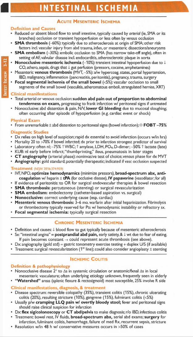
Inte
st I
sch
em
3-12
INTESTINAL ISCHEMIAA cute Mesenteric Ischemia
Definition and Causes• Reduced or absent blood flow to small intestine, typically caused by arterial (ie, SMA or its
branches) occlusion o r transient hypoperfusion or less often by venous occlusion• SM A throm bosis ( -60%): typically due to atherosclerosis at origin of SMA; other risk
factors ind. vascular injury from abd trauma, infxn, or mesenteric dissections/aneurysms• SM A em bolism (~30%): embolic occlusion to SMA (has narrow take-off angle), often in
setting of AF, valvular disease incl. endocarditis, atherosclerotic plaque in aorta• Nonocclusive m esenteric ischemia (-10%): transient intestinal hypoperfusion due to i
CO, athero, sepsis, drugs that I gut perfusion (pressors, cocaine, amphetamines)• Mesenteric venous throm bosis (MVT, ~5%): a/w hypercoag. states, portal hypertension,
IBD, malignancy, inflammation (pancreatitis, peritonitis), pregnancy, trauma, surgery• Focal segmental ischemia o f the small bowel (<5%): vascular occlusion to small
segments of the small bowel (vasculitis, atheromatous emboli, strangulated hernias, XRT)
Clinical manifestations• Total arterial o r venous occlusion: sudden abd pain o u t o f p ro p o rtio n to abdom ina l
tenderness on exam , progressing to frank infarction w/ peritoneal signs if untreated• Nonocclusive: abd distention & pain, N/V, low er G l bleeding due to mucosal sloughing;
often occurring after episode of hypoperfusion (e.g. cardiac event or shock)
Physical Exam• From unremarkable ± abd distention to peritoneal signs (bowel infarction);© FO B T -75%
Diagnostic Studies• Dx relies on high level of suspicion; rapid dx essential to avoid infarction (occurs w/in hrs)• Mortality 20 to >70% if bowel infarcted; dx prior to infarction strongest predictor of survival• Laboratory: often nl; -75% T WBC; T amylase, LDH, PO4, D-dimer; -50% T lactate (late)• KUB: nl early before infarct;“ thumbprinting,” ileus, pneumatosis in later stages• C T ang iography (arterial phase): noninvasive test of choice; venous phase for dx MVT• Angiography: gold standard; potentially therapeutic; indicated if vase occlusion suspected
Treatm ent (NE}M 2016:374:959)• IVF, NPO, op tim ize hem odynam ics (minimize pressors), broad-spectrum abx, an ti
coagulation w/ heparin ± tPA (for occlusive disease), IV papaverine (vasodilator; for all)• If evidence of peritonitis: to OR for surgical endovascular therapies & bowel resection• SM A th rom bos is : percutaneous (stenting) o r surgical revascularization• SM A em bo lism : embolectomy (catheter-based aspiration vs. surgical)• N onocclusive : correct underlying cause (esp. cardiac)• Mesenteric venous thrombosis: 3-6 mo. warfarin after initial heparinization. Fibrinolysis
or thrombectomy typically reserved for Pts w/ hemodynamic instability or refractory sx.• Focal segm enta l ischem ia: typically surgical resection
Chronic Mesenteric Ischemia
• Definition and causes: I blood flow to gut typically because of mesenteric atherosclerosis• Sx: "intestinal angina” = postprandia l abd pain, early satiety, & I w t due to fear of eating.
If pain becomes constant could represent acute thrombosis (see above).• Dx: angiography (gold std) « gastric tonometry exercise testing + duplex U/S (if available)• Treatment: surgical revascularization (1“ line); could also consider angioplasty ± stenting * •
Ischemic Colitis
D e fin itio n & pathophys io logy• Nonocclusive disease 2° to As in systemic circulation or anatomic/fxnal As in local
mesenteric vasculature; often underlying etiology unknown, frequently seen in elderly• “ W atershed” areas (splenic flexure & rectosigmoid) most susceptible, 25% involve R side
C lin ica l m an ifes ta tions , diagnosis, & tre a tm e n t• Disease spectrum: reversible colopathy (35%), transient colitis (15%), chronic ulcerating
colitis (20%), resulting stricture (10%), gangrene (15%), fulminant colitis (<5%)• Usually p/w cram ping LLQ pain w / overtly bloody stool; fever and peritoneal signs
should raise clinical suspicion for infarction• Dx:flex sig/colonoscopy or C T abd/pelvis to make diagnosis; r/o IBD, infectious colitis• Treatment: bowel rest, IV fluids, b road-spectrum abx, serial abd exams; surgery for
infarction, fulminant colitis, hemorrhage, failure of med Rx, recurrent sepsis, stricture• Resolution w/in 48 h w/ conservative measures occurs in >50% of cases

PANCREATITIS kA cute Pancreatitis (AP)
Pathogenesis• Pancreatic duct and acinar injury via direct or indirect toxicity -» impaired secretion and
premature activation of digestive enzymes -» autodigestion and acute inflammation
Etiologies (Loncet 2015;386:8S)• Gallstones (40%): 9 > d, usually small stones (<5 mm) or microlithiasis/sludge• Alcohol (30%): 6 > 9 , 1st attack after —10 y heavy use; usually chronic w/ acute flares• Anatomic: divisum, annular pancreas, duodenal duplication cysts, Sphincter of Oddi dysfxn• Autoimmune: can p/w chronic disease, pane mass or pane duct strictures, T lgG4, © ANA• Drugs: 5-ASA, 6-MP/AZA, ACEI, cytosine, didanosine, dapsone, estrogen, furosemide,
isoniazid, metronidazole, pentamidine, statins, sulfa, thiazides, tetracycline, valproate• Familial: a/w mutations in PRSS1, CFTR, SPINK1; suspect if early onset (age <20 y)• Infections: ascariasis, clonorchiasis, coxsackie, CMV, HIV, mumps, mycoplasma,TB, toxo• Ischemia: vasculitis, cholesterol emboli, hypovolemic shock, cardiopulmonary bypass• Metabolic: hypertriglyceridemia (TG >1000; type I and V familial hyperlipemia), hyperCa• Neoplastic: panc/ampullary tumors, mets (RCC most common, breast, lung, melanoma)• Post ERCP (5%): Ppx w/ PR indomethacin (NEJM 2012:366:1414), pane duct stent if high risk• Post trauma: blunt abdominal trauma, pancreatic/biliary surgery• Toxins: organophosphates, scorpion toxin, methanol
Clinical manifestations• Epigastric abdominal pain (90%), only 50% p/w classic bandlike pain radiating to back• 10% pain-free (due to analgesic/steroid use, immunosuppressed, AMS, ICU, post-op),
/ amylase/lipase in Pts w / unexplained shock (Am J Gastro 1991:86:322).• Nausea and vomiting (90%)• Abdominal tenderness/guarding, 4 bowel sounds (ileus), jaundice if biliary obstruction• Signs of retroperitoneal hemorrhage (Cullen's = periumbilical; Grey Turner’s = flank) rare• Ddx: acute cholecystitis, perforated viscus, SBO, mesenteric ischemia, IMI, AAA leak,
distal aortic dissection, ruptured ectopic pregnancyDiagnostic studies• Dx requires 2 of 3: characteristic abd pain; lipase or amylase >3x ULN; © imaging• Laboratory (Am J Gastro 2013:108:1400)
levels o f both amylase and lipase do not correlate w / severity of disease t amylase: >3x ULN >90% sensitive, >70% specific for acute pancreatitis
false ©: acute on chronic (eg, alcoholic); hypertriglyceridemia (4 amylase activity) false ©: other abd or salivary gland process, acidemia, renal failure, macroamylasemia
T lipase: >3x ULN 99% sensitive, 99% specific for acute pancreatitis false ©: renal failure, other abd process, diabetic ketoacidosis, HIV, macrolipasemia longer half-life than amylase: useful in Pts w/ delayed presentation after onset of sx lipase >10,000 has 80% PPV for biliary dx,99% NPV for EtOH (D/g D/s Sc/ 2011:56:3376)
ALT >3x ULN has 95% PPV for gallstone pancreatitis [Am]Gastro 1994;89:1863)• Imaging studies (AmJ Gastro 2013:108:1400)
Abd U/S: typically not useful to visualize pancreas (obscured by bowel gas) but should be ordered for all pts with AP to r/o biliary etiology (ie, gallstones, BD dilatation)
Abd CT: not rec for initial eval unless dx unclear (local complic. not yet visible & concern for AKI w/ IV contrast). However, if persistent pain and/or clinical deterioration after 48- 72 h,CT(l+) useful to r/o local complications (necrosis, fluid collections).
MRI/MRCP:Can detect necrosis; also used to assess for stones & ductal disruption Endoscopic U/S (EUS): limited role; useful for occult biliary disease (microlithiasis)
Severity (Am J Gastro 2009:104:710)• Severity defined by presence of organ failure (AKI, resp failure, GIB, shock) & local or
systemic complic. (pane necrosis, fluid collections, gastric outlet obstrxn, splenic & PVT). Mild: 80% of cases. No organ failure o r local/systemic complications, low mortality. Moderate: transient (<48 h) organ failure ± local/systemic complications, high morbidity Severe: persistent (>48 h) organ failure, very high mortality
Prognosis• Scoring systems (Crit Care Med 1999:27:2272; Am ] Gastro 2009; 104:966)
Ranson’s/APACHE II: earliest scoring systems predicting severity at 48 h using multiple physiological criteria; may have poor PPV for severe AP
BISAP: simple 5-point scoring system (BUN >25, impaired MS, SIRS, age >60, pleural effusion) used w/in first 24 h; score >3 predicts t risk of organ failure, mortality
CTSI: uses CT findings at 48-72h (fluid collections, necrosis) to predict mortality• O ther criteria: SIRS >48 h, rising BUN/Hct, obesity, comorbid disease predict T mortality
Pa
nc
rea
titis 3-13

Treatm ent (CBn C a s tro H e p o to l 2011;9;710; A m J C a s tro 2012; 107:1146; N E JM 2014;370:150)• Fluid resuscitation: early aggressive IVF, titrate to UOP >0.5 mL/kg/h, goal to 1 BUN &
Hct over first 12-24 h. LR may be superior to NS (! SIRS, CRP at 24 h; avoid if t Ca)• N utrition (Clin Castro Hepatol 2007;S:946; Intern Med 2012:51:523; Crit Care 2013;17:R118)
Early enteral feeding encouraged (maintains gut barrier, i bacterial translocation) though new data suggest may not be superior to oral feeding at 72 h (N E JM 2014:317:1983)
Mild: Start feeding once pain-free w/o ileus. Low-fat low-residue diet as safe as liquid diet. Severe: early (w/in 48-72 h) enteral nutrition indicated and preferred overTPN b/c
! infectious complications, organ failure, surgical interventions, and mortality. Nasogastric feeding shown to be non-inferior to nasojejunal feeding
• Analgesia: IV opioids (monitor respiratory status, adjust dosing if T renal impairment)• Gallstone pancreatitis: urgent (w/in 24 h) ERCP w/ sphincterotomy if cholangitis, sepsis,
orTbili >5. For mild disease, CCY during initial hosp to I risk of recurrence (Lancet 2015:386:1261); defer surgery if necrotizing AP until improvement in inflam., fluid collections.
• Hypertriglyceridem ia: insulin gtt (activates lipoprotein lipase), fibrates, ± apheresis• No role for ppx abx in absence of infectious complications (World; Gastroenterol 2012:18:279)
Complications• Systemic: ARDS, abdominal compartment syndrome, AKI, GIB (pseudoaneurysm), DIC• Metabolic: hypocalcemia, hyperglycemia, hypertriglyceridemia• Fluid collections:
Acute fluid collection: seen early, no t encapsulated, most resolve w /in 1-2 w k w /o Rx Pseudocyst: -4 wk after initial attack, encapsulated. No need fo r Rx if asx (regardless of
size/location). If sx -> endoscopic (Gostro 2013:145:583) vs. perc/surg drainage.• Pancreatic necrosis: Nonviable pancreatic tissue. CT-guided FNA if infection suspected
Sterile necrosis: if asx, can be managed expectantly, no role for ppx antibiotics Infected necrosis (5% of all cases, 30% of severe): high mortality. Rx w/ carbapenem or
MDZ+FQ."Step-up” Rx w/ perc drainage and minimally invasive surg debridement or endoscopic necrosectomy superior to open necrosectomy (NEJM 2010:362:1491)
Pancreatic abscess: circumscribed collection of pus (usually w/o pancreatic tissue), usually seen >4 wk into course. Rx with abx + drainage (CT-guided if possible).
C hronic Pancreatitis
Pathogenesis & Etiology• Often, but not always, recurrent acute attacks -> inflammatory infiltrate fibrosis ->
pancreatic insufficiency (need to lose 90% of pane fxn to develop DM, fat/protein malabs.)• Toxins (60-80% due to EtOH; smoking also important risk factor), idiopathic, genetic,
autoimmune, relapsing AP, obstruction
Clinical manifestations• Sxs include epigastric pain, N/V; over time will be painless and p/w steatorrhea and w t loss
Diagnostic studies• Labs: amylase/lipase t early, may be nl later. 0 fecal fat, 1 stool elastase & A1AT.
/ A le , consider lgG4/ANA & genetic testing (CFTR, SPINK 1.PRSS1) if young o r© FHx.• Imaging: Ca2' on KUB/CT. ERCP/MRCP/EUS high Sens for dx: stricture, dilated ducts
Treatm ent (Lancet 2016;387:1957)• Pancreatic enzyme replacement (may i pain by reducing CCK)• Pain control: smoking & EtOH cessation, analgesics, ESWL for duct stones, celiac nerve
plexus block, thoracoscopic splanchnicectomy, resection.
Complications• Pseudocysts, pseudoaneurysms, pancreatic ascites or pleural effusion,? risk o f pane Ca
A utoimmune PancreatitisPathogenesis• Lymphoplasmacytic sclerosing pancreatitis w / dense fibrosis and ? lgG4 (type 1), or
granulocytic epithelial lesions with minimal lgG4 cells (type 2)
Clinical Manifestations• A b d o m in a l pain, can p/w obstructive jaundice and pane mass mimicking pane Ca.• Extrapancreatic: Sjogren’s, interstitial nephritis, autoimmune thyroiditis, UC/PSC, RA
Diagnosis• Labs: cholestatic LFTs (T A0 > AST/ALT),? y-globulins and lgG4, © ANA, RF• HISORt criteria: Histology, Imaging (“ sausage pancreas” , bile duct stricture), Serology,
other Organ involvement, Response to therapy
Treatment• Corticosteroids 15t-line, immunomodulators (AZA, MMF, cyclophosphamide) if relapse

ABNORMAL LIVER TESTS l \Tests o f hep a to ce llu la r in ju ry o r cholestasis• Aminotransferases (AST,ALT): intracellular enzymes released 2° necrosis/inflammation
ALT more specific for liver than is AST (heart, skeletal muscle, kidney, brain, RBC/WBC) t levels seen w/ most types of hepatocellular injury; skeletal muse, injury, Ml (AST >ALT)
• Alkaline phosphatase (A0): enzyme bound in hepatic canalicular membrane.T levels seen w/ biliary obstrxn or intrahepatic cholestasisalso found in bone, intestines, kidney, placenta; confirm from liver w/: T GGT (or t 5'-NT).
• Bilirubin: product of heme metab (unconjugated,“ indirect” ) carried by alb to liverwhere taken up for conjugation (“ direct") to make soluble, then excreted into bile. ■ j T direct hyperbili seen with cholestasis, enzymatic disorders (eg, Dubin-Johnson, Rotor's) ]T indirect hyperbili seen with hemolysis, enzymatic disorders (eg, Crigler-Najjar, Gilbert’s) jaundice seen when bili >2.5 mg/dL (esp. in sclera or under tongue);
if hyperbili conjugated then T urine bilirubin
Tests o f hepa tic fu n c tio n• Albumin: marker for liver protein synthesis, 4 slowly in liver failure (tv2 -1 5 -1 8 d)• Prothrombin tim e (PT): depends on synthesis of coag factors by liver (except FVIII);
b/c tv2 o f some factors (eg,V,VII) is short, T PT can occur w/in hrs of liver dysfxn
Patterns of LFTsPattern ALT AST Ad BilirubinHepatocellular f t TT ±T ±T (direct)
Viral hepatitis, NASH Often ALT > AST ±T ±T (direct)Alcoholic hepatitis AST ALT * 2:1 ±T ±T (direct)Ischemic injury TTT TTT TT TT (direct)Wilson’s disease T T A<t>:Tbili < 4
Cholestatic ±T ±T TT TT (direct)Infiltrative near nl near nl TT ±TNonhepatic
Skeletal muscle injury AST » ALT nl nlBone disease nl nl T (w/ nl GGT) nlHemolysis nl nl nl T (indirect)
• R-value = ratio of ALT:A0 normalized to ULN for each = (ALT/ULN) + (Ad/ULN)R >5 suggests hepatocellular injury, <2 suggests cholestatic injury, 2-5 suggests mixed
Figure 3-3 Approach to abnormal liver tests with hepatocellular pattern
IViral
hepatitis
HAV, HBV, HCV, HDV, HEV, CMV, EBV, HSV, VZV
Hepatocellular injury(predom T AST & ALT ± 4 bili and A^)
viral markers autoAbs tox screen
I Iobesity, DM
hyperlipidemia
♦
hypotensionCHF
iAuto
immuneDrugs & Toxins
NAFLD Vascular
alcohol acetaminophen
meds, toxins
ischemiccongestiveBudd-Chiari
SOS
systemicdisease
Hereditary
hemochrom. al-AT defic.
Wilson’s celiac disease
* W orkup for acute enzyme elevation (often symptomatic)Severe ALT &AST elevation (>1000):
toxins (usu. acetaminophen) -> / tox screen, EtOH, acet. levels. Other toxins: INH, disulfiram, pyrazinamide, OTC/herbal, fenofibrate, niacin, amiodarone, MDMA.
ischemia (eg, sepsis, hypotension, Budd Chiari) / liver U/S w/ Doppler. Etiologies usually lead to T L D H ,u s u a lly ratio ALT:LDH <1.5 (vs. >1.5 w I toxins, viruses).
viruses (Hep A-E; HSV, CMV,VZV) -> / viral serologiesother (AIH, acute Wilson Disease, acute biliary obstrxn) -» seeALF & cirrhosis sections
Acute mild-moderate ALT & AST elevation: as above, think meds/toxins (see list at end of section), viruses, ischemia/vascular issues in hospitalized Pts, obstruction (if mixed picture), systemic disease (see “Workup for chronic enzyme elevation ” below)
• W orkup for chronic enzym e elevation (often asymptomatic)Screen for common causes: hep serologies, EtOH, liver U/S (? NAFLD, cirrhosis), meds
LFTs 3-15

LFTs
3-
16/ If suspect underlying systemic disease: iron studies (HFE);ANA,ASMA, Ig levels (AIH);
ceruloplasmin, urinary copper (Wilson); a. 1-AT (can cause liver dis even w/o lung involvement); celiac screening (rare cause of liver dis); thyroid studies; see "Cirrhosis"
If 0 evaluation -> lifestyle modification (w t loss, DM control) & repeat testing 3-6 mo If evidence of chronic liver disease or persistent lab abnl, consider liver bx
Figure 3-4 Approach to abnormal liver tests with cholestatic pattern
♦ W orkup for cholestatic pattern: / RUQ U/S to assess for ductal dilatation.If © (extrahepatic obstruction) Pt may need ERCP ± imaging (MRCP, CT) for dx/Rx If no dilatation on U/S -> / AMA (for PBC), viral serologies (Hep A-E, EBV, CMV); if
work-up negative, consider MRCP and liver bx. See offending med list below.
Figure 3-5 Approach to abnormal liver tests with infiltrative patcern
• W orkup for infiltrative pattern: / GGT level to ensure Gl source of A<t> elevation. If © (t GGT & T A<p) -» often imaging first step (RUQ U/S or CT; consider MRCP if these
studies negative); / SPEP (for amyloid), often need liver bx for definitive diagnosis.
Com m on medications that cause abnorm al liver tests (http://iivertox.nlm.nih.gov)
Hepatocellular Cholestatic Mixed
Acarbose Prednisone ACE inhibitors 6-MP Amox-ClavAcetaminophen Protease Anabolic OCP Azathioprine
Allopurinol Inhibitors Steroids Penicillins CarbamazepineAmiodarone Pyrazinamide Azathioprine Protease ClindamycinAzathioprine Risperidone Chlorpromazine Inhibitors MirtazapineClindamycin Statins Estrogens Sulfonamides Nitrofurantoin
Fibrates Sulfonamides Macrolides Terbinafine PenicillinsHydralazine Tamoxifen Methimazole Tricyclics Phenobarbital
Isoniazid Tetracyclines PhenytoinKetoconazole TNF-alpha ProteaseMethotrexate inhibitors InhibitorsMirtazapine Trazodone Sulfonamides
Nitrofurantoin Tricyclics Trazodone(Some) NSAIDs Valproic Acid Tricyclics
Phenytoin Valproic acidVerapamil

HEPATITIS MV iral
H epatitis A (ssRNA; 30-45% of acute viral hepatitis in U.S.)• Transmission: fecal-oral route; contaminated food, water, shellfish; daycare center outbreaks• Incubation: 2-6 wl<; no chronic carrier state• Sx: i appetite, malaise,fever, N/V, RUQ pain, jaundice; rarely fulminant (T w/ chronic HCV)• Diagnosis: acute hepatitis = © IgM anti-HAV; past exposure = © IgG anti-HAV (© IgM)• Rx for acute HAV: supportive care; refer to liver txplnt center if fulminant hepatitis• Postexposure ppx: age 1-40 y -> vaccine; age <1 y or >40 y, immunosupp, liver dis. -» Ig
H epatitis B (dsDNA; '-45% of acute viral hepatitis in U.S.; Loncet 2014:384:2053)• Transmission: blood (IVDU, transfusion), sexual, perinatal• Incubation: 6 w k-6 mo (mean 12-14 wk)• Acute infxn: 70% subclinical, 30% jaundice,<1 % fulminant hepatitis (up to 60% mortality)• Chronic infxn: HBsAg © >6 mo in <5% of adult-acquired (T if immunosupp), >90% of
perinatal;-40% chronic HBV -> cirrhosis ( t risk w/ HCV, HDV, o r HIV coinfxn, EtOH)• HCC: T risk if cirrhotic,® FHx HCC,African >20 y old,Asian 6 >40 y old or 9 >50 y old, or
>40 y old w/ T ALT ± HBV DNA >2000. Screen w/AFP & U/S q6mo.• Extrahepatic syndromes: PAN (<1%), membranous nephropathy, MPGN, arthritis• Serologic and virologic tests (see Anna/s 2014;161:58 for screening guidelines)
HBsAg: appears before sx;used to screen blood donors; persists >6 mo = chronic HBV HBeAg: evidence of viral replication and T infectivity IgM anti-HBc: 1st Ab to appear; indicates acute infection
window period = HBsAg becomes ©, anti-HBs not yet ©, anti-HBc only clue to infxn IgG anti-HBc: indicates previous (HBsAg 0 ) or ongoing (HBsAg ©) HBV infection anti-HBe: indicates waning viral replication, I infectivityanti-HBs: indicates resolution of acute disease & immunity (sole marker after vaccination) HBV DNA: presence in serum correlates w / active viral replication in liver
Diagnosis HbsAg anti-HBs anti-HBc HBeAg anti-HBe HBV DNA
Acute hepatitis ® i © IgM © © ©
Window period 0 © IgM ± ± ©
Recovery © © IgG © ± ©
Immunization 0 © 0 © © ©
Chronic hepatitisHBeAg ©
© 0 IgG © 0 ©
Chronic hepatitisHBeAg O
® © IgG © © ±*
+Precore mutant: HBeAg not made, but anti-HBe can develop due to x-reactivity w / HBeAg; a/w T HBV D N A
• Rx for acute HBV: supportive; hospitalize fo r A MS or T INR (liver transplant center);consider antiviral therapy if severe •
Phases o f Chronic HBV
Phase ALT(ULN*)
HBV DNA(lU/mL)
HBeAg Liver Histology(inflammation/fibrosis)
Progression to cirrhosis
Immune-tolerant Nl >106 ® Minimal <0.5%/yImmune-activeHBeAg ©
>2x >20k © Moderate-to-severe 2-5.5%/y
Inactive Nl <2k © Min necroinflam.; variable fibrosis
0.05%/y
Immune Reactivation; HBeAg © precore mutant
>2x >2k © Moderate-to-severe 8-10%/y
*ALT ULN <30 U/L fo r 6, <19 U/L fo r 9. Adapted from Hepatology 2016:63:261.
• Rx of chronic HBV: Rx in immune-active o r immune reactivation phases or cirrhotics w/elevated HBV DNA or decomp. Consider liver bx if ALT 1-2x ULN or in immune- tolerant phase if age >40 y; Rx if mod-to-severe inflammation o r fibrosis on bx.
• Entecavir or tenofovir: nudeo(s/t)ide analogs, well tolerated, low resistance; at 5 y,HBeAg seroconversion is 30—40% & loss of HBsAg is 5-10% (Castro 2012:142:1360; Lancet 2013:381:468).Tenofovir preferred if h/o lamivudine resistance.
• PEG IFN-a2a: A t 2 y, HBeAg seroconversion is 27%; contraindicated if autoimmunedisease, uncontrolled psych disorder, seizures, decompensated cirrhosis
Hepa
titis 3-17

• Rx duration: (1) HBeAg © immune-active w/o cirrhosis: if seroconversion (HBeAg ©,anti-HBe ©), can stop after 1 y if ALT nl & HBV DNA suppressed or until HBsAg clears;(2) HBeAg ©immune reactivation: indefinite; (3) cirrhotic: indefinite
• If undergo liver transplant: HBIG + nucleo(s/t)ide analogue effective in preventing reinfection• HIV/HBV coinfection: Rx w / 2 drugs active against both HBV & HIV (nejm 2007:356:1445)• Immunosuppression: prior to initiating chemoRx, anti-TNF, steroids {>20 mg/d > 1 mo),
screen Pts for HBV; Rx if moderate to high risk of reactivation (Castro 2015:148:215)• Postexposure (risk infxn -30%) ppx: HBIG -> vaccine (if unvac or known nonresponder)
H e p a titis C (ssRNA;-10% of acute viral hepatitis in U.S.; Lancet 2015:385:1124)• Transmission: blood (IVDU, transfusion rare cause) > sexual; 20-30% w/o clear precipitant• Incubation: 1-5 mo; mean 6-7 wk• Acute infxn: 80% subdinical; 10-20% sx hepatitis w/ jaundice; fulminant hepatitis rare; prob
of spent clearance a/w IL28B & HLA class II genotypes (Annals 2013:158:235)
• Chronic: up to 85% chronic hepatitis, 20-30% of whom develop cirrhosis (after -20 yT risk of cirrhosis in men, EtOH, HIV; HCC in 1^4% of cirrhotics/y
• Extrahepatic syndromes: mixed cryoglobulinemia, porphyria cutanea tarda, lichen planus,leukocytoclastic vasculitis, thyroiditis, MPGN, IPF, NHL and monoclonal gammopathies
• Serologic, virologic, & genetic testsanti-HCV (ELISA):© in 6 wk, does not = recovery or immunity; can be © after recovery HCV RNA: © w/in 2 wk, marker of active infectionHCV genotype (1-6):guides duration & predicts response to Rx;geno. 3 a/w T risk HCC
• Dx: acute hepatitis = © HCV RNA, ± anti-HCV; resolved = © HCV RNA, ± anti-HCV;chronic = © HCV RNA, © anti-HCV
• Treatment indications (www.hcvguidelines.org)Acute: if no spont. clearance at 12-16 wk, can Rx w/ same regimens for chronic HCV Chronic: Rx recommended for all except those with i life expectancy
• Rx: NS3/4A protease inhibitors (“ ...previr” ; PI), NS5a inhibitors (“ ...asvir” ; NS5ai), RNApolymerase inhibitors (“ ...buvir” ;RNAPi),ribavirin (RBV),pegylated-interferon (PEG-IFN) *
Approved HCV Regimens for Treatment-NaVve PatientsPI NS5ai R N A Pi RBV P E G -IFN Genotypes
Daclatasvir Sofosbuvir ± la, 1b, 2,3Ledipasvir Sofosbuvir la, 1b, 4,5,6Velpatasvir Sofosbuvir 1,2,3,4.5,6
Paritaprevir* Ombitasvir Dasabuvir ± la, 1bParitaprevir* Ombitasvir ± 4Simeprevir Sofosbuvir ± la, 1b
Sofosbuvir © ± 2,3,4,5,6Grazoprevir Elbasvir ± 1,4
*Boost with ritonavir, www.hcvguidelines.org. NEJM 2014:370:211,220,1483,1574, 1879,1889,1973, 1983.1993 & 2015:373:2608 & 2618; Lancet 2014,384:1756.
• Monitoring on Rx: CBC, INR, LFTs, GFR, HCV VL, and TSH (if IFN is used) prior tostarting Rx. Pis contraind. if decomp, liver dx (ascites, encephalopathy) or CTP score >7. D/c Rx if jaundice, N/V, weakness, lOx T in ALT, or significant t in bili.AtJ), INR after 4 wk.
• Goal is sustained virologic response (SVR) = 0 viremia 12 wk after completion of Rx.Success depends on genotype but SVR rates >90% with current regimens
• Special populations (HCV/HIV coinfection, decompensated cirrhosis,s/p liver transplant,renal impairment): www.hcvguidelines.com for updated recs on mgmt
• Vaccinate all chronic HCV patients against HBV and HAV if not immune• Postexposure (needlestick risk -3%) ppx: none; if HCV RNA -> ©, consider Rx w/in 3 mo
H e p a titis D (RNA)• Transmission: blood o r sexual; endemic in Africa & E. Europe. Generally requires host to
already have HBV infxn in order to cause co-infection or superinfection; in rare cases (immunosupp s/p liver txplt) can replicate autonomously.
* Natural hx: acute HBV-HDV coinfection resolves in >80% of cases; however acute HDVsuperinfection leads to chronic HBV-HDV in most cases (T progression to cirrhosis, HCC)
H e p a titis E (ssRNA; NEJM 2012:367:1237; Lancet 2012:379:2477)• Most common cause of acute viral hepatitis in endemic areas• Transmission: fecal-oral; travelers to central & SE Asia, Africa and Mexico, exp. to swine• Natural hx: acute hepatitis w /1 mort. (10-20%) if pregnant; rare chronic in transplant Pts• Dx: IgM anti-HEV (through CDC),HEV RNA• Extrahepatic sx: arthritis, pancreatitis, anemia, neuro (GBS, meningoencephalitis)
O th e r viruses (human pegivirus, CMV, EBV, HSV.VZV)
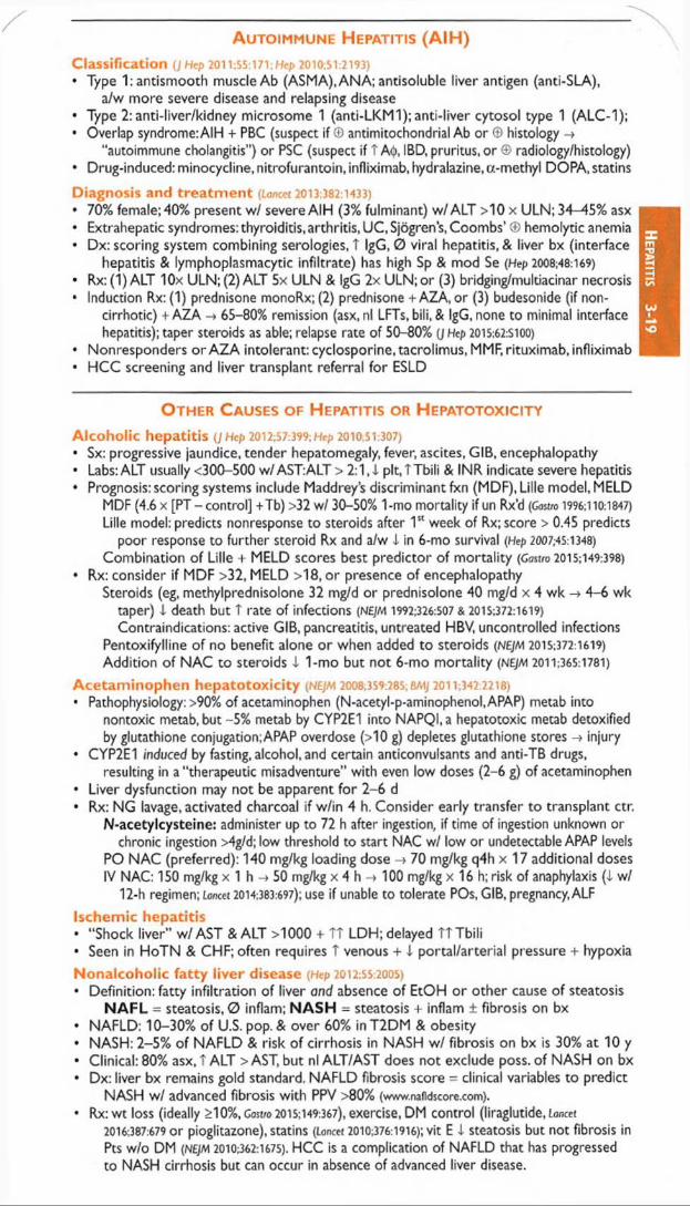
A utoimmune H epatitis (A IH )C la s s i f ic a t io n (J Hep 2011:55:171; Hep 2010:51:2193)• Type 1: antismooth muscle Ab (ASMA), ANA; antisoluble liver antigen (anti-SLA),
a/w more severe disease and relapsing disease• Type 2: anti-liver/kidney microsome 1 (anti-LKMI); anti-liver cytosol type 1 (ALC-1);• Overlap syndrome:AIH + PBC (suspect if ® antimitochondrial Ab or © histology
“autoimmune cholangitis") or PSC (suspect if t AO, IBD, pruritus, or © radiology/histology)• Drug-induced: minocycline, nitrofurantoin, infliximab, hydralazine, ct-methyl DOPA, statins
D ia g n o s is a n d t r e a t m e n t (Lancet 2013:382:1433)• 70% female; 40% present w / severe AIH (3% fulminant) w /ALT >10 x ULN; 34—45% asx• Extrahepatic syndromes: thyroiditis, arthritis, UC, Sjogren’s, Coombs’ © hemolytic anemia• Dx: scoring system combining serologies, t IgG, 0 viral hepatitis, & liver bx (interface
hepatitis & lymphoplasmacytic infiltrate) has high Sp & mod Se (Hep 2008:48:169)• Rx: (1) ALT lOx ULN; (2) ALT 5x ULN & IgG 2x ULN; or (3) bridging/multiacinar necrosis• Induction Rx: (1) prednisone monoRx; (2) prednisone + AZA, or (3) budesonide (if non
cirrhotic) + AZA -» 65-80% remission (asx, nl LFTs, bili, & IgG, none to minimal interface hepatitis); taper steroids as able; relapse rate of 50-80% (J Hep 2015:62:S100)
• Nonresponders o r AZA intolerant: cyclosporine, tacrolimus, MMF, rituximab, infliximab• HCC screening and liver transplant referral for ESLD
O ther Causes of H epatitis or H epatotoxicity
A lc o h o l ic h e p a t i t i s (J Hep 2012:57:399: Hep 2010;51:307)• Sx: progressive jaundice, tender hepatomegaly, fever, ascites, GIB, encephalopathy• Labs: ALT usually <300-500 w/ AST:ALT > 2:1, i pit, TTbili & INR indicate severe hepatitis• Prognosis: scoring systems include Maddrey’s discriminant fxn (MDF), Lille model, MELD
MDF (4.6 x [PT - control] +Tb) >32 w / 30-50% 1-mo mortality if un Rx’d (Gostro 1996:110:1847)
Lille model: predicts nonresponse to steroids after 1st week of Rx; score > 0.45 predicts poor response to further steroid Rx and a/w J. in 6-mo survival (Hep 2007,45:1348)
Combination of Lille + MELD scores best predictor of mortality (Gastro 2015:149:398)
• Rx: consider if MDF >32, MELD >18, or presence of encephalopathySteroids (eg, methylprednisolone 32 mg/d or prednisolone 40 mg/d x 4 wk -» 4-6 wk
taper) I death but T rate of infections (NEJM 1992,326:507 & 2015:372:1619)
Contraindications: active GIB, pancreatitis, untreated HBV, uncontrolled infections Pentoxifylline of no benefit alone or when added to steroids (NEJM 2015:372:1619)
Addition o f NAC to steroids i 1-mo but not 6-mo mortality (NEJM 2011:365:1781)
A c e t a m in o p h e n h e p a t o t o x ic i t y (NEJM 2008:359:285; BMJ 2011,342:2218)• Pathophysiology: >90% of acetaminophen (N-acetyl-p-aminophenol,APAP) metab into
nontoxic metab, but -5% metab by CYP2E1 into NAPQI, a hepatotoxic metab detoxified by glutathione conjugation; APAP overdose (>10 g) depletes glutathione stores - * *■injury
• CYP2E1 induced by fasting, alcohol, and certain anticonvulsants and anti-TB drugs,resulting in a “ therapeutic misadventure” with even low doses (2-6 g) of acetaminophen
• Liver dysfunction may not be apparent for 2-6 d• Rx: NG lavage, activated charcoal if w/in 4 h. Consider early transfer to transplant ctr.
N-acetylcysteine: administer up to 72 h after ingestion, if time of ingestion unknown or chronic ingestion >4g/d; low threshold to start NAC w/ low or undetectable APAP levels
PO NAC (preferred): 140 mg/kg loading dose -> 70 mg/kg q4h x 17 additional doses IV NAC: 150 mg/kg x 1 h -> 50 mg/kg x 4 h 100 mg/kg x 16 h; risk of anaphylaxis ( i w/
12-h regimen; Loncet 2014:383:697); use if unable to tolerate POs, GIB, pregnancy, ALF
I s c h e m ic h e p a t i t i s• “ Shock liver" w / AST & ALT >1000 + TT LDH; delayed TT Tbili• Seen in HoTN & CHF; often requires T venous + i portal/arterial pressure + hypoxia
N o n a lc o h o l ic f a t t y l i v e r d is e a s e (Hep 2012:55:2005)• Definition: fatty infiltration of liver and absence of EtOH o r other cause of steatosis
N A F L = steatosis, 0 inflam; N A S H = steatosis + inflam ± fibrosis on bx• NAFLD: 10-30% of U.S. pop. & over 60% inT2DM & obesity• NASH: 2-5% of NAFLD & risk of cirrhosis in NASH w / fibrosis on bx is 30% at 10 y• Clinical: 80% asx, T ALT > AST, but nl ALT/AST does not exclude poss. of NASH on bx• Dx: liver bx remains gold standard. NAFLD Fibrosis score = clinical variables to predict
NASH w/ advanced fibrosis with PPV >80% (www.nandscorc.com).• Rx: wt loss (ideally >10%, Gasuo 2015:149:367), exercise, DM control (liraglutide, Loncet
2016:387:679 or pioglitazone), statins (Loncet 2010:376:1916); vit E I steatosis but not fibrosis in Pts w/o DM (NEJM 2010:362:1675). HCC is a complication of NAFLD that has progressed to NASH cirrhosis but can occur in absence of advanced liver disease.
Hepa
titis 3-19

Live
r F
ail
ur
e 3-
20ACUTE LIVER FAILURE (ALF
D e f in i t io n• Acute insult to liver + coagulopathy + encephalopathy; most w/o known preexisting liver dis.• Fulminant if encephalopathy w/in 8 wk from jaundice onset; subfu/m/nont if 8 wk to 6 mo• Acute on chronic liver failure: acute insult to liver in Pt w / underlying chronic liver disease
E t io lo g y (Lancet 2010;376:190)• Drugs/toxins (nearly 80% of cases in U.S.; Hepatology 2010:52:2065)
Drugs: acetaminophen (most common cause; >40% of all cases in U.S., typically unintentional overdose); anti-TB drugs (INH, rifampin, pyrazinamide);AEDs (phenytoin, valproate, carbamazepine); NSAIDs (idiosyncratic, not dose-related); abx (eg, fluoroquinolones, macrolides); MDMA (ecstasy)
Toxins: Amanita phalloides (mushroom sp. in West Coast), certain herbal preparations• V iral (12% of cases in the US): HAV, HBV, HCV (rare), HDV + HBV, HEV (esp. if
pregnant). In immunosupp: HSV (50% have skin lesions), EBV,VZV, CMV, HHV6
• Vascular: Budd-Chiari, ischemic hepatitis, hepatic sinusoidal obstructive syndrome• O ther: Wilson’s disease, pregnancy-related ALF (acute fatty liver, preeclampsia, HELLP),
initial presentation of autoimmune hepatitis; idiopathic
C l in ic a l m a n i f e s ta t io n s• Initial presentation usually nonspecific: n/v, malaise; then jaundice & multiorgan failure• Neurologic: encephalopathy: grade 1 = attn deficit, tremor; grade 2 = asterixis, lethargy,
confusion, ataxia; grade 3 = somnolence, rigidity, clonus, hyporeflexia; grade 4 = coma cerebral edema: astrocyte swelling likely related to T ammonia levels
• Cardiovascular: hypotension with low SVR, shock• Pulmonary: respiratory alkalosis, impaired peripheral O 2 uptake, pulm edema, ARDS• Gl: bleeding (due to bleeding diathesis), pancreatitis (? due to ischemia, drugs, infxn)• Renal:ATN, hepatorenal syndrome, hyponatremia, hypokalemia, hypophosphatemia• Hematology: thrombocytopenia, T PT/PTT, X fibrinogen, bleeding diathesis (I synthesis
of coag factors balanced by i protein C/S; bleeding mostly due to low platelet count), DIC• Infection (- 90% of Pts): esp. with Staph, Strep, GNRs and fungi ( I immune fxn, invasive
procedures); SBP in 32% of Pts; fever and T WBC may be absent• Endocrine: hypoglycemia ( i glc synthesis), metabolic acidosis (T lactate),adrenal insuf.
W o r k u p (Hepatology 2012:55:965)• CBC, PT/PTT, LFTs,lytes,BUN/Cr, pH, lactate, N H 3, acetaminophen level, viral serologies
(qv) in all Pts, with additional labs as below if suspected• Autoimmune hep serologies & IgG levels, ceruloplasmin & serum/urine copper, preg test• Imaging studies (RUQ U/S o r abd CT, Doppler studies of portal and hepatic veins)• Liver biopsy if underlying etiology remains elusive after initial testing
M a n a g e m e n t (NEJM 2013:369:2525)• ICU care at liver transplant center for hemodynamic & ventilatory support; CWH for AKI• Early listing fo r liver transplantation in selected Pts (see below)• Cerebral edema: consider ICP monitoring if grade 3/4 enceph; if t ICP -> mannitol 0.5-1.0
mg/kg; if arterial NH3 >150, grade 3/4 enceph, AKI or on vasopressors -> prophylactic 3% saline for goal Na 145-155 mEq/L; barbiturates & hypothermia if refractory T ICP
• Encephalopathy: intubate for grade 3 or 4; lactulose is of little benefit• Coagulopathy: vit K, FFP/pIts/cryo if active bleeding or before invasive procedure; PPI ppx• Infection: low threshold for abx (broad spectrum, eg, vancomycin & 3rd-gen ceph.) if
suspect infection; anti-fungal coverage in high-risk Pts. Daily blood cultures.• Rx of specific causes: NAC if acetaminophen-related; antiviral (eg, entecavir) for HBV;
plasma exchange can be temporizing measure for Wilson’s; IV acyclovir for HSV; PCN-G for A. phalloides; delivery of child for pregnancy-related;TIPS, anticoag for Budd-Chiari. Lack of data for use of steroids in autoimmune, but often given (Hepotofogy 2014:59:612).
• NAC may benefit pts w / non-APAPALF but data inconclusive (Costro 2009;137:856)• Liver transplantation if poor prognosis but could survive surgery
P ro g n o s is• Non-acetaminophen ALF mortality - 80%, acetaminophen-induced ALF mortality -30%• Predictors of poor outcome (King’s College Hospital, UK):
Acetaminophen-induced: pH <7.25, INR >6.5 or PT>100, Cr >3.4, or grade 3/4 enceph. Non-acetamin.-induced: INR >6.5 or PT>100; or >3 of the following: unfavorable etiology
(seronegative hepatitis or drug reaction); age <10 or >40 y; INR >3.5 or PT >50;Tbili >17.5; duration of jaundice >7 d prior to onset of encephalopathy
• -25-30% of Pts undergo liver transplantation w/ 5-y survival rate o f 70%• BMI >30, Cr >2, age >50 y, need for pressors/vent support a/w poorer acute transplant
outcome

CIRRHOSISD e fin itio n (Hep 2011;54:1864 & 2012:56:1983;J Hep 2012;56:S13)• Definition: fibrosis and regenerative nodules resulting from hepatocellular injury• Decompensated = jaundice, variceal bleed, encephalopathy, ascites; worse prognosisEtiologies• Alcohol (-60-70%) and other toxins (eg, arsenic)• Viral hepatitis (~10%): chronic HBV, HCV, HDV infection• Autoim m une hepatitis; 9 , t IgG, © ANA, antismooth muscle Ab,anti-LKM-1,anti-LC1• Metabolic diseases ('-5%): hemochromatosis, W ilson’s disease, « i-AT deficiency• Biliary trac t diseases (~5%): primary biliary cholangitis, secondary biliary cirrhosis
(calculus, neoplasm, stricture, biliary atresia), primary sclerosing cholangitis• Vascular diseases: Budd-Chiari syndrome, R-sided CHF, constrictive pericarditis, SOS• Nonalcoholic fatty liver dis.(NAFLD, 10-15%) cause of most "cryptogenic cirrhosis”• Medications: amiodarone, methotrexate, vitamin A, valproate acid C lin ica l m an ifes ta tions• Nonspecific sx (anorexia, fatigue) o r jaundice, encephalopathy, ascites, variceal bleeding
Physical exam• Liver: initially enlarged, palpable (L lobe predom), firm; eventually shrunken, nodular• Signs of liver failure: jaundice (bili >2.5), spider angiomata & palmar erythema (T estra
diol), Dupuytren’s contractures, white nail lines (Muehrcke’s lines) & proximal nail beds (Terry’s nails), t parotid & lacrimal glands, gynecomastia, testicular atrophy, asterixis, encephalopathy, fetor hepaticus, clubbing, hypertrophic osteoarthropathy
• Signs of portal hypertension: splenomegaly, ascites, dilated superficial abdominal veins(caput medusae), epigastric Cruveilhier-Baumgarten venous hum
Laboratory studies• LFTs:T bili, T PT/INR (poor correlation w/ bleeding;factorVIII nl as not synthesized by
liver), I alb, ± T aminotransferases (AST > ALT if late) and T A(j) (variable)• Hematologic tests: anemia (marrow suppress., hypersplenism, Fe ± folate defic.), neutro
penia (hypersplenism), thrombocytopenia (hypersplenism, iT p o production, EtOH tox)• Chem: i Na (T ADH due to I EAV); T Fe/TIBC, T ferritin (released from hepatocytes)• Lab indices predictive of cirrhosis: AST/pIt >2; Lok index; Bonacini score (JAMA 2012307:832) W orkup (Lancet 2014:383:1749)
• Abd U/S w / Doppler: liver size & echotexture, r/o HCC, ascites, / patency of vasculature• Determine etiology: hepatitis serologies (HBsAg, anti-HBs, anti-HCV), autoimmune
hepatitis studies (IgG, ANA, anti-smooth muscle Ab), Fe and Cu studies, oti-AT, AM A• Assess fibrosis: biomarkers (FibroSURE = panel of 5 markers validated in HCV, t score
predictive of fibrosis); elastography (U/S or MR-based; measurement of liver stiffness)• Liver bx (gold standard): percutaneous or transjugular (consider if ascites or coagulopathy),
used to confirm presence of cirrhosis and dx etiology.Prognosis •
Modified Child-Turcotte-Pugh (C P S ) Scoring SystemPoin ts scored
1 2 3Ascites None Easily controlled Poorly controlledEncephalopathy None Grade 1 or 2 Grade 3 or 4
Bilirubin (mg/dL) <2 2-3 >3Albumin (g/dL) >3.5 2.8-3.5 <2.8PT (sec > control) or INR
<4<1.7
4-61.8-2.3
>6>2.3
ClassificationA B C
Total points 5-6 7-9 10-151-y survival 100% 80% 45%
• MELD (Model for End-Stage Liver Disease): used to stratify Pts on liver tx list & to predict3-mo survival in Pts w I cirrhosis and some acute forms of liver disease. Based on Cr,INR, & total bili. Calculator: www.mayoclinic.org/meld/mayomodel6.html (Gostro 2011:14:1952). If MELD <21 additional predictors of mortality include Na <130 (NE/M 2008:359:1018;Clin Gastroenterol Hepatol 2009:7:1236), refractory ascites, t HVPG and low QoL.
Ascites (see "Ascites" for details on dx evaluation;/Wnj Gostro 2009; 104:1802)• Due to portal HTN (defined as hepatic venous pressure gradient [HVPG] >5 mmHg)• Develops in 60% w/in 10 y; -50% mortality at 5 y
Cir
rh
osis
3-21

• Treatment: I N a in ta k e (1-2 g/d); restrict intake of free water if Na <125D iu re t ic s : goal diurese -1 L/d. Use spironolactone ± furosemide in 5:2 ratio (eg, 100 &
40 mg daily); urine Na/K >1 implies effective natriuresis if Pt compliant w/ low-Na diet Avoid NSAIDs in cirrhotic Pts as interfere w/ diuretic action and are nephrotoxic
• R e f r a c to r y a s c ite s : seen in 5-10% of Pts; 2-y survival 25%Diuretic-resistant on 2 g Na diet, minimal weight loss on maximal diuretic doses, or
diuretic-induced complications (AKI, Na <125, t K, encephalopathy)Med mgmt: conflicting evid.for d/c ’ing (3B (Hep 2016:63:1968); if limited by HoTN.add midodrine L a rg e -v o lu m e p a ra c e n te s e s (LVP; >5 L fluid removal):give 6 - 8 g albumin per L fluid
removed (above 5 L) as colloid replacement a/w I risk of post-para circulatory dysfxn & possibly I mortality (Hep 2012:55:1172). Avoid LVP if SBP present as T risk of AKI.
T r a n s ju g u la r in t r a h e p a t ic p o r t o s y s t e m ic s h u n t (TIPS) (C/m Gos Hep 2011:9:936)
■I ascites in 75%; t CrCI, T enceph, survival benefit over LVP remains controversial Contraindic: grade II enceph, CHF or pulm HTN, active infxn o r biliary obstruction Complications: bleeding, fistula; stent thrombosis (1-y patency w/ coated stents -80%);
infxn (“ endotipsitis” ); new or t enceph in 20-30%, hemolysis (Hep 2010;51:306) Consider for liver transplant if above fail
• H e p a t ic h y d r o t h o r a x : 2° diaphragmatic defect; often unilateral, R > L, ± ascitesTreatment: avoid chest tube (T complications); Rx same as ascites (TIPS if refractory) Spontaneous empyema can occur (even w/o SBP) dx thoracentesis; Rx abx
S p o n ta n e o u s b a c t e r ia l p e r i t o n i t i s (SBP; see "A s c ite s ” fo r de ta ils ; J Hep 2010:53:397)• Develops in -20%; 20% mortality; risk factors: ascitic TP <1 g/dL, hx of SBP, current GIB• Can p/w encephalopathy, abd pain, fever, but often (25%) asx; perform paracentesis
in all hospitalized cirrhotics w/ ascites• Micro: GNRs (£. coli, Klebs) > GPCs (S. pneumo, enterococcus) (see “Ascites” )• Rx: 3rd-gen.ceph or amox/clav x 5 d. If uncomplicated (no encephalopathy or AKI) can use
FQ but avoid if already on for ppx o r if in T FQ resistance area.IV a lb u m in 1.5 g/kg a t t im e o f d x & 1 g /kg on day 3 T su rv iva l (NEJM 1999:341:403)
If not improving, repeat paracentesis at 48 h: expect 25% i in PMNs if Rx working• Indefinite Ppx if (1) h/o SBP or (2) ascitic TP <1.5 plus: Na <130 or Cr >1.2 or BUN >25 or
[CPS >9 iT b ili £3] (Am] Gasvo 2009:4:993) -» cipro 500 mg qd or Bactrim DS qd.Short-term Ppx:CTX 1 g IV x 7 d if GIB; cipro 500 mg PO qd x 1 y if ascitic fluid TP <1.5
G a s t r o e s o p h a g e a l v a r ic e s ± U G IB (see also "GIB"; Lancet 2014:383:1749)• Presence of varices correlates w/ severity of liver dis (40% of Child A Pts -> 85% Child C)• t varix size, Child B/C, & red wale marks assoc w / T risk of bleeding• UGIB 1° prevention: screen at time of dx w/ EGD; data best for Pts w/ med-large varices
n o n s e le c t iv e P -b lo c k e rs : -50% i risk of bleeding & i mortality if med-large varices. Nadolol or propranolol typically used, titrate to max tolerated dose; carvedilol can be considered in nonresponders or if systemic HTN («i blockade -> I intrahepatic vase resistance, Gut 2013.62:1634). EG D not req.to document improvement
e n d o s c o p ic v a r ic e a l l ig a t io n (EVL): superior to (3B in i risk of 1st bleed but no diff in mortality (Ann Hep 2012:11:369); risk of serious complications (esoph perf, ulcers).Repeat q1-2wk until varices gone, w/ f/u EGD at 3 mo then q6-12mo
|3B vs. EVL: choice based on Pt/physician preference, (3B often 1st (Hepato/2008:47:1764);
using both [3B and EVL for primary prevention currently not recommended• 2° prevention: for all Pts after 1st bleed, given -50% risk of rebleed & -30% mortality
[3B + EVL > either alone (Annals 2008; 149:109);TIPS if refractory, or consider in Child B or C w/in 72 h of admission for esoph variceal bleed (T 1-y survival; NEjM 2010:362:2370)
P o r to s y s te m ic e n c e p h a lo p a th y (P S E ) (On Gas Hep 2012:10:1208)• Pathogenesis: failure of liver to detoxify N H j + other substances (eg, ADMA; j Hepatol
2013:58:38) that cause cerebral edema, i O 2 consumption, t ROS brain dysfxn• Precipitants: bleeding, infxn, med nonadherence, i K, i Na, dehydration, hypoxia,
portosystemic shunt (eg,TIPS), meds (eg, sedatives), acute insult to liver (eg, PVT)• Stages: see section in “ Acute Liver Failure"• Dx: N H 3 levels have poor Se for dx & monitoring Rx; remains a clinical dx• Rx: identify/correct precipitants; lactulose (acidification of colon: NH3 -> NH4+) w/ goal 2-4
stools/d (PEG may be more effective; JAMA im 2014:174:1727); alternatively, r i fa x im in 550 mg bid ( i gut bacteria -» I NH3 prod; rifaximin + lactulose may be more effective than lactulose alone; AmjGosuo 2013:108:1458); acarbose & probiotics may benefit
• 2° prevention: lactulose or rifaximin 550 bid (Castro 2009:137:885; NEJM 2010:362:1071)
H e p a to r e n a l s y n d r o m e ( H R S ) (NEJM 2009:361:1279; CrU Care 2012;16:R23(1))• Pathophys: splanchnic vasodilation and renal vasoconstriction w/ 1 renal blood flow• Criteria: (1) cirrhosis w/ ascites; (2) a c u te k id n e y in ju r y (serum Cr T >0.3 mg/dL w/in 48 h
or >50% T in serum Cr from baseline; Gut 2015,64:531); (3) 0 improvement in Cr after d/c

\diuretic & volume expansion (1 g/kg/d of albumin x 2 d); (4) 0 shock (prerenal azotemia/ATN); (5) 0 nephrotoxic meds; (6) 0 intrinsic kidney disease Type I: development in <2 wk; usually occurs in severe liver failure, often following
precipitating event (see later); median survival 2 wk Type II: more indolent course, median survival 6 mo; liver failure present < in type I
• Precipitants: GIB, overdiuresis, infection, serial LVP, drugs (aminoglycosides, NSAIDs)• Rx:if critically ill -» vasopressor (eg, norepinephrine or vasopressin) + albumin (1 g/kg,
max 100 g, bolus daily) to T MAP 10 mmHg. If not critically ill -> octreotide (100-200 meg SC tid) + midodrine (max 15 mg PO tid) + 1 g/kg (max 100 g) albumin on day of presentation followed by 20-60 g albumin qd to T MAP (Hep 2010,51:576).May need dialysis or TIPS as bridge to liver transplant.
Hepatocellular carcinoma (HCC) (Hep 2011 :53 :1020 , L a n c e t 2012 :379 :1245)
• Epi: worldwide, 6d' most prevalent cancer, 3rd most frequent cancer-related death, 80% ofcases due to HCV/HBV cirrhosis, in which annual risk of HCC is -3-8% (Gastro 2012:142:1264). Td risk w/ cirrhosis of any type but esp. w/ viral, HFE, PBC, ? al-AT.
• Clinical; asx vs. hepatic decompensation (eg, ascites, PSE), PVT w/ tumor thrombus• Dx: screen cirrhotics q6mo w/ U/S ± AFP, though many centers choose dual phase
CT/MRI (if arterial enhancing & venous phase or delayed washout, no bx req for dx)• Rx: radiofrequency ablation (RFA) for HCCs <3 cm in size; consider resection if single
lesion <2 cm and Child-Pugh A w/o portal HTN; transarterial chemoembolization (TACE) preferred for large cancers (not curative) or if not amenable to RFA (near IVC/lung); consider liver transplant if up to 3 HCCs <3 cm or 1 HCC <5 cm (Milan criteria)
Other Complications• Hepatopulmonary syndrome (HPS) (D/gD/s Sc/2015,60:1914)
Abnl gas exchange (A-a gradient >15 or PaC>2 <80) caused by intrapulmonary vascular dilatations leading to intrapulmonary shunting
S/S: platypnea-orthodeoxia, clubbing, cyanosisDx w/ contrast echo showing'late” A-V shunting (contrast in LA 3-6 cycles after RA) Rx: O 2; potential embolization if large vessel on CT, ? TIPS, liver tx only definitive Rx
• Portopulmonary hypertension (POPH) (Expert Rev Gaslro Hepatol 2015;9:983)Pulm HTN in Pt w/ portal HTN w/o other cause. ESLD-* * T endothelin-) pulm vasoconst. Rx w/ same therapies as for idiopathic PAH, ind prostacyclin analogs, endothelin receptor-
antagonists, sildenafil; liver transplant is often curative.• Cirrhotic cardiomyopathy: ! inotropic & chronotropic response,! systolic and
diastolic fxn, prolonged QT, hyperkinetic circulation; t troponin, BNP (JACC 2010:56:539)• Infxns: unless already immune, vaccinate for HAV, HBV, PCV13 & PPSV23; flu yearly.
Cellulitis in -20% of Pts hospitalized w/ cirrhosis, often in abd wall or LE a/w skin edema.• Endocrine: diabetes (15-30%), T frequency of adrenal insufficiency (H e p 2012:55:1282)• Coagulopathy: balanced defects w/ i synth of coag factors, hyperfibrinolysis, ! pit
balanced by ! synthesis anticoag factors (protein C/S), defic. of profibrinolytic factors, t levels of vWF. No support for routine administration of FFP, pit, cryo unless in DIC.
• Nutrition: monitor and supplement fat soluble vitamins, zinc• Meds: acetaminophen can be used up to 2 g/d; avoid ASA/NSAIDs; aminoglycosides
contraindicated; oral hypoglycemics if compensated but insulin if decompensated
Liver transplantation• Undertake evaluation when MELD >15. Exception points added if HCC as above• Indie: recurrent/severe enceph, refractory ascites, recurrent variceal bleeding, HRS, HPS,
PPH, HCC (if no single lesion is >5 cm or <3 lesions with largest <3 cm), acute liver failure• Contraindic: inadequate social support, active substance abuse (EtOH w/in 6 mo), sepsis,
advanced cardiopulm dis., extrahepatic Ca, cholangio Ca, hemangiosarcoma, persistent noncompliance,AIDS, fulminant LF w/ sustained ICP >50 mmHg or CPP <40 mmHg
• Survival: 1-y up to 90%, 5-y up to 80%, though lower with HCV; autoimmune liver disease,such as AIH/PBC/PSC may recur in 10-30% (or more) of allografts
O ther Etiologies of C irrhosis
H e m o ch ro m a to s is & Iron O ve rload S yndrom es (Lancet 2016:388:706)
• Recessive disorder of iron sensing o r transport leading to tissue iron depos ition• HFE mutations (85% of cases): typically C282Y homozyg. (- 0.5% of N. Europeans), rarely
C282Y/H63D compound heterozyg. C282Y homozygotes: 28% of <5 & 1% of 9 develop sx (delayed since menses ! Fe load). C282Y/H63D: only 1.5% manifest dis.
• Non-HFE mutations: hemojuvelin, hepcidin, transferrin receptor 2, & ferroportin• 2° causes of iron overload: iron-loading anemias (eg, thalassemia major, sideroblastic
anemia, aplastic anemia), parenteral iron-overload (RBC transfusions, long-term HD), chronic liver disease (due to ETOH, HBV, HCV, NASH, etc), dietary iron overload
Cirrhosis
3-23

Cir
rh
osi
s 3-
24• Sx: fatigue & arthralgias, loss of libido in d. In advanced disease (rare): bronze skin
{melanin + iron), hypogonadism (esp. in juvenile onset), DM, arthropathy (MCP), CHF, infxns (T risk Vibrio, Listeria, Yersinia), cirrhosis (t risk if EtOH/fatty liver disease; 15% risk of HCC). Disease also a/w ALS (H63D homozygotes) & porphyria.
• Dx: iron sat >45% (iron/TIBC x 100%); t ferritin (acute phase reactant, so poor Sp; often nlin young Pts). If T iron sat. -> / HFE to confirm dx, imaging by MRI (black liver).If HFE © & ferritin >1000 ng/mL or t LFTs liver bx for quant Fe index & to stage fibrosis
• Treatment: phlebotomy (250 mL = 1 unit, -250 mg of Fe) qwk until Fe sat <50% & ferritin50-100 pg/L, then q3-4mo; PPI i intestinal Fe absorption & may i need for phlebotomy; avoid vit C & uncooked seafood; deferoxamine if phleb. contraindic.; genetic counseling
W i ls o n ’s d is e a s e ()Hep 2012;56:671)
• Recessive disorder of copper transport (mutation in ATP7B) -> copper overload;primarily affects liver, but also other tissues (brain, eye)
• Epidemiology: 1 in 30,000, majority present b/t 5 & 35 y/o, only 3% of Pts present >40 y/o• Extrahepatic sIs: neuro vy disease, parkinsonism & movement disorder (hepatolenticular
disease), Kayser-Fleischer rings (© in 99% w/ neuro vy but in <50% w/ hepatic disease), Coombs 0 hemolytic anemia, renal disease
• Dx: T 24-h urine Cu, i. serum ceruloplasmin (Se 90%), rarely penicillamine challenge w/ Turine Cu excretion, liver bx w/ hepatic Cu content. In acute liver failure, Att>/bili <4 + AST/ALT >2.2 better Se & Sp than urine Cu or ceruloplasmin (Hepatology 2008,4:1167).
• Treatment: chelation w/ D-penicillamine (supplement B6 as d-pen inactivates); 2rd-linetrientine (I toxicity w/ efficacy, but $$). Zinc: i intestinal Cu transport & can help delay disease; best used in conjunction w/ chelation (must give 4-5 h apart from chelators).Elim. Cu-rich foods.Transplant for fulminant LF or for chronic dis. unresponsive to Rx.
a i - a n t i t r y p s in d e f ic ie n c y ( a i - A T ) ( j Hepatol 2016;65:413)• Abnl « i-AT -> polymerization in liver (cirrhosis) & uninhibited protease activity in lung
(emphysema). Affects 1/3000 of European ancestry.Varied presentations: neonatal hepatitis; cholestatic jaundice in children; T AST/ALT or cirrhosis in children/adults.
• Extrahepatic disease includes: emphysema, necrotizing panniculitis, A N C A vasculitis• Dx: serum oci-AT level (acute phase reactant), level <50% of nl typically diagnostic;
gold standard = phenotyping of protease inhibitor (Pi). Alleles most a/w hepatic dis.: Z (63% of ZZ adults found to have chronic liver dis) & M (malton) (Am j Respir Cm Care Med 2013:137:502). Liver bx shows characteristic PAS © cytoplasmic inclusion bodies.
• Treatment: standard Rx for cirrhosis/chronic liver dis., including liver transplantation
P r im a r y b i l ia r y c h o la n g i t is ( P B C ) (Lancet 2015:386:1565)
• Autoimmune destruction of intrahepatic bile ducts (previously “ primary biliary cirrhosis")• Epi: 9 40-60 y/o; a/w Sjogren’s, Raynaud’s, scleroderma, celiac & thyroid disease; may be
triggered by certain infxns or toxins; a/w X monosomy, variants in IL12a & IL12R genes• Sx (late): fatigue/sleep disturbance, pruritus, steatorrhea, xanthelasma, jaundice, cirrhosis• Ddx: PSC,AIH, hepatic sarcoidosis, meds, idiopathic adult ductopenia, biliary stricture/Ca• Dx: T A(j), T bili, T IgM, T chol, © antimitochondrial Ab (AMA) in 95%. If © AMA, liver bx not
needed due to high Se & Sp. 0.5% gen pop ©AM A & nl LFTs ->10% develop PBC at 6 y. If AMA ©.liver bx (Pts often ©ANA, smooth muscle Ab; same prognosis as ©AMA).
• Rx: ursodeoxycholic acid (13-15 mg/kg/d) regardless of stage-25% complete response, T survival & i histologic change & complications (eg, varices)
(Castro 2005:128:297). Budesonide may benefit in short term.Pruritus: cholestyramine (give 2-4 h after UDCA);if refractory sx: naltrexone, rifampin Fat-soluble vitamins; screen/Rx osteoporosis (risk independent of vit D deficiency) If ESLD: liver tx: -20% recur but no impact on long-term survival
P r im a r y s c le r o s in g c h o la n g i t is ( P S C ) (Loncet 2013;382:1587;Wor/d./HepatoJ 2016:8:265)
• Diffuse inflammation of intrahepatic and extrahepatic bile ducts leading to fibrosis &stricturing of biliary system. A/w HLA-B8 and -DR3 or -DR4, frequent © autoantibodies.
• Epi: 6 > 9 (20-50y) -70% Pts w/ PSC have IBD (usually UC); only 1-4% w / UC have PSC.• Clinical: fatigue, pruritus, jaundice, fevers, RUQ pain, concomitant IBD, ESLD• Ddx: extrahepatic obstruction, PBC, may also have overlap w/ AIH and similar presentation
to lgG4 autoimmune cholangitis (steroid responsive) (Castro 2008:134:706)
• Dx: MRCP ± ERCP -* multifocal beaded bile duct strictures, but may miss dx if confined tosmall intrahepatic ducts (-2% “small duct PSC” : better prognosis, ? different disease). Liver bx may show “ onion-skin” fibrosis around bile ducts but not necessary for dx.
• Treatment supportive care, fet-soluble vitamins; no meds have improved survivalUrsodeoxycholic acid may i colon CA risk in Pts w/ UC & improve LFTs in Pts w/o UC Dominant stricture: endoscopic dilation, short-term stenting o r surgical resection Cholangiocarcinoma (20%): ? biannual surveillance w / MRCP/RUQ U/S and CA19-9 Liver transplantation: -30% recurrence, though if UC, colectomy may i recurrence

HEPATIC VASCULAR DISEASE f lPortal vein thrombosis (P V T ) (Hepato/ogy 2009;49:1729 & 2015;61:660)• Definition: thrombosis, constriction o r invasion of portal vein -> portal HTN -> varices.
Isolated splenic vein thrombosis (eg, 2° to pancreatitis) -> isolated gastric varices.• Etiologies: cirrhosis, neoplasm (pancreas, HCC), abdominal infxn, hypercoag states,
pancreatitis, collagen vascular diseases, Behget’s, IBD, surgery, trauma, OCPs, preg• Clinical manifestations
acute: can p/w abd or lumbar pain; often asx w/ incidental finding on U/S or CT. If mesenteric vein involved may p/w intestinal infarct; if fever consider pylephlebitis,
chronic: asx/incidental finding; may p/w s/s of portal H TN -> hematemesis 2° variceal bleeding, splenomegaly, encephalopathy; ascites uncommon unless cirrhosis
• Diagnostic studies: LFTs usually normal; U/S w/ Doppler, MRA, CT (l+), angiography;“ portal cavernoma” network of hepatopetal collaterals in chronic PVT— can rarely cause
biliary obstruction and cholestatic LFTs = portal cholangiopathy (may require surgery)• Treatment:
Acute: If noncirrhotic, LMWH -> warfarin x 6 mo, or indefinitely if irreversible cause.If cirrhotic, preliminary studies support anticoag if no contraindications; should screen for high-risk varices prior to initiation (Not Rev Goitroentero/Hepatol 2014:11:435).
Chronic: Anticoag if noncirrhotic or hypercoag state; screen for varices prior to anticoag. Esophageal varices: 1° Ppx recommended; if bleed, endoscopic Rx and pB. If
refractory bleed consider TIPS, shunt.Isolated gastric varices 2° splenic vein thrombosis: splenectomy is curative
Budd-Chiari syndrome (HepotoJogy 2009:49:1729)• Occlusion of hepatic vein(s) o r IVC sinusoidal congestion and portal HTN• Etiologies: -50% due to myeloproliferative d/o a/w JAK2 mutations (esp. P. vera), other
hypercoag state, tumor invasion (HCC, renal, adrenal). IVC webs, trauma, 25% idiopathic• Symptoms: hepatomegaly, RUQ pain, ascites, dilated venous collaterals, acute liver failure• Dx: ± T aminotransferases & Atj>; Doppler U/S of hepatic veins (85% Se & Sp); CT (l+)
or MRI/MRV -> vein occlusion or T caudate lobe (separate venous drainage);"spider- web” pattern on hepatic venography; liver bx showing congestion (r/o right-sided CHF)
• Treatment: Rx underlying condition, anticoag (LMWH -> warfarin); consider thrombolysisacutely; if short stenosis, stent may be possible; consider TIPS (T occlusion risk c/w side- to-side portocaval shunt); liver transplant if ALF or failed shunt (J Castro Surg 2012;16:286)
Sinusoidal obstruction syndrome (SOS) (Hepatology 2009:49:1729)• Occlusion of hepatic venules and sinusoids (formerly veno-occlusive disease)• Etiologies: HSCT, chemo (esp. cyclophosphamide), XRT, Jamaican bush tea• Clinical manifestations: hepatomegaly, RUQ pain, ascites, weight gain, T bilirubin• Dx: U/S w/ reversal of portal flow, but often not helpful; dx made clinically (T bili, w t
gain/ascites and RUQ pain) or, if necessary, by liver bx or HVPG (>10 mmHg)• Treatment (20% mortality): supportive; ? defibrotide (adenosine agonist T TPA levels)• Ppx: defibrotide; ursodeoxycholic acid for high-risk HSCT pop; ? use of low-dose heparin
Figure 3-6 Hepatic vasculature
/ Plates of ' hepatocytes
Bite canaliculi
Blood mixes in sinusoids
Lobule
' Hepatic - artery
Portal vein
Inferior vena cava
Right hepatic vein
LiveN
Left hepatic vein
Portal vein from bowelPortal triad -
Central-vein
To hepatic vein, then to inferior vena cava
Hepatic- artery trom aorta
. ■ - iv*- A
& • ^ j
n ; . f
Modified from The Noture of Disease Pathology for the Health Professions, 2007. Hepatology 2009;49:1729.

ASCITES iIPathophysiology• Portal hypertension systemic vasodilatation (? due to release of NO) -> I effective
arterial volume -» renal Na retention -» volume overload and ascites• In malignant or inflammatory ascites, pathophysiology related to leaking of proteinaceous
material from tumor or from inflamed/infected/ruptured intraabdominal structures
E tio log ies
Portal H TN related (SAAG >1.1) N on-porta l H TN related (SAAG <1.1)Presinusoidal obstruction
portal or splenic vein thrombosis, schistosomiasis, sarcoidosis
Sinusoidal obstruction:cirrhosis (81%), including SBP, acute hepatitis, malignancy (HCC or mets)
Postsinusoidal obstructionright-sided CHF incl constriction &TR Budd-Chiari syndrome, SOS
Malig: peritoneal carcinomatosis; chylous ascites from malignant lymphoma; Meigs’ syndrome (ovarian tumor)
Infection: TB, chlamydia/gonorrhea (ie, Fitz-Hugh-Curtis syndrome)
Inflam: pancreatitis, ruptured pancreatic/ biliary/lymph duct; bowel obstrxn
Hypoalbuminemic states: nephrotic syndrome, protein-losing enteropathy
Symptoms• T abd girth, w t gain, new abd hernia, abd pain, dyspnea, nausea, early satiety
E va luation (JAMA 2008:299.1166; Hepatology 2009;29:2087)
• Physical exam: flank dullness {NPV -90%; >1500 mL needed), shifting dullness (Se -83%)• Radiologic: U/S detects >100 mL; MRI/CT (also help with Ddx)• Paracentesis (Hep 2013:57:1651); perform in all Pts w/ new ascites, consider in all hosp.
cirrhotics w/ ascites. Low complic. rate (-1% hematoma formation). Prophylactic FFP or pits does not i bleeding complic. Most useful tests: cell count, alb, total protein, culture.
• Serum -ascites a lbum in g ra d ie n t (S A A G ): serum alb (g/dL) - ascites alb (in g/dL)If >1.1 g/dL -> cause of ascites likely portal HTN (-95% accuracy; Anna/s 1992:117:215)If <1.1 g/dL -> non-portal hypertension relatedIf portal HTN + another cause (seen in -5% of cases) SAAG still >1.1
• Ascites fluid total protein (AFTP): useful when SAAG >1.1 to distinguish cirrhosis(AFTP <2.5 g/dL) from cardiac ascites (AFTP >2.5 g/dL). Low AFTP (<1 g/dL) assoc, w/ T risk of SBP (see "Cirrhosis" for guidelines on SBP Ppx based on AFTP).
• Cell count: normal limit of PMNs in ascitic fluid up to 250 PMNs/mm3. Bloody tap (typicallyfrom traumatic para) can skew cell count; subtract 1 PMN for every 250 RBCs to correct PMN count.Ascitic PMNs >250 suggest infection (see below).
• O ther tests: amylase (pancreatitis, gut perforation); bilirubin (test in dark brown fluid,suggests bile leak or proximal intestinal perf);TG (chylous ascites); BNP (HF); cytology (peritoneal carcinomatosis, -95% Se w/ 3 samples). SBP a/w 1 glc & t LDH is fluid.
T re a tm e n t (see “ Cirrhosis” for details)• If 2° to portal HTN: I Na in take + d iu re tics ; if refractory -» LVP or TIPS• If non-portal HTN related: depends on underlying cause (TB, malignancy, etc.)
B ac te ria l p e r ito n it is (Gut 2012.61 297)
Type Ascites cell count/m m 3 & cxSpontaneous bacterial periton itis (SBP):spontaneous bacterial translocation from gut to ascitic fluid. Ascitic fluid in cirrhosis has i opsonins (esp. if low AFTP), leading to T risk of infxn.
>250 polys; Cx 0 (1 org.) E.coli (37%), Klebs (17%),S. pneumo (12%), misc. GPC (14%), misc. GNR (10%)
C ulture-9 neutrocytic ascites (C N N A ): cell counts suggest infxn but cx 0. No recent abx, w/o other explanation for counts. Rare when sens cx methods.
>250 polys; Cx 0
Nonneutrocytic bacterascites (N N B A ): 0 cx w/oTPMNs. Natural course may resolve w/o tx or may progress to SBP.
<250 polys; Cx © (1 org.) Misc. GPC (30%), E. coli (27%), Klebs (11%), misc. GNR (14%)
2° bacterial periton itis : caused by intraabd abscess or perf.AFTP >1 g/dL, glc <50 mg/dL, LDH >225 U.Rx 3rd-gen ceph.+ MNZ; urgent abd imaging ± ex lap.
>250 polys; Cx © (polymicro)
Peritoneal dialysis-associated: cloudy fluid, abd pain, fever, nausea. Rx: vane + gent (IV load, then administer in PD)
>100, poly predom. Cx © (typ. 1 org.). Misc. GPC (50%), misc. GNR (15%).

BILIARY TRACT DISEASE MC h o l e l it h ia s is (G a l l s t o n e s )
Epidemiology & pathogenesis (; Hep 2008;48:S 124)• >10% adults in the U.S. have gallstones; a/w T overall mortality (Gostra 2011:140:508)• Bile = bile salts, phospholipids, cholesterol; T cholesterol saturation in bile + accelerated
nucleation + gallbladder hypomotility -» gallstones• Risk factors: 9; South, Central, Native American; T age (>40 y), obesity, pregnancy,TPN,
rapid 1 wt; drugs (OCPs, estrogen, clofibrate, octreotide, ceftriaxone); ileal disease• ? statin use >1 y I risk of sx gallstones & cholecystectomy (JAMA 2009:302:2001)
Types of gallstones• Cholesterol (90%): 2 subtypes
mixed: contain >50% cholesterol; typically smaller, multiple stones pure: 100% cholesterol; larger, yellow, white appearance
• Pigment (10%)Black: unconjugated bili & calcium; seen w/ chronic hemolysis, cirrhosis, CF, Gilbert synd Brown: stasis & infection in bile ducts bacteria deconjugate bilirubin -> precipitates w/
calcium; seen w/ duodenal diverticula, biliary strictures, parasites
Clinical manifestations• Asx in 80% of cases; biliary pain in ~2%/y; once sx, rate of complications ~2%/y• B ilia ry pain ( “ colic” ) = episodic RUQ o r epigastric abd pain that begins abruptly, is
continuous, resolves slowly and lasts for 30 min-3 h;± radiation to scapula; nausea• May be precipitated by fa tty foods• Physical exam: afebrile, ± RUQ tenderness or epigastric pain
Diagnostic studies• RUQ U/S: Se & Sp >95% for stones >5 mm; can show complications (cholecystitis);
should be performed only after fasting >8 h to ensure distended, bile-filled gallbladder
Treatm ent (J Hepatol 2016:65:146)
• Cholecystectomy (CCY), usually laparoscopic, if symptomatic• CCY in asx Pts w/: GB calcification (~7% risk of ca) (Surgery 2001:129:699), GB polyps >10
mm, Native American, stones >3 cm or bariatric surgery or cardiac transplant candidates• Ursodeoxycholic acid (rare) for cholesterol stones w/ uncomplicated biliary pain or if
poor surgical candidate; also reduces risk of gallstone formation with rapid w t loss• Biliary pain: NSAIDs (eg, diclofenac 50 mg IM) drug of choice, efficacy - opiates & I
c o m p lic a t io n s (Afment PhormacolTher 2012:35:1370)
Complications• Cholecystitis: 20% of sx biliary pain - * * cholecystitis w/in 2 y• Choledocholithiasis -» cholangitis o r gallstone pancreatitis• Mirizzi syndrome: common hepatic duct compression by cystic duct stone jaundice,
biliary obstruction• Cholecystenteric fistula: stone erodes through gallbladder into bowel• Gallstone ileus: SBO (usually at term ileum) due to stone in intestine that passed thru fistula• Gallbladder carcinoma: ~1% in U.S.
Cholecystitis (nejm 2008;358:2804)Pathogenesis• Acute cholecystitis: stone impaction in cystic duct -» inflammation behind obstruction -4
GB swelling ± secondary infection (50%) of biliary fluid• Acalculous cholecystitis: gallbladder stasis and ischemia inflammatory response; occurs
mainly in critically ill, hosp. Pts (postop major surgery,TPN, sepsis, trauma, burns, opiates, immunosuppression, infxn [eg, CMV, Crypto, Campylobacter, typhoid fever])
Clinical manifestations• History: RUQ/epigastric pain ± radiation to R shoulder/back, nausea, vomiting, fever• Physical exam: RUQ te n d e rn e s s , Murphy’s s ign = T RUQ pain and inspiratory arrest with
deep breath during palpation of R subcostal region, ± palpable gallbladder• Laboratory evaluation: T WBC, ± mild T bilirubin, A<$>, ALT/AST and amylase;
AST/ALT >500 U/L, bili >4 mg/dL or amylase >1000 U/L -> choledocholithiasis
Diagnostic studies• RUQ U/S: high Se & Sp for stones, but need specific signs o f cholecystitis: GB wall
thickening >4 mm, pericholecystic fluid and a sonographic Murphy's sign
Bilia
ry
3-27

Bili
ary
3-28
• H ID A scan: most Se test (80-90%) for acute cholecystitis. IV inj of HIDA (selectivelysecreted into biliary tree). In acute cholecystitis, HIDA enters BD but not GB. 10-20% false ® (cystic duct obstructed from chronic cholecystitis, lengthy fasting, liver disease).
Treatm ent• NPO, IV fluids, nasogastric tube if intractable vomiting, analgesia• A n tib io t ic s (E.coli, Klebsiella and Enterobacter sp. are usual pathogens)
([2nd- o r 3-generation cephalosporin or FQ] + MNZ) o r piperacillin-tazobactam• C C Y (typically laparoscopic) w/in 24 h I morbidity vs. waiting 7-45 d (Ann Surg 2013:258:385)• If unstable for surgery, EUS-guided transmural, ERCP-guided transcystic duct drainage, or
percutaneous cholecystotomy (if w/o ascites or coagulopathy) are alternatives to CCY (NEJM 2015:373:357)
• Intraoperative cholangiogram or ERCP to r/o choledocholithiasis in Pts w / jaundice,cholangitis o r stone in BD on U/S
Complications• Gangrenous cholecystitis: necrosis w/ risk of empyema and perforation• Emphysematous cholecystitis: infection by gas-forming organisms (air in GB wall)• Post CCY: bile duct leak, BD injury or retained stones, cystic duct remnant, sphincter
of Oddi dysfxn
Choledocholithiasis
Definition• Gallstone lodged in common bile duct (CBD)
Epidemiology• Occurs in 15% of Pts w / gallbladder stones; can form de novo in CBDClinical manifestations• Asymptomatic (50%)• RUQ/epigastric pain 2° obstrxn of bile flow T CBD pressure, jaundice, pruritus, nausea
Diagnostic studies• Labs: t bilirubin, A<>; transient spike in ALT or amylase suggests passage of stone• RUQ U/S: BD stones seen -50% of cases; usually inferred from dilated CBD (>6 mm)• ERCP preferred dx modality when likelihood high; cholangiogram (percutaneous,
operative) when ERCP unavailable o r unsuccessful; EUS/MRCP to exclude BD stones when suspicion low
Treatm ent• ERCP & papillotomy w/ stone extraction (± lithotripsy)• CCY typically w/in 6 wk unless contraindication (>15% Pts will develop indication
for CCY if left unRx’d)
Complications• Cholangitis, cholecystitis, pancreatitis, stricture * •
Cholangitis
Definition & etiologies• BD obstruction -> infection proximal to the obstruction• Etiologies: BD stone (- 85%)
Malignant (biliary, pancreatic) or benign stricture Infection w/ fluke (Clonorchis sinensis, Opisthorchis viverrini)
Clinical manifestations• Charcot’s triad: RUQ pain, jaundice, fever/chills; present in -70% of Pts• Reynolds’ pentad: Charcot’s triad + shock and A MS; present in -15% of Pts
Diagnostic studies• RUQ U/S• Labs: T WBC, bilirubin, A<J>, amylase; © BCx• ERCP; percutaneous transhepatic cholangiogram if ERCP unsuccessful
Treatm ent• Antibiotics (broad-spectrum) to cover common bile pathogens (see above)
ampicillin + gentamicin (or levofloxacin) ± MNZ (if severe); carbapenems; pip/tazo• - 80% respond to conservative Rx and abx -» biliary drainage on elective basis• -20% require urgent biliary decompression via ERCP (papillotomy, stone extrac
tion and/or stent insertion). If sphincterotomy cannot be performed (larger stones), decompression by biliary stent or nasobiliary catheter can be done; otherwise, percutaneous transhepatic biliary drainage or surgery.

ACID-BASE DISTURBANCESG e n e r a l
D e f i n i t i o n s
• Acidem ia -> pH <7.36, alkalem ia -+ pH >7.44• Acidosis -» process that increases [HT]; alkalosis -* process that decreases [H 1]• Primary disorders: metabolic acidosis or alkalosis, respiratory acidosis or alkalosis• Compensation
respiratory: hyper- or hypoventilation alters PaC 0 2 to counteract 1° metabolic process renal: excretion/retention of H+/HCOa~ to counteract 1° respiratory process respiratory compensation occurs in minutes; renal compensation takes hours to days compensation usually never fully corrects pH; if pH normal, consider mixed disorder
Consequences of Severe Acid-Base Disturbances (NEJM 1998:338:26 & 107)
Organ system Acidem ia (pH <7.20) Alkalemia (pH >7.60)Cardiovascular I contractility, arteriolar vasodilation
I MAP & CO; i response to catecholamines T risk of arrhythmias
Arteriolar vasoconstriction i coronary blood flow T risk of arrhythmias
Respiratory Hyperventilation, 1 resp. muscle strength HypoventilationMetabolic T K (resp. > metab.), insulin resistance i K, ICa, Mg, P04
Neurologic AMS A MS, seizures, tetany
W o r k u p (NEJM 2014:371:1434)• Traditional or physiologic approach (Bronsted-Lowry definition of acids & bases)
Determine prim ary disorder: / pH, PaC 0 2, HCO 3
Determine if degree of compensation is appropriate
Primary DisordersP rim ary d isorder Problem pH H C O 3 PaCO 2M etabolic acidosis gain o f H+ o r loss o f HCO 3 «l a I
M etabolic alkalosis gain o f HCO 3 o r loss o f H* T ft TRespira tory acidosis hypoventila tion I T ftR espira tory alkalosis hyperventila tion T i ft
Compensation for Acid/Base Disorders (JASN 2010:21:920)
P rim ary d isorder Expected compensationMetabolic acidosis i PaC 0 2 = 1.2 x AHCO3
or PaC 0 2 = (1.5 x HCO3) + 8 ± 2 (Winters’ formula) (also, PaC 0 2 = last 2 digits of pH)
Metabolic alkalosis T PaC 0 2 = 0.7 x AHCO3
Acute respiratory acidosis THCO 3 = 0.1 xAPaC 0 2 (also, 1 pH = 0.008 x APaC 0 2)
Chronic respiratory acidosis T HCO3 = 0.35 x APaC 0 2
(also, i pH = 0.003 x APaC 0 2)Acute respiratory alkalosis I H C 03 = 0.2 x APaC 0 2
(also, T pH = 0.008 x APaC 0 2)Chronic respiratory alkalosis i HCOj = 0.4 x APaC 0 2
• A lternative approachesBase excess/deficit {Curr Opin Oit Care 2006:12:569; 4m J Emerg Med 2016:34:626)
Strong Ion Difference or “ Stewart Method” (NEJM 2014:371:1821)
M ix e d d is o r d e r s ( m o re th a n 1 p r im a r y d is o rd e r a t th e sa m e t im e )
• If compensation less or greater than predicted, may be 2 disorders:PaC 0 2 too low -> concomitant 1° resp. alk.PaC 0 2 too high -> concomitant 1° resp. acid.HCO 3 too low -» concomitant 1° met. acid.HCO 3 too high -> concomitant 1° met. alk.
• Normal pH, b u t. . .T PaC 0 2 + T HCO 3 -> resp. acid. + met. alk.
, i PaC 0 2 + I HCO 3 -+ resp. alk. + met. acid.normal PaC 0 2 & HCO 3, but TAG AG met. acid. + met. alk.normal PaC 0 2l HCO 3, & AG -» no disturbance or non-AG met. acid. + met. alk.
• Cannot have resp. acid, (hypoventilation) and resp. alk. (hyperventilation) simultaneously

Ac
id-B
ase
4-2
/ Figure 4-1 Acid-base nomogram
(Adapted from Brenner BM, ed„ Brenner & Rector's The Kidney, 8th ed„ 2007; Ferri F, cd. Practical Guide to the Care of the Medical Patient, 7th ed.. 2007)
• ABG vs.VBG: concordant for pH (-0.04), HCOj ( -2 mEq) but not PC02 (~8±17 mmHg)VBG can be used to screen for hypercarbia w/ P C 02 cutoff >45 mmHg (100% Se), but may not accurately assess degree of hypercarbia (Am j EmergMed 2012:30:896).
M e t a b o l ic A c id o s is
I n i t ia l w o r k u p (NEJM 2014:371:1434)• / anion gap (AG) = Naf - (C f + H C O 3 ") = unmeasured anions - unmeasured cations
if T glc, use measured not corrected Naexpected AG is [albumin] x 2.5 (ie, 10 if albumin is 4 g/dL, 7.5 if albumin is 3 g/dL) t AG -» t unmeasured anions such as organic acids, phosphates, sulfates I AG -> i alb o r T unmeasured cations (Ca, Mg, K, Li, bromide, iodide, immunoglobulin)
• If t AG, / delta-delta (AA = A A G /A H C O 3) to assess if there is an additional metabolicacid-base disturbance; AAG = (calculated AG - expected AG), A H C O j = (24 - H C O 3)
AA = 1-2 -» pure AG metabolic acidosisAA < 1 -»AG metabolic acidosis and simultaneous non-AG acidosis AA > 2 -> AG metabolic acidosis and simultaneous metabolic alkalosis
Etiologies of AG Metabolic AcidosisKetoacidosis Diabetes mellitus, a lco h o lism , s ta rv a tio n (NEJM 2014:372:546)
Lacticacidosis(NEJM 2014;
371:2309)
Type A: im p a irm e n t in tissue oxygena tion eg, circulatory or respiratory failure, sepsis, ischem ic bow e l, ca rbon m o n o x id e , cyanide
Type B: no im p a irm e n t in tissue o xyg e na tio n , i c learance (eg, hepatic
dysfxn) o r t genera tio n [eg, malig, EtOH, th iam ine def., m eds (m e tfo rm in ,
NRTIs, salicyla tes, p ro p y le n e g lyco l, p ro p o fo l, ison iaz id , lin e z o lid )]
D-lactic acidosis: s h o rt bow el synd rom e -» prec ip by glc ingest -» m etab by
c o lo n ic b a c te ria to D -lacta te ; n o t d e te c te d by s tandard lac ta te assay
Renal failure A c c u m u la tio n o f o rgan ic an ions such as phosphates, sulfa tes, u ra te , e tc .
Ingestions
Methanol (w in d sh ie ld flu id , a n tifre e ze , so lven ts, fue l): m etab to fo rm ic acid
Ethylene glycol (an tifreeze ): m e tab to g lyco lic and o xa lic acids Propylene glycol (pha rm a ce u tica l so lve n t, eg, IV diazepam , lo razepam ,
and p h e n o b a rb ita l; an tifre e ze ): la c tic acidosis
Salicylates: m eta b o lic acidosis ( fro m lacta te, ke tones) + re s p ira to ry
alkalosis due to s tim u la tio n o f CNS re s p ira to ry c e n te r
Glutathione depletion: ace tam inophen T endogenous o rgan ic acid 5- o x o p ro lin e in suscep tib le hosts (m a lnou rished , fem ale , renal fa ilu re )

W orkup for AG m etabolic acidosis• / for ketonuria (dipstick acetoacetate) o r plasma (i-hydroxybutyrate (0OHB)
nb, urine acetoacetate often not present in early ketoacidosis due to shunting to POHB; /. acetoacetate may later turn © but does not signify worsening disease
• If 0 ketones, / renal function, lactate, toxin screen, and osmolal gap• Osmolal gap (OG) = measured osmoles - calculated osmoles
calculated osmoles = (2 x Na) + (glucose/18) + (BUN/2.8)(can + [EtOH/4,6] if have EtOH level and want to test if other ingestions)
OG >10 suggests ingestion (see below) but lacks specificity (can be elevated in lactic acidosis, DKA.and alcoholic ketoacidosis)for methanol/ethylene glycol; early on,OG precedes AG; later OG may be nl with ©AG
IngestionsAG OG Ingestion Other manifestations
T nlAcetaminophen HepatitisSalicylates Fever, tachycardia, tinnitus; met. acid. + resp. alkalosisEthanol Alcoholic fetor, AMS, hepatitis; keto + lactic acidosis
± met. alk. (vomiting)
T TMethanol AMS, blurred vision, pupillary dilation, papilledemaEthylene glycol AMS, cardiopulm. failure, hypoCa. Ca oxalate crystals -»
AKI. Urine fluoresces under UV light,Propylene glycol AKI
nl/T T Isopropyl alcohol AMS, fruity breath (acetone)
Etiologies of Non-AG Metabolic AcidosisGl losses of H C O 3 Diarrhea, intestinal or pancreatic fistulas or drainageRTAs See section on renal tubular acidoses belowEarly renal failure Impaired generation of ammoniaIngestions Acetazolamide, sevelamer, cholestyramine, tolueneDilutional Due to rapid infusion of bicarbonate-free IV fluidsPosthypocapnia Respiratory alkalosis -» renal wasting of H C O 3 ; rapid correction
of resp. alk. - » transient acidosis until H C O j regeneratedUreteral diversion Colonic CI7 HC0 3 exchange, ammonium reabsorption
Workup for non-AG metabolic acidosis (CJASN 2012:7:671)• Evaluate history for causes (see above)• / urine anion gap (UAG) = (UNa + U«) - Uq
UAG = unmeasured anions - unmeasured cations; as N h V is primary unmeasured cation, UAG is indirect assay for renal H+ excretion as N H 4 ' (NEyM 1988:318:594)
• 0 UAG -» T renal NHT excretion -> appropriate renal response to acidemiaDdx: Gl causes, proximal RTA, ingestions or dilutional
• ® UAG -> failure of kidneys to generate NHVDdx: distal o r hypoaldo RTA, early renal failure nb, plasma K usually i in distal and t in hypoaldo RTA
• UAG evaluation assumes Pt volume replete (Unj >25), Uph <6.5 & no AG met. acidosis(which causes © UAG due to excretion of organic anions)
Renal tubular acidoses (RTAs) (JASN 2002:13:2160: Int J Clin Pract 2011:65:350)• P rox im a l (Type II): i proximal reabsorption of H CO 3
1° (Fanconi’s syndrome = i proximal reabsorption of HCO 3, PO4, glc, amino acids), paraprotein (multiple myeloma, amyloidosis), meds (acetazolamide, heavy metals, ifosfamide), renal transplant, iV i t D, NRTIs
• Distal (Type I): defective distal H+ secretion1°, autoimmune (Sjogren’s, RA), nephrocalcinosis, meds (ampho, Li, ifosfamide);
normally a/w I K; if with t K -> sickle cell, obstruction, SLE, renal transplant• Hypoaldo (Type IV): T K -» i N H 3 synthesis/delivery i urine acid carrying capacity
I renin: diabetic nephropathy, NSAIDs, chronic interstitial nephritis, HIV normal renin, 4- aldo synthesis: 1° adrenal disorders, ACEI, ARBs, heparin i response to aldosterone
meds: K-sparing diuretics,TMP-SMX, pentamidine, calcineurin inhibitors tubulointerstitial disease: sickle cell, SLE, amyloid, diabetes
• Combined (Type III): rarely discussed or clinically relevant, also called juvenile RTA, hasdistal & proximal features, can be due to carbonic anhydrase II deficiency
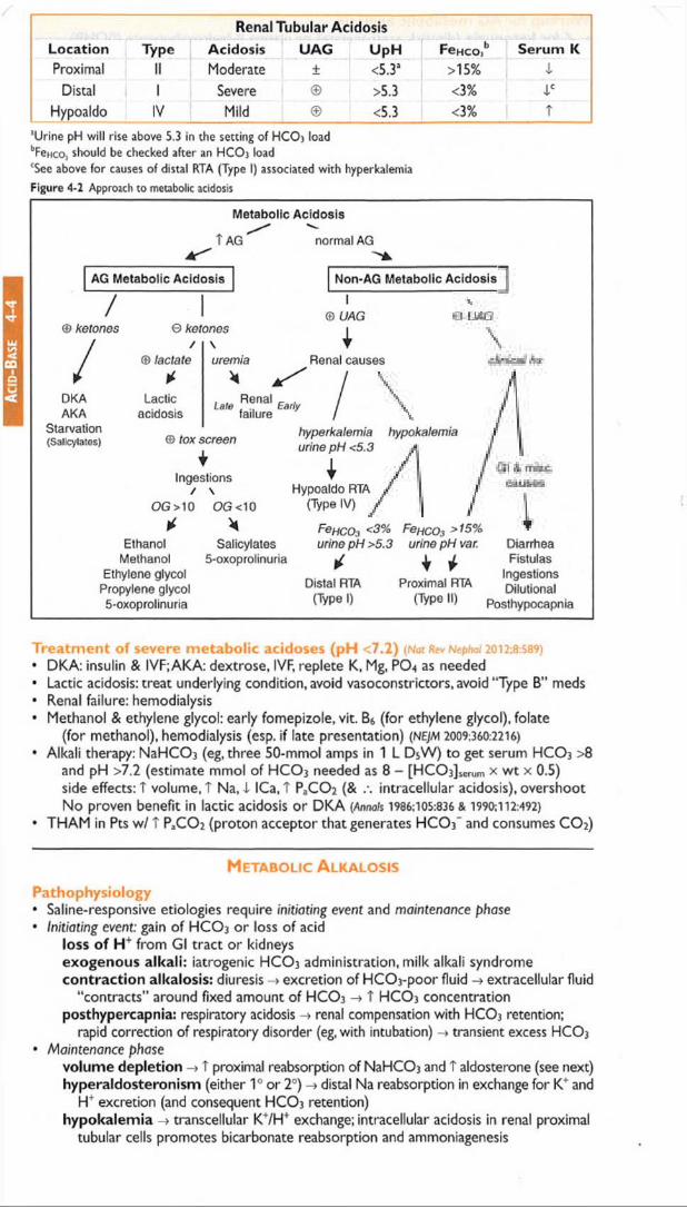
Renal Tubular AcidosisLocation Type Acidosis UAG UpH FeHco,b Serum KProximal II Moderate ± <5.3a >15% X
Distal 1 Severe >5.3 <3% l c
Hypoaldo IV Mild <5.3 <3% T
l Urine pH w ill rise above 5.3 in the setting o f H C O 3 load bFeHco, should be checked after an H C O j loadcSee above for causes o f distal RTA (Type I) associated w ith hyperkalemia
Figure 4-2 Approach to metabolic acidosis
Metabolic Acidosis
T AG normal AG
AG Metabolic Acidosis Non-AG Metabolic Acidosis
/© ketonos © ketones
1DKAAKA
Starvation(Salicylates)
© lactate
*Lactic
acidosis
\uremia
*
lm Ranal a * failure
I© UAG
IRenal causes
© tox screen
IIngestions
/ \OG >10
*Ethanol
Methanol Ethylono glycol
Propylene glycol 5-oxoprolinuria
OG <10
*Salicylates
5-oxoprolinuria
hyperkalemia urine pH <5.3
IHypoaldo RTA
(Type IV)
FeHC03 <3% urine pH >5.3
Distal RTA (Type I)
hypokalemia
FencOi >15%urine pH var.
♦ iProximal RTA
(Typo II)
DiarrheaFistulas
IngestionsDilutional
Posthypocapnia
Treatm ent of severe m etabolic acidoses (pH <7.2) (Not Rev Nephoi 2012;8:589)• DKA: insulin & IVF;AKA: dextrose, IVF, replete K, Mg, PO4 as needed• Lactic acidosis: treat underlying condition, avoid vasoconstrictors, avoid “Type B” meds• Renal failure: hemodialysis• Methanol & ethylene glycol: early fomepizole, vit. B6 (for ethylene glycol), folate
(for methanol), hemodialysis (esp. if late presentation) (NEJM 2009;360:2216)
• Alkali therapy: NaHCC>3 (eg, three 50-mmol amps in 1 L D 5W ) to get serum HCO 3 > 8
and pH >7.2 (estimate mmol of H C O 3 needed as 8 - [HCC^serum x w t x 0.5) side effects: T volume, t Na, -i ICa, T PaCC>2 (& intracellular acidosis), overshoot No proven benefit in lactic acidosis or DKA {/\nna/$ 1986:105:836 & 1990:112:492)
• THAM in Pts w / T PaCC>2 (proton acceptor that generates H CO 3 and consumes C O 2)
M e t a b o l ic A l k a l o s is
Pathophysiology• Saline-responsive etiologies require initiating event and maintenance phase• Initiating event: gain o f H CO 3 o r loss o f acid
loss of H + from Gl tract or kidneysexogenous alkali: iatrogenic HCO 3 administration, milk alkali syndrome contraction alkalosis: diuresis -♦ excretion of HCO j-poor fluid -> extracellular fluid
"contracts” around fixed amount of HCO 3 -» T HCO 3 concentration posthypercapnia: respiratory acidosis -» renal compensation with HCO3 retention;
rapid correction of respiratory disorder (eg, with intubation) -> transient excess HCO3
• Maintenance phasevolume depletion -> T proximal reabsorption of NaHCCh and T aldosterone (see next) hyperaldosteronism (either 1° or 2°) -» distal Na reabsorption in exchange for K+ and
H ' excretion (and consequent HCO 3 retention) hypokalemia -> transcellular K7H f exchange; intracellular acidosis in renal proximal
tubular cells promotes bicarbonate reabsorption and ammoniagenesis

Etiologies o f Metabolic Alkalosis
Saline-responsive Gl loss o f H+: vomiting, NGT drainage, villous adenoma D iuretic usePosthypercapnia, laxatives, cystic fibrosis
Saline-resistant Hypertensive (m inera locortico id excess)1° hyperaldosteronism (eg, Conn’s)2° hyperaldosteronism (eg, renovascular dis., renin-secreting tumor) non-aldo (Cushings, Liddle’s, exogenous mineralocorticoids, licorice)
Normotensivesevere hypokalemia; exogenous alkali loadBartter’s syndrome (loop-like); Gitelman’s syndrome (thiazide-like)
W o rk u p• Check vo lu m e sta tus and Uci
Uci <20 mEq/L saline-responsiveUci >20 mEq/L -> saline-resistant (unless currently receiving diuretics)(Uns unreliable determinant of volume status as alkalemia T H C O j excretion
t Na excretion; negatively charged HCO j “ drags” Na+ along)If Uci >20 and volume replete, / b lood pressure
Figure 4-3 Approach to metabolic alkalosis
M etabolic Alkalosis
UC( <20 UC1 >20
Saline-Responsive
Gl lossesvomiting
NGT drainage villous adenoma
▼Prior diuretics Vol. deplete
Posthypercapnia
Laxatives Cystic fbrosis
Saline-Resistant
/ \/ Hyp
Hypertensive
/Hypo- or normotonsive\
1 ° hyperaldo 2° hyperaldo
non-aldosterone
Current diuretics sever© I K
exogenous alkali Bartter's
Gitelman's
T re a tm e n t o f severe m e tabo lic a lkalosis (pH >7.6)• If volume depletion: d/c diuretics and correct volume deficit with isotonic saline
If cardiopulmonary disease precludes hydration, can use KCI, acetazolamide, HCI• If NGT drainage that cannot be stopped: PPI• Hyperaldosteronism: treat underlying condition
Respiratory A cidosis E tio log ies (also see “ Hypercapnia” )• CNS depression: sedatives, CNS trauma, O 2 in chronic hypercapnia (1 hypoxemic drive),
central sleep apnea• N eurom uscu la r disorders: myasthenia gravis, Guillain-Barre, poliomyelitis, ALS,
muscular dystrophy, severe hypophosphatemia, high spinal cord injury, drugs (paralytics)• U p p e r a irw ay a b n o rm a litie s : acute airway obstruction, laryngospasm, obstructive
sleep apnea, esophageal intubation• Low er a irw ay ab n o rm a litie s : asthma, COPD• Lung parenchyma abnormalities (often cause hypoxia -> T RR resp. alk., but eventual
muscle fatigue -» resp. acid.): pneumonia, pulmonary edema, restrictive lung disease• Thoracic cage abnormalities: pneumothorax, flail chest, kyphoscoliosis• Post infusion of bicarbonate in acidemic Pt w / limited ability to T minute ventilation
Respiratory A lkalosis
Etio log ies (NEJM 2002:347:43)• H ypox ia -» h yp e rve n tila tio n : pneumonia, pulm. edema, PE, restrictive lung disease• P r im a ry h yp e rve n tila tio n
CNS stimulation, pain, anxiety, fever, trauma, stroke, voluntary drugs: salicylates, progesterone, methylxanthines, nicotine pregnancy, sepsis, hepatic failure, fever
• P seudoresp ira to ry alkalosis: I perfusion w/ preserved ventilation (eg, CPR, severeHoTN) i delivery of C O 2 to lungs for excretion; low PaC02 but T tissue C O 2

Sod
ium
4-
6SODIUM AND WATER HOMEOSTASIS
O v e r v ie w
G enera l (NEJM 2015;372:5S & 373:1350)• Disorders of serum sodium are generally due to As in total body water, not sodium• Hyper- or hypo-osmolality rapid water shifts As in brain cell volume A MS, seizures
Key hormones• A n tid iu re tic ho rm one (A D H ): primary hormone that regulates sodium concentration
Stimuli for secretion: hyperosmolality, U effective arterial volume (EAV), angiotensin II Action: insertion of aquaporin-2 channels in collecting ducts -> passive water reabsorption u rine o sm o la lity is an indirect functional assay of the ADH-renal axis Uojoi range: 60 mOsm/L (no ADH) to 1200 mOsm/L (maximal ADH)
• A ld o s te ro n e : primary hormone that regulates total body sodium (and volume)St/mui/ for secret/on: hypovolemia (via renin and angiotensin II), hyperkalemia Action: iso-osmotic reabsorption of sodium in exchange for potassium or H+
H y p o n a t r e m ia
Pathophysiology (NEJM 2015:372:1349)
• Excess o f w ater relative to sodium; almost always due to t A D H• T ADH may be appropriate (eg, hypovolemia o r hypervolemia with i EAV)• T ADH may be inappropriate (SIADH)• Rarely, i ADH (appropriately suppressed), but kidneys unable to maintain nl (Na]5erum
T H2 O intake (1° polydipsia): ingestion of massive quantities (usually >12 L/d) of free H2O overwhelms diluting ability of kidney (normal dietary solute load -750 mOsm/d, minimum U^m = 60 mOsm/L -» excrete in -12 L; if H2O ingestion exceeds this amount
H2O retention)i solute intake (“ tea & toast” & "beer potomania"): W daily solute load -> insufficient solute
to excrete H2O intake (eg, if only 250 mOsm/d, minimum U0 = 60 mOsm/L -> excrete in -4 L; if H2O ingestion exceeds this amount -» H2O retention)
W orkup (JASN 2012:23:1140; Crit Care 2013:17:206; NEJM 2015:372:55)
• History: (1) acute vs. chronic (>48 h); (2) sx severity; (3) risk for neuro complications(alcoholism, malnourished, cirrhosis, older females on thiazides, hypoxia, hypoK)
• Measure plasma osmolalityHypotonic hyponatremia most common scenario; true excess of free H2O relative to Na Isotonic hyponatremia: rare lab artifact from hyperlipidemia or hyperproteinemia Hypertonic hyponatremia: excess of another effective osmole (eg, glucose, mannitol) that
draws H2O intravascularly; each 100 mg/dl_ t glc >100 mg/dL -> i [Na] by 24 mEq/L• For hypotonic hyponatremia, / volume status (vital signs, orthostatics, JVP, skin
turgor, mucous membranes, peripheral edema, BUN, Cr, uric acid)• U 0$m diagnostically useful in limited circumstances, because almost always >300
exceptions: U0im <100 in T H2O intake or i solute intakemoreover, U0Sm >300 * SIADH; must determine if T ADH appropriate or inappropriatehowever, U0Sm important when deciding on treatment (see below)
• If euvolemic and T Uosm, evaluate for glucocorticoid insufficiency and hypothyroidism
Figure 4-4 Approach to hyponatremia
Hypotonic Hyponatremias 1 •v
hypovolemic euvolemic hypervolemic
S '
Hypovolemic Euvolemic HypervolemicHyponatremia Hyponatremia Hyponatremia
/ \ / I t / \Un,i >20 6/Wa <10 Pt's clinical history UNa <10 U»a>20
FENa>1% FENa <1% Uosm >100 Uosm <100 Uosm var. FENa <1% FENa>1%
i \ i 1 < i \Renal losses Extraronal SIADH 1° polydipsia Reset CHF Renal
mineralocorticoid losses hypothyroidism |ow solute osmostat Cirrhosis failuredef. glucocorticoid dcf. Nephrosis

H y p o v o le m ic h y p o t o n ic h y p o n a t r e m ia (ie, i l total body Na, 4TBW )• Renal losses (Un3 >20 mEq/L, FEns >1%): diuretics (esp. thiazides, as loop diuretics
! tonicity of medullary interstitium and impair urine concentrating ability), salt- wasting nephropathy, cerebral salt wasting, mineralocorticoid deficiency
• Extrarenal losses (Unh<10 mEq/L, FEn3 <1%): hemorrhage, GI loss (diarrhea), third-spacing (pancreatitis), i PO intake, insensible losses
E u v o le m ic h y p o t o n ic h y p o n a t r e m ia (ie, T TBW relative to total body Na)• S IA D H (euvolemia or mild hypervolemia, inapprop T Uosm, approp. UNa, 4 BUN & UA)
malignancy: lung, brain, Gl, GU, lymphoma, leukemia, thymoma, mesothelioma pulmonary: pneumonia,TB, aspergillosis, asthma, COPD, PTX,© pressure ventilation intracranial: trauma, stroke, SAH, seizure, infxn, hydrocephalus, Guillain-Barre synd. drugs: antipsychotics, antidepress, (esp. SSRIs), chemotherapy, AVP, MDMA, NSAIDs miscellaneous: pain, nausea, postoperative state
• Endocrinopathies: T ADH activity seen in glucocorticoid deficiency (co-secretion of ADH &CRH) and severe hypothyroidism/myxedema coma (4 CO & 4 GFR)
• Psychogenic polydipsia (UOSm <100, 4 uric acid): usually requires intake >12 L/d• Low solute (4 UNa, 4 U0Sm) “ tea & toast’V'beer potomania”• Reset osmostat: chronic malnutrition (4 intracellular osmoles) or pregnancy (hormonal
effects) -h> ADH physiology reset to regulate a lower [Na],crumH y p e r v o le m ic h y p o t o n ic h y p o n a t r e m ia (ie, t total body Na, T T TBW)• 4 EAV -> activation of RAAS -> T aldosterone and TT ADH• C H F (4 CO & renal venous congestion -» 4 EAV; Un3 <10 mEq/L, FEn3 <1%)• Cirrhosis (splanchnic arterial vasodilation + ascites -> 4. EAV; Uni <10 mEq/L, FEn* <1%)• Nephrotic syndrome (hypoalbuminemia -> edema -> 4 EAV; U^a <10 mEq/L, FEn4 <1%)• Advanced renal failure (diminished ability to excrete free H2O; Un3 >20 mEq/L) T r e a t m e n t (Crit Core 2013:17:206:NE)M 2015:372:55)• Approach: depends on volume status, acuity of hyponatremia, and if symptomatic
Acute sx: initial rapid correction of [Na]sm)m (2 mEq/L/h for the first 2-3 h) until sx resolve Asx o r chronic symptomatic: correct [Na]senjm at rate of <0.5 mEq/L/h Rate of T Na should not exceed 6 (chronic) to 8 (acute) mEq/L/d to avoid central pontine
myelinolysis/osmotic demyelination syn. (CPM/ODS: paraplegia, dysarthria, dysphagia) If severe (<120) o r neuro sx: consider 3% NaCI + dDAVP (to prevent rapid
overcorrection) in consultation w / nephrology (AJKD 2013:61:571)• Frequent lab draws and IVF rate adjustments are cornerstones of treatment• Overly rapid correction: can lead to CPM/ODS. Should be emergently reversed w/
dDAVP ± D 5W ; partial neurologic recovery possible [CJASN 2014:9:229)• Effect of IV fluids {http://www.medcalc.com/sodium.htinl)
initial A [Na]serwm per L infusate =[Na]jnfusito [Na] scrum
TBW + 1TBW = w t (kg) X 0.6(d) o r 0.5 (9):
if elderly use 0.5 (d ) or 0.45 (9 )
If ’Na], = 110 mEq/L in 70 kg male:IVF type [N a] content 1 L IVF T [N a], Rate to T [N a], by 0.5 mEq/L/h5% NaCI 856 mEq/L 17.3 mEq/L -25 mL/h3% NaCI 513 mEq/L 9.4 mEq/L -50 mL/h0.9% NaCI 154 mEq/L 1 mEq/L -500 mL/hLR 130 mEq/L 0.5 mEq/L -1000 mL/h
however, above assumes entire infusate retained without any output o f Na or H2O if Pt euvolemic, as in SIADH, infused Na will be excretedeg, 1 L NS (154 mEq of Na or 308 mOsm of solute in 1 L free H2O) given to Pt with SIADH with U 0Jm = 616 -> 308 mOsm solute excreted in 0.5 L H2O -> net gain 0.5 L H2O -* 4 [Na]scrum
normal saline can worsen hyponatremia from SIADH if Uosm > infusate05m• Hypovolemic hyponatremia: volume repletion with normal saline at a slow rate.
Once volume replete -> stimulus for ADH removed (w/ very short ADH ty,) -» kidneys excrete free H2O -> serum Na will correct rapidly (D 5W ± ddAVP if overcorrection)
• S IA D H (NE//V1 2007:356:2064;AJKD 2015:65:435): free w ater restrict + treat underlying causehypertonic saline (± loop diuretic) if sx o r Na fails to T w/ free H2O restriction
1 L hypertonic saline (3% NaCI) will raise [Na]serum by ~10 mEq (see above)-50 mL/h will t [Na] by -0.5 mEq/L/h; 100-200 ml_/h will T [Na] by -1 -2 mEq/L/h formula only provides estimate; recheck serum Na frequently (at least q2h)
NaCI tabs: particularly if chronic and no CHFaquaresis: ? vaptans (vasopressin receptor antag) for refractory SIADH (nejm 2015:372:23) demeclocycline: causes nephrogenic Dl, I UOSm (rarely used)

8-t? wmaos
• Hypervolem ic hyponatremia: free w ater restrictmobilize excess Na & H jO (use loop diuretics; avoid thiazides) & T EAV (vasodilators to T
CO in CHF, colloid infusion in cirrhosis) aquaresis: vaptans sometimes used; however, no proven mortality benefit, hypoNa
recurs after stopping drug, risk of overcorrection, contraindicated in cirrhosis, and expensive (NE/M 2015:372:2207)
Hypernatremia
Pathophysiology (Crrt Care 2013:17:206; NE)M 2015:372:55)
• Deficit of water relative to sodium; by definition, all hypernatremic Pts are hypertonic• Usually loss of hypotonic fluid (ie, “ dehydration” ); occasionally infusion of
hypertonic fluid• And impaired access to free w ater (eg, intubation, A MS, elderly): hypernatremia
is a powerful thirst stimulus, usually only develops in Pts w/o access to H2O
W orkup• / U0jm, Un3. volume status (vital signs, orthostatics, JVP, skin turgor, BUN, Cr)
Figure 4-5 Approach to hypernatremia
Hypernatremia
*/ X
Ufa <25 Utj3 >100
s \Extrarenal H ,0 loss
Gl H20 loss Insensible loss
or
t Intracellu lar Osmoles
Seizure, Exercise
Na Overload
NaCL'NaHCOj IVF M neralooorticoids
Renal H20 loss/ \
<300 Unm 300-600
/Complete Dl
\Partial Dl
Osmotic diuresis Loop diuretics
Extrarenal H 2O loss (U05m >700-800)• G l H 2O loss: vomiting, NGT drainage, osmotic diarrhea, fistula• Insensible loss: fever, exercise, ventilation
Renal H 2O loss (U05m <700-800)• Diuresis: osmotic (glucose, mannitol, urea), loop diuretics• Diabetes insipidus (J Clin Endocrinol Metob 2012:97:3426)
ADH deficiency (central) or resistance (nephrogenic)Central: hypothalamic o r posterior pituitary disease (congenital, trauma/surgery,
tumors, infiltrative/lgG4); also idiopathic, hypoxic encephalopathy, anorexia, EtOH Nephrogenic (Arwafs 2006:144:186)
congenital (ADH receptorV2 mutation, aquaporin-2 mutation; Ped Nephrol 2012:27:2183)
drugs: lithium , amphotericin, demeclocycline, foscarnet, cidofovir metabolic: hypercalcemia, severe hypokalemia, protein malnutrition,congenital tubulointerstitial: postobstruction, recovery phase of A TN , PKD, sickle cell,
Sjogren’s, amyloid, pregnancy (placental vasopressinase)Dl usually presents as severe polyuria and mild hypernatremia
O th er (Uoim >700-800)• N a overload: hypertonic saline (eg, resuscitation w/ NaHCCh), mineralocorticoid excess• Seizures, T exercise: T intracellular osmoles -> H2O shifts -» transient T [Na]«rUmTreatm ent• Restore access to H 2O or supply daily requirement of H2O (>1 L/d)• Replace free H 2O deficit (also replace concurrent volume deficit if appropriate):
Free H 20 deficit (L)[Na]serUm - 140 ^ JB W TBW = wt (kg) X 0.6 («J) or 0.5 ($):
1 4 0 if elderly use 0.5 (c$) or 0.45 (V)
shortcut: for typical 70-kg man, free H2O deficit (L) - ([Na]scrx,m - 140)/3
A [Na] scnjm pe L infusate =[Na]sawn [N a]infulatt.
TBW + 1
eg, 1 L D 5 W given to 70-kg man w/ [Na] = 160 mEq/L will i [Na]serum by 3.7 mEq nb, do not forget to correct Na if hyperglycemia also present

Rate o f X o f Na should n o t exceed 0.5 m E q/L/h to avoid cerebral edema shortcut: in 70-kg man, 125 mL/h of free H2O will X [Na] by ~0.5 mEq/L/h
V2 NS (77 mEq/L) o r Va NS (38 mEq/L) provides both volume & free H2O (500 or 750 mL of free H2O per L, respectively); can give free H2O via NGT/OGT
Formulas provide only estimates; ,\ recheck serum Na frequently D l and o sm o tic diuresis: see “ Polyuria” section below N a overload: DsW + diuretic
P o l y u r ia
D e fin itio n and pathophys io logy• P o lyuria defined as >3 L UOP per day• Due to an osmotic o r a water diuresis; almost always due to osmotic diuresis in inpatients
W o rk u p• Perform a timed urine collection (6 h sufficient) and measure Uosni• 24-h osmole excretion rate = 24-h UOP (actual or estimate) x U05m
>1000 mOsm/d -> osmotic diuresis <800 mOsm/d -> water diuresis
O sm o tic d iuresis• Etiologies
Glucose (uncontrolled diabetes mellitus)MannitolUrea: recovering AKI, T protein feeds, hypercatabolism (burns, steroids), Gl bleed NaCi administration Propylene glycol
W a te r d iuresis• Etiologies: d iabetes insipidus (Dl) (Nascmm >143) or 1° po lyd ipsia (Naierum <136)
see “ Hypernatremia” above for list of causes of central and nephrogenic Dl• W orkup of Dl: UOSin <300 (complete) o r 300-600 (partial)
w a te r d e p riva tio n te s t (start in a.m., / Nasenjm, POSm, UOSm, UOP q1-2h)Deprive until POSm >295, then / U0$m. If U0Sm <300, then administer vasopressin (5 U SC)
or dDAVP (10 Lig intranasal), then check U0Sm in 1-2 h:Uosm T by >50% = central Dl Uosm unchanged = nephrogenic Dl
/A D H level before and after water deprivation to evaluate proper response
Figure 4-6 Approach to polyuria
T re a tm e n t• 1° po lyd ipsia : treat psychiatric illness, check meds, restrict access to free H2O• O sm o tic diuresis: address underlying cause, replace free H2O deficit (see
“ Hypernatremia” fo r formula to calculate) and ongoing losses• D l:
central Dl: desmopressin (dDAVP)nephrogenic Dl: treat underlying cause if possible; Na restriction + thiazide (mild volume
depletion -> X delivery of filtrate to dysfunctional diluting segment of kidney), consider amiloride for lithium-induced Dl (Krrf /nt 2009:76:44)
pregnancy-induced Dl: due to vasopressinase from placenta, Rx w / dDAVP
Sodium

Pota
ssiu
m
4-10
POTASSIUM HOMEOSTASISO verv iew (NEJM 2015;373:60)• Renal: potassium excretion regulated at d is ta l nephron (collecting tubule)
distal Na delivery & urine flow: Na absorption -> lumen electronegative -» K secretion metabolic alkalemia and aldosterone: increase Na absorption and K secretion nb, diurnal urinary K excretion (day > night), 24-h sample preferred over spot
• Transcellular shifts: most common cause of acute A in serum K (98% intracellular)Acid-base disturbance: K+/H + exchange across cell membranes Insulin -» stimulates Na-K ATPase -> hypokalemia (mitigates postprandial t K) Catecholamines -> stimulate Na-K ATPase -» hypokalemia; reversed by p-blockers Massive necrosis (eg, tumor lysis, rhabdo, ischemic bowel) -» release of intracellular K Hypo- o r hyperkalemic periodic paralysis: rare disorders due to channel mutations
• Diet: alone rarely causes T o r I K (total body store -3500 mEq, daily intake -100 mEq)
H y p o k a l e m ia
T ransce llu la r sh ifts (U|<:Cr <20 mEq/g)• Alkalemia, insulin, catecholamines, hypokalemic/thyrotoxic periodic paralysis, acute t in
hematopoiesis (megaloblastic anemia Rx w/ B12, AML crisis), hypothermia, chloroquine, barium/cesium intoxication, antipsychotic overdose (risperidone, quetiapine)
G l potassium losses (Uk <25 mEq/d, Uk o <20 mEq/g,TTKG <3)• Gl losses plus metabolic acidosis: diarrhea, laxative abuse, villous adenoma• Vomiting & NGT drainage usually manifest as renal losses due to 2° hyperaldo & met. alk.
Renal potassium losses (Uk >30 mEq/d, ? Uk o >20 mEq/gtTTKG >7)• Hypotensive o r normotensive
acidosis: DKA, RTA [proximal RTA (type II) and some distal RTAs (type I)] alkalosis: diuretics, vomiting/NGT drainage (via 2° hyperaldosteronism)
Bartter’s syndrome (loop of Henle dysfxn -> furosemide-like effect;N£/Ai 1999;340:1177) Gitelman’s syndrome (distal convoluted tubule dysfxn -> thiazide-like effect)
i Mg: ? release Mg-mediated inhib. of ROMK channel t K secretion (JASN 2007:18:2649)• Hypertensive: mineralocorticoid excess
1° hyperaldosteronism (eg, Conn's syndrome, glucocorticoid-remediable aldosteronism) 2° hyperaldosteronism (eg, renovascular disease, renin-secreting tumor) nonaldosterone mineralocorticoid (eg, Cushing’s, Liddle’s, exogenous mineralocort.,
licorice, congenital adrenal hyperplasia)
C lin ica l m an ifes ta tions• Nausea, vomiting, ileus, weakness, muscle cramps, rhabdomyolysis, polyuria• ECG: can have no As, U waves, t Q T interval, ventricular ectopy (PVCs,VT,VF)
W o rk u p (Not Rev Neph 2011;7:75)• Rule out transcellular shifts• / 24-h U k and tra n s tu b u la r potassium g ra d ie n t (TTKG) = (UK/Pic)/(Uosm/Poim)
Uk >30 mEq/d, ? Uk:D- >20 mEq/g, o r TTKG >7 -> suggests renal loss Uk <25 mEq/d, UgiCr <20 mEq/g, o r TTKG <3 suggests extrarenal loss
• If renal losses, / BP, acid-base, Uci (Un3 unreliable for volume status w/ alkalemia)
Figure 4-7 Approach to hypokalemia
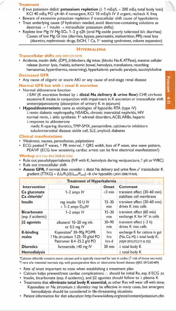
Treatm ent• I f true potassium deficit: potassium repletion (X 1 mEq/L = 200 mEq total body loss)
KCI 40 mEq PO q4-6h if nonurgent, KCI 10 mEq/h IV if urgent, recheck K freq• Beware of excessive potassium repletion if transcellular shift cause of hypokalemia• Treat underlying cause (if hydration needed, avoid dextrose-containing solutions as
dextrose -> t insulin -* intracellular potassium shifts)• Replete low Mg: IV Mg-SO* 1-2 g q2h (oral Mg-oxide poorly tolerated b/c diarrhea)
Causes of low Mg: Gl loss (diarrhea, bypass, pancreatitis, malnutrition, PPI); renal loss (diuretics, nephrotoxic drugs, EtOH, t Ca, 1° wasting syndromes, volume expansion)
H y p e r k a l e m ia
Transcellular shifts (BMJ 2009:339:1019)• Acidemia, insulin defic. (DM), [3-blockers, dig intox. (blocks Na-K ATPase), massive cellular
release (tumor lysis, rhabdo, ischemic bowel, hemolysis, transfusions, resorbing hematomas, hyperthermia, rewarming), hyperkalemic periodic paralysis, succinylcholine
Decreased GFR• Any cause of oliguric or anuric AKI or any cause of end-stage renal disease N orm al GFR but w ith 1 renal K excretion• Normal aldosterone function
I EAV (K excretion limited by 1 distal N a delivery & urine flow): CHF, cirrhosis excessive K intake: in conjunction with impairment in K excretion or transcellular shift ureterojejunostomy (absorption of urinary K in jejunum)
• Hypoaldosteronism: same as etiologies of hypoaldo RTA (type IV)X renin: diabetic nephropathy, NSAIDs, chronic interstitial nephritis, HIV normal renin, X aldo synthesis: 1° adrenal disorders, ACEI, ARBs, heparin X response to aldosterone
meds: K-sparing diuretics,TMP-SMX, pentamidine, calcineurin inhibitors tubulointerstitial disease: sickle cell, SLE, amyloid, diabetes
Clinical manifestations• Weakness, nausea, paresthesias, palpitations• ECG: peaked T waves, T PR interval, T QRS width, loss of P wave, sine wave pattern,
PEA/VF (ECG: low sensitivity, cardiac arrest can be first electrical manifestation!)W orkup (Crrt Care Med 2008:36:3246)
• Rule out pseudohyperkalemia (IVF with K, hemolysis during venipuncture, T pit orWBC)• Rule out transcellular shift• Assess GFR, if normal, then consider -I distal Na delivery and urine flow. / transtubular K
gradient (TTKG) = (UK/PK)/(UOSrr/PoSm) <6 c/w hypoaldo {JASN 2008:19:424).
Treatment of HyperkalemiaIntervention Dose Onset Com m ent
Ca gluconate Ca chloride3
1-2 amps IV <3 min transient effect (30-60 min) stabilizes cell membrane
Insulin reg. insulin 10 U IV + 1-2 amps DsoW
15-30min
transient effect (30-60 min) drives K into cells
Bicarbonate(esp. if acidemic)
1-2 amps IV 15-30min
transient effect (60 min) exchange K for H* in cells
(32 agonists albuterol 10-20 mg inh. or 0.5 mg IV
30-90min
transient effect (~2 h) drives K into cells
K-bindingresins
Kayexalateb 30-90g PO/PR ? Na zirconium 1.25-10 g/tid PO
Patiromer 8.4-25.2 g/d PO
hrshrs
hrs-d
exchange K for cations in gut (Na, Ca, H); X total body K; (NtJM 2015:372:211 & 222)
Diuretics furosemide >40 mg IV 30 min X total body KHemodialysis X total body K
’ Calcium chloride contains more calcium and is typically reserved fo r use in codes (T risk o f tissue necrosis) b? rare a/w intestinal necrosis esp. w ith postoperative ileus or obstructive bowel disease (AJKD 2012:60:409)
• Rote o f onset important to note when establishing a treatment plan• Calcium helps prevent/treat cardiac complications; should be initial Rx,esp. if ECG As• Insulin, bicarbonate (esp. if acidemic), and [32 agonists should follow to I plasma K• Treatments that eliminate total body K essential, as other Rxs will wear off with time;
Kayexalate or Na zirconium ± diuretics may be effective in many cases, but emergent hemodialysis should be considered in life-threatening situations
• Patient information for diet education: http://www.kidney.org/atoz/content/potassium.cfm
Po
ta
ss
ium
4
-11

AK
I 4-
12RENAL FAILURE
A c u t e K id n e y I njury (A K I)
Definition (CJASN 2008:3:844;KI Suppl 2012:2:19)• AKI: abrupt (<48 h) T Cr >0.3 mg/dL, T C r >50%, or UOP <0.5 ml_/kg/h for >6 h
additional gradations based on further t C r & i UOP but not used clinically• Cannot estimate GFR using Cr in setting of AKI o r A’ing C r (requires steady state)
Workup (NEJM 2007:357:797; toncet 2012:380:756)• H & P : recent procedures & meds; thirst:VS & vol status; s/s of obstruction, vase or
systemic dis.; ischemia (prerenal & ATN) accounts for >50% of in-hospital AKI• Urine evaluation: output, urinalysis, sediment, electrolytes, and osmolality• Fractional excretion of sodium (FENa) = (UNa/PNa)/(Ucr/Pcr)
<1% -> prerenal, contrast, HRS or glomerulonephritis; >2% -»ATN In setting of diuretics, / FEun = (UuN/PuN)/(Ucr/Pcr); <35% -> prerenal
• Renal U/S or CT: r/o obstruction & eval kidney size to estimate chronicity of kidney disease• Serologies (if indicated): see “ Glomerular Disease”• Renal biopsy (light microscopy, IF, and EM): may be necessary if etiology remains unclear
(esp. if hematuria and/or proteinuria). Relative contraindications: SBP>150,ASA/NSAID o r anticoag use. Consider dDAVP (0.3 pg/kg 30-60 min prior) for severe uremia.
Etiologies and Diagnosis of Acute Kidney Injury (Lancet 2012:380.756)
Etiologies U/A, Sediment, Indices4- Effective arterial volume (NEJM 2007:357:797) Bland
Hypovolemia, 4- cardiac contractility (eg, CHF), Transparent hyaline castsrt systemic vasodilatation (eg, sepsis) FENa <1%c<D Renal vasoconstriction: NSAIDs.ACEI/ARB, BUN/Cr >202! contrast, calcineurin inhib., HRS, hyperCa UNa <20a. Large vessel: RAS (bilateral + ACEI), vasculitis,
dissection, abd compartment synd., renal venous congestion,VTE
Uosm >500
Acute tubular necrosis (ATN) Pigmented granular muddy/sebemra: progression of prerenal disease brown casts in -75%Toxins (± in CIAKI)
Drugs: AG, amphotericin, cisplatin, HES (starch) ± RBCs & protein from tubularPigments: Hb, myoglobin (NEJM 2009:361:62) damageMonoclonal: Ig light chains (Blood 2010:116:1397) FEn i >2%, BUN/Cr <20, UNaCrystals: UA,ACV, MTX, indinavir, oral NaPO* >20 (except pigment, CIAKI)
U*J7! Contrast-induced AKI (CIAKI): I RBF + toxin UOJro <350
*L Acute interstitial nephritis (A IN ) WBCs,WBC casts, ± RBCs-M£ Allergic: [i-lactams, sulfa drugs, NSAIDs, PPIs w/ neg UCx
Infection: pyelonephritis, legionella,TB, leptospirosis © urine eos in abxInfiltrative: sarcoid, lymphoma, leukemia /Autoimmune: Sjogren’s,TINU syndrome, lgG4, SLE
© lymphs in NSAIDs
Small-med vessel: chol emboli, PAN.TMAs (TTP, ± RBCsHUS, atypical HUS, DIC, preeclampsia, APS, malignant HTN, scleroderma renal crisis)
© urine eos in chol emboli
Glomerulonephritis (see “Glomerular Disease") Dysmorphic RBCs, RBC castsBladder neck: BPH, prostate cancer, neurogenic Bland
</>A
bladder, anticholinergic meds ± nondysmorphic RBCs£ Ureteral (bilateral or unilateral in single kidney):
malig, LAN, retroperitoneal fibrosis, nephrolithiasisFEfvia variable
General trea tm en t (CJASN 2008;3:962)• Prerenal: isotonic IVF » alb (NEJM 2004:350:22), HES (starch) nephrotoxic (NEJM 2012:367:124)
• Avoid nephrotoxic insults; review dosing of renally cleared drugs• Optimize hemodynamics (both MAP & CO)• N o b e n e f it t o d o p a m in e (Anno/s 2005:142:510), d iu re t ic s (JAMA 2002:288:2547), o r m a n n ito l
Managing complications• May take 1-3 wk to recover from ATN; anticipate volume overload, T K, t PCX acidosis• Episodes of AKI T risk of CKD progression, even after recovery (NEJM 2014:371:58)
• Indications for urgent dialysis (when condition refractory to conventional therapy)Acid-base disturbance: refractory acidemiaElectrolyte disorder: generally hyperkalemia; occasionally hypercalcemia, tumor lysis

Intoxications (http://www.extrip-workgroup.org/): contact Poison Control (1-800-222-1222) Indicated for: methanol, ethylene glycol, metformin, Li, valproic acid, salicylates,
barbiturates, theophylline, thallium Also consider for: carbamazepine, acetaminophen, dig (also give Digibind), dabigatran
(also give idarucizumab)Overload of volume (CHF)Uremia: pericarditis, encephalopathy, bleeding
Data on benefit of early RRT remains mixed (NEJM 2016:375:122 & jam a 2016;315:2190)
D is e a s e -S p e c if ic M a n a g e m e n t
C ard io rena l syndrom e (CRS) (Not Kev N«p/) 2009:5:641 & 2013:9:99; CJASN 2013:8:1800)• Multifactorial pathophys including: 1) i CO, 2) T renal venous congestion, 3) T RAAS• Bidirectionality: acute CHF -* *AK I, and oliguric AKI can worsen CHF ()ACC 2008:52:1527)
• Treatment: IV loop d iure tics (bypass potential gut edema; see below for dosing); no diff.between high vs. low dose and bolus vs.gtt (nejm 2011:364797). No clinical benefit with vaptans (ADH receptor antag; JAMA 2007:297:1319), dopamine or nesiritide (nejm 2011:365:32;JAMA 2013:310:2533), or ultrafiltration (NEJM 2012:367:2296).
• Prognosis: 7% T mortality a/w each 10 mL/min i eGFR in ADHF (]ACC 2006:47:1987)
C on tras t-indu ced acu te k idney in ju ry (CIAKI; Ore 2015:132:1931)• Risk factors: CKD, DM, CHF, age, hypotension, T contrast volume (JACC 2004:44:1393)
• Clinical: AKI w/in 48 h of contrast exposure, peaks in 3-5 d, resolves in 7-10 d (if does notresolve, consider cholesterol emboli o r other etiology)
• Prevention: consider when eGFR <60 o r diabetes (CJASN 2013:8:1618)
Iso ton ic IV flu ids (unless contraindic, eg, CHF)Outpatients: 3 mL/kg/h x 1 h before, 1-1.5 mL/kg/h x 6 h after (jama 2004:291:2328) Inpatients: 1 mL/kg/h x 6-12 h before, during, and 6-12 h after;
if undergoing cardiac cath, consider rate of IVF based on LVEDP:5,3, or 1.5 mL/kg/h if LVEDP <13,13-18, or >18 mmHg (Lancet 2014:383:1814)
NaHC0 3 similar to NaCI (CJASN 2015:10:1519).Hold ACEI/ARB (AJKD 2012:60:576), NSAIDs, diuretics. ? high-dose statin (Ore 2012:126:3008)
Minimize contrast volume and use iso-osmolar contrast (JACC 2006:48:692)
N-acetylcysteine 600-1200 mg PO bid on day prior to & day of contrast; benefit in some but not all studies (Annofe20l6;l64:406);as safe, reasonable to consider in high-risk Pts
No proven benefit to Ppx RRT in addition to above, may be harmful (Am) Med 2012:125 66)
• Gadolinium: can cause AKI in stage IV CKD (Nepb Dial Trans 2006:21:697), no effective PpxNephrogenic systemic fibrosis: fibrosis of skin, joints, eyes, and internal organs ~2-4 wk
post exposure in Pts w / mod-severe CKD (JACC2009:53:1621). Postgado HD encouraged albeit no data. Physical therapy. Can be irreversible.
H epa to ren a l syndrom e (HRS; see “ Cirrhosis” ; AJKD 2013;62(6):1198)• Albumin + either octreotide & midodrine o r IV vasopressors
Rhabdom yolysis (NEJM 2009:361:62)• Multifactorial pathophys: myoglobin-induced oxidant injury, vasoconstriction, myoglobin
precipitation & pre-renal (extravasation). Can lead to i Ca, T K, and t PO4.• Diagnosis: UA © fo r heme but 0 RBCs (ie, myoglobinuria)• Generally low risk of AKI when CK <5000, but correlation imperfect. Rhabdo & risk of
AKI/death calculator: http://www.brighamandwomens.org/research/rhabdo/defaultaspx• Aggressive IVF resuscitation and augmenting UOP (tailor IVF to target UOP ~3 mL/kg and
ensure i CK). If urine pH <6.5, can consider NaHC0 3 solutions and watching pH./ K & Ca frequently. Monitor for compartment syndrome.
A c u te in te rs t it ia l n e p h ritis (AIN; Ki 2008:73:940 & 2010:77:956)• Commonly drug-induced: (i-lactams, sulfa drugs, NSAIDs, PPIs• If suspected, prompt removal of offending drug, consider early steroids w/in 7 d of dxT h ro m b o tic m ic roang iop a th ies (T M A s): please see "Hematology"
O b s tru c tive diseases• Dx: imaging w/ renal U/S if undifferentiated or abd/pelvic CT (I") if suspect nephrolithiasis• Treatment: Foley catheter vs. percutaneous nephrostomy for decompression• Following decompression, at risk of:
Hypotonic diuresis (2° buildup of BUN, tubular damage); Rx w/ IVF (eg, Va NS) Hemorrhagic cystitis (rapid A in size of bladder vessels); avoid by decompressing slowly
C h r o n ic K id n e y D isease ( C K D )
D e fin itio n and e tio log ies (Loncet 2012:379:165;JAMA 2015:313:837)
• >3 mo of reduced GFR (<60) and/or k idney dam age (path, markers, imaging)• Prevalence 13% in U.S.
CK
D
4-13
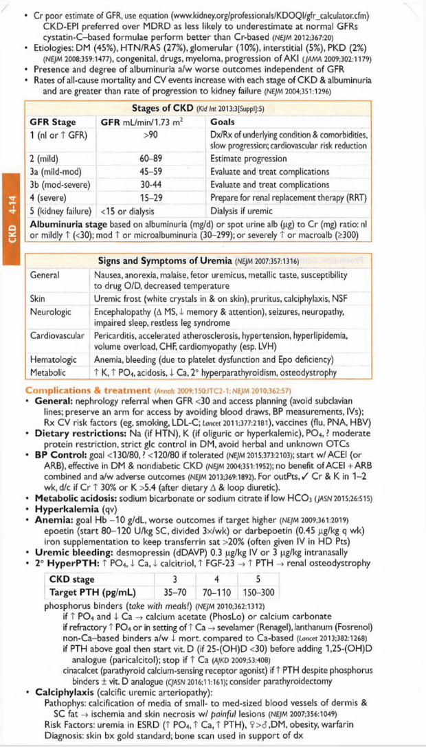
CK
D
4-14
/• Cr poor estimate of GFR, use equation (www.kidney.org/professionals/KDOQI/gfr_calculator.cfm)
CKD-EPI preferred over MDRD as less likely to underestimate at normal GFRs cystatin-C-based formulae perform better than Cr-based (NEJM 2012;367:20)
• Etiologies: DM (45%), HTN/RAS (27%), glomerular (10%), interstitial (5%), PKD (2%)(NEJM 2008;359:1477), c o n g e n ita l, d ru g s , m y e lo m a , p ro g re s s io n o f AKI (JAMA 2009;302:1179)
• Presence and degree of albuminuria a/w worse outcomes independent of GFR• Rates of all-cause mortality and CV events increase with each stage of CKD & albuminuria
and are greater than rate of progression to kidney failure (NEJM 2004:351:1296)
Stages Of CKD (Kid Int 2013:3[Suppl]:S)
GFR Stage GFR mL/min/1.73 m2 Goals1 (nl or T GFR) >90 Dx/Rx of underlying condition & comorbidities,
slow progression; cardiovascular risk reduction2 (mild) 60-89 Estimate progression3a (mild-mod) 45-59 Evaluate and treat complications3b (mod-severe) 30-44 Evaluate and treat complications4 (severe) 15-29 Prepare for renal replacement therapy (RRT)
5 (kidney failure) <15 or dialysis Dialysis if uremicAlbuminuria stage based on albuminuria (mg/d) or spot urine alb (pg) to Cr (mg) ratio: nl or mildly T (<30); mod T or microalbuminuria (30-299); or severely T or macroalb (>300)
Signs and Symptoms of Uremia (NEJM 2007,357:1316)
General Nausea, anorexia, malaise, fetor uremicus, metallic taste, susceptibility to drug O/D, decreased temperature
Skin Uremic frost (white crystals in & on skin), pruritus, calciphylaxis, NSFNeurologic Encephalopathy (A MS, i memory & attention), seizures, neuropathy,
impaired sleep, restless leg syndromeCardiovascular Pericarditis, accelerated atherosclerosis, hypertension, hyperlipidemia,
volume overload, CHF, cardiomyopathy (esp. LVH)Hematologic Anemia, bleeding (due to platelet dysfunction and Epo deficiency)Metabolic T K, T PO4, acidosis, I Ca. 2° hyperparathyroidism, osteodystrophy
C o m p l i c a t i o n s & t r e a t m e n t (Annals 2009;150:ITC2-1; NEJM 2010:362:57)• General: nephrology referral when GFR <30 and access planning (avoid subclavian
lines; preserve an arm for access by avoiding blood draws, BP measurements, IVs); Rx CV risk factors (eg, smoking, LDL-C; Lancet 2011:377:2181), vaccines (flu, PNA, HBV)
• D ietary restrictions: Na (if HTN), K (if oliguric or hyperkalemic), PO4, ? moderateprotein restriction, strict glc control in DM, avoid herbal and unknown OTCs
• BP Control: goal <130/80, > <120/80 if tolerated (NE]M 2015:373:2103); start w/ ACEI (orARB), effective in DM & nondiabetic CKD (NEJM 2004:351:1952); no benefit of ACEI + ARB combined and a/w adverse outcomes (NEJM 2013:369:1892). For ou tP ts ,/ Cr & K in 1-2 wk, d/c if Cr T 30% or K >5.4 (after dietary A & loop diuretic).
« Metabolic acidosis: sodium bicarbonate or sodium citrate if low H C O 3 (JASN 2015:26:515)• Hyperkalem ia (qv)• Anem ia: goal Hb -1 0 g/dL, worse outcomes if target higher (NEJM 2009:361:2019)
epoetin (start 80-120 U/kg SC, divided 3x/wk) o r darbepoetin (0.45 J.ig/kg q wk) iron supplementation to keep transferrin sat >20% (often given IV in HD Pts)
• U rem ic bleeding: desmopressin (dDAVP) 0.3 jug/kg IV o r 3 pg/kg intranasally• 2° H yperPTH : T PO4, i Ca, I calcitriol, T FGF-23 -> t PTH -> renal osteodystrophy
CKD stage 3 4 5Target PTH (pg/mL) 35-70 70-110 150-300
p h o s p h o ru s b in d e rs (teke with m eals!) (NEJM 2010:362:1312)if T PO4 and i Ca -> calcium acetate (PhosLo) or calcium carbonate if refractory t PO4 o r in setting of T Ca -> sevelamer (Renagel), lanthanum (Fosrenol) non-Ca-based binders a/w i mort. compared to Ca-based (toncet 2013:382:1268)
if PTH above goal then start vit. D (if 25-(OH)D <30) before adding 1,25-(OH)D analogue (paricalcitol); stop if t Ca (AJKD 2009:53:408)
cinacalcet (parathyroid calcium-sensing receptor agonist) if T PTH despite phosphorus binders ± vit. D analogue (CJASN 2016:11:161); consider parathyroidectomy
• Calciphylaxis (calcific uremic arteriopathy):Pathophys: calcification of media of small- to med-sized blood vessels of dermis &
SC fat ischemia and skin necrosis w / pa in fu l lesions {NEJM 2007:356:1049)
Risk Factors: uremia in ESRD (T PO4,T Ca, t PTH), ?><5,DM, obesity, warfarin Diagnosis: skin bx gold standard; bone scan used in support of dx

Treatment: multidisciplinary wound care, manage hyperPTH, avoid vit D & Ca suppl., IV & intralesional Na thiosulfate, cinacalcet; NOAC rather than warfarin
Prognosis: a/w 60% 1-y mort. in ESRD Pts (AJKD 2015;66(1):133)• Transplant evaluation
D iu r e s isGeneral considerations• Increases Na excretion for treatment of HTN or edema in CHF, renal failure,and cirrhosis• Daily w t most effective method of documenting successful diuresis
Loop diuretics (NEJM 1998:339:387)• Drugs: furosemide (Lasix), torsemide, bumetanide (Bumex), ethacrynic acid• Mech: inhib Na-K-2CI transporter in thick ascending limb (ThAL); 20-25% Na reabsorp.
Transient, immediate venodilation may aid in pulmonary congestion (NEJM 1973:288:1087) Response is fxn of amt of drug excreted;.-. T dose needed in renal insufficiency, CHF Sigmoidal dose response curve; t dose until induce diuresis, TT dose beyond that
point yields diminishing returns compared with T frequency of dosing• Dosing: PO bioavailability of furosemide -50%, /. IV dose ~2x as potent as PO dose
torsemide & bumetanide -90% bioavailability; use ethacrynic acid if sulfa allergy 40 mg furosemide PO = 20 mg furosemide IV = 20 mg torsemide PO ~ 1 mg bumetanide dose furosemide bid-qid; qd dosing can lead to initial diuresis -> antinatriuresis Continuous vs. bolus IV: similar results in acute CHF (NE]M 2011:364:797)? T diuresis w / co-administration of albumin if 4. serum albumin (Crit Care Med 2005:33:1681)
T h ia z id e d iu r e t ic s (nejm 2009:361:2153)• Drugs: hydrochlorothiazide (HCTZ), chlorothiazide (Diuril), metolazone (Zaroxolyn)• Mech: inhib Na-CI cotransporter in the distal convoluted tubule (DCT); 5% Na reabsorp.
synergistic with loop diuretic, esp. if chronic loop usei effect when GFR <30, except metolazone which is still effective in renal insufficiency
• Dosing: give prior to loop diuretic, typically -30 min before
K-sparing diuretics• Drugs: spironolactone (Aldactone), amiloride, triamterene, eplerenone• Mech: I Na reabsorption (-1%) in collecting duct (amiloride/triamterene inhibit principal
cell Na channel [ENaC];spironolactone/eplerenone inhibit mineralocorticoid receptor). Relatively weak natriuretic activity, useful in combination with thiazide or in cirrhosis.
Approach to Diuresis (if inadequate diuresis, go to next step)
Step Action1 Loop diuretic PO: / response at 3 h, redose at 2x prior dose if needed2 Add thiazide diuretic PO (potentiates response to loop diuretic)3 Loop diuretic IV: bolus bid-qid ± thiazide (may start here if inPt)
T dose needed w/ T Cr; initial effective IV Lasix dose = 30 x Cr (ie, if Cr = 4 -> 120 mg IV lasix)
4 Loop diuretic infusion: bolus + continuous IV infusion ± thiazide (PO or IV)5 RRT: consider ultrafiltration, CW H, or HD
Disease state specific regimens• Renal insufficiency: loop diuretic ( t dose to achieve effective delivery to ThAL) ± thiazide• CHF: loop diuretic (T frequency over T dose) + thiazide (watch K & Mg)• Nephrotic syndrome: urinary albumin binds secreted loop diuretic, use 2-3x normal dose• Cirrhosis: spironolactone (blocks 2° hyperaldosteronism) + Lasix in 2.5:1 ratio• Severe metabolic alkalosis: acetazolamide & treat underlying cause
Adverse effects• Loop: ± T Na, i K, I Mg, I Ca, hyperuricemia, ototoxicity, hypersensitivity (sulfa)• Thiazide: I Na, 1 K, i Mg, t Ca, hyperlipidemia, pancreatitis, T glucose, hypersensitivity• K-sparing: T K (esp. w / ACEI), metabolic acidosis, gynecomastia (spironolactone) * •
R e n a l R e p l a c e m e n t a n d D ialysisGeneral• Substitutes for renal solute and fluid removal• Acute indications: see "AKI"; choices C W H vs HD• Chronic indications: time of RRT initiation should factor in Pt QoL, uremic sx, risk of
development of urgent/acute indications; choices PD vs. HD
Hemodialysis (H D ) (ne;m 2010:363:1833)• Physiology: blood flows along one side of semipermeable membrane, dialysate along other
Fluid removal (ie, Na + H2Q) via transmembrane pressure (TMP) gradient
Diu
res
is
4-15
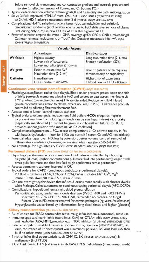
Dia
lysi
s 4-
16Solute removal via transmembrane concentration gradient and inversely proportional
to size effective removal of K, urea, and Cr, but not PO4)• Typical orders: duration, volume removal goals, K and Ca in dialysate bath,anticoagulation• 6 x vs. 3x/wk improved HTN, LV mass, QoL, but t vase issues (NEJM 2010;363:2287);
w/ 3x/wk HD, T adverse outcomes after 2-d interval (nejm 2011:365:1099)
• Complications: HoTN, arrhythmia, access issues (clot stenosis, infxn, recirculation),disequilibrium syndrome (sx of cerebral edema due to H20 shifts after removal of plasma urea during dialysis, esp. in new HD Pts w/ TT BUN), high-output HF
• Fever w / catheter: empiric abx (vane + GNR coverage qHD). GPC > GNR > mixed/fungal.Catheter removal, replacement or “ lock” abx. Consider metastatic infxn w/u (AJKD 2004:44:779;JASN 2014:25:2927).
Vascular AccessAdvantages Disadvantages
AV fistula Highest patencyLowest risk of bacteremiaLowest mortality (JASN 2013:24:465)
Long maturation time (2-6 mo) Primary nonfunction (20%)
AV graft Easier to create than AVF Maturation time (2-3 wk)
Poor 1° patency, often requiring thrombectomy or angioplasty
Catheter Immediate useUse as bridge to AVF/AVG
Highest risk of bacteremia i blood flow -» i HD efficiency
C o n t in u o u s v e n o - v e n o u s h e m o f i l t r a t i o n ( C V V H ) (NEJM 2012:367 26)
• Physiology: hemofiltration rather than dialysis. Blood under pressure passes down one sideof highly permeable membrane allowing H20 and solutes to pass across membrane via TMP gradient (convective clearance). Filtrate discarded. Replacement fluid infused (solute concentrations similar to plasma, except no urea, Cr, PO4). Fluid balance precisely controlled by adjusting filtrate/replacement fluid.
• Access: double-lumen central venous catheter• Typical orders: volume goals, replacement fluid buffer: H C O j (requires heparin
to prevent machine from clotting, although can be run heparin-free) vs. c itra te [hepatically metabolized (.-. cannot be given in cirrhosis/liver failure) to H C O 3; provides anticoagulation w/in machine via Ca chelation]
• Complications: hypotension, I PO4, access complications; 1 ICa (citrate toxicity in Ptswith hepatic dysfunction -> look for i ICa but normal/ T serum Ca and AG met acidosis)
• Potential advantages over HD: less hypotension, better volume control, removal ofinflammatory mediators; however, no survival advantage (Lancet 2006:368:379)
• No advantage for high-intensity CVVH over standard intensity (NEJM 2008:359:7)
P e r i to n e a l d ia ly s is ( P D ) (Pent Dial Int 200V.2V.2S; Perit Diallnt 2009;29:S59)
• Physiology: peritoneum acts as membrane. Fluid balance controlled by choosingdialysate [glucose] (higher concentrations pull more fluid into peritoneum); longer dwell times pulls first more and then less fluid as glc equilibrates across peritoneum
• Access: permanent catheter inserted in OR• Typical orders for CAPD (continuous ambulatory peritoneal dialysis):
PD fluid = dextrose (1.5%, 2.5%, o r 4.25%), buffer (lactate), Na+, Ca2+, Mg2" infuse 10 min, dwell 90 min-5.5 h, drain 20 min
• Can use overnight cycler device that infuses & drains more rapidly, w ith shorter dwells,while Pt sleeps. Called automated or continuous cycling peritoneal dialysis (APD.CCPD).
• Complications: hypoalbuminemia; right-sided pleural effusionPeritonitis: abd pain, tenderness, cloudy drainage (WBC >100 and >50% PMNs)
spectrum: 60-70% GPC, 15-20% GNR, remainder no bacteria o r fungal Rx:abx IV or in PD, catheter removal for certain pathogens (eg,yeast, Pseudomonas)
Hyperglycemia: exacerbated by inflammation, long dwell times, and higher [glucose]
K id n e y t r a n s p la n t a t io n (McdChnNAm 2016:100:435)
• Rx of choice for ESRD; contraindic: active malig, infxn, ischemia, noncompl, subst use• Immunosupp.: calcineurin inhib (tacrolimus, CsA) o r CTLA4 inhib (NEJM 2016:374:333),
antimetabolite (AZA,MMF),prednisone,! mTOR inhibitor (sirolimus) (NEy/vi 2004:351:2715)
• Late renal dysfxn: usual AKI causes + calcineurin tox, rejection (NEJM 2010:363:1451), BKvirus, recurrence of 1° disease; usual w/u + immunosupp levels, BK virus load, U/S,then bx if no other cause (CJASN 2008;3:S56;C/ASN2011;6:1774)
• T risk of infxn (incl opportunistic such CMV.JC, BK viruses; cjasn 2012:7:2058) &malignancy (incl PTLD)
• T CVD risk due to HTN (calcineurin inhib, RAS), DM & dyslipidemia (immunosupp meds)

GLOMERULAR DISEASE MG l o m e r u l o n e p h r i t i s ( G N )
Definition (Lancet 2016:387:2036)
• Pathologically: intraglomerular inflammation (ranging from focal proliferative [<50% ofglomeruli] to diffuse proliferative to crescentic) (Lancet 2006:368:404)
• Clinically: hematuria w / dysmorphic RBCs or RBC casts, ± subnephrotic proteinuriaoften w/ AKI, HTN, edema
• Progression: acute GN ~ days; rapidly progressive GN (RPGN) ~6 wks; chronic GN = mos;can simply have asx hematuria
• Crescentic GN (pathologic description) ~ RPGN (clinical description)
ANCA ©Vasculitis (pauci-immune, minimal staining) -40-45% of totalPathogen:? bacterial infxn, drugs (hydral, allopurinol, contam cocaine) (C//1SN 2011:6:2799)
Disease Gran Renal Pulm Asthma ANCA Type* ANCA ©Granulomatosis with polyangiitisb
© 80% 90%(+ ENT)
— anti-PR3(c-ANCA)
90%
Microscopicpolyangiitis
90% 50% — anti-MPO(p-ANCA)
70%
Eosinophilic gran with polyangiitisb
© 45%70% • anti-MPO
(p-ANCA)S0%
“Predominant A N C A type; either p- o r c-A N C A can be seen in all three diseases (NEJM 2012:367:214)
bGPA is form erly Wegener's granulomatosis and EGPA is form erly Churg-Strauss
Anti-GBM Disease (linear staining) <15% of totalDisease Glomerulonephritis Pulm hemorrhage Anti-GBMGoodpasture’s 0 0 0Anti-GBM disease 0 — ©
Immune Complex (1C) Disease (granular staining) -40-45% of totalRenal-limited diseases Systemic diseasesInfection-related GN(Staph & Strep; 4 C3,±ASLO)
SLE(© ANA. © anti-dsDNA, 4 C3,4 C4)
Membranoproliferative GN (MPGN) ( i C3)
Cryoglobulinemia(© cryocrit, © RF, © HCV, SPEP, 4 C3,4 C4)
Fibrillary and Immunotactoid GN(normal C3)
Endocarditis(fever, © BCx, valvular disease, 4 C3)
IgA nephropathy(normal C3) (NEJM 2013:368:2402)
Henoch-Schonlein purpura (IgA nephropathy + systemic vasculitis w/ IgA deposits, nl C3)
O n c o lo g y - r e la te d g lo m e r u lo p a t h y (Kid Int 2013:84:34; C/ASN 2012;7:1701)• Associations between malig (solid tumors & heme) and/or their Rx (HSCT & chemo-
therapeutics) and GN, nephrotic syndrome, and thrombotic microangiopathies (TMA)• Most common associations: membranous (solid tumors, HSCT), MCD (Hodgkin’s, solid
tumors), MPGN (CLL, MM),TMA (HSCT,VEGF,anti-EGFR, CNIs.TKIs, mTOR)
W o r k u p (Archives 2001:161:25: A)KD 2014;63(4):656)• Acute CN/RPGN ± lung hemorrhage is an emergency requires early Dx and Rx• / AN C A (Lancet 2006:368:404), anti-GBM, complement levels• Depending on clinical hx: ANA, ASLO, BCx, cryocrit, hepatitis serologies, skin bx• Consider GN mimics: thrombotic microangiopathies (qv), myeloma,AIN, cholesterol emboli• Renal biopsy w ith immunofluorescence (IF) ± electron microscopy (EM)
Figure 4-8 Approach to glomerulonephritis based on immunofluorescence pattern
Ne
phr
itic 4-17

Ne
phr
itic
4-
18/ T re a tm e n t (Kid Int Sup 2012.2:143;AJKD 2014;63{4):656)
• If acute GN/RPGN suspected, give 500-1000 mg methylpred. IV qd x 3d ASAP whileawaiting bx results. Consider plasmapheresis & further Rx based on underlying disease.
• SLE nephritis: induction w/ steroids + cyclophosphamide (CYC) or MMF (JASN 2010:21:2028)• A N C A © or anti-GBM: pulse steroids + CYC (or rituximab) ± plasma exchange (JASN
2007:18:2180; NEJM 2010;363:221;/\/KD 2011:57:566)• See "Vasculitis" for further disease specific treatment details
A s y m p t o m a t ic G l o m e r u l a r H e m a t u r ia
Definition and etiologies• Hematuria ± proteinuria of glomerular origin w/o renal insufficiency or systemic
disease (nonglomerular hematuria more common; see "Hematuria” )• Ddx: any cause of GN (esp. IgA); also consider A lport’s (X-linked, deafness, renal failure),
thin basement membrane nephropathy (autosomal dominant, benign;yASM 2006:17:813)
IgA nephropathy (NEJM 2013:368:25; Kl Suppl 2012:2:143; CJASN 2014:9:617)
• Most common cause of GN; 6 pred; peak incidence 20-30s; can also be post-infectious• W ide range of clinical presentations: asx hematuria (30—40%), gross hematuria -1 -3 d
after URI (30—40%), chronic GN (10%), nephrotic syndrome (5%), RPGN (<5%)• Though clinical presentation can be highly suggestive, definitive dx only w / bx• Prognosis: 2CM0% will reach ESRD w/in 20 y of presentation• Rx:ACEI/ARB (JASN 1999; 10:1772); steroids if proteinuria (JASN 2012:23:1108; NEJM 2015:373:
2225); ± cytotoxic Rx for crescentic GN & nephrotic sx, consider for prog, chronic GN * •
N e p h r o t ic Sy n d r o m e
D efin ition (NEJM 1998:338:1202)• Pathologically: abnormal glomerular podocyte permeability to protein• Clinically: proteinuria >3.5 g/d, albumin <3.5 g/dL, edema, t cholesterol, hypertension
Prim ary glom erular diseases (grouped by pathology)• Focal segmental glomerulosclerosis (40%; NEJM 2011:365:2398): 1° (? T soluble
urokinase receptor; Nat //led 2011:17;952), HIV (collapsing variant), NSAIDs, lymphomas, pamidronate, heroin, congenital, T filtration from prior nephron loss, obesity, vesicoureteral reflux, anabolic steroids, ApoL 7 mutation in AA (JASN 2015:26:1443)
• Membranous nephropathy (30%; CJASN 2014:9:609; Lancet 2015:385:1983): idiopathic (auto Ab tophospholipase A? or thrombospondin; NEJM 2009:361:11 & 2014;371:2277), infxn (esp. HBV, also HCV, syphilis), autoimmune (eg, SLE), carcinomas, drugs (NSAIDs, penicillamine)
• Minim al change disease (20%, more common in children; not 2003,l8:vi52)idiopathic, NSAIDs, Hodgkin’s disease, & other lymphoproliferative disorders
• Membranoproliferative G N (5%, mixed nephrotic/nephritic features; CJASN 2014:9:600)Immune complex-mediated: infection (esp. HCV ± cryos.lE, HBV,"shunt" nephritis, other
chronic infxns), SLE, cryos, Sjogren’s, lymphomas, dysproteinemia, idiopathic Complement-med (rare);abnl C3 convertase activity, dense deposit dis, C3GN
• Fibrillary-im m unotactoid glomerulopathy (1%; Kid Int 2003:63:1450)• Mesangial proliferative G N (? atypical forms of MCD/FSGS,5%) lgM,C1q nephropathy
Systemic diseases with secondary glom erular involvement• Diabetes mellitus: nodular glomerulosclerosis (Kimmelstiel-Wilson lesion); large kidneys
hyperfiltration -> microalbuminuria -» dipstick © nephrotic range (10-15 y) concomitant proliferative retinopathy seen in 90% of type 1 and 60% of type 2
• Amyloidosis: AL or light chain amyloid or AA amyloid secondary to inflammation• SLE: typically with membranous nephropathy (WHO class V)• Cryoglobulinemia: typically with membranoproliferative GN
W o r k u p (Archives 2001:161:25; BMJ 2008:336:1185)
• Urine sediment: usually benign;! oval fat bodies ("Maltese crosses” ; NEJM 2007:357:806)• Measure proteinuria: 24-h urine collection or spot urine prot/Cr ratio (not accurate in AKI)• r/o 2° causes: T HbAic + retinop. -» presumpt. dx of diab. nephrop.; / ANA, anti-dsDNA,
C3/C4, SPEP/light chains, fat pad bx, cryocrit, HBV/HCV, HIV, RPR,APLA2 recept.Ab• Renal biopsyTreatm ent (Kid Jnl Sup 2012:2:143; NEJM 2013:368:10)
• General: protein suppl.; diuretics for edema; treat hyperlipidemia, Na restriction (<2 g/d)• ACEI or ARB: decrease proteinuria -> slow nonimmunologic progression of renal disease• 1° glomerular dis: steroids ± cytotoxic therapy; cancer screening if membranous neph.• Secondary causes: treat underlying disease• Watch for malnutrition (protein loss), thrombosis (in -25%, esp. renal vein, b/c loss of ATIII
& other endogenous anticoags), infxn (esp. encaps. organisms b/c loss of Ig)

URINALYSISU rin e D ipstick
Measurement Significance and usesSpecific Estimate Uosm: each 0.001 above 1 =■ 30 osm (SG 1.010 -» U0im = 300)gravity 5G and Uoim useful in evaluating AKI, dysnatremias, polyuria
heavy substances (glucose, contrast) T SG more than U0SmPH Range: 4.5-8.5; useful in evaluation of stones, RTAs, infectionProtein Detects albumin (marker for glomerular dysfxn); see “ Proteinuria"Blood See “ Hematuria"; can also be © w/ few RBCs on sediment review in
myoglobinuria (rhabdomyolysis)False ©: semen, dilute urine (-> osmotic cell lysis), T pH, vaginal blood
W B C Suggests inflammation (UTI, interstitial nephritis, GN)Ketones Detects acetoacetate (ie, ketoacidosis) but not J3-hydroxybutyrateN itr ite Suggests presence of nitrate reductase ® bacteria (most enteric GNRs)Bilirubin T in biliary or hepatic diseaseGlucose © in hyperglycemia (>180 mg/dL), pregnancy, Fanconi’s syndrome
U rin e S ed im ent (m icroscopic exam ination ) (Am j Kidney Dis 2008:51:1052)
M ethod: Centrifuge fresh sample (prox. port if Foley) x 3-5 min at 1500-3000 rpm; pour- off supernatant in one motion; resuspend pellet by agitating base of tube; pour suspension onto slide w/ coverslip; view under “ high dry" power; phase contrast for RBC morphology Cells RBCs: assess amount & morphology (many dysmorphic -» glomerular)
WBCs: PMNs (UTI) vs. eosinophils (AIN; may require special stain)Epithelial cells: tubular (ATN), transitional (bladder or ureters), squamous
Casts Proteins molded in lumen of renal tubule ± entrapped cellular elements(see urinalysis RBC -> GN photo inserts WBC -»AIN, pyelonephritis, GNin appendix) Granular (“ muddy brown"): degenerating cellular casts -»ATN
Tubular cell -> ATNHyaline:Tamm-Horsfall protein (nonspecific)Waxy and broad -» advanced chronic kidney disease
Crystals Calcium oxalate monohydrate: spindle, oval, or dumbbell shaped(see Calcium oxalate dihydrate: envelope shaped or octahedralurinalysis Uric acid: variable shape; polychromatic under polarized light photo inserts Cystine: hexagon shapedin appendix] Struvite: coffin-lid shaped; seen in chronic UTI with urea-splitting organisms
Drugs: sulfa, protease inhibitors: “shocks of wheat"; acyclovir: fine needles
P r o t e in u r ia
Etiologies o f Prote inuria
C a teg o ry D escrip tion Etiologies
G lo m e ru la r Disruption of filtration Glomerulonephritis(can be >3.5 g/d) barrier -> lose albumin Nephrotic syndromeTubuloin terstitia l i reabsorption of freely filtered ATN;AIN(usually <1-2 g/d) proteins -» lose globulins Fanconi’s syndromeO verflow T production of freely filtered
proteinsMultiple myeloma Myoglobinuria
Isolated By def’n: asx, normal renal fxn, sed, & imaging, no h/o renal disease
Functional (fever, exercise, CHF) Orthostatic (only when upright) Idiopathic (transient or persistent)
• U r in e d ip stick1+ =30 mg/dL, 2+ =100 mg/dL, 3+ =300 mg/dL, 4+ >2 g/dL; interpretation depends
on SG; eg, 3+ in very concentrated urine might not indicate heavy proteinuria Insensitive for microalbuminuria and myeloma light chains (Bence-Jones protein)
• S p o t u rin e : protein (mg/dl_)/creatinine (mg/dL) = g/d of proteinuria (NEJM 1983:309:1543)unlike urine dipstick, will accurately measure myeloma light chainsreliable surrogate for 24-hr urine, esp. 1st morning void (JASN 2009:20:436); inaccurate if AKIdepends on Cr production, underestimates if muscular, overestimates if cachectic
• M ic ro a lb u m in u ria (30-300 mg/24h or mg/L or pg/mg of Cr): early sign of glomerularvascular disease; marker for T risk of CV adverse outcomes (jama 2001:286:421)
• Orthostatic proteinuria: typically in adolescents; -90% of young 6 with isolatedproteinuria have orthostatic proteinuria; typically resolves spontaneously
U/A
4-19

Neph
roli
th
4-20
H ematuria
Etiologies of Hematuria Extrarenal (far more common) IntrarenalNephrolithiasis Nephrolithiasis or crystalluriaNeoplasm: transitional cell, prostate NeoplasmInfxn: cystitis, urethritis, prostatitis Trauma/exercise (> extrarenal component)Foley trauma Vascular: renal infarcts, renal vein thromb., sickleBPH cell, ruptured hemangiomaSchistosoma haematobium Glomerular: IgA, thin BM > others; ! loin pain synd,
PKD (NE/M 2008:359:1477)
• W ide, overlapping ages for various etiologies, but general guide for common causes: <20 y: GN, UTI, congenital; 20-60 y: UTI, nephrolithiasis, cancer >60 y 6: prostatitis, cancer, UTI; >60 y 9: UTI, cancer
W orkup (JAMA 2015;314:1865 & 2016;315:2726; Annals 2016:164:488)• Urine dipstick: ® if >3 RBCs;® dipstick and 9 sediment -> myo- or hemoglobinuria• Urine sediment: dysmorphic RBCs or RBC casts GN -> consider renal bx• If no evidence of glomerulonephritis:
r/o UTI and non-GU causes (Gl or vaginal bleed)Urine cytology (Se -70%, Sp -95%), not adequate substitute for cystoscopy Renal imaging: helical CT ± contrast (r/o nephrolithiasis and neoplasia of upper tract),
cystoscopy (r/o bladder neoplasia, esp. >35 y),± MRI, retrograde pyelogram, U/S
NEPHROLITHIASISTypes of stones and risk factors (JCIm Endocrinol Mctobo! 2012,97:1847)• Calcium (Ca oxalate > Ca phosphate): 70-90% of kidney stones
Urine findings: T Ca, T oxalate (Ca-ox only), t pH (Ca-phos only), 4- citrate, i volume 2° hypercalciuria: 1° hyperparathyroidism, distal RTA, sarcoid 2° hyperoxaluria: Crohn’s, ileal disease w/ intact colon, gastric bypass, pancreatic insuffic. Diet: T animal protein, T sucrose, T Na, I K, i fluid, i fruits/vegetables, t vit. C, I Ca
• U ric acid: 5-10% of kidney stones, radiolucent on plain filmUrine findings: T uric acid, I pH (eg, from chronic diarrhea)
• Magnesium am m onium phosphate (“ struvite” o r “ triple phosphate” )Chronic upper UTI w/ urea-splitting organisms (eg, Proteus, Klebs) -» T urine NH3,pH >7
• Cystine: inherited defects of tubular amino acid reabsorption
Clinical manifestations• Hematuria (absence does not exclude diagnosis), flank pain, N/V, dysuria, frequency• Ureteral obstruction (stones >5 mm unlikely to pass spont.) -> AKI if solitary kidney• UTI: T risk of infection proximal to stone; urinalysis of distal urine may be normal
W orkup• Noncontrast helical C T scan (ureteral dilation w/o stone suggests recent passage) 97%
sens. 96% spec. (AJR 2008:191:396); U/S appears comparable (NEJM 2014:371:1100)
• Strain urine for stone to analyze; U/A & UCx; electrolytes, BUN/Cr, Ca, PO4, PTH• 24-h urine x 2 (>6 wk after acute setting) for Ca, PO4, oxalate, citrate, Na, Cr, pH, K, vol.
Acute trea tm en t (N£/M 2004;3S0:684)• Analgesia (narcotics ± NSAIDs; combination superior, Ann Emerg Med 2006:48:173), ensure
adequate fluid repletion, antibiotics if UTI• Consider alpha blocker > CCB to promote ureteral relaxation (Loncet 2006:368:1171)
• Indications for im m ediate urologic eval and/or hosp: obstruction (esp. solitary ortransplant kidney), urosepsis, intractable pain o r vomiting, significant AKI
• Urologic Rx: lithotripsy (NEJM 2012:367:50), stent, perc nephrostomy, ureteroscopic removal
Chronic trea tm en t (JClin Endocrinol Mciobol 2012,97:1847)• Increase fluid intake (>2 L/d) fo r goal UOP 2 L/d• Calcium stones: 24-h urine identifies specific urinary risk factors to trea t
1 Na and meat intake (NEJM 2002:346:77), thiazides: decrease urine CaDepending on 24-h urine: K-citrate, dietary oxalate restriction, allopurinolHigh dietary Ca is likely beneficial by i oxalate absorp., unclear role of Ca supplements
• Uric acid: urine alkalinization (K-citrate), allopurinol• Magnesium ammonium phosphate: antibiotics to treat UTI, urologic intervention,
acetohydroxamic acid: urease inhibitor, reserve for experienced clinician, poorly tolerated• Cystine: urine alkalinization (K-citrate), D-penicillamine, tiopronin

ANEMIAi in RBC mass: Hct <41% or Hb < 13.5 g/dL (men); Hct <36% or Hb < 12 g/dL (women)
C lin ica l m an ifes ta tions• Symptoms: i O 2 delivery -> fatigue, exertional dyspnea, angina (if CAD)• Signs: pallor (mucous membranes, palmar creases), tachycardia, orthostatic hypotension• O ther findings: jaund ice (hemolysis), sp lenom egaly (thalassemia, neoplasm, chronic
hemolysis), pe tech iae /purpura (bleeding disorder), glossitis (iron, folate, vitamin B12
defic.), ko ilonych ia (iron defic.), neuro log ic a b n o rm a litie s (B12 defic.)
D iagnostic eva lua tion• History: bleeding, systemic illness, drugs, exposures, alcohol, diet (including pica), FHx• CBC w / diff.; RBC params incl. reties, MCV (nb, mixed disorder can -* nl MCV), RDW• R e ticu locy te index (Rl) = [reticulocyte count x (Pt’s Hct/nl Hct)]/maturation factor
maturation factors for a given Hct: 45% = 1,35% = 1.5, 25% = 2,20% = 2.5 Rl >2% -» adequate marrow response; Rl <2% -> hypoproliferation
• Periphera l sm ear: select area where RBCs evenly spaced and very few touch eachother; / RBC size, shape, inclusions (see “ Appendix” & “ Peripheral Smear" inserts), WBC morphology, pit count
• Additional labs as indicated: hemolysis labs (if Rl >2%, see below), iron/TIBC, ferritin,folate, B12, LFTs, BUN & Cr.TFTs, Hb electrophoresis, enzyme/gene mutation screens
• Bone m a rro w (B M ) asp ira te and biopsy (bx ) with cytogenetics as indicated
Figure 5-1 Approach to anemia
Anemia ✓ \
Rl <2% ___________________ j rUNDERPRODUCTION
/ I \low normal high
MCV MCV MCV
/ I \microcytic normocytic macrocytic
anemia anemia anemia
Rl >2%> _______________________
INCREASED DESTRUCTION or LOSS
T LDH T bilirubin i haptoglobin
f ‘
\s/s of
bleeding
\hemolysis recent acute
blood loss
M i c r o c y t i c A n e m ia s
Figure 5-2 Approach to microcytic anemias (NEJfA 2014;371:1324)
Microcytic Anemia
1i Fe, ? TIBC
1normal iron studies 1 " 1
t Fe, nl TIBC ? ferritin
basophilic stippling ringed sideroblasts in BM1
i ferritin MCV/RBC <13 i Fe, i TIBCFe/TIBC <18% MCV/RBC >13 4- marrow Fe1
basophilic stippling ± T reties
± abnl Hb electro.iT ferritin
Fe/TIBC >18%
IIron deficiency
anemiaThalassemia Anemia of
chronic inflammationSideroblastic
anemia
Iron deficiency {NE}M 2015:372:1832;Loncer 2016:387:907)• i marrow iron & depleted body iron stores i heme synthesis microcytosis -» anemia• Special clinical manifestations: angular cheilosis, atrophic glossitis, pica (consumption of
nonnutritive substances such as ice, clay), koilonychia (nail spooning)Plummer-Vinson syndrome (iron deficiency anemia, esophageal web & atrophic glossitis)
• Etiologies: chronic bleeding (Gl— incl. cancer, menstrual, parasites, NSAIDs, etc.),I supply (malnutrition; I absorp. due to celiac sprue, Crohn’s, T gastric pH, subtotal gastrectomy), T demand (preg., Epo). Iron-refractory iron-defic. anemia (IRIDA; rare Fe refractory genetic disorder due to hepcidin dysregulation; Not Genet 2008;40:569).
• Diagnosis (eval ideally before Rx): i Fe, t TIB C , I ferritin (esp. <15), 4- transferrin sat(Fe/TIBC; esp. <15%), T soluble transferrin receptor;? pitUnless hx c/w other etiology, initiate workup for GIB, incl. H. pylori serology? Celiac sprue labs (an ti-TTG , antigliadin, antiendomysial Ab)Cytogenetics & molecular testing as indicated

An
em
ia
5-2
• Treatment oral Fe tid (-6 wk to correct anemia; -6 mo to replete Fe stores; nb, oral Fedoes not give ® Hemoccult). In excessive/persistent Gl losses or dialysis, cancer, CHF, or prior to Epo Rx, IV iron (Fe-sucrose, -gluconate, -dextran) should be considered.
Thalassemias (Loncet 2013:379:373)• ! synthesis of a- or [3-globin chains of Hb -> t subunits -> destruction of RBCs and erythroid
precursors; anemia from hemolysis and ineffective erythropoiesis• a-thalassemia {nejm 2014:371:1908): deletions in cx-globin gene complex (nl 4 a genes),
seen w / Southeast Asian, Mediterranean, African, Middle East ancestry 3 a -> a-thal-2 tra it = silent carrier; 2 a - » a-thal-1 trait or a-thal minor = mild anemia 1 a -» HbH (^4) disease = severe anemia, hemolysis and splenomegaly0 a genes -» Hb Barts (74) = intrauterine hypoxia and hydrops fetalis
• (3-thalassemia: mutations in p-globin gene -» absent or I gene productseen w/ Mediterranean (espec. Greek or Italian), African, o r Asian ancestry1 mutated [3 gene -> thal minor (or trait) = mild anemia (no transfusions)2 mutated (3 genes thal intermedia (occasional transfusions) or thal major (= Cooley’s
anemia; transfusion dependent) depending on severity of mutations• Severe clinical manifestations: chipmunk facies, pathologic fractures, hepatosplenomegaly
(due to extramedullary hematopoiesis), high-output CHF, bilirubin gallstones, Fe overload• Diagnosis: MCV <70, normal Fe, MCV/RBC count <13 [Mentzer Index, 60% Se, 98% Sp;
(Ann Hero 2007:86:486)], ± T reties, basophilic stippling; Hb electrophoresis: T HbA2 (0.282) in p-thal; normal pattern in a-thal t r a i t , PCR or supravital stain for dx
• Treatment: folate; transfusions + Fe chelator [either deferoxamine (IV) or deferasirox (PO)];? splenectomy if >50% T in transfusions; consider allo-HSCT in children w/ severe P-dial
Anem ia of chronic inflam m ation (see below)Sideroblastic anemia• Defective heme biosynthesis within RBC precursors• Etiologies: hereditary/X-linl<ed (ALAS2 mutations), idiopathic, MDS-RARS,
reversible (alcohol, lead, isoniazid, chloramphenicol, copper deficiency, hypothermia)• Special clinical manifestations: hepatosplenomegaly, iron overload syndromes• Dx: social, work & TB hx; can be micro-, normo- or macrocytic; variable populations of
hypochromic RBCs;T Fe, nITIBC.T ferritin, basophilic stippling, RBC Pappenheimer bodies (Fe-containing inclusions), ring sideroblasts (w/ iron-laden mitochondria) in BM
• Treatment: treat reversible causes; trial of pyridoxine, supportive transfusions forsevere anemia with chelation therapy; high-dose pyridoxine for some hereditary cases * •
N o r m o c y t ic A n e m ia s
Pancytopenia (see below)
Anem ia of chronic inflam m ation (ACI; NEJM 2012:366:4)• i RBC production due to impaired iron utilization and functional iron deficiency
from T hepcidin; cytokines (IL-6,TNF-a.) cause I Epo responsiveness/production• Etiologies: autoimmune disorders, chronic infection, inflammation, HIV, malignancy• Dx: I Fe, i T IB C (usually normal or low transferrin sat), ± T ferritin; usually
normochromic, normocytic (- 70% of cases) but can be microcytic if prolonged• Coexisting iron deficiency common. Dx clues include i serum ferritin levels, absence
of iron staining on BM bx, © response to a trial of oral iron and/or t soluble transferrin receptor/ferritin index (Stood 1997:89:1052).
• Treatment: treat underlying disease ± iron and/or erythropoiesis-stimulating agent(ESA; eg, Epo). Iron if ferritin <100 or Fe/TIBC <20%. Consider ESA if Epo <500. Avoid ESA in cancer if treatment goal is cure (Lancet 2009:373:1532). Unclear i f one should treat highly sx Pts w/ goal Hb 10-12 g/dL; weigh risk of thrombosis.
Anemias of other chronic disorders• Anemia of chronic kidney disease: i Epo; treat w / Epo (see “ Chronic Kidney Disease” )• Endocrine deficiencies: hypometabolism and I O 2 demand with thyroid, pituitary, adrenal,
o r parathyroid disease -> i Epo; can be normocytic or macrocytic
Sideroblastic anem ia (see above)
Pure red cell aplasia• Destructive antibodies o r lymphocytes -» ineffective erythropoiesis• Associated with thymoma, CLL and parvovirus infection, autoimmunity, drugs• Diagnostic studies: lack of erythroid precursors on BM bx, other lines normal• Treatment: thymectomy if thymus enlarged; IVIg if parvovirus infection; immuno-
suppression/chemoRx if CLL or idiopathic; supportive care w/ PRBC transfusions;? erythropoietin receptor agonist if due to antierythropoietin Ab (NEJM 2009,361:1848)
consider hematopoietic cell transplantation.

Macrocytic A nemias includes megaloblastic and nonmegaloblastic causes
Megaloblastic anemia• Im paired D N A synthesis -> cytoplasm matures faster than nucleus -> ineffective
erythropoiesis and macrocytosis; due to folate or B i2 deficiency; also in MDS• / folate and vitamin B12; T LDH & indirect bilirubin (due to ineffective erythropoiesis)• Smear: neutrophil hypersegmentation, macro-ovalocytes, anisocytosis.poikilocytosis
Folate deficiency• Folate present in leafy green vegetables and fruit; total body stores sufficient for 2-3 mo• Etiologies: m alnutrition (alcoholics, anorectics, elderly), 4 absorption (sprue),
impaired metabolism (methotrexate, pyrimethamine, trimethoprim; NEJM 2015;373:1649),
T requirement (chronic hemolytic anemia, pregnancy, malignancy, dialysis)• Diagnosis: 4 folate; 4 RBC folate, T homocyst, but nl methylmalonic acid (unlike B12 defic.)• Treatment: folate 1-5 mg PO qd for 1-4 mo or until complete hematologic recovery;
critical to r/o B12 deficiency first (see below)
Vitam in B 12 deficiency (NEJM 2013:368:149)
• B12 present only in foods of animal origin; total body stores sufficient for 2-3 y• Binds to intrinsic factor (IF) secreted by gastric parietal cells; absorbed in terminal ileum• Etiologies: malnutrition (alcoholics, vegans), pernicious anemia (PA, autoimmune dis
ease against gastric parietal cells, a/w polyglandular endocrine insufficiency and T risk of gastric carcinoma), other causes of 4 absorption (gastrectomy, sprue, Crohn’s disease), T competition (intestinal bacterial overgrowth, fish tapeworm)
• Clinical manifestations: neurologic changes (subacute combined degeneration)affecting peripheral nerves, posterior and lateral columns of the spinal cord and cortex -> numbness, paresthesias, 4 vibratory and positional sense, ataxia, dementia
• Dx: 4 B12; T homocysteine and methylmalonic acid; anti-IF Ab; Schilling test; T gastrin in PA• Treatment: 1 mg B12 IM qd x 7 d -> q wk x 4-8 wk -» q month for life
neurologic abnormalities are reversible if treated w/in 6 mo folate can reverse hematologic abnormalities of B12 deficiency but not neurologic
changes (and can lead to "steal” of Bi2 stores -> worsening of neuro complications) oral supplementation (2 mg qd) appears feasible as well (Cochrane Rev CD004655) even w/o IF
Nonmegaloblastic macrocytic anemias• Liver disease: often macrocytic, may see target cells, or spur cell anemia w / hemolysis• Alcoholism: BM suppression & macrocytosis independent of folate/B^ defic. or cirrhosis• Reticulocytosis• O th er causes: hypothyroidism; MDS; meds that impair DNA synthesis (zidovudine,
5-FU, hydroxyurea, Ara-C); hereditary orotic aciduria; Lesch-Nyhan syndrome * •
Pancytopenia
Etiologies• Hypocellular bone marrow (nl cellularity -100 - age): aplastic anemia, hypoplastic MDS• Cellular bone marrow: MDS, aleukemic leukemia, PNH, severe megaloblastic anemia• Marrow replacement (myelophthisis): myelofibrosis, metastatic solid tumors, granulomas• Systemic diseases: hypersplenism, sepsis, alcohol, toxins
Clinical manifestations• Anemia fatigue• Neutropenia -> recurrent infections« Thrombocytopenia -» mucosal bleeding & easy bruisability
Aplastic anem ia = stem cell failure (NEJM 2015:373:35)• Epidemiology: 2-5 cases/106/y; biphasic (major peak in adolescents, 2ni1 peak in elderly)• Diagnosis: pancytopenia w/ 4 reties, BM bx w / cytogenetics showing hypocellularity• Etiologies: idiopathic (Y2 - 7h of cases)
stem cell destruction: radiation, chemotherapy, chemicals (eg, benzene) idiosyncratic med rxn (eg, chloramphenicol, NSAIDs, sulfa drugs, gold,
carbamazepine, antithyroid) viruses (HHV-6, HIV, EBV, parvovirus B19);post-viral hepatic failure (not Hep A/B/C) im m une disorders (SLE, GVHD post-HSCT, thymoma)PNH (see below); Fanconi’s anemia (congenital disorder w/ pancytopenia, macrocytic
anemia, T risk of MDS, AML, & SCC of head & neck, and multiple physical anomalies); shortened telomeres: seen w/ telomerase (TERT, TERQ mut. (10% of aplastic anemia),
dyskeratosis congenita/DKCI mut; a/w IPF, cirrhosis (NEJM 2009:361:2353)
somatic mutations: PNH clones in -50% of aplastic anemia (Haematologica 2010:95:1075)

An
em
ia
5-4
• Treatment and prognosisallogeneic HSC T: for young Pts -> -80% long-term survival and significantly 1 risk
of malignant evolution, but has risk o f transplant-related morbidity & mortality; if possible avoid transfusions (and alloimmunization) pretransplant
immunosuppression (CsA/tacrolimus,ATG): 70-80% respond, with 80-90% 5-y survival in responders (96% vs. 76% w/ horse vs. rabbit ATG; NEJM 2011;365:430);15-20% 10-y incidence of clonal disorders (mostly MDS, AML, PNH)
T P O mlmetics (eg, eltrombopag) an option in refractory disease (Blood 2014:123:1818) supportive care: transfusions, antibiotics, possible utility of G-CSF and Epo (if Epo <500)
Myelodysplastic syndromes (M D S) (qv)
Paroxysmal nocturnal hemoglobinuria (P N H ) (B.w 2009:113:6522)• Acquired clonal stem cell disorder = inactivating somatic mutation of P/G-A gene -»
deficiency of GPI-anchor for CD55 & CD59 (inhib of complement) -> complement-mediated RBC lysis, pit aggreg., & hypercoagulability
• Clinical: intravascular hemolytic anemia, hypercoagulability (venous >arterial; esp. intraabdominal, cerebral), smooth muscle dystonias, deficient hematopoiesis (cytopenias); a/w aplastic anemia, MDS and evolution to AML
• Dx: flow cytometry ( i CD55 & CD59) on RBCs and granulocytes; urine hemosiderosis• Treatment: supportive care (iron, folate, transfusions); consider anticoagulation
allogeneic HSCT for hypoplasia o r severe thrombosiseculizumab (Ab inactivates terminal complement C5s): i hemolysis, improves QoL
& Stabilizes Hb levels (NEJM 2004:350:552 & 2006:355:1233: Lancet 2009:373:759); effective in pregnancy (NEJM 2015:373:1032); must have meningococcal vaccination
Myelophthisic anem ia (see also "Primary Myelofibrosis")• Infiltration of bone marrow by cancer, leukemia, infection, fibrosis (primary myelofi
brosis), granulomas, lysosomal storage disorders
H e m o l y t ic A n e m ia s
Causes of Hemolytic Anemia by Mechanism (Loncet 2000:355:1169 & 1260)Location Mechanism Examples Mode
Enzyme deficiency G6PD deficiency
IntrinsicHemoglobinopathies Sickle cell anemia, thalassemia HereditaryMembrane Hereditary spherocytosisabnormalities PNH, spur cell anemia in liver diseaseImmune-mediated Autoimmune; drug-induced, tx rxnTraumatic MAHA; prostheses (valves.TIPS) Acquired
Extrinsic Direct infections, toxins
Malaria, babesiosis; snake & spider venoms;Wilson’s; hypotonic infusions
Entrapment Hypersplenism
Diagnostic evaluation• T reticulocyte count (Rl >2%), T LDH, i haptoglobin (83% Se, 96% Sp), T indirect bili• Autoimmune hemolysis: Coombs’ test = direct antiglobulin test (DAT) -> © if
agglutination occurs when antisera against Ig or C3 are applied to patient RBCs• Intravascular: TT LDH, 44 haptoglobin; hemoglobinemia, hemoglobinuria, hemosiderinuria• Extravascular: splenomegaly• Family h/o anemia; personal or family h/o cholelithiasis
Glucose-6-phosphate dehydrogenase (G 6P D ) deficiency (Lancet 2008:371:64)• X-linked defect of metabolism (G6PD mutations) w / T susceptibility to oxidative damage• Most common in 6 of African o r Mediterranean descent (malaria-endemic areas)• Hemolysis precipitated by drugs (sulfonamides, dapsone, nitrofurantoin, rasburicase,
primaquine, doxorubicin, methylene blue), infection, D K A or foods (fava beans)• Diagnosis: smear may show RBC H einz bodies (oxidized Hb) that result in b ite cells
once removed by spleen; i G6PD levels (may be normal after acute hemolysis as older RBCs have already lysed and young RBCs may still have near normal levels)
Sickle cell anemia (Lancet 2016:387:2545.2554 & 2565)• Recessive |3-globin mutation -> structurally abnl hemoglobin (HbS). -8% African Americans
heterozygotes ("sickle trait";usually w/o sx);~1/400 homozygotes (sickle cell disease).• 1 O 2 -> HbS polymerizes -» RBC sickles, i RBC deformability -> hemolysis &
microvascular occlusion due to endothelial activ. & PMN adhesion (BW 2013:122:3892)• A nem ia : chronic hemolysis ± acute aplastic (parvo. B19) or splenic sequestration crises

• Vaso-occlusion and infarction: painful crises, acute chest syndrome, CVA, splenicsequestration, hand-foot syndrome, renal papillary necrosis, aseptic necrosis, priapism
• Infection: splenic infarction * overwhelming infection by encapsulated organisms;infarcted bone osteomyelitis (Salmonella, Staph, aureus)
• Diagnosis: sickle-shaped RBCs and Howell-Jolly bodies on smear; Hb electrophoresis• Treatment: hydroxyurea causes T HbF -» i painful crises, acute chest episodes and
may i mortality (NEJM 2008:358:1362); allogeneic HSCT may have a role in young Pts w/ severe disease (Bfood 2000:95:1918) and adults {NEJM 2009:361:2309; Blood 2012:120:4285)
• Supportive care: folic acid qd; pneumococcal, meningococcal, H. flu & HBV vaccination;pain crises Rx’d w/ hydration, O2 & analgesia; simple or exchange transfusion forTIA or stroke, severe acute chest syndrome, or preop (goal Hb 10 g/dL; Lancet 2013:381:930)
H ered itary spherocytosis (H S ) (Br j Hemotol 2004:126:4S5)• Defect in a cytoskeletal protein of RBC membrane -> membrane loss
mutations in ankyrin.ct- and (3-spectrin, band 3, and pallidin have been identified• Most common in N. European populations (1/5000 births);© FHx (75% of Pts)• Anemia, jaundice (mostly neonates), splenomegaly, pigmented gallstones• Diagnosis: spherocytes on smear, © osmotic fragility test (-80% Se), i eosin-5-maleimide
(EMA) binding (93% Se: 99% Sp;Hcemot 2012:97:516), acidified glycerol lysis test (Se 95%^• Treatment: folate, transfusions, splenectomy for moderate and severe HS (balance w/ T
risk of future thrombosis and infection; jThromb Haemost 2008:6:1289)Paroxysmal nocturnal hemoglobinuria (see above)A utoim m une hemolytic anem ia (A IH A )• Acquired, antibody-mediated RBC destruction• W a rm A IH A : IgG Abs opsonize RBCs at body temp -> removal by spleen
Etiologies: idiopathic, lymphoproliferative (CLL, NHL), autoimmune (SLE), drugs, HIV• Cold A IH A : IgM Ab binds to RBCs at temp <37°C -» com plem ent fixation
-> intravascular hemolysis and acrocyanosis on exposure to cold Etiologies: idiopathic, lymphoprolif. disorders (eg, Waldenstrom’s; monoclonal),
M ycoplasm a pneumoniae infxn and infectious mononucleosis (polyclonal)• Diagnosis: spherocytes on smear, © Coom bs’; / cold agglutinin titer, splenomegaly• Treatment: treat underlying disease
w arm A IH A : corticosteroids ± splenectomy, IVIg, cytotoxic agents, rituximab cold A IH A : avoid cold; steroids ineffective; rituximab (Blood 2004:103:2925)
vn1
Drug-induced hemolytic anemia• Acquired, antibody-mediated, RBC destruction precipitated by a medication:
abx: cephalosporins, sulfa drugs, rifampin, ribavirin CV: methyldopa, procainamide, quinidine, thiazidesTCAs, phenothiazines, NSAIDs, sulfonylureas, MTX, 5-FU, rasburicase (G6PD defic.)
• Diagnosis: Coombs’ usually negative, T LDH• Treatment: discontinue offending agentMicroangiopathic hemolytic anem ia (MAHA; nejm 2014:371:654)• Intra-arteriolar fibrin damages RBCs -> acquired intravascular hemolysis• Etiologies: hemolytic-uremic syndrome (HUS), throm botic thrombocytopenic
purpura (TTP), disseminated intravascular coagulation (DIC), malignancy, malignant HTN.eclampsia/HELLP.mech. cardiac valves, infected vascular prostheses
• Diagnosis: schistocytes ± thrombocytopenia ± abnormalities a/w specific disorders(eg, T PT in DIC.T C r in HUS,T LFTs in HELLP)
• Rx underlying dx; urgent plasma exchange w / TTP (replace low ADAMTS13) Hypersplenism• Stasis/trapping in spleen -» Mb attack & remodeling of RBC -> spherocytosis ^ hemolysis
Causes of SplenomegalyEtiology C om m ents*RES hyperplasia Hemolytic anemia, sickle cell disease, thalassemia majorImmunehyperplasia
Infxn [HIV, EBV, CMV.TB, malaria, kala azar (“ black water fever” from visceral leishmaniasis), Mycobacterium avium complex], autoimmune disorders (SLE, RA w/ Felty’s syndrome), sarcoidosis, serum sickness
Congestion Cirrhosis, CHF, portal/splenic vein thrombosis, schistosomiasisInfiltration(nonmalignant)
Lysosomal storage disorders (Gaucher’s, Niemann-Pick), glycogen storage diseases, histiocytosis X, splenic cysts
Neoplasm MPN (CML, PMF, PV, ET), CMML, leukemia, lymphoma (NHL, HL, hairy cell leukemia, CLL, PLL, WM),T-LGI_ myeloma, amyloid
RES = reticuloendothelial system; *boldface = causes of massive splenomegaly.
An
em
ia

Hem
ost
asi
s 5-
6
r | DISORDERS OF HEMOSTASIS_______ 1
FeatureClinical Characteristics of Bleeding Disorders
Platelet/vascular defect Coagulation defectSite Skin, mucous membranes Deep in soft tissues (muscles, joints)Lesions Petechiae, ecchymoses Hemarthroses, hematomasBleeding After minor cuts: yes
After surgery: immediate, mildAfter minor cuts: unusual After surgery: delayed, severe
Figure 5-3 Approach co abnormal hemostasis (NE/M 2014:370:847)
Abnormal Hemostasis/ \
mucocutaneous soft tissuebleeding bleeding
PLATELET DISORDER COAGULOPATHY
/\
normal/
® personal\© personal
i platelet platelet count & family Hx & family Hxcount * *
1 1 platelet lunction conaenital acquiredV / \ (eg, hemophilia, (eg, meds, liver
thrombocytopenia <$>Hx Q H x vWD) disease, vitamin K
/ Kdefic., DIC)
/ v \ congenital acouired
/ \ x (eg. vWD) (eg, meds,
/ \ \ uremia)
History, CBC, smear, BMBx
*■I production t destruction splenic seauostration
(eg, aplastic anemia) (eg, ITP, DIC, TTP)
Figure 5-4 Coagulation Cascade (NEJA1 2008;359:938)
T issue Factor (TF)
TF:Vlla ◄---------1 VII |\
.{J C
.c i P Ic £: CL -5,S< <TJ SUj CL £
‘ TFP1W a rfa rin (inhibits synthosis of II, VII, IX, X, PrC, PrS)
PrS +A P C
ATIII: F on d a p a rin u xO ra l Xa in h ib ito rs (eg, riva, apixa, edoxaban)
ATIII:UFH (an ti-lla -an ti-X a ) or LM W H (Xa > lla)
D ire c t th ro m b in in h ib ito rs (bivalirudin, lepirudin, argatroban, dabigatran)
Throm bom odulin
Prothrom bin ( I I) '-------► Thrombin ( lla )
IFibrinogen (I) -► Fibrin
X llla-► Cross-linked Fibrin
APC, activated protein C;AT, antithrombin; PrC, protein C; PrS, protein S;TF, tissue factor;TFPI, tissue factor pathway inhib.
Purpura (nonblanching purple/red lesions due to extravasation of RBCs into dermis)• Nonpalpable (macular; <3 mm in diameter = petechiae;>3 mm = ecchymoses)
platelet disorder: thrombocytopenia, defect in platelet fxn throm boem boli: DIC,TTP, cholesterol or fat emboli trauma or vascular fragility: amyloidosis, Ehlers-Danlos, scurvy
• Palpable (papular); vasculitis: leukocytoclastic, HSP, PAN, RMSF;infectious emboli: meningococcemia, bacterial endocarditis
• Purpura fulm inans (aka retiform purpura): purpura + hypotension + D IC ;typically due to infxn/sepsis, protein C or S deficiency o r APS (see section on DIC)

PLATELET DISORDERST h ro m b o c y to p e n ia (Pit c o u n t <150,000/|jL)
Throm bocytopenia and Risk o f Bleeding
P late le t count (cells/pL) Risk50,000-100,000 Risk with major trauma; can proceed with general surgery20,000-50,000 Risk with minor trauma or surgery<20,000<10,000
Risk of spontaneous bleeding (less so with ITP) Risk of severe, life-threatening bleeding
itio log ies I p ro d u c tio n
hypoce llu la r bone m a rro w : aplastic anemia (qv), rarely MDS, drugs (eg, thiazides, antibiotics), alcohol, cirrhosis
hype rce llu la r bone m arro w : MDS, leukemia, severe megaloblastic anemia m arrow rep lacem ent: myelofibrosis, hematologic and solid malignancies, granulomas
T d e s tru c tio nim m u n e -m e d ia te d (distinguish primary from secondary; Bfood 2009;113:2386)
Primary (idiopathic): immune thrombocytopenic purpura (ITP, see below) Secondary: infxn (HIV, HCV, HSV), collagen vascular diseases (SLE),APS,
lymphoproliferative (CLL, lymphoma), drugs (many, including heparin, abciximab, quinidine, sulfonamides, vancomycin), alloimmune (posttransfusion)
non-im m une-m ed ia ted : M A H A (DIC, HUS,TTP), ticlopidine/clopidogrel, vasculitis, preeclampsia/HELLP, cardiopulm bypass, CW H , IABP, cavernous hemangioma
• A b n o rm a l d is tr ib u tio n o r poo ling : splenic sequestration, dilutional, hypothermia* U nknow n: ehrlichiosis/anaplasmosis, babesiosis, RMSF
D iagnostic eva luation• H&P: meds, infxns, underlying conditions, splenomegaly, lymph nodes, b leeding hx• C BC w ith d iffe re n tia l: isolated thrombocytopenia vs, multilineage involvement• P eriphera l sm ear
T destruction -* look for large pits, sch istocytes (see “ Peripheral Smear” inserts) i production -> rarely limited to platelets -> look for blasts, hypersegmented PMNs,
leukoerythroblastic As; can see inclusion bodies (anaplasma), parasites (8obes/a) r/o pse u d o th ro m b o cy to p e n ia due to platelet clumping ( / platelet count in
non-EDTA-containing tube, eg, citrate o r heparin-containing tube)
Figure S-5 Approach to thrombocytopenia
T h ro m b o c y to p e n ia
isolated thrombocytopenia ' otherwise normal CBC & smear
\abnormal CBC and/or smear
autoimmune diseases? meds? infections?
/ \G culprits
iITP
© culprits
*2° immune
medications
I | 1 1, . . . . , pancytopenia leuko-
spherocytes sctatocyfos ^ Was(s e^ h rM asllc
I I I !Evan’s synd. DIC aplastic anemia myelophthisis
infections hypersplenism HUS/TTP MDS leukemia
• Additional laboratory evaluations as indicated (eg, viral titers, flow cytometry,ANA,APLA) if anemia: / reticulocyte count, LDH, haptoglobin, bilirubin to detect hemolysis if hemolytic anemia: / PT, PTT, fibrinogen, D-dimer, Coombs,ANA BM bx for unexplained thrombocytopenia, esp. if associated with splenomegaly
P rim a ry im m u n e th ro m b o c y to p e n ic p u rp u ra (IT P ) (Blood 2010:115:168)• Primary ITP: isolated thrombocytopenia due to immune pit destruction & i production
(auto-Ab to megakaryocytes); (2° ITP a/w disease/drug exposure; Rx underlying disorder)• Primary ITP is diagnosis of exclusion; no robust clinical or lab parameters, but typically:
CBC: isolated i pit (<100,000/f.tL); 10% have ITP + AIHA = Evans syndrome Peripheral smear: large plateletsBM bx: T megakaryocytes; perform in adults >60 y to r/o myelodysplasia R/o other etiologies: viral serologies (HIV, HCV, HBV, EBV), H. pylori Ab.ANA,
pregnancy test,APLA,TSH, parvovirus, & CMV PCR. Anti-pit Ab tests not useful.

• Clinical manifestations: insidious onset of mucocutaneous bleeding; 9 :6 = 3:1• Treatment: goals based on individual Pt
rarely indicated if pit >50,000/pL unless bleeding, trauma/surgery, anticoag, comorbidities steroids, IVIg, & splenectomy mainstay of initial Rx; romiplostim/eltrombopag if refractory
Treatment of Primary ITP in AdultsApproach Treatment Notes
Steroids: prednisone 0.5-2 mg/kg/d PO tapered -4 wk, or dexamethasone 40 mg PO x 4 d
i M<J> FcR & 1 anti-pit Ab 70-90% initial response -20% sustained remission
First-line Anti-Rh(D) Ig 75 pg/kg/d IV For Rh(D) © Pts w/ spleen Ab-coated RBCs overwhelm M<j> FcR
IVIg (1 g/kg/d IV x 2-3 d) consider i f need rapid T in pit
Blocks MQ FcR, I anti-pit Ab Up to 80% initial response
Splenectomy (? for ITP >6 mo) -65% long-term remissionRituximab (anti-CD20) ± dex anti-B-cell Ab
Second-line Romiplostim or eltrombopag TPO-R agonists -» T pit prodAzathioprine, cyclophosphamide ImmunosuppressantsDanazol, vincristine i pit clearanceAminocaproic acid Inhibits plasmin activation
BleedingMethylprednisolone Ig/d IV x 3 d See aboveIVIg See abovePlatelet transfusion Given w/ IVIg or anti-Rh(D)
RefractoryRomiplostim or eltrombopag See aboveAutologous HSCT Limited data, investigational
(NEJM 2003;349:831:2010:464:1889 & 2011:365:734; Wood 2013:121:537)
Overview of Heparin-Induced ThrombocytopeniasFeature Type 1 (historic) H IT (formerly type II)Mechanism Direct effect of heparin
(nonimmune)Immune (Ab)-mediatedIgG against pit factor A— heparin complex
Incidence 10-20% 1-3% with UFH, 0-0.8% LMWHOnset After 1-4 d of
heparin therapyAfter 4-10 d; but can occur in <24 h if prior exposure w/in 100 d (persistent Ab). Postop highest risk. Can occur after heparin d/c.
Platelet nadir >100,000/pL -60,000/pL, 1 >50%Sequelae None Thrombotic events (H IT T ) in 30-50%
Rare hemorrhagic complicationsManagement Can continue heparin
and observeDiscontinue heparinAlternative anticoagulation
(Chest 2012;141:e495S; NEJM 2015:373:252)
♦ Pathophysiology (type II): Ab binds heparin-PF4 -> immune complex binds to pit-> pit activation, further PF4 release -» pit aggregates removed from circulation thrombocytopenia; procoagulants released by pits and tissue factor released by endothelial cells damaged by HIT Abs -*■ prothrom botic state
• Diagnosis (need clinical + pathologic)Clinical: pit <100k or I 50% from baseline; or venous (DVT/PE) or arteria l (limb
ischemia, CVA, Ml) thrombosis (4:1 ratio); skin necrosis;? T heparin resistance Pathologic: © HIT Ab using PF4-heparin ELISA (>90% Se, IgG-specific ELISA Sp 94%),
may confirm w/ functional pit aggregation (serotonin-release) assay (>95% Se/Sp) Clinical context important: HIT Ab (esp. IgM ELISA) may be © in 10-20% of Pts on
UFH/LMWH (Am J Hem 1996:52:90), up to 50% of cardiac bypass PtS (Circ 1997;95:1242) Pretest prob w / “ 4 T ’$” criteria (Brood 2012:120:4160): <3 points 99% NPV, investigate
other causes; 4-5 points 22% PPV & 6-8 points 64% PPV, / lab test & replace UFH
Evaluation of Suspected H IT (“4T’s”)Factor 2 points 1 point 0 pointsThrombocytopenia
i >50% and nadir £20k 1 30-50% or nadir 10-19k i <30% or nadir <10k
Timing 5-10 d or <1 d if heparin w/in 30 d
? 5-10 d (but not clear), >10 d or <1 d if hep w/in 30-100 d
<4 d w/o recent hep
Thrombosis New thromb, skin necrosis, acute rxn after IV UFH
Prog/recurrent thromb, suspect thromb or non-nec skin lesion None
Other cause None apparent Possible Definite
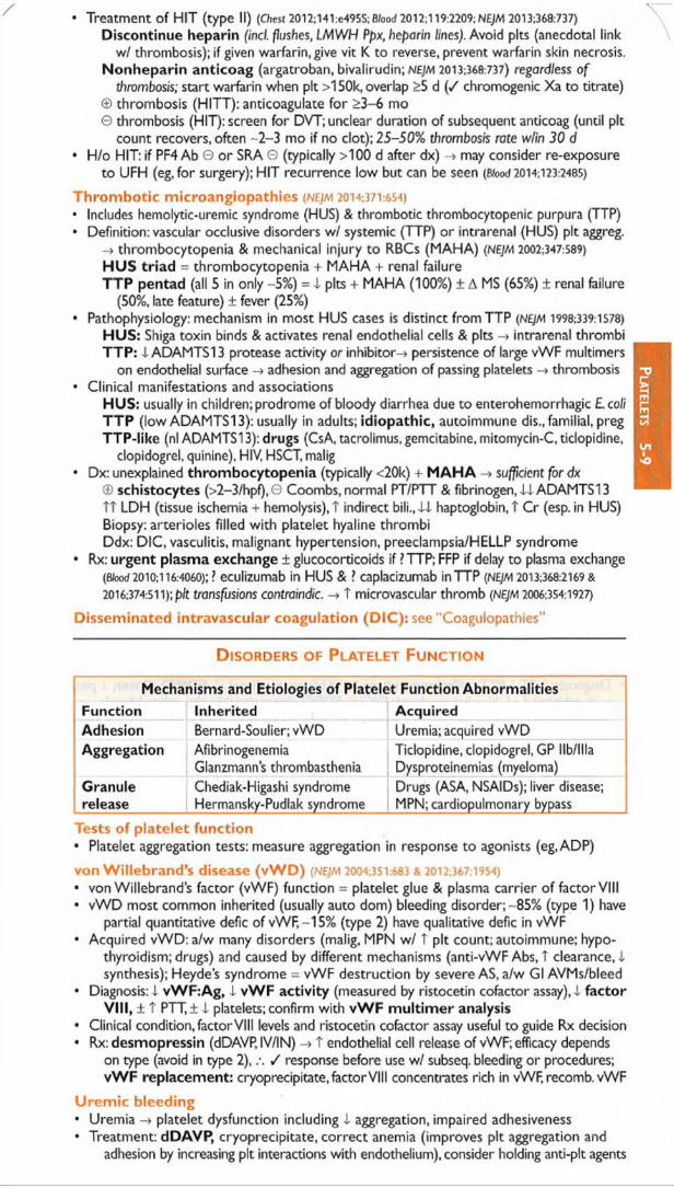
• Treatment of HIT (type II) (Chest 2012;141:e495S;B/oo£f 2012;119:2209; N£JM 2013:368:737)
Discontinue heparin find. flushes, LMWH Ppx, heparin lines). Avoid pits (anecdotal link w/ thrombosis); if given warfarin, give vit K to reverse, prevent warfarin skin necrosis.
Nonheparin anticoag (argatroban, bivalirudin; nejm 2013:368:737) regardless o f thrombosis; start warfarin when pit >150k, overlap >5 d ( / chromogenic Xa to titrate)
© thrombosis (HITT): anticoagulate for >3-6 mo© thrombosis (HIT): screen for DVT; unclear duration of subsequent anticoag (until pit
count recovers, often ~2-3 mo if no clot); 25-50% thrombosis rate wlin 30 d• H/o HIT: if PF4 Ab 0 o r SRA G (typically >100 d after dx) -> may consider re-exposure
to UFH (eg, for surgery); HIT recurrence low but can be seen (Btoocf 2014:123:2485)
Throm botic microangiopathies (nejm 2014:371:654)• Includes hemolytic-uremic syndrome (HUS) & thrombotic thrombocytopenic purpura (TTP)• Definition: vascular occlusive disorders w/ systemic (TTP) or intrarenal (HUS) pit aggreg.
-> thrombocytopenia & mechanical injury to RBCs (MAHA) (NEJM 2002:347:589)
H U S triad = thrombocytopenia + MAHA + renal failure TTP pentad (all 5 in only -5%) = I pits + MAHA (100%) ± A MS (65%) ± renal failure
(50%, late feature) ± fever (25%)• Pathophysiology: mechanism in most HUS cases is distinct from TTP (NEJM 1998:339:1578)
H U S : Shiga toxin binds & activates renal endothelial cells & pits -* intrarenal thrombi TTP: i ADAMTS13 protease activity or inhibitor-* persistence of large vWF multimers
on endothelial surface -> adhesion and aggregation of passing platelets -* thrombosis• Clinical manifestations and associations
H U S : usually in children; prodrome of bloody diarrhea due to enterohemorrhagic Lcoli T T P (low ADAMTS13): usually in adults; idiopathic, autoimmune dis., familial, preg TTP-like (nl ADAMTS13): drugs (CsA, tacrolimus, gemcitabine, mitomycin-C, ticlopidine,
dopidogrel, quinine), HIV, HSCT, malig• Dx: unexplained thrombocytopenia (typically <20k) + M A H A -> sufficient for dx
© schistocytes (>2-3/hpf),0 Coombs, normal PT/PTT & fibrinogen, 4XADAMTS13 Tt LDH (tissue ischemia + hemolysis), T indirect bili., i i haptoglobin, T Cr (esp. in HUS) Biopsy: arterioles filled with platelet hyaline thrombi Ddx: DIC, vasculitis, malignant hypertension, preeclampsia/HELLP syndrome
• Rx: urgent plasma exchange ± glucocorticoids if ? TTP; FFP if delay to plasma exchange(Blood 2010:116:4060); ? eculizumab in HUS & ? caplacizumab in TTP (nejm 2013:368:2169 &
2016:374:511); p/t transfusions contraindic. -» T microvascular thromb (nejm 2006:354:1927)
Disseminated intravascular coagulation (D IC ): see "Coagulopathies”
D iso r d e r s o f P l a t e l e t F u n c t io n
Mechanisms and Etiologies of Platelet Function AbnormalitiesFunction Inherited AcquiredAdhesion Bernard-Soulier; vWD Uremia; acquired vWDAggregation Afibrinogenemia
Glanzmann’s thrombastheniaTiclopidine, dopidogrel, GP llb/llla Dysproteinemias (myeloma)
Granulerelease
Chediak-Higashi syndrome Hermansky-Pudlak syndrome
Drugs (ASA, NSAIDs); liver disease; MPN; cardiopulmonary bypass
Tests of p latelet function• Platelet aggregation tests: measure aggregation in response to agonists (eg,ADP)
von W illebrand ’s disease (v W D ) (NEJM 2004:351:683 & 2012:367:1954)• von Willebrand’s factor (vWF) function = platelet glue & plasma carrier of factor VIII• vWD most common inherited (usually auto dom) bleeding disorder; - 85% (type 1) have
partial quantitative defic of vWF,-15% (type 2) have qualitative defic in vWF• Acquired vWD: a/w many disorders (malig, MPN w/ T pit count; autoimmune; hypo
thyroidism; drugs) and caused by different mechanisms (anti-vWF Abs, T clearance, i synthesis); Heyde’s syndrome = vWF destruction by severe AS, a/w Gl AVMs/bleed
• Diagnosis: I vW F:Ag, 4 vW F activity (measured by ristocetin cofactor assay), i factorV l l l , ± t PTT, ± I platelets; confirm with vW F m ultim er analysis
• Clinical condition, factorVIII levels and ristocetin cofactor assay useful to guide Rx decision• Rx: desmopressin (dDAVP, IV/IN) -> t endothelial cell release of vWF; efficacy depends
on type (avoid in type 2), / response before use w/ subseq. bleeding or procedures;vW F replacement: cryoprecipitate.factorVIII concentrates rich in vWF, recomb. vWF
Urem ic bleeding• Uremia -> platelet dysfunction including i aggregation, impaired adhesiveness• Treatment: dDAVP, cryoprecipitate, correct anemia (improves pit aggregation and
adhesion by increasing pit interactions with endothelium), consider holding anti-pit agents

Co
agul
opa
thie
s 5-
10COAGULOPATHIES
Screening Test Abnormalities in Inherited and Acquired CoagulopathiesPT PTT Factors Inherited AcquiredT <-> VII FVII defic. Vit. K defic.; liver dis.; factor inhib.
T VIII or IX Hemophilias, vWD Antiphospholipid Ab; factor inhib.T T 1,11,V or X Fbgn, Fll or FV defic. DIC; liver dis.; factor inhib.
F u r th e r coagu la tion tests• Mixing study: useful if T PT or PTT; mix Pc’s plasma 1:1 w/ normal plasma and retest
PT/PTT normalizes -» factor deficiency; PT/PTT remains elevated -> factor inhibitor• Coagulation factor levels: useful if mixing study suggests factor deficiency
DIC -» all factors consumed; I factors V and VIIIliver disease -» i all factors exceptVIII; .\1 factorV, normal factorVIIIvitamin K deficiency -» i factors II,VII, IX, X (and protein C, S); normal V and VIII
• D IC screen: fibrinogen (consumed), fibrin degradation products (FDPs,© due to intensefibrinolysis), D-dimer (more specific FDP test that detects degradation of X-linked fibrin)
H em oph ilias (Lancet 2016:388:187)• X-linked recessive factor V III (hemophilia A) or factor IX (hemophilia B) deficiency• Classification: mild (5-25% normal factor activity), moderate (1-5%) o r severe (<1%)• Clinical manifestations: hematomas, hemarthroses, bruising, bleeding (mucosal, Gl, GU)• Diagnosis: T PTT (normalizes w/mixing study), normal PT & vWF, i factor VIII or IX• Rx: purified/recomb. factor VIII (NEJM 2016:374:2054) or IX; desmopressin (mild dis.); amino-
caproic acid;cryo (FVIII); recomb, factor VII or IX-Fc fusion proteins have T ti/2, so 1-2x/wk dosing for Ppx (NEJM 2013:369:2313); ? emicizumab (binds FIX & X; nejm 2016:374:2044)
C oagu la tion fa c to r in h ib ito rs (most commonly anti-factor VIII)• Etiologies: hemophilia; postpartum; lymphoproliferative & autoimmune disorders; cancers• Diagnosis: T PTT (does not normalize w/mixing study); Bethesda assay quantitates tite r• Treatment: if high titer -» recomb, factor V ila , porcine factor concentrates, activated
prothrombin complex; for others -> high-purity human factor, plasmapheresis, immunosupp. w / steroids, CYC and/or RTX (Curr opm Hemotoi 2008:15:451)
D issem inated in travascu la r coagu la tion (D IC ) (nejm 2014:370:847)• Etiologies: trauma, shock, infection, malignancy (esp. APL), obstetric complications• Pathogenesis: mass/Ve activation of coagulation that overwhelms control mechanisms
thrombosis in microvasculature -» ischemia + microangiopathic hemolytic anemiaacute consumption of coagulation factors and platelets -» bleedingchronic DIC -> able to compensate by T factors and platelets -> thrombosis
• Diagnosis: t PT, T PTT, I fibrinogen (may be nl b/c acute phase), © FDP/D-dimer, i pits,© schistos, T LDH, i hapto; chronic DIC: © FDP/D-dimer, variable pits, other labs nl
• Treatment: Rx underlying process; support w/ FFP, cryo (goal fbgn >100 mg/dL) & pitsV itam in K deficiency• Etiologies: malnutrition, 4- absorption (antibiotic suppression o f vitamin K-producing
intestinal flora o r malabsorption), liver disease ( i stores), warfarin
Properties and Antidotes for Anticoagulants & Fibrinolytics (Ore 2016:134:248)
Anticoag. tl/2 Labs Rx for O/D w/ serious bleeding (+ d/c anticoag)UFH 60-90',
REST PTT Protamine IV 1 mg/100 U UFH (max 50 mg). For
infusions, dose to reverse 2x UFH given per h.LMWH 2-7°, K anti-Xa* Protamine reverses -60%Bivalirudin 25', K T PTT DialysisArgatroban 45', L T PTT ? DialysisFondaparinux 24°, K anti-Xa* > DialysisWarfarin 36°. L T PT No 6/eeding: INR 4.5-10, 0 Rx or vit. K 2.5 mg PO;
INR >10 give 5 mg PO (sup to SC, = IV at 24 h) Bleeding: vit. K 10 mg IV + FFP 2-4 U IV q6-8h; PCC (eg, KCentra) faster, i tfn (Ore 2013:128:360)
Fibrinolytic 20', LK 1 fbgn Cryoprecipitate, FFP, ± aminocaproic acidDabigatran -12°, K T PTT* Idarudzumab (NE/M 2015:373:511)
RivaroxabanApixabanEdoxaban
8-12°,K > L
T PT*anti-Xa*
Anti-fibrinolytic agent; consider PCC; specific reversal agents (eg, andexanet) under development (NEJM 2015:373:2413;J TTiromb Hoemost 2015;13.S187)
^Routine monitoring not performed. Mode of excretion: K, kidney; L, liver; RES, reticuloendothelial system. PCC: prothrombin complex concentrate (FII.VII, IX, X; Protein C & $). Anti-fibrinolytics: tranexamic,aminocaproic acid.

HYPERCOAGULABLE STATESSuspect in Pts with venous or arterial thrombosis at young age or unusual locations,
recurrent thromboses or pregnancy loss, or ® FHx
Inherited Hypercoagulable StatesRisk factor Prevalence VTE CommentsFactor V Leiden 3-7% 4.3x Activated protein C (APC) resist.Prothrombin mutation 2% 2.8x G20210A -» T prothrombin levelHyperhomocysteinemia 5-10% 2.5x Inherited or acquiredProtein C deficiency 0.02-0.05% 11x
Warfarin-induced skin necrosis riskProtein S deficiency 0.01-1% 32xAntithrombin III def. 0.04% 17.5x May be heparin-resistant
P reva lence is in C aucasians. (NEjAl 2001 ;344:1222; JA M A 2005:293:2352)
Vascular Beds Affected by Inherited and Acquired Hypercoagulable StatesVenous
1: Factor V Leiden■c Prothrombin mutation - i protein C, S or AT III
Venous and Arterial? factor V Leiden + smoking Hyperhomocysteinemia (inherited or acquired) Dysfibrinogenemia
Stasis: immobilization, ■g surgery, CHF • | Malignancy g" Hormonal: OCPs, HRT, < tamoxifen, pregnancy
Nephrotic syndrome
Platelet defects: myeloproliferative disorders, HIT, PNH (although venous > arterial)
Hyperviscosity: polycythemia vera, Waldenstrom’s macroglobulinemia, sickle cell, acute leukemia
Vessel wall defects: vasculitis, trauma, foreign bodies Others: antiphospholipid syndrome, IBD
D iagnostic eva lua tion (not routinely required for initial VTE)• APC resistance screen; prothrombin PCR test; functional assays for proteins C and S,
ATIII; homocysteine level; factor VI11 levels; anticardiolipin and lupus anticoagulant Ab. Also consider nephrotic syndrome, PNH (esp. if mesenteric thrombus).
• Consider JAK2 mutation testing if suspect MPN or splanchnic thrombosis• Proteins C & S and ATIII levels are affected by acute thrombosis and anticoagulation
levels best assessed >2 wk after completing anticoagulation course• Age-appropriate malignancy screening (occult cancer in -4% of initial unprovoked VTE; no
benefit of routine abd/pelvis CT; nejm 2015:373:697)T re a tm e n t• Asx w/ inherited risk factor: consider prophylactic anticoag. if develops acquired risk factor• Thrombosis w/ inherited risk factor: see "Venous Thromboembolism”A n tip h o sp h o lip id syndrom e (APS) (^Thromb Haemost 2006:4:295;NE}M 2013:368:1033)• Definition: dx requires >1 clinical & >1 laboratory criteria
Clinical: thrombosis (any) or complication of pregnancy (>3 spont. abortions before 10 wk or >1 fetal loss after 10 wk or premature birth before 34 wk)
Laboratory:© moderate-high titer anticardiolipin (ACL),© lupus anticoagulant (LA), or © p2-glycoprotein-l (fc-GP-l) Ab, on >2 occasions, at least 12 wk apart
• Clinical: DVT/PE/CVA, recurrent fetal loss, I pits, hemolytic anemia, livedo reticularis;"catastrophic APS": >3 organ systems in <1 wk wI © APLA & tissue microthrombi; 44% mortality (Artfi Rheum 2006:54:2568); Rx w/ plasmapheresis, rituximab
• Antiphospholip id antibodies (A P LA )/ if: SLE, age <40 y & arterial thromb, recurrent venous thromb, spontaneous abortion ACL:Ab against cardiolipin, a mitochondrial phospholipid; IgG more specific than IgM LA:Ab that prolongs phospholipid-dependent coagulation reactions; T PTT that
does not correct with mixing study but does correct with excess phospholipids or platelets; PT not affected b/c the reaction contains much more phospholipid
fc-GP-LAb against (^-glycoprotein-1, IgG or IgM (uncertain role of Abs in pathogenesis) False ©VDRL:nontreponemal test for syphilis in which cardiolipin is part of Ag complex Risk of thromboembolic phenomena may increase with titer ofAPLs
• Etiologies: primary (idiopathic) or secondary due to auto im m une syndromes(eg, SLE), malignancy, infections, drug reactions
• Treatment: UFH/LMWH -> warfarin after thromboembolic event (lifelong for most Pts)Intensity of anticoagulation controversial (Nat Rev Rheum 2015;11:586)Initial venous thrombosis: INR 2-3 (NEJM 2003;349:1133;J77?romb Haemost 2005;3:848)Initial arterial thrombosis: typically INR 2-3 + ASA 81, although some treat to INR 3-4 Recurrent thrombosis on warfarin: consider INR 3-4 vs, LMWH or fondaparinux (Arth
Rheum 2007:57:1487)Consider ASA prophylaxis for high-risk asx Pt (eg, SLE); no current evidence for NOACs
Hyper
co
ag
5-11

Le
uk
oc
yt
es
5-
12
I DISORDERS OF LEUKOCYTES i
L
Neutrophilia (>7500-10,000/^iL)Infection Usually bacterial; ± toxic granulations, Dohle bodiesInflammation Burn, tissue necrosis, Ml, PE, collagen vascular diseaseDrugs and toxins Corticosteroids, p-agonists, lithium, G-CSF; cigarette smokingStress Release of endogenous glucocorticoids and catecholaminesMarrow stimulation Hemolytic anemia, immune thrombocytopeniaAsplenia Surgical, acquired (sickle cell), congenital (dextrocardia)Neoplasm Can be 1° (MPN) or paraneoplastic (eg, carcinomas of lung, Gl)Leukemoid reaction >50,000/|jL + left shift, not due to leukemia; unlike CML, T LAP
Neutropenia (ANC <1000/jiL)Congenital
Infection
Myelokathexis, Shwachman-Diamond-Oski, Chediak-Higashi, retie dysgen., WHIM syndrome, cyclic neutropenia, monoMAC syndrome ( I monos, NKs) Viral (CMV, EBV, HIV); bacterial (brucella, Rickettsia, TB); malaria
Nutritional Vit B12 defic., copper defic.Drugs and toxins
Chemotherapeutics, clozapine, methimazole,TMP-SMX, NSAIDs, sulfasalazine, phenytoin (Am) Hem 2009:84:428), alcohol
Neoplasm MDS, leukemia (AML, ALL, hairy cell, LGL, others)
Lymphocytosis (>4000-5000 / lL)Infection Usually viral;“atypical lymphocytes” with mononucleosis syndromes
Other: pertussis, toxoplasmosisHypersensitivity Drug-induced, serum sicknessStress Cardiac emergencies, trauma, status epilepticus, postsplenectomyAutoimmune Rheumatoid arthritis (large granular lymphocytes), malignant thymomaNeoplasm Leukemia (eg, CLL, hairy cell, LGL), lymphoma (eg, mantle cell, folic.)
Monocytosis (>500/pL)Infection Usually TB, SBE, Listeria, Brucella, Rickettsia, fungi, parasitesInflammation IBD, sarcoidosis, collagen vascular diseasesNeoplasm Hodgkin lymphoma, leukemias, MPD, carcinomas
Eosinophilia (>500/jxL)Infection Usually parasitic (helminths)Allergic Drugs; asthma, hay fever, eczema; ABRACollagen vase dis. RA, Churg-Strauss syndrome, eosinophilic fasciitis, PANEndocrine Adrenal insufficiencyNeoplasm HL, CML, mycosis fungoides, carcinomas, systemic mastocytosisAtheroembolic dis. Cholesterol emboli syndromeHypereosinophilicsyndrome
Multiorgan involvement inch heart & CNS, a/w FIP1L1-PDGFRA fusion (NEJM 2003:348:1201)
Basophilia (>150/pL)Neoplasm MPN, Hodgkin lymphomaAlteration in BM or reticuloendothelial compartment Hemolytic anemia, splenectomy Inflammation or allergy IBD, chronic airway inflammation
LymphadenopathyViral HIV, EBV, CMV, HSV.VZV, hepatitis, measles, rubellaBacterial Generalized (brucellosis, leptospirosis,TB, atypical mycobacteria, syphilis)
Localized (streptococci, staphylococci, cat-scratch disease, tularemia)Fungal and parasitic
Histoplasmosis, coccidioidomycosis, paracoccidioidomycosis Toxoplasmosis
Immunologic Collagen vascular disease, drug hypersensitivity (eg, phenytoin), serum sickness, histiocytosis X, Castleman’s and Kawasaki disease
Neoplasm Lymphoma, leukemia, amyloidosis, metastatic carcinomaOther Sarcoidosis; lipid storage diseasesFactors that favor biopsy
Age (>40 y), size (>2 cm), location (supraclavicular is always abnormal), duration (>1 mo)
Consistency (hard vs. rubbery vs. soft) & tenderness are not reliable

TRANSFUSION THERAPY I 1
Packed red blood cells (PRBCs)(Anno/s 2012:157:49)
Platelets (pits) (Annc/s 2014:162:205)
Fresh frozen plasma (FFP)
Cryoprecipitate
Irradiated
CMV-negative
Leuko-reduced
Intravenous immune globulin(ivig)Therapeuticapheresis
Massive transfusion
Blood Products and Indications (Lancet 2013:381:1845)
For acute blood loss or to T C^-carrying capacity if end organ ischemia. 1 U PRBC -4 T Hb by ~1 g/dL. Conservative Hb goal >7 g/dL adequate for UGIB & critically ill PtS (NEJM 2013:368:11 & 2014:371:1381; bmj 2015;350:h 1354). Controversy remains re: coronary ischemia, although Hb >8 may be adequate (jAMAim Med 2013:173:132), but perhaps not peri-cardiac surgery (NEJM 2015.372:997; Anesth 2016;125:46).
For pits <10k (nejm 2010:362:600) or <20k w/ infxn or T bleeding risk or <50k w/ active bleeding or preprocedure. 6 U pooled donor pits 1 single donor pit apheresis unit ( ! alloimmunization) -4 T pit -30-60k.
Contraindic: TTP/HUS, HELLP, HITRefractory if T <5k 30-60' post-pits. Suggests alloimmunization -> trial
ABO-matched pits. If still refractory / panel reactive Abs to assess utility of HLA-matched pits.
Contains all coagulation factors. For bleeding due to deficiency of multiple coagulation factors (eg. DIC,TTP/HUS, liver disease, warfarin excess, dilution) Or INR >2 preprocedure (Transfusion 2006:46:1279).
Enriched for fibrinogen, vWF.VIII and XIII. For bleeding in vWD, factor XIII deficiency or fibrinogen <100 mg/dL.Prevents donor T-cell proliferation. Use if risk of transfusion-assoc. GVHD (HSCT, heme malignancy, congenital immunodeficiency).From CMV-negative donors. For CMV-seronegative pregnant women, transplant candidates/recipients, SCID.AIDS Pcs.WBCs cause HLA alloimmunization and fever (cytokine release) and carry CMV. For chronically transfused Pts, potential transplant recipients, h/o febrile nonhemolytic transfusion reaction, cases in which CMV-negative products are desired but unavailable.Polyvalent IgG from >1000 donors. For postexposure prophylaxis (eg, HAV), certain autoimmune disorders (eg, ITP, Guillain-Barre, MG,? CIDP), congenital or acquired hypogammaglobulinemia (CVID, CLL). Removes large molec wt subst. (eg. cryoglobulinemia, Goodpasture’s, Guillain-Barre, hyperviscosity syndrome,TTP) or cells (eg, leukemia w/ hyperleukocytosis, sx thrombocytosis, sickle cell) from plasma. Large-vol. PRBC -> i Ca, T K, I pit, T coags; ratio of PRBC:plt:FFP repletion controversial, follow labs (JTrauma 2006;60:S91 & 2008.65:272).
Transfusion Complications (NEJM 1999:340:438; JAMA 2003:289:959)
Noninfectious Risk (per unit) Infectious Risk (per unit)Febrile 1:100 CMV CommonAllergic 1:100 Hepatitis B 1:220,000Delayed hemolytic 1:1000 Hepatitis C 1:1,600,000Acute hemolytic <1:250,000 HIV 1:1,800,000Fatal hemolytic <1:100,000 Bacteria (PRBCs) 1:500,000TRALI Ln O O O • Bacteria (platelets) 1:12,000
Transfusion reactions• For all reactions (except minor allergic): stop transfusion; send remaining blood
product and fresh blood sample to blood bank• Acute hemolytic: fever, HoTN, flank pain, AKI w/in 24 h. Due to ABO incompatibility -4
preformed Abs vs. donor RBCs. Rx: IVF, T UOP w/ diuretics, mannitol or dopamine• Delayed hemolytic: generally less severe than acute hemolytic; 5-7 d after transfusion
Due to undetected allo-Abs against minor antigens -> anamnestic response.Rx: usually no specific therapy required; dx is important for future transfusion
• Febrile nonhemolytic: fever, rigors 0-6 h post transfusion. Due to Abs vs donor WBCsand cytokines in blood product Rx: acetaminophen ± meperidine; r/o infection, hemolysis
• Allergic: urticaria; rarely, anaphylaxis: bronchospasm, laryngeal edema, hypotensionReaction to transfused proteins; anaphylaxis seen in IgA-deficient Pts w/ anti-IgA Abs. Rx: urticaria -> diphenhydramine; anaphylaxis -4 epinephrine ± glucocorticoids
• Transfusion-associated circulatory overload (TACO): T volume pulm edema, t BPRx:slow transfusion rate, diuretics,O2,± nitrates,! positive pressure ventilation
• Transfusion-related acute lung injury (TRALI): noncardiogenic pulmonary edemaDue to donor Abs that bind recipient WBCs, which then aggregate in pulmonary vasculature and release mediators causing T capillary permeability. Rx: see ‘,ARDS.,,
Tr
an
sfusio
n 5-13

MD
S 5-
14| M Y E L O D Y S P L A S T I C S Y N D R O M E S ( M D S ) ^
M yelo id neoplasm ove rv iew (Blood 2016,127:2391)• Categories based on clinical features, morphology, immunophenotyping, and genetics
W H O 2016 classification o f myeloid neoplasms & acute leukemiaAcute myeloid leukemia Clonal myeloid stem cell (SC) disorder w/ >20% blastsMyelodysplastic syndromes
Myeloproliferative neoplasms MDS/MPN neoplasms
Dysplastic clonal myeloid SC disorder -» cytopenias; <20% blasts, risk of leukemic transformation Nondysplastic multipotent myeloid SC clonal expansion Features of MDS & MPN (eg, CMML, atypical CML)
Myeloid/lymphoid malig. w/ eos & rearrangements of PDCFR or FGFR 7 or w/ PCM 1-JAK2
May be responsive toTKI therapy (eg, imatinib) for PDGFR rearrangement
Mastocytosis Systemic disease, assoc w/ KIT mutationsMyeloid neoplasms w/ germ line predisposition
MDS, MDS/MPN, acute leukemias in background of predisposing germline mutations
M yelodysplastic syndrom es (M D S) ove rv iew (/oncet 2014:383:2239)• Acquired clonal stem cell disorder -» ineffective hematopoiesis -» cytopenias,
dysmorphic blood cells and precursors, variable risk of leukemic transform ation• Epidemiology: >10,000 cases/y; median age ~70 y; male predominance (1.8x)• Id io p a th ic o r 2° to chemo w/ a lky la ting agents; T risk w/ radiation, benzene• Clinical manifestations: anem ia (85%), neutropenia (50%), thrombocytopenia (40-65%)• Diagnosis: dysplasia (usually multilineage) in peripheral smear (oval macrocytes,
pseudo-Pelger-Huet anomaly) and bone marrow (>10% dysplasia with blasts ± RS)• Both cytogenetic [eg, del(5q), mono 7, del(7q), trisomy 8, del(20q)] and m o lecu la r abnl
(TP53, EZH2, ETV6, RUNX1.ASXL1, SF3B1, DNMT3A) may carry prognostic signif• Prior to dx MDS: exclude AML (>20% blasts) and CMML (monos >1 x 10⁹/L); r/o 2° BM As
(defic. of Bn, folate, copper); viral infx (eg, HIV); chemo; EtOH; lead, arsenic exposures
W H O 2016 Classification Systems fo r MDS (Bfood 2016:127:2391)
Classification W H O 2008 FeaturesMDS w/ single lineage dysplasia (MDS-SLD)
RCUD(RA/RN/RT)
1 dysplastic lineage, 1-2 cytopenias. <15% RS*, <5% BM/<1% PB blasts, no Auer rods
MDS w/ multilineage dysplasia (MDS-MLD)
RCMD 2-3 dysplastic lineages, 1-3 cytopenias,<15% RS* <5% BM/<1% PB blasts, no Auer rods
MDS w/ ring sideroblast (MDS-RS)
RARS >15% RS or >5% RS if SF3B1 mut. is present, <5% BM/<1% PB blasts, no Auer rods
MDS w/ isolated del(5q) MDS w/ excess blasts (MDS-EB)
Del(5q)RAEB-1RAEB-2
Del(5q) alone or w/ 1 abnl except -7 or del(7q) EB-1: 5-9% BM/2-4% PB blasts, no Auer rods EB-2:10-19% BM/5-19% PB blasts or Auer rods
MDS, undassifiable (MDS-U)
MDS-U w/ 1% PB blasts, single lineage dysplasia & pancytopenia, or defining cytogenetic alteration
Certain cytogenetics [eg, t(15;17), t{8;21), inv16, t(16;16), or MLL rearrangement] classified as AML, regardless of BM blast count. RS, ring sideroblast; BM, bone marrow; PB, peripheral blood, * <5% RS if SF3B1 mutation.
• Rx (Am J Hemoto/2012:87:692): intensity based on IPSS-R (qv), age, performance status (PS)Poor PS, any risk -» supportive care (transfusions, G-CSF, Epo.TPO-mimetic.abx prn) Low/intermediate risk -> Epo (if Epo level <500); lenalidomide (esp. for 5q syndrome;
N E JM 2006:355:1456); DNA hypomethylating agents (azacitidine or decitabine) Intermediate/high risk DNA hypomethylating agents (survival advantage w/ azacitidine;
Loncet Oncol 2009:10223), combination chem o (akin to AML Rx) or allogeneic HSCT Hypoplastic MDS (rare) -» consider im m unosuppression (CsA.ATG, pred), HSCT
• Prognosis: IPSS-R correlates with surviva l and progression to A M L
Revised International Prognostic Scoring System (IPSS-R) (BJood2012:120.2454)
Variable 0 0.5 1 1.5 2 3 4Cytogenetics Very good • Good - Intermed Poor Very poorBM blasts (%) <2 - >2 to <5 - 5-10 >10 -Hb (g/dL) >10 - 8 to <10 <8 - -Pit (k) >100 50 to <100 <50 - - -ANC >0.8 <0.8 - - - -
Total score £1.5 >1.5 to 3 >3 to 4.5 >4.5 to 6 >6Category Very low Low Intermec High Very highMedian survival (y) 8.8 5.3 3.0 1.6 0.8

MYELOPROLIFERATIVE NEOPLASMS (MPN)General (Am J Hcmatol 2012:87:285;JAMA Oncol 2015:1:97; Blood 2016,127:2391)• Results from clonal expansion of multipotent hematopoietic stem cell• Different from MDS in that the cells are not dysplastic (ie, normally developed)• Categories of MPN: polycythemia vera (PV); essential thrombocythemia (ET); primary
myelofibrosis (PM); chronic myelogenous leukemia (CML), BCR-ABL1 ©; chronic neutrophilic leukemia (CNL); chronic eosinophilic leukemia, not otherwise specified; myeloproliferative neoplasms, unclassifiable
• Mutations useful as clonal markers & dx tools:Gain of fxn mutations in JAK2V617F (janus kinase) frequently present (PV -95%,
ET -50%, PMF -50%; NEJM 2005:352:1779)
BCR-ABL fusion in all cases of CMLCALR exon 9 mutation (most MPNs w/o JAK2 or MPL mutation, including -25% of ET,
-35% of myelofibrosis Pts; nejm 2013:369:2379 & 2391)
MPL, TET2, & ASXL1 mutation w / lower frequency CSF3R mutation present in -60% of CNL
Po l y c y t h e m ia V era (P V )
Definition• T in RBC mass ± t granulocytes and platelets in the absence of physiologic stimulus
Etiologies of erythrocytosis• Relative T RBC ( ! plasma): dehydration;“ stress” erythrocytosis (Gaisbock’s syndrome)• /toso/ute ? RBC: 1° (PV, other MPD) or 2° due to hypoxia; carboxyhemoglobinemia;
inappropriate erythropoietin (renal, hepatic, cerebellar tumors); Cushing’s syndrome
Clinical manifestations (common between PV and ET)• Symptoms -> often termed “ vasomotor symptoms”
hyperviscosity (erythrocytosis): headache, dizziness, tinnitus, blurred vision thrombosis (hyperviscosity, thrombocytosis): transient visual disturbances (amaurosis,
ocular migraine); Budd-Chiari syndrome; erythromelalgia = intense burning, pain and erythema of extremities due to microvascular ischemia;? risk of DVT, M l, stroke. Risk of thrombosis highly correlated with T WBC in PV and ET (see below),
bleeding (abnormal platelet function): easy bruising, epistaxis, Gl bleeding ? histamine from basophils -> pruritus, peptic ulcers;? uric acid (cell turnover) -> gout
• Signs: plethora, splenomegaly, hypertension, engorged retinal veins• Expression profiling beyond JAK2 may define different phenotypes (NEJM 2014:371:808)
Diagnostic evaluation• Men: Hb >16.5 g/dL or HCT >49%, women: Hb >16 g/dL or HCT >48%, or ? red cell mass• BM bx -* hypercellularity for age, trilineage growth, pleomorphic mature megakaryocytes• JAK2 V617F mutation in -95% of PV; other Pts typically harbor JAK2 exon 12 mutations• / Epo to rule out secondary causes of erythrocytosis; if Epo i , PV m ore likely
If Epo ?, then / SaC>2 or Pa02, carboxyhemoglobin, BM exam• ± ? WBC, platelets, basophils; ? uric acid, leukocyte alkaline phosphatase, vit B12
• Peripheral smear -» no morphologic abnormalities
Treatm ent• Phlebotomy to goal Hct <45% (NEJM 2013,368:22), consider <42% in women• Low-dose A S A in all Pts (nejm 2004:350:114)• Hydroxyurea if high risk of thrombosis (age >60, prior thrombosis) or symptomatic
thrombocytosis (pit >1.5 x 1 06/|liL), o r if inadequate Hct by phlebotomy alone• Ruxolitinib (JAK1/2 inhibitor) if poor response, intolerant of hydroxyurea (NEJM 2015:372:426)• PEG IFNa-2a yields high response rate w/ limited toxicity (BJood 2008:112:3065)• Supportive: allopurinol (gout), H2-blockers/antihistamines (pruritus)
Prognosis• Median survival w / Rx -13.5 y (Blood 2014:124:2507); ? age, WBC, additional acquired
somatic mutations -» worse prognosis (Hoemotof 2013; 160:251)• Post-PV myelofibrosis (spent phase) occurs in 10-20% of cases, usually after 10 y• Risk of transformation into acute leukemia (<2-5%; higher if previous cytoreductive chemo)
E s s e n t ia l T h r o m b o c y t h e m ia (E T )
Definition♦ Sustained ? in platelets (>450,000/pL) ± ? RBC and granulocytes
MP
N
5-15

MP
N
5-16
SE tio log ies o f th ro m b o cy to s is• 1° = ET or other MPN; myelodysplastic syndromes (5q-syndrome); RARS-T• 2° = reactive th ro m b o cy to s is : inflammation (RA, IBD, vasculitis), infection, acute
bleeding, iron deficiency, postsplenectomy, neoplasms (eg, Hodgkin lymphoma)• O f patients with pit >106/pL, <1 in 6 will have ETC lin ica l m an ifes ta tions (also see “ Polycythemia Vera")• Thrombosis with erythromelalgia (risk of thrombosis highest in Pts with leukocytosis),
bleeding, pruritus; mild splenomegaly; migraine,TIA; early fetal lossD iagnostic eva lua tion• Peripheral smear: large hypogranular platelets• BM bx: megakaryocytic hyperplasia; absence of Philadelphia chromosome and very rarely,
minor increase in reticulin fibers; normal iron stores• JAK2 V617F present in -50% of ET; MPL or CALR mutations in majority of JAK2 w t• Patients should not meet W H O criteria for diagnosis of CML, PV, PMF o r MDS
Treatm ent o f ETRisk Features ASA 81 mg qd C ytoreduction
Low
Age <60 and no h/o thrombosis and pit <1.5 x 10⁶/pL and no CV risk factors
Consider for vasomotor symptoms No
Int. Neither low nor high ± Consider if pit >1.5 x 10⁶/liL
HighAge >60 or h/o thrombosis or pit >1.5 x 10⁶/piL
© (consider holding if pit >1 x 10⁶/,uL and lab evid.
of acquired vWD)
Hydroxyurea. Goal pit <0.4 x 10⁶/pL or sx free.
IFNra if young or pregnant.
Imetelstat (telomcrase inhib) under investigation (NEJM 2015:373:92)
Prognosis• Low-risk Pts have overall survival ~ control population• Risk of transformation into acute leukemia <2%; risk of progression to MF similar * •
P r im a r y M y e l o f ib r o s is (P M F )Definition• Clonal myeloproliferation with reactive marrow fibrosis & extramedullary hematopoiesis• Prefibrotic stage (pre-PMF): megakaryocyte prolif,grade 1 reticulin fibrosis, T BM cellularity.
Important to distinguish from ET:T thrombosis,? progression, A survival <Bfood 2012:120:569)Etiologies of myelofibrosis• Myeloproliferative neoplasm = primary myelofibrosis; post-PV/ET myelofibrosis• O ther hematologic (CML, AML, ALL, MDS) and solid cancers (breast, prostate)• Autoimmune (SLE and other collagen vascular disorders)• Toxins (benzene); radiation; granulomas (TB, fungal, sarcoid); deposition dis. (Gaucher's) Clinical manifestations (BJH 2012:158 453)
• Ineffective erythropoiesis -> anemia; extramedullary hematopoiesis -> massivesplenomegaly (abdominal pain, early satiety) ± hepatomegaly
• Tumor bulk and T cell turnover -> fatigue, weight loss, fever, sweatsDiagnostic evaluation (JAMA 2010,303:2S13; B/ood 2016:127:2391)
• Anemia with variable WBC and platelet counts• Peripheral smear -> “leukoerythroblastic” (teardrop cells, nucleated RBCs,
immature WBCs); large abnormal platelets• BM aspirate “dry” tap; BM bx -» severe fibrosis, replacement by reticulin & collagen• JAK2 V617F in 45-50%; CALR mut in 45-50%, MPL mut in 7-10%, triple neg in 1-2%• No BCR-ABL translocation; also does not meet criteria for PV or MDS Treatm ent (B/ood 2011:117:3494)
• In absence of adverse prognostic factors (eg, anemia o r sx) -> no treatment• Allogeneic HSCT only potential cure -» consider in young Pts with poor prognosis• Supportive care: transfusions; inconsistent benefit from androgens o r Epo;
? splenectomy if refractory to transfusions, failed chemoRx, painful splenomegaly• Hydroxyurea for significant leukocytosis o r thrombocytosis• Ruxolitinib (JAK1/JAK2 inhibitor) i sx, i splenomegaly, T survival (NEJM 2012:366:787 & 799)• Thalidomide and lenalidomide ± steroids may improve red cell count• Imetelstat (telomerase inhibitor) under investigation (nejm 2015:373:908)Complications and prognosis• Median survival ~6 y; transformation into AML occurs at a rate of -8%/y• Dynamic International Prognostic Scoring System (DIPPS plus): age >65,WBC >25k, Hgb
<10, blasts >1%,© symptoms, RBCTx, Pit <100K, karyotype (jeo 2011:29:392). IWG-MRT allows prognostication at any point during clinical course (Blood 2010:115:1703).

LEUKEMIAA c u t e L e u k e m ia
Definition• Clonal proliferation of hematopoietic progenitor with failed differentiation into mature
elements -> T blasts in bone marrow and periphery -» i RBCs, platelets and neutrophilsEpidemiology and risk factors• Acute myelogenous (AML): ~21k cases/y in U.S.; median age 67 y; >80% of adult acute
leukemias• Acute lymphocytic (ALL): -6 k cases/y in U.S.; median age 14 y but 2nd peak in older adults• Risk factors: radiation, chemo (alkylating agents, topo II inhib), benzene, smoking, ? rising
from acquired somatic mutations and clonal hematopoiesis (NEjM 2014:371:2477)• Secondary to acquired hematopoietic dis.: MDS, MPN (esp. CML), aplastic anemia, PNH• Inherited: Down’s, Klinefelter’s, Fanconi’s anemia, Bloom syndrome, ataxia telangiectasia Clinical manifestations• Cytopenias -> fatigue (anemia), infection (neutropenia), bleeding (thrombocytopenia)• More common in A M L
leukostasis (more often when blast count >50,000/|iiL): occluded microcirculation -> local hypoxemia and hemorrhage -> dyspnea, hypoxia, headache, blurred vision,TIA/CVA; look for hyperviscosity retinopathy (vascular engorgement, exudates, hemorrhage)
DIC (esp. with APL); leukemic infiltration of skin, gingiva (esp. with monocytic subtypes): chloroma: extramedullary tumor of leukemic cells, virtually any location
• More common in ALL:bony/lumbar pain, lymphadenopathy, hepatosplenomegaly (also in monocytic AML) CNS involvement (up to10%): cranial neuropathies, N/V, headache anterior mediastinal mass (esp. in T-cell); tumor lysis syndrome (qv)
Diagnostic evaluation (Blood 2009;i 14:937)• Peripheral smear: anemia, thrombocytopenia, variable WBC + circulating blasts (seen in
>95%;© Auer Rods in AML), peripheral flow cytometry for blast origin (ALL vs. AML)• Bone marrow: hypercellular with >20% blasts: test for cytogenetics and flow cytometry• Presence of certain cytogenetic anomalies, eg, t(15;17), t(8;21), inv(16) o r
t( 16; 16), are sufficient for dx of AML regardless of the blast count• / for tumor lysis syndrome (rapid cell turnover): T UA, T LDH, T K, t PO4, i Ca• Coagulation studies to r/o DIC: PT, PTT, fibrinogen, D-dimer, haptoglobin, bilirubin• LP (w/ co-admin of intrathecal chemotherapy to avoid seeding CSF w/ circulating
blasts) for Pts w/ ALL (CNS is sanctuary site) and for Pts w I AML w/ CNS sx• TTE if prior cardiac history o r before use of anthracyclines• H LA typing of Pt, siblings > parents/children for potential allogeneic HSCT candidates
A c u t e M y e l o g e n o u s L e u k e m ia (AML; NEjM2oi5;373:ii36)
Classification (W HO: Blood; 2016:127:2391)• Features used to confirm myeloid lineage and subclassify AML to guide treatment:
morphology: blasts, © granules, ± A uer rods (eosinophilic needle-like inclusions) cytochemistry: © myeloperoxidase and/or nonspecific esterase
• Immunophenotype: myeloid: CD13, CD33, CD117; monocyte: CD11 b, CD64, CD14, CD15• Cytogenetics: important for prognosis. Intermed. risk = no favorable/unfavorable features.
W H O 2016 Classification o f AM L (Blood 2016;127:2391)
4 M ajor Subtypes ExamplesRecurrent genetic abnl t(8;21); inv(16); PML-RARA; t(9;11), t(6;9), inv(3), t(1;22),
mutation in NP/V11, biallelic mutation in CEBPAMyelodysplasia-related A w/ or w/o antecedent MDS or MPNTherapy-related eg, alkylating agents or topoisomerase inhibitorsNot otherwise specified w/ min differentiation: w/ or w/o maturation; myelomonocytic;
monoblastic/cytic; pure erythroid; megakaryoblasticAlso: myeloid sarcoma, myeloid proliferations of Down’s syndrome
AM L Genetics (Blood 2010:115:453 & 1 16:354;NEJM 2016:374:2209)Favorable prognosis Unfavorable prognosis
Karyotype t(15;17) in APL;t(8;21); inv(16)/t(16;16)
-5 ;-7 ; 3q26 aberrations: t(6;9); 11q23 aberrations; complex karyotype
Gene mutations NPM1+; biallelic CEbPA FLT3 ITD; MLL-PTD; TPS3, RUNX 1Recurrent somatic mutations: DNMT3A; TET2; ASXL1; RAS; WT7; IDH1I2; spliceosome
Le
uk
em
ia 5-17
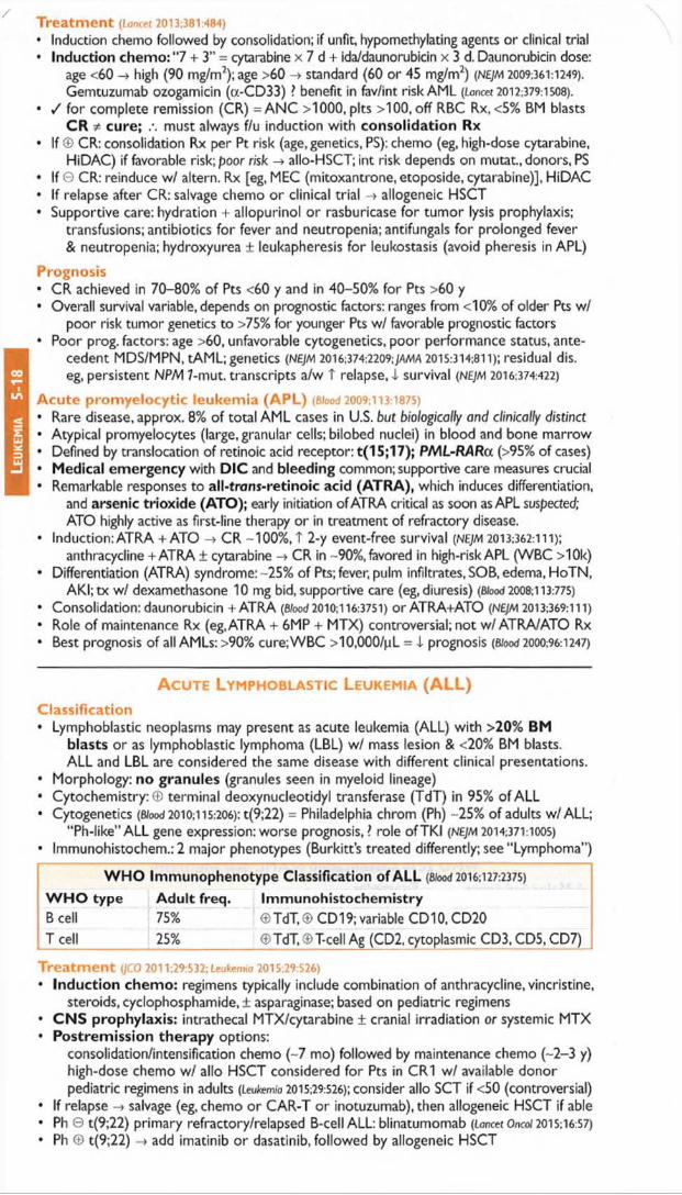
Leu
kem
ia
5-18
Treatm ent (lancet 2013;381:484)
• Induction chemo followed by consolidation; if unfit, hypomethylating agents or clinical trial• Induction chemo: "7 + 3" = cytarabine x 7 d + ida/daunorubicin x 3 d. Daunorubicin dose:
age <60 high (90 mg/m2);age >60 -» standard (60 or 45 mg/m2) (NEJM 2009:361:1249). Gemtuzumab ozogamicin (oc-CD33) ? benefit in fav/int risk AML {Lancet 2012:379:1508).
• / for complete remission (CR) = A N C >1000, pits >100, off RBC Rx, <5% BM blastsCR * cure; .\ must always f/u induction with consolidation Rx
• If © CR: consolidation Rx per Pt risk (age, genetics, PS): chemo (eg, high-dose cytarabine,HiDAC) if favorable risk; poor risk -> allo-HSCT; int risk depends on mutat., donors, PS
• If 0 CR: reinduce w/ altern. Rx [eg, MEC (mitoxantrone, etoposide, cytarabine)], HiDAC• If relapse after CR: salvage chemo or clinical trial -> allogeneic HSCT• Supportive care: hydration + allopurinol or rasburicase for tumor lysis prophylaxis;
transfusions; antibiotics for fever and neutropenia; antifungals for prolonged fever & neutropenia; hydroxyurea ± leukapheresis for leukostasis (avoid pheresis in APL)
Prognosis• CR achieved in 70-80% of Pts <60 y and in 40-50% for Pts >60 y• Overall survival variable, depends on prognostic factors: ranges from <10% of older Pts w/
poor risk tumor genetics to >75% for younger Pts w/ favorable prognostic factors• Poor prog, factors: age >60, unfavorable cytogenetics, poor performance status, ante
c e d e n t MDS/MPN, tAML; g e n e tic s (NEJM 2016:374:2209;jam a 2015:314:811); re s id u a l d is .
eg, p e rs is te n t NPM 7-mut. t r a n s c r ip ts a /w T re la p se , i s u rv iv a l {NEJM 2016:374:422)
Acute promyelocytic leukemia (APL) (B lo o d 2009:113:1875)
• Rare disease, approx. 8% of total AML cases in U.S. but biologically and clinically distinct• Atypical promyelocytes (large, granular cells; bilobed nuclei) in blood and bone marrow• Defined by translocation of retinoic acid receptor: t(15;17); PML-RARa (>95% of cases)• Medical emergency with DIC and bleeding common; supportive care measures crucial• Remarkable responses to all-trons-retinoic acid (ATRA), which induces differentiation,
and arsenic trioxide (ATO); early initiation of ATRA critical as soon as APL suspected; ATO highly active as first-line therapy or in treatment of refractory disease.
• Induction: ATRA + ATO -> CR -100%, T 2-y event-free survival (NEJM 2013:362:111);anthracycline +ATRA ± cytarabine -» CR in -90%, favored in high-risk APL (WBC >10k)
• Differentiation (ATRA) syndrome: -25% of Pts; fever, pulm infiltrates, SOB, edema, HoTN,AKI;tx w/ dexamethasone 10 mg bid, supportive care (eg, diuresis) (Wood 2008:113:775)
• Consolidation: daunorubicin + ATRA (Wood 2010; 116:3751) or ATRA+ATO {NEJM 2013:369:111)
• Role of maintenance Rx (eg, ATRA + 6MP + MTX) controversial; not w / ATRA/ATO Rx• Best prognosis of all AMLs: >90% cure;WBC >10,000/pL = i prognosis (Wood 2000:96:1247)
A c u t e L y m p h o b l a s t i c L e u k e m i a (ALL)C lass ifica tion• Lymphoblastic neoplasms may present as acute leukemia (ALL) with >20% BM
b l a s t s or as lymphoblastic lymphoma (LBL) w/ mass lesion & <20% BM blasts.ALL and LBL are considered the same disease with different clinical presentations.
• Morphology: n o g r a n u l e s (granules seen in myeloid lineage)• Cytochemistry:© terminal deoxynucleotidyl transferase (TdT) in 95% of ALL• Cytogenetics (Wood 2010;115:206): t(9;22) = Philadelphia chrom (Ph) -25% of adults w/ALL;
“ Ph-like” ALL gene expression: worse prognosis,? role ofTKI {NEJM 2014:371:1005)• Immunohistochem.: 2 major phenotypes (Burkitt’s treated differently; see “ Lymphoma” )
W H O I m m u n o p h e n o t y p e C la s s i f i c a t i o n o f A L L (8/ood 2016:127:2375)
W H O t y p e A d u l t f r e q . I m m u n o h i s t o c h e m i s t r yB cell 75% ©TdT,® CD 19;variable CD10.CD20T cell 25% ®TdT,©T-cell Ag (CD2, cytoplasmic CD3, CD5, CD7)
T r e a t m e n t (JCO 2011:29:532: leukemia 2015:29:526)
• Induction chemo: regimens typically include combination of anthracycline, vincristine,steroids, cyclophosphamide,! asparaginase; based on pediatric regimens
• C N S prophylaxis: intrathecal MTX/cytarabine ± cranial irradiation or systemic MTX• Postremission therapy options:
consolidation/intensification chemo (~7 mo) followed by maintenance chemo (-2-3 y) high-dose chemo w / alio HSCT considered for Pts in CR1 w / available donor pediatric regimens in adults (Leutem/o 2015:29:526); consider alio SCT if <50 (controversial)
• If relapse -» salvage (eg, chemo or CAR-T or inotuzumab), then allogeneic HSCT if able• Ph © t(9;22) primary refractory/relapsed B-cell ALL: blinatumomab (lancet Oncol 2015:16:57)
• Ph © t(9;22) -* add imatinib or dasatinib, followed by allogeneic HSCT

• MLL-AF4 t(4;11), hypodiploidy (<44 chromosomes), min residual disease consider allo-HSCT
• Infusion o f chimeric antigen receptor-modified T cells promising (N£JM 2014:371:1507)
Prognosis• Morphologic CR in >80% of adults; but minimal residual disease (MRD) at CR = poor prog.• Cure achieved in 50-60% if good prog, factors vs. 10-30% w/ poor prog, factors• Good prognostic factors: younger age.WBC <30,000/(LiL,T-cell immunophenotype,
absence of Ph chromosome or t(4 ;11), early attainment of CR w / MRD negative
C h r o n ic M y e l o g e n o u s L e u k e m ia (C M L )
Definition (8/ood 2009;114:937)
• Myeloproliferative neoplasm with clonal overproduction of hematopoietic myeloidstem cells that can differentiate
• Philadelphia chromosome (Ph) = t(9;22) -> BCR-ABL fusion -> T Abl kinase activityBCR-ABL required fo r Dx (make via karyotyping or FISH; PCR useful but not adequate)
• “Atypical CML” (BCR-ABL 0) now considered a separate disease and reclassified asMDS/MPN (qv) w/ many Pts © for CSF3R or SETBP1 mutations
Epidemiology and risk factors• -6600 new cases/y in U.S.; median age -64 at presentation; -15% of adult leukemias• T risk with irradiation; no clear relation to cytotoxic drugs
Clinical manifestations• Triphasic clinical course; 85% present in the chronic phase• Chronic phase: often asymptomatic but common features are fatigue, malaise, weight
loss, night sweats, abdominal fullness (splenomegaly 50%)• Accelerated phase: refractory leukocytosis, I pit and worsening sx -» fever, w t
loss, T splenomegaly, bone pain, bleeding, infections, pruritus (basophilia)• Blastic phase - acute leukemia -> severe constitutional symptoms, infection, bleeding,
and possible leukostasis (see “ Acute Leukemia")
Diagnostic evaluation• Peripheral smear: leukocytosis, left-shifted with all stages of myeloid maturation; anemia,
thrombocytosis, basophilia• Bone marrow: hypercellular, T myeloid to erythroid ratio, I leuk alkaline phosphatase• Chronic: <10% blasts (peripheral or BM)• Accelerated: 10-19% blasts, >20% basos, pits <100k, T spleen size, karyotypic prog.• Blastic: >20% blasts (2/j myeloid, 'A lymphoid), may see extramedullary leukemia
Treatm ent (Lancet 2015:385:1447)• Tyrosine kinase inhibitor (TK I): imatinib, dasatinib, nilotinib, bosutinib, & ponatinib are
selective inhibitors of BCR-ABL (JCO 2010:28:428; BW 2012:120:1390).Imatinib, nilotinib, & dasatinib approved as initial Rx.Resistance = recurrent dis. on TKI, often result of BCR-ABL mutation or amplification. Nilotinib, dasatinib, bosutinib, & ponatinib approved for resistant disease, w/ only ponatinib
effective onT315l resistance mutation (N£/A1 2012:367:2075 & 2013,369:1783).Side effects: nausea, diarrhea, muscle cramps, cytopenias, I PO4, T QT, rarely CHF;
dasatinib: pericardial & pleural effusions and pulm HTN; nilotinib: t bili & lipase, CV toxicity; ponatinib: thrombosis, pancreatitis and CV toxicity
• Chronic phase: TKI; continued indefinitely in responders (Blood 2012; 120:1390)• Accelerated phase: TKI upfront, consider allogeneic HSCT• Blastic phase: TKI vs.TKI + either ALL or AML induction (based on cell type); then HSCT• Allogeneic HSCT: possibility of cure, consider for Pts w/ available donor; Pts who present
in accelerated or blastic phase; or Pts with relapsed/refractory disease toTKIs
Goals of TK I Therapy (BJood 2013:122.872)
Response D efin ition O p tim a l tim eHematologic WBC <1 Ok, pit <450k, no immature cells in blood,
basophils <5%, spleen nonpalpable3 mo
Cytogenetic Absence of the Ph chromosome in metaphase cells 12 moMolecular <0.1% BCR-ABL = 3-log reduction by quantitative PCR 12 mo
Prognosis (Cancer 2013:119:2620)
• Chronic phase CML Rx’d w/ imatinib: 89% 5-y overall survival, 95% survival free of CML-re la te d dea ths, 7% p ro g re s s io n t o b la s t phase a t 5 y (NEJM 2006:355:2408)
• Accelerated phase CML Rx’d w/ imatinib: -50% overall survival at 4 y (Cancer 2005:103:2099)
• Poor prognostic factors: T age, T platelet count, t spleen size, T% of blasts/basophils
Le
uk
em
ia 5-19

Leu
kem
ia
5-20
/C hronic Lymphocytic Leukemia (CLL)
Definition {NE)M 2005,352:804; Blood 2008; 111:5446)• Monoclonal accumulation of functionally incompetent mature B lymphocytes• CLL (>5000/|uL malignant cells) & small lymphocytic lymphoma (SLL; <5000/pL malignant
cells, with + LAN ± splenomegaly) classified as same disease• Monoclonal B lymphocytosis (<5000/pL, nodes <1.5 cm, nl RBC and Pit counts): observe
Epidemiology and risk factors• -15,000 new cases/y; median age at dx is 71 y; most common adult leukemia• T incidence in Ist-degree relatives; no known association with radiation, chemicals, drugs
Clinical manifestations• Symptoms: o ften asx & identified when CBC reveals lymphocytosis; 10-20% p/w fatigue,
malaise, night sweats, weight loss (ie, lymphoma "B” sx)• Signs: lymphadenopathy (80%) and hepatosplenomegaly (50%)• Autoimmune hemolytic anemia (AIHA) (-10%) or thrombocytopenia (ITP) (-1-2%)• Hypogammaglobulinemia ± neutropenia -» T susceptibility to infections• Bone marrow failure in -13%; monoclonal gammopathy in -5%• Aggressive transformation: -5% develop Richter’s syndrome = transformation into
high-grade lymphoma (usually DLBCL) and sudden clinical deterioration
Diagnostic evaluation (see "Lymphoma” for general approach)• Peripheral smear: lymphocytosis (>5000/pL, mature-appearing small cells)
"smudge” cells from damage to abnl lymphs from shear stress of making blood smear• Flow cytometry: donality with dim surface Ig (slg); CD5+, CD19+, CD20(dim), CD23+.
CD38+ or ZAP70+ a/w unmutated Ig variable heavy chain region & worse prog.• Bone marrow: normo- or hypercellular; infiltrated w/ small B-cell lymphocytes (>30%)• Lymph nodes: infiltrated w/ small lymphocytic o r diffuse small cleaved cells = SLL• Genetics: del 11q22-23 & 17p13 unfavorable; trisomy 12 neutral; del 13q14 and mut IgVH
favorable. Nine significantly mutated genes, including TP53, NOTCH 1, MYD88 and SF381. Key role for spliceOSOme mutations (NEJM 2011;365:2497;)Ct 2012;122:3432).
CLL StagingRai system Median Binet system
Stage Description survival Description Stage0 Lymphocytosis only >
soA <3 node areas A
1 © lymphadenopathy 7-10 y >3 node areas BII © hepatosplenomegalyIII © anemia (not AIHA) 1-2 y Anemia or CIV © thrombocytopenia (not ITP) thrombocytopenia
Treatm ent {JAMA 2014:312:2265)• Treatment is primarily palliative -» early stage disease can be followed w/o Rx• Indications for treatment: Rai stages lll/IV, Binet stage C, disease-related sx,
progressive disease,AIHA or ITP refractory to steroids, recurrent infections• Options: combo superior to monoRx (Lancet 2007:370.230), but comorbidities/age important
purine analogues: fludarabine ("F” ), pentostatin ("P” )alkylating agents: cyclophosphamide ("C” ), bendamustine ("B” ), CVP, CHOP± monoclonal Ab against CD20 (rituximab,"R";ofatumumab) or CD52 (alemtuzumab)
• Healthy/younger (<70y): FCR t survival vs. FC (Loncet 2010:376:1164); FR also acceptable• Infirm/elderly: many options incl. ibrutinib (nejm 2015:373:2425); chlorambucil + anti-CD20 [eg,
obinutuzumab (NEJM 2014:370:1101) or ofatumumab (Loncet 2015:385:1873)], BR• Refractory disease: ibrutinib > ofatumumab (NE/M 2014:371:213); acalabrutinib (BTK; NEJM
2016:374:323), idelalisib (PI3K; NEJM 2014:370:997); venetoclax (a-BCL2; nejm 2016:374:311)• 17p- or TP53 mutat.: venetoclax, idelalisib, or ibrutinib ± rituximab (Loncet Onco/2014;10:1090)• Consider allo-HSCT in 17p-, TP53 mutation or refractory CLL (BJH 2012:158:174)• Supportive care: PCP, HSV.VZV prophylaxis; CMV monitoring for Pts receiving anti-CD52;
AIHA/ITP -> steroids; recurrent infections -> IVIgPrognosis (NEJM 2004:351:893;JCO 2006:24:4634)
• Survival varies substantially. Median overall survival -10 y (Am j Hemoto/2011:12:985)• Favorable prognosis: 13q14 deletion (-50% of CLL cases)• Factors a/w worse prognosis include:
unfavorable cytogenetics: eg, 17p- or TP53 mutation (JCO 2010:28:4473) unmutated (<2% c/w germline) IgVH gene (<8-10 y vs. >20-25 y if mutated) high (>20-30%) Zap-70 expression (part ofT cell receptor; correlated w/ unmutated IgVH) CD38 >30% or CD49d <30%: correlated with unmutated IgVH (Blood 2008:111:865) higher P2-microglobulin levels (correlate with disease stage and tumor burden)

LYMPHOMA ID e f in i t io n• Malignant disorder of lymphoid cells that reside predominantly in lymphoid tissues• Generally characterized as Hodgkin lymphoma (HL) or non-Hodgkin lymphoma (NHL)
Clinical manifestations• Lymphadenopathy (nontender)
HL: Reed-Sternberg (RS) cells; superficial (usually cervical/supraclavicular) ± mediastinal LAN; nodal disease with orderly, anatomic spread to adjacent nodes
N H L: diffuse; nodal and/or extranodal disease with noncontiguous spread; symptoms reflect involved sites (abdominal fullness, bone pain)
• Constitutional (“ B”) symptoms: fever (>38°), drenching sweats, 1 weight (>10% in 6 mo)HL: periodic, recurrent“ Pel-Ebstein” fever; 10-15% have pruritus;-35% "B” symptoms N H L: “ B” symptoms vary between subtypes,-15-50%
Diagnostic and staging evaluation• Physical exam: lymph nodes, liver/spleen size.Waldeyer’s ring, testes (-1% of NHL), skin• Pathology: excisional lymph node bx (not FNA b/c need surrounding architecture) with
immunophenotyping and cytogenetics; BM bx or PET (except in HL clinical stage IA/IIA w/ favorable features or CLL by flow); LP if CNS involvement clinically suspected
• Lab tests: CBC, BUN/Cr, LFTs, ESR, LDH, UA, Ca, alb; / HBV & HCV (and must/ HBsAg & anti-HBc if planning rituximab Rx, as can lead to HBV reactivation); consider HIV, HTLV, & EBV serologies and connective tissue diseases autoAbs
• Imaging: PET-CT scans as CT alone does not reliably detect spleen/liver involvement(espec. in HL, DLBCL). PET response to Rx can be prognostic & possibly guide Rx (NEJM 2015:372:1598 & 2016:374:2419). Head CT/MRI only if neurologic symptoms.
A n n A r b o r S t a g in g S y s t e m w i t h C o t s w o ld s M o d i f i c a t i o n sS ta g e F e a t u r e s
1 Single lymph node (LN) regionII >2 LN regions on the same side of the diaphragmIII LN regions on both sides of the diaphragmIV Disseminated involvement of one or more extralymphatic organs
Modifiers: A = no symptoms; B = fever, night sweats or weight loss; X = bulky disease = greatest transverse diam. of mediastinal mass/max diam. of chest wall >'/j on CXR or >10 cm if in abd; E = involves single contiguous extranodal site; H = hepatic; S = splenic
H o d g k i n L y m p h o m a ( H L ) (nejm 201 o:363:653)
E p i d e m i o l o g y a n d r i s k f a c t o r s• -9,000 cases/y; bimodal distribution (15-35 & >50 y);T d ;ro le of EBV in subsets of HL,
esp. immunocompromised patients (eg, HIV)
P a t h o l o g y• Affected nodes show RS cells (<1%) in background of non-neoplastic inflammatory cells• Classic RS cells: bilobed nucleus & prominent nucleoli with surrounding clear space (“ owl’s
eyes” ). RS cells are c l o n a l B - c e l l s : CD15-t-,CD30+,CD20- (rarely +). •
W H O H is t o l o g i c C la s s i f i c a t i o n o f C la s s ic a l H LN o d u l a rs c le r o s is
60-80% Collagen bands; frequent mediastinal LAN; young adults; female predominance; usually stage 1 or II at dx
M ix e dc e l l u l a r i t y
15-30% Pleomorphic; older age; male predominance; >50% stage III or IV at presentation; intermediate prognosis
L y m p h o c y t er i c h
5% Abundant normal-appearing lymphocytes; mediastinal LAN uncommon; male predominance; good prognosis
L y m p h o c y t ed e p l e t e d
<1% Diffuse fibrosis and large numbers of RS cells; older, male patients; disseminated at dx;seen in HIV; worst prognosis
• Nonclassical (5%): nodular lymphocyte predominant (NLP); involves peripheral LN 80% present in stages l- ll and Rx can be RT alone or combination chemo + RT w/ 80%
10-y progression-free survival, 93% overall survival (JCO 1997:15:3060)Consider rituximab as most NLP RS cells are CD20+Stages III—IV treated with combination chemo (see below)
Ly
mp
ho
ma
5-21

T re a tm e n t (Lancet 2012:380:836)• Stages l- l l: A B V D (doxorubicin, bleomycin, vinblastine, dacarbazine) ± RT
Lower intensity regimens comparable efficacy i f favorable prognosis (NEJM 2010:363:640)• Stages l l l- IV : A B V D x 6 cycles o r escalated BEACOPP (bleomycin, etoposide,
doxorubicin, cyclophosphamide, vincristine, procarbazine and prednisone)• Refractory/relapsed disease: salvage chemo + auto HSCT,± RT
brentuximab vedotin (CD30 antibody-drug conjugate): salvage (NE/M 2010:363:1812), or post-ASCT consolidation (Lancet 2015:385:1853)
PD1/PDL1 b lo c k a d e (eg, p e m b ro liz u m a b o r n iv o lu m a b ) (NEJM 2015:372:311)
• Late effects include T risk for:second cancers: -4.6x risk for up to 40 y (NEJM 2015:373:2499)
breast (if RT), annual screening at age 40 o r 8-10 y post RT lung, ? role of screening CXR or CT (controversial) acute leukemia/MDS; NHL
cardiac disease (if RT or anthracycline), ? role of echo/stress at 10 y (controversial) p u lm o n a ry to x ic ity (if bleomycin) h ypo thyro id ism (if RT), annual TSH (if neck RT)
International Prognostic Score (IPS) (JCO 2012:30:3383)
Negative prognostic indicators Total # o f indicators 5-y PFSAlbumin <4 g/dL; Hb <10.5 g/dL 0 88%Male; Age >45 y 1 84%Stage IV 2 80%WBC >15k/|iL 3 74%Lymphocytes <600/pL or <8% of differential 4 67%
>5 62%
N on -H odgkin Lymphoma (NHL)E p idem io logy and risk fac to rs• -70,000 new cases/y; median age at dx -65 y; 6 predominance; 85% B-cell origin• Associated conditions: immunodeficiency (eg, HIV, posttransplant); autoimmune
disorders (eg, Sjogren’s, RA, SLE); infection (eg, EBV, HTLV-I, H. pylori)• Burkitt lymphoma: (1) endemic or African (jaw mass, 80-90% EBV-related);
(2) sporadic o r American (20% EBV-related); (3) HIV-related
W H O Classification o f Lymphoid Malignancies (Bfood 2016:127:2375)
Type Examples Associated abnorm alitiesMature B cell
Diffuse large B-cell lymphoma (DLBCL) Follicular lymphoma CLL/small lymphocytic lymphoma Mantle cellMarginal zone lymphoma (nodal,
extranodal [MALT / H. pylon], splenic) Burkitt’s lymphomaHairy cell leukemia (p/w fatigue, i monos,
massive splenomegaly;©TRAP)
BCL2, MYC, MLL2, CREBBP, etc. IGH-BCL2, MLL2 IGVH, ZAP70JP53, SF3B1,etc. t(71; 74) BCL7-lgH -> cydin D1 dysreg AP 12-MALT 1 & BCL- 10-lg enhancer
8q24, c-MYC BRAFV600E
Mature T cell & NK cell
Peripheral T-cell lymphoma Mycosis fungoides (cutaneous
lymphoma)/Sezary syndrome (+ LAN) Anaplastic large-cell lymphoma Angioimmunoblastic T-cell lymphoma
TET2 and DNMT3A
Some ALK1 ©
Treatm ent {/.oncet 2012:380:848)• Treatment and prognosis determined by histopathologic classification rather than stage• Rituximab (antibody to CD20; nejm 2012:366:2008) if CD20+; no role if tum or is CD 20-• Indo len t: goal is sx mgmt (bulky dis., cytopenias,“ B” sx); not curable (except alio HSCT)
Options include RT for localized disease, rituximab ± chemo (bendamustine, CVP, fludarabine), ibrutinib
For MALT -* treat H. pylori if ©Rituximab maintenance T survival in relapsed disease (/NC/2009:101:248); growing role
for rituximab maintenance in indolent and aggressive disease (/.oncet 2011:377:42)
Hairy cell: dadribine; oral BRAF inhibitor if relapsed/refractory (NEJM 2015:373:1733)
• Aggressive (DLBCL, 30-40% of NHL): goal is cure (jco 2005:23:6387)R-CHOP (rituximab, cyclophosphamide, doxorubicin = hydroxydau norub icin, vincristine =
Oncovin, prednisone) (NEJM 2002:346:235 & 2008:359:613)
10-y progression-free survival = 45%; overall survival = 55% (Blood 2010:116:2040)

? R-ACVBP (ritux, doxorubicin = Adriamycin,cyclophosph,yindesine, bleo,prednisone) T 3-y OS vs. R-CHORbut T adverse events (Loncet 2011:378:1858)
+ Radiation for localized o r bulky diseaseConsider CNS prophylaxis w/ intrathecal or systemic high-dose methotrexate if
paranasal sinus, testicular, breast, periorbital, paravertebral, o r bone marrow involved; >2 extranodal sites + T LDH may also warrant
Refractory/relapsed disease: salvage chemo; high-dose chemo + auto-HSCT (NEjm 1995:333:1540); allo-HSCT if beyond 2nd relapse (/CO 2011:29:1342)
Mantle cell: ibrutinib for relapsed/refractory disease (tor?cet 2016:387:770)
Highly aggressive (Burkitt, lymphoblastic lymphoma, high-grade B-cell lymphoma w/ rearrangements of MYC and BCL2 and/or BCL6)Burkitt: intensive short-course chemo (Blood 2004:104:3009) + rituximab (BJH 2014:165:102)
Low risk defined as nl LDH & single focus of disease <10 cm; all others high risk Low-risk Rx: CODOX-M (cyclophosphamide, vincristine, doxorubicin, high-dose
methotrexate ± rituximab) (Leivlk Lymph 2004:45:761)
High-risk Rx: CODOX-M/IVAC (above w/ ifosfamide, etoposide, high-dose cytarabine), hyper-CVAD (cyclophosphamide, vincristine, doxorubicin, dexamethasone)
Dose-adjusted EPOCH-R w/ promise (see below; titrate to ANC) (NEJM 2013:369:1915)
All Pts receive CNS prophylaxis & tumor lysis syndrome prophylaxis Addition of rituximab improves EFS (Eoncei 2016:387:2402)
Lymphoblastic lymphoma (B o rT cell): treated like ALL (see "Acute Leukemia” ) High-grade B-cell lymphoma w/ rearrangements of MYC and BCL2 and/or BCL6:
previously ,ldouble-/triple-hit" lymphoma, assoc, w/ poor prognosis.
Prognosis• Indolent: typically incurable, but long median survival
Follicular Lymphoma International Prognostic Index (FLIPI) (Blood 2004:104:1258)
Factors: age >60, stages lll/IV, Hb <12 g/dL, >4 nodal areas, LDH >nl# factors 5-y overall survival 10-y overall survival
0-1 90% 71%2 78% 51%
>3 52% 35%
* Aggressive: t chance of cure, but overall worse prognosis
International Prognostic Index (IP I) for Aggressive NHL (Blood 2007;109:1857)
Factors: age >60, stage lll/IV, >2 extranodal sites, performance status >2, LDH > nl# factors Complete response 5-y overall survival
0-1 87% 73%2 67% 51%3 55% 43%
4-5 44% 26%Revised IPI Prognosis in Patients Rx’d with CHOP-R
Factors % at dx 4-y overall survival0 10% 94%
1-2 45% 79%
3-5 45% 55%
H IV -a s s o c ia te d N H L (Blood 2006:107:13)• HIV © imparts 60-1 OOx relative risk• NHL is an AIDS-defining malignancy along with Kaposi’s, cervical CA, anal CA• Concurrent HAART & chemotherapy likely provide survival benefit• DLBCL & immunoblastic lymphoma (67%): CD4 <100, EBV-associated
Treat as immunocompetent (CHOP-R), but avoid rituximab if CD4 <100 Alternative regimens include R-EPOCH (etop, pred, vincristine, cyclophos, doxorubicin)
• Burkitt lymphoma (20%): can occur with CD4 >200Treat as immunocompetent; prognosis is not significantly worse
• Primary CNS lymphoma (16%): CD4 <50, EBV-associated (also seen in Pts w /o HIV).Rx w/ high-dose MTX-based regimen + steroids ± temozolomide ± RT, consider auto HSCT.
• Primary effusion lymphoma (<5%): HHV8 driven; also can be seen in other immuno-supp. Pts such as s/p solid organ transplant or w/ chronic HBV.Treat with standard CHOP (often CD20-) o r consider EPOCH, overall poor prognosis.
Ly
mp
ho
ma
5-23

MM
5-
24PLASMA CELL DYSCRASIAS
M u l t i p l e M y e l o m a ( M M )
D e fin it io n and e p id e m io lo g y (NE]M 2011:364:1046)
• Malignant neoplasm of plasma cells producing a monoclonal Ig = “M protein”• - 27,000 new cases/y; median age at diagnosis 69 y; more common in African-Americans
C lin ic a l m a n ife s ta tio n s (CRAB criteria and other less common features)• HyperCalcemia due to T osteoclast activity• Renal disease: multiple mechanisms include toxic effect of filtered light chains -» renal
failure (cast nephropathy) or type II RTA; amyloidosis or light chain deposition disease -> nephrotic syndrome; hypercalcemia, urate nephropathy, type I cryoglobulinemia
• Anemia (normocytic) due to bone marrow involvement; rarely, may see AIHA• Bone pain due to T osteoclast activity -» lytic lesions, pathologic fx• Recurrent infxns due to relative hypogammaglob. (clonal plasma cells suppress nl Ig)• Neurologic: cord compression; POEMS (polyneuropathy, organomegaly, endocrinopathy,
M protein, skin changes) syndrome• Hyperviscosity: usually when IgM >4 g/dL, IgG >5 g/dL, o r IgA >7 g/dL• Coagulopathy: inhibition of or Ab against clotting factor; Ab-coated platelets• AL Amyloidosis (see “ Amyloidosis” )
D ia g n o s tic and stag ing e v a lu a tio n (Lancet Or?c 2014:1 S:e538)
• MM criteria: clonal BM plasma cells >10% or bx-proven plasmacytoma and >1 myelomadefining event:(a) myeloma-related organ or tissue impairment (RO TI) = lytic bone lesions,
Ca >11 mg/dL, C r >2 mg/dL, o r Hb <10 g/dL(b) any of the following biomarkers: BM plasma cells >60%, serum free light chain (FLC)
ratio >100:1, >1 focal lesion on MRI studies• Variants
smoldering MM: M protein >3 g/dL or plasmacytosis >10%, no myeloma-defining event or amyloidosis; risk of prog. 10%/y, depends on M protein concen., subtype, FLC ratio
solitary bone plasmacytoma: 1 lytic lesion w/o plasmacytosis or other ROTI extramedullary (nonosseous) plasmacytoma: usually upper respiratory tract plasma cell leukemia: plasma cell count >2000/pL in peripheral blood nonsecretory MM (~2% of MM Pts): no M protein, but marrow plasmacytosis & ROTI
• Ddx of M component: MM, MGUS (see below), CLL, lymphoma, sarcoidosis, RA.Polyclonal hypergam can be seen in inflammatory states: HIV, rheumatic dis., cirrhosis.
• Peripheral smear ->• rouleaux (see insert); / Ca, alb, Cr; I anion gap, T globulin, T ESR• Protein electrophoresis and im m unofixation
serum protein electrophoresis (SPEP): quantitates M component;® in >80% of Pts urine protein electrophoresis (UPEP): detects Pts who secrete only light chains (= Bence
Jones proteins), which are filtered rapidly from the blood immunofixation: shows component is monoclonal and identifies Ig type -> IgG (50%),
IgA (20%), IgD (2%), IgM (0.5%), light chain only (20%), nonsecretors (<5%) serum FLC assay: important for dx (esp. ligh chain only Pts) and f/up response to Rx
• (32-microglobulin and LDH levels reflect tum or burden• BM bx cytogenetics: normal karyotype better than abnl. Standard risk = hyperdiploidy
or t(1 1 ;14); high risk = hypodiploidy, del. 17p13 (-10% of Pts), t(4;14) & t(4; 16)• Gene mutations include TP53, NRAS, KRAS, BRAF, & NK-kB pathway (Nature 2011:471:467)• Skeletal survey (plain radiographs) to identify lytic bone lesions and areas at risk for
pathologic fracture; bone scan is not useful for detecting lytic lesions
Multiple Myeloma Staging Systems (OS does not account for cytogenetics)
Stage ISS criteria* Durie-Salmon (DS) criteria ISS Median OS
1
{ -microglobulin<3.5 mg/L
andalbumin >3.5 g/dL
all of the following:Hb >10 g/dL; Ca <12 mg/dL; 0-1 lytic bone lesions; IgG <5 g/dL or IgA <3 g/dL or urine light chain <4 g/24 h
62 mo
II fulfilling criteria for neither 1 nor III 44 mo
III 2 -microglobulin>5.5 mg/L
any of the following:Hb <8.5 g/dL; Ca >12 mg/dL;
>5 lytic bone lesions; IgG >7 g/dL or IgA >5 g/dL or
urine light chain >12 g/24 h
29 mo (30 mo if Cr
< 2 mg/dL; 15 mo if Cr >2 mg/dL)
Consider R-ISS ind chrom abnl & LDH (/CO 2005:23:3412 & 2015:61:2267).

Treatm ent (NEJM 2011:364:1046; Am ) Hemoiol 2012:87:79)• Decisions generally dictated by risk stratification and transplant eligibility• Active drugs incl. p ro te a so m e inhibitors: bortezomib (V), carfilzomib (Cz), ixazomib (I);
im m u n o m o d u la to rs : lenalidomide (R), thalidomide (T), pomalidomide; im m u n o th e ra p y : daratumumab (anti-CD38), elotuzumab (SLAMF7)O ther active drugs incl. prednisone (P), dexamethasone (D), melphalan (M),
panobinostat, cyclophosphamide (Cy);CAR-T cells promising (NEJM 2015;373:621&1207; Lancet 2016:387:1551)
• Induction Rx regimens w / best response rate combine proteasome inhib (V, Cz) &immunomod (R). Common induction regimens include doublets (RD,VD) or triplets (RVD, CyBorD), based on comorbidities and risk (NEJM 2014;371:906 & 2016:374:1621).
• If not transplant eligible: induction c h e m o T survival, not curative; consider maint chemo• If transplant eligible: induction chemo (eg, RVD,VCD, RD; Lancet 2010:376:2075) then
high-dose m elphalan + a u to -H S C T . Not curative, but T survival c/w chemo (NEJM 2014:371:895, Loncet One 2015:16:1617). Offer if good perf. status & no prohibitive comorbid. Maint Rx w/ R improves PFS/OS (NEJM 20l4;371:l0).Timing of HSCT (upfront vs. relapse) debatable.Tandem auto-HSCT & allo-HSCT T survival for some (NEJM 2003:349:2495).
• Relapsed/refractory: based on prior response & HSCT eligibility: HSCT (if good priorresponse, no prior HSCT), RD, CVD,VRD, CzRD, IRD, pomalidoimide+D, daratumumab
• Local radiation for solitary o r extramedullary plasmacytoma• Adjunctive Rx: bone: b isph o sp ho n ates (JCO 2007:25:2464), XRT for sx bony lesions
renal: avoid NSAIDs & IV contrast; consider plasmapheresis for acute renal failure hyperviscosity syndrome: plasmapheresis; infxns: consider IVIg for recurrent infections
• Common to x ic ities of Rx: melphalan -> myelosuppression; lenalidomide -> low pits &thromboembolism; bortezomib -> periph. neuropathy; steroids -> hyperglycemia, infxn
Monoclonal Gammopathy of U ncertain Significance (MGUS)D e fin it io n and e p id e m io lo g y (NEJM 2006:355:2765)• M prot. <3 g/dL, marrow plasmacytosis <10%, neither myeloma ROTI nor amyloidosis• Prevalence ~3% in population >50 y of age, ~5% in population >70 y of age, and
7.5% in p o p u la t io n >85 y o f age (NEJM 2006:354:1362)
M a n a g e m e n t• / CBC, Ca, Cr, SPEP, serum free light chains, UPEP w / immunofixation (to exclude MM)• Close observation: repeat SPEP in 6 mo, then yearly thereafter if stable
Prognosis (N E JM 2002:346:564)• ~1%/y or -25% lifetime risk -» MM,WM, amyloidosis, o r malign, lymphoproliferative dis.• Abnormal serum free light chain ratio: t risk of progression to MM (Blood 2005:105:812)
W aldenstrom ’s Macroglobulinemia (W M )Definition (Blood 2009;114:2375)• B -c e l l n e o p la s m ( ly m p h o p la s m a c y t ic l y m p h o m a ) t h a t s e c r e te s m o n o c lo n a l Ig M• 9 1 % w / MYD88 (NF-kB p a th w a y ) L 2 6 5 P m u t „ m a y d is t in g u is h f r o m M M (NEJM 2012:367:826)• N o evidence o f bone lesions ( Ig M M c o m p o n e n t + ly t i c b o n e le s io n s = “ Ig M m y e lo m a ” )
Clinical manifestations• Fatigue f r o m a n e m ia is m o s t c o m m o n s x• Tum or infiltration: B M ( c y t o p e n ia s ) , h e p a to m e g a ly , s p le n o m e g a ly , l y m p h a d e n o p a th y• Circulating monoclonal IgM
hyperviscosity syndrome ( - 1 5 % ) : N e u ro lo g ic : b lu r r e d v is io n ( “ s a u s a g e ” r e t in a l v e in s ) , H A , d iz z in e s s , A M S . C a rd io p u lm o n a ry : c o n g e s t iv e h e a r t fa ilu r e , p u lm . in f i l t r a te s ,
t y p e I cryoglobulinemia -> Raynaud’s phenomenon p la t e le t d y s f x n -> m u c o s a l b le e d in g
• IgM deposition ( s k in , in t e s t in e , k id n e y ) ; a m y lo id o s is a n d g lo m e r u lo p a t h y• Autoantibody activity of IgM: Chronic AIHA ( p r o m in e n t rouleaux; 1 0 % C o o m b s ’ © =
A IH A ) . Peripheral neuropathy: m a y b e d u e t o Ig M a g a in s t m y e lin -a s s o c ia te d g ly c o p r o te in .
Diagnostic evaluation• SPE P + im m u n o f ix a t io n w i t h Ig M >3 g / d L ; 2 4 - h u r in e f o r U P E P ( o n ly 2 0 % h a v e © U P E P )• B o n e m a r r o w b io p s y : T p la s m a c y to id ly m p h o c y te s ; p 2 - m ic r o g lo b u l in f o r p r o g n o s t ic e v a l• Relative serum viscosity: d e f in e d as r a t i o o f v is c o s i t y o f s e r u m t o H 2O (n l r a t i o 1 .8 )
h y p e r v is c o s i t y s y n d r o m e w h e n r e la t iv e s e r u m v is c o s i t y > 5 - 6
Treatment• H y p e r v is c o s i t y : plasmapheresis• S x (e g , p r o g , a n e m ia ) : r i t u x im a b ± c h e m o (e g , b e n d a m u s t in e , C y , e t c . ) ; i b r u t i n ib e s p . in
M Y D 8 8 m u t J C X C R 4 w t (NEJM 2015:372:1430). E v e r o l im u s o r H S C T in s a lv a g e .
WM
5-25

HSC
T 5-
26HEMATOPOIETIC STEM CELL TRANSPLANTATION (HSCT)
Transplantation o f donor pluripotent cells that can reconstitute all recipient blood lineages
Categories o f Stem Cell TransplantationFeature Allogeneic (A lio ) Autologous (A u to )Donor-recipient relationship Immunologically distinct Donor is also recipientGraft-vs.-host disease Yes NoGraft-vs.-tumor effect Yes NoRisk of graft contam. w/ tumor No YesRelapse risk (leukemia) Lower HigherTransplant-related mortality Higher Lower
• Types of Alio HSCT: based on donor/recipient matching o f major HLA antigens on Chr. 6(4 principal genes for serotyping: HLA-A, -B, -C, & -DR; each w/ 2 alleles ,\ 8 major Ag) Matched related (sibling matched at 8/8 major Ag): lowest risk of GVHD; preferred donor Mismatched related (eg, 1/8 Ag mismatch) o r haploidentical (mismatch at 4/8 Ag):
easiest to find, but T risk of GVHD, rejection: /. need additional immunosuppression Matched unrelated: T risk of GVHD; matching of 10 HLA alleles (DQ also) to i risk;
ch ance o f m a tc h c o rre la te s w / e th n ic ity (N£/M 2014:371:339)
Umbilical cord blood: HSC processed at birth & stored;! risk of GVHD; tolerate mismatch but much slower immune reconstitution (Blood 2010:116:4693)
• Graft-vs.-host disease (G V H D ): undesirable side effect of alio HSCTallogeneicT cells view host cells as foreign;? incid. w/ mismatch or unrelated donors
• Graft-vs.-tumor (G VT): desired effect in allo-SCT;graftT cells attack host tumor cells
Indications (BBMf 2015:21:1863; BMT 2015:50:1037)
• Malignant disease:Auto HSCT allows higher ablative chemo doses and then rescues the hematopoietic
system (used for lymphoma, multiple myeloma, testicular cancer, neuroblastoma)Alio H S C T produces graft-vs.-tum or (GVT) effect, in addition to
hematopoietic rescue (used for AML, ALL > CML, CLL, MDS, lymphoma)• Nonmalignant disease: alio HSCT replaces abnl lymphohematopoietic system w/ one
from nl donor (eg, immunodef., aplastic anemia, hemoglobinopathies, ? autoimmune dis.)
Transplantation procedure• Preparative regimen: chemotherapy and/or immunosuppression prior to transplantation
myeloablative conditioning (“ MAC” ): chemotherapy and/or total body irradiation. Goal is eradication of underlying disease for which transplant is being performed,
reduced intensity conditioning (“ RIC" or “ mini”): lower dose conditioning -> i toxicity to allow Pts w/ comorbidities or T age to tolerate HSCT Goal = transplant when in remission. Depends mostly on GVT; i transplant-related mortality, but T relapse (Bfocxf
2015:126:23). Otherwise eligible candidates should have MAC.• Sources of stem cells:
bone m arrow (BM): original source of HSCT, now less commonly used than PBSC peripheral blood stem cells (PBSC): easier to collect, more commonly used. BM vs.
PBSC ~ survival; BM i chronic GVHD, PBSC i graft failure, faster engraftment (NE)M
2012:367:1487)
umbilical cord blood (UCB): less stringent HLA-matching requirements, but fewer cells per donor (.*. 2 donors combined); slower engraftment, delayed immune recovery
haploidentical: most available; new conditioning makes safer/more common• Engraftment: absolute neutrophil count (ANC) recovers to 500/pL w/in - 2 wk w/ PBSC,
-2.5 wk w/ BM, -4 wk w/ UCB. G-CSF accelerates recovery by 3-5 d in all scenarios. Engraftment syndrome: fever, rash, noncardiogenic pulm edema, abnl LFTs,AKI, w t gain.
Dx of exclusion: r/o infection, GVHD; Rx w/ 1 mg/kg steroids, rapid taper over 3—4 d.
Complications• Either direct chemoradiotoxicities associated with preparative regimen or consequences
of interaction between donor and recipient immune systems• Sinusoidal obstruction syndrome (SOS): incidence -10%, mortality -30%
Previously known as veno-occlusive disease (V O D ) (BBMT 2016:22:400). Mechanism: direct cytotoxic injury to hepatic venules in situ thrombosis.
Symptoms: tender hepatomegaly, ascites, jaundice, fluid retention with severe disease -> liver failure, encephalopathy, hepatorenal syndrome
Diagnosis: ? ALT/AST, ? bilirubin; ? PT with severe disease; Doppler U/S may show reversal of portal vein flow; ? hepatic wedge pressure; abnl liver bx
Treatment: supportive; prophylaxis with ursodiol; treat w/ defibrotide (B/ood 2016,127:1656)
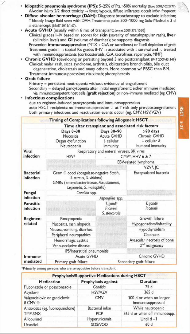
Idiopathic pneumonia syndrome (IPS): 5-25% of Pts,>50% mortality (B/ood2003; 102:2777)
Alveolar injury 2/2 direct toxicity -» fever, hypoxia, diffuse infiltrates; occult infxn frequent Diffuse alveolar hemorrhage (D A H ): Diagnosis: bronchoscopy to exclude infection;
T bloody lavage fluid seen with DAH.Treatment: pulse 500-1000 mg Solu-Medrol x 3 d ± etanercept (BBM7 2015;1:67).
Acute G V H D (usually within 6 mo of transplant; Lancet 2009.373:1550)
Clinical grades l-IV based on scores for skin (severity of maculopapular rash), liver (bilirubin level) and Gl (volume of diarrhea); bx supports diagnosis
Prevention: immunosuppression (MTX + CsA or tacrolimus) orT-cell depletion of graft Treatment: grade I topical Rx grades ll-IV -» associated with i survival and /. treated
with immunosuppressants (corticosteroids, CsA, tacrolimus, rapamycin, MMF) Chronic G V H D (developing or persisting beyond 3 mo posttransplant; BM7 2009:43:149)
Clinical: malar rash, sicca syndrome, arthritis, obliterative bronchiolitis, bile duct degeneration, cholestasis and many others. More common w/ PBSC than BM.
Treatment: immunosuppression; rituximab; photopheresis G raft failure
Primary = persistent neutropenia w ithout evidence of engraftment Secondary = delayed pancytopenia after initial engraftment; either immune mediated
via immunocompetent host cells (graft rejection) or non-immune mediated (eg, CMV) Infectious complications
due to regimen-induced pancytopenia and immunosuppression auto HSCT recipients: no immunosuppression .% at t risk only pre-/postengraftment both primary infections and reactivation events occur (eg, CMV, HSV.VZV)
Timing of Complications following Allogeneic HSCTTim e after transplant and associated risk factors
Days 0-30Mucositis
Days 30-90Acute GVHD
>90 daysChronic GVHD
Organ dysfunction Neutropenia
i cellular immunity
1 cellular & humoral immunity
Viral Respiratory and enteral viruses, BK virusinfection HSV* CMV* HHV 6 & 7
EBV-related lymphomaVZV*,JC
Bacterialinfection
Gram © cocci (coagulase-negative Staph., S. aureus, S. viridans)
GNRs (Enterobacteriaceae, Pseudomonas, Legionella, S. maltophilia)
Encapsulated bacteria
Fungal Candida spp.infection Aspergillus spp.Parasiticinfection
T. gondii P. carinii
T. gondii P. carinii
$. stercoralisRegimen- Pancytopenia Growth failurerelated Mucositis, rash, alopecia Hypogonadism/infertility
Nausea, vomiting, diarrhea HypothyroidismPeripheral neuropathies Cataracts
Hemorrhagic cystitis Avascular necrosis of boneVeno-occlusive disease 2r,d malignancy
IPS/Interstitial pneumonitisImmune- Acute GVHD Chronic GVHDmediated Primary graft failure Secondary graft failure
^Primarily among persons who are seropositive before transplant
Prophylaxis/Supportive Medications during HSCTMedication Prophylaxis against Duration
Fluconazole or posaconazole Candida 75 dAcyclovir HSV/VZV 365 dValgancidovir or ganciclovir if CMV ©
CMV 100 d or when no longer immunosuppressed
Antibiotics (eg, fluoroquinolone) Bacterial infxn While neutropenicTMP-SMX PCP 365 d or when off immunosupp.Allopurinol Hyperuricemia Until d -1Ursodiol SOS/VOD 60 d
HSC
T 5-27
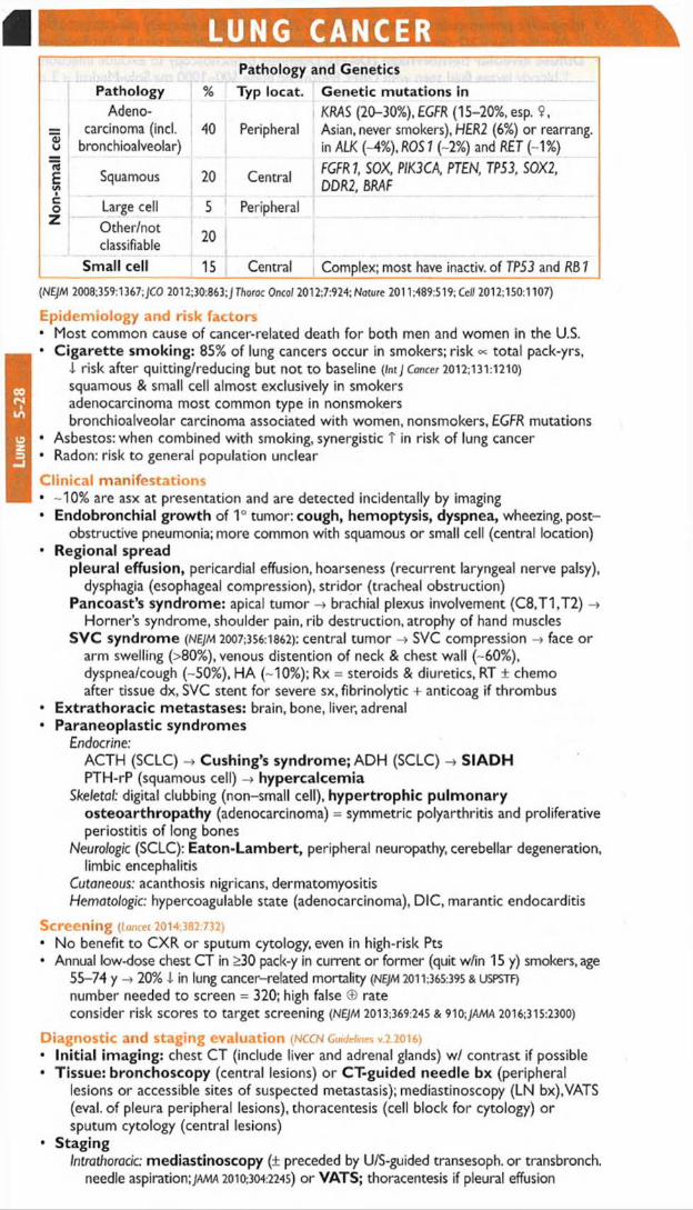
Lun
g
5-28
LUNG CANCER
<Uu
Ei/>ICoZ
(NEJM 2008;359:1367; JCO 2012;30:863;J TJiorac Oncol 2012:7:924; Nature 2011:489:519; Cefl 2012:150:1107)
E p idem io logy and risk fac to rs• Most common cause of cancer-related death for both men and women in the U.S.• C igarette smoking: 85% of lung cancers occur in smokers; risk « total pack-yrs,
i risk after quitting/reducing but not to baseline (Int J Cancer 2012:131:1210)
squamous & small cell almost exclusively in smokers adenocarcinoma most common type in nonsmokersbronchioalveolar carcinoma associated with women, nonsmokers, EGFR mutations
• Asbestos: when combined with smoking, synergistic T in risk of lung cancer• Radon: risk to general population unclear
C lin ica l m an ifes ta tions• -10% are asx at presentation and are detected incidentally by imaging• Endobronchial growth of 1° tumor: cough, hemoptysis, dyspnea, wheezing, post
obstructive pneumonia; more common with squamous or small cell (central location)• Regional spread
pleural effusion, pericardial effusion, hoarseness (recurrent laryngeal nerve palsy), dysphagia (esophageal compression), stridor (tracheal obstruction)
Pancoast’s syndrome: apical tumor -> brachial plexus involvement (C8,T1,T2) -» Horner’s syndrome, shoulder pain, rib destruction, atrophy of hand muscles
SVC syndrome (NEJM 2007:356:1862): central tum or -> SVC compression -» face or arm swelling (>80%), venous distention of neck & chest wall (-60%), dyspnea/cough (-50%), HA (-10%); Rx = steroids & diuretics, RT ± chemo after tissue dx, SVC stent for severe sx, fibrinolytic + anticoag if thrombus
• Extrathoracic metastases: brain, bone, liver, adrenal• Paraneoplastic syndromes
Endocrine:ACTH (SCLC) -» Cushing’s syndrome; ADH (SCLC) S IA D H PTH-rP (squamous cell) -4 hypercalcemia
Skeletal: digital clubbing (non-small cell), hypertrophic pulm onary osteoarthropathy (adenocarcinoma) = symmetric polyarthritis and proliferative periostitis of long bones
Neurologic (SCLC): Eaton-Lam bert, peripheral neuropathy, cerebellar degeneration, limbic encephalitis
Cutaneous: acanthosis nigricans, dermatomyositisHematologic: hypercoagulable state (adenocarcinoma), DIC, marantic endocarditis
Screen ing (Loncet 2014:382:732)
• No benefit to CXR or sputum cytology, even in high-risk Pts• Annual low-dose chest CT in >30 pack-y in current or former (quit w/in 15 y) smokers, age
55-74 y -> 20% i in lung c a n c e r- re la te d m o r ta lity (NE)M 2011:365:395 & USPSTF)
n u m b e r n e e d e d t o s c re e n = 320; h igh fa lse © ra te
consider risk scores to target screening (NEJM 2013:369:245 & 910;jama 2016:315:2300)
D iagnostic and staging eva lua tion (NCCN Guidelines v.2.2016)
• Initial imaging: chest CT (include liver and adrenal glands) w / contrast if possible• Tissue: bronchoscopy (central lesions) o r CT-guided needle bx (peripheral
lesions o r accessible sites of suspected metastasis); mediastinoscopy (LN bx),VATS (eval. of pleura peripheral lesions), thoracentesis (cell block for cytology) or sputum cytology (central lesions)
• StagingIntrathoracic: mediastinoscopy (± preceded by U/S-guided transesoph. or transbronch.
needle aspiration;JMW 2010:304:2245) or VATS; thoracentesis if pleural effusion
Pathology and GeneticsPathology % Typ locat. Genetic mutations in
Adenocarcinoma (incl.
bronchioalveolar)40 Peripheral
KRAS (20-30%), EGFR (15-20%, esp. $,Asian, never smokers), HER2 (6%) or rearrang. in ALK (-4%), ROS1 (-2%) and RET (-1%)
Squamous 20 CentralFGFRl SOX, PIK3CA, PTEN, TP53, S0X2, DDR2, BRAF
Large cell 5 PeripheralOther/notclassifiable 20
Small cell 15 Central Complex; most have inactiv. of TP53 and RB 7

Extrathoracic: PET-CT more Se than CT alone for detecting mediastinal and distant mets as well as bone mets (NEJM 2009:361:32); brain MRI for all Pts (except IA)
Genetics: / EGFR mut. & ALK, ROS1 or RET rearrang. for stage IV nonsquam NSCLC PFTs w / quantitative V/Q if planned treatment includes surgical resection; need to
have 30% of normal, predicted lung fxn after resection
TN M Staging System for NSCLC (7th Edition)N stage NO N1 N2 N3
T/Mstage Definition no 0
nodesipsilat.hilar
ipsilat.mediast.
contralat. or supraclav.
T1 T <2 cm (Tla) o rT >2-3 cm (Tib) IA IIAT2 T <5 cm (T2a) o rT 5-7 cm (T2b) IB/IIA IIA/B
T3T >7 cm or invasion of chest wall, diaph., mediast. pleura, pericard.
MB IIIA
T4Invasion of mediast., heart, great vessels, trachea, esoph, vertebrae; separate tumor nodule ipsilat. lobe
IIIB
MiaNodules contralat lobe; pleural nodules or malignant effusion IV
M lb Distant metastasis
NSC LC treatm ent (NCCN Guidelines v.2.2016)
• Stages I & II: surgical resection + adjuvant chemo (surgery alone for stage IA) {NEJM2004:350:351 & 2005:352:2589)
• Stage III: chem oradiation is main treatment modalityIIIA viewed as potentially resectable (tance t 2009:374:379) and IIIB as unresectable neoadjuvant chemoradiation may convert unresectable -> resectable
• Stage IV: chemotherapy t survival; early palliative care also T survival (NEJM 2010:363:733)
backbone of therapy is platinum-based doublet; cisplatin/pemetrexed better for adenocarcinoma; cisplatin/gemcitabine better for squamous (/CO 2008:26:3543)
PD-1 inhib (eg, nivolumab, pembrolizumab.atezolizumab) if progression on chemo (NEJM
2015:373:123; Loncet 2016:387:1540 & 1837); immune-related adverse events include pneumonitis, consider Rx w/ high-dose corticosteroids
bevacizumab (anti-VEGF mAb) + chemo T survival by 2 mo;T bleeding risk, .\ avoid if untreated, brain mets (JCO 2009:27:5255), hemoptysis or squamous (NEJM 2006:355:2542)
if EGFR mut.: EGFR tyrosine kinase inhibitor (TKI, eg, erlotinib) TMine Rx; next-gen EGFRTKI for those who develop resistance mutations (NEJM 2015 :372:1689 & 1700)
if ALK rearrang.: ALK TKI (crizotinib 1st-line Rx; NEJM 2 01 4 :3 7 1 :2 16 7 ); ceritinib 2nd-line TKI toxicities: rash & diarrhea (common); lung & liver injury (rare but potentially serious) palliative radiation used to control local sx caused by tumor or metastasis solitary brain metastasis: surgical resection + brain radiation may T survival
NSCLC Simplified Staging Schema,Treatment and 5-y SurvivalStage % at dx Definition Treatment 5-y (%)
1 10-20 Isolated lesion Surgery + chemo >60II 10-20 Hilar node spread Surgery ± radiation ± chemo 40-50
IIIA 15 Mediast. spread but resectable
Chemoradiation ± surgical resection
25-30
IIIB 15 UnresectableChemoradiation ± biologic ± surgery (selected cases) 10-20
IV 40 MetastaticChemo ± bevacizumab or tyrosine kinase inhibitor and/or supportive care
4
SCLC trea tm en t (NCCN G u id e lines v. 1.2016)
• SCLC usually disseminated at presentation but can be very responsive to chemoradiation• Chem otherapy (platinum + etoposide) is primary treatment modality• Thoracic radiation added to chemotherapy improves survival in limited-stage disease• Prophylactic cranial irradiation (PCI) T survival for limited disease in complete remission
(NE/M 1999:341:476) & i symptomatic brain mets in extensive disease (NEJM 2007:357:664)
SCLC Staging Schema and TreatmentStage % at dx Definition Treatment Median
survivalLimited 30-40 Confined to ipsilat hemithorax
w/in 1 radiation portRadiation + chemotherapy ± PCI
1-2 y
Extensive 60-70 Beyond 1 radiation port Chemotherapy ± PCI
Lu
ng
5-29

BREAST CANCER kiE p idem io logy and genetics (risk assessment tool: www.cancer.gov/bcrisktool/)• In U.S., most common cancer in women: 2nd leading cause of cancer death in women• Age: incidence rates T with age, with possible 4- in slope after menopause• Genetics (Nature 2012:490:61); mutations in TP53, PIK3CA, and GATA3; HER2 amplified.
15-20% have © FHx -> 2x T risk; -45% of familial cases a/w known germline mutation BRCA1/2: 35-85% lifetime risk of breast cancer & T risk of ovarian cancer; ? T colon &
prostate cancer; prog not worse than in noncarriers w/ breast cancer (NEJM 2007:357:115);BRCA2: a/w T male breast cancer & pancreatic cancer. Germline loss-of-function mutations in PALB2 a/w 35% T risk of breast cancer by age 70 (NEJM 2014:371:497).
• Estrogen: T r is k w i t h e a r ly m e n a rc h e , la te m e n o p a u s e , la te p a r i ty o r n u l l ip a r ity (NEJM
2006:354:270); T r is k w i th p ro lo n g e d HRT (RR = 1.24 a f te r 5.6 y; jama 2003:289:3243);
no T r is k s h o w n w ith OCP use (NEJM 2002:346:2025)
• Benign breast conditions: T risk w/ atypia (atypical ductal o r lobular hyperplasia; NEJM
2015:372:78) & proliferative (ductal hyperplasia, papilloma, radial scar, or sclerosing adenosis) features; no T risk w / cysts, fibroadenoma, or columnar changes
• T risk with h/o ionizing radiation to chest for treatment of Hodgkin lymphoma
P reven tion (with selective estrogen receptor modulator or Al; Annals 2013:159:698)• Tamoxifen: X risk contralat. breast CA as adjuvant Rx. Approved for 1° prevent, if T risk:
X invasive breast cancer, but T DVT & uterine CA; ? t in mortality (Lancet 2002,360:817).• Raloxifene: X risk of invasive breast cancer & vertebral fx, t risk of stroke & DVT/PE
(NEJM 2006:355:125); = tamoxifen in prevention of breast cancer w/ X risk of DVT/PE & cataracts, trend toward X uterine cancer {JAMA 2006:295:2727)
• Als in high-risk postmeno X breast cancer by >50% (NE/M 2011:364:2381; Lancet 2014:383:1041)• BRCA 7/2 ©: intensified surveillance. Prophylactic bilat. mastectomy -» -90% X risk; bilat.
salpingo-oophorectomy X risk of ovarian and breast cancer (NEJM 2016:374:454).
C lin ica l m an ifes ta tions• Breast mass (hard, irregular, fixed, nontender), nipple discharge (higher risk if unilateral,
limited to 1 duct, bloody, associated with mass)• Special types: Paget’s disease -*• unilateral nipple eczema + nipple discharge;
in flam m atory breast cancer -> skin erythema and edema (peau d’orange)• Metastases: lymph nodes, bone, liver, lung, brain
Screen ing (JAMA 2015:314:1599; Annals 2016:164:279)• Mammography; -20-30% X in breast cancer mortality (smaller abs. benefit in women
<50 y) (Lor?cct 2006:368:2053: Anno/s 2009:151:727); 75% of all abnl findings benign; suspicious: clustered microcalcifications, spiculated, enlarging
• ACS recommends annual mammo beginning at age 45 (consider biennial after age 54)• USPSTF recommends beginning at 50 and biennially (some may want to begin at age 40)• t risk: screen earlier w / CBE and mammo (age 25 in BRCA I f2 carrier, 5-10 y before
earliest FHx case, 8-10 y after thoracic RT, upon dx of T risk benign disease)• MRI: superior to mammo in high-risk Pts;consider annually if >20% lifetime risk (eg,©®
FHx, BRCAV2, p r io r c h e s t RT) (Lancet 2011:378:1804)
• Genetic testing should be considered in women with strong FHx
D iagnostic eva luation• Palpable breast mass: age <30 y h> observe for resolution over 1-2 menstrual cycles;
age <30 y, unchanging mass -> U/S -» aspiration if mass not simple cyst; age >30 y or solid mass on U/S or bloody aspirate or recurrence after aspiration
m am m o (detect other lesions) and either fine-needle asp. or core-needle bx clearly cancerous on exam or indeterminate read o r atypia on bx -> excisional bx
• Suspicious m am m ogram with normal exam: stereotactically guided bx• MRI: detects contralateral cancer in 3% of Pts w / recently dx breast cancer & 0 contra
la te ra l m a m m o (b u t PPV o n ly 21%) (NEJM 2007:356:1295); u t i l i t y re m a in s u n c le a r
Staging• Anatomic; tumor size, chest wall invasion, axillary LN mets (strongest prognostic factor)• Histopathologic: type (little prognostic relevance) & grade; lymphatic/vascular invasion
In situ carcinoma: no invasion of surrounding stroma Ductal (DCIS): T risk of invasive cancer in ipsilateral breast (~30%/10 y)Lobular (LCIS): marker of T risk of invasive cancer in either breast (-1%/y)
Invasive carcinoma: infiltrating ductal (70-80%); invasive lobular (5-10%); tubular, medullary and mucinous (10%, better prognosis); papillary (1-2%); other (1-2%)
Inflammatory breast cancer (see above): not a histologic type but a clinical reflection of tumor invasion of dermal lymphatics; very poor prognosis
Paget disease: ductal cancer invading nipple epidermis ± associated mass

B iom arke rs : / estrogen, progesterone receptor (ER/PR) and HER2/neu status Oncotype DX 21-gene risk recurrence score has predictive and prognostic value in ER ©,
HER2 0 , and node 0 Pts (NEJM 2015,373:2005)Circulating tumor DNA may serve as biomarker of met tumor burden (NEJM 2013:368:1199)
Simplified Staging System for Breast CancerStage Characteristics Description 5-y surv.
1 Tumor <2 cmOperablelocoregional
90%IIA Tumor >2 cm or mobile axillary nodes 80%IIB Tumor >5 cm 65%IDA Internal mammary or fixed axillary nodes Locally advanced 50%NIB Direct extension to chest wall or skin Inoperable 45%INC Infraclavicular or supraclavicular nodes locoregional 40%IV Distant metastases Metastatic 25%
Treatm ent• Local control: surgery and radiation therapy (RT)
8reast<onserving usual approach w/ lumpectomy + breast RT + axillary node dissection (ALND), unless multicentric dis., diffuse microCa2*, BRCA1/2 0 , prior RT, pregnant.? tumor >5 cm; cavity shaving ! risk of need for re-excision (NEJM 2015:373:503)
Sentinel lymph node dissection (SLND) prior to ALND preferred if w/o palp axillary LN$;T1-2 w/ 0 SLND & Rx w/ lumpect./RT/chemo may not need ALND (JAMA 2011:305:569)
Radiation therapy (RT) after mastectomy for >4 0 LN, tumor >5 cm, o r © surgical margins -» i locoregional recurrence and T survival (Lancet 2011:378:1707); regional nodal RT i recurrence and breast cancer mortality (N£/Ai 2015:373:307 & 317)
• Systemic therapy: for stage l-lll except tumors <1 cm (complex risk assessment needed).http://www.adjuvantonline.com/index.jsp can guide use of chemo and/or hormonal Rx. Chemotherapy: neoadjuvant (to T breast conservation; path complete response a/w t
disease-free survival; Lancet 2014:384:164) or adjuvant (anthracycline-based).Addition of taxane (eg, paclitaxel) -> small T survival (NEJM 2010:362:2053 & 2010:363:2200).
Consider platinum in triple © cancers (JCO 2015:33:13).
Anti-HER2 therapy (growing list of agents) in HER2 0 tumors (NEJM 2012:366:176)
trastuzum ab (anti-H£R2 mAb) T survival (NEJM 2011:365:1273); 1 y = 2 yr (Lancet
2013:382:1021); after anthracycline or w / taxane to i cardiotox (jco 2002:20:1215)
lapatinib (tyrosine kinase inhib. of HER2 & £GFR) + trastuzumab t survival after failing trastuzumab (JCO 2012:30:2585); dual inhib. initial Rx T response (Lancet 2012.379:633)
pertuzumab (anti-H£R2 mAb, prevents dimerization) T progression-free survival when added to trastuzumab as 1st-line Rx for metastatic dis. (NEJM 2015:372:724)
trastuzumab emtansine (T-DM1, HER2 mAb conjugated to microtubule inhibitor)T survival compared to 2nd-line lapatinib -I- capecitabine (NEJM 2012:367:1783)
Bevacizumab (anti-VEGF): ? in neoadjuvant Rx if HER2 © (NEJM 2012:366:299 & 310)
Hormonal (in ER/PR © or unknown status) tamoxifen: 39% I recurrence and 30% i breast cancer mortality in pre- and post
menopausal patients; 10 y of Rx superior to 5 y (Lancet2011;378:771 & 2013:381:805)
aromatase inhibitors (A l) (anastrozole, letrozole, exemestane):-18% I recurrence vs. tamoxifen in postmenopausal Pts (NEj/vi 2005:353:2747 & 2016:375:209)
2nd-line: ovarian ablation with LHRH agonists (goserelin) o r oophorectomy if premenopausal; pure antiestrogens (fulvestrant) if postmenopausal
Cell proliferation inhibitors (if postmenopausal & failed hormonal Rx) palbociclib (CDK 4/6 inhib): T progression-free survival (NEJM 2015:373:209)
everolimus (mTOR inhib): T progression-free survival (NEJM 2012:366:520)
Treatment of Carcinoma in situ and Invasive Carcinoma of the BreastLCIS Close surveillance ± chemoprevention; ? prophylactic bilat. mastectomyDCIS Mastect. or lump.-i- RT;ALND not indie.; + chemoprev (Lancet 2016:387:849 & 866)
1II
Surgery + RT+ Adjuvant chemo if T risk: tumor >1 cm or © LN or ER/PR © (Loncet 1998:352:930)
+ Hormonal therapy if ER/PR 0 (or unknown status) (Loncet 2009:374:2055)
+ anti-HER2 Rx if HER2 © and tumor >1 cm or © LN
IIINeoadjuvant chemo -> surgery + RT ± adjuvant chemotherapy + Hormonal therapy for ER/PR © (or unknown status) tumors + anti-H£R2 Rx if HER2 ©
IVER/PR©: hormonal Rx (NEJM 2012:367:435) or chemo ± everolimus/palbociclib ER/PR©: HER2 © -» chemo + anti-H£R2 therapy; HER2 © -> chemotherapy Bony mets: bisphosphonates & denosumab 4- fractures (Cochrane 2012;CD003474)
Brea
st 5-31

PROSTATE CANCER iiEpidemiology and risk factors (NEJM 2003:349 366)
• Most common cancer in U.S. men; 2nd most common cause of cancer death in men• Lifetime risk of prostate cancer dx -16%; lifetime risk of dying of prostate cancer ~3%• t risk with t age (rare if <45 y), in African Americans, 0 FHx, BRCA mutations
Clinical manifestations (usually asymptomatic at presentation)• Obstructive sx (more common with BPH): hesitancy, i stream, retention, nocturia• Irrita tive sx (also seen with prostatitis): frequency, dysuria, urgency• Periprostatic spread: hematuria, hematospermia, new-onset erectile dysfunction• Metastatic disease: bone pain, spinal cord compression, cytopenias
Screening (NEJM 201 2 ;3 6 7 :e 11 : yA/V1A 201 4 :3 1 1 :1 14 3 ; Lancet 2014:384:2027)
• Digital rectal exam (DRE): size, consistency, lesions• PSA: 4 ng/mL cut point neither Se nor Sp; can T with BPH, prostatitis, acute retention, after
bx orTURRand ejaculation (no s/gn/ficant T after DRE, cystoscopy);15% o f m en >62 y w / PSA <4 & nl DRE have b x - p r o v e n T I ca n c e r (nejm 2004:350:2239)
• ACS rec: >50 y (or > 45 y if African-Am or 0 FHx) should discuss PSA screening w/ MD;USPSTF rec. against screening in asx males (no i in prostate cancer-related mort.)
Diagnostic and staging evaluation• Transrectal ultrasound (TRUS) guided biopsy, with 6-12 core specimens• Histology: Gleason grade (2-10; low grade <6) = sum of the differentiation score (1 =
best, 5 = worst) of the 2 most prevalent patterns in the bx; correlates with prognosis• Imaging: to evaluate extraprostatic spread
bone scan: for PSA >10 ng/mL, high Gleason grade or clinically advanced tumor abdomen-pelvis CT: inaccurate for detecting extracapsular spread and lymph node mets endorectal coil MRI: improves assessment of extracapsular spread
TNM Staging & Treatment of Prostate Cancer (Loncet 2015:387:70)
Nodes,Stage Tumor Mets Treatment
Tla = NO, M0, Gleason
2-4
Surveillance: consider if life expect. <10 y. Dutasteride 4-
1 non-palp., not visible on imaging
risk of progression (Lancet 2012:379:1103).Radiation (external or brachy; NEJM 2006:355:1583). Short
term androgen deprivation i mort. (NEJM 2011:365:107)
li T1/T2 = w/ in prostate NO, M0
Radical prostatectomy (± RT and/or hormonal Rx if high-risk features): 4 prostate cancer mortality, espec. if <65 y and not low risk (NEJM 2014:370:932)
III T3 = extends thru capsule NO, MO Radiation + androgen deprivation (see below)
(Lance: 2011:378:2104)T4 = invades
adjacent structures
NO, MORadiation (for M0 disease)Androgen deprivation Rx (A D T) (NEJM 2009:360.2516)
GnRH analogues (leuprolide.goserelin)N l, M0 antiandrogens (flutamide, bicalutamide)
Docetaxel added to ADT improves overall survival in
IV
Any T Any N, Ml
metastatic disease (NE/M 2015:373:737)If castrate resistant: chemo (eg, docetaxel); androgen
synthesis inhib. (abiraterone; NEJM 2011:364:1995) or receptor signaling inhib. (enzalutamide; NEJM 2012:367:1187) i mort.; immuno Rx (NEJM 2010:363:411);olaparib (PARP inhib) if BRCA © (nejm 2015:373:1697)
Bone mets: bisphosph or denosumab, latter 4- bone mets & fx (NEJM 2009:361:745; Lancet 2011:377:813 & 2012:379:39);
radium-223 ! mortality by 30% (NEJM 2013:369.213)
Prognosis• PSA level, Gleason grade and age are predictors of metastatic disease• In surgically treated Pts, 5-y relapse-free survival >90% if disease confined to organ,
-75% if extension through capsule, and -40% if seminal vesicle invasion• PSA doubling time, Gleason, & time to biochemical recurrence predict mortality following
recurrence. For local recurrence following RP, salvage RT may be beneficial if low PSA.• Metastatic disease: median survival -44-57 mo (nejm 2015:373:737); a ll become castrate
resistant (in 15-20% discontinuation of antiandrogens results in paradoxical i in PSA)
Prevention• Finasteride and dutasteride i prostate cancers detected by bx, but T # of high Gleason
g ra d e tu m o rs ; n o A in o v e ra ll m o r ta l i t y (NEJM 2003:349:215:2010:362:1192:2013:369:603)

COLORECTAL CANCER (CRCEpidemiology and risk factors (Lancet 2010:375:1030; CA CancerJ Clin 2011;61:212)• 4th most common cancer in U.S. men & women; 2nd leading cause of all cancer death• Rare before age 40, w/ 90% of cases occurring after age 50. -75% are sporadic.• Family history: up to 25% of Pts have © FHx. Risk depends on # of 1 “ -degree relatives
(w/ CRC or polyp) and their age at dx; -5% have an identifiable germline mutation Familial adenomatous polyposis (FAP): mutation in APC gene -> 1000s of polyps at
young age -» -100% lifetime risk; T risk of thyroid, stomach, small bowel cancers Hereditary nonpolyposis colorectal cancer (HNPCC): most common hereditary CRC
(~3% of all CRC); mutations in DNA mismatch repair genes (eg,MSH2, MLH1) -» microsatellite instability (MSI) -» T tumor progression - 80% lifetime risk.Predom. right-sided tumors; T risk of endometrial, ovarian, stomach, urothelial, small
bowel and pancreatic cancers.Amsterdam criteria: >3 family members w/ HNPCC-related cancer, one of which is dx
before age 50, affecting 2 successive generations.MAP (MYH-assoc polyposis): autosomal recessive; consider if mult, polyps but © for FAP
• In flam m atory bowel disease: T risk with T extent and duration of disease• COX-2: 4 risk of adenomas w/ASA & NSAIDs.ASA a/w 4 CRC incidence, mets and mort.
(Lancet 2010:376:1741; 2012:379:1591 & 1602). ASA effect limited to PIK3CA-mut CRC (N E JM
2012:367:1596). ASA rec for 1° prevention if age 50-59 (69?) y & >10% 10-y risk of CRC.
Pathology and genetics (NEJM 2009:361:2449;Noture 2012:487:330)• Adenoma -» carcinoma sequence reflects accumulation of multiple genetic mutations.
T risk of malig. w / large (>2.5 cm), villous, sessile adenomatous polyps. Adenomas typically observed ~10 y prio r to onset of cancer (both sporadic & familial).
• Genetic profile in sporadic CRC: APC (-80%), KRAS (-40%), TPS3 (50-70%), DCC orSMAD4, or BRAF (~15%);chrom instability (majority) or mismatch repair defic (10-15%)
• Upfront genotyping may guide Rx; eg, benefit of anti-EGFR Ab cetuximab greater in KRASwild-type than KRAS mutant (nejm 2008:359:1757). BRAF mutation may guide clinical trials. Lack of CDX2 a/w t benefit from chemo (N E JM 2016:374:211).
Clinical manifestations• Distal colon: A bowel habits, obstruction, colicky abdominal pain, hem atochezia• Proximal colon: iron defic. anemia, dull vague abd pain; obstruction atypical
due to larger lumen, liquid stool and polypoid tumors (vs. annular distal tumors)• Metastases: nodes, liver, lung, peritoneum -> RUQ tenderness, ascites, supraclavicular LN• Associated with Streptococcus bovis bacteremia and Clostridium septicum sepsis
Screening (jama 2016:315:2564)• Average risk: colonoscopy starting at age 50 & repeat q10y strongly preferred method• t risk: earlier and/or more frequent screening. © FHx: age 40 or 10 y before index
dx, then q5y. IBD: 8-10 y after dx, then q1-2y. Known or suspected familial syndrome: genetic counseling & very early screening (eg, age 20-25 y),then q1-2y.
• ImagingColonoscopy: test of choice as examines entire colon; 90% Se for lesions >1 cm. Flex
sig less Se vs. colo and CTC (Gut 2009:58:241). If polyp found, re / in 3-5 y. Removal of adenomatous polyps associated with lower CRC mortality (N E JM 2012:366:687).
Sigmoidoscopy: 21% 4 incidence in CRC & 26% 4 mortality in distal CRC (N E JM
2012:366:2345). Benefit may also be seen w / 1-time flex-sig (Lancet 2010:375:9726).CT colonography (CTC): c/w colonoscopy,-90% Se for lesions >1 cm but considerably
less for smaller lesions (NEJM 2008:359:1207). In high-risk Pts, Se only 85% for advanced neoplasia >6 mm (JA/AA 2009:301:2453).At population level, T participation w/CTC, but 4 yield vs. colonoscopy; similar screening overall (Lancet 2012-,13:SS).
• Biochemical fecal testingOccult blood (FOBT): 4 mortality (N E JM 1993:328:1365 & 2000:343:1603); 3 card home testing
more Se (24% vs. 5%) than DRE/FOBT (>WJs 2005:142:81). Repeat qly.D N A : t Se, ~ Sp c/w FOBT but less Se than colonoscopy (N E J M 2004:351:2704) Combo DNA + Hb immunoassay w / -90% Se & Sp (nejm 2014;370:1287)
Staging (AJCC Cancer Staging Manual, 7th ed, 2010)• TNM staging: Size/depth of primary (T), locoregional nodes (N), distant metastases (M).
Staging is complex and based on pathologic correlation with observed survival data.• Colonoscopy + biopsy/polypectomy + intraoperative and pathologic staging
essential for evaluating extracolonic spread• CT scans of chest and abdomen/pelvis (inaccurate for depth of invasion & malignant LN)• Baseline CEA in Pt with known CRC has prognostic significance and is useful to fol
low response to therapy and detect recurrence; not a screening tool
Co
lor
ecta
l 5-33

Ch
emo
5-
34Treatment Based onTNM and Modified Dukes Staging of Colorectal Cancer
TN M Dukes Path, criteria 5-y surv. Treatment
1 A Into submucosa or muscularis 94-97% Surgery alone (resection and
analysis of >12 LN)IIA B Into serosa 83% Surgery; no established role forIIB B Into peritoneum 74% adjuvant chemo for colon cancer3
IIC B Direct invasion 56%Preop RT or 5-FU/RT added for
rectal cancer -> postop chemoIII A C <6 © LNs 86% Surgery + chemotherapy61IIB C Varying # © LNs 51-77% Preop RT or chemorad added forMIC C & local invasion 15-47% rec ta l ca n ce r (NEJM 2006:355:1114)
IV D Distant metastases 5%
Chemotherapy ± surgical resection for isolated mets (-30% 5-y surv)
Consider resection of 1° tumor ifperf, obstruction or bleeding
NCCN Clinical Practice Guidelines, www.nccn.org. 5-y survival data are approx, equivalent for colon and rectal cancers, shown as average, w/ ranges forTNM substaging, adapted from SEER data (/CO 2010:28:256 & 264). ‘Consider adjuvant chemo for high-risk stage II (obstruction, perf, adherence to adjacent structures, inadequate nodal sampling, lymphovasc invasion, poorly differentiated). MSI-high CRC benefit less from adjuvant chemo (NEJM 2003:349:247). bAdjuvant FOLFOX (see below) is standard of care chemo (NEJM 2004:350:2343).
• Chemotherapy (Lancet 2014:383:1490)FOLFOX (5-FU + leucovorin + oxalip latin), FOLFIRI or CapeOx (NEJM 2004:350:2343) ± Bevacizumab (anti-VEGF, NEJM 2004,350:2335) or cetuximab/panitumumab (anti-EGFR
mAb, NEJM 2004:351:337; benefit limited to Pts w/o RAS mutations; NEJM 2013:369:1023) Consider FOLFOXIRI-bevacizumab particularly if BRAF© mutations or needing response
for potentially curative resection (NEJM 2014:371:1609)Regorafenib (multikinase inhib.) andTAS102 (trifluridine + tipiracil) T survival in
progressive metastatic CRC (Loncet 2013:381:303: NEJM 2015:372:1909)
CHEMOTHERAPY SIDE EFFECTSNausea & vomiting common (NEJM 2016:374:1356:375:134 & 177)
Select Adverse Effects from ChemotherapyToxicity Common Agents Comments
Cardiotoxicity(ICO 2005:23:7685;
Anthracyclines Dose-dependent CMP; / EF pre-Rx; via topoisomerase lib (Nat Med 2012;18:1639)
NEJM 2013:368:1154) Stop agent i f adverse
event;? role for ACEI in
prevention (Circ 2006:114:2474).
5-FU Spasm -> ischemia; CCB may preventTrastuz. & PD-1 inhib CMP, esp. w/ concom anthracyclineTyrosine kinase inhib. (TKI) QTc prolongation, CMP, anginaCyclophosphamideCisplatin
Myopericarditis (esp. in BMT) HypoMg -> arrhythmia, ischemia
Busulfan -8% fibrosis or DAH; if severe -* steroidsBleomycin -10% IPF; d/c drug, Rx w/ steroids
Pulmonary(Sem Oncol 2006;33:98)
TKI (esp. dasatinib) Pulmonary effusionCyclophosphamide (<1%) Pneumonitis, progressive fibrosis; d/c drugBevacizumab Pulm hemorrhage (esp. NSCLC)Anti-PDI (eg, nivolumab) PneumonitisAnti-CTLA-4 (ipilimumab) Organizing pneumonia, sarcoidosis
Nephrotoxicity/ urologic toxicity
Platinum Rx (cisplatin) Methotrexate
Esp. proximal tubule; pretreat w/ IV saline Rx deposition; alkalinize urine, hydration
Cyclophosphamide Hemorrhagic cystitis; give MesnaPlatinum Rx (cisplatin) “ Stocking-glove;” vit. E Ppx (JCO 2003:21:927)
Cytarabine Cerebellar toxicity (irreversible 5-10%)
Neurotoxicity(Sem Oncol 2006;33:324)
Methotrexate (esp. intrathecal)
Late leukoenceph, meningitis; reverse w/ intrathecal glucarpidase, leucovorin
Ifosfamide Enceph (10-30%); ? Rx w/ methylene blue, thiamine, dexmedetomidine
Taxanes, vincristine Sensorimotor long fiber neuropathyHepatotoxicity TKI (eg, imatinib, nilotinib) T LFTs, rarely necrosis; hold Rx, ? steroids(Sem Oncol 2006:33:50) Gemcitabine Common T ALT/AST; 1 dose if T bili
Methotrexate T ALT/AST, rarely fibrosisDermatologic TKI (eg, imatinib) Dermatitis, can be severe (eg, SJS)

PANCREATIC TUMORSPathology and genetics (Arm Rev Pathol 200B;3:1S7; Nature 2012;491;399)• Histologic types: adenocarcinoma, acinar cell carcinoma, endocrine tumors, cystic
neoplasms (eg, IPMN, see below); rarely, mets to pancreas (eg, lung, breast, renal cell)• Pancreatic adenocarcinoma accounts for majority of pancreatic cancer ( -85%)• Location: -60% in head, 15% in body, 5% in tail; in 20% diffuse infiltration of pancreas• Mutations in adenoca.: KRAS (>90%), p 16 (80-95%), pS3 (50-75%), SMAD4 (-55%)
Epidemiology and risk factors (NEJM 2014;371:1039;./.cmcet 2016;388:73>• Pancreatic adenocarcinoma 4th leading cause of cancer death in U.S. men and women• 80% of pancreatic adenocarcinomas occur in Pts 60-80 y• Acquired risk factors: smoking (RR -1.5; 20% Pts), obesity, chronic pancreatitis, ?
diabetes• Hereditary risk factors: genetic susceptibility may play a role in 5-10% of cases
Hereditary chronic pancreatitis: mutation in cationic trypsinogen gene (PRSS1), SPINK 1 Familial cancer syndromes and gene mutations with T risk: familial atypical multiple mole
melanoma (CDKN2AJp 76), familial breast and ovarian cancer (BRCA2), Peutz-Jeghers (LKB 7), ataxia-telangiectasia (ATM), ? hereditary colorectal cancer (HNPCC and FAP)
Clinical manifestations• Painless jaundice (w/ pancreatic head mass), pain radiating to back, i appetite & w t• New-onset atypical diabetes mellitus (25%); unexplained malabsorption or pancreatitis• Migratory thrombophlebitis (Trousseau’s sign), not specific to pane cancer (jeo 1986;4:S09)
• Exam: abd mass; nontender, palpable gallbladder (Courvoisier’s sign, but more oftenseen w/ biliary tract cancers); hepatomegaly; ascites; left supraclavicular (Virchow’s) node & palpable rectal shelf (both nonspecific signs of carcinomatosis)
• Laboratory tests may show T bilirubin, T alk phos, anemia
Diagnostic and staging evaluation (NCCN Guidelines v.2.2012)• Pancreatic protocol C T scan (l+ w/ arterial & venous phase imaging) or MRI• If no lesion seen, -> EUS, ERCP, MRI/MRCP may reveal mass or malignant ductal strictures• Biopsy pancreatic lesion via EUS-guided FNA (preferred in potential surgical candidates)
or CT-guided (potential risk of seeding) or biopsy of possible metastasis• T CA19-9 (nb, also T in benign liver/biliary disease); may be useful to follow dis. postop
Clinical (Radiologic) Staging & Prognosis of Pancreatic AdenocarcinomaStage (% at dx) Criteria Median SurvivalResectable,15-20%
No extrapanc. dis. or bulky LAN Patent SMV & portal vein; celiac
axis & SMA not involved
10-20 mo (favorable: tumor <3 cm, © marg., well-differen.)
5-y -30% node © vs.-10% if ©Locally advanced(unresect.), 40%
Extensive PV/SMV, celiac axis or SMA involvement
8-12 mo
Metastatic, 40% Usually liver & periton.; occ lung Up to 11 mo w/ FOLFIRINOX
Treatm ent of pancreatic adenocarcinoma (NEJM 2014:371:1039; Lancet 2016.388:73)• Resectable: surgery ± adjuvant (neoadjuvant o r postoperative) therapy
pancreaticoduodenectomy = W hipp le procedure = resection of pancreatic head, duodenum, CBD and gallbladder ± partial gastrectomy
adjuvant therapy: T survival, but choice of regimen controversial (chemo vs. chemo/RT and gemcitabine vs. 5-FU (JSurg Oncol 2013; 107:78:JAMA 2013:310:1473)
• Locally advanced: optimal strategy controversial. Gemcitabine alone vs. gemcitabine + RT(ICO 2008:26:214s: Ann Oncol 2008:19:15 92;JCO 2011:29:4105).
• Metastatic: FOLFIRINOX (5-FU + leucovorin, irinotecan, oxaliplatin) if good perform, status{NEJM 2011:364:1817); gemcitabine + nab -pad itaxe l (NE/M 2013:369:1691) o r gem citab ine
m on o th e ra p y if p o o r pe rfo rm a n ce status (JCO 1997;15:2403). Offer clinical trials.• Palliative and supportive care:
obstructive jaundice or gastric outlet obstruction: endoscopic stenting or surgical bypass pain: opiates, celiac plexus neurolysis, radiation therapyweight loss: pancreatic enzyme replacement, nutrition consult, end-of-life discussions
Cystic lesions of the pancreas (NEJM 2004:351:1218; Oncotogrst 2009:14:125)• <10% of pancreatic neoplasms. Dx w / CT, ERCP, MRCP, o r EUS.• Serous cystadenoma: usually benign; central scar o r honeycomb appearance on imaging• Mucinous cystic neoplasm (MCN): predominantly young females; multiloculated tumors
in body or tail w/ ovarian-type stroma and mucin-rich fluid w/ T CEA levels; precancerous• Intraductal papillary mucinous neoplasm (IPMN): neoplasm arising in main pancreatic
duct or a branch; a/w ductal dilation w/ extrusion of mucinous material. Uncertain progression to cancer (? 5-20 y). Surgery based on age, size, location, & dysplasia.
Pancreatic
5-35

Onc
Em
erg
e 5-
36I
F ever a n d N e u t r o p e n ia (FN)Definition• Fever: single oral temp >38.3°C (101°F) o r >38°C (100.4°F) for >1 h• Neutropenia: ANC <500 cells/pL or <1000 cells/j.iL with predicted nadir <500 cells/uL
Pathophysiology and microbiology• Predisposing factors: catheters, skin breakdown, Gl mucositis, obstruction (lymphatics,
biliary tract, Gl, urinary tract), immune defect a/w malignancy• Most episodes thought to result from seeding of bloodstream by Gl flora• Neutropenic enterocolitis (typhlitis): RLQ pain, watery/bloody diarrhea, cecal wall thickening• GNRs (esp. P. aeruginosa) were historically most common• Gram © infections have recently become more common (60-70% of identified organisms)• Fungal superinfection often results from prolonged neutropenia & antibiotic use• Infection with atypical organisms and bacterial meningitis is rare
Prevention• Levofloxacin (500 mg qd) I febrile episodes & bacterial infections in chemo-related
high-risk neutropenic patients; no difference in mortality (nejm 2005:353:977 & 988)
Diagnostic evaluation• Exam: skin, oropharynx, lung, perirectal area, surgical & catheter sites; avoid DRE• Labs: CBC with differential, electrolytes, BUN/Cr, LFTs, U/A• Micro: blood (peripheral & through each indwelling catheter port), urine, & sputum cx;
for localizing s/s / stool (C. difficile, cx), peritoneal fluid, CSF (rare source)• Imaging: CXR; for localizing s/s -> CNS, sinus, chest o r abdomen/pelvis imaging• Caveats: neutropenia -» impaired inflammatory response ■-» exam and radiographic
fadings may be subtle; absence of neutrophils by Gram stain does not r/o infection
Risk stratification (factors that predict lower risk)• History: age <60 y, no symptoms, no major comorbidities, cancer in remission, solid
tumor, no h/o fungal infection or recent antifungal Rx• Exam: temp <39°C, no tachypnea, no hypotension, no A MS, no dehydration• Studies: ANC >100 cells/pL, anticipated duration of neutropenia <10 d, normal CXR
Initial antibiotic therapy (Gin Infeci Dis 2011:52:e56,NCCN Gur'defines v.2.2015)• Empiric regimens including drug w/ antipseudomonal activity; considerVRE coverage if
colonized; OR 3.8 forVRE ifVRE © (bbm t 2010,16:1576)
• PO abx may be used in low-risk Pts (<10 d neutropenia, nl hep/renal fxn, no N/V/D, noactive infxn, stable exam): cipro + amoxicillin-clavulanate (NEJM 1999:341:305)
• IV antibiotics: no clearly superior regimen; monotherapy or 2-drug regimens can be usedMonotherapy: ceftazidime, cefepime, imipenem, or meropenem2-drug therapy: aminoglycoside -t antipseudomonal (3-lactam PCN-allergic: levofloxacin + aztreonam or aminoglycoside
• Vancomycin in select cases (HoTN, PNA, clinically apparent catheter-related or soft-tissueinfxn, MRSA colonization, gram © BCx, h/o quinolone ppx);d/c when cultures 0 x 48 h
Modification to initial antibiotic regimen• Low-risk Pts who become afebrile w/in 3-5 d can be switched to PO antibiotics• Empiric antibiotics changed for fever >3-5 d o r progressive disease (eg, add vancomycin)• Antifungal therapy is added for neutropenic fever >5 d
liposomal amphotericin B, caspofungin, micafungin, anidulafungin, voriconazole, & posaconazole are all options (NEJM 2002:346:225; 2007;356:348)
Duration of therapy• Known source: complete standard course (eg, 14 d for bacteremia)• Unknown source: continue antibiotics until afebrile and ANC >500 cells/pL• Less clear when to d/c abx when Pt is afebrile but prolonged neutropenia
Role of hem atopoietic growth factors (NEJM 2013:368:1131)• Granulocyte (G-CSF) and granulocyte-macrophage (GM-CSF) colony-stimulating factors
can be used as 1° prophylaxis when expected FN incidence >20% or as 2° prophylaxis after FN has occurred in a previous cycle (to maintain dose-intensity for curable tumors). CSFs I rate of FN but have not been shown to impact mortality.
• Colony-stimulating factors can be considered as adjuvant therapy in high-risk FN Pts * *
C lin ica l m an ifes ta tions (Loncet N e m o 2008:7:459)* Metastases located in vertebral body extend and cause epidural spinal cord compression
S p in a l C o r d C o m p r e s s io n

Prosta te , breast and lung cancers are the most common causes, followed by renal cell carcinoma, NHL and myeloma
S ite o f invo lvem en t: th o ra c ic (60%), lumbar (25%), cervical (15%)Signs and symptoms: pain (>95%, precedes neuro As), weakness, a u to n o m ic
dysfunction (urinary retention, i anal sphincter tone), sensory loss
D iagnostic eva luation• Always take back pain in Pts with solid tumors very seriously• Do not wait for neurologic signs to develop before initiating evaluation b/c duration &
severity of neurologic dysfunction before Rx are best predictors of neurologic outcome• Urgent w ho le -sp ine M R I (Se 93%, Sp 97%); CT myelogram if unable to get MRI
T re a tm e n t• D exam ethasone (10 mg IV x 1 stat, then 4 mg IV o r PO q6h)
initiate immediately while awaiting imaging if back pain + neurologic deficits• Emergent RT or surgical decompression if confirmed compression/neuro deficits• Surgery + RT superior to RT alone for neuro recovery in solid tumors (Lancet 2005:366:643)
• If pathologic fracture causing compression -> surgery; if not surgical candidate -> RT
T umor Lysis Syndrome
Clinical manifestations {NEJM 2011:364:1844; BJH 2010:149:578)• Large tumor burden or a rapidly proliferating tumor -» spontaneous or
chemotherapy-induced release of intracellular electrolytes and nucleic acids• Most common w/ Rx of high-grade lymphomas (B u rk itt ’s) and leukemias (ALL, AML,
CML in blast crisis); rare with solid tumors; rarely due to spontaneous necrosis• Electrolyte abnormalities: T K, t uric acid, T PO4 - » -I Ca• Renal fa ilu re (urate nephropathy)
Prophylaxis• Allopurinol 300 mg qd to bid PO or 200-400 mg/m2 IV (adjusted for renal fxn) &
aggressive hydration prior to beginning chemotherapy or RT• Rasburicase (recombinant urate oxidase) 0.15 mg/kg o r 6 -mg fixed dose (except in obese
Pts) & aggressive hydration prior to beginning chemotherapy o r RT (see below)
T re a tm e n t• Avoid IV contrast and NSAIDs• Allopurinol + aggressive IV hydration ± diuretics to T UOP for goal 80-100 cc/h• Consider alkalinization of urine w/ isotonic NaHCOj to t UA solubility, I urate nephropathy
risk (controversial: avoid w/ rasburicase; may cause met. alkalosis or Ca3(P04)2 precip.)• Rasburicase (0.1-0.2 mg/kg x 1, repeat as indicated) for f t UA, esp. in aggressive malig;
UA level must be drawn on ice to quench ex vivo enzyme activity (JCO 2003:21:4402;
Acta Haematol 2006; 115:35). Avoid in G6PD deficiency as results in hemolytic anemia.• Treat hyperkalemia, hyperphosphatemia and symptomatic hypocalcemia• Hemodialysis may be necessary; early renal consultation fo r Pts w / renal insuffic. or ARF
CANCER OF UNKNOWN PRIMARY SITEEvaluation o f Cancer o f Unknown Prim ary {Lancet 2012:379:1428)
Path Possible sources Markers Imaging A dd itiona l path
Ade
no.
Colon, upper Gl, pane.HCCBreastOvarian, prostate Lung
CEA, CA19-9 AFP CA15-3 CA125.PSA
Endoscopy/EUS Abd/pelvic CT Mammography Pelvic U/S Chest CT
CDX1, CK7/20
ER/PR, GCDFPCA125.PSAPTTF1.CK7
Squ
am. Lung
Head & neck Esophageal Cervix, anus
None Chest CTLaryngoscopyEndoscopy
TTF1.CK7
Poo
rlyD
iffer
en.
Germ cell Lymphoma Thyroid GIST, sarcoma Neuroendocrine
hCG.AFPLDHThyroglob.
Testicular U/S PETThyroid U/S Abd/pelvic CT
PLAP, isochrom 12p LCA, flow, cytogenetics Thyroglobulin c-KIT, desmin.vimentin NSE, chromogranin Consider E/V1 for all
Additional studies for each possible source listed in same row.
• Bony mets: common primary tumors include breast, lung, thyroid, kidney, prostate
On
c E
me
rg
e

PNEUMONIA
Clinical settingMicrobiology of Pneumonia
EtiologiesCommunity- acquired (CAP)(NEJM 2014:371:1619 &
373:415; Loncet
2015:386:1097)
No pathogen identified in 50-60%, virus alone in -25%, bacteria alone in -10%, virus-bacteria coinfection in <5%
Viruses: influenza, RSV, hMPV, rhinovirus (unknown significance), parainfluenza virus, coronavirus
S. pneumoniae (most common bacterial cause)S. aureus (esp. postinfluenza)Mycoplasma, Chlamydia (esp. in young & healthy)H. influenzae, M. catarrho/fs (esp. in COPD)Legionella (esp. in elderly, smokers, i immunity,TNF inhibitors) Klebsiella & other GNR (esp. in alcoholics & aspiration)
Hospital-acquired or health care- assoc (HAP/HCAP)
S. aureus, Pseudo., Klebsiella, E. coli, Enterobacter, Acinetobacter(HCAP risk factors: hosp or abx w/in 90 d, nursing home, home infusion Rx or
dialysis w/in 30 d, home wound care, family member w/ MDR pathogen, immunosupp)
Immunosuppressed Above + PCP, fungi, Nocardia, non-TB mycobacteria (NTM), CMVAspiration(NEJM 2001:334:665; Curi Opin Pulm Med 2011;17:148)
Chemical pneumonitis due to aspiration of gastric contents Bacterial pneumonia >24-72 h after aspiration event
outPt: oral flora (strep, S. aureus, anaerobes) inPt or chronically ill: GNR (Pseudomonos) and S. aureus
Clinical manifestations• Presenting features are variable and depend upon several host factors (esp. age)• Classically (eg, w / S. pneumo): fever, cough w/ purulent sputum, consolidation on CXR E• Atypical pathogens (Legionella, Mycoplasma, Chlamydia, virus): historically classified as
"atypical" b/c they failed to grow on routine cx. Presentation varies from insidious to acute; » imaging features vary from interstitial infiltrates to tree-in-bud opacities, to dense consolid. !
• Clinical and imaging features do NOT distinguish "typical" from “ atypical"• Aspiration pneumonitis/PNA: can be infectious or non-infectious; may p/w acute
inflammatory syndrome (fever, T WBC, etc.) or insidious course (typically w/ putrid breath) Diagnostic studies• Sputum Gram stain/Cx: reliable if high quality (ie, sputum not spit; <10 squamous cells/lpf)
& if PNA should be purulent (>25 PMNs/lpf).Yield 1 >10 h after abx (CID 2014;S8:1782).• Blood cultures (before antibiotics!): © in -10% of inPts, depending on pathogen• C X R (PA & lateral; see Radiology inserts) -h> tap effusions if >5 cm or severe PNA• Other: Sa02 or Pa02, arterial pH (if severe), CBC w/ diff, Chem-20; HIV test (if unknown)• O ther micro based on clinical suspicion (paired serologies available for most atypicals):
Mycoplasma: PCR of throat o r sputum/BAL before first dose abxLegionella urinary Ag (detects L. pneumophila LI serotype, 60-70% of clinical disease)S. pneumoniae urinary Ag (Se 70%, Sp >90%)MTb: induced sputum for AFB stain and mycobacterial cx (empiric respiratory isolation
while pending); avoid quinolones if suspect TB; request rapid DNA probe if stain © Induced sputum for PCP if HIV © or known 1 cell-mediated immunity
• Viral testing (DFA or PCR) on nasopharyngeal swab or sputum• Bronchoscopy: consider if immunosupp., critically ill, failing to respond, or chronic
pneumonia. Also if suspected TB or PCP, o r inadequate or 0 sputum cx. Some pathogens need specific cx media (eg, Legionella on BCYE).
• Reasons for failure to improve on initial Rx:Insufficient time: may take >72 h to see improvement (fever persists >4 d in -20%) Insufficient drug levels for lung penetration (eg, vanco trough <15-20 pg/mL)Resistant organisms (or superinfxn): eg, MRSA, Pseudo.; consider bronchoscopy Wrong dx:fungal/viral, chemical pneumonitis, PE,CHF,ARDS, DAH, ILD; consider C T Parapneumonic effusion/empyema/abscess: if CXR ©, consider C T (dx tap ± chest
tube if effusion present, esp. if loculated)Metastatic infection (eg, endocarditis, meningitis, septic arthritis)
Prevention• Pneumococcal vaccine (PPSV23): all persons >65 y of age. If high-risk comorbidity, give at
younger age and consider additional vaccination with PCV13.• VAP precautions: HOB >30°, chlorhexidine rinse; aspiration precautions in high-risk Pts• Tdap booster: 1-time dose in adults with uncertain vaccination history (MMWR 2012:61:468)Prognosis• For low-risk Pts, can discharge immediately after switching to PO abx (CID 2007;44:S27)• CXR resolves in most by 6 wk; consider f/u to r/o underlying malig (esp. if >50 y or smoker)

Pneumonia Severity Index, Prognosis, & Recommended Triage (N£jM 1997;336:243)
Class Score M o rta lity Suggested Triage1 & II £70 <1% OutpatientIII 71-90 3% ? Brief inpatientIV 91-130 8% InpatientV >130 29% ICU
Variables PointsDemograph. Men (age in y), women (age - 10), nursing home resident (+10)Coexist, probs Neoplasm (+30), liver dis. (+20), CHF (+10), CVA (+10), renal dis. (+10) Exam A MS (+20), RR >30 (+20), SBP <90 (+20),T <35°/>40° (+15), HR >125 (+10)Laboratory pH <7.35 (+30), BUN >30 (+20), Na <130 (+20), glc >250 (+10),
Hct <30 (+10), PaC>2 <60 or StQ 2 <90 (+10), pleural effusion (+10)
Treatment (C/D 2007:44 Suppl:S27;JAMA 2016:315:593)
Scenario Regimen Special considerationsCAP(outPt)
Azithro or doxy Recent abx or multiple comorbidities: respiratory FQ or [azithro + amox/dav]
CAP(ward)
Resp FQ or [3rd-gen ceph + azithro]
Doxy can replace azithro
CAP(ICU)
Resp FQ + [3'd-gen ceph or amp-sulbactam]
Only cover MRSA or Pseudomonas if risk factors. If resp FQ contraindic., use azithro
HCAP(incl.VAP)
[Pip-tazo or cefepime or carbapen.] + [vanco or linezolid]
May add resp FQ (or azithro) when concerned re: atypicals
Aspiration Clindamycin, amox-dav, or p-lactam + metronidazole
• Consider TMP-SMX if PCP suspected in immunosupp. host. Consider oseltamivir for flu.• Steroids (pred 50 mg x 7 d or methylpred 0.5 mg/kg q12h x 5 d) may speed clinical
stabilization and i late resp failure (Lancet 2015;385:151 Vt]AMA 2015;313:677; Anna/s 2015:163:519), but not well studied in flu and not widely embraced yet
• Duration: CAP: 5-7 d if stable & afebrile fo r 48-72 hHCAP: 8 d (exception: 15 d for Pseudomonas or other nonfermenting GNR)
• When possible, de-escalate abx based on sensitivities * •
V iral Respiratory InfectionsUR/, bronchitis, bronchiolitis, pneumonia (Lancet 2011:377:1264)
M ic rob io logy & e p idem io lo gy (http://www.cdc.gov/flu/weekly)• Typical pathogens: short, mild = rhinovirus, coronavirus; longer, more severe or
complicated = influenza, parainfluenza, respiratory syncytial virus (RSV), adenovirus, metapneumovirus. Can be esp. severe in immunosupp.
• Seasonal flu: 365,000 hosp, 51,000 deaths per y in U.S.; most >65 y (NEJM 2008:359:2579)• Pandemic 2009 H I N1 (swine): more severe in younger and obese Pts (JAMA 2009:302:1896)• Sporadic 2011 H3N2:adults exposed to swine (also human-to-human) (MMWR 2011:60:1615)• H5N1 influenza (avian): ongoing small outbreaks globallyD iagnosis• Primarily clinical: cough, fever, myalgias, arthralgias, rhinorrhea, pharyngitis
(in contrast, viral bronchitis p/w cough ± low-grade temp; usually benign & self-limited)• Respiratory viral panel on nasal washing o r sputum/BAL• Rapid influenza test on nasal swab: Se -50-70% (? lower for pandemic flu), Sp >95%• DFA (Se -85%), RT-PCR (gold standard) avail, for influenza (PCR distinguishes type)T re a tm e n t {NEjM 2008:359:2579; Loncer 2015:385:1729)• Seasonal influenza: treat with neuraminidase inhib. (oseltamivir, zanamivir), which are
effective vs. A & B (shortens sx by -1 d), but resistance emerging. M2 inhib. (amantadine, rimantadine) not recommended due to widespread resistance (M M W R 20ll;60.i).
• Pandemic H 1N 1: nearly 100% sens, to ose ltam iv ir. H5N1: Uncertain resistance pattern.H7N9: n e w ly e m e rg in g in Asia (NEJM 2013:368:1888)
• Oseltamivir dosed 75 mg PO bid x 5 d. Must start w/in 48 h of sx for low-risk; for critically illor immunosupp., start ASAP even if >48 h.
• Consider inhaled ribavirin for RSV in immunosupp. (eg, BMT, lung tx); limited adult dataP revention• Inactivated influenza vaccine: incl. H1N1. Rec for all >6 mo of age and esp. if pregnant,
>50 y, im m u n o s u p p ., o r HCW (MMWR 2012:61:613)• Isolation, droplet precautions for inPts strongly recommended• Prophylaxis for high-risk contacts of confirmed influenza: oseltamivir 75 mg PO daily x 10 d

FUNGAL INFECTIONS kCandida species• M icrob io logy: normal Gl flora; C. albicans & nonalbicans spp. (consider azole resistance
if h/o Rx or nonalbicans; C. parapsi/osis T echinocandin resistant). Sensi testing available.• Risk fac to rs : neutropenia, immunosupp., broad-spectrum abx, intravascular
catheters (esp. if TPN), IVDU, abd surgery, DM, renal failure, age >65• C lin ica l m an ifes ta tions
Mucocutaneous: cutaneous (eg, red, macerated lesions in intertriginous zones); oral thrush (exudative, erythematous or atrophic; if unexplained, r/o HIV); esophageal (odynophagia;! oral thrush); vulvovaginal, balanitis
Candiduria: typically colonization due to broad-spectrum abx and/or indwelling catheterCandidemia: r/o retinal involvement (ophtho consult in all cases, req T Rx duration);
endocarditis rare but serious (esp. w/ nonalbicans & prosthetic valve). May present with erythematous papules or pustules in immunocompromised.
Hepatosplenic: occurs w/ neutrophil recovery
T reatm ent (CID 2016:62:409)
Mucocutaneous Clotrimazole, nystatin, fluconazole, itraconazoleCandiduria (must determine colonization vs. infection)
Fluconazole or intravesical ampho* if sx, severely immunosupp. or will undergo GU procedure
Candidemia w/o neutropenia Echinocandin or fluconazole or ampho, remove any intravascular catheters if possible
Febrile neutropenia Echinocandin or ampho
*See IDSA guidelines fo r ampho dosing. Liposomal preparation preferred, if available.
Cryptococcus (C/D 2010:50:291)• Epidemiology: immunosupp. (esp. AIDS) most susceptible; can occur in healthy host,
esp. elderly, EtOH, DM. Consider C. gattii (typically in healthy host).• Clinical manifestations
CNS (meningitis): HA, fever, meningismus, T ICP, CN abnl, ± stupor, often subacute.Dx: CSF CrAg, India ink stain, fungal cx. Cell counts vary; serum CrAg >1:8 Se/Sp in AIDS.
Other sites: pulm, GU, cutaneous, CNS cryptococcoma. With any crypto dx, LP all Pts.• T reatm ent
CNS: If T ICP, repeat large-volume LPs or temp, lumbar drain; few require VP shunt In HIV © or immunosupp. Pts, CNS Rx has induction (ampho ± flucytosine),
consolidation and maintenance (fluconazole) phases (NEJM 2013:368:1291). If r/o CNS disease, then fluconazole. Dosing and duration vary by host.
Non-CNS disease in healthy Pts: fluconazole vs. observation, based on clinical setting
Histoplasmosis (C/D 2007;4S;807)• Hyperendemic to central & SE US, but sporadic cases throughout U.S.• Clinical manifestations
Acute: often subclinical, but may see mild to severe PNA ± cavitary & hilar LAN Chronic puInr.T productive cough, w t loss, night sweats, apical infiltrates, cavitation Disseminated (typically in immunosupp.): fever, w t loss, HSM, LAN, oral ulcers, skin
lesion, fibrosing mediastinitis, reactive arthritis, pericarditis• Treatm ent: itraconazole (monitor levels); ampho ± steroids if severe or immunosupp.
Coccidioidomycosis (C/D 2005:41:1217)• Endemic: SW U.S. (San Joaquin o r “Valley” fever)• Clinical manifestations
Acute: 50-67% subclinical; PNA w / cough, chest pain, fever, arthralgias, fatigue Chronic pulm: nodule(s), cavity o r progressive fibrocavitary PNA (can be asx o r sx) Disseminated (typically in immunosupp.): fever, malaise, diffuse pulmonary process,
bone, skin,& meningeal involvement• Treatm ent: monitor mild disease closely q3-6m o;for severe disease: fluconazole,
itraconazole or amphotericin
Blastomycosis (ClD 2008:46:1801)• Endemic: south central, SE and Midwest U.S.• Clinical manifestations
Acute: 50% subclinical; cough, multilobar PNA; can progress to ARDS Chronic pulm: cough, w t loss, malaise, CT w/ masses & fibronodular infiltrates Disseminated: (25-40% of all but T in immunosupp.): verrucous & ulcerated skin
lesions, bone, & GU involvement; CNS rare unless immunosupp.• Treatm ent: itraconazole (monitor levels); ampho B if severe, disseminated o r
immunosupp.
Fu
ng
al
6-3

/ Aspergillosis (C/D 2008.46:327; NEJM 2009;360:1870)• ABPA; hypersensitivity pneumonitis: see “ Interstitial Lung Disease”• Aspergilloma: usually in pre-existing cavity (from TB, etc.); most asx, but can lead to
hemoptysis; sputum cx © in <50%; CT -> mobile intracavitary mass with air crescent Rx: antifungals w/o benefit; embolization or surgery for persistent hemoptysis
• Necrotizing tracheitis: white necrotic pseudomembranes in Pts w/AIDS or lungTx• Chronic necrotizing: mild immunosupp.; sputum production, fever, w t loss;
CT; infiltrate ± nodule ± thick pleura; lung bx -> invasion• Invasive: seen if immunosupp. (neutropenia for >10 d, transplant, high-dose
corticosteroids, AlDS); s/s PNA w/ chest pain & hemoptys/s; CT: nodules, halo sign (cavitates w/ Rx -> air crescent sign); dx w/ galactomannan >0.5 (serum or BAL)
• Rx (necrotizing/invasive): voriconazole (or isavuconazole) superior to am pho;/ drug levels
Zygom ycetes (eg, Mucor, Rhizopus)• Epidemiology: diabetes (70%, esp. DKA), heme malignancy, s/p transplant, chronic
steroids, deferoxamine or iron overload, trauma, h/o voriconazole Rx or Ppx• Clinical manifestations: rhinocerebral = periorbital/forehead pain (more extensive than
orbital cellulitis), ± fever (may appear nontoxic at first), exophthalmos, 1 EOM, CNs (V > VII); nasal turbinates ± black eschar but exam can be quite nl. Also, pulmonary (PNA w/ infarct & necrosis); cutaneous (indurated painful cellulitis ± eschar); G l (necrotic ulcers).
• Treatment: debridement + Rx (ampho, posaconazole, or isavuconazole); high mortality
Fungal diagnostics• Culture: Candida grows in blood/urine Cx, but 1 Se of BCx in deep tissue infection;
others (eg, Crypto, Histo) i i Se of BCx; if suspect Coccidio alert lab (biohazard)
I* Antibody detection: only clinically useful for Coccidio • Antigen detection
Histo urine/serum Ag: Se of urine Ag 90% (serum 80%) if dissem; Sp limited by X-react Crypto Ag (serum, CSF): serum Ag >90% Se & Sp in invasive infxn, less for pulm only 1,3-p-D-glucan: Se for many fungal infxns (Candida,Aspergillus, Histo, Coccidio,
Fusarium, Pneumocystis, Sporothrix), but not Crypto, Blasto, Mucor, Rhizopus; not Sp Galactomannan: serum levels Se - 65%, Sp -90% for invasive aspergillosis. BAL levels
in Pts w/ hematologic malignancy T Se, but i Sp (false © seen w/ colonization) Blastomyces: urine > serum Ag, high Se but modest Sp given X-react w/other fungi
• Biopsy (ie, histopathology): nb, no grinding of tissue if Zygomycetes suspected
INFXNS IN IMMUNOSUPPRESSED HOSTSOverview• Many Pts have >1 risk (eg, DM, ESRD, transplant, extremes of age)• The following is not an exhaustive list but a delineation of common or classic etiologies
Predisposition Classic Infectious EtiologiesHumoral immune dysfunction (eg, CVID, myeloma) and asplenia
Encapsulated bacteria: S. pneumo, H. flu, N. meningitidis (vaccinate against these 3, ideally prior to splenectomy)
Other bacteria: E coli and other GNRs, Capnocytophaga Parasites: Babesia, Giardia; Viruses: VZV, echovirus, enterovirus
Granulocytopenia or neutropenia(includes DM, ESRD functional impairment)
Bacteria: Gram positive: coa? © staph. S. aureus, viridans strep. S. pneumo, other strep; Corynebacterium spp., Bacillus spp. Gram negative: E. coli. Klebsiella. Pseudomonas
Fungi: Yeast Candida albicans and other Candida spp.Molds: /\sperg;//us. Mucor spp.. endemic fungi and others
Viruses: VZV, HSV1 and 2, CMVImpaired cell- mediated immunity (CM I) (eg, HIV, chronic steroids, posttransplant, DM, ESRD)
Bacteria: Salmonella spp., Campylobacter, Listeria, Yersinia, Legionella {Lancet 2016:387:376), Rhodococcus, Nocardia, TB, non-TB mycobacteria
Fungi: Candida, Crypto, Histo, Coccidio, Aspergillus,Pneumocystis, Zygomycetes spp. and other molds
Viruses: HSV.VZV, CMV, EBV.JC virus, BK virus Parasites: Toxoplasma, Cryptosporidium, Isospora, Microsporidia
Babesia; StrongyloidesOrgan dysfunction Liver (esp. cirrhosis): Vibrio spp., encapsulated bacteria
ESRD: impaired granulocyte fxn and CMI as aboveIron overload (or deferoxamine Rx): Yersinia, Zygomycetes
Biologies (eg,TNF inhibitors, anti-B-cell Rx; / for TB before starting)
Bacteria: sepsis, septic arthritis,TB, NTM, Listeria, Legionella Fungi: Pneumocystis, Histo, Coccidio, Aspergillus, endemic fungi Viruses: JC virus (PML), EBV, HSV.VZV, HBV Parasites: Sfrongy/o/des reactivation
(NEJM 2007; 3$7:2601;Am J Med 2007; 120;764; C/D 2011:53:798)

URINARY TRACT INFECTIONSDefinitions• Anatomic
lower: urethritis, cystitis (superficial infection of bladder)upper: pyelonephritis (inflam of renal parenchyma), renal/perinephric abscess, prostatitis
• Clinicaluncomplicated: cystitis in immunocompetent 9 w/o underlying structural/neuro disease complicated: upper tract infection in women or any UTI in men or pregnant women or
UTI with underlying structural/neuro disease, bladder dysfxn or immunosuppression
Microbiology• Uncomplicated UTI: E. coli (80%), Proteus, Klebsiella, S. saprophyticus (C/D 2004:39:75).
In healthy, nonpregnant women, lactobacilli, enterococci, Group B strep and coag-neg staph (except S. saprophyticus) usually contaminants (Annofs 2012;156:ITC3).
• Complicated UTI: E. coli (30%), enterococci (20%), PsA (20%), S. epi (15%), other GNR• Catheter-associated UTI: yeast (30%), E. coli (25%), other GNR, enterococci, S. epi• Urethritis: Chlamydia trachomatis, Neisseria gonorrhoeae, Ureaplasma urealyticum,
Trichomonas vaginalis, Mycoplasma genitalium, HSV• S. aureus: uncommon primary urinary pathogen in absence of catheter o r recent
instrumentation; consider bacteremia w / hematogenous seeding
Clinical manifestations• Cystitis: dysuria, urgency, frequency, hematuria, suprapubic pain; fever usually absent
R/o vaginitis if symptoms of cystitis and urethritis.• U rethritis: similar to cystitis except urethral discharge can be present• Prostatitis
chronic: similar to cystitis except symptoms of obstruction (hesitancy, weak stream) acute: perineal pain, fever, tenderness on prostate exam
• Pyelonephritis: fever, chills, flank or back pain, nausea, vomiting, diarrhea• Renal abscess (intrarenal, perinephric): identical to pyelonephritis w / persistent fever
despite appropriate antibiotics
Diagnostic studies (NEJM 2016:374:562)• Urinalysis: pyuria + bacteriuria ± hematuria ± nitrites• U rine Cx (clean-catch midstream or straight-cath): obtain cx only if sx
Significant bacterial counts: typically >10s CFU/mL in women, >103 CFU/mL in men or catheterized Pts. Counts may vary depending on dilution & stage of infxn; interpret in context of sx and host.
Pyuria & © UCx = sterile pyuria -> urethritis, nephritis, renal tuberculosis, foreign body• Blood cultures: obtain in febrile Pts; consider in complicated UTIs• DNA detection/cx for C. trachomatis/N. gonorrhoeae in high-risk Pts o r sterile pyuria• If ? prostatitis: 1st void, midstream, prostatic expressage & postprostatic massage UCx• Abdominal CT: r/o abscess in Pts with pyelo who fail to defervesce after 72 h• Urologic w/u (renal U/S w/ PVR.abd CT, voiding cystography) if recurrent UTIs in men
Treatment of UTIs
Scenario Em piric tre a tm e n t guidelines*CystitisOAMA 2014:16:1677)
Uncomp: nitrofurantoin6 100 mg x 5 d or TMP-SMX DS PO x 3 d or fosfomycin (3 g x 1). Refer to dosing guidelines for T Cr.
Complicated: FQ or TMP-SMX PO x 7-14 d Asx bacteriuria in pregnancy or prior to urologic surgery -> abx x 3 d
Catheterized Abx as above & remove catheter. Exchange if removal impossible.Urethritis Treat for both Neisser/a and Chlamydia
Neisseria: CTX 250 mg IM x 1 and 1 g azithro PO x 1 Chlamydia: doxy 100 mg PO bid x 7 d or azithro 1 g PO x 1 M genital/um: 1 g azithro PO x 1
Prostatitis FQ or TMP-SMX PO x 14-28 d (acute) or 6-12 wk (chronic)Pyelonephritis OutPt: FQ X 7 d or TMP-SMX PO X 14 d (Loncet 2012:380:452)
InPt: CTX or amp/sulbactam or aminoglycoside x 14 d (A IV -> PO when clinically improved & afebrile 24-48 h)
Renal abscess Drainage + antibiotics as for pyelonephritis
^Choice of agent individualized based on h/o allergies and adherance, local practice patterns, community prevalence and uropathogen resistance patterns, availability, cost, and Pt and provider threshold fo r failure. For empiric outPt Rx, community resistance to abx should be <20% for cystitis o r <10% fo r pyelonephritis. Beta- lactams have less efficacy than other abx for UTI (CD 2011;52:e103;NE/M 2012:366:1028) bNote risk o f pulmonary fibrosis w ith prolonged o r recurrent use.

Soft
Tis
sue
6-6
SOFT TISSUE AND BONE INFECTIONSS k in a n d S o f t T is s u e I n f e c t i o n s (S S T I; 002014;S9:«io)
Clinical •• Cellulitis: infxn of demnis/sc fat, w/ erythema, edema, warmth, pain (rubor, tumor, calor, dolor)• Erysipelas: infxn of upper dermis (more superficial than cellulitis), often caused by strep, w /
raised erythematous lesion w/ clear demarcation from normal skin• Impetigo: infxn of superficial layers, often caused by staph, typically in children, w/ purulent
lesions, often on face/extrem, ± bullae, ± gold crust• Lymphangitis: proximal red streaking ± regional lymphadenopathy• Toxic shock syndrom e can occur w/ staph or strep infxn. Fever, HA, N/V, diarrhea,
myalgias, pharyngitis, diffuse rash w / desquamation, HoTN, shock. BCx may be ©.M icrobio logy (C/D 2014;59:e10)• Primarily strep and staph, including MRSA; may include GNRs in diabetics/immunosupp.• M RSA (NEJM 2005:352:1485 & 2006:355:666) causes up to 75% of purulent skin/soft tissue
infxns, depending on local epi (rapidly increasing), often assoc, w/ purulent drainage or exudate. Often TMP-SMX sensitive; variably clindamycin sensitive (may falsely appear susceptible on lab testing, requires confirmation w/ D-test; NEJM 2007:357:380),
• Bites: skin and oral flora (incl anaerobes) + special exposures:
Feature Microbiology ClinicalCat bite P. multocida
Rapid onsetP multocida
Dog bite C. canimorsus Sepsis w/ symmetric, peripheral gangrene in asplenic/cirrhosis and other immunosupp.
Penetrating injury Pseudomonas Can be a/w deep tissue abscessGardening Sporothrix Ulcerating nodules, lymphatic spreadSalt H2O or raw oysters/fish
V. vulnificus Hemorrhagic bullae & sepsis (esp. in cirrhotics). If suspected, Rx w/ doxy + ceftaz.
Erysipelothrix Rapid onset, endocarditis can developFresh H2O Aeromonas Myonecrosis/rhabdo can occur. If suspected, Rx
w/ doxy + cipro.
Diagnosis• Largely clinical diagnosis; BCx low yield (-5-10%) but useful if ©• Aspirate of bulla or pus from furuncle or pustule may provide microbiologic dx
Cellulitis Treatment (NEJM 2014:370:2238: CID 2014;59:e 10; JAMA 2016:316:325)
Purulent M icro Severity T rea tm en tP-hemolytic Mild PCN, didox, cephalosporin or clinda
No Strep > S. aureus
Mod PCN, CTX, cefazolin or clindaSevere Vane + pip/tazo
S. oureus (incl. MRSA)» ( } -
hemolytic Strep.
Mild l&D only
Yes Mod TMP-SMX or doxy; some data for clinda (NEJM 2015:372:1093), but MRSA sensitivity variable
Severe Vane, dapto, linezolid, ceftaroline, or telavancin
Mild: no systemic signs o f infection: moderate: systemic signs; severe: SIRS o r immunocompromised N arrow abx per Cx data. Dalbavancin & oritavancin being studied (NEJM 2014:370:2169 & 2180).
• L im b elevation ; erythema may worsen after starting abx b/c bacterial killing -> inflam.• In obese Pts, adequate drug dosing important to avoid treatment failure (J Infect 2012;2:128) * •
N e c r o t iz in g Fa s c iit isDefinition• Infection and necrosis of superficial fascia, subcutaneous fat and deep fascia (necrosis of
arteries and nerves in subcutaneous fat -> gangrene)• Fournier’s gangrene: necrotizing fasciitis o f the male genitalia o r female perineumEpidemiology• Affects healthy individuals but t risk: DM, PVD, EtOH abuse, IVDU, immunosupp., cirrhosisMicrobiology• Type I (after abd/perineal surgery or trauma; in DM, PVD): polymicrobial (w/ anaerobes)• Type II (usually extremities): Strep pyogenes ± MRSA, often healthy w/o obvious
portal of entry; up to V2 have toxic shock syndrome (TSS)

Clinical manifestations• Need high degree o f clinical suspicion because of nonspecific physical exam• Most common sites: extremities, abdominal wall, and perineum, but can occur anywhere• Cellulitic skin As with poorly defined margins + rapid spread + systemic toxicity• Pain out of proportion to apparent cellulitis; skin hyperesthetic and later anesthetic• Bullae, darkening of skin to bluish-gray ± crepitus or radiographically visible gas Diagnostic signs• Clinical dx sufficient to initiate urgent surgical exploration• Aspiration of necrotic center; BCx; Gram stain; / CK for tissue necrosis• Imaging: noncontrast CT, but do not delay therapy (Arch Surg 2010:145:452)• Microbiologic dx from Gram stain and culture of surgical specimensT re a tm e n t• Definitive treatment is surgical d e b rid e m e n t of necrotic tissue and fasciotomy• Type I: empiric Tx w/vanc + pip-tazo• Type II: PCN + clinda. If t risk of CA-MRSA, + vanco. If concern for strep, IVIG.Prognosis• Generally fatal if untreated; reported mortality 20-50%
C lostridial Myonecrosis (Gas Gangrene)D e fin itio n• Life-threatening, fulminant clostridial infection of skeletal muscle• W ound contam ination w I clostridial spores after traum a (penetrating or crush injury)• Most commonly C. perfr/'ngens; C. septicum assoc w/ cancer (Gl, heme), even w/o traumaC lin ica l m an ifes ta tions• Incubation period 6 h to 2-3 d• Sense of heaviness/pain, often at site of trauma; rapid worsening; marked systemic toxicity• Bronze skin discoloration, tense bullae, serosanguineous o r dark fluid and necrotic areas• C re p itu s present but not prominent (gas is in muscle), may be obscured by edemaD iagnostic studies• Gram stain: large, G ram © rod w / b lu n t ends (can be Gram-variable), few polys• Bacteremia in -15%• Plain radiographs: gas dissecting into muscleT re a tm e n t• Surgical e xp lo ra tio n w ith d eb ridem e n t, fasciotomies and amputation if necessary• A n tib io tics : high-dose pen ic illin G 24 MU IV divided q2-3h + c linda 900 mg IV q8h
N europathic Foot U lcer Leading cause o f DM-related hosp. days & nontrauma amputations
Microbiology• Mild (superficial, no bone or joint involvement): usually S. aureus o r aerobic streptococci• Limb- or life-threatening = deep, bone/joint involvement, systemic tox., limb ischemia• Mono- o r polymicrobial w ith aerobes + anaerobes
aerobes = S. aureus, strep, enterococci and GNR (including Pseudomonas) anaerobes = anaerobic streptococci, Bacteroides, Clostridium (rare)
Clinical manifestations• Clinical dx: >2 classic s/s of inflammation (erythema, warmth, tenderness [may be absent in
neuropathy], pain or induration) or purulent secretions ± crepitus (indicating gas and mixed infection w/ GNR & anaerobes or Clostridium)
• Complications: osteomyelitis, systemic toxicity (fever, chills, leukocytosis, hyperglycemia)Diagnostic studies• Avoid superficial swabs (only helpful if © for S. aureus and suspect infxn); wound cx
(eg, deep tissue sample o r curettage at ulcer base after debridement) has T Se• Blood cx should be obtained in all Pts,© in 10-15%• Osteomyelitis should always be ruled out: probe to bone test for all open wounds in a
diabetic foot (high Sp but low Se); imaging (see below); bone biopsy bestTreatm ent (CID 2012;54:c132)
Severity o f Infxn Em piric A ntib io tics
Mild PCNase-resistant PCN or 1st-gen. ceph. (TMP-SMX if ? MRSA)Chronic, previously treated or serious
(FQ or ceftriaxone + clinda) or amp-sulbactam or ticar-dav or ertapenem. If MRSA, add vanco or TMP-SMX or linezolid or telavancin or dapto or ceftaroline.
Limb or life- threatening
Vanco + anti-Pseudomonal agent: imipenem or pip-tazo or (aztreonam + metronidazole)
Soft Tissue

Soft
Tis
sue
6-8
• Elevation, non-weight-bearing status, w ound care, glycemic control• Evaluation and treatment for venous insufficiency and arterial ischemia• Many require surgery: early, aggressive and repeated debridement; revascularization or
amputation may be necessary• Management by multidisciplinary team improves outcomes
O s t e o m y e l it isInfection of bone due to hematogenous seeding or direct spread from contiguous focus
M ic r o b io l o g y (NEJM 1997;336:999; lancet 2004:364:369)
• Hematogenous: S. aureus; mycobacterial infection of vertebral body = Pott’s disease• Contiguous focus (may be acute or chronic)
open fracture, orthopedic surgery, etc.: S. aureus and S. epi skin breakdown + vase, insuffic. (eg, diabetic foot): polym icrob ia l GU source (GNR, Enterococcus)
Clinical manifestations• Surrounding soft tissue compromise ± fistula to superficial skin• ± Fever, malaise and night sweats (more common in hematogenous than contiguous)• Vertebral osteomyelitis (esp. IVDU): unremitting, focal back pain, usually febrile (NE/M
2010:362:1022)
D ia g n o s t i c s t u d ie s (JAMA 2008:299:806)
• Identification of the causative organism is key• Tissue cx (aspiration bx Se 30-74%) unless © blood Cx. Do not rely on swabs of ulcers or
fistulae drainage.• High suspicion in diabetic foot (see above) if can probe ulcer to bone or ulcer >2 cm2• Blood cultures (more often © with acute hematogenous osteomyelitis)• ESR >70 greatly increases likelihood of osteo (jAfAA 2008:299:806)• Imaging
Plain radiographs: normal early in disease; lytic lesions seen after 2-6 wk MRI: most sensitive imaging study (overall Se 90%, Sp 82%; Archives 2007:167:125) CT:can demonstrate periosteal reaction and cortical and medullary destruction CT & MRI very Se but 4 Sp; false © if contig focus w/ periosteal reaction, Charcot As Radionuclide imaging: very Se but non-Sp (false © if soft tissue inflammation)
Treatm ent• Antibiotics: based on cx data. Duration depends on Rx strategy/goals of Rx management
(eg, 6 wks for vertebral osteo; Lancet 2015:385:875).• Surgery should be considered for any of the following: acute osteo that fails to respond to
medical Rx, chronic osteo, complications of pyogenic vertebral osteo (eg, neurologic compromise, spinal instability, epidural abscess) or infected prosthesis * •
E p id u r a l A bscess
Etiology• Hematogenous spread (2h): skin infection, soft tissue (dental abscess) or endocarditis• Direct extension (Vj): vertebral osteo, sacral ulcer, spinal anesthesia or surgery, LP• Risk factors: diabetes, renal failure, alcoholism, IVDU, immunosupp.• S. oureus most common pathogen, increasing incidence of MRSA
Clinical manifestations• Back pain (unremitting including midline) + often fever ± nerve root o r cord signs
Diagnostic studies• MRI• Aspiration of abscess fluid for Gram stain & cx o r operative Gram stain & cx• Blood cx (frequently ©)
Treatm ent• A ntib io tics ± surgery (decompressive laminectomy and debridement) for failure to
improve on medical Rx. Emergent surgery for early s/s of cord compression (w/ vertebral osteo and epidural abscess, may see paraplegia 48-72 h after first signs)

I N F E C T I O N S OF T H E N E R V O U S S Y S T E M I 1A cute Bacterial Meningitis
Clinical m anifestations (NEJM 2006,354:44; Lancet 2012:380:1684)• Fever (77%), headache (87%), stiff neck (31%), photosensitivity, A MS (69%) (defined
as GCS <14), seizures (5%); 2 of 4 (fever, HA, stiff neck, A MS) present in 95%• Presentation may be atypical (eg, lethargy w/o fever) in elderly and immunosupp.
Physical exam• Nuchal rigidity (Se 31%), Kernig’s sign (Pt supine, hip flexed at 90°, knee flexed at
90°;© if passive extension of knee -> resistance), Brudzinski’s sign (Pt supine and limbs supine;© if passive neck flexion -> involuntary hip and/or knee flexion) nb, Kernig’s o r Brudzinski’s signs © in only -10% of Pts (Lancet 2012:380:1684)
• ± Focal neuro findings (-30%; hemiparesis, aphasia, visual field cuts, CN palsies)• ± Funduscopic findings: papilledema, absent venous pulsations• ± HEENT findings: sinus tenderness, clear rhinorrhea (CSF leak)• ± Skin findings: petechial rash (N, meningitidis), genital or oral ulcers (HSV)
M icrobiology in Bacterial Meningitis (N E /M 2011:364:2016)E tio lo g y C o m m e n tsS. pneumoniae(30-60%)
Assess for distant infxn (eg, Osier’s triad = meningitis, PNA, IE) Drug-resistant S. pneumoniae;
-40% PCN-resistant (even intermediate resistance problematic) -<10% 3r<j-gen. cephalosporin-resistant
Vaccine may have reduced rate of invasive diseaseN. meningitidis(10-35%)
Primarily in those <30 y; may be a/w petechiae or purpura. Deficiencies in terminal complement predispose to recurrent meningococcemia & rarely meningitis.
Vaccine rec for all adolescents, college freshmen living in dorm, military recruits, s/p splenectomy or C5-9 deficiency
H. influenzae(<5%)
1 Incidence in children b/c vaccine. Look for risk factors in adults (eg, CSF leak, neurosurgical procedure, trauma, mastoiditis).
L monocytogenes (5-10%)
T Incid in elderly, alcoholics or Pts w/ cancer, immunosupp. or iron overload. Outbreaks a/w contaminated dairy & raw vegetables. Despite name, a/w poly-predominant pleocytosis.
GNRs (1-10%) Usually health care associated, postprocedure or in elderly or immunosuppressed
Staphylococci(5%)
Seen with indwelling CSF shunt (S. epidermidis) or following neurosurgery or head trauma (S. aureus)
Mixed infection Suspect parameningeal focus or CSF leakFungal Seen if immunosuppressed or after neurosurgery
o
Sequential approach to bacterial meningitis(1) Stat BCx -» antibiotics + corticosteroids (see below)(2) C T head (if indicated, see below)(3) LP (if not contraindicated); yield of CSF cx unlikely to be changed if obtained w/in -4 h of
initiation of abx
Diagnostic studies (Loncet 2012;380:1684)• Blood cultures x2 before abx• W B C count: >10,000 in >90% of bacterial meningitis in healthy hosts• Consider head C T to r/o mass effect before LP i f > 1 high-risk feature (age
>60 y, immunosupp., h/o CNS disease, new-onset seizure, A MS, focal neuro findings, papilledema); absence of all these has NPV 97%; however, in Pts w/ mass effect, herniation may occur w/o LP and may not occur even w/ LP (NEJM 2001:345:1727)
• Lum bar puncture (nejm 2006;355:ei2)CSF G ram sta in has 30-90% Se; cx 80-90% Se if LP done prior to abx opening pressure typically T in bact meningitis; must measure w/ Pt’s legs extended rule o f 2s: CSF W BC >2k, glc <20, &TP >200 has >98% Sp for bacterial meningitis repeat LP only if no clinical response after 48 h of appropriate abx o r CSF shunt
• Additional CSF studies based on clinical suspicion: AFB smear & cx, India ink prep,cryptococcal Ag, fungal cx.VDRL, PCR (HSV,VZV, enteroviral), cytology
Me
nin
gitis

Typical CSF Findings in MeningitisType Appearance Pressure
(cm H 2O )W B C /m m Predom type
Glc(m g/dL)
TP(m g/dL)
Normal Clear 9-18 0-5lymphs
50-75 15-40
Bacterial Cloudy 18-30 100-10,000polys
<45 100-1000
TB Cloudy 18-30 <500lymphs
<45 100-200
Fungal Cloudy 18-30 <300lymphs
<45 40-300
Aseptic Clear 9-18 <300 polys -> lymphs
50-100 50-100
Treatment of Bacterial Meningitis (Lo/icet 2012:380:1693)
Clinical scenario Normal adult
Em piric tre a tm e n t guidelines* *Ceftriaxone 2 g IV q12h + Vancomycin 15-20 mg/kg IV q12hIf >50 y or alcoholic: add ampicillin 2 g IV q4h for Listeria (5-lactam allergy: substitute cipro 400 mg q8h or aztreonam 2 g
q6h for CTX. Substitute TMP/SMX for amp. Immunosuppressed Ampicillin + ceftazidime 2 g IV q8h + vancomycin CSF shunts, recent Vancomycin + ceftazidime 2 g IV q8h (NEJM 2010:362:146)
neurosurgery or head traumaCorticosteroids: dexamethasone 10 mg IV q6h x 4 d - » l neuro disability & mortality by -50% w/ S. pneumo & GCS 8-11. Consider steroids in all bacterial meningitis prior to organism identification. Must start before or w/ 1st dose of abx (NEJM 2002:347:1549).
Nb, do not give steroids in cryptococcal meningitis (NEJM 2016:374:542).
Prophylaxis: rifampin (600 mg PO bid x 2 d) or ciprofloxacin (500 mg PO x 1) or ceftriaxone (250 mg IM x 1) for close contacts of Pt w/ N. meningitidis meningitis Precautions: droplet precautions until N. meningitidis is r/o
*When possible, organism-directed Rx, guided by sensitivities or local patterns of drug resistance should be used
Prognosis• For community-acquired S. pneumo mort. 19-37%; 30% have long-term neuro sequelae
A s e p t i c M e n i n g i t i s
Definition• CSF pleocytosis w / 0 blood & CSF cx; typically lymphocyte predominant• Less likely to be bacterial, but can be infectious o r noninfectious
Etiologies (Neurology 2006:66:75)• Viral: enteroviruses (most common), HIV, HSV (type 2 > 1),VZV, mumps, lymphocytic
choriomeningitis virus, encephalitis viruses, adenovirus, polio, CMV, EBV, W N V• Parameningeal focus of infection (eg, brain abscess, epidural abscess, septic
thrombophlebitis of dural venous sinuses o r subdural empyema)• Partially treated bacterial meningitis• TB , fungal, spirochetal (Lyme, syphilis, leptospirosis), rickettsial, Coxiella, Ehrlichia• Medications: TMP/SMX, NSAIDs, IVIG, PCN, INH, lamotrigine• Systemic illness: SLE, sarcoidosis, Behcet’s, Sjogren’s syndrome, RA• Neoplasm: intracranial tumors (or cysts), lymphomatous or carcinomatous meningitis
(CSF cytology o r flow may be reactive and dx may require meningeal bx)
Empiric trea tm en t• No abx if suspect viral (cell count <500 w/ >50% lymphs,TP <80-100 mg/dL, normal glc,
© Gram stain, not elderly/immunosupp.); o/w start empiric abx, wait for cx data• If suspect MTb: antimycobacterial Rx + dexamethasone (NEJM 2004:351:1741)• If suspect fungal: ampho lipid formulation, ± 5-fluorouracil
YE n c e p h a l it is
D e fin itio n• Infection of brain parenchyma with evidence of neurologic dysfunctionEtio log ies (specific etiology found in <20% of cases; Neurolog/ 2006:66:75; CIO 2008;47:303)• HSV-1 (~9%): all ages/seasons; MRI: temporal lobe lesions/edema; EEG: temporal focus• vzv (~9%): 1° or reactivation;! vesicular rash;all ages (favors elderly),all seasons• A rboviruses (~9%): Eastern/Western equine, St. Louis, Japanese, Powassan.W. Nile
(NE/M 2005,353:287): fever, HA, flaccid paralysis, rash. Risk factors for severe dis: renal dis., cancer, EtOH, DM, HTN (Am JTmp Med Hyg 2012:87:179).
• Enteroviruses (coxsackie, echo): viral syndrome; peaks in late summer/early fall• Others: CMV, EBV, HIV, JC virus (PML), measles, mumps, rubella, rabies, flu, adenovirus• Nonviral mimics: autoimmune/paraneoplastic (anti-NMDAR, anti-Hu, anti-Ma2, anti-
CRMP5), bacterial endocarditis, brain abscess, toxoplasmosis,TB, toxins, vasculitis, Whipple’s disease, subdural hematoma, encephalomyelitis (eg, ADEM), seizure
C lin ica l m an ifes ta tions• Fever, HA, A MS, ± seizures and focal neuro findings (latter atypical for viral meningitis)Diagnostic studies (CfD 2 0 i3 ;S 7 : ii i4 )• Lum bar puncture: lymphocytic pleocytosis; PCR for HSV (95% Se & Sp at 2-3 d),VZV,
CMV, EBV, HIV, JC, adeno/enterovirus.W. Nile (<60% Se);W. Nile CSF IgM 80% Se• Consider testing for autoimmune etiologies (anti-NMDAR, etc.) in approp. setting• MRI (CT if MRI unavailable); HSV w/temporal lobe involvement,W. Nile w/ thalamic
hyperintensity• EEG to r/o seizure; findings in encephalitis are nonspecificT re a tm e n t• HSV,VZV: acyclovir 10 mg/kg IV q8h (often empiric Rx given frequency of HSV/VZV)• CMV: ganciclovir ± foscarnet; supportive care for most other etiologies
B e l l ’s Pa l s y
Definition & etiology• Acute idiopathic unilat facial nerve palsy (CN VII), often presumed HSV-1 reactivation
Clinical manifestations• Unilateral facial muscle weakness, hyperacusis, i taste/lacrimation/salivation
Diagnosis• Dx of exclusion: r/o brainstem lesion, Lyme (often bilateral), zoster (incl sine herpete),
HIV/AIDS, sarcoid (often bilateral)
Treatm ent (NEJM 2007:357:1598;JAMA 2009:302:985)• -80% recover spontaneously by 9 mo (much lower rate in DM)• Corticosteroids (prednisolone 25 mg PO bid x 10 d) started w/in 72 h of sx onset improve
odds of recovery (note: no conclusive data for use in DM, immunosupp.)• No conclusive data to support the use of acyclovir o r valacyclovir * •
Z o s t e r
Definition & etiology• Zoster = herpes zoster = shingles: acute, unilat, painful derm atom al skin eruption• VZV reactivation in peripheral nerve distribution from latency in dorsal root ganglion
Clinical manifestations• Neuritic pain in a dermatomal distribution, then acute dermatomal eruption of
clustered rash (vesicles > papules/pustules > macules) in varying stages of evolution• Consecutive dermatomes may be seen in all Pts; more widespread in immunosupp.• Lesions in V I distribution of facial nerve require urgent ophthalmologic evaluation• Post-herpetic neuralgia (PHN) = severe pain lasting >90 d after episode; may last mos to y,
more frequent w/ T age and delay of antiviral Rx
Diagnosis• Appearance of rash; DFA is most Se from scrape of newly unroofed vesicle.Tzanck does
not distinguish HSV or VZV, cx insensitive for VZV (unlike HSV).
Treatm ent• Rx if can initiate w/in 72 h of skin lesions in healthy Pt or at any time in immunosupp.• Valacyclovir or famciclovir x 7-14 d, or until lesions fully crusted; acyclovir 10 mg/kg IV
q8h if dissem. or high-risk Pt (medically ill, immunosupp.,VI zoster w/ ophthalmic sIs, etc.)• Prevention: vaccine approved for Pts >50 y (1 lifetime risk from 20% to 10%, also i PHN)
En
ceph
alitis
6-11

En
do
ca
rd
itis
6
-12
BACTERIAL ENDOCARDITIS i1
Definition• Infection o f endothelium of heart (including but not limited to the valves)• Acute (ABE): infxn of normal valves w/ virulent organism (eg, S. aureus, [3-hemolytic strep,
Strep pneumo)• Subacute (SBE): more indolent infxn w / less virulent organism (eg, S. viridans,
Enterococcus); often abnl valves Predisposing conditions• Abnorm al valve
High risk: prior endocarditis, rheumatic heart disease, AoV disease (incl. bicuspid), complex cyanotic lesions, prosthesis (annual risk 0.3-1%)
Medium risk: MV disease (including MVP w/ MR or thickened leaflet), HCMP• Risk o f bacteremia: IVDU, indwelling venous catheters, poor dentition, hemodialysis, DM,
prosthetic material in heart (eg, pacemaker, ICD, graft)
Modified Duke Criteria
M ajor M inor• BCx with common endocarditis
pathogen (grown in 2 separate cultures)• Coxiello serology >1:800• Endocardial involvement, w/ either:
echocardiogram w/ vegetation, abscess, or prosthetic dehiscence
new valvular regurgitation
• Predisposing condition (see above)• Fever• Vascular phenomena: septic arterial
or pulmonary emboli, mycotic aneurysms, ICH, Janeway lesions
• Immune phenomena: © RF, GN,Osier’s nodes, Roth spots
• © BCx not meeting major criteriaDefinitive (ie, highly probable): 2 major or 1 major + 3 minor or 5 minor criteria Possible: 1 major + 1 minor or 3 minor criteria
Se -90%, Sp >95%, NPV >92% (CD 2000:30:633). ^Serologic o r molecular tests fo r other known agents o f Cx 0 endocarditis (see below) not yet included as major criterion, but may help dx.
Microbiology o f Endocarditis
N ative valve endocarditis (N V E )
Prosthetic valve endocarditis (P V E )
Etiology N o n -IV D A IV D UEarly
(<60 d post)Late
(>60 d post)S. viridans et al. 36% 13% <5% 20%Enterococcus 11% 5% 8% 13%S. aureus 28% 68% 36% 20%S. epidermidis 9% <5% 17% 20%GNR <5% <5% 6% <5%Other <5% <5% 10% 10%Fungal3 1% 1% 9% 3%Culture ©b 11% <5% 17% 12%
aT risk w / DM, indwelling lines, immunosupp. bCx © = abiotrophic strep, HACEK (HaemopMus para- influenzae & aphrophilus.Actinobacillus, Cardiobacterium, Eikenella and K/ngef/o), T. whipplei, Bartonella, Coxielta, Chlamydia, Legionella, Brucella (JAMA 2007:297:1354: Annafs 2007:147:829;} Clin Microbiol 2012:50:216)
Clinical manifestations (Loncef 2016:387:882)• Persistent bacteremia: fever (80-90%), rigors, night sweats, anorexia, w t loss, fatigue• Valvular or perivalvular infection: CHF, conduction abnormalities• Septic emboli: systemic emboli (eg, to periphery, CNS, kidneys, spleen, or joints; jACC
2013:62:1384), stroke, PE (if right-sided), mycotic aneurysm, Ml (coronary artery embolism)• Im m une complex phenomena: arthritis, glomerulonephritis, © RF, T ESR• SBE: can p/w fatigue, nonspecific sx in Pts w/o risk fac to rs ;n e e d high index of suspicion Physical exam• HEENT: Roth spots (retinal hemorrhage + pale center), petechiae (conjunctivae, palate)• Cardiac: m u rm ur (85%), new valve regurgitation (40-85%) ± thrill (fenestrated valve or
ruptured chordae), muffled sounds (PV). Frequent exoms for A murmurs, s/s CHF.• Abdomen: tender splenomegaly; musculoskeletal: arthritis, vertebral tenderness• Extremities (typically seen in SBE, not ABE)
Janeway lesions (septic emboli -> nontender, hemorrhagic macules on palms or soles) Osier's nodes (immune complexes -> tender nodules on pads of digits) proximal nail bed splinter hemorrhages (8-15%); petechiae (33%); clubbing
• Neuro: A MS or focal deficits• Devices: erythema, tenderness or drainage at catheter site, PM/ICD pocket tenderness
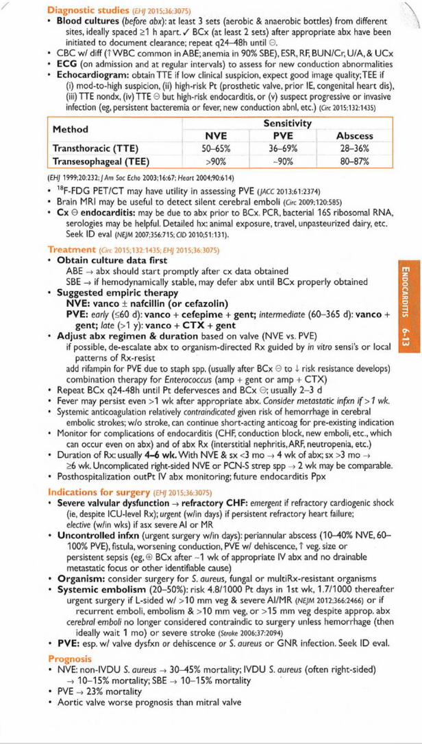
Diagnostic studies {EHJ 2015:36:3075)
• Blood cultures (before abx): at least 3 sets (aerobic & anaerobic bottles) from differentsites, ideally spaced >1 h apart. / BCx (at least 2 sets) after appropriate abx have been initiated to document clearance; repeat q24—48h until ©.
• CBC w/ diff (T WBC common in ABE; anemia in 90% SBE), ESR, RF, BUN/Cr, U/A, & UCx• ECG (on admission and at regular intervals) to assess for new conduction abnormalities• Echocardiogram: obtain TTE if low clinical suspicion, expect good image quality;TEE if
(i) mod-to-high suspicion, (ii) high-risk Pt (prosthetic valve, prior IE, congenital heart dis), (iii)TTE nondx, (iv)TTE 0 but high-risk endocarditis, or (v) suspect progressive or invasive infection (eg, persistent bacteremia or fever, new conduction abnl, etc.) (Ore 2015:132:1435)
M ethodSensitivity
N V E PVE AbscessTransthoracic (TTE) 50-65% 36-69% 28-36%Transesophageal (TEE) >90% -90% 80-87%
{EHJ 1999;20:232; j dm Soc Echo 2003:16:67; Heart 2004:90:614)
• 18F-FDG PET/CT may have utility in assessing PVE (JACC 2013:61:2374)• Brain MRI may be useful to detect silent cerebral emboli (Ore 2009:120:585)• Cx 0 endocarditis: may be due to abx prior to BCx. PCR, bacterial 16S ribosomal RNA,
serologies may be helpful. Detailed hx: animal exposure, travel, unpasteurized dairy, etc. Seek ID eval (NEJM 2007;3S6:715; C/D 2010:51:131).
Treatm ent (Grr 2015;132:1435; EHJ 2015:36:3075)• Obtain culture data first
ABE -* abx should start promptly after cx data obtainedSBE -» if hemodynamically stable, may defer abx until BCx properly obtained
• Suggested em piric therapyNVE: vanco ± nafcillin (o r cefazolin)PVE: early (<60 d): vanco + cefepime + gent; intermediate (60-365 d): vanco +
gent; late (>1 y): vanco + C T X + gent• Adjust abx regimen & duration based on valve (NVE vs. PVE)
if possible, de-escalate abx to organism-directed Rx guided by in vitro sensi s or local patterns of Rx-resist
add rifampin for PVE due to staph spp. (usually after BCx © to i risk resistance develops) combination therapy for Enterococcus (amp + gent or amp + CTX)
• Repeat BCx q24-48h until Pt defervesces and BCx ©; usually 2-3 d• Fever may persist even >1 wk after appropriate abx. Consider metastatic infxn i f > 1 wk.• Systemic anticoagulation relatively contraindicated given risk of hemorrhage in cerebral
embolic strokes; w/o stroke, can continue short-acting anticoag for pre-existing indication• Monitor for complications of endocarditis (CHF, conduction block, new emboli, etc., which
can occur even on abx) and of abx Rx (interstitial nephritis, ARF, neutropenia, etc.)• Duration of Rx: usually 4-6 wk. With NVE & sx <3 mo -> 4 wk of abx; sx >3 mo -»
>6 wk. Uncomplicated right-sided NVE or PCN-S strep spp ^ 2 wk may be comparable.• Posthospitalization outPt IV abx monitoring; future endocarditis Ppx
Indications fo r surgery {EHJ 2015:36:3075)• Severe valvular dysfunction -> refractory CHF: emergent if refractory cardiogenic shock
(ie, despite ICU-level Rx); urgent (w/in days) if persistent refractory heart failure; elective (w/in wks) if asx severe Al or MR
• Uncontrolled infxn (urgent surgery w/in days): periannular abscess (10-40% NVE, 60-100% PVE), fistula, worsening conduction, PVE w/ dehiscence, T veg. size or persistent sepsis (eg,® BCx after ~1 wk of appropriate IV abx and no drainable metastatic focus o r other identifiable cause)
• Organism: consider surgery for S. aureus, fungal o r multiRx-resistant organisms• Systemic embolism (20-50%): risk 4.8/1000 Pt days in 1st wk, 1.7/1000 thereafter
urgent surgery if L-sided w/ >10 mm veg & severe AI/MR (NEJM 2012:366:2466) or if recurrent emboli, embolism & >10 mm veg, or >15 mm veg despite approp. abx
cerebral emboli no longer considered contraindic to surgery unless hemorrhage (then ideally wait 1 mo) or severe stroke (Stroke 2006:37:2094)
• PVE: esp. w/ valve dysfxn or dehiscence or S. aureus o r GNR infection. Seek ID eval.
Prognosis• NVE: non-lVDU S. aureus -» 30^45% mortality; IVDU S. aureus (often right-sided)
-> 10-15% mortality; SBE 10-15% mortality• PVE -> 23% mortality• Aortic valve worse prognosis than mitral valve
En
do
ca
rd
itis
6-1
3
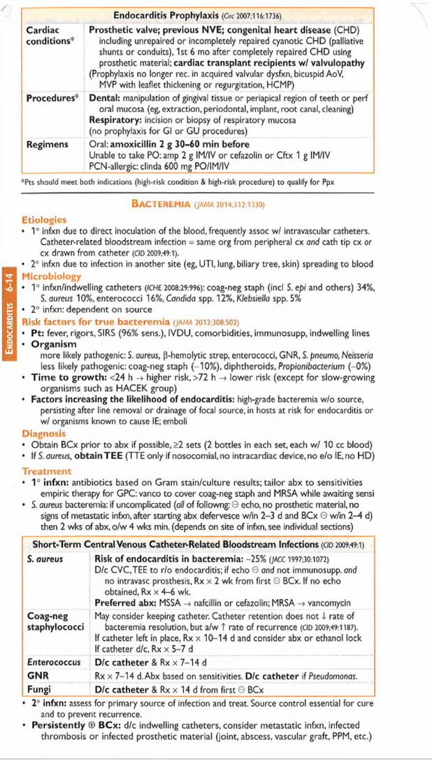
En
do
ca
rd
itis
6-
14Endocarditis Prophylaxis (Ore 20O7;l 16:1736)
Cardiacconditions*
Prosthetic valve; previous NVE; congenital heart disease (CHD) including unrepaired or incompletely repaired cyanotic CHD (palliative shunts or conduits), 1st 6 mo after completely repaired CHD using prosthetic material; cardiac transplant recipients w/ valvulopathy
(Prophylaxis no longer rec. in acquired valvular dysfxn, bicuspid AoV,MVP with leaflet thickening or regurgitation, HCMP)
Procedures* Dental: manipulation of gingival tissue or periapical region of teeth or perf oral mucosa (eg, extraction, periodontal, implant, root canal, cleaning)
Respiratory: incision or biopsy of respiratory mucosa (no prophylaxis for Gl or GU procedures)
Regimens Oral: amoxicillin 2 g 30-60 min beforeUnable to take PO:amp 2 g IM/IV or cefazolin or Cftx 1 g IM/IV PCN-allergic: clinda 600 mg PO/IM/IV
*Pts should meet both indications (high-risk condition & high-risk procedure) to qualify fo r Ppx
B a c te re m ia (ja m a 20i4;3i2:i330)
Etiologies• 1° infxn due to direct inoculation of the blood, frequently assoc w/ intravascular catheters.
Catheter-related bloodstream infection = same org from peripheral cx and cath tip cx or cx drawn from catheter (CID 2009;49:l).
• 2° infxn due to infection in another site (eg, UTI, lung, biliary tree, skin) spreading to bloodMicrobiology• 1° infxn/indwelling catheters (ICHE 2008;29:996): coag-neg staph (incl S. epi and others) 34%,
S. aureus 10%, enterococci 16%, Candida spp. 12%, Klebsiella spp. 5%• 2° infxn: dependent on sourceRisk factors for true bacterem ia (jama 2012:308 502)
• P t: fever, rigors, SIRS (96% sens.), IVDU, comorbidities, immunosupp, indwelling lines• O rgan ism
more likely pathogenic: S. aureus, |3-hemolytic strep, enterococci, GNR, S. pneumo, Neisseria less likely pathogenic: coag-neg staph (-10%), diphtheroids, Propionibacterium (-0%)
• T im e to g row th : <24 h -> higher risk, >72 h -> lower risk (except for slow-growingorganisms such as HACEK group)
• Factors increasing the likelihood of endocarditis: high-grade bacteremia w/o source,persisting after line removal or drainage of focal source, in hosts at risk for endocarditis or w/ organisms known to cause IE; emboli
Diagnosis• Obtain BCx prior to abx if possible, >2 sets (2 bottles in each set, each w/ 10 cc blood)• If S. aureus, ob ta in TEE (TTE only if nosocomial.no intracardiac device, no e/o IE,no HD)
Treatm ent• 1° in fxn : antibiotics based on Gram stain/culture results; tailor abx to sensitivities
empiric therapy for GPC: vanco to cover coag-neg staph and MRSA while awaiting sensi• S. aureus bacteremia: if uncomplicated (all of followng: 0 echo, no prosthetic material, no
signs of metastatic infxn, after starting abx defervesce w/in 2-3 d and BCx 0 w/in 2-4 d) then 2 wks of abx, o/w 4 wks min. (depends on site of infxn, see individual sections)
Short-Term Central Venous Catheter-Related Bloodstream Infections (CID 2009;49:1)
S. aureus Risk of endocarditis in bacteremia: -25% (JACC 1997;301072)
D/c CVC.TEE to r/o endocarditis; if echo © and not immunosupp. and no intravasc prosthesis, Rx x 2 wk from first © BCx. If no echo obtained, Rx x 4-6 wk.
Preferred abx: MSSA -» nafcillin or cefazolin; MRSA -> vancomycinCoag-neg May consider keeping catheter. Catheter retention does not 4 rate ofstaphylococci bacteremia resolution, but a/w T rate of recurrence (CIO 2009;49:1187).
If catheter left in place, Rx x 10-14 d and consider abx or ethanol lock If catheter d/c, Rx x 5-7 d
Enterococcus D/c catheter & Rx x 7-14 dGNR Rx x 7-14 d.Abx based on sensitivities. D/c catheter if Pseudomonas.Fungi D/c catheter & Rx x 14 d from first © BCx
♦ 2° infxn: assess for primary source of infection and treat. Source control essential for cureand to prevent recurrence.
• Persistently © BCx: d/c indwelling catheters, consider metastatic infxn, infectedthrombosis or infected prosthetic material (joint, abscess, vascular graft, PPM, etc.)

TUBERCULOSIS kEpidemiology• U.S.: 10-15 million infected (lOx T risk if foreign-born or minority); worldwide:-2 billion• After resurgence in U.S. 1984-1992, rates have declined• Multidrug resistant (MDR) TB: resistant to isoniazid (INH) and rifampin (RIF). Can occur as
primary infxn if exposed in former Soviet Republics, China• Extensively drug resistant (X D R )T B resistant to INH, RIF, FQ and injectables• Pts m o re like ly to develop TB disease (NEJM 2011:364:1441)
High-prevalence populations (more likely to be exposed & infected): immigrant from high- prevalence area, homeless, IVDU or medically underserved, resident or worker in jail or long-term facility, HCW at facility w/TB, close contact to Pt w/ active TB
High-risk populations (infected & likely to progress to active disease): HIV ©,immunosupp. incl. biologies, uncontrolled DM & smoking, close contact w/ active TB Pt, underweight, CKD, organ Tx, IVU, EtOH, malnourished, cancer, gastrectomy
Microbiology & natural history• Transmission of Mycobacterium tuberculosis via small-particle aerosols (droplet nuclei)• 90% of infected normal hosts will never develop clinically evident disease• Localized disease: healing & calcification or progressive 1°TB (at site of infection)• Hematogenous spread: latent infection ± reactivation TB or progressive dissem.TBScreening for latent infection• W h o m to screen: high-prevalence and high-risk populations (HIV © Pts should have
PPD testing as part of initial evaluation and annually thereafter)• H ow to screen: Mantoux tuberculin test (ie, purified protein derivative or PPD)
inject 5-TU (0.1 mL) intermed. strength PPD intradermally -> wheal; examine 48-72 h• H ow to in te rp re t a PPD: determine max. diameter of induration by palpation •
Size of reaction Persons considered to have © test
>5 mm HIV © or immunosupp (eg, prednisone 15 mg/d x >1 mo)Close contacts of Pt w/ active TB; CXR w/ apical fibrosis c/wTB
>10 mm All other high-risk or high-prevalence populations Recent conversion (T in induration by >10 mm in last 2 y)
>15 mm Everyone elseFalse © Faulty application, anergy (including from active TB), acute TB
(2-10 wk to convert), acute non-TB mycobacteria (NTM), malignancyFalse © Improper reading, cross-reaction with NTM, BCG vaccination
(although usually <10 mm by adulthood)Booster effect t induration b/c immunologic boost by prior skin test in prev sensitized
individual (byTB, NTM or BCG).Test © -> © but not true conversion due to recent infxn. 2nd test true baseline. Can be 1 y after initial test.
(NEJM 2002:347:1860)
• IFN-y release assays (IGRA): (Ag-stimulated IFN-y release from Pt’sT-cells): can use toscreen when PPD could be used (MMWR 2010;S9:1); t Sp,esp. in BCG Rx’d Pts (Anna's 2008:149:177). Does not distinguish active vs. latent or past infxn. Relies on host immune fxn;Se limited in immunosupp. (JO/n Epi 2010:63:257; GD 2011:52:1031).
Clinical manifestations (Lancet 2016;387:1211)
• Prim ary TB pneumonia: middle or lower lobe consolidation. ± effusion,! cavitation• TB pleurisy: can occur w/ primary or reactivation. Due to breakdown of granuloma w/
spilling of contents into pleural cavity and local inflammation. Pulmonary effusion ± pericardial and peritoneal effusions (tuberculous polyserositis).
• Reactivation TB pulmonary disease: apical infiltrate ± volume loss ± cavitation• M iliary TB: acute or insidious; due to hematogenous dissemination; usually in
immunosupp, DM, EtOH, elderly or malnourished. Constitutional sx (fever, night sweats, weight loss) usually prominent. Pulm disease w/ millet seed-like lesions (2-4 mm) on CXR or chest CT (latter more Se) present in 60-80% of those w/ miliary TB.
• Extrapulmonary TB: lymphadenitis, pericarditis, peritonitis, meningitis, nephritis ± sterilepyuria, osteomyelitis (vertebral = Pott’s disease), hepatitis, splenitis, cutaneous, arthritis
• TB and HIV: HIV © at t risk infxn, progressive 1° infxn & reactivation. Risk of progressionfrom infxn to disease >8-10%/y, higher risk with I CD4. Reinfection (also w/ MDR) significant, esp. in hyperendemic areas.
D iagnostic studies fo r active TB (high index of suspicion is key!)• AFB smear (rapid dx) and culture (T Se & allows sensitivity testing) of sputum, BAL,
pleura, etc.; avoid FQ if considering TB (can compromise dx yield)
TB
6-15
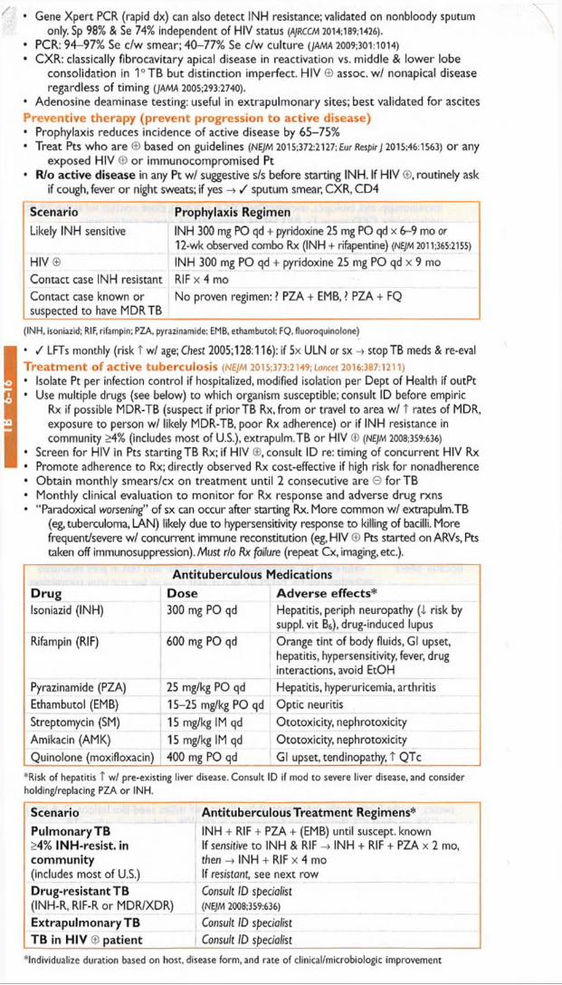
• Gene Xpert PCR (rapid dx) can also detect INH resistance; validated on nonbloody sputumonly. Sp 98% & Se 74% independent of HIV status (AjRCCM 2014;189;1426).
• PCR: 94-97% Se c/w smear; 40-77% Se c/w culture (ja m a 2009;301:1014)• CXR: classically fibrocavitary apical disease in reactivation vs. middle & lower lobe
consolidation in 1°TB but distinction imperfect. HIV © assoc, w / nonapical disease regardless of timing (JAMA 2005:293:2740).
• Adenosine deaminase testing: useful in extrapulmonary sites; best validated for ascites Preventive therapy (prevent progression to active disease)• Prophylaxis reduces incidence of active disease by 65-75%• Treat Pts who are © based on guidelines (NEJM 2015:372:2127; Eur RespirJ 2015:46:1563) or any
exposed HIV © or immunocompromised Pt• Wo active disease in any Pt w/ suggestive sis before starting INH. If HIV ©, routinely ask
if cough, fever or night sweats; if yes -> / sputum smear, CXR, CD4
Scenario Prophylaxis Regimen
Likely INH sensitive INH 300 mg PO qd + pyridoxine 25 mg PO qd x 6-9 mo or 12-wk observed combo Rx (INH + rifapentine) (NE/M 2011:365:2155)
HIV© INH 300 mg PO qd + pyridoxine 25 mg PO qd x 9 moContact case INH resistant RIF x 4 moContact case known or suspected to have MDRTB
No proven regimen: ? PZA + EMB, > PZA + FQ
(INH, isoniazid; RIF, rifampin: PZA. pyrazinamide; EMB, ethambutol; FQ, fluoroquinolone)
• / LFTs monthly (risk T w/ age; Chest 2005; 128:116): if 5x ULN or sx -► stopTB meds & re-eval T re a tm e n t o f active tubercu los is (NEJM 2015;373:2149; lancet 2016:387:1211)• Isolate Pt per infection control if hospitalized, modified isolation per Dept of Health if outPt• Use multiple drugs (see below) to which organism susceptible; consult ID before empiric
Rx if possible MDR-TB (suspect if prior TB Rx, from or travel to area w/ 1 rates of MDR, exposure to person w/ likely MDR-TB, poor Rx adherence) or if INH resistance in community >4% (includes most of U.S.), extrapuIm.TB or HIV © (nejm 2008:359:636)
• Screen for HIV in Pts starting TB Rx; if HIV ®, consult ID re: timing of concurrent HIV Rx• Promote adherence to Rx; directly observed Rx cost-effective if high risk for nonadherence• Obtain monthly smears/cx on treatment until 2 consecutive are © forTB• Monthly clinical evaluation to monitor for Rx response and adverse drug rxns• “ Paradoxical worsening” of sx can occur after starting Rx. More common w/ extrapuIm.TB
(eg, tuberculoma, LAN) likely due to hypersensitivity response to killing of bacilli. More frequent/severe w/ concurrent immune reconstitution (eg, HIV © Pts started on ARVs, Pts taken off immunosuppression). Must r/o Rx failure (repeat Cx, imaging, etc.).
Antitubercu lous Medications
D ru g Dose A d ve rse e ffe c ts *Isoniazid (INH) 300 mg PO qd Hepatitis, periph neuropathy ( i risk by
suppl. vit B&), drug-induced lupusRifampin (RIF) 600 mg PO qd Orange tint of body fluids, Gl upset,
hepatitis, hypersensitivity, fever, drug interactions, avoid EtOH
Pyrazinamide (PZA) 25 mg/kg PO qd Hepatitis, hyperuricemia, arthritisEthambutol (EMB) 15-25 mg/kg PO qd Optic neuritisStreptomycin (SM) 15 mg/kg IM qd Ototoxicity, nephrotoxicityAmikacin (AMK) 15 mg/kg IM qd Ototoxicity, nephrotoxicityQuinolone (moxifloxacin) 400 mg PO qd Gl upset, tendinopathy, T QTc
*Risk of hepatitis T w/ pre-existing liver disease. Consult ID if mod to severe liver disease, and consider holding/replacing PZA or INH.
Scenario Antitubercu lous Treatm ent Regimens*
Pulmonary TB INH + RIF + PZA + (EMB) until suscept. known>4% INH-resist. in If sens/tive to INH & RIF -> INH + RIF + PZA x 2 mo,com m unity then -> INH + RIF x 4 mo(includes most of U.S.) If resistant, see next rowDrug-resistantTB Consult ID specialist(INH-R, RIF-R or MDR/XDR) (NEJM 2008:359:636)
Extrapulm onary TB Consult ID specialistTB in H IV © patient Consult ID specialist
individualize duration based on host, disease form, and rate of dinical/microbiologic improvement

HIV/AIDS i \Definition• AIDS: HIV + CD4 <200/mm3 or AIDS-defining opportunistic infection (Ol) or malignancy
Epidemiology• ~1 million Americans living w/ HIV; -36 million worldwide• 13% in U.S. unaware of infxn, many dx w/ late disease. CDC rec testing all people for HIV.• Routes: sexual (risk is 0.3% for male-to-male, 0.2% for male-to-female, 0.1% for female-to-
male transmission), IVDU, transfusions, needlesticks (0.3%), vertical (15-^10% w/oARV)
Prophylaxis (JAMA 2014:312:390)• Postexposure (PEP): risk infxn -0.3%; Rx: 2 NRTIs + 11x4 wks• Preexposure (PrEP):TDF/FTC qd or on-demand effective (44-86% !) & safe in high-risk,
adherent populations w/o renal insufficiency (NEJM 2010;363:2587 & 2015:373:2237; Lancet 2016:387:53). Monitor renal fxn, STDs, preg, & HIV status.
Acute retroviral syndrome• Occurs in -40-90% of Pts -2 -6 wk after infxn; ± ELISA ©, © viral load (2 wk after infxn);
e a rly ART m ay be b e n e fic ia l (NEJM 2013:368:207 & 218)
• Mono-like syndrome (T mucocut. & neuro manifestations compared to EBV or CMV)
Diagnostic studies• E L IS A for HIV-1 Ab/Ag: © 1-12 wk after acute infxn; >99% Se; 1° screening test• If ©, Ab differentiation assay confirms and differentiates HIV-1 vs. -2 (MMWR 2013:62:489)
• R a p id te s ts : Ab tests; use saliva, plasma, blood or serum; 99% Se &96-99% Sp (Mnols 2008:149:153); PPV in low prev populations is low; needs confirmation
• PCR (viral load): detects HIV-1 RNA in plasma; assay range is 20-10 million copics/mL-2% false ©, but usually low # copies; in contrast, should be very high (>750 k) in 1° infxn
• A t le a s t 1 - t im e H I V s c r e e n in g r e c o m m e n d e d f o r a l l a d u l ts (dnnn/s 2013:159 51)• CD4 count: not a dx test, b/c can be HIV © w/ normal CD4 or be HIV © w/ low CD4
Approach to newly diagnosed H IV © Pt (Loneei 2014:384:258)• D o c u m e n t H I V in fe c t io n ; counseling re: treatment options, adherence, & disclosure• H & P (including focus on h/o Ols, STDs); r e v ie w a ll c u r r e n t m e d s• L a b e v a lu a t io n : CD4 count, PCR, HIV genotype, CBC w / diff., Cr. lytes, LFT$,A1c, &
fasting lipids; PPD or IGRA, syphilis & toxo screen & CMV IgG; HAV, HBV, & HCV serologies; Chlamydia & gonorrhea screen; baseline CXR; Pap smear/anal pap in 9/6
Common Antiretrovirals (ARVs) Common Side Effects
NR
TI
abacavir (ABC; Ziagen) emtricitabine (FTC; Emtriva) lamivudine (3TC; Epivir) tenofovir (TAF orTDF) zidovudine (AZT; Retrovir)
C/ass: Gl intol, lipoatrophy, lactic acidosis ABC: hypersensitivity (3% ),/ HLA-B*5701 AZT: BM suppression (esp. macrocytic anemia) TDF: renal toxicity TAF: minimal renal toxicity
NN
RT
I efavirenz (EFV; Sustiva) etravirine (ETR; Intelence) nevirapine (NVP;Viramune) rilpivirine (RPV; Edurant)
Class: rash, hepatitis, mixed CYP450 inducer/inhib EFV: CNS effects (incl depression)NVP: rash and hypersensitivity [risk factors are
female, CD4 >250, pregnancy (.’. avoid)]
Q.
atazanavir (ATV; Reyataz) darunavir (DRV; Prezista) lopinavir/riton. (LPV/r; Kaletra) ritonavir (RTV; Norvir)
C/ass; Gl intol; hepatotoxicity; inhibit CYP450 (caution w / $tatins);T2DM; truncal obesity; hyperlipid (less w/ATV); Ml (NEJM 2007:356:1723)
ATV: crystalluria -> nephrolithiasis DRV: rash (10%); possible sulfa cross-reactivity
uZ enfuvirtide (T20; Fuzeon) injection site reaction
LJ maraviroc (MVC; Selzentry) dizziness, hepatotoxicity; / CCR5 tropism assay
=
dolutegravir (DTG;Tivicay) elvitegravir (EVG;Vitekta) raltegravir (RAL; Isentress)
Class: diarrhea & other Gl intol; T CPK DTG + metformin requires glc monitoring
*CO ritonavir (r); cobicistat (COBI) drug interactions (inhibit CYP450)
NRTI, nudeoside/tide reverse transcriptase inhibitor; NNRTI, nonnucleoside RTI; PI, protease inhibitor; FI, fusion inhibitor; El, entry inhibitor (CCR5 antagonist); II, integrase inhibitor; booster to give w / other ARVs; several multiclass combination pills exist
• ARVs should be given in consultation wl HIV specialist (JAMA 2016:316:191)
• Counseling re: strict adherence to ARVs is essential; genotype prior to ART-initiation• All HIV © Pts should be treated w/ARVs (NEJM 2015:373:795; http://aidsinfo.nih.gov); especially
those w/AIDS-defining illness, preg, HIV-assoc. nephropathy, HCV/HBV co-infxn• Rec regimens include: 2 NRTI (eg.TAF + FTC) + either II or boosted PI (eg, DRV/r)
HIV
/AID
S
6-17

• Initiation of ARVs may transiently worsen existing Ols for several wks due to immunereconstitution inflammatory syndrome (IRIS)
A pproach to p reviously established H IV © P t• H & P (mucocutaneous, neurocognitive, Ols, malignancies, STDs); meds• Review ARVs (past and current); if any must be interrupted, stop all to i risk of resistance• Failing regimen = unable to achieve undetectable viral load, t viral load, I CD4 count or
clinical deterioration (with detectable viral load consider genotypic or phenotypic assay)
O l Prophylaxis (https://aidsinfo.nih.gov/guidelines)
O l In d ic a tio n 1° P rophy lax isTuberculosis © PPD (>5 mm)/IGRA
or high-risk exposureINH + vit B& x 9 mo
Pneumocystis jiroveci (PCP)
CD4 <200/mm3 or CD4 <14% or thrush
TMP-SMX DS or SS qd or DS tiw or dapsone 100 mg qd or atovaquone 1500 mg qd or pentamidine 300 mg inh q4wk
Toxoplasmosis CD4 <100/mm3 and © Toxo IgG
TMP-SMX DS qd or dapsone 50 mg qd + pyrimethamine
50 mg qwk + leucovorin 25 qwkMAC CD4 <50/mm3 azithro 1200 mg qwk or clarithro 500 mg bidStop 1° prophylaxis if CD4 in itiation threshold >3-6 mo on ARVsStop 2° prophylaxis (maintenance therapy for prior Ol; drugs and doses differ by Ol) if clinical resolution or stabilization and CD4 thresholds have been exceeded x 3-6 mo
Complications of HIV/AIDSCD4 Count Com plications
<500 Constitutional sx; noninfectious disease (CVD, bone, oncologic) Mucocutaneous: Kaposi’s sarcoma; seborrheic dermatitis; oral hairy
leukoplakia; lymphoma; candidiasis; HSV;VZV Recurrent bacterial infections,TB (pulm and extrapulm); neurosyphilis
<200 PCP, Toxo, Bartonella, Crypto, Histo, Coccidio<50-100 CMV, MAC, CNS lymphoma, PML, death (<50 is medical emergency)
Invasive aspergillosis, bacillary angiomatosis (disseminated Bartonella)
Fever• Etiologies (Infect Dis Clin North Am 2007:21:1013)
infxn (82-90%): MAC, TB, CMV, early PCP, Histo, Crypto, Coccidio, Toxo, endocarditis noninfectious: lymphoma, drug reaction. Non 1° HIV itself rarely (<5%) cause of fever.
• Workup: guided by CD4 count, s/s, epi, & exposuresCBC, chem, LFTs, BCx, CXR, UA, mycobact. & fungal cx, / meds, ? / chest & abd CT CD4 <100-200 -> serum crypto Ag, LP, urinary Histo Ag, CMV PCR or antigenemia pulmonary s/s -> CXR;ABG; sputum for bacterial cx, PCP.AFB; bronchoscopy diarrhea -> stool cx, O&P, AFB; direct visualization with bx on colonoscopy cytopenias -> BM bx for, path & cx of aspirate including for mycobacteria & fungi abnormal LFTs abd CT, liver bx for path & cx including for mycobacteria & fungi
Cutaneous• Seborrheic dermatitis; eosinophilic folliculitis; w arts (HPV); HSV & VZV; MRSA skin & soft
tissue infxns; scabies; candidiasis; eczema; prurigo nodularis; psoriasis; drug eruptions• Dermatophyte infx: prox subungual onychomycosis (at nail bed); pathognomonic for HIV• Molluscum contagiosum (poxvirus): 2-5 mm pearly papules w/ central umbilication• Kaposi’s sarcoma (KSHV or HHV8): red-purple nonblanching nodular lesions• Bacillary angiomatosis (disseminated Bartonella): friable violaceous vascular papules O p h th a lm o lo g ic• C M V retinitis (CD4 usu <50); Rx:gan- or valganciclovir, ganciclovir implant or cidofovir• HZV,VZV, syphilis (at any CD4 count) o r Toxo: CD4 usually <100
O ra l• Aphthous ulcers; KS; thrush (oral candidiasis): curd-like patches typically w/ burning
or pain; oral hairy leukoplakia: painless proliferation of papillae w/ adherent white coating usually on lateral tongue, caused by EBV but not precancerous
Endocrine/metabolic• Hypogonadism; adrenal insufficiency (CMV, MAC.TB, HIV or med-related); wasting
osteopenia/porosis (at all CD4 counts); fragility fractures• Lipodystrophy: central obesity, peripheral lipoatrophy, dyslipidemia, hyperglycemia
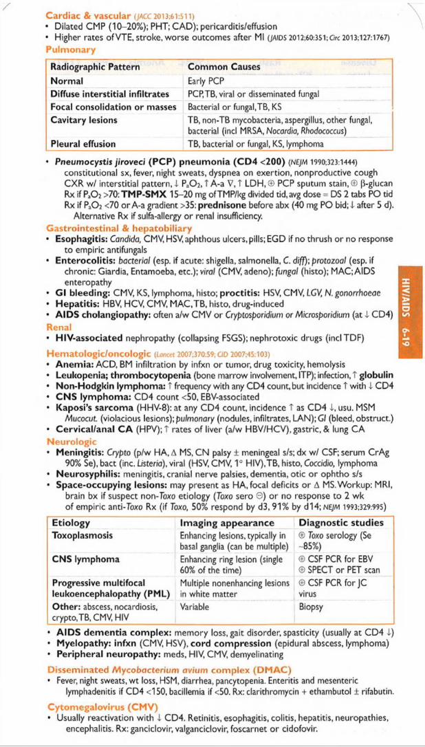
C ardiac & vascular (JACC2013:61:511)• Dilated CMP (10-20%); PHT; CAD); pericarditis/effusion* Higher rates ofVTE, stroke, worse outcomes after Ml (JAIDS 2012-,60:351;C/rc 2013;127:1767)P u lm ona ry
Radiographic Pattern Com m on Causes
Norm al Early PCPDiffuse in te rs titia l in filtra tes PCP.TB, viral or disseminated fungalFocal consolidation o r masses Bacterial or fungal,TB, KSCavitary lesions TB, non-TB mycobacteria, aspergillus, other fungal,
bacterial (incl MRSA, Nocardia, Rhodococcus)Pleural effusion TB, bacterial or fungal, KS, lymphoma
• Pneumocystis jiroveci (PC P) pneumonia (C D 4 <200) (NEJM 1990:323:1444)constitutional sx, fever, night sweats, dyspnea on exertion, nonproductive cough CXR w/ interstitial pattern, i Pa02, T A-a V, T LDH, © PCP sputum stain, © [3-glucan Rx if P ,02 >70: TM P-SMX 15-20 mg ofTMP/kg divided tid, avg dose = DS 2 tabs PO tid Rx if PaC>2 <70 or A-a gradient >35: prednisone before abx (40 mg PO bid; i after 5 d).
Alternative Rx if sulfa-allergy or renal insufficiency.G as tro in tes tina l & h e p a to b ilia ry• Esophagitis: Candida, CMV, HSV, aphthous ulcers, pills; EGD if no thrush or no response
to empiric antifungals• Enterocolitis: bacterial (esp. if acute: shigella, salmonella, C. diff)\ protozoal (esp. if
chronic: Giardia, Entamoeba, etc.); viral (CMV, adeno); fungal (histo); MAC; AIDS enteropathy
• G l bleeding: CMV, KS, lymphoma, histo; proctitis: HSV, CMV, LGV, N. gonorrhoeae• Hepatitis: HBV, HCV, CMV, MAC.TB, histo, drug-induced• A IDS cholangiopathy: often a/w CMV or Cryptosporidium or Microsporidium (a t! CD4)Renal• HIV-associated nephropathy (collapsing FSGS); nephrotoxic drugs (inclTDF)
H em a to log ic /o nco log ic (Lancet 2007:370:59; C/D 2007:45:103)• Anem ia: ACD, BM infiltration by infxn o r tumor, drug toxicity, hemolysis• Leukopenia; thrombocytopenia (bone marrow involvement, ITP); infection, T globulin• Non-Hodgkin lymphoma: t frequency with any CD4 count, but incidence T with 1 CD4• CNS lymphoma: CD4 count <50, EBV-associated• Kaposi’s sarcoma (HHV-8): at any CD4 count, incidence T as CD4 1, usu. MSM
Mucocut. (violacious lesions); pulmonary (nodules, infiltrates, LAN);G/ (bleed, obstruct.)• Cervical/anal C A (HPV); T rates of liver (a/w HBV/HCV), gastric, & lung CAN eu ro log ic• Meningitis: Crypto (p/w HA, A MS, CN palsy ± meningeal s/s; dx w/ CSF; serum CrAg
90% Se), bact (inc. Listeria), viral (HSV, CMV, 1° HIV),TB, histo, Coccidio, lymphoma• Neurosyphilis: meningitis, cranial nerve palsies, dementia, otic or ophtho s/s• Space-occupying lesions: may present as HA, focal deficits or A MS. Workup: MRI,
brain bx if suspect non-Toxo etiology (Toxo sero ©) or no response to 2 wk o f empiric anti-Toxo Rx (if Toxo, 50% respond by d3, 91% by d14;N£/M 1993:329:995)
E tio lo g y Im ag ing appearance D ia g n o s tic s tud iesToxoplasmosis Enhancing lesions, typically in
basal ganglia (can be multiple)© Toxo serology (Se -85%)
CNS lymphoma Enhancing ring lesion (single 60% of the time)
© CSF PCR for EBV © SPECT or PET scan
Progressive m ultifocal leukoencephalopathy (PML)
Multiple nonenhancing lesions in white matter
©CSF PCR for JC virus
O ther: abscess, nocardiosis, crypto, TB, CMV, HIV
Variable Biopsy
• A ID S d e m e n tia com p lex : memory loss, gait disorder, spasticity (usually at CD4 i )• M ye lopathy: in fxn (CMV, HSV), co rd com press ion (epidural abscess, lymphoma)• Periphera l neuropa thy : meds, HIV, CMV, demyelinating
D issem inated M ycobacterium avium com p lex (D M A C )• Fever, night sweats, wt loss, HSM, diarrhea, pancytopenia. Enteritis and mesenteric
lymphadenitis if CD4 <150, bacillemia if <50. Rx: clarithromycin + ethambutol ± rifabutin.
C y tom ega lov iru s (C M V )* Usually reactivation with i CD4. Retinitis, esophagitis, colitis, hepatitis, neuropathies,
encephalitis. Rx: ganciclovir, valganciclovir, foscarnet o r cidofovir.
HIV/AIDS 6-19
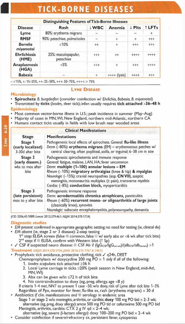
Lym
e
6-20
[ TICK-BORNE DISEASESDistinguishing Features ofT ick-Borne Illnesses
Disease Rash 4 W B C A n e m ia i P its t LFTsLyme 80%: erythema migrans - - +RMSF 90%: petechiae, palms/soles - + + +++
Borreliamiyamotoi
<10% ++ + +++ +++
Ehrlichiosis(HME)
25%: maculopapular, petechiae
+++ ++ ++++ ++++
Anaplasmosis(H G A)
<5% +++ + +++ -H-H-
Babesia - + ++++ (lysis) ++++ +++
<15%. +: 15-25%, ++: 25-50%, +++: 50-75%, ++++: > 75%
Lyme D iseaseM icrob io logy• Spi roch e te 8. burgdorferi (consider coinfection w/ Ehrlichia, Babesia, 8. miyamotoi)• Transmitted by ticks (/xodes, deer tick); infxn usually requires t ic k a ttached >36-48 hE p idem io logy• Most common vector-borne illness in U.S.; peak incidence in summer (May-Aug)• Majority of cases in M N,W I, New England, northern mid-Atlantic, northern CA• Humans contact ticks usually in fields with low brush near wooded areas
Clinical Manifestations
Stage M a n ife s ta tio n sStage 1
(early localized)3-30d after bite
Pathogenesis: local effects of spirochete. General: flu-like illness Derm (-80%): erythem a migrans (EM) = erythematous patches w/
central clearing, often popliteal, axilla, or inguinal; 6-38 cm in size
Stage 2 (early dissem.)wks to mos after
bite
Pathogenesis: spirochetemia and immune responseGeneral: fatigue, malaise, LAN, HA; fever uncommonDerm: m ultip le (1-100) annular lesions « EMRheum (~10%): m ig ra to ry arthralgias (knee & hip) & myalgiasNeurologic (~15%): cranial neuropathies (esp. CN VII), aseptic
meningitis, mononeuritis multiplex (± pain), transverse myelitis Cardiac (~8%): conduction block, myopericarditis
Stage 3 (late persistent)mos to y after bite
Pathogenesis: immune responseDerm, acroderm atitis chronica atrophicans, panniculitis Rheum (~60%): recurren t mono- o r o ligoarth ritis of large joints
(classically knee), synovitis Neurologic: subacute encephalomyelitis, polyneuropathy, dementia
(CJD 2006:43:1089; Loncet 2012:379:461; NE/M 2014:370:1724)
Diagnostic studies• EM present: confirmed in appropriate geographic setting; no need for testing (ie, clinical dx)• EM absent (ie, stage 2 or 3 disease): 2-step testing
1st step: ELISA screen (false © common, false G w/ early abx or <6 wk after tick bite) 2nd step: if © ELISA, confirm with Western blot (T Sp)
• / CSF if suspected neuro disease:© CSF Ab if (lgGcsF/lgGserum)/(albc$F/albsemm) >1 T re a tm e n t {NEJM 2014;370:1724;./AA1A 2016:315:1767 & 2461)• Prophylaxis: tick avoidance, protective clothing, tick / q24h, DEET
Chemoprophylaxis w/ doxycycline 200 mg PO x 1 only if all of the following:1. Ixodes scapularis tick attached >36 h2. Local Lyme carriage in ticks >20% (peak season in New England, mid-Atl,
M N.W I)3. Abx can be given w/in <72 h of tick bite4. No contraindication to doxy (eg, preg, allergy, age <8 y)
If criteria 1-4 met, NNT to prevent 1 case -50; w/o doxy, risk of Lyme after tick bite 1-3% Regardless of Ppx, monitor for fever, flu-like sx, rash (erythema migrans) x 30 d
• Antibiotics: i f clin. manifestations and © serology in endemic areaStage 1 or stage 2 w/o meningitis, arthritis, or carditis: doxy 100 mg PO bid x 2-3 wk;
alternative (eg, preg, doxy allergy): amox 500 mg PO tid o r cefuroxime 500 mg PO bid Meningitis, arthritis, carditis: CTX 2 g IV qd x 2-4 wk;
alternative (eg, severe (3-lactam allergy): doxy 100-200 mg PO bid x 2-4 wk• Consider coinfection if severe/refractory sx, persistent fever, cytopenias

Rocky Mountain Spotted Fever (RMSF)M icrobiology & epidem iology• Infection with Rickettsia rickettsii (Gram © obligate intracellular bacterium)• Transmitted by Dermacentor variabilis, D. andersoni (dog tick); peak in spring/early summer• Occurs in m id-Atl, SE, Midwest, New Engl, NW, Canada, Mexico, Central & S.America• Consider other rickettsial spp.: R. akari (Rickettsial pox), R. conorii (Mediterranean spotted
fever), R. africae (African tick bite fever), R. felis (Flea rickettsiosis)Clinical m anifestations (typically w/in 1 wk of tick exposure)• Nonspecific: fever, H A , AMS, myalgias, N/V, occasionally abdominal pain• Rash (2-5 d after onset) = centripetal: starts on ankles and wrists trunk, palms, &
soles; progresses from macular to maculopapular to petechial• Severe cases -> vasculitis, hypoperfusion/shock, end-organ damage; more likely in elderly• Up to 75% mortality if untreated, 5-10% even w/ Rx (esp. if delayed) (NEJM 2005:353:551) Diagnosis• Usually a clinical dx; requires early clinical suspicion given risks of delayed Rx• Acute illness dx by skin bx for rickettsiae (Se -70%); 7-10 d after sx onset, serology ©Trea tm ent• Doxycycline 100 mg PO bid (give empirically if clinical suspicion)
Ehrlichiosis/A naplasmosisM icrobio logy• Gram © obligate intracellular bacterium; human monocytic ehrlichiosis (E chaffeensis,
HME); human granulocytic anaplasmosis (A. phagocytophilum, HGA)• Transmission: HME by Amblyomma americanum, Dermacentor variabilis; HGA by IxodesEpidem iology• HGA cases typically in New Engl, mid-Atl, MN; HME in SE and south central US• Peak incidence spring and early summer; can be transmitted by blood transfusionClinical m anifestations (typically w/in 3 wk of tick exposure)• Asx or nonspecific: fever, myalgias, malaise, HA, cough, dyspnea; onset often acute• Laboratory: leukopenia, thrombocytopenia, t aminotransferases, LDH, A<j), renal insuff• More severe disease can occur with bacterial superinfection in HGADiagnosis• Acute: intraleukocytic morulae on peripheral blood smear (rare); PCR; later: serologyTrea tm ent (jama 2016:315:1767)• Start Rx based on clinical suspicion; definitive dx requires PCR (may not detect all spp.)• Doxycycline 100 mg PO bid (often x 10 d); should defervesce in <48 h, else reconsider dx
BabesiosisM icrobiology & epidem iology• Infxn w/ parasite Babesia microti (U.S.), transmitted by Ixodes ticks; also a/w transfusion• Europe & U.S. (more commonly MN.WI, coastal areas & islands of MA, NY, NJ, Rl, CT)• Peak incidence June-August (MAIWR 2012:61:505)Clinical m anifestations (typically 1-4 wk after tick exposure; <9 wk if transfusion)• Range from asx to fevers, sweats, myalgias, & HA to severe hemolytic anemia,
hemoglobinuria, & death (degree of parasitemia correlates roughly with severity)• Risk factors for severe disease: asplenia, i cellular immunity,TNF inhib, T age, pregnancyDiagnosis (NEJM 2012:366:2397)• Clinical syndrome + blood smear w/ intraerythrocytic parasites• Repeat smears (q12-24h) if sx persist despite negative initial smear• PCR serum if smear 0 and high clinical suspicion, serum IgG can help but some false ©T rea tm ent (jama 2016:315:1767)• Atovaquone & azithro for mild/mod illness; clinda & quinine if severe (more toxic)• Duration depends on host; immunosupp Pts often need longer Rx• Exchange transfusion if parasitemia >10%, severe hemolysis o r SIRS * •
T u l a r e m iaM icrob io logy• Infxn w/ Francisella tularensis via contact w / animal tissue, aerosol, tick/insect biteC lin ica l m an ifes ta tions (typically w/in 2-10 d of exposure)• Acute onset of fever, HA, nausea; ulcer w/ black eschar at site of entry; LAN; PNA
Diagnosis & tre a tm e n t• Hazardous and difficult to Cx, alert lab. Serology © by wk 2. PCR by research lab.• Streptomycin or gentamicin x 7-14 d; empiric Rx may be needed given challenges in dx
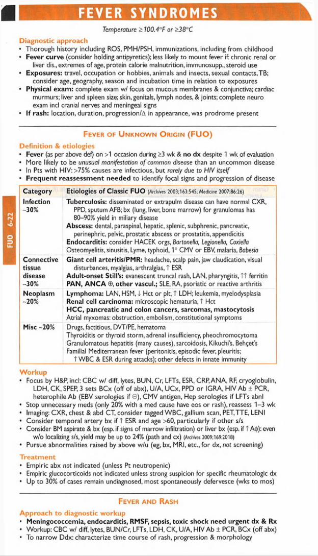
FUO
6-
22FEVER SYNDROMES
Temperature > 100.4°F or >38°C
D iagnostic approach• Thorough history including ROS, PMH/PSH, immunizations, including from childhood• Fever curve (consider holding antipyretics); less likely to mount fever if chronic renal or
liver dis., extremes of age, protein calorie malnutrition, immunosupp., steroid use• Exposures: travel, occupation or hobbies, animals and insects, sexual contacts,TB;
consider age, geography, season and incubation time in relation to exposures• Physical exam: complete exam w/ focus on mucous membranes & conjunctiva; cardiac
murmurs; liver and spleen size; skin, genitals, lymph nodes, & joints; complete neuro exam ind cranial nerves and meningeal signs
• If rash: location, duration, progression/A in appearance, was prodrome present
Fever of U nknow n O rigin (FUO)D e fin itio n & e tio log ies• Fever (as per above def) on >1 occasion during >3 wk & no dx despite 1 wk of evaluation• More likely to be unusua/ manifestation o f common disease than an uncommon disease• In Pts with HIV: >75% causes are infectious, but rarely due to HIV itself• F requen t reassessm ent needed to identify focal signs and progression of disease
Category Etiologies o f Classic FUO {Archives 2003:163:545; Medicine 2007:86:26)
Infection-30%
Tuberculosis: disseminated or exttapulm disease can have normal CXR, PPD, sputum AFB; bx (lung, liver, bone marrow) for granulomas has 80-90% yield in miliary disease
Abscess: dental, paraspinal, hepatic, splenic, subphrenic, pancreatic, perinephric, pelvic, prostatic abscess or prostatitis, appendicitis
Endocarditis: consider HACEK orgs, Bartonella, Legionella, Coxiella Osteomyelitis, sinusitis, Lyme, typhoid, 1° CMV or EBV, malaria, Babesia
Connectivetissuedisease-30%
Giant cell arteritis/PMR: headache, scalp pain, jaw claudication, visual disturbances, myalgias, arthralgias, t ESR
Adult-onset Still's: evanescent truncal rash, LAN, pharyngitis, TT ferritin PAN, A N C A ©, o ther vascul.; SLE, RA, psoriatic or reactive arthritis
Neoplasm-20%
Lymphoma: LAN, HSM, I Hct or pit, T LDH; leukemia, myelodysplasiaRenal cell carcinoma: microscopic hematuria, t HctHCC, pancreatic and colon cancers, sarcomas, mastocytosisAtrial myxomas: obstruction, embolism, constitutional symptoms
Misc -20% Drugs, factitious, DVT/PE, hematomaThyroiditis or thyroid storm, adrenal insufficiency, pheochromocytoma Granulomatous hepatitis (many causes), sarcoidosis, Kikuchi’s, Behcet’s Familial Mediterranean fever (peritonitis, episodic fever, pleuritis;
T WBC & ESR during attacks); other defects in innate immunity
W o rk u p• Focus by H&P, inch CBC wf diff, lytes, BUN, Cr, LFTs, ESR, CRP, ANA, RF, cryoglobulin,
LDH, CK, SPEP, 3 sets BCx (off of abx), U/A, UCx, PPD or IGRA, HIV Ab ± PCR, heterophile Ab (EBV serologies if© ), CMV antigen, Hep serologies if LFTs abnl
• Stop unnecessary meds (only 20% with a med cause have eos or rash), reassess 1-3 wk• Imaging: CXR, chest & abd CT, consider tagged WBC, gallium scan, PET,TTE, LENI• Consider temporal artery bx if T ESR and age >60, particularly if other s/s• Consider BM aspirate & bx (esp. if signs of marrow infiltration) or liver bx (esp. if T A<{>): even
w/o localizing s/s, yield may be up to 24% (path and cx) (Archives 2009; 169:2018)• Pursue abnormalities raised by above w/u (eg, bx, MRI, etc., for dx, not screening)
T re a tm e n t• Empiric abx not indicated (unless Pt neutropenic)• Empiric glucocorticoids not indicated unless strong suspicion for specific rheumatologic dx• Up to 30% of cases remain undiagnosed, most spontaneously defervesce (wks to mos) * •
Fever and Rash A pproach to d iagnostic w o rku p• Meningococcemia, endocarditis, RMSF, sepsis, toxic shock need urgent dx & Rx• Workup: CBC w/ diff, lytes, BUN /Cr, LFTs, LDH, CK, U/A, HIV Ab ± PCR, BCx (off abx)• To narrow Ddx: characterize time course of rash, progression & morphology

/ Erythema multiforme: symmetric "target” lesions often of palms, soles, & mucous memb Infxn etiol: HSV V?, Mycoplasma, syphilis, tick-borne diseases, etc.Non-infxn etiol: meds (eg, NSAIDs, sulfa), malignancy, autoimmune & rheum disease
Erythema nodosum: tender erythematous or violaceous nodules usually symmetric on LE Infxn etiol: Strep,TB, EBV, Bartonella, HBV, psittacosis, fungal, L venereum, etc. Non-infxn etiol: sarcoidosis, IBD, Behcet’s, other rheum, pregnancy/OCP use
Pursue specific dx based on exposure hx & exam, including serologies, viral swab PCR, antigen tests and possibly skin biopsy ± exam of vesicular or bullae fluid if present
Etiologies more broad in immunosupp. Pts, dx testing should be earlier and more extensive; higher risk of critical illness due to disseminated or rapidly progressive infxns
Variable Possible EtiologySummer/fall > other seasons EnterovirusWinter Parvovirus, MeningococcemiaSpring/summer Measles/rubella, Lyme, RMSFYear-round Adenovirus, MycoplasmaCat and dog exposure Bartonella, Pasteurella, Toxoplasma, CapnoeytophogaTick exposure Lyme, RMSF, Ehrlichiosis, AnaplasmosisAdult <30 y Mononucleosis (EBV or CMV)Inadequate immunization Measles, Rubella,VZV, influenzaSexually active HIV, syphilis, disseminated gonococcal infection, HSV2Consider noninfectious causes: allergy/DRESS, DVT, phlebitis, vasculitides, neutrophilic dermatoses, gout, connective tissues dis., malignancy, foreign body rxn
Treatm ent• Empiric abx not indicated (unless Pt neutropenic or critically ill)
Fever in a Returned T raveler •
Region or Exposure Common EtiologiesSub-Saharan Africa Malaria » dengue, rickettsial disease, enteric diseaseSoutheast Asia Dengue > malaria, enteric disease (S. typhi), ChikungunyaCentral & S. America Enteric disease, malaria, dengue, ZikaCaribbean & Mexico Middle East & S. Korea
Dengue » Chikungunya > malaria. Also consider Zika. Middle East Respiratory Syndrome
Freshwater swimming Schistosomiasis, leptospirosisUnpurified drinking water Enteric disease (E. coli » S. typhi, Campylobacter, hepatitis E
> Vibrio cholerae), amebic liver abscessLacking immunizations HAV/HBV, S, typhi, influenza, measles, rubella, yellow feverAnimal bite RabiesAfrican "safari" Rickettsial disease,African trypanosomiasisAdult <30 years Mononucleosis (EBV or CMV)
(NEJM 2002:347:505; CID 2007;44:1560; Cm Opin Infect Dis 2007:20:449)
• Pts visiting friends and relatives abroad are most likely to contract illness during travel• Geography influences Ddx in returned travelers: http://www.nccdc.gov/travel/notices• Emerging pathogens: Influenza occurs year-round in the tropics. Chikungunya and dengue
w/ T areas of transmission, hemorrhagic fevers primarily in Central Africa.• Consider domestic infxns, STIs, & non-infxn causes. Enteric parasites rarely cause fever.Select clinical manifestations• Ebola: fever in traveler from area with active transmission of Ebola w/in 21 d: isolate &
contact state health department (http://www.cdc.gov/vhf/ebola)• Malaria: nonspecific symptoms including diarrhea, myalgias, cough, altered mental status• Dengue: nonspecific symptoms including headache, severe myalgias, rash/petechiae• Chikungunya: nonspecific symptoms including joint pain, moderate myalgias, fever• Typhoid (Lancet 2015:385:1136): constipation, abd pain, possible rash, relative bradycardia• Rickettsial disease: headache, myalgias, lymphadenopathy, possible rash/eschar• Zika: fever, rash, arthralgia, H/A, conjunctivitis (http://www.cdc.gov.zika)
W orkup• Routine testing: CBC w/ diff, lytes, LFTs, BCx, UA, rapid malaria test• Fever in a traveler from a malaria zone is malaria until proven otherwise; consider
hospitalization and empiric Rx. One 9 smear does not r/o malaria.• Other tests based on s/s, labs, exposure, incubation period, geography and seasonality.
O&P exam, CXR, blood smears for filaria/Babesiosis/Borre//o, serologies, STI & HIV, PPD or IGRA, bone marrow aspirate, bx of lymph nodes or skin lesions, CSF studies.
Fever
6-23

PITUITARY DISORDERSH ypopituitary Syndromes
P anhypo p itu ita rism (Lancet 2016;epub)
• EtiologiesP rim ary: surgery, radiation (develops after avg 4-5 y), tumors (primary or metastatic),
infection, infiltration (sarcoid, hemochromatosis), autoimmune, ischemia (including Sheehan’s syndrome caused by pituitary infarction intrapartum), carotid aneurysms, cavernous sinus thrombosis, trauma, medications (eg, ipilimumab)
Secondary (hypothalamic dysfunction o r stalk interruption): tumors (including craniopharyngioma), infection, infiltration, radiation, surgery, trauma
• Clinical manifestationsH orm ona l: acute - * * weakness, easy fatigability, hypotension, polyuria and polydipsia;
chronic -» bradycardia, sexual dysfxn, loss of axillary & pubic hair, w t loss, amenorrhea Mass effect: headache, visual field As, cranial nerve palsies, galactorrhea Apoplexy (pituitary hemorrhage or infarction, usually w/ underlying pituitary adenoma):
sudden headache, N/V, visual field As, cranial nerve palsies, meningismus, A MS, hypoglycemia, hypotension
• Diagnostic studiesH o rm o n a l studies
chronic: ! target gland hormone + I or normal trophic pituitary hormone acute: target gland hormonal studies may be normal partial hypopituitarism is more common than panhypopituitarism
P itu ita ry MRI• Treatment
Replace deficient target gland hormonesMost important deficiencies to recognize and treat in inpatients are adrenal insufficiency
and hypothyroid/sm; if both present, treat with glucocorticoids first, then replace thyroid hormone so as not to precipitate adrenal crisis
I A C T H• Sx similar to 1° adrenal insufficiency (see “ Adrenal Disorders” ) except:
no salt cravings or hypokalemia (b/c aldo preserved) no hyperpigmentation (b/c ACTH/MSH is not t )
I T S H• Sx of central hypothyroidism similar to 1° (see "Thyroid Disorders” ) except absence of goiter• Dx with freeTn in addition toTSH, asTSH may be low or inappropriately normali PRL• Inability to lactatei G H• t chronic risk for osteoporosis, fatigue, weight gain• Dx with failure to T GH w/ appropriate stimulus (eg, insulin tolerance test, glucagon
stimulation)• GH replacement in adults controversial (Annals 2003:35:419)4. FSH & LH• Clinical manifestations: i libido, impotence, oligomenorrhea or amenorrhea, infertility,
i muscle mass, osteoporosis• Physical exam: i testicular size; loss of axillary, pubic and body hair• Dx with: i a.m. testosterone or estradiol (also assess SHBG, esp. in obese) and i o r
normal FSH/LH (all levels i in acute illness, do not measure in hospitalized Pts)• Treatment: testosterone or estrogen replacement vs. correction of the underlying cause •i A D H (hypothalamic or stalk disease): diabetes insipidus• Typically from mass lesion extrinsic to sella; pituitary tumor does not typically present w/ Dl• Clinical manifestations: severe polyuria, mild hypernatremia (severe if i access to H2O)• Diagnostic studies: see “ Sodium and W ater Homeostasis”
Hyperpituitary SyndromesP itu ita ry tu m o rs• Pathophysiology: adenoma excess of trophic hormone (if tumor fxnal, but 30-40%
not) and potentially deficiencies in other trophic hormones due to compression; cosecretion of PRL and growth hormone in 10% of prolactinomas
• Clinical manifestations: syndromes due to oversecretion of hormones (see below)± mass effect: headache, visual As, diplopia, cranial neuropathies
• Workup: MRI, hormone levels, ± visual field testingif <10 mm, no mass effect, no hormonal effects, can f/up q3-6mo
Pituitary
7-1

Hyperprolactinem ia (NEJM 2010:362:1219 and JCEM 2011:96:273)
• Etiologyprolactinoma (50% of pituitary adenomas)stalk compression due to nonprolactinoma -» i inhibitory dopamine -» T PRL (mild)
• Physiology: PRL induces lactation and inhibits GnRH -> I FSH & LH• Clinical manifestations: am enorrhea, galactorrhea, infertility, i libido, impotence• Diagnostic studies
T PRL ( / fasting levels), but elevated in many situations, .\ r/o pregnancy or exogenous estrogens, hypothyroidism, dopamine agonists (eg, psych meds, antiemetics), renal failure ( i clearance), cirrhosis, stress, T carb diet. Watch for hook effect: assay artifact yielding falsely low PRL if very high serum PRL levels; retest with sample dilution.
MRI to evaluate for tumor• Treatment
If asx (no HA,galactorrhea, hypogonadal sx) & microadenoma (<10 m m ),follow w/ MRI If sx o r macroadenoma (>10 m m ) options include;medical with dopamine agonist such as cabergoline (70-100% success rate) or
bromocriptine (not as well tol); side effects include N/V, orthostasis, nasal congestion surg/ca/: transsphenoidal surgery (main indications: failed or cannot tolerate medical Rx,
GH cosecretion or neurologic sx not improving); 10-20% recurrence rate radiation: if medical o r surgical therapy have failed o r are not tolerated
Acromegaly ( t GH; 10% of adenomas; NEJM 2006;35S:2558 & JCEM 2014:99:3933)• Physiology: stimulates secretion of insulin-like growth factor 1 (IGF-1)• Clinical manifestations: t soft tissue, arthralgias, jaw enlargement, headache, carpal tunnel
syndrome, macroglossia, hoarseness, sleep apnea, amenorrhea, impotence, diabetes mellitus, acanthosis/skin tags, T sweating, HTN/CMP, colonic polyps
• Diagnostic studies: no utility in checking random CH levels because o f pulsatile secretionT IGF-1 (somatomedin C );± T PRL; OGTT -* GH not suppressed to <1 (<0.3 if
newer assay) ng/mL; pituitary MRI to evaluate for tumor• Treatment: surgery, octreotide (long- and short-acting preparations), dopamine
agonists (if PRL co-secretion), pegvisomant (GH receptor antagonist), radiation• Prognosis: w/o Rx 2-3x t mortality, risk of pituitary insufficiency, colon cancer
Cushing’s disease (i ACTH) 10-15% of adenomas; see “ Adrenal Disorders”
Central hyperthyroidism (TTSH.T r/.-subunit): extremely rare; see “ Thyroid Disorders”
t FSH & LH: usually non-fxn, presents as hypopituitarism b/c of compression effects
Multiple Endocrine Neoplasia (MEN) SyndromesType Main features
1 Parathyroid hyperplasia/adenomas -» hypercalcemia (-100% penetrance)(MEN IN Pancreatic islet cell neoplasia (gastrin,VIP, insulin, glucagon)inactiv.) Pituitary adenomas (fxn or non-fxn)
2A Medullary thyroid carcinoma (MTC)(RET proto- Pheochromocytoma (-50%)oncogene) Parathyroid hyperplasia -> hypercalcemia (15-20%)
2B Medullary thyroid carcinoma (MTC)(RET proto- Pheochromocytoma (-50%)oncogene) Mucosal and gastrointestinal neuromas
TypeAutoimmune Polyglandular Syndromes (APS)
Features1 (children) Mucocutaneous candidiasis, hypoparathyroidism, adrenal insufficiencyII (adults) Adrenal insufficiency, autoimmune thyroid disease, diabetes mellitus type 1

THYROID DISORDERSDiagnostic Studies in Thyroid Disorders
T es t C o m m e n ts
Thyroid-stimulating hormone (TSH)
Most sensitive test to detect 1° hypo- and hyperthyroidism May be inappropriately normal in central etiologies i ’d by dopamine, glucocorticoids, severe illness
Free T 4 (FT4) Unbound T4, not influenced by TBGTotal T3 andT4 Total serum concentrations (/. influenced by TBG)Thyroxine-binding globulin (TBG)
TTBG (.*. TT4): estrogen (OCP, pregnancy), hepatitis, opioids, hereditary
i TBG (.•. 1 T4): androgens,glucocorticoids, nephritic syndrome, cirrhosis, acromegaly, antiepileptics, hereditary
Reverse T3 Inactive, Td in sick euthyroid syndromeThyroid antibodies Antithyroid peroxidase (TPO) seen in Hashimoto’s (high titer),
painless thyroiditis and Graves’ disease (low titer) Thyroid-stimulating Ig (TSI) and thyrotropin-binding inhibitory
immunoglobulin (TBII) seen in Graves’ diseaseThyroglobulin f ’d in goiter, hyperthyroidism and thyroiditis
J-’d in factitious ingestion of thyroid hormone Tumor marker for thyroid cancer only after total
thyroidectomy and radioiodine therapyRadioactive iodine uptake (RAIU) scan
Useful to differentiate causes of hyperthyroidism T uptake
homogeneous = Graves' diseaseheterogeneous = multinodular goiter1 focus of uptake w/ suppression of rest of gland = hotnodule
no uptake = subacute painful (de Quervain’s) or silent thyroiditis, exogenous thyroid hormone, recent iodine load, struma ovarii or antithyroid drugs
(Loncet 2001;357:619 & Thyroid 2003;13:19)
Figure 7-1 Approach to thyroid disorders
Free J A
TSH \decreased normal increased
^ t■— ? secondary -—
(central) diseaseFreeT,,
normal \
decreased
decreasednormal
Central I increasedHypothyroidism ▼
Subclinicalhyperthyroidism ^ w hypo-
Primary Primary thyroidismhyperthyroidism hypothyroidism
increased
\ /i
I
TSH-secreting adenoma
Subclinical (central hyperthyroidism)
or Thyroid hormone
resistance
RAIU
diffuse none
/ \homogeneous heterogeneous
s \Graves’ Toxicdisease multinodular
goiter
serum thyroglobulin
Functioningadenoma
l
Thyrotoxicosisfactitia
Thyroiditis Iodine load,
or Struma ovarii
H y p o t h y r o i d i s m
Etiologies• Primary (>90% of cases of hypothyroidism; i fre e T 4, T TSH)
Goitrous: Hashim oto’s thyroiditis (after hyperthyroid phase of thyroiditis), iodine deficiency, lithium, amiodarone

Th
yr
oid
7-
4Nongoitrous: surgical destruction, s/p radioactive iodine or XRT, amiodarone
• Secondary (central): A freeTr.TSH low, inappropriately nl, o r slightly high (althoughfunctionally inactive due to abnormal glycosylation); due to hypothalamic or pituitary failure
Hashim oto’s thyroiditis• Autoimmune destruction with patchy lymphocytic infiltration• Associated with other autoimmune disease and may be part of APS Type II• © antithyroid peroxidase (anti-TPO) and antithyroglobulin (anti-Tg) Abs in >90%
Clinical manifestations (/Wna/s 2009;isi:iTC6i)• Early: weakness, fatigue, arthralgias, myalgias, headache, depression, cold intolerance,
weight gain, constipation, menorrhagia, dry skin, coarse brittle hair, brittle nails, carpal tunnel syndrome, delayed DTRs (“ hung up” reflexes), diastolic HTN, hyperlipidemia
• Late: slow speech, hoarseness, loss of outer third of eyebrows, m yxedem a(nonpitting skin thickening due to t glycosaminoglycans), periorbital puffiness, bradycardia, pleural, pericardial, & peritoneal effusions, atherosclerosis
• M yxedema crisis: hypothermia, hypotension, hypoventilation, A MS (including coma)hyponatremia, hypoglycemia; often precipitated by infection or major cardiopulmonary or neurologic illness (Med Gin North Am 2012:96:385)
Diagnostic studies• A FT4; T TSH in primary hypothyroidism;© antithyroid Ab (TPO) in Hashimoto’s thyroiditis• May see hyponatremia, hypoglycemia, anemia, T LDL, A HDL and T CK• Screening recommended for pregnant women
Treatm ent of overt hypothyroidism• Levothyroxine (1.5-1.7 pg/kg/d), re / TSH q5-6wk & titrate until euthyroid (can take mos)• Lower starting dose (0.3-0.5 pg/kg/d) if at risk for ischemic heart disease or elderly• T dose typically needed if:
poor Gl absorption: meds that A absorption (iron, calcium, cholestyramine, sucralfate, PPI), celiac disease, IBD
meds that accelerate T4 catabolism (eg, phenytoin, phenobarbital) initiation of estrogen replacement; pregnancy (-30% t by wk 8 ):TSH goals change by
trimester: 1st = 0.1-2.5 mlU/L,2rd - 0.2-3.0 mlU/L, 3rd = 0.3-3.0 mlU/L (Thymi 2011:21:1081)• Myxedema coma: load 5-8 pg/kgT4 IV, then 50-100 pg IV qd; b/c peripheral
conversion impaired, may also give 5-10 pgT j IV q8h if unstable w/ bradycardia and/or hypothermia (T3 more arrhythmogenic); must give empiric adrenal replacement therapy first as A adrenal reserves in myxedema coma
Subclinical hypothyroidism (Lancet 2012;379:1142)• Mild T TSH and n o rm a l f r e e T 4 with only subtle or no sx• If TSH <7 or © anti-TPO Ab, ~Vi euthyroid after 2 y (JCEM 2012:97:1962)
if t titers of antithyroid Abs, progression to overt hypothyroidism is -4%/y• Rx controversial: follow expectantly or treat to improve mild sx o r dyslipidemia
most initiate Rx if TSH >10 mU/L, goiter, pregnancy o r infertilityif TSH 5-10 mU/L Rx if age <60 y (usually don’t Rx if >60 b/c t risk CV complications) * •
H yperthyroidism
Etio log ies (Lancet 2016;epub)• Graves’ disease (60-80% of thyrotoxicosis)• Thyroiditis: thyrotoxic phase of subacute (granulomatous) or painless (lymphocytic)• Toxic adenomas (single or multinodular goiter)• TSH-secreting pituitary tumor or pituitary resistance to thyroid hormone (TTSH. T freeT4)• Misc: amiodarone, iodine-induced, thyrotoxicosis factitia, struma ovarii (3% of ovarian
dermoid tumors and teratomas), hCG-secreting tumors (eg, choriocarcinoma), large deposits of metastatic follicular thyroid cancer
C lin ica l m an ifes ta tions o f h yp e rth y ro id ism• Restlessness, sweating, tremor, moist warm skin, fine hair, tachycardia, AF, weight loss,
T frequency of stools, menstrual irregularities, hyperreflexia, osteoporosis, stare and lid lag (due to sympathetic overactivity)
• Apathetic thyrotoxicosis: seen in elderly who can present with lethargy as only sx• Thyroid storm (extremely rare): delirium, fever, tachycardia, systolic hypertension but
wide pulse pressure and i MAP, Gl symptoms; 20-50% mortality
L a b o ra to ry tes ting• T FT4 and FT3; A TS H (except in TSH-secreting tumors)• RAIU scan is very useful study to differentiate causes (see table on page 7-3); cannot do if
recent IV contrast or amio load b/c iodine blocks uptake, so / autoantibodies instead• Rarely need to / for autoantibodies except in pregnancy (to assess risk of fetal Graves’)• May see hypercalciuria ± hypercalcemia, T A<|>, anemia
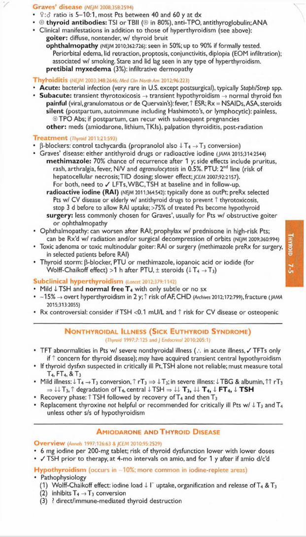
Graves’ disease (NE)M 2008,358:2594)
• 9 :6 ratio is 5-10:1, most Pts between 40 and 60 y at dx• © thyroid antibodies: TSI orTBII (© in 80%), anti-TPO, antithyroglobulin;ANA• Clinical manifestations in addition to those of hyperthyroidism (see above):
goiter: diffuse, nontender, w / thyroid bruitophthalmopathy (NEJM 2010:362:726): seen in 50%; u p to 90% if formally tested.
Periorbital edema, lid retraction, proptosis, conjunctivitis, diplopia (EOM infiltration); associated w / smoking. Stare and lid lag seen in any type of hyperthyroidism,
pretibial m yxedem a (3%): infiltrative dermopathy
Thyhoiditis (NEJM 2003:348:2646; Med Clin North Am 2012:96:223)
• Acute: bacterial infection (very rare in U.S. except postsurgical), typically Staph/Strep spp.• Subacute: transient thyrotoxicosis -> transient hypothyroidism -> normal thyroid fxn
painful (viral,granulomatous or de Quervain’s):fever, T ESR;Rx = NSAIDs, ASA, steroids silent (postpartum, autoimmune including Hashimoto’s, o r lymphocytic): painless,
©TPO Abs; if postpartum, can recur with subsequent pregnancies other: meds (amiodarone, lithium,TKIs), palpation thyroiditis, post-radiation
Treatm ent (Thyroid 2011 a 1:593)
• P-blockers: control tachycardia (propranolol also XT4 ->T 3 conversion)• Graves’ disease: either antithyroid drugs or radioactive iodine (JAMA 2015:314:2544)
m ethim azole: 70% chance of recurrence after 1 y; side effects include pruritus, rash, arthralgia, fever, N/V and agranulocytosis in 0.5%. PTU: 2nd line (risk of hepatocellular necrosis;TID dosing; slower effect;jCEfA 2007:92:2157).
For both, need to / LFTs.WBC.TSH at baseline and in follow-up. radioactive iodine (RAI) (NEJM 2011:364:542): typically done as outPt;preRx selected
Pts w/ CV disease or elderly w/ antithyroid drugs to prevent T thyrotoxicosis, stop 3 d before to allow RAI uptake; >75% of treated Pts become hypothyroid
surgery: less commonly chosen for Graves’, usually for Pts w / obstructive goiter or ophthalmopathy
• Ophthalmopathy: can worsen after RAI; prophylax w/ prednisone in high-risk Pts;can be Rx’d w / radiation and/or surgical decompression of orbits (NE)M 2009:360:994)
• Toxic adenoma or toxic multinodular goiter: RAI or surgery (methimazole preRx for surgery,in selected patients before RAI)
• Thyroid storm: (i-blocker, PTU o r methimazole, iopanoic acid or iodide (forWolff-Chaikoff effect) >1 h after PTU,± steroids (XT4 T j)
Subclinical hyperthyroidism (Loncet 2012:379:1142)
• Mild XTSH and norm al fre e T 4 with only subtle o r no sx• -15% -» overt hyperthyroidism in 2 y;T risk of AF, CHD (Archives 2012:172:799), fracture (JAMA
2015:313:2055)
• Rx controversial: consider if TSH <0.1 mU/L and T risk for CV disease or osteopenic
N onthyroidal Illness (S ick Euthyroid Syndrome)(Thyroid 1997:7:125 and ) Endocrinol 2010:205:1)
• TFT abnormalities in Pts w/ severe nonthyroidal illness (.* *. in acute illness, / TFTs onlyif t concern for thyroid disease); may have acquired transient central hypothyroidism
• If thyroid dysfxn suspected in critically ill Pt,TSH alone not reliable; must measure totalT4, FT4, & T3
• Mild illness:XT4 ->T3 conversion,? rT 3 = 6 XT3; in severe illness:XTBG & albumin,TT rT 3
=> XX T3,? degradation of T4, central XTSH => XX T 3, XX T 4, X FT4, X TSH• Recovery phase: T TSH followed by recovery o fT 4 and thenT]• Replacement thyroxine not helpful or recommended for critically ill Pts w / XT3 andT4
unless other s/s of hypothyroidism
A miodarone and T hyroid D isease
O verv iew (Annoh 1997;126:63 & JCEM 2010:95:2529)
• 6 mg iodine per 2 0 0 -mg tablet; risk of thyroid dysfunction lower with lower doses• / TSH prior to therapy, at 4-mo intervals on amio, and for 1 y after if amio d/c’d
H yp o th y ro id ism (occurs in -10%; more common in iodine-replete areas)• Pathophysiology
(1) Wolff-Chaikoff effect: iodine load X I" uptake, organification and release o fT 4 & T 3
(2 ) inhibitsT4 ->T3 conversion(3) ? direct/immune-mediated thyroid destruction
Th
yr
oid
7-5

Th
yr
oid
7-
6• Normal individuals: -I T«; then escape Wolff-Chaikoff effect and have T T4, ^ 3, T TSH;
then TSH normalizes (after 1-3 mo)• Susceptible individuals (eg, subclinical Hashimoto’s, / anti-TPO) do not escape effects• Treatment: thyroxine to normalize TSH; may need larger than usual dose
H yp e rth y ro id ism (3% of Pts on amio;-10-20% of Pts in iodine-deficient areas)• Type 1 = underlying multinodular goiter o r autonomous thyroid tissue
jod-Basedow effect: iodine load -» t synthesis o fT 4 andT3 in autonomous tissue• Type 2 = destructive thyroiditis
T release of preformed T 4 & T 3 -> hyperthyroidism -> hypothyroidism -> recovery• Doppler U/S: type 1 w/ T thyroid blood flow; type 2 w / i flow• Treatment: not absolutely necessary to d/c amio b/c amio .IT 4 ->T3 conversion
methimazole fo r type 1; steroids (eg, 40 mg prednisone qd) for type 2 often difficult to distinguish, so Rx for both typically initiated (JCEM 200i;86:3) consider thyroidectomy in severely ill patient
T h y r o id N o d u l e s (nejm 2015:373:2347 & tv o ;< /2016:26: i )
• Prevalence 5-10% (50-60% if screen with U/S), 9 > d,~7-15% malignant• Features associated w/ T risk of malig: age <20 or >70 y, d, h/o neck XRT, hard & immobile
mass, cervical LAN, dysphonia• Worrisome U/S findings: hypoechoic, solid, irregular borders, microcalcifications,
height > width, >20 mm (]AMA IM 2013:173:1788)• Features associated w/ benign dx: cystic nodules, "spongiform” sonographic pattern• Screening U/S recommended for those with FHx of MEN2 or medullary thyroid cancer,
personal h/o neck XRT, palpable nodules or multinodular goiter• Any evidence of tracheal deviation or compression -> consider / PFTs & refer to surgery• >10-mm nodule: FNA if hypoechoic solid or solid component of cystic; T suspicion of malig
if irregular margins, microcalcifications, rim Ca2+, height > width, or extrathyroidal extension• >15-mm nodule: FNA if solid isoechoic, or partially cystic w ith mural solid component• >20-mm nodule: FNA if spongiform/other benign solid pattern (no FNA if purely cystic)• Molecular testing if indeterminate pattern on FNA (occurs in -15-30%)• Suppressive Rx w/ high-dose levothyroxine no longer recommended for benign nodules in
iodine-sufficient regions• Cancer very rare in asx nodules diagnosed as benign (JCEM 2014,99:510 &JAMA 2015:313:926)
• After complete surgical resection of thyroid cancer, RAI in medium- and high-risk Pts (Lancet2013:381:1046 & 1058)
Figure 7-2 Approach to thyroid nodules (NEJM 2015:373:2347 & Thyroid 2016;26:1)
Thyroid nodule
Benign
▼Observe
Atypia or Follicular neoplasmfollicular lesion or suspicious for
of undetermined follicular neoplasmsignificance (FN or SFN)
(AUS or FLUS) /
\ /Molec testing ± repeat bx vs.
observation vs. hemithyroidectomy
Suspicious for malignancy
Malignant
Surgery: hemi vs. total thyroidectomy ± central neck LN dissection

ADRENAL DISORDERSCushing’s Syndrome (H ypercortisolism)
D e fin itio n s (Lancet 2015;386:913)
• Cushing’s syndrome = cortisol excess• Cushing’s disease = Cushing’s syndrome 2° to pituitary ACTH hypersecretion
E tio log ies o f hyp e rco rtiso lism• Most commonly iatrogenic caused by exogenous glucocorticoids (though underreported)• Cushing's disease (60-70%): ACTH-secreting pituitary adenoma (usually
microadenoma) or hyperplasia• Adrenal tu m o r (15-25%): adenoma o r (rarely) carcinoma• Ectopic A C T H (5-10%): SCLC, carcinoid, islet cell tumors, medullary thyroid cancer, pheo
C lin ica l m an ifes ta tions (Loncet 2006;367 13)
• Nonspecific: glucose intolerance or DM, HTN, obesity, oligo- or amenorrhea, osteoporosis• More specific: central obesity w / extremity wasting, dorsocervical fat pads, spont. bruising• Most specific: proximal myopathy, rounded facies, facial plethora, wide purple striae• Other: depression, insomnia, psychosis, impaired cognition, hypokalemia, acne, hirsutism,
hyperpigmentation (if T ACTH), fungal skin infxns, nephrolithiasis, polyuria
Figure 7-3 Approach to suspected Cushing's syndrome (nb, very difficult to dx os an inPt) ()CEM 2008;93:1526)
Suspect Cushing’s Syndrome C linically 1
3 Options for Screening for Hypercortisolism:24-hr UFC or Overnight 1 mg DST or 11 p.m. Salivary Cortisol
/ 1© and low © or© but high © but in Pt w/
clinical suspicion clinical suspicion acute illness, EtOH, depression
S 4Not Cushing's v' (or repeat) 24-hr UFC re / after resolution of
illness or / combined1 48-hr LD DST + CRH© / N1 © ©1 ^ 4
Differentiate level of defect by checking serum ACTH ? Pseudo-Cushing’s
ACTH- dependent ** normal low » ACTH - Independen t
ior high
J
48-hr or O/N high-dose DST adrenal CT or MRI(or CRH test)
i \Will not suppress Will suppress
(or © stim) (or® stim)
t > 4 ©chest/abd MRI pituitary MRI
somatostatin scan - © “ IT © " 1i
© BIPSS ©f ^search " © ^ | 1f
Ectopic ACTH Cushing's disease Adrenal tumor
CRH, corticotropin-releasing hormone; DST, dexamethasone suppression test; UFC, urinary free cortisol
Overnight 1 mg DST = give 1 mg at 11 p.m.; / 8 a.m. serum cortisol (suppression if <1.8 J.lg/dL); <5% false 0 (primarily used to evaluate subclinical Cushing's in adrenal "incidentalomas")
11 p.m. salivary cortisol = abnl if level t ; 24-h UFC = abnl if level t , > 4X ULN virtually diagnostic
48-h LD DST + CRH = 0.5 mg q6h X 2 d, then IV CRH 2 h later; / serum cortisol 15 min later ( 0 = >1.4 |lg/dL)
48-h LD DST = 0.5 mg q6h X 2 d; / 24-h UFC at base. & during last 24 h of dex (suppress if <10% of base)
48-h HD DST = 2 mg q6h X 2 d; / 24-h UFC as per LD DST
O/N HD DST = 8 mg at 11 p.m.; / 9 a.m. serum cortisol (suppression if <32% of baseline)
CRH test = 1 pg/kg IV; / cortisol and ACTH (0 stim if > 35% T in ACTH or >20% T in cortisol above baseline)
BlPSS.bilac inferior petrosal sinus vein sampling; / petrosaliperipheral ACTH ratio ( 0 = 2 basal, >3 after CRH)

Ad
ren
al
7-8
yT re a tm e n t o f C ush ing ’s syndrom e (JCE/A 2015; 100:2807)
• Surgical resection of pituitary adenoma, adrenal tumor or ectopic ACTH-secreting tumor• If transsphenoidal surgery (TSS) not successful repeat TSS. Can do pituitary XRT, but
XRT not effective immediately, initiate medical Rx w/ mitotane, ketoconazole, or metyrapone to i cortisol, and/or mifepristone to block cortisol action at glucocorticoid receptor, or bilat surgical adrenalectomy if med Rx fails or is contraindicated.
• Glucocorticoid replacement therapy x 6-36 mo after TSS (lifelong glucocorticoid +mineralocorticoid replacement if medical or surgical adrenalectomy)
H y p e r a l d o s t e r o n is m
Etiologies• Prim ary (adrenal disorders, renin-independent increase in aldosterone;)CEM 201 S:100;l)
adrenal hyperplasia (60-70%), adenoma (Conn’s syndrome, 30-40%), carcinoma glucocorticoid-remediable aldosteronism (GRA;ACTH-dep. rearranged promoter)
• Secondary (extra-adrenal disorders, T aldosterone is renin-dependent)Primary reninism; renin-secreting tum or (rare)Secondary reninism: renovascular disease: RAS, malignant hypertension;
edematous states w / i effective arterial volume: CHF, cirrhosis, nephrotic syndrome; hypovolemia, diuretics,T2D, Bartter’s (defective Na/K/2CI transporter = receiving loop
diuretic), Gitelman’s (defective renal Na/CI transporter = receiving thiazide diuretic)• Nonaldosterone m ineralocorticoid excess mimics hyperaldosteronism
11p-HSD defic. (-> lack of inactivation of cortisol, which binds to mineralocorticoid recept.) Black licorice (glycyrrhizinic acid inhibits 11p-HSD), extreme hypercortisolism
(overwhelming 1 ip-HSD), exogenous mineralocorticoids Liddle’s syndrome (constitutively activated/overexpressed distal tubular renal Na channel)
Clinical manifestations• Mild to moderate H TN (11% of Pts w/ HTN refractory to 3 drugs; Lancet 2008:371:1921),
headache, muscle weakness, polyuria, polydipsia; no peripheral edema because of "escape” from Na retention; malignant HTN is rare
• Classically hypokalemia (but often normal), metabolic alkalosis, mild hypernatremia
Diagnostic Studies (/CEM 2008:93:3266)• 5-10% of Pts w/ HTN; .*. screen if HTN + hypoK, adrenal mass, refractory/early onset HTN• Screening: aldo (>15-20 ng/dL) and plasma aldo:renin ratio (>20 if 1°) obtain 8 a.m.
paired values (off spironolactone & eplerenone for 6 wk); Sc & Sp >85%• ACEI/ARB, diuretics, CCB can T renin activity -> i PAC/PRA ratio and (3Bs may t
PAC/PRA ratio;.*, avoid, a-blockers generally best to control HTN during dx testing.• Confirm with sodium suppression test (fail to suppress aldo after sodium load)
oral salt load (+ KCI) x 3 d, / 24-h urine (© if urinary aldo >12 ug/d while urinary Na >200 mEq/d) or 2L NS over 4 h, measure plasma aldo at end of infusion (© if aldo >5 ng/dL)
Figure 7-4 Approach to suspected hyperaldosteronism
Suspect Hyperaldosteronism
IPlasma renin & aldosterone AM collection
1 renin ' i renin i aldosterone T aldosterone (>15)
aldo.renin >20
I reninT aldosterone aldo:renin £10
Non-aldostcrone salt suppression test 2s Hyperaldosteronismmineralocorticoid excess
© Renovascular diseaseCushing's syndrome CAH (some forms)
11()-OHSD deficiency Licorice (chronic ingestion)
Liddle’s syndrome Exogenous mineralocorticoids
1° Hyperaldosteronism
suspect carcinoma, lesion >4 cm
i
Adrenal CT or MRI
y \unilateral lesion no or bilateral lesions
*Probable localize Adrenal vein Adenoma A --------------------- sampling
CHp cirrhosis, nephrotic Hypovolemia & diuretic use
Bartter’s syndrome Gitelman’s syndrome Renin-secreting tumor
no
Recommendsurgery
presurgical localization ^ recommended (espec if Pt >40 y)
localizationHyperplasia
or GRA
(Adapted from JCEM 2008:93:3266 and Surg Clin NAm 2014:94:643)

Treatm ent (Surg Clin NAm 2014;94:643)• Adenoma -» adrenalectomy vs. medical Rx w/ spironolactone or eplerenone• Hyperplasia -> spironolactone or eplerenone; GRA -> glucocorticoids ± spironolactone• Carcinoma -» adrenalectomy
A d r e n a l I n s u f f ic ie n c y
E t io lo g ie s• Prim ary = adrenocortical disease = Addison’s disease
autoim m une: isolated o r in assoc w/APS (see table on page 7-2) infection: TB, CMV, histoplasmosis, paracoccidioidomycosisvascular: hemorrhage (usually in setting of sepsis), adrenal vein thrombosis, HIT, trauma m etastatic disease: (90% of adrenals must be destroyed to cause insufficiency) deposition diseases: hemochromatosis, amyloidosis, sarcoidosis drugs: azole antifungals.etomidate (even after single dose), rifampin, anticonvulsants
• Secondary = pituitary failure of ACTH secretion (but aldosterone intact b/c RAA axis)any cause of primary or secondary hypopituitarism (see “ Pituitary Disorders” ) glucocorticoid therapy (can occur after <2 wk of “ suppressive doses” ; dose effect variable;
even <10 mg of prednisone daily chronically can be suppressive) megestrol (a progestin with some glucocorticoid activity)
C l in ic a l m a n i f e s ta t io n s (Loncet 2014;383:21 S2)• Prim ary or secondary: weakness and fatigability (99%), anorexia (99%),
orthostatic hypotension (90%), nausea (86%), vomiting (75%), hyponatremia (88%)• Primary only (extra s/s due to lack of aldosterone and T ACTH): marked orthostatic
hypotension (because volume-depleted), salt craving, hyperpigmentation (seen in creases, mucous membranes, pressure areas, nipples), hyperkalemia
• Secondary only: ± other manifestations of hypopituitarism (see “ Pituitary Disorders” )
D ia g n o s t ic S tu d ie s (JCEM 2016;101:364)• Early a.m. serum cortisol: <5 pg/dL virtually diagnostic; >18 pg/dL rules it out (except in
severe septic shock— see below)• Standard (250 pg) cosyntropin stimulation test (testing ability of ACTH -> T cortisol)
normal = 60-min (or 30-min) post-ACTH cortisol >18 pg/dL abnormal in primary b/c adrenal gland diseased and unable to give adequate output abnormal in chronic secondary b/c adrenals atrophied and unable to respond (very rarely, may be normal in acute pituitary injury b/c adrenals still able to respond
use early a.m. cortisol instead)• Other tests (w/ guidance by endocrinologist): renin, aldosterone, insulin-induced
hypoglycemia (measure serum cortisol response); metyrapone (blocks cortisol synthesis and therefore stimulates ACTH, measure plasma 11-deoxycortisol and urinary 17- hydroxycorticosteroid levels)
• Other lab abnormalities: hypoglycemia, eosinophilia, lymphocytosis, ± neutropenia• ACTH: T in 1°, I o r low-normal in 2°• Imaging studies to consider
pituitary MRI to detect anatomical abnormalitiesadrenal CT: small, noncalcified adrenals in autoimmune, enlarged in metastatic disease,
hemorrhage, infection or deposition (although they may be normal-appearing)
A d r e n a l in s u f f ic ie n c y & c r i t i c a l i l ln e s s (NEJM 2003:348:727;jama 2009;301:2362)• T circulating cortisol despite L ACTH due to I clearance and possibly stimulation by
cytokines; low cortisol binding proteins; dx of adrenal insufficiency problematic (NEJM 2013:368:1477)
• Nonetheless, reasonable to perform ACTH stim ASAP in hypotensive Pt suspected tohave absolute adrenal insufficiency
• Reasonable to perform 250-pg ACTH stim and initiate glucocorticoid replacement if T incortisol <9 pg/dL or absolute cortisol level <10 pg/dL, but decision to Rx should be based on clinical assessment; unlikely to require Rx if spot or post-ACTH cortisol >18 pg/dL
• Initiate corticosteroids early: use hydrocortisone 50-100 mg IV q6-8h; prior to ACTH stimtest, use dexamethasone 2—4 mg IV q6h + fludrocortisone 50 pg daily
• Rx of relative adrenal insufficiency controversial (see “ Sepsis")
T r e a t m e n t• Acute insufficiency: volume resuscitation w/ normal saline + hydrocortisone IV as above• Chronic insufficiency
prednisone - 5 mg PO qam.or hydrocortisone: 15-25 mg PO qd (2A a.m,’A early p.m.) fludrocortisone (not needed in 2° adrenal insufficiency): 0.05-0.1 mg PO qam backup dexamethasone 4-mg IM prefilled syringe given to Pt for emergency situations

Pheochromocytoma & Paraganglioma
C lin ica l m an ifes ta tions (five P s ) (tower 200S.366 665)
• Pressure (hypertension, paroxysmal in 50%, severe & resistant to Rx.occ orthostatic)• Pain (headache, chest pain)• P a lp ita tion s (tachycardia, tremor, w t loss, fever)• P e rsp ira tion (profuse)• P a llo r (vasoconstrictive spell)• “ Rule of 10” : 10% extra-adrenal (known as paraganglioma), 10% in children,
10% multiple or bilateral, 10% recur (T in paraganglioma), 10% malignant (T in paraganglioma), 10% familial, 10% incidentaloma
• Paroxysms can be triggered by meds (eg, p-blockers) abdominal manipulation• Associated with MEN2A/2B, von Hippel Lindau, neurofibromatosis type 1, familial
paraganglioma (mutations in succinate dehydrogenase gene B, C and D)
Diagnostic Studies (JC£M 2014;99:1915)
• 24° urinary fractionated metanephrines: 85-97% Se, 69-95% Sp. Screening test of choiceif low-risk (as false © with severe illness, renal failure, OSA, labetalol due to assay interference, acetaminophen,TCAs, medications containing sympathomimetics).
• Plasma-free metanephrines: 89-100% Sc, 79-97% Sp (JAMA 2002:287:1427). Screeningtest of choice if high risk, but t rate of false © in low-prevalence population. Draw blood in supine position after Pt supine for 30 min, estimated 2.8x T false © if seated.
• Adrenal CT generally better than MRI; PET for known metastatic disease or to localizenonadrenal mass but usually easy to find; consider MIBG scintigraphy if CT/MRI ©
• Consider genetic testing if bilateral disease, young Pt, © FHx, extra-adrenal
T re a tm e n t• cc-blockade first (usually phenoxybenzamine) ± P-blockade (often propranolol) -» surgery• Preoperative volume expansion is critical due to possible hypotension after tumor excision
A drenal Incidentalomas
Epidemiology• 4% of Pts undergoing abdominal CT scan have incidentally discovered adrenal mass;
prevalence T with age
Differential diagnosis• Nonfunctioning mass: adenoma, cysts, abscesses, granuloma, hemorrhage, lipoma,
myelolipoma, primary o r metastatic malignancy• Functioning mass: pheochromocytoma, adenoma (cortisol, aldosterone, sex hormones),
nondassical CAH, other endocrine tumor, carcinoma
Hormonal workup (NEJM 2007;356:601;JCEM 2010:95:4106)
• Rule out subclinical Cushing’s syndrome in all Pts using 1 mg overnight DST(Sp 91%). Abnormal results require confirmatory testing.
• Rule out hyperaldosteronism i f hypertensive w/ plasma aldo & renin (see above)• Rule out pheochromocytoma in ALL Pts (b/c of morbidity unRx’d pheo) using 24-h
urine fractionated metanephrines o r plasma free metanephrines
Malignancy workup• CT and MRI characteristics may suggest adenoma vs. carcinoma
Benign features: size <4 cm; smooth margins, homogenous and hypodense appearance; unenhanced CT <10 Hounsfield units o r CT contrast-medium washout >50% at 10 min. Can follow such incidentalomas w/ periodic scans.
Suspicious features: size >6 cm or T size on repeat scan; irregular margins, heterogeneous, dense o r vascular appearance; h/o malignancy or young age.Such incidentalomas warrant resection or repeat scan at short interval.
• Rule out metastatic cancer (and infection) as in Pts w/ h/o cancer, -50% of adrenalincidentalomas are malignant
Follow-up• If hormonal workup © and appearance benign, yearly fxnal testing for 4 y w / follow-up
imaging at 6,12, & 24 mos reasonable approach, but controversial
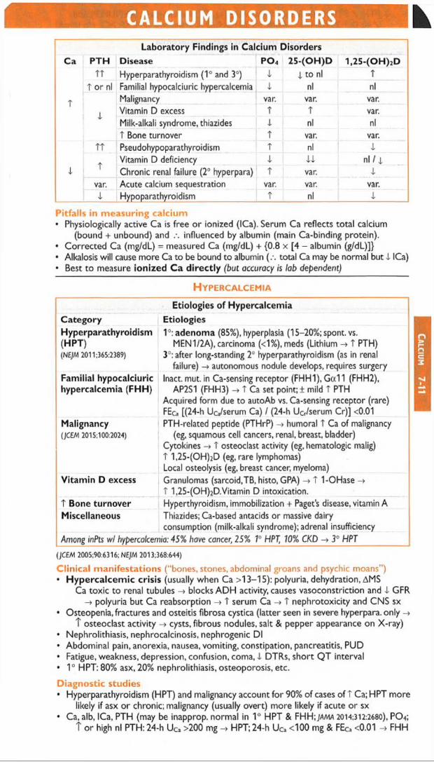
CALCIUM DISORDERS MLaboratory Findings in Calcium Disorders
PTH Disease PO4 25-(OH)D 1,25-(OH)2Dt r Hyperparathyroidism (1° and 3C) X X to nl T
T or nl Familial hypocalciuric hypercalcemia X nl nlMalignancy var. var. var.
X Vitamin D excess T T var.Milk-alkali syndrome, thiazides X nl nlt Bone turnover T var. var.
TT Pseudohypoparathyroidism T nl X
tVitamin D deficiency X XX nl / xChronic renal failure (2° hyperpara) T var. X
var. Acute calcium sequestration var. var. var.X Hypoparathyroidism T nl X
Pitfalls in measuring calcium• Physiologically active Ca is free o r ionized (ICa). Serum Ca reflects total calcium
(bound + unbound) and influenced by albumin (main Ca-binding protein).• Corrected Ca (mg/dL) = measured Ca (mg/dL) + {0.8 x [4 - albumin (g/dL)]}• Alkalosis will cause more Ca to be bound to albumin (.-. total Ca may be normal but X ICa)• Best to measure ionized Ca directly (but accuracy is lab dependent)
H y p e r c a l c e m i a
Etiologies of HypercalcemiaCategory EtiologiesHyperparathyroidism(HPT)(NEJM 2011:365:2389)
I 3:adenoma (85%), hyperplasia (15-20%;spont. vs.MEN 1/2A), carcinoma (<1%),meds (Lithium -> t PTH)
3°: after long-standing 2° hyperparathyroidism (as in renal failure) -> autonomous nodule develops, requires surgery
Familial hypocalciuric hypercalcemia (FHH)
Inact. mut. in Ca-sensing receptor (FHH1), Gal 1 (FHH2), AP2S1 (FHH3) -> t Ca set point; ± mild t PTH
Acquired form due to autoAb vs. Ca-sensing receptor (rare) FEca [(24-h Uca/serum Ca) / (24-h Ucr/serum Cr)j <0.01
Malignancy(JCEM 2015:100:2024)
PTH-related peptide (PTHrP) -> humoral T Ca of malignancy (eg, squamous cell cancers, renal, breast, bladder)
Cytokines -» T osteoclast activity (eg, hematologic malig) t 1,25-(OH)2D (eg, rare lymphomas)Local osteolysis (eg, breast cancer, myeloma)
Vitamin D excess Granulomas (sarcoid,TB, histo, GPA) -> T 1-OHase -> T 1,25-(OH)2D.Vitamin D intoxication.
T Bone turnover Hyperthyroidism, immobilization + Paget’s disease, vitamin AMiscellaneous Thiazides; Ca-based antacids or massive dairy
consumption (milk-alkali syndrome); adrenal insufficiencyAmong inPts wl hypercalcemia: 45% have cancer, 25% 1° HPT, 10% CKD 3° HPT
(jCEM 2005:90:6316; NEJM 2013:368:644)
Clinical manifestations ("bones, stones, abdominal groans and psychic moans")• H y p e rc a lc e m ic crisis (usually when Ca >13-15): polyuria, dehydration, AMS
Ca toxic to renal tubules -» blocks ADH activity, causes vasoconstriction and X GFR -» polyuria but Ca reabsorption -> T serum Ca -> T nephrotoxicity and CNS sx
• Osteopenia, fractures and osteitis fibrosa cystica (latter seen in severe hyperpara, only -*T osteoclast activity -> cysts, fibrous nodules, salt & pepper appearance on X-ray)
• Nephrolithiasis, nephrocalcinosis, nephrogenic Dl• Abdominal pain, anorexia, nausea, vomiting, constipation, pancreatitis, PUD• Fatigue, weakness, depression, confusion, coma, X DTRs, short QT interval• 1° HPT: 80% asx, 20% nephrolithiasis, osteoporosis, etc.
Diagnostic studies• Hyperparathyroidism (HPT) and malignancy account for 90% of cases of T Ca; HPT more
likely if asx or chronic; malignancy (usually overt) more likely if acute or sx• Ca, alb, ICa, PTH (may be inapprop. normal in 1° HPT & FHH; ja/via 2014:312:2680), PO4;
T or high nl PTH: 24-h UCa >200 mg -> HPT; 24-h UCa <100 mg & FEca <0.01 FHH
Ca
lciu
m 7-11

i PTH: / PTHrP, A6, & search for malig (eg, CT, mammogram, SPEP/UPEP) and / vit D:T 25-(OH)D -> meds;? 1,25-(OH)2D granuloma ( / CXR.ACE, r/o lymph)
Acute Treatment of HypercalcemiaT r e a tm e n t Onset Duration CommentsNormal saline(4-6 L/d)
h during Rx Natriuresis -» T renal Ca excretion
± Furosemide h during Rx Use cautiously, only if volume overloadedBisphosphonates 1 -2 d var. Inhibit osteoclasts, useful in malignancy:caution
in renal failure: risk of jaw osteonecrosisCalcitonin h 2-3 d Quickly develop tachyphylaxisGlucocorticoids days days i Useful in some malig,granulomatous disorders
& vitamin D intox.Denosumab(JCEM 2014:99:3144)
days months Monoclonal Ab against RANKL; typically used in hyperCa of malignancy; not renally cleared
Hemodialysis min during Rx If other measures ineffective or contraindicated
(M l 2015;350:h2723)
Treatm ent of asym ptom atic 1° H P T (J C E M 2014 99:3561)• Surgery if: age <50 y; serum Ca >1 mg/dL >ULN; CrCI <60 mL/min, DEXAT score <-2.5• If surgery declined/deferred, can Rx with, cinacalcet ( I Ca & PTH but may not t BMD)• If not yet candidate for surgery: / serum Ca & Cr annually and BMD q1-2y
Calciphylaxis (calcific uremic arteriopathy)• Calcification of media of small- to med-sized blood vessels of dermis & SC fat• Ischemia & skin necrosis. See “ Chronic Kidney Disease” for further details.
H ypocalcemia
Etiologies of HypocalcemiaCategory EtiologiesHypoparathyroidism(NEJM 2008:359:391)
Iatrogenic (s/p thyroidectomy, rarely after parathyroidectomy); sporadic; familial (APS 1, activating Ca-sensing receptor mutations; see page 7-2);Wilson’s, hemochromatosis; hypoMg ( i secretion and effect); activating Ca-sensing receptor autoAb
Pseudohypoparathyroidism(JCEM 2011:96:3020)
la and lb: PTH end-organ resistance (.-. t serum PTH) la: + skeletal abnormalities, short stature, & retardation Pseudopseudohypoparathyroidism = la syndrome but n/ Ca & PTH
Vit D defic. or resist(NEJM 2011:364:248;JCEM 2012:97:1153)
Nutritional/sunlight deprivation; Gl disease/fat malabs.; drugs (anticonvulsants, rifampin, ketoconazole, 5-FU/leucovorin); genetic (1a-hydroxylase,VDR mutations)
Chronic renal failure i 1,25-(OH)2D production, t PO* from 1 clearanceAccelerated net bone formation
Postparathyroidectomy, Rx o f severe vit D deficiency or Paget’s disease (NEJM 2013:368:644), osteoblastic metastases
Calciumsequestration
Pancreatitis, citrate excess (after blood transfusions),acute TT PO* (ARF, rhabdomyolysis, tumor lysis), bisphosphonates
Clinical manifestations• N eurom uscu lar irritability: perioral paresthesias,cramps,© Trousseau’s (inflation of BP
cuff >3 min -> carpal spasm), © Chvostek’s (tapping facial nerve -> contraction of facial muscles), laryngospasm; irritability, depression, psychosis, T ICR seizures, T QT
• Rickets and/or osteomalacia: chronic i v it D -» 1 Ca, i PO* -> i bone/cartilagemineralization, growth failure, bone pain, muscle weakness
• R enal osteodystrophy ( i vit D & T PTH in renal failure): osteomalacia [ i mineralizationof bone due to i Ca and 1,25-(OH)2D] & osteitis fibrosa cystica (due to T PTH)
Diagnostic studies• Ca, alb, ICa, PTH, 25-(OH)D, 1,25-(OH)2D (if renal failure or rickets), Cr, Mg, PO4,Ac}), UcaT r e a t m e n t (also treat concomitant vitamin D deficiency)• Severely symptomatic: Ca gluconate (1-2 g IV over 20 min) + oral Ca + calcitriol (but
takes hrs to work) ± Mg (50-100 mEq/d); 10% CaCh in codes o r via CVL• Consider gtt o r PO to follow as effect of IV bolus typically lasts only a few hours• Chronic: oral Ca (1-3 g/d; Ca citrate better absorbed than Ca carbonate, esp. if
achlorhydria o r on PPI) and typically calcitriol (0.25-2 meg/d), and replete vitamin D deficiency. Consider thiazide to i urinary Ca or recombinant PTH 1-84.
• Chronic renal failure: phosphate binder(s), oral Ca, calcitriol or analogue

DIABETES MELLITUS KDefinition (D/afaetes Care 2016:39:513)• Either HbAic >6.5, fasting glc >126 mg/dL, o r glc 2 h after OGTT >200 mg/dL x 2 (for any
test) or single random glc >200 mg/dL w/ classic sx of hyperglycemia; all tests equally reasonable (nb, may be © on one test but not another); OGTT preferred during preg
• Blood glc higher than normal, but not frank DM (“ prediabetics,” ~40% U.S. population)HbAic 57-6.4%, impaired fasting glc (IFG) 100-125 mg/dL, or 2 h prandial glc 140-199 Preventing progression to DM: diet & exercise (58% 4), metformin (31% i\NEJM
2002:346:393),TZD (60% 4; Lancet 2006:368:1096)
Categories• Type 1 (Lancet 2014:383:69): islet cell destruction; absolute insulin deficiency; ketosis in
absence of insulin; prevalence 0.4%; usual onset in childhood but can occur throughout adulthood; T risk if © FHx; HLA associations; anti-GAD, anti-islet cell & anti-insulin autoAb
• Type 1 (Annafe 2015;162:ITC1): insulin resistance + relative insulin 4; prevalence 8%; onsetgenerally later in life; no HLA assoc.; risk factors: age, © FHx, obesity, sedentary lifestyle
• Type 2 DM p/w D K A (‘‘ketosis-prone type 2 diabetes” or"Flatbush diabetes” ): most oftenseen in nonwhite, ± anti-GAD Ab, eventually may not require insulin {Endo Rev 2008:29:292)
• Mature-Onset Diabetes of the Young (MODY): autosomal dom. forms of DM due todefects in insulin secretion genes; genetically and clinically heterogeneous (NEJM 2001:345:971)
• Secondary causes of diabetes: exogenous glucocorticoids, glucagonoma (3 Ds = DM,DVT, diarrhea), pancreatic (pancreatitis, hemochromatosis, CF, resection), B endocrinopathies (Cushing’s disease, acromegaly), gestational, drugs (protease inhibitors, atypical antipsychotics)
Clinical manifestations• Polyuria, polydipsia, polyphagia with unexplained weight loss; can also be asymptomatic
Diabetes Treatment Options
Diet Type 1:carb counting;Type 2: wt reduction diet + exerciseMetformin (biguanide) First-line pharmacoRx for allT2D
4 hepatic gluconeogenesis. 4 HbAic '-1.5%.W t neutral, N/V & diarrhea, rare lactic acidosis Contraindic. in renal (eg, Cr >1.5) or liver failure
Sulfonylureas (SU) t insulin secretion, 4 HbAic -1.5%. Hypoglycemia, wt gain.Thiazolidinediones (TZD) (PPARy agonists)
T insulin sens, in adipose & muscle. 4 HbAic -1%.W t gain, hepatotoxicity, fluid retention & CHF, bone fractures ? t Ml w / rosiglitazone; not pioglitazone (8M/20ll;342:dl309)
Contraindic. in liver disease and NYHA lll-IV, monitor LFTsGLP-1 agonists t glc-depend insulin secretion, 4 HbAic - 0.5%. 4 CV events
(NEJM 2016:375:311). Wt loss, N/V & diarrhea (30-45%).DPP-4 inhibitors Block degrad, of GLP-1 & GIP -> T insulin. 4 HbAic -0.5%.
? t risk Of CHF with some (NEJM 2013:369:1317 & 2015:373:232)
SGLT-2 inhibitors (block renal tubular glc uptake)
T glucosuria. 4 HbAic -0.6-1 %.Wt loss, 4 CV death & HF, slows progression of kidney disease (NEJM 2015:373:2117 &
NEJM 2016:375:323), T risk of normoglycemic DKA (Dwbetes Care 2016:39:532), fungal GU infxn & UTIs, hypovolemia, T LDL.
Glinides (nonsulfonylurea insulin secretagogues)
T insulin secretion, 4 HbAic -1.5% Hypoglycemia (but less than w/ SU), wt gain
a-glucosidase inhibitors 4 intestinal CHO absorption, 4 HbAic 0.5-0.8%.GI distress (gas).Pramlintide Delays gastric emptying & 4 glucagon, 4 HbAic 0.5%. Gl sx.
To be used as adjunctive Rx w/ insulin in T ID orT2DInsulin(Additional T1D options: insulin pump, pancreatic or islet cell transplant)
Hypoglycemia, wt gain.TID: generally combine intermed./ long-acting (NPH or glargine) & short-/rapid-acting (regular or lispro) insulin.T2D: consider if mono oral Rx not adequate (esp. if HbAic high) and start if combo oral Rx not adequate.
Gastric bypass Can cure DM & prevent complications (NEJM 2014:370:2002)
(Loncct 2014:383:1068; JAMA 2015:314:1052; Diabetes Care 2016;39:S52; Endocr Pract 2016:22:84)
Insulin Preparations (jama 2014:311:2315)Preparation Onset Peak D uration Side effects/Com m entsLispro, aspart 5-15 min 60-90 min 2-4 h Give immediately before mealRegular 30-60 min 2-4 h 5-8 h Give -30 min before mealNPH 1-2 h 4-8 h 12-18h Can cause protamine Ab prodGlargine 2 h No peak 20-24 h Once daily (a.m. or p.m.)Detemir 1-3 h No peak 18-26 h Once daily
Diabetes
7-13
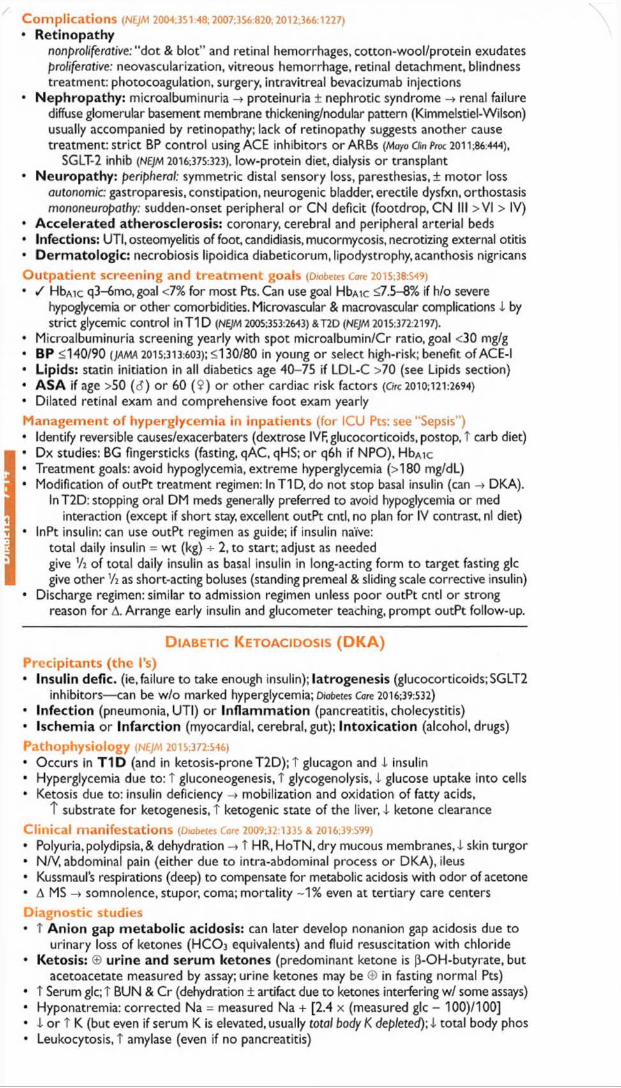
Complications (N£JA1 2004;351:48; 2007:356:820; 2012;366:1227)
• Retinopathynonproliferative: “ dot & blot” and retinal hemorrhages, cotton-wool/protein exudates proliferative: neovascularization, vitreous hemorrhage, retinal detachment, blindness treatment: photocoagulation, surgery, intravitreal bevacizumab injections
• Nephropathy: microalbuminuria -> proteinuria ± nephrotic syndrome -> renal failurediffuse glomerular basement membrane thickening/nodular pattern (Kimmelstiel-Wilson) usually accompanied by retinopathy; lack of retinopathy suggests another cause treatment: strict BP control using ACE inhibitors or ARBs (Mayo Clin Proa 2011,86:444),
SGLT-2 inhib (NE)M 2016:375:323), low-protein diet, dialysis o r transplant• Neuropathy: peripheral: symmetric distal sensory loss, paresthesias,! m otor loss
autonomic: gastroparesis, constipation, neurogenic bladder, erectile dysfxn, orthostasis mononeuropathy: sudden-onset peripheral or CN deficit (footdrop, CN III >VI > IV)
• Accelerated atherosclerosis: coronary, cerebral and peripheral arterial beds• Infections: UTI, osteomyelitis of foot, candidiasis, mucormycosis, necrotizing external otitis• Derm atologic: necrobiosis lipoidica diabeticorum, lipodystrophy,acanthosis nigricansO utpatien t screening and trea tm en t goals (D/obetes Care 2015;38:S49)
• / HbAic q3-6mo,goal <7% for most Pts. Can use goal HbAic <7.5-8% if h/o severehypoglycemia or other comorbidities. Microvascular & macrovascular complications 4 by strict glycemic control in T1D (NEJM 2005:353:2643) &T2D (NEJM 2015:372:2197).
• Microalbuminuria screening yearly with spot microalbumin/Cr ratio, goal <30 mg/g• BP <140/90 (JAMA 2015:313:603); <130/80 in young or select high-risk; benefit o f ACE-I• Lipids: statin initiation in all diabetics age 40-75 if LDL-C >70 (see Lipids section)• ASA if age >50 (c?) or 60 (9 ) or other cardiac risk factors (Ore 2010:121:2694)
• Dilated retinal exam and comprehensive foot exam yearly
Managem ent of hyperglycemia in inpatients (for ICU Pts: see “Sepsis”)• Identify reversible causes/exacerbaters (dextrose IVF, glucocorticoids, postop, t carb diet)• Dx studies: BG fingersticks (fasting, qAC, qHS; o r q6 h if NPO), HbAic• Treatment goals: avoid hypoglycemia, extreme hyperglycemia (>180 mg/dL)• Modification of outPt treatment regimen: InT ID , do not stop basal insulin (can -> DKA).
lnT2D: stopping oral DM meds generally preferred to avoid hypoglycemia or med interaction (except if short stay, excellent outPt cntl, no plan for IV contrast, nl diet)
• InPt insulin: can use outPt regimen as guide; if insulin naive:total daily insulin = w t (kg) 2 , to start; adjust as neededgive Vi o f total daily insulin as basal insulin in long-acting form to target fasting glc give other as short-acting boluses (standing premeal & sliding scale corrective insulin)
• Discharge regimen: similar to admission regimen unless poor outPt cntl or strongreason for A. Arrange early insulin and glucometer teaching, prompt outPt follow-up. * •
D ia b e t ic K e t o a c id o s is ( D K A )
Precipitants (the I’s)• Insulin defic. (ie,failure to take enough insulin); Iatrogenesis (glucocorticoids;SGLT2
inhibitors— can be w/o marked hyperglycemia; D/obetes Core 2016:39:532)• In fec tion (pneumonia, UTI) o r In fla m m a tio n (pancreatitis, cholecystitis)• Ischem ia o r In fa rc tio n (myocardial, cerebral, gut); In to x ica tio n (alcohol, drugs)
Pathophysiology (N£JA1 2015;372:S46)
• Occurs in T 1D (and in ketosis-prone T2D); T glucagon and 4 insulin• Hyperglycemia due to: t gluconeogenesis, T glycogenolysis, 4 glucose uptake into cells• Ketosis due to: insulin deficiency -» mobilization and oxidation o f fatty acids,
T substrate for ketogenesis, T ketogenic state of the liver, 4 ketone clearanceClinical manifestations (Diabetes Care 2009:32:1335 & 2016;39:S99)
• Polyuria, polydipsia, & dehydration -» T HR, HoTN,dry mucous membranes, 4 skin turgor• N/V, abdominal pain (either due to intra-abdominal process o r DKA), ileus• Kussmaul’s respirations (deep) to compensate for metabolic acidosis with odor of acetone• A MS -> somnolence, stupor, coma; mortality -1% even at tertiary care centers
Diagnostic studies• T A n io n gap m e ta b o lic acidosis: can later develop nonanion gap acidosis due to
urinary loss of ketones (H C O j equivalents) and fluid resuscitation with chloride• Ketosis: © u rine and serum ketones (predominant ketone is [3-OH-butyrate, but
acetoacetate measured by assay; urine ketones may be © in fasting normal Pts)• T Serum glc; T BUN & Cr (dehydration ± artifact due to ketones interfering w/ some assays)• Hyponatremia: corrected Na - measured Na + [2.4 x (measured glc - 100)/100]• 4 o r T K (but even if serum K is elevated, usually total body K depleted); 4 total body phos• Leukocytosis, T amylase (even if no pancreatitis)

Typical DKA “Flow Sheet” Setup
VS UOP pH HCO3 AG Ketones Glc K PO4 IVF InsulinNote: Main ketone produced is (3-OH-butyrate ((30HB), but ketone m e a s u re d b y n itro p ru s s id e is a c e to a c e ta te
(A c -A c ).A s D K A is tre a te d , (30HB -> A c-A c , AG can decrease while measured ketones can increase.___
Treatment of DKA (D/abetes Care 20O9;32:133S)
Rule out possible precipitants Infection, intra-abdominal process, Ml, etc. (see above)
Aggressive hydration NS 10-14 mL/kg/h, tailor to dehydration & CV statusInsulin 10 U IV push followed by 0.1 U/kg/h
Continue insulin drip until AG normal If glc <250 and AG still high -> add dextrose to IVF
and continue insulin to metabolize ketones AG normal -» SC insulin (overlap IV & SC 2-3 h)
Electrolyte repletion K: add 20-40 mEq/L IVF if serum K <4.5insulin promotes K entry into cells -> i serum K careful K repletion in Pts with renal failure
HCO3: ! replete if pH <7 or if cardiac instability PO4: replete if <1
H y p e r o s m o l a r H y p e r g l y c e m ic S t a t e
Definition, precipitants, pathophysiology {Diabetes Care 2003;26:S33)
• Extreme hyperglycemia (w/o ketoacidosis) + hyperosm. + A MS in T2D (typically elderly)• Precip same as for DKA, but also include dehydration and renal failure• Hyperglycemia osmotic diuresis -> vol depletion prerenal azotemia -> T glc, etc.
Clinical manifestations & dx studies {Diabetes Care 2016;39:S99)
• Volume depletion and A MS• T serum glc (usually >600 mg/dL) and T meas. serum osmolality (>320 mOsm/L)
effective Osm = 2 x Na (mEq/L) + glc (mg/dL)/18• No ketoacidosis; usually T BUN & Cr; [Na] depends on hyperglycemia & dehydration
Treatm ent (r/o possible precipitants;'-15% mortality due to precipitating factors)• Aggressive hydration: initially NS, then V2 NS, average fluid loss up to 8-10 L• Insulin (eg, 10 U IV followed by 0.05-0.1 U/kg/h) •
H y p o g l y c e m i a
Clinical manifestations (glucose <-55 mg/dL)• CNS: headache, visual As, A MS, weakness, seizure, LOC (neuroglycopenic sx)• A u to n o m ic : diaphoresis, palpitations, trem or (adrenergic sx)
Etiologies in diabetics• Excess insulin, oral hypoglycemics, missed meals, renal failure ( i insulin & SU clearance)• fi-blockers can mask adrenergic symptoms of hypoglycemia
Etiologies in nondiabetics• T insu lin : exogenous insulin, sulfonylureas, insulinoma, anti-insulin antibodies• 1 glucose production: hypopituitarism, adrenal insufficiency, glucagon deficiency,
hepatic failure, renal failure, CHF, alcoholism, sepsis, severe malnutrition• t IG F-II: non-islet tumor• Postprandial, esp. postgastrectomy o r gastric bypass: excessive response to glc load• Low glc w/o sx can be normal
Evaluation in nondiabetics (/cem 2009;94:709)• If clinically ill: take measures to avoid recurrent hypoglycemia; / BUN, Cr, LFTs.TFTs,
prealbumin; IGF-I/IGF-II ratio when appropriate• If otherwise healthy: 72-h fast w / monitored blood glc; stop for neuroglycopenic sx• At time of hypoglycemia: insulin, C peptide ( t w / insulinoma and sulfonylureas, i w/
exogenous insulin), [i-OH-butyrate, sulfonylurea levels• A t end of fast, give 1 mg glucagon IV and measure response of plasma glc before feeding
Treatm ent• Glucose tablets, paste, fru it juice are first-line Rx fo r Pts who can take POs• If IV access available, give 25-50 g of D 50 (50% dextrose)• If no IV, can give glucagon 0.5-1 mg IM or SC (side effect: N/V)
Dia
be
tes
7
-15

Lip
id
7-16
LIPID DISORDERSMeasurements• Lipoproteins = lipids (cholesteryl esters & triglycerides) + phospholipids + proteins
include: chylomicrons.VLDL, IDL, LDL, HDL, Lp(a)• Measure after 12-h fast; LDL typically calculated: LDL-C =TC - HDL-C - (TG/5)
underestim. ifTG >400 or LDL-C <70 mg/dL; directly measure LDL-C levels stable up to 24 h after ACS, then 1 and may take 6 wk to return to nl
• PEx clues: tendon xanthomas (eg, Achilles), imply LDL >300 mg/dL; eruptive xanthomason extensor surfaces implyTG >1000 mg/dL; xanthelasma (yellowish streaks on eyelids)
• Metabolic syndrome (£3 of following): waist >40" (d) or >35" (9);TG >150; HDL <40 mg/dL(d) or <50 mg/dL (9); BP >130/85 mmHg; fasting glc >100 mg/dL (Ore 2009:120:1640)
• Lp(a) = LDL particle bound to apo(a) via apoB; genetic variants a/w Ml {nejm 2009:361:2518)Dyslipidemias• 1°: familial hyperchol. (FH, 1:500): defective LDL receptor; TT chol, nl TG; T CAD; familial
hypertrig. (FHTG, 1:500): TTG ,± T chol, i HDL, pancreatitis; and many others• 2°: DM ( t TG, I HDL), hypothyroidism (T LDL, T TG), nephrotic syndrome (T LDL, T TG),
liver failure (-1- LDL), alcohol (TTG,T HDL), thiazides (T LDL, TTG), protease inhib (TTG)
D rugTrea tm entDrug i LDL T H D L f TG Side effects/com m entsStatins 20-60% 5-10% 10-25% T ALT in 0.5-3%; / before starting and then prn
Myalgias <10%, rhabdo <0,1%, dose-dependent t risk of DM; screen if risk factors
Ezetimibe 15-20% — — Well toleratedFibrates 5-15% 5-15% 35-50% Myopathy risk T w/ statin. T C r ; / renal fxn q6mo.Niacin 10-25% -30% 40% Flushing (ASA preRx may i), t glc & UA. No
benefit if on statin w/ low LDL-C (NEJM 2014:371:203).Resins 20% 3-5% T Bloating, binds other meds£2-3 FA 5% T 3% 25-50% Dyspepsia, diarrhea, skin As, bleeding; ? effective
(JAMA 2012:308:1024), definitive trials underwayPCSK9i 40-65% 5-10% 15-25% mAb inj SC q2w or q4w (JACC 2015:65:2638)
T re a tm e n t o f LDL-C (Lancet 2014:384:607)• Statins: every 1 mmol (39 mg/dL) i LDL-C 22% i major vascular events (CV
death, Ml, stroke, revasc) in individuals w/ & w/o CAD (Loncet 2010:376:1670)• Ezetimibe: i major vascular events incl Ml & stroke when added to statin post-ACS, w/ mag
nitude of benefit consistent w/ LDL-statin relationship (IMPROVE-IT,NE)M 2015:372:2387)• PCSK9 inhibitors: -60% i LDL on top of statin, as monoRx, and in FH (EH) 2014:35:2249);
prelim data w/ encouraging i CV outcomes (nejm 2015:372:1500), definitive trials ongoing T re a tm e n t o f o th e r lip id frac tions (Lancet 2014:384 618 & 626)• H D L-C : low levels a/w T risk of Ml, but no clinical yet benefit by raising• Triglycerides: reasonable to treat levels >500-1000 mg/dL w/ fibrates or £2-3 FA to i risk
of pancreatitis; genetically mediated lower levels a/w i risk of CAD (NE]M 2014:371:22); modest benefit of fibrates on CV outcomes {NEJM 2010:362:1563 & 2013:368:1800)
• Lp(a): consider -t to <50 mg/dL w / niacin in intermed- to high-risk Pts (EHJ 2010:31:2844)
2013 A C C /A H A Guideline & 2016 Expert Consensus Decision PathwayPopulation 10-y CV risk S tatin Recom m endationClinical ASCVD n/a High intensity (? moderate if age >75 y)LDL-C >190 mg/dL n/a High intensityDM, age 40-75 y n/a High intensity (> moderate if risk <7.5%)
Age 40-75 y (and none of the above)
>7.5% High or moderate intensity5—<7.5% Reasonable to offer moderate intensity
<5% Consider statin if additional risk factorConsider EZE or PCKS9i if LDL-C >70 & hloACS, athero event while on statin, DM, or FH
C rc 2014;129(Suppl 2):S1 &JACC 2016:68:92.ASCVD incl h/o ACS, stable angina,art. revasc,stroke,TIA, PAD.10-y CV Risk Score: http://my.americanheart.org/cvriskcalculator. Additional risk factors to consider: LDL-C >160 mg/dL genetic hyperlipid., FHx premature ASCVD, hsCRP >2 mg/L,CAC score >300 or 275th %ile,ABI <0.9
Statin Doses & LDL-C Reduction (doubling of dose -» 6% further I LDL-C)Intensity i LDL-C Rosuva A to rva Simva Prava Lova Fluva PitavaHigh >50% 20-40 40-80 (80)Mod 30-50% 5-10 10-20 20-40 40-80 40 80 2-4Low <30% 10 10-20 20 20-40 1
Doses are in mg. Simva 80 mg has f myopathy risk and should not be used unless dose already tolerated >12 mo.

APPROACH TO RHEUMATIC DISEASEA pproach to p a tie n t w ith jo in t pain• A rt ic u la r vs. pe ria rticu la r (bursitis, tendinitis) source of pain: typically active ROM more
painful than passive ROM in periarticular process• In flam m ato ry vs. non in flam m atory pain: features of inflammatory pain include swelling,
warmth or redness in specific joint, prolonged morning stiffness (>30 min), improvement of pain/stiffness w/ motion/exercise
• Physical exam (see table): localize complaint and identify objective signs of inflammation• The physical exam is only 50-70% sensitive for detecting inflammatory arthritis *
Key Physical Exam Findings in Joint Pain
Articular (jo in t) disease Periarticular/soft tissue
Physicalexam
OA Inflam m atoryarthritis*
A rth ra lg ia Bursitis or tendin itis
Myofascial
Swelling varies yes no yes noErythema no varies no yes noWarmth no yes no yes noTenderness joint line yes varies periarticular yesROMb limited limited full or
limitedfull, often
limited by painfull
Pain w/ active or passive
both both usually both active > passive
usually both
“May initially present as arthralgia w/o overt arthritis. bRange of motion of joint or joint a/w bursa or tendon.
Figure 8-1 Approach to arthritis
Duration?Acute (days) *"* Chronic (wks-mos)
Infection (bacterial vs. viral) Inflammation?Trau ma'Hemarthrosis / \
Crystal-induced )fes WoReactive / X
Early chronic cause * Osteoarthritis# Joints involved Internal derangement^ 1 V (eg, torn meniscus)
mono oligo poly Osteonecrosis
1Tumor
Lyme Indolent infection RA/PsorialicIndolent infection Spondyloarthritis CTD (eg. SLE)Crysial-induced Crystal-induced Systemic vasculitisEarly oligo poly Still’s disease Myositis
barly/Atypical poly Crystal-inducedStill's disease
Analysis o f Joint Fluid
Test N orm a l N onin flam m In flam m ato ry SepticAppearance clear clear, yellow clear to opaque
yellow-whiteopaque
WBC/mm3 <200 <2000 >2000 >2000 (usuo/fy >50k*)Polys <25% <25% >50% >75%Culture © © © ©IntracellularCrystals
© © © in some (eg, gout)
©
*W BC count of aspirated fluid in septic bursitis ofcen < WBC count in septic arthritis.
R adiolog ic fea tu res o f m a jo r a r th r it id e s• O A: plain films: osteophytes, asym joint space narrowing (JSN), subchondral sclerosis
& cysts. MRI may show early disease not seen on plain films; U/S = MRI for structural damage.
• RA: plain films: early = periarticular osteopenia; late = erosions, symmetric JSNMRI & U/S able to detect early and subclinical disease; MRI = U/S for erosions.
• Gout: plain films: early = nonspec swelling; late = tophus, joint erosions w/ overhangingedges. U/S used for detection of microtophi (double contour sign); MRI = U/S for erosions.
• S pondy loa rth ritis (sacroiliac joint): plain films: pseudo-widening of joint space (early),sclerosis, erosions, ankylosis. MRI most Se for early A; U/S ~ MRI to detect enthesitis.

IHEU
MAT
IC
8-2
Comparison of Major ArthritidesFeature OA RA Gout/CPPD SpondyloarthritisOnset gradual gradual acute variableInflammation 0 © © ©Pathology degeneration pannus microtophi enthesitis# of joints poly poly mono to poly oligo or polyTypical joint involvement
hips, knees, spine, 1st CMC
DIR PIP
MCP, PIP wrists, feet,
ankles, knees
MTP feet, ankles,
knees
sacroiliac spine
large periphJoints often spared
MCP, shoulder, elbow, wrist
L & T spine, DIPs
spine any jo/nt con be involved
Specialarticularfindings
Bouchard’s & Heberden's
nodes
ulnar dev. swan neck
boutonniere deformities
urate/CPPDcrystalstophi
dactylitis enthesitis
(eg, Achilles) bamboo spine
syndesmophytesExtra-articularfeatures
SC nodules pulmonary
sicca
olec. bursitis renal stones
psoriasisIBD
uveitisLab data normal often © RF &
anti-CCPT UA (may be nl during flare)
± HLA-B27
Inflammatory Marker & A utoantibody T esting
In flam m atory m arkers (Mod R/ieomotoJ 2009:19:469)
• ESR: ind/rect measure of inflammation (T RBC aggregation due to acute-phase proteins); slow to rise; T w/ age, pregnancy, anemia, obesity
• CRP: direct measure of inflammation (protein produced by liver, part of innate immune system); typically rises and falls before the ESR w / treatment/resolution of process
Autoantibody testing (Best Pract Res Clin Rheumatol 2014:28:907)• ANA: screening test for Ab directed against nuclear proteins and found in autoimmune
conditions, most useful in testing for suspected connective tissue diseases• Ab against dsDNA & Ro/La/Smith/RNP are highly specific for various CTD and can be
used to w/u © ANA further in setting of clinical suspicion• Order ANA only when clinical suspicion for disease b/c nonspecific: 1:40 (low ®, 25-30%
of healthy people); 1:80 (low ©, 10-15% of healthy people); >1:160 (©,5% of healthy).
a May be © in PtS prior to clin manifest (NEJM 2003;349:1526:ArthntJS Res Ther 2011:13:1).• ANA does not correlate well w/ disease activity, no clinical value in serial testing • RF and anti-CCP can be seen in CTD but are not specific in this clinical setting
Ddx & A pproach to Common Inpatient Rheum Presentations
Presentation Rheum Ddx Rheum Lab WorkupFever ofunknownorigin
GCA/PMR, adult-onset Still’s, SLE, inflammatory arthritis,Takayasu’s, PAN, ANCA © vase, cryo, HSP
ESR, CRP, ANA, RF,ANCA,± cryo
Pulmonaryhypertension
Scleroderma (limited > diffuse), MCTD, SLE, PM/DM (less common)
ANA, Sd-70, centromere, RNA Pol III, RNP
Diff alveolar hemorrhage
ANCA © vase, Goodpasture’s, SLE,APS
ANCA, GBM, ANA, C3/C4
Interstitiallungdisease
Scleroderma (diffuse > limited), sarcoid, RA, DM/PM, antisynthetase syndrome,
Sjogren’s, MCTD, SLE (esp. pleura), ANCA © vase (esp. MPA)
ANA, Ro/La, RF/anti-CCP, ANCA, ± myositis panel
Pleuro-pericarditis
SLE, RA, MCTD, DM/PM, ANCA © vase, Sjogren’s, PAN
ANA, dsDNA, Sm, RNP, Ro/La, RF, anti-CCP, ANCA
Acute kidney injury
SLE (GN or nephrotic), ANCA © vase (GN), scleroderma renal crisis (diffuse), Sjogren's (RTA/TIN), PAN (infarct), HSP,
Goodpasture’s (GN), cryo
ANA, Ro/La (RTA/TIN) dsDNA, C3/C4, RNA Pol III
(SRC), Sd-70 (SRC), ANCA, GBM, cryos
Neuropathy ANCA © vase, SLE, RA, PAN, Sjogren’s, cryo, sarcoid
ANA, Ro/La, ANCA, cryo RF/anti-CCP, HCV, HBV

RHEUMATOID ARTHRITIS (RA)Definition & epidemiology (f.oncet 2010:376:1094; NEJM 2011 ;365:2205;>\nn Rlieum Dis 2010,69:70)• C h r o n ic , s y m m e t r ic , d e b i l i ta t in g a n d d e s t r u c t iv e in f la m m a to r y p o ly a r th r i t is c h a r a c te r iz e d b y
p r o l i f e r a t iv e s y n o v ia l t is s u e (p a n n u s ) f o r m a t io n in a f fe c te d jo in t s• P a th o g e n e s is in v o lv e s o v e r - p r o d u c t io n o f T N F , I L - 1 , a n d IL -6 (.*. u s e d as d r u g t a r g e t s )• R is k s te m s f r o m c o m b in a t io n o f g e n e t ic ( - 5 0 % o f r i s k ) , e n v i r o n m e n t a l in f lu e n c e s (e g ,
s m o k in g , s i l ic a d u s t ) , P t f a c t o r s ( p e r io d o n t a l d is e a s e . A s in g u t m ic r o b io m e )• H L A - D R B 1 h a p lo t y p e a / w d is e a s e s u s c e p t , s e v e r i t y & r e s p o n s e t o R x {JAMA 2015:313:1645)• P r e v a le n c e = 1 % a d u l t s ; 5 % o f V > 7 0 y ; 9 t o 6 r a t i o = 3 :1 ; p e a k in c id e n c e 5 0 - 7 5 yClinical manifestations (Medicine 2010:38:167)• U s u a l ly in s id io u s o n s e t pain, swelling a n d im p a i r e d f u n c t io n o f jo in t s ( t y p ic a l ly P IP s ,
M C P s , w r is t s , k n e e s , a n k le s , M T P s a n d c e rv ic a l s p in e ) w i t h morning stiffness f o r >1 h• T y p ic a l ly p o ly a r t ic u la r (6 0 % s m a ll jo in ts , 3 0 % la rg e jo in ts , 1 0 % b o t h ) , m a y b e m o n o a r t i c u la r
( k n e e , s h o u ld e r , w r i s t ) e a r ly in c o u r s e ; n b , r h e u m a to id jo in t s c a n b e c o m e in fe c te d
• J o in t d e fo r m i t ie s : ulnar deviation, swan neck ( M C P f le x io n , P IP h y p e r e x te n s io n , D IPf le x io n ) , boutonniere (P IP f le x io n , D IP h y p e r e x te n s io n ) , cock-up deformities ( to e s )
• C 1 -C 2 instability -> m y e lo p a th y , . . / C - s p in e f l e x / e x t f i lm s p r i o r t o e le c t iv e in t u b a t io n• C o n s t i t u t i o n a l s y m p to m s : lov/-grade fe v e r , w e ig h t lo s s , m a la is e• Extra-articular manifestations ( 1 8 - 4 1 % o f P ts ) c a n o c c u r a t a n y t im e ; T f r e q u e n c y in
s e r o p o s i t iv e ( ® RF o r a n t i - C C P ) a n d w i t h a c t iv e d is e a s e (Amoimmun Rev 2011:11:123)
Extra-Articular Manifestations (EAMs)Skin Rheumatoid nodules (20-30%, usually sero ®): extensor surface, bursae; can
be in lung, heart, sclera Raynaud’s, pyoderma gangrenosum, cutan. vasculitis (ulcers, purpura, etc.)
Pulm ILD, pleuritis, effusions (low glc), nodules, airway disease, PHT 20% of the time precedes joint manifestations
CV Pericarditis (effusions in V j of sero ©), myocarditis, accelerated athero/MI, AF, coronary/systemic vasculitis, t risk CV death (Arth Rheum 2015:67:2311).
Nervous Mono/polyneuritis multiplex, CNS vasculitis, stroke, nerve entrapmentOcular Scleritis, episcleritis, keratoconjunctivitis sicca (2° Sjogren’s)Heme Anemia of chronic disease
NeutropeniaFelty’s syndrome (1%, typically long-standing RA): splenomegaly large granular lymphocyte leukemia: bone marrow infiltrated w/
lymphocytes ± myeloid hypoplasia NHL, amyloidosis
Renal Glomerulonephritis (usually mesangial), nephrotic synd (2° amyloidosis) NSAIDs and MTX may also cause renal damage
Vasculitis Small & medium vessels (usually T RF titer, long-standing RA); pericarditis, ulcers, scleritis, & neuropathy most common (Curr Opm Rheum 2009:21:35)
Laboratory & radiologic studies (Aw>ofs 2007:146 797)• RF (IgM/lgA/IgG anti-IgGAb) in -70% of Pts; also seen in other rheumatic diseases (SLE,
Sjogren’s), infection (SBE, hepatitis,TB), types II & III cryo, 5% of healthy population• A n ti-C C P (Ab to cyclic citrullinated peptide): in -80% of Pts, similar Se (-70%), more Sp
(>90%) than RF particularly for early RA (Arth Rheum 2009:61:1472); a/w increased joint damage and low remission rates
• -20% are seronegative (RF and anti-CCP negative)• T ESR/CRP but nl in -30%; © A N A in -40%; T globulin during periods of active disease• Radiographs of hands and wrists: periarticular osteopenia, bone erosions, joint subluxation• Increasing use of MSK U/S to diagnosis synovitis and erosive disease
ACR /EU LA R classification criteria (Arth Rheum 2010:62:2569)• Used in clinical research, but not in clinical practice• Relevant for Pts with >1 joint with synovitis not better explained by another disease• Likelihood of RA T w/ higher # (espec.>4) of small joints involved,® (espec. high titer) RA
or anti-CCP ANA, t ESR or CRRand duration >6 wk
Management (Ann Rheum Djs 2014:73:516)• Early dx and Rx (esp. DMARD) w/ frequent follow-up and escalation of Rx as needed to
achieve clinical remission or low disease activity• i time to remission = t length of sustained remission (Art/irte ResTher 2010;12:R97)• Sero-® disease (eg, RF o r anti-CCP) a/w aggressive joint disease & EAM• A t dx, start both rapid-acting agent (to acutely i inflammation) and Disease-Modifying Anti-
Rheumatic Drug (D M A R D ) (typically take 1-3 mo to have max effect)
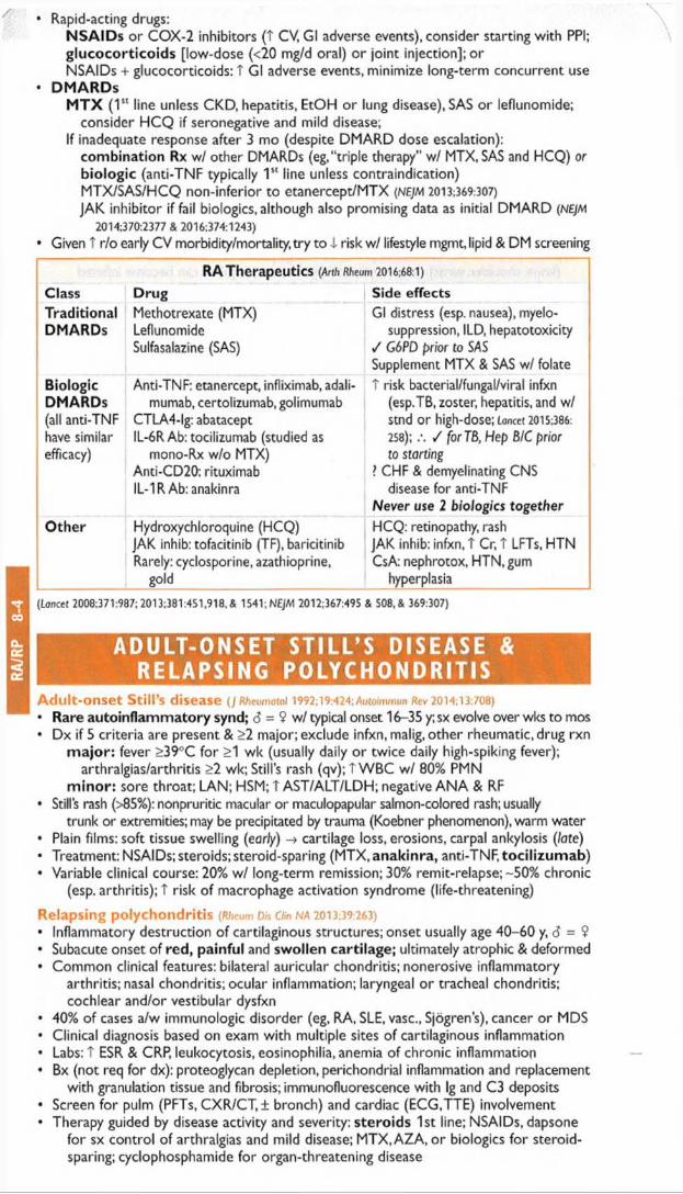
RA
/RP
8-4
• Rapid-acting drugs:NSAIDs or COX-2 inhibitors (T CV, Gl adverse events), consider starting with PPI;glucocorticoids [low-dose (<20 mg/d oral) o r joint injection]; orNSAIDs + glucocorticoids: T Gl adverse events, minimize long-term concurrent use
• DM ARDsM T X (1st line unless CKD, hepatitis, EtOH or lung disease), SAS o r leflunomide;
consider HCQ if seronegative and mild disease;If inadequate response after 3 mo (despite DMARD dose escalation):
combination Rx wI other DMARDs (eg,"triple therapy" w/ MTX, SAS and HCQ) or biologic (anti-TNF typically 1st line unless contraindication)MTX/SAS/HCQ non-inferior to etanercept/MTX (NE]M 2013:369:307)
JAK inhibitor if fail biologies, although also promising data as initial DMARD (NEJM 2014:370:2377 & 2016:374:1243)
• Given T r/o early CV morbidity/mortality.try to i risk w/ lifestyle mgmt, lipid & DM screening
RA Therapeutics (Arth Rheum 2016:68:1)
Class Drug Side effectsTraditionalDMARDs
Methotrexate (MTX) Leflunomide Sulfasalazine (SAS)
Gl distress (esp. nausea), myelo- suppression, ILD, hepatotoxicity
/ G6PD prior to SAS Supplement MTX & SAS w/ folate
BiologicDMARDs(all anti-TNF have similar efficacy)
Anti-TNF: etanercept, infliximab, adali- mumab, certolizumab, golimumab
CTLA4-lg: abatacept IL-6RAb: tocilizumab (studied as
mono-Rx w/o MTX)Anti-CD20: rituximab IL-1R Ab: anakinra
T risk bacterial/fungal/viral infxn (esp.TB, zoster, hepatitis, and w/ Stnd or high-dose; Lancet 2015:386:
258); / for 78, Hep B/C prior to starting
? CHF & demyelinating CNS disease for anti-TNF
Never use 2 biologies togetherOther Hydroxychloroquine (HCQ)
JAK inhib: tofacitinib (TF), baricitinib Rarely: cyclosporine, azathioprine,
gold
HCQ: retinopathy, rash JAK inhib: infxn, T Cr, t LFTs, HTN CsA: nephrotox, HTN, gum
hyperplasia
{Lancet 2008;371:987;2013;381:4S1,918,& 1541;NEJM 2012;367:495 & 508, & 369:307)
ADULT-ONSET STILL’S DISEASE & RELAPSING POLYCHONDRITIS
Adult-onset Still's disease (J Rheumatol 1992; 19:424;Awto/mmon Rev 2014:13:708)• Rare autoinflammatory synd; <5 = 9 w/ typical onset 16-35 y; sx evolve over wks to mos• Dx if 5 criteria are present & >2 major; exclude infxn, malig, other rheumatic, drug rxn
m ajor: fever >39°C for >1 wk (usually daily or twice daily high-spiking fever);arthralgias/arthritis >2 wk; Still’s rash (qv); t W BC w / 80% PMN
m inor: sore throat; LAN; HSM; T AST/ALT/LDH; negative AN A & RF• Stills rash (>85%): nonpruritic macular or maculopapular salmon-colored rash;usually
trunk or extremities; may be precipitated by trauma (Koebner phenomenon), warm water• Plain films: soft tissue swelling (early) -> cartilage loss, erosions, carpal ankylosis (late)• Treatment: NSAIDs;steroids;steroid-sparing (MTX,anakinra, anti-TNF, tocilizumab)• Variable clinical course: 20% w/ long-term remission; 30% remit-relapse; -50% chronic
(esp. arthritis); T risk of macrophage activation syndrome (life-threatening)
Relapsing polychondritis (Rheum Dis Clin NA 2013:39:263)• Inflammatory destruction of cartilaginous structures; onset usually age 40-60 y, 6 = 9• Subacute onset of red, painful and swollen cartilage; ultimately atrophic & deformed• Common clinical features: bilateral auricular chondritis; nonerosive inflammatory
arthritis; nasal chondritis; ocular inflammation; laryngeal o r tracheal chondritis; cochlear and/or vestibular dysfxn
• 40% of cases a/w immunologic disorder (eg, RA, SLE, vase., Sjogren’s), cancer or MDS• Clinical diagnosis based on exam with multiple sites of cartilaginous inflammation• Labs: T ESR & CRP, leukocytosis, eosinophilia, anemia of chronic inflammation• Bx (not req for dx): proteoglycan depletion, perichondrial inflammation and replacement
with granulation tissue and fibrosis; immunofluorescence with Ig and C3 deposits• Screen for pulm (PFTs, CXR/CT, ± bronch) and cardiac (ECG.TTE) involvement• Therapy guided by disease activity and severity: steroids 1st line; NSAIDs, dapsone
for sx control of arthralgias and mild disease; MTX, AZA, o r biologies for steroidsparing; cyclophosphamide for organ-threatening disease

C R Y S T A L D E P O S I T I O N A R T H R I T I D E SComparison of Gout and Pseudogout
Gout (NEJM 2011:364:443) Pseudogout (Rheum 2009:48:711)
Acuteclinical
Sudden onset painful monoarticular arthritis (classically podagra [MTP of great toe]) or bursitis; freq. nocturnal
Polyarticular in subseq flares Can mimic cellulitis (esp. in foot)
Mono- or asymmetric oligoarthritis (esp. knees, wrists and MCP joints); rare axial involvement (eg, crowned dens syndrome)
Chronicclinical
Solid crystal deposition (tophus) in joints (esp. toes, fingers, wrists, knees) & tissue (esp. olecranon bursa, pinna, Achilles)
“ Pseudo-RA” w/ polyarticular arthritis w/ morning stiffness or “ Pseudo-OA"
Assoc.conditions
Metabolic syndrome; CKD;CHF 3 H’s: Hyperparathyroidism; Hypomagnesemia; Hemochromatosis
Crystal Monosodium urate Calcium pyrophosphate dihydratePolarizedmicroscopy*
Needle-shaped, negatively birefringent
Rhomboid-shaped, weakly positively birefringent crystals
Radio-graphicfindings
Erosions w/ overhanging edge (late);“ double contour sign” on MSK US
Chondrocalcinosis: punctate, linear densities in articular cartilage, menisci, fibrocartilage of wrist, hands, symphysis pubis
Other a/w uric acid stones; urate nephropathy
/ Ca, Mg, Fe, ferritin,TIBC, UA, PTH in young or severe cases
K ry s ta ls should be in trace llu la r; In fection can coexist w ith acute a ttccks , always / Gram stain & Cx
G o u t
Definition & epidemiology (Lorjcet 2010:375:318; Nat Rev Rheumatol 2015;11:649)• Humans lack enzyme to metabolize urate (end-product of purine metabolism)• Monosodium urate (MSU) crystal deposition in joints promotes inflammation• 6 > 9 (9:1); peak incidence 5th decade; most common cause of inflammatory arthritis in 6
over 30 y, rare in premenopausal 9 (estrogens promote renal urate excretion)
Etiologies (Ann Rheum Dis 2012;71:1448)
• U A underexcre tion (85-90%): meds (eg, diuretics); idiopathic; i renal function; obesity• Uric acid (UA) overproduction (10-15%): t meat, seafood, EtOH, psoriasis, idiopathic,
myelo- and lymphoproliferative disease, chronic hemolytic anemia, cytotoxic drugs, rare inherited enzyme defic, genetic variants (Lancet 2008;372:1953)
Diagnosis• t UA is not diagnostic: 25% of measurements nl during flare; ± T W BC & ESR• Arthrocentesis is gold standard: negatively birefringent needles (see table above)• 2015 ACR/EULAR Classification Criteria (Ann Rheum Dis 2015:74:1789) used 1° in research
Acute trea tm en t (Arthritis Care Res 2012:64:1447; Am Fam Physicion 2014:90:831)
• No superior option; start w/in 24 h of sx onset; continue until acute flare resolves; for severecases, consider combination therapy; rest and ice;w/o treatment self-limited in 3-10 d
Acute Treatment for GoutDrug Initial dose Comments
NSAIDs(nonselect or
COX-2)
Full anti-inflammatory dose -> tapering
Gastritis & GIB; avoid in CKD & CVD ~ efficacy among NSAIDs never compared with colchicine
Colchicine(PO; IV no longer available in U.S.)
1.2 mg then 0.6 mg 1 h later -> 0.6 mg bid
N/V, diarrhea ( t w/ T dose); 1 dose in renal insufficiency (however, not nephrotoxic)
a/w BM supp., myopathy, neuropathyCorticosteroids
(PO, IA, IV, IM) or Corticotropin
eg, prednisone - 0.5 mg/kg/d x 5-10 d ±
taper
Rule out joint infection 1stComparable to NSAID as 1st-line treatment
(Annals 2016:164:464)
IL-1 inhibitors(Curr Opin Rheumatol
2015:27:156)
anakinra (100 mg SC qd x 3 d)
canakinumab (150 mg SC x 1)
t t cost; anakinra a/w injection site pain(ArthriusResTher 2007:9:R28);
canakinumab approved in EU (Ann Rheum Dis
2012:71:1839; Arth Rheum 2010:62:3064)

CP
PD
8-
6Chronic trea tm en t (Lancet 2011:377:165;Am Fam Physician 2014:90:831)• Approach: i f >2 a ttacks /y ,> 1 to p h u s , jo in t e ro s io n s o r u ro lith ia s is -> s ta r t u ra te lo w e r in g Rx
& p h a rm a co lo g ic p ro p h y la x is to I r is k o f acu te a ttacks• Pharmacologic prophylaxis: c o n t in u e f o r a t leas t 6 m os o r lo n g e r if f re q u e n t a tta c k s :
lo w -d o s e colchicine ( -5 0 % i r is k o f a c u te f la re ;J Rheum 2004:31:2429), NSAIDs (less
e v id e n c e ; Ann Rheum D/s 2006:65:1312), lo w -d o s e steroids, IL-1 inhibitors (see a b o ve )
• U rate-low ering Rx: goa l UA <6 m g /d L ; do NOT discontinue during acute attack or acutekidney injury (unless allopurinol hypersensitivity syndrome)
• Lifestyle As (Rheum Dis On na 2014,40:581): i intake of meat, E tO H & seafood, T low-fat dairyp ro d u c ts , w t loss, avo id d e h y d ra tio n
Urate-Lowering Therapy (Chronic Treatment for Gout)Drug (route) Mechanism CommentsAllopurinol (PO) Xanthine
oxidase inhib1st line; adjust starting dose in CKD; titrate T q2-5wk a/w rash, hypersensitivity syndrome (see below),
diarrhea, dyspepsia, BM suppression, hepatitis; monitor CBC & LFTs; not nephrotoxic
max dose = 800 mg/dFebuxostat (PO) Nonpurine
xanthine oxidase inhib
2nd line; use if allopurinol intolerant a/w LFT A, rash, arthralgias, nausea start 40 mg, max dose =120 mg/d
Pegloticase (IV) Recombinanturicase
For refractory tophaceous gout; infusion reactions (including anaphylaxis); A b formation may limit use ()AMA 2011:306:711)
Probenecid (PO) Uricosuric Rarely used; risk of urolithiasis
• Allopurinol hypersensitivity syndrome: 10-25% mortality; I risk by starting w/ dose100 mg/d if eGFR >40 or 50 mg/d if eGFR <40; titrate up by 100 mg/d (if eGFR >40) or 50 mg/d (if eGFR <40) q2-5wk until UA <6 mg/dL (dose can be >300 mg/d even in CKD)
• Associated with HLA-B5801 , esp. Han Chinese, Koreans,Thai;screen in these high-riskpopulations prior to initiating allopurinol {Curr Opm Rheumatoi 2014:26:16) * •
Calcium Pyrophosphate D ihydrate (CPPD)D eposition D isease/Pseudogout
Definition• Deposition of CPPD crystals w/in tendons, ligaments, articular capsules, synovium,
cartilage; frequently asymptomatic
Etiologies (Rheumoto/ogy 2012;51:2070)
• Most cases id iopathic; consider further metabolic eval in young (<50 y) and florid forms• Metabolic (3 H ’s): hemochromatosis; hyperparathyroidism; hypomagnesemia
(esp. in Gitelman’s o r Bartter’s syndromes)• Joint trauma (incl. previous surgery); intra-articular hyaluronate can precipitate attacks• Familial chondrocalcinosis (autosomal dominant disorder); early-onset, polyarticular dis.
Clinical manifestations (Rheum Dis Clm NA 2014:40:207)
• Chondrocalcinosis: calcification of cartilage, resulting from CPPD deposition in articularcartilage, fibrocartilage o r menisciT incidence w / age; 20% >60 y have knee chondrocalcinosis in autopsy studies
• Pseudogout: acute CPPD crystal-induced mono- or asymmetric oligoarticular arthritis,indistinguishable from gout except through synovial fluid exam for crystals location: knees, wrists and MCP joints precipitants: surgery, trauma or severe illness
• Chronic forms:“ Pseudo-RA” and pyrophosphate arthropathy (may involve axial skeleton,resembles OA)
Diagnostic studies• Arthrocentesis is gold standard: rhomboid-shaped, weakly positively birefringent
crystals (yellow perpendicular & blue parallel to axis on polarizer; see table above)• Radiographs: see table aboveTreatm ent (NE)M 2016:374:2575)
• Asymptomatic chondrocalcinosis requires no treatment• Acute therapy for pseudogout: no RCTs, extrapolated from practice in g o u t ; s a me as for
gout, though colchicine not as effective• If associated metabolic disease, Rx o f underlying disorder may improve arthritis sx• Low-dose daily colchicine or NSAID may be effective for prophylaxis or chronic arthropathy

S E R O N E G A T I V E S P O N D Y L O A R T H R I T I SC lassifica tion system (NEJM 2016:374 2563)• 5 subtypes: ankylosing spondylitis (most common), reactive arthritis, psoriatic arthritis, IBD-
associated arthritis and undifferentiated• Can also distinguish axial-predominant from peripheral-predominant joint involvement• All subtypes share common clinical manifestations: inflammatory spine disease, peripheral
arthritis, enthesitis and extra-articular manifestations (primarily ocular and skin disease)
E p idem io logy & pathogenesis (Nat Rev Rheumatol 2015:10:110)• T prevalence of HLA-B27; HLA-B27 accounts for -30% of attributable genetic risk• Environmental factors likely critical fo r disease, esp. reactive arthritis (eg, infection)• Prevalence of 0.5-2% of population, worldwide
Spondyloarthritis (SpA) Epidemiology and Key Presentation Features
Disease Epidem iology O the rAnkylosingspondylitis
d:9 = 3:1; onset in teens to mid-20s (rare after 40 y)
Progressive limitation of spine motion; “bamboo spine”
Psoriaticarthritis
6 = 9 ;peak incidence 45-54 y; seen in 20-30% of Pts w/ psoriasis (Ann Rheum Dis 2005;64:ii14)
In 13-17%, arthritis precedes psoriasis by yrs. Does not correlate with psoriasis activity. A/w HIV.
Reactivearthritis
6 » 9; 20-40 y; 10-30 d s/p post-GI or GU infxn* in genetically susceptible host
Previously "Reiter’s syndrome” : arthritis, urethritis and conjunctivitis. Most resolve w/in 12 mo.
IBD-associated
d = 9; seen in 20% of IBD Pts; Crohn’s > UC
Type 1 <5 joints: correlates w/ IBD Type II >5 joints or axial disease;
does not correlate w/ IBD
r GU: Chlamydia, U reoplasm a urea lyticum ; Gl: S/iigel/a, Salmonella, Yersinia, Cam pylobacter, C. d iff.
M a jo r c lin ica l m an ifes ta tions (Lancet 2011:377:2127)• In fla m m a to ry back pain: SI joints (sacro iliitis ), apophyseal joints of spine
characterized by IPA IN (Insidious onset, Pain at night, Age of onset <40 y, Improves w/ exercise/hot water, No improvement w/ rest),a.m. stiffness, responsive to NSAIDs
• Peripheral a r th r it is : typically asymmetric,oligoarticular, large joints, lower > upper limb;however, can be symmetric & polyarticular (thus, mimic RA), esp. in psoriatic arthritis
• Enthesitis: inflammation at site of tendon/ligament insertion into bone, esp. Achilles, prepatellar, elbow epicondyles, plantar fascia
• R ig id ity o f spine: bamboo spine by X-ray, ankylosis due to progressive growth of bonyspurs which bridge intervertebral disc
• D a c ty lit is (“ sausage digit’’): inflammation of entire digit (joint + tenosynovial inflamm)• U ve itis : anterior uveitis most common extra-articular manifestation; p/w pain, red eye,
blurry vision, photophobia, usually unilateral
Distinguishing Features
Axial-predom Peripheral-predominantFeature Ankylosing
spondylitisPsoriatic Reactive IBD-assoc
Axial involv. 100% 20-40% 40-60% 5-20%Sacroiliitis Symmetric Asymm Asymm SymmetricPeriph involv. Less common (-50%) Frequent Frequent FrequentPeriphdistrib.
Lower > Upper Upper > Lower (see below)
Lower > Upper Lower > Upper
HLA-B27 80-90% 20% 50-80% 5-30%Enthesitis Frequent Frequent Frequent RareDactylitis Uncommon Common Common UncommonOcular Uveitis in 25-40% Conjunctivitis,
uveitis,episcleritis,
Conjunctiv itis(noninfectious), uveitis, keratitis
Uveitis
Skin None Psoriasis; nail pitting and onycholysis
Circinatebalanitis,
keratodermablennorrhagica
E. nodosum, pyoderma-
gangrenosum
Imaging Bamboo spine "Pencil-in-cup" Asymmetric Periph dis.(symm syndes.) DIP deformity syndesmophytes rarely erosive
Other T CAD; aortitis,Al, conduction defects
T CAD U re th ritis ,Al, conduction
defects
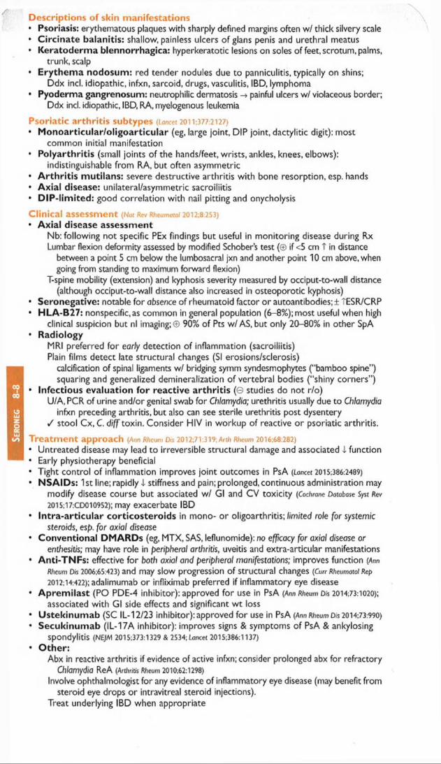
Ser
one
g
8-8
Descriptions of skin manifestations• Psoriasis: erythematous plaques with sharply defined margins often w/ thick silvery scale• C ircinate balanitis: shallow, painless ulcers of glans penis and urethral meatus• Keratoderma blennorrhagica: hyperkeratotic lesions on soles of feet, scrotum, palms,
trunk, scalp• Erythem a nodosum: red tender nodules due to panniculitis, typically on shins;
Ddx incl. idiopathic, infxn, sarcoid, drugs, vasculitis, IBD, lymphoma• Pyoderma gangrenosum: neutrophilic dermatosis -» painful ulcers w/ violaceous border;
Ddx incl. idiopathic, IBD, RA, myelogenous leukemia
Psoriatic arthritis subtypes (Lancet 2011:377:2127)• M onoarticular/oligoarticular (eg, large joint, DIP joint, dactylitic digit): most
common initial manifestation• Polyarthritis (small joints of the hands/feet, wrists, ankles, knees, elbows):
indistinguishable from RA, but often asymmetric• A rth ritis mutilans: severe destructive arthritis with bone resorption, esp. hands• Axial disease: unilateral/asymmetric sacroiliitis• D IP-lim ited: good correlation with nail pitting and onycholysis
Clinical assessment (Nat Rev Rheumatol 2012:8:253)• Axial disease assessment
Nb: following not specific PEx findings but useful in monitoring disease during Rx Lumbar flexion deformity assessed by modified Schober’s test (© if <5 cm T in distance
between a point 5 cm below the lumbosacral jxn and another point 10 cm above, when going from standing to maximum forward flexion)
T-spine mobility (extension) and kyphosis severity measured by occiput-to-wall distance (although occiput-to-wall distance also increased in osteoporotic kyphosis)
• Seronegative: notable for obsence of rheumatoid factor or autoantibodies; ± TESR/CRP• HLA-B27: nonspecific, as common in general population (6-8%); most useful when high
clinical suspicion but nl imaging;© 90% of Pts w/AS, but only 20-80% in other SpA• Radiology
MRI preferred for early detection of inflammation (sacroiliitis)Plain films detect late structural changes (SI erosions/sclerosis)
calcification of spinal ligaments w/ bridging symm syndesmophytes (“bamboo spine”) squaring and generalized demineralization of vertebral bodies (“ shiny corners” )
• Infectious evaluation for reactive arthritis (G studies do not r/o)U/A, PCR of urine and/or genital swab for Chlamydia; urethritis usually due to Chlamydia
infxn preceding arthritis, but also can see sterile urethritis post dysentery / stool Cx, C. diff toxin. Consider HIV in workup of reactive or psoriatic arthritis.
Treatm ent approach (Ann Rheum Djs 2012;71:319; Arth Rheum 2016:68:282)• Untreated disease may lead to irreversible structural damage and associated I function• Early physiotherapy beneficial• Tight control of inflammation improves joint outcomes in PsA (Lancer 2015:386:2489)
• NSAIDs: 1 st line; rapidly i stiffness and pain; prolonged, continuous administration maymodify disease Course but associated W/ Gl and CV tOXicity (Cochrane Database Syst Rev 2015:17:CD010952); may exacerbate IBD
• In tra-articu lar corticosteroids in mono- or oligoarthritis; limited role for systemicsteroids, esp. for axial disease
• Conventional DM ARDs (eg, MTX, SAS, leflunomide): no efficacy for axial disease orenthesitis; may have role in peripheral arthritis, uveitis and extra-articular manifestations
• Anti-TNFs: effective for both axial and peripheral manifestations; improves function (Ar?nRheum Dis 2006:65:423) and may slow progression of structural changes (Curr Rheumatol Rep 2012:14:422); adalimumab or infliximab preferred if inflammatory eye disease
• Aprem ilast (PO PDE-4 inhibitor): approved for use in PsA (Ann Rheum Dis 2014:73:1020);associated with Gl side effects and significant wt loss
• Ustekinumab (SC IL-12/23 inhibitor): approved for use in PsA (Ann Rheum Dis 2014:73:990)• Secukinumab (IL-17A inhibitor): improves signs & symptoms of PsA & ankylosing
spondylitis (NEJM 2015:373:1329 & 2534; Lancet 2015:386:1137)
• O ther:Abx in reactive arthritis if evidence of active infxn; consider prolonged abx for refractory
Chlamydia ReA (Arthritis Rheum 2010:62:1298)Involve ophthalmologist for any evidence of inflammatory eye disease (may benefit from
steroid eye drops or intravitreal steroid injections).Treat underlying IBD when appropriate

INFECTIOUS ARTHRITIS & BURSITISEtiologies & D iagnosis of Infectious A rthritis
Etio log ies (Curr Rheumaiol Rep 2013;15:332)
• Bacterial (nongonococcal): early diagnosis required• Gonococcal (N. gonorrhea): consider in sexually active young adults• Viral: parvovirus, HCV, HBV, acute HIV; typically polyarticular, may mimic RA• Mycobacterial: monoarticular o r axial (Pott’s disease)• Fungal: Candida (esp. prosthetic joints), coccidiomycosis (valley fever), histoplasmosis• Other: Lyme, Mycoplasma, Salmonella (2° to anti-TNF Rx), Brucellosis (unpast, dairy)
Diagnosis (JAMA 2007:297:1478)• H&P w/ poor sensitivity and specificity for septic arthritis; ,\ arthrocentesis should be
performed as soon as suspected and prior to starting antibiotics if possible• Take care not to tap through an infected area, thus introducing infxn into joint space• / Synovial fluid cell count w / differential, Gram stain, bacterial culture, crystals
W B C >50k w/ poly predom suspicious for bact. infxn; crystals do not rlo septic arthritis!
B a c t e r i a l ( N o n g o n o c o c c a l ) A r t h r i t is
E p idem io logy & risk fac to rs• Im m unocom prom ised host: DM, EtOH use, HIV, age >80, SLE, cancer, steroid use, etc.• Damaged joints: RA, OA,gout, trauma, prior surgery/prosthetic, prior arthrocentesis (rare)• B acte ria l seeding: bacteremia secondary to IVDU, endocarditis o r skin infection
direct inoculation o r spread from contiguous focus (eg, cellulitis, septic bursitis, osteo)
C lin ica l m an ifes ta tions (JAMA 2007;297:1478; Lancet 2010:375:846)
• Acute onset m onoarticu la r a rth r itis (>80%) w/ pain (Se 85%), swelling (Se 78%), warmth• Location: knee (most common), hip, wrist, shoulder, ankle. In IVDU, tends to involve
other areas inc. axial joints (eg, SI, symphysis pubis, sternoclavicular, manubrial joints).• C o n s tit. sx: fevers (Se 57%), rigors (Se 19%), sweats (Se 27%), malaise, myalgias, pain• Infection can track from initial site to form fistulae, abscesses o r osteomyelitis• Septic bursitis must be differentiated from septic intra-articular effusion
A d d itio n a l d iagnostic studies (jAfAA 2007;297:1478)
• Synovial fluid: W B C usually >50k (Se 62%, Sp 92%) but can be <10k, >90% polys;Gram stain ® in -75% of Staph, -50% of GNR; Cx © in >90%. Synovial bx most sens.
• Leukocytosis (Se 90%, Sp 36%); e levated ESR/CRP (Se >90%)• B lood cu ltu res © in >50% of cases, -80% when more than 1 joint involved• Conventional radiographs should be obtained but usually normal until after ~2 wk of
infection when bony erosions, joint space narrowing, osteomyelitis, periostitis can be seen• C T & MRI useful esp. fo r suspected hip infection or epidural abscess
T re a tm e n t fo r native jo in ts (Curr Rheumatol Rep 2013:15:332)
• Prompt empiric antibiotics guided by Gram stain after surgical drainage. If Gram stain ©, empiric Rx w/ vancomycin; add anti-pseudomonal agent if elderly, immunosupp.
Com m on microbes Population In itia l an tib io tic regimen(by Gram stain) (tailor based on Gram stain, cx, clinical course)
S. aureus (most common)
Normal joints Prosthetic joints Damaged joints
Vancomycin*
GPC S. epidermidis Prosthetic joints Postprocedure
Vancomycin*
Streptococci Healthy adults Splenic dysfunction
PCN-G or ampicillin
Diplococci: N. gonorrhea
Sexually active young adults
Ceftriaxone or cefotaxime
GN Rods: £. coli, Pseudomonas, Serratia
IVDU, Gl infection immunosupp, trauma elderly
Cefepime or piperacillin/tazobactam + antipseudomonal aminoglycoside in IVDU
*Can later A to antistaphylococcal penicillin based on sensitivities
• IV antib iotics x >2 wk followed by oral antibiotics; varies by clinical course & microbiology• Joint must be drained, often serially; arthroscopic drainage for larger joints and as initial
treatment but may also be accomplished by arthrocentesis. Serial synovial fluid analyses should demonstrate 1 in WBC and sterility.
• Prognosis: 70-50% mortality depending on virulence of organism, time to Rx, host
Infxn Arth

Infx
n A
rth
8-
10Prosthetic jo int infections {Infect Drs O n NorrtiAm 2012:26:29; C/D 2013;56:e1)• T risk in first 2 y s/p procedure; rate generally low (0.5-2.4%); risk factors include obesity,
RA, immunocompromised state, steroids, & superficial surgical site infxn• Staphylococci (coag negative & S. aureus) in >50%; polymicrobial in 10-20%• Early (<3 mo s/p surgery) o r delayed (3-24 mo) onset typically acquired during
implantation; early w / virulent organisms (eg, MRSA) and delayed w/ less virulent organisms (eg, P. ocnes, coag negative Staph) & more indolent presentation
• Late (>24 mo) onset typically related to secondary hematogenous seeding• Diagnosis requires arthrocentesis by orthopedics; ESR & CRP (CRP Se 73-91%, Sp 81-
86%; NEJM 2009:361:787) can be helpful• Treatment typically requires prolonged abx & 2-stage jo int replacement (joint
retention a/w -40% failure rate; C/D 2013:56:182) or life-long suppressive abx. ID and orthopedics consultation required.
D isseminated Gonococcal Infection (DGI)Epidemiology (infect Dis Clin North Am 200S;19:853)• N. gonorrhea; most frequent type of infectious arthritis in sexually active young adults• N orm al host as well as Pts w / deficiencies of terminal components of complement• 9 :6 = 4:1; t incidence during menses, pregnancy, & postpartum period, SLE; T incidence in
homosexual males; rare after age 40 y
Clinical manifestations• Preceded by mucosal infection (eg, endocervix, urethra o r pharynx) that is often asx• Two distinct syndromes, although Pts can have both:
Joint localized: purulent arthritis (40%), usually 1-2 joints (knees > wrists > ankles) DGI: triad of polyarthralgias, tenosynovitis, skin lesions; purulent arthritis rare
acute onset of tenosynovitis (60%) in wrists, fingers, ankles, toes rash (>50%): gunmetal gray pustules with erythematous base on extremities & trunk
• Rare complications: Fitz-Hugh-Curtis syndrome (perihepatitis), pericarditis, meningitis,myocarditis, osteomyelitis from direct extension of joint-localized infection
Additional diagnostic studies• Synovial fluid: W B C >50k (but can be <10k), poly predom inant
Gram stain © in -25%; culture © in up to 50% if done w/Thayer-Martin media• Blood culture: more likely © in DGI; rarely in joint localized disease• Gram stain and culture of skin lesions occasionally ©• Cervical, urethral, pharyngeal, rectal PCR or cx on Thayer-Martin media; / Chlamydia
Treatm ent• Ceftriaxone or cefotaxim e x 7 d w / em piric doxycycline for Chlamydia
(fluoroquinolones no longer recommended due to resistance)• Joint arthroscopy/lavage may be required if purulent arthritis; rarely >1 time * •
O lecranon & Prepatellar Bursitis
Epidemiology & risk factors (Infect Dis North Am 2005:19:991)
• >150 bursae in the body; 2 most commonly infected are o lecranon and p re p a te lla r• Most commonly (esp. superficial bursae) due to direct trauma, percutaneous inoculation or
contiguous spread from adjacent infection (eg, cellulitis)• O ther risk factors: recurrent noninfectious inflammation (eg, gout, RA, CPPD), diabetes• S. aureus (80%) most common, followed by streptococci
Diagnosis• Physical exam: discrete bursal swelling, erythema, maximal tenderness at center of
bursa with preserved joint range of motion• Aspirate bursa if concern for infxn, / cell count, Gram stain, bacterial cx, crystals
W B C >20k w / po ly p redom inance suspicious for bacterial infection, but lower counts common (crystals do not rule out septic bursitis!)
• Assess for adjacent joint effusion, which can also be septic• Take care not to tap through infected skin, thus introducing infxn into bursa
Initial therapy• Prompt empiric coverage for staphylococci and streptococci: PO abx acceptable for mild
presentation; vancom ycin if ill-appearing; broaden spectrum based on risk factors• Modify antibiotics based on Gram stain, culture results, & clinical course. Duration of tx is
1-4 wks. Serial aspirations every 1-3 d until sterile or no reaccumulation of fluid.• Surgery if unable to drain bursa through aspiration, evidence of foreign body or necrosis,
recurrent/refractory bursitis w/ concern for infxn of adjacent structures.

CONNECTIVE TISSUE DISEASESApprox Prev of Autoantibodies in Rheumatic Diseases
Disease A N A dsDNA Sm Ro/ Scl- RNA Centr jo-1 U1- RFLa 70 Pill RNP
SLE >95 75 20 25 © 0 © 0 45 35Sjogren’s >95 rare © 45 © © © © rare >75DiffuseSSc
>90 © 0 rare 40 20 rare © rare 30
LimitedSSc
>90 0 © rare 10 rare 60 © rare 30
IM 75-95 0 0 0 rare © © 25 © 15MCTD >95 © 0 rare © © © 0 always 50RA 40 0 0 0 © © © © © 70
Centr, centromere; IM, inflammatory myopathies; RF, rheumatoid factor; SSc, systemic sclerosis. (Primer on the R heum atic Diseases, 12°' ed., 2001; Lancet 2013;382:797).• Auto-Ab testing directed by clinical findings, as auto-Ab do not define a particular CTD• Overlap syndromes may be reflected by multiple autoantibodies
see " Systemic Lupus Erythematosus” and “ Rheumatoid Arthritis” for those diseases
S y s t e m i c S c l e r o s is a n d S c l e r o d e r m a D is o r d e r s
D e fin itio n & ep idem io lo gy (Best Proa Res C lin Rheumatol 2010.24:857)• Scleroderm a refers to the presence of tight, thickened skin• Localized scleroderma: morphea (plaques of fibrotic skin), linear (fibrotic bands),
“en coup de saber” (linear scleroderma on one side of scalp and forehead ~ saber scar)• Systemic sclerosis (SSc) = scleroderma + internal organ involvement
SSc w / limited cutaneous disease: formerly CREST syndrome (see below)SSc w / diffuse cutaneous disease: often rapidly progressive disorder affecting skin SSc sine scleroderma (visceral disease w ithout skin involvement, rare)
• Peak onset of SSc between ages 30-50; 9 > 6 (7:1); African American > white• 1-2/100,000 annual incidence of systemic disease in the U.S.• Pathogenesis: immune damage to endothelial cells and reactive O 2 species production -* •
persistent oxidative stress -> perivascular inflammation -> fibroblast activation and fibrosis. Cytokines, growth factors, genetics, environmental factors and autoantibodies (against PDGF receptor, endothelial cells and fibroblasts) all contribute (NEjM 2009;360:1989).
A C R /E U L A R SSc c lassification c r ite r ia {A nn Rheum D is 2013:72:1747)• Sufficient for dx: skin thickening of fingers of both hands extending proximal to MCPs• Other items considered in criteria: Raynaud’s, SSc-related auto-Ab, PAH and/or ILD,
abnormal nailfold capillaries, telangiectasia, fingertip lesions (ulcers, scars), skin thickening limited to fingers (not beyond MCPs)
• Rule out other causes of thickened skin: diabetes (scleredema * scleroderma), toxin,hypothyroidism, nephrogenic systemic fibrosis, eosinophilic fasciitis, amyloidosis, GVHD
D iagnostic studies & m o n ito r in g {Sem/n Arthritis Rheum 2005;35:35)• Autoantibodies: >95% Pts w/ auto-Ab; generally mutually-exclusive
© anti-Scl-70 (antitopoisomerase 1):a/w diffuse SSc; T risk pulm fibrosis © anticentrom ere: a/w limited SSc;T risk of severe digit ischemia and PHT © anti-R N A -Pol III: a/w diffuse SSc; T risk renal crisis; a/w cancer © A N A (>90%),© RF (30%),© anti-UI-RNP a/w overlap syndrome Other: anti-Th/To (a/w limited SSc),U3-RNP (a/w ILD),PmSd (polymyositis-SSc overlap)
• CXCL4 levels reported to help diagnose disease and be correlated w/ degree of lung &skin fibrosis and disease progression but awaits validation (NE/M 2014;370.433)
• A t baseline: / BUN/Cr & UA for proteinuria, PFTs (spirometry, lung volumes, D|CO), high-res chest CT (if diffuse disease),TTE (RVSP for PHT), RHC if T RVSP or suspect PHT
• Annual PFTs;TTE q1-2y• Skin bx not routine, but helpful to assess other possible causes fo r skin thickening• T risk of malignancy compared to general population, therefore must be vigilant• Frequent (eg, daily) BP / to m onitor for HTN suggestive of scleroderma renal crisis
CTD
8-11

CTD
8-12
Clinical Manifestations of Systemic SclerosisSkin T ig h te n in g a n d th ic k e n in g o f e x t r e m it ie s , fa c e , t r u n k ( b x n o t re q f o r d x )
" P u f f y " h a n d s , c a rp a l tu n n e l s y n d ro m e , s d e r o d a c ty ly N a i l f o ld c a p il la ry d i la ta t io n & d r o p o u tIm m o b i le , p in c h e d , " m o u s e - l ik e " fa c ie s a n d " p u r s e - s t r in g ” m o u th C a lc in o s is c u t is (s u b c u ta n e o u s c a lc if ic a t io n ) , te la n g ie c ta s ia s
Arteries R a y n a u d ’s p h e n o m e n o n (8 0 % ); d ig ita l o r v is c e ra l is c h e m iaRenal Scleroderma renal crisis (SRC) = a c c e le ra te d d e v e lo p m e n t o f H T N
(re /a tiVe in c re a se in Pt BP as c o m p a re d w ith base line BP), M A H A u r in e s e d im e n t ty p ic a lly b la n d ; p a th w / " o n io n - s k in " h y p e r t r o p h y o f
c a p il la r ie s ; a f fe c ts 5 - 1 0 % o f P ts , 66% w / in 1 “ y e a r (Rheum 2009;48:iii32)
T r is k w / > 1 5 m g /d o f p re d n is o n e (Arthritis Rheum 1998:41:1613) p o o r p ro g n o s is w / 5 0 % m o r t a l i t y
Gl(> 8 0 % o f P ts )
G E R D a n d e ro s iv e e s o p h a g it isE so p h a g e a l d y s m o t i l i t y -> d y s p h a g ia , o d y n o p h a g ia , a s p ir a t io n G a s t r ic d y s m o t i l i t y -> e a r ly s a t ie ty a n d g a s t r ic o u t le t o b s t r u c t io n S m a ll in te s t in a l d y s m o t i l i t y -> m a la b s o r p t io n , b a c t o v e r g r o w th , b lo a t in g
Musculoskel A r th r a lg ia s /a r t h r i t is ; m y o s it is ; j o in t c o n t r a c tu r e s ; t e n d o n f r ic t io n ru b sCardiac M y o c a rd ia l f ib ro s is ; p e r ic a rd ia l e f fu s io n ; c o n d u c t io n a b n o rm a l i t ie sPulmonary P u lm o n a ry f ib r o s is ( ty p ic a l ly d e v e lo p s w / in 4 y ); p u lm o n a r y a r te r ia l
h y p e r te n s io n ( ty p ic a l ly d e v e lo p s a f te r m a n y y rs ) . # 1 c a u s e o f m o r t a l i t yEndocrine A m e n o r r h e a a n d in fe r t i l i t y c o m m o n ; t h y r o id f ib ro s is ± h y p o th y r o id is m
SSc Subgroup ComparisonLimited Diffuse
General Fatigue, weight lossSkin Thickening on extremities d is ta l to
elbows/knees and face onlyThickening of distal a n d p ro x im a l
ext, face an d tru n k
Pulmonary PAH (rapidly progressive) > fibrosis Fibrosis > PAHGl PBCRenal SRC later in disease course SRC earlier & more commonCardiac Restrictive cardiomyopathyOther CREST syndrome = Calcinosis,
Raynaud’s, Esophageal dysmotility, Sderodactyly, Telangiectasias
Raynaud’s
Antibodies Centromere (1(M0%) Scl 70, RNA-Pol III (40%)Prognosis Survival >70% at 10 y Survival 40-60% at 10 y
Treatm ent (Ann Rheum Dis 2009:68:620)
• Minimize steroid exposure to reduce risk o f renal crisis• Pulmonary fibrosis: cyc lophospham ide (NEJM 2006:354:2655;Arth Rheum 2006:54:3692), MMF
under investigation; improvement may be minimal (Rheum Dis Clin NA 2015:41:237)
PAH: pulmonary vasodilators (see“ Pulm Hypertension"), early Rx a/w better outcomes• Renal crisis: ACEI (not ARB) for Rx, not prophylaxis (SemfnArthritis Rheum 2015:44:687)
• Gl: PPI and/or H2-blockers for GERD; antibiotics for malabsorptionhypomotility: metoclopramide or erythromycin; nonoperative Rx of pseudo-obstruction
• Cardiac: NSAIDs o r steroids for pericarditis• Arthritis: acetaminophen, NSAIDs, hydroxychloroquine, MTX• Myositis: MTX, AZA, steroids■ Skin: PUVA for morphea. For pruritus: emollients, topical or oral steroids ( I dose). MTX or
MMF effectiveness for skin fibrosis debated (Ann Rheum D/s 2011:70:1104). * •
Inflammatory Myopathies
Definition & epidemiology (JAMA 2013:305:183; NE/M 2015:372:1734)• All lead to skeletal muscle inflammation & weakness, variable extramuscular involvement• Polymyositis (PM): idiopathic diffuse polymyopathy, onset typically 40s-50s; 9 > 6• Dermatomyositis (DM): similar to PM; also occurs in childhood, but differentiated from
other myopathies by skin manifestations; malignancy a/w PM (10%) & DM (24%)• Necrotizing autoimmune myositis (NM): usually in adults;occurs after viral infections,
statin exposure (© anti-HMGCR)• Inclusion body myositis (IBM):onset after age 50; 6 > 9 ; often misdiagnosed as PMClinical manifestations (NEy/vi 2015:372:1734)• Muscle weakness: gradual (wks -> mos) except in NM, progressive and painless

DM/PM/NM: proximal and symmetric; difficulty climbing stairs, arising from chairs, brushing hair; fine m otor skills (eg, buttoning) lost late
IBM: may be asymmetric and distal D e rm a to lo g ic : may precede myositis by mos to yrs (uncommon fo r converse)
e ry th e m a to u s rash on sun-exposed skin: neck & shoulders (shawl sign), face, chest h e lio tro p e rash (purplish discoloration) over upper eyelids ± periorbital edema G o ttro n ’s papules (in >80% & pathognomonic): violaceous often scaly areas
symmetrically over dorsum of PIP and MCP joints, elbows, patellae, medial malleoli subungual erythema,"mechanic’s hands” (skin cracks on digits), pruritus
DM sine m yositis (amyopathic DM): dermatologic features w/o myositis, in 10-20% Polyarthralgias o r polyarthritis: usually early; nonerosive; small joints > large joints Raynaud’s (30%, DM and overlap CTD) w/ dilatation & dropout of nail bed capillaries V iscera l in vo lve m e n t (/ Rheumatol 2009;36:2711)
pu lm ona ry : acute alveolitis; ILD; respiratory muscle weakness; aspiration cardiac (33%):often asx;conduction abnl;myo/pericarditis;HF uncommon;! CK-MB/Tn G l: dysphagia, aspiration
Antisyn the tase syndrom e (PM > DM): fever, ILD, Raynaud’s, mechanic’s hands, arthritis DDx: drug-induced myopathy (statins, cocaine, steroids, colchicine); infxn (HIV, EBV,
CMV); metabolic (hypothyroid, hypo-K, hypo-Ca); neuromuscular dis. (eg, myasthenia gravis); glycogen storage disease; mitochondrial cytopathy; muscular dystrophy
D iagnostic studies• t CK (rarely >100,000 U/L,can be TTT in NM). aldolase, SGOT, LDH;± T ESR & CRP• Autoantibodies: © AN A (>75%) (Curr Rheumatol Rep 2013:15:335)
©anti-Jo-1 (25%): most common specific Ab; a/w antisynthetase syndrome ©anti-Mi-2 (DM > PM 15-20%) is a/w disease that responds well to steroids ©anti-SRP is a/w NM,poor Rx response;© anti-HMGCR in NM a/w statin exposure
• Consider EMG (T spontaneous activity,! amplitude, polyphasic potentials w/ contraction)or MRI (muscle edema, inflammation, atrophy) for evaluation; may guide biopsy
• Pathology and muscle biopsy: all with interstitial mononuclear infiltrates, muscle fibernecrosis, degeneration & regeneration (required for definitive diagnosis)PM: T cell-mediated muscle injury; endomysial inflam, surrounds non-necrotic fibers DM: immune complex deposition in blood vessels with complement activation; perimysial,
perivascular inflam (B & C D 4T cells), complement in vessels.NM: necrotic fibers wl macrophagesIBM: T cell-mediated muscle injury, vacuole formation; same as PM with eosinophilic
inclusions and rimmed vacuoles (EM)
T re a tm e n t (PM & DM, no effective treatment for IBM) (Auto/mmun Rev 2011:11:6)• Steroids (prednisone 1 mg/kg); MTX or AZA early if mod/severe or taper fails (2-3 mo)• For resistant (3(M0%) or severe disease: AZA/M TX combo, IVIg (DM ± PM), rituximab
(Arthritis Rheum 2013:65:314), MMF, cyclophosphamide (esp. if ILD or vasculitis)• IVIg w / pulse steroids acutely for life-threatening esoph or resp muscle involvement• / for occult malignancy (esp. if DM); monitor respiratory muscle strength with spirometry• NM: discontinue statin if taking; steroids + MTX or IVIG if needed {Aiosc/e Nerve 2010:41:185)
Myositides, Myopathies and Myalgias
Disease Weakness Pain t CK T ESR BiopsyDM/PM/NM 0 © 0 ± as aboveIBM 0 © © ® as aboveHypothyroidism 0 ± 0 0 mild necrosis
inflam, atrophySteroid-induced © © © © atrophyPMR © © 0 © normalFibromyalgia(JAtAA 2014:311:1547)
© ©(tender points)
© © normal
Sjogren’s Syndrome
D e fin itio n & ep idem io lo gy• Chronic dysfxn of exocrine glands (eg, salivary/lacrimal) due to lymphoplasmacytic
infiltration. Extraglandular manifestations common in primary form.• Can be primary o r secondary (a/w RA, scleroderma, SLE, PM, hypothyroidism, HIV)• More prevalent in 9 than 6; typically presents between 40 & 60 y of age
C lin ica l m an ifes ta tions• D ry eyes (keratoconjunctivitis sicca): I tear production; burning, scratchy sensation• D ry m ou th (xerostomia): difficulty speaking/swallowing; dental caries; xerotrachea; thrush
CTD
8-13

CTD
8-
14• P a ro tid gland e n la rgem e n t: intermittent, painless, typically bilateral• Vaginal dryness and dyspareun ia• R ecu rren t nona lle rg ic rh in itis /s inus itis due to upper airway gland involvement• Extrag landu lar m anifestations: arthritis; interstitial nephritis (40%); type I RTA (20%);
cutaneous vasculitis (25%); neuropathies (10%); PNS or CNS disease; ILD; PBC• T risk of lymphoproliferative disorders (~50x t risk of lymphoma and W M in 1° Sjogren’s)Diagnostic studies• Autoantibodies: © A N A (95%), © RF (75%)
Primary Sjogren’s:© anti-R o (anti-SS-A, 56%) and/or © an ti-L a (anti-SS-B, 30%)• S c h irm e r tes t: filter paper in palpebral fissures to assess tear production• Rose-Bengal staining: dye that reveals devitalized epithelium of cornea/conjunctiva• O cu la r s ta in ing score: substitute for Rose-Bengal staining to determine degree of
keratoconjunctivitis sicca using fluorescein and lissamine green• B iopsy (minor salivary, labial, lacrimal o r parotid gland): lymphoplasmacytic infiltration Classification criteria (2 of 3 have 93% Se & 95% Sp; Arthritis Core Kes 2012:64:475)1. © anti-Ro or anti-La or RF + A N A >1:3202. Labial salivary gland bx w / lymphocytic sialadenitis and score >1 focus/4 mm23. Keratoconjunctivitis sicca w / ocular staining score >3Treatm ent (Arth Rheum 2005;S2:27 & 2007:57:310;Arth ResThcr 2013;15:R172)• Ocular: artificial tears, cyclosporine eyedrops, autologous tears• Oral: sugar-free gum, lemon drops, saliva substitute, hydration, pilocarpine, cevimeline• Systemic: NSAIDs, steroids, DMARDs, rituximab (RCTs are needed)
M ix e d C o n n e c t iv e T issu e D isease ( M C T D )
Definition (Best Pract Res Clin Rheumatol 2012:26:61)• Features of SLE, system ic sclerosis, and/or po lym yos itis that appear gradually and
often evolve to a dominant phenotype of SLE o r systemic sclerosis• Different from undifferentiated CTD (UCTD): fail to meet criteria for any CTD; 30% go
on to develop CTD over 3-5 y (usually SLE)Clinical & laboratory manifestations (variable clinical course)• Raynaud's phenom enon typical presenting symptom (75-90%); see below• Hand edema (“ puffy hands” ), sclerodactyly, RA-like a r th r it is w/o erosions, polyarthralgias• Pulmonary involvement (85%) with p u lm o n a ry hypertens ion , fibrosis• Pericarditis most frequent cardiovascular manifestation; Gl: dysmotility (70%)• Membranous & mesangial GN common (25%); low risk for renal HTN crisis or severe GN• © A N A (>95%);© RF (50%); a n ti-U I-R N P in all but not specific (seen in -50% SLE) Treatm ent: As per specific rheumatic diseases detailed above
R a y n a u d ’ s P h e n o m e n o n
Clinical manifestations (NEJM 2016:375:556)
* Episodic, reversible digital ischemia, triggered by cold temp, or stress, classically:blanching ( w h i t e , is c h e m ia ) -> cyanosis ( b lu e , h y p o x ia ) -» rubor ( r e d , r e p e r fu s io n ) ; c o lo r A u s u a lly w e l l d e m a r c a te d ; a f fe c ts f in g e r s , t o e s , e a rs , n o s e .
Primary vs. Secondary Raynaud's PhenomenonPrimary (80-90%) Secondary (10-20%)
Vessel wall Functionally abnl Structurally abnlEtiologies Idiopathic, however can be
exacerbated by comorbid conditions, including HTN, athero, CAD. DM
SSc, SLE, PM-DM, MCTD, Sjogren’s, RA Arterial dis (athero, Buerger’s); trauma Heme (cyro, Waldenstrom’s, APLAS) Drugs (ergopeptides, estrogens, cocaine)
Epidem. 20-40 y; 9 > 6 (5:1) >35 yClinical Mild, symm. episodic attacks
No PVD, tissue injury, or systemic sx
Tissue ischemia & injury (eg, digital ulcers); can be assoc w/ systemic sx
Auto Ab © Depends on above etiology, often ©Nailfold Normal Dropout and/or enlarged or distorted loops
Treatm ent (Curr Opin Rheumatol 2011:23:555; BMJ 2012;344:e289)
• All: avoid cold, maintain warmth of digits & body; avoid cigarettes, drugs, caffeine & trauma• Mild-mod: long-acting CCB, topical nitrates, SSRI, ARB, cx-blockers, ASA/clopidogrel• Severe: PDE inhibitors, anti-ET-1 receptor (if ulcers esp. w/ PHT), digital sympathectomy• Digit-threatening: IV prostaglandins, digital sympathectomy, ± anticoagulation• Others: fish oil (1° RP only; Am J Med 1989:86:158), abx for infected ulceration

SYSTEMIC LUPUS ER YTH EM ATO SU S (S L E )Multisystem in f la m m a to ry a u to im m u n e d ise a se w ith a b ro a d s p e c tru m o f c lin ic a l
m a n ife s ta t io n s in a s s o c ia tio n w ith a n t in u c le a r a n t ib o d y (A N A ) p ro d u c t io n
E p idem io logy (Lancet 2014;384:1878)• Prevalence 15-50/100,000; predominantly affects women 2nd to 4th decade• 9 :6 ratio = 8:1;African American:Caucasian ratio = 4:1• Complex genetics; some HLA association; rarely C lq & C2 deficiency
Systemic Lupus International Collaborating Clinics (SLICC) Classification CriteriaClinical Criteria SLICC Classification Criteria O ther Clinical FeaturesConstit (84%) Fever, malaise, anorexia, -I wtCutaneous/Oral/Ophthalmologic(81%)
1. Acute or subacute cutaneous changes
2. Chronic cutaneous changes3. Oral or nasal ulcers4. Nonscarring alopecia
Malar rash (spares nasolabial folds), discoid rash (papules w1 keratosis & plugging), bullous SLE, urticaria,TEN
Photosens, (n/v, rash, fever) Vasculitis, panniculitis (lupus
profundus)Raynaud’s, nailfold cap As, Sicca syndrome Conjunctivitis, episcleritis
Musculoskeletal(85-95%)
5. Joint disease: synovitis or tenderness & morning stiffness involving >2 joints
Arthralgias and myalgias Avascular necrosis of bone
Cardiopulmonary(33%)
6. Serositis: pleuritis (37%) or pleural effusion, pericarditis (29%) or pericardial effusion
Pneumonitis, IPF, shrinking lung, PAH, DAH
Myocarditis, CAD Libman-Sacks endocarditis
Renal(77%)
7, Proteinuria (>0.5 g/dL) or RBC casts
Nephrotic syndrome Lupus nephritis (qv)
Neurologic(54%)
8. Seizures or psychosisw/o other cause
Cognitive dysfxn, stroke, cranial or periph neuropathies, transverse myelitis, mononeuritis multiplex
Gl(-30%)
Serositis (peritonitis, ascites) Vasculitis (bleeding, perf.) Hepatitis, pancreatitis
Hematologic 9. Hemolytic anemia10. Leukopenia (<4000/mm3)
or lymphopenia (<1000/mmJ)11. Thrombocytopenia
(<100,000/mm3)
Anemia of chronic disease Antiphospholipid synd (VTE w/
© ACL Ab, lupus anticoag, and/or B2GPI Ab)
Splenomegaly, LANImmunologic 12. © ANA; 13. © anti-ds-DNA
14. © anti-Sm; 15. © APLA16. 1 Complement17. © Direct Coombs’ (w/o #9)
T ESR/CRR © anti-Ro/La, © anti-RNP,© RF,© anti-CCP
Expert opinion, not dx criteria fo r SLE: >4/17 SUCC criteria, including >1 clinical & >1 immunologic, or bx proven SLE nephritis w / © A N A o r anti-ds-DNA (/Wrti Rheum 2012:64:2677)
Autoantibodies in SLE (NE/M 2008:358:929)
A uto -A b Frequency (approx) Clinical associations T im e lineA N A 95-99% if active disease
90% if in remission Homogeneous or speckled
Any or all of broad spectrum of clinical manifestations
Sensitive but not specific May appear yrs before overt diseaseRo
La15-35%© anti-Ro may be seen w/ © or low titer ANA
Sjogren’s/SLE overlap Neonatal lupus Photosens.; subacute cutan.
ds-DNA 70%; -95% Sp; titers may parallel dis. activity, esp. renal
Lupus nephritis Vasculitis
Appears mos before or at
Sm 30%; very specific for SLE Lupus nephritis dx, but may
U1-RNP 40% MCTD; Raynaud’s Tend not to have nephritis
become © after dx
Histone 90% in DLE; 60-80% in SLE Mild arthritis and serositis At diagnosis
SLE 8-15

SLE
8-16
W orkup• Autoantibodies: ANA, if © -» / anti-ds-DNA, anti-Sm, anti-Ro, anti-La, anti-UI-RNP• Lytes, BUN, Cr, U/A, urine sed, spot microalb:Cr ratio or 24-h urine for CrCI and protein• CBC.APLA (© in 20-40%;ACL, B2GP1, lupus anticoagulant), total complement,C3 & C4• If i GFR, active sediment, hematuria or proteinuria (>0.5 g/dL) -> renal bx to guide Rx
Treatment of SLE (Curr Rheumatol Rep 2011:13:308;Arthritis Care Res 2015:67:1237)
Drug Indication Adverse effectsHydroxychloroquine(H C Q )
All Pts as ! flares (N£JM
1991:324:150); monoRx for arthritis, serositis, skin disease
Retinal damage (<1%) Stevens-Johnson; myopathy Not immunosuppressive
NSAIDs Arthritis, myalgias, serositis Gastritis, UGIB, renal failureImmunosuppressive agents
Corticosteroids Low dose (10-15 mg) for arthritis, serositis; high-dose (1 mg/kg) ± pulse (1 g x 3 d) for major dis (eg, renal, CNS, heme)
Adrenal suppression, DM. cataracts, osteopenia, avascular necrosis of bone, myopathy
Mycophenolate(MMF)
Nephritis (induction/maint) Nonrenal refractory to HCQ
Cytopenias.T LFTs, diarrhea, teratogen
Cyclophosphamide(CYC)
Nephritis CNS disease(induction, minimize exposure)
Cytopenias, infertility, teratogen, myeloproliferative disorders, hemorrhagic cystitis, bladder cancer
Azathioprine (AZA) Nephritis (maintenance) Non-renal disease refractory to
HCQ
Myelosuppression (/TPMT), hepatotoxicity, teratogen lymphoproliferative disorders
Methotrexate(M TX )
Arthritis (preferred over MMF/AZA)Skin disease & serositis
Myelosuppression, hepatotoxicity, pneumonitis, alopecia, stomatitis, teratogen
Cyclosporine (CsA) Renal disease Hyperplastic gums, HTN hirsutism, CKD, anemia
Belimumab(NEJM 2013:368:1528)
Arthritis, serositis, skin disease (esp. if © ds-DNA or 1 C3/C4)
B-cell depletion (< RTX, different mechanism)
Rituximab (RTX) Refractory SLE, ITP.AIHA Allergic rxn; serum sickness; PML
Lupus Nephritis (Artfirta Core Res 2012:64:797)
Class Presentation Treatment (all benefit from HCQ)1: Min. mesangial Normal U/A & creatinine No specific treatmentII: Mesangial prolif Micro hematuria/proteinuria No specific treatment ±ACEIIII: Focal prolif Hematuria/proteinuria, ±
HTN, ! GFR, ± nephrotic Induce: MMF or CYC + steroidsIV: Diffuse prolif Hematuria/proteinuria and
H T N ,! GFR,± nephroticMaintenance: ? MMF > AZA
V: Membranous (Can coexist with class III or IV)
Proteinuria, nephrotic ACEIIf nephrotic range proteinuria induce
w/ MMF + steroids Maint.: MMF superior to AZA
VI:Adv. sclerotic ESRD Renal replacement therapy
(Ann Rheum Dis 2010:69:2083; NEJM 2004:350:971 & 2005:353:2219 & 2011:365:1886)
Prognosis (Arth Rheum 2006:54:2550: Rheum [OxfordJ 2016:55:252)• 5-y survival rate >90%, 10-y survival rate >80%• Leading causes of morbidity and mortality: infection, renal failure, neurologic and
cardiovascular events; thrombotic complications (Medicine 2003:82:299)
Drug-induced lupus (D LE ) (Drug Saf2011;34:357; Curr Opm Rheumatol 2012:24:182)• Many drugs: procainamide, hydralazine, penicillamine, minocycline, INH, methyldopa,
quinidine, chlorpromazine, diltiazem, a n ti-T N F (esp. infliximab), interferons• Idiosyncratic onset; generally mild disease with arthritis, serositis, skin disease• ©Anti-histone (95%) (may be © in anti-TNF);0 anti-ds-DNA (often 0 in anti-TNF even
w/o manifestations of DLE) & anti-Sm; normal complement levels• Usually reversible w/in 4-6 wk after stopping medication
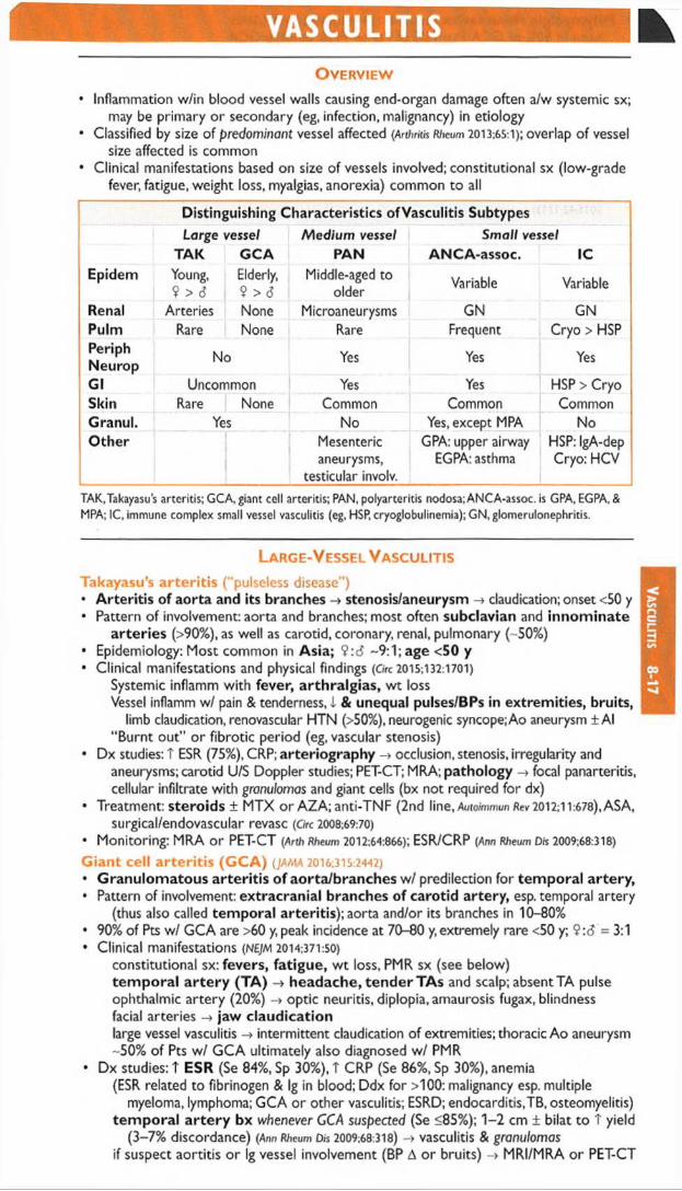
VASCULITISO v e r v i e w
• Inflammation w/in blood vessel walls causing end-organ damage often a/w systemic sx;may be primary or secondary (eg, infection, malignancy) in etiology
• Classified by size of predominant vessel affected (Arthritis Rheum 2013.65:1); overlap of vesselsize affected is common
• Clinical manifestations based on size of vessels involved; constitutional sx (low-gradefever, fatigue, weight loss, myalgias, anorexia) common to all * •
Distinguishing Characteristics ofVasculitis SubtypesLarge vessel Medium vessel Small vessel
TAK GCA PAN ANCA-assoc. 1CEpidem Young, Elderly,
9 > 6 9 > 6Middle-aged to
older Variable Variable
Renal Arteries None Microaneurysms GN GNPulm Rare None Rare Frequent Cryo > HSPPeriphNeurop No Yes Yes Yes
Gl Uncommon Yes Yes HSP > CryoSkin Rare None Common Common CommonGranul. Yes No Yes, except MPA NoOther Mesenteric
aneurysms, testicular involv.
GPA: upper airway EGPA: asthma
HSP: IgA-dep Cryo: HCV
TAK.Takayasus arteritis; GCA, giant cell arteritis; PAN, polyarteritis nodosa; ANCA-assoc. is GPA, EGPA, & MPA; 1C, immune complex small vessel vasculitis (eg, HSP, cryoglobulinemia); GN, glomerulonephritis.
L a r g e - V e s s e l V a s c u l i t i s
Takayasu’s arteritis (“ pulseless disease” )• Arteritis of aorta and its branches -» stenosis/aneurysm -» claudication; onset <50 y• Pattern of involvement: aorta and branches; most often subclavian and innominate
arteries (>90%), as well as carotid, coronary, renal, pulmonary (-50%)• Epidemiology: Most common in Asia; 9 :6 ~9:1; age <50 y• Clinical manifestations and physical findings (Grc 2015;132:1701)
Systemic inflamm with fever, arthralgias, w t lossVessel inflamm w/ pain & tenderness, i & unequal pulses/BPs in extremities, bruits,
limb claudication, renovascular HTN (>50%), neurogenic syncope; Ao aneurysm ± Al “ Burnt out” or fibrotic period (eg, vascular stenosis)
• Dx studies: T ESR (75%), CRP; arteriography -> occlusion, stenosis, irregularity andaneurysms; carotid U/S Doppler studies; PET-CT; MRA; pathology -» focal panarteritis, cellular infiltrate with granulomas and giant cells (bx not required for dx)
• Treatment: steroids ± MTX or AZA; anti-TNF (2nd line, Auto/mmun Rev 2012:11:678), ASA,surgical/endovascular revasc (Grc 2008:69:70)
• Monitoring: MRA o r PET-CT (Arth Rheum 2012:64:866); ESR/CRP (Ann Rheum Drs 2009:68:318)
Giant cell arteritis (G C A ) (JAMA 2016;315:2442)• Granulomatous arteritis of aorta/branches w/ predilection for tem poral artery,• Pattern of involvement: extracranial branches of carotid artery, esp. temporal artery
(thus also called temporal arteritis); aorta and/or its branches in 10-80%• 90% of Pts w / GCA are >60 y, peak incidence at 70-80 y, extremely rare <50 y; 9:6 =3:1• Clinical manifestations (NEJM 2014:371:50)
constitutional sx: fevers, fatigue, w t loss, PMR sx (see below) tem poral a rtery (TA ) -» headache, tender TAs and scalp; absentTA pulse ophthalmic artery (20%) -» optic neuritis, diplopia, amaurosis fugax, blindness facial arteries -> jaw claudicationlarge vessel vasculitis intermittent claudication of extremities; thoracic Ao aneurysm -50% of Pts w/ GCA ultimately also diagnosed w/ PMR
• Dx studies: T ESR (Se 84%, Sp 30%), T CRP (Se 86%, Sp 30%), anemia(ESR related to fibrinogen & Ig in blood; Ddx for >100: malignancy esp. multiple
myeloma, lymphoma; GCA or other vasculitis; ESRD; endocarditis,TB, osteomyelitis) tem poral a rtery bx whenever GCA suspected (Se <85%); 1-2 cm ± bilat to T yield
(3-7% discordance) (Ann Rheum Dis 2009:68:318) -» vasculitis & granulomas if suspect aortitis o r Ig vessel involvement (BP A o r bruits) -> MRI/MRA o r PET-CT
Va
scu
litis 8-17

Vasc
ulit
is
8-18
• Polymyalgia rheumatica (Lancet 2013:381:63;yAMA 2016:315:2442)
seen in 50% of GCA Pts; 15% of Pts w / PMR develop GCAage >50 y;ESR >40 mm/h (and/or T CRP); bilateral pain & morning stiffness (>30 min),
involving 2 of 3 areas: neck or torso, shoulders or prox. arms, hips or prox. thighs; nighttime pain; ± subdeltoid bursitis on U/S; exclude other causes of sx (eg, RA); nl CK
• Rx: steroids (d o n o t await bx/path results to begin steroids, have at least 2 wk to bx)GCA: 40-60 mg/d w/ s/ow taper, ASA daily; consider IV pulse if vision threatened. Adding
tocilizumab to steroid may be beneficial (Lancet 2016:387:1921); await phase III results.PMR: 12.5-25 mg/d; if clinical improvement, initiate slow taper. If no improvement, T
dose. Consider MTX if at high risk for steroid side effects (Ann Rheum Dis 2015:74:1799).
• Follow clinical status & ESR/CRP (Ann Rheum Dis 2009:68:318)1-1/3 relapse over 2 y (J Rheum2015:42:1213)
Medium-Vessel V asculitis
Polyarteritis nodosa ( “ c la s s ic ” P A N ) (Arth Rtieum 2010:62:616)• Necrotizing nongranulomatous vasculitis of m edium and small arteries (w/ muscular
media) w/o glomerulonephritis or capillary involvement (ie, DAH), not a/w ANCA• Epidemiology: 6 > $; average age of onset -50 y; primary or HBV-associated (-10%)• Clinical manifestations
constitutional sx (80%): w t loss, fever, fatigueneuro (79%): mononeuritis multiplex, peripheral neuropathies, stroke musculoskeletal (64%): extremity pain, myalgias, arthralgias, arthritis renal (51%): HTN, hematuria, proteinuria, renal failure, g/omerulonephritis unusual Gl (38%):abd pain, GIB/infarction, cholecystitis; GU (25%): ovarian or testicular pain skin (50%): livedo reticularis, purpura, nodules, ulcers, Raynaud’s ophthalmic (9%): retinal vasculitis, retinal exudates, conjunctivitis, uveitis cardiac (22%); coronary arteritis, cardiomyopathy, pericarditis i f lung involvement, suspect other vasculitis
• Dx studies: T ESR/CRP, 0 ANCA; / HBs Ag; i C3/C4 if HBV-associatedangiogram (mesenteric or renal vessels) -» m icroaneurysm s & focal vessel narrowing CTA may be adequate to make dx, but conventional angiogram is most sensitive biopsy (sural nerve, skin or affected organ) -» vasculitis of small and medium vessel
arteries with fibrinoid necrosis without granulomas• Treatment: steroids ± CYC (if severe o r failure to induce remission); antivirals if a/w HBV
A N C A -A ssociated Small-Vessel Vasculitis Microvascular vasculitis (eg, capillaries, postcapillary venules, & arterioles)
Disease Gran Renal Pulm Asthma A N C A Type3 A N C A ©Granulomatosis with polyangiitisb
© 80% 90%(+ ENT)
— anti-PR3(c-ANCA)
90%
Microscopicpolyangiitis
90% 50% — anti-MPO(p-ANCA)
70%
Eosinophilic granulomatosis with polyangiitisb
© 45% 70% anti-MPO(p-ANCA)
50%
'Predominant A N C A type; either p- o r c-AN C A can be seen in all three diseases (NEJM 2012:367:214). bGPA is form erly Wegener’s granulomatosis and EGPA is form erly Churg-Strauss.
D iffe re n tia l d iagnosis o f ANCA (Lancet 2006;368404)• a n t i-P R 3 ( c - A N C A ) : granulomatosis w/ polyangiitis, eosinophilic granulomatosis and
polyangiitis, microscopic polyangiitis (rarely)• a n t i - M P O ( p - A N C A ) : microscopic polyangiitis, eosinophilic granulomatosis and
polyangiitis, granulomatosis w/ polyangiitis, drug-induced vasculitis, nonvasculitic rheumatic diseases
• A t y p ic a l A N C A p a t te r n s : drug-induced vasculitis, nonvasculitic rheumatic diseases,ulcerative colitis, primary sclerosing cholangitis, endocarditis, cystic fibrosis
G ra n u lo m a to s is w ith p o lyang iitis (GPA, formerly Wegener’s granulomatosis)• N e c r o t iz in g g r a n u lo m a to u s s y s te m ic v a s c u l i t is frequently affecting nose, sinuses
and/or upper respiratory tract in addition to kidneys, lungs, etc.• Epidemiology: any age, but T incidence in young and middle-aged adults; 6=9• Clinical manifestations
respiratory (90%): upper: sinusitis, rhinitis, oral/nasal ulcers, saddle-nose deformity, otitis, hearing loss, subglottic stenosis; lower: pulmonary infiltrates, nodules, pulmonary hemorrhage, hemoptysis, pleurisy

renal (80%): RPGN (pauci-immune), RBC casts, dysmorphic RBCs, hematuria ocular (50%): episcleritis, scleritis, uveitis, orbital granulomas -» proptosis, cornea! ulcer neurologic: cranial and peripheral neuropathies, mononeuritis multiplex skin (50%): palpable purpura, livedo reticularishematologic:! incidence DVT/PE (20x) when disease active (Ann Intern Med 2005; 142:620)
• Dx studies: 90% © A N C A (80% PR3,20% MPO), less Se in limited upper airway diseaseCXR or CT -» nodules, infiltrates, cavities; sinus CT sinusitis ± bone erosions T BUN & Cr, proteinuria, hematuria; sediment w/ RBC casts, dysmorphic RBCs Biopsy -» necrotizing granulomatous inflammation of arterioles, capillaries, veins
• Treatment: assess disease severity with BVAS/WG score (Arth Rheum 2001:44:912)
Mild disease (no end-organ dysfxn; BVAS 0-3): M TX + steroids (Arth Rheum 2012:64:3472)
Severe disease (end-organ damage incl. pulm hemorrhage, RPGN etc.; BVAS >3): Induction: [R TX 375 mg/m2/wk x A wk or C YC 2 mg/kg/d x 3-6 mo or pulse
15 mg/kg q2-3wk] + steroids 1 g IV x 3 d -» 1-2 mg/kg/d (NE)M 200S;352:351,
2010:363:211. & 2013:369:417; Annals 2009; 150:670;Ann R/ieum D/s 2015:74:1178)
If RPGN: ± plasma exchange to > i risk of ESRD (Am j Kidney Dis 2011:57:566)
Maintenance: RTX q6mo superior to AZA or watchful waiting (Arth Rheum
2012:64:3760; NEJM 2014:371:1771)
Relapse: mild -» steroids ± MTX or AZA; severe -> reinduce w/ steroids + RTX or CYC T AN C A w/o clinical evidence of flare should not prompt A Rx (Annals 2007:147:611)
Microscopic polyangiitis (M PA) (Rheum D/s Clin North Am 2010:36:545)
• Similar to GPA, but w/o ENT/airw ay involvement & nongranulomatous• Epidemiology: d > V;avg onset 50-60 y• Clinical manifestations: similar to GPA w/o upper respiratory involvement;
renal (80-100%): glomerulonephritispulmonary (25-50%): pulmonary capillary alveolitis, pulmonary fibrosis constitutional and neuro sx similar to GPA; skin lesions (eg, palpable purpura) in 30-60%
• Dx studies: 70% ® A N C A (almost all anti-MPO)biopsy -» necrotizing, nongranulomatous inflammation of small vessels, pauci-immune
(minimal deposition of complement or Ig; contrast w/ HSP, cryoglobulinemia, etc.) urine sediment and CXR findings similar to those seen in GPA
• Treatment: as for GPA; i relapse rate compared to GPA
Eosinophilic granulomatosis w ith polyangiitis (EGPA, formerly Churg-Strauss)• Similar to GPA w/ more frequent cardiac involvement, a/w asthma and eosinophilia• Epidemiology: rare; can present at any age (typically 30-40 y); a/w HLA-DRB4• Clinical manifestations (CurrR/ieumoto/Rep2011;13:489)
initial sx: asthma, sinusitis, allergic rhinitis (new asthma in adult raises suspicion) eosinophilic infiltrative disease: transient pulm infiltrates, gastroenteritis, or esophagitis systemic small-vessel vasculitis: neuropathy (mononeuritis multiplex), renal
(glomerulonephritis), skin (palpable purpura, petechial, nodules) cardiac: coronary arteritis, myocarditis, CHF, valvular insufficiency (Medicine 2009:88:236)
• Dx studies: 50% © A N C A (MPO > PR3), eosinophilia (5-10 k/pL, 80-100%),biopsy - * * microgranulomas, fibrinoid necrosis and thrombosis of small arteries and
veins with eosinophilic infiltrates• Treatment: high-dose corticosteroids + cyclophosphamide (if severe)
Renal-lim ited vasculitis• Small vessel pauci-immune vasculitis causing RPGN w/o other organ involvement• Dx studies: 80% © A N C A (MPO > PR3); biopsy with pauci-immune GN ± granulomas• Treatment identical to that for GPA/MPA
Immune Complex (IC )-A ssociated Small-Vessel Vasculitis Henoch-Schonlein purpura (H SP)• IgA-m ediated vasculitis w / predilection for skin, Gl trac t and kidneys• Epidemiology: 6 > 9, children > adults, onset in w inter > summer• May develop after upper respiratory tract infection (esp. Strep) or drug exposure• Clinical manifestations
palpable purpura on extensor surfaces (lower extremity first) & buttocks polyarthralgias (nondeforming) esp. involving hips, knees, & ankles colicky abdominal pain ± GIB or intussusception nephritis ranging from microscopic hem aturia & proteinuria to ESRD
• Dx studies: skin bx w / immunofluorescence -> leukocytoclastic vasculitis w/ IgAand C3 deposition in vessel wall; renal bx -» mesangial IgA deposition
• Treatment: often self-limiting over 4 wk; steroids ± DMARDs for renal or severe disease
Va
scu
litis 8-19

IgG
4-R
el
8-20
Connective tissue disease-associated vasculitis• Small vessel vasculitis a/w RA, SLE or Sjogren’s syndrome• Clinical manifestations
distal arteritis: digital ischemia, livedo reticularis, palpable purpura, cutaneous ulceration visceral arteritis: pericarditis and mesenteric ischemia peripheral neuropathy
• Dx studies: skin/sural nerve bx, angiography, EMG; i C ' in SLE; © RF or anti-CCP in RA• Treatment: steroids, cyclophosphamide, MTX (other DMARDs)
Cutaneous leukocytoclastic angiitis• Most common type of vasculitis; heterogeneous group of clinical syndromes due to 1C
deposition in capillaries, venules and arterioles; includes hypersensitivity vasculitis• Etiologies
drugs: PCN, ASA, amphetamines, levamisole, thiazides, chemicals, immunizations infections: Strep, Stoph, endocarditis,TB, hepatitis malignancy (paraneoplastic)
• Clinical manifestations: abrupt onset of palpable purpura and transient arthralgias afterexposure to the offending agent; visceral involvement rare but can be severe
• Dx studies: T ESR, i complement levels, eosinophilia; / U/A; skin biopsy -»leukocytoclastic vasculitis w/o IgA deposition in skin (to distinguish from HSP); if etiology not clear, consider ANCA, cryoglobulins, hepatitis serologies, ANA, RF
• Treatment: withdrawal of offending agent ± rapid prednisone taper
Behcet’s syndrome (Cure Rheum Opin 2010:12:429)
• Systemic vasculitis affecting all vessel sizes, a/w oral and/or genital ulcers• Epidemiology: usually young adults (25-35 y);a/w HLA-B51 in areas of highest prevalence
on the old Silk Road (Turkey, Middle East, and other Asian countries)• Classification criteria (#1 + >2 others is 91% Se & 96% Sp; Lancet 1990:335:1078)
1. recurrent oral aphthous ulceration (>3x in 1 y, usually 1st manifestation)2. recurrent genital ulceration (labia in females, scrotum in males)3. eye lesions: uveitis, scleritis, retinal vasculitis, optic neuritis (may threaten vision)4. skin lesions: pustules, papules, folliculitis, erythema nodosum (scarring)5. © pathergy test (prick forearm w/ sterile needle -» pustule) (not sensitive in Caucasians)
• O ther clinical manifestations: most recur but are not chronicarthritis: mild, ± symmetric, nondestructive, involving knees and ankles neurologic: usually involvement of midbrain parenchyma; peripheral neuropathy rare vascular: superficial or deep vein thrombosis (25%); arterial stenosis, occlusion and
aneurysm can also occur; low incidence of thromboembolism• Dx studies: T ESR/CRP; ulcer swab to r/o HSV; ulcer bx nonspecific; ophtho eval if sx• Treatment (Rheumcto/ogy 2007:46:736; Ann R/ieu/n Dis 2008:67:1656 & 2009:68:1528)
mucocutaneousmild: topical steroids, colchicine (esp.for erythema nodosum), dapsone.apremilast
(PDE-4 inhib) for oral ulcers and ? genital ulcers (NEJM 2015:372:1510),
severe: oral steroids, steroid-sparing agents arthritis: NSAIDs, colchicine, steroids, steroid-sparing agents ocular: topical and/or systemic steroids ± steroid-sparing agents steroid-sparing:AZA, anti-TNF, CYC (large vessel and CNS ds), CsA, MTX, IFNoc-2A, venous thrombosis: steroids and anticoagulation (careful if aneurysm present)
IgG4-RELATED DISEASEDefinition & etiology (NE]M 2012:366:539; Ann Rev Pathol 2014:9:315)
• Characterized by tumor-like inflammatory lesions that can affect nearly any organ• Etiology unclear: ? autoimmune; unclear role of lgG4 Ab; Pt may have h/o atopy
Clinical manifestations (Loncer 2015:385:1460; Arth Rheum 2015:67:2466)
• Commonly pancreatitis, aortitis, cholangitis, sialadenitis, thyroiditis, orbital myositis ±pseudotumor, retroperitoneal fibrosis
• Multiple lesions may be present synchronously o r metachronously
Diagnosis (Arm Rheum Dis 2015:74:1 & 14)
• Biopsy w/ specific histopathology & immunohistochemistry findings: lymphoplasmacyticinfiltrate w/ significant lgG4+ plasma cell infiltrate, fibrosis, obliterative phlebitis
• T serum lgG4 (Se 90%, Sp 60%); not specific seen in GPA, bronchiectasis (Ann Rheum Dis2014:74:14)
Treatm ent (Arth Rheum 2015:67:1688)
• P rednisone vs. rituximab (Ann Rheum Dis 2015:74:1171)

CRYOGLOBULINEMIADefinition & types (Lancet 2012;379:348; Onco/ogy 2013:37:1098)• Proteins due to chronic immune stimulation and/or lymphoproliferation that precipitate on
exposure to cold and redissolve on rewarming, characterized by their composition• C ryoglobulins = proteins that precipitate from serum a n d p la sm a when cooled• Distinguish from cryofib rinogenem ia = proteins (eg, fibrin, fibrinogen) that precipitate only
from plasm a; found in autoimmune dis, malignancies, infxns; unclear clinical significance
Types of CryoglobulinemiaFeature Type 1 Type II Type III
(monoclonal) (mixed) (mixed)Frequency 10-15% 50-60% 25-30%
Cryoglobulincomposition
m onoclonal Ig (usually IgM or IgG)
m onoclonal IgM w/ RF activity + polyclonal IgG
polyclonal IgG and IgM
Commonetiologies
Plasma cell dyscrasias
Infection, malignancy,autoimmune syndromes
Autoimmune synd., infxn
Primarymanifestations
Hyperviscosity± thrombosis -> ischemia
IC-mediated vasculitis, w / multiorgan involvement. Can be asx.
Etiologies• Hematologic diseases
type I: multiple myeloma, MGUS,Waldenstrom's, chronic lymphocytic leukemia type II: B-cell lymphomas, solid organ malignancies
• Infections (types II & III): viral (H C V [>80% RNA ©], HBV, HIV, HAV, EBV, CMV), bacterial(endocarditis, strep, etc.), fungal (coccidiomycosis, etc.), parasitic (malaria, amoebiasis)
• Autoimmune syndromes (type III > II): Sjogren’s syndrome, SLE, RA, PAN• Renal transplant recipients (CUn Nephrol 2008:69:239)
• Essential (idiopathic) in 10% of cases
Pathophysiology• Type I: cryo precipitation in microcirculation -> hyperviscosity & vascular occlusion• Types ll/lll: defective/insufficient immune complex (1C) clearance -> IC-mediated
inflammation of blood vessels w/ complement activation -> vasculitis
Clinical manifestations• Most patients with circulating cryoglobulins are asx• Type I: hyperviscosity (cold worsens sx) -> H/A, visual disturbance, livedo, digital ischemia• Type ll/lll: vasculitis (sx not affected by cold exposure)
“ Meltzer’s triad” (purpura, arthralgias, weakness) seen in 25-30% of Pts General: weakness, low-grade feverDermatologic (54-80%): lower extremity purpura, livedo reticularis, leg ulcers Joint (44-70%): symmetric, migratory arthralgias of small or medium joints Renal (50%): glomerulonephritis (proteinuria, hematuria, ARF, HTN, edema) Neurologic (17-60%): peripheral neuropathy (polyneuropathy > mononeuritis multiplex) Hematologic: anemia, thrombocytopenia,! risk of B-cell lymphoma Gl (5%): abdominal pain, hepatosplenomegaly, abnormal LFTs
Diagnostic studies• / Cryoglobulins; must keep blood warmed to 37°C at all times en route to lab; early
cooling causes false 0 cryoglobulin, loss of RF and 41 complement• Cryocrit is quantification of cryoprotein, does not always correlate w/ disease activity• False T in W BC o r pit on automated CBC, due to cryoprecipitation• Type I: / serum viscosity, symptomatic if >4.0 centipoise; complement levels normal• Type II: i C4 levels, variable C3 levels, t ESR, 0 rheumatoid factor (RF)
/ HCV, HBV, & H IV serologies in all Pts w / mixed cryoglobulinemia Bx of affected tissue: hyaline thrombi; vasculitis w/ mixed inflammatory infiltrates of
small vessels; leukocytoclastic vasculitis in purpuric lesions
T r e a t m e n t ( B W 2012;119:5996; Medicine 2013:92:61)
• Treat underlying disorder:Lymphoproliferative disease: chemotherapy and/or radiationHCV: antivirals ± immunosuppression for severe disease (NEJM 2013:369:1035)
Connective tissue-related disease: DMARD/steroids ± rituximab• Type I: Plasma exchange if hyperviscosity; steroids, alkylating agents, rituximab, chemo• Type II: NSAIDs fo r control of mild symptoms for Pts w / normal renal function.
Rituximab or cyclophosphamide for major organ involvement. For mixed cryo, plasmapheresis o r plasma exchange only in severe, life-threatening disease.
Cryo
8-21

AMYLOIDOSISDeposition o f misfolded and insoluble fibrous proteins in normal organs and tissues
Classification of AmyloidosisType Precursor Causative diseases Main organs affectedAL (Primary) Most common -2000 cases/y
Monoclonal Ig light chain
MMLight chain disease (A. > k) MGUS.WM
Renal, cardiac, Gl, neuro, cutaneous,hepatic, pulmonary
AA(Secondary)
Serum amyloid A (SAA)
Inflam: RA, IBD, FMF Chronic infxns: ostco.TB
Renal, Gl, hepatic,neuro, cutaneous
HereditaryT incid A fr Am
Mutant TTR, etc. Mutant proteins Neurologic, cardiac
Senile Norm al TTR Normal proteins; 2° aging Cardiac, aorta, GlA(32M (i2-microglobulin Dialysis-associated |32m
(normally renally excreted)Musculoskeletal
Localized [i-amyloid protein Peptide hormones
Localized production and processing
NeurologicEndocrine
TTR, transthyretin (prealbumin).Adapted from NEJM 1997:337:898; 2003:349:583; 2007:356:2361.
Clinical Manifestations of Amyloidosis (Lancet 2016:387:2641)
System Manifestations AmyloidRenal Proteinuria o r nephrotic syndrome AL.AACardiac CMP (either restrictive o r dilated); orthostatic hypoTN
I QRS amplitude, conduction abnormalities, AFAL, hereditary,
senileGl Diarrhea, malabsorption, protein loss
Ulceration, hemorrhage, obstruction Macroglossia -» dysphonia and dysphagia
all systemic
Neurologic Peripheral neuropathy w ith painful paresthesias Autonomic neuro -> impotence, dysmotility, i BP Carpal tunnel syndrome
hereditary,AL, organ-specific,
A (i2MCutaneous Waxy, nonpruritic papules; periorbital ecchymoses
“ Pinch purpura” = skin bleeds w ith minimal traumaAL
Hepatic & splenic
Hepatomegaly, usually without dysfunction Splenomegaly, usually without leukopenia o r anemia
all systemic
Endocrine Deposition w ith rare hormonal insufficiency organ-specificMusculoskel Arthralgias and arthritis (especially shoulder) AL.AfoMPulmonary Airway obstruction; pleural effusions AL.AAHematologic Factor X deficiency AL
Diagnostic studies• Biopsy (abdominal SC fat pad, rectal o r affected tissue) apple-green birefringence on
Congo red stain; fat pad bx Se 60-85%, Sp 90-100%• If suspect AL -> / SIEP & UIEP (T Se vs. SPEP & UPEP) & free light chains,! BM bx• If suspect renal involvement / U/A for proteinuria• If suspect cardiac involvement / ECG ( I voltage, conduction abnl),TTE (biventricular
thickening w/ granular sparkling appearance; T wall w/o T volt 75% Se, 95% Sp), MRI• Genetic testing for hereditary forms
Treatment of AmyloidosisAL Limited involvement: high-dose melphalan -> auto HSCT (NEJM 2007:357:1083)
N o t HSCT candidate: [low-dose melphalan + dexamethasone] o r [cyclophosphamide + bortezom ib + dexamethasone] (6Jood20i5:126:612)
Relapsed: lenalidomide, thalidomide, o r bortezom ib (Bfood 2010:116:1990 & 2014:124:2498)
AA Rx underlying disease. Colchicine fo r FMF, esp. to prevent renal disease. Eprodisate promising fo r renal disease (NEJM 2007,356:2349)? Biologies (anakinra, tocilizumab) fo r rheum associated disease (Anh Rheum
2003:48:2019; CUn Exp Rheumatoi 2015;33:46)
ATTR Liver Tx prevents fu rthe r protein deposition (Musde Nerve 2013:47:157) Small interfering RNA under study (NEJM 2013:369:819;JACC 2015:66:2451)
• C learance o f am yloid by A b against serum am yloid P under study (NEJM 2015:373:1106)• Cardiac involvem ent: d iuretics; avoid dig, CCB, and vasodilators; ? IC D fo r 10 prevention• H eart, kidney and liver T x may be considered in those w / advanced disease• Median survival: 1 2 -1 8 mos fo r A L (~6 if cardiac); 11 y fo r A A ; variable fo r o thers

CHANGE IN MENTAL STATUSC o n s c io u s n e s s /A ro u s a l (descrip tion o f pa tien t & tim ing is m os t helpful)• Spectrum from awake/alert -♦ drow sy -> s tupo r -» coma.Vague term s, thus m ost useful
to simply describe response to increasing stim ulation (eg. voice -» noxious stim uli).• Coma: lack o f response to external stimuli. Degree form alized in Glasgow Com a Scale.
Caused by focal lesions in upper brainstem (eg, re ticu la r activating system, thalami) o r diffuse dysfxn o f cerebral hemispheres bilaterally. Mimics: locked-in synd., catatonia.
• Nb, quality o f thought can be disturbed w /o affecting level o f consciousness (eg, d iso rien t)• Delirium/acute confusional state: altered attention & awareness, develops over hrs to
days, often fluctuating, accompanied by cognitive As (eg, disorientation, memory loss, perceptual As); sometimes w/ sleep-wake dysregulation, autonomic As, emotionality
• Dementia: progressive cognitive im pairm ent beyond baseline, develops over mos to yrs,often affecting memory, language, and executive function
Etiologies of Decreased Responsiveness
1° neurologic (usually with focal signs) Systemic (esp>. in elderly or prior CNS injury)Vase: ischemic stroke, ICH, ven. throm b Seizure: postictal, status, nonconvulsive Infxn: meningitis, encephalitis, abscess Traumatic brain injury/concussion T intracranial pressure: mass,
hydrocephalus, herniation Transient global amnesia Autoimmune/paraneoplastic encephalitis Neurodeg: late-stage (eg, Alzheimer’s); o r
rapidly progressive (eg. CJD) dementia
Cardiac: global ischemia, CHF, H TN enceph Pulmonary: I Pa02, ? PaCC>2 Gl: liver failure, T NH3
Renal: uremia, dialysis, i o r T Na Endo: i glc, DKAyHHNS, hypothyr., Addisonian ID: pneumonia, UTI, sepsis Hypothermia & hyperthermia Intoxication o r withdrawal: EtOH, sedatives,
opiates, carbon monoxide, anticholinergic Psychiatric: catatonia
In i t ia l e v a lu a t io n• History (witness & background crucial): tempo, prem orbid sx (eg, focal neuro deficits, HA,
infxn, pain, falls), medical cond itions (eg, dem entia, epilepsy, one, cardiac, psych, in fection/im m une status), accompanied by head traum a, cu rre n t meds (eg, sedatives, opioids, anticoag, anticonvulsants, im m unosuppressants), drug/a lcohol use
• General exam: VS, nuchal rigidity (may be present in m eningitis o r SAH, do not test ifpossible traum a/cervical spine fx), breathing pattern (eg, Cheyne-Stokes), ecchymoses, rash, signs o f head trauma (eg, Battle’s, raccoon eyes, hemotympanum, CSF rhinorrhea), asterixis, liver disease stigmata, em bolic phenom ena/endocarditis, signs o f drug use
• Neuro exam (see below): perform o ff sedatives/paralytics if possible, look fo r deficits thatsuggest structural cause (eg, stroke, herniation syndrome), sis o f T ICP (eg, HA, vomiting, papilledema,abducens nerve palsy,unilateral dilated pupil,? BPAIHR,fixed downgaze)
Neuro Exam in Patients with Decreased ResponsivenessMental status Arousal (behavioral response to t intensity o f stimulation, GCS)
Cranial nerves Pupils: pinpoint -> opiates, pontine lesion; midposition & fixed -» midbrain lesion; fixed & diloted -> severe anoxic enceph, hern., anti-cholin.
Extraocular movements / vestibulo-ocular reflex tests:oculocephalic maneuver (“ doll’s eyes” ): nl = eyes move opposite
head movement (do not test i f possible cervical spine trauma) vestibular (cold) caloric stimulation: in coma, nl = eyes move slowly
to lavaged ear, then quickly away Corneal reflex, facial grimace to nasal tickle Gag & cough reflexes (w ith ET tube manipulation if necessary)
M oto r Tone, spont movements, flexor/extensor posturing o f arms/legs, strengthSensory
ReflexesResponse to painful stimuli: purposeful vs. reflexive/posturingDeep tendon reflexes, Babinski,"triple" flexion (ankle, knee, & hip flexionto noxious stimulation -» not suggestive of intact cortical function)
Glasgow Coma Scale (sum points from each of 3 categories to calculate score)
Eye opening Best verbal response Best m o to r response PointsFollows commands 6
Oriented Localizes pain 5Spontaneous Confused W ithdraws from pain 4
To voice Inappropriate words Flexor posturing 3To painful stimuli Unintelligible sounds Extensor posturing 2
None None (intubated = IT ) None 1

Initial trea tm en t• Immobilization of C-spine if concern for cervical trauma• Thiamine 100 mg IV dextrose 50 g IVP (order to prevent exacerbation of Wernicke's)• If opiates suspected: naloxone 0.01 mg/kg; supportive care important in nearly all tox cases• If concern for T ICP ± herniation: T head of bed; osmotherapy w/ mannitol or hypertonic
saline; T ventilation; dexamethasone for tumor edema; c/s neurosurgery (? decompress)
Diagnostic studies (Continuum 2011:17:967)• All patients; check CBC, electrolytes, BUN/Cr, tox screen, tox screen, U/A• Based on clinical suspicion:
labs: N H 3.TSH, am cortisol, Bu, ABG, ESR,ANA,TPO, thyroglobulin, BCx imaging: head CT, then MRI; radiographs to r/o C-spine fracture lumbar puncture to r/o meningitis, SAH, or noninfectious inflammation (eg, autoimmune) EEG to evaluate for nonconvulsive seizures, toxic/metabolic encephalopathy
Further trea tm en t of delirium (Armafe 2011:154:746)• Treat underlying acute illness, eliminate precipitating factors, & provide supportive care• Address sensory & cognitive impairments (frequent reorientation, etc.)• Decrease/prevent infection/restraints if possible, remove lines/catheters if unnecessary• Promote good sleep: reduce noise & nighttime interventions; sedative med if necessary• Meds: consider antipsychotics; avoid benzos except for alcohol withdrawal or seizures * •
ANOXIC Brain Injury (at risk if >5 min cerebral hypoxia)
Initial evaluation (Circulation 2010:S768)• Neuro exam: arousal/verbal, eyes & other cranial nerves, m otor response to pain• Imaging: CT usually not informative w/in first day after arrest, but should be done
prior to initiating hypothermia if patient found down o r has had head trauma
Tem perature m anagem ent {Circulation 2015;132:2448)• Indications: comatose (eg, no meaningful response to verbal stimuli) <6 h following
cardiac arrest (not isolated resp. arrest). Fully studied only in VT/VF, but consider after asystole o r PEA arrest o r 6-12 h after cardiac arrest.
• Exclusion: preg, CV instability despite pressors/assist devices, other cause o f coma,persistent i O 2
• Relative contraindications: major head trauma, coagulopathy/bleeding, major surgery<14 d, systemic infection/sepsis
• Target temp: 32-36°C x >24 h. Initial studies showing benefit targeted 32-34DC, butsubsequent study showed = outcomes for 36°C vs. 33°C (NEJM 2013:369:2197). Some still target 32-34°C and reserve 36°C for Pts w/ contraindic to more aggressive cooling.
• Method: can use cold saline infusions; ice packs to head, neck & torso; cooling blankets;cooling vest or endovascular catheter if available. Goal to achieve target temp <6 h (but no benefit to prehosp cooling; jama 2014311:45). Start rewarming 24 h after cooling is initiated (rewarm <0.5°C per h).
• Complicationsdysrhythmias (brady most common): if signif o r hemodynamic instability, rewarm coagulopathy (can receive lytics, GP llb/llla inhibitors, etc.); / PT and PTT. infection: / surveillance blood cultures during coolinghyperglycemia during cooling, hypoglycemia w/ rewarming; stop insulin if glc <200 mg/dl_ hypokalemia during cooling, hyperkalemia w/ rewarming; keep K 4-5 mEq/L
Ongoing evaluation• Neuro exam: daily focus on coma exam. No exam finding is reliable <24 h or on sedation.
Pt needs to be off sedation for an adequate time to evaluate (depends on doses used, duration of Rx, metabolic processes in the individual Pt).
• Labs: daily CBC, PT/PTT, electrolytes. Serum neuron-specific enolase (NSE) on days 1-3• Imaging: noncontrast CT 24 h after arrest; if unrevealing, consider MRI around days 3-5• EEG: consider in all to exclude seizures; greatest risk during rewarming• Somatosensory evoked potentials (SSEP): helpful for prediction of poor outcome if
cortical responses are absent bilaterally; perform 48 h after arrest (72 h if cooled)
Prognosis (Nat Rev Neuro 2014:10:190)• For inPt arrest, -20% survive, -70% of Pts who survive have good long-term prognosis• Prior to cooling era, uniformly poor prognosis could be predicted at 72 h only in Pts who
have absent pupillary and corneal reflexes, and no motor response to pain; or with absent SSEPs at 48 h. W ith cooling, it is less clear if the prior measures are as reliable.
• Otherwise, prognosis requires multifactorial approach considering exam, age, comorbiddiseases, ancillary data (NSE, EEG, SSEP; imaging is less reliable for poor outcome)
• When in doubt, err on giving more time (esp. if younger or induced hypothermia)

SEIZURESD e fin itions (Epj/epsra 2014;55:475)
• Seizure: transient neurologic symptoms due to excessive synchronous neuronal activity;may be provoked by a reversible factor lowering the seizure threshold, or unprovoked
• Epilepsy: >2 unprovoked seizures occurring >24 h apart or 1 unprovoked seizure w/ >60%probability of further seizures over the next 10 yr (see below for prognostication)
• G enera lized seizures (involves brain diffusely)Tonic<lonie (grand mal): tonic phase (10-20 sec) with contraction of muscles
(causing expiratory moan, cyanosis, pooling of secretions, tongue biting) -> clonic phase (~30 sec) with interm ittent relaxing and tensing of muscles
Absence (petit mal): transient lapse of consciousness w/o loss of postural tone, usu pedi Myoclonic (infantile spasms & juvenile myoclonic epilepsy): sudden, brief contraction
• Focal seizures (involves discrete brain area, implies a structural lesion)w/o impaired consciousness: focal motor/autonomic sx (formerly “ simple partial seizure")
or focal sensory/psychic symptoms (eg, aura) wl impaired consciousness: dyscognitive features (formerly “ complex partial seizure") evolving to bilateral, convulsive seizure (formerly “ secondarily generalized seizure” )
• Status epilepticus: continuous convulsive seizure >5 min or >2 seizures w/o resolution ofpostictal encephalopathy; life-threatening
• Nonconvulsive status epilepticus: alteration of awareness (ranging from confusion tocoma) w/o m otor manifestations of seizure; dx with EEG.
D iffe re n tia l diagnosis • Syncope (Loncet Neuro/ 2006;5:171)
Feature Seizure Syncope
Aura Unusual behavior/automatisms Diaphoresis, nausea, tunnel visionConvulsions Variable duration Usually <10 sec
Postictal state Yes; can be >30 min None or shortOther clues Tongue biting, incontinence Skin pallor, clamminess
• Nonepileptic seizure (aka “ psychogenic"): may see side-to-$ide head turning, asymmetriclarge-amplitude limb movements, diffuse shaking w /o LO C, crying/talking during event
• O th e r: metabolic disorders (eg, alcoholic blackouts, hypoglycemia), m igraine,TIA, transientglobal amnesia, narcolepsy (cataplexy), nonepileptic myoclonus, tics, asterixis
Etiologies of seizures (vary strongly by age)• W ith o u t focal lesion: genetic p red isposition to seizures o r epilepsy syndrom e;
alcohol w ithd raw al, illic it drugs;meds (eg, |3-lactams, buprop ion , tram ado l, M N Z , m eperid ine , CsA, antidepressants); e lec tro ly te (hyponatremia) & o th e r m etabolic (eg. uremia, liver failure, hypoglycemia); auto im m une encephalitis, id iopa th ic (-60% )
• W ith focal lesion: tum or, traum a, s troke, subdural hematomas, p o s te rio r reversib leencephalopathy syndrom e, mesial tem pora l sclerosis, focal co rtica l dysplasia
Clinical manifestations• Aura (sec to mins): p rem onition w ith paresthesias, focal m o to r contractions, abnormal
smells/tastes, fear, depersonalization, dej& vu, au tonom ic changes, automatism s• Ictal period (sec to mins): tonic and/or clonic movements o f head, eyes, trun k o r extrem .• Postictal period (mins to h): slow ly resolving pe riod o f confusion, d iso rien ta tion ,
and lethargy. May be accompanied by focal neuro log ic deficits (Todd’s paralysis).
Clinical evaluation• History key in differentiating seizure from other causes o f transient loss o f consciousness.
M ust ta lk to witnesses. A sk about p ro d rom e , unusual behavior before spell, type & pattern o f abnl movements incl. head tu rn ing & eye deviation (gaze preference usually away fro m seizure focus), loss o f responsiveness.
• Recent events: illnesses/fevers, head traum a, sleep depriva tion• PMH: p rio r seizures o r © FHx, p rio r meningitis/encephalitis, p rio r stroke o r head trauma• M edications (new o r noncom pliance), a lcohol and illic it d rug use• General physical exam should include the skin, looking fo r neuroectoderm al d isorders
(eg, neuro fibrom atosis, tuberous sclerosis) th a t are a/w seizures• N eurologic exam should look fo r focal abnormalities -> underlying structural abnorm ality
Diagnostic studies (Neurology 2007:69:1996)
• Lab: full lytes, B U N , Cr, glc, LFTs, to x screen, med levels (if on valpro ic acid, phenytoin;consider fo r o th e r AEDs bu t may take days; levetiracetam level rarely useful unless? noncom pliance)

• Routine EEG (-30 min): useful in workup of 1st-time unprovoked seizure, as maydetermine risk of seizure recurrence. Caveat interictal EEG nl in 50% of Pts w/ epilepsy, and interictal epileptiform activity (spikes or sharp waves) may be seen in up to 2% of nl population: sleep deprivation and repeated studies t dx yield of EEG.
• Long-term EEG monitoring (hrs to days): useful for differentiating epileptic from nonepileptic spells; video monitoring may help w / nonepileptic seizures
• MRI to r/o structural abnormalities;! Se w/ fine coronal imaging of frontal & temporal lobes• LP (if no space-occupying lesion on imaging): if suspect meningitis (eg, fever, TW BC,
nuchal rigidity) o r encephalitis and in all HIV © Pts Treatm ent (Neurology 2015;84:1705; Lancet 2015:385:884)
• Treat any underlying precipitants, including CNS infections, intoxication, withdrawal, etc.• Antiepileptic drug (AED) Rx usually reserved for Pts w / >2 unprovoked seizures, single
seizure w/ high risk of recurrence (see below), o r underlying structural abnormality. Provoked seizures generally treated by addressing underlying cause; consider AED if status epilepticus on presentation, focal neuro exam, postictal Todd’s paralysis.
• After 1st unprovoked sz, weigh risks of recurrence vs AED. t risk of recurrence if abnlEEG, MRI, o r nocturnal sz. If EEG & MRI nl -> 65% sz-free at 5 y (Lancet Neurol 2006;5:317)
• Immediate treatment w/ AED after 1st unprovoked seizure ! risk of recurrence over 2 y, butdoes not A long-term prognosis
• If AED Rx indicated, choice dependent on type of seizure, side effects, cost, mechanism ofelimination (if hepatic or renal insufficiency), teratogenesis and drug interactions
• Introduce gradually, m onitor carefully• May consider withdrawal if seizure-free (typically for at least 1 y) and normal EEG• Individual state laws mandate seizure-free duration before being allowed to drive
Antiepileptic Drugs and Side EffectsM edication Avg daily dose C om m on side effects
System/'c Neuro/ogic (all: sedation)
Carbamazepine 400-1600 mg Aplastic anemia, -I WBC, rash, hepatotoxicity, I Na
Diplopia, confusion, ataxia
Ethosuximide 500-1500 mg Rash, BM suppression Behavioral AsGabapentin 900-3600 mg Gl upset, wt gain Nystagmus, ataxiaLacosamide 200-400 mg Prolonged PR interval Dizziness, diplopiaLamotrigine 100-300 mg Rash (Stevens-Johnson) Tremor, HA, blurred
vision, insomniaLevetiracetam 1000-3000 mg Gl upset (rare) Emotional labilityOxcarbazepine 600-2400 mg Hyponatremia, rash Diplopia, dizzinessPhenobarbital 50-200 mg Rash Cognitive slowingPhenytoin 20O-H00 mg Gum hyperplasia Dizziness, ataxiaTopiramate 100-400 mg ! wt, hypohidrosis, kidney
stones, glaucoma, met acidCognitive slowing
Valproic acid 500-2500 mg Hepatotox, T NH3, t wt, 1 hair TremorZonisamide 200-600 mg i wt, hypohidrosis, nephrolith Cog slowing, fatigue
(NEJM 2008:359:166; Lancet N euro l 2011;10:446)Status epilepticus (Neurocrit Care 2012;17:3)• ABCs: vital signs, oral airway or endotracheal intubation. Place Pt in semiprone position to
1 risk of aspiration. Obtain IV access. Give thiamine, dextrose, IV normal saline.• STAT glc, metabolic panel, CBC, tox screen, lactate, AED levels, consider head CT. LP• Start standing AED after loading dose.
Treatment of Status EpilepticusT im e(m in )
A ntiep ileptic Dosing regim en Typical adult dose
<5 Lorazepam or Midazolam or Diazepam*
0.1 mg/kg IV 0.2 mg/kg IM 0.2 mg/kg PR
Successive 2-4 mg IV pushes Up to 10 mg IM
< 1 0 Phenytoin or Fosphenytoin or Valproate or Levetiracetam
2 0 mg/kg 20 mg PE/kg 20-30 mg/kg 1 0 0 0 mg
1.0- 1.5 g IV over 20 min1.0- 1.5 g PE IV over 5-10 min1.0- 1.5 g IV over 5-10 min IV over 10-15 min
Subsequent steps mandate intubation, EEG monitoring and ICU admission<(1__kfi nnon l u/itn /*AntIru i/m ic /*\ h 1<30-60 General anesthesia with continuous midazolam, pentobarbital, or propofol
PE, phenytoin equivalents. ^Consider PR diazepam i( no IV access and IM midazolam is contraindicated.

ALCOHOL WITHDRAWALPathophysio logy• Alcohol is a CNS depressant• Chronic use - j insensitivity to inhibitory neurotransmitter y-aminobutyric acid (GABA)• Abrupt alcohol cessation -> CNS overactivity
C lin ica l m an ifes ta tions• Minor withdrawal sx (6—48 h after last drink): mild anxiety, tremulousness, HA• W ithd raw a l seizures: typically w/in 48 h after last drink; if unRx’d, Vi delirium tremens• A lcoho lic hallucinosis: isolated hallucinations (typically visual) 12-48 h after last drink• D e lir iu m tre m e n s (DT): disorientation, agitation, hallucinations, T HR & BP, fever,
diaphoresis; begins 48-96 h after last drink, lasts 5-7 d• Consider other dx: CNS infxn or bleed, sz, drug O/D, coingestions, acute liver failure, GIB
C lin ica l In s titu te W ith d ra w a l Assessm ent scale fo r a lcoho l (C IW A -A r)• Assign points for each of the 10 criteria; each criteria is scored 0-7, except orientation,
which is scored 0—4; add points to calculate score
C IW A -A r Scale
Points A n x ie ty A g ita t io n T re m o r H A O rie n ta t io n0 None None None None Oriented1 Somewhat Not visible, but
felt at fingertipsVery mild Cannot do serial
additions2 Mild Disorient, by <2 d3 Moderate Disorient, by >2 d4 Guarded Restless Moderate w/
hands extendedMod severe Disoriented to
person or place5 Severe n/a6 Very severe n/a7 Panic Pacing or
thrashingSevere Extremely
severen/a
Points N/V Sweats A ud ito ryhalluc.
Visualhalluc.
Tactile disturb
0 None None None None None1 Moist
palmsVery mild Very mild
photosens.Very mild
paresthesias2 Mild Mild
photosens.Mild paresth.
3 Moderate Modphotosens.
Mod paresth.
4 Intermit, w / dry heaves
Beads Mod severe Mod severe visual halluc.
Mod severe hallucinations
5 Severe Severe Severe6 Very severe Very severe Very severe7 Constant Drenching Cont. Continuous Continuous
SCORE: <8 none to minimal withdrawal; 8-15 mild; 16-20 moderate; >20 severe
NO
Treatm ent (NEJM 2003;348:1786)
• Benzodiazepines (BDZ)Drug: diazepam (long-acting w / active metab; i risk of recurrent withdrawal), lorazepam
(short half-life), chlordiazepoxide, oxazepam (no active metab; good if cirrhosis)Route: start IV, transition to PODosing: typically start w/ diazepam 10-15 mg IV q10-15min (or lorazepam 2-4 mg IV
q15-20min) until appropriate sedation achieved, then titrate to CIWA-Ar scale, evaluating q l h until score <8 x 8 h, then q2h x 8 h, and if stable, then q4h (JAMA 1994:272:519)
• If refractory to BDZ prn -> BDZ gtt, phenobarb, dexmedetomidine.or propofol (& intubation)• Avoid |3B (mask sx)• Mechanical restraints as needed until chemical sedation achieved• Volume resuscitation as needed; thiamine then glc to prevent Wernicke's encephalopathy
(ataxia, ophthalmoplegia, short-term memory loss); replete K, Mg, PO4
• Prophylaxis: if min sx or asx (ie, CIWA score <8 ) but prolonged heavy EtOH consumption or h/o withdrawal seizures or DTs -» chlordiazepoxide 25-100 mg (based on severity of EtOH use) q6 h x 24 h, then 25-50 mg q6 h x 2 d
EtO
H

Str
ok
e 9-
6STROKE
Is c h e m ic St r o k e
Etiologies• Embolic (-75%): artery -> artery, cardioembolic, paradoxical, cryptogenic (AF found in -12%)• Thrombotic (~25%): large vessel (atherosclerosis) vs. small vessel ("lacunar,” lipohyalinosis of
small arteries, often related to HTN, hyperlipidemia, & DM)• Other: dissection, vasculitis, vasospasm, prothrombotic states, hypoperfusion, geneticClinical Manifestations• Timing: embolic -> sudden onset; thrombotic -> stuttering course
Stroke syndromes by vascular territoryA rte ry Deficits
ICA -» Ophth ACA
Amaurosis fugax (transient monocular blindness)Hemiplegia (leg > arm), abulia, urinary incontinence, primitive reflexes
MCA Hemiplegia (face & arm > leg); hemianesthesia; homonymous hemianopia Aphasia if dom. hemisphere: sup. div. -» expressive; inf. div -> receptive Apraxia & neglect if nondom. hemisphere.
PCA Macular-sparing homonymous hemianopia; alexia w/o agraphia Thalamic syndromes with contralateral hemisensory disturbance
Vertebral,PICA
Wallenberg syndrome = numbness of ipsilateral face and contralateral limbs, diplopia, dysarthria, ipsilateral Horners, hiccups
Basilar Pupillary As (midbrain-dilated, pons=pinpoint), long tract signs (quadriplegia, sensory loss),CN abnl, cerebellar dysfxn.Top of basilar -> “ locked in” synd.
Cerebellar Vertigo, N/V, diplopia, dysarthria, nystagmus, ipsilateral limb ataxiaLacunar
(arterioles)5 major syndromes: pure hemiplegia, pure hemianesthesia, ataxic hemiparesis, dysarthria + clumsy hand, mixed sensorimotor
Transient ischemic attack (T IA )• Sudden deficit due to cerebral ischemia; no stroke on imaging; sx resolve <24 h (most <1 h)• Ddx: seizure, migraine, hypoglycemia, amyloid spells,TGA, anxiety• Risk of subsequent stroke ~2% by 1 wk {NE]M 2016;374;1533). Can stratify based on A B C D 2:
Age >60 y (+1); BP >140/90 (+1); Clin features: unilat. weak. (+2), speech impair, w/o weakness (+1); Duration >60 (+2) or 10-59 min (+1); DM (+1)
Physical exam• General: murmurs, carotid & subclavian bruits, peripheral emboli, endocarditis sequelae• Neurologic exam, NIH stroke scale (http://www.ninds.nih.gov/doctors/NIH_Stroke Scale.pdf)Acute workup• Electrolytes, C r (relevant for contrast); glc, CBC, coags (see exclusion criteria for lysis)• Cardiac biomarkers, 12-lead ECG, tox screen• S T A T C T to r/o ICH prior to lysis (Se = MRI, faster, more widely available)
early signs: hyperdense artery, loss of gray-white differentiation, edema, insular ribbon CT can be nl in 1st hrs after sx onset, not Se for small strokes & brainstem strokes obtain CT-angio head & neck if endovascular intervention indicated
Acute trea tm en t of ischemic stroke (jama 2015:313:1451 & 314:1832)
• Thrombolysis (IV): tPA 0.9 mg/kg (max 90 mg), w / 10% as bolus over 1 min, rest over 1 hconsider if onset w/in 4.5 h,0 ICH, 0 contraindic. (incl. current/prior ICH; head trauma or
stroke w/in 3 mo; intracranial neoplasm, AVM or aneurysm; recent intracranial/intraspinal surgery;active internal bleeding,noncompressiblearterial puncture;? BP;multilobar infarct; pit <100k, INR >1.7, on Xa inhib, PTT >40, glc <50)
0-3 h: 12% absolute T in good neuro outcome (min/no disability), 5.8% absolute ? in ICH, trend toward 4% absolute i mortality
3—4.5 h: 7.4% absolute T in good neuro outcome, 1.8% absolute T in ICH,0 mortality benefit (nb, trial excluded patients with previous strokes + DM)
0.6 mg/kg (tested 1° in Asians): ? slightly -I efficacy but Vi ICH rate (NEJM 2016:374:2313)• BP: lower to <185/110 to consider lysis; if lyse keep <180/105 x 24 h (consider labetalol or
nicardipine), o/w permissive HTN unless >220/120 or sx; if sx HoTN consider vasopressors• Initiate ASA w/in 24-48 h; avoid anticoagulation w/in 24 h of lysis; see below for long-term Rx• Cerebral edema -> herniation: often occurs 1-5 d post large MCA or cerebellar strokes, T risk
in young.Temporize: elevate HOB >30°; mannitol ± 23% NaCI. Hemicraniectomy I mortality (toncet Neuro/2007:6:215). Neurosurgery consult in select MCA and all large cerebellar strokes.
• Endovascular thrombectomy (JACC intv 2016:9:307): if anterior circulation prox cutoff(mostly MCA) and w/in -6 h of sx onset, addition of thrombectomy to IV tPA T odds of fxnal independence by 71%, w/ no A in ICH or mortality (NEJM 2015:372:11,1009,1019.2285 & 2296: Lancet 2016:387:1723)
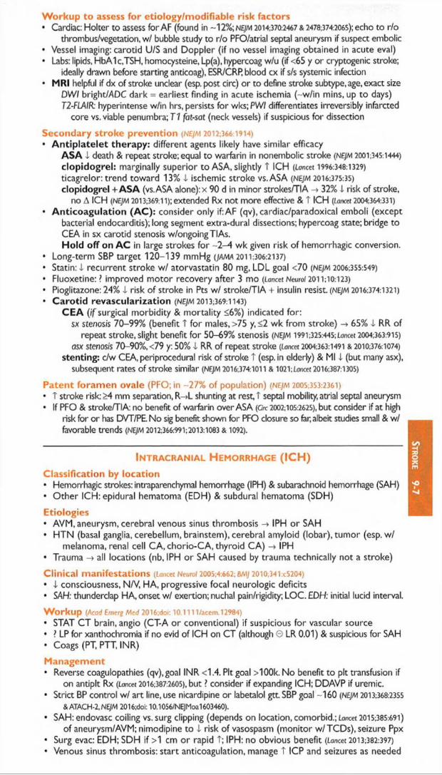
W orkup to assess for etiology/modifiable risk factors• Cardiac: Holter to assess for AF (found in -12%; nejm 2014:370:2467 & 2478:374:2065); echo to r/o
thrombus/vegetation, w/ bubble study to r/o PFO/atrial septal aneurysm if suspect embolic• Vessel imaging: carotid U/S and Doppler (if no vessel imaging obtained in acute eval)• Labs: lipids, HbAIc.TSH, homocysteine, Lp(a), hypercoag w/u (if <65 y or cryptogenic stroke;
ideally drawn before starting anticoag), ESR/CRR blood cx if s/s systemic infection• MRI helpful if dx of stroke unclear (esp. post circ) or to define stroke subtype, age, exact size
DWI bright/ADC dark = earliest finding in acute ischemia (-w /in mins, up to days) T2-FLAIR: hyperintense w/in hrs, persists for wks; PWI differentiates irreversibly infarcted
core vs. viable penumbra; T1 fat-sat (neck vessels) if suspicious for dissection
Secondary stroke prevention (nejm 2012:366:1914)• A n tip la te le t the rapy : different agents likely have similar efficacy
A S A 4 death & repeat stroke; equal to warfarin in nonembolic stroke (nejm 2001:345:1444)
c lop idog re l: marginally superior to ASA, slightly T ICH (Loncet 1996:348:1329)
ticagrelor: trend toward 13% 4 ischemic stroke vs. ASA (nejm 2016:375:35)
clopidogrel + A S A (vs. ASA alone): x 90 d in minor strokes/TIA -> 32% i risk of stroke, no A ICH (NEJM 2013;369:11); extended Rx not more effective & t ICH (Loncet 2004:364:331)
• A n tico a g u la tio n (A C ): consider only if: AF (qv), cardiac/paradoxical emboli (exceptbacterial endocarditis); long segment extra-dural dissections; hypercoag state; bridge to CEA in sx carotid stenosis w/ongoingTIAs.H o ld o ff on A C in large strokes for -2 -4 wk given risk of hemorrhagic conversion.
• Long-term SBP target 120-139 mmHg (JAMA 2011:306:2137)
• Statin: 4 recurrent stroke w / atorvastatin 80 mg, LDL goal <70 (nejm 2006:355:549)
• Fluoxetine: ? improved m otor recovery after 3 mo (Lancet NeuroJ 2011:10:123)
• Pioglitazone: 24% 4 risk of stroke in Pts w / stroke/TIA + insulin resist. (NEJM 2016:374:1321)
• C a ro tid revascu la riza tion (NEJM 2013;369:i 143)
C EA ( if surgical morbidity & mortality <6%) indicated for: sx stenosis 70-99% (benefit T for males, >75 y, <2 wk from stroke) -» 65% 4 RR of
repeat Stroke, slight benefit for 50-69% stenosis (NEJM 1991:325:445; Loncet 2004:363:915)
asx stenosis 70-90%, <79 y: 50% 4 RR of repeat stroke [Lancet 2004:363:1491 & 2010:376:1074)
stenting: c/w CEA, periprocedural risk of stroke T (esp. in elderly) & Ml 4 (but many asx), subsequent rates of stroke similar (NEJM 2016:374:1011 & 1021: Loncet 2016:387:1305)
Patent foram en ovale (PFO; in -27% of population) (nejm 2005:353:2361)• T stroke risk: £4 mm separation, R->L shunting at rest, T septal mobility, atrial septal aneurysm• If PFO & stroke/TIA: no benefit of warfarin over ASA (Grc 2002:105:2625), but consider i f at high
risk for or has DVT/PE No sig benefit shown for PFO closure so far, albeit studies small & w/ favorable trends (NEJM 2012;366:991;2013:1083 & 1092). * •
Intracranial H emorrhage (ICH)Classification by location• Hemorrhagic strokes: intraparenchymal hemorrhage (IPH) & subarachnoid hemorrhage (SAH)• O ther ICH: epidural hematoma (EDH) & subdural hematoma (SDH)
Etiologies• AVM, aneurysm, cerebral venous sinus thrombosis -» IPH or SAH• HTN (basal ganglia, cerebellum, brainstem), cerebral amyloid (lobar), tumor (esp. w /
melanoma, renal cell CA,chorio-CA, thyroid CA) IPH• Trauma -> all locations (nb, IPH or SAH caused by trauma technically not a stroke)
Clinical manifestations (Lancet Neurof 200S;4:662; BA1J 2010;341:c5204)• 4 consciousness, N/V, HA, progressive focal neurologic deficits• SAH: thunderclap HA, onset w/ exertion; nuchal pain/rigidity; LOC. EDH: initial lucid interval.
W orkup (Acad Emerg Med 2016;doi: 10.1111/acem. 12984)• STAT CT brain, angio (CT-A or conventional) if suspicious for vascular source• ? LP for xanthochromia if no evid of ICH on CT (although 0 LR 0.01) & suspicious for SAH• Coags (PT, PTT, INR)
Management• Reverse coagulopathies (qv).goal INR <14. Pit goal >100k. No benefit to pit transfusion if
on antipit Rx (Lancet 2016:387:2605), but ? consider if expanding ICH; DDAVP if uremic.• Strict BP control w/ art line, use nicardipine or labetalol g tt SBP goal -160 (NEJM 2013:368:2355
& ATACH-2,NEJM 2016;doi: 10.1056/NEJMoa1603460).
• SAH: endovasc coiling vs. surg clipping (depends on location, comorbid.; Lancet 2015:385:691)
of aneurysm/AVM; nimodipine to 4 risk of vasospasm (monitor w/TCDs), seizure Ppx• Surg evac: EDH; SDH if >1 cm or rapid T; IPH: no obvious benefit (Lancet 2013:382:397)
• Venous sinus thrombosis: start anticoagulation, manage t ICP and seizures as needed

Ne
ur
om
us
c
9-8
WEAKNESS & NEUROMUSCULAR DYSFUNCTIONFeature U pper m o to r
neuronLow er m o to r
neuronN eurom uscular
junctionMyopathy
Distribution of UE Ext, LE Flex, Distal, Ocular, bulbar. Proximal,weakness hip abductors segmental proximal limb symmetricAtrophy None Severe None Mild
Fasciculations None Common None NoneTone T 1 Normal Normal or i
Reflexes (DTRs) f i Normal Normal or iBabinski Present Absent Absent Absent
Peripheral N europathies
Etiologies based on presentation• Mononeuropathy (1 nerve): if acute -> trauma; if chronic -> entrapment, compression,
DM, Lyme. Commonly seen: median n. (carpal tunnel synd.); ulnar n. (at elbow or wrist); common peroneal n. (at knee w/ habitual leg crossing); lat femoral cutan. n. (at inguinal lig).
• Mononeuropathy multiplex (axonal loss of multiple, separate, noncontig. nerves): vascu-litic synd. (eg, PAN, Churg-Strauss, Wegener’s, cryo, SLE, RA, Sjogren’s), DM, Lyme, leprosy, HIV, hereditary neuropathy w / pressure palsies; sarcoid, lymphoma, leukemia
• Polyneuropathy (multiple symmetric nerves, generally length dependent). 50% idiopathic.W l autonomic features: DM, EtOH, paraneoplastic, Bn def, amyloid, chemo, 1° dysauto. Painful (small fiber neuropathies): DM, EtOH, amyloid, chemo, heavy metals, porphyria Demyelinating. Acute: acute inflam demyelinating polyneuropathy (AIDP) = Guillain-Barre
Subacute: meds (taxanes), paraneoplasticChronic: idiopathic, DM, CIDR hypothyroidism, toxins, paraproteinemia, hereditary
Axonal. Acute: acute m otor axonal neuropathy (AMAN), porphyria, vasculitis, uremia Subacute: DM, meds (cisplatin, paclitaxel, vincristine, INH, ddl), EtOH, sepsis, paraneo. Chronic: DM, uremia, lead, arsenic, HIV, paraproteinemia, Bn defic
Clinical manifestations• Weakness, fasciculations, numbness, dysesthesias (burning/tingling), allodynia• ± Autonomic dysfxn (orthostasis, bowel/bladder retention/incontinence, impotence)• Depressed o r absent DTRs (may be normal in small fiber neuropathy)
Diagnostic studies• Distal symmetric polyneuropathy: CBC, lytes, BUN/Cr, HbAic, Bn.TSH, ESR, SPEP + IF• EMG & NCS (often no change in 1st 10-14 d o r in small fiber neuropathy)• Based on H&P: LFTs.ANA, anti-Ro/La, HIV, Cu, Lyme titers, RPR, UA, UPEP+IF.ACE,
ANCA, genetic testing, heavy metal screen, LP (AIDP, CIDP), cryo, paraneoplastic panel• Autonomic testing/skin bx (small fiber), nerve bx (mononeuropathy multiplex)• MRI if possible radiculopathy o r plexopathy (after EMG)
Pharmacologic trea tm en t of neuropathic pain (Lancet Neurol 201S; 14:162)• Pregabalin, gabapentin.TCAs (nortriptyline, amitriptyline), SNRIs (duloxetine, venlafaxine)• 2nd line: tramadol, topicals (lidocaine, capsaicin); 3rd line: opiates, botulinum toxin A * •
G u il l a in -B arre Sy n d r o m e (G B S )
Definition & epidemiology (Not Rev Neurol 2014;10:469)• AIDP (60-80%); acute m otor axonal neuropathy (AMAN; 7-30%; w/o sensory loss; a/w
anti-GM1,GD1a Ab); Miller Fisher synd. (ophthalmoplegia & ataxia; a/w anti-GQ 1b Ab).• Incidence 1-2 per 100,000; most common acute/subacute paralysis• Precipitants in 60%: viral illness (CMV, EBV, HIV), URI (Mycop/asma), gastroenteritis
(Campy/obacter), Lyme, immunizations (no proven risk w / current), surgery
Clinical manifestations (Lancet 2016:388:717)• Pain (55-90%), distal sensory dysesthesias & numbness often 1st sx. back pain common• Progressive sym paralysis in legs and arms over hrs to days; plateau in 1-4 wk• Hypoactive then absent reflexes. <10% w/ reflexes on presentation, but all develop
hypo/areflexia during course. Minority of AMAN w / preserved reflexes throughout.• Resp failure requiring mech vent occurs in 25%; autonomic instability & arrhythmias in 60%
Diagnostic studies (results may be normal in first several days)• LP: albuminocytologic dissociation = T protein w/o pleocytosis (<10 WBCs) seen in up to
64% of Pts. t protein in ’A in 1st wk, Va by 3rd wk of sx. Unlikely to be GBS ifWBC >50.• EMG & NCS: i nerve conduction velocity, conduction block; can be nl in 1st 2 wks• FVC & NIF: to assess for risk of resp. failure (cannot rely on Pa02 or $a02).

Treatm ent• Plasma exchange or IVIg of equal efficacy (Neuro 2012;78:1009); steroids not beneficial 4 Supportive care with monitoring in ICU setting if rapid progression or resp. failure 4 Watch for autonomic dysfunction: labile BP, dysrhythmias (telemetry)4 Most recover near baseline in 1 y; 3-5% mortality. Residual deficits: pain, fatigue.
Myasthenia G ravis
Definition & epidemiology (Lancet Neuro/2015:14:1023)4 Autoimmune disorder with Ab against acetylcholine receptor (AChR, 80%), muscle specific
kinase (MusK, 4%), lipoprotein-related protein 4 (LRP4,2%), or other NMJ proteins 4 Prevalence: 1 in 7500; affects all ages, peak incidence 20s-30s (women), 60s-70s (men)• 15% of AchR MG a/w thymoma: 30% of Pts w/ thymoma develop AchR MG
Clinical manifestations4 Fluctuating weakness w/ fatigability (worse w / repetitive use, relieved by rest)• Cranial muscles involved early 60% present initially w/ ocular sx (ptosis, diplopia); 20%
will only have ocular sx; 15% w/ bulbar (difficulty chewing, dysarthria, dysphagia). Often later progresses to generalized weakness.
4 Limb weakness proximal > distal; DTRs preserved; minimal/no atrophy 4 MusK MG (F » M): mostly cranial/bulbar, neck, and resp weakness.• LRP4 MG: mostly ocular and limb weakness. Resp failure rare.4 Exacerbations triggered by stressors such as URI, surgery, pregnancy or postpartum,
meds (eg, aminoglycosides, macrolides, fluoroquinolones, procainamide, phenytoin, D- penicillamine). Prednisone can worsen sx acutely.
4 Myasthenic crisis = exacerbation -> need for respiratory assistance• Cholinergic crisis = weakness due to overtreatment with anticholinesterase meds; may
have excessive salivation, abdominal cramping and diarrhea; rare at normal doses
Diagnostic studies4 Bedside: ptosis at baseline or after >45 sec of sustained upgaze; improved ptosis with ice pack
over eyes for 2-5 min, Se 77%, Sp 98%4 Neostigmine test: temporary T strength; false © & © occur; premedicate w/ atropine 4 EMG: I response with repetitive nerve stimulation (vs. T response in Lambert-Eaton)4 Anti-AChR Ab: Se 80%, 50% if ocular disease only; Sp >90%; muscle specific receptor
tyrosine kinase (MuSK) Ab, AchR modulating Ab.4 CT or MRI o f thorax to evaluate thymus (65% hyperplasia, 10% thymoma)
Treatm ent4 Thymectomy if thymoma; may lead to improvement in up to 85% Pts w/o thymoma 4 Cholinesterase inhib (eg, pyridostigmine) most rapid acting (benefit in 30-60 min).
Less effective for MusK MG. Side effects: cholinergic stim (brady, diarrhea, drooling). 4 Immunosuppression: prednisone (benefit in wks) + AZA (benefit in 6-15 mo).
If no response: mycophenolate, rituximab, MTZ, CsA.4 Myasthenic crisis: treat precipitant; consider d/c cholinesterase inhib. if suspect cholinergic
crisis. IVIg or plasmapheresis; if no response, high-dose glucocorticoids (in monitored setting as risk for initial worsening). ICU if rapid or severe (follow FVC, NIF).
MyopathiesEtiologies4 Hereditary: Duchenne, Becker, limb-girdle, myotonic, metabolic, mitochondrial 4 Endocrine: hypothyroidism, hyperparathyroidism, Cushing syndrome 4 Toxic; statins, fibrates, glucocorticoids (incl. critical illness myopathy), zidovudine, alcohol,
cocaine, antimalarials, colchicine, penicillamine 4 Infectious: HIV, HTLV-1, trichinosis, toxoplasmosis4 Inflammatory (see “ Rheumatology” ): polymyositis, dermatomyositis, inclusion body myositis
Clinical manifestations 4 Progressive or episodic weakness (not fatigue)4 Weakness most often symmetric, proximal > distal (stairs, rising from sitting, etc.)4 ± Myalgias (though not prominent or frequent), cramps, myotonia (impaired relaxation) 4 May develop either pseudohypertrophy (dystrophies) o r mild muscle atrophy 4 Assoc, organ dysfxn: cardiac (arrhythmia, CHF), pulmonary (ILD), dysmorphic features
Diagnostic studies4 CK, aldolase, LDH, electrolytes, ALT/AST, PTH.TSH, ESR, HIV 4 Autoantibodies (anti-Jol, antisynthetase, anti-Mj-2, anti-SRP, ANA, RF)4 EMG/NCS: low-amp, polyphasic units w / early recru itm ent,! fibrillation potentials 4 Muscle biopsy, molecular genetic testing (where indicated)

HEADACHE 1
Primary headache syndromes (International Headache Society Classification)• Tension-type: bilateral, pressure-like pain of mild-mod intensity, not throbbing or aggra
vated by physical activity. A/w photophobia or phonophobia, not N/V Freq a/w myofascial sensitivity in neck/head.Triggers: stress, sleep deprivation, dehydration, hunger. Rx: NSAIDs, acetaminophen (risk of med overuse HA) if episodic;TCAs if chronic.
• Cluster H A and other trigeminal autonomic cephalalgias (TACs) (Continuum 2015:21:1041)Characterized by unilateral rhinorrhea, red/tearing eye, miosis/ptosis, lid edema,
sweating, pain is orbital o r temporal, differentiated by timing Cluster: 6 > 9, unilateral eye pain, restlessness, attacks 15 min-3 h, worsened by EtOH.
Ppx: CCB (verapamil). Rx: high-flow O 2 via non-rebreather, sumatriptan IN/SC. Paroxysmal hemicranio: similar to cluster, but 9 > <5, attacks 2-30 min. Rx: indomethacin. Hemicronia continue: 9 > 6, ice pick-like pain lasting >3 mo. Rx: indomethacin. Short-lasting unilateral neuralgiform HA (SUNA/SUNCT): <3 > 9, excruciating, stabbing,
electrical pain, 5 sec-4 min, up to 200x/d. Rx; lamotrigine, gabapentin, topiramate.• Migraine: see below
Secondary causes of headaches• Traumatic: postconcussion, SAH, SDH, postcraniotomy• t ICP: mass (tumor, abscess, vascular malformations, ICH), hydrocephalus, idiopathic
intracranial hypertension (pscudotumor cerebri), altitude associated cerebral edema• i ICP: post-LP headache, CSF leak/dural tear, overshunting• Vascular causes: stroke (esp. posterior circ), dissection, vasculitis (incl. temporal arteritis),
reversible cerebral vasoconstriction syndrome (RCVS), ICH, venous sinus thrombosis• Meningeal irritation: meningitis, SAH• Extracranial: sinusitis,TMJ syndrome, glaucoma• Systemic causes: hypoxia, hypercapnia, dialysis HA, HTN, hypoglycemia, 4TSH• Medication overuse (analgesics), withdrawal (caffeine, opioids, estrogen)
Clinical evaluation ()Am 2006:296:1274 & 2013:310:1248)• History: onset (sudden vs. gradual), quality, severity, location, duration, triggers, alleviating
factors, positional component, hormonal triggers (menstruation), preceding trauma, associated sx (visual As,"floaters,” N/V, photophobia, focal neurologic sx)
• Medications (analgesics), substance abuse (opioids, caffeine)• General and neurologic exam (funduscopic exam, visual fields)• W arning signs (should prom pt neuroimaging)
!exp/osive onset (vase); “worst HA of my life” (SAH, RCVS); meningismus (SAH, infxn) positional: lying > standing (T ICP); N/V (T ICP; migraines) visual sx: diplopia, blurring, i acuity (GCA, glaucoma, t ICP); eye pain (glaucoma,
trigeminal autonomic cephalalgia) abnl neuro exam (struct, lesion, poss. in migraine); i consciousness (± fever): infxn, ICH age >50 y; immunosuppression (CNS infections, PRES)
* LP if ? SAH ( / for xanthochromia), idiopathic intracranial HTN (/opening press); image first! * •M igraine
Epidemiology: affects 15% of women and 6% of men; onset usually by 30 y
Definition & clinical manifestations• Migraine w/o aura (most common): >5 attacks lasting 4-72 h and with both (a) N/V or
photophobia & phonophobia, and (b) >2 of following: unilat, pulsating, mod-severe intensity, aggravated by routine activity
• Migraine w/ aura: >2 attacks w/: (a) aura defined as >1 fully reversible sx: visual As(flickering spots, visual loss), sensory sx (paresthesias, numbness), speech disturbance; and (b) unilateral progression of sx(s) over >5 but <60 min; and (c) HA w/in 60 min of aura
• Aura may occur w/o HA (“acephalgic migraine”), must r/o TlA/stroke (typically rapid onset)• If motor weakness, consider sporadic hemiplegic migraine: aura of fully reversible motor-
weakness lasting up to 24 hr, also w/visual and sensory aura + typical migraine HA• Precipitants: stress, hunger, foods (cheese, chocolate) and food additives (MSG),
fatigue, alcohol, menstruation, exercise
Treatm ent (Cephalalgia 2015:35:271)• Abortive Rx: 5-HT1 agonists (“ triptans” ) effective if given early in migraine attack, contraindic if
motor aura, CAD, prior stroke.Also consider acetaminophen, caffeine, NSAIDs, steroids;IV options include Mg, metoclopramide, prochlorperazine, valproate, dihydroergotamine (caution if CAD, recent triptan use). Avoid butalbital, opioids.
• Prophylaxis: valproic acid, topiramate, pB.TCAs, butterbur, NSAIDs, magnesium, riboflavin(Neurology 2012;78:1337 & 1346)

BACK AND SPINAL CORD DISEASED iffe re n tia l diagnosis o f back pain• Musculoskeletal: involving spine (vertebra, facet joints), paraspinal muscles and
ligaments, sacroiliac joint, or hip joint. Spondylolisthesis, vertebral fx, OA, inflam, spondyloarthritis (RA, ankylosing spondylitis, reactive, psoriatic), musculoligamentous "strain,” myofascial pain syndrome, trochanteric bursitis.
• Spinal cord (myelopathy)/nerve root (radiculopathy):Degenerative/traumatic: disc herniation, foraminal or lumbar stenosis, spondylolisthesis Neoplastic: lung, breast, prostate, RCC, thyroid, colon, multiple myeloma, lymphoma Infectious (also see ID section): osteomyelitis, epidural abscess, zoster, Lyme, CMV, HIV
• Referred pain from visceral disease:Gl: PUD, cholelithiasis, pancreatitis, pancreatic cancerGU: pyelonephritis, nephrolithiasis, uterine or ovarian cancer, salpingitisVascular: aortic dissection, leaking aortic aneurysm
In itia l eva lua tion• History: location, radiation, trauma, w t loss, cancer hx, fever, immunocompromised,
neurologic symptoms, saddle anesthesia, incontinence, urinary retention, IV drug use• General physical exam: local tenderness, ROM, signs of infection or malignancy;
paraspinal tenderness o r spasm in musculoskeletal strain• Signs of radiculopathy (sharp/lancinating pain radiating into limb):
Spurting sign (cervical radiculopathy): radicular pain w/ downward force to extended & ipsilaterally rotated head; 30% Se, 93% Sp
Straight leg raise (sciatica or lumbosacral radiculopathy): radicular pain at 30-70°;ipsilateral: 92% Se, 28% Sp; crossed (contralateral leg raised): 28% Se, 90% Sp
Patrick/FABER test (sacroiliac joint syndrome): severe pain on hip external rotation; 70% Se, 100% Sp
Neurogenic claudication in lumbar stenosis (see table on next page)• Neurologic exam: full motor (including sphincter tone), sensory (including perineal region)
and reflexes including bulbocavernous, anal wink (S4), and cremasteric (L2)• Red flags: upper motor neuron signs (hyperreflexia, upgoing toes), cauda equina or conus
medullaris syndromes (saddle anesthesia, bowel or bladder dysfunction, reduced rectal tone, loss of sacral reflexes).
• Laboratory (depending on suspicion): CBC, ESR, Ca, PCu, CSF• Neuroimaging: low yield if nonradiating pain, high false © rate (incidental spondylosis)
depending on suspicion: X-rays, CT or CT myelography, MRI, bone scan• EMG/NCS: may be useful to distinguish root/plexopathies from peripheral neuropathies
Spinal Cord Compression
Clinical manifestations• Acute: flaccid paraparesis and absent reflexes ("spinal shock” )• Subacute-chronic: spastic paraparesis and hyperactive reflexes• Posterior column dysfunction in legs (loss of vibratory sense or proprioception)• Sensory loss below level of lesion• @ Babinski responses ± ankle clonus
Evaluation & trea tm en t• Empiric spine immobilization (collar, board) for all trauma patients• STAT MRI (at and above clinical spinal level, with gadolinium) or CT myelogram• Emergent neurosurgical and/or neurology consultation• Urgent radiation therapy ± surgery fo r compression if due to metastatic disease• High-dose steroids depending on cause:
Tumor: dexamethasone 16 mg/d IV (usually 4 mg q6h) with slow taper over wks Trauma: methylprcdnisolone 30 mg/kg IV over 15 min then 5.4 mg/kg/h x 24 h (if started
w/in 3 h of injury) or x 48 h (if started 3-8 h after injury) (Coc/irone 2012:CD001046) * •
N erve Root Compression
Clinical manifestations• Radicular pain aggravated by activity (esp. bending, straining, coughing), relieved by lying• Sciatica = radicular pain radiating from buttocks down lateral aspect of leg, often to
knee or lateral calf ± numbness and paresthesias radiating to lateral foot. Caused by compression of nerve roots, plexus, o r sciatic nerve.

Disc Herniation: Cervical and Lum bar Radiculopathy
D isc R o o t Pa in /paresthesias S ensory loss M o to r loss Reflexloss
C4-C5 C5 Neck, shoulder, upper arm
Shoulder, lateral arm
Deltoid, biceps, infraspinatus
Biceps
C5-C6 C6 Neck, shoulder, lat. arm, radial forearm,
thumb & index finger
Radial forearm, thumb
& index finger
Bicepsbrachioradialis
Biceps,brachioradialis,
supinator
C6-C7 C7 Neck, lat. arm, ring & index fingers
Index & middle fingers
Triceps, extensor carpi
ulnaris
Triceps,supinator
C7-T1 C8 Ulnar forearm and hand
Ulnar half of ring finger, little
finger
Intrinsic hand muscles, flexor dig profundus
Fingerflexion
L3-L4 L4 Anterior thigh, inner shin
Anteromedial lower leg, inner
foot
Quadriceps Patella
L4-L5 L5 Lat. thigh & calf, dorsum of foot,
great toe
Lat. calf & great toe
Foot dorsiflexion, invers. & evers., toe extension
Medialhamstring
L5-S1 SI Back of thigh, lateral posterior
calf, lat. foot
Lateral foot & toes, sole of foot
Gastrocnemius Achilles
Nb. lumbar disc protrusion tends to compress the nerve roo t that exits 1 vertebral level below the protrusion.
Neurogenic vs.Vascular Claudication
F ea tu res N e u ro g e n ic c la u d ica tio n V ascu la r c la u d ica tio nCause Lumbar spinal stenosis
(with nerve root compression)Peripheral artery disease
(with limb ischemia)Pain Radicular backybuttock pain
Radiating down legsCramping leg pain
Mostly in calves; radiating up legsWorse with Walking & standing
Hyperextension/lying proneWalkingBiking
Better with Bending forward, sitting Rest (standing or sitting)Other sx Numbness/paresthesias Pale, cool extremity
Exam ± Focal weakness, I reflexes i Lumbar extension
Preserved pulses
Diminished/absent pulses (dorsalis pedis/posterior tibialis)
PallorDiagnostic
studiesMRI lumbar spine
CT myelogram (if no MRI) EMG/NCS
Arterial Doppler studies Ankle-brachial index (ABI)
ArteriographyTreatment PT (flexion exercise), NSAIDs,
steroid injections (ESI) Surgery (if other Rx fails)
Modify vascular risk factors, exercise rehab, antiplatelet Rx,
revascularization
Nb, diagnosis complicated by overlap between presentations & possibility of both diagnoses in the same patient. (W£/M 2007:356:1241 & 2008:358:818)
T re a tm e n t o f nerve ro o t com press ion (nejm 2016:374:1763)
• Conservative: avoid bending/lifting; soft cervical collar (cervical radiculopathy); NSAIDs;muscle relaxants; Rx neuropathic pain (see “ Peripheral Neuropathies” ); physical therapy.
• Spinal epidural steroid injections (ESI): limited short-term relief of refractory radicular pain(Pain 2013:154:2249)
• Surgery: cord compression or cauda equina syndrome; progressive m otor dysfunction;bowel/bladder dysfunction; failure to respond to conservative Rx after 3 mo (NE)M
2007:356:2245)
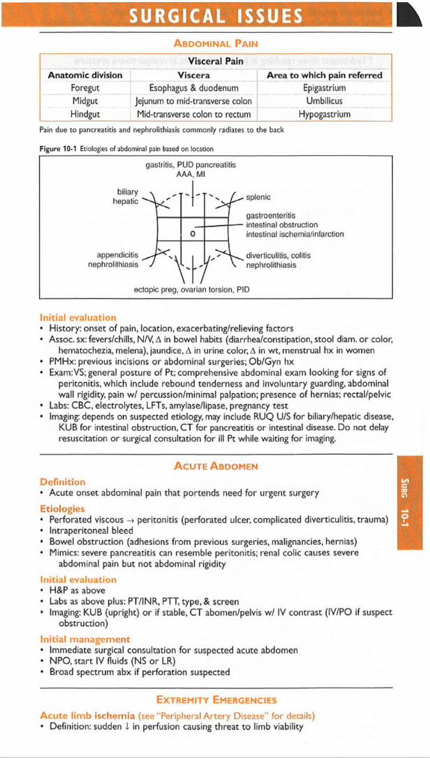
SURGICAL ISSUESA b d o m in a l P a in
Visceral PainAnatom ic division Viscera Area to which pain referred
Foregut Esophagus & duodenum EpigastriumMidgut Jejunum to mid-transverse colon UmbilicusHindgut Mid-transverse colon to rectum Hypogastrium
Pain due to pancreatitis and nephrolithiasis commonly radiates to the back
Figure 10-1 Etiologies of abdominal pain based on location
gastritis, PUD pancreatitis AAA, M l
biliaryhepatic
appendicitisnephrolithiasis - r -
splenic
gastroenteritis intestinal obstruction intestinal ischemia/infarction
diverticulitis, colitis nephrolithiasis
ectopic preg, ovarian torsion, PID
In itia l eva luation• History: onset of pain, location, exacerbating/relieving factors• Assoc, sx: fevers/chills, N/V, A in bowel habits (diarrhea/constipation, stool diam. o r color,
hematochezia, melena), jaundice, A in urine color, A in wt, menstrual hx in women• PMHx: previous incisions or abdominal surgeries; Ob/Gyn hx• Exam:V$; general posture o f Pt; comprehensive abdominal exam looking for signs of
peritonitis, which include rebound tenderness and involuntary guarding, abdominal wall rigidity, pain w/ percussion/minimal palpation; presence of hernias; rectal/pelvic
• Labs: CBC, electrolytes, LFTs, amylase/lipase, pregnancy test• Imaging: depends on suspected etiology, may include RUQ U/S for biliary/hepatic disease,
KUB for intestinal obstruction, CT for pancreatitis or intestinal disease. Do not delay resuscitation or surgical consultation for ill Pt while waiting for imaging.
A c u t e A b d o m e n
D e fin it io n• Acute onset abdominal pain that portends need for urgent surgery
Etiologies• Perforated viscous -> peritonitis (perforated ulcer, complicated diverticulitis, trauma)• Intraperitoneal bleed• Bowel obstruction (adhesions from previous surgeries, malignancies, hernias)• Mimics: severe pancreatitis can resemble peritonitis; renal colic causes severe
abdominal pain but not abdominal rigidity
Initial evaluation• H&P as above• Labs as above plus: PT/INR, PTT, type, & screen• Imaging: KUB (upright) o r if stable, CT abomen/pelvis w / IV contrast (IV/PO if suspect
obstruction)
Initial m anagem ent• Immediate surgical consultation for suspected acute abdomen• NPO, start IV fluids (NS or LR)• Broad spectrum abx if perforation suspected
E x t r e m it y E m e r g e n c ie s
A cu te lim b ischem ia (see "Peripheral A rte ry Disease” for details) • Definition: sudden i in perfusion causing threat to limb viability
SURG 10-1

Sur
g
10-2
• Evaluation: detailed vascular exam; CT angiography o r arteriography• Initial management: anticoag for embolism/thrombosis; immediate surgical consultation
C om partm ent syndrome (Clin Orthop Relat Res 2010:468:940)• Definition: T intracompartmental pressure w/ compressive closure of venules ->
T hydrostatic force resulting in further increases in compartment pressure• Etiologies: orthopedic (fracture), vascular (ischemia-reperfusion), iatrogenic (eg, vascular
injury in anticoagulated Pt), soft tissue injury (eg, prolonged limb compression)• Clinical manifestations: pain esp. on passive movement, swollen/tense compartment,
paraesthesia, pallor, pulselessness, paralysis (late)• Evaluation: surgical evaluation of compartment pressures; intracompartment pressure >30
or difference between diastolic & intracompartment pressure of >10-30 is diagnostic• Treatment: fasciotomy
SurgicalT ubes, D rains,W ounds
Tracheostomy (Otolaryngol Head Neck Surg 2013;148:6)• Typically a cuffed tube, which creates a tight seal to facilitate ventilation throughout tube• Speaking valve (eg, Passy-Muir): 1-way valve that allows inhalation through tube, but
exhalation around tube through vocal cords (nb, cuff should not be inflated)• 1st routine tube A fo r percutaneously placed tubes should be -1 0 d postop; surgically
placed tubes can be A’d >5 d postop; first A should be overseen by experienced person• Accidental dislodgcment: intubate from above (if airway/vent nec & anatomically possible)
w/in 7 d of placement: emergent surgical consultation>7 d after placement: replace with a similar size tube o r smaller
Chest tubes (EurJ Cardiothorac Surg 2011 ;40:291)• Inserted for PTX, chest trauma or after thoracic surg for drainage of air/ fluid from thoracic
cavity. Range from small (8-10 Fr for spont PTX) to large (28-32 Fr after pulm resections)• Connected to 3-chamber chest drainage system:
1st: collection chamber for pleural fluid2' d: water seal chamber used to allow air to exit pleural space on exhalation and
prevent air from entering on inhalation 3rd: suction control chamber which regulates suction transmitted to pleural space
• Monitor for output and presence of air leak (indicated by bubbling in water seal chamber)• Removal determined by overall daily outputs and presence of air leak• If accidentally removed or dislodged, tube should be completely removed and an occlusive
dressing (eg, 4 x 4 covered w/Tegaderm or silk tape) should be placed rapidly over site. CXR STAT; new tube should be placed if persistent PTX.
Gastrostomy/jejunostomy tubes (Paediotr Child Health 2011:16:281)• Placed for tube feedings, hydration and delivery of medications• Securely anchor to skin to prevent inadvertent removal• Should not be removed for >6-8 wk to allow establishment of mature gastrocutaneous tract• Obstructed tubes can be cleared by flushing with agents such as carbonated water, meat
tenderizer, & pancreatic enzymes. 1 obstruction by flushing before & after meds and flushing q4-6h when receiving continuous feeds.
• Inadvertent removal: place Foley catheter of similar size o r smaller into tract immediatelyto prevent stoma from closing.Tube then replaced and confirmed via fluoro study.
Suture/staple removal• Should be done in consultation w/ surgical team; timing depends on location of wound• Should not be removed if there is evidence o f wound separation during removal!• A fter removal, wound should be reapproximated w/ Steri-Strips
Decubitus Ulcers (J Wound Ostomy Continence Nun 2012;39:3)• Sores in dependent areas exposed to repeated pressure (commonly sacrum, heels)• Risk factors: immobility, poor nutritional status• Stage I (non-blanchable erythema); Stage II (partial thickness); Stage III (full thickness skin
loss); Stage IV (full thickness tissue loss)• Treatment: offload area, air mattress, pillows and/or support boots• Surgical consultation for debridement of ulcers with necrotic or infected tissue, may require
plastic surgical reconstruction for advanced ulcers once clean• Wound vac (negative pressure vacuum dressing) therapy may accelerate healing * •
Maximizing a Surgical Consult
• For ill Pt, call surgical consult early, do not wait for labs & imaging results• If potential surgical emergency, make Pt NPO, start IVF, / coags, type, & screen• Have appropriate-level MD who knows & has examined Pt call consult

OB/GYN ISSUESVaginal Bleeding
Abnormal bleeding from lower (vulva, vagina, cervix) or upper genital tract (uterus)
Etiologies• Premenopausal
Not pregnant menses, dysfunctional uterine bleeding (menorrhagia), leiomyoma, polyp, trauma, cervical dysplasia/cancer (rare), endometrial hyperplasia/cancer (rare)
Pregnant1st trimester: threatened abortion, spont. abortion (missed, incomplete or complete),
ectopic pregnancy, molar pregnancy (partial or complete hydatidiform mole)2nd or 3,d trimester: preterm labor, placenta previa, placental abruption
• Postmenopausal: atrophy, polyp, leiomyoma, endometrial hyperplasia/cancer, cervicaldysplasia/cancer
History & exam• Age, menopausal status, gestational age if preg; volume & duration of current bleeding• If premenopausal: menstrual hx including age of onset, interval between & duration of
menses, any assoc, sx and LMP to assess timing of menstrual cycle• Past Ob/Gyn hx (any structural abnl, STD, and contraception)• Health maint. (Pap smear, HPV screening); domestic violence; anticoag or antipit meds• General physical & abdominal exam (incl. tenderness, masses)• Pelvic exam: external (quantity of bleeding seen on vulva, any lesions, any trauma); also,
w/ assistance from Ob/Gyn, speculum exam (quantity of bleeding; cervical os open or close and if open, dilation; any polyps), & bimanual exam (uterine size and tenderness, adnexal mass and tenderness)
Laboratory evaluation & imaging• Urine (rapid test) & serum pregnancy test (beta-hCG); Hct/hemoglobin• Pelvic U/S: visualize intrauterine preg to r/o ectopic; if preg, intrauterine not seen, &
PHCG > discrim, zone ? ectopic; if pHCG < discrim, zone -» follow (3HCG; nl placental position to r/o placenta previa and likely severe abruption
• Ectopic pregnancy is life-threatening dx, /. must rule out i f Pt pregnant (JAMA 2013:309:1722)
V aginal D ischarge Fluid or mucus from vagina, cervix, or uterus
Etiologies• In fe c tio u s : b a c te r ia l v a g in o s is , C and ida v u lv o v a g in it is , t r ic h o m o n ia s is
• Noninfectious: physiologic (in preg or non-preg), rupture of membranes, foreign-body rxn
Initial evaluation• Age, LMP, gestational age if preg. o r menopausal status• Discharge quantity, color, consistency, odor, assoc, sx (itchiness, redness, abd/pelvic pain)• Past gyn hx incl STD and contraception usage (condoms i STD risk)• Tampon or condom use as risk factors for retained foreign body• Pelvic exam: external (quantity & quality of discharge on vulva, any lesions);
speculum (discharge, appearance of cervix), bimanual (cervical motion tenderness)• Laboratory: pH of discharge; microscopy (saline & KOH wet mounts); urine pregnancy test
Treatm ent• Bacterial vaginosis: oral o r vaginal metronidazole o r clindamycin• Candida vulvovaginitis: oral or topical antimycotic medications• Trichomoniasis: oral metronidazole •
A dnexal Mass in N on-Pregnant W oman
Mass arising from ovary, fallopian tube, or surrounding connective tissue
Etiologies• Ovarian: functional (follicular and corpus luteum) or hemorrhagic cyst, endometriomas.
ovarian torsion, tubo-ovarian abscess, benign & malignant ovarian tumors• Fallopian tube: paratubal cyst, hydrosalpinx, ovarian torsion, tubo-ovarian abscess
Initial evaluation• LMP/menopausal status; associated sx of abd/pelvic pain, FHx of gyn cancers• Abd exam (distension, tenderness, masses); bimanual (uterine or adnexal masses)• Preg test if premenopausal (if ©, then mass likely pregnancy); CA-125 if postmenopausal• Pelvic U/S (even if mass 1st identified on CT, as U/S is best modality); U/S appearance of
mass most important factor used to determine risk of malignancy

Op
hth
alm
ic
10-4
OPHTHALMIC ISSUESInitial Evaluation
• Ocular symptom: onset (sudden or progressive) & duration of sx; unilateral vs. bilateral;pain; photophobia; discharge; A in near (eg, book) o r far (eg,TV across room) vision
• Pre-existing ocular conditions, eye meds (incl any As), recent h/o ocular surgery, trauma• Ocular exam: vision ( / with Pt’s correction [glasses/contacts]) w/ each eye; pupillary exam;
EOM; confrontation visual fields (important if suspect CNS problem)• OveralLVS, immunocomp., s/s of infxn, h/o malig, CNS issues, A in meds, CBC, coags
Common V isual Symptoms
• Fluctuation in vision (ie, blurry): med-induced refractive error (eg, systemicsteroids, chemoRx), hyperglycemia, dry eye (common). Visual defect may p/w "blurred vision" Bilateral:glaucoma (common), homonymous contral.CNS lesion; bitemporal: pituitary, toxic/nutritional. Unilateral: ipsilateral orbital, retinal, or optic nerve lesion.
• Red eye:Bilateral: viral conjunct., (starts in 1 eye; also w/ lid swelling, discharge); chronic
inflammation (dry eyes, rosacea, autoimmune disease)Unilateral: subconj. hemorrhage, infxn, o r inflam (eg, episcleritis, iritis, uveitis, scleritis);
acute angle closure (qv). Scleritis & acute angle closure p/w severe pain, H/A, nausea.• Double vision (diplopia): fixed double vision w/ ophthalmoplegia from orbital
process or cranial nerve palsy (III, IV,VI).Transient "diplopia” due to fatigue or sedation.• Flashing lights/floaters: vitreous detach, (common, benign); retinal detach, (unilateral
visual field defect; urgent ophthalmology consult); hemorrhage; intraocular lymphoma
A cute V isual C hanges
Etiologies of Acute Vision Loss (italics indicates o/w pain)Unilateral Bilateral
Transient(<24 h, often <1 h)
Ret. art. embolism, impending retinal artery or vein occlusion (amaurosis fugax), vasospasm, carotid disease
Ocular surface dis. (dry eye), bilat. carotid dis.,TIA, migraine, high ICP (papilledema)
Prolonged(>24 h)
Retinal art/vein ocd, retinal detach., retina/vitreous heme, retinitis, ant. optic neurop./corneof ulcer, GCA, acute angle closure glaucoma
Visual cortex stroke, post, ischemic neuropathy (profound hypotension during surgery), post, reversible enceph. synd., GCA
Common O c u la r C ond itions (fr o n t t o back)
Orbit: orbital cellulitis (fever, proptosis, i EOM; emergent obx & referral)Lids: hordeolum or chalazion (stye); preseptal cellulitis; ptosis (age; Horner’s; C N III palsy:
EOM restricted in all directions except laterally [eye is “ down & out” ],a /w ptosis & mydriasis, seen w/ uncal herniation, aneurysm of post com art., GCA, HTN, DM); incomplete lid closure (C N 7th palsy)
Conjunctiva: conjunctivitis (red eye); subconj. hemorrhage (HTN, blood thinner); ocular surface disease (dry eyes); episcleritis/scleritis (deep vessels of sclera)
Cornea: contact lens related ulcer; herpetic keratitis/scarring/neurotropic ulcers (C N V paresis); pterygium; keratoconus; corneal dystrophy
Ant. chamber: iritis (inflam, cells); hyphema (blood, post trauma); hypopyon (inflam./infxn) Pupil: Anisocoria (physiologic); Horner’s, CN IIILens: cataract (age, trauma, medication, radiation, congenital); post cataract surgery infxn Vitreous/Retina/Macula: diabetic retinopathy; macular degen; retinal detachment; retinal ±
vitreous hemorrhage; retinitis (infectious)Optic nerve (C N II): ischemic neuropathy p/w acute unilat visual loss, altitudinal field defect;
a/w GCA; nonarteritic a/w HTN, hyperchol., DM, thrombophilia. Optic neuritis: often p/w unilat central scotoma, pain w/ EOM.T visual loss over days; a/w demyelinating disease (eg, MS), also seen w/ sarcoidosis & CTD. Optic neuropathy (glaucoma common). * *
O cular Emergencies
* Chemical splash: alkali worse than acid; immediate eye flush; pH 7.3-7.4 normal* Acute angle closure glaucoma: fixed mid-dilated pupil, corneal edema, high intraocular
pressure (typically >50; normal 8-21). Rx w/ topical drops; may require AC tap/laser.* Penetrating eye injury: protect eye (no patching), IV antibiotics, NPO, surgical prep

ICU MEDICATIONSDrug Class
Doseper kg average
Pressors, Inotropes and ChronotropesPhenylephrine <Xi 10-300 pg/minNorepinephrine Oti > Pi 1-40 pg/minVasopressin V! 0.01-0.1 U/min (usually <0.04)Epinephrine CCl,CC2,p1,p2 2-20 pg/min
Isoproterenol Pi. P2 0.1-10 pg/min
DopamineD
P,D a. p, D
0.5-2 pg/kg/min 2-10 pg/kg/min >10 pg/kg/min
50-200 pg/min 200-500 pg/min
500-1000 pg/minDobutamine Pi > Pi 2-20 pg/kg/min 50-1000 pg/min
Milrinone PDE± 50 pg/kg over 10 min then
0.25-0.75 pg/kg/min3-4 mg over 10 min then 20-50 pg/min
VasodilatorsNitroglycerin NO 10-1000 pg/minNitroprusside NO 0.25-10 pg/kg/min 10-800 pg/min
Labetalol a i. Pi and p2 blocker
20-80 mg qlOmin or 10-120 mg/h
Fenoldopam D 0.1-1.6 ug/kg/min 10-120 pg/minClevidipine CCB 1-16 mg/hEpoprostenol vasodilator 2-20 ng/kg/min
Antiarrhythmics
Amiodarone K et at. (Class III)
150 mg over 10 min, then 1 mg/min x 6 h, then 0.5 mg/min x 18 h
Lidocaine Na channel (Class IB)
1-1.5 mg/kg then 1-4 mg/min
100 mg then 1-4 mg/min
Procainamide Na channel (Class IA)
17 mg/kg over 60 min then 1-4 mg/min
1 g over 60 min then 1-4 mg/min
Ibutilide K channel (Class III)
1 mg over 1< may repeat
3 min, x 1
Propranolol P blocker 0.5-1 mg q5min then 1-10 mg/h
Esmolol Pi > P2blocker
500 pg/kg then 50-200 pg/kg/min
20-40 mg over 1 min then 2-20 mg/min
Verapamil CCB 2.5-5 mg over 1-2', repeat 5-10 mg in 15-30' prn 5-20 mg/h
Diltiazem CCB0.25 mg/kg over 2 min
reload 0.35 mg/kg x 1 prn then 5-15 mg/h
20 mg over 2 min reload 25 mg x 1 prn
then 5-15 mg/hAdenosine purinergic 6 mg rapid push; if no response: 12 mg -> 12-18 mg
SedationMorphine opioid 1-30 (in theory, unlimited) mg/hFentanyl opioid 50-100 ug then 50-800 (? unlimited) pg/h
Propofol anesthetic 1-3 mg/kg then 0.3-5 mg/kg/h
50-200 mg then 20-400 mg/h
Dexmedetomidine 0.2 agonist 1 pg/kg over 10 min -> 0.2-0.7 ug/kg/hDiazepam BDZ 1-5 mg q1-2h then q6h prnMidazolam BDZ 0.5-2 mg q5min prn; 0.02-0.1 mg/kg/h or 1-10 mg/hLorazepam BDZ 0.01-0.1 mg/kg/hNaloxone opioid antag. 0.4-2 mg q2-3min to total of 10 mg
Flumazenil BDZ antag. 0.2 mg over 30 sec then 0.3 mg over 30 sec prn may repeat 0.5 mg over 30 sec to total of 3 mg

ICU
Med
s 11
-2Drug Class
Doseper kg average
/Vl/sce//aneous
Aminophylline PDE 5.5 mg/kg over 20 min 250-500 mg then 0.5-1 mg/kg/h then 10-80 mg/h
Octreotidesomatostatin
analog 50 jig then 50 jjg/h
Glucagon hormone 3-10 mg IV slowly over 3-5 min then 3-5 mg/h
Mannitol osmole 1.5-2 g/kg over 30-60 min repeat q6-12h to keep osm 310-320
Figure 11-1 ACLS pulmonary edema, hypotension or shock algorithm
Acute Pulmonary Edema, Hypotension or Shock
IABCs, IV Access, 0 2,12-lead ECG, focused H&P, CXR
/ I \W h a t i s t h e n a t u r e o f t h e p r o b l e m ?
S ' I N*Volume Purnp/Vasc Rateproblem problem problem
I I IFluids and /or blood W hat is BP? Go to tachycardia or
Vasopressors as needed while (after empiric 500 cc NS bradycardia algorithm
volume resuscitating bolus unless in HF)I
look lor signs of organ hypoperfusion
f \
r i r ▼
I f i n p u l m o n a r y e d e m a , c o n s i d e r :I______ IFurosem ide 0.5-1.0 mg/kg IV
M orphine 2-4 mg IV O xygen/noninvasive vent./intub.
further interventions based on etiology
(Adapted from ACLS 2005 Guidelines)

ANTIBIOTICSThe following tables of spectra o f activity for different antibiotics are generalizations.
Sensitivity data at your own institution should be used to guide therapy.
PenicillinsGeneration Properties SpectrumNatural(eg. penicillin)
Some GPC, GPR, GNC, most anaerobes (except 8<?cteroides)
Group A streptococci Enterococci, Listeria, Pasteurella Actinomyces, Syphilis
Anti-staph(eg, nafcillin)
Active vs. PCNase-producing Staph Little activity vs. Gram ©
Staphylococci (except MRSA) Streptococci
Amino(eg, ampicillin)
Penetrate porin channel of Gram © Not stable against PCNases
£. coli, Proteus, H. influenzae Salmonella, Shigella Enterococci, Listeria
Extended(eg, piperacillin)
Penetrate porin channel of Gram © More resistant to PCNases
Most GNR incl. Enterobacter, Pseudomonas, Serratia
Carbapenems(eg, imipenem)
Resistant to most P-lactamases Most Gram © & ©, including anaerobes; not MRSA or VRE
Monobactams(aztreonam)
Active vs. Gram 0 but not Gram © Gram © bacterial infxn in Pt w/ PCN or Ceph allergy
p-lact. Inhib.(eg, sulbactam)
Inhibit plasma-mediated p-lactamases Adds staph, 8. fragilis & some GNR (H. flu, M. cot, some Klebs); intrinsic activity against Acinetobacter (sulbactam only)
Cephalosporins
Resistant to most p-lactamases. No activity vs. enterococci.Gen. Spectrum Indications1st(eg, cefazolin)
Most GPC (incl. staph & strep, not MRSA) Some GNR (incl. E. coli, Proteus, Klebsiella)
Used for surgical Ppx & skin infxns
2"d (eg.cefuroxime,cefotetan)
1 activity vs. GPC, T vs. GNR. 2 subgroups: Respiratory: H. influenzae & M. catorrha//s GI/GU: T activity vs. 8. /rag<7/s
PNA/COPD flare Abdominal infxns
3rd (eg,ceftriaxone)
Broad activity vs. GNR & some anaerobes Ceftazidime active vs. Pseudomonas
PNA, sepsis, meningitis
4th(eg, cefepime)
T resistance to p-lactamases (incl. of staph and Enterobacter)
Similar to 3rd gen. MonoRx for nonlocalizing febrile neutropenia
5th (eg,ceftaroline)
Only class of cephalosporin with MRSA activity. NOT active vs Pseudomonas
MRSA. Not 1st line for MRSA bacteremia.
Other AntibioticsAntibiotic SpectrumVancomycin Gram © bacteria incl. MRSA, PCNase-producing pneumococci and
enterococci (except VRE)LinezolidDaptomycin GPC incl. MRSA &VRE (check susceptibility for VRE)Quinupristin/DalfopristinQuinolones Enteric GNR & atypicals. 3rd & 4th gen. T activity vs. Gram ©.Aminoglycosides GNR. Synergy w/ cell-wall active abx (P-lactam, vanco) vs. GPC.
i activity in low pH (eg, abscess). No activity vs. anaerobes.Macrolides GPC, some respiratory Gram ©, atypicalsTMP/SMX Some enteric GNR, PCP, Nocardia,Toxoplasma, most community-
acquired MRSAClindamycin Most Gram © (except enterococci) & anaerobes (incl. 8. fragilis)Metronidazole Almost all anaerobic Gram ©, most anaerobic Gram ©Doxycycline Rickettsio, Ehrlichia, Anaplasma, Chlamydia, Mycoplasma, Nocardia, LymeTigecycline Many GPC incl. MRSA & VRE; some GNR incl. ESBL but not
Pseudomonas or Proteus. Approved for abdominal or skin/soft tissue infections. Check susceptibility if organism isolated.

FORMULAE AND QUICK REFERENCE 1
C a r d io l o g y
Hemodynamic parameters Normal value
M . . . /m a d\ SBP + ( D B P x 2 ) Mean arterial pressure (MAP) = ---------- 70-100 mmHg
Heart rate (HR) 60-100 bpmRight atrial pressure (RA) <6 mmHg
Right ventricular (RV) systolic 15-30 mmHg diastolic 1-8 mmHg
Pulmonary artery (PA)systolic 15-30 mmHg
mean 9-18 mmHg diastolic 6-12 mmHg
Pulmonary capillary wedge pressure (PCWP) <12 mmHgCardiac output (CO) 4-8 L/min
COCardiac index (Cl) = ------
BSA2.6^1.2 L/min/m2
Stroke volume (SV) =HR
60-120 mL/contraction
ClStroke volume index (SVI) = -----
HR40-50 ml_/contraction/m2
Systemic vascular resistance (SVR)MAP - mean RA
= -------------------------- x 80CO
800-1200 dynes x sec/cm5
Pulmonary vascular resistance (PVR)mean PA - mean PCWP
= ---------------------------------------x 80CO
120-250 dynes x sec/cms
“ Rule of 6s” for PAC: RA <6, RV <30/6, PA <30/12,WP <12. Nb 1 mmHg = 1.36 cm water or blood.
Ficl< cardiac outputOxygen consumption (L/min) = CO (L/min) x arteriovenous (AV) oxygen difference CO = oxygen consumption/AV oxygen differenceOxygen consumption must be measured (can estimate w / 125 mL/min/m2, but inaccurate) AV oxygen difference = Hb (g/dL) x 10 (dL/L) x 1.36 (mL 0 2 /g of Hb) x (Sa02-SMv02)
Sa0 2 is measured in any arterial sample (usually 93-98%)Smv0 2 (mixed venous 0 2) is measured in RA, RV or PA (assuming no shunt) (nl - 75%)
.-. C a r d ia c o u tp u t (L/min) = ____ Oxygen consum ption____Hb (g/dL) x 13.6 (Sa0 2 - S v0 2)
Shunts- Oxygen consum ptionQ p = ----------------------------------------- (if no R -» L shunt, PV 0 2 sat - Sa0 2)
Pulm. vein 0 2 sat - Pulm. arte ry 0 2 sat
Q t = -----O xygen consum ption— (M V 02 drawn proximal to potential L R shunt)Sa0 2 — mixed venous 0 2 sat
Qp _ SaP 2 - MV O 2 sat ^ S3Q 2 - M V O .sa t jf Qn|y l R and no R -> L shunt) Q s PV 0 2 sat - PA 0 2sat Sa0 2 - P A 0 2sat
V a lve e q u a tio n sSimplified Bernoulli: Pressure gradient (AP) = 4 x v2 (where v = peak flow velocity) Continuity (conservation of flow): Areai xVelocityi = A2 xV 2 (where 1 & 2 different points)
o r AVA (unknown) = A LV outflow x (all of which can be measured on echo)' Aov y
/- 1. . w , CO/(DEP o r SEP) x HR . , , . . . .Gorlin equation: Valve area = — - ------------------ — - j= - (constant = 1 for AS, 0.85 for MS)
Hakki equation: Valve area =
44.3 xcons tan t x n/ a P CO
Va p

P u l m o n a r y
Chest Imaging (CXR & CT) PatternsPattern Pathophysiology DdxConsolidation Radiopaque material in air
space & interstitium patent airway ->"air bronchograms"
Acute: water (pulm edema), pus (PNA), bloodChronic: neoplasm (BAC, lymphoma), aspiration,
inflammatory (BOOR eosinophilic PNA), PAR granuloma (TB/fungal, alveolar sarcoid)
Ground glass (CT easier than CXR)
Interstitial thickening or partial filling of alveoli (but vessels visible)
Acute: pulm edema, infxn (PCP, viral, resolving bact. PNA)
Chronic: ILD w/o fibrosis: acute hypersens., DIP/RB, PAP w/ fibrosis: IPF
Septal lines Kerley A & B
Radiopaque material in septae
Cardiogenic pulm edema, interstitial PNA viral, mycoplasma), lymphangitic tumor
Reticular Lace-like net (ILD) ILD (esp. IPF, CVD, bleomycin, asbestos)Nodules Tumor
GranulomasAbscess
Cavitary: Primary or metastatic cancer.TB (react, or miliary), fungus,Wegener’s, RA septic emboli, PNA
Noncawwry: any of above + sarcoid, hypersens, pneum., HIV, Kaposi’s sarcoma
Wedge opac. Peripheral infarct PE, cocaine, angioinv. aspergillus,Wegener’sTree-in-bud (best on CT)
Inflammation of small airways
Bronchopneumonia, endobronchial TB/MAI, viral PNA, aspiration, ABPA, CF, asthma, BOOP
Hilar fullness T LN or pulm arteries Neoplasm (lung, mets, lymphoma)Infxn (AIDS); Granuloma (sarcoid/TB/fungal) Pulmonary hypertension
Upper lobe n/a TB, fungal, sarcoid, hypersens, pneum., CF, XRTLower lobe n/a Aspiration, bronchiect., IPF, RA, SLE, asbestosPeripheral n/a BOOR IPF & DIR eos PNA, asbestosis
C X R in h e a rt fa ilu re• T cardiac silhouette (in systolic dysfxn, not in diastolic)• Pulmonary venous hypertension: cephalization of vessels (vessels size > bronchi in
upper lobes), peribronchial cuffing (fluid around bronchi seen on end small circles), Kerley B lines (horizontal 1-2-cm lines at bases), T vascular pedicle width, loss of sharp vascular margins, pleural effusions (-75% bilateral)
• Pulmonary edema: ranges from ground glass to consolidation; often dependentand central, sparing outer third ("bat wing" appearance)
Dead space = lung units that are ventilated but not perfused Intrapulm onary shunt = lung units that are perfused but not ventilated
P COAlveolar gas equation: PA0 2 = [F ,02 x (760 - 47)] - -- ----- - (where R * 0.8)
pao 2 = 150-P,CP2
0.8(on room air)
A-a gradient = Pa02 - Pa0 2 [normal A-a gradient ~ 4 + (age/4)]Minute ventilation (Ve) = tidal volume (V j) x respiratory rate (RR)(nl 4-6 L/min) Tidal volume ( V t ) = alveolar space (VA) + dead space (VD)
Fraction of tidal volume that is dead space( V )U J
PaC Q 2 — PeXpiredCQ2p , c o 2
PaC 0 2 - k - xC 0 2 Production
alveolar vent:lation= k x 'C O }
(R R x Vt x 1 - V o "
VtJ

N e p h r o l o g y
/
Anion gap (AG) = Na - (Cl + HCO 3) (normal = [alb] x 2.5; typically 12 ± 2 mEq) Delta-delta (AA) = [A AG (ie.calc. AG - expected) / A HCO3 (ie,24 - measured HCO3)] Urine anion gap (UAG) = ( U n e + U k ) - Uci
Calculated osmoles = (2 x Na) +gic^ r b u n '118 j + l 2 . 8 ,
EtOH \
4.6 JOsmolal gap (O G ) = measured osmoles - calculated osmoles (normal <10)
Estimated creatinine clearance = X w t (x 0.85 in women)serum C r (mg/dL) X 72
Fractional excretion of Na (FEne, %) =
UNa(mEq/L)
PNa(mEq/L)x 1 0 0 %
UCr(mg/mL)
. Pcr(mg/dL)x 100 (mL/dL)
Corrected Na in hyperglycemia
estimate in all Pts: corrected Na = measured Na + 2.4 x(measured glc - 1 0 0 )
100however, A in Na depends on glc {Am J Med 1999;106:399)A is 1.6 mEq per each 100 mg/dL T in glc ranging from 100-440 A is 4 mEq per each 100 mg/dL t in glc beyond 440Total body w ater (TBW) - 0.60 x IBW (x 0.85 if female and x 0.85 if elderly)
Free H 2Q deficit = TB W x[N a]itrt,m *140 ^ ( [N a ]seru[ll 1 4 0 1 70-kg Pt)
140
Peripheral Smear Findings (also see Photo Inserts)Feature Abnormalities and diagnosesSize normocytic vs. microcytic vs. macrocytic -> see belowShape anisocytosis -> unequal RBC size; poikilocytosis irregular RBC shape
acanthocytes = spur cells (irregular sharp projections) -> liver disease bite cells (removal of Heinz bodies by phagocytes) -> G6 PD deficiency
echinocytes = burr cells (even, regular projections) -> uremia, artifact pencil cell -» long, thin, hypochromic - very common in adv. iron deficiency rouleaux -» hyperglobulinemia (eg, multiple myeloma) schistocytes, helmet cells -* MAHA (eg. DIC.TTP/HUS), mechanical valve spherocytes -> HS.AIHA; sickle cells -> sickle cell anemia stomatocyte -» central pallor appears as curved slit -> liver disease, EtOH target cells -> liver disease, hemoglobinopathies, splenectomy tear drop cells = dacryocytes -* myelofibrosis, myelophthisic anemia,
megaloblastic anemia, thalassemiaIntra-RBCfindings
basophilic stippling (ribosomes) -> abnl Hb, sideroblastic, megaloblastic Heinz bodies (denatured Hb) -» G6 PD deficiency, thalassemia Howell-Jolly bodies (nuclear fragments) - » splenectomy or functional
asplenia (eg advanced sickle cell) nucleated RBCs -> hemolysis, extramedullary hematopoiesis
W BCfindings
blasts - » leukemia, lymphoma; Auer rods -» acute myelogenous leukemia hypersegmented (>5 lobes) PMNs: megaloblastic anemia (B^/folate def.) pseudo-Pelger-Huet anomaly (bibbed nucleus,“ pince-nez") MDS toxic granules (coarse, dark blue) and Dohle bodies (blue patches of dilated
endoplasmic reticulum) (sepsis, severe inflammationPlatelet clumping artifact, repeat pit count
# periph blood pit count -10,000 pit for every 1 pit seen at hpf (lOOx) size -» MPV (mean platelet volume) enlarged in ITP
(NEJM 2005:353:498)

Heparin for Thromboembolism80 U/kg bolus
18 U/kg/hPTT Adjustment
A -U o bolus 5000 U, T rate 300 U/h40-49 bolus 3000 U, T rate 200 U/h50-59 T rate 150 U/h60-85 no A86-95 I rate 100 U/h
96-120 hold 30 min, i rate 100 U/h>120 hold 60 min, i rate 150 U/h
(Modified from Chest 2008;133:141S)
Heparin for ACS60 U/kg bolus (max 4000 U) 12 U/kg/h (max 1000 U/h)
PTT Adjustment<40 bolus 3000 U,T rate 100 U/h
40-49 T rate 100 U/h50-75 no A76-85 I rate 100 U/h
86-100 hold 30 min, i rate 100 U/h>100 hold 60 min, I rate 200 U/h
(Modified from Circ 2007; 116:e148 & Chest 2008:133:670)
/ PTT q6h after every A (t& of heparin ~90 min) and then qd or bid once PTT is therapeutic / CBC qd (to ensure Hct and pit counts are stable)
Warfarin Loading Nomogram
Day INR<1.5 1.5-1.9 2-2.5 2.6-3 >3
1-3 5 mg (7.5 mg if >80 kg) 2.5-5 mg 0-2.5 mg 0 mg4-5 10 mg 5-10 mg 0-5 mg 0-2.5 mg6 Dose based on requirements over preceding 5 d
(AnnoJs 1997; 126:133;Archives 1999; 159:46) or.go t o www.warfarindosing.org
W a rfa r in -h e p a rin overlap the rapy• Indications: when failure to anticoagulate carries T risk of morbidity or mortality
(eg, DVT/PE, intracardiac thrombus)• Rationale: (1) Half-life of factor VII (3-6 h) is shorter than half-life of factor II (60-72 h);
warfarin can elevate PT before achieving a true antithrombotic stote (2) Protein C also has half-life less than that of factor II;,\ theoretical concern of hypercoagulable state before antithrombotic state
• Method: (1) Therapeutic PTT is achieved using heparin(2) Warfarin therapy is initiated(3) Heparin continued until INR therapeutic for >2 d and >4-5 d of warfarin (roughly corresponds to -2 half-lives of factor II o r a reduction to -25%)
Common Warfarin-Drug InteractionsDrugs that t PT Drugs that 1 PTAmiodaroneAntimicrobials: erythromycin, ? darithro,
ciprofloxacin, MNZ, sulfonamides Antifungals: azolesAcetaminophen, cimetidine, levothyroxine
Antimicrobials: rifampin CNS: barbiturates, carbamazepine,
phenytoin (initial transient T PT) Cholestyramine
O t h e r
Ideal body weight (IBW) = [50 kg (men) o r 45.5 kg (women)] + 2.3 kg/inch over 5 feet
Body surface area (BSA, m2) = (crn) x w eight (kg)V 3600
Diseasepresent absent
Test© a
(true 0)b
(false ©)
0 c(false 0)
d(true 0)
true positives a _ , true negatives HSensitivity = ---------------- = ------------ S pecific ity - -5------- = —2!__
all diseased a + c all healthy b + d
© Predictive value = true P0sitives = _ L _all positives a + b
^ , . true negatives d© Predictive value = ----------------------= ---------
all negatives c + d

ABBREVIATIONSS'-NT 5'-nucleotidase6-MP 6-mercaptopurinea/w associated withAAA abdominal aortic aneurysmAAD antiarrhythmic drugAb antibodyABE acute bacterial endocarditisABG arterial blood gasabnl abnormalABPA allergic bronchopulmonary
aspergillosisabx antibioticsAC assist controlACE angiotensin-converting enzymeACEI ACE inhibitorACI anemia of chronic inflammationACL anticardiolipin antibodyACLS advanced cardiac life supportACS acute coronary syndromeACTH adrenocorticotrophic hormoneACV acyclovirADA adenosine deaminaseADH antidiuretic hormoneADL activities of daily livingAF atrial fibrillationAFB acid-fast bacilliAFL atrial flutterAFP a-feto proteinAFTP ascites fluid total proteinAG aminoglycoside anion gapAg antigenAGN acute glomerulonephritisAl aortic insufficiency
aromatase inhibitorAIDS acquired immunodefic. synd.AIH autoimmune hepatitisAIHA autoimmune hemolytic anemiaAIN acute interstitial nephritisAIP acute interstitial pneumoniaAKI acute kidney injuryALF acute liver failureALL acute lymphoblastic leukemiaALS amyotrophic lateral sclerosisALT alanine aminotransferaseAMA anti-mitochondrial antibodyAMI anterior myocardial infarctionAML acute myelogenous leukemiaa my amylaseANA antinuclear antibodyANCA antineutrophilic cytoplasmic AbAoD aortic dissectionAoV aortic valveAPAP acetyl-para-aminophenolAPC activated protein CAPL acute promyelocytic leukemiaAPLA antiphospholipid AbAPS antiphospholipid Ab synd.ARB angiotensin receptor blockerARDS acute resp distress synd.ARV antiretroviralARVC arrhythmogenic RV CMPAS aortic stenosisASA aspirinASD atrial septal defectAST aspartate aminotransferaseasx asymptomaticAT atrial tachycardiaATM angiotensin IIATIII antithrombin IIIATN acute tubular necrosisATRA all-trans-retinoic acidAV atrioventricularAVA aortic valve area
AVB atrioventricular blockAVNRT AV nodal reentrant tachycardiaAVR aortic valve replacementAVRT AV reciprocating tachycardiaAZA azathioprineA<j> alkaline phosphatase
pB beta-blockerb/c becauseBAL bronchoalveolar lavageBBB bundle branch blockBCx blood cultureBD bile ductBDZ benzodiazepinesbili. bilirubinBiPAP bilevel positive airway pressureBiV biventricularBM bone marrow
bowel movementBMD bone mineral densityBMI body mass indexBMS bare metal stentBNP B-type natriuretic peptideBOOP bronchiolitis obliterans with
organizing pneumoniaBP blood pressureBPH benign prostatic hypertrophyBRBPR bright red blood per rectumBS breath soundsBT bleeding timeBUN blood urea nitrogenbx biopsyBYCE buffered charcoal yeast extract
C' complementc/s consultc/w compared with
consistent withCABG coronary artery bypass graftingCAD coronary artery diseaseCAH congenital adrenal hyperplasiaCALLA common ALL antigenCAPD chronic ambulatory peritoneal
dialysisCBC complete blood countCBD common bile ductCCB calcium channel blockerCCI4 carbon tetrachlorideCCP cyclic citrullinated peptideCCS Canadian Cardiovascular
SocietyCCY cholecystectomyCD Crohn’s diseaseCEA carcinoembryonic antigen
carotid endarterectomyceph. cephalosporinCF cystic fibrosisCftx ceftriaxoneCFU colony forming unitsCHB complete heart blockCHD congenital heart diseaseCHF congestive heart failureCl cardiac indexCIAKI contrast-induced AKICIDP chronic inflammatory
demyelinating polyneuropathy
CJD Creutzfeldt-Jakob diseaseCK creatine kinaseCKD chronic kidney diseaseCLL chronic lymphocytic leukemiaCMC carpometacarpal (joint)CML chronic myelogenous leukemia
Ab
br
ev
12
-1
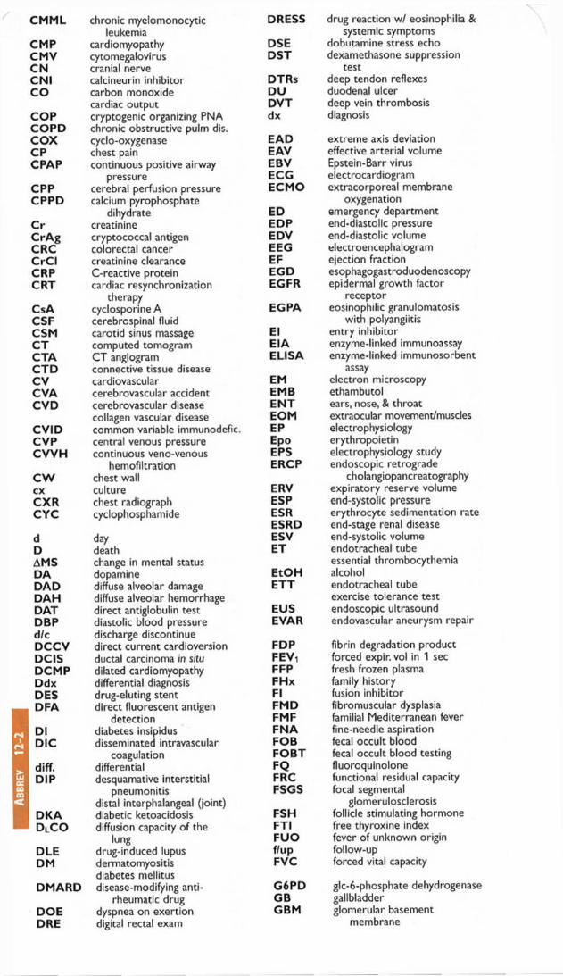
Ab
bre
v
12-2
CMML chronic myelomonocytic leukemia
CMP cardiomyopathyCMV cytomegalovirusCN cranial nerveCNI calcineurin inhibitorCO carbon monoxide
cardiac outputCOP cryptogenic organizing PNACOPD chronic obstructive pulm dis.COX cyclo-oxygenaseCP chest painCPAP continuous positive airway
pressureCPP cerebral perfusion pressureCPPD calcium pyrophosphate
di hydrateCr creatinineCrAg cryptococcal antigenCRC colorectal cancerCrCI creatinine clearanceCRP C-roactive proteinCRT cardiac resynchronization
therapyCsA cyclosporine ACSF cerebrospinal fluidCSM carotid sinus massageCT computed tomogramCTA CT angiogramCTD connective tissue diseaseCV cardiovascularCVA cerebrovascular accidentCVD cerebrovascular disease
collagen vascular diseaseCVID common variable immunodefic.CVP central venous pressureCVVH continuous veno-venous
hemofiltrationC W chest wallcx cultureCXR chest radiographCYC cyclophosphamide
d dayD deathAMS change in mental statusDA dopamineDAD diffuse alveolar damageDAH diffuse alveolar hemorrhageDAT direct antiglobulin testDBP diastolic blood pressuredie discharge discontinueDCCV direct current cardioversionDCIS ductal carcinoma in situDCMP dilated cardiomyopathyDdx differential diagnosisDES drug-eluting stentDFA direct fluorescent antigen
detectionDl diabetes insipidusDIC disseminated intravascular
coagulationdiff. differentialDIP desquamative interstitial
pneumonitis distal interphalangeal (joint)
DKA diabetic ketoacidosisDl CO diffusion capacity of the
lungDLE drug-induced lupusDM dermatomyositis
diabetes mellitusDMARD disease-modifying anti
rheumatic drugDOE dyspnea on exertionDRE digital rectal exam
DRESS drug reaction w/ eosinophilia & systemic symptoms
DSE dobutamine stress echoDST dexamethasone suppression
testDTRs deep tendon reflexesDU duodenal ulcerDVT deep vein thrombosisdx diagnosis
EAD extreme axis deviationEAV effective arterial volumeEBV Epstein-Barr virusECG electrocardiogramECMO extracorporeal membrane
oxygenationED emergency departmentEDP end-diastolic pressureEDV end-diastolic volumeEEG electroencephalogramEF ejection fractionEGD esophagogastroduodenoscopyEGFR epidermal growth factor
receptorEG PA eosinophilic granulomatosis
with polyangiitisEl entry inhibitorEIA enzyme-linked immunoassayELISA enzyme-linked immunosorbent
assayEM electron microscopyEMB ethambutolENT ears, nose, & throatEOM extraocular movement/musclesEP electrophysiologyEpo erythropoietinEPS electrophysiology studyERCP endoscopic retrograde
cholangiopancreatographyERV expiratory reserve volumeESP end-systolic pressureESR erythrocyte sedimentation rateESRD end-stage renal diseaseESV end-systolic volumeET endotracheal tube
essential thrombocythemiaEtOH alcoholETT endotracheal tube
exercise tolerance testEUS endoscopic ultrasoundEVAR endovascular aneurysm repair
FDP fibrin degradation productFEVi forced expir. vol in 1 secFFP fresh frozen plasmaFHx family historyFI fusion inhibitorFMD fibromuscular dysplasiaFMF familial Mediterranean feverFNA fine-needle aspirationFOB fecal occult bloodFOBT fecal occult blood testingFQ fluoroquinoloneFRC functional residual capacityFSGS focal segmental
glomerulosclerosisFSH follicle stimulating hormoneFTI free thyroxine indexFUO fever of unknown originf/up follow-upFVC forced vital capacity
G6PD glc-6-phosphate dehydrogenaseGB gallbladderGBM glomerular basement
membrane
GBS Guillain-Barre syndrome ICa ionized calciumGCA giant cell arteritis ICD implantable cardiac defibrillatorGCS Glasgow coma scale ICH intracranial hemorrhageG-CSF granulocyte colony stimulating ICP intracranial pressure
factor ICU intensive care unitGE gastroesophageal IE infective endocarditisgen. generation IGF insulin-like growth factorGERD gastroesophageal reflux disease IGRA interferon-y release assayGFR glomerular filtration rate II integrase inhibitorGGT y-glutamyl transpeptidase IIP idiopathic interstitial PNAGH growth hormone ILD interstitial lung diseaseGIB gastrointestinal bleed IMI inferior myocardial infarctionGIST gastrointestinal stromal tumor infxn infectionglc glucose inh inhaledGMCSF gra n u locyte -mac roph age INH isoniazid
colony-stimulating factor INR international normalized ratioGN glomerulonephritis IPAA ileal pouch-anal anastomosisGNR gram-negative rods IPF idiopathic pulmonary fibrosisGrtRH gonadotropin-releasing ITP idiopathic thrombocytopenic
hormone purpuraGPA granulomatosis w/ polyangiitis IVB intravenous bolusGPC gram-positive cocci IVC inferior vena cavaGPI glycoprotein llb/llla inhibitor IVDU intravenous drug use(r)GRA glucocorticoid-remediable IVF intravenous fluids
aldosteronism IVIg intravenous immunoglobulinGU gastric ulcerGVHD graft-versus-host disease JVD jugular venous distention
JVP jugular venous pulseh hourH2RA H2-receptor antagonist KS Kaposi’s sarcomaHA headache KUB kidney-ureter-bladderHACA human antichimeric antibody (radiography)HAV hepatitis A virusHb hemoglobin LA left atrium long-acting lupusHBIG hepatitis B immunoglobulin anticoagulantHBV hepatitis B virus LABA long-acting ^-agonistHCC hepatocellular carcinoma LAD left anterior descendingHCMP hypertrophic cardiomyopathy coronary arteryHct hematocrit left axis deviationHCV hepatitis C virus LAE left atrial enlargementHCW health care worker LAN lymphadenopathyHD hemodialysis LAP left atrial pressureHDL high-density lipoprotein leukocyte alkaline phosphataseHDV hepatitis D virus LBBB left bundle branch blockHELLP hemolysis, abnl LFTs, low pits LCA left coronary arteryHEV hepatitis E virus LCIS lobular carcinoma in situHF heart failure LCx left circumflex cor. art.HGPRT hypoxanthine-guanine LDH lactate dehydrogenase
phosphoribosyl transferase LDL low-density lipoproteinHHS hyperosmolar hyperglycemic state LE lower extremityHIT heparin-induced LES lower esophageal sphincter
thrombocytopenia LFTs liver function testsHK hypokinesis LGIB lower gastrointestinal bleedHL Hodgkin lymphoma LH luteinizing hormoneh/o history of LLQ left lower quadrantHOB head of bed LM left main coronary arteryHoTN hypotension LMWH low-molecular-weight heparinhpf high-power field LN lymph nodeHPT hyperparathyroidism LOC loss of consciousnessHR heart rate LOS length of stayHRT hormone replacement therapy LP lumbar punctureHS hereditary spherocytosis Ipf low-power fieldHSCT hematopoietic stem cell LQTS long QT syndrome
transplantation LR lactated Ringer’sHSM hepatosplenomegaly LUSB left upper sternal borderHSP Henoch-Schonlein purpura LV left ventricleHSV herpes simplex virus LVAD LV assist deviceHTN hypertension LVEDP LV end-diastolic pressureHUS hemolytic uremic syndrome LVEDV LV end-diastolic volumehx history LVH left ventricular hypertrophy
LVOT left ventricular outflow tractl&D incision & drainage LVSD LV systolic dimensionIABP intra-aortic balloon pumpIBD inflammatory bowel disease m Ab monoclonal antibodyIBS irritable bowel syndrome MAC mitral annular calcification1C inspiratory capacity Mycobacterium avium complex

Ab
br
ev
12
-4MAHA microangiopathic hemolytic
anemiaMALT mucosa-assoc. lymphoid tissueMAO monoamine oxidaseMAP mean arterial pressureMAT multifocal atrial tachycardiaMCD minimal change diseaseMCP metacarpal phalangeal (joint)MCS mechanical circulatory supportMCTD mixed connective tissue dis.MCV mean corpuscular volumeMDI metered dose inhalerMDMA 3.4-methylenedioxymetham
phetamine (Ecstasy)MDR multidrug resistantMDS myelodysplastic syndromeMEN multiple endocrine neoplasiaMG myasthenia gravisMGUS monoclonal gammopathy of
uncertain significanceMl myocardial infarctionmin minutemin. minimalMM multiple myelomaMMEFR max. mid-expir. flow rateMMF mycophenolate mofetilMN membranous nephropathyMNZ metronidazolemo monthmod. moderateMODS multiple organ dysfxn synd.MPA microscopic polyangiitisMPGN membranoproliferative
glomerulonephritisMPN myeloproliferative neoplasmMR magnetic resonance
mitral regurgitationMRA magnetic resonance angiographyMRCP MR cholangiopancreatographyMRI magnetic resonance imagingMRS A methicillin-resistant S. aureusMS mitral stenosisMSA multisystem atrophyMTb Mycobacterium tuberculosismTOR mechanistic target of rapamycinMTP metatarsal phalangeal (joint)MTX methotrexateMV mitral valveMVA mitral valve areaMVP mitral valve prolapseMVR mitral valve replacementM<|) macrophage
NAC N-acetylcysteineNAFLD non-alcoholic fatty liver diseaseNASH non-alcoholic steatohepatitisNG nasogastricNGT nasogastric tubeNHL non-Hodgkin lymphomaNIDCM non-ischemic dilated CMPNIF negative inspiratory forceNJ nasojejunalnl normalNM neuromuscularNMJ neuromuscular junctionNNRTI non-nucleoside reverse
transcriptase inhibitorNNT number needed to treatNO nitric oxideNPJT nonparoxysmal junctional
tachycardiaNPO nothing by mouthNPPV noninvasive positive pressure
ventilationNPV negative predictive value
NRTI nucleoside reverse transcriptase inhibitor
NS normal salineNSAID nonsteroidal anti-inflam, drugNSCLC non-small cell lung cancerNSF nephrogenic systemic fibrosisNTG nitroglycerinN/V nausea and/or vomitingNVE native valve endocarditisNYHA New York Heart Association
O/D overdoseo/w otherwiseO&P ova & parasitesOA osteoarthritisOCP oral contraceptive pillOG osmolal gapOGT orogastric tubeOGTT oral glucose tolerance testOl opportunistic infectionOM obtuse marginal cor. art.OSA obstructive sleep apneaOTC over-the-counter
p/w prcsent(s) withPA pulmonary arteryPAC pulmonary artery catheterPAD peripheral artery diseasePAN polyarteritis nodosaPASP PA systolic pressurePAV percutaneous aortic
valvuloplastypb problemPBC primary biliary cholangitisPCI percutaneous coronary
interventionPCN penicillinPCP Pneumocystis proved pneumoniaPCR polymerase chain reactionPCT porphyria cutanea tardaPCWP pulmonary capillary wedge
pressurePD Parkinson’s disease peritoneal
dialysisPDA patent ductus arteriosus
posterior descending cor. art.PE pulmonary embolismPEA pulseless electrical activityPEEP positive end-expiratory
pressurePEF peak expiratory flowPET positron emission tomographyPEx physical examinationPFO patent foramen ovalePFT pulmonary function testPGA polyglandular autoimmune
syndromePHT pulmonary hypertensionPI protease inhibitorPID pelvic inflammatory
diseasePIF prolactin inhibitory factorPIP peak inspiratory pressure
proximal interphalangeal (joint)PKD polycystic kidney diseasePM polymyositisPMF primary myelofibrosisPMHx past medical historyPMI point of maximal impulsePML progressive multifocal
leukoencephalopathyPMN polymorphonuclear leukocytePMR polymyalgia rheumaticaPMV percutaneous mitral
valvuloplasty

PMVT polymorphic ventricular tachycardia
PNA pneumoniaPND paroxysmal nocturnal dyspneaPNH paroxysmal nocturnal
hemoglobinuriaPNS peripheral nervous systemPO oral intakePOTS postural orthostatic tachycardia
syndromePPD purified protein derivativePPH primary pulmonary HTNPPI proton pump inhibitorsPplat plateau pressurePPM permanent pacemakerPPV positive predictive valuePpx prophylaxisPR PR segment on ECG
pulmonary regurgitationPRBCs packed red blood cellsPRL prolactinPRPP phosphoribosyl-l-
pyrophosphatePRWP poor R wave progressionPS pressure support
pulmonic stenosisPSA prostate specific antigenPsA Pseudomonas aeruginosaPSC primary sclerosing cholangitisPSGN post streptococcal
glomerulonephritisPSHx past surgical historyPSV pressure support ventilationPt patientPT prothrombin timePTA percutaneous transluminal
angioplastyPTH parathyroid hormonePTH-rP PTH-related peptidePTT partial thromboplastin timePTU propylthiouracilPTX pneumothoraxPUD peptic ulcer diseasePUVA psoralen + ultraviolet APV polycythemia vera
portal veinPVD peripheral vascular diseasePVE prosthetic valve endocarditisPVR pulmonary vascular resistancePZA pyrazinamide
qac before every mealqhs every bedtimeQoL quality of lifeQw Q wave
r/i rule inr/o rule outRA refractory anemia
rheumatoid arthritis right atrium
RAA renin-angiotensin-aldosteroneRAD right axis deviationRAE right atrial enlargementRAI radioactive iodineRAIU radioactive iodine uptakeRAS renal artery stenosisRAST radioallergosorbcnt testRBBB right bundle branch blockRBC red blood cellRBF renal blood flowRBV ribavirinRCA right coronary arteryRCMP restrictive cardiomyopathyRCT randomized controlled trial
RDW red cell distribution widthRE reticuloendothelialRF rheumatoid factor risk factorRHD rheumatic heart diseaseRl reticulocyte indexRIBA recombinant immunoblot assayRMSF Rocky Mountain spotted feverROS review of systemsRPGN rapidly progressive
glomerulonephritisRR respiratory rateRRT renal replacement therapyRT radiation therapyRTA renal tubular acidosisRTX rituximabRUQ right upper quadrantRUSB right upper sternal borderRV residual volume
right ventricleRVAD RV assist deviceRVH right ventricular hypertrophyRVOT RV outflow tractRVSP RV systolic pressureRx therapyRYGB roux-en-Y gastric bypass
SA sinoatrialSAAG serum-ascites albumin gradientSAH subarachnoid hemorrhageSAS sulfasalazineSBE subacute bacterial endocarditisSBO small bowel obstructionSBP spontaneous bacterial
peritonitis systolic blood pressure
SBT spontaneous breathing trialSC subcutaneousSCD sudden cardiac deathSCID severe combined immunodefic.SCLC small-cell lung cancers/e side effectSe sensitivitysec secondSERM selective estrogen receptor
modulatorsev. severeSHBG steroid hormone binding
globulinSIADH synd. of inappropriate ADHSIBO small intestine bacterial
overgrowthSIEP serum immunoelectrophoresisSIMV synchronized intermittent
mandatory ventilationSIRS systemic inflammatory response
syndromeSJS Stevens-Johnson syndromeSLE systemic lupus erythematosusSMA superior mesenteric arterySMV superior mesenteric veinSMX sulfamethoxazoleSOS sinusoidal obstructive synd.s/p status postSp specificitySPEP serum protein electrophoresisSR sinus rhythmsis signs and symptomsSSCY Salmonella, Shigella,
Campylobacter, YersinioSSRI selective serotonin reuptake
inhibitorSSS sick sinus syndromeST sinus tachycardiaSTD sexually transmitted disease
ST-segment depression
Ab
br
ey
12-5

Ab
br
ev
12-6
STE ST-segment elevationSV stroke volumeSVC superior vena cavaSVR systemic vascular resistanceSVT supraventricular tachycardiasx symptom(s) or symptomatic
T1D type 1 diabetes mcllitusT2D type 2 diabetes mellitusT 3RU Tj resin uptakeTAA thoracic aortic aneurysmTB tuberculosisTBG thyroid binding globulinTCA tricyclic antidepressantTCD transcranial DopplerTCN tetracyclineTdap tetanus, diphtheria, pertussisTdP torsades de pointesTdT terminal deoxynucleotidyl
transferaseTEE transesophageal echotfn transfusionTFTs thyroid function testsTG triglyceridesTGA transposition of the great
arteriesTIA transient ischemic attackTIBC total iron binding capacityT IN U tubulointerstitial nephritis and
uveitisTIPS transjugular intrahepatic
portosystemic shuntTKI tyrosine kinase inhibitorTLC total lung capacityTMP trimethoprimTn troponinTP total proteinTPM T thiopurine methyltransferaseTPN total parenteral nutritionTpo thrombopoietinTPO thyroid peroxidaseTR tricuspid regurgitationTRALI transfusion-related acute lung
injuryTRH thyrotropin-releasing hormoneTRS TIMI risk scoreTRUS transrectal ultrasoundTS tricuspid stenosisTSH thyroid-stimulating hormoneTSI thyroid-stimulating
immunoglobulinTSS toxic shock syndrome
transsphenoidal surgeryTTE transthoracic echoTTKG transtubular potassium gradientTTP thrombotic thrombocytopenic
purpuraTV tricuspid valveTw T waveTW F T-wave flattening
T W I T-wave inversionTx transplantTZD thiazolidinediones
U/A urinalysisUA unstable angina uric acidUAG urine anion gapUC ulcerative colitisUCx urine cultureUES upper esophageal sphincterUFH unfractionated heparinUGIB upper gastrointestinal bleedUIP usual interstitial pneumonitisULN upper limit of normalUOP urine outputUPEP urine protein electrophoresisUR urgent revascularizationURI upper resp, tract infxnU/S ultrasoundUTI urinary tract infection
V/Q ventilation-perfusionVAD ventricular assist deviceVAP ventilator-associated PNAVATS video-assisted thoracoscopic
surgeryVBI vertebrobasilar insufficiencyVC vital capacityVD vessel diseaseVDRL venereal disease research
laboratory (test for syphilis)VEGF vascular endothelial growth
factorVF ventricular fibrillationVLDL very-low-density
lipoproteinsVOD veno-oedusive diseaseVS vital signsVSD ventricular septal defectVt tidal volumeVT ventricular tachycardiaVTE venous thromboembolusvW D von Willebrand’s diseasevW F von Willebrand’s factorVZV varicella zoster virus
w / withW BC white blood cell (count)W C T wide-complex tachycardiaW H O World Health Organizationwk weekW M Waldenstrom’s
macroglobulinemiaW M A wall motion abnormalityw/o withoutW P W Wolff-Parkinson-White
syndromew/u workup
XRT radiation therapy

INDEXAA-a gradient, 2-18,11-5 abdominal CT scan, P-7 abdominal pain, 10-1 acanthosis nigricans, 5-28 accessory pathway, 1-33 acetaminophen
as cause of metabolic acidosis, 4-2 hepatotoxicity, 3-19
achalasia, 3-1acid-base disturbances, 4-1 acquired immunodeficiency syndrome
(AIDS), 6-17 acromegaly, 7-2 activated protein C
resistance, 5-11 acute abdomen, 10-1 acute aortic syndrome, 1-31 acute coronary syndromes, 1-6 acute interstitial nephritis, 4-12, 4-13 acute interstitial pneumonia, 2-10 acute kidney injury, 4-12 acute limb ischemia, 1-41 acute respiratory distress syndrome
(ARDS), 2-22 acute tubular necrosis, 4-12 Addison’s disease, 7-9 adnexal mass, non-pregnant woman,
10-3adrenal disorders, 7-7 adrenal incidentalomas, 7-10 adrenal insufficiency, 7-9 adrenal mass, 7-10advanced cardiac life support, ACLS-1 albuminuria, 4-14 alcohol withdrawal, 9-5 allergic bronchopulmonary aspergillosis,
2-10alphai-antitrypsin deficiency
as cause o f cirrhosis, 3-24 as cause o f COPD, 2-5
alveolar gas equation, 11-5 amaurosis fugax, 9-6 amiodarone, thyroid disease and, 7-5 amyloidosis, 8-22
cardiac manifestations, 1-19 anaphylaxis, 2-4 anaplasmosis, 6-21 anemia, 5-1
aplastic, 5-3autoimmune hemolytic, 5-5, P-13 o f chronic inflammation, 5-2 Cooley’s, 5-2 Fanconi’s, 5-3 folate deficiency, 5-3 hemolytic, 5-4 iron deficiency, 5-1, P-13 macrocytic, 5-3 megaloblastic, 5-3, P-13 microangiopathic hemolytic, 5-5 microcytic, 5-1 myelophthisic, 5-4 normocytic, 5-2
pernicious, 5-3 sickle cell, 5-4, P-14 sideroblastic, 5-2
angina, 1-3 angioectasia, 3-3 angioedema, 2-4 angioplasty, 1-5 anion gap, 4-2,11-6 ankylosing spondylitis, 8-7 anoxic brain injury, 9-2 antibiotics, 11-3 antibodies
anticardiolipin, 5-11, 8-16 anti-centromere, 8-11 anti-cyclic citrullinated peptide
(CCP), 8-3 anti-ds-DNA, 8-15 anti-GBM, 4-17 antihistone, 8-15 anti-Jo-1,8-13 anti-La, 8-14, 8-16 anti-Mi-2, 8-13 antimitochondrial, 3-24 anti-MPO,4-17,8-18 antineutrophil cytoplasmic (ANCA),
4-17, 8-18 antinuclear (ANA), 8-15 antiphospholipid, 5-11 anti-PR3, 4-17, 8-18 anti-Ro, 8-14, 8-15 anti-Scl-70, 8-11 anti-Sm, 8-15 anti-smooth muscle, 3-19 anti-SRP, 8-13 anti-TPO, 7-4, 7-5, 7-6 anti-UI-RNP, 8-14, 8-15 autoantibodies, 8-2 in connective tissue diseases,
8-11anticoagulants, 5-6, 5-10 anti-GBM disease, as cause of
glomerulonephritis, 4-17 antiphospholipid syndrome, 5-11 aortic aneurysm, 1-30 aortic dissection, 1-31 aortic regurgitation, 1-21 aortic stenosis, 1-20 aortoenteric fistula, 3-4 arrhythmias, 1-32 arthralgias, 8-1 arthritis, 8-1
IBD-associated (enteropathic),8-7
infectious, 8-9 osteoarthritis, 8-1 psoriatic, 8-7 reactive, 8-7 rheumatoid, 8-3
asbestosis, 2-10 ascites, 3-26
treatment of, in cirrhotics,3-21
aspergillosis, 6-4

asplenia, 6-4 asthma, 2-2atrial fibrillation, 1-32, 1-35 atrial flutter, 1-32, 1-36 autoimmune polyglandular syndromes
(APS), 7-2 auto-PEEP, 2-20 AV block, 1-37 AV dissociation, 1-32
Bbabesiosis, 6-21 bacillary angiomatosis, 6-18 back pain, 9-11 bacteremia, 6-14 Barrett’s esophagus, 3-2 Bartter’s syndrome, 4-5,4-10, 7-8 basophilia, 5-12 basophilic stippling, 5-2, 11-6 Beck’s triad, 1-26 Behcet's syndrome, 8-20 Bell’s palsy, 6-11 Bernard-Soulier disease, 5-9 berylliosis, 2-10bilevel positive airway pressure (BiPAP),
2-19biliary tract disease, 3-27 bite cells, 5-4,11-6 biventricular pacing, 1-16, 1-39 blastomycosis, 6-3 body surface area, 11-7 Boerhaave syndrome, 1-3 bone infections, 6-6 bone marrow transplantation, 5-26 bradycardia, 1-32 breast cancer, 5-30 Brockenbrough sign, 1-18 bronchiectasis, 2-7bronchiolitis obliterans w ith organizing
pneumonia, 2-10 bronchitis, chronic, 2-5 Brudzinski’s sign, 6-9 Brugada syndrome, 1-34 B-type natriuretic peptide, 1-14,
2-1Budd-Chiari syndrome, 3-25 bundle branch blocks, 1-1 burr cells, 11-6 bursitis, 8-1,8-10
Ccalciphylaxis, 4-14 calcium disorders, 7-11 calcium pyrophosphate dehydrate
deposition disease, 8-6 Cameron’s lesions, 3-4 cancer o f unknown primary site,
5-37Candida species, 6-3 carbon monoxide poisoning, 2-18 carcinoid, 3-6 cardiac output, 1-12, 11-4 cardiac resynchronization therapy, 1-6
1-39cardiac shunt, 11-4
cardiomyopathy, 1-17arrhythmogenic RV, 1-17, 1-34 dilated, 1-17 hypertrophic, 1-18 peripartum, 1-17 restrictive, 1-19vs constrictive pericarditis, 1-19 Takotsubo, 1-17
cardiorenal syndrome, 4-13 carotid revascularization, 9-7 cauda equina syndrome, 9-11 celiac disease, 3-7 cellulitis, 6-6central venous catheter-related
blood streams infections, 6-14 cerebrovascular disease, 9-6 Chagas, 1-17 Charcots triad, 3-28 Chediak-Higashi syndrome, 5-9 chemotherapy side effects, 5-34 chest pain, 1-3 chest tubes, 10-2Child-Turcotte-Pugh scoring system,
3- 21 cholangitis, 3-28 cholecystitis, 3-27 choledocholithiasis, 3-28 cholelithiasis, 3-27 cholera, 3-5 cholestasis, 3-16cholesterol emboli syndrome, 1-5 chronic kidney disease, 4-13 chronic obstructive pulmonary disease
(COPD), 2-5, P-1 Churg-Strauss syndrome, 8-19
as cause of asthma, 2-2 as cause of glomerulonephritis,
4 - 17as cause o f interstitial lung disease,
2-10Chvostek’s sign, 7-12 cirrhosis, 3-21claudication, neurogenic vs. vascular,
9-12clostridial myonecrosis, 6-7 Clostridium d/flvcf'/e-associated diarrhea,
3-6coagulation cascade, 5-6 coagulopathies, 5-10 coarctation o f aorta, 1-28 coccidioidomycosis, 6-3 cold caloric, 9-1 colonic polyps, 3-9 colonic pseudoobstruction, 3-8 colonoscopy, screening, 5-33 colorectal cancer (CRC),5-33 coma, 9-1compartment syndrome, 10-2 confusion, 9-1connective tissue diseases, 8-11 Conn’s syndrome, 7-8 constipation, 3-8 constrictive pericarditis, 1-27 continuous positive airway pressure
(CPAP), 2-19, 2-20

continuous veno-venous hemofiltration,4-16
contrast-induced acute kidney injury.4-13
conus medullaris syndrome, 9-11 cord compression, 5-36,9-11 corneal acrus, 7-16 coronary angiography, 1-5, P-13
computed tomographic, 1-4 coronary arteries, P-13 coronary artery bypass grafting
(CABG), 1-5 coronary artery calcium score, 1-4 coronary revascularization, 1-5 Courvoisier’s sign, 5-35 creatinine clearance, 11-6 CREST syndrome, 8-12 C rohn’s disease, 3-10 cryoglobulinemia, 8-21 Cryptococcus, 6-3cryptogenic organizing pneumonia, 2-10crystal deposition arthritides, 8-5Cullen’s sign, 3-13Cushing’s reflex, 3-20Cushing’s syndrome, 7-7cutaneous leukocytoclastic angiitis, 8-20CXR/chest CT scan, P-1, P-5cyanide poisoning, 2-18cyanosis, 2-18cystic fibrosis, 2-7cystitis, 6-5cytomegalovirus, 6-19
Ddactylitis, 8-7 decubitus ulcer, 10-2 deep venous thrombosis, 2-13 delirium, 9-1 delirium tremens, 9-5 delta-delta, 4-2, 11-6 dementia, 9-1 dengue, 6-23 dermatomyositis, 8-12 desquamative interstitial pneumonia,
2-10diabetes insipidus, 4-8,4-9 diabetes mellitus, 7-13 diabetic foot, 6-7diabetic ketoacidosis (DKA), 7-14dialysis, 4-15diarrhea, 3-5Dieulafoy’s lesion, 3-4diffuse alveolar damage, 2-22diffuse alveolar hemorrhage, 2-10,5-27diplopia, 10-4disc herniation, 9-12discriminant function, 3-19disseminated gonococcal arthritis,
8-10disseminated intravascular coagulation
(DIC), 5-10 diuresis, 4-15 diverticular disease, 3-9 Dohle bodies, 5-12,11-6 doll’s eyes, 9-1
Dressler’s syndrome, 1-11,1-25 Duke treadmill score, 1-4 duodenal ulcer, 3-2 dyslipidemias, 7-16 dysphagia, 3-1 dyspnea, 2-1 dysuria, 6-5
EEaton-Lambert syndrome, 5-28, 9-9 echocardiography, P-9 Ehlers-Danlos syndrome, 1-30, 1-31 ehrlichiosis, 6-21 electrocardiography, 1-1 emphysema, 2-5 encephalitis, viral, 6-11 endocarditis, 6-12 endomyocardial fibrosis, 1-19 enthesitis, 8-7 eosinophilia, 5-12 eosinophilic granulomatosis w ith
polyangiitis, 8-19 as cause o f asthma, 2-2 as cause o f glomerulonephritis,
4-17as cause o f interstitial lung disease,
2-10eosinophilic pneumonias, 2-10 epidural abscess, 6-8 epidural hematoma, 9-7 epilepsy, 9-3 erosive gastropathy, 3-3 erysipelas, 6-6 erythema migrans, 6-20 erythema multiforme, 6-23 erythema nodosum, 2-9, 6-23, 8-8,
8-20erythrocyte sedimentation rate, 8-17erythromelalgia, 5-15esophageal reflux, 3-1esophageal ring, 3-1esophageal spasm, 1-3esophageal web, 3-1esophagitis, 3-1,3-3essential thrombocythemia, 5-15ethylene glycol intoxication, 4-2exercise tolerance test, 1-4
Ffactor V Leiden, 5-11 familial adenomatous polyposis, 5-33 familial hypocalciuric hypercalcemia,
7-11familial Mediterranean fever, 6-22 Fanconi’s syndrome, 4-3 Felty's syndrome, 8-3 fever
neutropenia and, 5-36 Pel-Ebstein, 5-21 syndromes, 6-22
fibromyalgia, 8-13 Fitz-Hugh-Curtis syndrome, 8-10 focal segmental glomerulosclerosis,
4-18folate deficiency, 5-3

Ind
ex
1-
4folliculitis, 6-6food poisoning, 3-5Fournier’s gangrene, 6-6fractional excretion of Na, 4-12, 11-6fractional flow reserve, 1-5free H 2O deficit, 4-8, 11-6fungal infections, 6-3furunculosis, 6-6
GGaisbock’s syndrome, 5-15 Gallavardin effect, 1-20 gallstone, 3-13 gallstone ileus, 3-27 gas gangrene, 6-7 gastric antral vascular ectasia, 3-4 gastric ulcer, 3-2 gastroesophageal reflux disease
(GERD), 3-1 gastrointestinal bleeding, 3-3 gastroparesis, 3-8 gastrostomy tubes, 10-2 giant cell arteritis, 8-17 Gitelman’s syndrome, 4-5,4-10, 7-8 Glanzmann’s thromboasthenia, 5-9 Glasgow Coma Scale, 9-1 glaucoma, 10-4 glomerulonephritis, 4-17 glucose-6-phosphate dehydrogenase
(G6PD) deficiency. 5-4 glycemic control, in critical care, 2-23 goiter, 7-3, 7-5Goodpasture’s syndrome, 210
as cause of alveolar hemorrhage, 2-10 as cause of glomerulonephritis, 4-17
G o ttron ’s papules, 8-13 gout, 8-5graft-versus-host disease (GVHD), 5-26,
5-27granulomatosis w ith polyangiitis, 8-18
as cause o f glomerulonephritis, 4-17 as cause o f interstitial lung disease,
2-10Graves’ disease, 7-5 Grey Turner’s sign, 3-13 Guillain-Barre syndrome, 9-8
HHamman-Rich syndrome, 2-10 Hashimoto’s thyroiditis, 7-3 headache, 9-10 heart failure, 1-14
with preserved EF, 1-16 Heinz bodies, 5-4, 11-6 Helicobacter pylori infection, 3-2 heliotrope rash, 8-13 hematemesis, 3-3 hematochezia, 3-3hematopoietic stem cell transplantation,
5-26 hematuria, 4-20 hemochromatosis
as cause o f cirrhosis, 3-23 as cause o f DCM, 1-17 as cause o f RCM, 1-19
hemodialysis, 4-15 hemolytic-uremic syndrome, 5-9 hemophilia, 5-10 hemoptysis, 2-7 hemostasis disorders, 5-6 Henoch-Schonlein purpura, 8-19
as cause o f glomerulonephritis, 4-17 heparin-induced thrombocytopenia, 5-8 heparin nomograms, 11-7 hepatic encephalopathy, 3-22 hepatic hydrothorax, 2-12, 3-22 hepatitis, 3-17
alcoholic, 3-19 autoimmune, 3-19 ischemic, 3-19 viral, 3-17
hepatocellular carcinoma, 3-23 hepatopulmonary syndrome, 3-23 hepatorenal syndrome, 3-22 hereditary nonpolyposis colorectal
cancer, 5-33 hereditary spherocytosis, 5-5 Hermansky-Pudlak syndrome, 5-9 herpes zoster, 6-11 histoplasmosis, 6-3 Howell-Jolly bodies, 5-5, 11-6 human immunodeficiency virus (HIV),
6 - 17hyperaldosteronism, 7-8
as cause of hypokalemia, 4-10 as cause of metabolic alkalosis, 4-4
hyperbilirubinemia, 3-15 hypercalcemia, 7-11 hypercapnia, 2-18 hypercholesterolemia, 7-16 hypercoagulable states, 5-11 hypercortisolism, 7-7 hyperhomocysteinemia, 5-11 hyperkalemia, 4-11 hypernatremia, 4-8 hyperosmolar hyperglycemic state,
7 - 15hyperparathyroidism, 7-11
secondary, 4-14 hyperpituitary syndrome, 7-2 hyperprolactinemia, 7-2 hypersensitivity pneumonia, 2-10 hypersensitivity vasculitis, 8-20 hypersplenism, 5-5 hypertension, 1-28 hypertensive crisis, 1-29 hyperthyroidism, 7-4 hypertriglyceridemia, 7-16 hypertrophic pulmonary
osteoarthropathy, 5-28 hypoaldosteronism, 7-9
as cause of hyperkalemia, 4-11 as cause of metabolic acidosis, 4-3
hypocalcemia, 7-12 hypoglycemia, 7-15 hypokalemia, 4-10 hyponatremia, 4-6 hypoparathyroidism, 7-12 hypopituitary syndromes, 7-1 hypothermia, induced, 9-2

hypothyroidism, 7-3 hypoxemia, 2-18
I
ICU medications, 11-1ideal body weight, 11-7idiopathic interstitial pneumonia, 2-10idiopathic pulmonary fibrosis, 2-10IgA nephropathy, 4-18lgG4-related disease, 8-20ileus, 3-6immune thrombocytopenic purpura, 5-7 impetigo, 6-6implantable cardiac defibrillator, 1-16,
1-39infections in susceptible hosts, 6-4 inflammatory bowel disease, 3-10 inflammatory markers, 8-2 influenza, 6-2interstitial lung disease, 2-9 intracranial hemorrhage (ICH), 9-7 intraductal papillary mucinous
neoplasm, 5-35 intramural hematoma (aortic), 1-31 iron deficiency, 5-1 irritable bowel syndrome (IBS), 3-7,
3-8ischemic colitis, 3-12 isopropyl alcohol intoxication, 4-3
JJaneway lesions, 6-12 jaundice, 3-15 Jod-Basedow effect, 7-6 jo in t fluid, analysis, 8-1
KKaposi’s sarcoma, 6-19 Kernig’s sign, 6-9 ketoacidosis, 4-2 kidney transplantation, 4-16 koilonychia, 5-1 Kussmaul’s sign, 1-27
Llactic acidosis, 4-2 lactose intolerance, 3-6 Lady W inderm ere syndrome, 2-7 Langerhans cell granulomatosis, 2-10 left ventricular hypertrophy, 1-1 left ventricular noncompaction, 1-17 left ventricular thrombus, 1-11 leukemia, 5-17, P-14
acute lymphoblastic, 5-18 acute myelogenous, 5-17 acute promyelocytic, 5-18 chronic lymphocytic, 5-20 chronic myelogenous, 5-19 hairy cell, 5-22
leukostasis, 5-17 Libman-Sacks endocarditis, 8-15 Liddle’s syndrome, 4-5,4-10, 7-8 Light’s criteria, 2-11 limb ischemia, acute, 1-41, 10-1 lipodystrophy, 6-18
liver failure, 3-20 liver tests, abnormal, 3-15 liver transplantation, 3-23 Loeys-Dietz syndrome, 1-30,1-31 Loffler’s endocarditis, 1-19 Loffler’s syndrome, 2-10 Lofgren’s syndrome, 2-9 long Q T syndrome, 1-1, 1-34 lung cancer, 5-28 lung transplantation, 2-24 lupus anticoagulant, 5-11 lupus pernio, 2-9 Lyme disease, 6-20 lymphadenopathy, 5-12 lymphangioleiomyomatosis, 2-10 lymphocytic interstitial pneumonia, 2-10 lymphocytosis, 5-12 lymphoma, 5-21
CNS.6-19 Hodgkin, 5-21 non-Hodgkin, 5-22
Mmacro-ovalocytes, 5-3 malabsorption, 3-7 malaria, 6-23Mallory-Weiss tear, 1-4, 3-4 mammography, 5-30 Marfan syndrome, 1-31, 1-32 mechanical circulatory support, 1-15 mechanical ventilation, 2-19 mechanic’s hands, 8-13 Meckel’s diverticulum, 3-4 Meigs' syndrome, 2-11,3-26 MELD score, 3-21 melena, 3-3membranoproliferative
glomerulonephritis, 4-18 membranous nephropathy, 4-18 meningitis
acute bacterial, 6-9 aseptic, 6-10
mental status, change in, 9-1 mesenteric ischemia, acute, 3-12 metabolic acidosis, 4-2 metabolic alkalosis, 4-4 metabolic syndrome, 7-16 methanol intoxication, 4-2 methemoglobinemia, 2-18 microscopic colitis, 3-7 microscopic polyangiitis, 8-19
as cause o f glomerulonephritis, 4-17 as cause of interstitial lung disease, 2-10
migraine headache, 9-10 milk-alkali syndrome, 7-11 minimal change disease, 4-18 Mirizzi syndrome, 3-27 mitral regurgitation, 1-22 mitral stenosis, 1-23 mitral valve prolapse, 1-23 mixed connective tissue disease
(MCTD), 8-14 molluscum contagiosum, 6-18 monoclonal gammopathy of uncertain
significance, 5-25
Ind
ex
1-5

Ind
ex
1-
6monocytosis, 5-12mucinous cystic neoplasm of pancreas,
5-35Mucor infection, 6-4 multiple endocrine neoplasia (MEN)
syndromes, 7-2 multiple myeloma, 5-24 murmurs, eponymous
Austin Flint, 1-21 Graham Steel, 2-14, 2-16
Murphy’s sign, 3-27 myalgias, 8-13 myasthenia gravis, 9-9 Mycobacterium avium complex,
disseminated, 6-19 mycosis fungoides, 5-22 myelodysplastic syndromes, 5-14 myelofibrosis, primary, 5-16 myeloid neoplasms, 5-14 myeloproliferative neoplasms, 5-15 myocardial infarction (Ml)
non ST elevation, 1-7 ST elevation, 1-9
myocardial viability, 1-4 myocarditis, 1-3, 1-17 myopathies, 8-12,9-9 myositis, 8-12 myxedema, 7-4
Nnecrotizing fasciitis, 6-6 nephrogenic systemic fibrosis, 4-13 nephrolithiasis, 4-20 nephrotic syndrome, 4-18 nerve ro o t compression, 9-11 neuropathies, 9-8 neutropenia, 5-12, 5-36, 6-4 neutropenic enterocolitis, 5-36 neutrophilia, 5-12 New York Heart Association
classification, 1-14 nonalcoholic fatty liver disease
(NAFLD), 3-19 noninvasive ventilation, 2-19 non-TB mycobacterium, 2-7 nonspecific interstitial pneumonia,
2-10nonvitamin K antagonist oral
anticoagulants (NOACs), 4-10 nutrition, in hospitalized Pt, 3-8
Oobstructive sleep apnea, 2-8 ocular m oto r palsies, 10-4 oculocephalic maneuver, 9-1 Ogilvie’s syndrome, 3-8 optic neuritis, 10-4 optic neuropathy, ischemic, 10-4 oral hairy leukoplakia, 6-18 orbital cellulitis, 10-4 orthostatic hypotension, 1-37 Osier’s nodes, 6-12 osmolal gap, 4-3, 11-6 osteoarthritis, 8-1 osteomyelitis, 6-8
Ppacemakers, 1-39 Paget’s disease
o f bone, 7-11 o f breast, 5-30
Pancoast's syndrome, 5-28 pancreatic cancer, 5-35 pancreatic insufficiency, 3-7 pancreatitis, 3-13 pancytopenia, 5-3 panhypopituitarism, 7-1 papillary muscle rupture, 1-10 Pappenheimer bodies, 5-2 paroxysmal nocturnal hemoglobinuria,
5- 4patent foramen ovale, 9-7 PEEP, 2-20peptic ulcer disease (PUD), 1-3, 3-2, 3-3 percutaneous coronary intervention
(PCI), 1-5 pericardial effusion, 1-25 pericardial tamponade, 1-26 pericarditis, 1-25 periodic paralysis
hyperkalemic, 4-11 hypokalemic, 4-10
peripheral arterial disease, 1-40 peripheral smear, findings in, 11-6 peritoneal dialysis, 4-16 peritonitis, 3-26 petechiae, 5-6 pheochromocytoma, 7-10 phlegmasia cerulea dolens, 2-13 pica, 5-1pituitary disorders, 7-1 pituitary tumors, 7-1 plasma cell dyscrasias, 5-24 platelet disorders, 5-7 pleural effusion, 2-11, P-4 pleuritis, 1-3Plummer-Vinson syndrome, 5-1 pneumoconioses, 2-10 Pneumocystis proved pneumonia,
6 - 19pneumonia, 6-1, P-2 pneumothorax, P-4 POEMS syndrome, 5-24 polyarteritis nodosa, 8-18 polycythemia vera, 5-15 polydipsia, 4-9polymyalgia rheumatica, 8-13, 8-18 polymyositis, 8-12 polyuria, 4-9porphyria cutanea tarda, 3-18 portal hypertension, 3-21, 3-26 portal vein thrombosis (PVT), 3-25 portopulmonary hypertension, 2-16,
3-23portosystemic encephalopathy, 3-22 Potts disease, 6-8, 6-15 preexcitation, 1-33 pregnancy, ectopic, 10-3 preoperative risk assessment,
1-40prerenal azotemia, 4-12

primary biliary cholangitis, 3-24 primary sclerosing cholangitis, 3-24 Prinzmetal’s angina, 1-6 progressive multifocal
leukencephalopathy, 6-19 prolactinoma, 7-1,7-2 propylene glycol intoxication, 4-2 prostate cancer, 5-32 prostate-specific antigen (PSA) testing,
5- 32 prostatitis, 6-5prosthetic heart valves, 1-24 proteinuria, 4-19 prothrombin mutation, 5-11 pseudogout, 8-6pseudo-hypoparathyroidism, 7-12 pseudo-Pelger-Huet cells, 5-14, 11-6 pseudotumor cerebri, 9-10 pulmonary alveolar proteinosis, 2-10 pulmonary artery catheter, 1-12,
11-4pulmonary edema
CXR pattern in, P-2 treatment of, 1 -15,11 -2
pulmonary embolism, 2-14, P-6 pulmonary fibrosis, idiopathic, 2-10,
P-6pulmonary function tests, 2-1 pulmonary hypertension, 2-16 pulmonary infiltrates w ith eosinophilia,
2-10pulsus paradoxus, 1-26pure red cell aplasia, 5-2purified protein derivative (PPD) test,
6 - 15 purpura, 5-6 pyelonephritis, 6-5pyoderma gangrenosum, 3-10, 8-8
QQ T interval, 1-1
Rradiculopathies, 9-11 radioactive iodine uptake scan, 7-3 Raynaud’s phenomenon, 8-14 red eye, 10-4 Reed-Sternberg cells, 5-21 refeeding syndrome, 3-8 Reiter’s syndrome, 8-7 relapsing polychondritis, 8-4 renal abscess, 6-5 renal artery stenosis, 1-28 renal failure, 4-12 renal osteodystrophy, 7-12 renal replacement therapy, 4-15 renal tubular acidosis, 4-3 respiratory acidosis, 4-5 respiratory alkalosis, 4-5 respiratory bronchiolitis-associated
interstitial lung disease, 2-10 respiratory failure, 2-18 reticulocyte index, 5-1 Reynolds’ pentad, 3-28 rhabdomyolysis, 4-13
rheumatoid factor, 8-3 Rhizopus infection, 6-4 Richter’s syndrome, 5-20 Rocky Mountain spotted tick fever
(RMSF), 6-21 Roth spots, 6-12
Ssalicylate intoxication, 4-2 Samter’s syndrome, 2-2 sarcoidosis, 2-9, P-6
cardiac manifestations of, 1-19 schistocytes, 5-5, 11-6, P-14 sciatica, 9-11 scleroderma, 8-11 seizures, 9-3 sepsis, 2-23Sequential Organ Failure Assessment
(SOFA), 2-23 seronegative spondyloarthritis, 8-7 serum-ascites albumin gradient,
3-26Sezary syndrome, 5-22 Sheehan’s syndrome, 7-1 shock, 1-13,11-2
cardiogenic, 1-13 septic, 2-3
sicca syndrome, 8-13 sick euthyroid syndrome, 7-5 sick sinus syndrome, 1-32 silicosis, 2-10sinusoidal obstruction syndrome, 3-25,
5-26Sjogren’s syndrome, 8-13 small intestinal bacterial overgrowth,
3-7smudge cells, 5-20 soft tissue infections, 6-6 solitary pulmonary nodule, 2-8 spinal cord compression, 5-36, 9-11 spinal stenosis, 9-12 splenomegaly, 5-5spontaneous bacterial peritonitis, 3-26
treatment o f in cirrhosis, 3-22 spur cells, 11-6, P-14 statistics, 11-7 status epilepticus, 9-4 ST depression, 1-2 ST elevation, 1-2 stent thrombosis, 1-5 steroids, in critical care, 2-23 Still’s disease, adult onset, 6-22, 8-4 stool osmotic gap, 3-7 stress test, 1 -4 stroke, 9-6 struma ovarii, 7-4 subarachnoid hemorrhage, 9-7 subdural hematoma, 9-7 superior vena cava syndrome, 5-28 syncope, 1-37syndrome o f inappropriate antidiuretic
hormone (SIADH), 4-7 systemic lupus erythematosus (SLE),
8-15systemic sclerosis, 8-11

Ind
ex
1-
8/
Ttachycardias, 1-32
atrial, 1-32atrioventricular reciprocating, 1-32,
1- 34AV nodal reentrant tach, 1-32 multifocal atrial, 1-32 nonparoxysmal junctional, 1-32 sinus, 1-32supraventricular, 1-32 ventricular, 1-34 wide-complex, 1-34
Takayasu’s arteritis, 8-17 target cells, 11-6 teardrop cells, 5-16, P-14 temporal arteritis, 8-17 thalassemias, 5-2 thrombocytopenia, 5-7 throm botic microangiopathies, 5-9 throm botic thrombocytopenic purpura,
5-9 thrush, 6-18 thyroid disorders, 7-3 thyroid function tests, 7-3 thyroiditis, 7-4, 7-5 thyroid nodules, 7-6 thyroid storm, 7-4TIMI risk score fo r UA/NSTEMI, 1-8 Todd’s paralysis, 9-3 torsades de pointes, 1-34 total body water, 11-6 toxic megacolon, 3-6, 3-10 toxic shock syndrome, 6-6 toxicology, 2-24 toxoplasmosis, 6-19 tracheostomy, 10-2 transfusion-related acute lung injury,
2- 22,5-13 transfusion therapy, 5-13 transient ischemic attack (TIA), 9-6 trans-tubular potassium gradient, 4-10,
11-6tricuspid regurgitation, 1-24 triglycerides, 7-16 troponin, 1-3, 1-6 Trousseau’s sign
of hypocalcemia, 7-12 of malignancy, 5-35
tuberculosis, 6-15 tularemia, 6-21 tum or lysis syndrome, 5-37 T wave inversion, 1-2 typhlitis, 5-36 typical angina, 1-6 typhoid fever, 6-23
Uulcerative colitis, 3-10 ulcers, 3-2 unstable angina, 1-7 uremia, 4-13
uremic bleeding, 5-9 urethritis, 6-5 urinalysis, 4-19urinary tract infection (UTI), 6-5 urine anion gap, 4-3 urine osmolality, 4-6 urine sediment, 4-19, P-15 usual interstitial pneumonia, 2-10 uveitis, 8-7
Vvaginal bleeding, 10-3 vaginal discharge, 10-3 varices, 3-4, 3-22 vasculitis, 8-17 veno-ocdusive disease
hepatic, 3-25, 5-26 pulmonary, 2-16
venous thromboembolism, 2-13 ventricular aneurysm, 1-11 ventricular pseudoaneurysm, 1-11 ventricular septal defect, 1-10 Verner-Morrison syndrome, 3-6 vestibular caloric stimulation, 9-1 V irchow ’s node, 5-35 visual changes, 10-4 visual field defect, 10-4 vitamin Bn deficiency, 5-3 vitamin D deficiency, 7-12 vitamin K deficiency, 5-10 von W illebrand’s disease, 5-9 V/Q mismatch, 2-18
WW aldenstrom’s macroglobulinemia,
5-25warfarin loading nomogram, 11-7 warfarin overdose, 5-10 Wegener’s granulomatosis, 8-18
as cause of glomerulonephritis, 4-17 as cause o f interstitial lung disease,
2-10W ernicke’s encephalopathy, 9-5 W hipple’s disease, 3-7 W ilson’s disease, 3-24 Wolff-Chaikoff effect, 7-5 W olff-Parkinson-White syndrome,
1-33
Xxanthelasma, 7-16 xanthomas, 7-16
Yyellow-nail syndrome, 2-11
ZZenker’s diverticulum, 3-1 Zollinger-Ellison syndrome, 3-2, 3-6 zoster, 6-11 zygomycetes, 6-4

R a d i o l o g y
1 N o rm a l PA C XR .The convex right cardiac border is formed by the right atrium (straight arrows), and the curved arrows indicate the location of the superior vena cava. The left cardiac and great vessels border what might be considered as 4 skiing moguls. From cephalad to caudad.the moguls are the aortic arch, the main and left pulmonary arteries, the left atrial appendage, and the left ventricle. {Rad/o/ogy 101,3rd ed,2009.)
Right ventricle Lett atrium
Lett venlricle
2 N o rm a l la te ra l C X R . (Rotfotogy 101,3rd ed.2009.)
Ph
ot
o
Ins
er
t

Phot
o In
sert
P-
2
4 Interstitial pulmonary edema: with Kerley A, B, and C lines and cephalization of the vascular markings. (Fund. Diag. Radiology 3rd ed. 2006.)

9 Right lower lobe pneumonia (lateral). (Radiology 70 7.3rd ed, 2009.)
Photo Insert

Phot
o In
sert
P-
4
10 Bilateral pleural effusions (curved arrows) and enlarged azygous vein (straight a rrow ) (PA). {RacMogy 101,3rd ed,2009.)
12 Pneum othorax. (Radiology 101, 3rt cd,2009.)

Sup
erio
r ven
a----
-.ca
va
Rig
ht p
ulm
onar
y ar
tery
Rig
ht m
ain
stem
bro
nchu
s
13 N
orm
al c
hest
CT
at le
vel o
f pul
mon
ary
arte
ries
(par
ench
ymal
win
dow
s).
(Rad
iolo
gy 1
01,3
rd e
d, 2
009.
)
Mai
n pu
lmon
ary
arte
ry
Asc
endi
ng
aorta
Left
pulm
onar
y ar
tery
Left
mai
n st
em
bron
chus
D
esce
ndin
g ao
rtaE
soph
agus
S-d
1H3S
N|
010H
J

Ph
ot
o
Ins
er
t
P-6
14 Bilateral PE (m ediastinal w ind ow s). {Radiology 70 7,3r<ied,2009.)
16 Idiopathic pulm onary fibrosis. (Fund.Diag.Radiol'ogy 3rd ed,2006.)

\
Air
in s
tom
ach
Left
hepa
tic lo
be
Rig
ht h
epat
ic lo
be
Bar
ium
in s
tom
ach
Cel
iac
arte
ryIn
ferio
r ve
na c
ava
Aor
taR
ight
and
left
adre
nal
glan
ds
Spl
een
Left
kidn
ey
Rig
ht k
idne
y
17 N
orm
al a
bdom
en C
T a
t le
vel
of
live
r &
spl
een.
(Rad
iolog
y 10
1,3rd
cd,
200
9.)
Z”d
1H
3SN
| O
iOH
d

Sup
erio
r mes
ente
ric
vein
-por
tal v
ein
conf
luen
ce
18 N
orm
al a
bdom
en C
T a
t le
vel
of
panc
reas
. (R
ad
ido
g/ J
O 7.3
™ ed
, 200
9.)
Stom
ach
air
Rig
ht h
epat
ic lo
beD
uode
num
sec
ond
porti
onPa
ncre
as h
ead
and
body
Sup
erio
r m
esen
teric
arte
ryA
orta
Pan
crea
s ta
il D
iaph
ragm
cru
raIn
ferio
r ven
a ca
vaP
soas
mus
cle
Rig
ht a
nd le
ft ki
dney
s

\X ____________________E chocard iography
Right ventricle
Noncoronary cusp
Posterior mitral leaflet
Right coronary cusp
Left ventricular outflow tract
Anterior mitral leaflet
Left atrium
Ventricular septum
Coronary sinus Posteromedial papillary
muscle
1 Parasternal long-axis view allows visualization o f the right ventricle (RV), ventricular septum (VS), posterior wall (PW) aortic valve cusps, left ventricle (LV), mitral valve, left atrium (LA), and ascending thoracic aorta (Ao). ^Pulmonary artery. (Top: From
Mayo Clinic Proceedings. [Tajik AJ, Seward JB, Hagler DJ, et al.Two-dimensional real-time ultrasonic imaging of the
heart and great vessels:Technique, image orientation, structure identification, and validation. Mayo Clinic Proceedings, 1978;53:271-303], with permission. Bottom: From Oh JK, Seward JB,Tajik AJ. The Echo Manual, 3rd ed. Philadelphia: Lippincott Williams & Wilkins, 2006. By permission of Mayo Foundation fo r Medical Education and
Research.AII rights reserved.)
Ph
oto Insert

Phot
o In
sert
P-
10
Right coronary artery
Right atrium
NoncoronarycuspInferior
vena cava
AnteriorRight ventricular
outflow tracttricuspid Right
leaflet coronaryPulmonary j valve
/ Left coronary artery
Left atrial appendage
Left coronary cusp
Left atrium
septumCoronary sinus
2 Parasternal short-axis view at the level of the aorta: LA, left atrium; PV, pulmonary valve; RA, right atrium; RVOT, right ventricular outflow tract. (Top: From Mayo Clinic Proceedings. [Tajik AJ, Seward JB. Hagler DJ, et al.Two-dimensional real-time ultrasonic imaging of the heart and
great vessels:Technique. image orientation, structure identification, and validation. Mayo Clinic Proceedings, 1978;53:271-303], with permission. Bottom: From Oh JK, Seward JB,Tajik AJ. The Echo Manual, 3'* ed.Philadelphia: Lippincott Williams & Wilkins, 2006. By permission of Mayo Foundation for Medical Education and Research.AII rights reserved.)

Anterolateral free wall
Left ventricle
Anterolateralpapillarymuscle
Ventricular septum Posteromedial papillary
muscle
Posterolateralwall
3 Parasternal short-axis view at the level of the papillary muscles: AL,anterolateral papillary muscle; PM, posteromedial papillary muscle; RV, right ventricle; VS, ventricular septum; LV, left ventricle. (Top: From Mayo Clinic Proceedings. (Tajik AJ, Seward JB,
Hagler DJ, et al.Two-dimensional real-time ultrasonic imaging of the heart and great vessels:Technique, image orientation, structure identification, and validation. Mayo Clinic Proceedings, 1978;53:271-303], w ith permission.
Bottom: From Oh JK, Seward JB,Tajik AJ. The Echo Manual, 3rd ed. Philadelphia: Lippincott Williams & Wilkins,
2006. By permission o f Mayo Foundation fo r Medical Education and Research. All rights reserved.)
Pho
to Insert

Pho
to I
nser
t P-
12Papillary muscles: Left v©ntricle
Posteromedial
Anterolateral
Posterolateralwall
Mitral valve leaflets:
Ventricular septum
ventricular wall
Right ventricle
Anterior
SeptalTricuspid valve
leaflets:Anterior
PosteriorPosterior
Right atrium
Inferior Atrial vena cava
septumPulmonary veins
4 Apical fo im cham ber view: Note that at some institutions the image is reversed so that the left side of the heart appears on the right side of the screen.I_A, left atrium; LV, left ventricle; RA, right atrium; RV, right ventricle. (Top: From Mayo Clinic Proceedings. [Tajik AJ. Seward JB. Hagler DJ,et al.Two-dimensional real-time ultrasonic imaging o f the heart and
great vessels:Technique, image orientation, structure identification, and validation. Mayo Oink Proceedings. 1978;53:271-303], w ith permission. Bottom: From Oh JK, Seward JB,Tajik AJ, The Echo Manual, 3rd ed. Philadelphia:
Lippincott Williams & Wilkins, 2006. By permission of Mayo Foundation for Medical Education and Research.
All rights reserved.)

Coronary Angiography
1. Left a n te rio r d e s c e n d in g a rte ry (L A D )2. Ramus medianus artery3. Diagonal branches4. Septal branches5. L eft c irc u m fle x a rte ry (L C x )6. Left atrial circumflex artery7. Obtuse marginal branches
R IG H T C O R O N A R Y A R T E R Y
1. Conus artery2. SA node artery3. Acute marginal branches4. Posterior descending artery (PDA)5. AV node artery6. Posterior left ventricular artery (PLV)
Coronary arteries. (From Grossman W G. Cardiac Catheterization and Angiography, 4th ed. Philadelphia: Lea & Febiger, 1991, w ith permission.)
Peripheral Blood Smears
1 Normal smear.
5 Q / > 'Ar, Q ^ AST 6 ' v . O i
* p -« ° 6 Q»BC%
o V ■»<? 0So <Po£ o'Loo)® o '^
) Q , o£ ^C,0 f t g c•v'fr' /s
fC' /% X
2 Hypochromic, microcytic anemia due to iron-deficiency.
3 Macrocytic anemia due to pernicious anemia; note macro-ovalocytes and hypersegmented neutrophils.
4 Spherocytes due to autoimmune hemolytic anemia.
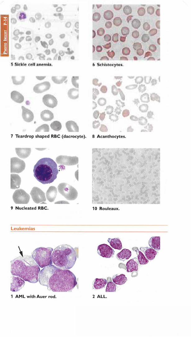
Ph
oto
In
ser
t P
-14
7 Teardrop shaped RBC (dacrocyte). 8 Acanthocytes.
9 Nucleated RBC. 10 Rouleaux.
L eu kem ias
1 A M L with A uer rod. 2 ALL.

\
4 CLL.3 CML.
All photos excluding Leukemias Fig. 4: From W in trobe ’s Clin. Hematol. 12th ed, 2009: Leukemias Fig. 4 From Devita, Heilman, and Rosenberg’s Cancer: Princip. & Prac.of Oncol. 8th ed, 2008.
U rinalys is
1 “Muddy brown” or granular cast 2 Hyaline cast (courtesy Nicholas Zwang, MD)
(courtesy Nicholas Zwang, MD)
3 “W axy broad” cast (courtesy Nicholas Zwang, MD)
4 Renal tubu lar epithelial cell(courtesy Nicholas Zwang, MD)
Photo Insert

5 RBC Cast. (Dis. of Kidney & Urinary Tract, 8th ed, 2006.)
6 W B C cast. (Gin. Lab. Medicine, 2nd ed,
2002.)
7 Calcium oxalate crystals (courtesy
Mailika Mendu, MD). Calcium monohydrate (arrow), calcium dihydrate (dashed arrow), and amorphous calcium crystals (arrowhead)
8 “Struvite” magnesium ammonia phosphate crystals (courtesy Brett Carroll,
MD)
9 Cystine crystals (Clin. Lab. Medicine, 1994.) 10 Sulfadiazine “ shock of wheat” crystals(courtesy Nicholas Zwang. MD)

ACLS ALGORITHMSFigure ACLS-1 ACLS Tachycardia Algorithm
TACHYCARDIA w/ Pulse
unstable = hypotension, A MS, s/s shock, chest pain, heart fa ilure
unstable
r/o sinus tach Synch card iovers ion(rarely needed for HR <150)
orde fib rilla tion fo r PMVT
IV Access, 0 2, m onitor BP & rhythm, 12-lead ECG
NARROW COMPLEX (QRS <120 msec)! WIDE COMPLEX (QRS >120 msec)T I I I
regular irregular regular irregular
_____i_____AF w / aber.
Control rate w / diltiazem or metoprolol
AF + W PW
ibutilide, proc, or am io
avoid adeno, dig, CCB, pB
PMVT (nl QT)
treat ischemia amio or lidocaino
& prepare for defibrillation
Torsades {T QT) correct lytes & other precip.
Mg 2 g IV overdrive pacing or
_____ isoproterenol_____
CARDIOVERSION MEDICATIONSAncillary equipm ent
0 2 sat m onitor suction, IV line intubation equipm ent
Prem cdicato call anesthesia service m idazolam 1 -5 mg fenfanyl 100 -300 meg
Svnch card ioversion 50-200 J biphasic 100 -200 J m onophasic
adenosine 6 mg rapid I VP then 20-cc NS bolus,12 mg IVP q2m in x 2 if needed
amiodarono 150 mg IV over 10 mindiltiazem 1 5 -2 0 mg IV over 2 m in, 2 0 -2 5 mg 15' la ter prn, 5 -1 5 mg/h ibutilide 1 mg over 10 min, repeat x 1 if needed
lidocaine 1 .0 -1 .5 m g/kg IVP, repeat in 5 -1 0 min
metoprolol 5 mg IV qSmin x 3procainamide 17 m g/kg at 2 0 -5 0 m g/m in (avoid if EF I ) sotalol 100 mg IV over 5 minverapamil 2 .5 -5 mg IV over 2 min, 5 -1 0 mg 15 -3 0 min la ter prn
(Adapted from ACLS 2015 Guidelines & Ore 2016;133:e506)
Figure ACLS-2 ACLS Bradycardia Algorithm
BRADYCARDIA w/ Pulse (HR <50 & inadequate for clin ica l condition)|
Airway, IV Access, 0 ?, m onitor BP & rhythm , 12-lead ECG
t(eg, hypotension A MS Unstable? _ No _ ► Observe
s/s shock, chest pain, heart fa ilure)
Yes
i f Type II2 °A V B io r3 °A V B
proceed to pacingatropine 0.5 mg IV q3 -5m in , m ax 3 mg
lASAP ▼
transcutaneous pacing, or dopamine 2 -1 0 pg/kg/m in, or while awaitingepinephrine 2 -1 0 pg/m in, or pacer or if pacer
isoproterenol 2 -1 0 pg/m in ±
ineffective
▼transvenous pacing
(Adapted from ACLS 2015 Guidelines)

Figure ACLS-3 VF/PulselessVT, Asystole & PEA Algorithms
CD(Adapted from ACLS 2015 Guidelines & Cite 2015;132(Suppl 2):S444)


![Dictionary of Chemistry [6th Ed.]](https://static.fdokumen.com/doc/165x107/6316ba01f68b807f88035f31/dictionary-of-chemistry-6th-ed.jpg)