Pittiruti - PICC e tip navigation.pptx - GAVeCeLT
192
Il punto sulla tip navigation: quale è la strategia più costo-efficace? Mauro Pittiruti
-
Upload
khangminh22 -
Category
Documents
-
view
0 -
download
0
Transcript of Pittiruti - PICC e tip navigation.pptx - GAVeCeLT

Il punto sulla tip navigation: quale è la strategia più costo-efficace? Mauro Pittiruti

Tipnavigation

‘tipnavigation’methods
Methodswhichmaybeusedduringtheproceduretohelptheoperatorindirectingtheguidewireand/orthecatheterintherightdirection.
Theydonotreplace‘tiplocation’methods
Still,theymaybeusefultoreducetheriskofprimarymalpositionswhenintra-procedural‘tiplocation’methodsarenotused.

‘tipnavigation’methodsVisual methods
direct ultrasound fluoroscopy Navigator (Corpak) indirect (projection) Sherlock (Bard)
Non-visual methods doppler-based – VPS (Teleflex) pressure-based – Catfinder (Elcam) ECG-based – Delta (Romedex)
‘tipnavigation’–visualmethods
Theyprovideinformationaboutthepresumedpositionofthecathetertipduringitstrajectory
Theyhelpustoidentifythe‘wrong’directionofthetip
-intheipsilateralinternaljugularvein
-inthecontrolateralveins(brachio-cephalic,subclavian,jugular)

‘tipnavigation’–visualmethods
Visual methods direct (localization) ultrasound fluoroscopy Navigator (Corpak) indirect (projection) Sherlock (Bard)

Visual methods for tip navigation
Cathfinder Navigator
Sherlock

Cath-Finder (Pharmacia, 1993)
Magnetic stylet in the catheter
Detector and monitor integrated in a single
hand-held device

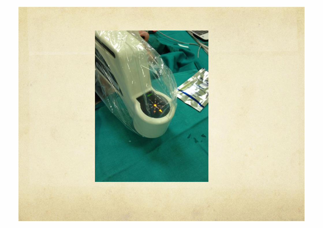
Navigator (Corpak)
Plastic disposable stylet with a magnetic tip

Navigator
Naylor, JAVA 2007

Viasys/CORPAK MedSystems, Inc.


Sherlock (Bard)
hands-free monitor
Magnetic stylet
Magnetic “Tracking”
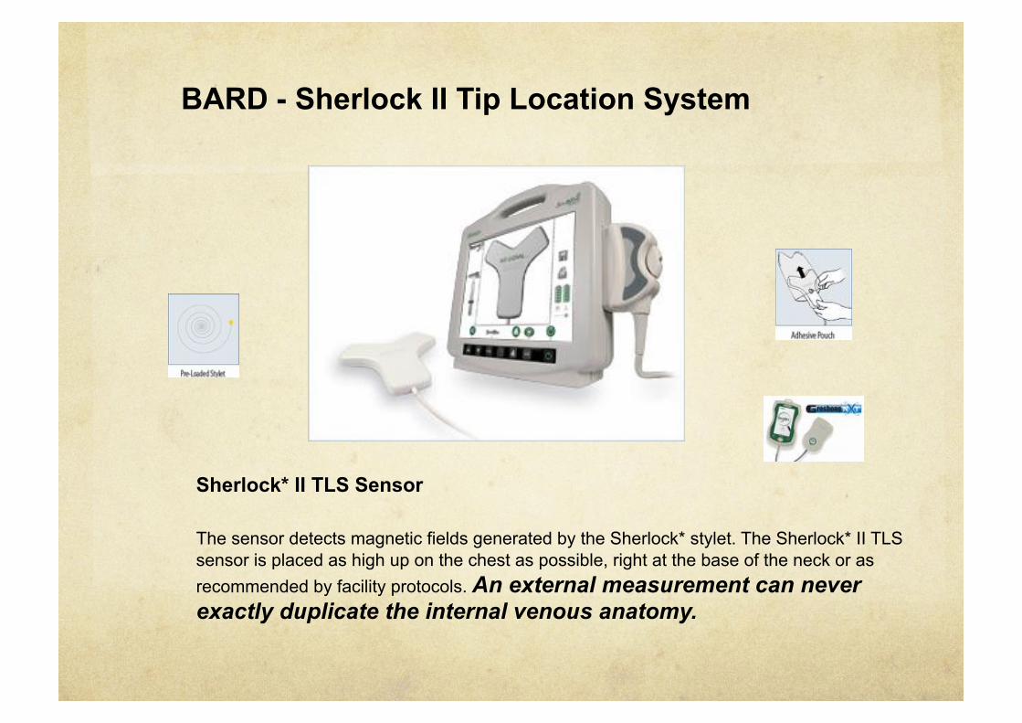
BARD - Sherlock II Tip Location System
Sherlock* II TLS Sensor
The sensor detects magnetic fields generated by the Sherlock* stylet. The Sherlock* II TLS sensor is placed as high up on the chest as possible, right at the base of the neck or as recommended by facility protocols. An external measurement can never exactly duplicate the internal venous anatomy.


2013

Clin Imaging. 2013 Sep-Oct;37(5):917-21.
Epub 2013 Jul 15.
Analysis of the Sherlock II tip location system for inserting peripherally inserted central venous catheters.
Lelkes V, Kumar A, Shukla PA, Contractor S, Rutan T.
University of Medicine and Dentistry of New Jersey, Newark, NJ 07101, USA. IT IS NOT ‘TIP LOCATION’…...!

LumenVue

LumenVue
Founders – Greg Schears, MD (Mayo Clinic) and David Wilson, PhD (U Penn)
Technology uses near-infrared, optical fiber combined with a guidewire, and a camera that detects and displays the light
SonoSite acquired LumenVu – July 2007


Micronix/MedComp
CathRite™
A key point of difference with the Micronix technology is that the "transmitter" is placed in the catheter tip and the "receiver" is outside of the body.
In fase sperimentale Abbandonato

Visualmethodsfortipnavigation
Imprecise(projectionofthepositionofthetipontheskinsurface)

‘tipnavigation’–non-visualmethods
Theytelluswhetherthetipisdirectedintherightdirection(followingbloodflow)orinthewrongdirection(againstbloodflow)
doppler-based–VPS(Teleflex)
pressure-based–Catfinder(Elcam)
ECG-based–Delta(Romedex)
ALLINTEGRATEDWITHTIPLOCATION

Conductanceguidewire(CGW)

Fluoroscopyfortipnavigation

Fluoroscopy?You should not use fluoroscopy for tip navigation (or tip location)
Is not safe
Is not accurate
Is not cost-effective
See recommendations of AHRQ 2013, INS 2016, etc.

Ultrasoundfortipnavigation

11
Ultrasound f o r t i p navigation
12
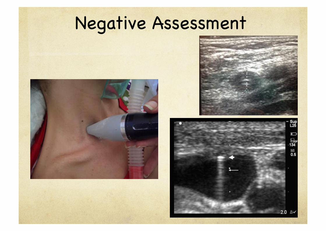
Negative Assessment

34

Catheter


15
Microconvex Probe
Ao
AP
VCS


RBCV LBC
V
SVC AoA

17

ULTRASOUND
Manypublishedstudies
DifferentUSmethodshavebeenvalidatedforbothtiplocationandtipnavigation
Cost-effective–butrequirestraining

Advantagesofultrasound Noadditionalcost
Completelysafe
Veryaccurate,particularlyinpediatricpatientsandinskinnypatients
Itcanbeusedfor‘redirecting’thecatheterortheguidewireintherightdirection

Thebestsystemfor‘redirecting’

Thebestsystemfor‘redirecting’

‘Navigator’fortipnavigation

In 30 PICC insertions, we adopted Navigator (Corpak) for tip navigation and IC-ECG for tip location (performed using Nautilus, Romedex). The Navigator device consists of a sterile stylet (diameter 1.1 Fr, length 106 cm) placed inside the catheter so that the tip of the stylet is at 1 mm from the tip. During insertion, the tip of the catheter can be followed and detected by an electromagnetic device. Also, the system tells whether the tip is pointing in the right direction or not.
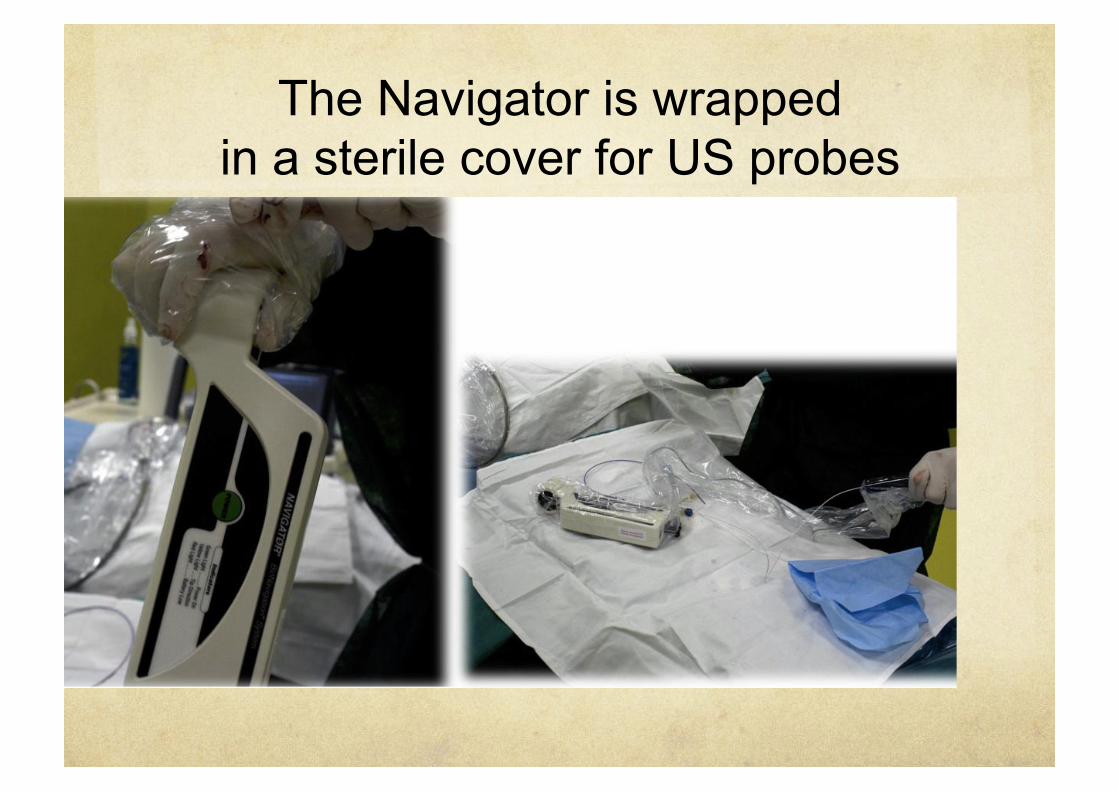
The Navigator is wrapped in a sterile cover for US probes
The Navigator is wrapped in a sterile cover for US probes

The stylet of the PICC is removed and
replaced by the stylet of the Navigator

The cable of the Navigator stylet is inserted into a connecting device wrapped in the sterile
cover

In all patients, the tip location verified by IC-ECG corresponded to the electromagnetic detection of the tip below the third intercostal space, with the tip pointing downward. In 3 cases, the Navigator detected that the tip had accidentally entered the ispilateral internal jugular vein and allowed us to correct its direction. In 2 cases, IC-ECG was not confirming the correct tip location, though the PICC had been threaded for the estimated length: the Navigator detected the tip of the catheter in the contralateral brachio-cephalic vein, pointing to the contralateral clavicle: this allowed to correct its direction.

Tip in ipsilateral IJV
Tip in ipsilateral IJV

Tip in controlateral BCV

Tip at the cavo-atrial junction

Navigator
The tip navigation system we tested – associated with IC-ECG - was clinically effective and easy to use. The Navigator has several advantages if compared to other navigation systems: (a) it can be utilized with any type of central VAD; (b) it tells both the approximated location of the tip below the thoracic cage and its direction; (c) it is highly cost-effective, since it may be used only if required (i.e., when difficulties can be anticipated and/or when they occur during the procedure).

NAVIGATOR
Abstractsininternationalconferences
Nopublishedstudies
Cost-effectiveandaccurate

AdvantagesofNavigator Easytouse
Itcanbeusedwithanycentralline
Expensive,butcost-effectivesinceitcanbeusedonlywhensomedifficultyisanticipatedorexperienced
Itcanbeseenasa‘surrogate’tiplocationmethodinAF
Itgives‘location’and‘direction’ofthetip


Though…
Notipnavigationmethodhasanysensetodayifitisnotcoupledwithatiplocationmethod
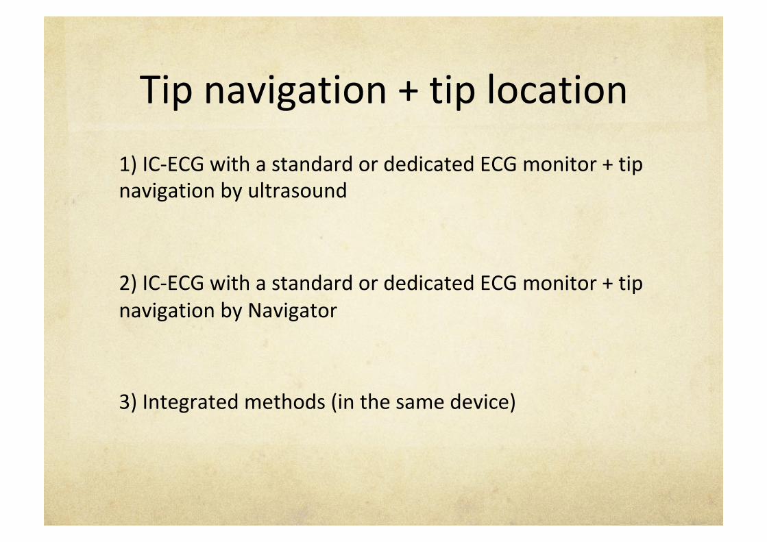
Tipnavigation+tiplocation1)IC-ECGwithastandardordedicatedECGmonitor+tipnavigationbyultrasound
2)IC-ECGwithastandardordedicatedECGmonitor+tipnavigationbyNavigator
3)Integratedmethods(inthesamedevice)
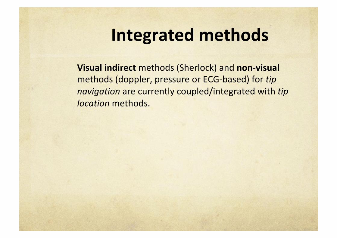
IntegratedmethodsVisualindirectmethods(Sherlock)andnon-visualmethods(doppler,pressureorECG-based)fortipnavigationarecurrentlycoupled/integratedwithtiplocationmethods.

IntegratedMethods(navigation+location)
Sherlock3CG(Bard)
VasonovaVPS(Teleflex)
Catfinder(Elcam)
Delta(Romedex)

Sherlock3CG(Bard)
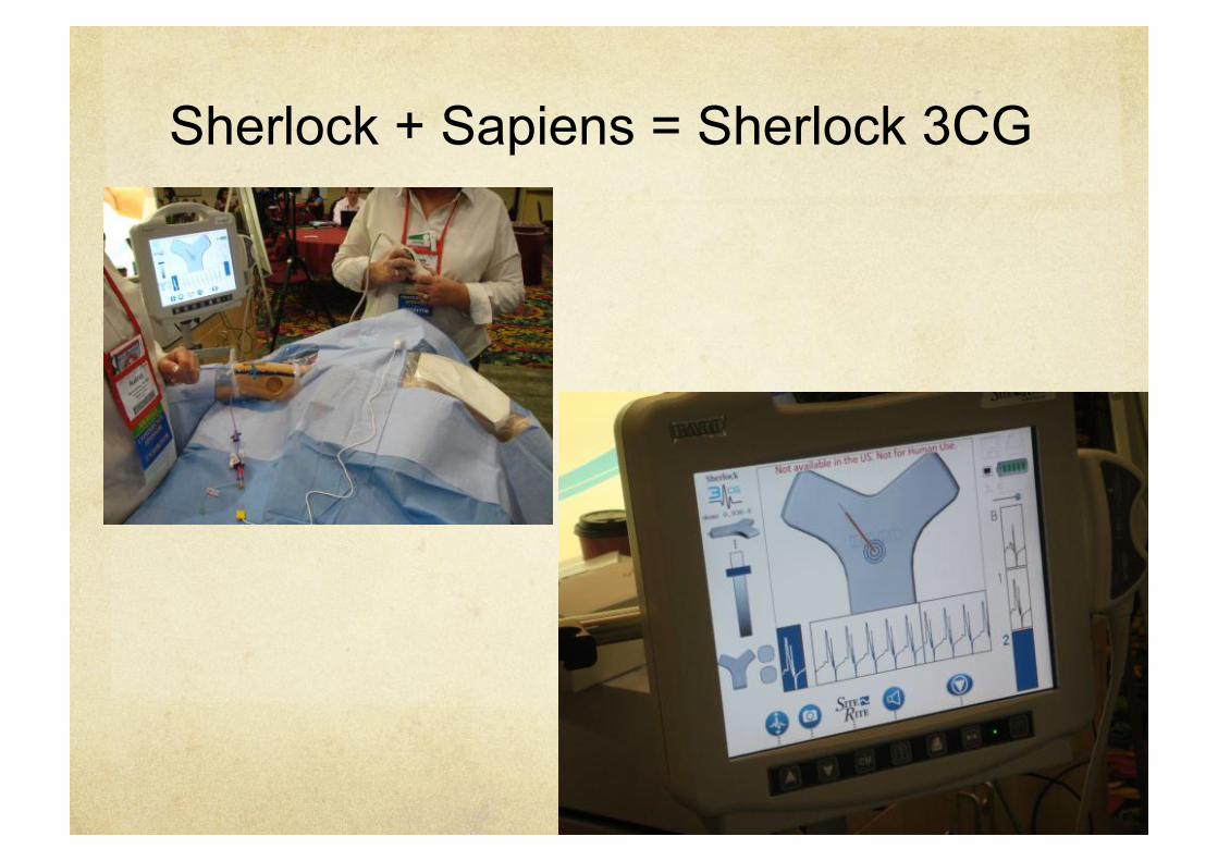
Sherlock + Sapiens = Sherlock 3CG
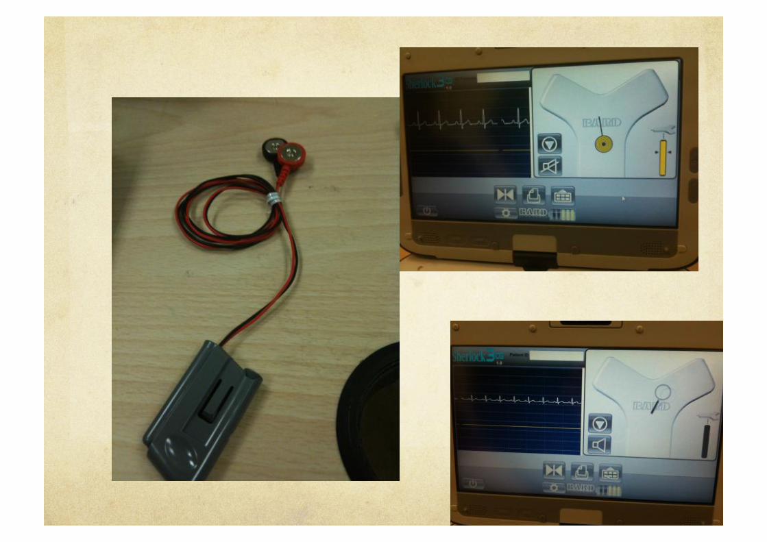


Sherlock3CG
Manyabstractsininternationalconferences
Afewpublishedpaperonpeer-reviewedjournals(asfromPubMed)

Anaesthesia. 2014 Jul 10. doi: 10.1111/anae.12785. [Epub ahead of print]
Evaluation of the Sherlock 3CG Tip Confirmation System on peripherally inserted central catheter malposition rates.
Johnston AJ, Holder A, Bishop SM, See TC, Streater CT. John Farman Intensive Care Unit, Addenbrooke's Hospital, Cambridge University Hospitals NHS Foundation Trust, Cambridge, UK.
20.5% malposition (?????) Unacceptable study ?
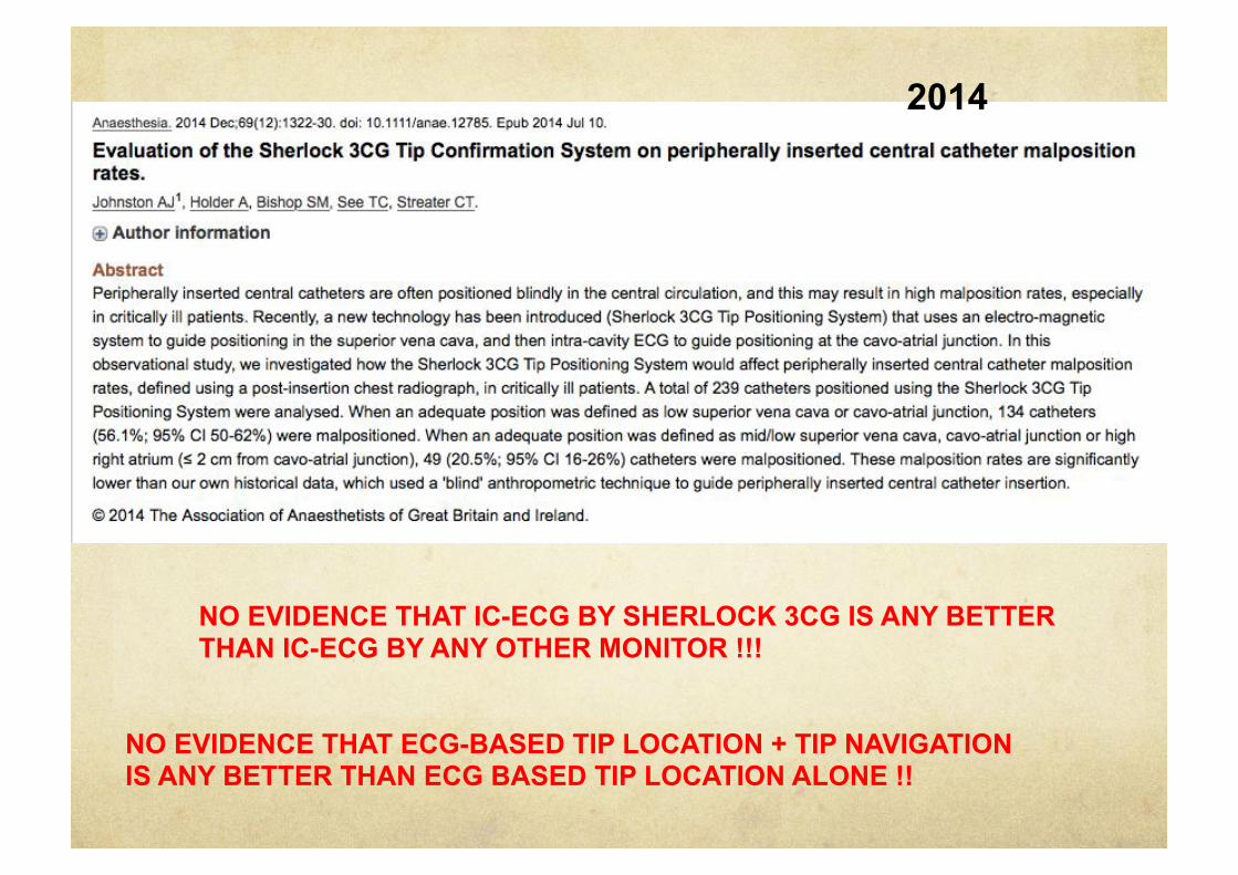
NO EVIDENCE THAT IC-ECG BY SHERLOCK 3CG IS ANY BETTER THAN IC-ECG BY ANY OTHER MONITOR !!!
NO EVIDENCE THAT ECG-BASED TIP LOCATION + TIP NAVIGATION IS ANY BETTER THAN ECG BASED TIP LOCATION ALONE !!
2014

Morerecently…2015
NO EVIDENCE THAT ECG-BASED TIP LOCATION + TIP NAVIGATION IS ANY BETTER THAN ECG BASED TIP LOCATION ALONE !!
NO EVIDENCE THAT IC-ECG BY SHERLOCK 3CG IS ANY BETTER THAN IC-ECG BY ANY OTHER MONITOR !!!

2016
NO EVIDENCE THAT ECG-BASED TIP LOCATION + TIP NAVIGATION IS ANY BETTER THAN ECG BASED TIP LOCATION ALONE !!
NO EVIDENCE THAT IC-ECG BY SHERLOCK 3CG IS ANY BETTER THAN IC-ECG BY ANY OTHER MONITOR !!!

2016
NO EVIDENCE THAT ECG-BASED TIP LOCATION + TIP NAVIGATION IS ANY BETTER THAN ECG BASED TIP LOCATION ALONE !!
NO EVIDENCE THAT IC-ECG BY SHERLOCK 3CG IS ANY BETTER THAN IC-ECG BY ANY OTHER MONITOR !!!

NO EVIDENCE THAT ECG-BASED TIP LOCATION + TIP NAVIGATION IS ANY BETTER THAN ECG BASED TIP LOCATION ALONE !!
NO EVIDENCE THAT IC-ECG BY SHERLOCK 3CG IS ANY BETTER THAN IC-ECG BY ANY OTHER MONITOR !!!

Sherlock3CGBigchallenge:
TotrytoprovethatIC-ECG+SherlocknavigationhasclearadvantagesoverIC-ECGaloneintermofcost-effectiveness.
(clinicalstudyatCatholicUniversity–justcompleted)

AVA2015Clinical use of Sherlock-3CG for positioning power injectable PICCs
Mauro Pittiruti, Giancarlo Scoppettuolo, Laura Dolcetti

Goals Tip location with Sherlock-3CG
Safety
Feasibility
Accuracy
Tip navigation with Sherlock-3CG Safety
Feasibility
Accuracy

Methods All consecutive patients candidate to PICC insertion in
our Day Hospital of Infectious Disease or Oncology Adults Outpatients – PICC needed for DH/home Informed consent Availability of post-procedural chest x-ray
In all patients: US-guided PICC placement using intra-procedural Sherlock-3CG + intra-procedural Nautilus + post-procedural chest x-ray

PatientsandPICCs130 adult patients 123 neoplastic + 7 infect.dis. 128 sinusal rhythm + 1 AF + 1 PM 76 females + 54 males Age range 24 - 84 y.o. BMI range 17 – 42
130 Bard Power PICCs, non-valved 94 picc 4F SL + 36 picc 5F DL 92 insertion in the basilic vein, 38 in a brachial vein 104 on the right arm, 26 on the left arm

Insertion130insertions
-129successes+1failure(but:successaftershiftingside)-in9cases,tunnellingwasrequired(veintoosmallin
the‘green’zone:punctureofveininthe‘yellow’zone’)-nopuncture-relatedcomplications -nonerveinjury -noarterialpuncture-in15cases,anadditionalmicrointroducerkit(from
GaltMedical)wasneeded -problemswithBardguidewire -problemswithBardintroducer/dilator
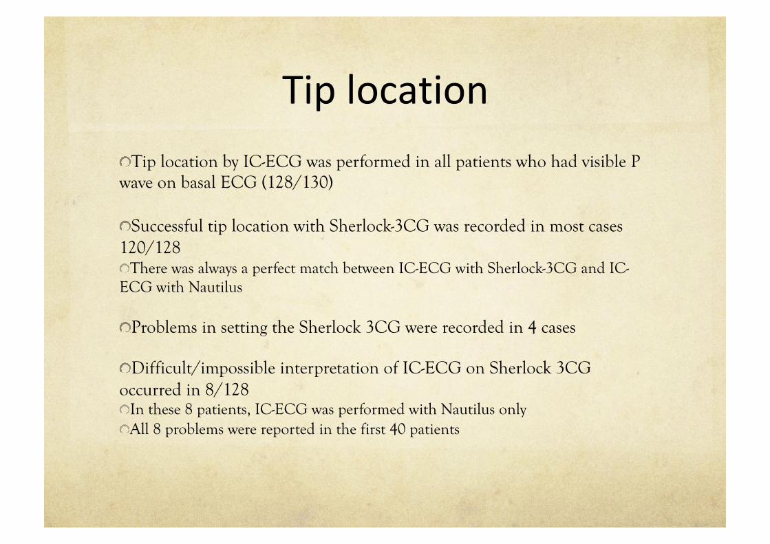
Tiplocation Tip location by IC-ECG was performed in all patients who had visible P
wave on basal ECG (128/130)
Successful tip location with Sherlock-3CG was recorded in most cases 120/128 There was always a perfect match between IC-ECG with Sherlock-3CG and IC-
ECG with Nautilus Problems in setting the Sherlock 3CG were recorded in 4 cases
Difficult/impossible interpretation of IC-ECG on Sherlock 3CG occurred in 8/128 In these 8 patients, IC-ECG was performed with Nautilus only All 8 problems were reported in the first 40 patients

ProblemsinsettingtheSherlock3CG
Onlyin4cases 2casesinthefirst40patients 2casesinthesecond90patients
Problem:loose/defectiveconnectionbetweenshieldandcable
Inall4cases,thetiplocationwasnonethelessperformed,thoughwithsomedifficulties(inconstantECGreading)

Difficult/impossibleinterpretationofIC-ECGonSherlock3CG
Occurredin8/128 All8problemswerereportedinthefirst40patients
Abnormal/disturbedECGtrace Artifacts Lowwavevoltage NoPincrease
Inthese8patients,tiplocationbyIC-ECGwasperformedwithNautilusonly

Post-proceduralchestx-ray AP view + lateral view in 101 cases
Only standard AP view in 29 cases
Tip visualized in 129/130 cases
In all 128 cases performed with IC-ECG, the location of the tip was correct according to x-ray criteria No malpositions ‘Sweet spot’ criteria: all tips were ok ‘Carina’ criteria: all tips were in ‘acceptable’ position
108 caths were perfect (1-5 cm below the carina) 14 caths were short (0-1 cm below the carina) 5 caths were long (5-7 cm below the carina)

Tipnavigation Tip navigation was successfully performed with
Sherlock-3CG in 105/130
In 25/130, there were problems No visualization Wrong visualization (tip ok according to IC-ECG, but
wrongly directed according Sherlock) Poor visualization (transient or unstable) Failure of tip navigation occurred 17 times in the first
40 patients + 8 times in the following 90 patients (suggesting an effect of training)
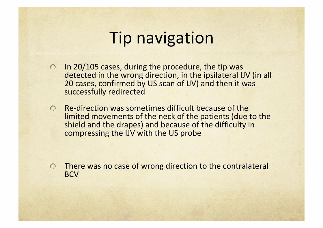
Tipnavigation In20/105cases,duringtheprocedure,thetipwasdetectedinthewrongdirection,intheipsilateralIJV(inall20cases,confirmedbyUSscanofIJV)andthenitwassuccessfullyredirected
Re-directionwassometimesdifficultbecauseofthelimitedmovementsoftheneckofthepatients(duetotheshieldandthedrapes)andbecauseofthedifficultyincompressingtheIJVwiththeUSprobe
TherewasnocaseofwrongdirectiontothecontralateralBCV

GeneralcommentsonSherlock-3CG
Calibrationwasthemainproblem Requirestraining(asprovenbythehigherincidenceofnavigationfailuresinthefirst40patientsvsthefollowing90patients)
Evenaftertraining,thecalibrationwas‘felt’bythenursesasstressfulandtime-consuming
Keyfactorwasthepositionofthecablevsthepositionoftheshield
Anotherimportantdisturbingfactorwasthepresenceofmetallicobjectsonthepatients(typically:inclothesoffemalepatients)

GeneralcommentsonSherlock-3CG
Anothermajorproblemwasthehighsensitivityofthesystemtopossiblesourcesofelectrical/magneticinterference Cellphones Otherelectricaldevices,ifplugged(ultrasound,Nautilus,pumps,etc.)
ThisapparentlyaffectedboththeperformanceoftheIC-ECGandofthenavigation

GeneralcommentsonSherlock-3CG
Other technical aspects: difficult connection between the shield and the cable (serious
issue in 4 cases) The shield was well tolerated by the patients, but the overall
draping system was quite rigid and implied limited movements of the neck of the patient Higher risk of wrong direction of the cath to the IJV Some difficulties in redirecting the tip Landmark measurement may be difficult
Printer failure (!?) Poor quality of the micro-introducer kit – if compared to our
standard PICCs Lack of a safety block of the stylet – if compared to our standard
PICCs
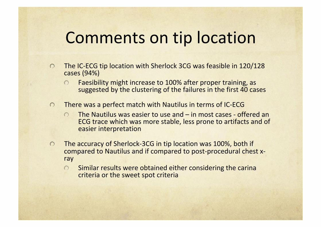
Commentsontiplocation TheIC-ECGtiplocationwithSherlock3CGwasfeasiblein120/128
cases(94%) Faesibilitymightincreaseto100%afterpropertraining,as
suggestedbytheclusteringofthefailuresinthefirst40cases
TherewasaperfectmatchwithNautilusintermsofIC-ECG TheNautiluswaseasiertouseand–inmostcases-offeredan
ECGtracewhichwasmorestable,lesspronetoartifactsandofeasierinterpretation
TheaccuracyofSherlock-3CGintiplocationwas100%,bothifcomparedtoNautilusandifcomparedtopost-proceduralchestx-ray Similarresultswereobtainedeitherconsideringthecarina
criteriaorthesweetspotcriteria
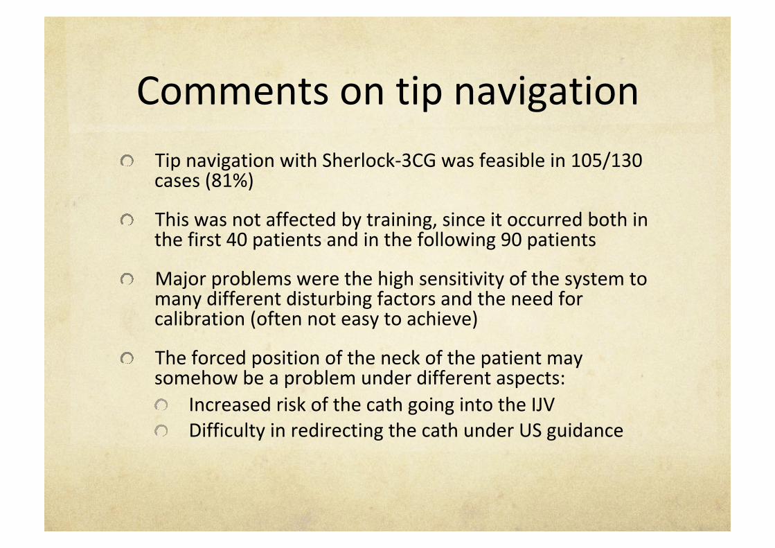
Commentsontipnavigation TipnavigationwithSherlock-3CGwasfeasiblein105/130cases(81%)
Thiswasnotaffectedbytraining,sinceitoccurredbothinthefirst40patientsandinthefollowing90patients
Majorproblemswerethehighsensitivityofthesystemtomanydifferentdisturbingfactorsandtheneedforcalibration(oftennoteasytoachieve)
Theforcedpositionoftheneckofthepatientmaysomehowbeaproblemunderdifferentaspects: IncreasedriskofthecathgoingintotheIJV DifficultyinredirectingthecathunderUSguidance

Conclusions TiplocationandtipnavigationwithSherlock-3CGwerenot
associatedwithanycomplication–safety=100%.
Asregardstiplocation,Sherlock-3CGshoweda94%feasibilityand100%accuracy Feasibilitymightreach100%afterpropertraining.
Asregardstipnavigation,Sherlock-3CGshoweda81%feasibilityand100%accuracy Feasibilitymightincrease,tosomeextent,afterproper
training,thoughitmayalwaysbelimitedbythefeaturesofthesystem(whichisnotuser-friendlyanditishighlysensitivetomanydisturbingfactors)

Conclusions(2) Theoverallcost-effectivenessofSherlock3CGfortiplocation
withIC-ECG-ifcomparedtoNautilusortootherdedicatedornon-dedicatedECGmonitors-mightbequestioned,consideringthehighercostandthehighercomplexityinsettingandoperatingthesystem,whiletheresultsaresimilarorslightlyworse.
Theoverallcost-effectivenessofSherlock3CGfortipnavigation–ifcomparedtocurrentmethodsfordetectingawrongdirection(USscan,ECGnavigation,CorpakNavigator,etc.)–isverypoor,consideringthehighercost,thehighercomplexityandthepoorperformance.

Insummary:TheuseofSCGwasnotassociatedwithanycomplication(100%safety).Asregardstiplocation,SCGshowed94%feasibilityand100%accuracy(though,feasibilitymightreach100%afterextendedtraining).Asregardstipnavigation,SCGshowed81%feasibilityand100%accuracy(feasibilitymightincrease,tosomeextent,afterextendedtraining).

ProblemswithSherlock3CG Expensive
Notcosteffective
Noteasytouse
CanbeusedonlywithaveryspecificbrandofPICCs
Tiplocationisok,buttipnavigationisnotalwaysfeasible

VasonovaVPS(Teleflex)

Vasonova: EKG + Doppler

VasonovaVPSManyabstractsininternationalconferences
Nopublishedpaperonpeer-reviewedjournals(asfromPubMed)

Girgenti et al
Successfully Eliminating Chest Radiography by Replacing It with Dual Vector Technology and an Algorithm for PICC Placement
(JAVA, June 2014).
30 patients (5 with AF)

VasonovaVPSBigchallenge:
TotrytoprovethatIC-ECG+DopplernavigationhasclearadvantagesoverIC-ECGaloneintermofcost-effectiveness.
MaybeinAF???

ProblemswithVasonova Very expensive
Not cost effective
Not easy to use
The real role of doppler for tip location is unclear and unproven

Catfinder(Elcam)

Catfinder Elcam


Catfinder(Elcam)Afewabstractsavailable
Nopublishedpaperonpeer-reviewedjournals(asfromPubMed)
AstudyjustcompletedinourUniversityHospital

TheprimaryendpointofourstudywastoevaluatetheaccuracyoftheCatFinderNavigationalDevice(CFND)asatiplocationmethodforperipherallyinsertedcentralvenousaccessdevicesinadultpatients,ascomparedtotheIntracavitaryECGmethod(IECG).
ThesecondaryendpointwastoevaluateCFNDasatipnavigationmethod,abletodetectthewrongdirectionofthecatheterduringinsertion.


Method WestudiedadultpatientscandidatetoplacementofPICCsorPICC-ports.
PatientswithknownECGabnormalitiesorcardiacdiseaseofanytypewereexcluded.
Thetargetwastoplacethetipatthecavo-atrialjunction.Inallcases,tiplocationwasverifiedbyIECG.Anycaseofsuspectedwrongdirectionofthecatheterwascheckedbyultrasoundscan.

Results(1) Outof136enrolledpatients,CFNDwasapplicablein131cases(5caseswereexcludedbecauseofabnormalECG)andfeasiblein111cases(in20cases,technicalproblemsoccurredduringtheprocedure:airbubblesinthesystem,cathetercuttooshort,abnormalpressurereading,humanerrorsinthemethod,etc.).


Results(2) TherewerenocomplicationsdirectlyorindirectlyrelatedtoCFND.
ComparingwithIECG,87tipswereplacedwithin2cmfromthetarget,while17wereplaced>2cmfromtarget(13at+3cm;4at+4cm).
In7casesCFNDdetectedawrongdirection(totheispilateraljugularvein),confirmedbyultrasound.

Conclusions(1) Applicability of CFND was 96%
feasibility was 85%
safety was 100%
(feasibility is expected to improve by solving the technical issues above described)

Conclusions(2)- IfcomparedtoIECG,accuracywas84%(consideringarangeof
+2cm)and96%(+3cm).
- Unacceptabletippositionswere3%(inallofthesecases,thecatheterwastooshort).
- TheaccuracyinthediagnosisofamalpositionintheIJVwas100%.
- OurstudyconfirmsthepotentialroleofCFNDforrealtimetiplocationandtipnavigation.

ProblemswithCatfinder Not cost effective
Not easy to use
Limited applicability
It takes time
Accuracy: high for tip navigation, somehow less for tip location

Delta(Romedex)


ECG navigation (Romedex)


PICCinsertionusingDelta


PICCinsertionwithDelta(2)


OurstudieswithDeltaPilot study on tip location (207 pts)
WoCoVA 2014
Tip location study in children (85 pts)
AVA 2015
Pilot study on tip location + navigation (26 pts)
AVA 2014

A NEW WIRELESS DEVICE FOR THE INTRACAVITARY ECG
TECHNIQUE
Introduction The intracavitary ECG method (IC-ECG) is adopted in clinical practice as an easy, cost-effective and accurate methodology for assessing the central location of the tip of venous access devices (VAD).

Introduction (2) We report our preliminary experience with a new wireless system specifically dedicated to the IC-ECG (Nautilus Handy/Delta, Romedex), which consists of a small box connected to the ECG cables, sending data to a smartphone or a tablet by bluetooth technology.
The phone/tablet is provided with a software application which allows to display both the surface and intracavitary ECG.
The system can be operated by command buttons placed on the box or directly by touching the screen of the phone/tablet.


Methods
The IC-ECG method is performed according to the standard procedure.
The identification of the peak of the P wave (corresponding to the cavo-atrial junction) is made easy by the freeze function, which can be operated either from the box or from the phone/tablet.
At any time, the display can be saved and/or printed for documentation.


Results The new device was adopted for tip location in 207 central VADs (154 PICCs, 49 ports, 2 short term CICCs and 2 cuffed-tunneled catheters) placed after cannulation of different veins (96 basilic, 41 brachial, 57 axillary-subclavian, 6 internal jugular, 7 brachio-cephalic) .
The P wave was evident on basal ECG in all patients.
A peak of the P wave was easily detected in all patients.

Results (2) The P wave was evident on basal ECG in all patients.
A peak of the P wave was easily detected in all patients.
In 36 patients (28 PICCs and 8 ports), the procedure was simultaneously carried out both with a standard dedicated ECG device (Nautilus, Romedex) and with the new wireless device: no differences were noted in terms of performance.


Conclusions This new wireless system for IC-ECG had an optimal clinical performance in terms of applicability and feasibility.
Transmission of the data to the moveable device by bluetooth simplified the wire connections.

Conclusions (2) Some potential advantages over other ECG monitors are:
- the system is light and easy to carry - which makes it ideal for bedside insertion;
- it can be operated by the same professional inserting the VAD;
- it implies no risk of electrical hazard;
- it can be used on a personal portable device, allowing easy storage of data and easy printing for documentation.

Centralvenousaccessinneonatesandchildren:tiplocationusinganewwirelessdeviceforintracavitaryECG

Purpose
Tiplocationofcentrallinesisparticularlyimportantinchildrenandideallyitshouldbeassessedduringtheprocedure.
WehaveadoptedtheintracavitaryECGmethod(IC-ECG)sinceadecade.
WereportourrecentexperiencewithawirelessdeviceforIC-ECG.

MethodsWe reviewed all centrally (CICC) and peripherally inserted central catheters (PICC) placed in our Pediatric Intensive Care Unit (PICU) using a wireless IC-ECG device (Delta, Romedex).
All insertions were performed according to our PICU protocol: sedation or general anesthesia, ultrasound scan of all veins, maximal barrier precautions, skin antisepsis with 2% chlorhexidine, ultrasound guided venipuncture using a micro-introducer kit, tip location by IC-ECG (maximal height of the P wave = cavo-atrial junction), securement of the catheter by cyanoacrylate glue, sutureless device and transparent dressing.

Results(1)Wireless IC-ECG was used in 85 children (age range 2 hrs – 12 y.o.: 58 patients were < 2 y.o.). Lowest weight was 1100 g.
We inserted 81 non-cuffed catheters (power injectable, polyurethane, non-valved, open ended; 3Fr single lumen or 4Fr double lumen): 55 CICCs (in 95%, cannulation of the brachio-cephalic vein + tunneling to the infraclavicular area) and 26 PICCs (cannulation of deep veins of the arm; in 75%, cannulation of the axillary vein at the axilla + tunneling to the arm or to the lateral thoracic area).
In 4 cases, we inserted tunneled, cuffed CICCs (5Fr single lumen) for long term I.V. therapy: all 4 were children > 6 years.

Results(2)
Wehadnoinsertion-relatedcomplications.
IC-ECGwaseasilyperformedinallcases.
Post-proceduralconfirmationoftiplocationwasperformedbyechocardiographyor(inafewcases)bychestx-ray:nomalpositionwasdetected.

Conclusion
TiplocationwiththenewwirelessIC-ECGdevicewasapplicable,feasible,safeandaccuratein100%ofourpediatricpatients,eveninsmallneonates.

Tipnavigation+tiplocation

Purpose Intracavitary ECG (IC-ECG) is currently used for assessing catheter tip location of central venous access devices (CVAD) at or around the cavo-atrial junction (CAJ).
Navigation support for CVAD is currently provided by other methods, such as electromagnetic or doppler-based.
In this pilot study, we tested a brand new application of IC-ECG for catheter tip navigation.

Method IC-ECG-based tip navigation was performed using a wireless device (Delta, Romedex) - which is already available on the European market for IC-ECG tip location - modified so to support a new original software.

Method One control electrode is placed on the patient’s chest over the manubrium of the sternum just below the presternal notch.
The catheter is connected to a second electrode using a saline adapter.
A third electrode is placed for reference on the patient’s left lower abdomen.
A novel ECG-based navigation signal is computed in real time combining the ECG signals from the tip of the catheter and from the control electrode, and is transmitted from the wireless device to a smartphone, by bluetooth.


Control electrode

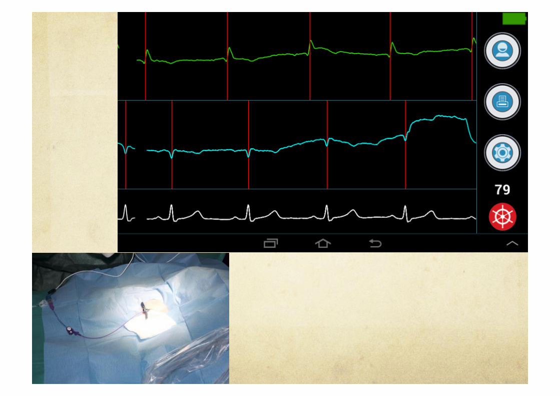
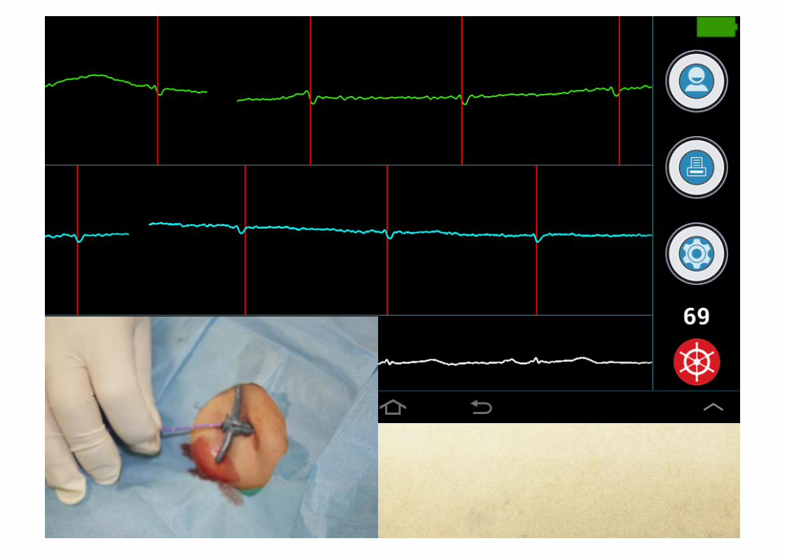
Results The new technique was used in 26 PICC placements.
In all procedures, IC-ECG-based navigation signal successfully indicated whether the tip was moving towards or away from the CAJ; the catheter tip location at CAJ was confirmed by using the maximum P-wave criterion, as in traditional IC-ECG methods.
There were no procedure-related complications.

Control electrode

Right below the control electrode

Moving towards the right atrium

At the cavo-atrial junction (target)

ECG navigation in inferior vena cava
Control electrode


Landmark measurement


Moving towards the right atrium

Right below the control electrode (target)

Conclusions In our pilot study, this new methodology was successful and particularly easy to apply.
The relevant clinical implication is that clinicians may use a single IC-ECG device for both tip navigation and tip location.
Further data about the applicability, feasibility and accuracy of this technique will be provided as soon as available.

DELTA
Abstractsininternationalconferences
Nopublishedstudyyet
Verypromising:easy,accurate,inexpensive,cost-effective

AdvantagesoftiplocationbyDelta
Easytouse
Accurate
Noadditionalcost
Itcanbeusedwithanykindofcentralline
Itcanbedonewiththesame3electrodesusedforECG-basedtiplocation
Wideapplicability(alsoincaseswhereECGbasedtiplocationisnotapplicable)

Conclusions

FirstconclusionThereisnohardevidencethattipnavigationisnecessaryduringPICCinsertion,thoughitmaybeuseful(or‘reassuring’fortheoperator).
Onthecontrary,apropermethodfortiplocationisalwaysnecessary.

SecondconclusionInmostcentrallineinsertion,ultrasoundmaybetheeasiest,simplest,mosteasilyavailableandmostcost-effectivemethodfortipnavigation.
Itaccuratelydetectsthe‘wrong’directiontotheipsilateralIJVandthe‘right’directionintotheBCVinadults;itdetectsthedirectioninsidebothBCVandSVCinneonatesandinfants.
Ithelpstore-directthecatheterortheguidewiretotherightdirection