Phytochemical analysis and gastroprotective activity of an olive leaf extract
11
J. Serb. Chem. Soc. 74 (4) 367–377 (2009) UDC 616.33:615.37:633.852.73:633.879.1 JSCS–3838 Original scientific paper doi: 10.2298/JSC0904367D 367 Phytochemical analysis and gastroprotective activity of an olive leaf extract DRAGANA DEKANSKI 1 * , SNEŽANA JANIĆIJEVIĆ-HUDOMAL 2 , VANJA TADIĆ 3 , GORAN MARKOVIĆ 3 , IVANA ARSIĆ 3 and DUŠAN M. MITROVIĆ 4 1 Biomedical Research, R & D Institute, Galenika a.d., Pasterova 2, 11000 Belgrade, 2 Phar- macology Department, School of Medicine, University of Priština (Kosovska Mitrovica), 3 Institute for Medicinal Plant Research “Dr Josif Pančić”, Tadeuša Košćuška 1, 11000 Belgrade and 4 Institute of Medical Physiology, School of Medicine, University of Belgrade, Višegradska 26, 11000 Belgrade, Serbia (Received 8 September, revised 7 November 2008) Abstract: Some medicinal features of olive leaf have been known for centuries. It has been traditionally used as an antimicrobial and to prevent and treat dia- betes mellitus and heart disease. Whether olive leaf, a natural antioxidant, in- fluences the gastric defense mechanism and exhibits gastroprotection against experimentally-induced gastric lesions remains unknown. In this study, the content of total phenols, total flavonoids and tannins in olive leaf extract (OLE) were determined. Seven phenolic compounds were identified and quantified (oleuropein, caffeic acid, luteolin, luteolin-7-O-glucoside, apigenin-7-O-gluco- side, quercetin, and chryseriol). Furthermore, the protective activity of the OLE in gastric mucosal injury induced by a corrosive concentration of ethanol was investigated. In relation to the control group, pretreatment with OLE (40, 80 and 120 mg kg -1 ) significantly (p < 0.001) attenuated the gastric lesions indu- ced by absolute ethanol. The protective effect of the OLE was similar to that obtained with a reference drug, ranitidine. The results obtained indicate that OLE possesses significant gastroprotective activity, and that the presence of compounds with antioxidative properties would probably explain this effect. Keywords: olive leaf extract; phenols; flavonoids; tannins; gastroprotection. INTRODUCTION Research on flavonoids and other polyphenols, their antioxidant properties, biological activities and their effects in disease prevention truly began in the last decade. There is an increasing interest in medicinal plant extracts, the greatest va- lue of which may be due to constituents that contribute to the modulation of the oxidative balance in vivo. Benefits of the olive (Olea europaea L.) leaf have been * Corresponding author. E-mail: [email protected]
Transcript of Phytochemical analysis and gastroprotective activity of an olive leaf extract
J. Serb. Chem. Soc. 74 (4) 367–377 (2009) UDC 616.33:615.37:633.852.73:633.879.1 JSCS–3838 Original scientific paper
doi: 10.2298/JSC0904367D 367
Phytochemical analysis and gastroprotective activity of an olive leaf extract
DRAGANA DEKANSKI1*, SNEŽANA JANIĆIJEVIĆ-HUDOMAL2, VANJA TADIĆ3, GORAN MARKOVIĆ3, IVANA ARSIĆ3 and DUŠAN M. MITROVIĆ4
1Biomedical Research, R & D Institute, Galenika a.d., Pasterova 2, 11000 Belgrade, 2Phar-macology Department, School of Medicine, University of Priština (Kosovska Mitrovica),
3Institute for Medicinal Plant Research “Dr Josif Pančić”, Tadeuša Košćuška 1, 11000 Belgrade and 4Institute of Medical Physiology, School of Medicine,
University of Belgrade, Višegradska 26, 11000 Belgrade, Serbia
(Received 8 September, revised 7 November 2008)
Abstract: Some medicinal features of olive leaf have been known for centuries. It has been traditionally used as an antimicrobial and to prevent and treat dia-betes mellitus and heart disease. Whether olive leaf, a natural antioxidant, in-fluences the gastric defense mechanism and exhibits gastroprotection against experimentally-induced gastric lesions remains unknown. In this study, the content of total phenols, total flavonoids and tannins in olive leaf extract (OLE) were determined. Seven phenolic compounds were identified and quantified (oleuropein, caffeic acid, luteolin, luteolin-7-O-glucoside, apigenin-7-O-gluco-side, quercetin, and chryseriol). Furthermore, the protective activity of the OLE in gastric mucosal injury induced by a corrosive concentration of ethanol was investigated. In relation to the control group, pretreatment with OLE (40, 80 and 120 mg kg-1) significantly (p < 0.001) attenuated the gastric lesions indu-ced by absolute ethanol. The protective effect of the OLE was similar to that obtained with a reference drug, ranitidine. The results obtained indicate that OLE possesses significant gastroprotective activity, and that the presence of compounds with antioxidative properties would probably explain this effect.
Keywords: olive leaf extract; phenols; flavonoids; tannins; gastroprotection.
INTRODUCTION
Research on flavonoids and other polyphenols, their antioxidant properties, biological activities and their effects in disease prevention truly began in the last decade. There is an increasing interest in medicinal plant extracts, the greatest va-lue of which may be due to constituents that contribute to the modulation of the oxidative balance in vivo. Benefits of the olive (Olea europaea L.) leaf have been
* Corresponding author. E-mail: [email protected]
368 DEKANSKI et al.
known for centuries and it has been traditionally used to prevent and treat dif-ferent diseases.
Olive leaf is used to enhance the immune system, as an antimicrobial and in heart disease. Folk medicine uses also include hypertonia, arteriosclerosis, rheu-matism, gout, diabetes mellitus, and fever.1 Recently, experimental animal stu-dies have demonstrated hypoglycemic,2,3 hypotensive,4 anti-arrhythmic,5 anti-athe-rosclerotic,6 and vasodilator effects,7 as well as a stimulatory effect on the acti-vity of the thyroid.8 Antimicrobial,9–12 antiviral,13,14 anti-tumor,15,16 and anti- -inflammatory activity17 were also reported.
Despite the number of papers published on olive leaf and the effects of its constituents, none has focused on its influence on the gastric defense mechanism and gastroprotective activity.
It is well known that oleuropein, one of the iridoide monoterpenes, is the main phenolic constituent of olive leaves, which is thought to be responsible for their pharmacological effects. Furthermore, olive leaves contain triterpenes, fla-vonoids, and chalcones.1,18 Its chemical content makes olive leaf one of the most potent natural antioxidant.
The gastric mucosa plays the role of a barrier that limits exposure of the gas-tric mucosal cells to numerous injurious luminal agents and irritants of exoge-nous and endogenous origin. Pretreatment with different substances could effe-ctively prevent the gastric mucosa from the development of erosions and ulce-ration. This action, called gastro- or cyto-protection is not related to the inhibition of gastric acid secretion and is known to account for gastroprotection by various irritants. The role of oxygen-derived free radicals in the generation of gastric in-jury is also well-known. Effects of reactive oxygen species (ROS) on the gastric mucosa in various experimental models of stress-induced mucosal injury were proven. Previous studies demonstrated that the damaging action of absolute etha-nol could be attributed to the enhancement of the ROS and the ROS-dependent increase in lipid peroxidation and inhibition of antioxidative enzyme activity.19
In light of the above considerations, the chemical composition and the pro-tective effect of an olive leaf extract (OLE) on ethanol-induced gastric mucosal damage in rats were investigated, since in this experimental model the patho-genesis of the lesions has been related with production of reactive oxygen spe-cies. This study represents the first step in the recognition of the gastroprotective properties of the olive leaf.
EXPERIMENTAL
Materials
Standardized dry olive leaf extract, EFLA® 943, was purchased from Frutarom Industry Ltd. (Wädenswil, Switzerland). Ranitidine tablets were obtained from Galenika a.d. (Belgra-de, Serbia). Sodium bicarbonate (analytical grade) was purchased from Sigma-Aldrich (Schnell-dorf, Germany). Analytical grade reagents ethyl acetate, acetone, hydrochloric acid (HCl) and
ANALYSIS AND ACTIVITY OF OLIVE LEAF EXTRACT 369
absolute ethanol were purchased from Merck (Darmstadt, Germany). HPLC grade acetonitrile (MeCN) and methanol were also purchased from Merck (Darmstadt, Germany). Reference HPLC standards were purchased from Carl Roth (Karlsruhe, Germany).
Determination of total phenols content
The total content of phenols was determined by the Folin–Ciocalteu method.20 A total of 100 μl of a methanolic solution of dry extract (17.5, 13.1, and 8.8 μg ml-1 final quantity) was mixed with 0.75 ml of Folin–Ciocalteu reagent (previously diluted 10-fold with distilled wa-ter) and allowed to stand at 22 °C for 5 min; 0.75 ml of sodium bicarbonate (60 g l-1) solution was added to the mixture. After 90 min at 22 °C, the absorbance was measured using a Hewlett Packard 8453 UV–Vis spectrophotometer (Agilent Technologies, Santa Clara, CA) at λmax 725 nm. Results are expressed as gallic acid equivalents (GAE), and presented as mean ± ± standard deviation (SD) of three determinations.
The percentage content of tannins was calculated using the method described in the Eu-ropean Pharmacopoeia, Ph. Eur., 6.0.21 The content of tannins, expressed as pyrogallol per-centage, is presented as the mean ± SD of three determinations.
Determination of total flavonoids content
The percentage content of flavonoids expressed as hyperoside was calculated using the method described in the Deutsches Arzneibuch, DAB (German Pharmacopoeia) 10.22 Briefly, the sample was extracted with acetone/HCl under a reflux condenser; the AlCl3 complex of the flavonoid fraction was extracted with ethyl acetate and measured by a UV–Vis spectro-photometer at λmax 425 nm. The content of flavonoid, expressed as the hyperoside percentage, is presented as the mean ± SD of three determinations.
High pressure liquid chromatography (HPLC) procedure
A HPLC fingerprint of the extract and quantification of the identified compounds was achieved by HPLC (Agilent Technologies 1200). Detection was performed using a diode array detector (DAD) and the chromatographs were recorded at λ = 260 nm (for flavonoids and oleuropein) and at 325 nm (for caffeic acid). The spectra recorded at 360 nm were used to identify luteolin and chryseriol. HPLC separation of components was achieved using a Li-Chrospher 100 RP 18e (5 μm), 250 mm×4 mm i.d. column with a mobile phase flow rate of 1.0 ml min-1. The mobile phase A consisted of 500 ml of H2O plus 9.8 ml of 85 % H3PO4 (w/w), while B was MeCN. A combination of gradient modes: 92–75 % A, 0–8 min; 75–60 % A, 35–55 min and 60–50 % A, 55–60 min. The sample was prepared by dissolving 66.4 mg of the extract in 10 ml of methanol, filtered through 0.20 μm PTFE membrane filters. The iden-tification was realized according to retention time and spectra matching. Once spectra match-ing succeeded, the results were confirmed by spiking with the respective standards to achieve a complete identification by means of the so-called peak purity test. Peaks not fulfilling these requirements were not quantified. Quantification was performed by external calibration with standards.
Gastric lesions induction and evaluation
This study was approved by the Ethical Committee, School of Medicine, University of Belgrade, and run in accordance to the statements of the European Union regarding the hand-ling of experimental animals. Wistar male rats (n = 30), weighing between 200 and 220 g were randomly divided into 5 groups. The animals were placed in individual metabolic cages. Before the experiment, they were fasted overnight, but had free access to water.
370 DEKANSKI et al.
The first, control group, received distilled water intragastrically (i.g.) 30 min prior to administration of 1.0 ml absolute ethanol. Three different doses of OLE were applied on the next three groups, and finally, the last group (positive control) received 50 mg kg-1 of raniti-dine, an H2 receptor antagonist, as a reference drug. For extrapolation of the dosage from hu-mans to rats, the metabolic body size or food intake rather than body weight was used as the criterion.23,24 Hence, 40 mg kg-1 of OLE was administered as the minimum dose but higher doses of 80 and 120 mg kg-1 were also given to test for a dose response. Both OLE and rani-tidine were suspended in distilled water before administration. One hour after i.g. applied ethanol, the animals were sacrificed under the light ether anesthesia, the abdomen was opened by a midline incision, the stomach was removed, opened along the greater curvature, rinsed gently with water and pinned open for macroscopic examination and for photodocumentation by a digital camera (Hewlett Packard PhotoSmart R507). The areas of gastric lesions were measured by planimetry using the NIH ImageJ computer program25 and the ulcer index (UI) were estimated from the formula:
UI = (Ulcerated area/Total stomach area)×100
Results are expressed as means ± SD. Statistical analysis was achieved using the t-test. Differences with p < 0.05 were considered as significant.
RESULTS AND DISCUSSION
Many different commercial preparations of olive leaf and extracts are avail-able and vary in strength. Various extraction techniques, as well as different origin of the olive leaves, results in some differences in the chemical composition of the extracts. Standardization of commercially available extracts is strictly based on their oleuropein content, although other constituents of olive leaf are not less important in explaining its medicinal features but data are unavailable.
In this study, an olive leaf extract standardized to 18–26 % of oleuropein, with confirmed stability and chemical and microbiological purity, was employed.
Group of active constituents
Quantitative analysis of the contents of total phenols, flavonoids and tannins were performed.
The total phenols content of the OLE, determined by the Folin–Ciocalteu method, was 197.8±11.3 μg GAE per g of dry extract. This indicates the expec-table high total phenols content (19.8 %) of the OLE. Further phytochemical in-vestigation yielded flavonoids and tannins, 0.29 and 0.52 %, expressed on total dry OLE, respectively.
Flavonoids are a widely distributed group of polyphenolic compounds, iden-tified in recent years as antioxidants in various biological systems. It is well known that one of the important effects of flavonoids is the scavenging of oxygen-deri-ved free radicals, and that flavonoids can prevent injury caused by free radicals, including experimentally induced gastric mucosal injury.26,27
Low concentration of tannins are known to “tan” the outermost layer of the gastric mucosa and to render it less permeable and more resistant to chemical and
ANALYSIS AND ACTIVITY OF OLIVE LEAF EXTRACT 371
mechanical injury or irritation.28,29 The gastroprotective effect of tannins was ex-perimentally confirmed when the administration of tannins was found to signi-ficantly lower stomach free radical concentrations in rats.30
In order to further elucidate the chemical composition of the OLE, the phe-nolic compounds were identified and quantified.
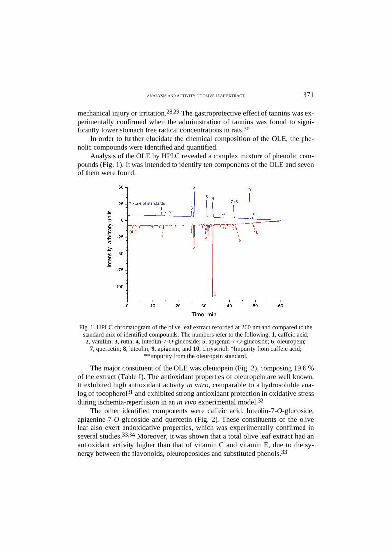
Analysis of the OLE by HPLC revealed a complex mixture of phenolic com-pounds (Fig. 1). It was intended to identify ten components of the OLE and seven of them were found.
Fig. 1. HPLC chromatogram of the olive leaf extract recorded at 260 nm and compared to the
standard mix of identified compounds. The numbers refer to the following: 1, caffeic acid; 2, vanillin; 3, rutin; 4, luteolin-7-O-glucoside; 5, apigenin-7-O-glucoside; 6, oleuropein;
7, quercetin; 8, luteolin; 9, apigenin; and 10, chryseriol. *Impurity from caffeic acid; **impurity from the oleuropein standard.
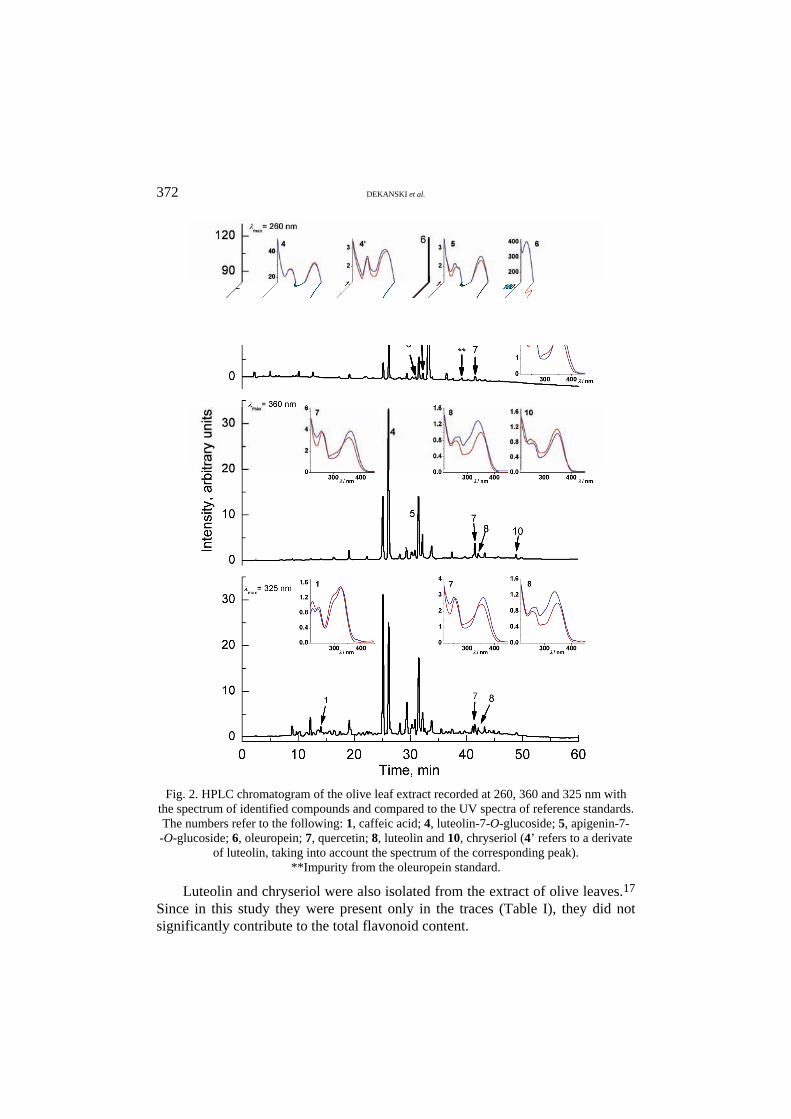
The major constituent of the OLE was oleuropein (Fig. 2), composing 19.8 % of the extract (Table I). The antioxidant properties of oleuropein are well known. It exhibited high antioxidant activity in vitro, comparable to a hydrosoluble ana-log of tocopherol31 and exhibited strong antioxidant protection in oxidative stress during ischemia-reperfusion in an in vivo experimental model.32
The other identified components were caffeic acid, luteolin-7-O-glucoside, apigenine-7-O-glucoside and quercetin (Fig. 2). These constituents of the olive leaf also exert antioxidative properties, which was experimentally confirmed in several studies.33,34 Moreover, it was shown that a total olive leaf extract had an antioxidant activity higher than that of vitamin C and vitamin E, due to the sy-nergy between the flavonoids, oleuropeosides and substituted phenols.33
372 DEKANSKI et al.
Fig. 2. HPLC chromatogram of the olive leaf extract recorded at 260, 360 and 325 nm with
the spectrum of identified compounds and compared to the UV spectra of reference standards. The numbers refer to the following: 1, caffeic acid; 4, luteolin-7-O-glucoside; 5, apigenin-7- -O-glucoside; 6, oleuropein; 7, quercetin; 8, luteolin and 10, chryseriol (4’ refers to a derivate
of luteolin, taking into account the spectrum of the corresponding peak). **Impurity from the oleuropein standard.
Luteolin and chryseriol were also isolated from the extract of olive leaves.17 Since in this study they were present only in the traces (Table I), they did not significantly contribute to the total flavonoid content.
ANALYSIS AND ACTIVITY OF OLIVE LEAF EXTRACT 373
TABLE I. Quantitative determination of flavonoids, phenolcarbonic acids and oleuropein in the studied olive leaf extract
Compound namea Amount
mg % Caffeic acid (1) 0.013 0.02 Vanillin (2) Not found – Rutin (3) Not found – Luteolin-7-O-glucoside (4) 0.027 0.04 Apigenin-7-O-glucoside (5) 0.046 0.07 Oleuropein (6) 13.147 19.8 Quercetin (7) 0.027 0.04 Luteolin (8) Traceb – Apigenin (9) Not found – Chryseriol (10) Traceb – aThe numbers refer to the compounds marked on the HPLC chromatogram (Figs. 1 and 2); bdetermination was not possible – present in the extract under the limit of quantitative analysis
Luteolin-7-O-glucoside is widespread in plant species and its anti-radical ac-tivity is well-known. Its presence in olive leaf was previously confirmed,12,35 as well as its anti-ulcer activity.29 This flavonoid was also identified and quantified in this study, composing 0.04 % of the extract (Table I).
Apigenin, vanillin and rutin were identified in olive leaf in some analytical studies.12,33,35 Their presence was not confirmed in this investigation, but 0.04 % of quercetin, 0.07 % of apigenine-7-O-glucoside and 0.02 % caffeic acid (Tab-le I) were found.
Quercetin is the most abundant of the flavonoid molecules and it is found in many medicinal botanicals. It has been reported to prevent gastric mucosal le-sions induced by ethanol.36 Quercetin increases the amount of neutral glycopro-teins in the gastric mucosa37 and thus participates in the recovery of the mucosal defensive capacity against aggression from absolute ethanol. Other possible me-chanisms include inhibition of lipid peroxidation,36 inhibition of the gastric pro-ton pump,37 and scavenging of free radicals associated with a significant enhan-cement in the glutathione peroxidase activity.38
Radical scavenging abilities for apigenine-7-O-glucoside and for caffeic acid were also reported.33
Effect of intragastrically applied OLE on gastric lesions induced by absolute ethanol
There are various plant-originating gastroprotectors with different composi-tions that have been used in clinical and folk medicine due to their beneficial effects on the gastric mucosa. The documented literature has centered primarily on their pharmacological action in experimental animals. Many studies have de-monstrated that substances with antioxidant properties (especially polyphenolic compounds) may protect against the gastric-damaging effects of absolute etha-nol.39–42 The beneficial properties of the polyphenols of olive leaf, the same as in
374 DEKANSKI et al.
olive oil, are further enhanced by their good bioavailability. The results obtained for oleuropein and its metabolites, tyrosol and hydroxytyrosol, indicated that they are readily absorbed through the gastrointestinal tract, resulting in significant levels in the circulation.43,44
In this study, the protective effect of OLE, a natural antioxidant, on the gas-tric mucosal damage induced by absolute ethanol in rats was studied.
The administration of absolute ethanol to fasted rats resulted in severe gas-tric damage, visible from the outside of the stomach as thick reddish-black lines. After opening, gastric lesions were found in the mucosa and consisted of elonga-ted bands, 1–10 mm long, usually parallel to the long axis of the stomach. They were located mostly in the corpus, the portion of the stomach secreting acid and pepsin. No visible lesions developed in the non-secretry part of the stomach.
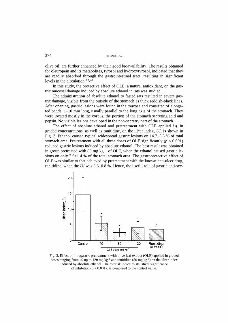
The effect of absolute ethanol and pretreatment with OLE applied i.g. in graded concentrations, as well as ranitidine, on the ulcer index, UI, is shown in Fig. 3. Ethanol caused typical widespread gastric lesions on 14.7±5.5 % of total stomach area. Pretreatment with all three doses of OLE significantly (p < 0.001) reduced gastric lesions induced by absolute ethanol. The best result was obtained in group pretreated with 80 mg kg–1 of OLE, when the ethanol caused gastric le-sions on only 2.6±1.4 % of the total stomach area. The gastroprotective effect of OLE was similar to that achieved by pretreatment with the known anti-ulcer drug, ranitidine, when the UI was 3.6±0.8 %. Hence, the useful role of gastric anti-sec-
Fig. 3. Effect of intragastric pretreatment with olive leaf extract (OLE) applied in graded doses ranging from 40 up to 120 mg kg-1 and ranitidine (50 mg kg-1) on the ulcer index
induced by absolute ethanol. The asterisk indicates statistical significance of inhibition (p < 0.001), as compared to the control value.
ANALYSIS AND ACTIVITY OF OLIVE LEAF EXTRACT 375
retry medication in the prevention of gastric mucosal damage was also confirmed. The obtained results indicate that the gastroprotective potential of OLE most
probably results from the ability of its constituents to scavenge reactive oxygen species, produced in ethanol-induced gastric injury, which initiate lipid peroxi-dation. The actual potential is probably related to its ability to maintain the integ-rity of the cell membrane, by its anti-lipid peroxidative activity and to protect in this way the gastric mucosa against oxidative damage, and by its ability to strengthen the mucosal barrier, the first line of defense against exogenous and endogenous ulcerogenic agents.
CONCLUSIONS
The investigated olive leaf extract caused a significant attenuation of the gas-tric damage induced by a corrosive concentration of ethanol, suggesting a res-pectable gastroprotective activity. This activity could be related to its antioxi-dative properties, since phytochemical analysis of OLE showed a high content of phenolic compounds, well-known antioxidants. However, in order to elucidate the mechanism of OLE gastroprotective effect, and to understand better the ac-tual potential, further investigation will be focused on the determination of lipid peroxidation and antioxidative enzyme activity in the gastric mucosa.
И З В О Д
ФИТОХЕМИЈСКА АНАЛИЗА И ГАСТРОПРОТЕКТИВНО ДЕЈСТВО ЕКСТРАКТА ЛИСТА МАСЛИНЕ
ДРАГАНА ДЕКАНСКИ1, СНЕЖАНА ЈАНИЋИЈЕВИЋ-ХУДОМАЛ2, ВАЊА ТАДИЋ3,
ГОРАН МАРКОВИЋ3, ИВАНА АРСИЋ3 и ДУШАН М. МИТРОВИЋ4
1Biomedicinska ispitivawa, Institut za istra`ivawe i razvoj, Galenika a.d., Pasterova 2, 11000
Beograd, 2Katedra za farmakologiju, Medicinski fakutet, Univerzitet u Pri{tini (Kosovska
Mitrovica), 3Institut za prou~avawe lekovitog biqa “Dr Josif Pan~i}”, Tadeu{a Ko{}u{ka 1,
11000 Beograd i 4Institut za medicinsku fiziologiju, Medicinski fakultet,
Univerzitet u Beogradu, Vi{egradska 26, 11000 Beograd
Нека лековита својства листа маслине су позната вековима. Он се традиционално ко-ристи као антимикробни агенс и за превенцију и лечење шећерне болести и срчаних обо-љења. Остало је непознато да ли примена листа маслине утиче на одбрамбене механизме слузнице желуца и да ли показује гастропротективно дејство код експериментално индуко-ваних лезија желуца. У екстракту листа маслине прво је одређен садржај укупних фенола, флавоноида и танина, а потом идентификовано и квантификовано 7 фенолних једињења (олеуропеин, кафена киселина, лутеолин, лутеолин-7-О-глукозид, апигенин-7-О-глукозид, кверцетин и хризериол). Испитано је и протективно дејство екстракта листа маслине код оштећења желудачне мукозе корозивном концентрацијом етанола. Претретман екстрактом листа маслине у дозама од 40, 80 и 120 mg kg-1 значајно је смањио оштећење желуца у односу на контролну групу (p < 0,001). Ово дејство је било слично дејству референтног лека, ранитидина. Добијени резултати указују да екстракт листа маслине поседује значајно гастро-протективно дејство, које би се донекле могло објаснити присуством антиоксиданаса у њего-вом хемијском саставу.
(Примљено 8. септембра, ревидирано 7. новембра 2008)
376 DEKANSKI et al.
REFERENCES
1. Physician’s Desk References for Herbal Medicine, Medical Economics Company, Montvale, NJ, 2000, p. 556
2. M. Gonzales, A. Zarzuelo, M. J. Gamez, M. P. Utrilla, J. Jimenez, I. Osuna, Planta Med. 58 (1992) 513
3. H. F. Al-Azzawie, M. S. Alhamdani, Life Sci. 78 (2006) 1371 4. M. T. Khayyal, M. A. el-Ghazaly, D. M. Abdallah, N. N. Nassar, S. N. Okpanyi, M. H.
Kreuter, Arzneim.-Forsch. 52 (2002) 797 5. L. I. Somova, F. O. Shode, M. Mipando, Phytomedicine 11 (2004) 121 6. L. Wang, C. Geng, L. Jiang, G. Gong, D. Liu, H. Yoshimura, L. Zhong, Eur. J. Nutr. 47
(2008) 235 7. A. Zarzuelo, J. Duarte, J. Jimenez, M. Gonzales, M. P. Utrilla, Planta Med. 57 (1991) 417 8. A. A. Al-Quarawi, M. A. Al-Damegh, S. A. El-Mougy, Phytother. Res. 16 (2002) 286 9. G. Bisignano, A. Tomaino, R. Lo Cascio, G. Crisafi, N. Uccella, A. Saija, J. Pharm.
Pharmacol. 51 (1999) 971 10. P. M. Furneri, A. Marino, A. Saija, N. Ucella, G. Bisignano, Int. J. Antimicrob. Agents 20
(2002) 293 11. D. Markin, L. Duek, I. Berdicevsky, Mycoses 46 (2003) 132 12. A. P. Pereira, I. C. Ferreira, F. Marcelino, P. Valentao, P. B. Andrade, R. Seabra, L. Este-
vinho, A. Bento, J. A. Pereira, Molecules 12 (2007) 1153 13. S. Lee-Huang, L. Zhang, P. L. Huang, Y. T. Chang, Biochem. Biophys. Res. Commun.
307 (2003) 1029 14. V. Micol, N. Caturla, L. Perez-Fons, V. Mas, L. Perez, A. Estepa, Antiviral Res. 66
(2005) 129 15. H. K. Hamdi, R. Castellon, Biochem. Biophys. Res. Commun. 334 (2005) 769 16. L. Abaza, T. P. N. Talorete, P. Yamada, Y. Kurita, M. Zarrouk, H. Isoda, Biosci. Biotech-
nol. Biochem. 71 (2007) 1306 17. A. Pieroni, D. Heimler, L. Pieters, B. van Poel, A. J. Vlietinck, Pharmazie 51 (1996) 765 18. J. Meirinhos, B. M. Silva, P. Valentao, R. M. Seabra, J. A. Pereira, A. Dias, P. B. Andra-
de, F. Ferreres, Nat. Prod. Res. 68 (2005) 189 19. S. Kwiecien, T. Brzozowski, S. J. Konturek, J. Physiol. Pharmacol. 53 (2002) 39 20. V. L. Singleton, R. Orthofer, R. M. Lamuela-Raventós, in Methods in Enzymology,
Oxidants and Antioxidants (Part A), L. Packer, Ed., Academic Press, San Diego, CA, 1999, p. 152
21. European Pharmacopoeia 6.0, Council of Europe, Strasbourg, 2008, p. 255 22. Weißdornblätter mit Blüten, in Deutsches Arzneibuch, Band 2, Monographien R-Z, Am-
litche Ausgabe Deutscher Apotheker Verlag, Govi-Verlag GmbH, Stuttgart, 1991 23. R. Rucker, D. Storms, J. Nutr. 132 (2002) 2999 24. R. B. Rucker, J. Anim. Physiol. Anim. Nutr. 91 (2007) 148 25. ImageJ, Image Processing and Analysis in Java, http://rsb.info.nih.gov/ij/ (May, 2008) 26. O. S. Zayachkivska, S. J. Konturek, D. Drozdowicz, P. C. Konturek, T. Brzozowski, M.
R. Ghegotsky. J. Physiol. Pharmacol. 56 (2005) 219 27. R. J. Nijveldt, E. van Nood, D. E. C. van Hoorn, P. G. Boelens, K. van Norren, P. A. M.
van Leeuwen, Am. J. Clin. Nutr. 74 (2001) 418 28. I. U. Asuzu, O. U. Onu. Int. J. Crude Drug. Res. 28 (1990) 27 29. F. Borelli, A. A. Izzo, Phytother. Res. 14 (2000) 581
ANALYSIS AND ACTIVITY OF OLIVE LEAF EXTRACT 377
30. R. O. Ramirez, C. C. Roa, Jr., Clin. Hemorheol. Microcirc. 29 (2003) 253 31. E. Speroni, M. C. Guerra, A. Minghetti, N. Crespi-Perellino, P. Pasini, F. Piazza,
Phytother. Res. 12 (1998) S98 32. I. Andreadou, E. K. Iliodromitis, E. Mikros, M. Constantinou, A. Agalias, P. Magiatis, A.
L. Skaltsounis, E. Kamber, A. Tsantili-Kakoulidou, D. Th. Kremastinos, J. Nutr. 136 (2006) 2213
33. O. Benavente-Garcia, J. Castillo, J. Lorente, A. Ortuno, J. A. Del Rio, Food Chem. 68 (2000) 457
34. R. Briante, M. Paturni, S. Terenziani, E. Bismuto, F. Febbraio, R. Nucci, J. Agric. Food Chem. 50 (2002) 4934
35. N. De Laurentis, L. Stefanizzi, M. A. Milillo, G. Tantillo, Ann. Pharm. Fr. 56 (1998) 268 36. C. Alarcon de la Lastra, M. J. Martin, V. Motilva, Pharmacology 48 (1994) 56 37. G. Di Carlo, N. Mascolo, A. A. Izzo, F. Capasso, Life Sci. 65 (1999) 337 38. M. J. Martin, C. La Casa, C. Alarcon de la Lastra, J. Cabezza, I. Villegas, V. Motilva, Z.
Naturforsch. 53 (1998) 82 39. V. M. Tadić, S. Dobrić, G. M. Marković, S. M. Đorđević, I. A. Arsić, N. R. Menković, T.
Stević, J. Agric. Food Chem. 56 (2008) 7700 40. S. B. Olaleye, E. O. Farombi. Phytother. Res. 20 (2006) 14 41. S. Đorđević, S. Petrović, S. Dobrić, M. Milenković, D. Vučićević, S. Žižić, J. Kukić, J.
Ethnopharmacol. 109 (2007) 458 42. M. P. Germano, V. D’Angelo, T. Biasini, T. C. Miano, A. Braca, M. De Leo, R. De
Pasquale, R. Sanogo, J. Ethnopharmacol. 115 (2008) 271 43. F. Visioli, C. Galli, F. Bornet, A. Mattei, R. Patelli, G. Galli, D. Caruso, FEBS Lett. 468
(2000) 159 44. M. N. Vissers, P. L. Zock, A. J. C. Roodenburg, R. Leenen, M. B. Katan, J. Nutr. 132
(2002) 409.