Una formación cultural italiana en Buenos Aires (1890- 1910)

A
aftofast©
K
1
wHbdotrlpltsaei
1d
Journal of Cultural Heritage 10 (2009) 501–508
Case Study
Noninvasive physicochemical characterization of two 19thcentury English ferrotypes
Emiliano Carretti , Marco Milano , Luigi Dei ∗, Piero BaglioniDepartment of Chemistry and CSGI Consortium, University of Florence, via della Lastruccia, 3, 50019 Sesto Fiorentino (Firenze), Italy
Received 8 February 2008; accepted 25 February 2009
bstract
The present work was one of the first attempts to analyze the conservation status of two ferrotypes, ancient photographic plates realized onsupport made of iron. The photographic material was constituted of collodion as binder for the photosensitive silver halides grains. The two
errotypes studied belonged to a private collection of a family from Durham, UK, and were made at the end of the 19th century. The analyticalechniques used for the morphological and physicochemical characterization were noninvasive. The surface morphology was studied by meansf optical microscopy (OM) and environmental scanning electron microscopy (ESEM) coupled with an energy dispersive X-rays (EDX) systemor the elemental analysis. These techniques, together with microreflectance Fourier transform infrared spectroscopy (�-FTIR) and contact angle,
llowed to obtain information on both the chemical – elemental – composition of the materials constituting the ferrotypes, and the conservationtatus of these photographic plates. The study showed that the physicochemical diagnostics allowed to characterize the two ferrotypes that, despiteheir similar age and provenance, showed different conservation status, surface properties, and elemental composition.2009 Elsevier Masson SAS. All rights reserved.
iltoosbtgt(im[t
eywords: Ferrotypes; Collodion; Photographs; Physicochemical diagnostics
. Introduction
The photographic process called ferrotype or also tintypeas patented in the United States on February 19th, 1856 byamilton Smith, professor at Kenyon College, in Ohio [1] andecame soon one of the most diffused processes until the intro-uction of the gelatin based systems and the invention by Kodakf the commercial photocameras [2]. In particular, with respecto the ambrotypes, in the ferrotypes the support glass plate waseplaced by an iron plate coated with a surface layer of wet col-odion. As in the case of ambrotypes, in all the ferrotypes thehotosensitive grains are formed on the upper layer of the col-odion [3,4]. In these photographic techniques, the first step ishe preparation of the support (metal or glass) which dimensionshould be decided as a function of the camera’s size, taking into
ccount that the maximum possible size of the final image isxactly the one of the plate. Then, in order to produce a bigmage (i.e. 13 × 18 cm), a camera of compatible dimensions∗ Corresponding author. Fax: +39 055 4573036.E-mail address: [email protected] (L. Dei).
g1i
A
A
296-2074/$ – see front matter © 2009 Elsevier Masson SAS. All rights reserved.oi:10.1016/j.culher.2009.02.002
s needed. Once defined the size of the picture, the collodionayer is deposed directly onto the support surface as a concen-rated solution of nitrocellulose in alcohol plus ether. Then, thebtained plate, not completely dried, is immersed into a solutionf AgNO3 in order to precipitate the silver halide AgX from thealts (usually binary mixtures of alkaline and/or earth-alkalineromides or iodides) which are homogeneously dispersed intohe wet collodion layer. The precipitation of the photosensitiverains occurs within the upper layer of the plate. While still wet,he ferrotype is placed in the camera and an exposure is madeusually the exposition time varies from 1 to 30 seconds). Thiss the reason why this process is known as the “wet collodion
ethod”, the same that is used for the production of ambrotypes5]. During the exposition, the interaction between the light andhe silver halide grains induces a photoreduction of the AgX,iving the formation of clusters like Ag+
n and Ag0n (usually n >
0) containing free metal silver atoms. The reaction mechanisms described below:
gX(crystal) + hν(radiation) → Ag+ + X + e− (1)
g+ + e− → Ag0 (2)
5 ultural Heritage 10 (2009) 501–508
ictsttbt[tm[ppatgnctatsthbimuvad(tldacswtotfttp
vcpEgc
r
F
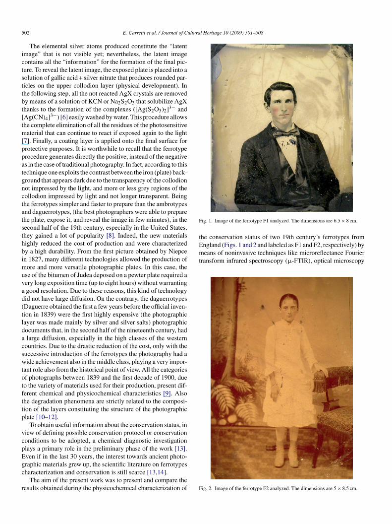
the conservation status of two 19th century’s ferrotypes fromEngland (Figs. 1 and 2 and labeled as F1 and F2, respectively) bymeans of noninvasive techniques like microreflectance Fouriertransform infrared spectroscopy (�-FTIR), optical microscopy
02 E. Carretti et al. / Journal of C
The elemental silver atoms produced constitute the “latentmage” that is not visible yet; nevertheless, the latent imageontains all the “information” for the formation of the final pic-ure. To reveal the latent image, the exposed plate is placed into aolution of gallic acid + silver nitrate that produces rounded par-icles on the upper collodion layer (physical development). Inhe following step, all the not reacted AgX crystals are removedy means of a solution of KCN or Na2S2O3 that solubilize AgXhanks to the formation of the complexes ([Ag(S2O3)2]3− andAg(CN)4]3−) [6] easily washed by water. This procedure allowshe complete elimination of all the residues of the photosensitiveaterial that can continue to react if exposed again to the light
7]. Finally, a coating layer is applied onto the final surface forrotective purposes. It is worthwhile to recall that the ferrotyperocedure generates directly the positive, instead of the negatives in the case of traditional photography. In fact, according to thisechnique one exploits the contrast between the iron (plate) back-round that appears dark due to the transparency of the collodionot impressed by the light, and more or less grey regions of theollodion impressed by light and not longer transparent. Beinghe ferrotypes simpler and faster to prepare than the ambrotypesnd daguerrotypes, (the best photographers were able to preparehe plate, expose it, and reveal the image in few minutes), in theecond half of the 19th century, especially in the United States,hey gained a lot of popularity [8]. Indeed, the new materialsighly reduced the cost of production and were characterizedy a high durability. From the first picture obtained by Niepcen 1827, many different technologies allowed the production of
ore and more versatile photographic plates. In this case, these of the bitumen of Judea deposed on a pewter plate required aery long exposition time (up to eight hours) without warrantinggood resolution. Due to these reasons, this kind of technologyid not have large diffusion. On the contrary, the daguerrotypesDaguerre obtained the first a few years before the official inven-ion in 1839) were the first highly expensive (the photographicayer was made mainly by silver and silver salts) photographicocuments that, in the second half of the nineteenth century, hadlarge diffusion, especially in the high classes of the western
ountries. Due to the drastic reduction of the cost, only with theuccessive introduction of the ferrotypes the photography had aide achievement also in the middle class, playing a very impor-
ant role also from the historical point of view. All the categoriesf photographs between 1839 and the first decade of 1900, dueo the variety of materials used for their production, present dif-erent chemical and physicochemical characteristics [9]. Alsohe degradation phenomena are strictly related to the composi-ion of the layers constituting the structure of the photographiclate [10–12].
To obtain useful information about the conservation status, iniew of defining possible conservation protocol or conservationonditions to be adopted, a chemical diagnostic investigationlays a primary role in the preliminary phase of the work [13].ven if in the last 30 years, the interest towards ancient photo-
raphic materials grew up, the scientific literature on ferrotypesharacterization and conservation is still scarce [13,14].The aim of the present work was to present and compare theesults obtained during the physicochemical characterization of F
ig. 1. Image of the ferrotype F1 analyzed. The dimensions are 6.5 × 8 cm.
ig. 2. Image of the ferrotype F2 analyzed. The dimensions are 5 × 8.5 cm.

E. Carretti et al. / Journal of Cultura
Fr
(ct
2
lwS1
msippsws
arcs
uUafN(md1Amethe images collected in polarized light were in crossed nicolsmodality.
ig. 3. Microreflectance �-FTIR spectrum of a nitrocellulose standard in theange 800–4000 cm-1 (A) and magnification of the fingerprint region (B).
OM) and environmental scanning electron microscopy (ESEM)oupled with EDX analysis, all techniques usually employed forhe characterization of several artifacts [15–20].
. Materials and methods
Diethylether, ethanol (for both purity > 99.0%) and nitrocel-ulose were purchased from Aldrich and used as received, wateras purified with a Millipore MilliRO-6 and MilliQ (Organexystem) apparatus: the resistance of the water was greater than5 M� cm.
ESEM images were collected by means of a FEI Quanta 200icroscope coupled with an EDX (SUTW detector). The dimen-
ions of the microscope chamber were 20 × 15 cm, allowing thentroduction of the full plates and avoiding the necessity of sam-ling. The working distance was 10 mm and the acceleration
otential 30 KV. For the morfological analysis, ESEM was cho-en because it was possible to collect the electronic micrographsithout graphitizing or gilding the sample surface. This noninva-ive technique is commonly used for biological samples [21,22]
Ffo
l Heritage 10 (2009) 501–508 503
nd also for the study of artistic [23] and photographic mate-ials like albumen photographs [24]. The EDX spectra wereollected using a microprobe coupled with the ESEM micro-cope.
�-FTIR spectra were obtained in the micro-reflectance modesing a BioRad FTS-40 spectrometer equipped with a BioRadMA500 microscope (MCT detector) with 8 cm−1 resolution
nd 512 scans. Contact angle measurements [25,26] were per-ormed onto the ferrotype surface by means of a PC-controlledRLRamé-Hart Inc. apparatus interfaced to a PC. Water droplets
5 �l) were deposed onto the solid surface using a Hamiltonicrosyringe and the contact angle was determined 5 s after the
eposition. The contact angles reported were the average over0 measurements and the errors were the standard deviations.ll the optical micrographs were collected in the reflectanceode by means of a Reichert Zetopan optical microscope,
quipped with 5.5×, 11×, 28× objectives and a 8× ocular. All
ig. 4. Microreflectance �-FTIR spectra collected from the front side of theerrotype F1 (A, spectrum 1) and F2 (B, spectrum 1) compared with the spectrumf the standard of nitrocellulose (spectrum 2) in the fingerprint region.

504 E. Carretti et al. / Journal of Cultural Heritage 10 (2009) 501–508
F2 (C
3
lapa(li(iCgOt
fadeatsgvweat
fves
oEFtfmFtlctbfiaidsstp
Fig. 5. Optical micrographs of the ferrotype F1 (A and B) and
. Results and discussion
For the FTIR analysis, first a standard of not aged nitrocellu-ose was prepared by solubilization of this polymer (7% w/w) insolution of ether plus alcohol. A portion of this solution wasut onto a Teflon plate and, once dried, the obtained film wasnalyzed by means of micro-reflectance �-FTIR. The spectrumFig. 3) was used as reference and compared with the ones col-ected from the ferrotype surface. The diagnostic peaks for thedentification of nitrocellulose were the ones below 1800 cm−1
Fig. 3B). In particular, the peak at 1730 cm−1, due to the stretch-ng of the C O bond, the peak at 1650 cm−1 (stretching of the
N bond), and the peak at 1280 cm−1 (stretching of the O NO2roup). The band at 3500 cm−1 is due to the stretching of theH; the peaks between 3100 cm−1 and 2800 cm−1 are due to
he stretching of aliphatic CH2 and CH3 (Fig. 3A).Fig. 4 reports the microreflectance �-FTIR spectra collected
rom the front side of the two ferrotypes (spectra 1A for F1nd 1B for F2). By comparison between them and the stan-ard spectrum of nitrocellulose (spectra 2A and 2B), it wasvident that, as expected for ferrotypes, collodion has been useds binder for the photosensitive grains that allow the forma-ion of the image. In both cases, a correspondence of the FTIRignals with the ones of the standard was observed in the fin-erprint region. The two surfaces of F1 and F2 (front side) wereery similar, with the presence of collodion in both cases. It
as interesting to notice that both the spectra showed the pres-nce of the peak at around 1560 cm−1 attributable to the C Omide II typical of proteinaceous materials. This could be dueo the presence of some protein-based protective coatings, base
obdF
and D). B and D are magnifications of A and C, respectively.
or retouching/pre-coatings [3]. No natural resins as protectivearnishes were detected by �-FTIR: this could be due to the pres-nce of the collodion whose strong peaks matches the typicalpectral features of natural resins.
In order to deepen the investigation, morphological analysisf the two ferrotypes was performed by means of optical andSEM microscopy. As indicated in the optical micrographs inig. 5A and C, at a microscopic level, the morphology of the
wo ferrotypes was totally different. While the surface of theerrotype F1 was characterized by a roughness (in the order oficrometers) similar in all the regions of the plate, the one of2 appeared completely flat. This finding could be attributed to
he presence of a morphologically different protective surfaceayer deposed onto the F2 ferrotype. This was also confirmed byontact angle measurements. While the contact angle value forhe ferrotype F2 was almost the same in both the front and theack sides (around 60 ± 7◦), in the plate F1 this parameter variedrom a value close to 60o in the back and in some regions of themage where the grazing light analysis showed the presence ofshiny surface layer, to a much higher value (around 75 ± 5◦)
n the regions where this layer was not detectable. From theseata, it was possible to conclude that some regions of the frontide of F1 were less hydrophilic than its backside and than bothides of F2. This was in agreement with the presence on bothhe surfaces of a material more hydrophilic than collodion (i.e.roteinaceous), except in the degraded regions of the front side
f F1 where the coating was lost and the wettability showed aehaviour typical of more hydrophobic substances (i.e. collo-ion). These findings, associated with the morphology shown inig. 5, indicated a worse state of conservation of the ferrotype
E. Carretti et al. / Journal of Cultural Heritage 10 (2009) 501–508 505
F ined wb perfo
Fts
FdathllcrlcssUd
dscrtps
tcbit–apwptwthe contamination coming from other sources. The presence ofS came from the fixing process made by S2O3
2−. EDX (Table 1)indicated also the presence of Fe, K, and Mg. The first was prob-ably due to the presence of Fe-compounds originated from the
Table 1Elemental composition from EDX (expressed as atomic %) of the Points 1 and2 in Fig. 6C.
Element Atomic % Point 1 Atomic % Point 2
Mg 11.6 11.5K 1.7 1.9
ig. 6. ESEM micrographs obtained from the surface of the ferrotype F1 obtaack-scattered electrons indicating the two points where elemental analysis was
1. Then it was possible to conclude that the conservation sta-us of the two plates was different and particularly F2 showed aurface texture better than F1.
The pattern seen in the optical micrographs reported inig. 5A and B were extraordinary similar to the well-knownewetting figures [27]. This effect could be due to two differentlternative mechanisms of dewetting of the collodion fromhe support, leading in certain cases, to the appearance of trueoles. (i) The amount of solvent used for its preparation was tooarge causing the formation of a low viscous collodion-basedayer inducing a rapid local dewetting from the plate; as aonsequence, after the exposition to the light, the solventapidly evaporated causing the formation of the nitrocelluloseayer embedding the reduced Ag0 clusters, the residual AgXrystals, and characterized by a morphologically irregularurface. (ii) An extremely slow dewetting process on the timecale (years) typical of photographic collodion deterioration.nfortunately, we did not have experimental evidences toiscriminate between these two possibilities.
In the optical micrographs in Fig. 5A and B, it was also evi-ent the presence of small circular features (Point 1 in Fig. 5A). Aimple morphological investigation suggested that they could beonstituted of crystals or solid particles that precipitate as impu-
ities into the collodion layer. In order to verify this hypothesis,he investigation by means of the ESEM/EDX microscope wasarticularly useful. Fig. 6 shows the micrographs obtained withecondary and back-scattered electrons (Fig. 6A and B, respec-SCAF
ith secondary (A) and back-scattered electrons (B). (C) Image obtained withrmed. (D) EDX spectrum collected in the Point 1.
ively) that did not show any difference in the tone of the grey inorrespondence of these spots. This finding was also confirmedy the elemental analysis carried out both in a dark spot and alson a point of the surrounding area (Fig. 6C Points 1 and 2, respec-ively) and reported in Table 1. This indicated that the chemicalelemental – composition was almost the same. This result wasfurther confirmation that the “dark circular features” were sim-ly surface imperfections and that the drying mechanism of theet collodion layer could be considered as the main cause of thearticular morphology of the ferrotype surface. The presence ofhe chloride element (between 4 and 5%) could be attributed to aashing process carried out using Cl–-contaminated water, or to
5.0 4.5l 4.9 4.2g 72.3 73.2e 4.5 4.6

506 E. Carretti et al. / Journal of Cultural Heritage 10 (2009) 501–508
F olloda magnw turally
dop
itaoratdwctpi
TEF
E
NSSAKF
aFanEtlS2O3
2− was used as sodium salt. Finally, the presence of Sicould be attributed to the deposition of powder onto the regionwere the collodion lacuna was present.
ig. 7. (A) Image of the ferrotype F1; in the inset the region where a lacuna of crrow indicates the region where the elemental analysis was performed (C is theas collected). (D) SEM micrograph obtained with secondary electrons of a na
egradation of the plate; the second ones were due to remainingf impurities of potassium and magnesium halides used for therecipitation of the photosensitive materials.
From the ESEM/EDX analysis, it was also possible to achievenformation about the composition – elemental – of the two pho-ographic plates. As it is shown in Fig. 7A and indicated by therrow in Fig. 7B, in the F1 plate there was a region where partf the collodion layer was lost, but �-FTIR indicated that aesidual amount of collodion was still present. The elementalnalysis performed in that area (Fig. 7C and Table 2) showedhat the amount of Ag was much smaller than the one previouslyetected onto the surface of the same plate (Table 1). This effectas not surprising due to the fact that for ferrotypes Ag is con-
entrated onto the superficial layer of the collodion film where
he photosensitive grains of AgX are precipitated. Also the mor-hology of F1 was completely different from that of F2 (videnfra, Fig. 8) and it was similar to the one of a naturally 80 yearsable 2lemental composition from EDX (expressed as atomic %) of the Points 1 inig. 7C.
lement Atomic %
a 4.9i 5.3
70.1g 2.6
5.8e 11.3 F
2
ion was present. (B) ESEM micrograph obtained with secondary electrons, theification of B in proximity of the arrow and X is the point were EDX spectrumaged nitrocellulose film.
ged nitrocellulose film which ESEM micrographs is reported inig. 7D. This sample was obtained from a portion of a naturallyged Italian motion-picture film of the 1920s made by celluloseitrate. Particularly, the pattern of the fractures observed in theSEM micrographs (Fig. 7C and D) was similar, indicating that
heir formation was mainly due to the aging of the collodionayer. Finally, Na was also detected because the fixing agent
ig. 8. ESEM micrographs collected from the surface of the ferrotype F2. 1 andindicate the points where the elemental analysis was performed.

E. Carretti et al. / Journal of Cultura
Table 3Elemental composition from EDX (expressed as atomic %) of the Points 1 and2 in Fig.8.
Element Atomic % Point 1 Atomic % Point 2
Ag 28.0 72.2FS
FrftpocF
4
tsEarwaf
oesaaFacfabl
ttisltst
eopd
A
withDtlMpfd
R
[
[
[
[
[
[
e 72.0 22.2/ 5.6
ESEM analysis was also performed onto the F2 surface (seeig. 8 and Table 3): this investigation highlighted that, withespect to F1, there were three main differences. First, the sur-ace appeared much smoother and more homogeneous; second,he amount of Ag was less homogeneous in passing from aoint to another zone of the surface; third, the concentrationf iron was quite higher. This difference in Ag and Fe con-entrations could be ascribable to a thinner collodion layer for2.
. Conclusions
The multi-technique approach presented in this paper allowedo study for the first time the chemical – elemental – compo-ition and the conservation status of two ferrotypes made inngland at the end of the 19th century, without the collection ofny samples, i.e. by means of noninvasive techniques. Micro-eflectance FTIR indicated that some proteinaceous materialsere present, ascribable to protective coatings for retouching
nd/or pre-coating and no presence of natural varnishes wasound.
The morphological investigation, performed both by meansf optical and ESEM microscopy, allowed to establish the differ-nt conservation status of the two ferrotypes, indicating that F2ample surface was homogeneous and not meaningfully dam-ged, while F1 surface roughness was clearly high. Contactngle measurements further confirmed this feature: indeed, only1 surface was characterized by the presence of a texturettributable to the surface dewetting process of the collodionaused by the kinetics of the evaporation of the solvents usedor the preparation of the collodion layer. Moreover, contactngle measurements clearly indicated that for F1 the wetta-ility was altered, probably by a not homogenous protectiveayer.
The analysis carried out by EDX spectra showed that evenhe chemical – elemental – composition was different comparinghe two plates. In fact, F2 plate presented Ag and Fe amountsn agreement with a quite thin collodion layer, whereas F1 platehowed lower concentration of Fe indicating a thicker collodionayer. This was quite interesting since we were able to concludehat the thickness of the collodion stratum and concentration ofilver in its upper layers are parameters not directly associatedo the conservation status of the ferrotypes.
In conclusion, our study allowed to compare the morphology,
lemental composition, and surface wettability of two ferrotypesf the same period enabling to underline the differences of thesearameters associated to the different status of conservation,espite their similar age and provenance.[
l Heritage 10 (2009) 501–508 507
cknowledgements
The Authors wish express their gratitude to the Reviewerhose comments and criticisms were fundamental for improv-
ng the paper. Thanks to Mr. Leopoldo Morandi, who gave ushe opportunity to study the ferrotypes and some very importantistorical indications about the two ferrotypes analyzed, and tor. Giovanna Poggi for useful suggestions. Authors also thank
he Cineteca Comunale, Bologna, Italy, for giving the cellu-ose nitrate motion-picture film sample. Thanks are also due to
IUR, Italy (PRIN 2006), to the Consorzio Interuniversitarioer lo sviluppo dei Sistemi a Grande Interfase (CSGI-Firenze)or financial support, and to the University of Florence (Fondi’Ateneo ex-60%).
eferences
[1] William Welling, Photography in America, Crowell, New York, USA, 1978.[2] L. Scaramella, Materiali della cultura artistica, in: Fotografia. Storia e
riconoscimento dei procedimenti fotografici, Edizioni De Luca, Roma,1999.
[3] E.M. Eastbrooke, The Ferrotype and How to Make it, Linsday Publications,USA, 2007.
[4] G. Berkhofer, Wet Collodion Photography – A Short Manual, Lulu.comPublications, USA, 2007.
[5] C.B. Neblette, Fundamentals of Photography, Van Norstrand Reinhold Co,Princeton, 1970.
[6] The reaction that allows the solubilization of AgX is:AgX + 2S2O3
2– → [Ag(S2O3)2]3– + X-. Then, the equilibrium constant is:
Keq=[
[X−][Ag(S2O3)3−2 ]
]/[(S2O3)3−
2 ]2=Kps(AgX)×Kst[Ag(S2O3)3−
2 ]
Kps(AgCl) = 1.8 × 10⁻¹⁰; Kst[Ag(S2O3)3−2 ] = 2.9 × 10¹³; Kps(AgBr) =
5.0 × 10⁻¹³ If X corresponds to Cl, then the Keq = 5.2 × 10³, if X is Br,Keq is 14.4. In both cases the solubilization of AgX is complete. A similarbehaviour is also observed if CN- is used as fixing agent instead of S2O3
2–
Kst[Ag(CN)3−4 ] = 4.0 × 10²⁰. All the constant values are from: J.A. Dean,
Lange’s Handbook of Chemistry, McGraw-Hill, New York, 1985.[7] C.E.K. Mees, T.H. James, The Theory of the Photographic Process, third
ed., The Macmillan Co, New York, 1966.[8] W. Crawford, L’età del collodio, Cesco Capanna Editore, Milano, 1981.[9] B. Cattaneo, D. Chelazzi, R. Giorgi, T. Serena, C. Merlo, P. Baglioni,
Physico-chemical characterization and conservation issues of photographsdated between 1890–1910, Journal of Cultural Heritage 9 (2008) 277–284.
10] K.B. Hendriks, The stability and preservation of recorded images, in: J.Sturge, al. et (Eds.), Imaging Processes and Materials, Van Nostrand Rein-hold, New York, 1989, pp. 637–684.
11] R.W. Henn, D.G. Wiest, Microscopic spots in processed microfilms: theirnature and prevention, Photographic Science and Engineering 7 (1963)253–261.
12] G. Di Pietro, Silver mirroring on silver gelatin glass negatives, PhD Thesis,University of Basel, 2002.
13] N. Davis, Tintypes: preliminary research and testing, in: Proceedings ofthe art conservation training programs conference, Coopers Town, 1984,pp. 13–28.
14] Bovis M., Les anciens procédés photographiques : V. Amphitypie, fer-rotypie, collodion sec, Photo-Cine-Revue, October 1971, pp. 450–453.
15] S. Masanori, Y. Sasaki, Studies on ancient Japanese silk fibres using FTIRmicroscopy, in: Postprints of the First Annual Conference Scientific Anal-
ysis of Ancient and Historic Textiles: Informing Preservation, Display andInterpretation, 13–15 July 2004, Archetype Publications, London, 2005,pp. 44–47.16] J. Van der Weerd, A. Van Loon, J. Boon, FTIR studies of the effects ofpigments on the aging of oil, Studies in Conservation 50 (2005) 3–22.

5 ultura
[
[
[
[
[
[
[
[
[
08 E. Carretti et al. / Journal of C
17] S.A. Centeno, M.I. Guzman, A. Yamazaki-Kleps, C.O. Védova, Char-acterization by FTIR of the effect of lead white on some properties ofproteinaceous binding media, Journal of the American Institute for Conser-vation 43 (2004) 139–150.
18] A. Langley, A. Burnstock, The analysis of layered paint samples frommodern paintings using FTIR microscopy, in: Preprints of the 12th ICOMTriennial Meeting, Lyon, 29 August–3 September 1999, James & James,London, 1999, pp. 234–241.
19] D. Magaloni, M. Aguilar, V. Castano, Electron and optical microscopy ofPrehispanic mural paintings, in: P.B. Vandiver, J. Druzik, G.S. Wheeler(Eds.), Proceedings of the Symposium Materials Issues in Art andArchaeology II, San Francisco, April 17–21, Materials Research Society,Pittsburgh, 1991, pp. 145–150.
20] H.B. Berg, Characterization of printing papers by optical and electronmicroscopy, Tappi 49 (1966) 554–562.
21] L. Muscariello, F. Rosso, G. Marino, A critical overview of ESEM appli-cations in the biological field, Journal of Cellular Physiology 205 (2005)328–334.
[
[
l Heritage 10 (2009) 501–508
22] V.A. Edward, V.L. Pillay, P. Swart, Localisation of Thermomyces lanug-inosus SSBP xylanase on polysulphone membranes using immunogoldlabelling and environmental scanning electron microscopy (ESEM), Pro-cess Biochemistry 38 (2003) 939–943.
23] A. Masic, E. Badea, R. Ceccarelli, G. Della Gatta, S. Coluccia. Compara-tive DSC and SEM/EDX study of ancient and artificially aged parchment,Proceedings of the II National Conference Lo stato dell’Arte 2: Conser-vazione e Restauro, Confronto di Esperienze, Genova, 27–29 settembre2004, Il Prato, Saonara, pp 52–61.
24] P. Messier, T. Vitale, Cracking in albumen photographs: an ESEM investi-gation, Microscopy Research and Technique 25 (1993) 374–383.
25] J. Drelich, J.L. Wilbur, J.D. Miller, G.M. Whitesides, Langmuir 12 (1996)1913–1922.
26] J. Drelich, J.D. Miller, A. Kumar, G.M. Whitesides, Colloids and SurfacesA: Physicochemical and Engineering Aspects 93 (1994) 1–13.
27] C. Neto, K. Jacobs, R. Seemann, R. Blossey, J. Becker, G. Grün, Satellitehole formation during dewetting: experiment and simulation, Journal ofPhysics: Condensed Matter 15 (2003) 3355–3366.
Copyright © 2022 FDOKUMEN


![Der Gebirgsbote 1910 [Jg. 62]](https://static.fdokumen.com/doc/165x107/6314a5b0aca2b42b580d9b81/der-gebirgsbote-1910-jg-62.jpg)