Physical fitness assessment during pregnancy
144
-
Upload
khangminh22 -
Category
Documents
-
view
1 -
download
0
Transcript of Physical fitness assessment during pregnancy


Physical fitness assessment during pregnancy:
validity and reliability of fitness tests and association with maternal-fetal health.
The GESTAFIT project
Lidia Romero Gallardo

International Doctoral Thesis / Tesis Doctoral Internacional
Physical fitness assessment during pregnancy: validity and reliability of fitness tests and
association with maternal-fetal health.
Evaluación de la condición física durante el embarazo: validez y fiabilidad de pruebas de
fitness y su relación con la salud materno-fetal. El proyecto GESTAFIT
PROGRAMA DE DOCTORADO EN BIOMEDICINA
DEPARTMENT OF PHYSICAL EDUCATION AND SPORT
FACULTY OF SPORT SCIENCES
UNIVERSITY OF GRANADA
Lidia Romero Gallardo
2021

Editor: Universidad de Granada. Tesis Doctorales Autor: Lidia Romero Gallardo ISBN: 978-84-1117-026-0 URI: http://hdl.handle.net/10481/70442


A Raúl, por ser mi mejor compañero de vida.
A mi familia, por todos los valores
que me habéis inculcado.
To Raúl for being my best life partner
To my family for all the values that they have instilled in me.


1
CONTENTS
Research projects and funding .................................................................................................... 1
List of Tables ..................................................................................................................................... 2
List of Figures ................................................................................................................................... 3
Abbreviations .................................................................................................................................... 4
ABSTRACT ......................................................................................................................................... 6
RESUMEN ........................................................................................................................................... 8
INTRODUCTION .............................................................................................................................. 12
1. The concept of physical fitness.............................................................................................. 12
2. The relation of physical fitness with health -related outcomes in different populations. 13
3. Physical fitness and the pregnant women. .......................................................................... 14
3.1. PF during pregnancy and health related outcomes .................................................... 14
3.1. Objectively measured physical fitness .......................................................................... 15
AIMS ................................................................................................................................................... 18
OBJETIVOS ...................................................................................................................................... 19
METHODS ......................................................................................................................................... 22
Study I: Assessing physical fitness during pregnancy: validity and reliability of fitness tests, and relationship with health-related outcomes. A systematic review. ....................... 22
Study II: International FItness Scale -IFIS: Validity and association with health-related quality of life in pregnant women. .............................................................................................. 52
RESULTS .......................................................................................................................................... 57
Study I: Assessing physical fitness during pregnancy: validity and reliability of fitness
tests, and relationship with health-related outcomes. A systematic review. ....................... 57
Study II: International FItness Scale -IFIS: Validity and association with health-related quality of life in pregnant women ............................................................................................... 75
DISCUSSION .................................................................................................................................... 86
Study I: Assessing physical fitness during pregnancy: validity and reliability of fitness
tests, and relationship with health-related outcomes. A systematic review. ....................... 86
Study II: International FItness Scale -IFIS: Validity and association with health-related quality of life in pregnant women ............................................................................................... 90
LIMITATIONS AND STRENGTHS................................................................................................ 93
CONCLUSIONS ............................................................................................................................... 95
CONCLUSIONES............................................................................................................................. 96
REFERENCES.................................................................................................................................. 97
ANEXES .......................................................................................................................................... 120
Short CV .......................................................................................................................................... 124
Agradecimientos/ Acknowledgements................................................................................... 130

1
Research projects and funding
The present Doctoral Thesis was carried out within the GESTAFIT Project
(https://clinicaltrials.gov/ct2/show/NCT02582567?term=GESTAFIT&rank=1), which was
supported by:
• Andalucía Talent Hub Program launched by the Andalusian Knowledge agency, co-founded
by the European Union´s Seventh Framework Program, Marie Sklodowska-curie actions
(COFUND – Grant Agreement nº 291780), and the Ministry of Economy, Innovation, Science
and Employment of the Junta de Andalucía.
• Regional Ministry of Health of the Junta de Andalucía (PI-0395-2016). University of Granada,
Plan Propio de Investigación 2016, Excellence actions: Units of Excellence: Unit of Excellence
on Exercise and Health (UCEES), and by the Junta de Andalucía, Consejería de
Conocimiento, Investigación y Universidades and European Regional Development Fund
(ERDF), ref. SOMM17/6107/UGR.
The candidate was supported from 04/03/2017 to 03/05/2018 by:
• Full-time predoc research contract (cód. 401) in the context of Ley de la Ciencia (Ley
14/2011), associated to the project “Efectos de un programa de ejercicio físico supervisado
durante el embarazo sobre la longitud de los telómeros y marcadores de expresión génica
relacionados con la adiposidad en la madre y el neonato. Ensayo controlado aleatorizado"
(reference PI-0395 2016), funded by Consejería de Salud (Junta de Andalucía), whose
principal investigator was Dr. Virginia A. Aparicio.

2
List of Tables
Table 1. Search strategy used and number of articles found in Pubmed.
Table 2. Search strategy used and number of articles found in Web of Science.
Table 3. Quality assessment criteria to evaluate validity and reliability studies.
Table 4. Quality assessment criteria to evaluate reliability studies.
Table 5. Quality assessment criteria to evaluate health-related outcomes studies.
Table 6. Overview of studies included in the systematic review and description of physical
fitness tests.
Table 7. Number (%) of articles that assessed the different components of physical fitness
during pregnancy and protocols used for its assessment.
Table 8. Overview of studies that assessed the validity and/or reliability of fitness tests during
pregnancy or the association of physical fitness with health-related outcomes (HrO) in
pregnant women.
Table 9. Characteristics of the pregnant women at early second trimester of pregnancy
(gestational week 16).
Table 10. Objectively measured physical fitness across categories of the International FItness
Scale (IFIS)
Table 11. Physical component summary and mental component summary across International
FItness Scale (IFIS) and physical fitness.

3
List of Figures
Figure 1. Physical Fitness components.
Figure 2. Flow chart of the literature search and paper selection process.
Figure 3. Scheme of the fitness tests and the different protocols divided by PF component.
Figure 4. Distributions of the answers for the 5 questions of the International FItness Scale
(IFIS) in pregnant women..
Figure 5. Analysis of variance (ANOVA-p trend) assessing the linear association of self-
reported fitness with the different dimensions of health-related quality of life in pregnant
women.
Figure 6. Associations between Summary Physical Component (a) and Mental Component
Summary (b) of health-related quality of life assessed by Short-Form Health Survey-36 (SF-
36) and self-reported physical fitness (IFIS) categories in pregnant women. Data represent
means.
Figure 7. Analysis of variance assessing the linear association of objectively measured
physical fitness with the different dimensions of health-related quality of life in pregnant women

4
Abbreviations
PF: Physical Fitness
CRF: Cardiorespiratory Fitness
HRQoL: Health related quality of life
IFIS: International Fitness Scale
MEsH: Medical Subjec Heading
BMI: Body Mass Index
ICC: Intraclass Correlation Coefficient
HRMax: Maximal Heart Rate
Ad hoc test: Test designed specifically
for that study
NR: Not reported
PFS: Physical Fitness Score
kpm: kilopoundimeter
min: minutes
sec: seconds
VO2 max: maximum oxygen
consumption
RPE: rate of perceived exertion
AT: anaerobic threshold
ICC: intraclass correlation coefficient
MVCF: maximal voluntary contraction
force
HGS: hand-grip strength
m: meters
mm: millimeters
FP: force platform
PP: pressure platform
RPM: revolutions per minute
GW: gestational week
Hz: herzios
cm: centimeters
Kg: kilograms
reps: repetitions
mph: miles per hour
bpm: beats per minute
km/h: kilometers/hour
vt: ventilatory threshold
METs: Metabolic Equivalents

5

6
ABSTRACT
Physical fitness (PF) is an important marker of health and a significant predictor of morbidity
and mortality across the lifespan. During pregnancy, higher PF seems to be associated with
better maternal and neonatal health-related outcomes. Consequently, assessing PF in
pregnant women is of clinical relevance. However, a battery of fitness tests specific for
pregnant women is not available. In fact, PF during pregnancy has been assessed with a wide
variety of tests that have not been compilled to date. It must also be noted that PF can be
assessed objectively through either laboratory or field-based fitness tests, and also
subjectively through with self-reports, such as the International FItness Scale (IFIS). However,
the validity and reliability both of objective measures of PF and the IFIS for subjective
evaluation in pregnant women is unknown. The main aims of this International Doctoral Thesis
were to provide a compilation of the fitness tests that have been used to assess PF in pregnant
women and to assess the potential usefulness of the IFIS in this population. The association
of objectively measured and self-reported PF with maternal and/or fetal health was also
assessed. To address these aims, 2 studies were conducted.
Study I is a systematic review performed through PubMed and Web of Science that included
all studies (n=189) evaluating one or more components of PF in pregnant women, to answer
two research questions: 1) What fitness tests have been previously employed in pregnant
women? and 2) What is the validity and reliability of these tests and their relationship with
health-related outcomes? Two independent reviewers systematically examined the articles in
each database. The information from the included articles was summarized by a single
researcher.
Study II is a cross-sectional study assessing the construct validity of the IFIS to discriminate
between different objectively measured PF and health-related quality of life (HRQoL) levels in
pregnant women. A sample of 159 pregnant women completed the IFIS, performed the Bruce
test to assess cardiorespiratory fitness (CRF), the handgrip to assess muscular strength, back
scratch test to assess flexibility, and the 36-item short form health survey (SF-36) to assess
HQRoL.
The main findings of this Doctoral Thesis were: I) PF has been assessed through a wide variety
of protocols, mostly lacking validity and reliability data, and that no consensus exists on the
most suitable fitness tests for pregant women; II) Information regarding the association of PF
with maternal-fetal outcomes is scarce although it suggest that higher PF might be associated
with favourable health outcomes; III) The IFIS is a useful, simple and quick tool to identify three

7
physical fitness levels (low, medium and high) in pregnant women; IV) IFIS is able to
discriminate between pregnant women with different levels of HRQoL even better than
objectively measured PF.
The results of this Doctoral Thesis enhance our understanding about physical fitness
assessment during pregnancy as well as about the validity and reliability of the most frequently
used protocols in this population.

8
RESUMEN
La condición física (CF) es un importante marcador de salud y un predictor de morbilidad y
mortalidad a lo largo de toda la vida. Durante el embarazo, los niveles más altos de CF
parecen estar asociados con una mejor salud materno-fetal. Por lo tanto, evaluar la CF en
mujeres embarazadas tiene una gran relevancia clínica. Sin embargo, no existe una batería
validada específica de evaluación de la CF en mujeres embarazadas. De hecho, la CF durante
el embarazo ha sido evaluada a través de una gran variedad de tests muy diferentes que no
han sido compilados y resumidos hasta la fecha. Existen diferentes formas de evaluar la CF,
bien a través de pruebas objetivas a través de tests de laboratorio o de campo o de forma
subjetiva a través de cuestionarios como el IFIS, de las siglas en ingés International FItness
Scale. Sin embargo, la validez y fiabilidad tanto de las herramientas objetivas como subjetivas
para evaluar la CF en las mujeres embarazadas aún es desconocida. Los principales objetivos
de esta Tesis Doctoral Internacional pretenden proporcionar una recopilación de las pruebas
más usadas y evaluar la utilidad y validez de IFIS en esta población. La asociación de la CF
objetiva y subjetiva con parámetros de salud materno-fetal también se evaluó. Para ello, se
realizaron 2 estudios:
El estudio 1 es una revisión sistemática realizada en dos importantes bases de datos PubMed
y Web of Science, que incluye estudios que evalúan uno o más componentes de la CF en
mujeres embarazadas, y que responde dos preguntas de investigación: 1) ¿Qué pruebas de
CF han sdio previamente realizadas en mujeres embarazadas) y 2) ¿Qué validez, fiabilidad y
asociación con la salud materno-fetal tienen esas pruebas? La búsqueda se realizó por 2
revisores independientes - en cada base de datos. La información de los artículos incluidos
fue resumida y analizada por un único investigador.
El estudio 2 es un estudio longitudinal que evalúa la validez de constructo de IFIS y la relación
de la calidad de vida en mujeres embarazadas. Una muestra de 159 mujeres embarazadas
rellenó el cuestionario IFIS, realizaron el test de Bruce para la evaluación de la capacidad
cardiorrespiratoria, un test de fuerza máxima de agarre manual con dinamómetro, “Back-
Scratch test” para evaluar la flexibilidad y el cuestionario SF-36 para evaluar la calidad de
vida.
Los principales hallazgos y conclusiones fueron: I) La CF ha sido evaluada a través de una
gran variedad de protocolos, la mayoría sin datos de validez y fiabilidad. Además, no consiste
ningún consenso entre los tests más adecuados a utilizar para mujeres embarazadas; II) La
información respecto a la asociación de la CF con la salud materno-fetal es escasa, aunque

9
lo publicado sugiere que niveles altos de CF podrían ser asociados con mejores resultados
de salud; III) IFIS es una herramienta útil, simple y rápida para identificar tres niveles de
condición físisca (baja, media y alta) en mujeres embarazadas: IV) IFIS es capz de discriminar
entre diferentes mujeres embarazadas con diferentes niveles de calidad de vida incluso mejor
que la CF medida objetivamente.
Los resultados de esta tesis doctoral incrementan y mejoran la comprensión acerca de la
evaluación de la condición física durante el embarazo, así como la validez y fiabilidad de los
protocolos más usados en esta población.

10

11

12
INTRODUCTION
1. The concept of physical fitness
Physical Fitness (PF) has been defined as the ability to carry out daily tasks with vigor
and alertness, without undue fatigue and with ample energy to enjoy leisure-time pursuits and
meet unforeseen emergencies1,2. Physical fitness can be divided into health-related
components and skill-related components. (Figure 1) The health-related components include
cardiorespiratory fitness (CRF; the ability to perform large muscle, dynamic, moderate-to-
vigorous intensity exercise for prolonged periods of time), muscular fitness (strength, defined
as the muscle’s ability to exert a maximal force on one occasion; and endurance, defined as
the ability of the muscle to continue to perform without fatigue) and flexibility (the ability to
move a joint through its complete range of motion)1,2. The skill-related components includes
agility (the ability to change the position of the body in space with speed and accuracy),
coordination (the ability to use the senses, such as sight and hearing, together with body parts
in performing tasks smoothly and accurately), balance (the maintenance of equilibrium while
stationary or moving), power (the rate at which one can perform work), reaction time (the time
elapsed between stimulation and the beginning of the reaction to it) and speed (the ability to
perform a movement within a short period of time).1,2
Figure 1. The components of physical fitness. Elaboration by the autor

13
2. The relation of physical fitness with health-related outcomes in different populations.
Physical fitness is considered a powerful marker of health that is associated with a lower risk
of cardiovascular events, cancer and all-cause mortality in all ages3–7. In particular, CRF, also
known as aerobic capacity or aerobic fitness, is the most widely studied health-related
component of PF. Compeling evidence demonstrate that moderate to high levels of CRF are
associated with a lower risk of all-cause and disease-specific mortality independently of sex
and other cardiovascular diseases risk factors such as age, blood cholesterol, blood pressure,
obesity, smoking status, family history of diseases, blood glucose and type 2 diabetes4,8–10. In
healthy individuals, low levels of CRF (e.g. <5 metabolic equivalent, (METs; a multiple of the
resting metabolic rate approximating 3.5 mL·kg−1·min−1)) are associated with high risk of
mortality8, hypertension, metabolic syndrome, and hypercholesterolemia11. High levels of CRF
(e.g, >8 to 10 METs) was associated with increased survival12 and improved cognitive
functioning13 between the adult population. Among children and youth, higher CRF levels are
positively associated with more favourable cardiovascular and metabolic profiles14. Moreover,
small increases in CRF (eg, 1-2 METs) are associated with lower adverse cardiovascular
events including stroke, heart failure and cardiac surgery8. Higher CRF is also associated with
a lower risk of mortality individuals with hypertension, dyslipidemia15 and diabetes16, and with
better results during the pharmacological treatment of cardiovascular diseases12.
Muscular fitness (MF) is the second most frequently studied health-related component of PF.
Recent metanalyses have shown that higher muscular strength was associated with lower
cardiovascular disease all-cause mortality risks in healthy adults17,18. Individuals with low levels
of MF present more difficulties to perform activities of daily living and greater loss of muscle
mass (sarcopenia)17. Higher levels of upper- and lower-body muscular fitness are associated
with a lower risk of mortality in adults, in particular, adults with higher knee extension strength
presenting a 14% lower risk of death17. In outpatient populations with chronic diseases, low
levels of muscular strength was associated with a increased risk of mortality in patients with
cancer, critical illness, renal disease, metabolic and vascular diseases and chronic obstructive
pulmonary disease19.
The relathinchip of flexibility with health-related outcomes has not been studied in detail.
However, a recent systematic review has concluded that skeletal muscle stretching causes a
significant microcirculatory response, which alters conduit arterial blood flow, shear rate, and
the relationship between O2 availability and O2 utilization, thus flexibility might serve as a novel,
alternative, low intensity intervention to combat the age- and sedentary-associated decline in
cardiovascular function20.
For all of the above, assessing PF of wide relevance because it allows: (a) to inform individuals
about their PF levels in comparison with people of the same characteristics, (b) enable tailoring

14
of exercise programs, (c) provide baseline and follow-up data to evaluate the effects of
exercise interventions, (d) motivate individuals towards more specific physical
activities/exercise, and (e) help with people’s disease risk stratification2.
3. Physical fitness and the pregnant women.
3.1. PF during pregnancy and health related outcomes
Pregnancy is characterized by different anatomical, biomechanical, physiological and
psychological changes2,21,22 which might compromise PF levels23–25. Several studies have
underlined the association of PF with maternal and neonatal health26–33. For instance, low CRF
levels are associated with higher newborn pH34 and arterial umbilical PO234,35, higher maternal
heart rate36, higher risk of caesarea35, higher pre-pregnancy weight37, poor postpartum
recovery29 and increased risk of gestational diabetes mellitus27,28. Pomerance et al.37 and
Wong et al.36 revealed that high CRF levels were associated with shorter labor duration, and
optimal duration of gestation (e.g, neither preterm nor posterm birth)38. Similarly, muscular
fitness has been positively associated with an optimal weight birth26,35,39. Lastly, balance
deserves special attention since the center of gravity is ahead during pregnancy. Some studies
have indicated that pregnant women have poorer balance with back pain and a higher risk of
falling40.
Quality of life is another important health-related outcome that is to be considered during
pregnancy, and is defined as “how well a person functions in their life and his or her perceived
wellbeing in physical, mental, and social domains of health”41. Functioning refers to an
individual’s ability to carry out some pre-defined activities, while well-being refers to an
individual’s subjective feelings41,42. Health-related quality of life (HRQoL) might be
compromised during pregnancy43–45 and although exercise interventions seem to improve
HRQoL46,47, the association of physical fitness with HRQoL in pregnant women has received
only limited attention28 .
Given the link between fitness and health during pregnancy, and the fact that PF is a modifiable
factor that can be enhanced through physical activity and/or exercise interventions in pregnant
women48, assessing PF in this period of the women’s life is of major clinical importance. The
assessment of PF is dependent on the aforementioned changes, setting, equipment available,
temporary and personnel availability. The PF components, previously cited, can be assessed
quickly and subjectively through questionnaires, objectively and accurately through laboratory
tests, and efficiently, economically and easily through field-based tests.

15
3.1. Objectively measured physical fitness
Despite the clinical and public health relevance of assessing PF during pregnancy, a specific
fitness battery for this purpose does not exist. In fact, a wide variety of fitness tests have been
used to assess PF During pregnancy, and a compilation of these tests has not been published
to date. Collecting all fitness tests performed in pregnant women would help practitioners to
select the most useful test according to their purpose. It is also important to note that, although
laboratory tests are generally the gold standard for assessing PF, these tests are not generally
accessible to everyone because they need sophisticated and expensive equipment, and it is
not possible to evaluate a relatively large sample in a short period of time. As an alternative, a
number of field test exist that provide an opportunity to assess PF in a more accessible way2.
However, there is no consensus on which fitness tests should be used to assess PF in
pregnant women, and the validity and reliability of many of the tests used to assess PF during
pregnancy is unknown49.
Since the assessment of PF in pregnant women requires special considerations to preserve
fetal and maternal health25,50,51, understanding which fitness tests are valid, reliable, and
associated with health-related outcomes, would provide a framework for improving PF
assessment during pregnancy and also for improving exercise prescription in this population
(Study I).
3.2. Self-reported physical fitness
Objective assessment of physical fitness (e.g. either through laboratory or field tests) is not
always feasible due to time constraints in routine clinical practice, and the need of complex
evaluated tools or qualified personnel. Therefore, other forms of PF assessment, such as self-
reports might also be of interest. The International FItness Scale (IFIS) is a self-reported
questionnaire that assesses the person’s perceived physical fitness levels and has been
suggested as a useful, quick, and inexpensive alternative to objectively measured physical
fitness assessment52. In fact, several researchers have recommended the use of both
subjective and objective measures of physical fitness because it can provide information about
overestimation or unreal physical fitness levels53–55. The IFIS has shown acceptable construct
and discriminant validity and reliability in different populations including children56,
adolescents57, young52 and older adults58, and women with fibromyalgia59. However, its validity
to discriminate different objectively measured physical fitness levels during pregnancy is
unknown (Study II) Moreover, as the IFIS is a rather simple and quick-to-use tool that could be
implemented in clinical practice, it is of clinical interest to investigate whether IFIS can
discriminate between both objectively measured physical fitness and HRQoL levels in
pregnant women (Study II)

16

17

18
AIMS
The main aims of this International Doctoral Thesis were to provide a compilation of the fitness
tests that have been used to assess PF in pregnant women and to assess the potential
usefulness of the IFIS in this population. The association of objectively measured and self-
reported PF with maternal and/or fetal health was also assessed.
The outcomes of this Doctoral Thesis are organized in two studies, based on the following
specific aims:
1. To compile the fitness tests that have been used to evaluate PF (ie, cardio-respiratory
fitness, muscular fitness, flexibility, balance and speed) in pregnant women (study I).
2. To evaluate the validity and reliability of the fitness tests used to assess PF in
pregnant women and their relationship with health-related outcomes (study I).
3. To examine the construct validity of the IFIS to discriminate between different
objectively measured physical fitness levels in pregnant women (study II).
4. To assess the extent to which IFIS is able to discriminate between pregnant women
with different levels of HRQoL during the early second trimester of pregnancy (study II).

19
OBJETIVOS
Los principales objeitvos de esta Tesis Doctoral Internacional se centraron en realizar una
compilación de los tests de CF más usados en mujeres embarazadas, así como su validez y
fiabilidad y evaluar la validez del cuestionario IFIS, en esta población. La asociación de la
condición física objetiva y auto-reportada, también fue evaluada.
Los resultados de esta Tesis Doctoral, se organizaron en dos estudios que comprenden los
siguientes objetivos:
1. Describir qué tests han sido usados para evaluar la condición cardio-respiratoria, la
condición muscular, la flexibilidad, el equilibrio y la velocidad en mujeres embarazadas
(estudio I).
2. Evaluar la validez y fiabilidad de las pruebas para evaluar la condición física en
mujeres embarazadas y su posible relación con los efectos relacionados con la salud materno-
fetal.
3. Determinar la validez de constructo de un cuestionario de fitness auto-reportado,
IFIS, para discriminar entre diferentes niveles de condición física medida de de forma objetiva
en mujeres embarazadas (estudio II).
4. Evaluar si el cuestionario IFIS es capaz de discriminar entre mujeres embarazadas
con diferentes niveles de calidad de vida durante el comienzo del primer trimestre.

20

21

22
METHODS
This doctoral thesis includes two studies with two different methodologies described bellow:
The first study (Study I) was a systematic review following the PRISMA protocol and using
validity, reliability and health-related outcomes quality scores.
The second study (Study II) was a cross-sectional and construct validity analysis.
Study I: Assessing physical fitness during pregnancy: validity and reliability of fitness
tests, and relationship with health-related outcomes. A systematic review.
This systematic review was prospectively registered at PROSPERO (CRD42018117554;
available at http://www.t.ly/fS6a). In addition, the review followed the PRISMA explanation and
elaboration60 and the PRISMA Checklist 61 is included in anexes (anexe I).
Search Strategy
Articles were searched from two major databases, MEDLINE (PubMed) and the Web of
Science (WOS) from inception until January 2021. Two independent reviewers examined the
articles in each database following the same search strategy. The reviewers screened studies
conducted in healthy pregnant women (no restriction regarding gestational week) that included
at least one field-based or laboratory fitness test.
The complete search strategy is shown in detail in table 1 for PubMed and table 2 for WoS.
For PubMed, we used Medical Subject Heading (MeSH) terms. This is a powerful method to
enhance the quality of the search. In addition, all MeSH terms were included without the
command MeSH attached, to consolidate our results and avoid losing those papers not
included in MeSH database. This is because some MeSH terms were introduced in a specific
date (e.g., ‘physical fitness’ was included in 1996). Hence papers published in a previous date
would be lost. The same process was developed with terms not available in the MeSH
database such as agility, aerobic capacity, etc. (see table 2) for search criteria and related
terms.
All terms were combined using the connector OR for similar criteria. The connector ‘AND’ was
used to combine population group (i.e., pregnant women), to delimit date of publication
("0001/01/01"[PDat]:"2021/01/15"[PDat], to include full text papers, and to include studies
performed in humans. A similar search strategy and terms combination was undertaken in
WoS although MeSH terms and its appropriate terms connection were not used as they are
exclusive for PubMed.

23
Table 1. Search strategy used and number of articles found in Pubmed.
Search Strategy
("Pregnant Women"[Mesh] OR “Pregnant Women” OR "Pregnancy"[Mesh] OR “Pregnancy”) AND ("Physical Fitness"[Mesh] OR "Physical Fitness" OR
“Physical Conditioning” OR "Exercise Test"[Mesh] OR "Exercise Test" OR "Fitness Trackers"[Mesh] OR “Fitness Trackers" OR “Muscle Strength”[MeSH]
or “Muscle Strength” OR “Muscular fitness” OR “Range of motion, articular”[Mesh] OR “Range of motion, articular” OR “Postural Balance”[MeSH] OR
“Postural Balance” OR “Walk Test”[Mesh] OR “Walk Test” OR “Cardiorespiratory Fitness” [Mesh] OR “Cardiorespiratory Fitness” OR “Agility” OR “running
speed” OR “aerobic fitness” OR “aerobic capacity” OR “maximal oxygen consumption” OR “V02max” OR “Physical function”) AND full text[sb] AND (
"0001/01/01"[PDat] : "2021/01/15"[PDat] ) AND Humans[Mesh]
Search criteria 1
MeSH
Entry Terms for
Criteria 1
Search criteria 2
MeSH
Entry Terms for
Criteria 2
Pregnant Women
(MeSH)
Pregnancy (MeSH)
Women, Pregnant
Pregnant Woman
Woman, Pregnant
Physical fitness (MeSH)
Fitness, Physical
Exercise Test (MeSH) Exercise Tests
Test, Exercise
Tests, Exercise
Arm Ergometry Test
Arm Ergometry Tests
Ergometry Test, Arm
Ergometry Tests, Arm
Test, Arm Ergometry
Tests, Arm Ergometry
Bicycle Ergometry Test
Bicycle Ergometry Tests
Ergometry Test, Bicycle
Ergometry Tests, Bicycle
Test, Bicycle Ergometry
Tests, Bicycle Ergometry
Fitness Testing
Fitness Testings
Testing, Fitness
Testings, Fitness

24
Step Test
Step Tests
Test, Step
Tests, Step
Stress Test
Stress Tests
Test, Stress
Tests, Stress
Treadmill Test
Test, Treadmill
Tests, Treadmill
Treadmill Tests
Physical Fitness Testing
Fitness Testing, Physical
Fitness Testings, Physical
Physical Fitness Testings
Testing, Physical Fitness
Testings, Physical Fitness
Cardiopulmonary Exercise Test
Cardiopulmonary Exercise Tests
Exercise Test, Cardiopulmonary
Exercise Tests, Cardiopulmonary
Test, Cardiopulmonary Exercise
Fitness Trackers (MeSH) Fitness Tracker
Tracker, Fitness
Trackers, Fitness
Physical Fitness Trackers
Fitness Tracker, Physical
Fitness Trackers, Physical
Physical Fitness Tracker
Tracker, Physical Fitness
Trackers, Physical Fitness
Activity Trackers

25
Activity Tracker
Tracker, Activity
Trackers, Activity
Personal Fitness Trackers
Fitness Tracker, Personal
Fitness Trackers, Personal
Personal Fitness Tracker
Tracker, Personal Fitness
Trackers, Personal Fitness
Muscle Strength (MeSH) Strength, Muscle
Muscle strength dynamometer
(MeSH)
Dynamometer, Muscle Strength
Dynamometers, Muscle Strength
Muscle Strength Dynamometers
Range of motion, articular (MeSH)
Joint Range of Motion
Joint Flexibility
Flexibility, Joint
Range of Motion
Passive Range of Motion
Postural Balance (MeSH) Musculoskeletal Equilibrium
Equilibrium, Musculoskeletal
Postural Equilibrium
Equilibrium, Postural
Balance, Postural
Walk Test
(MeSH)
Test, Walk
Tests, Walk
Walk Tests
6-Minute Walk Test
6 Minute Walk Test
6-Minute Walk Tests
Test, 6-Minute Walk
Tests, 6-Minute Walk
Walk Test, 6-Minute
Walk Tests, 6-Minute

26
The search recruited articles published until 15.01.21: no starting date limit was set for the search. MeSH (Medical Subject Headings) is the National Library of Medicine
controlled vocabulary thesaurus used for indexing articles for PubMed
Incremental Shuttle Walk Test
Endurance Shuttle Walk Test
Cardiorespiratory fitness (MeSH) Fitness, Cardiorespiratory
Total items
found
Without filters: 1657
With Humans filter: 1135
With Full Text filter: 1388
With Humans & Full Text Filter: 930

27
Table 2. Search strategy used and number of articles found in Web of Science
The search recruited articles published until 15.01.21 no starting date limit was set for the search.
The first step of the search was to look for systematic reviews and meta-analysis within the field
of this systematic review. Since there was no such article published regarding our topic, the
research team agreed on starting the search with no limit on the publication date. Then, an initial
search was undertaken in both databases following the strategy explained. The results from both,
were merged.
Inclusion Criteria for selected articles
The inclusion criteria were: 1) Healthy pregnant women, 2) At least one component of PF
assessed either through field-based or laboratory tests, 3) Access to full text, 4) Only one original
article from the same study/project using the same test were included, and 5) Text in English or
Spanish.
Quality assessment of the articles
To assess the quality of the articles included for aim 2 of this doctoral thesis, we used three quality
scores.
The first quality score62, was used to evaluate the quality of the articles that assessed validity.
This list included three items based on sample size, description of the article population and
statistical analysis to assess validity of each article. The validity quality score ranged from 0 to 6
(table 3). A score of 0-2 defined a very low-quality article; a score of 3-4 defined a low-quality
article; and a score of 5-6 defined a high-quality article.
Search Strategy
TS=("Pregnant women" OR "pregnancy" OR "pregnan*") AND (("Physical
Conditioning" OR "Physical fitness" OR "Exercise Test*" OR "Arm Ergometry Test*"
OR "Bicycle Ergometry Test*" OR "Step Test*" OR "Treadmill Test*" OR "Physical
Fitness Test*" OR "Cardiopulmonary Exercise Test*" OR "Fitness Tracker*" OR
"Physical Fitness Tracker*" OR "Activity Tracker*" OR "Personal Fitness Tracker*")
OR ("Muscle Strength" OR "Muscular Fitness" OR "Muscle strength dynamometer*")
OR ("Joint Range of motion" OR "Joint flexibility" OR "Flexibility" OR "Range of
motion" OR "Passive Range of Motion") OR ("Postural Balance" OR "Musculoskeletal
Equilibrium" OR "Equilibrium" OR "Postural Equilibrium") OR ("Walk Test*" OR "6-
Minute Walk Test*" OR "Incremental Shuttle Walk Test*" OR "Endurance Shuttle Walk
Test") OR ("Cardiorrespiratory Fitness" OR "Cardiovascular Fitness OR “Aerobic
Fitness” OR “Aerobic Capacity” OR “Maximal Oxygen Consumption” OR “V02max")
OR (“Agility” OR “running speed” OR “aerobic fitness” ))
Total items found 1687

28
Table 3. Quality assessment criteria to evaluate validity and reliability studies.
Grading system
parameter Grade Criterion
Number of study subjects 0 n < 10
1 n= 11-50
2 n>51
Description of the study
population regarding to
age, sex, health status,
fitness levels, etc
0 Less items than required for grade 1
1 At least age and week of gestation.
2 Age, week of gestation, health status and
fitness levels and more.
Statistical analysis
included in the study 0 Those not included in grade 1
1 Error indexes or regression analysis
2
≥3 items of Bland-Altamn plot and or ANOVA
for repeated measurements
The second quality score 63 was employed to rate the studies that measured reliability (table 4). This
ranking was formed by four items based on description of the participants, the time interval, the
results and appropriateness of statistical analyses. Each item in both, was rated from 0 (the lowest
quality) to 2 (the highest quality). The reliability quality score ranged from 0 to 8. A score of 0-1
defined a very low-quality article; a score of 2-5 defined a low-quality article; and a score of 6-8
defined a high-quality article.
Table 4. Quality assessment criteria to evaluate reliability studies.
Grading system parameter Grade Criterion
Description of the participants 0 Less items than required for grade 1.
1 At least age and week of gestation.
2 Age, week of gestation, health status and
fitness levels and more.
Description of the time interval 0 Interval unknown.
1 Vague and imprecise information about
interval.
2 Precise and complete description about
interval.
Description of the results 0 Less results presented than required for
grade.
1 Description of test-retest results or
description of the differences.
2 Description of test-retest results and
description of the differences.

29
The third quality score (table 5) was created to evaluate those studies that assessed association of
PF with health-related outcomes. We adapted a score previously used in the Effective Public Health
Practice Project (EPHPP) 64 which has been used in similar reviews 65. The health-related outcomes
quality score ranged from 0 to 5. A score of 0-2 defined a very low-quality article, a score of 3-4
defined a low-quality article, and a score of 5 defined a high-quality score. Three quality scores were
calculated by counting the number of positive items.
Table 5. Quality assessment criteria to evaluate health-related outcomes studies.
Grading system parameter Grade Criterion
Description of the study sample
regarding to number of
participants, age, sex, health
status, fitness levels, etc
0 n ≤ 25 and including less item than
required for grade 1.
1 N ≥26 and at least age and
gestational week.
Adequate assessment and
report of physical fitness test. 0
Items for grade 1 are not included
within the article.
1
Validity and/or reliability reported of
test and detailed description of
testing protocol.
Adequate assessment of
health-related outcomes 0
Items for grade 1 are not included
within the article.
1
Validity or reliability of the outcome
measure reported and/or
measurement procedure
adequately described.
Adequate adjustment of
confounders 0 No adjustment was done.
1 Adjustment of confounders such as
age and sex were done.
Description of both number and
reasons to withdrawal and
dropout.
0 No description included.
1 Description included.
Appropriateness of
statistic 0 Only coefficient of variation
1
Everything between grades 0 and 2
(normally – but not always – correlation plus
an additional statistic).
2
At least paired statistics, ANOVA for
repeated measures (or non-parametrical
corresponding tests) or Bland- Altman
method.

30
Process and data extraction
After checking manually for inclusion/exclusion criteria in title and abstract of each
selected article, only the studies meeting all inclusion criteria were placed in a reference
manager software (Mendeley 2.2.1 2021, Mendeley Ltd). One folder was created for each PF
component, articles analyzing one single PF component were saved in each respective folder.
For articles analyzing more than one PF component a folder named “mixed” was created.
This process was done independently by both reviewers. Once this process was
completed, the reviewers discussed articles inclusion for final analysis. In the event of
disagreement between reviewers concerning the selection of any article, further discussion to
meet consensus was undertaken until resolved (there was no need of a third person).
Afterwards, a snowball search was performed. Finally, the extracted information reference, age,
sample size and fitness test description (in case of the three last parameters were shown) have
been summarized and collected in a table 6.

31
Table 6. Overview of studies included in the systematic review and description of physical fitness tests.
REFERENCE
(AUTHORS,
YEAR)
SAMPLE
SIZE (N)
GESTATION
WEEKS (SD) OR
RANGE IN WEEKS
MEAN AGE
(SD), OR
RANGE, IN
YEARS
FITNESS TEST AND SHORT DESCRIPTION
CARDIORESPIRATORY FITNESS
CYCLE-ERGOMETER PROTOCOL
Pomerance et
al., (1974)37
54 17.5-27 35-37 Ad hoc, steady-state test at 60 rpm at 450, 600 and 300
kpm.
Erkkola,
(1976)66
120 (2 weeks before
term)
20-26 1) Ad hoc, incremental submaximal test at 150, 300 and
450 kpm/min. 2) Arstila ECG test.
Morton et al,
(1985)67
23 40.15 (1.5) 28.5 (2.1) Ad hoc, steady-state test at 40 to 50 rpm and at 300 kpm
. min-1 for 6 min.
Veille et al.,
(1985)68
17 35 (2) 31 (1) Ad hoc, incremental submaximal test at 50 and 60 rpm at
50W for 10-15 min to 70% HR max (no formula).
Jovanovic et
al., (1985)69
6 37.1 (0.9) 28.5 (1.7) Ad hoc, incremental submaximal self-administered test to
50% VO2 max or exertion equivalent to usual training.
Wong &
mckenzie,
(1987)36
20 3 time-points, (10-14;
22-24; 34-36)
29.13 Ad hoc, incremental submaximal test at 50 rpm at 25, 50,
75 and 100 W for 5-6 min to 150 bpm.

32
Kulpa et al.,
(1987)70
141 First trimester 18-34 Bruce protocol to 75% of HR max.
Carpenter et
al., (1988)71
45 29 (3.7) 25.2 (3) Ad hoc, incremental test. 2 phases: a) submaximal: at 0,
30 and 60W for 6 min. b) maximal: at 60W to volitional
fatigue.
Moore et al.,,
(1988)72
11 21.3 26.6 Ad hoc, incremental submaximal test at free pace for 20
min to 60 to 75% HR max. (220-age).
Sady &
carpenter,
(1988)73
40 29.2 (3.9) 25.9 (3.3) 2 incremental tests: 1) Submaximal test at 0 W, 30 W and
60 W at 30%, 50% and 70% of VO2 max. 2) Maximal test
increasing 10 W every 2-min stage to volitional fatigue.
Artal et al.,
(1989)74
37 29.8 (0.5) 28.3 (1.8) Ad hoc, incremental maximal test at 25, 50, 75W and
increments of 25W every 2-min stage until exhaustion.
Hume et al.,
(1990)75
30 NR 28 Ad hoc, steady-state submaximal test at 60% VO2 max for
20min.
Sady et al.,
(1990)76
9 25.6 (3.0); 29 (4.9) Ad hoc, incremental test. 2 phases: a) submaximal: at 0,
30 and 60 W for 18 min. 6-min each stage. b) maximal:
incremental continuous to volitional fatigue.
Field et al.,
(1991)77
13 33 ± 2 30 (4) Modified Balke protocol to 70% HR max (no formula)
Rafla &
beazely,
(1991)78
21 28-37 NR Ad hoc, incremental submaximal test from 60 rpm to 70%
HR max (220-age)
Bung et al.,
(1991)79
1 3 time-points, (24,
28, 37)
25 Ad hoc, incremental submaximal test from 15 W to 150
bpm.

33
Young &
treadway,
(1992)80
5 33 (1) 29 (1) Ad hoc, steady-state submaximal test at 50% VO2 max for
30 min.
Clapp et al.,
(1993)81
120 16-39 NR Ad hoc, steady-state submaximal test at 60% ± 3% VO2
max for 30 min.
Lotgering et al.,
(1995)82
33 3 time-points, 16.1
(1); 25 (0.7); 35 (0.6)
30.9 (0.7) Ad hoc, incremental submaximal test. After 3 min at 15W,
to increase 10 W every 30 sec until peak aerobic power.
Artal et al.,
(1995)83
7 33.86 1.46 24.9 (2.18) Ad hoc, incremental submaximal test. After 5 min per
stage at 25, 50 and 75W, to increase 25W every 2 min to
volitional fatigue.
O’neill, (1996)84 11 35.8 (1.1) 30.3 (3.3) 1) Ad hoc, steady-state test at 62.5 W for 15 min. 2) Ad
hoc, steady-state test at 87.5 W for 15 min. 3) Ad hoc,
steady-state test at 62.5 W for 30 min.
Soultanakis et
al., (1996)85
20 27.1 (1.3) 31.4 (1.5) 1) Incremental maximal with modified Balke protocol,
increasing 25 W every 2-min at 60 rpm to VO2max 2) Ad
hoc, steady-state submaximal test during 1 hour at 50%-
60% VO2max at 60 rpm.
Manders et al.,
(1997)86
12 29-32 20-36 Ad hoc, incremental maximal test. After 5-min per stage
at 50W, to increase 25 W/min to volitional fatigue.
Kemp et al.,
(1997)87
23 33 (1) NR Ad hoc, incremental maximal test at 20 W for 4 min. Then,
increasing 20 W/min until exhaustion.
Mcgrath et al.,
(1999)88
41 3 time-points: 17.45
(0.45); 26.5 (0.2) and
37.15 (0.15)
29.4 (0.85) Ad hoc, steady-state test with three 6-min stages and
exercise brief (<5-min) between them. 1) 20 W to 110
bpm, 2) 45 W to 130 bpm 3) 70 W to 150 bpm.

34
Brenner et al.,
(1999)89
20 27.0 (1.0) and 37.0
(1.0)
29 (3.35) Ad hoc incremental submaximal test for 3 min without
resistance, then, increased 30 W/min to 170 bpm or RPE
of 18.
Macphail et al.,
(2000)90
23 32 (4) 20-40 Idem Kemp et al., (1997)
Heenan et al.,
(2001)91
28 34.7 (0.4) 30.8 (1.5 Idem Kemp et al., (1997)
Kennelly et al.,
(2002)92
22 32.1 (1.4) 25.9 (4.9) Ad hoc incremental maximal test. After 2-min at 30 W,
increasing 10 W/min at 50-60 rpm to achieve AT.
Heenan &
wolfe, (2003)93
22 37.0 (0.2) 29 (1.1) 1) Ad hoc, incremental submaximal test at 20 W for 4 min.
Then, to increase 20 W/min until 170 bpm. 2) Ad hoc,
incremental ramp test from 0 W increasing work rate in
30-sec periods to 70 or 110% of VT.
Wolfe et al.,
(2003)94
18 3 time-points: 19.2
(0.8) 27.8 (0.3) 37.0
(0.3)
28.3 (0.25) Ad hoc incremental submaximal test for 3 min of no
resistance. Then, to increase 30 W/min to 170 bpm or
RPE of 18.
Lindqvist et al.,
(2003)95
14 5 time-points: 8, 15,
22, 29 and 36.
29 (5) Ad hoc incremental submaximal test for 2 min of no
resistances. Then, to increase 20 W every 2 min to HR
max or pulse oximetry below 95%.
Lynch et al.,
(2003)96
23 16, 20, 24, 28, 32, 36 28.7(4) Ad hoc incremental submaximal test at 60 rpm no
resistance. Then, to increase 0.5 or 1 kP during two 3-min
stages to 130 ± 5 bpm and 1 stage more to 145 ± 5
beats/min.

35
Heenan et al.,
(2003)97
39 37.0 (0.2) 28.5 (1.4) 1) Ad hoc incremental submaximal test at 20 W for 4 min.
Then, to increase 20 W/min until 170 bpm. 2) Ad hoc,
incremental ramp test from 0 W increasing work rate in
30-sec period. (70 or 110% of VT)
Pirhonen et al.,
(2003)98
14 5 time-points: (8, 15,
22, 29, 36)
29.2 (4.6) Ad hoc incremental submaximal test at 0 W and 20 W for
2 min. Then, to increase at 40 W and thereafter 30 W/min
to 85% HR max (220-age) or pulse oximetry below 95%.
Kardel, (2005)99 41 17, 30, 36 27.7 (1.95) Ad hoc, incremental maximal test for 3-min stages at 50
W, 100 W and 150 W. After a rest, (no longer than 3-min)
work maximally (200-280 W) for the first 30 seconds of 3-
min stages.
Mcauley et al.,
(2005)100
14 17.05 (2.05) 29.9 (0.85) Ad hoc incremental submaximal and maximal test for 4
min at 20 W at 60-80 rpm. Then, to increase 20 W/min to
170 bpm or volitional fatigue.
Weissgerber et
al., (2006)101
11 7 - 22 25-40 Ad hoc incremental submaximal test at 20 W for 4 min.
Then, increasing 5 W/min until volitional fatigue or 170
bpm.
Jensen et al.,
(2007)102
22 3 time-points: 19.7
(1.2), 28.2 (0.3), 36.3
(0.3)
30.9 (0.9) Idem test 1 of Heenan & Wolfe (2003).
Jensen et al.,
(2008)103
15 34-38
30.6 (1.0)
Ad hoc incremental maximal test from 6-min resting
period. After 25 W/2 min at cadence of 60 and 70 rpm to
the point of volitional fatigue.
Kardel et al.,
(2009)104
40 35-37 20-40 Ad hoc incremental maximal test at 20 W for 2 min. Then,
to increase (8-12 min) to ramp up 10% of the predicted
maximal load.

36
Ong et al.,
(2009)105
12 2 time-points: 18 and
28.
30 (4) Ad hoc, incremental submaximal test increasing 25 W/min
to 75 % HR Max (220-age).
Thorell et al.,
(2010)106
520 4 time-points: 10.9,
24.0, 29.7, 36.5.
29.0 (4.4) Ad hoc incremental submaximal test at 50 or 75 W (based
on previous level) increasing 25 W/min to ≥125 bpm.
Rojas-vega et
al., (2011)107
20 34±1.6 35.2 (3.6) Ad hoc incremental submaximal test free of cadence and
speed for 2 min. Then, to increase 25 W/ 2 min at 60 rpm
to 150 bpm.
Thorell et al.,
(2015)38
520 10.9 29.6 Idem Thorell et al. (2010).
Kim et al.,
(2015)108
32 13-35 24.8 (2.5) Ad hoc, steady-state test with three 20-min phases:1)
standing 2) pedalling at 50 W for 20 min 3) sitting.
Nakagaki et al.,
(2016)109
20 25.1(6.3) 33.7(4.2) Ad hoc, incremental submaximal test at 50 rpm to 160
bpm or impossibility to maintain the pedalling rate.
Jedrzejko,et al.,
(2016)110
22 37-41 24.4 (3.92) Ad hoc, incremental submaximal test on supine cycle
divided into three 4-min constant stages increasing from
25 W to 75 W.
Sussman et al.,
(2019)111
23 2 time-points: 14-15
and 33-34 gw
30 (3) YMCA protocol. Incremental test on semirecumbent to
60-80% HRMax or RPE of 14 out 20.
Purdy et al.,
(2019)112
63 4 groups: 10-12, 20-
27, 30-37
30.5 (4.5) Ad hoc, incremental maximal test on recumbent cycle at
25 W at 50 rpm for 5 min. Then, to increase 25 W/min at
same speed to volitional fatigue.
Bilodeau et al.,
(2019)113
58 3 time-points: 16.5
(1.0), 35.6 (0.9); 39.8
(1.1) gw
30 (3.7) Modified Bruce ramp protocol.

37
Matenchuk et
al., (2019)114
47 4 groups:
nonpregnant; 1st
trimester, 2nd
trimester, 3rd
trimester
NR Ad hoc, incremental maximal test at 25 W at 50 rpm for 5
min. Then, to increase 25 W/min to volitional fatigue.
Correa et al.,
(2020)115
48 2 time-points: 18; 36
gw.
NR Ad hoc, incremental ramp submaximal test at 4 W for 4
min. Then, to increase 20 W/min until symptom limitation
or HRMax (220-age).
Bijl et al.,
(2020)116
40 11 (1) NR Ad hoc, incremental submaximal on an upright cycle
ergometer for 3-min at 40rpm. Then, to increase at 60-70
rpm at 25 W followed by a rise of 5 Watt in every 12-s to
70% HRmax (Tanaka formula).
TREADMILL PROTOCOL
Sibley et al.,
(1981)117
13 2 time-points: 21.9
(2.3); 33.9 (2.3)
24.3 (1.4) Balke protocol to 140 bpm.
Veille, (1985)68 17 35 (2) 31 (1) Ad hoc, incremental submaximal walking test to 70%
HRMax (no equation to calculate HRmax shown).
Lewis et al.,
(1988)118
28 2 time-points: 22 wg
and 30 wg
27.8 (3.3) Modified Balke protocol.
Artal et al.,
(1989) 74
37 30.3 (1.9) 25.9 (2.5) Modified Balke protocol.
Clapp, little &
capeless,
(1993)81
120 16-39 NR 1) Ad hoc, steady-state test at 40% ± 3% VO2 max for 30
min. 2) Idem at 60% ± 3% VO2max.

38
Winn et al.,
(1994)119
12 26-36 32 (4) Modified Bruce Protocol to 75% HR Max (220-age).
Marquez-
sterling et al.,
(2000)120
15 19.1 (2.15) 29.5 (3.1) Ad hoc incremental test at 4 km/h and 0% grade for 2-
min. Then, increasing 6 km/h and 2.5% every 2-min to
150 bpm.
Santos et al.,
(2005)121
72 17.9 (3.6) 27.3 (4.65) Ad hoc, incremental ramp test from 2.4 km/h and 0%
grade to AT.
Yeo et al.,
(2005)122
9 19 (5) 30 (3) 2 Cornell Protocol (85%MHR; Karvonen formula) with 2
systems (VO2000 and CPX/D).
Mottola et al.,
(2006)123
156 16-22 30.8 (3.7) Modified Balke protocol with this equation VO2 peak
(predicted) = (0.055*peak HR) + (0.381* incline) + (5.541*
speed (mph)) + (-0.090*BMI) -6.846: incremental walking
test at 3 mph for 5 min, 0% grade. Then, increase 2%
every 2 min. Max inclination permitted 12% grade. Then,
increasing speed 0.2 mph every 2-min to volitional
fatigue.
Davenport, et
al., (2008)124
106 16-20 20-39 Modified Balke protocol. Idem Mottola et al., 2006.
Oliveria et al.,
(2012)125
187 3 time-points: 13, 20,
28.
24.7 (5.5) Modified Balke protocol. Idem Mottola et al. (2006).
Ruchat et al.,
(2012)126
44 2 time-points: 16-20
and 34-36
30.8 (4.2) Modified Balke protocol. Idem Mottola et al. (2006).
Szymanski,
(2012)127
45 30.4 (1) 33.36 Modified Balke protocol. Idem Mottola et al. (2006).

39
Salvesen et al.,
(2012)128
6 25.5 32 Ad hoc, incremental maximal test at 6% grade increasing
speed in periods of 1km/h every 5-min to volitional fatigue.
Mottola et al.,
(2013)129
40 35.7 (0.4) 33.5 (0.7) Ad hoc, steady-state test for 40-min, preceded by a 5-min
warm-up increasing speed and inclination to 95% VT.
Bisson et al.,
(2013)26
65 16 29.9 (4.5) Modified Balke protocol.
Lemoyne et al.,
(2014)25
67 1st trimester, 2nd
trimester, 3rd
trimester
29.6 (5.5)
30.1 (3.1)
32.3 (3.7)
Ebbeling single-stage submaximal treadmill walking test.
Bisson et al.,
(2014)130
61 16 (0.6) 30.0 (4.5) Modified Balke protocol.
Marshall et al.,
(2015)131
51 3 time-points: 20, 32 29.2(5.3) Ad hoc, incremental submaximal test at 0% grade and
3.21 km/h for 5-min. Then, two 5-min stages with speed
and grades self-administered to moderate (brisk walk)
and vigorous (jog/run) respectively.
Santos et al.,
(2016)132
28 30.51 (3.3) 26 (6.9) Modified Balke protocol.
Hesse et al.,
(2018)133
25 22.1 (1.4) 30 (3.6) Bruce protocol until volitional fatigue.
Baena-garcía et
al., (2020)35
127 16 32.9 (4.6) Modified Bruce protocol until 85% HRMax
Dobson et al.,
(2020)134
22 3 time-points: Early-
(13–18 gw), mid-
(24–28 gw) and late-
31.4 (3.7) Submaximal incremental Walking Exercise Test (SWET)
during 21-min on a treadmill. From 3.2 km/hr at 4 min at
2% grade, to increase 2% every 3 min over seven stages.

40
pregnancy (34–37
gw).
ON TRACK
Bung et al.,
(1991)79
1 3 time-points (24, 28,
37)
25 Ad hoc, maximal test. 3 sprints of 200 m and one of 100
m on track
Da silva et al.,
(2010)135
74 37 21.5 6-minute walk test.
Ramírez-vélez
et al., (2011)136
64 2 time-points: 18.6
(3.4) and 16 weeks
later.
19.5 (2.3) 6-minute walk test.
Hjorth et al.,
(2012)137
304 25.0 (7.3) 23.0 Ad hoc, steady-state walking test for 250 m on ground
level at their normal walking pace.
Price et al.,
(2012)30
62 5 time-points: 12–14,
18–20, 24–26 and
30–32
29.05 Ad hoc test walking or running as fast a as possible within
comfort zone at a steady pace. Power = (weight x
distance) / time.
Radzikowska et
al., (2017)138
45 3-7 24-36 6-minute walk test.
Oviedo-caro et
al., (2018)139
134 20 32.5 (4.2) 6-minute walk test.
Dennis et al.,
(2019)140
300 37 (1.3) 31 (4.2) 6-minute walk test.
Amola et al.,
(2019)141
34 3rd trimestre 25.1 (7.5) 6-minute walk test.

41
Birnbaumer et
al., (2020)142
39 26 (7) 26 (3.4) Ad hoc, incremental walking test on a 400 Walking speed
was paced by audio every 10 m and started at 3 km/h.
Then, to increase 0.5 km/h every 50 m to participants
were unable to walk the given pacer speed.
STEP PROTOCOL
Dibblee &
graham
(1983)143
16 3 time-points: (the
last month of each
trimester)
23-31 Canadian Home Fitness Test.
Williams, reilly
et al. (1988)144
16 (10
pregnant
and 6 non-
pregnant)
First, second and
third trimester.
25.6 (3.6) Ad hoc, incremental test at 115, 135, and 155 bpm for 5
min.
Melzer et al.,
(2010)145
44 38.27 31 (5.6) Ad hoc, incremental test at 15-32.5 body lifts per minute
(rate of change: 2.5 body lifts/ min2). Mechanical power
was calculated as: 9.81 m/s2 x step height (m) x lift
frequency (number of body weight lifts/ min) and
expressed in J/min/kg
MUSCULAR FITNESS
Baker &
johnson
(1994)146
200 NR 28-32 Hand Grip Sphygmomanometer Test: Pressing an
inflated cuff of for 30-sec to MVCF over 3-min period.

42
Rogers &
tomilson
(1998)147
20 NR 5 times: 12,
18, 24, 30,
36
Hand Grip Sphygmomanometer Test at 30% of MVCF for
2-min.
Feiner et al.
(2000)148
34 22-36 22-35 Isometric Hand-Grip Test with dominant hand for 3 min at
one-third of MVCF.
Gutke et al.,
(2008)149
301 12-18 29 1) Maximal voluntary isometric hip extension test with a
fixed sensor holding a sling around the thigh and pulling
for 5 sec during 3 reps with 5-10-sec of rest. 2) Isometric
back flexors endurance: Maintaining an abdominal crunch
for a maximum of 120 sec.
Thorell et al.,
(2010)106
520 1 time-points: 10.9 29.0 (4.4) Sit-up test. Supine position with the knees at a 90º angle
and the feet flat on the floor. 3 sets per 5 repetitions,
without a rest or to stop when they were unable to perform
of 15 repetitions of sit-ups.
O’connor et al.,
(2011)150
32 21-25 18-38 Ad hoc 5 tests: 1) Seated leg press; 2) Leg curls; 3) Leg
extension; (4) Lat pull; (5) Back extension.
Hjorth et al.,
(2012)137
304 25.0 (7.3) 23.0 Hand-Grip maximal strength test twice on dominant and
non-dominant side alternatively.
Price et al.
(2012)30
62 5 time-points: 12–14,
18–20, 24–26 and
30–32
29.1 Ad hoc test. Lifting a 7-kg medicine ball from the floor to
waist height as many times possible for 1 min.
Bisson et al.
(2013)26
65 16 29.9 (4.5) Hand-Grip maximal strength test twice on dominant and
non-dominant side alternatively. Adjusting the handle of
dynamometer.

43
Atay et al.,
(2015)151
37 2 time-points: 20 and
32
29.6 (5.9) Hand-Grip maximal strength test in a sitting position.
Petrov et al.,
(2015)152
92 2 time-points: 13 and
35
30.7 (3.5) Hand-Grip isometric peak strength.
Wickboldt
(2015)153
43 32 (4) 37-42 Hand-Grip maximal strength test during the uterine
contraction.
Kalliokoski et
al. (2016)154
51 NR 28.3(6.4) 1) Hand-Grip maximal strength test for 10 sec 3-times in
each hand.
2) Ad hoc upper leg performance test through 3
movements: a) To rise once after a squat b) to stand on
one leg for 30 sec 3) Trendelenburg’s test. It was
evaluated able or unable.
Ngaka et al.
(2016)155
50 >37 28.8 (5.7)
Hand-Grip maximal strength test in a supine position.
Rodriguez-díaz
et al., (2017)156
105 24-30 32.2 (4.7) Hand-Grip maximal strength test for each hand.
Zelazniewicz,
(2018)39
95 3 time-points (once
in each trimester)
29.6 (3.4) Hand-Grip maximal strength test twice on dominant and
non-dominant side alternatively.
Takeda et al.,
(2019)157
21 22 and 23.25 gw. 32 (3.3) 1) Toe grip dynamometer 2) Hand-held dynamometer
fixed to the legs of the chair with a belt not stretchable to
assess quadriceps strength

44
Baena-garcía et
al., (2020)35
156 16 32.9 (4.6) 1) Hand-grip maximal strength twice on dominant and
non-dominant side alternatively with 30 sec rest between
them.
2) 30-sec Chair Stand Test
Yenisehir et al.,
(2020)158
167 Second and third
trimester.
28.4 (4.6) 5 Times Sit to Stand test, 5 repetitions of sit-to-stand
maneuver as fast as possible with fold arms across the
chest.
FLEXIBILITY
Gilleard et al.
(2002)159
21 4 time-points: 18 or
less, 24, 32, 38
21-40 3 tests measured with Expert Vision™ Motion Analysis
System: 1) Seated and standing forward flexion 2) Seated
and standing side-to-side flexion 3) Seated axial rotation
Marnach et al.
(2003)160
46 3 time-points: 8-12,
16-22, 34-36.
28.8 (0.8) Wrist flexion-extension and medial-lateral deviation using
goniometer
Garshasbi et al.
(2005)161
212 17-22 26.4 (4.7) Side bending test: Both sides.
Rice et al.,
(2012)30
62 5 time-points: 12–14,
18–20, 24–26 and
30–32.
29.1 Sit-and-reach test.
Lindgren et al.
(2014)162
200 3 time-points: 11, 24
and 36.
28.4 (5.9) Ad hoc machine to test passive abduction of the left fourth
finger.
Atay et al.,
(2015)151
37 2 time-points: 20 and
32,
29.6 (5.9) Back scratch test.
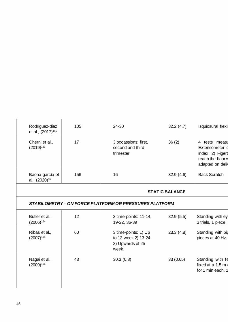
45
Rodriguez-díaz
et al., (2017)156
105 24-30 32.2 (4.7) Isquiosural flexibility test by goniometer.
Cherni et al.,
(2019)163
17 3 occassions: first,
second and third
trimester
36 (2) 4 tests measured with optoelectronical system: 1)
Extensometer of the metacarpophalangeal joint of the
index. 2) Figertrip to floor test: from 20cm platform, to
reach the floor with knees extended; 3) Sit-and-reach test
adapted on delivery bed 4) Beighton score
Baena-garcía et
al., (2020)35
156 16 32.9 (4.6) Back Scratch
STATIC BALANCE
STABILOMETRY – ON FORCE PLATFORM OR PRESSURES PLATFORM
Butler et al.,
(2006)164
12 3 time-points: 11-14,
19-22, 36-39
32.9 (5.5) Standing with eyes open and eyesclosed for 30 sec each.
3 trials. 1 piece. Force Platform.
Ribas et al.,
(2007)165
60 3 time-points: 1) Up
to 12 week 2) 13-24
3) Upwards of 25
week.
23.3 (4.8) Standing with bipedal support and eyes open for 5 sec. 2
pieces at 40 Hz.
Nagai et al.,
(2009)166
43 30.3 (0.8) 33 (0.65) Standing with feet parallel, gazing a black 12-cm circle
fixed at a 1.5 m distance with eyes open and eyes closed
for 1 min each. 1 piece.

46
Oliveira et al.,
(2009)167
20 3 time-points: 15.1
(1.8); 24.0 (2.4); 34.5
(2.5)
28.7 (6.2) Standing with 4 protocols at 50Hz and 2-min rest periods
between them: 1) Eyes open with feet comfortably apart;
2) Eyes closed with feet comfortably apart; 3) Eyes open
with feet together; 4) Eyes closed with feet together. 1
piece.
Karadag-saygi
et al., (2010)168
35 33 (3) 29.8 (4.5) Standing for 60 sec.
Yu et al.,
(2013)169
21 NR 30.2 (3.05) Standing with heels on a line at 1.0 m from visual target
with visual tasks and inspection tasks.
Ersal et al.,
(2014)170
69 2 time-points: 20.9
(1.2) and 35.8 (1.5)
28.3 (5.0) Standing with feet hip-width apart and staring straight
ahead on Equitest platform.
7opala-berdzik
et al., (2014)171
31 36.2 (1.2) 28.2 (3.6) Standing with arms at both sides and in a comfortable
stance on a stable force platform with eyes open and eyes
closed for 2 trials of 30-sec and 1-min rest between them.
Opala-berdzik
et al., (2015)172
45 2 time-points: 13.1
(2.5) and 36.2 (1.2)
28.2 (3.6) Idem Opala-Berdzik et al., (2014)
Ozturk,
(2016)173
68 31.5 (4.73)
30.3 (3.6)
Standing and arms extended in 6 different positions for 32-
sec: 1) facingforward eyes open and eyes closed; 2) Eyes
closed head rotated at 45º to the right 3) Idem 45º to the
left; 4) Eyes closed, head tilted at 30º backward and 5)
Idem 30º forward; 6) Standing on an unstable cushion,
facing forward eyes open and eyes closed. 4 pieces.
Shibayama et
al., (2016)174
161 28-33 33.3 (4.7) Standing and feet together for 30 sec on force platform. 1
piece.

47
Takeda et al.,
(2018)175
100 2nd and 3rd trimester 20-30 Standing with the medial malleoli 100 mm apart for 10-sec.
Then, moving forward, backward, right and left for 10-sec
each. 2 pieces.
Moreira et al.,
(2017)176
30 1st and 3rd trimester 26.8 (5.1) Standing with each foot positioned on each triaxial force
plate (feet apart by ~20 cm) and arms along the body with
eyes open focusing on a target located ~2 m in front and
eyes closed for 3 trails of 60-sec each and 2-min rest. 2
pieces.
Opala-berdzik
et al., (2018)177
70 10.8 (1.6) 28.6 (4.4) Standing with arms at both sides and in a comfortable
stance on a stable force platform, with eyes open looking
straight ahead at a wall 3m away for 2 trials of 30-sec and
1-min rest between them.
Catena et al.,
(2019)178
17 9 time-points: 16-20
gw, 36-40 gw and 1
time per month up to
7 months postpartum
28.9 (4.0) 2 trials: 1) quiet static in anatomical position for 10 s on a
force plate; 2) Idem 1 on a back-board spanning two force
plates.
Fontana et al.,
(2020)179
24 23 (3) 30 (6) Standing barefoot two-legged stance with arms at both
sides with eyes open at 2 m from a cross placed on a wall
at eye level during 3 x 30s trials with 30 s rest intervals.
The mean was retained on force platform.
Valerio et al.,
(2020)180
40 30.8 (3.9) 28 (2.5) Standing barefoot with freestanding supports inside the
platform and arms by their sides. And staring at a mark on
the opposite wall. 3 trials with the eyes open and three
trials with eyes closed, with 30 s rest intervals.

48
Takeda et al.,
(2019)157
21 22 and 23.25 gw. 32 (3.3) Standing barefoot on 2 stabilometers. 3 trials: 1) 10-sec
standing position; 2) 10-sec moving in the anterior
position; 3) 10-sec moving in the posterior position.
OTHERS
Atay et al.,
(2015)151
37 2 time-points: 20 gw
and 32 gw
29.6 (5.9) One-legged stand test.
DYNAMIC BALANCE
ON PLATFORMS
Davies et al.,
(2002)181
150 Day of labour 30.2 (5.8) Balance Master Platform Tests: 1) Sit to Stand; 2) Walk
Test, 3) Step and Quick Turn, 4) Step Up and Over.
Karadag-saygi
et al., (2010)168
35 33 (3) 29.75 (4.5) Walking barefoot 4 m.
Mccrory et al,
(2010)182
81 2 time-points: 20.9
(1.2) and 35.8 (1.5)
28 (5.7) The Motor Control Test protocol with translational
perturbations. Equitest posture platform.
Branco et al.,
(2013)183
22 27 (1.3) 32.5 (2.6) Walking barefoot for 10 m between 2 points in a straight
line at a natural and comfortable speed for 3 min.
Cakmak et al.,
(2014)184
41 6-12 26.5 (4.7) Standing with knee flexed, arms placed across the chest
and glare fixed ahead with open eyes on a movable
platform provides up to 20º of surface tilt in a 360º range
of motion for 3 trails of 20 sec each.

49
Inanir et al.,
(2014)185
110 3 groups: 1st
trimester, 2nd
trimester and 3rd
trimester.
24.7 (5.2) Idem to Cakmak et al., (2014)
3-D CAMERA MOTION CAPTURE SYSTEM
Wu et al.,
(2004)186
25 27 33.1 Walking on a treadmill at different velocities
(incrementing 0.11 m/s, from 0.17 up to 1.72 m/s; for
3 min at each level).
Forczeck et al.,
(2012)187
13 NR 29.2 (3.5) Walking barefoot at a self-selected speed across the
room during 15 gait cycles.10
Takeda et al.,
(2012)188
16 24.85 (1.95) 35 (1.4) Stand-to-sit motion assessing the time taken to sit
down; the leg joint moment; the antero-posterior and
vertical floor reaction forces; and the range of motion
of the lower limbs and trunk.
Gottschall et
al., (2013)189
13 2 time-points: 20 and
32
31.3 (4.5) Walking along 25 m on a custom-built portable
apparatus composed of a 2.4 m ramp inclined at 15°
continuous with a 4.8 m plateau.
Mccrory et al.,
(2014)190
69 28.35 (1.35) 28.0 (5.7) Walking along the 8-m runway.
Krkeljas,
(2018)191
35 3 time-points: 9-12
gw; 20-22 gw and
28-32 gw.
27 (6.1) Walking on a straight line, at a self-selected pace
along the 15-m walkway.
Catena et al.,
(2019)192
15 5 time-points: 16-20;
20-24, 24-28; 28-32;
32-36 gw.
29.3 (3.7) 60-second trial of semi-continuous stand-to-sit
motion. 54 reflective markers were adhered to body
land.

50
Catena et al.,
(2019)193
15 7 time-points: 12-16;
16-20; 20-24, 24-28;
28-32; 32-36; 36-40
gw.
28.1 (4.3) Walking on a treadmill for 60 seconds at a self-
selected comfortable speed.
Forczek et al.,
(2019)194
30 3 time-points: 12, 25,
36 gw.
30.3 (3.4) Walking barefoot at a self-selected speed during
12m intervals. 10 gait cycles.
Forczek et al.,
(2019)195
14 2 time-points: pre-
pregnancy; 1st
trimester.
20-40 Walking barefoot across room at a self-selected
during 50 m with 1 min rest intervals. 10 gait cycles.
Catena et al.,
(2020)196
23 5 time-points: 18, 22,
26, 30, 34 gw.
Walking on a treadmill for 60 seconds at a self-
selected comfortable speed.
Gimunova et
al., (2020)197
41 4 time-points: 14, 28,
37 gw.
30.5 (4.1) Walking barefoot along a 6-meter walkway at a self-
selected.
Mccrory et al.,
(2020)198
95 2 time-points: 2nd and
3rd trimester.
28.4 (5.5) Walking along the 8m laboratory runway until
walking speed stabilized.
Rothwell et al.,
(2020)199
17 2 time-points: 16-20;
36-40 gw.
22-37 Walking on a treadmill for 60 seconds at a self-
selected comfortable speed.
Forczek et al.,
(2019)200
36 3 time-points: 12; 25;
36 gw.
30.3 (3.4) Walking across the room 50 m with 1-min rest
interval. 10 gait cycles.
OTHERS
Sawa et al.,
(2015)201
27 2 groups: early
pregnancy (<27 gw)
or late pregnancy
(>27 gw)
30.9 (4.2) Walking at self-pace speed along a 15-m smooth,
horizontal corridor. It was recorded with 2 wireless
motion-recording-sensor units and one piezo-
resistive triaxial accelerometer.

51
Błaszczyk et
al., (2016)171
28 1st trimestrer and 3rd
trimester
28.2 (3.4) Walking along 10-m long walkway (back and forth 10
times) at self-space speed. It was recorded by
custom made; self-adhesive copper foil electrodes
attached to the soles of their shoes.
SPEED
Evensen et al.,
(2015)202
17 28.7 (7.4) 31.1(2.3) Ten-metres Timed walk Test (10mTWT)
Evensen et al.,
(2016)203
18 28.9 (7.3) 31.4 (2.7) 10mTWT
MULTICOMPONENT
Evensen et al.,
(2015)202
17 28.7 (7.4) 31.1(2.3) Timed Up and Go Test (TUG)
Evensen et al.,
(2016)203
18 28.9 (7.3) 31.4 (2.7) TUG
Christensen et
al., (2019)204
74 23 31.2 (3.7) TUG
Ad hoc: test designed specif ically for that study; NR: Not reported; PFS: Physical Fitness Score; kpm: kilopoundimeter; min: minutes; sec: seconds; HRMax: Maximal
Heart Rate; VO2 max: oxygen consumption maximum; RPE: rate of perceived exertion; AT: anaerobic threshold; MHR: maternal heart rate; ICC: intraclass correlation
coef f icient; MVCF: maximal voluntary contraction force; HGS: hand-grip strength; m: meters; mm: millimeters; FP: force platform; PP: pressure platform; RPM:
revolutions per minute: GW: gestational week; Hz: herzios; CM: centimeters; KG: kilograms; REPS: repetitions; MPH : miles per hour; BPM: beats per minute; KM/H:
kilometers/hour; VT: ventilatory threshold

52
Study II: International FItness Scale -IFIS: Validity and association with health-related
quality of life in pregnant women.
Study design and sample
Pregnant women included in this study are part of the baseline evaluation of the GESTAtion
and FITness (GESTAFIT) Project carried out in Granada (Spain). Briefly, the GESTAFIT
Project is a quasi-experimental study with a supervised concurrent physical exercise
intervention in groups with moderate-to-vigorous intensity, 3 days/week, 60 min/session, from
the 17th gestational week until the delivery day. The full methodology has already been
published elsewhere205. A total of 222 women were invited to participate at their 12th gestational
week through the Gynaecology and Obstetrics Unit of the “San Cecilio” and “Virgen de las
Nieves” University Hospitals (Granada, Spain). Of them, 159 women met the inclusion criteria
and were assessed at the 16th (±2) gestational week. For this specific cross-sectional study,
only pregnant women with complete data in all the variables analysed here were included. This
study was performed between 2015 and 2018, and the analyses were performed in 2018.
Ethical issues
The Ethics Committee on Clinical Research of Granada, Regional Government of Andalusia
(Spain) reviewed and approved this study (code: GESFIT-0448-N-15), which followed the
ethical guidelines of the Declaration of Helsinki, lastly modified in 2013.
Variables assessed
a) Weight and height
Body weight was measured using a body weight scale (model 803, Seca, Ltd). Height was
measured using a stadiometer (Seca 22, Hamburg). Body Mass Index (BMI) was calculated
[BMI = Weight (kg) / Height (m2)].
b) Sociodemographic and lifestyle characteristics
All women completed an initial and general anamnesis with questions regarding age, marital
status, living with partner or not, educational level, number of children and some life-style habits
like tobacco or alcohol consumption.
c) Self-reported physical fitness
Self-reported physical fitness was measured through the Spanish version of the IFIS52
questionnaire. The IFIS comprises five questions asking participants to rate their physical
fitness in comparison with the average person of the same age in a Likert scale with the
categories “very poor”, “poor”, “average”, “good” or “very good”. There is 1 question about

53
overall physical fitness, and 4 questions about its specific components (i.e. CRF, muscular
strength, speed/agility, and flexibility) and it can be completed in just one minute. This
questionnaire has been validated in European adolescents52, Spanish children56, young
adults206, older adults58, women with fibromialgia59, and Colombian adolescents57. The
questionnaire is available in different languages at the website of the PROFITH research group:
https://profith.ugr.es/ifis (anexe 2)
d) Objectively measured physical fitness
All the tests that we employed to assess PF in pregnant women are secure and feasible and
have been previously validated in adults. Muscular strength, flexibility and CRF were assessed
in this respective order with the objective of preventing induced fatigue, which might potentially
influence the results. Speed-agility was not assessed because of its potential risk for pregnant
women.
Cardiorespiratory Fitness
The Bruce test is an incremental, multistage, continuous treadmill test for measuring,
predicting, and evaluating maximal oxygen intake. In this upright test, there are progressive
increments in the workload every 3 minutes to individually determine limits of maximal possible
exertion. This test has been shown to be safe and easy to administer to pregnant women207,208.
Upper-body muscular strength
The upper-body muscular strength was measured through handgrip strength with a digital
dynamometer (TKK 5101 Grip-D; Takey, Tokyo, Japan), following the protocol described by
Ruiz et al.209. The test was performed twice for each hand, with a rest of 30 seconds when
alternating hands. The best value for each hand was selected, and the average value between
right and left hand was calculated. Nowadays, this test is widely used and is a gold standard of
muscle strength measure209.
Flexibility
The upper-body flexibility was measured with the back-scratch test, in which the participants
place one arm over their head towards their back and the other arm towards their back from
their waist and attempt to touch their hands. Then, the distance between the two middle fingers
is measured with a tape measure (cm) to determine the range of motion of the shoulder. The
total score is obtained by calculating the average of the best attempt for each hand210.

54
e) Health-related quality of life
The Spanish version of the Short-Form Health Survey-36 (SF-36)211 was used to assess
HRQoL. This scale has been developed by RAND Corporation (Santa Mónica, CA)212, and it
has been validated and translated into Spanish211. This questionnaire has been used in
previous studies to evaluate the HRQoL during pregnancy28,213–215. The 36 items of the SF-36
measure eight health domains: 1) physical functioning (10 items); 2) physical role (4 items); 3)
bodily pain (2 items); 4) social functioning (2 items); 5) mental health (5 items); 6) emotional
role (3 items); 7) vitality (4 items), and 8) General Health perception211,212. These 8 domains are
grouped in two overall scores: physical component summary (physical function, role-physical,
bodily pain, and general health) and mental component summary (vitality, social function, role-
emotional, and mental health). The scales range from 0 to 100, where higher scores mean
better HRQoL28,216.
Statistical Analysis
The validity of the IFIS to discriminate between objectively measured physical fitness levels in
pregnant women (objective 3) was assessed by a one-way analysis of variance (ANOVA) with
the corresponding objectively measured physical fitness component as outcome variable, and
its IFIS self-reported counterpart as fixed factor. Post–hoc group comparisons with Tukey’s
correction were applied to assess the differences in objectively measured physical fitness
across IFIS categories. To account for the “overall fitness” question of IFIS, a clustered score
of measured physical fitness was computed as the average of the standardized scores ([value-
mean]/SD) from the 3 objective fitness tests (Bruce test, hand grip, and back-scratch test).
To assess the extent to which IFIS and the different fitness tests are able to discriminate
between pregnant women with different levels of HRQoL (objective 4), a lineal tendency
ANOVA test and a linear regression was used with the SF-36 components as dependent
variables and the different self-reported fitness variables as independent. The same statistical
treatment was conducted/performed with the objectively measured physical fitness. The
statistical analyses were performed with Stata v.13.1 (Stata Corp LP., Texas, USA), and the
statistical significance was set at p<0.05.

55

56

57
RESULTS
The results of 2 studies comprising the present Doctoral Thesis are presented below.
Study I: Assessing physical fitness during pregnancy: validity and reliability of fitness
tests, and relationship with health-related outcomes. A systematic review.
A comprehensive PRISMA flow diagram is presented in figure 2. This diagram shows articles
excluded, articles by outcomes, and articles that analyzed validity, reliability, and association
with health-related outcomes.
Overall results, quality assessment and gestational week.
Our review identified 2617 studies, of which 149 fulfilled inclusion criteria (Figure 2). These
articles contained a sum of 191 fitness tests, using 149 different protocols that were included
to answer objective 1. A comprehensive scheme of the fitness tests and the different protocols
performed to date, divided by PF component, is presented in figure 3. A summary of the
number and percentage of articles that assessed PF during pregnancy and protocols used for
its assessment, divided by PF components, is presented in table 7.
With regards to aim 1, a total of 99 tests (that included 75 different protocols) were used to
assess cardiorespiratory fitness, 28 tests (that included 16 different protocols) were used to
assess muscular fitness, 14 tests (that included 13 different protocols) were used to assess
flexibility, 45 tests (that included 40 different protocols) were used to assess balance, 2 tests
using the same protocol to assess speed, and 3 tests using the same protocol were
multidimensional. No results were found for other PF components such as agility or
coordination.
We identified a limited number of articles related to aim 2. A total of 19 articles (13% of the
total number of included articles) either assessed validity (n=4 tests) or reliability assessment
(n=5 tests) of fitness tests, or the relationship of PF with health-related outcomes (n=16). Of
these 16 articles, 11 were classified as very low quality 31,34,36,39,153,162,166,182,217,218 and 5 were
classified as low quality 26,38,39,173,204. Of the 3 articles 122,123,203 that assessed validity, 2 articles
were classified as low quality 122,203 and 1 as high quality 123. Of the 4 articles that assessed
reliability criteria, 3 were considered high quality 122,158,202 and 1 low quality 149 (table 8).
The gestational week at PF assessment ranged from 8 to 41 across articles. Some articles
assessed PF several times throughout pregnancy; therefore, we divided pregnancy into two
stages. Early pregnancy (i.e., from week 0 to week 20 of gestation) and late pregnancy (i.e.,
from week 21 to week 40). Using this approach, 11 articles (7%) were performed in early

58
pregnancy; 57 articles (38%) were performed in late pregnancy; 55 articles (37%) were
performed several times (i.e., range 2 to 5 times) throughout pregnancy; 7 (5%) articles
specified a range of weeks that included early to late pregnancy; 14 articles (9%) reported only
the trimester without specifying gestational week; 4 articles (3%) provided no information, and
1 article (1%) was performed on the day of labor. (table 6).

59
Records identif ied through PubMed
database searching
(n = 930)
Scre
en
ing
In
clu
de
d
Eli
gib
ilit
y
Iden
tifi
cati
on
Records identif ied through WoS database
searching
(n = 1687)
Excluded duplicated articles
(n = 565)
Records screened
(n = 2052)
Records excluded af ter checking
title and abstract
(n = 1862)
Full-text articles assessed
for eligibility
(n = 189)
Full-text articles excluded, with
reasons
(n = 40)
1.- Physical f itness not assessed
or insuf f iciently described (n=20)
2.- Not original article (n=11)
3.- Fitness not assessed during
pregnancy (n=4)
4.- Identical data derived f rom the
same research project (n=5)
Articles included to address objective 1: Fitness tests have been used to evaluate
f itness
(n = 149 )
Papers identif ied in the search
(n= 2617)
Articles included to address objective 2:
Validity, reliability and health-related outcomes
(n = 19)
Physical fitness tests included in 149 articles: (n = 191 ) Physical fitness tests included in 19 articles: (n = 23)
Figure 2. Flow chart of the literature search and paper selection process.

60
Fig
ure
3. S
ch
em
e o
f th
e fitn
ess te
sts
an
d th
e d
iffe
ren
t p
roto
co
ls d
ivid
ed
by P
F c
om
po
ne
nt.

61
CRF: Cardio-respiratory f itness; MF: Muscular Fitness
The gestational week at PF assessment ranged from 8 to 41 across articles. Some articles
assessed PF several times throughout pregnancy; therefore, we divided pregnancy into two
stages. Early pregnancy (i.e., from week 0 to week 20 of gestation) and late pregnancy (i.e.,
from week 21 to week 40). Using this approach, 11 articles (7%) were performed in early
pregnancy; 57 articles (38%) were performed in late pregnancy; 55 articles (37%) were
performed several times (i.e., range 2 to 5 times) throughout pregnancy; 7 (5%) articles
specified a range of weeks that included early to late pregnancy; 14 articles (9%) reported only
the trimester without specifying gestational week; 4 articles (3%) provided no information, and
1 article (1%) was performed on the day of labor.
Cardiorespiratory fitness
a) Tests used
We identified 99 tests assessing CRF, of which 61 (62%) were performed on a cycle-
ergometer, 25 (25%) on treadmill, 10 (10%) on track and there were 3 (3%) step protocols. Of
the 99 tests, a total of 75 corresponded to different protocols. For instance, there were 56
different protocols using cycle-ergometer, distributed as follows: only one article used the
Arstila test 34; one used the Bruce Protocol at 75% HRmax 70; one applied the Modified Bruce
ramp protocol at anaerobic threshold 113; two employed the Modified Balke protocol at 70%
HRmax 77,85; one used YMCA protocol 111; and the remaining articles (n=55) used ad-hoc tests
(i.e. specifically designed for the purpose of the investigation); 11 of which
67,75,80,81,85,108,217,219,220 used steady-state tests and 44 34,36,38,68,69,71–74,78,79,82,83,85–87,89–101,103–106,109–
116,221–224 used incremental tests. When analyzing the type of test based on intensity, we found
that 13 articles used maximal tests 74,86,87,90–92,99,103,104,112–114,222, 37 submaximal tests 34,36–38,67–
69,72,78–83,89,94–98,100,101,105,108–111,115,116,220,223,225,226 and 3 used mixed tests 71,85,221 containing
submaximal and maximal stages within the same protocol.
There were 25 treadmill tests that used 14 different protocols, distributed as follows: the
Modified Balke protocol was used in 10 articles 26,74,118,123–126,130,227,228; the Modified Bruce
protocol in 2 articles 31,119; and the traditional Balke protocol -twice in the same article- 229; the
TOTAL CRF MF Flexibility Balance Speed Multidimensional
Fitness tests
191 99 (52) 28 (15) 14 (7) 45 (24) 2 (1) 3 (1)
Protocols 149 75 (50) 16 (11) 13 (9) 40 (29) 1 (0.5) 1 (0.5)
Table 7. Number (%) of articles that assessed the different components of physical fitness during pregnancy and protocols used for its assessment.

62
traditional Bruce protocol 133, the Cornell protocol 122, the SWET protocol and the Ebbelling
single-stage protocol 25 were each used in 1 article. There were other 7 ad hoc tests of which
2 were steady-state 81,129, and 5 were incremental tests 68,120,128,131,207. According to intensity,
3 were maximal tests 128,129,207 and 4 submaximal tests 68,81,120,131.
Of the 10 tests on track, 6 articles performed the 6-minute walk test protocol 135,136,138,140,141,230,
and 4 were ad hoc tests (i.e. 1 maximal and 4 were submaximal). In regards to the 3 step tests,
1 Canadian Home Fitness test 143 was used and 2 ad hoc incremental submaximal tests were
used 144,218.
b) Reliability and validity
We identified 2 articles examining validity 122,123. Yeo et al. 122 aimed to validate a portable
metabolic testing system (VO2000) on healthy sedentary pregnant women. The VO2000
consistently overestimated VO2 measurements, compared to the same manufacturer’s
reference system, by 4.4±3.6 standard deviation (SD) ml/kg/min although the Pearson
correlation was significant (r=0.48; p=0.01). When VO2000 was used twice, the mean
difference was statistically significant (1.0±1.8 ml/kg/min; t(45)=3.9, p<0.001). Mottola et al. 123
provided a prediction equation for VO2peak in pregnant women between 16 and 22 weeks of
gestation, using a modified Balke protocol. The results of this equation revealed an adjusted
R2 of 0.71 (p value not reported). When the authors used this equation to predict VO2peak in a
cross-validation group (n=39), they found a predicted value of 23.38±4.03 mL×kg -1×min-1,
while the actual value was 23.54 ± 5.9 mL×kg -1×min-1 (p value not reported).
c) Relationship with health-related outcomes
A total of 6 articles analyzed the association of CRF with health-related outcomes. Pomerance
et al. 217 observed that VO2max was inversely associated with the length of labor in multiparas
(r=-0.65; p=0.001) and pre-pregnancy weight (r=-0.63; p=0.001). However, VO2max was neither
correlated with newborn weight, length, or head circumference, nor with the one-minute Apgar
score (all p>0.05). In the same line, Wong & McKenzie 36, observed that fit mothers showed
lower HR at submaximal exercise intensity (p<0.05) and the second stage of labor was shorter
(no statistics reported) compared to unfit pregnant mothers. However, there was no difference
between fit and unfit in the length of gestation or weight gained (no statistics reported). In the
same article, the authors showed neither positive nor negative effect of maternal fitness on
newborn weight or Apgar scores.
In addition, Erkkola et al. 34 found that newborn from fit pregnant women had higher pH than
fetuses of less physically fit women (p<0.01). In this article, participants with low physical
performance were more likely to have asphyxiated neonates than neonates of physically fit
women (p<0.05). In the same line, Baena-García et al. 31 observed that maternal CRF at the

63
16th gestational week was related to higher arterial umbilical cord PO2 (r=0.267, p<0.05), and
women who had caesarean sections had significantly lower CRF compared with the women
who had vaginal births (p<0.001).
On the other hand, Bisson et al. 26 studied the association of CRF in early pregnancy with
physical activity before and during early pregnancy. The authors found that a higher VO 2 peak
in early pregnancy was positively associated with physical activity spent at sports and exercise
before and during early pregnancy (p<0.001).
Muscular fitness
a) Tests used
A total of 28 tests (i.e., 14% of all included articles) that included 16 different procols assessed
muscular fitness, of which 10 performed maximal hand-grip strength tests 26,30,31,39,137,151–156, 3
performed endurance hand-grip test, 2 for 3-min 146,148 and 1 for 2-min period 147. In 2 of the
articles conducting an endurance hand-grip test 146,147, the equipment used was a hand grip
sphygmomanometer instead of dynamometry. On the other hand, 1 used hand-held
dynamometer fixed a chair to assess quadriceps strength 157 and 1 used toe-grip dynamometer
157. Moreover, 2 ad hoc isometric tests were used to assess maximal voluntary hip extension
and back flexors endurance in the same article 231. Finally, 13 dynamic endurance tests were
found, 9 were listed as ad hoc tests 30,150,154 and other 3 (30-sec Chair Stand Test, 5 Times Sit
to Stand test, Trendelenburg’s test) were classified as others dynamic tests 31,154,158.
b) Reliability and validity
Only 2 muscular fitness tests assessed reliability 149,158. Yenisehir et al. 158 analyzed reliability
and validity of Five Times Sit-to-Stand. Inter-rater reliability was excellent for subjects with and
without pelvic girdle pain (ICC = 0.999, 95% CI = 0.999–1.000; ICC = 0.999, 95% CI = 0.999–
0.999, respectively). Test-retest reliability was also very high for subjects with and without
pelvic girdle pain (PGP) (ICC = 0.986, 95% CI = 0.959–0.995; ICC = 0.828, 95% CI = 0.632–
0.920, respectively).
On the one hand, Gutke et al.149 analyzed the reliability for an ad hoc test. This test consisted
in a maximal voluntary isometric hip extension with a fixed sensor holding a sling around the
thigh and pulling for 5 seconds during 3 reps with 5-10 seconds of rest (r=0.82 for the right leg
and r=0.88 for the left leg; ICC=0.87 for the right leg and 0.85 for the left leg; with p value no
reported).

64
c) Relationship with health-related outcomes.
The hand-grip strength has been associated with neonatal outcomes. Bisson et al.26 observed
that hand-grip strength was positively associated with infant birth weight (r=0.34, p=0.0068)
even after adjustment for confounders (r=0.27, p=0.0480). Accordingly, Zelazniewicz et al. 39
observed that hand-grip strength was associated with offspring birth weight when controlled
for the newborn sex and gestational age at delivery (F(2.182)=3.15; p=0.04). Baena-García et
al. 31 found greater hand-grip strength weakly associated with greater neonatal birth weight (r
= 0.191, p<0.05). On the other hand, Wickboldt 153 found that hand-grip strength was
moderately correlated with pain scores. Mean hand-grip strength during contractions had the
highest correlation coefficient (r=0.67; p<0.001) compared with peak handgrip strength
(r=0.56; p<0.001) and the area under the curve of handgrip force (r=0.55; p<0.001).
Flexibility
a) Tests used
Our search identified 14 (7%) tests that assessed flexibility using 13 different protocols,
including the side bending test 161, the sit-and-reach test 30, the back-scratch test (twice) 31,151,
the Motion Analysis (i.e. including 3 different tests such as the seated and standing forward
flexion, seated and standing side to side flexion and seated axial rotation 159) and optoelectrical
system (i.e. performing 4 different tests) 163. Goniometry was used in two different articles to
measure isquiosural flexibility, 232 wrist flexion-extension and medial lateral deviation 160. Only
one article used an ad hoc machine to test passive abduction of the left fourth finger 162.
b) Reliability and validity
Lindgren et al.162 designed an ad hoc machine to test passive abduction of the left fourth finger
and its relationship with low-back pain during pregnancy and early postpartum. Abduction
angle was measured at three different times throughout the pregnancy and once in the
postnatal period. Reliability of the abduction angle was analyzed by the intra-individual
coefficient of variance. The coefficients of variance between the first and second measurement
was 0.077, between the second and third 0.070 and between the third and fourth 0.071.
c) Relationship with health-related outcomes.
Only 2 flexibility tests evaluated associations with health-related outcomes. Lindgren et al. 162
found that women with greater passive abduction angle of the left fourth finger was associated
with highest back pain incidence (OR=1.09; CI=1.01-1.17; p=0.022) and the highest number
of previous pregnancies (OR=3.24; CI=1.57-6.68; p=0.002). Baena-Garcia et al. 31 found
increased flexibility associated with a more alkaline arterial pH (r = 0.220, p<0.05), higher
arterial PO2 (r = 0.237, p<0.05) and lower arterial PCO2 (r =-0.331, p<0.01) in the umbilical

65
cord blood. No other articles assessed the association of flexibility with health-related
outcomes in pregnant women.
Balance
a) Tests used
We identified 45 (24%) articles assessing balance of which 19 analyzed static balance and 26
dynamic balance trough 40 different protocols. With regard to static balance, 18 were
laboratory tests of which 12 assessed balance through stabilometry tests on force platform 164–
169,172,174,176–179, one on pressures platform 168 and another on Equitest® platform 170. Four
articles did not mention the type of platform used (40,150,176,177). Regarding protocols, all
articles conducted the tests with participants standing with bipedal support. However, standing
position varied between articles. Ten articles maintained a standing posture with feet
separated 157,166,170–172,176–180, 1 with feet together 174, 2 used mixed protocols 167,173, 1 with
medial malleoli separated 175 and 4 did not mention the standing posture 164,165,168,169. Moreover,
3 articles used protocols with eyes open 165,177,179 exclusively, 8 articles used mixed protocols
with eyes open and closed, 1 used visual target and visual tasks 169 and 6 did not specify
whether participants kept their eyes closed or opened. Only 1 article used a field test, the one-
legged standing protocol 151. On the other hand, 1 test was field-test without platform.
In relation to the 26 articles measuring dynamic balance, 9 of them assessed balance using
platforms. Each of these articles used different testing tool such as balance master platform
181, pressure platform 168, force platform 183, Equitest® platform 182 and movable platform, which
was used in two articles 184,185. Two of these articles were walking protocols 168,183, 1 with
translational perturbations172, one was standing with one knee flexed and arms across the
chest 184,185. Another 15 articles used 3-D camera motion capture systems using 13 different
protocols. Twelve of the 15 articles were walking protocols 187,189–191,193–200,233 and 2 used a
stand to sit motion protocol 188,192. Moreover, 1 article used a triaxial accelerometer201; another
article assessed balance through recording (without specification of camera type) 234, and
another using instrumented insoles 235. All three articles used walking protocols.
b) Reliability and validity
No validity or reliability assessments were performed regarding balance tests.
c) Relationship with health-related outcomes.
3 articles associated balance with health-related outcomes. Ozturk 173 observed that static
balance decreased and fall risk increased in pregnant women with lower back pain
(49.90±24.47 vs 28.47±19.60; p<0.0001). In relation to exercise, McCrory et al. 236 showed
that exercise may play a role in fall prevention in pregnant women (p=0.005); they also found

66
that dynamic balance is altered in pregnant women who have fallen compared with non-fallers
and non-pregnant women (p<0.001). Nagai et al. 166 studied the relationship between anxiety
and balance. They concluded that when anxiety increases during pregnancy, the standing
posture is destabilized (r=0.559, p=0.020), which may increase the chance of falling.
Speed.
a) Test used.
The only protocol that was used to assess speed during pregnancy was the ten-meters timed
walk test (10mTWT). However, the same test was identified in two different articles. 202,203. In
the 10mTWT, the participants commenced standing at a chair. When told to start, subjects
walk as fast as possible along 14 meters marked with white blank tapes placed at 0 m, 2 m,
12 m and 14 m. The time (100th of a second) required to walk between the 2 m and 12 m
markers was recorded and converted into speed in meters per second (m/sec).
b) Reliability and validity.
Validity and reliability for 10mTWT was studied by Evensen et al. in two different articles 202,203.
In 2015, Evensen al. 202 analyzed the test-retest reliability of 10mTWT showing an intraclass
correlation coefficient (ICC) of (0.74). Intertester reliability was determined in the first 13
participants with strong correlation (ICC =0.94). In 2016 203 the same authors analyzed the
convergent validity of 10mTWT by comparing performances with scores achieved on the
Active Straight Leg Raise (ASLR) test and observed moderate positive correlations between
10mTWT and ASLR (r=0.65, p=0.003).
c) Relationship with health-related outcomes.
This systematic review did not find any articles that analyzed the association of speedwith
health-related outcomes.
Agility and coordination
No articles of agility and coordination were identified.
Multidimensional
a) Test used.
Our search identified a walking multidimensional test that was used in three studies 202–204. In
Timed Up and Go Test (TUG) the participant begins seated in a chair with their arms on
armrests and their toes against a start line. The purpose is to cross the front white line at three

67
meters away, turn around, and walk back to the chair and sit down as fast as possible. The
performance is measured in time (100th of a second).
b) Reliability and validity.
Validity and reliability for TUG was analyzed by Evensen et al. in two different studies 202,203.
The TUG showed good test-retest reliability (ICC=0.88) and intertester reliability (ICC=0.95).
Regarding reliability, strong correlations were found between the TUG and Active Straight Leg
Raise (r= 0.73, p= 0.001).
c) Relationship with health-related outcomes.
The time on TUG among pregnant women with pelvic girdle pain was significantly higher (mean
(95% CI) 6.9 (6.5, 7.3) seconds) than for asymptomatic pregnant (5.8 (5.5, 6.0), p < 0.001) and
non-pregnant (5.5 (5.4, 5.6), p < 0.001) women.
.

68
Table 8. Overview of studies that assessed the validity and/or reliability of fitness tests during pregnancy or the association of physical fitness with health-
related outcomes in pregnant women.
REFERENCENCE
(AUTHORS,
YEAR)
VALID
ITY
RELIAB
ILITY HRO
CAPACITY EVALUATED, SHORT TEST DESCRIPTION AND HEALTH-
RELATED OUTCOMES OR STATISTICAL RESULTS
QUALITY
SCORE
CARDIO-RESPIRATORY FITNESS
Pomerance et
al., (1974) 37
No No Yes
Ad hoc, continuous test on CE.
1: PFS was inversely associated with pre-pregnancy weight (r=-0.63;
p=0.001).
2: PFS was significantly associated with the length of labor in multiparas
(r=-0.65; p=0.05).
3: PFS was not correlated with pre-pregnancy weight; infant birth weight,
new-born length, new-born head circumference or one-minute Apgar score
(all p>0.05).
2
Erkkola et al.,
(1976) 34 No No Yes
Ad hoc, incremental submaximal test on CE.
1: Fetal pH in fit pregnant women is higher than the pH in less physically fit
pregnant women (p<0.01).
2: Pregnant women with low physical performance are more liable to have
asphyxiated babies than the physically fit pregnant women (p<0.05).
2

69
Wong &
McKenzie,
(1987) 36
No No Yes
Ad hoc, incremental test on CE.
1: HR at submaximal exercise was lower in fit pregnant women than unfit
pregnant women (p<0.05).
2: Second stage of labor was shorter in fit pregnant women; (no statistics
reported).
3: There were no differences between fit and unfit participants in the length
of gestation, weight gained vs pre-pregnancy measures. There were neither
positive nor negative effects of maternal fitness on the new-born (Apgar
test, birth weight); (no statistics reported).
1
Thorell et al.,
(2015) 38
No No Yes
Ad hoc, incremental maximal test on CE.
Absolute VO2peak, was inversely correlated with duration of gestation among
women with miscarriage (r=-0.52; p=0.02) and positively with duration of
gestation among women with spontaneous onset of labor (r=0.12; p=0.01).
3
Melzer et al.,
(2010) 218 No No Yes
Ad hoc, incremental step protocol test.
1: Active women have better aerobic fitness (VO2max) as compared to
inactive women (34.9 vs 30.3 mL/kg/min; p=0.01).
2
Yeo et al.,
(2005) 122 Yes Yes No
Cornell Protocol on treadmill platform.
Validity: Bland-Altman plots. The mean difference was 4.4 ± 3.6 ml/kg/min.
Data indicated that VO2000 over- estimates VO2 by an average of 4.4
ml/kg/min compared to CPX/D. Pearson correlation coefficient between the
average and difference of paired measurements was close but not
significant (r=0.48; p>0.01). Reliability: Paired t test (t(45) = 3.9, p < .001).
Linear regression: y = 0.96X-1.6; 95% CI for the slope: 0.94 to 1.1; R2=
0.91, p<0.001
4
and
8

70
Mottola et al.,
(2006) 123 Yes No No
Modified Balke protocol on treadmill platform.
Validity: Pearson Correlation: R2=0.72, R2 adjusted=0.71, and SE = 2.7 (no
statistics reported).
5
Baena-García et
al., (2020) 31 No No Yes
Modified Bruce protocol until 85% HRmax
1) Maternal CRF at the 16th gestational week was related to higher arterial
umbilical cord PO2 (r=0.267, p<0.05).
2) The women who had caesarean sections had lower CRF (p<0.001) at
the 16th gestational week compared with the women who had vaginal
births.
2
MUSCULAR FITNESS
Gutke et al.,
(2008) 149 No Yes No
Maximal voluntary isometric hip extension
Reliability: Spearman’s rho (r) and Intercorrelation coefficient (ICC). Right leg:
r=0.82; ICC=0.87. Left leg: r=0.88; ICC=0.85 (both p value no reported).
3
Bisson et al.,
(2013) 26 No No Yes
Hand Grip Strength was positively associated with infant birth weight (r=0.27;
p=0.048). 3
Wickboldt et al.,
(2015) 153 No No Yes
Hand Grip Strength was moderately correlated with pain scores on the
Numerical Rate Scale. Mean handgrip force during contractions had the
highest correlation coefficient (r=0.67; p<0.001) compared with peak handgrip
force (r=0.56; p<0.001) and area under the curve of handgrip force (r=0.55;
p<0.001).
2

71
Zelazniewicz et
al., (2018) 39 No No Yes
Hand Grip Strength in pregnancy was positively associated with offspring birth
weight when controlled for a child's sex and week at delivery (F (2,182) = 3.15,
p=0.04). Women with greater hand grip strength in each trimester were more
likely to give birth to a boy (p<0.05).
4
Baena-García
et al., (2020) 31 No No Yes
Hand Grip Strength was associated with greater neonatal birth weight (r =
0.191, p<0.05) 2
Yenisehir et al.,
(2020) 158 No Yes No
Five Times Sit to Stand Test
Reliability: Inter-rater reliability of 5TSS was excellent for subjects with and
without PGP (ICC ¼ 0.999, 95% CI ¼ 0.999–1.000; ICC ¼ 0.999, 95% CI ¼
0.999–0.999, respectively). Test-retest reliability of 5TSS was also very high
for subjects with and without PGP (ICC ¼ 0.986, 95% CI ¼ 0.959–0.995; ICC
¼ 0.828, 95% CI ¼ 0.632–0.920, respectively).
5-7
FLEXIBILITY
Lindgren et al.,
(2014) 162
No Yes Yes Ad hoc passive abduction of the left fourth finger.
The highest back pain incidence was showed by women with the greatest
passive abduction angle of the left fourth finger (OR=1.09; CI=1.01-1.17;
p=0.022) and the highest number of previous pregnancies (OR=3.24;
CI=1.57-6.68; p=0.002). Reliability: Intra-individual coefficient of variance. 1)
Between the first and second measurement = 0.077; 2) Between the second
and third = 0.070 and between the third and fourth = 0.071.
2

72
Baena-García
et al., (2020) 31
No No Yes Back-Scratch
Maternal flexibility was associated with a more alkaline arterial pH (r = 0.220,
p<0.05), higher arterial PO2 (r = 0.237, p<0.05) and lower arterial PCO2 (r =-
0.331, p<0.01) in umbilical cord blood.
2
BALANCE
Nagai et al.,
(2009) 166
No No Yes Static balance on force platform.
Increases in anxiety during pregnancy is associated with a destabilized
standing posture (r= 0.559, p=0.020)
2
Ozturk et al.,
(2016) 173
No No Yes Static balance on non-specific platform in different positions.
Postural equilibrium decreases and fall risk increases in pregnant patients with
lower back pain (49.90±24.47 vs 28.47±19.60; p<0.0001).
3
McCrory et al.,
(2010) 236
No No Yes Dynamic balance on Equitest posture platform.
Dynamic balance is altered in pregnant women who have fallen compared with
non-fallers and controls. (p<0.001). Exercise may play a role in fall prevention
in pregnant women.
2

73
SPEED
Evensen et al.,
(2015) 202
No Yes No Ten-meters Timed walk Test
Reliability: ICC from a one-way random effects model and reporting the
95% confidence interval (CI). Coefficients for test-retest reliability for
10mTWT: (ICC= 0.74; 95% CI=0.42–0.90; SEM=0.17 m s-1; MDC95= 0.47
m s-1) Coefficients for intertester reliability 10mTWT: (ICC=0.94; 95%
CI=0.82–0.98; SEM=0.09 m s-1; MDC95=0.25 m s-1).
8
Evensen et al.,
(2016) 203
Yes No No Ten-meters Timed walk Test
Validity: Spearman correlation coefficient. Between the 10mTWT and
ASLR (r=-0.65, p=0.003). Between the 10 mTWT and PGQ (r =-0.25 to -
0.56).
3
MULTIDIMENSIONAL
Evensen et al.,
(2015) 202
No Yes No Timed Up and Go Test (TUG)
Reliability: ICC from a one-way random effects model and reporting the
95% confidence interval (CI). Coefficients for test-retest reliability TUG:
(ICC=0.88; 95% CI=0.70–0.95; SEM=0.42 seconds; MDC95=1.16
seconds.) Coefficients for intertester reliability TUG: (ICC= 0.95; 95%
CI=0.84–0.98; SEM=0.36 m s-1; MDC95= 1.00 m s1).
8
Evensen et al.,
(2016) 203
Yes No No Timed Up and Go Test (TUG)
Validity: Spearman correlation coefficient. Between the TUG and ASLR
(rs=0.73, p=0.001). Between the TUG and ASLR (rs=0.73, p= 0.001).
Between the TUG and PGQ (rs=0.41 to 0.52).
3

74
HrO: Health related outcome; CRF: Cardio-respiratory f itness; MS: Muscular Strength; PFS: Physical Fitness Score; CE: Cyclo-ergometer; HR: Heart Rate; VO2max:
volum maximum oxygen; VO2peak: volum peak oxygen; HRmax: maximal heart rate; PGP:Pelvic girdle pain; ICC: intraclass correlation coef ficient (relative reliability);
95% CI: 95% conf idence interval; SE: Standard Error; SEM: standard error of measurement (absolute reliability); MDC95, minimal detectable change at 95% CI; OR:
odds ratio; TUG: Timed Up and Go test; 10mTWT: Ten-meter Timed Walk Test; ASLR: Active straight leg raise.
Christensen et
al., (2019) 204
No No Yes Timed Up and Go Test (TUG)
The time on TUG among pregnant women with PGP was significantly
higher (mean (95% CI) 6.9 (6.5, 7.3) seconds) than for asymptomatic
pregnant (5.8 (5.5, 6.0), p < 0.001) and non-pregnant (5.5 (5.4, 5.6), p <
0.001) women.
4

75
Study II: International FItness Scale -IFIS: Validity and association with health-related quality
of life in pregnant women
Of the 222 women interested, 63 refused to participate due to the care of other children or the
lack of time. A total of 159 women participated in this study, although only those with valid data
regarding both self-reported and objectively measured physical fitness with IFIS (n=106; mean age
32.7, SD 4.4 years) were finally included in the analyses of the present study. The descriptive
characteristics of the study participants are presented in table 9.
The distribution of IFIS responses revealed that only a small number of pregnant women rated
their physical fitness as either “very poor” or “very good” (figure 4). Consequently, we re-categorized
“very poor” and “poor” into a single category, and “good” and “very good” into another single category
to avoid a lack of statistical power caused by the low number of participants in extreme categories.
Figure 4. Distributions of the answers for the 5 questions of the International FItness Scale (IFIS) in pregnant women. CRF, cardiorespiratory fitness; MS, muscular strength; SP-AG, speed-agility; FLEX, f lexibility;
Overall, overall physical f itness.

76
Table 9. Characteristics of the pregnant women at early second trimester of pregnancy
(gestational week 16).
Values are shown as mean and standard deviation (SD), or n (%).
Variable Mean SD n
Age (y) 32.7 4.4 106
Body mass index (kg/m2) 24.7 4.1 106
Marital Status (n, %) 106
Married 63 (59.4)
Single 43 (40.6)
Living with a partner (n, %) 106
Yes 104 (98.1)
No 2 (1.9)
Educational Status (n, %) 106
Primary school 12 (11.3) 12
Professional training 19 (17.9) 19
Secondary school 14 (13.2) 14
University medium
degree 23 (21.7) 23
University higher
degree 38 (35.9) 38
Number of Children 0.6 0.08 106
Number of abortions 0.51 0.07 106
Smoking Status (n, %)
Yes 97 (91.6) 106
No 9 (09.4) 106
Alcohol Consumption (n,
%)
Yes 0 (100.0) 106
No 106 (0.0) 106
Objectively measured
physical fitness
Bruce VO2max
(ml/kg/min) 23.1 5.5 87
Handgrip strength (kg) 27.6 4.0 106
Back-scratch (cm) 8.4 12.3 106

77
Construct validity of IFIS in pregnant women (aim 3)
The comparison between objective physical fitness levels across IFIS categories are displayed
in Table 10. There were between-group differences in objectively measured physical fitness
across IFIS categories (p<0.05), regardless of the physical fitness component evaluated.
Regarding CRF, the post-hoc analyses revealed that women who self-rated their CRF (i.e.
through IFIS) as “very poor/poor” had lower estimated VO2max than those in any other
categories of IFIS (p<0.05). In relation to muscle strength, women who self-rated their strength
as “good/very good” presented higher handgrip strength than those who rated it either as
“average” or “poor-very poor” (p<0.05). Regarding flexibility, women who self -rated their
flexibility as “poor-very poor” presented lower range of motion in the back-scratch test than
those who rated it as “good-very good” (p<0.05).

78
Table 10. Objectively measured physical fitness across categories of the International FItness Scale (IFIS)
CRF=Cardiorespiratory f itness; SE=Standard Error; Dif f=Difference between groups; CI: Conf idence Interval.
Very Poor/Poor
(1) Average
(2) Good/Very Good (3)
ANOVA Pair-Wise Comparisons – Tukey’s correction
n Mean (SE) Mean (SE) Mean (SE) P Comparison: 1 vs. 2 Comparison: 1 vs. 3 Comparison: 2 vs. 3
CRF Diff 95% CI P Diff 95% CI P Diff 95% CI
P
Bruce test (ml/kg/min)
87 21.36 (0.75) 25.43 (0.98) 25.03 (1.57) <0.001 4.07 (1.11-7.02) <0.001 3.67 (-0.48-7.82) 0.09 -0.39 (-4.81-4.02) 0.98
Muscular fitness
Handgrip (kg) 106 26.53 (0.86) 26.92 (0.53) 29.40 (0.67) 0.01 0.39 (-2.0-2.78) 0.92 2.87 (0.29-5.46) 0.03 2.49 (0.46-4.50) 0.01
Flexibility
Back-scratch (cm)
106 4.64 (2.20) 6.35 (2.06) 12.51 (1.79) 0.01 1.71 (-5.47-8.90) 0.84 7.88 (1.12-14.63) 0.02 6.15 (-0.34-12.66) 0.07

79
Ability of the IFIS to discriminate Health-Related Quality of Life (HRQoL) in pregnant women
(aim 4)
Figure 5 shows the linear association of self-reported physical fitness with the different
dimensions of HRQoL in pregnant women. Higher self-reported overall physical fitness showed
a positive linear trend with HRQoL in all domains of the SF-36 (p<0.05), except in the Physical
Role and Social Functioning dimensions (p>0.05). With some exceptions, this tendency was
consistent across the different physical fitness components as self -reported through IFIS.
Regarding the physical and mental component summary (PCS and MCS, respectively), there
was a generally consistent linear trend so that higher self -reported physical fitness was
associated with higher PCS and MCS scores, except for self -reported muscle strength
(p>0.05) (figure 6 and table 11).
The post-hoc analyses revealed significant differences in HRQoL between the extreme groups
(“very poor/poor” and “good/very good”) in the overall physical fitness, CRF and flexibility
categories.
Overall, objectively measured CRF and flexibility showed a positive linear relationship with
several dimensions of HRQoL, although the overall physical fitness score did not show such
relationship (figure 7)
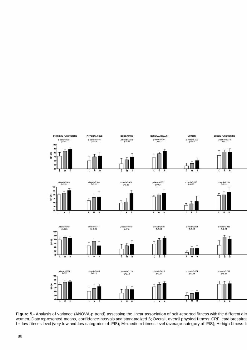
80
Figure 5.- Analysis of variance (ANOVA-p trend) assessing the linear association of self -reported f itness with the dif ferent dimensions of health-related quality of life in pregnant
women. Data represented means, conf idence intervals and standardized β; Overall, overall physical f itness; CRF, cardiorespiratory f itness; MS, muscular strength. FLEX, f lexibility. L= low f itness level (very low and low categories of IFIS); M=medium f itness level (average category of IFIS); H=high f itness level (good and very good categories of IFIS)

81
Figure 6.- Associations between Summary Physical Component (a) and Mental Component Summary (b) of health-related quality of life assessed by Short-Form Health
Survey-36 (SF-36) and self -reported physical f itness (IFIS) categories in pregnant women. Data represent means. All signif icance levels were p<0.05 less MS (a). All signif icance levels were p<0.05, excepting CRF and FLEX (b). CRF, cardiorespiratory f itness; FLEX, f lexibility; MS, muscular strength; OF, overall phys ical f itness.

82
Table 11. Physical component summary and mental component summary across International FItness Scale (IFIS) physical fitness (n=103).
Overall=overall physical f itness; CRF=cardiorespiratory f itness; MS=muscular strength. FLEX=f lexibility.
Very Poor/Poor
(1)
Average
(2)
Good/Very Good
(3)
Mean Mean Mean
Physical component
IFIS Overall 43.48 (9.34) 47.28 (6.54) 49.65 (6.87)
IFIS CRF 45.98 (6.83) 47.87 (6.83) 52.09 (5.60)
IFIS MS 45.43 (8.52) 48.36 (6.57) 47.01 (7.95)
IFIS Flex 44.78 (7.85) 47.50 (8.32) 48.98 (6.00)
Mental
Component
IFIS Overall 46.36 (8.21) 51.5 (6.62) 52.51 (6.82)
IFIS CRF 49.77 (7.33) 51.99 (6.12) 53.35 (8.62)
IFIS MS 44.92 (8.41) 48.36 (6.38) 51.97 (5.11)
IFIS Flex 49.44 (6.84) 51.87 (7.18) 51.19 (7.54)

83
Figure 7. Analysis of variance assessing the linear association of objectively measured physical f itness with the dif ferent dimensions of health-related quality of life in pregnant women. Data represented means, conf idence intervals and standardized β. Abbreviations: Overall, overall physical f itness; CRF,
cardiorespiratory f itness; MS, muscular strength. FLEX, f lexibility.

84

85

86
DISCUSSION
Study I: Assessing physical fitness during pregnancy: validity and reliability of
fitness tests, and relationship with health-related outcomes. A systematic review.
1) Summary of tests used to assess physical fitness during pregnancy.
This systematic review revealed that PF has been assessed through a wide variety of tests
during pregnancy. However, very little is known on the validity and reliability of the tests
performed. These findings have important research and clinical implications. First, until a
specific battery of fitness assessments for pregnant women is developed and validated, the
confidence in PF data during pregnancy is limited and potentially unreliable and may prove
harmful if unreliable values are used for exercise recommendation during pregnancy. Second,
the large variety of tests used makes is challenging to compare results from different studies.
Third, provided the lack of rigorous information on validity and reliability of PF tests, it is also
difficult to evaluate the association of PF with health-related outcomes, which is of wide clinical
and public health interest. However, some studies have attempted to present associations of
PF with health-related outcomes, which undoubtedly needs to be replicated once a PF test
battery is released. Before that, exhaustive research must be performed in validating such
battery of tests.
a)Cardiorespiratory Fitness
This systematic review identified that cycle-ergometer has been the equipment most frequently
used to assess PF followed by treadmill and field tests; although step tests have also been
conducted. There is a large disparity of protocols and wide variety of ad hoc tests used, which
makes comparing results between studies difficult. However, the Modified Balke treadmill
Protocol validated by Mottola et al. 123 for pregnant women has been the most frequently used
test. There have been more incremental tests used for CRF tests during pregnancy compared
to steady-state tests and more submaximal compared to maximal tests. There is no consensus
regarding test termination criteria for submaximal tests, which undoubtedly needs further
research. Some articles used relative intensity using physiological variables such as %HRmax or
%VO2max, and other used absolute intensity, such as specific HR (beats per minute). Among the
studies that used %HRmax as a test termination criterion, there was a variety of percentages
such as 70% 68,77,78,116, 75% 70,72,105,119 or 85% 31,98,122. Among the studies that used %VO2max,
there were different percentages such as 40% 81, 50% 69,80, 60% 75,81, or 70% 73. Among the
studies that used absolute HR as a test termination criterion, the HR for finalizing the tests were
set either at 125 106, 150 36,79,220,223, 155 144, 160 109, or 170 93,94,100,101 beats per minute. Some
studies even used the rate perceived exertion as complementary criteria 89,94,111 or peak aerobic
power 82. These complementary criteria have been recommended and studied in pregnant

87
women by authors like Hesse et al. 133 since the physical and emotional changes during
pregnancy limit performance. It must be noted that the same equation was not used to estimate
HRmax . Some articles used the traditional 220-age formula 72,78,98,105,119 while others used the
Karvonen 122 or Tanaka 116 formulas. There were articles that did not mention how HRmax was
estimated 68,70,77. This heterogeneity could be due to the physiological complexity of the
pregnant woman, in terms of cardiac changes and response to exercise and the lack of scientific
information in this regard. Moreover, the gestational week could be a determinant for
physiological response since Bijl et al. 116 observed a slower hemodynamic recovery and an
increased ventilatory response to exercise in early pregnancy compared to non-pregnant
women. With regards to maximal tests, different terms have been used with the same sense
such as volitional fatigue 73,86,87,90,91,112,114,133,221, exhaustion 74, anaerobic threshold 113,128,207,222,
and point of symptom limitation 103,104,115.
This lack of consensus has many drawbacks that need to be resolved in view of the need to
accurately assess CRF during pregnancy. We advocate for an expert consensus to be
developed in the following years to achieve the goal of appropriate and effective CRF
assessment during pregnancy. In particular, it seems essential to develop a treadmill and a
cycle ergometer submaximal test that reveals sufficient validity to confidently estimate VO2max
throughout gestation.
b) Muscular fitness
Muscular fitness tests included muscular strength, endurance and power 2. The studies
included in this systematic review show that muscular strength was the most frequently
assessed component of muscular fitness, since only 6 studies 30,31,150,154,158,237 assessed
endurance and none of them assessed power in pregnant women. In most studies, muscular
strength was evaluated through handgrip maximal strength using a dynamometer. However, 2
studies used a handgrip sphygmomanometer test 146,147. Some of the handgrip tests were
performed in standing position 26,152, while others used sitting position 151 or supine position 155,
and others did not reveal the position used for the assessment 39,137,154,232. Some tests were
completed 3 times 154, others twice 26,39,137, and others only once 151,155,232. This clearly reveals
a large methodological variability that might influence the results and make comparing results
between studies difficult. Another limitation is the fact that the main strength outcome was
handgrip strength. While handgrip strength is a good marker of health 17, it is unclear whether
handgrip responds to changes following exercise interventions. Therefore, validating other
muscular strength tests, including lower limb strength tests, is needed in order for researchers
and practitioners to confidently assess muscular strength during pregnancy.
There were no validity studies and the reliability was assessed only in one maximal isometric
hip extension test 149. This test has limitations since the pregnant abdomen must be on a bed

88
and, as acknowledged by the authors, it cannot be performed during the third trimester. It must
be noted that higher handgrip strength was associated with higher birth weight 26,39. Moreover,
increased hand-grip strength was produced during uterine contraction 153. The advantage of
using handgrip is that it represents an inexpensive, rapid, and easy-to-use assessment with
minimal training needed to appropriately administer. However, assessing the performance of
pregnant athletes with this test seems clearly insufficient. More quality in tests employed is
necessary since the association of muscular strength with health-related outcomes is of clinical
importance. Moreover, other studies are needed to understand the extent to which preserving
strength throughout pregnancy and postpartum relates to clinical outcomes.
c) Flexibility
Although there were 7 studies assessing flexibility, none of them used the same protocol. Once
again, this reflects a lack of agreement when assessing the same component of PF. Moreover,
Lindgren et al. 162 found that a higher flexibility showed a higher low back pain. Despite the
limitation of a finger laxity test, we considered these findings an interesting association that
warrants further article since passive stretching is one of the most common practical
prescriptions for exercise professionals instead of mobility and breathing exercises. On the other
hand, the results of Baena-García et al. 31 are very relevant to foetal health since the flexibility
was associated with a better pH, PO2 and PCO2 in umbilical cord blood. Hence, more research
about flexibility tests, their outcomes and their prescription are needed.
d) Balance
We identified that balance was the second PF component most frequently evaluated during
pregnancy, following CRF. This makes sense since the center of gravity changes during
pregnancy as a result of expansion of the uterus and the risk of falls increases. However, there
is a great heterogeneity between the protocols employed in different studies. For static balance,
the protocol most frequently used was stabilometry on force platform with bipedal support and
eyes open and eyes closed within the same test 164–170,173–177. For dynamic balance, there was
a greater heterogeneity across protocols both in the platform used and in the movements over
the platforms. Regarding the assessment tool, the 3-D camera was the device most frequently
used 170,187–189,191,233. Likewise, we observed differences between the number of platform pieces,
trials and Hz utilized. Some protocols were performed on 2 piece-platforms 165,175,176, others on
1 piece-platforms 164,166,167,171,172,174,177 and others did not specify the type of platform 168–170.
Although the number of trials and the frequency of recording (i.e. Hz) are important parameters
that should be carefully described about the protocols, only 5 (out of 13) articles described the
number of trials 164,171,172,176,238 and 1 described frequency of recording 165. The usefulness of
these tests are restricted to the research area and all of them use expensive technological tools;
therefore, it is difficult to extrapolate these tests to fitness centers or clinical settings. We could

89
prevent falls during pregnancy if we could assess balance easily. For this reason, it is necessary
to develop an inexpensive and easy-to-use balance field-test.
2) Validity, reliability and health-related outcomes
Unfortunately, studies that examine validity and reliability of PF tests are scarce. The physical
fitness component most frequently studied was CRF, however we have only found two studies
that analyzed the validity of the CRF tests and no studies examined the reliability of these tests.
On treadmill platform, Mottola et al. 123, validated a special equation for modified Balke protocol
that has been used by numerous other authors. In contrast, Yeo tried to validate a portable
metabolic testing system (mod. VO2000) but it overestimated VO2 measurements for pregnant
women compared to non-pregnant women and males122.
Regarding muscular fitness, hand grip test was most commonly used; this test was used as the
gold standard for muscular fitness during pregnancy. Only Gutke et al. 149, studied the reliability
of a test for hip extension. However, the p value was not reported, and the position adopted in
the test could be uncomfortable for pregnant participants. Finally, the studies evaluating validity
and reliability of speed tests and multidimensional components of PF have been researched by
Evensen et al. 202,203. They demonstrated that TUG and 10mTWT are reliable and valid tests for
pregnant women.
The validity and reliability of balance (without tests), agility and/or coordination tests has not
been investigated to date.
We suggest that specific tests to be performed in pregnant women are needed and their validity
and reliability must be assessed to understand the extent to which one might rely on such
measures when prescribing exercise, or making clinical recommendations.
Regarding health-related outcomes, we can conclude that more research is also necessary.
Nevertheless, from this review we can highlight some interesting associations with different
fitness components. A better CRF was associated with a shorter labor 36,37 and a lower risk of
cesarean section 31. However, no association was found regarding other fetal outcomes such
as Apgar scores or the newborn anthropometrics 36,37. By contrast, muscular strength was
associated with optimum infant birth weight 26,31,39. Other neonatal outcomes like fetal umbilical
cord pH were positively associated with maternal CRF 34. On the other hand, better balance
scores are associated with decreased fall risk 40,166,239. These results are very useful for exercise
professionals, as it implies that protocols during pregnancy must be implemented with balance
exercises. Finally, Evensen et al.204 found that pelvic girdle pain could be a limiting factor to

90
assess physical fitness in pregnant women since the time of TUG was significantly higher in
women with pain than in asymptomatic pregnant and non-pregnant women.
To note that none of the studies reviewed in this article have described adverse events during
PF assessment. Moreover, official institutions such as the American College of Obstetrician and
Gynaecology or the Canadian Society of Exercise Physiology have highlighted the benefits of
an adequate PF assessment, and assert the need of consensus in the PF assessment during
pregnancy 240.
Study II: International FItness Scale -IFIS: Validity and association with health-
related quality of life in pregnant women
The main findings of this study indicate that the IFIS might be a useful questionnaire to
discriminate pregnant women with “low/very low” physical fitness and low HRQoL. This simple
and easy-to-implement questionnaire might be a useful tool for clinical practitioners and
exercise professionals to obtain complementary information about the health status of pregnant
women.
1) Construct validity
The distribution of IFIS mainly showed three fitness categories. Among pregnant women, there
were only a few participants who responded that their physical fitness was “very good” or “very
poor”. Therefore, as we explained previously, we re-classified in the three categories (“very poor
and poor”, “average”, and “good and very good”) where the vast majority of answers fell. All of
this is consistent with the results and central tendency showed by Merellano-Navarro et al.58 in
older adults and Ortega et al.241 in young adults avoiding low levels of ceiling effect (i.e. most of
the scores showing “very good” levels) and floor effect (i.e. most of the scores showing “very
poor” levels). However, these findings differ when the population is younger, like in children56 or
adolescents52, remaining at least 4 differentiated categories, showing more cases in “very
good”. This may be because children tend to over-report upper categories as they do not identify
health problems as theirs. Furthermore, the gender of the participants could be another reason
for this difference, as women tend to report central values instead of overestimate their physical
fitness, as shown in previous studies59,242. Our sample was composed of women, who usually
over-report less than men, except for flexibility (our participants showed higher levels of self -
reported flexibility along with CRF than other physical fitness components) in agreement with
Sánchez-López et al56.
We observed differences in objectively measured physical fitness across categories of self-
reported physical fitness (IFIS), in concordance with previous studies in young adults241 and

91
women with fibromyalgia59.
Regarding CRF, the post-hoc analyses showed that IFIS-CRF discriminated between “unfit” 3
women (i.e. those with the lowest levels of objectively measured CRF) and those with average
or higher CRF. From a public-health perspective, this outcome is particularly relevant because
low CRF fitness is a risk factor for cardiovascular diseases, psychiatric disorders, and all-cause
mortality/morbidity 10,243,244. In this line, IFIS-CRF could be a useful complementary tool to be
used in clinical practice to detect pregnant women with very low CRF. This might have important
clinical implications in the follow up of pregnancy; further research to understand the potential
of IFIS to identify women at risk of pregnancy complications is warranted. We propose that IFIS
could be a useful complementary tool to be implemented in clinical practice, although further
research is needed to understand its potential to identify women at risk of pregnancy
complications.
In regard to muscular strength, the post-hoc analyses revealed that those participants that self-
reported “good/very good” muscular strength had higher objectively measured muscular (i.e.
handgrip) strength than those who self-reported “average” or “poor/very poor” levels. These
results for muscular strength differ from those of CRF, in concordance with previous studies in
young adults241,242. Although this is a matter of further research, these results suggest that IFIS-
muscular strength could be used to detect pregnant women with higher levels of muscular
strength, which has been associated with healthier perinatal outcomes26. Finally, self-reported
flexibility differed more between the extreme groups (“very poor/poor” and “good/very good”), in
line with previous studies reported in adults58,59. The extent to which flexibility is associated to
health-related outcomes during pregnancy is still to be investigated.
2) Ability of IFIS to discriminate between levels of HRQoL
On the other hand, with respect to the relation of physical fitness and HRQoL during pregnancy,
Engberg et al.28 showed that better CRF was positively associated with better HRQoL. Besides
that, practicing physical activity improves the physical fitness. Other researchers, such as
Mourady el al.245, found positive associations between total and light intensity PA with the
psychological domain of quality of life and social-relationships assessed with the WHOQOL-
brief questionnaire. Kolu et al.246 observed that meeting PA recommendations before pregnancy
(150 minutes per week) was associated with a better HRQoL and Montoya et al.47 showed that
the pregnant women which followed an aerobic exercise program (PA structured and scheduled
along time) improved HRQoL.
To the best of our knowledge, there are not validated questionnaires to estimate physical fitness
in pregnant women. Our study has shown that IFIS is able to discriminate levels of HRQoL
better than objectively measured physical fitness. Indeed, the dimension SF36-General Health

92
was associated with all IFIS components. Similarly, all IFIS components were positively
associated with SF36-General Health. The CRF-IFIS and overall-IFIS were the components
which were more related to HRQoL. Contrarily, the association between objectively measured
physical fitness and HRQoL was weaker. This makes sense since the IFIS and SF-36 are both
self-reported tools in contrast to the objective assessment of physical fitness. In this context,
IFIS might be reliable to detect very low levels of HRQoL and could represent a more useful
tool than objective physical fitness assessment, especially in those circumstances with time
constrains and limited equipment.

93
LIMITATIONS AND STRENGTHS
Limitations
The studies comprising this Doctoral Thesis have several limitations that must be highlighted.
In regard to study I, we need to assume that, although PubMed and WOS are among the most
relevant databases in the medical literature, the possibility that a small number of studies have
been overlooked cannot be discarded. Nevertheless, these two databases are the biggest
databases in sports medicine and sports sciences and, therefore, include the vast majority of
studies. On the other hand, regarding study II, the sample size was relatively small and
composed only of Spanish pregnant women, while IFIS was designed to be used in large
populations52. Future studies with larger sample sizes and with women from different
regions/countries are needed to confirm or contrast these findings. This reason justifies the
merge in 3 big categories (“very poor/poor”, “average” and “good/very good”), which may
explain some non-significant results in the post-hoc comparisons and some borderline results.
The test-retest reliability of IFIS could not be assessed and is currently to be investigated.
Finally, objectively measured speed-agility (one of the questions of the IFIS) was not assessed
because these tests could involve an obstetric risk during pregnancy. Consequently, we
considered leaving this component of physical fitness out of the scope of this thesis.
Strengths
One strength of study I is the fact that, to the best of our knowledge, this is the first review to
comprehensively analyze PF assessments, the validity and reliability of fitness tests, and their
relationship with health-related outcomes during pregnancy.
The results from this systematic review provide an overall picture of how PF is being assessed
in this population, what type of tests are being performed, their specific characteristics, whether
these tests have been tested for validity and/or reliability; and whether PF is associated with
health-related outcomes. All this information is of wide and undoubted clinical interest.
As strength of study II that this was, to the best of out knowledge, the first to analyze the
construct validity of the IFIS in pregnant women, a population that has been traditionally
understudied. Finally, the IFIS can be useful to determine physical fitness levels beyond field-
based physical fitness tests, which demand more time, trained personnel and greater space.

94

95
CONCLUSIONS
The results of the present International Doctoral Thesis suggest that:
1. During pregnancy, physical fitness has been assessed through a wide variety of
protocols without risk for maternal or neonatal health. Cardiorespiratory fitness and balance
have been the fitness components most frequently assessed. To date, there is no consensus
on the most suitable tests to assess physical fitness in pregnant women. Extensive research
is needed to design and validate a battery of fitness tests to be used for the safe and effective
assessment of physical fitness during pregnancy.
2. The validity and reliability of most of the fitness protocols used to assess PF during
pregnancy has not been determined. In addition, there is little information on the association
of PF with maternal-fetal outcomes. Although it seems that higher PF levels is associated with
more favorable outcomes, and considering its clinical and public health importance, further
research using validated tests is needed to corroborate and expand current knowledge.
3. The IFIS is a useful, simple and quick tool to identify three (low, medium and high)
physical fitness levels in pregnant women, which might have practical implications for both
health and exercise professionals. In particular, the IFIS might be a useful questionnaire to
discriminate pregnant women with “low/very low” levels of cardiorespiratory fitness, which is
wide clinical interest.
4. The IFIS might also detect low (very poor/poor) levels of HRQoL, even to a greater
extent than objectively measured PF. Consequently, the IFIS could be an interesting tool for
clinical practitioners and exercise professionals to obtain complementary information about the
health status of pregnant women.

96
CONCLUSIONES
Los resultados de la presente Tesis Doctoral Internacional sugieren que:
1. Durante el embarazo, la condición física ha sido evaluada a través de una gran
variedad de protocolos que no han supuesto ningún riesgo maternal ni fetal. La capacidad
aeróbica y el equilibrio han sido los componentes más evaluados. Hasta la fecha, no existe
consenso acerca de los tests más adecuados para evaluar la condición física durante el
embarazo. Se necesita de una mayor investigación para diseñar y validar una batería de
pruebas físicas para que sea segura y efectiva la evaluación de condición física en esta etapa.
2. La mayoría de protocolos de condición física evaluados carecen de datos de validez
y fiabilidad. La asociación de los resultados de esas pruebas con los efectos de salud materno-
fetal es escasa y debería ser evaluada en el futuro, ya que es de relevancia pública y clínica.
Por lo tanto, una vez detectada la nececesidad de evaluar la CF durante el embarazo y la
importancia que tiene tanto en el estado físico como para la prescripción adecuada de ejercicio
en esta población, debe desarrollarse un consenso de expertos con el fin de diseñar y validar
una batería específica de fitness para mujeres embarazadas.
3. El cuestionario de fitness auto-percibido, IFIS, es una herramienta útil, simple y rápida
para identificar 3 niveles diferentes de condición física (bajo, medio y alto) en mujeres
embarazadas, lo cual tiene implicaciones prácticas tanto para los profesionales de la salud
como para los profesionales del ejercicio físico. Profundizando, IFIS detecta mejor, niveles
bajos de capacidad aeróbica que niveles altos y niveles altos de fuerza muscular que niveles
bajos. Por ello, usar IFIS podría tener importantes implicaciones clínicas en el seguimiento del
embarazo.
4. IFIS, siendo una herramienta de medición subjetiva de la condición física, es capaz
de discriminar niveles de calidad de vida, mejor que los tests objetivos. Por ello, IFIS podría
ser una interesante herramienta para identificar niveles bajos de calidad de vida, además de
evaluar la CF en mujeres embarazadas.

97
REFERENCES
1. Caspersen CJ, Christenson GM. Physical Activity, Exercise, and Physical Fitness:
Definitions and Distinctions for Health-Related Research. Public Health Rep.
1985;100(April):126-131. Accessed October 30, 2020.
/pmc/articles/PMC1424733/?report=abstract
2. American College of Sports Medicine, In Riebe D, In Ehrman JK, In Liguori G, In
Magal M. ACSM’s Guidelines for Exercise Testing and Prescription.10th Edition.; 2018.
3. Blair S, Kohl H, Barlow C. Changes in physical fitness and allcause mortality. A
prospective study of healthy and unhealthy men. JAMA. 1995;273:1093-1098.
4. Kodama S, Saito K, Tanaka S, et al. Cardiorespiratory Fitness as a Quantitative
Predictor of All-Cause Mortality and Cardiovascular Events. JAMA. 2009;301(19):2024-2035.
5. Gibbons LW, Blair SN, Cooper KH, Smith M. Association between coronary heart
disease risk factors and physical fitness in healthy adult women. Circulation. 1983;67(5):977-
983. doi:10.1161/01.CIR.67.5.977
6. Ortega F, Cadenas-Sanchez C, Lee D, Ruiz J, Blair S, Sui X. Fitness and Fatness as
Health Markers through the Lifespan: An Overview of Current Knowledge. Prog prev med.
2018;3(2):e0013. doi:10.1097/pp9.0000000000000013
7. Ortega FB, Ruiz JR, Castillo MJ, Sjöström M. Physical fitness in childhood and
adolescence: A powerful marker of health. Int J Obes. 2008;32(1):1-11.
doi:10.1038/sj.ijo.0803774
8. Ross R, Blair SN, Arena R, et al. Importance of Assessing Cardiorespiratory Fitness
in Clinical Practice: A Case for Fitness as a Clinical Vital Sign: A Scientific Statement from the
American Heart Association. Vol 134.; 2016. doi:10.1161/CIR.0000000000000461
9. Harber MP, Kaminsky LA, Arena R, et al. Impact of Cardiorespiratory Fitness on All-
Cause and Disease-Specific Mortality: Advances Since 2009. Prog Cardiovasc Dis.
2017;60(1):11-20. doi:10.1016/j.pcad.2017.03.001
10. Blair S., Kohl HW, Paffenbarger RS, Clark DG, Cooper KH, Gibbons LW. Physical
Fitness and All-Cause Mortality A Prospective Study of Healthy Men and Women. JAMA.
1989;262(17):2395-2401.
11. Lee DC, Sui X, Church TS, Lavie CJ, Jackson AS, Blair SN. Changes in fitness and
fatness on the development of cardiovascular disease risk factors: Hypertension, metabolic

98
syndrome, and hypercholesterolemia. J Am Coll Cardiol. 2012;59(7):665-672.
doi:10.1016/j.jacc.2011.11.013
12. Myers J, Kokkinos P, Arena R, LaMonte MJ. The impact of moving more, physical
activity, and cardiorespiratory fitness: Why we should strive to measure and improve fitness.
Prog Cardiovasc Dis. 2021;64:77-82. doi:10.1016/j.pcad.2020.11.003
13. Davenport MH, Hogan DB, Eskes GA, Longman RS, Poulin MJ. Cerebrovascular
reserve: The link between fitness and cognitive function? Exerc Sport Sci Rev. 2012;40(3):153-
158. doi:10.1097/JES.0b013e3182553430
14. Lang JJ, Tomkinson GR, Janssen I, et al. Making a Case for Cardiorespiratory Fitness
Surveillance among Children and Youth. Exerc Sport Sci Rev. 2018;46(2):66-75.
doi:10.1249/JES.0000000000000138
15. Kokkinos PF, Faselis C, Myers J, Panagiotakos D, Doumas M. Interactive effects of
fitness and statin treatment on mortality risk in veterans with dyslipidaemia: A cohort study.
Lancet. 2013;381(9864):394-399. doi:10.1016/S0140-6736(12)61426-3
16. Kokkinos P, Myers J, Faselis C, Doumas M, Kheirbek R, Nylen E. BMI-mortality
paradox and fitness in African American and Caucasian men with type 2 diabetes. Diabetes
Care. 2012;35(5):1021-1027. doi:10.2337/dc11-2407
17. García-Hermoso A, Cavero-Redondo I, Ramírez-Vélez R, et al. Muscular Strength as
a Predictor of All-Cause Mortality in an Apparently Healthy Population: A Systematic Review
and Meta-Analysis of Data From Approximately 2 Million Men and Women. Arch Phys Med
Rehabil. 2018;99(10):2100-2113.e5. doi:10.1016/j.apmr.2018.01.008
18. Wu Y, Wang W, Liu T, Zhang D. Association of Grip Strength With Risk of All-Cause
Mortality, Cardiovascular Diseases, and Cancer in Community-Dwelling Populations: A Meta-
analysis of Prospective Cohort Studies. J Am Med Dir Assoc. 2017;18(6):551.e17-551.e35.
doi:10.1016/j.jamda.2017.03.011
19. Jochem C, Leitzmann M, Volaklis K, Aune D, Strasser B. Association Between
Muscular Strength and Mortality in Clinical Populations: A Systematic Review and Meta-
Analysis. J Am Med Dir Assoc. 2019;20(10):1213-1223. doi:10.1016/j.jamda.2019.05.015
20. Kruse NT, Scheuermann BW. Cardiovascular Responses to Skeletal Muscle
Stretching: “Stretching” the Truth or a New Exercise Paradigm for Cardiovascular Medicine?
Sport Med. 2017;47(12):2507-2520. doi:10.1007/s40279-017-0768-1
21. Bonnie L, Bermas M. Musculoskeletal changes and pain during pregnancy and
postpartum. UpToDate. Published online 2016:1-17.

99
22. Tan EK, Tan EL. Alterations in physiology and anatomy during pregnancy. Best Pract
Res Clin Obstet Gynaecol. 2013;27(6):791-802. doi:10.1016/j.bpobgyn.2013.08.001
23. Miller MJ, Kutcher J, Adams KL. Effect of Pregnancy on Performance of a
Standardized Physical Fitness Test. Mil Med. 2017;182(11):e1859-e1863.
doi:10.7205/MILMED-D-17-00093
24. Treuth MS, Butte NF, Puyau M. Pregnancy-Related Changes in Physical Activity,
Fitness, and Strength. Med Sci Sport Exerc. 2005;37(5):832-837.
doi:10.1249/01.MSS.0000161749.38453.02
25. LeMoyne EL, Curnier D, Ellemberg D. Pregnancy and cognition: Deficits in inhibition
are unrelated to changes in fitness. J Clin Exp Neuropsychol. 2014;36(2):178-185.
doi:10.1080/13803395.2013.875520
26. Bisson M, Alméras N, Plaisance J, et al. Maternal fitness at the onset of the second
trimester of pregnancy: Correlates and relationship with infant birth weight. Pediatr Obes.
2013;8(6):464-474. doi:10.1111/j.2047-6310.2012.00129.x
27. Gar C, Rottenkolber M, Grallert H, et al. Physical fitness and plasma leptin in women
with recent gestational diabetes. Hu C, ed. PLoS One. 2017;12(6):e0179128.
doi:10.1371/journal.pone.0179128
28. Engberg E, Tikkanen HO, Koponen A, et al. Cardiorespiratory fitness and health-
related quality of life in women at risk for gestational diabetes. Scand J Med Sci Sports.
2017;(April):1-9. doi:10.1111/sms.12896
29. Weissgerber TL, Wolfe LA, Davies GAL, Mottola MF. Exercise in the prevention and
treatment of maternal – fetal disease : a review of the literature. 2006;674:661-674.
doi:10.1139/H06-060
30. Price BB, Amini SB, Kappeler K. Exercise in Pregnancy: Effect on Fitness and
Obstetric Outcomes-A Randomized Trial. Med Sci Sport Exerc. 2012;44(12):2263-2269.
doi:10.1249/MSS.0b013e318267ad67
31. Baena-García L, Coll-Risco I, Ocón-Hernández O, et al. Association of objectively
measured physical fitness during pregnancy with maternal and neonatal outcomes. The
GESTAFIT Project. Luo Z-C, ed. PLoS One. 2020;15(2):e0229079.
doi:10.1371/journal.pone.0229079
32. Marín-Jiménez N, Acosta-Manzano P, Borges-Cosic M, et al. Association of self-
reported physical fitness with pain during pregnancy: The GESTAFIT Project. Scand J Med
Sci Sport. 2019;29(7):1022-1030. doi:10.1111/sms.13426

100
33. Romero-Gallardo L, Soriano-Maldonado A, Ocón-Hernández O, et al. International
Fitness Scale—IFIS: Validity and association with health-related quality of life in pregnant
women. Scand J Med Sci Sport. 2019;(October):1-10. doi:10.1111/sms.13584
34. Erkkola R, Rauramo L. Correlation of Maternal Physical Fitness During Pregnancy
with Maternal and Fetal Ph and Lactic Acid at Delivery. Acta Obstet Gynecol Scand.
1976;55(5):441-446. doi:10.3109/00016347609158527
35. Baena-García L, Coll-Risco I, Ocón-Hernández O, et al. Association of objectively
measured physical fitness during pregnancy with maternal and neonatal outcomes. The
GESTAFIT Project. PLoS One. 2020;15(2):e0229079. doi:10.1371/journal.pone.0229079
36. Wong SC, McKenzie DC. Cardiorespiratory fitness during pregnancy and its effect on
outcome. Int J Sports Med. 1987;8(2):79-83. doi:10.1055/s-2008-1025645
37. Pomerance JJ, Gluck L, Lynch VA. Physical fitness in pregnancy: its effect on
pregnancy outcome. Am J Obstet Gynecol. 1974;119(7):867-876. Accessed November 14,
2018. https://www.sciencedirect.com/science/article/pii/0002937874900015?via%3Dihub
38. Thorell E, Goldsmith L, Weiss G, Kristiansson P. Physical fitness, serum relaxin and
duration of gestation. BMC Pregnancy Childbirth. 2015;15(1):1-7. doi:10.1186/s12884-015-
0607-z
39. Żelaźniewicz A, Pawłowski B. Maternal hand grip strength in pregnancy, newborn sex
and birth weight. Early Hum Dev. 2018;119:51-55. doi:10.1016/j.earlhumdev.2018.03.004
40. Ozturk G, Kulcu DG, Aydog E, Kaspar C, Ugurel B. Effects of lower back pain on
postural equilibrium and fall risk during the third trimester of pregnancy. J Matern NEONATAL
Med. 2016;29(8):1358-1362. doi:10.3109/14767058.2015.1049148
41. Karimi M, Brazier J. Health, Health-Related Quality of Life, and Quality of Life: What
is the Difference? Pharmacoeconomics. 2016;34(7):645-649. doi:10.1007/s40273-016-0389-
9
42. Wilson IB, Cleary PD. Linking Clinical Variables With Health-Related Quality of Life:
A Conceptual Model of Patient Outcomes. JAMA J Am Med Assoc. 1995;273(1):59-65.
doi:10.1001/jama.1995.03520250075037
43. Alidost F, Dolatian M, Shams J, Nasiri M, Sarkhoshpour E. The Correlation of Sexual
Dysfunction with Prenatal Stress and Quality of Life: A Path Analysis. Iran Red Crescent Med
J. 2017;19(7). doi:10.5812/ircmj.55686
44. Rogers RG, Ninivaggio C, Gallagher K, Borders AN, Qualls C, Leeman LM. Pelvic
floor symptoms and quality of life changes during first pregnancy: a prospective cohort study.

101
Int Urogynecol J. 2017;28(11):1701-1707. doi:10.1007/s00192-017-3330-7
45. Kazemi F, Nahidi F, Kariman N. Disorders affecting quality of life during pregnancy:
A qualitative study. J Clin Diagnostic Res. 2017;11(4):QC06-QC10.
doi:10.7860/JCDR/2017/23703.9560
46. Campolong K, Jenkins S, Clark MM, et al. The association of exercise during
pregnancy with trimester-specific and postpartum quality of life and depressive symptoms in a
cohort of healthy pregnant women. Arch Womens Ment Health. Published online 2017:1-10.
doi:10.1007/s00737-017-0783-0
47. Montoya Arizabaleta AV, Orozco Buitrago L, Aguilar de Plata AC, et al. Aerobic
exercise during pregnancy improves health-related quality of life: a randomised trial. J
Physiother. 2010;56(4):253-258. doi:10.1016/S1836-9553(10)70008-4
48. American College of Obstetricians and Gynecologists. Physical Activity and Exercise
During Pregnancy and the Postpartum Period. Comm Opin. 2015;(650):268-273.
doi:10.1097/aog.0000000000001214
49. Meah VL, Backx K, Davenport MH, International Working Group on Maternal
Hemodynamics. Functional hemodynamic testing in pregnancy: recommendations of the
International Working Group on Maternal Hemodynamics. Ultrasound Obstet Gynecol Off J
Int Soc Ultrasound Obstet Gynecol. 2018;51(3):331-340. doi:10.1002/uog.18890
50. May LE, Allen JJB, Gustafson KM. Fetal and maternal cardiac responses to physical
activity and exercise during pregnancy. Early Hum Dev. 2016;94:49-52.
doi:10.1016/j.earlhumdev.2016.01.005
51. Wolfe LA, Weissgerber TTL. Clinical physiology of exercise in pregnancy: a literature
review. J Obstet Gynaecol Can. 2003;25(6):473-483. doi:10.1016/s1701-2163(16)30309-7
52. Ortega FB, Ruiz JR, España-Romero V, et al. The International Fitness Scale (IFIS):
Usefulness of self-reported fitness in youth. Int J Epidemiol. 2011;40(3):701-711.
doi:10.1093/ije/dyr039
53. Hoeymans N, Feskens EJM, Bos GAM Van Den. Measuring Functional Status : Cross
H Sectional And Longitudinal Associations Between Performance And Self - Report ( Zutphen
Elderly Study 1990-1993 ). 1993;49(10):1993.
54. Kivinen P, Sulkava R, Halonen P, Nissisen A. Self-reported and performance-based
functional status and associated factors among elderly men: the Finnish cohorts of the Seven
Countries Study. J Clin Epidemiol. 1998;51:1243-1252.
55. Amris K, Wæhrens E, Stockmarr A, H B, DAnneskiold-Samsøe B. Factors influencing

102
observed and self-reported functional ability in women with chronic widespread pain: a cross-
sectional study. J Rehabil Med. 2014;46:1014-1021.
56. Sánchez-López M, Martínez-Vizcaíno V, García-Hermoso A, Jiménez-Pavón D,
Ortega FB. Construct validity and test-retest reliability of the International Fitness Scale (IFIS)
in Spanish children aged 9-12 years. Scand J Med Sci Sport. 2015;25(4):543-551.
doi:10.1111/sms.12267
57. Ramírez-Vélez R, Cruz-Salazar SM, Martínez M, et al. Construct validity and test–
retest reliability of the International Fitness Scale (IFIS) in Colombian children and adolescents
aged 9–17.9 years: the FUPRECOL study. PeerJ. 2017;5:e3351. doi:10.7717/peerj.3351
58. Merellano-Navarro E, Collado-Mateo D, García-Rubio J, Gusi N, Olivares PR. Validity
of the International Fitness Scale “IFIS” in older adults. Exp Gerontol. 2017;95:77-81.
doi:10.1016/j.exger.2017.05.001
59. Álvarez-Gallardo IC, Soriano-Maldonado A, Segura-Jiménez V, et al. International
fitness scale (IFIS): Construct validity and reliability in women with fibromyalgia: The al-ándalus
project. Arch Phys Med Rehabil. 2016;97(3):395-404. doi:10.1016/j.apmr.2015.08.416
60. Liberati A, Altman DG, Tetzlaff J, et al. The PRISMA Statement for Reporting
Systematic Reviews and Meta-Analyses of Studies That Evaluate Health Care Interventions:
Explanation and Elaboration. PLoS Med. 2009;6(7):e1000100.
doi:10.1371/journal.pmed.1000100
61. Moher D, Liberati A, Tetzlaff J, Altman DG. Preferred Reporting Items for Systematic
Reviews and Meta-Analyses: The PRISMA Statement. PLoS Med. 2009;6(7):e1000097.
doi:10.1371/journal.pmed.1000097
62. Castro-Piñero J, Artero EG, España-Romero V, et al. Criterion-related validity of field-
based fitness tests in youth: A systematic review. Br J Sports Med. 2010;44(13):934-943.
doi:10.1136/bjsm.2009.058321
63. Artero EG, Espaa-Romero V, Castro-Piero J, et al. Reliability of field-based fitness
tests in youth. Int J Sports Med. 2011;32(3):159-169. doi:10.1055/s-0030-1268488
64. Armijo-Olivo S, Stiles CR, Hagen NA, Biondo PD, Cummings GG. Assessment of
study quality for systematic reviews: A comparison of the Cochrane Collaboration Risk of Bias
Tool and the Effective Public Health Practice Project Quality Assessment Tool: Methodological
research. J Eval Clin Pract. 2012;18(1):12-18. doi:10.1111/j.1365-2753.2010.01516.x
65. Rodriguez-Ayllon M, Cadenas-Sánchez C, Estévez-López F, et al. Role of Physical
Activity and Sedentary Behavior in the Mental Health of Preschoolers, Children and

103
Adolescents: A Systematic Review and Meta-Analysis. Sport Med. 2019;49(9):1383-1410.
doi:10.1007/s40279-019-01099-5
66. Erkkola R. The influence of physical training during pregnancy on physical work
capacity and circulatory parameters. Scand J Clin Lab Invest. 1976;36(8):747-754.
doi:10.3109/00365517609081933
67. Morton MJ, Paul MS, Campos GR, Hart M V., Metcalfe J. Exercise dynamics in late
gestation: Effects of physical training. Am J Obstet Gynecol. 1985;152(1):91-97.
doi:10.1016/S0002-9378(85)80187-3
68. Veille J-CC, Hohimer ARR, Burry K, Speroff L. The effect of exercise on uterine
activity in the last eight weeks of pregnancy. Am J Obstet Gynecol. 1985;151(6):727-730.
doi:10.1016/0002-9378(85)90505-8
69. Jovanovic L, Kessler A, Peterson CM. Human maternal and fetal response to graded
exercise. J Appl Physiol. 1985;58(5):1719-1722. doi:10.1152/jappl.1985.58.5.1719
70. Kulpa PJ, White BM, Visscher R. Aerobic exercise in pregnancy. Am J Obstet
Gynecol. 1987;156(6):1395-1403. doi:10.1016/0002-9378(87)90006-8
71. Carpenter MW, Sady SP, Hoegsberg B, et al. Fetal heart rate response to maternal
exertion. JAMA J Am Med Assoc. 1988;259(20):3006-3009. doi:10.1001/jama.259.20.3006
72. Moore DH, Jarrett JC 2nd, Bendick PJ. Exercise-induced changes in uterine artery
blood flow, as measured by Doppler ultrasound, in pregnant subjects. Am J Perinatol.
1988;5(2):94-97. doi:10.1055/s-2007-999663
73. Sady SP, Carpenter MW, Sady MA, et al. Prediction of VO2max during cycle exercise
in pregnant women. J Appl Physiol. 1988;65(2):657-661. doi:10.1152/jappl.1988.65.2.657
74. Artal R, Masaki DI, Khodiguian N, Romem Y, Rutherford SE, Wiswell RA. Exercise
prescription in pregnancy: weight-bearing versus non-weight-bearing exercise. Am J Obstet
Gynecol. 1989;161(6 Pt 1):1464-1469. doi:10.1016/0002-9378(89)90905-8
75. Hume RFFJ, Bowie JDD, McCoy C, et al. Fetal umbilical artery Doppler response to
graded maternal aerobic exercise and subsequent maternal mean arterial blood pressure:
predictive value for pregnancy-induced hypertension. Am J Obstet Gynecol. 1990;163(3):826-
829. doi:10.1016/0002-9378(90)91077-p
76. Sady MA, Haydon BB, Sady SP, Carpenter MW, Thompson PD, Coustan DR.
Cardiovascular response to maximal cycle exercise during pregnancy and at two and seven
months post partum. Am J Obstet Gynecol. 1990;162(5):1181-1185. doi:10.1016/0002-
9378(90)90012-v

104
77. Field SK, Bell SG, Cenaiko DF, Whitelaw WA. Relationship between inspiratory effort
and breathlessness in pregnancy. J Appl Physiol. 1991;71(5):1897-1902.
doi:10.1152/jappl.1991.71.5.1897
78. Rafla NM, Beazely JM. The effect of maternal exercise on fetal umbilical artery
waveforms. Eur J Obstet Gynecol Reprod Biol. 1991;40(2):119-122. doi:10.1016/0028-
2243(91)90102-Q
79. Bung P, Huch R, Huch A. Maternal and Fetal Heart Rate Patterns: A Pregnant Athlete
during Training and Laboratory Exercise Tests; a Case Report. Vol 39.; 1991. Accessed
November 27, 2018. https://www.ejog.org/article/0028-2243(91)90143-9/pdf
80. Young JC, Treadway JL. The effect of prior exercise on oral glucose tolerance in late
gestational women. Eur J Appl Physiol Occup Physiol. 1992;64(5):130-133.
doi:10.1136/bmj.39546.498796.34
81. Clapp JF, Little KD, Capeless EL. Fetal heart rate response to sustained recreational
exercise. Am J Obstet Gynecol. 1993;168(1):198-206. doi:10.1016/S0002-9378(12)90914-X
82. Lotgering FK, Struijk PC, Doorn MBVAN, Spinnewijn WEM, Wallenburg HCS.
Anaerobic threshold and respiratory compensation in pregnant women. J Appl Physiol.
1995;85(6):1772-1777. http://www.ncbi.nlm.nih.gov/pubmed/7649911
83. Artal R, Fortunato V, Welton A, et al. A comparison of cardiopulmonary adaptations
to exercise in pregnancy at sea level and altitude. Am J Obstet Gynecol. 1995;172(4):1170-
1180. doi:10.1016/0002-9378(95)91475-7
84. O’Neill ME. Maternal rectal temperature and fetal heart rate responses to upright
cycling in late pregnancy. Br J Sports Med. 1996;30(1):32-35. doi:10.1136/bjsm.30.1.32
85. Soultanakis HN, Artal R, Wiswell RA. Prolonged exercise in pregnancy: glucose
homeostasis, ventilatory and cardiovascular responses. Semin Perinatol. 1996;20(4):315-
327. doi:10.1016/s0146-0005(96)80024-3
86. Manders MA., Sonder GJ., Mulder EJ., Visser GH. The effects of maternal exercise
on fetal heart rate and movement patterns. Early Hum Dev. 1997;48(3):237-247.
doi:10.1016/S0378-3782(96)01858-0
87. Kemp JG, Greer FA, Wolfe LA. Acid-base regulation after maximal exercise testing in
late gestation. J Appl Physiol. 1997;83(2):644-651.
88. Mcgrath MJ, Wolfe LA, Preston RJ, Burggraf GW, Mcgrath MJ. Effects of pregnancy
and chronic exercise on maternal cardiac structure and function. Artic Can J Physiol
Pharmacol. Published online 1999. doi:10.1139/cjpp-77-11-909

105
89. Brenner IKM, Wolfe LA, Monga M, McGrath MJ. Physical conditioning effects on fetal
heart rate responses to graded maternal exercise. Med Sci Sports Exerc. 1999;31(6):792-799.
doi:10.1097/00005768-199906000-00006
90. MacPhail A, Davies GA., Victory R, Wolfe LA. Maximal exercise testing in late
gestation: fetal responses. Obstet Gynecol. 2000;96(4):565-570. doi:10.1016/S0029-
7844(00)00940-6
91. Heenan AP, Wolfe LA, Davies GA. Maximal exercise testing in late gestation:
maternal responses. Obstet Gynecol. 2001;97(1):127-134. doi:10.1016/S0029-
7844(00)01089-9
92. Kennelly MM, Geary M, McCaffrey N, McLoughlin P, Staines A, McKenna P. Exercise-
related changes in umbilical and uterine artery waveforms as assessed by Doppler ultrasound
scans. Am J Obstet Gynecol. 2002;187(3):661-666. doi:10.1067/MOB.2002.125741
93. Heenan AP, Wolfe LA. Plasma osmolality and the strong ion difference predict
respiratory adaptations in pregnant and nonpregnant women. Can J Physiol Pharmacol.
2003;81(9):839-847. doi:10.1139/y03-072
94. Wolfe LA, Heenan AP, Bonen A. Aerobic conditioning effects on substrate responses
during graded cycling in pregnancy. Can J Physiol Pharmacol. 2003;81(7):696-703.
doi:10.1139/y03-059
95. Lindqvist PG, Marsal K, Merlo J, Pirhonen JP. Thermal response to submaximal
exercise before, during and after pregnancy: a longitudinal study. J Matern neonatal Med Off
J Eur Assoc Perinat Med Fed Asia Ocean Perinat Soc Int Soc Perinat Obstet. 2003;13(3):152-
156. doi:10.1080/jmf.13.3.152.156
96. Lynch A-MM, McDonald S, Magann EF, et al. Effectiveness and safety of a structured
swimming program in previously sedentary women during pregnancy. J Matern Neonatal Med.
2003;14(3):163-169. doi:10.1080/jmf.14.3.163.169
97. Heenan AP, Wolfe LA, Davies GAL, McGrath MJ. Effects of human pregnancy on
fluid regulation responses to short-term exercise. J Appl Physiol. 2003;95(6):2321-2327.
doi:10.1152/japplphysiol.00984.2002
98. Pirhonen JP, Lindqvist PG, Marsal K. A longitudinat study of maternal oxygen
saturation during short-term submaximal exercise. Clin Physiol Funct Imaging. 2003;23(1):37-
41. doi:10.1046/j.1475-097X.2003.00467.x
99. Kardel KR. Effects of intense training during and after pregnancy in top-level athletes.
Scand J Med Sci Sport. 2005;15(2):79-86. doi:10.1111/j.1600-0838.2004.00426.x

106
100. McAuley SE, Jensen D, McGrath MJ, Wolfe LA. Effects of human pregnancy and
aerobic conditioning on alveolar gas exchange during exercise. Can J Physiol Pharmacol.
2005;83(7):625-633. doi:10.1139/y05-054
101. Weissgerber TL, Wolfe LA, Hopkins WG, Davies GALL. Serial respiratory adaptations
and an alternate hypothesis of respiratory control in human pregnancy. Respir Physiol
Neurobiol. 2006;153(1):39-53. doi:10.1016/j.resp.2005.09.004
102. Jensen D, Webb KA, Wolfe LA, O’Donnell DE. Effects of human pregnancy and
advancing gestation on respiratory discomfort during exercise. Respir Physiol Neurobiol.
2007;156(1):85-93. doi:10.1016/J.RESP.2006.08.004
103. Jensen D, Webb KA, Davies GAL, O’Donnell DE. Mechanical ventilatory constraints
during incremental cycle exercise in human pregnancy: implications for respiratory sensation.
J Physiol. 2008;586(19):4735-4750. doi:10.1113/jphysiol.2008.158154
104. Kardel KR, Johansen BBBB, Voldner N, Iversen PO, Henriksen T. Association
between aerobic fitness in late pregnancy and duration of labor in nulliparous women. ACTA
Obstet Gynecol Scand. 2009;88(8):948-952. doi:10.1080/00016340903093583
105. Ong MJ, Guelfi KJ, Hunter T, Wallman KE, Fournier PA, Newnham JP. Supervised
home-based exercise may attenuate the decline of glucose tolerance in obese pregnant
women. Diabetes Metab. 2009;35(5):418-421. doi:10.1016/J.DIABET.2009.04.008
106. Thorell E, Svärdsudd K, Andersson K, Kristiansson P. Moderate impact of full-term
pregnancy on estimated peak oxygen uptake, physical activity and perceived health. Acta
Obstet Gynecol Scand. 2010;89(9):1140-1148. doi:10.3109/00016349.2010.487894
107. Vega SR, Kleinert J, Sulprizio M, Hollmann W, Bloch W, Strueder HK. Responses of
serum neurotrophic factors to exercise in pregnant and postpartum women.
Psychoneuroendocrinology. 2011;36(2):220-227. doi:10.1016/j.psyneuen.2010.07.012
108. Kim J-H, Roberge RJ, Powell JB. Effect of external airflow resistive load on postural
and exercise-associated cardiovascular and pulmonary responses in pregnancy: a case
control study. BMC Pregnancy Childbirth. 2015;15(1):45. doi:10.1186/s12884-015-0474-7
109. Nakagaki A, Inami T, Minoura T, Baba R, Iwase S, Sato M. Differences in autonomic
neural activity during exercise between the second and third trimesters of pregnancy. J Obstet
Gynaecol Res. 2016;42(8):951-959. doi:10.1111/jog.12990
110. Jędrzejko M, Nowosielski K, Poręba R, Ulman-Włodarz I, Bobiński R. Physical
efficiency and activity energy expenditure in term pregnancy females measured during

107
cardiopulmonary exercise tests with a supine cycle ergometer. J Matern Neonatal Med.
2016;29(23):3800-3805. doi:10.3109/14767058.2016.1147550
111. Sussman D, Saini BS, Schneiderman JE, et al. Uterine artery and umbilical vein blood
flow are unaffected by moderate habitual physical activity during pregnancy. Prenat Diagn.
2019;39(11):976-985. doi:10.1002/pd.5517
112. Purdy GM, James MA, Wakefield PK, et al. Maternal cardioautonomic responses
during and following exercise throughout pregnancy. Appl Physiol Nutr Metab. 2019;44(3):263-
270. doi:10.1139/apnm-2018-0397
113. Bilodeau J-F, Bisson M, Larose J, et al. Physical fitness is associated with
prostaglandin F-2 alpha isomers during pregnancy. PROSTAGLANDINS Leukot Essent Fat
ACIDS. 2019;145:7-14. doi:10.1016/j.plefa.2019.05.001
114. Matenchuk BA, James M, Skow RJ, et al. Longitudinal study of cerebral blood flow
regulation during exercise in pregnancy. J Cereb BLOOD FLOW Metab. 2020;40(11):2278-
2288. doi:10.1177/0271678X19889089
115. Correa M da S, Catai AM, Milan-Mattos JC, Porta A, Driusso P. Is pelvic floor muscle
training able to alter the response of cardiovascular autonomic modulation and provide a
possible cardiovascular benefit to pregnant women? Neurourol Urodyn. 2020;39(8):2272-
2283. doi:10.1002/nau.24481
116. Bijl RC, Cornette JMJ, van Der Ham K, et al. The physiological effect of early
pregnancy on a woman’s response to a submaximal cardiopulmonary exercise test. Physiol
Rep. 2020;8(21). doi:10.14814/phy2.14624
117. Sibley L, Ruhling R, Ph D, Christensen C, Bolen T. Swimming and Physicalfitness
Duringpregnancy. 1981;26(6):3-12.
118. Lewis RD, Yates CY, Driskell JA. Riboflavin and thiamin status and birth outcome as
a function of maternal aerobic exercise. Am J Clin Nutr. 1988;48(1):110-116.
doi:10.1093/ajcn/48.1.110
119. Winn HN, HESS O, Goldstein I, Wackers F, HOBBINS JC. FETAL RESPONSES TO
MATERNAL EXERCISE - EFFECT ON FETAL BREATHING AND BODY MOVEMENT. Am J
Perinatol. 1994;11(4):263-266. doi:10.1055/s-2007-994588
120. Marquez-Sterling S, Perry AC, Kaplan TA, Halberstein RA, Signorile JF. Physical and
psychological changes with vigorous exercise in sedentary primigravidae. Med Sci Sports
Exerc. 2000;32(1):58-62. doi:10.1097/00005768-200001000-00010
121. Santos IA, Stein R, Fuchs SC, et al. Aerobic exercise and submaximal functional

108
capacity in overweight pregnant women - A randomized trial. Obstet Gynecol.
2005;106(2):243-249. doi:10.1097/01.AOG.0000171113.36624.86
122. Yeo S, Ronis DLDL, Antonakos CLCL, Roberts K, Hayashi R. Need for population
specific validation of a portable metabolic testing system: A case of sedentary pregnant
women. J Nurs Meas. 2005;13(3):207-218. doi:10.1891/jnum.13.3.207
123. Mottola MF, Davenport MH, Brun CR, Inglis SD, Charlesworth S, Sopper MM.
VO2peak prediction and exercise prescription for pregnant women. Med Sci Sports Exerc.
2006;38(8):1389-1395. doi:10.1249/01.mss.0000228940.09411.9c
124. Davenport MH, Charlesworth S, Vanderspank D, Sopper MM, Mottola MF.
Development and validation of exercise target heart rate zones for overweight and obese
pregnant women. Appl Physiol Nutr Metab. 2008;33(5):984-989. doi:10.1139/H08-086
125. de Oliveria Melo AS, Silva JLPJLP, Tavares JS, Barros VO, Leite DFBB, Amorim
MMRR. Effect of a Physical Exercise Program During Pregnancy on Uteroplacental and Fetal
Blood Flow and Fetal Growth A Randomized Controlled Trial. Obstet Gynecol. 2012;120(2,
1):302-310. doi:10.1097/AOG.0b013e31825de592
126. Ruchat S-M, Davenport M, Giroux I, et al. Walking Program of Low or Vigorous
Intensity During Pregnancy Confers an Aerobic Benefit. Int J Sports Med. 2012;33(08):661-
666. doi:10.1055/s-0032-1304635
127. Szymanski LM, Satin AJ. Strenuous exercise during pregnancy: is there a limit? Am
J Obstet Gynecol. 2012;207(3):179.e1-179.e6. doi:10.1016/j.ajog.2012.07.021
128. Salvesen K a. Å, Hem E, Sundgot-Borgen J. Fetal wellbeing may be compromised
during strenuous exercise among pregnant elite athletes. Br J Sports Med. 2012;46(4):279-
283. doi:10.1136/bjsm.2010.080259
129. Mottola MF, Inglis S, Brun CR, Hammond J-A. Physiological and metabolic responses
of late pregnant women to 40 min of steady-state exercise followed by an oral glucose
tolerance perturbation. J Appl Physiol. 2013;115(5):597-604.
doi:10.1152/japplphysiol.00487.2013
130. Bisson M, Rhéaume C, Bujold E, Tremblay A, Marc I. Modulation of blood pressure
response to exercise by physical activity and relationshipwith resting blood pressure during
pregnancy. J Hypertens. 2014;32(7):1450-1457. doi:10.1097/HJH.0000000000000185
131. Marshall MR, Pivarnik JM. Perceived Exertion of Physical Activity During Pregnancy.
J Phys Act Heal. 2015;12(7):1039-1043. doi:10.1123/jpah.2013-0458
132. Perales M, Santos-Lozano A, Ruiz JR, Lucia A, Barakat R. Benefits of aerobic or

109
resistance training during pregnancy on maternal health and perinatal outcomes: A systematic
review. EARLY Hum Dev. 2016;94:43-48. doi:10.1016/j.earlhumdev.2016.01.004
133. Hesse CM, Tinius RA, Pitts BC, et al. Assessment of endpoint criteria and perceived
barriers during maximal cardiorespiratory fitness testing among pregnant women. J Sports
Med Phys Fitness. 2018;58(12):1844-1851. doi:10.23736/S0022-4707.17.07750-7
134. Dobson KL, da Silva DF, Dervis S, Mohammad S, Nagpal TS, Adamo KB. Physical
activity and gestational weight gain predict physiological and perceptual responses to exercise
during pregnancy. Birth Defects Res. Published online September 23, 2020:bdr2.1808.
doi:10.1002/bdr2.1808
135. Da Silva EG, De Godoy I, De Oliveira Antunes LC, Da Silva EG, Peraçoli JC.
Respiratory parameters and exercise functional capacity in preeclampsia. Hypertens
Pregnancy. 2010;29(3):301-309. doi:10.3109/10641950902779271
136. Ramírez-Vélez R, Aguilar de Plata AC, Escudero MM, et al. Influence of regular
aerobic exercise on endothelium-dependent vasodilation and cardiorespiratory fitness in
pregnant women. J Obstet Gynaecol Res. 2011;37(11):1601-1608. doi:10.1111/j.1447-
0756.2011.01582.x
137. Hjorth MF, Kloster S, Girma T, et al. Level and intensity of objectively assessed
physical activity among pregnant women from urban Ethiopia. BMC Pregnancy Childbirth.
2012;12(1):154. doi:10.1186/1471-2393-12-154
138. Radzikowska E, Wiatr E, Franczuk M, Bestry I, Roszkowski-Sliz K. Lung Function in
Pregnancy in Langerhans Cell Histiocytosis. In: Pokorski, M, ed. PULMONARY DISORDERS
AND THERAPY. Vol 1023. Advances in Experimental Medicine and Biology. ; 2018:73-83.
doi:10.1007/5584_2017_72
139. Oviedo-Caro MA, Bueno-Antequera J, Munguía-Izquierdo D. Explanatory factors and
levels of health-related quality of life among healthy pregnant women at midpregnancy: A
cross-sectional study of The PregnActive Project. J Adv Nurs. 2018;74(12):2766-2776.
doi:10.1111/jan.13787
140. Dennis AT, Salman M, Paxton E, et al. Resting Hemodynamics and Response to
Exercise Using the 6-Minute Walk Test in Late Pregnancy: An International Prospective
Multicentre Study. Anesth Analg. 2019;129(2):450-457. doi:10.1213/ANE.0000000000003818
141. Amola M, Pawara S, Kalra S. Effect of Inspiratory Muscle Training and Diaphragmatic
Breathing Exercises on Dyspnea, Pulmonary Functions, Fatigue and Functional Capacity in
Pregnancy during Third Trimester. J Clin DIAGNOSTIC Res. 2019;13(8):YC1-YC4.
doi:10.7860/JCDR/2019/41732.13037

110
142. Birnbaumer P, Dietz P, Watson ED, et al. Absolute Accelerometer-Based Intensity
Prescription Compared to Physiological Variables in Pregnant and Nonpregnant Women. Int J
Environ Res Public Health. 2020;17(16). doi:10.3390/ijerph17165651
143. Dibblee L, Graham TEE. A longitudinal study of changes in aerobic fitness, body
composition, and energy intake in primigravid patients. Am J Obstet Gynecol.
1983;147(8):908-914. doi:10.1016/0002-9378(83)90244-2
144. WILLIAMS A, Reilly T, CAMPBELL I, Sutherst J. Investigation of changes in
responses to exercise and in mood during pregnancy. Ergonomics. 1988;31(11):1539-1549.
doi:10.1080/00140138808966803
145. Melzer K, Schutz Y, Soehnchen N, et al. Effects of recommended levels of physical
activity on pregnancy outcomes. Am J Obstet Gynecol. 2010;202(3):266.e1-6.
doi:10.1016/j.ajog.2009.10.876
146. Baker PN, Johnson IR. The use of the hand-grip test for predicting pregnancy-induced
hypertension. Eur J Obstet Gynecol Reprod Biol. 1994;56(3):169-172. doi:10.1016/0028-
2243(94)90165-1
147. Rogers MS, Tomlinson B. Change in cardiovascular indices with position and
isometric exercise throughout pregnancy: Assessment by impedance cardiography and
oscillometric sphygmomanometry. Hypertens Pregnancy. 1998;17(2):191-202.
doi:10.3109/10641959809006075
148. Feiner B, Weksler R, Ohel G, Degani S. The influence of maternal exercise on
placental blood flow measured by Simultaneous Multigate Spectral Doppler Imaging (SM-SDI).
Ultrasound Obstet Gynecol. 2000;15(6):498-501. doi:10.1046/j.1469-0705.2000.00146.x
149. Gutke A, Östgaard HC, Öberg B. Association between muscle function and low back
pain in relation to pregnancy. J Rehabil Med. 2008;40(4):304-311. doi:10.2340/16501977-
0170
150. O’Connor PJ, Poudevigne MS, Cress ME, Motl RW, Clapp JF. Safety and efficacy of
supervised strength training adopted in pregnancy. J Phys Act Health. 2011;8(3):309-320.
Accessed November 12, 2018. http://www.ncbi.nlm.nih.gov/pubmed/21487130
151. Atay E, Başalan İz F. Investigation of the effect of changes in muscle strength in
gestational age upon fear of falling and quality of life. Turkish J Med Sci. 2015;45(4):977-983.
doi:10.3906/sag-1404-9
152. Petrov Fieril K, Glantz A, Fagevik Olsen M. The efficacy of moderate-to-vigorous
resistance exercise during pregnancy: A randomized controlled trial. Acta Obstet Gynecol

111
Scand. 2015;94(1):35-42. doi:10.1111/aogs.12525
153. Wickboldt N, Savoldelli G, Rehberg-Klug B. Continuous assessment of labour pain
using handgrip force. Pain Res Manag. 2015;20(3):159-163. doi:10.1155/2015/281976
154. Kalliokoski P, Rodhe N, Bergqvist Y, Lofvander M, Löfvander M. Long-term
adherence and effects on grip strength and upper leg performance of prescribed supplemental
vitamin D in pregnant and recently pregnant women of Somali and Swedish birth with 25-
hydroxyvitamin D deficiency: a before-and-after treatment study. BMC Pregnancy Childbirth.
2016;16(1):353. doi:10.1186/s12884-016-1117-3
155. Ngaka TCC, Coetzee JFF, Dyer RAA. The Influence of Body Mass Index on
Sensorimotor Block and Vasopressor Requirement During Spinal Anesthesia for Elective
Cesarean Delivery. Anesth Analg. 2016;123(6):1527-1534.
doi:10.1213/ANE.0000000000001568
156. Rodriguez-Diaz L, Ruiz-Frutos C, Maria Vazquez-Lara J, Ramirez-Rodrigo J,
Villaverde-Gutierrez C, Torres-Luque G. Effectiveness of a physical activity programme based
on the Pilates method in pregnancy and labour. Enferm Clin. 2017;27(5):271-277.
doi:10.1016/j.enfcle.2017.05.007
157. Takeda K, Yoshikata H, Imura M. Do Squat Exercises With Weight Shift During
Pregnancy Improve Postural Control? Int J WOMENS Heal Reprod Sci. 2019;7(1):10-16.
doi:10.15296/ijwhr.2019.02
158. Yenişehir S, Çıtak Karakaya İ, Sivaslıoğlu AA, Özen Oruk D, Karakaya MG. Reliability
and validity of Five Times Sit to Stand Test in pregnancy-related pelvic girdle pain.
Musculoskelet Sci Pract. 2020;48:102157. doi:10.1016/j.msksp.2020.102157
159. Gilleard W, Crosbie J, Smith R. Effect of pregnancy on trunk range of motion when
sitting and standing. Acta Obstet Gynecol Scand. 2002;81(11):1011-1020. doi:10.1034/j.1600-
0412.2002.811104.x
160. Marnach ML, Ramin KD, Ramsey PS, Song S-W, Stensland JJ, An K-N.
Characterization of the relationship between joint laxity and maternal hormones in pregnancy.
Obstet Gynecol. 2003;101(2):331-335. doi:10.1016/s0029-7844(02)02447-x
161. Garshasbi A, Faghih Zadeh S. The effect of exercise on the intensity of low back pain
in pregnant women. Int J Gynecol Obstet. 2005;88(3):271-275. doi:10.1016/j.ijgo.2004.12.001
162. Lindgren A, Kristiansson P. Finger joint laxity, number of previous pregnancies and
pregnancy induced back pain in a cohort study. BMC Pregnancy Childbirth. 2014;14(1):61.
doi:10.1186/1471-2393-14-61

112
163. Cherni Y, Desseauve D, Decatoire A, et al. Evaluation of ligament laxity during
pregnancy. J Gynecol Obstet Hum Reprod. 2019;48(5):351-357.
doi:10.1016/j.jogoh.2019.02.009
164. Butler EE, Colon I, Druzin ML, Rose J. Postural equilibrium during pregnancy:
Decreased stability with an increased reliance on visual cues. Am J Obstet Gynecol.
2006;195(4):1104-1108. doi:10.1016/j.ajog.2006.06.015
165. Ribas I S, Guirro ECO. Analysis of plantar pressure and postural balance during
different phases of pregnancy. BRAZILIAN J Phys Ther. 2007;11(5):391-396.
166. Nagai M, Isida M, Saitoh J, Hirata Y, Natori H, Wada M. Characteristics of the control
of standing posture during pregnancy. Neurosci Lett. 2009;462(2):130-134.
doi:10.1016/j.neulet.2009.06.091
167. Oliveira LF, Vieira TMM, Macedo AR, Simpson DM, Nadal J. Postural sway changes
during pregnancy: A descriptive study using stabilometry. Eur J Obstet Gynecol Reprod Biol.
2009;147(1):25-28. doi:10.1016/J.EJOGRB.2009.06.027
168. Karadag-Saygi E, Unlu-Ozkan F, Basgul A. Plantar Pressure and Foot Pain in the
Last Trimester of Pregnancy. Foot Ankle Int. 2010;31(2):153-157. doi:10.3113/FAI.2010.0153
169. Yu Y, Chung HC, Hemingway L, Stoffregen TA. Standing body sway in women with
and without morning sickness in pregnancy. Gait Posture. 2013;37(1):103-107.
doi:10.1016/j.gaitpost.2012.06.021
170. Ersal, T., McCrory, J. L., & Sienko KH, Ersal T, McCrory JL, Sienko KH. Theoretical
and experimental indicators of falls during pregnancy as assessed by postural perturbations.
Gait Posture. 2014;39(1):218-223. doi:10.1016/j.gaitpost.2013.07.011
171. Opala-Berdzik A, Bacik B, Markiewicz A, et al. Comparison of Static Postural Stability
in Exercising and Non-Exercising Women During the Perinatal Period. Med Sci Monit.
2014;20:1865-1870. doi:10.12659/MSM.890846
172. Opala-Berdzik A, Błaszczyk JW, Bacik B, et al. Static Postural Stability in Women
during and after Pregnancy: A Prospective Longitudinal Study. McCrory JL, ed. PLoS One.
2015;10(6):e0124207. doi:10.1371/journal.pone.0124207
173. Öztürk G, Geler Külcü D, Aydoğ E, Kaspar Ç, Uğurel B. Effects of lower back pain on
postural equilibrium and fall risk during the third trimester of pregnancy. J Matern Neonatal
Med. 2016;29(8):1358-1362. doi:10.3109/14767058.2015.1049148
174. Shibayama Y, Kuwata T, Yamaguchi J, et al. Changes in standing body sway of
pregnant women after long-term bed rest. J Obstet Gynaecol (Lahore). 2016;36(4):479-482.

113
doi:10.3109/01443615.2015.1086983
175. Takeda K, Yoshikata H, Imura M. Changes in Posture Control of Women That Fall
During Pregnancy. Published online 2018. doi:10.15296/ijwhr.2018.43
176. Moreira LS, Elias LA, Gomide AB, Vieira MF, Do Amaral WN. A longitudinal
assessment of myoelectric activity, postural sway, and low-back pain during pregnancy. Acta
Bioeng Biomech. 2017;Vol. 19(nr 3). doi:10.5277/ABB-00753-2016-02
177. Opala-Berdzik A, Błaszczyk JW, Świder D, Cieślińska-Świder J. Trunk forward flexion
mobility in reference to postural sway in women after delivery: A prospective longitudinal
comparison between early pregnancy and 2- and 6-month postpartum follow-ups. Clin
Biomech. 2018;56:70-74. doi:10.1016/j.clinbiomech.2018.05.009
178. Catena RD, Campbell N, Wolcott WC, Rothwell SA. Anthropometry, standing posture,
and body center of mass changes up to 28 weeks postpartum in Caucasians in the United
States. Gait Posture. 2019;70:196-202. doi:10.1016/j.gaitpost.2019.03.009
179. Fontana Carvalho AP, Dufresne SS, de Oliveira M, et al. Effects of lumbar stabilization
and muscular stretching on pain, disabilities, postural control and muscle activation in pregnant
woman with low back pain. Eur J Phys Rehabil Med. 2020;56(3):297-306.
doi:10.23736/S1973-9087.20.06086-4
180. Valerio PM, Goncalves VE, Zordao CC, Rezende MS, Dantas Moises EC, de Olveira
Guirro EC. Influence of type 1 diabetes on the postural control of women in the third gestational
trimester. Clin Biomech. 2020;77. doi:10.1016/j.clinbiomech.2020.105062
181. Davies J, Fernando R, McLeod A, Verma S, Found P. Postural stability following
ambulatory regional analgesia for labor. Anesthesiology. 2002;97(6):1576-1581.
doi:10.1097/00000542-200212000-00033
182. McCrory JLL, Chambers AJJ, Daftary A, Redfern MSS. Dynamic postural stability
during advancing pregnancy. J Biomech. 2010;43(12):2434-2439.
doi:10.1016/j.jbiomech.2009.09.058
183. Branco M, Santos-Rocha R, Aguiar L, Vieira F, Veloso A. Kinematic analysis of gait
in the second and third trimesters of pregnancy. J Pregnancy. 2013;2013:718095.
doi:10.1155/2013/718095
184. Cakmak B, Inanir A, Nacar MC, Filiz B. The Effect of Maternity Support Belts on
Postural Balance in Pregnancy. PM&R. 2014;6(7):624-628. doi:10.1016/j.pmrj.2013.12.012
185. Inanir A, Cakmak B, Hisim Y, Demirturk F. Evaluation of postural equilibrium and fall
risk during pregnancy. Gait Posture. 2014;39(4):1122-1125.

114
doi:10.1016/j.gaitpost.2014.01.013
186. Wu X, Yeoh HT. Intrinsic factors associated with pregnancy falls. Workplace Health
Saf. 2014;62(10):403-408. doi:10.3928/21650799-20140902-04
187. Forczek W, Staszkiewicz R. Changes of kinematic gait parameters due to pregnancy.
Acta Bioeng Biomech. 2012;14(4):113-119. doi:10.5277/abb120413
188. Takeda K. A Kinesiological Analysis of the Stand-to-Sit during the Third Trimester. J
Phys Ther Sci. 2012;24(7):621-624. doi:10.1589/jpts.24.621
189. Gottschall JS, Sheehan RC, Downs DS. Pregnant women exaggerate cautious gait
patterns during the transition between level and hill surfaces. J Electromyogr Kinesiol Off J
Int Soc Electrophysiol Kinesiol. 2013;23(5):1237-1242. doi:10.1016/j.jelekin.2013.04.011
190. McCrory JL, Chambers AJ, Daftary A, Redfern MS. The pregnant “waddle”: An
evaluation of torso kinematics in pregnancy. J Biomech. 2014;47(12):2964-2968.
doi:10.1016/j.jbiomech.2014.07.009
191. Krkeljas Z. Changes in gait and posture as factors of dynamic stability during walking
in pregnancy. Hum Mov Sci. 2018;58:315-320. doi:10.1016/j.humov.2017.12.011
192. Catena RD, Bailey JP, Campbell N, Music HE. Stand-to-sit kinematic changes during
pregnancy correspond with reduced sagittal plane hip motion. Clin Biomech. 2019;67:107-114.
doi:10.1016/j.clinbiomech.2019.05.014
193. Catena RD, Campbell N, Werner AL, Iverson KM. Anthropometric Changes During
Pregnancy Provide Little Explanation of Dynamic Balance Changes. J Appl Biomech.
2019;35(3):232-239. doi:10.1123/jab.2018-0345
194. Forczek W, Ivanenko Y, Curyło M, et al. Progressive changes in walking kinematics
throughout pregnancy-A follow up study. Gait Posture. 2019;68:518-524.
doi:10.1016/j.gaitpost.2019.01.004
195. Forczek W, Masłoń A, Frączek B, Curyło M, Salamaga M, Suder A. Does the first
trimester of pregnancy induce alterations in the walking pattern? PLoS One.
2019;14(1):e0209766. doi:10.1371/journal.pone.0209766
196. Catena RD, Bailey JP, Campbell N, Stewart BC, Marion SJ. Correlations between
joint kinematics and dynamic balance control during gait in pregnancy. Gait Posture.
2020;80:106-112. doi:10.1016/j.gaitpost.2020.05.025
197. Gimunova M, Zvonar M, Sebera M, Turcinek P, Kolarova K. Special footwear
designed for pregnant women and its effect on kinematic gait parameters during pregnancy

115
and postpartum period. PLoS One. 2020;15(5). doi:10.1371/journal.pone.0232901
198. McCrory JL, Chambers AJ, Daftary A, Redfern MS. Torso kinematics during gait and
trunk anthropometry in pregnant fallers and non-fallers. Gait Posture. 2020;76:204-209.
doi:10.1016/j.gaitpost.2019.11.012
199. Rothwell SA, Eckland CB, Campbell N, Connolly CP, Catena RD. An analysis of
postpartum walking balance and the correlations to anthropometry. Gait Posture. 2020;76:270-
276. doi:10.1016/j.gaitpost.2019.12.017
200. Forczek W, Ivanenko Y, Salamaga M, et al. Pelvic movements during walking
throughout gestation - the relationship between morphology and kinematic parameters. Clin
Biomech. 2020;71:146-151. doi:10.1016/j.clinbiomech.2019.11.001
201. Sawa R, Doi T, Asai T, Watanabe K, Taniguchi T, Ono R. Differences in trunk control
between early and late pregnancy during gait. Gait Posture. 2015;42(4):455-459.
doi:10.1016/j.gaitpost.2015.07.058
202. Evensen NM, Kvale A, Braekken IH, Kvåle A, Braekken IH. Reliability of the Timed
Up and Go test and Ten-Metre Timed Walk Test in Pregnant Women with Pelvic Girdle Pain.
Physiother Res Int. 2015;20(3):158-165. doi:10.1002/pri.1609
203. Evensen NM, Kvåle A, Brækken IH. Convergent validity of the Timed Up and Go Test
and Ten-metre Timed Walk Test in pregnant women with pelvic girdle pain. Man Ther.
2016;21:94-99. Accessed November 10, 2018.
https://www.sciencedirect.com/science/article/pii/S1356689X15001319
204. Christensen L, Vollestad NK, Veierod MB, Stuge B, Cabri J, Robinson HS. The Timed
Up & Go test in pregnant women with pelvic girdle pain compared to asymptomatic pregnant
and non-pregnant women. Musculoskelet Sci Pract. 2019;43:110-116.
doi:10.1016/j.msksp.2019.03.006
205. Aparicio VA, Ocón O, Padilla-Vinuesa C, et al. Effects of supervised aerobic and
strength training in overweight and grade I obese pregnant women on maternal and foetal
health markers: the GESTAFIT randomized controlled trial. BMC Pregnancy Childbirth.
2016;16(1):290. doi:10.1186/s12884-016-1081-y
206. Sloan RA, Sawada SS, Martin CK, Church T, Blair SN. Associations between
cardiorespiratory fitness and health-related quality of life. Health Qual Life Outcomes.
2009;7:3-7. doi:10.1186/1477-7525-7-47
207. Santos IA, Stein R, Fuchs SC, et al. Aerobic Exercise and Submaximal Functional
Capacity in Overweight Pregnant Women. Obstet Gynecol. 2005;106(2):243-249.

116
doi:10.1097/01.AOG.0000171113.36624.86
208. Barker D, Mason G, Schlosshan D, McLoughlin H, Tan L-BB. Maximal exercise
testing can be safely used to assess pregnant cardiac patients. Am J Obstet Gynecol.
2005;193(6, S):S80. doi:10.1016/J.AJOG.2005.10.790
209. Ruiz-Ruiz J, Mesa JLM, Gutiérrez A, Castillo MJ. Hand size influences optimal grip
span in women but not in men. J Hand Surg Am. 2002;27(5):897-901.
doi:10.1053/JHSU.2002.34315
210. Rikli, R, Jones J. Development and validation of a functional fitness test for
community-residing older adults. J Aging Phys Act. 1999;7(2):129-162.
211. Alonso J, Prieto L, Antó JM. La versión española del SF-36 Health Survey
(Cuestionario de Salud SF-36): un instrumento para la medida de los resultados clínicos. Med
Clin. 1995;104:771-776.
212. Ware JE, Sherbourne CD. The MOS 36-item short-form health survey (SSF-36): I.
Conceptual framework and item selection. Med Care. 1992;30(6):473-483.
213. Do NC, Secher AL, Cramon P, et al. Quality of life, anxiety and depression symptoms
in early and late pregnancy in women with pregestational diabetes. Acta Obstet Gynecol
Scand. 2017;96(2):190-197. doi:10.1111/aogs.13048
214. Abbasi M, Van Den Akker O, Bewley C. Persian couples’ experiences of depressive
symptoms and health-related quality of life in the pre- and perinatal period. J Psychosom
Obstet Gynecol. 2014;35(1):16-21. doi:10.3109/0167482X.2013.865722
215. Akmeşe ZB, Oran NT. Effects of Progressive Muscle Relaxation Exercises
Accompanied by Music on Low Back Pain and Quality of Life During Pregnancy. J Midwifery
Women’s Heal. 2014;59(5):503-509. doi:10.1111/jmwh.12176
216. Gutiérrez V, Luque GT, Gómez-salgado J. Calidad de vida relacionada con la salud
en una población de gestantes sanas tras un programa de actividad física en el medio acuático
(pafmae). 2017;91:1-10.
217. Pomerance JJ, Gluck L, Lynch VA. Maternal exercise as a screening test for
uteroplacental insufficiency. Obstet Gynecol. 1974;44(3):383-387. Accessed November 14,
2018. http://www.ncbi.nlm.nih.gov/pubmed/4853105
218. Melzer K, Schutz Y, Soehnchen N, et al. Effects of recommended levels of physical
activity on pregnancy outcomes. Am J Obstet Gynecol. 2010;202(3):266.e1-266.e6.
doi:10.1016/J.AJOG.2009.10.876

117
219. O’Neill ME, Cooper KA, Hunyor SN, Boyce S. Cardiorespiratory response to walking
in trained and sedentary pregnant women. J Sports Med Phys Fitness. 1993;33(1):40-43.
Accessed November 27, 2018. http://www.ncbi.nlm.nih.gov/pubmed/8350606
220. Mcgrath MJ, Wolfe LA, Preston RJ, Burggraf GW, Mcgrath MJ. Effects of pregnancy
and chronic exercise on maternal cardiac structure and function. Artic Can J Physiol
Pharmacol. 1999;77(11):909-917. doi:10.1139/cjpp-77-11-909
221. Sady MA, Haydon BB, Sady SP, Carpenter MW, Thompson PD, Coustan DR.
Cardiovascular response to maximal cycle exercise duringpregnancy and at two and seven
months postpartum. Am J Obstet Gynecol. 1990;162(5):1181-1185. doi:10.1016/0002-
9378(90)90012-V
222. Kennelly MM, McCaffrey N, McLoughlin P, Lyons S, McKenna P. Fetal heart rate
response to strenuous maternal exercise: not a predictor of fetal distress. Am J Obstet
Gynecol. 2002;187(3):811-816. doi:10.1067/MOB.2002.125892
223. Rojas Vega S, Kleinert J, Sulprizio M, Hollmann W, Bloch W, Strüder HK. Responses
of serum neurotrophic factors to exercise in pregnant and postpartum women.
Psychoneuroendocrinology. 2011;36(2):220-227. doi:10.1016/J.PSYNEUEN.2010.07.012
224. Jensen D, Webb KA, O’Donnell DE. Chemical and mechanical adaptations of the
respiratory system at rest and during exercise in human pregnancy. Appl Physiol Nutr Metab.
2007;32(6):1239-1250. doi:10.1139/H07-120
225. Sady SP, Carpenter MW, Sady MA, et al. Prediction of VO2max during cycle exercise
in pregnant women Prediction of VO 2 max during cycle exercise in pregnant women. J Appl
Physiol. 1988;65(2):657-661.
226. ONeill ME, O’Neill ME. Maternal rectal temperature and fetal heart rate responses to
upright cycling in late pregnancy. Br J Sports Med. 1996;30(1):32-35.
doi:10.1136/BJSM.30.1.32
227. Szymanski LM, Satin AJ. Strenuous exercise during pregnancy: Is there a limit? Am
J Obstet Gynecol. 2012;207(3):179.e1-179.e6. doi:10.1016/j.ajog.2012.07.021
228. Santos CM dos, Santos WM dos, Gallarreta FMP, et al. Effect of maternal exercises
on biophysical fetal and maternal parameters: a transversal study. Einstein (São Paulo).
2016;14(4):455-460. doi:10.1590/s1679-45082016ao3758
229. SIBLEY L, RUHLING RO, Cameron-Foster J, et al. Swimming and physical fitness
during pregnancy. J Nurse Midwifery. 1981;26(6):3-12. doi:10.1016/0091-2182(81)90169-5
230. Angel Oviedo-Caro M, Bueno-Antequera J, Munguia-Izquierdo D. Associations of 24-

118
hours activity composition with adiposity and cardiorespiratory fitness: The PregnActive
project. Scand J Med Sci Sports. 2020;30(2):295-302. doi:10.1111/sms.13566
231. Gutke A, Östgaard HC, Öberg B. Predicting Persistent Pregnancy-Related Low Back
Pain. Spine (Phila Pa 1976). 2008;33(12):E386-E393. doi:10.1097/BRS.0b013e31817331a4
232. Rodríguez-Díaz L, Ruiz-Frutos C, Vázquez-Lara JM, Ramírez-Rodrigo J, Villaverde-
Gutiérrez C, Torres-Luque G. Efectividad de un programa de actividad física mediante el
método Pilates en el embarazo y en el proceso del parto. Enfermería Clínica. 2017;27(5):271-
277. doi:10.1016/J.ENFCLI.2017.05.008
233. Wu W, Meijer OG, Lamoth CJCC, et al. Gait coordination in pregnancy: Transverse
pelvic and thoracic rotations and their relative phase. Clin Biomech. 2004;19(5):480-488.
doi:10.1016/j.clinbiomech.2004.02.003
234. Błaszczyk JW, Opala-Berdzik A, Plewa M. Adaptive changes in spatiotemporal gait
characteristics in women during pregnancy. Gait Posture. 2016;43:160-164.
doi:10.1016/j.gaitpost.2015.09.016
235. Martinez-Marti F, Martinez-Garcia MS, Carvajal MA, Palma AJ, Anguiano M, Lallena
AM. Fractal behavior of the trajectories of the foot centers of pressure during pregnancy.
Biomed Phys Eng EXPRESS. 2019;5(2). doi:10.1088/2057-1976/aaf0f3
236. McCrory J, Chambers A, Daftary A, Redfern M. Dynamic postural stability in pregnant
fallers and non-fallers. BJOG An Int J Obstet Gynaecol. 2010;117(8):954-962.
doi:10.1111/j.1471-0528.2010.02589.x
237. Gutke A, Kjellby-Wendt G, Oberg B. The inter-rater reliability of a standardised
classification system for pregnancy-related lumbopelvic pain. Man Ther. 2010;15(1):13-18.
doi:10.1016/j.math.2009.05.005
238. Cakmak B, Ribeiro AP, Inanir A. Postural balance and the risk of falling during
pregnancy. J Matern Neonatal Med. 2016;29(10):1623-1625.
doi:10.3109/14767058.2015.1057490
239. McCrory JL, Chambers AJ, Daftary A, Redfern MS. Ground reaction forces during gait
in pregnant fallers and non-fallers. Gait Posture. 2011;34(4):524-528.
doi:10.1016/J.GAITPOST.2011.07.007
240. Birsner ML, Gyamfi-Bannerman C. Physical Activity and Exercise During Pregnancy
and the Postpartum Period ACOG Committee Opinion Summary, Number 804. Obstet
Gynecol. 2020;135(4):E178-E188. doi:10.1097/AOG.0000000000003772
241. Ortega FB, Sánchez-López M, Solera-Martínez M, Fernández-Sánchez A, Sjöström

119
M, Martínez-Vizcaino V. Self-reported and measured cardiorespiratory fitness similarly predict
cardiovascular disease risk in young adults. Scand J Med Sci Sport. 2013;23(6):749-757.
doi:10.1111/j.1600-0838.2012.01454.x
242. Mikkelsson L, Kaprio J, Kautiainen H, Kujala UM, Nupponen H. Associations between
self-estimated and measured physical fitness among 40-year-old men and women. Scand J
Med Sci Sport. 2005;15(5):329-335. doi:10.1111/j.1600-0838.2004.00429.x
243. Henriksson P, Henriksson H, Tynelius P, Berglind D, Löf M, Lee I et al. Fitness and
Body Mass Index During Adolescence and Disability Later in Life: A Cohort Study. Ann Intern
Med. 2019;170:230-239.
244. Ross R, Blair SN, Arena R, et al. Importance of Assessing Cardiorespiratory Fitness
in Clinical Practice: A Case for Fitness as a Clinical Vital Sign: A Scientific Statement From the
American Heart Association.
245. Mourady D, Richa S, Karam R, et al. Associations between quality of life, physical
activity, worry, depression and insomnia: A cross-sectional designed study in healthy pregnant
women. PLoS One. 2017;12(5):1-15. doi:10.1371/journal.pone.0178181
246. Kolu P, Raitanen J, Luoto R. Physical activity and health-related quality of life during
pregnancy: a secondary analysis of a cluster-randomised trial. Matern Child Health J.
2014;18(9):2098-2105. doi:10.1007/s10995-014-1457-4

120
ANEXES
Anexe I. PRISMA Checklist of the literature review
Section/topic # Checklist item Reported on
page #
TITLE
Title 1 Identify the report as a systematic review, meta-analysis, or both. 1
ABSTRACT
Structured summary 2 Provide a structured summary including, as applicable: background; objectives; data
sources; study eligibility criteria, participants, and interventions; study appraisal and
synthesis methods; results; limitations; conclusions and implications of key findings;
systematic review registration number.
6
INTRODUCTION
Rationale 3 Describe the rationale for the review in the context of what is already known. 12
Objectives 4 Provide an explicit statement of questions being addressed with reference to
participants, interventions, comparisons, outcomes, and study design (PICOS).
18
METHODS
Protocol and
registration
5 Indicate if a review protocol exists, if and where it can be accessed (e.g., Web address),
and, if available, provide registration information including registration number.
22
Eligibility criteria 6 Specify study characteristics (e.g., PICOS, length of follow-up) and report characteristics
(e.g., years considered, language, publication status) used as criteria for eligibility,
giving rationale.
27
Information sources 7 Describe all information sources (e.g., databases with dates of coverage, contact with
study authors to identify additional studies) in the search and date last searched.
22
Search 8 Present full electronic search strategy for at least one database, including any limits
used, such that it could be repeated.
23-25
Study selection 9 State the process for selecting studies (i.e., screening, eligibility, included in systematic
review, and, if applicable, included in the meta-analysis).
31-51

121
Data collection process 10 Describe method of data extraction from reports (e.g., piloted forms, independently, in
duplicate) and any processes for obtaining and confirming data from investigators.
30
Data items 11 List and define all variables for which data were sought (e.g., PICOS, funding sources)
and any assumptions and simplifications made.
22
Risk of bias in
individual studies
12 Describe methods used for assessing risk of bias of individual studies (including
specification of whether this was done at the study or outcome level), and how this
information is to be used in any data synthesis.
22
Summary measures 13 State the principal summary measures (e.g., risk ratio, difference in means). -
Synthesis of results 14 Describe the methods of handling data and combining results of studies, if done,
including measures of consistency (e.g., I2) for each meta-analysis.
-
Risk of bias across
studies
15 Specify any assessment of risk of bias that may affect the cumulative evidence (e.g.,
publication bias, selective reporting within studies).
-
Additional analyses 16 Describe methods of additional analyses (e.g., sensitivity or subgroup analyses, meta-
regression), if done, indicating which were pre-specified.
28-29
RESULTS
Study selection 17 Give numbers of studies screened, assessed for eligibility, and included in the review, with
reasons for exclusions at each stage, ideally with a flow diagram.
57
Study characteristics 18 For each study, present characteristics for which data were extracted (e.g., study size,
PICOS, follow-up period) and provide the citations.
58-59
Risk of bias within
studies
19 Present data on risk of bias of each study and, if available, any outcome level assessment
(see item 12).
68-74
Results of individual
studies
20 For all outcomes considered (benefits or harms), present, for each study: (a) simple
summary data for each intervention group (b) effect estimates and confidence intervals,
ideally with a forest plot.
31-51
Synthesis of results 21 Present results of each meta-analysis done, including confidence intervals and measures
of consistency.
-
Risk of bias across
studies
22 Present results of any assessment of risk of bias across studies (see Item 15). -
Additional analysis 23 Give results of additional analyses, if done (e.g., sensitivity or subgroup analyses, meta-
regression [see Item 16]).
-

122
DISCUSSION
Summary of evidence 24 Summarize the main findings including the strength of evidence for each main outcome;
consider their relevance to key groups (e.g., healthcare providers, users, and policy
makers).
86-89
Limitations 25 Discuss limitations at study and outcome level (e.g., risk of bias), and at review level (e.g.,
incomplete retrieval of identified research, reporting bias).
93
Conclusions 26 Provide a general interpretation of the results in the context of other evidence, and
implications for future research.
96
FUNDING
Funding 27 Describe sources of funding for the systematic review and other support (e.g., supply of
data); role of funders for the systematic review.
1
From: Moher D, Liberati A, Tetzlaff J, Altman DG, The PRISMA Group (2009). Preferred Reporting Items for Systematic Reviews and Meta-Analyses:
The PRISMA Statement. PLoS Med 6(7): e1000097. doi:10.1371/journal.pmed1000097

123
Anexe II. IFIS questionnaire

124
Short CV
1. PERSONAL INFORMATION
Name: Lidia Romero Gallardo
Address: C/ Kárate, 3 Bajo A.
Phone: +34 627977868
Date of birth: 01/02/1988
Nationality: Spanish
2. CURRENT PROFESSIONAL STATUS
Founder and Director of OWA (Only about Women Academy) www.owacademy.com
3. ACADEMIC TRAINING
• GRADUATE DEGREE IN SPORT SCIENCES. Faculty of Sport Sciences, University of
Granada. Date 2006-2011
• MASTER DEGREE IN PERSONAL TRAINING. Faculty of Sport Sciences, University of
Granada. Date: 2011-2012.
• RESEARCH STAY (3 months). School of Sport and Exercise Science, Swansea
University. Date: 01/09/2018 to 01/12/2018.
4. RESEARCH AND EXTRACURRICULAR COURSES:
2018/11: Certificate of Professional Development. Swansea University. Academy for
Professional Development. Hand on Anatomy training incorporating 2 days
practical experience and human cadaveric dissection
2017/10: “El tratamiento de los datos en Ciencias del Deporte” Biofanex. 8 hours.
University of Sevilla.
2017/05: Early Programming: factores que condicionan la salud posnatal. Introducción
metodológica e implicaciones clínicas” 15 hours. University of Granada. Spain.
2017/09: Scientific research, publishing and ethics” by Igor Vlahović of the University of
Zagreb, Croatia, 9 hours. Granada.

125
5. INVITED LECTURES:
2016/04: ¿Las mujeres embarazadas deben de entrenar a lo largo de su embarazo hasta
el momento del parto? Department of Physical Education and Sport, Faculty of
Sport Sciences, University of Granada. Spain. 1 hour.
2017/03: ¿Estás aconsejando bien a tus pacientes acerca del suelo pélvico? Una visión
interdisciplinar desde la fisioterapia y el ejercicio físico. III Congreso de
Estudiantes de Investigación Biosanitaria (III CEIBS). Faculty of Medicine,
University of Granada. Spain. 1 hour
2017/07: El entrenamiento de fuerza durante el embarazo ¿incompatibles?. Simposio de
Fuerza y Poblaciones Especiales. Vigo. Spain. 1 hour.
2017/11: Entrenamiento de fuerza en el embarazo “Better By Science” Congress.
Granada. Spain. 50 min.
2017/12: Entrenamiento de fuerza durante el embarazo. II Congreso Profesional de
Entrenamiento Personal. Valencia. Spain. 2 hours.
2018/01: GESTAFIT Project: Proyecto de investigación sobre el efecto del ejercicio físico
en la salud materno-fetal. XIII Encuentro de Investigación del Departamento de
Educación. University of Almería. Spain. 90 min.
2018/05: Entrenamiento y Suelo Pélvico: Charlas FID. FIDIAS. Sevilla. Spain. 1 hour.
2018/10: The Gestation and Fitness Project: improving maternal and foetal biomarkers.
School of Sport and Exercise Science, Swansea University. Wales. 90 min
2018/10: Planning physical exercise in pregnant women. International workshop: Exercise
and pregnancy. Bay Campus, Swansea University. Wales. 1 hour.
2019/10: Women in sports Barça Innovation Hub, FC Barcelona’s. XI ASPC International
forum on elite sport. Spain. 1 hour.
2019/10: Planificación del entrenamiento en atletas embarazadas. Sociedad Andaluza de
Medicina del Deporte (SAMEDE). University of Almeria. Spain. 1 hour
2019/10: “Mujer y entrenamiento: ¿Tenemos que entrenar diferente? ¿Por qué y para
qué? II Congreso Andaluz de Profesionales de las Ciencias del Deporte. Colegio
Oficial de Licenciados en Educación Física y en Ciencias de la Actividad Física
y del Deporte de Andalucía, University of Granada.Spain. 1 hour.
2020/12: Planificación del entrenamiento en la mujer embarazada. IX International
Symposium in Strength Training held in Faculty of Physical Activity and Sports
Sciences (INEF), Technical University of Madrid. Spain. 1 hour.

126
6. CONFERENCES ATTENDED
Invited upon abstract submission
2016/10: L. Romero-Gallardo, Olga Ocón, P. Acosta-Manzano, I. Coll-Risco, M.
Rodriguez-Ayllón, M. Borges-Cosic, P. Ruiz-Cabello, A. Soriano- Maldonado,
V. A. Aparicio. Influencia del sobrepeso/obesidad sobre la calidad de vida
relacionada con la salud de la embarazada. El proyecto GESTAFIT.
Symposium Exernet: Exercise is Medicine (poster presentation).
2016/12: Romero - Gallardo, L., Ocón, O., Montero-Hernandez J., Campos-Marcos F.,
Gutiérrez Sáinz, A: Respuesta materna y fetal durante un protocolo de fuerza
para la búsqueda de una Repetición Máxima en una embarazada de 24
semanas. Presentación de un caso. Simposium de Fuerza. Universidad
Politécnica de Madrid, 2016. Abstract selected among top ten. (oral
presentation).
2018/10: Romero-Gallardo, L., O. Ocón-Hernández, I. Coll-Risco, M. Borges-Cosic, P.
Acosta-Manzano, C. Navarro-Mateos, A. De la O, C. Reguera-Fernández, M.T.
Jiménez-Oviedo, M.M. Jiménez Quirantes, V.A. Aparicio: Association of
sedentary time and moderate-to-vigorous physical activity with health-related
of quality of life during early pregnancy. Findings from the gestafit project.
Neurocientific Seminars. Granada, 2018 (oral presentation).
2017/06: Romero-Gallardo L., Coll-Risco I., Acosta-Manzano P., Borges-Cosic M.,
Espinosa-Rosso M., Corcuera-Ortiz L., Ocón-Hernández O., Soriano-
Maldonado A., Aparicio VA. A novel concurrent exercise training program
designed for a healthy pregnancy. The Gestafit Project. Comunicación Oral. I
Jornadas Científicas del Centro de Investigación Biomédica (CIBM). Granada.
(oral presentation).
2017/07: Romero-Gallardo, L., Coll-Risco, I , Ocón, O. , Borges-Cosic, M., Acosta-
Manzano, P., Espinosa-Rosso, M., López-Criado, S., Gallo JL., Padilla-Vinuesa
C, Aparicio, VA: The effects of a concurrent exercise training program on the
quality of life of pregnant women. The GESTAFIT Project. International
Symposium Active Brains for All: Exercise, Cognition and Mental Health.
Granada (poster presentation).
2018/12: Romero-Gallardo, L., Baena-García, L., Soriano-Maldonado, A., Serrano, M.,
Coll-Risco, I., Acosta-Manzano, P., Moreno-Hoyos, MC, Aparicio, V.A.:
“Association of muscular strength in early second trimester of pregnancy with
birth outcomes and neonate weight. The GESTAFIT Project”. XI International
Symposium in Strength Training held in Faculty of Physical Activity and Sports
Sciences (INEF) Technical University of Madrid, Madrid (Spain). (oral
presentation).

127
7. TEACHING EXPERIENCES:
• 90 hours of teaching in Master Degree in Personal Training at University of Granada.
Module Exercise during pregnancy and postpartum.
• More than 100 hours in several Masters Degree relationated with Physical Activity,
Physical Exercise, Pregnancy and Postpartum. Various universities: Cádiz, Navarra,
CEU San Pablo (Sevilla), European of Madrid, etc.
• More than 1000 hours in an extra academic course: Certificación en Entrenamiento
durante el embarazo y tras el parto. Authorship of the own agenda and intellectual
property registration with number 04/2018/2938.
• Supervision of 4 different students with practicum and 5 master degree theses.
8. AWARDS:
• First Oral Communication Award to “Association of muscular strength in early second
trimester of pregnancy with birth outcomes and neonate weight. The GESTAFIT Project”
by Romero-Gallardo, L, Baena-García, L; Soriano-Maldonado, A.; Serrano, M.2; Coll-
Risco, I.; Acosta-Manzano, P.; Moreno-Hoyos, M.C.; Aparicio, V.A. Madrid 15 december
2018
• First Award Andalusia Entrepreneurship by Almería. December 2020.
• Finalist Awards of Entrepreneurship Association of Young entrepreneurs. Almería 2019.
9. SCIENTIFIC PUBLICATIONS DIRECTLY DERIVED FROM THE PRESENT
DOCTORAL THESIS:
a) STUDY I: Romero-Gallardo L, Soriano-Maldonado A, Ocon-Hernandez O, et al.
International Fitness Scale-IFIS: Validity and association with health-related quality of life
in pregnant women. Scand J Med Sci Sports. 2020;30(3):505-514.
doi:10.1111/sms.13584.(Q1 Sports Sciences)
b) STUDY II: Assessing physical fitness during pregnancy: validity and reliability of
fitness tests, and relationship with health-related outcomes. A systematic review.
(Submitted)
10. PUBLICATION LIST:
1. Rodriguez-Ayllon, M., Acosta-Manzano, P., Coll-Risco, I., Romero-Gallardo, L.,
Borges-Cosic, M., Estévez-López, F. and Aparicio, V.A. (2021). Associations of physical
activity, sedentary time, and physical fitness with mental health during pregnancy: The
GESTAFIT project. Journal of Sport and Health Science, [online] 10(3), pp.379–386.
Available at: https://pubmed.ncbi.nlm.nih.gov/34024352/

128
2. Baena-García, L., Coll-Risco, I., Ocón-Hernández, O., Romero-Gallardo, L.,
Acosta-Manzano, P., May, L. and Aparicio, V.A. (2020). Association of objectively
measured physical fitness during pregnancy with maternal and neonatal outcomes. The
GESTAFIT Project. PLOS ONE, [online] 15(2), p.e0229079. Available at:
https://pubmed.ncbi.nlm.nih.gov/32069319/
3. Baena-García, L., Acosta-Manzano, P., Ocón-Hernández, O., Borges-Cosic, M.,
Romero-Gallardo, L., Marín-Jiménez, N. and Aparicio, V.A. (2020). Objectively
measured sedentary time and physical activity levels in Spanish pregnant women. Factors
affecting the compliance with physical activity guidelines. Women & Health, [online] 61(1),
pp.27–37. Available at: https://pubmed.ncbi.nlm.nih.gov/33143596/
4. Baena-García, L., Ocón-Hernández, O., Acosta-Manzano, P., Coll-Risco, I.,
Borges-Cosic, M., Romero-Gallardo, L., de la Flor-Alemany, M. and Aparicio, V.A.
(2018). Association of sedentary time and physical activity during pregnancy with maternal
and neonatal birth outcomes. The GESTAFIT Project. Scandinavian Journal of Medicine
& Science in Sports, [online] 29(3), pp.407–414. Available at:
https://pubmed.ncbi.nlm.nih.gov/30450596/
5. Acosta-Manzano, Coll-Risco, Van Poppel, Segura-Jiménez, Femia, Romero-
Gallardo, Borges-Cosic, Díaz-Castro, Moreno-Fernández, Ochoa-Herrera and Aparicio
(2019). Influence of a Concurrent Exercise Training Intervention during Pregnancy on
Maternal and Arterial and Venous Cord Serum Cytokines: The GESTAFIT
Project. Journal of Clinical Medicine, [online] 8(11), p.1862. Available at:
https://pubmed.ncbi.nlm.nih.gov/31684183/
6. Marín‐Jiménez, N., Acosta‐Manzano, P., Borges‐Cosic, M., Baena‐García, L.,
Coll‐Risco, I., Romero‐Gallardo, L. and Aparicio, V.A. (2019). Association of self‐
reported physical fitness with pain during pregnancy: The GESTAFIT
Project. Scandinavian Journal of Medicine & Science in Sports. [online] Available at:
https://pubmed.ncbi.nlm.nih.gov/30933387/
7. Aparicio VA, Ocón O, Padilla-Vinuesa C, Soriano-Maldonado A, Romero-
Gallardo L, Borges-Cósic M, Coll-Risco I, Ruiz-Cabello P, Acosta-Manzano P, Estevez-
López F, Alvarez-Gallardo IC, Delgado-Fernandez M, Ruiz JR, Van Poppel MN, Ochoa-
Herrera JJ: Effects of supervised aerobic and strength training in overweight and grade I
obese pregnant women on maternal and foetal health markers: the GESTAFIT
randomized controlled trial. BMC Pregnancy Childbirth. 2016 Sep 29;16(1):290.
8. Aparicio VA, Ocón O, Romero L, Soriano-Maldonado A. The role of physical
activity on weight gain and hypertensive disorders during pregnancy. Letter to de Editor.
Am J Hypertens. 2016 Oct;29(10):e3.
9. Soriano-Maldonado, A., Romero, L., Femia, P., Roero C., Ruiz J.R., Gutierrez,
A. (2013) "A Learning Protocol Improves the Validity of the Borg 6–20 RPE Scale During
Indoor Cycling". International Journal of Sports Medicine. Octubre 2013

129
11. DIVULGATIVE ARTICLES
1. 2020: Revista HOLA: Ejercicios que si puedes hacer en el último trimestre de
embarazo: https://www.hola.com/ninos/galeria/20200918175548/ejercicios-embarazo-
tercer-trimestre/1/
2. 2020: Portal Embarazo.net: Entrenar de forma segura en casa:
https://blog.elembarazo.net/entrevista-estas-embarazada-y-quieres-entrenar-en-casa-
de-forma-segura-lidia-romero-te-ofrece-todas-las-claves.html
3. 2020: Revista HOLA: Si estás embarazada, así puedes seguir entrenando en
casa: https://www.hola.com/ninos/galeria/20200406165039/embarazo-ejercicios-casa-
cuarentena/1/
4. 2017: Entrenamiento de la fuerza durante el embarazo. Sport Life Mujer.
5. 2016: Ejercicios durante el embarazo ¡A ENTRENAR! Especial de 13 páginas
en Sport Life Mujer.
6. 2016: ¿Se puede practicar ejercicio durante el embarazo?
http://www.iAnfosalus.com/mujer/noticia-puede-practicar-ejercicio-embarazo-
20160429060036.html
7. 2017: Deportistas con barriga:
http://www.diariosur.es/sociedad/201701/09/deportistas-barriga-20170109204840.html
8. 2016: Deporte y embarazo no son palabras antagónicas::
http://www.diariodesevilla.es/vivirensevilla/Deporte-embarazo-palabras-
antagonicas_0_1027997320.html
9. 2014: Running tras el parto, preguntas y consejos.:
http://www.cmdsport.com/running/running-tras-el-parto-preguntas-y-consejos/
10. 2014: ELLAS: Deporte y sus etapas vitales:
http://issuu.com/sportvicious/docs/n__mero_16
11. 2014: “¿Por que la mujer prefiere el running como deporte?”.
http://www.menecesitas.com/2014/03/10/en-que-aspectos-importantes-difiere-el-
entrenamientofemenino-del-masculino/

130
Agradecimientos/ Acknowledgements/
Aún no sé por qué, pero desde siempre me ha llamado la atención el proceso de
fecundación, gestación y alumbramiento que implica un embarazo y su parto. Quizá,
pueda deberse a la necesidad constante que tengo de preguntarme el por qué y para qué
de determinados procesos vitales y fisiológicos en la salud de la mujer o quizá, pueda ser
porque me es innato el crear proyectos para ayudar a las personas (de un tiempo atrás,
a través de la programación del ejercicio físico y promoción de hábitos saludabes durante
esos procesos vitales). Si atiendo a esta segunda razón entiendo que, como ha pasado
con este proceso de doctorado y culminación con la tesis, para que cualquier proyecto
vea la luz se requiere de momentos y personas que permitan esa fecundación, gestación
y alumbramiento. Así, quiero aprovechar este símil, para describir mi proceso de
doctorado y agradecer a todas esas personas que (que son muchas más de las que
aparecen aquí) han permitido que hoy, sea el día del alumbramiento de mi tesis, porque
muchas han sido claves en alguna de esas etapas.
FECUNDACIÓN – INICIOS EN EL MUNDO DE LA INVESTIGACIÓN:
Mi curiosidad investigadora, comienza en 4º de carrera y gracias a Ángel Gutiérrez.
Gracias Ángel por abrirme las puertas de la investigación y de la fisiología con aquella
beca de iniciación investigadora. Gracias porque no sólo has formado parte de mis inicios
como investigadora, sino a posteriori, me ofreciste un contrato laboral junto a Carolina
que me permitió estar compatibilizándolo con el doctorado. Gracias también por poner tu
laboratorio siempre a las preguntas de investigación prácticas y ausentes de respuesta,
como fueron los diferentes casos clínicos con las atletas embarazadas. Gracias infinitas.
En esta etapa, mientras estaba en el laboratorio, tuve el placer de compartir muchas
horas con Magdalena Cuenca, otra de mis referentes. Gracias Magdalena, por volcarte
tanto conmigo y enseñarme todo lo que estaba en tu mano sin pedir nunca nada a
cambio. De ti, me llevo muchas cosas, pero sin duda me quedo con el cariño que siempre
me has tratado y tu espíritu trabajador y constante. Te aprecio muchísimo.
Gracias a estar en ese pequeño-gran laboratorio, también tuve el placer de escuchar
sobre Jonatan Ruiz (gracias por las formaciones en IMUDS y por animarme a que
iniciara el doctorado), Fran Ortega (gracias infinitas por toda la ayuda con el artículo de
IFIS. Nuestra pasión compartida por la condición física me hace seguir queriendo
aprender siempre más y más de ti) y David Jiménez (gracias David por confiar en mí
junto a Ana para vuestro Máster inicialmente, y más aún gracias por confiarme la puesta
en práctica de mis conocimientos con tu mujer. Eres un gran referente para mí, por el
carácter práctico y riguroso con el que impregnas tus ponencias y proyectos de

131
investigación). Gracias también a Ana Carbonell, por ponerte en mis manos durante tu
embarazo y gracias por animarme siempre tanto y con tanto cariño. Eres referente,
Gracias a todos vosotros.
Tras esa primera fase vivida en investigación, en 2011, hubo una gran pausa, donde me
focalicé en formarme como entrenadora y trabajar de ello. Sin esa etapa y las personas
de las que me rodeé, mi trayectoria profesional no sería la que es hoy.
Gracias al Máster de Entrenamiento Personal, concretamente a Elisa Torre y a Pepe
Conde, por animarme con la especialización en entrenamiento durante el embarazo y
tras el parto, ya que por aquel entonces, no teníamos referentes en España de
entrenadoras cuya especialiadad fuera ésta. Gracias por la confianza, el buen hacer y el
animarme con mi trabajo final de máster, para que no se quedara sólo en un trabajo.
Gracias infinitas, porque de ahí nació la primera Certificación en entrenamiento durante
el embarazo. Os tengo siempre muy presentes.
Gracias a todo el equipo de FIDIAS, especialmente a Adán Martín por confiar en mí para
comenzar con estas certificaciones y que, a día de hoy, me ha permitido vivir de ello.
Gracias por confiar en mí, cuando aún nadie me conocía. Os estaré etarnemente
agradecida por lo que aprendí de vosotros y por la confianza mostrada.
GESTACIÓN – INTERVENCIÓN PROYECTO GESTAFIT
Durante esta fase, tengo que dar las gracias a Alberto Soriano, mi director y amigo con
categoría de hermano mayor, que ya nos conocíamos desde la carrera y habíamos
compartido momentos en investigación en el laboratorio con Ángel. Hemos vivido mucho
desde el 2006, momentos buenos y no tan buenos. Aún recuerdo tu llamada un domingo,
cuando yo volvía de uno de mis cursos, para hablarme de GestaFit y de que necesitaban
ayuda para el plan de intervención. Gracias por acordarte de mí y recomendarme, porque
si no, nada de esto habría ocurrido y mucho menos tendría el carácter de internacional.
En el proceso final de la tesis, he de confesarte que he llegado a tener una relación amor-
odio con tus propuestas de mejora para alcanzar la excelencia a las que yo he sentido
en muchas ocasiones que no podía llegar a cumplir. Sin embargo, sé y siempre he sabido
que lo has hecho para sacar lo mejor de mí. Gracias además por toda la ayuda real a
nivel de correcciones de los artículos, la estadística y miles de cosas más. Gracias por el
tiempo y energía que me has dedicado. Que hoy esté escribiendo estas palabras en el
libro de mi tesis y tenga la calidad que tiene, es sin duda, gracias a ti. Te admiro
muchísimo alberto. Te quiero mucho, hermano.
Después de esa llamada, Alberto ya me puso en contacto con Virginia Aparicio, que, si
bien por temas burocráticos no aparece como directora de tesis, sin duda alguna como
directora del proyecto al que va vinculado mi tesis, ha sido una persona realmente

132
importante para mí en este proceso. Gracias Virginia, por confiar en un perfil como el mío,
con la limitación a nivel de producción científica que tiene. Siento no tener habilidades
para alcanzar la producción científica que GestaFit se merece, pero te estoy eternamente
agradecida por dejarme formar parte del proyecto. Durante el período de intervención, he
puesto todo mi ser para que no sólo fuera un proyecto más, sino el gran proyecto que te
mereces. Para mí, ha supuesto un antes y un después en mi desarrollo profesional.
Tienes las dotes e ideas de gran investigadora que las mujeres en el ámbito del ejercicio
físico relacionado con la salud, necesitamos. Sé que no lo ponen fácil, pero no te rindas,
porque con tu trabajo estás dejando huella y atendiendo preguntas que, hasta ahora, no
se habían respondido. Muchísimas gracias, te tengo una admiración infinita.
El propio proyecto GestaFit, me ha permitido conocer personas y profesionales
maravillosas como mi co-directora, Olga Ocón. Todo lo que te exponga por aquí es poco.
He aprendido tantísimo de ti tanto en lo profesional como en lo personal, que sin duda
eres un ejemplo que para mí. Gracias por tu generosidad tanto conmigo, como con el
mundo del entrenamiento y embarazo. Gracias por abrir puertas y querer responder
preguntas que son delicadas para la obstétricia como la respuesta materno-fetal a alta
intensidad. Gracias por tu afán de unir ciencia y praxis, con el que me siento muy
identificada. Te valoro muchísimo y te admiro más. Gracias.
Gracias por supuesto a todo el resto del equipo GestaFit del que aprendí muchísimo.
Gracias Irene y Milkana, por enseñarme todo de nuevo cuando llegué y no tenía ni idea.
Gracias Pedro, por tu ayuda en el área más estadística y científica y estar dispuesto a
ayudar. Gracias Laura por tu ayuda y compañía en nuestra estancia en Swansea, te
recuerdo con mucho cariño. Gracias Marta, que llegaste para quedarte y volar con
GestaFit. Te deseo todo lo mejor en tu carrera como científica. Y gracias a todas y todos
los voluntarios que hicieron que GestaFit fuera posible cuando no teníamos medios
personales. Gracias Sandra, gracias Romi, gracias Carmen, gracias Nuria, gracias
María, gracias Antonio, gracias Esther, gracias Teresa, gracias Jesús, gracias Carlos,
gracias Sonia y gracias a tantos otros que nos dedicasteis vuestro tiempo de forma
voluntaria.
Gracias a todas mis mamis gestafitas. ¿Qué sería de todo esto sin vosotras? Sin duda,
nada. En el tiempo que compartimos entrenamientos y evaluaciones, me disteis tanto
amor y tanto cariño que era imposible que nada saliese mal. Hicisteis piña vosotras
mismas, con nosotras y especialmente yo me sentí muy querida por todas vosotras. Es
una satisfacción enorme, seguir recibiendo mensajes por vuestra parte, interesándoos
por mí. Gracias por poneros al servicio de la ciencia para hacer un mundo mejor,
desmontar mitos y encontrar hallazgos acerca del entrenamiento y el embarazo. Os
mando un abrazo virtual a todas vosotras. Os llevo siempre, siempre en mi corazón.
Habéis sido una parte muy importante para mí, tanto en lo profesional como en lo

133
personal.
Y, por último, pero no menos importante, gracias a personas que no forman parte de
GestaFit, pero sí de IMUDS y que sin duda alguna me han marcado. Gracias infinitas
Borja por todo lo que me has enseñado de excel, de Google Forms, de PRISMA y mil
herramientas más. Gracias por quererme ayudar siempre y por ser un profesional que
admiro por tu capacidad de excelencia tanto en lo científ ico como en lo social y humano.
Créeme que te admiro mucho por esto y me gustaría parecerme a ti, al menos un poco.
Gracias Guille por enseñarme muchísimo también, aunque hayamos compartido menos
momentos en IMUDS, ya sabes que te tengo un cariño y admiración infinita. Gracias a
Javi, mi “lusentino” por sus risas, cariño y notas en el escritorio que me subían la moral.
Eres genial. Gracias Alex de la O, Fran Amaro y Lucas por vuestra ayuda incondicional
con la isocinética y su implatanción, habéis sido claves para esas pruebas. Gracias Alex
“Biomecánica” por toda tu ayuda con la plataforma de equilibrio. Todos vosotros habéis
sido muy importantes para mí y para la medición de muchas variables de GestaFit.
Esta fase de gestación no puedo cerrarla sin agradecer el papel de Olga Roldán en mi
estancia internacional. Eres tan buena, bondadosa y con un corazón tan grande, que te
mereces todo lo bueno. Eres una trabajadora incansable que me acogió con los brazos
abiertos y me dio todo lo que necesité para que la estancia en Gales, fuera lo más
fructífera posible. Ojalá el futuro nos permita hacer más cosas juntas. Te quiero un
montón.
PARTO Y ALUMBRAMIENTO – FASE DE ESCRITURA Y PUBLICACIONES:
Una vez gestado el proyecto, el parto ha sido largo y tedioso, pero tengo que estar
agradecida enormemente a las personas que me han impedido tirar la toalla, cuando así,
yo lo sentía.
Gracias a mis amigas. Gracias a Estefanía, a Claudia y a Sara, por vuestros ánimos.
Gracias mis hermanitas Patri y Cristina por vuestra confianza siempre. Bien es cierto
que estos últimos meses he estado más distanciada por tener todo mi foco en finalizar
esta etapa, pero estoy deseando ponerle cierre para disfrutar más de vosotras y con
vosotras.
Gracias a Eva y Carmela, por cuidar de mi salud mental cuando tanto lo he necesitado.
Gracias por enseñarme sobre mi mente y mi alma, más de lo que imaginé. Habéis sido
muy importantes para mí y para que pueda llegar hasta aquí.
Gracias a mis padres y hermanos por su ánimo y sus palabras de aliento en la recta
final y sobre todo por inculcarme unos valores de pequeña de los que me siento muy
orgullosa de adulta, y que me hacen ser quien soy hoy como persona. Gracias por

134
enseñarme que el éxito sólo está delante del trabajo en el diccionario y por inculcarme
que la humildad, la bondad y el buen hacer siempre ha de estar por delante de todo.
Aunque a veces no se valore, sé que es lo importante porque lo que sí deja, es huella.
Finalmente, las GRACIAS en mayúsculas te las tienes que llevar tú, Raúl. Mi compañero
de vida, mi amor y mi amante. Solo tú sabes lo que esta recta final me ha supuesto. Por
ello, no puedo escribir estas palabras sin lágrimas en los ojos. Te estoy eternamente
agradecida no sólo por cuidarme y amarme, sino por levantarme y no dejarme abandonar
cuando me han faltado las fuerzas. Gracias por confiar en mí, incluso más que yo misma.
Te quiero infinito amor, te mereces y nos merecemos todo lo bueno que sin duda está
por llegar. Si he llegado hasta aquí y estoy escribiendo estas palabras, es gracias a ti,
por eso el último párrafo de mi tesis tenía que ser para ti. Te amo con locura y con cordura.
Hacemos el mejor equipo, #bettertogether.

135
To download the Doctoral Thesis
Descarga la tesis

136