Physical Activity And Cardiorespiratory Fitness: Does The 2:1 Concept Hold True?
10
Physical Activity and Cardiorespiratory Fitness Are Beneficial for White Matter in Low-Fit Older Adults Agnieszka Zofia Burzynska 1 *, Laura Chaddock-Heyman 1 , Michelle W. Voss 2 , Chelsea N. Wong 1 , Neha P. Gothe 3 , Erin A. Olson 3 , Anya Knecht 1 , Andrew Lewis 1 , Jim M. Monti 1 , Gillian E. Cooke 1 , Thomas R. Wojcicki 3 , Jason Fanning 3 , Hyondo David Chung 3 , Elisabeth Awick 3 , Edward McAuley 1,3 , Arthur F. Kramer 1 1 The Beckman Institute for Advanced Science and Technology, University of Illinois, Urbana, Illinois, United States of America, 2 Department of Psychology, University of Iowa, Iowa City, Iowa, United States of America, 3 Department of Kinesiology and Community Health, University of Illinois, Urbana, Illinois, United States of America Abstract Physical activity (PA) and cardiorespiratory fitness (CRF) are associated with better cognitive function in late life, but the neural correlates for these relationships are unclear. To study these correlates, we examined the association of both PA and CRF with measures of white matter (WM) integrity in 88 healthy low-fit adults (age 60–78). Using accelerometry, we objectively measured sedentary behavior, light PA, and moderate to vigorous PA (MV-PA) over a week. We showed that greater MV-PA was related to lower volume of WM lesions. The association between PA and WM microstructural integrity (measured with diffusion tensor imaging) was region-specific: light PA was related to temporal WM, while sedentary behavior was associated with lower integrity in the parahippocampal WM. Our findings highlight that engaging in PA of various intensity in parallel with avoiding sedentariness are important in maintaining WM health in older age, supporting public health recommendations that emphasize the importance of active lifestyle. Citation: Burzynska AZ, Chaddock-Heyman L, Voss MW, Wong CN, Gothe NP, et al. (2014) Physical Activity and Cardiorespiratory Fitness Are Beneficial for White Matter in Low-Fit Older Adults. PLoS ONE 9(9): e107413. doi:10.1371/journal.pone.0107413 Editor: Daniel Margulies, Max Planck Institute for Human Cognitive and Brain Sciences, Germany Received March 19, 2014; Accepted August 12, 2014; Published September 17, 2014 Copyright: ß 2014 Burzynska et al. This is an open-access article distributed under the terms of the Creative Commons Attribution License, which permits unrestricted use, distribution, and reproduction in any medium, provided the original author and source are credited. Data Availability: The authors confirm that all data underlying the findings are fully available without restriction. All relevant data are within the paper and its Supporting Information files. Funding: This work was supported by the National Institute on Aging (NIA) grant (R37-AG025667) and a grant from Abbott Nutrition through the Center of Nutrition, Learning and Memory at the University of Illinois. A.Z.B was supported by the ‘‘Changing Viewpoints’’ grant from the Robert Bosch Foundation (http:// www.bosch-stiftung.de/content/language2/html/39607.asp). The funders had no role in study design, data collection and analysis, decision to publish, or preparation of the manuscript. Competing Interests: The authors have declared that no competing interests exist. * Email: [email protected] Introduction Participation in physical activity and high levels of cardiorespi- ratory fitness (CRF) have protective effects on brain structure and function and are associated with later onset or lower degree of age- related cognitive decline [1,2]. Disruption of axons and myelin in white matter (WM) is considered one of the primary mechanisms underlying age-related cognitive decline [3,4]. Therefore, main- taining WM structural connectivity may be one of the key factors for preserving brain function and high cognitive performance necessary for independent living in old age. However, little is known about the associations of different levels of physical activity (PA), sedentary behavior, and CRF with the integrity of aging WM. Much of the research examining PA and brain health has relied on subjective assessments of PA. Such self-reports, in contrast to quantitative measures such as accelerometry, fail to provide objective and accurate estimates of non-exercise lifestyle activities such as moderate PA (climbing stairs), light PA (housework, gardening) and sedentary behavior (prolonged periods of sitting with little movement, such as watching TV [5]). Importantly, different levels of PA are associated with different physiological mechanisms and benefits; for example, general sedentariness may even negate the benefits of sporadic MV-PA [6,7]. Surprisingly, objective measures of PA using accelerometry have never been examined in association with brain health in older adults. Thus, the independent effects of sedentariness, PA, and CRF are on brain health remain unclear. Epidemiological studies using objective measures of energy expenditure have shown that greater overall PA is related to lower incident of dementia [8]. In adults who do not exercise, which is true for most older adults, it is low intensity PA, not MV-PA, that accounts for most energy expenditure [9]. In addition, more time spent on light PA means less sedentary behavior, which has strong adverse effects on the cardiovascular system, metabolism, and is related to higher mortality [10]. These findings highlight the necessity of objectively measuring PA to tease apart physiological mechanisms associated with PA of different intensity levels, which may differentially relate to the brain health. Finally, although MV-PA is more efficient for increasing CRF than light PA [11], studies using objective measures of PA revealed that middle and older age women who meet the guidelines for engagement in limited MV-PA do not spend less time in sedentary behavior than their low-active peers [6]. Therefore, adults engaging in MV-PA may still experience some negative physiological effects of sedentary behavior [7], which may obscure the associations between CRF and brain PLOS ONE | www.plosone.org 1 September 2014 | Volume 9 | Issue 9 | e107413
-
Upload
independent -
Category
Documents
-
view
1 -
download
0
Transcript of Physical Activity And Cardiorespiratory Fitness: Does The 2:1 Concept Hold True?
Physical Activity and Cardiorespiratory Fitness AreBeneficial for White Matter in Low-Fit Older AdultsAgnieszka Zofia Burzynska1*, Laura Chaddock-Heyman1, Michelle W. Voss2, Chelsea N. Wong1,
Neha P. Gothe3, Erin A. Olson3, Anya Knecht1, Andrew Lewis1, Jim M. Monti1, Gillian E. Cooke1,
Thomas R. Wojcicki3, Jason Fanning3, Hyondo David Chung3, Elisabeth Awick3, Edward McAuley1,3,
Arthur F. Kramer1
1 The Beckman Institute for Advanced Science and Technology, University of Illinois, Urbana, Illinois, United States of America, 2 Department of Psychology, University of
Iowa, Iowa City, Iowa, United States of America, 3 Department of Kinesiology and Community Health, University of Illinois, Urbana, Illinois, United States of America
Abstract
Physical activity (PA) and cardiorespiratory fitness (CRF) are associated with better cognitive function in late life, but theneural correlates for these relationships are unclear. To study these correlates, we examined the association of both PA andCRF with measures of white matter (WM) integrity in 88 healthy low-fit adults (age 60–78). Using accelerometry, weobjectively measured sedentary behavior, light PA, and moderate to vigorous PA (MV-PA) over a week. We showed thatgreater MV-PA was related to lower volume of WM lesions. The association between PA and WM microstructural integrity(measured with diffusion tensor imaging) was region-specific: light PA was related to temporal WM, while sedentarybehavior was associated with lower integrity in the parahippocampal WM. Our findings highlight that engaging in PA ofvarious intensity in parallel with avoiding sedentariness are important in maintaining WM health in older age, supportingpublic health recommendations that emphasize the importance of active lifestyle.
Citation: Burzynska AZ, Chaddock-Heyman L, Voss MW, Wong CN, Gothe NP, et al. (2014) Physical Activity and Cardiorespiratory Fitness Are Beneficial for WhiteMatter in Low-Fit Older Adults. PLoS ONE 9(9): e107413. doi:10.1371/journal.pone.0107413
Editor: Daniel Margulies, Max Planck Institute for Human Cognitive and Brain Sciences, Germany
Received March 19, 2014; Accepted August 12, 2014; Published September 17, 2014
Copyright: � 2014 Burzynska et al. This is an open-access article distributed under the terms of the Creative Commons Attribution License, which permitsunrestricted use, distribution, and reproduction in any medium, provided the original author and source are credited.
Data Availability: The authors confirm that all data underlying the findings are fully available without restriction. All relevant data are within the paper and itsSupporting Information files.
Funding: This work was supported by the National Institute on Aging (NIA) grant (R37-AG025667) and a grant from Abbott Nutrition through the Center ofNutrition, Learning and Memory at the University of Illinois. A.Z.B was supported by the ‘‘Changing Viewpoints’’ grant from the Robert Bosch Foundation (http://www.bosch-stiftung.de/content/language2/html/39607.asp). The funders had no role in study design, data collection and analysis, decision to publish, orpreparation of the manuscript.
Competing Interests: The authors have declared that no competing interests exist.
* Email: [email protected]
Introduction
Participation in physical activity and high levels of cardiorespi-
ratory fitness (CRF) have protective effects on brain structure and
function and are associated with later onset or lower degree of age-
related cognitive decline [1,2]. Disruption of axons and myelin in
white matter (WM) is considered one of the primary mechanisms
underlying age-related cognitive decline [3,4]. Therefore, main-
taining WM structural connectivity may be one of the key factors
for preserving brain function and high cognitive performance
necessary for independent living in old age. However, little is
known about the associations of different levels of physical activity
(PA), sedentary behavior, and CRF with the integrity of aging
WM.
Much of the research examining PA and brain health has relied
on subjective assessments of PA. Such self-reports, in contrast to
quantitative measures such as accelerometry, fail to provide
objective and accurate estimates of non-exercise lifestyle activities
such as moderate PA (climbing stairs), light PA (housework,
gardening) and sedentary behavior (prolonged periods of sitting
with little movement, such as watching TV [5]). Importantly,
different levels of PA are associated with different physiological
mechanisms and benefits; for example, general sedentariness may
even negate the benefits of sporadic MV-PA [6,7]. Surprisingly,
objective measures of PA using accelerometry have never been
examined in association with brain health in older adults. Thus,
the independent effects of sedentariness, PA, and CRF are on
brain health remain unclear. Epidemiological studies using
objective measures of energy expenditure have shown that greater
overall PA is related to lower incident of dementia [8]. In adults
who do not exercise, which is true for most older adults, it is low
intensity PA, not MV-PA, that accounts for most energy
expenditure [9]. In addition, more time spent on light PA means
less sedentary behavior, which has strong adverse effects on the
cardiovascular system, metabolism, and is related to higher
mortality [10]. These findings highlight the necessity of objectively
measuring PA to tease apart physiological mechanisms associated
with PA of different intensity levels, which may differentially relate
to the brain health. Finally, although MV-PA is more efficient for
increasing CRF than light PA [11], studies using objective
measures of PA revealed that middle and older age women who
meet the guidelines for engagement in limited MV-PA do not
spend less time in sedentary behavior than their low-active peers
[6]. Therefore, adults engaging in MV-PA may still experience
some negative physiological effects of sedentary behavior [7],
which may obscure the associations between CRF and brain
PLOS ONE | www.plosone.org 1 September 2014 | Volume 9 | Issue 9 | e107413
health measures. Clearly, some aspects of lifestyle behaviors that
influence brain aging cannot be captured by the CRF measure
alone. CRF typically measured objectively as maximum oxygen
consumption is a sum of oxygen ‘‘supply factors’’ (pulmonary
diffusion capacity, cardiac output, erythrocyte levels, and capillary
density in muscles) and muscle mitochondrial respiration rate [12–
14]. Although CRF can be increased by PA, genetic factors also
contribute to CRF that can boost or limit activity-induced gains
[15]. Thus, in order to understand the independent effects of
physical activity (sedentariness, light PA, MV-PA) and CRF on
WM integrity in aging, different intensities of PA need to be
objectively assessed, in addition to CRF.
Magnetic resonance imaging enables the non-invasive assess-
ment of integrity of aging WM. Age is the primary predictor of the
appearance of lesions called WM hyperintensities (WMH) on T2-
weighted images [16]. The pathogenesis of WMH is complex and
includes occlusion of small cerebral vessels, local ischemic changes,
and damage to the blood-brain barrier with chronic leakage of
ventricular and blood plasma fluid into the WM [17,18]. Second,
age-related degeneration of WM microstructure can be captured
as decreased fractional anisotropy (FA) measured with diffusion
tensor imaging (DTI). FA is a measure of the directional
dependence of diffusion [19], and reflects fiber density, integrity,
and coherence within a voxel [20]. Reduced FA in aging has been
linked to loss of axon and myelin integrity [21,22]. Thus far, only
four studies have investigated the relation between CRF and WM
structure in healthy older adults. Three relatively small cross-
sectional studies including a large fraction of very high fit older
adults showed a positive relationship between CRF and FA in the
anterior body corpus callosum ([23]; n = 26), anterior cingulum
([24]; n = 15), and single clusters in the inferior and superior
longitudinal fasciculi ([25]; n = 20). A one-year aerobic exercise
intervention study showed that increases in CRF were positively
related to increases in general frontal and temporal lobe FA ([26];
n = 70). In addition, [25] showed that life-long engagement in
MV-PA was linked to 83% reduction in deep WMH volume in
Master’s athletes compared to low-active controls. Although not
representative of a typical low-active aging population, these
preliminary studies on samples with very fit older adults are
important by showing there may be a positive relationship
between CRF and WM microstructure in anterior corpus
callosum, cingulum, fronto-parietal connections and temporal
WM, as well as associations between life-long history of MV-PA
and WMH volume in healthy older individuals.
Our study goes beyond the above studies by combining
objective measures of PA and CRF with two MRI measures of
WM health (FA and WMH volume) in a large group of typically
low–fit healthy adults (n = 88, age 60–78, 33 males). PA was
objectively measured with an accelerometer worn on the hip for at
least 10 waking hours for 7 days and was classified into sedentary
behavior, light PA, and MV-PA [5]. We assessed CRF as the
maximum oxygen consumption during a graded maximal exercise
test. Unlike previous studies (e.g. [23]), we did not exclude
participants with WMH but instead quantified the lesion volume
on T2-weighted images as a marker of age-related WM damage.
This made our sample more generalizable to a normal aging
population and allowed inclusion of participants older than 70,
given that only 4% of adults above age of 65 do not present WMH
[27].
Our study had two main aims. First, we aimed to test whether in
healthy low-active and low-fit older adults CRF conjointly with
and PA is related to WM health, measured by combining FA with
WMH volume. We predicted that, in general, greater CRF and
PA would be related to higher WM integrity. The second aim,
given that our first hypothesis is true, was to investigate in more
detail the multiple interacting pathways linking PA to WM health.
Here, our main goal was to identify in post-hoc exploratory
analyses the relationships between different levels of PA intensity
and WM health in older adults, and to test the role of CRF in
these relationships. We predicted that greater PA would be related
to lower WMH volume [25] and higher FA in five regions. These
included two regions within the temporal lobe: parahippocampal
WM and core temporal WM containing the inferior longitudinal
fasciculus, given that exercise interventions and higher CRF
showed beneficial effects on temporal regions and the hippocam-
pus [28–33]. In addition, we revisited previous findings by
measuring FA in three frontal regions that related to greater
CRF: anterior corpus callosum, dorsal anterior cingulum, and
superior longitudinal fasciculi [23–25]. With regard to PA
intensity, we considered two alternative hypotheses. Better WM
health could be related to greater MV-PA, as MV-PA is most
efficient in increasing CRF and inducing CRF-related protective
effects on the brain. Alternatively, in older population, higher
levels of light PA or less sedentary behavior could act on WM via
higher overall energy expenditure and reducing risks related to
sedentariness. In addition, although FA and WMH are often
related in the posterior periventricular regions, in other regions
they probe different aspects of WM health and are not redundant
[22,34]. As outlined above, the same holds for PA and CRF. Thus,
we hypothesized there may be a dissociation of the effects of CRF
and PA on WMH and FA in healthy aging.
Our findings confirmed our expectations that better physical
health was related to WM health. In particular, greater PA and
lower sedentariness were associated with lower WMH volume and
higher FA, especially in the temporal WM regions, but our data
did not support a significant relationship between CRF and WM
health in the frontal regions.
Methods
ParticipantsWe recruited 103 community-dwelling healthy, older adults (33
males). The sample contained more females because less older
males met the inclusion criteria or showed willingness to
participate in the study. Eligible participants met the following
criteria: (1) were between the ages of 60 and 79 years old, (2) were
free from psychiatric and neurological illness and had no history of
stroke or transient ischemic attack, (3) scored $23 on the Mini-
Mental State Exam (MMSE) and .21 on a Telephone Interview
of Cognitive Status (TICS-M) questionnaire, (4) scored ,10 on the
geriatric depression scale (GDS-15), (5) scored $75% right-
handedness on the Edinburgh Handedness Questionnaire, (6)
demonstrated normal or corrected-to-normal vision of at least 20/
40 and no color blindness, (7) cleared for suitability in the MRI
environment; that is, no metallic implants that could interfere with
the magnetic field or cause injury, no claustrophobia, and no
history of head trauma. The participants were a pre-intervention
cross-sectional subsample from an on-going randomized con-
trolled exercise trial (‘‘Influence of Fitness on Brain and Cognition
II’’ at ClinicalTrials.gov, clinical study identifier NCT01472744),
from whom good quality DTI and T2 data (as described in the
following sections), as well as CRF and PA data were available. We
furthermore excluded participants who had MMSE score ,27, in
order to limit the analyses to cognitively healthy older adults and
exclude those with possible mild cognitive impairment. Seventy-
two participants (83%) of the sample reported no participation in
regular PA (maximum of two moderate bouts of PA/week) in the
past six months. The remaining 17% reported engaging in some
Physical Activity and Aging White Matter
PLOS ONE | www.plosone.org 2 September 2014 | Volume 9 | Issue 9 | e107413
exercise upon recruitment; however, the subsequent accelerometer
data analysis revealed that as many as 40% of them did not met
the minimum recommendations for PA (.150 min of moderate
PA per week). Similarly, fourteen (19%) of the self-reported ‘‘low-
active’’ adults met the criteria of .150 min of moderate PA per
week. This highlights the necessity of objective assessment of PA
for accurate sample description, in addition to self-reports. In total,
based on the accelerometer data, only 26% (n = 23, 9 women) of
the 88 participants met the minimum criteria for PA for older
adults and only 3 of them met the recommended criteria based on
vigorous activity (.60 min/week; [35]; one was subsequently
excluded due to high CRF, see Section 2.3). In sum, we define our
sample as low-fit and low-active although capable of performing
exercise (i.e. no physical disability that prohibits mobility).
The 88 participants had mean body mass index (BMI) MBMI
= 3066 kg/m2, systolic blood pressure MsysBP = 131613 mm Hg
and diastolic blood pressure MdiasBP = 8167 mm Hg. Out of 88
participants, 69 (78%) were normotensive, while 18 (22%) were
hypertensive, defined as either systolic BP .140 mmHg or
diastolic BP .90 mm Hg. Twenty participants (23%) had normal
BMI, while 68 (77%) were overweight (BMI .25). If stratified
based on BMI or BP, the groups did not differ on any of the WM
or fitness variables of interest, and therefore BMI and BP were not
included as in the following analyses.
Physical activity assessmentParticipants were instructed to wear the GT36 ActiGraph
accelerometer (ActiGraph; Pensacola, Florida) for seven consec-
utive days on an elastic belt on the left hip during all waking hours,
except for when bathing or swimming. The participants completed
a daily log to record the time that the accelerometer was worn, and
this log was used to verify the accelerometer data for processing
with the ActiLife v5.6.0 software. For the purposes of this study, a
valid day of data consisted of at least 10 hours of valid wear-time,
with a valid hour defined as no more than 30 consecutive minutes
of zero counts with one minute sampling epochs (Table 1). Only
data for individuals with a minimum of three valid days of wear
time were included in analyses [36]. Based on this criterion, two
females were excluded from analyses. The remaining 86 partic-
ipants (28 males) had on average 6.860.8 valid days of
measurement (range 3–8), resulting in 90.7% of the sample
having 6 or more valid days required to reliably measure sedentary
behavior [36].
Each valid measurement epoch was classified into sedentary,
light, moderate, and vigorous physical activity based on displace-
ment magnitude and frequency. We used activity intensity cut-off
ranges appropriate for older adults [5] using MeterPlus v4.2
software (Santech, Inc.; San Diego, CA). Sedentary behavior was
defined as ,100, light activity as 100–1951, moderate activity as
1952–5723, and vigorous activity as .5724 counts/epoch. The
total epochs (i.e. minutes) of each intensity, divided by total valid
days, yielded average time spent daily in a specific physical activity
intensity (note that in Table 1 we further divided these variables
by 60 to express the time in hours). Only nine participants showed
any vigorous activity during the measurement week. We therefore
summed moderate and vigorous activity to obtain a ‘‘moderate-to-
vigorous activity’’ (MV-PA) variable [37]. Observed MV-PA was
positively skewed and we performed a natural log-transformation
of this variable for further analyses. The daily valid hours of two
participants were .2.5SD and were winsorized with respect to the
distribution of the whole sample before being entered into
correlations and regressions.
Cardiorespiratory fitness assessmentAll participants obtained physician’s approval to engage in
cardiorespiratory fitness (CRF) testing. CRF was defined as peak
oxygen consumption [ml/kg/min], measured with indirect calo-
rimetry during a modified Balke graded maximal exercise test on a
motor-driven treadmill test. Oxygen consumption (VO2) was
calculated from expired air sampled at 30-s intervals until peak
VO2 was reached or the test was terminated due to volitional
exhaustion and/or symptom limitation. CRF was defined as the
highest recorded VO2 value (VO2max) after two of three criteria
were met: (1) a plateau in VO2 after increase in workload; (2) a
respiratory exchange ratio .1.10, and (3) a maximal heart rate
within 10 bpm of their age-predicted maximum. Our subjects
represented a broad range of CRF values, with extremely high
values for 2 female participants (.62.5 SD and beyond the 90%
peak VO2max percentile (superiorly/excellent fit) according to
gender- and age-specific norms; ACSM’s Guidelines for Exercise
Testing and Prescription, www.acsm.org. Accessed 2014 June 12).
They were therefore considered outliers in our largely low-fit
sample and their values were removed from further analyses. As
one female participant did not complete the treadmill test, the final
sample for CRF assessment was 83 (28 males).
MRI acquisitionDiffusion-weighted and high in-plane resolution T2-weighted
images (for WMH volume estimation) were acquired on a 3T
Siemens Trio Tim system with 45 mT/m gradients and 200 T/
m/sec slew rates (Siemens, Erlangen, Germany). All images were
obtained parallel to the anterior-posterior commissure plane with
no interslice gap. T2-weighted images consisted of 35 4-mm-thick
slices with an in-plane resolution of 0.8660.86 (2566256 matrix,
TR/TE = 2400/63 ms, FA = 120). DTI images were acquired
with a twice-refocused spin echo single-shot Echo Planar Imaging
sequence [38] to minimize eddy current-induced image distor-
tions. The protocol consisted of a set of 30 non-collinear diffusion-
weighted acquisitions with b-value = 1000 s/mm2 and two T2-
weighted b-value = 0 s/mm2 acquisitions, repeated two times
(TR/TE = 5500/98 ms, 1286128 matrix, 1.761.7 mm2 in-plane
resolution, FA = 90, GRAPPA acceleration factor 2, and
bandwidth of 1698 Hz/Px, comprising 40 3-mm-thick slices).
Assessment of White Matter Hyperintensity (WMH)volume
We estimated WMH volume on T2-weighted images using a
semi-automated procedure based on FMRIB’s Automated Seg-
mentation Tool (FAST in FSL v.5.0.1, [39]. The procedure
included: a) removal of the skull and non-brain tissue using the
Brain Extraction Tool (BET) [40], b) segmentation of the image
into three tissue types (grey and white matter, cerebrospinal fluid).
All segmentation results were visually checked by AZB, c) manual
masking of WMH regions within the cerebrospinal fluid segmen-
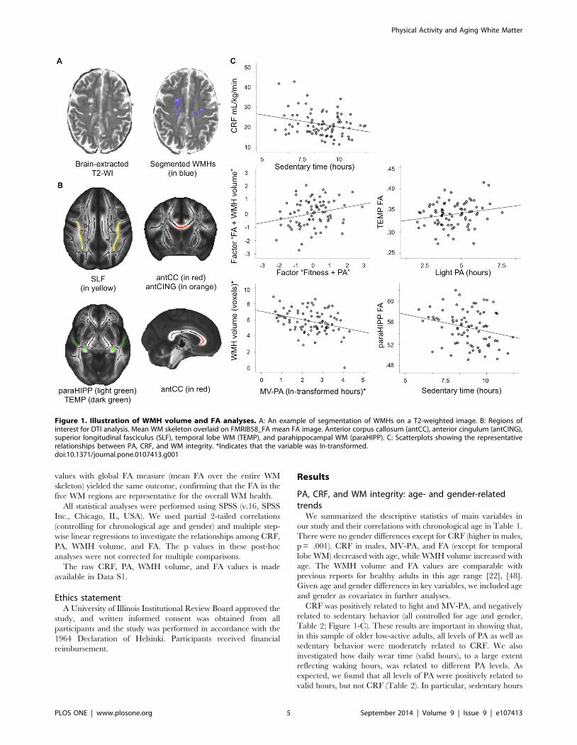
tation (done by AZB, Figure 1-A). WMH volume was reported in
voxel number classified in the above procedure as hyperintense,
where voxel size was 0.8660.8664 mm3. The WMH volume was
positively skewed (Kolmogorov-Smirnov test D(60) = 0.28, p,
0.05) and we performed a natural log-transformation of this
variable for further analyses.
DTI analysisDTI allows inferences about WM microstructure in vivo by
quantifying the magnitude and directionality of diffusion of water
within a tissue [20]. Visual checks were performed on every
volume of the raw data of every participant by AZB. In one
Physical Activity and Aging White Matter
PLOS ONE | www.plosone.org 3 September 2014 | Volume 9 | Issue 9 | e107413
dataset, one volume with the corresponding b-vectors and b-values
was deleted from the dataset before processing due to artifact.
Next, DTI data were processed using the FSL Diffusion Toolbox
v.3.0 in a standard multistep procedure, including: a) motion and
eddy current correction of the images and corresponding b-
vectors, b) removal of the skull and non-brain tissue using the
Brain Extraction Tool [40], and c) voxel-by-voxel calculation of
the diffusion tensors. Using the diffusion tensor information, FA
maps were computed using DTIFit within the FDT. All motion-
and eddy-current outputs, as well as FA images were visually
inspected.
We used tract-based spatial statistics (TBSS; 41,42], a toolbox
within FSL v5.0.1 [43], to create a representation of main WM
tracts common to all subjects (WM ‘‘skeleton’’). This included: (1)
nonlinear alignment of each participant’s FA volume to the
16161 mm3 standard Montreal Neurological Institute (MNI152)
space via the FMRIB58_FA template using the FMRIB’s
Nonlinear Registration Tool (FNIRT, [44]), (2) calculation of
the mean of all aligned FA images, (3) creation of the WM
‘‘skeleton’’ by perpendicular non-maximum-suppression of the
mean FA image and setting the FA threshold to 0.25, and (4)
perpendicular projection of the highest FA value (local center of
the tract) onto the skeleton, separately for each subject. The
outputs of all the above processing steps were carefully inspected
by AZB.
To verify how the tracts of interest were related to CRF and PA,
we extracted FA values from five regions: anterior corpus callosum
(antCC; two most anterior sections of the corpus callosum as
segmented after [45], anterior cingulum (antCING), superior
longitudinal fasciculi (SLF), temporal lobe WM (TEMP), and WM
of the medial temporal lobe (parahippocampal WM, paraHIPP;
Figure 1-B; [46]. The regions were identified on the TBSS
skeleton with the use of the DTI WM atlas to probe FA in the core
parts of the selected tracts [47]. DTI data of five participants did
not entirely cover the temporal lobes. Therefore, the above
analyzes were done twice: once with all participants and once
without the five participants (n = 78). The full sample analysis
resulted in common mask not covering the temporal lobes and
therefore those five subjects had missing data in those tracts.
It is important to note that the TBSS analysis used here
inherently focuses on normal appearing WM, as the highest FA
values perpendicular to the tract are being projected to the WM
skeleton for further analysis. This means that there may be some
bias for excluding voxels affected by WMH from FA analyses. We
thus consider TBSS approach most suitable for the current study
as it maximizes the independence of FA measures from WMH, in
addition to circumventing the inter-subject anatomical variability
in older populations.
An exploratory analysis relating CRF with FA across the whole
WM skeleton was carried out in randomize, a voxel-wise
permutation-based (5000 permutations) inference (Nichols and
Holmes, 2002), with the threshold-free cluster enhancement
option, and controlling for age and gender.
Statistical analysesTo test the first study hypothesis, we performed a dimensionality
reduction by running two separate principal component analyses
(PCA) with varimax rotation. The first model included the four
variables: CRF, sedentary time, light PA, and MV-PA. The
second model included the WM health indices: five FA values and
WMH volume. Both analyses yielded a one-component model and
therefore represented ‘‘CRF and PA’’ and ‘‘WM health’’,
respectively. Importantly, a PCA analysis replacing the five FA
Table 1. Variables of interest: descriptive statistics and correlations with age.
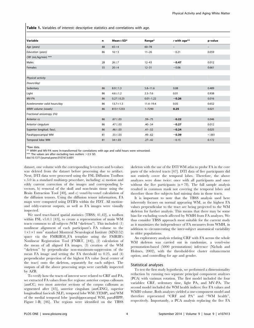
Variable n Mean±SD* Range* r with age** p-value
Age (years) 88 6564 60–78 – –
Education (years) 86 1663 11–26 20.21 0.059
CRF (mL/kg/min) ***
Males 28 2667 12–43 20.47 0.012
Females 55 2064 12–31 20.06 0.661
Physical activity
(hours/day)
Sedentary 86 8.961.3 5.8–11.6 0.08 0.489
Light 86 4.661.2 2.3–7.6 0.01 0.938
MV-PA 86 0.2760.25 0.01–1.22 20.26 0.016
Aceelerometer valid hours/day 86 13.761.3 11.4–19.4 0.05 0.652
WMH volume (voxels) 86 81361233 1–7290 0.25 0.021
Fractional anisotropy (FA)
Anterior cc 86 .676.03 .59–.75 20.22 0.046
Anterior cingulum 86 .476.03 .40–.54 20.27 0.012
Superior longitud. fasci. 86 .466.03 .41–.52 20.24 0.025
Parahippocampal WM 81 .556.03 .49–.62 20.39 ,.001
Temporal lobe WM 81 346.03 .27–.42 20.15 0.172
*Raw data.** WMH and MV-PA were ln-transformed for correlations with age and valid hours were winsorized.*** The values are after excluding two outliers .2.5 SD.doi:10.1371/journal.pone.0107413.t001
Physical Activity and Aging White Matter
PLOS ONE | www.plosone.org 4 September 2014 | Volume 9 | Issue 9 | e107413
values with global FA measure (mean FA over the entire WM
skeleton) yielded the same outcome, confirming that the FA in the
five WM regions are representative for the overall WM health.
All statistical analyses were performed using SPSS (v.16, SPSS
Inc., Chicago, IL, USA). We used partial 2-tailed correlations
(controlling for chronological age and gender) and multiple step-
wise linear regressions to investigate the relationships among CRF,
PA, WMH volume, and FA. The p values in these post-hoc
analyses were not corrected for multiple comparisons.
The raw CRF, PA, WMH volume, and FA values is made
available in Data S1.
Ethics statementA University of Illinois Institutional Review Board approved the
study, and written informed consent was obtained from all
participants and the study was performed in accordance with the
1964 Declaration of Helsinki. Participants received financial
reimbursement.
Results
PA, CRF, and WM integrity: age- and gender-relatedtrends
We summarized the descriptive statistics of main variables in
our study and their correlations with chronological age in Table 1.
There were no gender differences except for CRF (higher in males,
p = .001). CRF in males, MV-PA, and FA (except for temporal
lobe WM) decreased with age, while WMH volume increased with
age. The WMH volume and FA values are comparable with
previous reports for healthy adults in this age range [22], [48].
Given age and gender differences in key variables, we included age
and gender as covariates in further analyses.
CRF was positively related to light and MV-PA, and negatively
related to sedentary behavior (all controlled for age and gender,
Table 2; Figure 1-C). These results are important in showing that,
in this sample of older low-active adults, all levels of PA as well as
sedentary behavior were moderately related to CRF. We also
investigated how daily wear time (valid hours), to a large extent
reflecting waking hours, was related to different PA levels. As
expected, we found that all levels of PA were positively related to
valid hours, but not CRF (Table 2). In particular, sedentary hours
Figure 1. Illustration of WMH volume and FA analyses. A: An example of segmentation of WMHs on a T2-weighted image. B: Regions ofinterest for DTI analysis. Mean WM skeleton overlaid on FMRIB58_FA mean FA image. Anterior corpus callosum (antCC), anterior cingulum (antCING),superior longitudinal fasciculus (SLF), temporal lobe WM (TEMP), and parahippocampal WM (paraHIPP). C: Scatterplots showing the representativerelationships between PA, CRF, and WM integrity. *Indicates that the variable was ln-transformed.doi:10.1371/journal.pone.0107413.g001
Physical Activity and Aging White Matter
PLOS ONE | www.plosone.org 5 September 2014 | Volume 9 | Issue 9 | e107413
accounted for most of the variability in the wearing time among
our participants.
Finally, we examined the relationships between WMH volume
and FA in the five WM regions. We found no significant
relationship between WMH volume and FA (controlled for age
and gender). This confirmatory analysis showed that in the five
regions, WMH and DTI are not redundant measures of WM
health.
Greater CRF and PA are associated with better WM healthDimensionality reduction using PCA separately on WM and
CRF and PA measures yielded single factors for ‘‘CRF and PA’’
and ‘‘FA and WMH volume’’. In other words, participants with
greater ‘‘CRF and PA’’ factor scores had higher CRF, showed
more light and MV-PA, and less sedentary behavior. Participants
with higher WM health factor score had higher FA in all five
regions and lower WMH volume. As predicted, we found a
positive association between ‘‘CRF and PA’’ and ‘‘FA and WMH
volume’’ (r = .24, p = .037, n = 77), which was a prerequisite for
the following post-hoc analyses (Figure 1-C).
Relationships of PA with WM health (FA and WMHvolume)
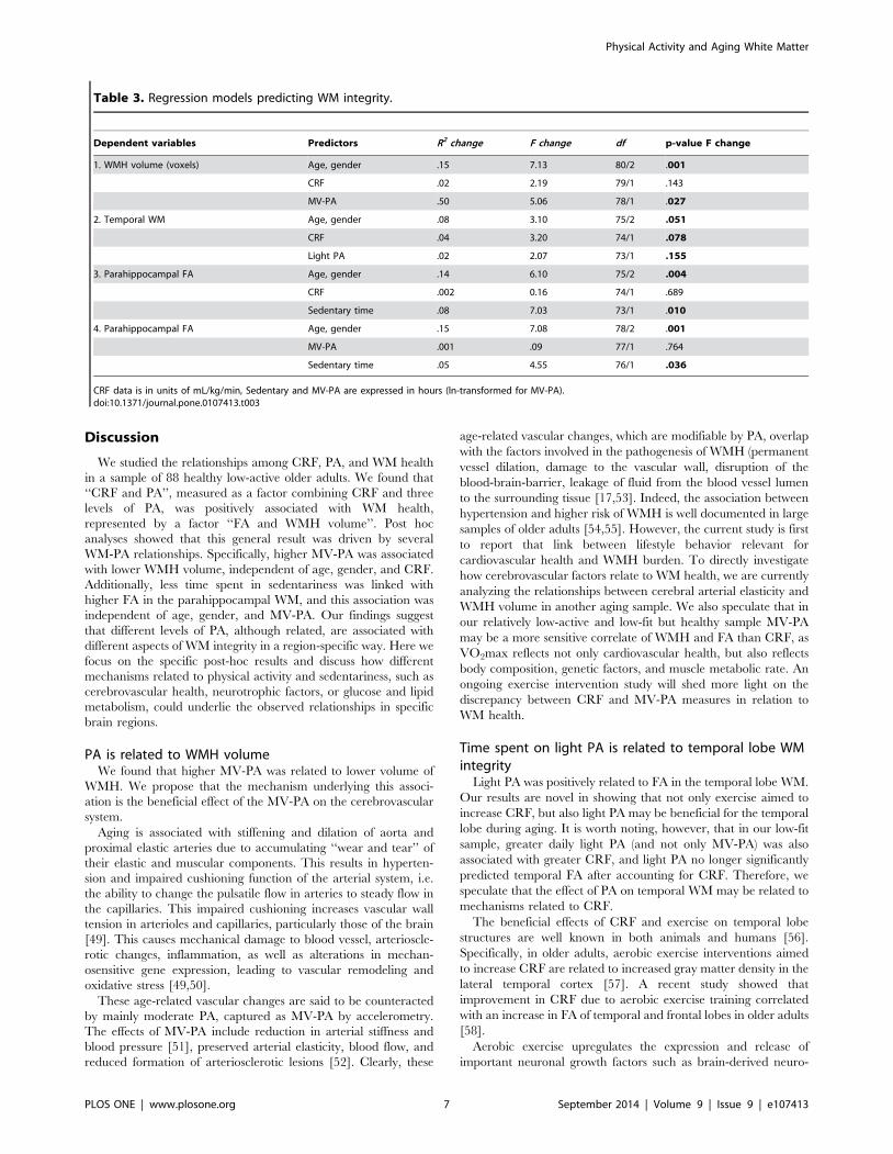
MV-PA (pr = 2.31 p = .004, df = 82), but not sedentary or light
daily PA was related to WMH volume (all controlled for age and
gender; Figure 1-C). A multiple hierarchical regression showed
that MV-PA was associated with WMH volume in addition to
variance explained by age, gender, and CRF (Model 1 in Table 3).
Greater light PA (pr = .25, p = .026, df = 77) was associated
with higher FA in the temporal lobe WM (Figure 1-C). A multiple
hierarchical regression showed that light PA was not associated
with temporal lobe FA in addition to variance explained by age,
gender, and CRF (Model 2 in Table 3).
Sedentary behavior was inversely related to FA in the
parahippocampal WM. That is, participants who spent more
time on sedentary behavior had lower FA adjacent to the
hippocampus (pr = 2.24, p = .035, df = 77; controlled for age
and gender; Figure 1-C). The negative relationship between
sedentary behavior and temporal lobe FA (pr = 2.21, p = .066,
df = 77) was at trend level. Again, as sedentary behavior was
negatively associated with CRF, we examined whether the effect of
sedentariness on parahippocampal FA was independent of CRF in
a hierarchical multiple regression. We found that sedentary
behavior explained a significant amount of variance in para-
hippocampal FA, independently of variance explained by age,
gender, and CRF (Model 3 in Table 3). The results of all of the
above analyses were unchanged if valid hours were included as
covariates in addition to age, gender, and CRF. Only the partial
correlation between sedentary time and the parahippocampal
WM FA was at a trend level after controlling for valid hours in
addition to age and gender (pr = 2.20, p = .076, df = 76). This was
expected, given that the variance in valid hours was mainly
associated with variance in sedentary time and, to a lesser extent,
to light and MV-PA. Together, these results suggest that daily
valid PA measurement hours, likely reflecting waking hours, are to
some extent related to the PA counts, but do not account for the
relationships between PA and WM health.
Finally, the mechanisms linking sedentary behavior to brain
integrity may differ from those related to MV-PA [7]. Indeed, a
hierarchical multiple regression showed that sedentary behavior
explained a significant amount of variance in parahippocampal
FA, independently of variance explained by age, gender, and MV-
PA (Model 4 in Table 3).
Figure 1-C shows scatterplots of the representative relationships
between CRF, PA, and WM integrity.
Relationships of CRF with WM health (FA and WMHvolume)
To revisit the findings from previous studies [23–25] we tested
the relationship between CRF and FA three prefrontal WM
regions in our low-active and low-fit sample. We found no
significant associations in the ROI-based analysis as well as whole-
brain exploratory TBSS analysis. There was no significant
relationship between CRF and WMH volume (pr = 2.16, p =
.143, df = 79).
We found a positive association only between temporal lobe FA
and CRF; however, after controlling for age and gender it
remained at trend level (pr = .20, p = .078, df = 74).
Table 2. Partial correlations between CRF and levels of PA.
1. Valid hours 2. Sedentary 3. Light PA 4. MV-PA 5. CRF
1. Valid hours 1 pr = .53 pr = .45 pr = .22 pr = .18
p, .001 p, .001 p = .041 p = .106
df = 82 df = 82 df = 82 df = 79
2. Sedentary 1 pr = 2.48 pr = 2.27 pr = 2.36
p, .001 p = .013 p = .001
df = 82 df = 82 df = 79
3. Light PA - 1 pr = .35 pr = .43
p = .001 p, .001
df = 82 df = 79
4. MV-PA - - 1 pr = .55
p, .001
df = 79
5. CRF - - - 1
All correlations controlled for age and gender. pr denotes partial correlation. CRF data is in units of mL/kg/min, Sedentary, Light, and MV-PA are expressed in hours (ln-transformed for MV-PA, winsorized for valid hours).doi:10.1371/journal.pone.0107413.t002
Physical Activity and Aging White Matter
PLOS ONE | www.plosone.org 6 September 2014 | Volume 9 | Issue 9 | e107413
Discussion
We studied the relationships among CRF, PA, and WM health
in a sample of 88 healthy low-active older adults. We found that
‘‘CRF and PA’’, measured as a factor combining CRF and three
levels of PA, was positively associated with WM health,
represented by a factor ‘‘FA and WMH volume’’. Post hoc
analyses showed that this general result was driven by several
WM-PA relationships. Specifically, higher MV-PA was associated
with lower WMH volume, independent of age, gender, and CRF.
Additionally, less time spent in sedentariness was linked with
higher FA in the parahippocampal WM, and this association was
independent of age, gender, and MV-PA. Our findings suggest
that different levels of PA, although related, are associated with
different aspects of WM integrity in a region-specific way. Here we
focus on the specific post-hoc results and discuss how different
mechanisms related to physical activity and sedentariness, such as
cerebrovascular health, neurotrophic factors, or glucose and lipid
metabolism, could underlie the observed relationships in specific
brain regions.
PA is related to WMH volumeWe found that higher MV-PA was related to lower volume of
WMH. We propose that the mechanism underlying this associ-
ation is the beneficial effect of the MV-PA on the cerebrovascular
system.
Aging is associated with stiffening and dilation of aorta and
proximal elastic arteries due to accumulating ‘‘wear and tear’’ of
their elastic and muscular components. This results in hyperten-
sion and impaired cushioning function of the arterial system, i.e.
the ability to change the pulsatile flow in arteries to steady flow in
the capillaries. This impaired cushioning increases vascular wall
tension in arterioles and capillaries, particularly those of the brain
[49]. This causes mechanical damage to blood vessel, arterioscle-
rotic changes, inflammation, as well as alterations in mechan-
osensitive gene expression, leading to vascular remodeling and
oxidative stress [49,50].
These age-related vascular changes are said to be counteracted
by mainly moderate PA, captured as MV-PA by accelerometry.
The effects of MV-PA include reduction in arterial stiffness and
blood pressure [51], preserved arterial elasticity, blood flow, and
reduced formation of arteriosclerotic lesions [52]. Clearly, these
age-related vascular changes, which are modifiable by PA, overlap
with the factors involved in the pathogenesis of WMH (permanent
vessel dilation, damage to the vascular wall, disruption of the
blood-brain-barrier, leakage of fluid from the blood vessel lumen
to the surrounding tissue [17,53]. Indeed, the association between
hypertension and higher risk of WMH is well documented in large
samples of older adults [54,55]. However, the current study is first
to report that link between lifestyle behavior relevant for
cardiovascular health and WMH burden. To directly investigate
how cerebrovascular factors relate to WM health, we are currently
analyzing the relationships between cerebral arterial elasticity and
WMH volume in another aging sample. We also speculate that in
our relatively low-active and low-fit but healthy sample MV-PA
may be a more sensitive correlate of WMH and FA than CRF, as
VO2max reflects not only cardiovascular health, but also reflects
body composition, genetic factors, and muscle metabolic rate. An
ongoing exercise intervention study will shed more light on the
discrepancy between CRF and MV-PA measures in relation to
WM health.
Time spent on light PA is related to temporal lobe WMintegrity
Light PA was positively related to FA in the temporal lobe WM.
Our results are novel in showing that not only exercise aimed to
increase CRF, but also light PA may be beneficial for the temporal
lobe during aging. It is worth noting, however, that in our low-fit
sample, greater daily light PA (and not only MV-PA) was also
associated with greater CRF, and light PA no longer significantly
predicted temporal FA after accounting for CRF. Therefore, we
speculate that the effect of PA on temporal WM may be related to
mechanisms related to CRF.
The beneficial effects of CRF and exercise on temporal lobe
structures are well known in both animals and humans [56].
Specifically, in older adults, aerobic exercise interventions aimed
to increase CRF are related to increased gray matter density in the
lateral temporal cortex [57]. A recent study showed that
improvement in CRF due to aerobic exercise training correlated
with an increase in FA of temporal and frontal lobes in older adults
[58].
Aerobic exercise upregulates the expression and release of
important neuronal growth factors such as brain-derived neuro-
Table 3. Regression models predicting WM integrity.
Dependent variables Predictors R2 change F change df p-value F change
1. WMH volume (voxels) Age, gender .15 7.13 80/2 .001
CRF .02 2.19 79/1 .143
MV-PA .50 5.06 78/1 .027
2. Temporal WM Age, gender .08 3.10 75/2 .051
CRF .04 3.20 74/1 .078
Light PA .02 2.07 73/1 .155
3. Parahippocampal FA Age, gender .14 6.10 75/2 .004
CRF .002 0.16 74/1 .689
Sedentary time .08 7.03 73/1 .010
4. Parahippocampal FA Age, gender .15 7.08 78/2 .001
MV-PA .001 .09 77/1 .764
Sedentary time .05 4.55 76/1 .036
CRF data is in units of mL/kg/min, Sedentary and MV-PA are expressed in hours (ln-transformed for MV-PA).doi:10.1371/journal.pone.0107413.t003
Physical Activity and Aging White Matter
PLOS ONE | www.plosone.org 7 September 2014 | Volume 9 | Issue 9 | e107413
trophic factor (BDNF) [31,58–60] and insulin-like growth factor I
(IGF-1; a potent survival factor for neurons and oligodendrocytes)
[61]. Therefore, structural and functional brain changes related to
improved oxidative capacity have been largely attributed to the
action of neurotrophins. For example, an aerobic walking
intervention in older adults resulted in increased temporal lobe
functional connectivity, associated with increased BDNF, IGF-1,
and vascular endothelial growth factor serum levels [58].
BDNF supports survival and growth of many neuronal subtypes
and is a mediator of synaptic efficacy and use-dependent plasticity
[62]. Still, the effects of BDNF on WM are poorly understood.
Therefore the link between CRF, neurotrophin upregulation, and
temporal WM integrity in healthy humans is less straightforward.
Healthy adult Val/Val homozygotes of the BDNF Val66Met
polymorphism (increased levels of activity-dependent release) had
decreased FA compared to Met-allele carriers [63,64], and lower
structural brain connectivity [65]. Therefore, while for grey matter
the met66 allele is almost invariably protective, BDNF seems to be
a ‘‘punitive’’ signal in axonal pruning, i.e. higher BDNF secretion
from stimulated axons results in its higher binding to p75
neurotrophin receptor on less active axons, which leads to neurite
elimination [66] BDNF has neuroprotective role of on WM in
clinical studies [67,68] and plays a role in normal axonal pruning
and maintenance observed in animals [66,69]. Therefore, further
research combining various MRI modalities to serum levels of
neurotrophins is necessary to understand their role in mature as
well as aging WM, and the link between light PA and temporal
lobe WM.
It is also possible that the positive effects of light PA on temporal
lobe WM integrity are mediated by changes in cerebrovascular
supply factors related to CRF, reducing the oxygenative stress in
the tissue [12]. Aerobic training correlates with increased capillary
density in brain parenchyma [70] and higher number and
integrity of small blood vessels as measured by MRI angiography
[71]. Together, these changes may lead to increased ratio of blood
vessels to brain volume [72] and more efficient oxygen delivery.
An alternative, although not mutually exclusive, explanation for
the observed association between PA and WM integrity (both FA
and WMH) is that individuals with higher WMH burden or
decreases in FA are at more risk of impaired mobility [73], gait
and balance dysfunction [74–79], and physical disability [80–82].
This could lead to their lower mobility and engagement in PA.
Still, a study comparing athletes with non-exercising controls
indicated that life-long exercise is associated with reduced WMH
and age-related decline in FA [25]. We will test these alternative
hypotheses in an ongoing randomized control study with exercise
intervention.
In sum, our results suggest that not only structured exercise, but
also light PA may be associated with greater WM microstructural
integrity in temporal regions, known to be prone to age-related
changes and sensitive to CRF-related benefits. Future analyses
linking temporal WM FA to cognitive measures, local structure,
and neural activity will allow assessing the functional relevance of
the reported associations.
Sedentary behavior may relate to WM via differentmechanisms than PA
We found that older adults who spent more time in sedentary
behavior had lower FA in the parahippocampal WM. Important-
ly, more sedentary behavior was related not only to less light PA,
but also to less MV-PA, and lower CRF. Therefore, more
sedentary older individuals were less exposed to beneficial effects
of light and MV-PA, but also more prone to deleterious effects of
sedentariness, such as increased metabolic risk [83,84]. The
association between sedentariness and parahippocampal WM,
however, was independent from MV-PA. This is in line with
recent hypotheses that sedentariness may act via different
mechanisms than MV-PA, and may even offset the benefits of
MV-PA [7,85]. Specifically, both acute and chronic sedentariness
is related to reduced activity of lipoprotein lipase activity [86],
which facilitates uptake of fatty acids into muscle and adipose
tissue. This causes increased plasma levels of triglycerides, total
cholesterol, and decreased levels of high-density lipoprotein
cholesterol [84,87]. In addition, sedentary behavior is related to
insulin resistance and increased glucose plasma levels [84].
Physiological differences between sedentary behavior and lack
of MV-PA [7,85] may partly explain the specificity of sedentary
behavior for parahippocampal WM integrity. For instance, insulin
receptors are highly expressed in the hippocampus and play a
modulatory role in learning and memory processing [88]. Both
insulin deficiency and resistance impairs hippocampus-dependent
memory, long-term potentiation along the perforant pathway, and
adult neurogenesis in rats [89]. Insulin resistance in humans is
associated with similar cognitive deficits as in rodents [90].
Similarly, elevation in serum cholesterol change hippocampus
lipid metabolism and induce oxygenative stress in rats [91]. As
hyperlipidemia often presages insulin resistance, we speculate that
sedentariness-induced changes in plasma lipids and glucose may
have negative effect on hippocampus in low-active older adults,
even in the absence of diagnosed diabetes. Deleterious changes in
medial temporal lobe structures should be tightly linked to WM
tracts connecting these regions. Little is known, however, about
mechanisms linking plasma lipids and glucose levels to WM
integrity, i.e. myelin turnover and repair, oligodendrocyte viability,
axonal transport and integrity. Clearly, more research is needed to
understand how sedentary behavior physiology interacts with the
integrity of WM. Ideally, future studies will combine PA measures,
structural and functional MRI with blood metabolite assays.
Finally, we observed no relationships between CRF with WMH
volume or FA in the corpus callosum, the cingulum bundle, or
superior longitudinal fasciculi, as reported previously by [23–25].
The earlier studies may have limited generalizability due to size
(n#26) and characteristics of the sample: [23] sample consisted of
26% superiorly fit individuals according to ACSM’s Guidelines for
Exercise Testing and Prescription (V02max .42.7 for men in 7th
decade of life), 53% participants in [24] had a history of minimum
10 years of exercise 3 hours/week and mean V02max = 38, while
half of [25] sample included Masters athletes. These effects
observed in small samples including superiorly fit older individuals
might not be reproducible in larger, low-active aging samples with
narrower range of CRF, like ours. The causality of the
relationships is also speculative, as some genetic or environmental
factors, unrelated to PA, may have predisposed Masters athletes
for high performance in aerobic sports and may be also linked to
WM properties.
ConclusionsWe explored the associations of objective measures of CRF and
PA with measures of WM health in older low-fit and low-active
adults. We showed that higher levels of MV-PA were linked to
lower WMH volume. We propose that MV-PA allows keeping
WMHs in check via cerebrovascular mechanisms, such as
preserving higher blood vessel elasticity and wall integrity. We
found an association between light PA and WM integrity in the
temporal lobe while sedentary behavior was related to lower FA in
the parahippocampal WM. We speculate that these associations
are related to neurotrophic, cerebrovascular, lipid and insulin
metabolic mechanisms related to PA or lack thereof. Together, our
Physical Activity and Aging White Matter
PLOS ONE | www.plosone.org 8 September 2014 | Volume 9 | Issue 9 | e107413
findings suggest that PA is associated with WM health in aging in
an intensity- and region-specific manner. Our results are optimistic
in showing that modifiable lifestyle factors such as increasing PA
and reducing sedentariness may be beneficial for brain health. We
are currently assessing in a randomized exercise intervention trial
whether increases in CRF and changes in PA behavior may induce
changes in WM health over shorter periods of time.
We conclude that PA and CRF are related but not equivalent in
their relationships with WM health in aging. Specifically, the
effects are WM measure- and region-specific, showing the
importance of using multiple, objective, and independent
measures to assess fitness, physical activity and sedentariness,
and brain integrity. Our findings pave the way for testing more
targeted hypotheses linking bodily fitness and WM integrity in
longitudinal designs, with improved public health recommenda-
tions on PA being the longer-term outcome of such studies.
Supporting Information
Data S1 The file contains the raw demographic, PA,CRF, DTI, and WMH data of the 103 participants.(XLSX)
Acknowledgments
The authors thank Chanheng He, Meera Zukosky, Kishan Patel for help
with data management, Holly Tracy and Nancy Dodge for MRI data
collection, Sarah Banducci for help in data analyses, and Susan H. Herrel
for project coordination.
Author Contributions
Conceived and designed the experiments: MWV AFK EM. Performed the
experiments: AZB GEC JM CNW NPG EAO AK AL TRW JF HDC EA.
Analyzed the data: AZB. Contributed reagents/materials/analysis tools:
AZB CNW. Wrote the paper: AZB LCH MWV AFK.
References
1. Kramer AF, Hahn S, Cohen NJ, Banich MT, McAuley E, et al. (1999) Ageing,fitness and neurocognitive function.
2. Hillman CH, Erickson KI, Kramer AF (2008) Be smart, exercise your heart:
exercise effects on brain and cognition. Nat Rev Neurosci 9: 58–65.
3. Madden DJ, Bennett IJ, Burzynska A, Potter GG, Chen N-K, et al. (2012)
Diffusion tensor imaging of cerebral white matter integrity in cognitive aging.Biochim Biophys Acta 1822: 386–400.
4. Raz N, Rodrigue KM (2006) Differential aging of the brain: patterns, cognitive
correlates and modifiers. Neurosci Biobehav Rev 30: 730–748.
5. Freedson PS, Melanson E, Sirard J (1998) Calibration of the Computer Science
and Applications, Inc. accelerometer. Med Sci Sports Exerc 30: 777–781.
6. Craft LL, Zderic TW, Gapstur SM, Vaniterson EH, Thomas DM, et al. (2012)Evidence that women meeting physical activity guidelines do not sit less: an
observational inclinometry study. Int J Behav Nutr Phys Act 9: 122.
7. Voss MW, Carr LJ, Clark R, Weng T (2014) Revenge of the ‘‘sit’’ II: Does
lifestyle impact neuronal and cognitive health through distinct mechanismsassociated with sedentary behavior and physical activity? Ment Health Phys Act.
8. Middleton LE, Manini TM, Simonsick EM, Harris TB, Barnes DE, et al. (2011)
Activity energy expenditure and incident cognitive impairment in older adults.Arch Intern Med 171: 1251–1257.
9. Donahoo WT, Levine JA, Melanson EL (2004) Variability in energy expenditure
and its components. Curr Opin Clin Nutr Metab Care 7: 599–605.
10. Dunstan DW, Howard B, Healy GN, Owen N (2012) Too much sitting–a health
hazard. Diabetes Res Clin Pract 97: 368–376.
11. Helgerud J, Høydal K, Wang E, Karlsen T, Berg P, et al. (2007) Aerobic high-intensity intervals improve VO2max more than moderate training. Med Sci
Sports Exerc 39: 665–671.
12. Bassett DR, Howley ET (2000) Limiting factors for maximum oxygen uptake
and determinants of endurance performance. Med Sci Sports Exerc 32: 70–84.
13. Spina RJ, Ogawa T, Kohrt WM, Martin WH, Holloszy JO, et al. (1993)Differences in cardiovascular adaptations to endurance exercise training
between older men and women. J Appl Physiol 75: 849–855.
14. Hoppeler H, Howald H, Conley K, Lindstedt SL, Claassen H, et al. (1985)
Endurance training in humans: aerobic capacity and structure of skeletal muscle.J Appl Physiol 59: 320–327.
15. Peter I, Papandonatos GD, Belalcazar LM, Yang Y, Erar B, et al. (2014) Genetic
modifiers of cardiorespiratory fitness response to lifestyle intervention. Med Sci
Sports Exerc 46: 302–311.
16. Baloh RW, Vinters HV (1995) White matter lesions and disequilibrium in olderpeople. II. Clinicopathologic correlation. Arch Neurol 52: 975–981.
17. Pantoni L (2002) Pathophysiology of age-related cerebral white matter changes.
Cerebrovasc Dis 13 Suppl 2: 7–10.
18. Schmidt R, Enzinger C, Ropele S, Schmidt H, Fazekas F (2003) Progression of
cerebral white matter lesions: 6-year results of the Austrian Stroke PreventionStudy. Lancet 361: 2046–2048.
19. Basser PJ (n.d.) Inferring microstructural features and the physiological state of
tissues from diffusion-weighted images. NMR Biomed 8: 333–344.
20. Beaulieu C (n.d.) The basis of anisotropic water diffusion in the nervous system -
a technical review. NMR Biomed 15: 435–455.
21. Bennett IJ, Madden DJ, Vaidya CJ, Howard DV, Howard JH (2010) Age-related differences in multiple measures of white matter integrity: A diffusion
tensor imaging study of healthy aging. Hum Brain Mapp31: 378–390.
22. Burzynska AZ, Preuschhof C, Backman L, Nyberg L, Li S-C, et al. (2010) Age-
related differences in white matter microstructure: region-specific patterns ofdiffusivity. Neuroimage 49: 2104–2112.
23. Johnson NF, Kim C, Clasey JL, Bailey A, Gold BT (2012) Cardiorespiratory
fitness is positively correlated with cerebral white matter integrity in healthyseniors. Neuroimage 59: 1514–1523.
24. Marks BL, Katz LM, Styner M, Smith JK (2011) Aerobic fitness and obesity:
relationship to cerebral white matter integrity in the brain of active and
sedentary older adults. Br J Sports Med 45: 1208–1215.
25. Tseng BY, Gundapuneedi T, Khan MA, Diaz-Arrastia R, Levine BD, et al.
(2013) White matter integrity in physically fit older adults. Neuroimage 82: 510–
516.
26. Voss MW, Heo S, Prakash RS, Erickson KI, Alves H, et al. (2012) The influence
of aerobic fitness on cerebral white matter integrity and cognitive function in
older adults: Results of a one-year exercise intervention. Hum Brain Mapp.
27. De Leeuw FE, de Groot JC, Achten E, Oudkerk M, Ramos LM, et al. (2001)
Prevalence of cerebral white matter lesions in elderly people: a population based
magnetic resonance imaging study. The Rotterdam Scan Study. J Neurol
Neurosurg Psychiatry 70: 9–14.
28. McAuley E, Szabo AN, Mailey EL, Erickson KI, Voss M, et al. (2011) Non-
Exercise Estimated Cardiorespiratory Fitness: Associations with Brain Structure,
Cognition, and Memory Complaints in Older Adults. Ment Health Phys Act 4:
5–11.
29. Szabo AN, McAuley E, Erickson KI, Voss M, Prakash RS, et al. (2011)
Cardiorespiratory fitness, hippocampal volume, and frequency of forgetting in
older adults. Neuropsychology 25: 545–553.
30. Voss MW, Erickson KI, Prakash RS, Chaddock L, Malkowski E, et al. (2010)
Functional connectivity: a source of variance in the association between
cardiorespiratory fitness and cognition? Neuropsychologia 48: 1394–1406.
31. Erickson KI, Voss MW, Prakash RS, Basak C, Szabo A, et al. (2011) Exercise
training increases size of hippocampus and improves memory. Proc Natl Acad
Sci U S A 108: 3017–3022.
32. Erickson KI, Prakash RS, Voss MW, Chaddock L, Hu L, et al. (2009) Aerobic
fitness is associated with hippocampal volume in elderly humans. Hippocampus
19: 1030–1039.
33. Voss MW, Heo S, Prakash RS, Erickson KI, Alves H, et al. (2013) The influence
of aerobic fitness on cerebral white matter integrity and cognitive function in
older adults: results of a one-year exercise intervention. Hum Brain Mapp34:
2972–2985.
34. Vernooij MW, de Groot M, van der Lugt A, Ikram MA, Krestin GP, et al.
(2008) White matter atrophy and lesion formation explain the loss of structural
integrity of white matter in aging. Neuroimage 43: 470–477.
35. Haskell W, Lee I-M, Pate R, Powell K, Blair S, et al. (2007) Physical Activity and
Public Health: Updated Recommendation for Adults From the American
College of Sports Medicine and the American Heart Association. Circulation
116: 1081–1093.
36. Hart TL, McClain JJ, Tudor-Locke C (2011) Controlled and free-living
evaluation of objective measures of sedentary and active behaviors. J Phys Act
Health 8: 848–857.
37. Troiano RP, Berrigan D, Dodd KW, Masse LC, Tilert T, et al. (2008) Physical
activity in the United States measured by accelerometer. Med Sci Sports Exerc
40: 181–188.
38. Reese TG, Heid O, Weisskoff RM, Wedeen VJ (2003) Reduction of eddy-
current-induced distortion in diffusion MRI using a twice-refocused spin echo.
Magn Reson Med 49: 177–182.
39. Zhang Y, Brady M, Smith S (2001) Segmentation of brain MR images through a
hidden Markov random field model and the expectation-maximization
algorithm. IEEE Trans Med Imaging 20: 45–57.
40. Smith SM (2002) Fast robust automated brain extraction. Hum Brain Mapp17:
143–155.
41. Smith SM, Jenkinson M, Johansen-Berg H, Rueckert D, Nichols TE, et al.
(2006) Tract-based spatial statistics: voxelwise analysis of multi-subject diffusion
data. Neuroimage 31: 1487–1505.
Physical Activity and Aging White Matter
PLOS ONE | www.plosone.org 9 September 2014 | Volume 9 | Issue 9 | e107413
42. Smith SM, Johansen-Berg H, Jenkinson M, Rueckert D, Nichols TE, et al.
(2007) Acquisition and voxelwise analysis of multi-subject diffusion data withtract-based spatial statistics. Nat Protoc 2: 499–503.
43. Smith SM, Jenkinson M, Woolrich MW, Beckmann CF, Behrens TEJ, et al.
(2004) Advances in functional and structural MR image analysis andimplementation as FSL. Neuroimage 23 Suppl 1: S208–19.
44. Rueckert D, Sonoda LI, Hayes C, Hill DL, Leach MO, et al. (1999) Nonrigidregistration using free-form deformations: application to breast MR images.
IEEE Trans Med Imaging 18: 712–721.
45. Hofer S, Frahm J (2006) Topography of the human corpus callosum revisited–comprehensive fiber tractography using diffusion tensor magnetic resonance
imaging. Neuroimage 32: 989–994.46. Burzynska AZ, Garrett DD, Preuschhof C, Nagel IE, Li S-C, et al. (2013) A
scaffold for efficiency in the human brain. J Neurosci 33: 17150–17159.47. Mori S, Wakana S, Nagae-Poetscher LM, Van Zijl PCM (2005) MRI Atlas of
Human White Matter. Elsevier, editor Elsevier.
48. Bhagat YA, Beaulieu C (2004) Diffusion anisotropy in subcortical white matterand cortical gray matter: changes with aging and the role of CSF-suppression.
J Magn Reson Imaging 20: 216–227.49. O’Rourke MF, Hashimoto J (2007) Mechanical factors in arterial aging: a
clinical perspective. J Am Coll Cardiol 50: 1–13.
50. Ungvari Z, Kaley G, de Cabo R, Sonntag WE, Csiszar A (2010) Mechanisms ofvascular aging: new perspectives. J Gerontol A Biol Sci Med Sci 65: 1028–1041.
51. McDonnell BJ, Maki-Petaja KM, Munnery M, Yasmin, Wilkinson IB, et al.(2013) Habitual exercise and blood pressure: age dependency and underlying
mechanisms. Am J Hypertens 26: 334–341.52. Vaitkevicius PV, Fleg JL, Engel JH, O’Connor FC, Wright JG, et al. (1993)
Effects of age and aerobic capacity on arterial stiffness in healthy adults.
Circulation 88: 1456–1462.53. Schmidt R, Petrovic K, Ropele S, Enzinger C, Fazekas F (2007) Progression of
leukoaraiosis and cognition. Stroke 38: 2619–2625.54. Longstreth WT, Manolio TA, Arnold A, Burke GL, Bryan N, et al. (1996)
Clinical correlates of white matter findings on cranial magnetic resonance
imaging of 3301 elderly people. The Cardiovascular Health Study. Stroke 27:1274–1282.
55. Van Dijk EJ, Breteler MMB, Schmidt R, Berger K, Nilsson L-G, et al. (2004)The association between blood pressure, hypertension, and cerebral white
matter lesions: cardiovascular determinants of dementia study. Hypertension 44:625–630.
56. Voss MW, Vivar C, Kramer AF, van Praag H (2013) Bridging animal and
human models of exercise-induced brain plasticity. Trends Cogn Sci 17: 525–544.
57. Colcombe SJ, Erickson KI, Scalf PE, Kim JS, Prakash R, et al. (2006) Aerobicexercise training increases brain volume in aging humans. J Gerontol A Biol Sci
Med Sci 61: 1166–1170.
58. Voss MW, Erickson KI, Prakash RS, Chaddock L, Kim JS, et al. (2013)Neurobiological markers of exercise-related brain plasticity in older adults. Brain
Behav Immun 28: 90–99.59. Rasmussen P, Brassard P, Adser H, Pedersen M V, Leick L, et al. (2009)
Evidence for a release of brain-derived neurotrophic factor from the brainduring exercise. Exp Physiol 94: 1062–1069.
60. Zoladz JA, Pilc A, Majerczak J, Grandys M, Zapart-Bukowska J, et al. (2008)
Endurance training increases plasma brain-derived neurotrophic factorconcentration in young healthy men. J Physiol Pharmacol 59 Suppl 7: 119–132.
61. Carro E, Nunez A, Busiguina S, Torres-Aleman I (2000) Circulating insulin-likegrowth factor I mediates effects of exercise on the brain. J Neurosci 20: 2926–
2933.
62. Cotman CW, Berchtold NC (2002) Exercise: A behavioral intervention toenhance brain health and plasticity. Trends Neurosci 25: 295–301.
63. Chiang M-C, Barysheva M, Toga AW, Medland SE, Hansell NK, et al. (2011)BDNF gene effects on brain circuitry replicated in 455 twins. Neuroimage 55:
448–454.
64. Tost H, Alam T, Geramita M, Rebsch C, Kolachana B, et al. (2013) Effects ofthe BDNF Val66Met polymorphism on white matter microstructure in healthy
adults. Neuropsychopharmacology 38: 525–532.65. Ziegler E, Foret A, Mascetti L, Muto V, Le Bourdiec-Shaffii A, et al. (2013)
Altered white matter architecture in BDNF met carriers. PLoS One 8: e69290.66. Cao L, Dhilla A, Mukai J, Blazeski R, Lodovichi C, et al. (2007) Genetic
modulation of BDNF signaling affects the outcome of axonal competition in
vivo. Curr Biol 17: 911–921.67. Husson I, Rangon C-M, Lelievre V, Bemelmans A-P, Sachs P, et al. (2005)
BDNF-induced white matter neuroprotection and stage-dependent neuronalsurvival following a neonatal excitotoxic challenge. Cereb Cortex 15: 250–261.
68. Weinstock-Guttman B, Zivadinov R, Tamano-Blanco M, Abdelrahman N,
Badgett D, et al. (2007) Immune cell BDNF secretion is associated with whitematter volume in multiple sclerosis. J Neuroimmunol 188: 167–174.
69. Singh KK, Park KJ, Hong EJ, Kramer BM, Greenberg ME, et al. (2008)
Developmental axon pruning mediated by BDNF-p75NTR-dependent axondegeneration. Nat Neurosci 11: 649–658.
70. Black JE, Isaacs KR, Anderson BJ, Alcantara AA, Greenough WT (1990)Learning causes synaptogenesis, whereas motor activity causes angiogenesis, in
cerebellar cortex of adult rats. Proc Natl Acad Sci U S A 87: 5568–5572.
71. Bullitt E, Rahman FN, Smith JK, Kim E, Zeng D, et al. (2009) The effect of
exercise on the cerebral vasculature of healthy aged subjects as visualized by MR
angiography. AJNR Am J Neuroradiol 30: 1857–1863.
72. Thomas C, Baker CI (2013) Teaching an adult brain new tricks: a critical review
of evidence for training-dependent structural plasticity in humans. Neuroimage73: 225–236.
73. Wakefield DB, Moscufo N, Guttmann CR, Kuchel GA, Kaplan RF, et al. (2010)White matter hyperintensities predict functional decline in voiding, mobility, and
cognition in older adults. J Am Geriatr Soc 58: 275–281.
74. Whitman GT, Tang Y, Lin A, Baloh RW, Tang T (2001) A prospective study ofcerebral white matter abnormalities in older people with gait dysfunction.
Neurology 57: 990–994.
75. Starr JM, Leaper SA, Murray AD, Lemmon HA, Staff RT, et al. (2003) Brain
white matter lesions detected by magnetic resonance [correction of resosnance]
imaging are associated with balance and gait speed. J Neurol NeurosurgPsychiatry 74: 94–98.
76. Srikanth V, Beare R, Blizzard L, Phan T, Stapleton J, et al. (2009) Cerebralwhite matter lesions, gait, and the risk of incident falls: a prospective population-
based study. Stroke 40: 175–180.
77. Srikanth V, Phan TG, Chen J, Beare R, Stapleton JM, et al. (2010) The location
of white matter lesions and gait—a voxel-based study. Ann Neurol 67: 265–269.
78. Bhadelia RA, Price LL, Tedesco KL, Scott T, Qiu WQ, et al. (2009) Diffusiontensor imaging, white matter lesions, the corpus callosum, and gait in the elderly.
Stroke 40: 3816–3820.
79. Van Impe A, Coxon JP, Goble DJ, Doumas M, Swinnen SP (2012) White
matter fractional anisotropy predicts balance performance in older adults.Neurobiol Aging 33: 1900–1912.
80. Sachdev PS, Wen W, Christensen H, Jorm AF (2005) White matter
hyperintensities are related to physical disability and poor motor function.J Neurol Neurosurg Psychiatry 76: 362–367.
81. Gouw AA, Van der Flier WM, van Straaten ECW, Barkhof F, Ferro JM, et al.(2006) Simple versus complex assessment of white matter hyperintensities in
relation to physical performance and cognition: the LADIS study. J Neurol 253:
1189–1196.
82. Zheng JJJ, Delbaere K, Close JCT, Sachdev P, Wen W, et al. (2012) White
matter hyperintensities are an independent predictor of physical decline incommunity-dwelling older people. Gerontology 58: 398–406.
83. Demiot C, Dignat-George F, Fortrat J-O, Sabatier F, Gharib C, et al. (2007)WISE 2005: chronic bed rest impairs microcirculatory endothelium in women.
Am J Physiol Heart Circ Physiol 293: H3159–64.
84. Hamburg NM, McMackin CJ, Huang AL, Shenouda SM, Widlansky ME, et al.(2007) Physical inactivity rapidly induces insulin resistance and microvascular
dysfunction in healthy volunteers. Arterioscler Thromb Vasc Biol 27: 2650–2656.
85. Tremblay MS, Colley RC, Saunders TJ, Healy GN, Owen N (2010)Physiological and health implications of a sedentary lifestyle. Appl Physiol Nutr
Metab 35: 725–740.
86. Hamilton MT, Hamilton DG, Zderic TW (2004) Exercise physiology versusinactivity physiology: an essential concept for understanding lipoprotein lipase
regulation. Exerc Sport Sci Rev 32: 161–166.
87. Yanagibori R, Kondo K, Suzuki Y, Kawakubo K, Iwamoto T, et al. (1998)
Effect of 20 days’ bed rest on the reverse cholesterol transport system in healthy
young subjects. J Intern Med 243: 307–312.
88. Wickelgren I (1998) Tracking insulin to the mind. Science 280: 517–519.
89. Stranahan AM, Arumugam TV, Cutler RG, Lee K, Egan JM, et al. (2008)Diabetes impairs hippocampal function through glucocorticoid-mediated effects
on new and mature neurons. Nat Neurosci 11: 309–317.
90. Greenwood CE, Winocur G (2005) High-fat diets, insulin resistance and
declining cognitive function. Neurobiol Aging 26 Suppl 1: 42–45.
91. Stranahan AM, Cutler RG, Button C, Telljohann R, Mattson MP (2011) Diet-induced elevations in serum cholesterol are associated with alterations in
hippocampal lipid metabolism and increased oxidative stress. J Neurochem 118:611–615.
Physical Activity and Aging White Matter
PLOS ONE | www.plosone.org 10 September 2014 | Volume 9 | Issue 9 | e107413