Phylogenetic analysis of the Cenozoic family Sieblosiidae (Insecta: Odonata), with description of...
15
Original article Phylogenetic analysis of the Cenozoic family Sieblosiidae (Insecta: Odonata), with description of new taxa from Russia, Italy and France Analyse phylogénétique de la famille cénozoïque Sieblosiidae (Insecta : Odonata), avec la description de nouveaux taxons de Russie, Italie et France André Nel a, *, Julian F. Petrulevic ˇius a,b , Guiseppe Gentilini c , Xavier Martínez-Delclòs d a Laboratoire d’Entomologie and UMR CNRS 8569, Muséum National d’Histoire Naturelle, 45, rue de Buffon, 75005 Paris, France b Laboratoire d’Entomologie, Muséum National d’Histoire Naturelle, 45, rue de Buffon, 75005 Paris, France and Conicet, Argentina c Via Adriatica 78, 47843 Misano Adriatico, Rimini, Italy d Departament d’Estratigrafia, Paleontologia i Geociències Marines, Facultat de Geologia, Universitat de Barcelona, 08071 Barcelona, Spain Received 1 September 2003; accepted 19 October 2003 Available online 26 February 2005 Abstract We describe the following Sieblosiidae: an unamed “gen. and sp.A” from the Miocene of Italy, Miostenolestes zherikhini nov. gen., nov. sp., Paraoligolestes stavropolensis nov. sp., Stenolestes fasciata nov. sp. (all from the Miocene of North Caucasus), Stenolestes (?) adyge- ianensis nov. sp. (Oligocene of North Caucasus), and Stenolestes cerestensis nov. sp. (Oligocene of France). The genus Sieblosia Handlirsch, 1906 is restored. A new phylogenetic analysis of the Sieblosiidae is proposed. The two taxa “gen. and sp. A” and Oligolestes fall in most inclusive positions in the same clade with the Sieblosiidae. Within the Sieblosiidae sensu stricto, the two clades (Paraoligolestes +(Paras- tenolestes + Stenolestes)) and (Parastenolestes + Stenolestes) are the best supported. The family Sieblosiidae seems to be restricted to the Oligocene–Miocene of Europe. © 2005 Elsevier SAS. All rights reserved. Résumé Nous décrivons les Sieblosiidae suivants : un « gen. et sp. A » du Miocène d’Italie, Miostenolestes zherikhini nov. gen., nov. sp., Parao- ligolestes stavropolensis nov. sp., Stenolestes fasciata nov. sp. (tous du Miocène, Caucase du nord), Stenolestes (?) adygeianensis nov. sp. (Oligocène, Caucase du nord), et Stenolestes cerestensis nov. sp. (Oligocène de France). Le genre Sieblosia Handlirsch, 1906 est restauré. Une nouvelle analyse phylogénétique des Sieblosiidae est proposée. Les deux taxons « gen. et sp. A » et Oligolestes sont en positions les plus internes mais dans le même clade que les Sieblosiidae. Au sein des Sieblosiidae sensu stricto, les deux clades (Paraoligolestes +(Parasteno- lestes + Stenolestes)) et (Parastenolestes + Stenolestes) sont les mieux soutenus. La famille Sieblosiidae semble être restreinte à l’Oligocène- Miocène d’Europe. © 2005 Elsevier SAS. All rights reserved. Keywords: Insecta; Odonata; Sieblosiidae; Phylogeny; Taxonomy; Oligocene–Miocene; France; Italy; Russia Mots clés : Insecta ; Odonata ; Sieblosiidae ; Phylogénie ; Taxonomie ; Oligocène-Miocène ; France ; Italie ; Russie * Corresponding author. E-mail address: [email protected] (A. Nel). Geobios 38 (2005) 219–233 http://france.elsevier.com/direct/GEOBIO/ 0016-6995/$ - see front matter © 2005 Elsevier SAS. All rights reserved. doi:10.1016/j.geobios.2003.10.007
Transcript of Phylogenetic analysis of the Cenozoic family Sieblosiidae (Insecta: Odonata), with description of...

Original article
Phylogenetic analysis of the Cenozoic family Sieblosiidae(Insecta: Odonata), with description of new taxa from Russia,
Italy and France
Analyse phylogénétique de la famille cénozoïque Sieblosiidae(Insecta : Odonata), avec la description de nouveaux taxons
de Russie, Italie et France
André Nel a,*, Julian F. Petrulevicius a,b, Guiseppe Gentilini c, Xavier Martínez-Delclòs d
a Laboratoire d’Entomologie and UMR CNRS 8569, Muséum National d’Histoire Naturelle, 45, rue de Buffon, 75005 Paris, Franceb Laboratoire d’Entomologie, Muséum National d’Histoire Naturelle, 45, rue de Buffon, 75005 Paris, France and Conicet, Argentina
c Via Adriatica 78, 47843 Misano Adriatico, Rimini, Italyd Departament d’Estratigrafia, Paleontologia i Geociències Marines, Facultat de Geologia, Universitat de Barcelona, 08071 Barcelona, Spain
Received 1 September 2003; accepted 19 October 2003
Available online 26 February 2005
Abstract
We describe the following Sieblosiidae: an unamed “gen. and sp. A” from the Miocene of Italy, Miostenolestes zherikhini nov. gen., nov.sp., Paraoligolestes stavropolensis nov. sp., Stenolestes fasciata nov. sp. (all from the Miocene of North Caucasus), Stenolestes (?) adyge-ianensis nov. sp. (Oligocene of North Caucasus), and Stenolestes cerestensis nov. sp. (Oligocene of France). The genus Sieblosia Handlirsch,1906 is restored. A new phylogenetic analysis of the Sieblosiidae is proposed. The two taxa “gen. and sp. A” and Oligolestes fall in mostinclusive positions in the same clade with the Sieblosiidae. Within the Sieblosiidae sensu stricto, the two clades (Paraoligolestes + (Paras-tenolestes + Stenolestes)) and (Parastenolestes + Stenolestes) are the best supported. The family Sieblosiidae seems to be restricted to theOligocene–Miocene of Europe.© 2005 Elsevier SAS. All rights reserved.
Résumé
Nous décrivons les Sieblosiidae suivants : un « gen. et sp. A » du Miocène d’Italie, Miostenolestes zherikhini nov. gen., nov. sp., Parao-ligolestes stavropolensis nov. sp., Stenolestes fasciata nov. sp. (tous du Miocène, Caucase du nord), Stenolestes (?) adygeianensis nov. sp.(Oligocène, Caucase du nord), et Stenolestes cerestensis nov. sp. (Oligocène de France). Le genre Sieblosia Handlirsch, 1906 est restauré. Unenouvelle analyse phylogénétique des Sieblosiidae est proposée. Les deux taxons « gen. et sp. A » et Oligolestes sont en positions les plusinternes mais dans le même clade que les Sieblosiidae. Au sein des Sieblosiidae sensu stricto, les deux clades (Paraoligolestes + (Parasteno-lestes + Stenolestes)) et (Parastenolestes + Stenolestes) sont les mieux soutenus. La famille Sieblosiidae semble être restreinte à l’Oligocène-Miocène d’Europe.© 2005 Elsevier SAS. All rights reserved.
Keywords: Insecta; Odonata; Sieblosiidae; Phylogeny; Taxonomy; Oligocene–Miocene; France; Italy; Russia
Mots clés : Insecta ; Odonata ; Sieblosiidae ; Phylogénie ; Taxonomie ; Oligocène-Miocène ; France ; Italie ; Russie
* Corresponding author.E-mail address: [email protected] (A. Nel).
Geobios 38 (2005) 219–233
http://france.elsevier.com/direct/GEOBIO/
0016-6995/$ - see front matter © 2005 Elsevier SAS. All rights reserved.doi:10.1016/j.geobios.2003.10.007

1. Introduction
The small damselfly family Sieblosiidae is strictly fossil,only known from the Oligocene and Miocene (30-15 M.y.ago) of Western Europe (France, Germany and Spain), withfive described genera (Nel and Papazian, 1986; Nel, 1986,1991; Nel and Escuillié, 1992, 1993; Nel and Paicheler, 1994;Riou and Nel, 1995; Nel et al., 1997). The exact affinities ofthis group remain uncertain. Their wing venation shows strongsimilarities with those of the Cenozoic and extant Lestidaebut also with the Paleogene Thaumatoneuridae: Dysagrioni-nae and Frenguelliidae (Bechly, 1996; Petrulevicius and Nel,2003). Nel and Paicheler (1994) considered that Sieblosiidaewere related to Lestidae, because of the great similarities intheir wing venation. Bechly (1996) transferred them to thevery base of the zygopterid clade Caloptera sensu Bechly(1996) (=Sieblosiidae and Amphipterygida and Calopterygo-morpha) but Fleck et al. (2004) doubted this position, relat-ing them instead with the Epiproctophora Bechly, 1996 (for-merly “Anisozygoptera”). Under the hypotheses of bothBechly (1996) and Fleck et al. (2004), the Sieblosiidae shouldbe in a very basal position in the clade Odonata sensu Bechly(1996). This would imply a very important gap in their fossilrecord, till now restricted to the Oligocene and Miocene,because both sister clades Epiproctophora and Zygoptera areknown since at least the Triassic. Another problem is theirrestricted palaeogeographic distribution in a relatively small
area. Thus the new discoveries of Sieblosiidae in the Easternpart of Europe (Caucasus, Russia) and of a new Miocenegenus, probably closely related to Sieblosiidae in Italy are ofgreat interest for the diversity and palaeogeographic distribu-tion of this family.
In this work, we follow the wing venation nomenclatureof Riek (1976); Riek and Kukalová-Peck (1984); amendedby Kukalová-Peck (1991); Nel et al. (1993); Bechly (1996).The higher classification of fossil and extant Odonatoptera isbased on the phylogenetic system of Bechly (1996).
2. Systematic palaeontology
Family uncertain.Genus and species A.Figs. 1–3.Genus diagnosis: Broad wing; petiole short; base of
RP2 in a very distal position; presence of an oblique cross-vein “O”; bases of RP3/4 and IR2 distinctly midway betweenarculus and nodus; discoidal cells elongate; CuA curved; longpterostigma covering numerous cells; no pterostigmal brace;ScP not crossing through nodus; subnodus and nodal cross-vein Cr of pronounced normal obliquity; part of CuA distalof nodus level shorter than proximal part; IR1 not stronglycurved.
Fig. 1. Gen. and sp. A, specimen N 1734 (scale bar represents 10 mm).Fig. 1. Gen. and sp. A, spécimen N 1734 (l’échelle représente 10 mm).
220 A. Nel et al. / Geobios 38 (2005) 219–233

Material: This specimen will be deposited in the futureMuseum of Pesaro, Italy; temporally deposited in the per-sonal collection of Gabrielle Stroppa (N 1734), Pesaro, Italy.
Species diagnosis: Presence of a dark zone, four cells basaland to middle half of pterostigma, and reaching level of RP3/4.
Geological settings: Lower Messinian, Upper Miocene,base of the unit “Bituminous Marls”, 10 m below the level“Strato degli insetti”, in which more than 90% of all fossildragonflies of Monte Castellaro have been discovered (Gen-tilini, 1989, 1992; Gentilini and Peters, 1993). Its age may beseveral thousand years older than that of other dragonfliesfrom the same deposit. Monte Castellaro, Pesaro, Marches,Central Italy.
Description: Thorax with four basal segments of abdo-men and four wings partly overlapping. Thorax 7.0 mm long,5.2 mm wide, meso-metathoracic suture present althoughweak; abdomen 1.3 mm wide; four wings nearly identical,hyaline except for a dark zone, four cells basal and to middlehalf of pterostigma, and reaching level of RP3/4; hind wing31.0 mm long, 4.9 mm wide at nodus; distance from base toarculus about 5.2 mm; from arculus to nodus 6.2 mm, fromnodus to pterostigma 14.2 mm, from pterostigma to apex
0.2 mm; nodus in a basal position; pterostigma long and broad,3.5 mm long, 1.0 mm wide, covering six cells; pterostigmalbrace absent; only three primary antenodal cross-veins Ax0,Ax1 and Ax2 present, Ax2 aligned with arculus, distancebetween Ax1 and Ax2 1.9 mm; discoidal cell unicellular,1.8 mm long and 0.8 mm wide, rather long and narrow, notbroadened in its distal part, and with its distal side MAb notparallel with basal side, basal side of discoidal cell 0.6 mmlong, anterior side 1 mm long, posterior side 1.8 mm long,MAb 2.1 mm long; ScP not crossing through nodus, obliq-uity of nodal cross-vein Cr and subnodus Sn of normal typebut well pronounced; 19 postnodal cross-veins, not alignedwith the 23 postsubnodal cross-veins; bases of RP3/4 andIR2 between arculus and nodus; RP3/4 closer to arculus thanto nodus; IR2 closer to nodus; base of RP2 six cells and4.6 mm distal of subnodus; base of IR1 three cells and 2.2 mmdistally from the base of RP2; oblique cross-vein “O” sevencells and 4.2 mm distal of base of RP2; vein CuP 0.4 mmdistal of base of AA; cubito-anal area rather narrow, with tworows of cells between CuA and posterior wing margin; CuAreaching posterior wing margin distally, 2.1 mm distal ofnodus level; part of CuA basal of nodus level 5.0 mm long,
Fig. 2. Gen. and sp. A, specimen N 1734, interpretive drawing (scale bar represents 5 mm).Fig. 2. Gen. and sp. A, spécimen N 1734, dessin interprétatif (l’échelle représente 5 mm).
221A. Nel et al. / Geobios 38 (2005) 219–233

part of CuA distal of nodus level 3 mm long; area betweenMP and posterior wing margin broad, with five rows of cells;postdiscoidal area narrow, 1 mm wide; area between MA andRP3/4 distally very broad; area between RP3/4 and IR2 nar-row, 0.4 mm wide; IR2 very slightly zigzagged in its distalfourth; four secondary longitudinal veins in area betweenIR2 and RP2; basal part of IR1 straight and distal part slightlycurved; two secondary longitudinal veins in area betweenRP2 and IR1; one row of cells in area between IR1 and RP1;one row of cells in area between RP1 and RA distal ofpterostigma; CuP clearly curved between AA and MP + Cu.
Discussion: The new species shares with the Sieblosiidaethe following characters: broad wing, petiole short, base ofRP2 in a very distal position, presence of an oblique cross-vein “O”, bases of RP3/4 and IR2 distinctly midway between
arculus and nodus, discoidal cells quadrangular and closed;CuA curved; long pterostigma covering numerous cells, nopterostigmal brace.
It strongly differs from Oligolestes and the sieblosiid gen-era and falls in the most basal position in the present phylo-genetic analysis (see below) because of its subnodus and nodalcross-vein Cr of pronounced normal obliquity, and its ScPnot crossing through nodus (plesiomorphies). Nevertheless,it shares with (Paraoligolestes + (Parastenolestes + Steno-lestes)) the derived character state “part of CuA distal of noduslevel shorter than proximal part”.
Remarks: Gen. and sp.A has a CuP clearly curved as thoseof the Sieblosiidae genera Stenolestes and Parastenolestes.This character is a potential synapomorphy within Epiproc-tophora (Fleck et al., 2004).
Fig. 3. Gen. and sp. A, specimen N 1734, wing bases (scale bar represents 2 mm).Fig. 3. Gen. and sp. A, spécimen N 1734, bases des ailes (l’échelle représente 2 mm).
222 A. Nel et al. / Geobios 38 (2005) 219–233

Family SIEBLOSIIDAE Handlirsch, 1906 sensu nov.Type genus: Sieblosia Handlirsch, 1906 stat. rest. Other
genera: Stenolestes Scudder, 1895, Parastenolestes Nel andPaicheler, 1994, Paraoligolestes Nel and Escuillié, 1993,Miostenolestes nov. gen. The two genera Oligolestes Schmidt,1958 and gen. and sp.A are probably related to the stem groupof the Sieblosiidae.
Diagnosis: Highly specialised nodus apparently traversedby ScP, as the terminal kink of CP is shifted basally togetherwith the nodal and subnodal veinlets and the nodal mem-brane sclerotisation is reduced. This character is the only oneto be restricted to the set [Sieblosia (with some doubt, seebelow), Stenolestes, Parastenolestes, Paraoligolestes, Mios-tenolestes], but it is plesiomorphically absent in the two taxagen. and sp. A and Oligolestes. In order to define the Sieblosi-idae as a monophyletic family, we characterise it on this lastcharacter alone, and thus exclude the two taxa gen. and sp. Aand Oligolestes from it.
Remarks: Bechly (1996) proposed the following othersynapomorphies for the Sieblosiidae: (1) nodal furrowreduced, character shared by the Frenguelliidae Petrulevi-cius and Nel, 2003; (2) nodal and subnodal veinlets lessoblique than in other Odonata, transverse or even withreversed obliquity, character shared by Frenguellidae andDysagrioninae Cockerell, 1908, and plesiomorphically absentin gen. and sp. A; (3) pterostigmata very elongated, charactershared by the Frenguellidae.
The wing venations of gen. and sp. A and Oligolestes arevery similar to those of the Sieblosiidae sensu n. They allshare the following unique combination of characters: broadwing; petiole short; base of RP2 in a very distal position, fourcells or more distal of subnodus; presence of an oblique cross-vein “O”; bases of RP3/4 and IR2 distinctly midway betweenarculus and nodus; discoidal cells quadrangular and closed;CuA curved; long pterostigma covering six cells or more; nopterostigmal brace; a long and well defined IR1. Thus, gen.and sp. A and Oligolestes could be closely related to the Sie-blosiidae sensu stricto, but there is no proof supporting thishypothesis because all these characters are individuallypresent in other damselfly lineages. We consider gen. and sp.A and Oligolestes as Odonata of uncertain family position,probably belonging to the stem group of the Sieblosiidae.
Wedmann (2000: Pl. 9, Fig. 1) figured from the Oligoceneof Germany an adult specimen probably related to the generaOligolestes or Sieblosia, because it has a subnodus subverti-cal, elongate discoidal cell, and long CuA. This fossil is ofgreat interest because its colours are preserved, viz. its thoraxis green metallic and its abdominal tergites are blue metallic,as in extant Zygoptera: Calopterygoidea or Lestoidea.
The presence of very short apical abdominal appendagesin the type specimen of Stenolestes cerestensis nov. sp. is notcongruent with the hypothesis of affinities between the Sie-blosiidae and the Lestida and Eucaloptera that have adultmales with long to very long forceps-like cerci (Bechly, 1996).The very short appendages of this Stenolestes are more simi-lar to those of the Coenagrionomorpha, but in this last clade,
the lestine oblique vein “O” is absent (apomorphy afterBechly, 1996). Because of the state of preservation of thisfossil, it is not possible to decide definitively if this specimenhad four abdominal appendages as in Zygoptera or three asin Epiproctophora, but only three are apparently visible, i.e. avery small median one with a median furrow and two slightlylonger lateral appendages, which could correspond to the“epiproctophoran” epiproct and paraprocts (see in S. cerest-ensis nov. sp.). Under this interpretation, the Sieblosiidaeshould be considered as very basal Epiproctophora, support-ing Fleck et al. (2004).
Genus Miostenolestes nov.Type species: M. zherikhini nov. sp.Etymology: Ater Miocene and Stenolestes.Diagnosis: ScP crossing through nodus; subcosta nearly
perpendicular to RP; discoidal cell elongate; part of CuA dis-tal of nodus level longer than proximal part; oblique cross-vein “O” close to base of RP2; IR1 without any strong curve;One row of cells between RP3/4 and IR2, except for the pres-ence of two rows close to posterior wing margin; postnodalcross-veins numerous; one row of cells in cubito-anal area.This last character can be considered as a “weak” autapomor-phy of the genus because it corresponds to a reversal in ouranalysis.
Miostenolestes zherikhini nov. sp.Figs. 4–6.Material: Holotype PIN 254/2169, Palaeoentomological
Laboratory at the Academy of Science of Russia, Moscow.Etymology: In honour of our friend and colleague, the late
ProfessorVladimir Zherikhin, palaeontomologist at the Palae-oentomological Laboratory at theAcademy of Science of Rus-sia.
Diagnosis: As for genus.Geological settings: Middle Miocene, Stavropol,
Stavropol Province, North Caucasus, Russia.
Fig. 4. M. zherikhini nov. gen., nov. sp., holotype PIN 254/2169 (scale barrepresents 10 mm).Fig. 4. M. zherikhini nov. gen., nov. sp., holotype PIN 254/2169 (l’échellereprésente 10 mm).
223A. Nel et al. / Geobios 38 (2005) 219–233

Description: Four wings and the five basal abdominal seg-ments attached to thorax. Thorax 4.6 mm high; male second-ary genital organs visible in abdominal segment 2; all wingshyaline; hind wing 37.4 mm long, 8.6 mm wide; distancefrom base to arculus 5.5 mm; from arculus to nodus 4.7 mm,from nodus to pterostigma 19.3 mm, from pterostigma to apex4.9 mm; nodus in a very basal position; pterostigma long andbroad, 4.4 mm long, 1.0 mm wide, covering six cells;pterostigmal brace absent; only three primary antenodal cross-veins Ax0, Ax1 and Ax2 present, Ax2 aligned with arculus,distance between Ax1 and Ax2 2.0 mm; discoidal cell unicel-lular, 1.6 mm long and 0.6 mm wide, rather long and narrow,not broadened in its distal part, and with its distal side MAbnot parallel with basal side, basal side of discoidal cell 0.4 mmlong, anterior side 1.0 mm long, posterior side 1.6 mm long,MAb 0.9 mm long; ScP crossing through nodus, nodal cross-vein Cr nearly perpendicular to RA and subnodus Sn onlyslightly oblique; 23 postnodal cross-veins, not aligned withthe 20 postsubnodal cross-veins; bases of RP3/4 and IR2 be-tween arculus and nodus, that of RP3/4 closer to arculus thanto nodus, 1.5 mm distal of arculus, that of IR2 closer to nodus,2.8 mm distal of arculus; base of RP2 seven cells and 6.5 mmdistal of subnodus; base of IR1 three cells and 2.6 mm dis-tally; oblique cross-vein “O” two cells and 1.4 mm distal ofbase of RP2; vein CuP 0.3 mm distal of base of AA; cubito-anal area narrow, with only one row of cells between CuAand posterior wing margin; CuA reaching posterior wing mar-gin very distally, 3.7 mm distal of nodus level; part of CuAbasal of nodus level 3.5 mm long, part of CuA distal of nodus
level 4.3 mm long; area between MP and posterior wing mar-gin broad, with four rows of cells; postdiscoidal area narrow,0.7 mm wide; area between MA and RP3/4 distally verybroad; area between RP3/4 and IR2 narrow, 0.7 mm wide,but with two rows of cells near posterior wing margin;IR2 slightly zigzagged in its distal fourth; four secondary lon-gitudinal veins in area between IR2 and RP2; basal part ofIR1 weakly zigzagged and distal part slightly curved; twosecondary longitudinal veins in area between RP2 and IR1;one row of cells in area between IR1 and RP1, except for tworows for a short distance just distal of pterostigma; one rowof cells in area between RP1 and RA distal of pterostigma;preserved part of fore wing very similar to hind wing, exceptin the following points: wing 38.2 mm long; discoidal cellshorter and broader, 1.5 mm long and 0.7 mm wide; obliquecross-vein “O” three cells distal of base of RP2; 24 postnodalcross-veins and 22 postsubnodal cross-veins; curvature ofIR2 just distal of pterostigma slightly more pronounced.
Discussion: Its shortly petiolated but large wings, ScPcrossing through nodus, nodal Cr vertical and subnodus sub-vertical, bases of veins RP3/4 and IR2 midway between arcu-lus and nodus show that this fossil is a sieblosiid (Nel, 1986;Nel and Escuillié, 1992, 1993; Nel and Paicheler, 1994; Nelet al., 1997; Riou and Nel, 1995). Miostenolestes nov. gen.strongly differs from gen. and sp. A and Oligolestes in hav-ing its vein ScP clearly crossing through nodus (apomorphy).Other differences with Oligolestes grandis (Statz, 1935) areas follows: “pterostigma of Miostenolestes nov. gen. shorterand covering less cells (six instead of eight in O. grandis)”;
Fig. 5. M. zherikhini nov. gen., nov. sp., holotype PIN 254/2169, fore wing (scale bar represents 2 mm).Fig. 5. M. zherikhini nov. gen., nov. sp., holotype PIN 254/2169, aile antérieure (l’échelle représente 2 mm).
Fig. 6. M. zherikhini nov. gen., nov. sp., holotype PIN 254/2169, hind wing (scale bar represents 2 mm).Fig. 6. M. zherikhini nov. gen., nov. sp., holotype PIN 254/2169, aile postérieure (l’échelle représente 2 mm).
224 A. Nel et al. / Geobios 38 (2005) 219–233

“wings narrower”; “cubito-anal area with only one row ofcells, instead of three in O. grandis”, and “more postnodalcross-veins (24 instead of 18)” (Statz, 1935; Schmidt, 1958;Fischer, 1974: Fig. 2). Miostenolestes nov. gen. also differsfrom Sieblosia in the following characters: “more postnodalcross-veins”; number and structure of secondary longitudi-nal veins in areas between RP3/4 and IR2, between RP2 andIR1, and between IR1 and RP1, number of rows of cells incubito-anal area (see below the new diagnosis of Sieblosia).
Miostenolestes nov. gen. shares with Oligolestes, Sieblo-sia and Paraoligolestes, unlike Parastenolestes and Steno-lestes, the plesiomorphic character state “IR1 not stronglycurved”. These four genera also differ from Stenolestes intheir “fore and hind wing discoidal cells elongate, longer thanbroad”. Miostenolestes, Sieblosia and Oligolestes differ fromthe clade (Paraoligolestes + (Parastenolestes + Stenolestes))as follows: “part of CuA distal of nodus level shorter thanproximal part”; “presence of two or more rows of cellsbetween RP3/4 and IR2 close to posterior wing margin” (ple-siomorphy, but more precisely two long secondary longitu-dinal veins in Sieblosia, relatively short in Oligolestes, andtwo rows of cells for a distance of three cells in Miosteno-lestes). But this last character is reversed in Stenolestesronzonense (Maneval, 1936) and Stenolestes iris Scudder,1895. Miostenolestes differs from Paraoligolestes in the“oblique cross-vein ‘O’ in a basal position, only two cellsdistal of base of RP2”.
Genus Sieblosia Handlirsch, 1906 stat. rest.Type species: Sieblosia jucunda (Hagen, 1858) stat. rest.Diagnosis: Pterostigma basally recessed, with a long area
between RA and RP1 distal of pterostigma (autapomorphy);IR1 not curved, three long secondary longitudinal veinsbetween RP2 and IR1, part of CuA distal of nodus level longerthan proximal part, discoidal cell long, four rows of cells incubito-anal area, nodal Cr and subnodus perpendicular to RPor nearly so, base of first secondary longitudinal vein betweenMA and RP3/4 opposite nodus (autapomorphy); 18 post-nodal cross-veins; one long zigzagged secondary vein betweenIR1 and RP1 just distal of pterostigma.
Geological settings: Oligocene, Sieblos, Germany.Remark: The location of the holotype of S. jucunda is
unknown. It is probably lost (Fischer, 1974).
Discussion: Hagen (1858) originally described this fossilin the genus Heterophlebia. Handlirsch (1906) transferred itto his monotypic genus Sieblosia, type genus of the familySieblosiidae Handlirsch 1906. Fischer (1974) re-analysed thedescription and the figures of Hagen. Nel (1986) synonymisedSieblosia with Stenolestes. But S. jucunda lacks the mainsynapomorphies of (Parastenolestes + Stenolestes) or ofStenolestes alone, i.e. “IR1 distinctly curved”, “discoidal cellnot elongate”, “more than 20 postnodal cross-veins”, “part ofCuA distal of nodus level shorter than proximal part”. Wehave included S. jucunda in the phylogenetic analysis of thesieblosiid genera. It falls in a trichotomy with Oligolestes andthe clade that comprises all the other sieblosiid genera (seethe most parsimonious cladogram of the sieblosiid genera).This ambiguous situation is due to the conflict between theplesiomorphic character state “area between RP3/4 andIR2 with two secondary longitudinal veins”, absent in otherSieblosiidae, and the apomorphic character state “more thantwo secondary longitudinal veins between RP2 and IR1”,which is shared by Sieblosia and Stenolestes.
Genus Paraoligolestes Nel and Escuillié, 1993.Type species: Paraoligolestes miocenicus Nel and
Escuillié, 1993.Other species included: P. stavropolensis nov. sp.
Paraoligolestes stavropolensis nov. sp.Fig. 7.Material: Holotype PIN 254/2171 (a complete wing),
paratype PIN 254/2202 (a fragment of a wing base), Palae-oentomological Laboratory at theAcademy of Science of Rus-sia, Moscow.
Etymology: After the locality Stavropol.Diagnosis: Closely related to P. miocenicus, the main dif-
ferences being as follows: wing larger (42.5 mm long insteadof 40.0 mm in P. miocenicus), more numerous postnodalcross-veins (25 instead of 21), and presence of only two rowsof cells in cubito-anal area instead of three in P. miocenicus.
Geological settings: Middle Miocene, Stavropol,Stavropol Province, North Caucasus, Russia.
Description: Wing hyaline, 42.5 mm long, 9.1 mm wide;distance from base to arculus about 3.8 mm; from arculus tonodus 6.1 mm, from nodus to pterostigma 25.0 mm, from
Fig. 7. P. stavropolensis nov. sp., holotype PIN 254/2171, wing (scale bar represents 2 mm).Fig. 7. P. stavropolensis nov. sp., holotype PIN 254/2171, aile (l’échelle représente 2 mm).
225A. Nel et al. / Geobios 38 (2005) 219–233

pterostigma to apex 4.8 mm; nodus in a very basal position;pterostigma long and broad, 4.3 mm long, 1.0 mm wide, cov-ering four cells; pterostigmal brace absent; only three pri-mary antenodal cross-veins Ax0, Ax1 and Ax2 present,Ax2 aligned with arculus, distance between Ax1 and Ax22.1 mm; discoidal cell unicellular, 1.6 mm long and 0.6 mmwide, rather long and narrow, not broadened in its distal part,and with its distal side MAb not parallel with basal side, basalside of discoidal cell 0.5 mm long, anterior side 1.1 mm long,posterior side 1.6 mm long, MAb 0.8 mm long; ScP crossingthrough nodus, nodal cross-vein Cr nearly perpendicular toRA and subnodus Sn only slightly oblique; 25 postnodalcross-veins, not aligned with the 27 postsubnodal cross-veins; bases of RP3/4 and IR2 between arculus and nodus,that of RP3/4 closer to arculus than to nodus, 2.0 mm distalof arculus, that of IR2 closer to nodus, 2.7 mm distal of arcu-lus; base of RP2 nine cells and 8.2 mm (PIN 254/2171) orseven cells (PIN 254/2202) distal of subnodus; base ofIR1 four cells and 3.0 mm distally; oblique cross-vein “O”six cells and 4.7 mm distal of base of RP2; vein CuP 0.4 mmdistal of base of AA; cubito-anal area rather narrow, with tworows of cells between CuA and posterior wing margin; CuAreaching posterior wing margin very distally, 3.1 mm distalof nodus level; part of CuA basal of nodus level 5.1 mm long,part of CuA distal of nodus level 4.3 mm long; area betweenMP and posterior wing margin broad, with five rows of cells;postdiscoidal area narrow, 0.8 mm wide; area between MAand RP3/4 distally very broad; area between RP3/4 andIR2 narrow, 0.8 mm wide; IR2 slightly zigzagged in its distalfourth; four secondary longitudinal veins in area betweenIR2 and RP2; basal part of IR1 straight and distal part slightlycurved; two secondary longitudinal veins in area betweenRP2 and IR1; one row of cells in area between IR1 and RP1;one row of cells in area between RP1 and RA distal ofpterostigma.
Discussion: Its shortly petiolated but large wings, ScPcrossing through nodus, nodal Cr vertical and subnodus sub-vertical, bases of veins RP3/4 and IR2 midway between arcu-
lus and nodus show that P. stavropolensis is a sieblosiid. Itshares with the group [Miostenolestes + (Paraoligolestes +(Parastenolestes + Stenolestes))] the character state “ScPcrossing through nodus”. P. stavropolensis shares with theclade (Paraoligolestes + (Parastenolestes + Stenolestes)) thetwo characters “part of CuA distal of nodus level shorter thanproximal part” and “only one row of cells between RP3/4 andIR2, even close to posterior wing margin”. It differs fromParastenolestes and Stenolestes and it is very similar to P.miocenicus in its discoidal cell distinctly longer than broad,and its IR1 not strongly curved. It also shares with this lastspecies the oblique vein in a very distal position.
Genus Stenolestes Scudder, 1895.Type species: S. iris Scudder, 1895.Other species included: S. ronzonense (Maneval, 1936),
Stenolestes coulleti Nel and Papazian, 1986, Stenolestes fal-loti (Théobald, 1937), Stenolestes fischeri Nel, 1986, Steno-lestes camoinsi Nel, 1986, Stenolestes hispanicus Nel, 1991,Stenolestes dauphinensis Nel et al., 1997, Stenolestes belli-gaudi Nel et al., 1997, Stenolestes andancensis Riou and Nel,1995, S. fasciata nov. sp., S. cerestensis nov. sp.
Stenolestes fasciata nov. sp.Figs. 8 and 9.Material: Holotype PIN 254/3192 (a complete wing),
Palaeoentomological Laboratory at the Academy of Scienceof Russia, Moscow.
Etymology: After the presence of a dark zone crossing thedistal two-thirds of wing.
Diagnosis: Presence of a dark zone crossing the distal two-thirds of wing; CuA well prolonged distal of nodus level;numerous (35) postnodal cross-veins; four rows of cells incubito-anal area; two rows of cells between IR1 and RP1 justdistal of pterostigma; base of RP2 nine cells distal of subno-dus; no long zigzagged secondary veins in distal fourth ofarea between RP3/4 and IR2; seven rows of cells betweenRP2 and IR1; presence of two rows of cells in areas belowpterostigma and distal of pterostigma.
Fig. 8. S. fasciata nov. sp., holotype PIN 254/3192 (scale bar represents 5 mm).Fig. 8. S. fasciata nov. sp., holotype PIN 254/3192 (l’échelle représente 5 mm).
226 A. Nel et al. / Geobios 38 (2005) 219–233

Geological settings: Middle Miocene, Vishnevaya Balkacreek, Stavropol, Stavropol Province, North Caucasus, Rus-sia.
Description: Wing 45.4 mm long, 10.4 mm wide, hya-line, except for presence of a dark zone crossing its distaltwo-thirds; distance from base to arculus about 5.5 mm; fromarculus to nodus 6.2 mm, from nodus to pterostigma 22.4 mm,from pterostigma to apex 4.1 mm; nodus in a very basal posi-tion; pterostigma long and broad, 4.7 mm long, 1 mm wide,covering 14 cells, some of then being disposed in two rows;pterostigmal brace absent; only three primary antenodal cross-veins Ax0, Ax1 and Ax2 present, Ax2 nearly aligned witharculus, distance between Ax1 and Ax2 2.2 mm; discoidalcell unicellular, 1.7 mm long and 1 mm wide, rather shortand broad, broadened in its distal part, with its distal sideMAb not parallel with basal side, basal side of discoidal cell0.6 mm long, anterior side 0.9 mm long, posterior side 1.1 mmlong, MAb 1.1 mm long; ScP crossing through nodus, nodalcross-vein Cr and subnodus Sn perpendicular to RA; 35 post-nodal cross-veins, not aligned with the 37 postsubnodal cross-veins, some of them being in two rows; bases of RP3/4 andIR2 between arculus and nodus; RP3/4 closer to arculus thanto nodus, 1.5 mm distal of arculus; IR2 closer to nodus,3.4 mm distal of arculus; base of RP2 nine cells and 6.8 mmdistal of subnodus; base of IR1 four cells and 2.6 mm dis-tally; oblique cross-vein “O” four cells and 1.9 mm distal ofbase of RP2; vein CuP 0.8 mm distal of base of AA; cubito-anal area rather broad, with three rows of cells between CuAand posterior wing margin; CuA reaching posterior wing mar-gin very distally, 4.5 mm distal of nodus level; part of CuAbasal of nodus level 5.2 mm long, part of CuA distal of noduslevel 5.5 mm long; area between MP and posterior wing mar-gin broad, with seven rows of cells; postdiscoidal area nar-row, 1.1 mm wide; area between MA and RP3/4 distally verybroad, with six long secondary longitudinal veins; areabetween RP3/4 and IR2 narrow, distally 0.6 mm wide; IR2 notzigzagged; four secondary longitudinal veins in area betweenIR2 and RP2; basal part of IR1 slightly zigzagged and distalpart strongly curved opposite distal half of pterostigma; twosecondary longitudinal veins and seven rows of cells in areabetween RP2 and IR1; two rows of cells in area betweenIR1 and RP1; one to two rows of cells in area betweenRP1 and RA distal of pterostigma.
Discussion: Its shortly petiolated but large wings, ScPcrossing through the nodus, nodal Cr and subnodus vertical,bases of veins RP3/4 and IR2 midway between arculus andnodus show that this fossil is a sieblosiid. It has the maincharacters of the genus Stenolestes, viz. ScP crossing throughnodus, part of CuA distal of nodus level shorter than basalpart, more than 20 postnodal cross-veins, IR1 with a strongcurve, area between MA and RP3/4 with more than four longsecondary longitudinal veins, discoidal cell rather short andbroad, more than three rows of cells in area between IR1 andRP2, cells of cubito-anal area not elongate (Nel and Escuillié,1993; Nel and Paicheler, 1994).
S. fasciata nov. sp. can be separated from S. fischeri in itsCuA prolonged distal of nodus level (Nel, 1986; Nel et al.,1997). S. dauphinensis has its base of RP2 only two cellsdistal of subnodus (Nel et al., 1997). S. belligaudi has dis-tinctly fewer postnodal cross-veins (24 instead of 35 in S.fasciata nov. sp.), it has four rows of cells in the cubito-analarea, and its part of CuA distal of nodus level is distinctlyshorter than in S. fasciata nov. sp. S. falloti has only 28 post-nodal cross-veins but its wing is longer (48 mm long insteadof 43 mm in S. fasciata nov. sp.) (Nel and Paicheler, 1994). S.coulleti differs from S. fasciata nov. sp. as follows: CuA reach-ing posterior wing margin opposite or just distal of noduslevel, three rows of cells between IR1 and RP1 just distal ofpterostigma, instead of two in S. fasciata nov. sp., five rowsof cells in cubito-anal area. S. hispanicus differs from S. fas-ciata nov. sp. as follows: base of RP2 only five cells distal ofsubnodus, instead of nine cells in S. fasciata nov. sp., CuAreaching posterior wing margin opposite nodus (Nel, 1991).S. andancensis differs from S. fasciata nov. sp. as follows:postnodal cross-veins less numerous (23 instead of 35), sub-nodus of normal obliquity, presence of only four rows of cellsbetween RP2 and IR1 instead of seven (Riou and Nel, 1995).S. camoinsis differs from S. fasciata nov. sp. as follows: fourto five rows of cells in cubito-anal area, instead of three in S.fasciata nov. sp., only four rows of cells between RP2 andIR1, base of RP2 four to five cells distal of subnodus. S.ronzonense differs from S. fasciata nov. sp. in the presenceof two rows of cells in postdiscoidal area at nodus level, andthe presence of six long secondary longitudinal veins in areabetween RP2 and IR1, instead of two in S. fasciata nov. sp.(Maneval, 1936; Nel, 1986; Nel and Paicheler, 1994). S. iris
Fig. 9. S. fasciata nov. sp., holotype PIN 254/3192, wing (scale bar represents 2 mm).Fig. 9. S. fasciata nov. sp., holotype PIN 254/3192, aile (l’échelle représente 2 mm).
227A. Nel et al. / Geobios 38 (2005) 219–233

differs from S. fasciata nov. sp. as follows: presence of twolong zigzagged secondary veins in distal fourth of areabetween RP3/4 and IR2 (Scudder, 1895). The wing base of S.iris is unknown but other characters of distal halves are simi-lar in S. iris and S. fasciata nov. sp.
Stenolestes (?) adygeianensis nov. sp.Fig. 10.Material: Holotype PIN 4705/1, Palaeoentomological
Laboratory at the Academy of Science of Russia, Moscow.Etymology: After Adygeia Republic.Diagnosis: Wing with a large black zone in its distal two-
thirds (limits shown in Fig. 10 by dotted lines); oblique cross-vein “O” in a very distal position; numerous, 26 postnodalcross-veins; presence of long secondary longitudinal veins inareas between MA and RP3/4, RP3/4 and IR2, IR2 and RP2,but not in postdiscoidal area; seven rows of cells in areabetween RP2 and IR1; two rows of cells between IR1 andRP1; cubito-anal area broad, with three rows of cells; part ofCuA distal of nodus level shorter than proximal part.
Geological settings: Lower Oligocene, Belaya riverupstream Abadzekhskaya village, North Caucasus, AdygeiaRepublic, Russia.
Description: Wing apex and base not preserved; basal partof wing hyaline, part between nodus and pterostigma black,wing about 38.7 mm long, 10.4 mm wide; distance from baseto arculus unknown, probably about 5.3 mm; from arculus tonodus 7.7 mm, from nodus to pterostigma 17 mm, frompterostigma to apex 4.9 mm; nodus in a basal position;pterostigma long and broad, 4 mm long, 0.8 mm wide, cov-ering probably about 10 cells; pterostigmal brace absent; onlyAx2 Ax2 preserved, aligned with arculus, discoidal cell uni-cellular, 1.9 mm long and about 0.6 mm wide, rather longand narrow, not broadened in its distal part, and with its distalside MAb not parallel with basal side, anterior side of discoi-dal cell 0.9 mm long, MAb 1 mm long; extreme apex of ScPpoorly preserved, nodal cross-vein Cr slightly oblique andsubnodus Sn perpendicular to RA and RP; 26 postnodal cross-veins, not aligned with the 31 postsubnodal cross-veins, themost distal postsubnodal cross-veins disposed in two rows
opposite pterostigma; bases of RP3/4 and IR2 between arcu-lus and nodus; RP3/4 closer to arculus than to nodus, 1.9 mmdistal of arculus; IR2 closer to nodus, 3.8 mm distal of arcu-lus; base of RP2 four cells and 3 mm distal of subnodus; baseof IR1 three cells and 2.3 mm distally; oblique cross-vein“O” in a very distal position, 15 cells and 5.2 mm distal ofbase of RP2; cubito-anal area with three rows of cells betweenCuA and posterior wing margin; CuA reaching posterior wingmargin distally, 2.7 mm distal of nodus level; part of CuAbasal of nodus level 6.4 mm long, part of CuA distal of noduslevel 3.4 mm long; area between MP and posterior wing mar-gin very broad, with 12 rows of cells; postdiscoidal area nar-row, 1 mm wide, but with a six-cells long secondary longitu-dinal vein near posterior wing margin; area between MA andRP3/4 distally very broad, with six long secondary longitu-dinal veins between them, the base of the most basal one beingopposite base of RP2; area between RP3/4 and IR2 relativelybroad, with one to two long secondary longitudinal veinsbetween them, distal of level of base of RP2, 1 mm wide;IR2 with a double slight curve; two visible secondary longi-tudinal veins in area between IR2 and RP2; basal part ofIR1 zigzagged, part basal of pterostigma straight, but distalpart not preserved; seven rows of cells and five to six second-ary longitudinal veins in area between RP2 and IR1; two rowsof cells in area between IR1 and RP1.
Discussion: Its very large wings, ScP probably crossingthrough nodus, nodal Cr subvertical and subnodus vertical,bases of veins RP3/4 and IR2 midway between arculus andnodus show that Stenolestes (?) adygeianensis is a sieblosiid,related to the clade [Sieblosia and Miostenolestes and (Parao-ligolestes + (Parastenolestes + Stenolestes))]. It shares withthe clade (Paraoligolestes + (Parastenolestes + Stenolestes))the character “part of CuA distal of nodus level shorter thanproximal part”, but it does not share the character state “onlyone row of cells between RP3/4 and IR2, even close to pos-terior wing margin”. It differs from Miostenolestes, Paraoli-golestes, and Parastenolestes in the “presence of more thantwo long secondary longitudinal veins between RP2 and IR1”.This last character is present in Stenolestes and Sieblosia. Itdiffers from Sieblosia in having the part of CuA distal of nodus
Fig. 10. Stenolestes (?) adygeianensis nov. sp., holotype PIN 4705/1, wing (scale bar represents 2 mm).Fig. 10. Stenolestes (?) adygeianensis nov. sp., holotype PIN 4705/1, aile (l’échelle représente 2 mm).
228 A. Nel et al. / Geobios 38 (2005) 219–233
level distinctly shorter than proximal part. Nevertheless, itshares with Sieblosia the presence of very long secondaryveins between MA and RP3/4. But this last character is con-vergently present in S. ronzonense. Thus, we provisionallyattribute this fossil to the genus Stenolestes. Nevertheless, thisattribution will need further confirmation after the discoveryof its distal part of IR1 and of its wing base.
Stenolestes (?) adygeianensis nov. sp. greatly differs fromthe other Stenolestes spp., except S. ronzonense in the greatnumber of cells and secondary longitudinal veins in the distaltwo-thirds of the wing. It shares with S. ronzonense the pres-ence of numerous secondary veins in areas between RP3/4 andMA, RP3/4 and IR2, and between IR2 and RP2. But S.ronzonense has also a secondary longitudinal vein in the post-discoidal area, unlike S. (?) adygeianensis nov. sp. Also, theoblique cross-vein “O” is in a very basal position in S. ronzon-ense, unlike S. (?) adygeianensis nov. sp.
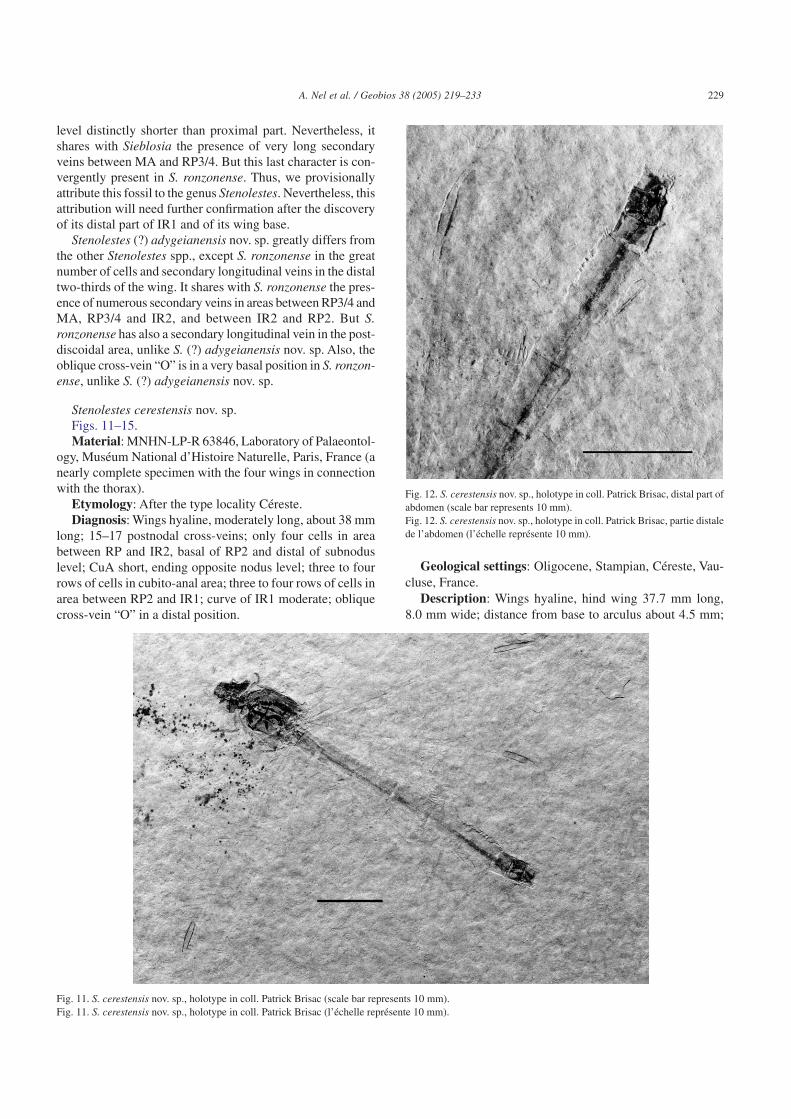
Stenolestes cerestensis nov. sp.Figs. 11–15.Material: MNHN-LP-R 63846, Laboratory of Palaeontol-
ogy, Muséum National d’Histoire Naturelle, Paris, France (anearly complete specimen with the four wings in connectionwith the thorax).
Etymology: After the type locality Céreste.Diagnosis: Wings hyaline, moderately long, about 38 mm
long; 15–17 postnodal cross-veins; only four cells in areabetween RP and IR2, basal of RP2 and distal of subnoduslevel; CuA short, ending opposite nodus level; three to fourrows of cells in cubito-anal area; three to four rows of cells inarea between RP2 and IR1; curve of IR1 moderate; obliquecross-vein “O” in a distal position.
Geological settings: Oligocene, Stampian, Céreste, Vau-cluse, France.
Description: Wings hyaline, hind wing 37.7 mm long,8.0 mm wide; distance from base to arculus about 4.5 mm;
Fig. 11. S. cerestensis nov. sp., holotype in coll. Patrick Brisac (scale bar represents 10 mm).Fig. 11. S. cerestensis nov. sp., holotype in coll. Patrick Brisac (l’échelle représente 10 mm).
Fig. 12. S. cerestensis nov. sp., holotype in coll. Patrick Brisac, distal part ofabdomen (scale bar represents 10 mm).Fig. 12. S. cerestensis nov. sp., holotype in coll. Patrick Brisac, partie distalede l’abdomen (l’échelle représente 10 mm).
229A. Nel et al. / Geobios 38 (2005) 219–233

from arculus to nodus 8.1 mm, from nodus to pterostigma17.3 mm, from pterostigma to apex about 4.0 mm; pterostigmalong and broad, 4.9 mm long, 0.8 mm wide, covering sevencells; only three primary antenodal cross-veins Ax0, Ax1 andAx2 present, Ax2 nearly aligned with arculus, distancebetween Ax1 and Ax2 2.0 mm; discoidal cell unicellular,1.5 mm long and 1.0 mm wide, rather short and broad, broad-ened in its distal part but longer than that of fore wing, withits distal side MAb nearly parallel with basal side, basal sideof discoidal cell 0.7 mm long, anterior side 1.2 mm long,posterior side 1.5 mm long, MAb 1.0 mm long; ScP crossingthrough nodus, nodal cross-vein Cr of normal obliquity; sub-nodus Sn perpendicular to RA; 17 postnodal cross-veins, notaligned with the 16 postsubnodal cross-veins; bases ofRP3/4 and IR2 between arculus and nodus; RP3/4 closer to
arculus than to nodus, 2.7 mm distal of arculus; IR2 closer tonodus, 5.0 mm distal of arculus; base of RP2 four cells and4.5 mm distal of subnodus; only four cells in area betweenRP and IR2, basal of RP2 and distal of subnodus level; baseof IR1 four cells and 3.3 mm distally; oblique cross-vein “O”seven cells and 4.8 mm distal of base of RP2; vein CuP 0.8 mmdistal of base ofAA; cubito-anal area 1.8 mm wide, with threerows of cells between CuA and posterior wing margin; CuAreaching posterior wing margin very basally, nearly oppositenodus level; area between MP and posterior wing marginbroad, with eight to nine rows of cells; postdiscoidal area nar-row, 1.0 mm wide; area between MA and RP3/4 distally verybroad, with two main secondary longitudinal veins; areabetween RP3/4 and IR2 narrow, distally 1.1 mm wide, butwith a long secondary longitudinal vein between them;IR2 zigzagged in its distal end; three secondary longitudinalveins in area between IR2 and RP2; basal part of IR1 slightlyzigzagged and distal part rather weakly curved opposite dis-tal half of pterostigma; two secondary longitudinal veins andthree rows of cells in area between RP2 and IR1; two rows ofcells in area between IR1 and RP1; one row of cells in areabetween RP1 and RA distal of pterostigma; fore wing verysimilar to hind wing, except in the following points: fore wing37.5 mm long, 8.3 mm wide; distance from base to arculusabout 4.5 mm; from arculus to nodus 7.7 mm, from nodus topterostigma 17.5 mm, from pterostigma to apex 4.6 mm;nodus in a very basal position; pterostigma long and broad,4.3 mm long, 1.0 mm wide, covering only four cells; pterostig-mal brace absent; discoidal cell 1.25 mm long and 1.25 mmwide, distinctly shorter and broader than that of hind wing,
Fig. 13. S. cerestensis nov. sp., holotype in coll. Patrick Brisac, last abdomi-nal segments (scale bar represents 1 mm).Fig. 13. S. cerestensis nov. sp., holotype in coll. Patrick Brisac, dernierssegments abdominaux (l’échelle représente 1 mm).
Fig. 14. S. cerestensis nov. sp., holotype in coll. Patrick Brisac, hind wing (scale bar represents 2 mm).Fig. 14. S. cerestensis nov. sp., holotype in coll. Patrick Brisac, aile postérieure (l’échelle représente 2 mm).
Fig. 15. S. cerestensis nov. sp., holotype in coll. Patrick Brisac, fore wing (scale bar represents 2 mm).Fig. 15. S. cerestensis nov. sp., holotype in coll. Patrick Brisac, aile antérieure (l’échelle représente 2 mm).
230 A. Nel et al. / Geobios 38 (2005) 219–233

only 15 postnodal cross-veins, not aligned with the 17 post-subnodal cross-veins; base of IR1 two cells and 2.4 mm dis-tal of that of RP2; cubito-anal area 2.5 mm wide, with fourrows of cells between CuA and posterior wing margin; areabetween RP3/4 and IR2 narrower than in hind wing, with nosecondary longitudinal vein between them; distal part ofIR1 more strongly curved opposite distal half of pterostigma;two secondary longitudinal veins and four rows of cells inarea between RP2 and IR1.
Abdomen complete, first segment very short, 3.5 mm long,second 6.0 mm long, third and fourth 6.5 mm long, fifth7.0 mm long, sixth 4.5 mm long, seventh and eight 2.5 mmlong. Only three apical abdominal appendages apparently vis-ible, i.e. a very small median one with a median furrow andtwo slightly longer lateral appendages, 0.7 mm long, whichcould correspond to the “epiproctophoran” epiproct and para-procts.
Discussion: This fossil can be attributed to the genus Steno-lestes for the same reasons as for S. fasciata nov. sp. It differsfrom all the other Stenolestes spp., except S. belligaudi, S.hispanicus, and S. fischeri, in its short CuA, ending oppositenodus level and low number of postnodal cross-veins. In par-ticular, S. coulleti from the same locality has five to six rowsof cells in cubito-anal area and more than 28 postnodal cross-veins. S. falloti has a longer CuA, extending well distal ofnodus level. The wings of S. fischeri are completely dark blueand distinctly longer (44 mm long instead of 38 mm in S.cerestensis nov. sp.). The wings of S. belligaudi are hyalinebut as long as those of S. fischeri. The preserved structures ofS. hispanicus are very similar to those of S. cerestensis nov.sp., except for the presence of seven cells in the area betweenRP and IR2, basal of RP2 and distal of the subnodus level,instead of four.
3. Phylogenetic analysis of the “sieblosiid-like” genera
Nel and Escuillié (1993) proposed a phylogenetic analysisof the Sieblosiidae, including Oligolestes. We have under-taken a new analysis of the “sieblosiid-like” genera (Sieblosi-idae plus gen. and sp. A and Oligolestes). The chosen out-groups are the genera Lestes Leach, 1815 (Oligocene toRecent, Zygoptera: Lestidae), Dysagrion Scudder, 1878(Eocene, Zygoptera: Dysagrioninae) and Frenguellia Petru-levicius and Nel, 2003 (Eocene, ?Epiproctophora: Frenguel-liidae), because they show strong similarities of different kindswith the Sieblosiidae in their wing venations. The choice ofoutgroups that would be closely related to the Sieblosiidae isproblematic because the exact affinities of the Sieblosiidae inOdonata remain uncertain (Fleck et al., 2004). The analysisis based on 13 unordered and equally weighted characters ofwing venation (see Tables 1 and 2). We rejected several othercharacters of the wing venation because they are variablewithin the sieblosiid genera (position of oblique vein “O” ofspecific value in the Sieblosiidae, variable in the genus Steno-lestes (Nel and Paicheler, 1994), number of supplementaryveins between RP3/4 and MA).
The analysis was made using the software Paup 4.0b10 forPC, using the branch and bound option. The choice and orderof introduction of the outgroup(s) do not perturb the result ofthe analysis. The resulting most parsimonious tree is 21 stepslong, with a consistency index CI 0.66, CI excluding uninfor-mative character 0.63, retention index RI 0.65, and RC 0.43(see Fig. 16).
Gen. and sp. A, Oligolestes and the Sieblosiidae fall in thesame clade, supported by the characters states “base ofRP2 well distal of subnodus”, and “pterostigma long, cover-ing six cells or more”. The monophyly of the clade (Oligo-lestes + Sieblosiidae) is supported by the character state “anglebetween MAb and MP + CuA very opened”. The clade [Sie-blosia and Miostenolestes and (Paraoligolestes + (Parasteno-lestes + Stenolestes))] is supported by the character states “ScPcrossing through nodus”. But the exact structure of the nodusis uncertain in Sieblosia, even if, after the figures in Hagen(1858), ScP probably crossed through the nodus, as in Steno-lestes. We have chosen to code the state “1” for this character
Table 1List of charactersListe des caractères
1. Vein ScP...Not crossing through the nodus. 0Crossing through the nodus. 1
2. Subnodus...Of pronounced normal obliquity. 0Nearly vertical, vertical or of inversed obliquity. 1
3. Discoidal cell...Longer than wide. 0Broader than long. 1
4. Angle between MAb and MP + CuA...Very sharp, between 45° or less. 0Opened, more than 45° and less than 90°. 1
5. Part of CuA distal of nodus level...Longer than proximal part of CuA. 0Shorter than proximal part of CuA. 1
6. Cubito-anal area...With one row of cells. 0With two rows of cells or more. 1With more than three rows of cells. 2
7. Base of RP2...Close to subnodus (four cells or less). 0Distal of subnodus (more than four cells). 1
8. Area between RP3/4 and IR2...With one to two long secondary longitudinal veins. 0Without secondary longitudinal veins. 1
9. Number of postnodal cross-veins...Less than 15. 0More than 15. 1
10. IR1...Strongly zigzagged and/or poorly defined. 0Well-defined, weakly zigzagged but without a strong curve. 1With a strong curve. 2
11. Number of secondary longitudinal veins between RP2 and IR1...Two or less. 0More than two. 1
12. Pterostigma...Relatively short, covering less than six cells. 0Long, covering six cells or more. 1
13. General shape of wing...Not very broad, with a long petiole. 0Very broad, with a short petiole. 1
231A. Nel et al. / Geobios 38 (2005) 219–233

in Sieblosia in the present analysis. But if this character iscoded “unknown” (or “0”) for Sieblosia, the analysis givesnine (or seven) minimal cladograms (Length 21 steps, CI 0.66,CI excluding uninformative characters 0.63, RI 0.63, RC0.42), with a clade that comprises all the Sieblosiidae plusOligolestes and gen. and sp. A, but also with a polytomybetween Oligolestes, Sieblosia, Miostenolestes, Paraoligo-
lestes, and (Parastenolestes + Stenolestes) in their strict con-sensus tree, because of the conflicts affecting other charac-ters.
The clade (Paraoligolestes + (Parastenolestes + Steno-lestes)) is supported by the synapomorphy “part of CuA dis-tal of nodus level shorter than proximal part”, but also presentin gen. and sp. A. The clade (Parastenolestes + Stenolestes)is supported by the character state “IR1 with a strong curve”.
4. Palaeobiogeographic remarks
The present discoveries show that the Sieblosiidae are notrestricted to the Western Europe but were also present anddiverse in the Oligocene and Miocene of Caucasus. Thisregion was connected to Western Europe during the Eocene.The Sieblosiidae are still unknown in the Cenozoic of theAsiatic part of Russia and in North America. If the Cenozoicodonatofauna of Siberia is still very badly known, it is not thecase for the North American one. Interestingly, there was anepicontinental sea crossing through Russia at the limitbetween Europe and Asia during the Eocene. It could haveacted as a boundary, preventing the arrival of the Sieblosiidaein Asia and North America in the Upper Eocene and LowerOligocene. Later, the opening of the Bering Detroit in theMiocene also acted as a barrier between Asia and NorthAmerica for this group of poor flyers.
Acknowledgements
Many thanks to Mr. Gabriele Stroppa of Pesaro, Italy, forthe loan of the specimen of gen. and sp. A, and its deposit inthe future Museum of Pesaro, and to Mr. Patrick Brisac ofMalataverne, France, for the photography, loan, and futuredeposit of the type specimen of S. cerestensis nov. sp. in theMuseum of Paris. We are also greatly in debt towards the lateDr. Vladimir Zherikhin, Palaeoentomologist at the Academyof Science of Moscow.
Table 2List of characters and taxaListe des caractères et des taxons
Taxa/characters 1 2 3 4 5 6 7 8 9 10 11 12 13Lestes 0 0 0 0 0 0 0 0 0 0 0 0 0Dysagrion 0 1 1 1 0 1 0 0 1 1 0 0 1Frenguellia 0 1 ? 0 0 1 0 1 1 1 0 0 1Gen. and sp. A 0 0 0 0 1 1 1 1 1 1 0 1 1Oligolestes 0 1 0 1 0 1 1 1 1 1 0 1 1Paraoligolestes 1 1 0 1 1 1 1 1 1 1 0 1 1Miostenolestes nov. gen. 1 1 0 1 0 0 1 1 1 1 0 1 1Sieblosia 1 1 0 1 0 1 1 0 1 1 1 1 1Parastenolestes 1 1 0 1 1 1 1 1 1 2 0 1 1Stenolestes 1 1 1 1 1 1 1 1 1 2 1 1 1
Fig. 16. Most parsimonious cladogram of the sieblosiid genera.Fig. 16. Cladogramme le plus parcimonieux des genres de Sieblosiidae.
232 A. Nel et al. / Geobios 38 (2005) 219–233

References
Bechly, G., 1996. Morphologische Untersuchungen am Flügelgeäder derrezenten Libellen und deren Stammgruppenvertreter (Insecta; Pterygota;Odonata), unter besonderer Berücksichtigung der PhylogenetischenSystematik und des Grundplanes der Odonata. Petalura Special Volume2, 1–402.
Fischer, C., 1974. Systematische Stellung der Gattung Sieblosia Handlirsch,1906 (Zygoptera: Lestinoidea: Sieblosiidae). Odonatologica 3, 211–220.
Fleck, G., Bechly, G., Martínez-Delclòs, X., Jarzembowski, E.A., Nel, A.,2004. A revision of the Mesozoic dragonfly family Tarsophlebiidae, witha discussion on the phylogenetic positions of the Tarsophlebiidae andSieblosiidae (Odonatoptera: Panodonata). Geodiversitas 26, 33–60.
Gentilini, G., 1989. The Upper Miocene dragonflies of Monte Castellaro(Marche, Central Italy) (Odonata: Libellulidae). Memorie della SocietaEntomologica Italiana 67, 251–272 (for 1988).
Gentilini, G., 1992. Two new species of Epitheca from Lower Messinian ofCentral Italy (Odonata: Corduliidae). Bolletino della Societa Entomo-logica Italiana 123, 201–208.
Gentilini, G., Peters, G., 1993. The Upper Miocene aeshnids of MonteCastellaro, Central Italy, and their relationships to extant species(Odonata: Aeshnidae). Odonatologica 22, 147–178.
Hagen, H.A., 1858. Zwei Libellen aus der Braunkohle von Sieblos. Palae-ontographica 5, 121–126.
Handlirsch, A., 1906–1908. Die fossilen Insekten und die Phylogenie derrezenten Formen. Ein Handbuch für Paläontologen und Zoologen.1430 pp. (Engelman, V.W. publ., Leipzig) [published in parts between1906 and 1908 as follows: pp. i–vi, 1–160 (May 1906); pp. 161–320(June 1906); pp. 321–480 (August 1906); pp. 481–640 (October 1906);pp. 641–800 (February 1907); pp. 801–960 (June 1907); pp. 961–1120(November 1907); pp. 1121–1280 (January 1908); pp. vii–ix, 1281–1430 (July 1908). Dated from publication information given on p. ix].
Kukalová-Peck, J., 1991. Fossil history and the evolution of hexapod struc-tures. In: Naumann, I.D. (Ed.), The Insects of Australia, A Textbook forStudents and Research Workers, 1. Melbourne University Press, Mel-bourne, pp. 141–179.
Leach, W.E., 1815. The zoological miscellany. Brief descriptions of new, orinteresting animals. Nodder and son. London 2, 1–154.
Maneval, H., 1936. Insectes fossiles des calcaires oligocènes de Ronzon (LePuy). Annales de la Société Linnéenne de Lyon 79, 23–27 (NS).
Nel, A., 1986. Révision du genre cénozoïque Stenolestes Scudder, 1895.Description de deux espèces nouvelles (Insecta, Odonata, Lestidae).Bulletin du Muséum National d’Histoire Naturelle (4) (c) 8 (4), 447–461.
Nel, A., 1991. Description de quelques Sieblosiidae fossiles nouveaux(Odonata, Zygoptera, Lestoidea). Nouvelle Revue d’Entomologie 8,367–375 (NS).
Nel, A., Escuillié, F., 1992. Présence du genre Stenolestes Scudder,1895 dans les laminites oligocènes du Revest-des-Brousses (Lubéron,France). L’Entomologiste 48, 337–349.
Nel, A., Escuillié, F., 1993. Découverte d’un nouveau genre de Sieblosiidaedans le Miocène supérieur de l’Ardèche (France) (Odonata, Zygoptera,Lestoidea, Sieblosiidae). Nouvelle Revue d’Entomologie 10, 233–242(NS).
Nel, A., Martínez-Delclòs, X., Paicheler, J.-C., Henrotay, M., 1993. Les« Anisozygoptera » fossiles. Phylogénie et classification (Odonata).Martinia, Numéro Hors Série 3, 1–311.
Nel, A., Martínez-Delclòs, X., Papier, F., Oudard, J., 1997. New Tertiaryfossil Odonata from France. (Sieblosiidae, Lestidae, Coenagrioniidae,Megapodagrionidae, Libellulidae). Deutsche Entomologische Zeit-schrift 44, 231–258.
Nel, A., Paicheler, J.-C., 1994. Les Lestoidea (Odonata, Zygoptera) fossiles :un inventaire critique. Annales de Paléontologie 80, 1–59.
Nel, A., Papazian, M., 1986. Sur une nouvelle espèce d’Odonate fossile duStampien de Céreste (Lubéron) (Odonata, Lestidae). Nouvelle Revued’Entomologie 3, 227–233 (NS).
Petrulevicius, J.F., Nel, A., 2003. Frenguelliidae, a new family of dragonfliesfrom the earliest Eocene of Argentina (Insecta: Odonata): phylogeneticrelationships within Odonata. Journal of Natural History 37, 2909–2917.
Riek, E.F., 1976. A new collection of insects from the Upper Triassic ofSouth Africa. Annals of the Natal Museum 22, 791–820.
Riek, E.F., Kukalová-Peck, J., 1984. A new interpretation of dragonfly wingvenation based upon early Carboniferous fossils from Argentina(Insecta: Odonatoidea) and basic character states in pterygote wings.Canadian Journal of Zoology 62, 1150–1166.
Riou, B., Nel, A., 1995. Nouveaux Odonates fossiles du Miocène supérieurde l’Ardèche. (Odonata: Sieblosiidae, Lestidae, Libellulidae, Corduli-idae, Aeshnidae). Travaux de l’École Pratique des Hautes Études, Biolo-gie et Évolution des Insectes 7/8, 125–144.
Schmidt, E., 1958. Bemerkungen über Lestiden. 3. Über Oligolestes grandisStatz, 1935 und eine neue Lestes-Art aus dem mittel-Oligozän von Rottim Siebengebirge. Decheniana 111, 1–7.
Scudder, S.H., 1878. An account of some insects of unusual interest from theTertiary rocks of Colorado and Wyoming. Bulletin of the United StatesGeological and Geographical Survey of the Territories 4, 519–543.
Scudder, S.H., 1895. The Miocene insect-fauna of Œningen, Baden. Geo-logical Magazine 2 (4), 116–122.
Statz, G., 1935. Drei neue Insektenarten aus dem Tertiär von Rott amSiebengebirge. Wissenschaftliche Mitteilungen des Vereins fürNaturkunde und Heimatkunde im Köln 1, 10–14.
Théobald, N., 1937. Les insectes fossiles des terrains oligocènes de France.Bulletin mensuel (Mémoires) de la Société des Sciences de Nancy 1,1–473.
Wedmann, S., 2000. Die Insekten der oberoligozänen FossillagerstätteEnspel (Westerwald, Deutschland). Systematik, Biostratinomie undPaläoökologie. Mainzer Naturwissenschaftliches Archiv 23, 1–154.
233A. Nel et al. / Geobios 38 (2005) 219–233