Six new species of Agalliopsis from Southeastern Brazil (Insecta: Hemiptera: Cicadellidae:...
18
Accepted by C. Dietrich: 22 Jan. 2009; published: 18 Feb. 2009 1 ZOOTAXA ISSN 1175-5326 (print edition) ISSN 1175-5334 (online edition) Copyright © 2009 · Magnolia Press Zootaxa 2014: 1–18 (2009) www.mapress.com/ zootaxa/ Article Six new species of Agalliopsis from Southeastern Brazil (Insecta: Hemiptera: Cicadellidae: Agalliinae) ANA CLARA GONÇALVES 1,2 , GABRIEL MEJDALANI 1 & LUCI B. N. COELHO 3 1 Departamento de Entomologia, Museu Nacional, Universidade Federal do Rio de Janeiro, Quinta da Boa Vista, São Cristóvão, 20940-040, Rio de Janeiro, RJ, Brasil. E-mail: [email protected]; [email protected] 2 Pós-graduação em Zoologia, Museu Nacional, Universidade Federal do Rio de Janeiro. 3 Departamento de Zoologia, Instituto de Biologia, Universidade Federal do Rio de Janeiro, Caixa Postal 68044, 21944-970, Rio de Janeiro, RJ, Brasil. E-mail: [email protected] Abstract Six new species of Agalliopsis Kirkaldy, 1907 are described from the Atlantic Forest of Southeastern Brazil (Minas Gerais State): A. variegata sp. nov., A. dutrai sp. nov., A. chaelata sp. nov., A. pentaspinata sp. nov., A. mutabilis sp. nov., and A. felixi sp. nov. Illustrations of external features and male and female genitalic characters that distinguish the new taxa are given. Variation in the color pattern and male genitalic characters of A. mutabilis is recorded. The new taxa are assigned to species complexes within Agalliopsis. Key words: taxonomy, morphology, leafhopper, Membracoidea, Neotropical Region Introduction Agalliopsis Kirkaldy, 1907, type-species Jassus novellus Say, 1831, is apparently limited to the New World (Oman 1970). Many species in this genus were described by Oman (1933, 1934, 1938) in his works on North, Central, and South American faunas, respectively. Additional relevant contributions were published by Kramer (1964), Linnavuori & DeLong (1979), and Nielson & Godoy (1995), resulting in a total of approximately 110 Neotropical species. According to Nielson & Knight (2000), Agalliopsis is of probable Neotropical origin, having radiated to the Nearctic Region after South and North America were joined during the Miocene. The posterior margin of the crown, which is sinuate or slightly curved behind the eyes, is an important diagnostic feature of Agalliopsis (Nielson & Godoy 1995). Besides the form of the posterior coronal margin, Oman (1933) and Nielson & Godoy (1995) have mentioned other features of Agalliopsis, such as: crown very short medially, generally longer near the eyes; face with interocellar distance greater than distance between ocelli and inner margin of eyes; pronotum surface slightly granulose; styles forked posteriorly; aedeagus frequently with processes. In their key to the Agalliinae genera of Central America, Nielson & Godoy (1995) used a combination of the interocellar distance and the form of the posterior coronal margin to distinguish Agalliopsis from other genera. Several groups of species have been recognized in the genus, e.g., novella complex (Oman 1970) and sagittata, basispina, hamata, and cuculla complexes (Nielson & Godoy 1995). Agalliopsis is a taxon of considerable economic importance. Examples of plants on which Agalliopsis species were recorded are common bean, lettuce, tomato, alfafa, and clover (Nielson 1968; Zanol & de Menezes 1982). Nielson (1968) reviewed A. novella as a vector in the United States of the potato yellow dwarf virus, clover club leaf virus, and wound tumor virus of clover. In the present paper, six new Agalliopsis species are described from an area of Atlantic Forest in the Municipality of Viçosa, Minas Gerais State, Southeastern Brazil. A brief description of the type locality was TERMS OF USE This pdf is provided by Magnolia Press for private/research use. Commercial sale or deposition in a public library or website is prohibited.
Transcript of Six new species of Agalliopsis from Southeastern Brazil (Insecta: Hemiptera: Cicadellidae:...

Accepted by C. Dietrich: 22 Jan. 2009; published: 18 Feb. 2009 1
ZOOTAXAISSN 1175-5326 (print edition)
ISSN 1175-5334 (online edition)Copyright © 2009 · Magnolia Press
Zootaxa 2014: 1–18 (2009) www.mapress.com/zootaxa/ Article
Six new species of Agalliopsis from Southeastern Brazil (Insecta: Hemiptera: Cicadellidae: Agalliinae)
ANA CLARA GONÇALVES1,2, GABRIEL MEJDALANI1 & LUCI B. N. COELHO3
1Departamento de Entomologia, Museu Nacional, Universidade Federal do Rio de Janeiro, Quinta da Boa Vista, São Cristóvão, 20940-040, Rio de Janeiro, RJ, Brasil. E-mail: [email protected]; [email protected]ós-graduação em Zoologia, Museu Nacional, Universidade Federal do Rio de Janeiro.3Departamento de Zoologia, Instituto de Biologia, Universidade Federal do Rio de Janeiro, Caixa Postal 68044, 21944-970, Rio de Janeiro, RJ, Brasil. E-mail: [email protected]
Abstract
Six new species of Agalliopsis Kirkaldy, 1907 are described from the Atlantic Forest of Southeastern Brazil (Minas Gerais State): A. variegata sp. nov., A. dutrai sp. nov., A. chaelata sp. nov., A. pentaspinata sp. nov., A. mutabilis sp. nov., and A. felixi sp. nov. Illustrations of external features and male and female genitalic characters that distinguish the new taxa are given. Variation in the color pattern and male genitalic characters of A. mutabilis is recorded. The new taxa are assigned to species complexes within Agalliopsis.
Key words: taxonomy, morphology, leafhopper, Membracoidea, Neotropical Region
Introduction
Agalliopsis Kirkaldy, 1907, type-species Jassus novellus Say, 1831, is apparently limited to the New World (Oman 1970). Many species in this genus were described by Oman (1933, 1934, 1938) in his works on North, Central, and South American faunas, respectively. Additional relevant contributions were published by Kramer (1964), Linnavuori & DeLong (1979), and Nielson & Godoy (1995), resulting in a total of approximately 110 Neotropical species. According to Nielson & Knight (2000), Agalliopsis is of probable Neotropical origin, having radiated to the Nearctic Region after South and North America were joined during the Miocene.
The posterior margin of the crown, which is sinuate or slightly curved behind the eyes, is an important diagnostic feature of Agalliopsis (Nielson & Godoy 1995). Besides the form of the posterior coronal margin, Oman (1933) and Nielson & Godoy (1995) have mentioned other features of Agalliopsis, such as: crown very short medially, generally longer near the eyes; face with interocellar distance greater than distance between ocelli and inner margin of eyes; pronotum surface slightly granulose; styles forked posteriorly; aedeagus frequently with processes. In their key to the Agalliinae genera of Central America, Nielson & Godoy (1995) used a combination of the interocellar distance and the form of the posterior coronal margin to distinguish Agalliopsis from other genera. Several groups of species have been recognized in the genus, e.g., novellacomplex (Oman 1970) and sagittata, basispina, hamata, and cuculla complexes (Nielson & Godoy 1995). Agalliopsis is a taxon of considerable economic importance. Examples of plants on which Agalliopsis species were recorded are common bean, lettuce, tomato, alfafa, and clover (Nielson 1968; Zanol & de Menezes 1982). Nielson (1968) reviewed A. novella as a vector in the United States of the potato yellow dwarf virus, clover club leaf virus, and wound tumor virus of clover.
In the present paper, six new Agalliopsis species are described from an area of Atlantic Forest in the Municipality of Viçosa, Minas Gerais State, Southeastern Brazil. A brief description of the type locality was
TERMS OF USEThis pdf is provided by Magnolia Press for private/research use. Commercial sale or deposition in a public library or website is prohibited.

TERMS OF USEThis pdf is provided by Magnolia Press for private/research use. Commercial sale or deposition in a public library or website is prohibited.
provided by Gonçalves et al. (2006). An additional species from this locality, known only from females, is also described (Agalliopsis sp.).
Material and methods
The specimens have been collected with a modified “Luiz de Queiroz” light trap (described in detail by Coelho 1997) during the crepuscular-nocturnal period. Morphological terminology follows mainly Oman (1933, 1936), except for the head (Mejdalani 1998) and for the female genitalia (Davis 1975). Use of the term gonoplac (= third ovipositor valvula) follows Mejdalani (1998). Techniques for preparation of male and female genital structures follow, respectively, Oman (1949) and Mejdalani (1998). The dissected parts are stored in microvials with glycerin. Label data are given inside quotation marks with a reversed virgule (\) separating lines. The specimens studied belong to the collections of the Departamento de Zoologia (Coleção Entomológica Prof. José Alfredo P. Dutra), Instituto de Biologia, Universidade Federal do Rio de Janeiro (Rio de Janeiro, DZRJ), Departamento de Entomologia, Museu Nacional, Universidade Federal do Rio de Janeiro (Rio de Janeiro, MNRJ), and Museu Regional de Entomologia, Universidade Federal de Viçosa (Viçosa, UFV).
Results
Agalliopsis variegata Gonçalves, Mejdalani & Coelho, sp. nov.(Figs 1, 4–12)
Length. Male holotype 4.3 mm; male paratype 4.6 mm.Description (holotype). Head and thorax (color). General ground color brownish-yellow. Crown (Figs
4, 5), in dorsal view, with five dark brown maculae: round one near each inner eye margin, two irregular ones about equally distant between eyes and median line, and triangular one on median portion. Face (Fig. 5) with pair of small dark brown maculae near superior inner margin of eyes; inverted Y-shaped dark brown macula contiguous on its upper portion with triangular crown macula; area adjacent to ocelli with pair of dark brown maculae. Frons (Fig. 5) with pair of longitudinal, irregular slender maculae with superior portion dark brown and remainder brown, fused to each other at lower region; frons lateral portions dark brown; clypeus brown with yellow macula at basal portion; genae with dark brown irregular markings; lorae with inner margin dark brown. Pronotum (Fig. 4) with two large irregular brown maculae. Mesoscutum (Fig. 4) with pair of large lateral brown maculae; mesoscutellum with pair of dark brown maculae on lateral basal portions and pair of brown maculae contiguous with those of mesoscutum. Forewings with diffuse brown maculae or stripes (Fig. 1). Fore and middle legs with dark brown region on femur, hind leg with large dark brown macula on tibia (Fig. 1).
Male genitalia. Pygofer (Fig. 6), in lateral view, with posterior margin subacute, with several spiniform projections ending in acute process directed inward (Fig. 7). Subgenital plates (Fig. 8), in ventral view, elongate, fused to valve, slightly broader at basal half, separated from each other along apical 1/3, with small scattered setae; in lateral view (Fig. 6), extending posteriorly beyond pygofer apex, with longer setae on dorsal margin. Styles (Fig. 9), in dorsal view, well developed, forked apically, outer fork with subacute apex and scattered setae on inner margin of subapical region, inner fork with basal portion strongly curved and with truncate apex bearing few setae. Connective (Fig. 10), in lateral view, elongate, fused to aedeagus; Y-shaped in dorsal view. Aedeagus (Fig. 10) strongly curved, with dorsal and ventral projections: dorsal projection small, narrowing towards apex; ventral projection well developed with apex (Fig. 11) with two pairs of processes, median pair long and U-shaped, lateral pair short and with concave apices; aedeagal shaft with apex (Fig. 12) setiform. Anal tube (Fig. 6) with segments X and XI well developed.
GONÇALVES ET AL.2 · Zootaxa 2014 © 2009 Magnolia Press

TERMS OF USEThis pdf is provided by Magnolia Press for private/research use. Commercial sale or deposition in a public library or website is prohibited.
Female. Unknown.Etymology. The species epithet, variegata, refers to the diffuse color pattern of the forewings.Type material. Holotype: male, Brazil, “Viçosa, MG [Minas Gerais State], Brasil\ Data 20/X/1992\ P. S.
Fiuza F. [Ferreira]” (DZRJ). One male paratype with same data as holotype, except “08/X/1992” (UFV).
FIGURES 1–3. Body in laterodorsal view: (1) Agalliopsis variegata sp. nov., male holotype; (2) Agalliopsis sp., female; (3) A. mutabilis sp. nov., male paratype.
Zootaxa 2014 © 2009 Magnolia Press · 3SIX NEW SPECIES OF AGALLIOPSIS

TERMS OF USEThis pdf is provided by Magnolia Press for private/research use. Commercial sale or deposition in a public library or website is prohibited.
FIGURES 4–12. Agalliopsis variegata sp. nov., male holotype. (4) Crown, pronotum and mesonotum, dorsal view; (5) face; (6) genital capsule, lateral view; (7) pygofer process, dorsal view; (8) valve fused to subgenital plates, ventral view; (9) style, dorsal view; (10) aedeagus, connective and style, lateral view; (11) apex of ventral aedeagal process, dorsal view; (12) apex of aedeagus, dorsal view.
Agalliopsis dutrai Gonçalves, Mejdalani & Coelho, sp. nov.(Figs 13–19, 65, 66)
Length. Male holotype 3.9 mm; male paratypes 3.8–4.0 mm; female paratypes 3.9–4.4 mm.Description (holotype).Head and thorax (color). Ground color yellowish-brown. Crown (Figs 13, 14)
with pair of dark brown maculae approximately equidistant between eyes and median line; pair of brown to dark brown maculae near inner eye margins; with three triangular brown maculae contiguous to pronotal maculae. Eyes with red tonality. Face (Fig. 14) yellow with slender, inverted Y-shaped brown macula between
GONÇALVES ET AL.4 · Zootaxa 2014 © 2009 Magnolia Press

TERMS OF USEThis pdf is provided by Magnolia Press for private/research use. Commercial sale or deposition in a public library or website is prohibited.
ocelli; tiny brown maculae around ocelli. Frons (Fig. 14) with small brown maculae forming pair of lateral rows. Pronotum (Fig. 13) with two large, circular brown stripes, each one forming oval figure including brown macula; stripes fused to each other anteriorly, extending to crown and delimiting pair of anterior yellow areas. Mesoscutum (Fig. 13) broadly covered by brown marks; mesoscutellum with pair of dark brown maculae at laterobasal portions.
FIGURES 13–19. Agalliopsis dutrai sp. nov. (13–18) Male holotype: (13) crown, pronotum and mesonotum, dorsal view; (14) face; (15) genital capsule, lateral view; (16) valve fused to subgenital plates, ventral view; (17) aedeagus, connective and style, lateral view; (18) apex of aedeagus, dorsal view. (19) Female paratype: abdomen, ventral view.
Male genitalia. Pygofer (Fig. 15), in lateral view, with posterior margin subtruncate. Subgenital plates (Fig. 16), in ventral view, elongate with basal half narrower than apical half; plates fused basally with valve
Zootaxa 2014 © 2009 Magnolia Press · 5SIX NEW SPECIES OF AGALLIOPSIS
TERMS OF USEThis pdf is provided by Magnolia Press for private/research use. Commercial sale or deposition in a public library or website is prohibited.
and fused with each other along basal half; in lateral view, extending beyond pygofer apex, dorsal surface with longer scattered setae. Styles (Fig. 17), in lateral view, elongate, with few setae at apical portion, outer fork smaller and rounded, inner fork larger with its basal portion strongly curved, subapical portion with small tooth-like process directed downward. Connective (Fig. 17), in lateral view, linear, fused to aedeagus; subtriangular in dorsal view. Aedeagus (Fig. 17), in lateral view, strongly curved, basal half directed anteriorly and apical half directed posteriorly, curved region enlarged; apical portion of shaft with pair of long spiniform processes directed ventrally; shaft apex, in dorsal view, rounded, gonopore apical (Fig. 18). Anal tube (Fig. 15) with segment X well developed, divided into two parts, with pair of processes expanded towards apex and bearing two tooth-like ventral projections.
Female (color). Abdominal sternite VII (Fig. 19) mostly yellow; laterotergites from segment VIII yellow; pygofer (Fig. 19), in ventral view, yellow, with pair of brown maculae laterally; gonoplacs brown. Other features similar to those of the male holotype.
Female genitalia. Abdominal sternite VII (Fig. 19), in ventral view, transverse, rectangular, posterior margin with lateral portions oblique and central area slightly concave. First valvulae, in lateral view, dorsally curved from base; dorsolateral surface with oblique rows of scale-like processes on distal 1/2 of shaft; ventroapical region with scale-like processes; apex acute. Second valvulae (Fig. 65), in lateral view, dorsally curved from base, with broadest region at apical 1/4; dorsal hyaline area present; dorsal prominence slightly pronounced; teeth on apical half of dorsal margin, basally small and uniform, increasing in size towards apex (Fig. 66); ventroapical region with tiny rounded teeth; shaft apex rounded. Gonoplacs, in lateral view, expanded along apical half; apex rounded; surface with tiny spiniform processes and setae at apical portion and extending anteriorly along ventral margin.
Etymology. The new species is described in honor of the late Prof. José Alfredo Pinheiro Dutra (Instituto de Biologia, Universidade Federal do Rio de Janeiro), who has contributed to our knowledge of Brazilian Agalliinae.
Type material. Holotype: male, Brazil, “Viçosa, MG [Estado de Minas Gerais], Brasil\ Data 22/XII/1992\ P. S. Fiuza F. [Ferreira]” (DZRJ). Twenty-three paratypes with same data as holotype, except: one female “02/XII/1981” (DZRJ), one female “01/IX/1982”, two females “13/X/1982”, one female “24/XI/1982”, one female “09/II/1983”, one female “01/VI/1983”, one male “11/VIII/1986”, one female “11/III/1987”, one female “14/X/1987”, one female “10/XI/1987”, one male and two females “08/I/1988”, one female “20/V/1988”, one female “08/X/1992”, one female “20/X/1992”, two females “29/XII/1992” (UFV), one female “10/XI/1982”, one female “02/II/1983”, two males “14/X/1987” (MNRJ).
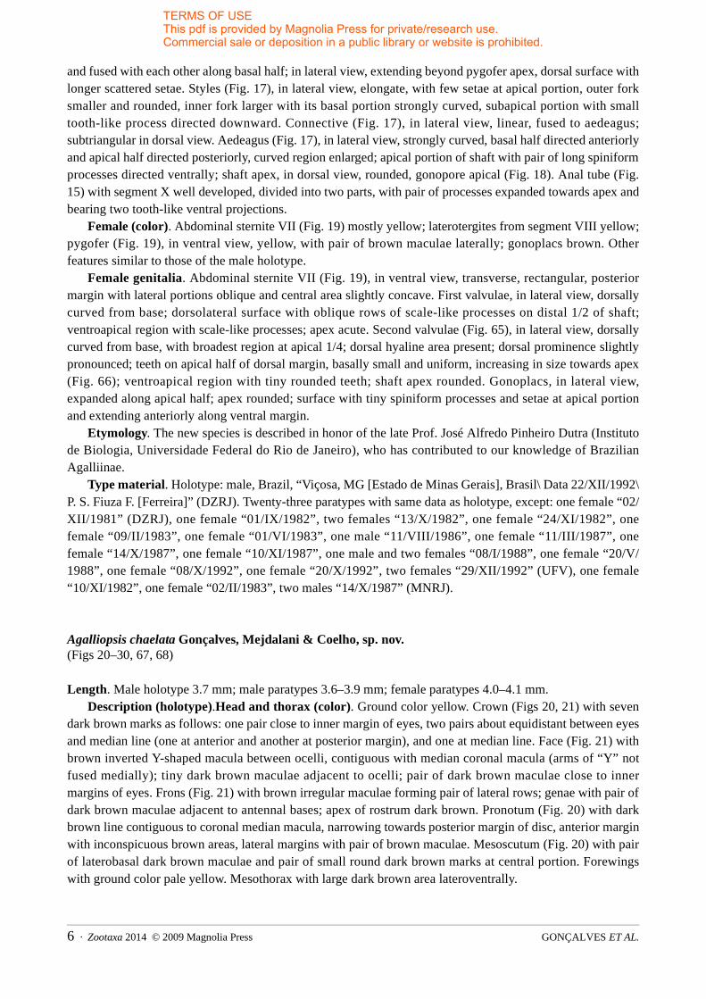
Agalliopsis chaelata Gonçalves, Mejdalani & Coelho, sp. nov.(Figs 20–30, 67, 68)
Length. Male holotype 3.7 mm; male paratypes 3.6–3.9 mm; female paratypes 4.0–4.1 mm.Description (holotype).Head and thorax (color). Ground color yellow. Crown (Figs 20, 21) with seven
dark brown marks as follows: one pair close to inner margin of eyes, two pairs about equidistant between eyes and median line (one at anterior and another at posterior margin), and one at median line. Face (Fig. 21) with brown inverted Y-shaped macula between ocelli, contiguous with median coronal macula (arms of “Y” not fused medially); tiny dark brown maculae adjacent to ocelli; pair of dark brown maculae close to inner margins of eyes. Frons (Fig. 21) with brown irregular maculae forming pair of lateral rows; genae with pair of dark brown maculae adjacent to antennal bases; apex of rostrum dark brown. Pronotum (Fig. 20) with dark brown line contiguous to coronal median macula, narrowing towards posterior margin of disc, anterior margin with inconspicuous brown areas, lateral margins with pair of brown maculae. Mesoscutum (Fig. 20) with pair of laterobasal dark brown maculae and pair of small round dark brown marks at central portion. Forewings with ground color pale yellow. Mesothorax with large dark brown area lateroventrally.
GONÇALVES ET AL.6 · Zootaxa 2014 © 2009 Magnolia Press

TERMS OF USEThis pdf is provided by Magnolia Press for private/research use. Commercial sale or deposition in a public library or website is prohibited.
FIGURES 20–30. Agalliopsis chaelata sp. nov. (20–29) Male holotype: (20) crown, pronotum and mesonotum, dorsal view; (21) face; (22) genital capsule, lateral view; (23) pygofer process, lateral view; (24) valve and subgenital plates, ventral view; (25) apical portion of subgenital plates, lateral view; (26) style, dorsal view; (27) connective, dorsal view; (28) aedeagus, connective and style, lateral view; (29) apex of aedeagus, dorsal view. (30) Female paratype: apical portion of abdomen, ventral view.
Male genitalia. Pygofer (Fig. 22), in lateral view, short, posterior margin rounded and with chelate process (Fig 23) directed inward. Subgenital plates (Fig. 24), in ventral view, elongate, triangular, fused to each other at basal portion, not fused to valve; in lateral view (Figs 22, 25), extending beyond pygofer, with
Zootaxa 2014 © 2009 Magnolia Press · 7SIX NEW SPECIES OF AGALLIOPSIS

TERMS OF USEThis pdf is provided by Magnolia Press for private/research use. Commercial sale or deposition in a public library or website is prohibited.
longer setae on superior portion. Styles (Fig. 26), in dorsal view, large, outer fork small and rounded with few setae on preapical region, inner fork much more developed, narrowing towards rounded apex; dorsal style surface with concavity for articulation with connective. Connective (Fig. 28), in lateral view, linear, not fused to aedeagus; Y-shaped in dorsal view (Fig. 27). Aedeagus (Fig. 28), in lateral view, broad basally, with slender and sinuous shaft curved above base and directed caudally; with complex structure at apical region expanded dorsally, ventrally and posteriorly, dorsal rami from this structure connected to anal tube; apical region of shaft (Fig. 29) divided into two ends with bifurcate and acute apices. Anal tube (Fig. 22), in caudal view, with segment XI bearing pair of delicate, ventral elongate processes.
Female (color). Abdominal sternite VII (Fig. 30) yellow; laterotergites of segment VIII yellow with pair of tiny brown maculae on inner basal region; pygofer (Fig. 30), in ventral view, yellow; gonoplacs pale brown. Other features similar to those of the male holotype.
Female genitalia. Abdominal sternite VII (Fig. 30), in ventral view, moderately produced posteriorly; posterior margin convex, subtruncate at median portion. First valvulae, in lateral view, slightly dorsally curved from base; dorsolateral surface with oblique rows of scale-like processes on posterior 2/3 of shaft; ventroapical region with scale-like processes; apex subacute. Second valvulae (Fig. 67), in lateral view, slightly dorsally curved from base, with broadest area at apical 1/4; dorsal hyaline area present; dorsal prominence slight, inconspicuous; small teeth (Fig. 68) along apical half of dorsal margin, increasing in size slightly towards apex, without denticles; ventroapical region with tiny teeth; shaft apex rounded. Gonoplacs as in A. dutrai sp. nov.
Etymology. The species epithet, chaelata, refers to the peculiar chelate processes of the pygofer and aedeagus, which are diagnostic of the new taxon.
Type material. Holotype: male, Brazil, “Viçosa, MG [Minas Gerais State], Brasil\ Data 2/X/1987\ P. S. Fiuza F. [Ferreira]” (DZRJ). Eight paratypes with same data as holotype, except: one female “02/XII/1986” (DZRJ), one male “22/IX/1987”, one male “31/VII/1987”, one male “05/XI/1992”, one female “26/V/1982” (UFV), one male “20/II/1987”, one male “14/X/1987”, one female “26/IV/1988” (MNRJ).
Agalliopsis pentaspinata Gonçalves, Mejdalani & Coelho, sp. nov.(Figs 31–38, 69, 70)
Length. Male holotype 3.7 mm; male paratypes 3.7–3.8 mm; female paratypes 3.9–4.1 mm.Description (holotype).Head and thorax (color). Ground color of dorsum brownish-yellow. Crown
(Figs 31, 32) with five dark brown maculae: two irregular ones close to eyes, two about equidistant between eyes and median line, and triangular one at median line. Face (Fig. 32) yellow with inconspicuous, inverted brown Y-shaped mark; inner area adjacent to ocelli with small dark brown dots. Frons (Fig. 32) with pair of pale brown arched stripes; genae with brown maculae adjacent to antennal bases. Pronotum (Fig. 31) anterior portion with inconspicuous maculae, lateral portions with pair of small marks and posterior 2/3 of disc with median line, brown. Mesoscutum (Fig. 31) with pair of laterobasal triangular maculae and pair of small rounded marks at central portion, brown; mesoscutellum with pair of brown maculae at laterobasal portions, pair of dark brown oblique maculae at central portion, and light brown median line extending from scutoscutellar suture to posterior portion. Forewings with veins partially covered with brown, mostly at basal portion.
Male genitalia. Pygofer (Fig. 33), in lateral view, with well developed anterior apodeme with circular apex; posterior margin rounded. Subgenital plates (Fig. 34), in ventral view, very elongate; fused to valve and to each other along basal half; narrowing gradually on distal half towards apex; in lateral view (Fig. 33), extending posteriorly beyond pygofer apex; with longer setae on dorsal surface. Styles (Fig. 35), in lateral view, with dorsal oblique projection on basal half directed anteriorly; outer fork small, rounded, with several setae at subapical region, with acute apex directed dorsally; inner fork well developed with basal portion strongly curved, apical portion directed ventrally, apex acute. Connective (Fig. 35), in lateral view, elongate,
GONÇALVES ET AL.8 · Zootaxa 2014 © 2009 Magnolia Press

TERMS OF USEThis pdf is provided by Magnolia Press for private/research use. Commercial sale or deposition in a public library or website is prohibited.
linear, fused with aedeagus; in dorsal view, with form of longitudinal bar narrowed on median portion. Aedeagus (Fig. 35), in lateral view, elongate and strongly curved; basal region expanded with projection directed dorsally; basal half of shaft directed anterodorsally; curved region expanded anteriorly; apical half of shaft narrowing towards apex; subapical region with pair of bifurcated lateral projections, originating two pairs of processes (Fig. 36), anterior pair with irregular border apically and posterior pair with acute apex; apical region of shaft acute (Fig. 36). Anal tube (Fig. 33), in lateral view, small, segment X divided into two parts, basal one with process bearing digitiform apex (Fig. 37).
FIGURES 31–38. Agalliopsis pentaspinata sp. nov. (31–37) Male holotype: (31) crown, pronotum and mesonotum, dorsal view; (32) face; (33) genital capsule, lateral view; (34) valve fused to subgenital plates, ventral view; (35) aedeagus, connective and style, lateral view; (36) apex of aedeagus, dorsal view; (37) anal tube process, lateral view. (38) Female paratype: apical portion of abdomen, ventral view.
Zootaxa 2014 © 2009 Magnolia Press · 9SIX NEW SPECIES OF AGALLIOPSIS

TERMS OF USEThis pdf is provided by Magnolia Press for private/research use. Commercial sale or deposition in a public library or website is prohibited.
FIGURES 39–47. Agalliopsis felixi sp. nov. (39–40) Male paratype: (39) crown, pronotum and mesonotum, dorsal view; (40) face. (41–47) Male holotype: (41) genital capsule, lateral view; (42) posterior margin of pygofer showing distribution of setae, lateral view; (43) pygofer process, lateral view; (44) valve and subgenital plates, ventral view; (45) aedeagus, connective and style, lateral view; (46) apex of aedeagus, dorsal view; (47) anal tube process, lateral view.
Female (color). Abdominal sternite VII (Fig. 38) yellow; laterotergites of segment VIII yellow; pygofer (Fig. 38), in ventral view, brown with inner margins (adjacent to gonoplacs) yellow; gonoplacs brown. Other features similar to those of the male holotype.
Female genitalia. Abdominal sternite VII (Fig. 38), in ventral view, forming transverse plate with triangular emargination at median area of posterior margin. First valvulae, in lateral view, elongate; dorsolateral surface with oblique rows of scale-like processes on distal 1/2 of shaft; ventroapical region with scale-like processes. Second valvulae (Fig. 69), in lateral view, elongate and slender, with broadest point at apical 1/4; dorsal hyaline area absent; dorsal prominence inconspicuous; teeth (Fig. 70) on apical 1/3 of dorsal margin, basally subtriangular, becoming subrectangular and increasing in size and distance between them towards apex; denticles may be present on teeth; ventroapical region of shaft with tiny teeth; apex of shaft
GONÇALVES ET AL.10 · Zootaxa 2014 © 2009 Magnolia Press

TERMS OF USEThis pdf is provided by Magnolia Press for private/research use. Commercial sale or deposition in a public library or website is prohibited.
rounded. Gonoplacs as in A. dutrai sp. nov.Etymology. The species epithet, pentaspinata, refers to the five projections of the aedeagus apex, a
diagnostic feature of the new taxon.Type material. Holotype: male, Brazil, “Viçosa, MG [Minas Gerais State], Brasil\ Data 22/X/1986\ P. S.
Fiuza F. [Ferreira]” (DZRJ). Ten paratypes with same data as holotype, except: one female “07/X/1981” (DZRJ), one male “20/XI/1986”, two females “02/XII/1986”, one female “27/X/1987”, one female “05/XI/1992” (UFV), one female “21/VII/1986”, one male “20/XI/1986”, one male “10/XI/1987”, one female “20/XI/1987” (MNRJ).
Agalliopsis mutabilis Gonçalves, Mejdalani & Coelho, sp. nov.(Figs 3, 51–64, 73, 74)
Length. Male holotype 3.8 mm; male paratypes 3.6–3.7 mm; female paratype 4.0 mm.Description (holotype).Head and thorax (color). Ground color brownish-yellow. Crown (Figs 51, 52)
with five maculae as follows: dark brown pair approximately equidistant between eyes and median line, dark brown pair near inner margin of eyes, and inconspicuous brown one medially. Face (Fig. 52) with brown, incomplete inverted Y-shaped macula between ocelli; with pair of dark brown maculae adjacent to ocelli. Frons (Fig. 52) with several irregular brown maculae forming pair of lateral rows; clypeus brown on lower portion; genae with dark brown macula adjacent to antennal bases. Pronotum (Fig. 51) with four distinct maculae: one dark brown pair, elongate, adjacent to anterior margin and one brown pair, round, on median region. Mesoscutum (Fig. 51) with pair of lateral, triangular dark brown maculae; mesoscutellum with irregular brown macula at median region. Forewings with conspicuous, elongate brown macula on costal anterior area, veins on basal half partially covered by brown marks.
Male genitalia. Pygofer (Fig. 53), in lateral view, with posterior margin with scattered setae (Fig. 54), bearing process with bifurcate apex directed inward (Fig. 55). Subgenital plates (Fig. 56), in ventral view, elongate, subtriangular, fused to valve and to each other along basal half, narrowing gradually towards apical 1/4; apex broad; in lateral view (Fig. 53), extending posteriorly slightly beyond pygofer apex. Styles (Fig. 57), in dorsal view, well developed, outer fork small and rounded, inner fork larger with basal portion strongly curved and apical portion digitiform. Connective (Fig. 64), in lateral view, linear, fused to aedeagus; in dorsal view, with form of short linear bar. Aedeagus (Figs 58, 59), in lateral view, with basal region well developed, subtriangular; shaft directed ventrally and then dorsally beyond basal region, posterior half with ventral and dorsal keels. Anal tube (Fig. 53) with segment X bearing pair of elongate processes, directed caudoventrally, with hook-shaped apex (Fig. 60); segment XI with spiniform projection on inferior margin.
Female (color). Abdominal sternite VII (Fig. 61) yellow; laterotergites of segment VIII yellow; pygofer (Fig. 61), in ventral view, with outer lateral margins brown; gonoplacs brown. Other features similar to those of the male holotype.
Female genitalia. Abdominal sternite VII (Fig. 61), in ventral view, with oblique posterolateral margins and small emargination on apical portion. First valvulae, in lateral view, dorsally curved from base; dorsolateral surface with oblique rows of scale-like processes on distal 1/2 of shaft; ventroapical region with scale-like processes; apex acute. Second valvulae (Fig. 73), in lateral view, dorsally curved from base, with broadest point at apical 1/4; dorsal prominence inconspicuous; small teeth (Fig. 74) along apical 1/3 of dorsal margin; denticles may be present on teeth; ventroapical region with tiny teeth; apex of shaft subacute. Gonoplacs as in A. dutrai sp. nov.
Intraspecific variation (male paratypes). Crown (Figs 62, 63) without brown macula on median region; face (Fig. 63) with Y-shaped macula more conspicuous than that of the holotype. Pronotum, mesonotum (Fig. 62), and forewings may present larger brown areas, being distinctly darker than in the holotype. Aedeagus (Fig. 64) without ventral keel.
Zootaxa 2014 © 2009 Magnolia Press · 11SIX NEW SPECIES OF AGALLIOPSIS

TERMS OF USEThis pdf is provided by Magnolia Press for private/research use. Commercial sale or deposition in a public library or website is prohibited.
FIGURES 48–50. Agalliopsis sp., female. (48) Crown, pronotum and mesonotum, dorsal view; (49) face; (50) apical portion of abdomen, ventral view.
Etymology. The species epithet, mutabilis, refers to the intraspecific variations observed in the color pattern and aedeagus shape.
Type material. Holotype: male, Brazil, “Viçosa, MG [Minas Gerais State], Brasil\ Data 13/I/1987\ P. S. Fiuza F. [Ferreira]” (DZRJ). Three paratypes with same data as holotype, except: one female “29/IX/1992” (DZRJ), one male “27/I/1982” (UFV), one male “05/III/1987” (MNRJ).
Agalliopsis felixi Gonçalves, Mejdalani & Coelho, sp. nov.(Figs 39–47)
Lenght. Male holotype 3.4 mm; male paratype 3.3 mm.Description (holotype).Head and thorax (color). Ground color mostly brownish-yellow. Crown (Figs
39, 40) with pair of ovoid, dark brown maculae, about equidistant from eyes and median line. Face (Fig. 40) with pair of transverse brown stripes below ocelli. Frons (Fig. 40) with pair of inconspicuous, elongate lateral brown maculae. Pronotum (Fig. 39) with four irregular dark brown maculae, one pair on median area and another pair near lateral margins; median area with irregular macula, posteriorly brown and anteriorly pale brown. Mesoscutellum (Fig. 39) with pair of maculae on laterobasal portions and macula on central region, brown. Forewings with veins partially covered by brown on basal half and by yellow on distal half. Lateral portions of mesothorax with large dark brown macula.
Male genitalia. Pygofer (Fig. 41), in lateral view, with basal apodeme rounded apically; posterior portion with rounded lobe separated from anterior portion by non-sclerotized line, with scattered setae (Fig. 42); posteroventral margin with hook-shaped process (Fig. 43). Subgenital plates (Fig. 44), in ventral view, elongate, fused to valve (with faint suture between plates and valve), narrowing gradually towards obtuse apex; in lateral view (Fig. 41), extending posteriorly slightly beyond pygofer apex, with longer setae dorsally. Styles (Fig. 45), in lateral view, with oblique projection at dorsal portion for articulation with connective; outer fork small, rounded, subapical region with setae; inner fork more developed with small projection at subapical portion directed ventrally. Connective (Fig. 45), in lateral view, linear, fused to aedeagus; T-shaped in dorsal view. Aedeagus (Fig. 45), in lateral view, with basal region subquadrate, directed anteriorly, narrowing towards shaft; the latter directed ventrally and then dorsally, elongate and narrow, with pair of long subapical processes (Fig. 46) directed anteriorly and pair of short apical processes (Fig. 46) directed outward; gonopore apical (Fig. 46). Anal tube (Fig. 41) short; segment X with pair of processes, each one (Fig. 47) with superior and inferior rami, superior ramus bifurcated distally.
Female. Unknown.
GONÇALVES ET AL.12 · Zootaxa 2014 © 2009 Magnolia Press

TERMS OF USEThis pdf is provided by Magnolia Press for private/research use. Commercial sale or deposition in a public library or website is prohibited.
FIGURES 51–64. Agalliopsis mutabilis sp. nov. (51–60) Male holotype: (51) crown, pronotum and mesonotum, dorsal view; (52) face; (53) genital capsule, lateral view; (54) posterior margin of pygofer showing distribution of setae, lateral view; (55) pygofer process, lateral view; (56) valve and subgenital plates, ventral view; (57) style, dorsal view; (58) aedeagus, lateral view; (59) apex of aedeagus, dorsal view; (60) anal tube process, lateral view. (61) Female paratype: abdomen, ventral view. (62–64) Male paratype: (62) crown, pronotum and mesonotum, dorsal view; (63) face; (64) aedeagus, connective and style, lateral view.
Zootaxa 2014 © 2009 Magnolia Press · 13SIX NEW SPECIES OF AGALLIOPSIS

TERMS OF USEThis pdf is provided by Magnolia Press for private/research use. Commercial sale or deposition in a public library or website is prohibited.
FIGURES 65–74. Second valvulae of the ovipositor; left column: general lateral view, right column: apical portion in a higher magnification. (65–66) Agalliopsis dutrai sp. nov. (67–68) A. chaelata sp. nov. (69–70) A. pentaspinata sp. nov.(71–72) Agalliopsis sp. (73–74) A. mutabilis sp. nov. DP: dorsal prominence, DHA: dorsal hyaline area, TO: tooth.
Etymology. The new species is described in honor of Dr. Márcio Felix (Instituto Oswaldo Cruz, Rio de Janeiro), who has contributed to our knowledge of the Neotropical Cicadellidae.
Type material. Holotype: male, Brazil, “Viçosa, MG [Minas Gerais State], Brasil\ Data 24/XI/1982\ P. S. Fiuza F. [Ferreira]” (DZRJ). One male paratype with same data as holotype, except “20/X/1992” (UFV).
Agalliopsis sp.(Figs 2, 48–50, 71, 72)
Length. Females 4.4–4.5 mm.Head and thorax (color). Ground color dark yellow. Crown (Figs 48, 49) with pair of dark brown
GONÇALVES ET AL.14 · Zootaxa 2014 © 2009 Magnolia Press

TERMS OF USEThis pdf is provided by Magnolia Press for private/research use. Commercial sale or deposition in a public library or website is prohibited.
maculae about equidistant between median line and eyes and with median brown macula. Face (Fig. 49) with conspicuous, inverted Y-shaped macula; area adjacent to ocelli with pair of large dark brown maculae connected to Y-shaped macula. Frons (Fig. 49) with inconspicuous brown marks on lateral margins; genae with brown maculae adjacent to antennal bases and below eyes. Pronotum (Fig. 48) with large, T-shaped dark brown macula. Mesoscutum (Fig. 48) dark brown; mesoscutellum with pair of dark brown maculae on laterobasal portions. Forewings (Fig. 2) with conspicuous macula extending from base to apical half, covering most of corium. Sternite VII (Fig. 50) with basal region brown and apical region dark brown, with pair of yellow lateral maculae; laterotergites of segment VIII brown; pygofer (Fig. 50), in ventral view, dark brown; gonoplacs dark brown with brown apex.
Female genitalia. Abdominal sternite VII (Fig. 50), in ventral view, with rounded posterolateral margins; median apical margin with two small rounded projections. First valvulae, in lateral view, dorsally curved from base; dorsolateral surface with oblique rows of scale-like processes on distal 2/3 of shaft; ventroapical region with scale-like processes; apex acute. Second valvulae (Fig. 71), in lateral view, dorsally curved from base, with broadest point at apical 1/4; dorsal hyaline area present; dorsal prominence distinct; small teeth (Fig. 72) along distal 1/2 of dorsal margin, increasing slightly in size towards apex; ventroapical region with tiny rounded teeth; shaft apex rounded. Gonoplacs as in A. dutrai sp. nov.
Male. Unknown.Material examined. Brazil, Minas Gerais State. Two females, “Viçosa, MG [Minas Gerais State], Brasil\
Data 13/X/1982\ P. S. Fiuza F. [Ferreira]” (UFV, DZRJ); one female with same data as former ones, except “20/X/1992” (MNRJ).
Discussion
Agalliopsis variegata sp. nov. (Fig. 1), A. dutrai sp. nov., and the series of female specimens here designated Agalliopsis sp. (Fig. 2) are the only species in this work that have a remarkable color pattern, an unusual condition in most other Agalliinae. However, cases of marked intraspecific color variation are known in Agalliopsis and in other Agalliinae genera (e.g., A. mutabilis sp. nov. and A. agrestis, described by Oman 1934). Thus, examination of the male genitalia is almost always necessary for a precise identification of the species.
All species herein described were collected in a small area of secondary Atlantic Forest in Southeastern Brazil (Figs 75, 76). Other new Agalliinae species from this locality have also been recently described (Gonçalves et al. 2006, 2007). Such a relatively large number of new species in a restricted area shows how poorly the Neotropical diversity of leafhoppers is currently known (see, e.g., Dietrich & Wallner 2002). Three other Agalliopsis species were recorded from this area by Coelho (1997): A. ornaticollis Oman, A. pulchellaOman, and A. vicosa Oman.
Agalliopsis variegata and A. dutrai are herein placed in the hamata complex (Nielson & Godoy 1995), which is characterized by the presence of two projections on the aedeagus, one ventral and another dorsal. Other species in this complex include A. brunnea Oman, A. angularis Nielson & Godoy, A. spira Nielson & Godoy, A. patula Nielson & Godoy, and A. furcata Nielson & Godoy (Nielson & Godoy 1995), which have aedeagi similar to those of A. variegata and A. dutrai. However, A. variegata can be easily distinguished by the apex of the ventral aedeagal process (Fig. 11) and by the pygofer process (Fig. 7), whereas A. dutrai can be recognized by the aedeagal apex (Fig. 18) and anal tube process (Fig. 15). In addition to the species included by Nielson & Godoy (1995) in the hamata complex, A. zenestra Kramer (Kramer 1964) has the aedeagus, styles, and pygofer more similar to those of A. variegata than any other species in the genus. Thus, A. zenestraalso belongs in this complex. However, A. variegata can be easily separated from A. zenestra by the ventral aedeagal process (Fig. 11).
Agalliopsis chaelata sp. nov. is placed in the cuculla complex (Nielson & Godoy 1995), which can be recognized by the aedeagus with a single ventral process. Among the species in this complex, A. cuculla
Zootaxa 2014 © 2009 Magnolia Press · 15SIX NEW SPECIES OF AGALLIOPSIS

TERMS OF USEThis pdf is provided by Magnolia Press for private/research use. Commercial sale or deposition in a public library or website is prohibited.
Nielson & Godoy and A. bilingua Nielson & Godoy have aedeagi most similar to that of A. chaelata. Another species with an aedeagus similar to that of A. chaelata, although not included in the cuculla complex, is A. longipennis Oman (Oman 1934). The new species can be separated from others in the complex, as well as from A. longipennis, by the chelate processes on the aedeagal apex (Fig. 29) and on the pygofer (Fig. 23).
FIGURES 75–76. Two views of the type locality (secondary Atlantic Forest) of the Agalliopsis species herein described (Mata do Paraíso, Municipality of Viçosa, Minas Gerais State, Brazil). (75) Aspect of the forest canopy. (76) Area of
understory near the spot where the light trap was mounted. Photographs: Elidiomar Da-Silva.
GONÇALVES ET AL.16 · Zootaxa 2014 © 2009 Magnolia Press

TERMS OF USEThis pdf is provided by Magnolia Press for private/research use. Commercial sale or deposition in a public library or website is prohibited.
Agalliopsis pentaspinata sp. nov. is placed in the basispina complex (Nielson & Godoy 1995), which is characterized by the fusion of the aedeagus and connective and fusion of the subgenital plates and valve. None of the species of this complex has an aedeagus similar to that of the new species (Figs 35, 36). However, two species not assigned to any complex, A. curiche Linnavuori & DeLong and A. virgator Linnavuori & DeLong (Linnavuori & DeLong 1979), have aedeagi similar to that of A. pentaspinata. The new species can be separated from A. curiche and A. virgator by the form of the aedeagus (Fig. 35), including its apical processes (Fig. 36). Females of A. pentaspinata have unusually long ovipositors (Fig. 38), and are the only ones that do not have a dorsal hyaline area on the second valvulae (Fig. 69).
Agalliopsis mutabilis sp. nov. is also placed in the basispina complex. The aedeagus of A. mutabilis (Figs 58, 59, 64) differs from those of the species included in this complex. Three species, which were not assigned to the complexes herein mentioned, have male genitalia similar to those of the new species, A. bifidaLinnavuori & DeLong, A. appendiculata Linnavuori & DeLong, and A. ornaticollis Oman (Linnavuori & DeLong 1979; Oman 1938). Among the latter three taxa, A. ornaticollis has the male genitalia that most resemble those of A. mutabilis, due to similarities in the aedeagus and processes of the segment X of the anal tube. Agalliopsis mutabilis can be separated from the above-mentioned species by the bifurcate process of the pygofer (Fig. 55), by the form of the aedeagus (Figs 58, 59, 64), and by the processes of the anal tube (Figs 53, 60).
Agalliopsis felixi sp. nov. is assigned to the sagittata complex (Nielson & Godoy 1995), which can be recognized by the fusion of the aedeagus and connective and by the presence of a suture between the subgenital plates and valve. None of the known species within the complex has the aedeagus similar to that of the new species (Figs 45, 46).
Agalliopsis sp. was collected in the same locality as the six new species. Only three females with a distinctive color pattern (Figs 2, 48) are known. Hopefully, our description will be useful for the association of males. The most remarkable characteristics of the specimens are the pronotum (Fig. 48) with a large brown T-shaped macula, the forewings (Fig. 2) with a conspicuous brown macula extending from base to apical half, and the comparatively large teeth on the apical portion of the second valvulae (Fig. 72).
The hamata, cuculla, basispina, sagittata, and novella complexes do not exhibit a disjunct pattern of distribution, as each complex is widely distributed in the New World, occurring both in South and Central America, and even in North America. The novella complex, for instance, is represented in Argentina, Brazil, Costa Rica, Jamaica, and United States, among other countries. Comprehensive taxonomic, phylogenetic, and biogeographic studies are necessary to access the status of these species complexes.
Acknowledgements
We are grateful to Paulo Fiuza Ferreira (UFV) for making available for study specimens under his care. Elidiomar Da-Silva provided access to the photographic equipment of the Laboratório Integrado de Microscopia e Análise de Imagens (Universidade Federal do Estado do Rio de Janeiro) and gave us the pictures of the type locality. Early drafts of the manuscript benefited from the useful comments of Elidiomar Da-Silva, Alcimar Carvalho (MNRJ), Rachel Carvalho (MNRJ), Márcio Felix (Instituto Oswaldo Cruz, Rio de Janeiro), and Mervin Nielson (retired). A fellowship from Coordenação de Aperfeiçoamento de Pessoal de Nível Superior (CAPES) to ACG is greatly acknowledged.
References
Coelho, L.B.N. (1997) Análise faunística de Cicadellidae (Insecta: Homoptera) em área de Mata Atlântica. Unpublished M.Sc. dissertation, Universidade Federal de Viçosa, xi + 73 pp.
Davis, R.B. (1975) Classification of selected higher categories of auchenorrhynchous Homoptera (Cicadellidae and
Zootaxa 2014 © 2009 Magnolia Press · 17SIX NEW SPECIES OF AGALLIOPSIS

TERMS OF USEThis pdf is provided by Magnolia Press for private/research use. Commercial sale or deposition in a public library or website is prohibited.
Aetalionidae). Technical Bulletin of the United States Department of Agriculture, 1494, 1–52.Dietrich, C.H. & Wallner, A.M. (2002) Diversity and taxonomic composition of Cicadellidae in the Amazonian
rainforest canopy (Hemiptera, Cicadomorpha, Membracoidea). 11th International Auchenorrhyncha Congress, Abstracts of Talks and Posters, p. 18.
Gonçalves, A.C., Mejdalani, G. & Coelho, L.B.N. (2006) Agalliota maculata sp. nov. from Brazil and a key to the species of the genus (Insecta: Hemiptera: Cicadellidae: Agalliinae). Zootaxa, 1225, 31–38.
Gonçalves, A.C., Mejdalani, G. & Coelho, L.B.N. (2007) A new species of Agallia Curtis, 1833 from Southeastern Brazil (Insecta: Hemiptera: Cicadellidae: Agalliinae) with taxonomic notes on the genus. Studies on Neotropical Fauna and Environment, 42, 221–224.
Kramer, J.P. (1964) New World leafhoppers of the subfamily Agalliinae: a key to genera with records and descriptions of species (Homoptera: Cicadellidae). Transactions of the American Entomological Society, 89, 141–163, pls XI–XV.
Linnavuori, R.E. & DeLong, D.M. (1979) New species of South American Agalliinae leafhoppers (Homoptera: Cicadellidae). Entomologica Scandinavica, 10, 244–256.
Mejdalani, G. (1998) Morfologia externa dos Cicadellinae (Homoptera, Cicadellidae): comparação entre Versigonaliaruficauda (Walker) (Cicadellini) e Tretogonia cribrata Melichar (Proconiini), com notas sobre outras espécies e análise da terminologia. Revista Brasileira de Zoologia, 15, 451–544.
Nielson, M.W. (1968) The leafhopper vectors of phytopathogenic viruses (Homoptera, Cicadellidae). Taxonomy, biology, and virus transmission. Technical Bulletin of the United States Department of Agriculture, 1382, 1–386.
Nielson, M.W. & Godoy, C. (1995) The Agalliinae of Central America (Homoptera: Cicadellidae). Contributions on Entomology, International, 1, 103–181.
Nielson, M.W. & Knight, W.J. (2000) Distributional patterns and possible origin of leafhoppers (Homoptera, Cicadellidae). Revista Brasileira de Zoologia, 17, 81–156.
Oman, P.W. (1933) A classification of North American agallian leaf hoppers. Technical Bulletin of the United States Department of Agriculture, 372, 1–94.
Oman, P.W. (1934) The agallian leafhoppers of the Biologia material. Annals of the Entomological Society of America, 27, 445–461.
Oman, P.W. (1936) A generic revision of American Bythoscopinae and South American Jassinae. Science Bulletin of the University of Kansas, 24, 343–420.
Oman, P.W. (1938) A contribution to the classification of South American agallian leafhoppers. Annals of the Carnegie Museum, 25, 351–461.
Oman, P.W. (1949) The Nearctic leafhoppers (Homoptera: Cicadellidae). A generic classification and check list. Memoirs of the Entomological Society of Washington, 3, 1–253.
Oman, P.W. (1970) Leafhoppers of the Agalliopsis novella complex (Homoptera: Cicadellidae). Proceedings of the Entomological Society of Washington, 72, 1–29.
Zanol, K.M.R. & de Menezes, M. (1982) Lista preliminar dos cicadelídeos (Homoptera, Cicadellidae) do Brasil. Iheringia, Série Zoologia, 61, 9–65.
GONÇALVES ET AL.18 · Zootaxa 2014 © 2009 Magnolia Press