Comparative morphological assessment and phylogenetic significance of the wing base articulation in...
11
Zoomorphology (2008) 127:37–47 DOI 10.1007/s00435-007-0049-x 123 ORIGINAL PAPER Comparative morphological assessment and phylogenetic signiWcance of the wing base articulation in Psylloidea (Insecta, Hemiptera, Sternorrhyncha) David Ouvrard · Daniel Burckhardt · Adeline Soulier-Perkins · Thierry Bourgoin Received: 1 October 2007 / Accepted: 7 October 2007 / Published online: 30 October 2007 © Springer-Verlag 2007 Abstract The wing articulation sclerites, as well as wing base environment, of phylogenetically distant Psylloidea taxa were examined by optical and electron microscopy in order to estimate the phylogenetic signiWcance of observed morphological patterns. The basiradial bridge is strongly developed and links the fused humeral plate, basisubco- stale, basiradiale and second axillary sclerite to the fused veins R + M + Cu. The proximal median plate has a verti- cal orientation, which may have a role in moving the wing forward and backward. The weak sclerotization posteriad of the second axillary sclerite and anteriad to the third axil- lary sclerite facilitates the backward movement of the wing. The horizontal hinge (= basal hinge), the vertical hinge and the torsional hinge are the most important fold- and Xexion- lines for the mobility of the wing, whereas humeral folds and the anterior axillary fold-line play a minor role. The basalare presents two horns or processes that are autapo- morphic traits for the superfamily Psylloidea. The mono- phyly of Psylloidea is also supported by the absence of the subalare, of the median notal wing process and of the ante- rior arm of the third axillary sclerite (lacking articulation with second axillary sclerite). Major interspeciWc variations are observed in tegula, Wrst axillary sclerite and basalare shape and size. The second distal median plate is absent in Homotoma Wcus (Homotomidae) and Glycaspis brimble- combei (Spondyliaspidinae), whereas it is present in Calop- hya schini (Calophyidae) and Psylla buxi (Psyllinae/ Arytaininae); the presence of this sclerite could be a syna- pomorphy linking Calophyidae and the “psyllid assem- blage”. Keywords Jumping plant-lice · Psylloidea · Axillary sclerites · Phylogeny · Forewing Introduction Jumping plant-lice or Psylloidea are small sap-sucking insects. Most of the some 3,500 described species are asso- ciated with dicotyledonous plants and have very narrow host plant-ranges (Hodkinson 1974; Hollis 2004; Burck- hardt 2005). Often related psyllid species develop on related host plants. For this reason they may constitute an excellent model group for studying coevolutionary patterns and processes. However, the base for this kind of studies, i.e. a well corroborated phylogeny, is currently not avail- able for psyllids. In particular, the phylogenetic relation- ships at family or subfamily-level are still poorly understood, as in all major Sternorrhyncha lineages (Gullan and Koztarab 1997). White and Hodkinson (1985) pub- lished a detailed psyllid phylogeny based on morphological characters, although not using parsimony-based methodol- ogy. Ouvrard (2002) investigated the basal psyllid lineages using both molecular and morphological characters. How- ever, due to the small taxon sampling, the results were diY- cult to interpret. With increasing taxon sampling the number of characters should increase as well for getting resolution. While sequencing genes is now a straightforward procedure, hypothesizing new homologous morphological D. Ouvrard (&) · A. Soulier-Perkins · T. Bourgoin Département Systématique & Evolution, Muséum national d’Histoire naturelle, USM601 MNHN/UMR5202 CNRS, C.P. 50, (Bât. Entomologie) 45 rue BuVon, 75005 Paris, France e-mail: [email protected] D. Ouvrard · D. Burckhardt Naturhistorisches Museum, Augustinergasse 2, 4001 Basel, Switzerland
Transcript of Comparative morphological assessment and phylogenetic significance of the wing base articulation in...

Zoomorphology (2008) 127:37–47
DOI 10.1007/s00435-007-0049-xORIGINAL PAPER
Comparative morphological assessment and phylogenetic signiWcance of the wing base articulation in Psylloidea (Insecta, Hemiptera, Sternorrhyncha)
David Ouvrard · Daniel Burckhardt · Adeline Soulier-Perkins · Thierry Bourgoin
Received: 1 October 2007 / Accepted: 7 October 2007 / Published online: 30 October 2007© Springer-Verlag 2007
Abstract The wing articulation sclerites, as well as wingbase environment, of phylogenetically distant Psylloideataxa were examined by optical and electron microscopy inorder to estimate the phylogenetic signiWcance of observedmorphological patterns. The basiradial bridge is stronglydeveloped and links the fused humeral plate, basisubco-stale, basiradiale and second axillary sclerite to the fusedveins R + M + Cu. The proximal median plate has a verti-cal orientation, which may have a role in moving the wingforward and backward. The weak sclerotization posteriadof the second axillary sclerite and anteriad to the third axil-lary sclerite facilitates the backward movement of the wing.The horizontal hinge (= basal hinge), the vertical hinge andthe torsional hinge are the most important fold- and Xexion-lines for the mobility of the wing, whereas humeral foldsand the anterior axillary fold-line play a minor role. Thebasalare presents two horns or processes that are autapo-morphic traits for the superfamily Psylloidea. The mono-phyly of Psylloidea is also supported by the absence of thesubalare, of the median notal wing process and of the ante-rior arm of the third axillary sclerite (lacking articulationwith second axillary sclerite). Major interspeciWc variationsare observed in tegula, Wrst axillary sclerite and basalareshape and size. The second distal median plate is absent in
Homotoma Wcus (Homotomidae) and Glycaspis brimble-combei (Spondyliaspidinae), whereas it is present in Calop-hya schini (Calophyidae) and Psylla buxi (Psyllinae/Arytaininae); the presence of this sclerite could be a syna-pomorphy linking Calophyidae and the “psyllid assem-blage”.
Keywords Jumping plant-lice · Psylloidea · Axillary sclerites · Phylogeny · Forewing
Introduction
Jumping plant-lice or Psylloidea are small sap-suckinginsects. Most of the some 3,500 described species are asso-ciated with dicotyledonous plants and have very narrowhost plant-ranges (Hodkinson 1974; Hollis 2004; Burck-hardt 2005). Often related psyllid species develop onrelated host plants. For this reason they may constitute anexcellent model group for studying coevolutionary patternsand processes. However, the base for this kind of studies,i.e. a well corroborated phylogeny, is currently not avail-able for psyllids. In particular, the phylogenetic relation-ships at family or subfamily-level are still poorlyunderstood, as in all major Sternorrhyncha lineages (Gullanand Koztarab 1997). White and Hodkinson (1985) pub-lished a detailed psyllid phylogeny based on morphologicalcharacters, although not using parsimony-based methodol-ogy. Ouvrard (2002) investigated the basal psyllid lineagesusing both molecular and morphological characters. How-ever, due to the small taxon sampling, the results were diY-cult to interpret. With increasing taxon sampling thenumber of characters should increase as well for gettingresolution. While sequencing genes is now a straightforwardprocedure, hypothesizing new homologous morphological
D. Ouvrard (&) · A. Soulier-Perkins · T. BourgoinDépartement Systématique & Evolution, Muséum national d’Histoire naturelle, USM601 MNHN/UMR5202 CNRS, C.P. 50, (Bât. Entomologie) 45 rue BuVon, 75005 Paris, Francee-mail: [email protected]
D. Ouvrard · D. BurckhardtNaturhistorisches Museum, Augustinergasse 2, 4001 Basel, Switzerland
123

38 Zoomorphology (2008) 127:37–47
structures implies that prior morphological descriptions ofstill unused features (for phylogenetic purpose) must bedone carefully. In that sense, Yoshizawa and Saigusa(2001) renew the phylogenetic interest in wing base scle-rites for comparative study of Paraneoptera, which includesgroups with small-sized species as aphids or psyllids. Theyconclude for Sternorrhyncha that some modiWcationsobserved within the superfamilies Psylloidea and Aphidoi-dea are probably useful for phylogenetic purpose at thefamily or genus level. In their study at ordinal level, thesevariations are considered as synapomorphies inside Stern-orrhyncha and are neglected. Here, we investigate from amorphological point of view the wing base articulation inorder to select new characters and evaluate their phyloge-netic signiWcance in inferring relationships at the family orsubfamily level in psyllids. First, all the structures of theaxillary apparatus are described precisely, including scle-rites and fold- and Xexion-lines. Second, variation is esti-mated between homologous structures in evolutionary,widely divergent jumping plant-lice taxa.
This particular area of the thorax, which links the wingto the body, has been very poorly studied in insects. Themost important contributions treat aspects of functionalmorphology and Xight “mechanics” (Pringle 1957; Brodsky1994) or evolution (Kukalová-Peck 1991). These studiesput little emphasis on the description of the axillary appara-tus and focus on the wing itself including folds and veins.Betts (1986) is an exception in associating, in Heteroptera,functionality with comparative morphology. The wing basehas the particular advantage of being similarly circum-scribed in all pterygotes by the notal and pleural sclerites onone side, and by the wing membrane on the other side.However, its relevancy in systematics is often limited bythe reduced size of the axillary sclerites that compose thisregion. It explains why this area has been only used in sys-tematic studies of large insects such as scarabaeid beetles(Browne et al. 1993; Browne and Scholtz 1995, 1998) orlarge Neuropterida (Hörnschemeyer 1998). Here, wedescribe the wing articulation in Psylloidea, based on theseprevious works and on the innovative work of Yoshizawaand Saigusa (2001), and assess its interspeciWc variability.
Materials and methods
Terminology
Terminology for wing axillary sclerites and articulationfolds follows mainly Matsuda (1970), Brodsky (1994) andYoshizawa and Saigusa (2001), and that of associatedstructures of the notum and the pleurae accords with Ouvr-ard et al. (2002).
Taxonomic sampling
Following psylloid material from the collections of theMuseum of Natural History, London (BMNH) and theMuséum national d’Histoire naturelle, Paris (MNHN) wasexamined with SEM and/or optical microscopy:
Calophyidae
Apsylla cistellata (Buckton, 1896), India, C. Thakur(BMNH, dry).Calophya schini Tuthill, 1959, Mexico, Edo. de México,Chapingo, 28 x 2000, R. A. Zagoya (MNHN, 70% ethanol).
Homotomidae
Homotoma Wcus (Linné, 1758), Switzerland, Ticino, Cadro,12 vi 1976, D. & L. Matile (MNHN, 70% ethanol).
Psyllidae—Psyllinae
Psylla buxi (Linné, 1758), France, Grignon (78), vi 1950, J.Balazuc (MNHN, 70% ethanol).
Psyllidae—Spondyliaspidinae
Glycaspis brimblecombei Moore, 1964, USA, California,Alameda, 15 ii 2000, D. Dahlsten (MNHN, 70% ethanol).
Dissections and light microscopy
Specimens were Wrst relaxed and cleared by boiling in satu-rated potassium hydroxide, rinsed, and then dissected indistilled water. Head and abdomen were Wrst removed, aswell as major parts of the thorax. To manipulate the speci-men during observation more easily, only parts of thenotum and pleurae connected with the wing base articula-tion were kept. The cuticle was stained with chlorazol blackaccording to the method of Carayon (1969) and examinedin glycerin in an excavated slide in order to orientateobjects easily. A dissecting microscope Leica MZ125 wasused for the observations.
Scanning electron microscopy
Specimens were progressively dehydrated in a gradedseries of increasing concentrations of ethanol in water toa Wnal concentration of 95% and then air-dried. Speci-mens were mounted on standard SEM stubs using dou-ble-sided tape, sputter-coated with gold-palladium, andexamined with a JEOL JSM-840 scanning electronmicroscope.
123

Zoomorphology (2008) 127:37–47 39
Results
The general organization of Psylloidea is described below.Variations between species under study are summarized inTable 1, and state conditions are given for each species.
Forewing articulation environment (Fig. 1a)
Anteriad of the wing articulation area, the lateral edge ofthe notum does not reach down laterally as in the pronotum.However, a long and narrow process extends downwardsand slightly backwards to the mesepisternum, with which itarticulates underneath the wing base: this is the prealarbridge (prb).
The notum articulates with the wing base via two exten-sions called the anterior notal wing process (anwp, lateralextension of the scutum) and the posterior notal wing pro-cess (pnwp), hardly visible on preparations. The anal veinlies in this groove when the wing is folded back. The mediannotal wing process described by Yoshizawa and Saigusa(2001) in Sternorrhyncha is, however, absent in Psylloidea.
The X-shaped scutellum (scl2) is much smaller than thescutum (sc2) and bears a long thin axillary cord (axc2) thatruns laterally to the wing base. This axillary cord is animportant landmark to determine the posterior limits ofboth the mesonotum and the wing articulation.
The posterior edge of the prealar bridge delimits anteri-orly a small region, also delimited medially by the antero-lateral edge of the scutum and posteriorly by the anteriornotal wing process, which contains two tubercle-like scle-rites: the parapterum (ppt) anteriorly and the tegula (tg)posteriorly.
Forewing articulation sclerites
As detailed earlier, the most anterior sclerite in this area isthe tegula: it lies just backward of the prealar bridge. Shapeand relative size compared to the close parapterum varies,from reduced to a very small scale in C. schini (Fig. 2a, b)and P. buxi (Fig. 2c) to a large and globulous in G. brimble-combei (Fig. 3a, b).
The Wrst axillary sclerite (1ax) shows a long anteriorarm (a) in P. buxi (Fig. 2d), which articulates on the medianside with the anterior notal wing process, and on the wingside with the basisubcostale (bsc). This arm is much morereduced in the other examined species: we observed in G.brimblecombei (Fig. 3a, b) and C. schini (Fig. 2a, b) thatthe Wrst axillary sclerite lacks the anterior arm present in P.buxi. The elongated body (b) of this Wrst axillary scleriteleans against the lateral edge of the scutum instead of artic-ulating with a true median notal wing process, which isabsent in Psylloidea. Finally, its distal rim articulates withthe second axillary sclerite (2ax), which is the largest struc-ture of this assemblage.
The humeral plate (hp), usually diVerentiated at the baseof the costal vein, backward or laterally to the tegula, is inpsyllids completely fused with the basisubcostale. The bas-isubcostale is deWned by Brodsky (1994) as a sclerotizationof the proximal extremity of the subcostal vein, also dis-tinctly identiWed by its median longitudinal ridge asdescribed by Yoshizawa and Saigusa (2001). The humeralplate shows an anterior bump in all observed species,except for G. brimblecombei (Fig. 3a, b), which displays anevenly rounded antero-lateral edge of the humeral plate. Atthe point of articulation between the anterior notal wingprocess, the anterior arm of the Wrst axillary sclerite and thebasisubcostale, the latter presents a depression that can beinterpreted as the cavity of a condyle articulation. Theobserved fusion between the basisubcostale and thehumeral plate (originally found at the proximal end of thecostal vein) is consistent with the fusion of costal and sub-costal veins in psyllids. H. Wcus is the only examined spe-cies to present a groove between the humeral plate and thebasisubcostale (Fig. 3c, d).
The group humeral plate + basisubcostale is found ante-rior to the basiradiale (br). As its name indicates, this basi-radiale is located proximally to the basal end of the radialvein. In psyllids, the proximal part of this vein is fused withthe proximal parts of median and cubital veins to form veinR + M + Cu. Posteriorly, a particularly developed structurelinks this basiradiale to the anterior rim of the triangular dis-tal median plate (dmp): this is the basiradial bridge (brb).
Table 1 Axillary characters and their states in the species observed by SEM in the present study
Calophya schini Glycaspis brimblecombei Homotoma Wcus Psylla buxi
Shape and size of the tegula compared to the pronotum
Scale-like, very small
Globular, large Globular, small Globular, small
Anterior arm of the Wrst axillary sclerite Not present Not present Not present Present
Anterior bump of the humeral plate Present Not present Present Present
Groove between humeral plate and basisubcostale
Not present Not present Present Not present
Anterior lobe of the distal arm of the third axillary sclerite
Distant from the median distal plate
In contact with the median distal plate
Distant from the median distal plate
Distant from the median distal plate
123

40 Zoomorphology (2008) 127:37–47
Fig. 1 a Scanning electron microscopy showing the thorax of Psylla buxi (Linné, 1758), dorsal view. b The dorsal limit of the mesopleuron of Psylla buxi (Linné, 1758), internal view (the two arrows point out the two processes of the basalare)
123

Zoomorphology (2008) 127:37–47 41
Implicit posterior margins of the basisubcostale proxi-mally, of the basiradiale and of the basiradial bridge moredistally are fused with the anterolateral margin of the sec-ond axillary sclerite to form together with the humeralplate, the basisubcostale and the basiradiale a continuouscompact ovoid structure. Anteriorly, this second axillarysclerite bears an antero-median projection, just caudally tothe point of articulation. As the antero-lateral limit of thesecond axillary sclerite is hardly visible, the particularmicrosculptures of this sclerite allow recognising it. It iswell contrasted from the smooth surface of the basiradialbridge (Fig. 2b).
Caudally, the third axillary sclerite (3ax) is unusuallyweakly sclerotized in psyllids and therefore diYcult toidentify in microscopic preparations; it contrasts stronglywith the group of fused sclerotized sclerites anteriorly. Itmarks the posterior limit of the wing articulation. From thethree arms described by Brodsky (1994), two are recog-nized here. The posterior arm (e) joins the posterior notalwing process (Fig. 2d). The distal arm is characterized byan anterior lobe (c) oriented toward the distal median plate,and a posterior lobe (d) oriented toward the anal vein (A).The anterior lobe of the distal arm is in contact with themedian distal plate in G. brimblecombei (Fig. 3a, b),whereas it is distant in P. buxi (Fig. 2d) and C. schini(Fig. 2b).
The proximal median plate (pmp) is a triangular scleritediYcult to observe in psyllids due to extreme concavity ofthe area situated posterior to the second axillary. Usually,this plate is related to the third axillary sclerite posteriorly,to the lateral edge of the second axillary sclerite on theproximal side, and to the proximal edge of the median dis-tal plate on the wing side. In psyllids, this median proximalplate has a vertical orientation, as shown in Fig. 2d whereonly one pointed angle of this sclerotized triangle (pmp) isvisible in P. buxi. Furthermore, the weak sclerotization ofthe third axillary does not allow to situate clearly the pos-terior side of this triangular sclerite.
Finally, the distal median plate is characterized by thedivision into a proximal part (dmp1) and a distal part(dmp2), all of it having also a triangular shape. The anterioredge of this triangle lies along the posterior edge of veinR + M + Cu; the median edge articulates with the lateraledge of the median proximal plate; and the distal edge runsalong vein R + M + Cu anteriorly to the beginning of theanal vein posteriorly.
Below the wing articulation, a single small sclerite ispresent, the basalare (bas), which bears dorsally two typicalhorns or processes (Fig. 1b), dorsally oriented, on which lieseveral axillary sclerites. The basalare lies on a particularstructure of the pleuron described in Ouvrard et al. (2002),the anepisternal disk (adk), only visible internally.
Forewing articulation fold and Xexion lines (Fig. 4)
To be complete, the description of the wing articulationmust also depict the diVerent folds present in the area thatare important topological landmarks. The horizontalhinge (as deWned by Brodsky 1994) is a fold (concave)that can be interpreted as a hinge for horizontal move-ments of the wing. In psyllids, this hinge is clearly visible:the two strap hinges being the notum and the Wrst axillarysclerite. The second hinge (convex), called vertical hinge,allows the wing to move forward and backward, and isdescribed by Brodsky (1994) as a meeting point of threestructures. First, the neutral humeral fold that originatesbetween the tegula and the humeral plate, from where itruns between the antero-lateral basiradial and the postero-proximal second axillary sclerite. Then, it joins the con-cave (inferior) fold of the third axillary sclerite and theconvex (superior) jugal fold. A last hinge allows the wingto be twisted along its longitudinal axis, and runs alongthe distal edge of the triangle distal median plate: this isthe torsional hinge.
Discussion
Stabilisation of terminology and homology assumptions
Parapterum vs. tegula. The parapterum and the tegula areimproperly called anterior and posterior basalares by Craw-ford (1914) and Taylor (1918). The tegula has also oftenbeen confused with a putative posterior parapterum. In fact,Lefeuvre (1969) describes the tegula in Blattaria as an ovalprominence covered by many trichoid sensillae. Referringto the deWnition given by Chapman (1969) for tegula, i.e. aplate belonging to the articulary membrane, close to thehumeral plate, at the base of the costal vein, the most pos-terior sclerite is then the tegula, which is conWrmed by thepresence of trichoid sensillae. Crampton (1914) errone-ously synonymizes the terms tegula and parapterum aswell as pterygode and épaulet, and calls “tegula” any moreor less globular sclerite situated just anteriad the wing baseof the mesothorax.
Posterior notal wing process origin
Furthermore, there is still doubt whether the posterior notalwing process belongs to the scutellum or to the scutum: thepostero-lateral scutal suture (plss), or oblique suture sensuBrodskiy (1992), takes its rise from the postero-lateral edgeof the scutum, anteriad of the posterior notal wing process,and isolates the postero-lateral scutal angle which bears theposterior wing process.
123

42 Zoomorphology (2008) 127:37–47
123

Zoomorphology (2008) 127:37–47 43
�
Basiradial bridge signiWcance
Caudally to the basiradiale, we associate the basiradialbridge, characterized by the absence of any cuticular micro-sculpture, with the sclerotized bridge deWned by Brodsky(1994) linking together the subcostal and the radial veins.This is in contrast to the interpretation of this structure byYoshizawa and Saigusa (2001) as being the distal fragmentof the second axillary.
Third axillary sclerite interpretation
Yoshizawa and Saigusa (2001) consider the posterior lobeof the distal arm of the third axillary sclerite as an indepen-dent sclerite called basanale, but our observations show thatit is at least partially fused with the third axillary sclerite, asstated by Brodsky (1994). The anterior arm of this thirdaxillary has not been observed on our preparations. Also,no connection has been observed between this sclerite andthe second axillary sclerite. Yoshizawa and Saigusa (2001)assume that this absence of connection is due to artefactduring specimen preparation for observation, but it seemsrather that no real articulation exists between the third andthe second axillary sclerite in Psylloidea. The third axillarysclerite and its associated muscle are of particular impor-tance in Neoptera because they allow the wing to fold in theresting position (Hörnschemeyer 2002).
Yoshizawa and Saigusa (2001), based on Wootton(1979), describe additional fold- and Xexion-lines to thosegiven in our Results. These are: (1) the convex axillary Xex-ion-line running between the Wrst and the second axillarysclerites and joining caudally the concave axillary fold-line,(2) the anterior axillary fold-line running between the basi-radial bridge and the Wrst distal median plate, and (3) thedistal axillary Xexion-line running between the distalmedian plates (1 and 2) and the vein R + M + Cu. The ter-minology of the axillary fold- and Xexion-lines used inBrodsky (1994) and Yoshizawa and Saigusa (2001) is com-pared in Table 2. The horizontal hinge is considered byWootton (1992) as the principal wing hinge.
InterspeciWc variability
The present study shows that there is some variation in thewing base morphology between the examined psyllid spe-cies. In some species like Apsylla cistellata a suture sepa-rates the prealar bridge from the prescutum. However, it isunknown if this is a true suture or the trace of an internalridge, even if the latter interpretation making this structure
a true process and not an independent sclerite is in accor-dance with Matsuda (1979) for whom this prealar bridge isdeWnitely of prescutal nature. In Cicadidae, this bridgeappears bilobed (Taylor 1918). C. schini (Fig. 2a, b) and P.buxi (Fig. 2d) share the Xattened, scale-like tegula and thesubdivided distal median plate. The two characters mayconstitute synapomorphies linking the two groups (Calo-phyidae and psyllid assemblage sensu Burckhardt 2005).The tegulae are polyneuronal sensory organs that seem tohave a minor role in insect Xight (Fischer and Ebert 1999;Frye and Gray 2005). As psyllid Xight is weak (Clark1962), observed variations in size and shape are likely tooccur (by a relaxation of selective constraints), but arealmost impossible to code in a morphological charactermatrix, and a morphometric study may help. G. brimble-combei diVers from the other three species in the evenlyrounded humeral plate (Fig. 3a, b), rather than angular(Figs. 2b, c, 3d) which may be autapomorphic. P. buxi isthe only species with a long anterior arm on the Wrst axil-lary sclerite (Fig. 2c, d). According to Hörnschemeyer(2002) and Yoshizawa (2002), such a long anterior projec-tion of the Wrst axillary sclerite is close to the wing-baseground-plan in insects. Despite this, within Psylloidea thisappears to be a derived condition, the generalized conditionof psyllids being short and compact. More taxa should beexamined for testing these hypotheses. Note also that onlythree of the four angles described by Brodsky (1994) forthis Wrst axillary can be (with diYculty) observed here, andthat the overall shape of this sclerite is not triangular as inmost Neoptera (Hörnschemeyer 2002; Wootton 1992) butrather rod-like adpressed between the notum and the secondaxillary sclerite. Variation in Wrst axillary sclerite may bephylogenetically signiWcant, and investigations must becarried out in that sense despite the small size of this struc-ture. The reduction of the anterior arm, also called “narrowneck/head area” by Hörnschemeyer (2002) or “neckregion” by Yoshizawa (2002), and observed in C. schini(Fig. 2b) and G. brimblecombei (Fig. 3b) is considered bythese authors as a derived state. The length ratio betweenthe anterior and the posterior basalare processes could beprospected for phylogenetic purpose, but SEM is deWnitelyrequired. The second distal median plate shows also varia-tions in size and degree of sclerotization, and may be absentin H. Wcus (Fig. 3c, d) and G. brimblecombei (Fig. 3a, b),whereas it is clearly present in P. buxi (Fig. 2d).
Phylogenetic implications
The present study has led to the discovery of new autapo-morphies for the superfamily Psylloidea (Table 3): absenceof subalare; absence of the median notal wing process;absence of the anterior arm of 3ax and no articulation with2ax; a weakly-sclerotized 3ax; two-horned basalare. The
Fig. 2 Scanning electron microscopy showing the forewing articula-tion of a, b Calophya schini Tuthill, 1959. Large (a) and close (b) dorsalview. c, d Psylla buxi (Linné, 1758). Large (c) and close (d) dorsal view
123

44 Zoomorphology (2008) 127:37–47
absence of an isolated basanale and jugum is discussedlater. The subalare is deWned in Psocomorpha by Yoshiz-awa (2005) as a relatively large, oval sclerite placed just
dorsal to the epimeron, posterior of the pleural wing pro-cess, and of tergal or laterotergal origin for Matsuda (1963)or of pleural origin according to Ivanov and Kozlov (1987).
Fig. 3 Scanning electron microscopy showing the forewing articula-tion of a, b Glycaspis brimblecombei Moore, 1964. Large (a) and close(b) dorsal view. c, d of Homotoma Wcus (Linné, 1758), the arrow
points out the groove between humeral plate and basisubcostale. Large(c) and close (d) dorsal view
123

Zoomorphology (2008) 127:37–47 45
Its reduction, but not its loss, has also been observed inHymenoptera (Hörnschemeyer 2002), and Owen (1977)stated that no muscles were inserted on this sclerite in dip-teran Culicidae implying that it plays a minor role in theXight of this group. The role of the third axillary sclerite inmoving the wing up and backward has been described byRheuben and Kammer (1987). These authors show theimportance of the point of articulation between the anteriorarm of the third axillary sclerite and the posterior margin of
the second axillary sclerite, which is close to the rotationpoint of the wing during this movement. The weak scleroti-zation of the third axillary sclerite, the absence of an ante-rior arm and articulation point between the second and thirdaxillary sclerites in Psylloidea suggests that the up move-ment of the wing is accomplished by other structures andmuscles than those invoked by Rheuben and Kammer(1987). According to Brodsky (1994), in Neoptera themajor role in wing movement is played by the basalar
Fig. 4 a, b Schematic drawing showing fold- and Xexion-lines in the forewing articulation of Psylla buxi (Linné, 1758), dorsal view (thick greylines for fold-lines; thick hatched grey lines for Xexion-lines)
123
46 Zoomorphology (2008) 127:37–47
sclerite, instead of the pleural wing process playing the roleof fulcrum in the generalized wing apparatus. This is partic-ularly true in Psylloidea, with the striking development ofthe two upward-oriented horns of this sclerite, associated tothe anepisternal disk (Fig. 1b), in order to counterbalancethe weakness of the incomplete pleural suture (Ouvrardet al. 2002). The terms basanale and jugum are usuallyused in morphological descriptions of Lepidoptera (Mag-genti and Gardner 2005) and Diptera (Knight and LaVoon1970); the homology made by Yoshizawa (2005) andYoshizawa and Saigusa (2001) with structures in Psocop-tera and Psylloidea, respectively, are questionable. In ourobservations, no such structures are present in the posteriorarea of the axillary region.
Conclusion
Starting from the work of Yoshizawa and Saigusa (2001)on the phylogenetic signiWcance of the wing base structureat family or genus level in Sternorrhyncha, the wing articu-lation in psyllids was analysed with respect to comparative
morphology. The observations with both light and electronmicroscopy of the phylogenetically distant species C. schini(Calophyidae), G. brimblecombei (Psyllidae, Spondylias-pidinae), H. Wcus (Homotomidae) and P. buxi (Psyllidae,Psyllinae) suggest that there is at least some phylogeneticinformation in this body region. In particular, there is onegain, in addition to some loss characters supporting themonophyly of Psylloidea, and there are two potential syna-pomorphies linking the Calophyidae with the “psyllidassemblage”. Despite the small sample size the presentstudy demonstrates the crucial need of detailed comparativemorphological studies for elucidating phylogenetic ques-tions at high taxonomic levels.
Acknowledgments We are grateful to Rebeca Álvarez Zagoya, Dav-id Hollis and Kathleen L. Chan for providing some of the specimensused in this investigation. We also thank Imre Foldi and Gérard Masca-rell for providing help and advice in scanning electron microscopy.Gilbert Hodebert made the line drawing and Laurent Fauvre helped inpreparing the illustrations. This work was supported in part by theEuropean Community’s Sixth Framework Programme to D.O. (MarieCurie Outgoing International Fellowship).
References
Betts CR (1986) The comparative morphology of the wings and axillaeof selected Heteroptera. J Zool 1:255–282
Brodskiy AK (1992) Structure, function and evolution of the terga ofwing-bearing segments of insects. II. Organizational features ofthe terga in diVerent orders of insects. Entomol Rev 71(9):8–28
Brodsky AK (1994) The evolution of insect Xight. Oxford UniversityPress, Oxford
Browne DJ, Scholtz CH (1995) Phylogeny of the families of Scarabae-oidea (Coleoptera) based on characters of the hindwing articula-tion, hindwing base and wing venation. Syst Entomol 20:145–173
Browne DJ, Scholtz CH (1998) Evolution of the scarab hindwing artic-ulation and wing base: a contribution toward the phylogeny of theScarabaeidae (Scarabaeoidea: Coleoptera). Syst Entomol23(4):307–326
Browne DJ, Scholtz CH, Kukalovà-Peck J (1993) Phylogenetic signiW-cance of wing characters in the Trogidae (Coleoptera: Scarabae-oidea). Afr Entomol 1(2):195–206
Burckhardt D (2005) Biology, ecology, and evolution of gall-inducingPsyllids (Hemiptera: Psylloidea). In: Raman A, Schaefer CW,Withers TM (eds) Biology, ecology, and evolution of gall-induc-ing arthropods. Science Publishers, EnWeld, pp 143–157
Carayon J (1969) Emploi du noir chlorazol en anatomie microscopiquedes Insectes. Ann Soc Entomol Fr 5:179–193
Chapman RF (1969) The insects. Structure and function. The EnglishUniversities Press LTD, London
Clark LR (1962) The general biology of Cardiaspina albitextura (Psy-llidae) and its abundance in relation to weather and parasitism.Aust J Zool 10(4):537–586
Crampton GC (1914) On the misuse of the terms parapteron, hypopter-on, tegula, squamala, patagium and scapula. J N Y Entomol Soc22:248–261
Crawford DL (1914) A monograph of the jumping plant-lice or Psylli-dae of the new world. Smithsonian Institution, United States Na-tional Museum Bulletin, 85. Government Printing OYce,Washington
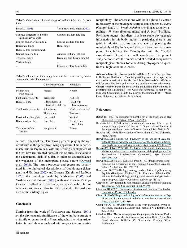
Table 2 Comparison of terminology of axillary fold- and Xexion-lines
Brodsky (1994) Yoshizawa and Saigusa (2001)
Concave (inferior) fold of the third axillary sclerite
Concave axillary fold-line
Convex (superior) jugal fold Convex axillary fold-line
Horizontal hinge Basal hinge
Humeral fold (distal branch) –
Neutral humeral fold Anterior axillary fold-line
Torsional hinge Distal axillary Xexion-line (?)
Vertical hinge –
– Convex axillary Xexion-line
Table 3 Characters of the wing base and their states in Psylloideacompared to other Paraneoptera
Other Paraneoptera Psylloidea
Median notal wing process
Present Absent
First axillary sclerite Triangular Rod-like
Humeral plate DiVerentiated at base of costal vein
Fused with basisubcostale
Third axillary sclerite Sclerotized Weakly sclerotized
Three arms Two arms
Proximal median plate Horizontal Vertical
Distal median plate One plate Divided into two plates
Two horns of the basalare
Not present Present
123
Zoomorphology (2008) 127:37–47 47
Fischer H, Ebert E (1999) Tegula function during free locust Xight inrelation to motor pattern, Xight speed and aerodynamic output. JExp Biol 202(6):711–721
Frye MA, Gray JR (2005) Mechanosensory integration for Xight con-trol in insects. In: Christensen TA (ed) Methods in insect sensoryneuroscience. CRC Press, Boca Raton, pp 107–128
Gullan PJ, Kosztarab M (1997) Adaptations in scale insects. Annu RevEntomol 42:23–50
Hodkinson ID (1974) The biology of the Psylloidea (Homoptera): a re-view. Bull Entomol Res 64:325–339
Hollis D (2004) Australian Psylloidea: jumping plantlice and lerp in-sects. Australian Biological Resources Study, Canberra
Hörnschemeyer T (1998) Morphologie und Evolution des Flügelenksder Coleoptera und Neuropterida. Bonner Zoologische Mono-graphien, 43. Zoologisches Forschungsinstitut und MuseumAlexander Koenig, Bonn
Hörnschemeyer T (2002) Phylogenetic signiWcance of the wing-baseof the Holometabola (Insecta). Zoolog Scripta 31(1):17–29
Ivanov VD, Kozlov MV (1987) Comparative analysis of pterothoracicmusculature of caddis-Xies (Insecta Trichoptera) (in Russian).Zool Zh 66(10):1484–1498
Knight KL, LaVoon JL (1970) A mosquito taxonomic glossary. IV.Adult thoracic appendages. Mosq Systemat Newslett 2(4):165–177
Kukalovà-Peck J (1991) Fossil history and the evolution of hexapodstructures. In: Naumann ID, CSIRO (ed) The insects of Australia,2nd edn. Melbourne University Press, Melbourne, Australia, pp141–179
Lefeuvre JC (1969) Recherches sur les organes alaires des Blattaria.PhD Thesis. Université de Rennes, Rennes, France
Maggenti AR, Gardner SL (2005) Online dictionary of invertebratezoology, Version 2.0. Posted October 20 2005. http://digitalcom-mons.unl.edu
Matsuda R (1963) Some evolutionary aspects of the insect thorax.Annu Rev Entomol 8:59–76
Matsuda R (1970) Morphology and evolution of the insect thorax.Memoirs of the Entomological Society of Canada, 76. Entomo-logical Society of Canada, Ottawa
Matsuda R (1979) Morphologie du thorax et des appendices thoraci-ques des insectes. In: Grassé P-P (ed) Traité de Zoologie. T. VIII.Fasc. II. Masson, Paris, France, pp. 1–289
Ouvrard D. (2002) Systématique phylogénétique des Hemiptera Psyl-loidea: morphologie comparée du thorax et structures secondairesdes ARNr 18S. Bull Soc Zool Fr 127(4):345–357
Ouvrard D, Bourgoin T, Campbell BC (2002) Comparative morpho-logical assessment of the psyllid pleuron (Insecta, Hemiptera,Sternorrhyncha). J Morphol 252(3):276–290
Owen WB (1977) Morphology of the thoracic skeleton and muscles ofthe mosquito, Culiseta inornata (Williston), (Diptera: Culicidae).J Morphol 153(3):427–460
Pringle JWS (1957) Insect Xight. Cambridge monographs in experi-mental biology, 9. Cambridge University Press, Cambridge
Rheuben M, Kammer A (1987) Structure and innervation of the thirdaxillary muscle of Manduca relative to its role in turning Xight. JExp Biol 131(1):373–402
Taylor LH (1918) The thoracic sclerites of Hemiptera and Heteroptera.Ann Entomol Soc Am 11(3):225–254
White IM, Hodkinson ID (1985) Nymphal taxonomy and systematicsof the Psylloidea (Homoptera). Bull Nat Hist Mus (Entomol)50(2):153–301
Wootton RJ (1979) Function, homology and terminology in insectwings. Syst Entomol 4:81–93
Wootton RJ (1992) Functional morphology of insect forewings. AnnuRev Entomol 37(1):113–140
Yoshizawa K (2002) Phylogeny and higher classiWcation of suborderPsocomorpha (Insecta: Psocodea: ‘Psocoptera’). Zool J Linn Soc136(3):371–400
Yoshizawa K (2005) Morphology of Psocomorpha (Psocodea: ‘Pso-coptera’). Insecta Matsumurana NS 62:1–44
Yoshizawa K, Saigusa T (2001) Phylogenetic analysis of paraneopter-an orders (Insecta: Neoptera) based on forewing base structure,with comments on monophyly of Auchenorrhyncha (Hemiptera).Syst Entomol 26:1–13
123