Number of ovarian follicles in human fetuses with the 45,x karyotype
Upload
independentCategory
view
0download
0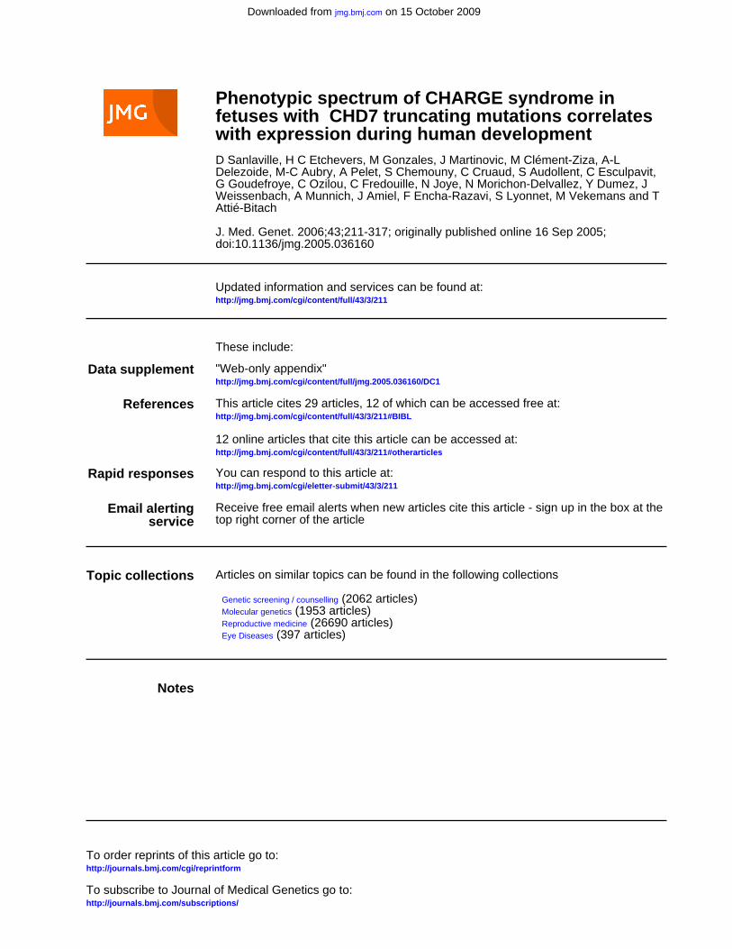
doi:10.1136/jmg.2005.036160 2006;43;211-317; originally published online 16 Sep 2005; J. Med. Genet.
Attié-Bitach Weissenbach, A Munnich, J Amiel, F Encha-Razavi, S Lyonnet, M Vekemans and T G Goudefroye, C Ozilou, C Fredouille, N Joye, N Morichon-Delvallez, Y Dumez, JDelezoide, M-C Aubry, A Pelet, S Chemouny, C Cruaud, S Audollent, C Esculpavit, D Sanlaville, H C Etchevers, M Gonzales, J Martinovic, M Clément-Ziza, A-L
with expression during human development truncating mutations correlatesCHD7fetuses with
Phenotypic spectrum of CHARGE syndrome in
http://jmg.bmj.com/cgi/content/full/43/3/211Updated information and services can be found at:
These include:
Data supplement http://jmg.bmj.com/cgi/content/full/jmg.2005.036160/DC1
"Web-only appendix"
References
http://jmg.bmj.com/cgi/content/full/43/3/211#otherarticles12 online articles that cite this article can be accessed at:
http://jmg.bmj.com/cgi/content/full/43/3/211#BIBLThis article cites 29 articles, 12 of which can be accessed free at:
Rapid responses http://jmg.bmj.com/cgi/eletter-submit/43/3/211
You can respond to this article at:
serviceEmail alerting
top right corner of the article Receive free email alerts when new articles cite this article - sign up in the box at the
Topic collections
(397 articles) Eye Diseases � (26690 articles) Reproductive medicine �
(1953 articles) Molecular genetics � (2062 articles) Genetic screening / counselling �
Articles on similar topics can be found in the following collections
Notes
http://journals.bmj.com/cgi/reprintformTo order reprints of this article go to:
http://journals.bmj.com/subscriptions/ go to: Journal of Medical GeneticsTo subscribe to
on 15 October 2009 jmg.bmj.comDownloaded from

ORIGINAL ARTICLE
Phenotypic spectrum of CHARGE syndrome infetuses with CHD7 truncating mutations correlateswith expression during human developmentD Sanlaville*, H C Etchevers*, M Gonzales*, J Martinovic, M Clement-Ziza,A-L Delezoide, M-C Aubry, A Pelet, S Chemouny, C Cruaud, S Audollent, C Esculpavit,G Goudefroye, C Ozilou, C Fredouille, N Joye, N Morichon-Delvallez, Y Dumez,J Weissenbach, A Munnich, J Amiel, F Encha-Razavi, S Lyonnet, M Vekemans,T Attie-Bitach. . . . . . . . . . . . . . . . . . . . . . . . . . . . . . . . . . . . . . . . . . . . . . . . . . . . . . . . . . . . . . . . . . . . . . . . . . . . . . . . . . . . . . . . . . . . . . . . . . . . . . . . . . . . . . . . . . . . . . . . . . . . . . .
Supplementary data areavailable online at http://www.jmedgenet.com/supplemental
See end of article forauthors’ affiliations. . . . . . . . . . . . . . . . . . . . . . .
Correspondence to:T Attie-Bitach,Departement de Genetiqueet Unite INSERM U-393,Hopital Necker-EnfantsMalades, 149, rue deSevres, 75743 ParisCedex 15, France; [email protected]
Revised version received11 August 2005Accepted for publication18 August 2005Published Online First23 September 2005
. . . . . . . . . . . . . . . . . . . . . . .
J Med Genet 2006;43:211–217. doi: 10.1136/jmg.2005.036160
Background: The acronym CHARGE refers to a non-random cluster of malformations including coloboma,heart malformation, choanal atresia, retardation of growth and/or development, genital anomalies, andear anomalies. This set of multiple congenital anomalies is frequent, despite rare patients with normalintelligence, and prognosis remains poor. Recently, CHD7 gene mutations have been identified inCHARGE patients; however, the function of CHD7 during development remains unknown.Methods: We studied a series of 10 antenatal cases in whom the diagnosis of CHARGE syndrome wassuspected, considering that a careful pathological description would shed light on the CHD7 functionduring development. CHD7 sequence analysis and in situ hybridisation were employed.Results: The diagnosis of CHARGE syndrome was confirmed in all 10 fetuses by the identification of aCHD7 heterozygous truncating mutation. Interestingly, arhinencephaly and semi-circular canal agenesiswere two constant features which are not included in formal diagnostic criteria so far. In situ hybridisationanalysis of the CHD7 gene during early human development emphasised the role of CHD7 in thedevelopment of the central nervous system, internal ear, and neural crest of pharyngeal arches, and moregenerally showed a good correlation between specific CHD7 expression pattern and the developmentalanomalies observed in CHARGE syndrome.Conclusions: These results allowed us to further refine the phenotypic spectrum of developmentalanomalies resulting from CHD7 dysfunction.
CHARGE syndrome refers to an association of defectsincluding ocular coloboma (C), heart disease (H),choanal atresia (A), retarded growth and/or anomalies
of the central nervous system (R), genito-urinary defects and/or hypogonadism (G), and ear anomalies and/or deafness(E). The acronym was coined by Pagon et al,1 but thesyndrome was first reported by Hall2 and Hittner et al.3 Otherdiagnostic criteria were proposed based on major/minoranomalies,4–6 however, all features have variable expressionand can be inconsistent, and many are non-specific. Theincidence of CHARGE syndrome has been estimated to rangefrom 0.1 to 1.2/100 000 live births with the highest incidence(to 1/8500 live births) in Canada.7
Recently, Vissers et al reported mutations in the CHD7 genein two out of three CHARGE patients tested.8 CHD7 belongsto a large family of evolutionarily conserved proteins thoughtto play a role in chromatin organisation. However, althoughmore than 200 newborns and infants with CHARGEsyndrome have been described, only two prenatally detectedcases have been reported to date9 10 and nothing is knownabout CHD7 function during fetal development. In the
present study, we analysed the coding sequence of theCHD7 gene in 10 fetuses presenting with clinical featuresof CHARGE syndrome, considering that a careful path-ological description would shed light on the CHD7 functionduring development. Clinical features as well as criterianecessary to establish the diagnosis of CHARGE syndromeprenatally remained to be defined. In fetuses, coloboma,heart malformation, choanal atresia, intrauterine growthretardation, genital anomalies, and external ear anomaliescan be identified, while clinical symptoms such as mentalretardation and deafness have to be replaced by theirequivalents: central nervous system (CNS) anomalies andsemi-circular canal (SCC) hypoplasia or agenesis, respec-tively. In this series, four of the above major anomalieswere required for the diagnosis of CHARGE syndromebecause the other features, such as growth retardation,cryptorchidism, or cranial nerve dysfunction, appear onlyin the postnatal period. All fetuses underwent autopsy anda detailed neuropathological examination with closeattention to olfactory bulbs and tracts, brainstem, andcerebellum.
211
www.jmedgenet.com
on 15 October 2009 jmg.bmj.comDownloaded from
We identified a truncating mutation in all cases examined,confirming the diagnosis of CHARGE syndrome in all 10 fetalcases and allowing the phenotypic spectrum of the develop-mental anomalies resulting from CHD7 dysfunction to befurther envisaged. In addition, in situ hybridisation analysisof the CHD7 gene at different stages of early humandevelopment emphasised the role of CHD7 in the develop-ment of the central nervous system, internal ear, and neuralcrest of pharyngeal arches, and more generally showed agood correlation between specific CHD7 expression patternand the developmental anomalies observed in CHARGEsyndrome.
METHODSPatientsIn all cases, the presence of severe malformations was notedat ultrasound (US) examination and pregnancies wereterminated in accordance with French law. Detailed clin-icopathological examination was carried out after parentalconsent. For all cases, the diagnosis of CHARGE syndromewas established either based on US evaluation or afterfetopathological examination using the diagnostic criteriadescribed above. For all cases, karyotype and CHD7 mutationscreening was performed. Clinical and genetic data aresummarised in table 1 and detailed in supplementary dataavailable online at http://www.jmedgenet.com/supplemental.
CHD7 sequence analysisDNA was extracted from frozen tissues according to standardprotocols. We designed 42 primer pairs covering the 37 CHD7coding exons (exons 2–38) and the corresponding exon-intron boundaries. Primer sequences and PCR conditions areavailable on request. PCR products were purified and directlysequenced in both directions on an ABI PRISM 3730 DNAsequencer (Perkin Elmer-Applied Biosystems, Courtaboeuf,France) using the dye terminator method according to themanufacturer’s instructions.
In situ hybridisationNormal human embryos and fetal tissues were obtained afterelective termination of pregnancy in agreement with Frenchlegislation (law no. 94-654 of July 29, 1994), Necker Hospital,and the National Ethics Committee recommendations (reportno. 1 of May 22, 1984). Embryonic stages were establishedaccording to the Carnegie staging (CS) classification.11 Sevendifferent embryonic stages (CS9 (d20), CS10 (d22), CS11(d24), CS12 (d26), CS14 (d33), CS15 (d34), and CS19 (d 47–48)) as well as two fetal stages (9 and 11 weeks) werestudied. Tissues were fixed in 4% phosphate bufferedparaformaldehyde, dehydrated, and embedded in paraffinblocks. Five micron thick serial sections were cut. Exon 35primers were selected for PCR amplification (F: 59-GCTGTTCCCAAACAACTAGACATTG-39 R: 59-GAAACATTCA-AGGAAAAGGCAGAG-39). A T7 promotor sequence exten-sion (TAATACGACTCACTATAGGGAGA) was added at the 59
end of each primer. T7F/R and F/T7R primers allowed theamplification of sense and antisense templates specific to theCHD7 gene. Riboprobes labelling, tissue fixation, hybridisa-tion, and developing were carried out according to standardprotocols,12 as previously described.13
RESULTSMolecular resultsDirect sequencing of the 37 CHD7 coding exons (gene ID55636, NM_017780, NCBI: http://www.ncbi.nlm.nih.gov/)detected ten heterozygous truncating mutations. They aresummarised in fig 1. Six were nonsense mutations (cases 2,3, 4, 7, 8, 10), and four were frameshift mutations predictinga premature stop codon (cases 1, 5, 6, 9). In seven cases,
parental DNA was studied (cases 1, 3, 6, 7, 8, 9, 10). Allmutations occurred de novo (table 1). None of the 10mutations was described previously. We found two muta-tions in exon 2, two mutations in exon 8 (at the end of thefirst chromodomain), and two identical mutations in exon 12(R987X, unrelated cases 2 and 8) located in the SNF2domain. In patient 5, the mutation (N1371fsX1374) islocated in another domain of CHD7 encoding the putativehelicase domain. None of the remaining mutations involvedknown functional domains of the protein.
Clinical analysisThis series of 10 fetuses (seven male and three female)ranged from 21 to 36 weeks of gestation (WG) with a meanof 27 WG. Mean paternal age was 35 years and 8 months andmean maternal age was 30 years and 6 months. Prenatal USwas abnormal in all patients. It disclosed polyhydramnios in3/8 cases. Although two fetuses (cases 7 and 9) weighedbetween the 3rd and the 10th percentile, intrauterine growthretardation was never observed. All fetuses presentedbilateral and asymmetric external ear abnormalities, semi-circular canal hypoplasia/agenesis, and arhinencephaly. Asshown in fig 2A, the ears were typically low set, asymmetricwith a small or absent lobe, posteriorly rotated and triangularor square shaped.
Coloboma was found in seven patients, always affectingthe retina or the choroid segment (fig 2Be,f). Microphthalmiawas observed in two out the 10 cases in accordance withearlier postnatal observations.14
A congenital heart defect was found in 9/10 patients. Thesedefects were complex cardiopathies and five involved theconotruncal region (cases 2, 3, 4, 5, 9). They frequentlyincluded atrial and/or ventricular septal defects, in accor-dance with earlier postnatal observations.15
Choanal atresia was found in six out of 10 patients (fourbilateral and two unilateral). Among the four remainingpatients, three had a cleft lip and/or palate. In two casesunilateral choanal atresia was found with a controlateral cleftpalate. In one patient (case 6), neither choanal atresia norfacial clefting was found.
CHD7 expression during human embryonicdevelopmentAt CS9 (d20; not shown), CS10 (d22; fig 3A), and CS11 (d24;fig 3B), CHD7 is ubiquitously expressed, with a distinct signalin the neural tube. At CS12 (d26), CHD7 is expressedthroughout the CNS and neural crest-containing mesench-yme of the pharyngeal arches. At CS14 (d33), CHD7transcripts continue to be expressed in the cephalicmesenchyme, pharyngeal arches, and the brain. Expressionis observed in the otic vesicle (fig 3C) as well as in the limbbud mesenchyme; it is more intense in the spinal cord anddorsal root ganglia (fig 3D). By CS15 (d34; fig 3E), expressioncontinues to be intense within the CNS but is restricted to thedorsal part of the otic vesicle. Interestingly, no expression isobserved in the nasal placode, neural retina, or pharyngealendoderm. Also no expression is detected in the heart at anyage. By CS19 (d47), CHD7 is strongly expressed in the neuralretina and rhombencephalon. It is moderately expressed inthe semicircular canals (fig 3F), as well as in the forebrain,pituitary gland, and olfactory bulbs and nerves (fig 3G).Expression at 9 weeks of development is localised in thenasal epithelia (fig 3H), the neural retina, the optic nervesheath (fig 3I), and the anterior and median lobes of thepituitary gland (fig 3J).
DISCUSSIONHere we report on a series of fetuses with CHARGE syndromeusing diagnostic criteria adapted to the prenatal situation. All
212 Sanlaville, Etchevers, Gonzales, et al
www.jmedgenet.com
on 15 October 2009 jmg.bmj.comDownloaded from

Table
1Su
mm
ary
ofth
ecl
inic
alfe
atur
esof
the
10
CH
ARG
Efe
tuse
s
Case
1(L
AS)
2(B
0S)
3(G
AL)
4(B
AR)
5(G
OD
)6
(LA
N)
7(C
OQ
)8
(BEL
)9
(RA
B)10
(DER
)M
ean
Age
/sex
Feta
lage
(WG
)21
21
23
23
28
29
29
29
32
36
27.1
Sex
MM
MM
MM
FM
FF
7M
/3F�
Mat
erna
lage
35
26
32
30
24
31
37
31
28
31
30.5
Pate
rnal
age
30
35
41
30
38
39
39
46
28
31
35.7�
Gen
etic
sK
aryo
type
46,X
Y46,X
Y46,X
Y46,X
Y46,X
Y46,X
Y46,X
X46,X
Y46,X
X46,X
XFI
SH22q1
1.2
(Tup
le1)
NN
NN
NN
NN
NN
CH
D7
nucl
eotid
ech
ange
2550_2
554de
l2959C
.T
763C
.T
6157C
.T
4112_4
113in
sT808de
lG5458C
.T
2959C
.T
2260_2
266du
p2584A
.T
100%
CH
D7
prot
ein
chan
geK
850fs
X855
R987X
Q255X
Arg
2053X
N1371fs
X1374
A270fs
X304
P1820X
R987X
V754fs
X764
K862X
100%
De
novo
?Yes
?Yes
??
Yes
Yes
Yes
Yes
Yes
CH
ARG
Eac
rony
mC
olob
oma
++
++
22
++
+2
70%
Hea
rtm
alfo
rmat
ions
++
++
++
++
+2
90%
Atr
esia
choa
nal/
clef
tA
CC
lA
C/C
lC
lC
l2
AC
/Cl
AC
AC
AC
90%
AC
unila
tera
l/bi
late
ral
BU
UB
BB
Cle
ftpa
late
/lip
L+P
PL+
PP
L+P
CN
San
omal
ies
Arh
inen
ceph
aly
++
++
++
++
++
100%
Oth
ers
CN
San
omal
ies
++
++
++
++
22
80%
Gen
itala
nom
alie
s2
+*2
++*
+*+
+2
260%
Ears A
nom
alie
sof
exte
rnal
ears
++
++
++
++
++
100%
Age
nesi
s/hy
popl
asia
CSC
++
++
++
++
++
100%
Oth
ers
Thym
ichy
po/a
gene
sis
++
22
+2
++
++
70%
Lim
ban
omal
ies
++
22
22
+2
++
50%
Rena
lano
mal
ies
22
2+
2+
+2
2+
40%
Skel
etal
anom
alie
s+
+?
22
+2
2+
240%
Poly
hydr
amni
os2
22
22
+2
2+
+30%
Oes
opha
geal
anom
alie
s2
22
22
22
2+
+20%
IUG
R2
22
22
22
22
20%
N,
norm
al;
WG
,w
eeks
ofge
stat
ion.
*Ley
dig
cell
rare
fact
ion;
�the
incr
ease
dm
ale
popu
latio
nan
dpa
tern
alag
ein
this
stud
yw
ere
first
docu
men
ted
byBl
ake
etal
.31
CHD7 expression and mutation during development 213
www.jmedgenet.com
on 15 October 2009 jmg.bmj.comDownloaded from
fetuses underwent an extensive necropsy as well as molecularanalysis of the CHD7 gene. This study confirms the role of theCHD7 gene in CHARGE syndrome and allows delineation ofthe antenatal clinical spectrum of CHD7 mutations.
Molecular analysis of CHD7Nine different mutations were identified in our series. One,the R987X mutation, was found in two unrelated fetuses(cases 2 and 8). Interestingly, they differed as regards thepresence of distal arthrogryposis and hemivertebra (case 2),suggesting that other additional factors might be involved.However, based on our results and in agreement with Visserset al, so far it is not possible to establish a genotype/phenotypecorrelation.8 Large deletions and nonsense mutations in thefirst exon(s) of the CHD7 gene are observed. This suggeststhat haploinsufficiency of the CHD7 gene is probably themechanism accounting for CHARGE syndrome. Because themutations occurred de novo, genetic counselling can bereassuring, although germ-line mosaicism can never beexcluded.
Clinical analysisIn our series of fetal CHARGE syndrome, our detailedclinicopathological evaluation led to the identification ofthree constant features: anomalies of the external ear,agenesis/hypoplasia of the semi-circular canals, and arhi-nencephaly. Six other features occur frequently (in at least inseven of 10 cases), namely genital anomalies, thymichypoplasia, ocular coloboma, other CNS anomalies (otherthan arhinencephaly), choanal atresia/cleft palate, and heartdefect.
Bilateral and asymmetric external ear malformations arefound in all our cases. As shown in fig 2A, typical features aresmall, low set, posteriorly rotated ears, with a prominent cruxhelix, a small or absent lobe, and a triangular shape asdescribed previously.5 15 16 In the literature, internal earanomalies, ranging from subtle modular deficiencies toMondini malformation and semi-circular canal hypoplasiaor agenesis, are classically reported in CHARGE syn-drome.7 17 18 Semi-circular hypoplasia/agenesis was reportedin up to 100% of cases in the literature.15 18 We confirm thesedata and emphasise the necessity of detecting temporal boneanomalies though cephalic x ray evaluation for the antenataldiagnosis of CHARGE syndrome (fig 2Ba,b).
Interestingly, among CNS anomalies, arhinencephaly wasfound in all our cases (fig 2Bc,d). Arhinencephaly has alreadybeen reported by Lin et al in some patients.19 However, itsfrequency among CHARGE patients has not been evaluatedin the literature, aside from the observation of Harvey et alwho found arhinencephaly in 7/7 CHARGE postnatal cases.20
More recently, Chalouhi et al assessed olfactory deficiency in14 children with CHARGE syndrome. Half of them wereanosmic and the others had olfactory residual function(hyposmic). All nine MRIs showed anomalies of the olfactorytracts and bulbs varying from moderate hypoplasia tocomplete aplasia, without any relationship between theradiological and functional results.21 Our systematic neuro-pathological examination clearly shows that arhinencephalyis a constant sign of CHD7 mutation. We propose it should beconsidered a major sign for prenatal CHARGE diagnosis.Other CNS anomalies included brainstem and cerebellumabnormalities. They were observed in eight of our 10 fetuses,but were reported also in postnatal cohorts.19 20 They aremainly characterised by hypoplasia of the inferior cerebellarvermis and brain stem (cases 1, 5, 7) and severe cerebellarheterotopia (case 8).
The high incidence of heart defects, cleft lip/palate, andbrain anomalies in our series when compared to postnataldata can probably be explained by detection by US during
pregnancy. It has been shown that CHARGE individuals haveincreased mortality due to AVSD defects and cerebellar andor brain stem anomalies associated with ventriculomegaly.7 15
Moreover, males have been reported to have an increasedmortality compared to females.15 22 This study presents apopulation of 10 fetuses with a severe phenotype of CHARGEsyndrome, with an especially high number of complexcardiopathies, bilateral posterior choanal atresia, tracheo-oesophageal fistula,22 brain anomalies, and increased repre-sentation of males (7/10).
Among other frequent features, genital anomalies deservespecial attention: they were found in 7/10 cases. In threemale fetuses, they were noticed only at the histological level(Leydig cell rarefaction). To our knowledge these histologicalanomalies have not been described previously. This mayexplain the delay in puberty noticed more in males thanfemales that has been reported postnatally.23 In a femalefetus, the only genital anomaly found was a hypoplasticovary. According to the postnatal literature, female genitalhypoplasia is rare and micropenis or cryptorchidism are morefrequent features observed in males.24
Interestingly, thymus hypoplasia or agenesis was found inseven of out 10 cases. This contrasts with postnatal caseswhere thymic hypoplasia is rarely reported.5 The associationof CHARGE and DiGeorge syndromes reported previously25
suggests a neural crest defect causes the clinical overlap ofboth syndromes. Whether the CHD7 gene could be respon-sible for a CHARGE-DiGeorge association should be testedfurther, particularly in postnatal patients.
Our study also confirms that growth retardation is usuallyobserved postnatally.26 Indeed, no intrauterine growthretardation was observed in our antenatal series. In additionto the severe feeding problems related to gastro-oesophagealreflux and brain stem anomalies, the postnatal growthretardation could be explained in part by a pituitary glanddysfunction. Interestingly, CHD7 gene expression is observedwithin this tissue during the embryonic period.
It is worth stressing that case 9 presented with ectrodactyly(fig 2C), which has not been reported so far in CHARGEsyndrome. Four patients presented limb anomalies althoughminor, such as clinodactyly. Skeletal (including costal andvertebral defects) or renal anomalies were found in four outof 10 cases. Finally, among other features, tracheo-oesopha-geal fistulas were noticed twice.
Based on our clinicopathological observations, we considersemicircular canal agenesis as well as arhinencephaly highlypredictive diagnostic criteria of CHARGE syndrome. Wesuggest that they should be added to the two majordiagnostic criteria described by Pagon, along with theexternal ears malformations. Indeed, four of six majorcriteria of CHARGE (coloboma, choanal atresia and/or cleftlip/palate, heart defect, arhinencephaly, semi-circular canalagenesis, and external ear anomalies) were necessary andsufficient for the diagnosis of CHARGE in our series. Aspreviously suggested for postnatal cases, minor diagnosticcriteria such as facial dysmorphism and renal, digestive, andskeletal anomalies should also be considered for fetalCHARGE syndrome in addition to thymus hypoplasia/agenesis and polyhydramnios. Interestingly, in most patientswith three major features and three minor features proposedas CHARGE syndrome,5 the presence of arhinencephaly andsemi-circular canals hypoplasia/agenesis was not evaluatedand we recommend brain MRI to assess the diagnosis ofCHARGE syndrome in these cases.
Our study contributes to the development of a strategy forthe diagnosis of CHARGE syndrome during pregnancy.Indeed, features of CHARGE syndrome detected at routineUS such as hydramnios, heart defects, cleft lip/palate, CNSanomalies, and kidney or gastrointestinal anomalies are
214 Sanlaville, Etchevers, Gonzales, et al
www.jmedgenet.com
on 15 October 2009 jmg.bmj.comDownloaded from
common. More specific features would help in the diagnosisof CHARGE syndrome. We thus propose that focussed USand/or brain MRI should be performed for the detection ofexternal ear anomalies, choanal atresia, semi-circular canalagenesis, and arhinencephaly. This strategy will certainly leadto a higher prenatal detection rate of CHARGE syndrome.
Embryonic function of CHD7The CHD7 protein encompasses several important domains.The chromatin organisation modifier (chromo) domain is aconserved region of around 50 amino acids found in a varietyof chromosomal proteins that appear to play a role in thefunctional organisation of the eukaryotic nucleus.Experimental evidence shows that the chromodomain isinvolved in binding proteins to histone and possibly RNA.CHD7-like helicase domains are involved in ATP-dependentunwinding of DNA or RNA duplexes and histone deacetyla-tion.27 28 Certain CHD proteins have been shown to participatein nucleosome remodeling deacetylase (NuRD) proteincomplexes which interact with sequence-specific DNA-bind-ing factors for targeted repression.29 Presumably, the CHD7
protein plays an important role in chromatin remodellingduring early development and allows a level of epigeneticcontrol over target genes expressed in mesenchymal cellsderived from the cephalic neural crest.
We analysed the expression pattern of the CHD7 geneduring early human development. CHD7 is widely expressedin the undifferentiated neuroepithelium and in mesenchymeof neural crest origin. Towards the end of the first trimester itis expressed in dorsal root ganglia, cranial nerves/ganglia,and auditory, pituitary, and nasal tissues as well as in theneural retina. Absent from the myocardium, bones ofmesodermal or neural crest origin, and the genital ridge,CHD7 expression correlates with defects observed in thesetissues because of its presence in neural crest cells investingthe outflow tract of the heart, and in the hypothalamus andpituitary gland. Endocrine deficiency may occur centrally, inthe differentiation of hypothalamic nuclei secreting soma-tostatin or GnRH, or more peripherally in the differentiationof somatotropic or gonadotropic cells of the anterior pituitary.It could be related to the clinical findings of delayed pubertyin the adolescent and adult population with CHARGE
Figure 1 Schematic representation of the CHD7 mutations identified so far. The mutations found by Vissers et al8 are shown in orange; the tennonsense mutations found in our cohort are shown in blue. Circles indicate the nonsense mutations, squares the missense mutations, and triangles theintron-exon boundary mutations. On the bottom, functional CHD7 domains are indicated: chromodomains in red, SNF2 domain in green, and helicasedomain in yellow.
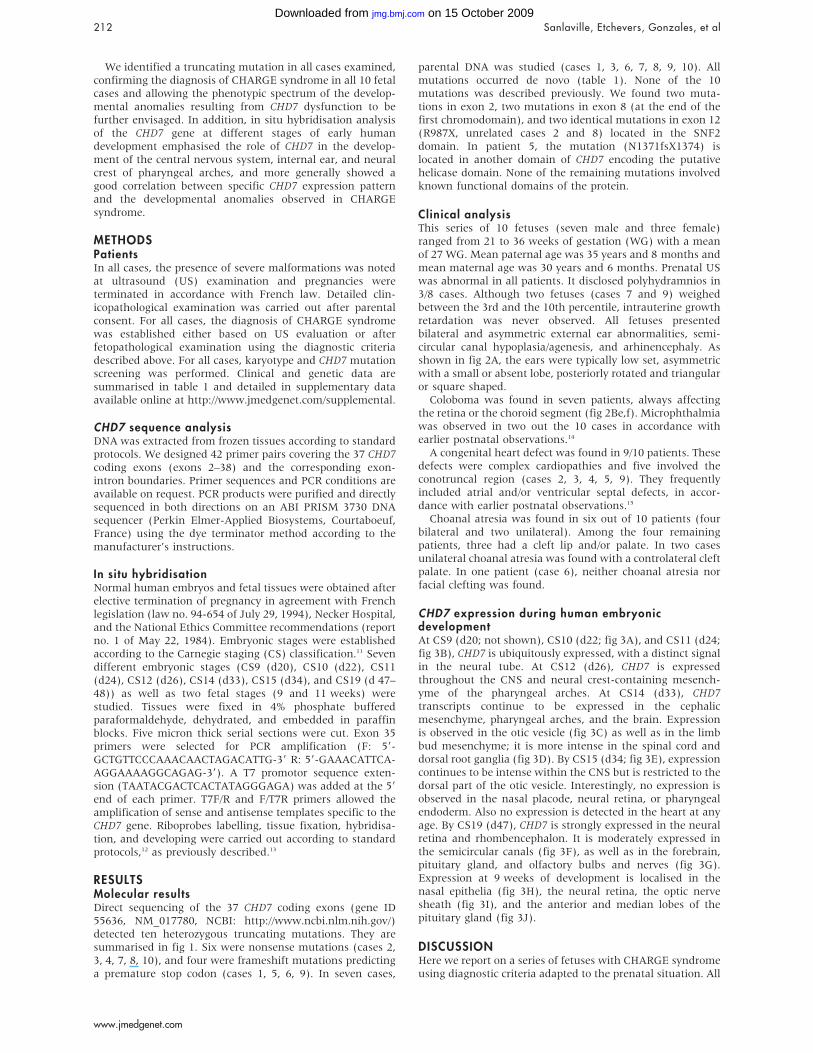
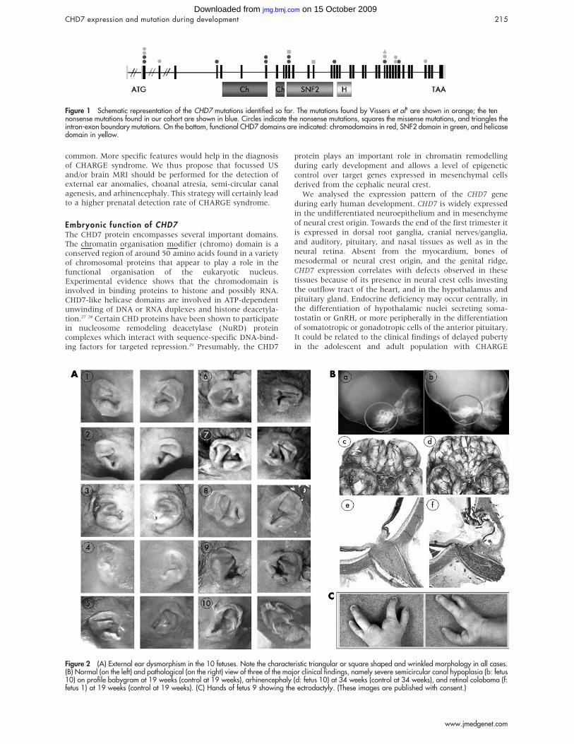
Figure 2 (A) External ear dysmorphism in the 10 fetuses. Note the characteristic triangular or square shaped and wrinkled morphology in all cases.(B) Normal (on the left) and pathological (on the right) view of three of the major clinical findings, namely severe semicircular canal hypoplasia (b: fetus10) on profile babygram at 19 weeks (control at 19 weeks), arhinencephaly (d: fetus 10) at 34 weeks (control at 34 weeks), and retinal coloboma (f:fetus 1) at 19 weeks (control at 19 weeks). (C) Hands of fetus 9 showing the ectrodactyly. (These images are published with consent.)
CHD7 expression and mutation during development 215
www.jmedgenet.com
on 15 October 2009 jmg.bmj.comDownloaded from
syndrome.23 A major testable hypothesis in this context isthat CHD7 regulates paired domain- or homeobox domain-containing transcription factor genes important for thedevelopment of the pituitary gland and other organ systems,such as Hesx1, Otx1, Prop1, Krox20, Pitx2, and Titf1/Nkx2a.
Other regulatory transcription factor genes such as Pax2 orTbx1, with multiple expression sites in many affected organsystems or inductive tissues for these organ anlages, stillremain attractive and non-exclusive CHD7 functional tar-gets.30
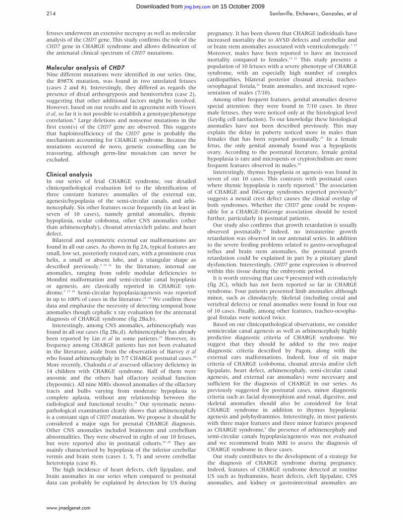
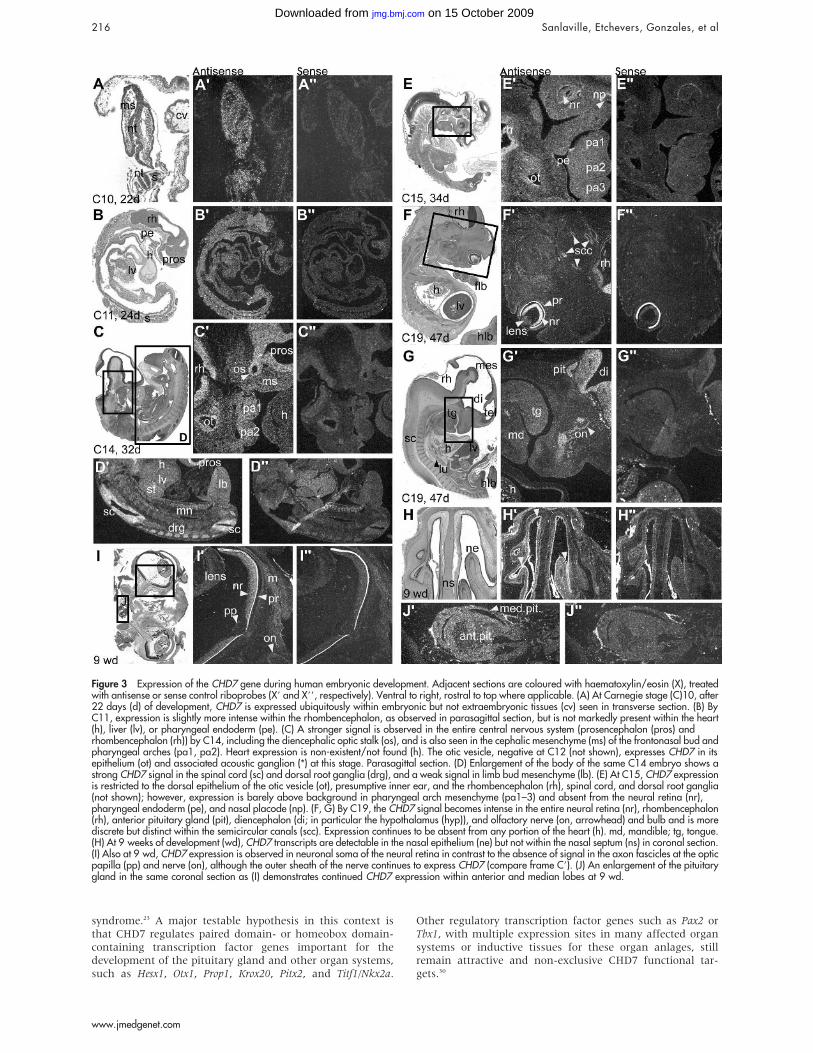
Figure 3 Expression of the CHD7 gene during human embryonic development. Adjacent sections are coloured with haematoxylin/eosin (X), treatedwith antisense or sense control riboprobes (X9 and X99, respectively). Ventral to right, rostral to top where applicable. (A) At Carnegie stage (C)10, after22 days (d) of development, CHD7 is expressed ubiquitously within embryonic but not extraembryonic tissues (cv) seen in transverse section. (B) ByC11, expression is slightly more intense within the rhombencephalon, as observed in parasagittal section, but is not markedly present within the heart(h), liver (lv), or pharyngeal endoderm (pe). (C) A stronger signal is observed in the entire central nervous system (prosencephalon (pros) andrhombencephalon (rh)) by C14, including the diencephalic optic stalk (os), and is also seen in the cephalic mesenchyme (ms) of the frontonasal bud andpharyngeal arches (pa1, pa2). Heart expression is non-existent/not found (h). The otic vesicle, negative at C12 (not shown), expresses CHD7 in itsepithelium (ot) and associated acoustic ganglion (*) at this stage. Parasagittal section. (D) Enlargement of the body of the same C14 embryo shows astrong CHD7 signal in the spinal cord (sc) and dorsal root ganglia (drg), and a weak signal in limb bud mesenchyme (lb). (E) At C15, CHD7 expressionis restricted to the dorsal epithelium of the otic vesicle (ot), presumptive inner ear, and the rhombencephalon (rh), spinal cord, and dorsal root ganglia(not shown); however, expression is barely above background in pharyngeal arch mesenchyme (pa1–3) and absent from the neural retina (nr),pharyngeal endoderm (pe), and nasal placode (np). (F, G) By C19, the CHD7 signal becomes intense in the entire neural retina (nr), rhombencephalon(rh), anterior pituitary gland (pit), diencephalon (di; in particular the hypothalamus (hyp)), and olfactory nerve (on, arrowhead) and bulb and is morediscrete but distinct within the semicircular canals (scc). Expression continues to be absent from any portion of the heart (h). md, mandible; tg, tongue.(H) At 9 weeks of development (wd), CHD7 transcripts are detectable in the nasal epithelium (ne) but not within the nasal septum (ns) in coronal section.(I) Also at 9 wd, CHD7 expression is observed in neuronal soma of the neural retina in contrast to the absence of signal in the axon fascicles at the opticpapilla (pp) and nerve (on), although the outer sheath of the nerve continues to express CHD7 (compare frame C9). (J) An enlargement of the pituitarygland in the same coronal section as (I) demonstrates continued CHD7 expression within anterior and median lobes at 9 wd.
216 Sanlaville, Etchevers, Gonzales, et al
www.jmedgenet.com
on 15 October 2009 jmg.bmj.comDownloaded from

CONCLUSIONIn the present study we confirmed the predominant role ofthe CHD7 gene during fetal development. The clinicopatho-logical spectrum of CHD7 mutations in a series of 10 fetusesexamined at the anatomical as well as histological levelallowed the phenotypic spectrum of the developmentalanomalies resulting from CHD7 dysfunction to be furtherenvisaged and led us to propose agenesis of the semi-circularcanals and arhinencephaly as major diagnostic criteria ofCHARGE syndrome. Further postnatal studies are needed toconfirm these data.
ACKNOWLEDGEMENTSWe are thankful to Eric Detrait, Sophie Delahaie, Boris Keren, DanielSidi, the French patients’ group for the CHARGE association, and theSOFFOET (Societe Francaise de FOETopathologie) for their help andcollaboration. We also thank Han Brunner for sharing unpublisheddata.
ELECTRONIC-DATABASE INFORMATION
The NCBI website is at http://www.ncbi.nlm.nih.gov/
Authors’ affiliations. . . . . . . . . . . . . . . . . . . . .
D Sanlaville, H C Etchevers, J Martinovic, M Clement-Ziza, A Pelet,S Audollent, C Esculpavit, G Goudefroye, C Ozilou, N Morichon-Delvallez, A Munnich, J Amiel, F Encha-Razavi, S Lyonnet,M Vekemans, T Attie-Bitach, Departement de Genetique et UniteINSERM U-393, Hopital Necker-Enfants Malades, AP-HP, 75015 Paris,FranceM Gonzales, C Fredouille, N Joye, Unite de Foetopathologie, HopitalSaint Antoine, AP-HP, 75012 Paris, FranceA-L Delezoide, Service de Biologie du Developpement, Hopital RobertDebre, AP-HP, 75019 Paris, FranceM-C Aubry, S Chemouny, Y Dumez, Service d’Obstetrique, HopitalNecker-Enfants Malades, AP-HP, 75015 Paris, FranceC Cruaud, J Weissenbach, Genoscope and CNRS UMR8030, Evry,France
DS was supported by the Programme de Recherche Clinique AOM 02-122. HCE was supported by the Association Francaise contre lesMyopathies and the INSERM Avenir program
Competing interests: none declared
*These authors contributed equally to this work
Consent has been given for the publication of the details in this report
REFERENCES1 Pagon RA, Graham JM Jr, Zonana J, Yong SL. Coloboma, congenital heart
disease, and choanal atresia with multiple anomalies: CHARGE association.J Pediatr 1981;99(2):223–7.
2 Hall BD. Choanal atresia and associated multiple anomalies. J Pediatr1979;95(3):395–8.
3 Hittner HM, Hirsch NJ, Kreh GM, Rudolph AJ. Colobomatous microphthalmia,heart disease, hearing loss, and mental retardation–a syndrome. J PediatrOphthalmol Strabismus 1979;16(2):122–8.
4 Oley CA, Baraitser M, Grant DB. A reappraisal of the CHARGE association.J Med Genet 1988;25(3):147–56.
5 Blake KD, Davenport SL, Hall BD, Hefner MA, Pagon RA, Williams MS, Lin AE,Graham JM Jr. CHARGE association: an update and review for the primarypediatrician. Clin Pediatr (Phila) 1998;37(3):159–73.
6 Verloes A. Updated diagnostic criteria for CHARGE syndrome: a proposal.Am J Med Genet A 2005;133(3):306–8.
7 Issekutz KA, Graham JM Jr, Prasad C, Smith IM, Blake KD. Anepidemiological analysis of CHARGE syndrome: preliminary results from aCanadian study. Am J Med Genet A 2005;133:309–17.
8 Vissers LE, van Ravenswaaij CM, Admiraal R, Hurst JA, de Vries BB,Janssen IM, van der Vliet WA, Huys EH, de Jong PJ, Hamel BC,Schoenmakers EF, Brunner HG, Veltman JA, van Kessel AG. Mutations in anew member of the chromodomain gene family cause CHARGE syndrome.Nat Genet 2004;36(9):955–7.
9 Becker R, Stiemer B, Neumann L, Entezami M. Mild ventriculomegaly, mildcerebellar hypoplasia and dysplastic choroid plexus as early prenatal signs ofCHARGE association. Fetal Diagn Ther 2001;16(5):280–3.
10 Hertzberg BS, Kliewer MA, Lile RL. Antenatal ultrasonographic findings in theCHARGE association. J Ultrasound Med 1994;13(3):238–42.
11 O’Rahilly R, Muller F. Developmental stages in human embryos. Washington,DC: Carnegie Institution of Washington, 1987.
12 Wilkinson DG. In situ hybridization: a practical approach. Oxford: IRL Press,1992.
13 Trueba SS, Auge J, Mattei G, Etchevers H, Martinovic J, Czernichow P,Vekemans M, Polak M, Attie-Bitach T. PAX8, TITF1, and FOXE1 geneexpression patterns during human development: new insights into humanthyroid development and thyroid dysgenesis-associated malformations. J ClinEndocrinol Metab 2005;90(1):455–62.
14 Russell-Eggitt IM, Blake KD, Taylor DS, Wyse RK. The eye in the CHARGEassociation. Br J Ophthalmol 1990;74(7):421–6.
15 Tellier AL, Cormier-Daire V, Abadie V, Amiel J, Sigaudy S, Bonnet D, deLonlay-Debeney P, Morrisseau-Durand MP, Hubert P, Michel JL, Jan D,Dollfus H, Baumann C, Labrune P, Lacombe D, Philip N, LeMerrer M,Briard ML, Munnich A, Lyonnet S. CHARGE syndrome: report of 47 cases andreview. Am J Med Genet 1998;76(5):402–9.
16 Davenport SL, Hefner MA, Thelin JW. CHARGE syndrome. Part I. External earanomalies. Int J Pediatr Otorhinolaryngol 1986;12(2):137–43.
17 Guyot JP, Gacek RR, DiRaddo P. The temporal bone anomaly in CHARGEassociation. Arch Otolaryngol Head Neck Surg 1987;113(3):321–4.
18 Morgan D, Bailey M, Phelps P, Bellman S, Grace A, Wyse R. Ear-nose-throatabnormalities in the CHARGE association. Arch Otolaryngol Head Neck Surg1993;119(1):49–54.
19 Lin AE, Siebert JR, Graham JM Jr. Central nervous system malformations in theCHARGE association. Am J Med Genet 1990;37(3):304–10.
20 Harvey AS, Leaper PM, Bankier A. CHARGE association: clinicalmanifestations and developmental outcome. Am J Med Genet1991;39(1):48–55.
21 Chalouhi C, Faulcon P, Le Bihan C, Hertz-Pannier L, Bonfils P, Abadie V.Olfactory evaluation in children: application to the CHARGE syndrome.Pediatrics 2005;116(1):e81–8.
22 Blake KD, Russell-Eggitt IM, Morgan DW, Ratcliffe JM, Wyse RK. Who’s inCHARGE? Multidisciplinary management of patients with CHARGEassociation. Arch Dis Child 1990;65(2):217–23.
23 Blake KD, Salem-Hartshorne N, Daoud MA, Gradstein J. Adolescent andadult issues in CHARGE syndrome. Clin Pediatr (Phila) 2005;44(2):151–9.
24 Ragan DC, Casale AJ, Rink RC, Cain MP, Weaver DD. Genitourinaryanomalies in the CHARGE association. J Urol 1999;161(2):622–5.
25 de Lonlay-Debeney P, Cormier-Daire V, Amiel J, Abadie V, Odent S,Paupe A, Couderc S, Tellier AL, Bonnet D, Prieur M, Vekemans M, Munnich A,Lyonnet S. Features of DiGeorge syndrome and CHARGE association in fivepatients. J Med Genet 1997;34(12):986–9.
26 Blake K, Kirk JM, Ur E. Growth in CHARGE association. Arch Dis Child1993;68(4):508–9.
27 Woodage T, Basrai MA, Baxevanis AD, Hieter P, Collins FS. Characterizationof the CHD family of proteins. Proc Natl Acad Sci U S A1997;94(21):11472–7.
28 Wade PA, Jones PL, Vermaak D, Veenstra GJ, Imhof A, Sera T, Tse C, Ge H,Shi YB, Hansen JC, Wolffe AP. Histone deacetylase directs the dominantsilencing of transcription in chromatin: association with MeCP2 and the Mi-2chromodomain SWI/SNF ATPase. Cold Spring Harb Symp Quant Biol1998;63:435–45.
29 Xue Y, Wong J, Moreno GT, Cote J, Wang W. NURD, a novel complex withboth ATP-dependent chromatin-remodeling and histone deacetylase activities.Mol Cell 1998;2(6):851–61.
30 Williams MS. Speculations on the pathogenesis of CHARGE syndrome.Am J Med Genet A 2005;133(3):318–25.
31 Blake KD, Brown D. CHARGE association looking at the future–the voice of afamily support group. Child Care Health Dev 1993;19(6):395–409.
CHD7 expression and mutation during development 217
www.jmedgenet.com
on 15 October 2009 jmg.bmj.comDownloaded from
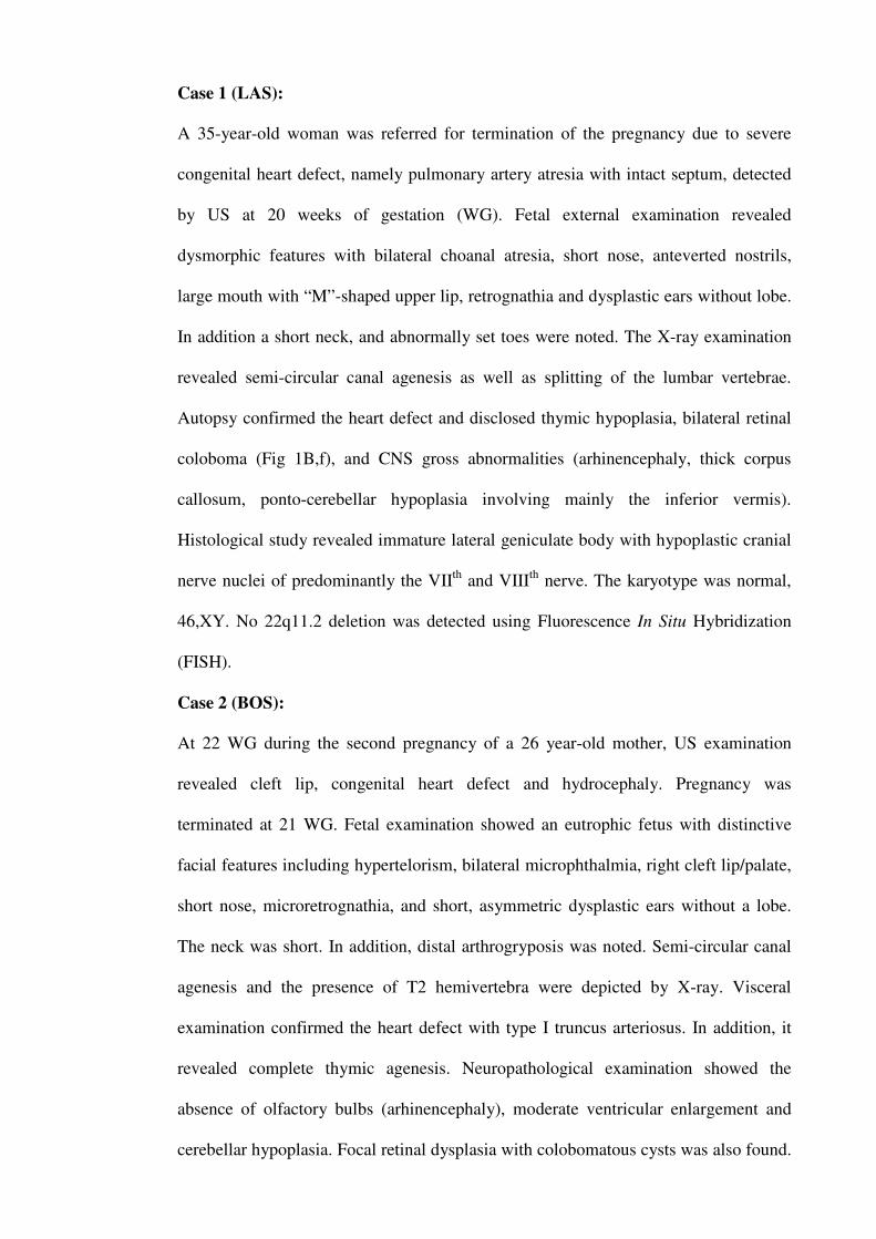
Case 1 (LAS):
A 35-year-old woman was referred for termination of the pregnancy due to severe
congenital heart defect, namely pulmonary artery atresia with intact septum, detected
by US at 20 weeks of gestation (WG). Fetal external examination revealed
dysmorphic features with bilateral choanal atresia, short nose, anteverted nostrils,
large mouth with “M”-shaped upper lip, retrognathia and dysplastic ears without lobe.
In addition a short neck, and abnormally set toes were noted. The X-ray examination
revealed semi-circular canal agenesis as well as splitting of the lumbar vertebrae.
Autopsy confirmed the heart defect and disclosed thymic hypoplasia, bilateral retinal
coloboma (Fig 1B,f), and CNS gross abnormalities (arhinencephaly, thick corpus
callosum, ponto-cerebellar hypoplasia involving mainly the inferior vermis).
Histological study revealed immature lateral geniculate body with hypoplastic cranial
nerve nuclei of predominantly the VIIth
and VIIIth
nerve. The karyotype was normal,
46,XY. No 22q11.2 deletion was detected using Fluorescence In Situ Hybridization
(FISH).
Case 2 (BOS):
At 22 WG during the second pregnancy of a 26 year-old mother, US examination
revealed cleft lip, congenital heart defect and hydrocephaly. Pregnancy was
terminated at 21 WG. Fetal examination showed an eutrophic fetus with distinctive
facial features including hypertelorism, bilateral microphthalmia, right cleft lip/palate,
short nose, microretrognathia, and short, asymmetric dysplastic ears without a lobe.
The neck was short. In addition, distal arthrogryposis was noted. Semi-circular canal
agenesis and the presence of T2 hemivertebra were depicted by X-ray. Visceral
examination confirmed the heart defect with type I truncus arteriosus. In addition, it
revealed complete thymic agenesis. Neuropathological examination showed the
absence of olfactory bulbs (arhinencephaly), moderate ventricular enlargement and
cerebellar hypoplasia. Focal retinal dysplasia with colobomatous cysts was also found.
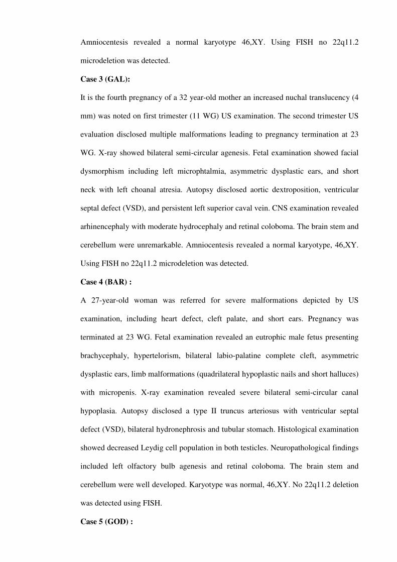
Amniocentesis revealed a normal karyotype 46,XY. Using FISH no 22q11.2
microdeletion was detected.
Case 3 (GAL):
It is the fourth pregnancy of a 32 year-old mother an increased nuchal translucency (4
mm) was noted on first trimester (11 WG) US examination. The second trimester US
evaluation disclosed multiple malformations leading to pregnancy termination at 23
WG. X-ray showed bilateral semi-circular agenesis. Fetal examination showed facial
dysmorphism including left microphtalmia, asymmetric dysplastic ears, and short
neck with left choanal atresia. Autopsy disclosed aortic dextroposition, ventricular
septal defect (VSD), and persistent left superior caval vein. CNS examination revealed
arhinencephaly with moderate hydrocephaly and retinal coloboma. The brain stem and
cerebellum were unremarkable. Amniocentesis revealed a normal karyotype, 46,XY.
Using FISH no 22q11.2 microdeletion was detected.
Case 4 (BAR) :
A 27-year-old woman was referred for severe malformations depicted by US
examination, including heart defect, cleft palate, and short ears. Pregnancy was
terminated at 23 WG. Fetal examination revealed an eutrophic male fetus presenting
brachycephaly, hypertelorism, bilateral labio-palatine complete cleft, asymmetric
dysplastic ears, limb malformations (quadrilateral hypoplastic nails and short halluces)
with micropenis. X-ray examination revealed severe bilateral semi-circular canal
hypoplasia. Autopsy disclosed a type II truncus arteriosus with ventricular septal
defect (VSD), bilateral hydronephrosis and tubular stomach. Histological examination
showed decreased Leydig cell population in both testicles. Neuropathological findings
included left olfactory bulb agenesis and retinal coloboma. The brain stem and
cerebellum were well developed. Karyotype was normal, 46,XY. No 22q11.2 deletion
was detected using FISH.
Case 5 (GOD) :
A 24-year-old woman was referred at 28 WG for heart defect, dysplastic ears,
hypoplastic thymus and vermis hypoplasia detected by US. Fetal X-ray disclosed
agenesis of semi-circular canals. The pathological examination of this eutrophic male
fetus revealed facial dysmorphism with broad nose tip, anteverted nostrils, asymmetric
small dysplastic ears, retrognathia, short neck and cleft palate. Visceral malformations
included conotruncal heart defect with aortic atresia and ventricular septal defect,
thymic agenesis, and left pulmonary isomerism. Histological examination revealed
decreased Leydig cell population. Neuropathological examination showed bilateral
arhinencephaly and inferior vermis hypoplasia. Karyotype was normal, 46,XY. No
22q11.2 deletion was detected using FISH.
Case 6 (LAN):
A 31 year-old woman was referred because of multiple fetal malformations detected
on US. The pregnancy was terminated at 29 WG for heart and brain malformations
and abnormal ears. X-ray examination revealed the absence of semi-circular canals
and a left 13th additional rib. External examination of this eutrophic male fetus
revealed brachycephaly with flat occiput, hypertelorism, hypoplastic alae nasi, malar
hypoplasia, microstomia, micrognathia, asymmetric and dysplastic ears, and short
neck. Fetal autopsy confirmed the atrio-ventricular septal defect with a right retro-
oesophageal subclavian artery, and disclosed left cryptorchidism with increased
testicular connective tissue. CNS abnormalities were bilateral agenesis of olfactory
bulbs, ventricular enlargement and posterior partial corpus callosum agenesis.
Histology demonstrated an increase of connective tissue in the testicles. Karyotype
was normal, 46,XY. No 22q11.2 deletion was detected using FISH.
Case 7 (COQ):
This was the first pregnancy of a young non-consanguineous couple (respectively 37
and 39 year-old mother and father). At 22 WG, US suspected cleft lip/palate. At 25
WG, the presence of a large facial clefting was confirmed. Additionally, bilateral renal

hypoplasia, small stomach, and a single umbilical artery were depicted. The
pregnancy was terminated at 29 WG. X-ray examination showed semi-circular canal
agenesis and the absence of ossification of the 12th
rib pair. The female fetus showed a
moderate growth retardation (10th
-20th
percentile), facial asymmetry with right
choanal atresia, left cleft lip/palate, dysplastic ears and short neck. Visceral
malformations included ectopic and hypoplastic thymus, congenital heart
malformation with atrioventricular septal defect (AVSD) and hypoplastic aortic arch,
single umbilical artery, bilateral renal hypoplasia, and small ovaries.
Neuropathological examination revealed arhinencephaly with brain stem and vermian
hypoplasia, and colobomatous retinal cysts. Karyotype was normal, 46,XX. No
22q11.2 deletion was detected using FISH.
Case 8 (BEL) :
This case is the first pregnancy of a 31 year-old mother. First trimester (11 WG) US
detected fetal hygroma. At the second trimester, US as well as Magnetic Resonance
Imaging (MRI) showed multiple malformations including cleft lip, thymic hypoplasia,
bilateral microphthalmia and heart defect. Pregnancy was terminated at 29 WG. X-ray
examination of the fetus showed severe hypoplasia of the semi-circular canals. Fetal
examination showed an eutrophic male fetus presenting bilateral choanal atresia,
blepharophimosis, an asymmetric microphthalmia (right > left) with right
microcornea, asymmetric dysplastic ears and phimosis. Visceral malformations
included right heart hypoplasia, atretic pulmonary artery with an intact septum, thymic
hypoplasia and absent ossification of the 12th
rib. Neuropathological examination
revealed bilateral retinal coloboma and arhinencephaly with a well-developed
brainstem and cerebellum but severe cerebellar heterotopias. The karyotype was
normal, 46,XY. No 22q11.2 deletion was detected using FISH.
Case 9 (RAB):
This was the second pregnancy of a young non-consanguineous couple (28 year-old
parents). A previous spontaneous abortion occurred at 12 WG (unknown cause). US at
24 WG revealed short nasal bones, frontal oedema, and suspected heart malformation,
which was confirmed by US at 32 WG. In addition, retrognathia and polyhydramnios
were observed. The pregnancy was terminated. Cranial X-ray examination revealed
semi-circular canal agenesis. Fetal examination disclosed bilateral choanal atresia and
craniofacial dysmorphic features including microcephaly with small anterior fontanel,
frontal bossing, hypertelorism, broad nasal bridge, large mouth with “M” shaped
upper lip, microretrognathia, and abnormal external ears. In addition, upper limb
malformations with bilateral ectrodactyly were observed (Fig. 1C). Visceral
malformations included Fallot’s tetralogy with a right retro-oesophageal subclavian
artery, severe thymic hypoplasia, abnormal left pulmonary segmentation, oesophageal
atresia (type III), and a small supernumerary spleen. Neuropathological examination
showed the absence of olfactory bulbs. Brain stem and cerebellum were
unremarkable. The karyotype was normal, 46,XX. No 22q11.2 deletion was detected
using FISH.
Case 10 (DER) :
In a 31-year-old woman, severe polyhydramnios, vermis hypoplasia, thin brainstem
and ventricular dilatation were detected on US examination during the second
trimester of the pregnancy. Brain malformations were confirmed by MRI and the
pregnancy was terminated at 36 WG. Fetal examination showed a female fetus with a
weight between the 3rd
and the 10th
percentile. Length and head circumference were
around the 50th
percentile. Hypoplasia of the semi-circular canals was the only
significant radiologic feature (Fig 1B,b). The fetus presented bilateral choanal atresia,
asymmetric dysplastic ears and retrognathia. Visceral malformations included thymic
hypoplasia, ventricular disequilibrium with right retro-oesophageal subclavian artery,
oesophageal type III atresia with tracheo-oesophageal fistula and left hydronephrosis.

Neuropathological examination revealed arhinencephaly (Fig 1B,d) and retinal
coloboma. The brain stem and cerebellum were well developed, except for an ectopic
olivary nucleus in the medulla. Karyotype was normal, 46,XX. No 22q11.2 deletion
was detected using FISH.
Copyright © 2022 FDOKUMEN