Pharmacology for students of bachelor's programmes at MF ...
277
Pharmacology for students of bachelor’s programmes at MF MU (special part) Alena MÁCHALOVÁ, Zuzana BABINSKÁ, Jan JUŘICA, Gabriela DOVRTĚLOVÁ, Hana KOSTKOVÁ, Leoš LANDA, Jana MERHAUTOVÁ, Kristýna NOSKOVÁ, Tibor ŠTARK, Katarína TABIOVÁ, Jana PISTOVČÁKOVÁ, Ondřej ZENDULKA Text přeložili: MVDr. Mgr. Leoš Landa, Ph.D., MUDr. Máchal, Ph.D., Mgr. Jana Merhautová, Ph.D., MUDr. Jana Pistovčáková, Ph.D., Doc. PharmDr. Jana Rudá, Ph.D., Mgr. Barbora Říhová, Ph.D., PharmDr. Lenka Součková, Ph.D., PharmDr. Ondřej Zendulka, Ph.D., Mgr. Markéta Strakošová, Mgr. Tibor Štark, Jazyková korektura: Julie Fry, MSc.
-
Upload
khangminh22 -
Category
Documents
-
view
0 -
download
0
Transcript of Pharmacology for students of bachelor's programmes at MF ...
Pharmacology for students of
bachelor’s programmes at MF MU
(special part)
Alena MÁCHALOVÁ, Zuzana BABINSKÁ, Jan JUŘICA, Gabriela DOVRTĚLOVÁ, Hana KOSTKOVÁ, Leoš
LANDA, Jana MERHAUTOVÁ, Kristýna NOSKOVÁ, Tibor ŠTARK, Katarína TABIOVÁ, Jana
PISTOVČÁKOVÁ, Ondřej ZENDULKA
Text přeložili: MVDr. Mgr. Leoš Landa, Ph.D., MUDr. Máchal, Ph.D., Mgr. Jana Merhautová, Ph.D.,
MUDr. Jana Pistovčáková, Ph.D., Doc. PharmDr. Jana Rudá, Ph.D., Mgr. Barbora Říhová, Ph.D.,
PharmDr. Lenka Součková, Ph.D., PharmDr. Ondřej Zendulka, Ph.D., Mgr. Markéta Strakošová, Mgr.
Tibor Štark,
Jazyková korektura: Julie Fry, MSc.
2
Obsah
2 Pharmacology of the autonomic nervous system ........................................................................ 10
2.1 Pharmacology of the sympathetic nervous system ................................................................... 10
2.1.1 Sympathomimetics ............................................................................................................... 12
2.1.2 Sympatholytics ..................................................................................................................... 17
2.2 Pharmacology of the parasympathetic system ......................................................................... 19
2.2.1 Cholinomimetics .................................................................................................................. 20
2.2.2 Parasympathomimetics ......................................................................................................... 21
2.2.3 Parasympatholytics ............................................................................................................... 21
3 Psychotropic substances, drug dependence ................................................................................. 23
3.1 Psychotropic substances ............................................................................................................ 23
3.1.1 Treatment of affective disorders (antidepressant drugs) ......................................................... 23
3.1.2 Treatment of anxiety disorders (anxiolytic drugs) ................................................................. 25
3.1.3 Treatment of sleep disorders (hypnosedatives) ...................................................................... 25
3.1.4 Treatment of mental integration disorders (antipsychotic drugs) ............................................ 26
3.1.5 Drugs affecting memory and cognitive functions .................................................................. 27
3.2 Drug dependence..................................................................................................................... 27
4. Substances affecting respiratory system..................................................................................... 30
4.1 Antiasthmatics ........................................................................................................................... 30
4.1.1 Glucocorticoids .................................................................................................................... 32
4.1.2 β2-sympathomimetics........................................................................................................... 33
4.1.3 Parasympatholytics (muscarinic antagonists) ........................................................................ 34
4.1.4 Methylxanthines ................................................................................................................... 35
4.1.5 Mast cells stabilizers (immunoprophylactics) ........................................................................ 35
4.1.6 Antileukotrienes ................................................................................................................... 36
4.1.7 Targeted antiasthmatic therapy ............................................................................................. 36
4.2 Cough medicines ....................................................................................................................... 36
4.2.1 Antitussives .......................................................................................................................... 37
4.2.2 Expectorants ......................................................................................................................... 38
4.2.3 Other expectorant agents ...................................................................................................... 39
5. Substances used in cardiology and haemathology ..................................................................... 41
5.1 Antihypertensive drugs, diuretics ............................................................................................. 41
5.1.1 Pharmaceuticals affecting the renin-angiotensin-aldosterone system ..................................... 42
5.1.2 Calcium channel blockers (CCB) .......................................................................................... 43
5.1.3 Diuretics............................................................................................................................... 44
5.1.4 Pharmaceuticals inhibiting the sympathetic nervous system (sympatholytics) ....................... 46
5.1.5. Central sympatholytics ........................................................................................................ 47
5.2 Haemostasis-affecting drugs ..................................................................................................... 47

3
5.2.1 Anticoagulant drugs ............................................................................................................. 48
5.2.2 Antiplatelet drugs ................................................................................................................. 51
5.2.3 Fibrinolytic drugs ................................................................................................................. 52
5.2.4 Pro-haemostatic drugs .......................................................................................................... 53
5.3 Antiarrhythmics ........................................................................................................................ 54
5.4 Cardiotonics (positive inotropes) .............................................................................................. 58
5.4.1 Cardiac (digitalis) glycosides................................................................................................ 58
5.4.2 Phosphodiesterase III inhibitors ............................................................................................ 59
5.4.3 Calcium sensitisers ............................................................................................................... 59
5.4.4 Sympathomimetics ............................................................................................................... 59
5.5 Direct-acting vasodilators ......................................................................................................... 59
5.5.1 Nitrovasodilators .................................................................................................................. 59
5.5.2 Other direct vasodilators ....................................................................................................... 61
5.6 Treatment of the ischaemic heart disease ................................................................................. 61
5.6.1 Pharmacological treatment of chronic IHD ........................................................................... 62
5.6.2 Pharmacological treatment of the acute coronary .................................................................. 63
5.7 Treatment of heart failure ........................................................................................................ 63
5.7.1 Acute heart failure ................................................................................................................ 64
5.7.2 Chronic heart failure ............................................................................................................. 64
5.8 The treatment of anaemias and haematopoiesis defects .......................................................... 65
5.8.1 Anaemia due to deficiency of haematinic agents ................................................................... 65
5.8.2 Erythropoiesis defects which are not linked to deficiency of nutrients ................................... 67
6. Medications for Gastrointestinal Disorders ............................................................................... 69
6.1 Drugs used for treatment of gastric ulcers (antiulcer drugs) ................................................... 69
6.1.1 Proton pump inhibitors (PPI) ................................................................................................ 69
6.1.2 H2 antihistamines.................................................................................................................. 70
6.1.3 Selective parasympatholytics ................................................................................................ 70
6.1.4 Antacids ............................................................................................................................... 71
6.1.5 Mucosal protectants .............................................................................................................. 71
6.1.6 Eradication of Helicobacter pylori ........................................................................................ 72
6.2 Prokinetic agents ....................................................................................................................... 72
6.3 Spasmolytics of the gastrointestinal tract ................................................................................. 73
6.3.1 Neurotropic spasmolytics ..................................................................................................... 74
6.3.2 Musculotropic spasmolytics.................................................................................................. 74
6.4 Antiemetics ................................................................................................................................ 75
6.4.1 Muscarinic receptor antagonists ............................................................................................ 75
6.4.2 H1 receptor antagonists ......................................................................................................... 76
6.4.3 D2 receptor antagonists ......................................................................................................... 76
6.4.5 NK1 receptor antagonists...................................................................................................... 77

4
6.4.6 Other drugs .......................................................................................................................... 77
6.5 Laxatives.................................................................................................................................... 78
6.5.1 Volume (bulk) laxatives ....................................................................................................... 78
6.5.2 Salinic laxatives ................................................................................................................... 78
6.5.3 Osmotic laxatives ................................................................................................................. 79
6.5.4 Stimulant laxatives ............................................................................................................... 79
6.5.6 Peripheral opioid receptor antagonists................................................................................... 79
6.6 Anti-diarrhoeal agents .............................................................................................................. 80
6.6.1 Intestinal adsorbents ............................................................................................................. 80
6.6.2 Intestinal antiseptics ............................................................................................................. 80
6.6.3 Antipropulsives (opioids) ..................................................................................................... 81
6.6.4 Other anti-diarrhoeal agents .................................................................................................. 81
7. Treatment of metabolic disorders and pharmacology of endocrine system .............................. 83
7.1 Treatment of diabetes mellitus ................................................................................................. 83
7.1.1 Insulin therapy...................................................................................................................... 84
7.1.2 Oral and new antidiabetic drugs ............................................................................................ 86
7.1.3 Treatment of acute coma related to diabetes .......................................................................... 91
7.2 Treatment of dyslipidaemias..................................................................................................... 91
7.2.1 Drugs for treatment of dyslipidaemias .................................................................................. 93
7.2.2 Indications of lipid-lowering drugs ....................................................................................... 94
7.3 Pharmacotherapy of obesity ..................................................................................................... 95
7.3.1 Centrally-acting obesity medications .................................................................................... 96
7.3.2 Peripheral obesity medications ............................................................................................. 97
7.3.3 Pharmacotherapy of eating disorders .................................................................................... 98
7.4 Pharmacology of hypothalamic and hypophyseal hormones ................................................... 99
7.4.1 Hypothalamic hormones ....................................................................................................... 99
7.4.2 Hormones of anterior pituitary ............................................................................................ 100
7.4.3 Hormones of neurohypophysis ........................................................................................... 102
7.5 Pharmacology of adrenocortical hormones ............................................................................ 103
7.6 Pharmacotherapy of thyroid diseases ..................................................................................... 104
7.6.1 Therapy of hypothyroidism................................................................................................. 104
7.6.2 Therapy of hyperthyroidism ............................................................................................... 105
7.6.3 Therapy of thyroid storm .................................................................................................... 106
7.7 Pharmacology of bone metabolism ......................................................................................... 106
7.7.1 Physiological regulation of bone metabolism ...................................................................... 106
7.7.2 Bone metabolism disorders ................................................................................................. 107
7.8 Sex hormones .......................................................................................................................... 108
7.8.1 Sex hormones and their derivatives ..................................................................................... 108
7.8.2 Hormonal Contraception .................................................................................................... 115

5
7.8.3 Hormone replacement therapy (HRT) ................................................................................. 117
8 Substances affecting pain, inflammation and functions of the immune system ....................... 119
8.1 Local anaesthetics ................................................................................................................... 119
Side effects ................................................................................................................................. 119
8.1.1 Local anaesthesia techniques .............................................................................................. 120
8.1.2 Classification of local anaesthetics ...................................................................................... 121
8.2 General anaesthetics ............................................................................................................... 122
8.2.1 Stages of general anaesthesia .............................................................................................. 122
8.2.2 Premedication before the general anaesthesia ...................................................................... 123
8.2.3 Inhalational anaesthetics ..................................................................................................... 123
8.2.4 Injection anaesthetics.......................................................................................................... 124
8.2.5 Complications of general anaesthesia .................................................................................. 124
8.3 Opioid analgesic drugs (anodynes) ......................................................................................... 125
8.3.1 Effects of opioid analgesics ................................................................................................ 125
8.3.2 Classification of opioid analgesics ...................................................................................... 126
8.4 Non-opioid analgesic drugs ..................................................................................................... 128
8.4.1 Non-steroidal anti-inflammatory drugs (NSAIDs) ............................................................... 128
8.4.2 Analgesics-antipyretics (AA) .............................................................................................. 132
8.4.3 Analgesic ladder ................................................................................................................. 133
8.5 Glucocorticoids........................................................................................................................ 133
8.5.1 Effects of glucocorticoids ................................................................................................... 134
8.5.2 Adverse effects ................................................................................................................... 135
8.5.3 Indications and dosage schemes .......................................................................................... 136
8.6 Therapy of allergies................................................................................................................. 137
8.6.1 Pharmacotherapy of allergies .............................................................................................. 138
8.6.2 H1 antihistamines................................................................................................................ 138
8.7 Immunomodulatory substances .............................................................................................. 141
8.7.1 Immunosuppressants .......................................................................................................... 141
8.7.2 Immunostimulants .............................................................................................................. 143
9 Anti-infective drugs.................................................................................................................... 145
9.1 Disinfectants and antiseptics ................................................................................................... 145
9.1.1 Heavy metals and their derivatives...................................................................................... 145
9.1.2 Oxidisers ............................................................................................................................ 146
9.1.3 Alcohols and phenols ......................................................................................................... 147
9.1.4 Aldehydes .......................................................................................................................... 147
9.1.5 Boric acid and borates ........................................................................................................ 148
9.1.6 Quaternary ammonium salts ............................................................................................... 148
9.1.7 Antiseptic dyes ................................................................................................................... 148
9.1.8 Oral antiseptics ................................................................................................................... 149

6
9.2 Fundamentals of the rational therapy of infectious diseases ................................................. 149
9.2.1 Mechanisms of action of antibiotic resistance ..................................................................... 151
9.3 Antibiotics ............................................................................................................................... 152
9.3.1 β-lactam antibiotics ............................................................................................................ 152
9.3.2 Other antibiotics interfering with cellular packaging ........................................................... 155
9.3.3 Antibiotics inhibiting protein synthesis ............................................................................... 156
9.3.4 New antibiotics similar to macrolides and tetracyclines....................................................... 158
9.3.6 Aminoglycosides ................................................................................................................ 159
9.3.7 Antibiotics for a local use ................................................................................................... 160
9.4. Anti-infectious chemotherapeutics ........................................................................................ 161
9.4.1 Sulfonamides...................................................................................................................... 161
9.4.2 Pyrimidines ........................................................................................................................ 162
9.4.3 Quinolones ......................................................................................................................... 162
9.4.4 Nitrofurans ......................................................................................................................... 163
9.4.5 Nitroimidazoles .................................................................................................................. 163
9.5 Antimycotics ............................................................................................................................ 163
9.5.1 Polyene antimycotics .......................................................................................................... 164
9.5.2 Azole antimycotics ............................................................................................................. 164
9.5.3 Echinocandins .................................................................................................................... 165
9.5.4 Other antimycotics ............................................................................................................. 165
9.6 Antiviral drugs ........................................................................................................................ 166
9.6.1 Drugs for herpes viruses ..................................................................................................... 167
9.6.2 Influenza treatment ............................................................................................................. 169
9.6.3 Antiretroviral therapy ......................................................................................................... 170
9.6.4 Drugs used to treat chronic viral hepatitis and other antiviral drugs ..................................... 173
10 Cancer treatment, principles of targeted therapy ................................................................... 174
10.1 Principles of cancer treatment .............................................................................................. 174
10.2 Principles of chemotherapy................................................................................................... 174
10.2.1 Mechanism of action of cytostatic agents .......................................................................... 175
10.2.2 Toxicity of chemotherapy, adverse effects of cytostatic agents, and their management ...... 176
10.3 Overview of cytostatic agents ................................................................................................ 181
10.3.1 DNA-damaging drugs....................................................................................................... 181
Bis(2-chloroethyl)amines .............................................................................................................. 181
Oxazaphosphorines ....................................................................................................................... 182
Nitrosourea derivatives ................................................................................................................. 182
Triazenes ....................................................................................................................................... 182
Alkyl sulfonates ............................................................................................................................. 182
Aziridines ...................................................................................................................................... 182
10.3.2 Drugs that inhibit key enzymes of DNA metabolism ......................................................... 184
7
Purine analogues ........................................................................................................................... 185
Pyrimidine analogues .................................................................................................................... 185
Folic acid analogues ...................................................................................................................... 186
Hydroxyurea ................................................................................................................................. 186
Topoisomerase I inhibitors – camptothecins ................................................................................ 187
Topoisomerase II inhibitors – podophyllotoxins .......................................................................... 187
10.3.3 Drugs that alter microtubules ............................................................................................ 187
10.3.4 Others .............................................................................................................................. 188
Drugs that inhibit protein synthesis – L-asparaginase ................................................................ 188
10.4 Immunomodulatory agents used to treat cancer diseases .................................................... 188
10.5 Endocrine therapy of cancer diseases ................................................................................... 189
10.6 Principles of targeted therapy ............................................................................................... 190
11 Drugs used in gynaecology, pharmacotherapy in pregnancy ................................................. 195
11.1 Uterotonics ............................................................................................................................ 195
Drugs inducing rhythmical uterine contractions ........................................................................... 195
Drugs inducing tonic contractions ............................................................................................... 196
11.2 Tocolytics ............................................................................................................................... 197
β2 sympathomimetics .................................................................................................................. 197
Oxytocin receptor antagonists ..................................................................................................... 198
Others ......................................................................................................................................... 198
11.3 Therapy of vulvovaginal infections ....................................................................................... 199
11.3.1 Trichomoniasis ................................................................................................................. 199
11.3.2 Bacterial vaginosis ........................................................................................................... 199
11.3.3 Mycotic vaginitis .............................................................................................................. 200
11.3.4 Lactobacillosis.................................................................................................................. 200
11.3.5 Aerobic vaginitis .............................................................................................................. 200
11.3.6 Herpes genitalis (genital herpes) ....................................................................................... 201
11.3.7 Atrophic vaginitis ............................................................................................................. 201
11.3.8 Other causes of vulvovaginal discomfort .......................................................................... 202
11.4 Pharmacotherapy in pregnancy and lactation ..................................................................... 202
11.4.1 Anxiolytics and hypnosedatives ........................................................................................ 202
11.4.2 Antiemetics ...................................................................................................................... 203
11.4.3 Pharmacotherapy of hypertension and dyslipidaemia ........................................................ 203
11.4.4 Pharmacology of diabetes mellitus .................................................................................... 204
12 Drugs affecting functions of musculoskeletal system .............................................................. 206
12.1. Muscle relaxants (neuromuscular blocking drugs) ............................................................. 206
12.2 Treatment of Parkinson’s disease ......................................................................................... 208
12.2.1 Substitution of lacking dopamine ...................................................................................... 208
12.2.2 Agonists of dopamine receptors ........................................................................................ 209

8
12.2.3 Inhibition of dopamine degradation .................................................................................. 209
12.2.4 Restoring the balance between dopamine and acetylcholine .............................................. 210
12.3 Drug-induced extrapyramidal symptoms and iatrogenic parkinsonism ............................. 210
12.4 The treatment of chorea ........................................................................................................ 211
12.5 The treatment of spasticity and cerebral palsy .................................................................... 212
12.6 The treatment of myasthenia gravis ..................................................................................... 214
12.7 Pharmacotherapy of Ménière’s disease ................................................................................ 216
12.8 Rheumatoid diseases and gout treatment ............................................................................. 217
12.8.1 Treatment of rheumatoid arthritis ...................................................................................... 217
12.8.1.1 Disease-modifying anti-rheumatic drugs (DMARDs) ..................................................... 217
12.8.1.2 Targeted therapy ............................................................................................................ 218
12.8.1.3 Nonsteroidal anti-inflammatory drugs (NSAIDs) ........................................................... 219
12.8.1.4 Glucocorticoids ............................................................................................................. 219
12.8.2 Treatment of gout ............................................................................................................. 219
13 Treatment of urgent conditions ............................................................................................... 221
13.1 General principles of intoxication treatment ........................................................................ 221
13.1.1 Common causes of intoxications ....................................................................................... 221
13.1.2 Diagnosis of intoxications................................................................................................. 222
13.1.3 Treatment of intoxications ................................................................................................ 222
13.2 Therapy of the shock states ................................................................................................... 225
13.2.1 The volume therapy .......................................................................................................... 227
13.2.2 Blood substitutes .............................................................................................................. 228
13.2.3 Inotropics and vasodilatation ............................................................................................ 229
13.3 Pharmacotherapy for pre-hospital care in patients with polytrauma ................................. 230
14 Radiopharmaceuticals and radiocontrast agents .................................................................... 233
14.1 Radiopharmaceuticals ........................................................................................................... 233
14.1.1 Classification of radiopharmaceuticals .............................................................................. 233
14.1.2 Diagnostic radionuclides (radiotracers) ............................................................................. 234
14.2 Radiocontrast agents ............................................................................................................. 235
14.2.1 Positive contrast media ..................................................................................................... 235
14.2.2 Negative contrast media.................................................................................................... 236
14.2.3 Contraindications and side effects ..................................................................................... 236
14.2.4 Application of radiocontrast agents ................................................................................... 237
15 Phytopharmacology ................................................................................................................. 238
15.1 Introduction to phytopharmacology ..................................................................................... 238
15.2 Herbal substances.................................................................................................................. 239
15.2.1 Carbohydrates .................................................................................................................. 240
15.2.2 Polyketides ....................................................................................................................... 240
15.2.3 Alkaloids .......................................................................................................................... 241

9
15.2.4 Shikimates ........................................................................................................................ 243
15.2.5 Mevalonates ..................................................................................................................... 244
15.3 Self-treatment of selected diseases with phytotherapy ......................................................... 246
15.4 Interactions of phytotherapeutics with co-administered drugs ........................................... 251
16 Drugs used in dentistry ............................................................................................................ 254
16.1 Drugs used in prevention and treatment of diseases in dental hard tissues ........................ 254
16.1.1 Enhancement of enamel and dental resistance ................................................................... 254
16.1.2 Dental desensitisation ....................................................................................................... 255
16.1.3 Treatment of dental pulp and root canals ........................................................................... 256
16.2 Drugs used to treat oral mucosal and periodontal disease ................................................... 256
16.3 Drugs used in salivary disorders ........................................................................................... 259
16.4 Herbal therapy in dentistry .................................................................................................. 260
Table 15: Medicinal plants and their use in dentistry ................................................................. 261
Galls on Quercus robur L. .......................................................................................................... 261
17 Ophthalmologic drugs.............................................................................................................. 263
17.1 Antimicrobial substances ...................................................................................................... 263
Antibiotics and chemotherapeutics .............................................................................................. 263
Virostatics................................................................................................................................... 264
Antimycotics............................................................................................................................... 264
Antiparasitics .............................................................................................................................. 264
Antiseptics .................................................................................................................................. 264
17.2 Anti-inflammatory, anti-allergic and immunosuppressive drugs ........................................ 264
17.3 Substances for treatment of ocular vascular disorders ........................................................ 265
17.4 Anti-glaucoma and miotic drugs ........................................................................................... 266
17.4.1. Substances reducing production of intraocular fluid ......................................................... 267
17.4.2 Substances affecting outflow of intraocular fluid .............................................................. 267
17.4.3 Substances with osmotic effect – hyperosmotics ............................................................... 268
17.5 Mydriatics and cyclophlegics ................................................................................................ 268
17.6 Other ophthalmologic drugs ................................................................................................. 269
17.6.1 Decongestion drugs .......................................................................................................... 269
17.6.2 Local anaesthetics............................................................................................................. 269
17.6.3 Diagnostics ....................................................................................................................... 269
17.6.4 Tear substitution in case of dry eye syndrome ................................................................... 270
17.6.5 Other ophthalmologic drugs.............................................................................................. 270

10
2 Pharmacology of the autonomic nervous system
The autonomic nervous system (ANS) is responsible for signal transduction between the
central nervous system (CNS) and effector tissues or organs independent of voluntary control
(e.g., smooth muscles, myocardium, exocrine glands, etc.) and regulates adaptive reactions of
the organism according to changes in the exogenous and endogenous environments.
Among the principal processes controlled by the ANS belong the contraction and relaxation
of smooth muscles, secretion from all exocrine and some endocrine glands, heart action, some
metabolic processes, and immune system functions. The ANS can be traditionally divided
into three main branches: the sympathetic, parasympathetic, and enteric nervous system.
Many physiological functions are influenced by the sympathetic and parasympathetic systems
in a contradictory manner, which is called physiological antagonism, such as their effect on
heart activity. Nevertheless, for some body functions, the synergism of both systems (i.e.,
sympathetic and parasympathetic) is required, for example in the regulation of gonadal system
functions.
2.1 Pharmacology of the sympathetic nervous system
The sympathetic nervous system (SNS) has a role in controlling main life functions through
its basal activity as well as in accordance with the need of the body to adapt to an acute
stressful event. Following exposure to a stressor (e.g., a tiger roaring toward us from a few
metres away), there is an immediate release of catecholamines from the adrenal medulla, and
in a few seconds, the body is activated for the “fight or flight” reaction. Activation of the
sympathetic system is essential for the maintenance of homeostasis and preparation of the
organism for physical activity or coping with an injury.
Under baseline conditions, noradrenaline is produced in nerve terminals, which is the
predominant status physiologically; whereas in a “fight or flight” situation, the activation of
the sympathetic receptors is further stimulated by adrenaline and noradrenaline released from
the adrenal medulla. These mediators bind to the adrenergic receptors, which can be divided
into five major subclasses: α1, α2, β1, β2, and β3 (see Tab. 4).
The consequences of adrenergic receptor activation can be rationally derived from demands
on the organism when exposed to a life-threatening situation in which a quick response is
required; emphasis is put on a sufficient supply of oxygen and energy sources for the heart,
lungs, brain, and muscles (provided by an increase in blood pressure, bronchodilation,
vasoconstriction in peripheral organs and skin, and limitation of kidney blood perfusion).
Further, stimulation of the process of platelet clotting and immune system response as well as
increased alertness and accommodation of the eye to distance occurs. Many physiological
functions (e.g., digestion) are less important in a situation of sudden physical activity. They
are inhibited and postponed for later management. Energy is saved for the organism to
maintain its basic life functions for survival.
11
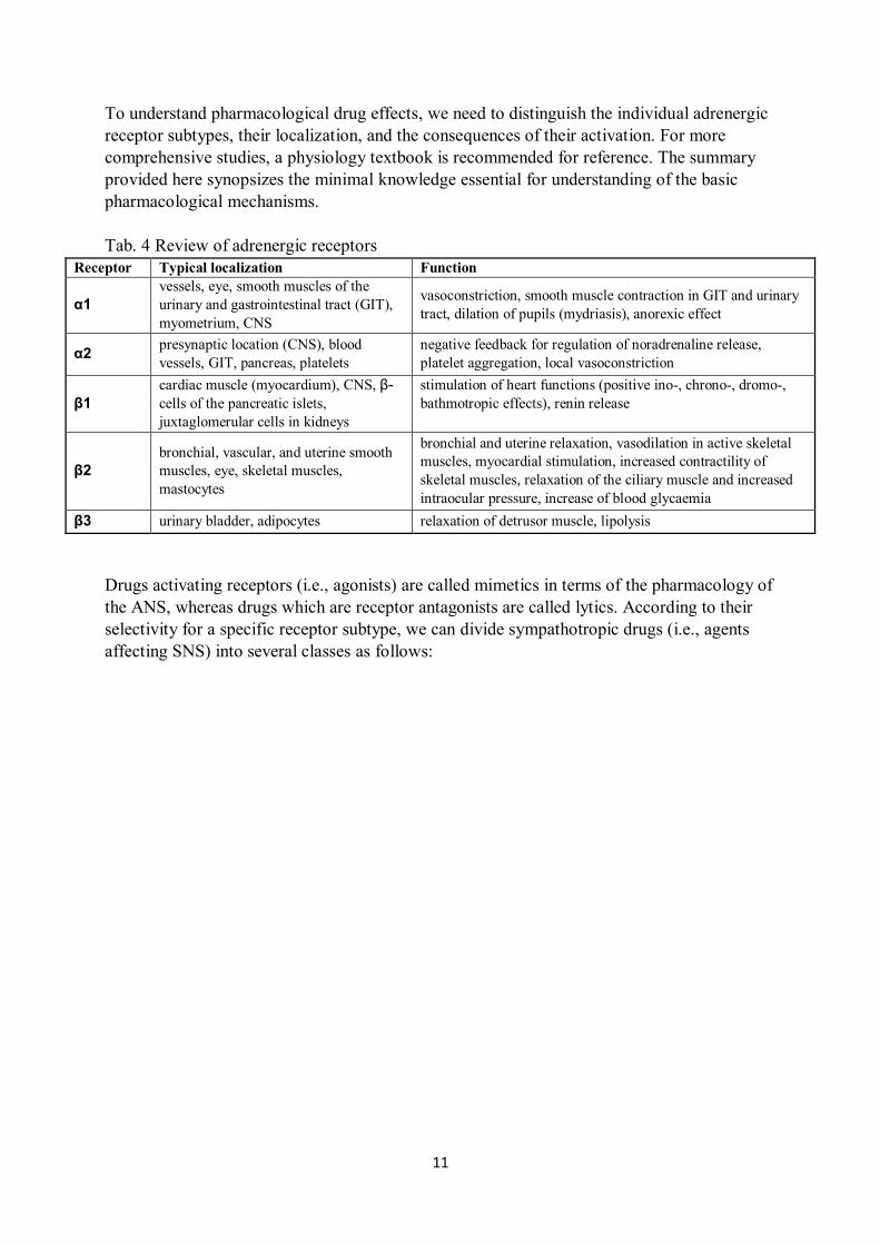
To understand pharmacological drug effects, we need to distinguish the individual adrenergic
receptor subtypes, their localization, and the consequences of their activation. For more
comprehensive studies, a physiology textbook is recommended for reference. The summary
provided here synopsizes the minimal knowledge essential for understanding of the basic
pharmacological mechanisms.
Tab. 4 Review of adrenergic receptors
Receptor Typical localization Function
α1
vessels, eye, smooth muscles of the
urinary and gastrointestinal tract (GIT),
myometrium, CNS
vasoconstriction, smooth muscle contraction in GIT and urinary
tract, dilation of pupils (mydriasis), anorexic effect
α2 presynaptic location (CNS), blood
vessels, GIT, pancreas, platelets
negative feedback for regulation of noradrenaline release,
platelet aggregation, local vasoconstriction
β1
cardiac muscle (myocardium), CNS, β-
cells of the pancreatic islets,
juxtaglomerular cells in kidneys
stimulation of heart functions (positive ino-, chrono-, dromo-,
bathmotropic effects), renin release
β2
bronchial, vascular, and uterine smooth
muscles, eye, skeletal muscles,
mastocytes
bronchial and uterine relaxation, vasodilation in active skeletal
muscles, myocardial stimulation, increased contractility of
skeletal muscles, relaxation of the ciliary muscle and increased
intraocular pressure, increase of blood glycaemia
β3 urinary bladder, adipocytes relaxation of detrusor muscle, lipolysis
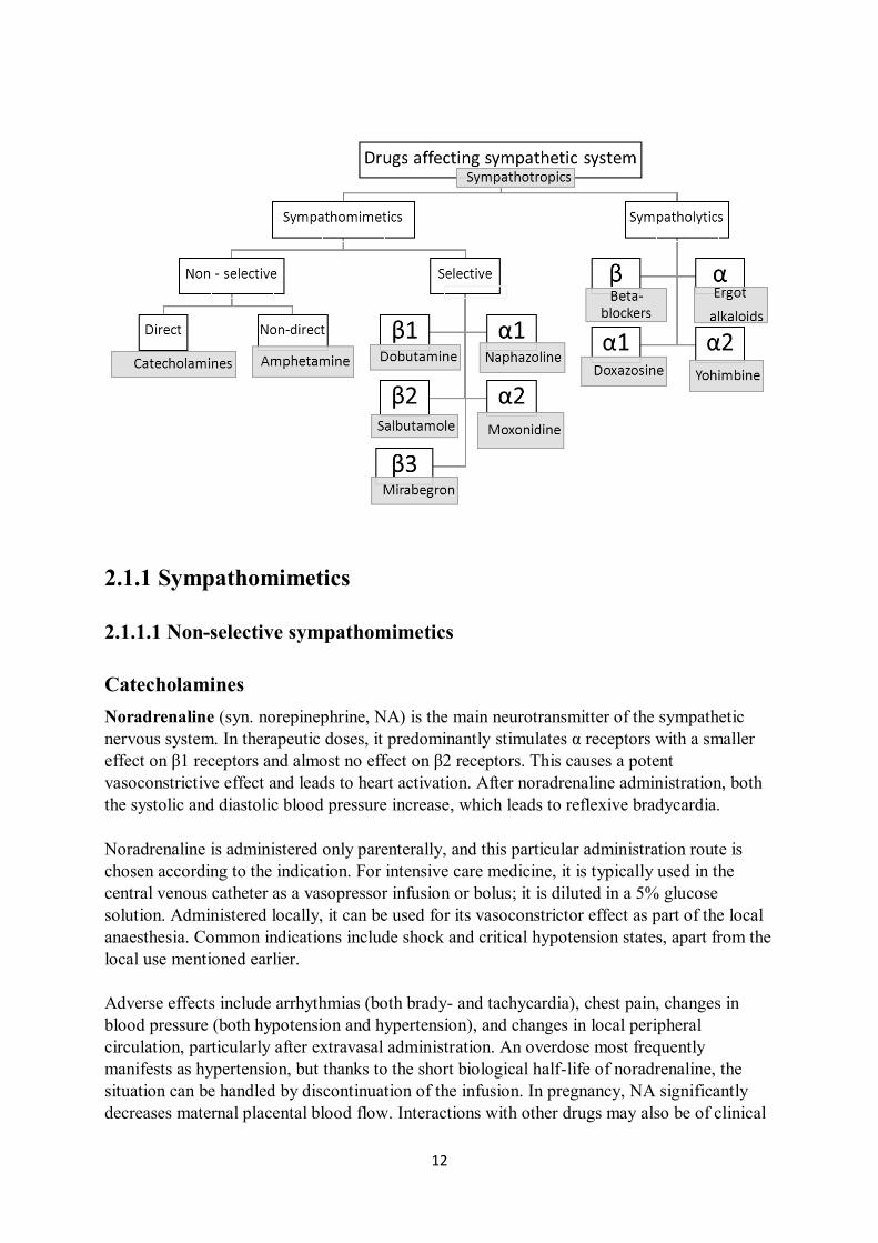
Drugs activating receptors (i.e., agonists) are called mimetics in terms of the pharmacology of
the ANS, whereas drugs which are receptor antagonists are called lytics. According to their
selectivity for a specific receptor subtype, we can divide sympathotropic drugs (i.e., agents
affecting SNS) into several classes as follows:
12
2.1.1 Sympathomimetics
2.1.1.1 Non-selective sympathomimetics
Catecholamines
Noradrenaline (syn. norepinephrine, NA) is the main neurotransmitter of the sympathetic
nervous system. In therapeutic doses, it predominantly stimulates α receptors with a smaller
effect on β1 receptors and almost no effect on β2 receptors. This causes a potent
vasoconstrictive effect and leads to heart activation. After noradrenaline administration, both
the systolic and diastolic blood pressure increase, which leads to reflexive bradycardia.
Noradrenaline is administered only parenterally, and this particular administration route is
chosen according to the indication. For intensive care medicine, it is typically used in the
central venous catheter as a vasopressor infusion or bolus; it is diluted in a 5% glucose
solution. Administered locally, it can be used for its vasoconstrictor effect as part of the local
anaesthesia. Common indications include shock and critical hypotension states, apart from the
local use mentioned earlier.
Adverse effects include arrhythmias (both brady- and tachycardia), chest pain, changes in
blood pressure (both hypotension and hypertension), and changes in local peripheral
circulation, particularly after extravasal administration. An overdose most frequently
manifests as hypertension, but thanks to the short biological half-life of noradrenaline, the
situation can be handled by discontinuation of the infusion. In pregnancy, NA significantly
decreases maternal placental blood flow. Interactions with other drugs may also be of clinical

13
relevance; especially risky interactions are those with other sympathomimetics, halothane,
and tricyclic antidepressants. Contrarily, sympatholytics can inhibit effects of noradrenaline.
Adrenaline (syn. epinephrine), compared with NA, has a different affinity to adrenergic
receptors. At low doses, it stimulates α1 receptors, and at higher doses, its vasoconstrict ive
effect is comparable with noradrenaline. Likewise, at higher doses, it also significantly
stimulates β receptors (including β2 receptor subtypes, which are almost unaffected by NA).
Thus, the effects of adrenaline differ from those of noradrenaline, and the difference is
thought to be due to β2 receptor activation. Therefore, effects of noradrenaline and adrenaline
are similar in many organs, however they are different in tissues with a high density of β2
receptors.
Heart activity is stimulated by adrenaline; specifically, the rise in systolic blood pressure and
heart rate frequency is significant, whereas the diastolic blood pressure can drop after a low
dose as increased peripheral vascular resistance due to vasoconstriction resulting from α1
receptor stimulation manifests only at higher doses. Contrarily, vasodilation occurs in other
tissues (e.g., due to β2 receptor effects, this especially occurs in cardiac and pulmonary tissues
as well as the blood vessels of active skeletal muscles). Importantly, effects on
bronchodilation are mediated via a direct effect on β2 receptors in the respiratory tract.
Adrenaline also has anti-allergic and anti-anaphylactic effects. These effects account for how
the blood circulation, respiratory tract, and immune system are modulated (i.e., inhibition of
histamine release from mast cells). Adrenaline also supports metabolic processes leading to
increased glycaemia (i.e., increased glucose plasma levels, especially due to the hepatic
glycogenolysis). Adrenaline also activates platelets. Its antispasmodic effect on the
myometrium (i.e., uterine smooth muscle) is not negligible either.
Adrenaline, like noradrenaline, is administered exclusively parenterally. Among the most
important indications belong asystole, peripheral circulatory collapse, anaphylaxis, septic
shock (i.e., endotoxin shock), bronchospasm, and Quincke's oedema, and after topical
administration, it can be used as a nasal decongestant, to reduce capillary bleeding, or
combined with local anaesthetics for its vasoconstrictive effects. Side effects of adrenaline are
similar to those of NA.
Dopamine differs from noradrenaline and adrenaline by its affinity to dopamine receptors,
indicated as D1-D5. In the organism, it acts preferentially as a neurotransmitter and is
synthesized in a large amount in the CNS. However, it is present in different concentrations in
many other tissues in the entire body.
Dopamine effects are dependent on the dose administered:
At low doses, it influences renal D receptors and induces vasodilation in renal and
splanchnic blood vessels. At this low dose, sometimes referred to as a “renal dose”,
dopamine increases renal blood flow and can be used as a renoprotective agent in
circulatory shock situations.
Intermediate doses (i.e., from two to 10 μg/kg/min) stimulate β1 receptors; thus, they
increase heart activity causing a rise in systolic blood pressure and positive ino- and
chronotropic effects.
14
At higher doses, dopamine also starts to influence α1 receptors (“pressoric doses”).
Vasoconstriction also occurs in renal vessels causing a rise in blood pressure.
Dopamine is indicated in acute maldistribution of renal and splanchnic blood flow, shock
situations, and heart failure. It is administered exclusively in a diluted form via an intravenous
infusion. Dopamine is chemically incompatible with alkaline solutions, including sodium
bicarbonate. Saline, 5% glucose, and Ringer solution are considered as suitable solvents.
Monitoring is required during dopamine infusion. Side effects include cardiac conduction
abnormalities (ventricular arrhythmias) and peripheral cyanosis due to impaired peripheral
blood circulation with a risk of ischaemic necrosis of tissue (gangrene). Dopamine does not
cross the blood-brain barrier; thus, it cannot be used for the therapy of Parkinson´s disease.
Ephedrine and pseudoephedrine
Ephedrine is a natural plant alkaloid with both direct and indirect effects on alpha and beta
receptors. Its effects include positive inotropic, chronotropic, dromotropic, and bathmotropic
effects on heart functions, vasoconstriction, bronchodilation, inhibition of motility of the
gastrointestinal (peristalsis) tract and genitourinary systems, and central stimulatory effects.
Ephedrine is indicated for the treatment of allergic disorders, such as bronchial asthma. It was
formerly used as a pressor agent in peripheral circulatory collapse (vasomotoric collapse) and
nycturia (nocturnal enuresis) and as a central nervous system stimulant in narcolepsy. It has a
wide range of side effects, such as drug addiction. Ephedrine is classified as a precursor of
narcotic and psychotropic drugs, and for its prescription, a special prescription form with blue
stripes is required (i.e., it is bound to the blue stripe prescription form).
Pseudoephedrine is a diastereomer of ephedrine. Its effects especially manifest in the
respiratory tract as bronchodilation and nasal decongestion. It is available as a constituent of
combined pharmaceutical preparations, which are used for symptomatic therapy of colds and
other respiratory inflammatory disorders (e.g., MODAFEN), but these preparations can be
misused for extraction of the pseudoephedrine component for synthesis of methamphetamine
(Pervitin). In the Czech Republic, these preparations are currently listed as OTC (“over the
counter”) drugs with a limited distribution; thus, there are a limited number of packages
handed out (issued), and the client must show her/his ID document with each purchase.
Indirect sympathomimetics
Their mechanism of action cannot be explained by the direct binding of adrenergic receptors,
instead they function by increasing endogenous neurotransmitter levels. Therefore, they
induce non-selective effects influencing all adrenergic receptors.
Ephedrine (mentioned above), amphetamine, and its analogues are examples of drugs which
stimulate neurotransmitter release from storage vesicles. Drugs, such as MDMA (commonly
known as ecstasy) and methamphetamine (Pervitin), belong to this group. These agents are
addictive, and their use is associated with a wide variety of adverse effects, both physical and
psychical. In addition to their stimulatory activity, they induce anorexia. Sometimes they are
available on the black market as food supplements, energy boosters, and weight loss products.

15
Phentermine, sometimes prescribed as an anti-obesity medication (anorexia drug), has a
similar effect.
Cocaine and tricyclic antidepressants are blockers of neurotransmitter reuptake from the
synaptic cleft. Thereby, they increase neurotransmitter levels.
Another mechanism leading to a higher level of neurotransmitters is achieved by inhibiting
the degradative enzyme responsible for their metabolism, in other words, blocking
neurotransmitter biodegradation. Thereby, MAO A (monoamine oxidase A) inhibitors are
used in clinics as antidepressants as are MAO B (monoamine oxidase B) inhibitors and
COMT (catechol-O-methyltransferase) inhibitors used in the clinical setting as antiparkinson
agents. Use of these drugs is associated with a higher incidence of severe drug interactions
when combined with other drugs, especially sympathomimetics, such as adrenaline and
noradrenaline.
2.1.1.2 Selective sympathomimetics – β1
Dobutamine is a chemical compound similar to dopamine, but with a dominant effect on β1
receptors. At doses used in clinics, it has an especially positive inotropic effect without a
significant influence on the heart rate. Among its indications are cardiac failure, shock
situations, and diagnostic tests. Dobutamine is administered intravenously by infusion.
2.1.1.3 Selective sympathomimetics – β2
Therapeutic use of these agents targets β2 receptors in the myometrium (uterine smooth
muscle) and respiratory tract. Their tocolytic effect is used for relaxation of the uterus to
manage premature contractions or to postpone delivery (i.e., labour suppressants). Their anti-
asthmatic effects include bronchodilation and immunoprophylaxis. Most side effects result
from concurrent β receptor stimulation in other tissues and organs. Examples include
tachycardia, ischemia (due to increased myocardial work), myocardial necrosis, and skeletal
muscle tremor.
Anti-asthmatics are mostly delivered as aerosol formulations through inhalers, in tablets, or in
acute situations given intravenously by injection. They can be divided according to the
duration of their effect into three categories:
Short-acting agents, which are used to manage acute bronchoconstriction (e.g., acute
asthmatic attack), for example salbutamol, hexoprenaline, and fenoterol,
Long-acting agents, such as those used for chronic anti-asthmatic therapy or night
asthma, examples include salmeterol, formoterol, and clenbuterol, and
Ultra-long-acting agents include indacaterol.
According to the onset of action, we can distinguish drugs used for attack relief through rapid
onset of action (RABA), such as formoterol, from those which have a slow onset of action (>
30 min), such as salmeterol, which cannot be used in case of an acute bronchospasm.
16
Tocolytic agents, such as hexoprenaline, should only be used short term (i.e., maximum
duration being 48 hours) for patients between the 22nd
and 37th week of gestation. Ritodrine is
not registered in the Czech Republic.
2.1.1.4 Selective sympathomimetics – β3
The most well-known representative of this modern class of beta-mimetics is mirabegron. By
influencing β3 adrenergic receptors in the detrusor muscle in the bladder, it leads to muscle
relaxation and an increase in urinary bladder capacity. Thus, it increases the period of bladder
filling and delays episodes when the patient suffers from urge incontinence. Mirabegron is
used for the therapy of an overactive bladder. Side effects include urinary tract infections and
increased basal heart rate and blood pressure, which are signs of cardiovascular stimulation
due to its influence on beta receptors in other localizations.
2.1.1.5 Selective sympathomimetics – α1
The stimulatory effect on α1 receptors can be used to stimulate vasoconstriction of blood
vessels, shrink swollen blood vessels and tissues (i.e., the mucosal effect of nasal
decongestants), strengthen sphincter muscles in the urinary tract (urine retention), and for
mydriasis in the eye. Agents with a strong peripheral vasopressor (antihypotensive) effect are
called peripheral analeptics, and they are indicated in critical shock situations.
Representative drugs of this group are phenylephrine and midodrine. Other frequently used
medicines are imidazole derivatives with names sharing the ending, “-zoline”, such as
naphazoline, tramazoline, xylometazoline, tetryzoline, and oxymetazoline. These have
a rapid vasoconstrictor action and relieve nasal congestion. Thus, they can be used as
additives to local anaesthetics; in the form of eye and nasal drops or sprays, they treat flu
(influenza virus) symptoms by reducing swelling when applied to mucous membrane, and
they treat nose bleeds and eye redness (conjunctivitis). After long-term use (i.e., more than
one week), they can cause rebound chronic congestion called rhinitis medicamentosa
(“sanorinismus” in Czech), followed by degenerative changes (i.e., atrophy of nasal mucous
membranes).
2.1.1.6 Selective sympathomimetics – α2
Stimulation of central α2 receptors in the CNS can induce a negative feedback response
leading to an inhibition of the sympathetic activity of the autonomic nervous system. These
agents are used as antihypertensive and antiglaucoma agents. They have sedative effects.
Central activation of the α2 receptors is thought to be responsible for side effects such as
sedation, dry mouth, confusion, and body weight gain. Sudden withdrawal can lead to a
hypertensive reaction, also referred to as rebound hypertension or the rebound phenomenon.
Examples of α2 agonistic agents are clonidine, moxonidine, and rilmenidine. Methyldopa
belongs to this class of centrally acting antihypertensives, as well. Apart from its
antihypertensive effect, it improves placental blood flow and is considered as a drug of choice
for the therapy of hypertension in pregnancy. It is necessary to mention that methyldopa is a
drug of first-choice for severe hypertensive states. The centrally acting agent, urapidil (i.e.,

17
an agonist of central 5-HT1A receptors in cardiovascular centres), has additional peripheral
antihypertensive effects mediated by a blockade of the α1 receptors. Thus, it can modulate
blood pressure both by its central effect and through peripheral vasodilation without causing
significant reflex tachycardia. More details can be found in the Chapter 5.1 Antihypertensive
drugs, diuretics.
2.1.2 Sympatholytics
2.1.2.1 Sympatholytics – β blockers
Beta blockers inhibit the activity of the sympathetic nervous system both centrally and
peripherally. Their effect on heart action is negatively chronotropic, inotropic, dromotropic,
and bathmotropic. Beta blockers also decrease renin secretion, inhibit tremor of the skeletal
muscles, and lower intraocular pressure. The most important pharmacological effect of beta-
adrenergic blockers is their cardioprotective effects including:
an anti-ischemic effect due to a decrease in cardiac output (i.e., to reduce oxygen
demands of the heart),
an antiarrhythmic effect owing to an increase of the fibrillation threshold,
and bradycardia which is associated with a longer diastole, and thereby, with better
coronary arterial blood flow.
Apart from the therapy of hypertension, beta blockers are used for the management of cardiac
arrhythmias, ischemic coronary disease (angina), glaucoma, and tremor.
Beta blockers can be divided into two categories. According to their selectivity for a specific
receptor subtype, we can distinguish between non-selective (i.e., influencing both β1 and β2
receptors) beta blockers and β1-selective beta blockers. According to their intrinsic
sympathomimetic activity (ISA) or their ability to activate the receptor (at least partially), we
can distinguish between agents with inner sympathomimetic activity (partial agonists) and
without any inner sympathomimetic activity (antagonists). Names of beta blockers typically
end with -olol. Examples of different agents are given in the table 5 below:
Tab. 5 Classification of beta blockers.
nonselective β1 selective (cardioselective)
without
ISA
competitive antagonists
propranolol, sotalol
with fewer extracardiac side effects
(asthmatic symptomatology)
betaxolol, atenolol, metoprolol,
bisoprolol, esmolol

18
with
ISA
with fewer cardiac side effects
(bradycardia, negative inotropic
effect), mild metabolic side effects
pindolol, bopindolol,
oxprenolol, alprenolol,
carteolol (topical use in glaucoma)
acebutolol, celiprolol
Labetalol and carvedilol are beta blockers with combined α and β effects on different
adrenergic receptor subtypes.
Common side effects of beta blockers include cardiac insufficiency and conduction disorders
with a potential risk of arrhythmias. The risk of bronchoconstriction increases for patients
with bronchial asthma as well as patients with chronic obstructive pulmonary disease
(COPD). Beta blockers have a disadvantageous metabolic effect in diabetic patients, and
additional caution is necessary because they can mask the symptoms of hypoglycaemia (e.g.,
palpitations, tremor). Beta blockers should not be abruptly withdrawn due to the risk of
rebound phenomenon occurring.
2.1.2.2 Sympatholytics – α
Ergot alkaloids
These are produced by fungi of the genus Claviceps (e.g., Claviceps purpurea) that attack
cereals as well as a variety of grass species. Ergot alkaloids are a large group comprised of
alkaloid compounds with complex effects influencing the vascular and uterine muscles (i.e.,
due to targeting α-adrenoreceptors and dopaminergic receptors as well as influencing
serotonin levels and indirect effects). Chemically, these compounds are related to lysergic
acid (LSD) because they too possess hallucinogenic properties due to their affinity for
serotonergic 5-HT receptors.
Modern clinical approaches include derivatives with a more targeted action: Vasodilator
effects are potentiated with dihydrogenated derivatives, such as dihydroergotamine (DHE),
dihydroergocristine, dihydroergotoxine, dihydroergocornine, and alpha- and beta-
dihydroergocryptine. These agents are less widely used owing to their adverse effects and
they are no longer permitted in some EU countries. Previously, they were used for the
treatment of central and peripheral blood circulation disorders (e.g., during recovery after a
stroke, age-related cognitive impairment) and to treat migraines.
The uterotonic effect is expressed more using methylated derivatives, such as
methylergometrine, which is generally used to control post-partum bleeding or to stop
bleeding after an abortion or other surgical intervention in gynaecology.
2.1.2.3 Sympatholytics - α1
19
The α1 receptor is localized in different tissues, but in pharmacotherapy, the α1 receptors in
vascular smooth muscle and the urinary tract are usually the targets. Alpha-blockers initiate
vasodilation in blood vessels as well as relaxation of prostatic smooth muscle and the
sphincters of the urinary tract. Apart from the therapy of hypertension, they can also be used
to treat symptomatic benign prostatic hyperplasia.
Prazosin is no longer registered in the Czech Republic. The only currently registered α1
receptor blocker is doxazosin. Its indications are hypertension (for its vasodilator effect, used
even in monotherapy) and urinary retention in patients with urinary flow restriction in the
urethra, for example due to benign prostatic hypertrophy (BPH). Its side effects are mediated
by its vasodilatory effect as well as its α1 binding at other localizations. Some side effects are
vertigo, orthostatic hypotension, dyspnea and respiratory tract infections, dyspepsia, and
xerostomia (dry mouth).
Tamsulosin is a selective blocker of the α-1A adrenergic receptor subtype present in smooth
muscles of the prostatic gland and ureter, which is used for the therapy of BPH. Its most
frequent side effects are retrograde ejaculation, vertigo, arrhythmias, and nasal congestion.
Another α1 antagonist used in clinical settings is the previously mentioned agent, urapidil.
2.1.2.4 Sympatholytics - α2
Yohimbine is contained in the bark (cortex) extract from the tree with the common name
Yohimbe (Pausinystalia johimbe), native to central and western Africa. Traditionally, it has
been used as aphrodisiac medication owing to its vasodilator effects in the pelvic area. Its
cardiovascular side effects are tachycardia and hypertension.
2.2 Pharmacology of the parasympathetic system
The parasympathetic part of the autonomic nervous system controls activities performed at
rest and during digestion. Its fibres extensively innervate the gastrointestinal tract, glandular
system, heart, lungs, eye, and genitourinary tract. The functions of the gastrointestinal system
are generally stimulated by the parasympathetic division of the autonomic nervous system,
while myocardial activity is inhibited (i.e., negative chronotropic effect). The parasympathetic
system is also responsible for defecation and urination.
The main neurotransmitter of the parasympathetic system is acetylcholine, which binds to
muscarinic receptors at the cellular level of effector organs. At the same time, acetylcholine
plays a signalling role in all autonomic ganglia (i.e., both the sympathetic and
parasympathetic ganglia) and at the neuromuscular junction in skeletal muscles. In both
above-mentioned localizations, it binds to the nicotinic type of acetylcholine receptors. Also,
acetylcholine acts as the neurotransmitter in the CNS. Therefore, according to localization and
function, it must be distinguished as to whether the neurotransmitter, acetylcholine, or other
drug groups have influence at a certain synapse.
20
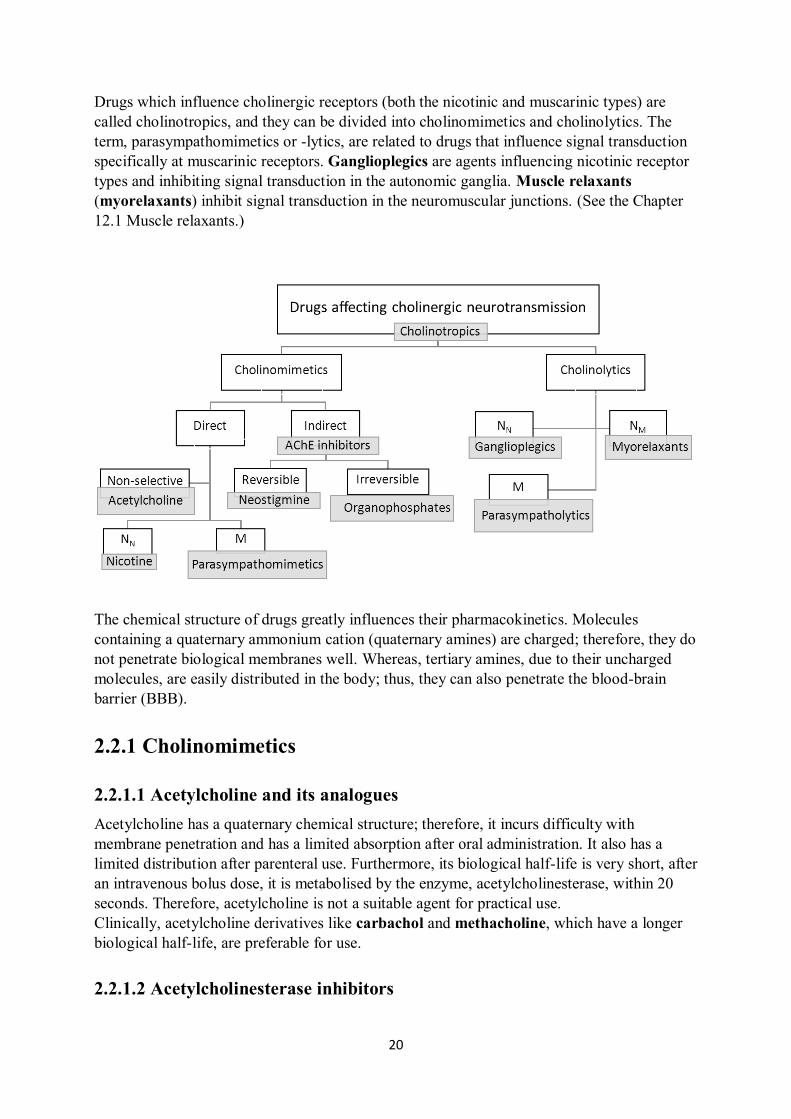
Drugs which influence cholinergic receptors (both the nicotinic and muscarinic types) are
called cholinotropics, and they can be divided into cholinomimetics and cholinolytics. The
term, parasympathomimetics or -lytics, are related to drugs that influence signal transduction
specifically at muscarinic receptors. Ganglioplegics are agents influencing nicotinic receptor
types and inhibiting signal transduction in the autonomic ganglia. Muscle relaxants
(myorelaxants) inhibit signal transduction in the neuromuscular junctions. (See the Chapter
12.1 Muscle relaxants.)
The chemical structure of drugs greatly influences their pharmacokinetics. Molecules
containing a quaternary ammonium cation (quaternary amines) are charged; therefore, they do
not penetrate biological membranes well. Whereas, tertiary amines, due to their uncharged
molecules, are easily distributed in the body; thus, they can also penetrate the blood-brain
barrier (BBB).
2.2.1 Cholinomimetics
2.2.1.1 Acetylcholine and its analogues
Acetylcholine has a quaternary chemical structure; therefore, it incurs difficulty with
membrane penetration and has a limited absorption after oral administration. It also has a
limited distribution after parenteral use. Furthermore, its biological half-life is very short, after
an intravenous bolus dose, it is metabolised by the enzyme, acetylcholinesterase, within 20
seconds. Therefore, acetylcholine is not a suitable agent for practical use.
Clinically, acetylcholine derivatives like carbachol and methacholine, which have a longer
biological half-life, are preferable for use.
2.2.1.2 Acetylcholinesterase inhibitors
21
Acetylcholine concentration in the synapse is thoroughly regulated in the organism, and its
fast degradation is essential for homeostasis and life functions. Acetylcholinesterase is the key
enzyme necessary for acetylcholine metabolism. Through the inhibition of its degradation, the
acetylcholine effect in the synaptic cleft can be prolonged. This effect is nonspecific to a
receptor subtype, and both muscarinic and nicotinic receptors are affected simultaneously.
The effect of acetylcholinesterase inhibitors is systemic and manifests as bradycardia, a
decrease in myocardial contractility, increased motility of the gastrointestinal (peristalsis) and
genitourinary tract, stimulation of exocrine glands, and skeletal muscle fasciculations and
twitching.
Acetylcholinesterase inhibitors can be divided into two groups according to the reversibility
of their effect. The agents with a reversible mechanism of action are used in pharmacotherapy
of myasthenia gravis (skeletal muscle weakness), glaucoma, urinary retention, and intestinal
atony, examples include neostigmine, physostigmine, pyridostigmine, and distigmine.
Agents with a long-term effect, such as donepezil, galantamine, and rivastigmine, are used
for the therapy of Alzheimer´s dementia, which has been associated with acetylcholine
deficiency in the CNS.
Irreversible acetylcholinesterase inhibitors, organophosphates, are toxicological in nature.
They include insecticides, parathion and malathion, and nerve gases (chemical weapons),
such as sarin, tabun, and soman. The symptoms of intoxication with irreversible
acetylcholinesterase inhibitors include salivation, lacrimation, urination, incontinence,
vomiting, miosis, tremor, skeletal muscle convulsions, and life-threatening bronchospasms
and bronchorrhea (i.e., increased mucous secretion in the respiratory tract).
In case of organophosphate intoxication, urgent therapy is required. It comprises mechanical
ventilation, therapy of muscle convulsions (intravenous diazepam), the parasympatholytic
drug, atropine, administered as an antidote until signs of atropinization (mydriasis) occur, and
obidoxime should be administered as an acetylcholinesterase reactivator (pralidoxime and
trimedoxime are not currently registered in the Czech Republic).
2.2.2 Parasympathomimetics
Parasympathomimetics are muscarine receptor agonists. Pilocarpine administered locally is
used as a miotic antiglaucoma agent in ophthalmology. Muscarine, a natural alkaloid from
the fly amanita (Amanita muscaria), having given its name to the acetylcholine receptor
subtype, has a toxicological basis. However, the muscarine content is too low to play a
significant role in symptomatology of poisoning with this mushroom.
2.2.3 Parasympatholytics
Parasympatholytics are direct muscarine receptor antagonists. Their effects include
tachycardia, bronchodilation, a decrease in glandular secretion, a decrease of gastrointestinal
motility, urinary retention, xerostomia (decrease in salivation/dry mouth), xerophthalmia (dry
eyes), mydriasis, and cycloplegia (i.e., blockade of ciliary muscle function and impaired
nearsightedness). The wide clinical importance of parasympatholytics surrounds their use as
antiasthmatics, antispasmodics, antiemetics, mydriatics, and antiparkinson agents.

22
2.2.3.1 Agents with a tertiary amine group
Atropine is an alkaloid from the deadly nightshade plant (Atropa belladonna), one of the
most poisonous plants in the northern hemisphere. It is used for the therapy of
bradyarrhythmia, as an antidote for an overdose with acetylcholinesterase inhibitors, in
premedication prior to a surgical procedure under general anaesthesia (to block the vagus
nerve reflexes), as a spasmolytic agent in urology and gastroenterology, and in ophthamology,
for therapeutic mydriasis by administering it locally.
Symptoms of Atropa belladonna intoxication cannot be attributed only to its atropine content,
but also to other natural alkaloids present, such as scopolamine (hyoscine). Intoxication is
presented by mydriasis, xerostomia, tachycardia, and dry hot skin (caused by an inhibition of
sweat production). After higher doses, urinary retention and constipation can be observed, and
there is a risk of hallucinations and coma. Therapy of intoxication with parasympatholytics is
symptomatic. Physostigmine can be used as an antidote.
Tropicamide and cyclopentolate, with shorter effects than atropine, are used to diagnose
mydriasis in ophthalmology.
2.2.3.2 Agents with a quaternary ammonium cation
Due to the poor absorption of quaternary amines, these are mostly administered parenterally.
As inhalation antiasthmatics, a short-acting antimuscarinic agent, ipratropium, and long-
acting antimuscarinic bromides, tiotropium, aclidinium, and umeclidinium, are used.
Butylscopolamine, available in parenteral forms, and phenpiverine, are spasmolytics.
Phenpiverine is often used in spasmoanalgesic combinations along with nonsteroidal anti-
inflammatory drugs (e.g., metamizole) and musculotropic spasmolytics (e.g., pitofenone).
Otilonium is used for the therapy of spasms of the distal part of the gastrointestinal tract, and
tolterodine and trospium are used to treat an overactive bladder.
2.2.3.3 Uroselective parasympatholytics
Parasympatholytics which bind selectively to M3 receptors in the urinary tract (i.e., M3
muscarinic receptors in the detrusor muscle of the bladder), darifenacin and solifenacin, are
used for the therapy of overactive bladder syndrome.

23
3 Psychotropic substances, drug dependence
3.1 Psychotropic substances
The main expected effects of psychotropic substances are changes in mental states and
affecting of mental functions (e.g., effects on anxiety, mood, vigilance). Psychotropic
substances are divided according to their main therapeutic effect.
3.1.1 Treatment of affective disorders (antidepressant drugs)
Depression is a severe and life threatening mental state that manifests as depressed mood in
the persons affected. Approximately 10-15% of population suffers from depressive disorder
and women are affected twice as frequently.
A widely-accepted hypothesis explaining the development of depression is based on
monoamine theory. It was postulated after observing which hypertensive patients treated with
reserpine (i.e., a plant alkaloid inducing depression) developed depression as result of this
treatment. According to monoamine theory, depression is a consequence of a deficit in
serotonergic and noradrenergic synaptic neurotransmission in the brain. Antidepressant drugs
increase monoamine levels in the synapse, which results in a whole cascade of processes
manifesting as the anti-depressive effect.
The effect of all antidepressant drugs appears earliest after two weeks of administration;
substances of choice are SSRIs.
3.1.1.1 Monoamine reuptake inhibitors
Tricyclic antidepressants
Tricyclic antidepressants and structurally similar antidepressant drugs non-selectively block
serotonin and norepinephrine reuptake from the synaptic cleft to presynaptic part of neuron.
This mechanism prolongs the presence of neurotransmitters in the synaptic cleft, which
subsequently leads to compensation of their shortage during depression. Typical tricyclic
antidepressants involve for example, amitriptyline, imipramine, dosulepin, and
clomipramine.
The most frequent adverse effects are as follows: sedation, weight gain, anticholinergic effect,
cardiotoxic effects (e.g., arrhythmia, QT interval prolongation), and postural hypotension.
They can interfere with corticosteroids and neuroleptic substances at the level of elimination,
and their concomitant intake with alcohol leads to respiratory depression. They can also be
used for the treatment of neuropathic pain in chronic pain syndromes.
Selective neurotransmitter (serotonin, norepinephrine and dopamine) reuptake
inhibitors
SSRI (Selective Serotonin Reuptake Inhibitors) are the first substances considered for the
treatment of depression. Furthermore, they are used in therapy of obsessive-compulsive

24
disorder (OCD), panic disorder, mental anorexia and bulimia, or pathological gambling.
Typical SSRIs are fluoxetine, citalopram, escitalopram, and paroxetine. They are well-
tolerated, have low toxicity, and adverse effects include nausea, diarrhoea, anorexia, sexual
dysfunction, and increased bleeding. SSRIs have a whole range of pharmacokinetic
interactions; they inhibit enzymes of cytochrome P450, thus slowing down metabolism of
other substances (metabolised by P450), and in this way they can cause the other substances’
levels to increase (i.e., creating a risk of toxicity, overdosing).
NRI or NERI (Norepinephrine Reuptake Inhibitors), for example, reboxetine, are used for
the treatment of depression associated with decreased psychomotor activity or attention
disorder.
SARI (Serotonin Antagonist and Reuptake Inhibitors): Aside from blocking serotonin
selective reuptake inhibition, these also block 5-HT2A receptors (e.g., trazodone). Trazodone
is suitable for therapy of depression associated with insomnia. It acts in lower doses
particularly as a hypnotic drug by blockade of the 5-HT2A receptor; in higher doses, it blocks
serotonin reuptake.
NDRI (Norepinephrine and Dopamine Reuptake Inhibitors): An example in this category is
bupropion. Unlike other antidepressant drugs, it has low rate of adverse effects on sexual
functions. Aside from treating depression, it can also be used for therapy during smoking
cessation and as a treatment for gambling.
SNRI (Serotonin and Norepinephrine Reuptake Inhibitors) such as venlafaxine, duloxetine,
milnacipran are good examples of this type of antidepressant. Their main advantage is their
fast onset and small number of adverse effects.
3.1.1.2 Inhibitors of monoamine degradation
Non-selective irreversible MAO inhibitors
Monoamine oxidase is an enzyme decomposing monoamines such as dopamine, serotonin, or
norepinephrine. Mood is improved particularly effectively as a consequence of serotonin
concentration increasing in the CNS; psychomotor activity is improved due to norepinephrine
and dopamine concentration increase.
Because of the MAO blockade, it is necessary to follow a dietetic regimen and avoid food rich
in tyramine (e.g., cheese, wine, beer, etc.), which is a precursor of catecholamine synthesis
and is normally decomposed by MAO. Its accumulation could cause a dramatic increase in
blood pressure and hypertonic crisis. Tranylcypromine is an example of a typical drug in this
group.
Selective MAO-A and MAO-B inhibitors
These can selectively affect MAO subtypes. Selective irreversible MAO-B inhibitor,
selegiline, besides being used for the therapy of depression, is used to treat Parkinson’s
disease and Alzheimer’s disease. Selective reversible MAO-A inhibitor, moclobemide, is a
newer substance, which does not require adjustment of the dietetic regime. It is indicated for
mild to moderate depression and is also well tolerated by older patients.
25
3.1.1.3 Noradrenergic specific serotoninergic antidepressants (NaSSA):
The NaSSA group includes, for example, mirtazapine. Its advantages are fast onset of
effects, and it has anti-depressive, anxiolytic, and sedative effects.
3.1.2 Treatment of anxiety disorders (anxiolytic drugs)
Anxiolytic drugs are used mainly for the therapy of anxiety disorders, phobias, and to treat
posttraumatic stress disorder.
These substances include the benzodiazepines (e.g., diazepam, oxazepam, bromazepam,
and alprazolam). They act through GABAA receptor complexes, which are coupled to
chloride channels. Benzodiazepines (similarly to barbiturates) bind to the GABAA receptor
and facilitate the binding of GABA (gamma-aminobutyric acid; a natural ligand of this
receptor), which leads to the chloride ion channel opening; this causes hyperpolarization and
inhibition of neural impulse transmission. Consequently, benzodiazepines possess anxiolytic,
anticonvulsive, myorelaxant, and in higher doses, amnestic effects.
Benzodiazepines have excellent bioavailability after oral administration; they bind to
plasmatic proteins in circulation, are biotransformed by the microsomal cytochrome P450
system, and finally excreted by kidneys.
Adverse effects involve sleepiness, dizziness, problems with coordination, and prolongation
of sleep. A common property of benzodiazepines is tolerance, which becomes apparent
through gradual fading of the desired effect. There is a high risk of psychological and physical
dependence following chronic use.
Contraindications include concurrent use with alcohol (risk of respiratory depression), use
during pregnancy and lactation (risk of cleft palate and abstinence syndrome in new-borns), in
patients with multiple sclerosis, or in patients with alcohol or drug dependence. In case of
overdose, flumazenil can be used, which is a specific antagonist of benzodiazepine receptors.
Anxiety can also be treated with serotonergic substances (e.g., 5-HT1A receptor agonist
buspirone) using low doses of antidepressants from the SSRI group, low doses of
antipsychotic drugs (see below), H1 antihistamines of the 1st generation (e.g.,
hydroxyzine, bisulepine, promethazine), guaifenesin, or beta-blockers (e.g., metipranolol).
3.1.3 Treatment of sleep disorders (hypnosedatives)
Hypnosedatives are substances damping the CNS. Hypnotics cause conditions similar to
physiologic sleep; sedatives are calming. The border between them is not sharp – an increased
dose of sedatives will cause a hypnotic effect; thus, they are termed hypnosedatives. The
indication for these substances is insomnia. They act on GABAA receptor complexes coupled
with chloride ion channels. Hypnosedatives are divided into three generations:
First Generation - Barbiturates
Barbiturates were the substances of choice prior to the discovery of BZDs. Presently, they are
not used for the treatment of insomnia because of their narrow therapeutic range and addictive

26
potential. Some of them are used as anaesthetic drugs (thiopental) and antiepileptic drugs
(phenobarbital).
Second Generation - Benzodiazepines (BZD)
Dissimilar to the third generation of hypnosedatives, benzodiazepines are not selective. They
also have hypnotic, anxiolytic, anticonvulsant, and myorelaxant effects. There is a risk of
physical and psychological dependence. They should not be used in geriatric patients due to
paradoxical reactions (e.g., excitation, aggressiveness).
Third Generation – “Z” substances
“Z” substances include zolpidem, zaleplon, and zopiclone. They have a selective hypnotic
effect and compared to the second generation, there is a lower risk of dependence. They act as
selective agonists of 1 BZD receptors. They do not disturb the physiologic structure of sleep
(i.e., the shortening of REM phase - deep phase of sleep with rapid eye movements) to such
extent as does the previous generation.
Newer hypnotic drugs involve substances that affect melatonin receptors (e.g., melatonin,
ramelteon), which successfully improve quality of sleep in patients older than 55 years of age
with primary insomnia. Also, phytopharmaca can be used for the treatment of insomnia (e.g.,
containing common balm, valerian, hop, hawthorn, passionflower or St. John’s wort).
3.1.4 Treatment of mental integration disorders (antipsychotic
drugs)
Neurotropic substances affecting mental integration involve antipsychotic drugs, alternatively
called neuroleptics. They are used particularly for the treatment of psychosis (e.g.,
schizophrenia, the manic phase of bipolar affective disorder). This group of drugs affects
dopaminergic transmission through inhibition of the D2 receptor in the mesolimbic and
mesocortical system.
Targeting the dopaminergic system to treat schizophrenia is due to the dopamine hypothesis.
According to this theory, schizophrenia is accompanied by hyperreactivity of the
dopaminergic system. Part of the patient’s clinical picture involves positive psychotic
symptoms: delusions, hallucinations, aggressiveness, agitation, disorganisation; and negative
psychotic symptoms: emotional and affective flattening, apathy, and anhedonia.
3.1.4.1 Classical (typical) antipsychotics
Sedative
These substances are intended for the treatment of psychomotor unrest; they have sedative
and hypnotic effects, and weaker neuroleptic effects. Adverse extrapyramidal symptoms (e.g.,
dyskinesia - rhythmic movements of tongue and mimic muscles, stereotypic swinging
movements of body, involuntary rhythmic movement of hands and legs) are rare. For
example, this group involves chlorpromazine and levomepromazine.
Incisive
27
Incisive antipsychotics are used for treatment of positive psychotic symptoms (e.g., delusions
and hallucinations). They have strong neuroleptic effects, but also cause significant adverse
extrapyramidal symptoms. Haloperidol serves as a good example.
3.1.4.2 Atypical antipsychotics
Atypical antipsychotics are used for the treatment of negative psychotic symptoms (e.g.,
apathy, anhedonia). They are also suitable for maintenance therapy of schizophrenia, newly
diagnosed schizophrenia, and when classical antipsychotics are not tolerated. Adverse effects
involve minimal stimulation of prolactin secretion. In addition, substances of this group cause
weight gain and increase risk of cardiovascular disorders.
D2, D3 selective antagonists: sulpiride, amisulpride, and tiapride
SDA (serotonergic and dopaminergic antagonists): risperidone, ziprasidone,
sertindole, and paliperidone
MARTA (multi-receptor antagonists): clozapine, olanzapine, quetiapine, and
zotepine
partial agonists of D2 receptors/antagonists on 5-HT receptors: aripiprazole
3.1.5 Drugs affecting memory and cognitive functions
3.1.5.1 Treatment of Alzheimer’s disease
Substances used for this indication improve cognitive functions such as attention and
memory. Alzheimer’s disease is characterised by an imbalance between dopamine and
acetylcholine in brain, or in other words a lack of acetylcholine. Centrally acting reversible
inhibitors of acetylcholinesterase are used (donepezil, galantamine, and rivastigmine).
Adverse effects involve diarrhoea, nausea, and tremor. For the treatment of Alzheimer’s
disease, memantine can be also used; this drug acts by partially inhibiting glutamatergic
NMDA (N-methyl-D-aspartate) receptors.
3.1.5.2 Nootropics (cognition-enhancing drugs)
Nootropics are a non-homogenous group of substances supporting the activity of the brain, for
example by improving blood flow or neuronal and glial metabolism. In practice, piracetam is
used due to its positive effect on memory function; furthermore pyritinol, an antioxidant
supporting metabolism of neurons, and vinpocetine, which improves microcirculation and
glucose and oxygen uptake in blood, are used, as well.
3.2 Drug dependence
Dependence is a mass of physiological, behavioural, and cognitive phenomena in which the
use of a substance or a class of substances takes on a much higher priority for a given
individual than other behaviours that once had greater value. A central descriptive
characteristic of dependence syndrome is the desire (often strong, sometimes overpowering)
to take psychoactive drugs (which may or may not have been medically prescribed), alcohol,
28
or tobacco. There may be evidence that return to the substance use after a period of abstinence
leads to a more rapid reappearance of other features of the syndrome than occurs with
nondependent individuals (definition according to ICD - 10).
Basic features of dependence are:
craving or feeling of urge to use a substance,
decreased self-control, difficulties with control of the substance use,
neglecting of work and hobbies,
tolerance development, physical dependence,
continuation of use despite clear evidence of harmful consequences (physical, mental,
social), and
withdrawal syndrome.
Tolerance means a gradual decrease in the effects of the dependence producing substance; in
other words, the dose must be increased to reach the same effect. Following dependence-
producing substance withdrawal, various somatic symptoms occur which are subjectively
unpleasant and in some cases life threatening, and along with the psychological symptoms,
so-called withdrawal syndrome arises.
The most frequent types of dependence producing substances
Central nervous system stimulants
The most well-known substances are nicotine and caffeine, but amphetamine,
methamphetamine (pervitin), cocaine, MDMA (3,4-methylenedioxymethamphetamine =
ecstasy), ephedrine, and some others are included in this group. Dependence is characterised
by increased psychomotor activity, decreased appetite, and aggressiveness and can result in
psychosis, hallucinations, and delirium. Indirect sympathomimetic effects on the
cardiovascular system may cause hypertension, arrhythmias and changes in blood pressure.
Central nervous system inhibitors
The most common substance in this group is alcohol, followed by opioids, benzodiazepines,
and barbiturates. Dependence is characterised by damping of the CNS resulting in sedation.
Inhibition of the respiratory centre (particularly in the case of opioids, benzodiazepines, and
barbiturates) is very dangerous and can even lead to death from asphyxia.
Hallucinogens and deliriogenics
This group involves cannabinoids (marijuana, hashish), psilocybin (found in some
mushrooms), LSD (lysergic acid diethylamide), mescaline, and ketamine. Dependency is
characterised by euphoria, hallucinations, fuzziness, loss of short-term memory, panic,
anxiety, further flashbacks, and even acute paranoid psychosis may occur.
Inhalants and organic solvents

29
Typical examples involve toluene, ether, acetylene, chloroform, and nitrous oxide.
Dependence is characterised by a short euphoric phase; dizziness, vomiting, and loss of
balance also appear. Overdosing is very easy, subsequent death is caused due to respiratory
centre paralysis or following a heart arrhythmia. The users emit an odour that is typical of this
type of substance abuse.
Pharmacological treatment of dependence
detoxification - slow elimination of the substance from body after dose decrease
substitution - replacement of the drug by a safer substance
prevention of relapses - application of an anti-craving drug
Sensitizing substances
These substances worsen tolerance to dependence-producing drugs. The aim is to lead the
dependent person to avoidance of the particular substance. In practice, disulfiram (Antabus)
is used for alcoholism treatment. When paired with alcohol, it causes vomiting.
Opioid antagonists
Naloxone and naltrexone are clinically used for the treatment of opioid intoxication.
Anti-craving drugs
Acamprosate is used to decrease craving for dependence-producing substances; naltrexone
and nalmefene specifically decrease cravings for alcohol.
Substitution therapy
This consists of using a legally administered dependence-producing substance as a
replacement for the original drug, for instance methadone and buprenorphine in persons
dependent on opioids and eventually nicotine chewing gum and other means for smoking
cessation.

30
4. Substances affecting respiratory system
4.1 Antiasthmatics
Asthma is a chronic inflammatory disease of the respiratory tract. A variety of immune cells
(e.g., mastocytes, eosinophils, T-lymphocytes) producing signal molecules called cytokines
(e.g., chemotactic cytokines - chemokines, leukotrienes and others) are implicated in the
pathophysiology of asthma. Chronic inflammation in asthma is associated with the
overreactivity of the bronchial tree and typical recurring airflow obstruction, which occurs
spontaneously and can be reversed with pharmacotherapy. Bronchial asthma represents a
multifactorial disease, which can occur in any age group. Endogenous (e.g., genetic
background, physical exercise) and exogenous (e.g., allergens, exposure to air pollution, viral
infections, or medication) environmental factors play a role in disease development. Most
cases of bronchial asthma have an allergic component, primarily manifesting as atopy,
wherein there is an excessive production of IgE antibodies in response to common allergens
found in the environment.
Asthma is characterized by recurrent episodes of wheezing, difficulty in breathing (i.e.,
dyspnea or shortness of breath), chest tightness, and coughing (typically in the early morning
or evening hours) after exposure to specific or nonspecific triggers in each individual patient.
The reason for these respiratory problems is the contraction of bronchial smooth muscles
leading to the narrowing of airways, oedema, thickening of the walls of the respiratory tract,
and increased secretion of mucus. Apart from exacerbations, the patient may be
asymptomatic; nevertheless, the inflammatory changes in airways are persistent. A prolonged
(and often pharmacoresistant) asthmatic attack is called status asthmaticus (i.e., an acute and
severe exacerbation), and it is a life-threatening, acute condition with the risk of irreversible
bronchial obstruction of the respiratory airways and risk of death (i.e., a lethal ending).
Therefore, it requires urgent hospitalisation in the intensive care unit (ICU).
The diagnosis of asthma is based on the anamnesis of the patient (i.e., personal, family, and
pharmacological recollection) and a battery of spirometric tests to examine pulmonary
functioning. Using spirometry, it is possible to detect pulmonary obstruction and assess its
reversibility and variability. The most commonly used spirometric parameters are FEV1
(Forced Expiratory Volume in 1 second), understood as the volume exhaled in one second
during the maximum expiratory effort, and PEF (Peak Expiratory Flow Rate). Often there is
additional examination of bronchial overreactivity with use of the so-called
bronchoconstriction trigger test. According to the parameters measured, occurrence of
daytime and night-time symptomatology, and eventually, according to the medication
necessary for optimal control of the disease, asthmatic patients can be divided into several
categories and the pharmacotherapeutic approach adjusted according to the severity of the
disease in the individual patient.
At present, there is a three-tier classification system for asthma, which is accepted according
to the corresponding level of clinical control: asthma under control, partially controlled

31
asthma, and uncontrolled asthma (i.e., with insufficient control of asthmatic
symptomatology). Optionally, classifications of asthma according to the severity of the
disease, such as intermittent, mild persistent, intermediate persisting, and severe
persistent asthma can be used to determine treatment based on the lowest degree of intensity
of pharmacologic intervention necessary to maintain optimal control of the disease. A useful
complementary diagnostic tool for differential diagnosis of asthma is represented by the
allergologic examination, which can help to identify if an allergic component to the asthma
(so-called atopic asthma) exists and to identify the risk factors for an attack. For this purpose,
there are skin prick tests available and/or laboratory examination of the specific serum IgE
antibodies responsible for symptom development.
Asthma is a disease which cannot currently be completely cured, but its symptoms can be
efficiently controlled. Thus, the aim of therapy is to control the asthma, which includes
alleviation of acute and chronic symptoms, maintenance of normal pulmonary function
without limitation of daily activities (including sports) with minimal use of “reliever
antiasthmatics” (see the Chapter 4.1.2.1 SABA), and the elimination of exacerbation while
minimising the side effects of antiasthmatic therapy.
Drugs used for the therapy of asthma can be divided into agents for quick relief (“relievers”)
and preventative agents, also called control agents (“preventers”). The quick-relief
medications (“rescue antiasthmatics” or “reliever agents”) are used preferentially to quickly
alleviate bronchoconstriction during acute exacerbation of asthma, such as bronchodilator
agents with a fast onset of action (e.g., β2 sympathomimetics with a quick and short-term
effect), parasympatholytics, methylxanthines with a short-term effect, or systemic
glucocorticoids. They should be only used for the shortest time necessary and at the lowest
possible dose and frequency of administration.
Contrarily, preventative antiasthmatics are used for the long term (i.e., on daily basis) to
provide control of asthma and prevent asthmatic attacks. For this purpose, anti-inflammatory
drugs are used most often to keep asthma under clinical control (e.g., glucocorticoids, mast
cell stabilizers, antileukotrienes, methylxanthines with a prolonged action, omalizumab, and
long-acting β2 sympathomimetics). H1-antihistamines are used to treat the allergic type of
asthma, preferentially those of the second and third generation (see the Chapter 8.6.2.2 H1-
antihistamines - 2nd
generation and the Chapter 8.6.2.3 H1-antihistamines - 3rd
generation).
Antiasthmatics can be administered orally or intravenously (in severe cases), but
administration via inhalation is generally preferred. The advantage of inhalation is the local
effect in airways (i.e., a high concentration of drug at the site of action). Thus, there is a rapid
onset of action with fewer systemic side effects. There are a wide range of the inhalation
systems on the market, and for the efficacy of an anti-asthmatic therapy, it is very important
to explain to the patient how to appropriately manipulate their individual inhalation system,
and he/she should repeatedly practice the proper technique of inhalation with that given
system.
Simplified, we can divide the inhalation systems into:
● Pressurized Aerosol inhalers, also-called MDI (Metered Dose Inhalers), with a

32
propellant gas,
● Inhalers for powders, also-called DPI (Dry Powder Inhalers), and
● Nebulizers, in other words devices producing humid aerosol.
Some patients, when using the aerosol metered dose inhalers, can have a problem with
coordination of their inspiration and proper activation of the inhaler. For this purpose, there
are inhalation spacers available, which can be inserted between the inhaler device and the
mouth of the patient, enabling the drug to be released into the chamber of the spacer and to be
inhaled subsequently. Breath Actuated Inhalers (BAI) are novel inhalation systems facilitating
antiasthmatic administration.
4.1.1 Glucocorticoids
Glucocorticoids (GC), with their multiple effects, are frequently used for a wide variety of
indications (see the Chapter 8.5 Glucocorticoids). For the therapy of bronchial asthma, they
represent an essential group of drugs due to having the most potent anti-inflammatory effects.
Glucocorticoids not only block phospholipase A2, an important enzyme for production of
arachidonic acids, thereby decreasing the production of proinflammatory factors
(eicosanoids), but they also decrease cytokine secretion, lower the permeability of endothelial
cells, prevent migration of inflammatory cells to the site of inflammation, etc. As a result,
there is decreased bronchial overreactivity, antiedematous effect, improved lung functions,
reduced frequency and severity of exacerbation, and other effects observed in clinics.
According to the route of administration, glucocorticoids can be divided into inhaled and
systemic GC.
4.1.1.1 Inhalation glucocorticoids
GC, administered in the inhaled form, represent the first drug of choice for persistent asthma
of all degrees of severity. These drugs belong to preventative antiasthmatics for long-term
use.
Risk of the typical side effects of GC administration (e.g., osteoporosis, fat redistribution,
etc.) can be decreased by using the inhalation administration route, but they cannot be
completely excluded. Even after inhalation, there is a partial systemic drug absorption, which
can differ according to the pharmacokinetic profile of the individual agent. Among side
effects occurring after local GC administration are oropharyngeal candidiasis, dysphonia, and
occasional coughing, owing to irritation of airways of the upper respiratory tract. To prevent
these local side effects, it is recommended to use an inhalation spacer and to wash out the oral
cavity with drinking water immediately after GC application.
33
If the standard dose of inhaled GC does not sufficiently control asthma, it is not effective to
further increase the dosage due to an increased risk of side effects. In this scenario, it is better
to include an add-on preventative antiasthmatic agent, preferably a long-acting β2-
sympathomimetic agent. The reason being the mutual positive potentiation of their effects, as
glucocorticoids decrease receptor desensitization for β2 agonists (i.e., increase the number of
β2 receptors). Thanks to their synergistic effects, lower doses of each individual agent can be
used in combination than would be necessary for single agent therapy. Currently, there are
fixed combinations available, thus the patient gets both the GC and β2-sympathomimetic
agents in one inhalation preparation. Examples of GC agents used for inhalation are
beclomethasone, budesonide, fluticasone, mometasone, and ciclesonide.
4.1.1.2 Systemic glucocorticoids
Long-term therapy with systemic GC is only used to achieve full control over the disease in
those with severe, persistent asthma. Oral administration is preferred to the parenteral drug
route, but in both cases, it is necessary to consider the potentially serious side effects of GC.
Asthmatic patients treated with systemic GC belong to the corticosteroid-dependent group of
asthmatics, which is a group that has an increased risk of sudden death from asthma
otherwise.
Systemic GC can also be administered as reliever antiasthmatics. Despite an extended onset
of action of 4-6 hours, they can prevent asthma progression and exacerbation.
Among systemic glucocorticoids belong prednisone and betamethasone. (For a more
comprehensive study, see the Chapter 8.5 Glucocorticoids.)
4.1.2 β2-sympathomimetics
β2-sympathomimetics are agents with a selective agonistic action on β2 receptor subtypes (for
more details, see the Chapter 2.1.1.3 Selective sympathomimetics – β2). They elicit relaxation
of the smooth muscles of the respiratory tract, increase mucociliary clearance, and decrease
vascular permeability. Furthermore, their effects on the immune system have also been
confirmed. For example, they inhibit the release of proinflammatory mediators from mast
cells (mastocytes). β2-sympathomimetics are either used as "reliever" inhalers for the
management of asthma symptoms or as a preventative medicine for asthma. They can be
administered orally or parenterally (in acute situations), but either way, inhalation remains the
most preferred administration route.
Possible side effects experienced by patients during antiasthmatic therapy are primarily
hypokalaemia, tachycardia, palpitation, or tremor of the skeletal muscle. Naturally, risk of
side effects is higher after their systemic use. β2-sympathomimetics can be categorized
according to the duration of their effect.
4.1.2.1 Short-acting β2-sympathomimetics (SABA)
The abbreviation, SABA, comes from English and means Short Acting Beta2-Agonists. These
compounds include inhaled β2-sympathomimetics with a fast onset of action (i.e., from three

34
to five minutes after administration) and with a short duration of action – from about four to
six hours. In this group belong agents, which are used as "reliever" inhalers in the
management of asthma symptoms, and according to the frequency of their use, the need for
further therapy can be estimated. It is good to keep in mind that frequent and regular SABA
administration for asthma management does not lead to satisfactory control of asthmatic
symptoms.
Drugs from this group which are used in clinics include salbutamol, terbutaline, and
fenoterol. Sometimes this pharmacological group is called the RABA (Rapid Acting Beta2-
Agonists) group, owing to their fast onset of the effect. Therefore, formoterol can be
included; despite its long-term effect, it has a very fast onset of action.
4.1.2.2 Long-acting β2-sympathomimetics (LABA - Long acting beta2-
agonists)
β2-sympathomimetics with effects lasting for a prolonged duration (i.e., up to 12 hours) are
available in inhalable and oral drug dosage forms. Due to the greater intensity and more
frequent development of adverse effects upon oral administration, administration via
inhalation is preferred. For long-term therapy of asthma, LABA should not be used in
monotherapy (inflammatory changes remain uninfluenced by these compounds), but should
always be combined with inhalable glucocorticoids (e.g., possibly in the above-mentioned
fixed combination) as a complement to long-term anti-inflammatory therapy. They can also
be combined with long-acting muscarinic antagonists (see the Chapter 4.1.3
Parasympatholytics).
Examples of agents of the LABA group are formoterol, salmeterol, clenbuterol, and
procaterol. Short-acting β2-sympathomimetics can be included, if available in a retarded
(modified-release) dosage form. Indacaterol and vilanterol are sometimes excluded from
this group, as they are considered ultra-long-acting β2-agonists (U-LABA).
4.1.3 Parasympatholytics (muscarinic antagonists)
Antimuscarinic agents block muscarinic receptors in the smooth muscle of the respiratory
tract and prevent bronchoconstriction to treat of asthma (also see the Chapter 2.2.3
Parasympatholytics). The only parasympatholytic currently used as a reliever, or for so-called
rescue asthma therapy, is ipratropium, which is used by inhalation. It has a relatively short-
term effect; it influences neither the early, nor the delayed allergic responses, and it has no
anti-inflammatory effect. During acute exacerbation, ipratropium can be combined with β2-
sympathomimetics (there are fixed combinations with fenoterol already registered), and it is
also an alternative therapy for patients whom, for any reason, have a SABA contraindication.
Side effects of parasympatholytics include headache, vertigo, coughing, and other side effects
typical of parasympatholytics (e.g., dry mouth, disorders of GIT motility, etc.). For
completeness, other substances such as tiotropium, aclidinium, and umeclidinium are worth
mentioning. They have long-term effects (sometimes called LAMA - long-acting muscarinic

35
antagonists) and are indicated for maintenance bronchodilator therapy in COPD, often in
combination with LABA (see the Chapter 4.1.2.2).
4.1.4 Methylxanthines
Methylxanthines are agents whose mechanism of action is not fully understood. At high
concentrations, they block the phosphodiesterase enzyme, thereby increasing the intracellular
cAMP concentration. Other putative mechanisms of action are via the inhibition of the
adenosine receptors or through modulation of the levels of intracellular calcium.
Methylxanthines have bronchodilator and weak anti-inflammatory effects (compared with
GC) and a variety of extrapulmonary effects (e.g., diuretic, CNS stimulant, and other effects).
Examples of methylxanthines are theophylline and aminophylline. Both are available in
parenteral drug dosage forms and can be taken orally in the form of sustained-release tablets.
Monotherapy can be used in patients with a mild persistent asthma, but more often they are
used in combination with GC, primarily as long-term preventive antiasthmatics. The use of
methylxanthines for the therapy of acute asthmatic exacerbation is controversial.
A disadvantage of theophylline use is its narrow therapeutic window, as it can cause serious
side effects (i.e., affecting many organs) in higher doses, and in severe cases, it can even lead
to a lethal end. Theophylline intoxication manifests as nausea, vomiting, diarrhoea,
palpitations, a rise in blood pressure, and tremors. Side effects can be avoided by dose
adjustment and the monitoring of serum concentrations. Theophylline is metabolised via
cytochrome P450, which may lead to clinically significant interactions with concomitantly
administered substances affecting this same metabolic pathway. Nicotine is a cytochrome
P450 inducer; therefore, higher theophylline doses are recommended for smokers.
4.1.5 Mast cells stabilizers (immunoprophylactics)
In this group of drugs, we include agents that prevent release of proinflammatory cytokines
through the stabilization of mast cell membranes. These agents are used for prophylaxis, and
they are not suitable for management of acute health problems. When used for prophylaxis,
they inhibit both the early and delayed phase of allergen-induced bronchoconstriction.
Immunoprophylactic agents include “chromones” and ketotifen. The term “chromones” is
used to represent sodium cromoglicate (cromolyn) and nedocromil, both of which are
inhaled to treat mild, persistent bronchial asthma. Their mechanism of action is not fully
elucidated, but apart from mast cell stabilization, they inhibit the chemotactic responses of
eosinophils and neutrophils.
Along with the stabilization of the mast cell membrane, ketotifen also acts as an H1 non-
competitive receptor antagonist (see the Chapter 8.6.2 H1 antihistamines). It is administered
orally for long-term prevention of asthmatic attacks. Side effects include dry mouth, vertigo,
nausea, and body weight gain.
36
4.1.6 Antileukotrienes
Several mediators are involved in the pathogenesis of bronchial asthma, including
leukotrienes, which have a pronounced bronchoconstriction effect. They influence
chemotaxis, increase vascular permeability, stimulate mucus secretion, etc. Leukotrienes are
produced intracellularly from arachidonic acid via the lipoxygenase enzyme.
Antileukotrienes are agents which inhibit leukotriene functions, either by blocking their
receptors or through inhibition of the lipoxygenase enzyme. Among agents which block
leukotriene receptors are montelukast and zafirlukast. They are given orally and used
long-term for mild and intermediate forms of persistent asthma. They can be used in
monotherapy, but when the sufficient control of asthma is unable to be accomplished, these
compounds can be combined with inhalable GC.
The 5-lipoxygenase inhibitor, zileuton, belongs among agents inhibiting the synthesis of
leukotrienes (currently, there is no lipoxygenase inhibitor registered in the Czech Republic).
4.1.7 Targeted antiasthmatic therapy
To date, the only targeted (biological) therapy approved for asthma, which was developed for
patients with an eosinophilic asthma phenotype (which is proven to be caused by increased
levels of IgE antibodies), is the monoclonal antibody, omalizumab. Its mechanism of action
is mediated via IgE binding, which leads to a reduced amount of circulating IgE, thereby
reducing their binding and preventing activation of the allergic response cascade to inhaled
allergens. Omalizumab has been shown to reduce both airway and circulating blood
eosinophils and is thought to be efficacious in reducing asthma exacerbations. It is
administered subcutaneously in severe persistent allergic asthma, in such a case as
inhalational/systemic corticosteroids (GC), even at very high doses, and long-acting inhalable
β2-sympathomimetics are insufficient in controlling the disease. Omalizumab´s main
disadvantage is its high price.
4.2 Cough medicines
Coughing is among one of the basic defence reflexes of the body. It protects the airways from
damage and it can help keep the throat clear from irritants. The centre of this reflex is in the
medulla oblongata and is also the site of action for some drugs. Often, coughing occurs as a
symptom of a pulmonary disease, but it can also be present in bronchial diseases and
conditions of the upper respiratory tract (e.g., inhalation of air pollutants, the presence of a
foreign body, and an affection of vocal cords). Cough can also be a side effect of some drugs,
such as ACE inhibitors, which are used in hypertension. The presence of a cough should not
be underestimated because although it is most commonly treated as a symptom of common
colds (i.e., respiratory infections), it may also be indicative of more serious diseases such as
chronic obstructive pulmonary disease (COPD), cystic fibrosis, bronchial asthma, and cancer.
In choosing the correct cough treatment, it is necessary to determine the cause and type of
cough. A chronic cough lasts longer than three weeks. Acute coughing lasts for any shorter

37
period than three weeks, and both can be further distinguished as either a dry cough or a
productive cough. The pattern of coughing may change during the course of an illness, and
the phase of dry, irritable (in some cases, emetogenic) cough is often followed by a productive
coughing phase.
According to the type of cough, agents suppressing cough (antitussives) or substances
(ranging in molecular mechanism) affecting the composition, production, and mobility of the
mucus in the airways (i.e., drugs for a productive cough) are prescribed. Concomitant
administration of substances from the two above-mentioned groups is often not advised
because they may produce counter-effects. For example, when combining an antitussive and
mucus-producing agent, there is a risk of mucus accumulation in bronchus. A general rule that
should be followed when treating all types of cough is to ensure a sufficient supply of fluids.
The majority of drugs used to treat cough are over-the-counter medicines.
4.2.1 Antitussives
Cough-relieving drugs functioning through central or peripheral mechanisms are particularly
useful for the treatment of dry, irritable cough, which is usually physically exhausting for the
patient and often occurs in night hours, disturbing sleep. Some representatives may also be
given to children. Antitussive therapy should only be taken for a limited time and should not
be prolonged if unnecessary. According to chemical structure, the groups can be divided into
codeine and non-codeine antitussives.
4.2.1.1 Codeine antitussives
These substances are derived from opioid analgesics, and their central mechanism of action is
reducing cough centre sensitivity in the medulla oblongata (see the Chapter 8.3 Opioid
analgesic drugs). In higher doses, or after long-term administration, other opioid-like effects
(e.g., respiratory depression, constipation, sedation, addiction, etc.) may manifest. They are
contraindicated during pregnancy and should not be used for breastfeeding women. Also, they
are used with caution in patients with bronchial asthma or COPD. At present, only products
with codeine and dextromethorphan are registered. Codeine is a natural alkaloid that can be
isolated from opium. In addition to its pronounced antitussive effect, which it achieves at low
doses, it is also used for its analgesic effect and ability to potentiate the effect of some non-
opioid analgesics, such as paracetamol (acetaminophen). Codeine administration is associated
with more serious side effects than those incurred with dextromethorphan; therefore, it is not
used for children, and codeine products are dispensed with a doctor’s prescription.
Dextromethorphan does not show analgesic properties. It also has no sedative effects at
therapeutic doses and does not affect the respiratory centre. Side effects are rare; preparations
with this substance are available over-the-counter (OTC), mostly in the form of tablets or
syrups and are suitable for children, as well. Co-administration of serotonergic drugs (e.g.,
antidepressants) requires caution as there is a risk of developing serotonin syndrome.
4.2.1.2 Non-codeine antitussives
38
These drugs represent a group of synthetic substances whose antitussive effect is either central
(i.e., inhibition of the cough centre), peripheral, or a combination of both mechanisms. The
peripheral effect of these substances is mediated by action along the efferent nerve pathways
of the cough reflex or by influencing its afferent fibres or nerve endings in bronchial mucosa
(i.e., reducing sensitivity to irritant stimuli). The advantage of non-codeine antitussives is they
do not affect the respiratory centre or carry risk of addiction. They can also be used in
children under two years of age after consultation with a doctor. Currently used substances
include butamirate, dropropizine, and levodropropizine. Butamirate has both a central and
peripheral bronchospasmolytic effect and can be used for cough suppression before and after
surgery (e.g. after intubation). Levodropropizine is the L-isomer of dropropizine with good
tolerability. In addition to its peripheral antitussive effect, it also exhibits antihistamine and
bronchospasmolytic effects.
4.2.2 Expectorants
Agents used for the treatment of a productive cough represent a heterogeneous group of
drugs, and their classification is not uniform. Terminology differs for the various agents
within this group. Depending on the mechanism of action, these agents can be divided into
several subgroups: mucolytic, secretolytic, and secretomotor agents. However, very often
their individual effects are combined; therefore, these drugs mostly belong to more than one
subgroup. Thus, in clinics it is more practical to rely on the knowledge of all the usable effects
of available drugs. They may have the ability to reduce mucosal viscosity (i.e., by influencing
physicochemical properties of the mucus - mucolytics), stimulate the activity of the bronchial
glands (secretolytics), or activate ciliary epithelium (i.e., through the stimulation of the
mucociliary apparatus - secretomotor substances). Some expectorants may have additional
antioxidant effects and can also potentiate antibiotic efficacy.
Drugs used for the therapy of a productive cough are not only used to treat acute
inflammation of the respiratory tract, but some are part of long-term medication for chronic
inflammatory diseases, such as COPD, cystic fibrosis, and chronic bronchitis, as well.
Administration of all expectorants should be supplemented with a sufficient intake of fluids,
whether administered as drops, syrups, or tablets.
Bromhexine is a prodrug whose active metabolite is ambroxol. It reduces the viscosity of
mucus by decomposing mucopolysaccharides. Furthermore, it activates the mucociliary
apparatus of the respiratory epithelium, thereby promoting mucus secretion. Bromhexine
should not be given to patients with gastroduodenal ulcers (as it may affect the mucosal
barrier) nor breastfeeding and pregnant women (especially in the first trimester). Concomitant
administration of ambroxol with some antibiotics leads to an improved penetration of the
antibiotic into lung tissue, which is why this combination is often used in practice. The
European Medicines Agency is currently critically reviewing the use of ambroxol and
bromhexine due to risk of allergic reaction (including anaphylaxis) as there have been reports
of this type of adverse event occurring more frequently in recent years.
Acetylcysteine and carbocysteine are derivatives of the amino acid, cysteine. Both
substances disrupt disulphide mucus bonds, thereby reducing mucus viscosity and facilitating
39
expectoration. In addition, carbocysteine normalizes the amount of mucus. They are not
recommended in bronchial asthma due to risk of bronchospasm.
The active metabolites of erdosteine also disrupt disulphide bonds in the mucus. In addition,
they have mild anti-inflammatory and antioxidant effects (i.e., capture free oxygen radicals),
and they reduce adhesion of bacteria to airway epithelium. Erdosteine is also used in patients
with chronic obstructive pulmonary disease.
Dornase alfa (deoxyribonuclease) is indicated primarily for patients with cystic fibrosis and
is given by inhalation.
Guaifenesin (INN, or guaiphenesin - former BAN), in addition to its effects on the
respiratory system (i.e., reduces mucosal viscosity and increases mucus secretion), it is
exerting sedative, anxiolytic and myorelaxant effects.
4.2.3 Other expectorant agents
In addition to the above-mentioned substances, healing plants or their extracts can also be
used in cough therapy. They can serve as alternative therapies, especially for higher risk
groups of patients (e.g., older people, pregnant women, and children).
Plants with a high content of mucilaginous substances (slime) are used for their hygroscopic
ability; polysaccharides absorb water and swell covering the mucous membranes, thereby
moisturizing and protecting them. Examples of plants with these properties are, for example,
the common marshmallow (Althaea officinalis), English plantain (Plantago lanceolata),
and coltsfoot (Tussilago farfara).
Saponin-containing plants are used to treat productive cough because saponins, with their
surfactant properties, can reduce the viscosity of bronchial mucus and stimulate bronchial
glands. They include such plants as common ivy (Hedera helix), common cowslip
(primrose) (Primula veris), and dense-flowered mullein (Verbascum densiflorum).
Essential oils from plants can also help to clear mucus as they affect bronchial glands, and
through their direct irritation, they stimulate secretion of sputum mucus. Essential oils also
have spasmolytic effects. Among plants containing a large volume of essential oils belong
thyme species, such as common thyme (Thymus vulgaris), Breckland wild thyme (Thymus
serpyllum), and dwarf mountainpine (Pinus mugo), etc. The above-mentioned plants may be
included in tea mixtures, herbal lozenges, syrups, nasal drops, or administered in the form of
oral drops.
The activity of the bronchial glands can also be stimulated by inorganic compounds such as
ammonium chloride and ammonium bromide, which help to treat productive cough. Their
irritating action on bronchial mucosa causes the production of excess respiratory tract fluid
(i.e., a secretolytic effect). These salts are contained in some medicinal mineral waters
suitable for gargling, sipping, and inhalation. Potassium iodide is medically used orally for
thyroid gland hyperfunction, radiation emergencies, and to prevent iodine deficiency, but it is
no longer used as a cough medicine.

40

41
5. Substances used in cardiology and haemathology
5.1 Antihypertensive drugs, diuretics
Hypertension can be defined as elevated blood pressure measured after 10 minutes of rest in
at least two out of three measurements performed over the course of several days. The
generally accepted threshold is a systolic blood pressure (SBP) of 140 mm Hg and a diastolic
blood pressure (DBP) of 90 mm Hg in patients with no other complication. These also
constitute the target values for treatment, while the values are lower for patients with chronic
kidney disease or diabetes (i.e., 130/85). Hypertension is often correlated with being
overweight, insulin resistant (or type 2 diabetes), and/or having dyslipidaemia. When seen
together in one patient, these conditions are termed “metabolic syndrome” (i.e., Reaven’s
syndrome, syndrome X). Hypertension is an established risk factor for ischaemic heart
disease, myocardial infarction, arrhythmias, ischaemic and haemorrhagic stroke, vascular
dementia, and chronic kidney disease. While secondary hypertension, constituting 5-10% of
all cases, is a result of a different underlying pathological state, most patients suffer from
primary (i.e., essential) hypertension that develops due to impairment of several regulatory
systems, such as increased water retention in kidneys, high vascular resistance, and
sympathetic nervous system (SNS) or renin-angiotensin-aldosterone system (RAAS)
hyperactivity. Therefore, the treatment typically addresses several mechanisms at once.
Pharmacological and non-pharmacological treatment can be distinguished when addressing
hypertension. Non-pharmacological approaches include lifestyle intervention focusing on
increasing physical activity, reducing psychical distress and weight, smoking cessation,
lowering the amount of salt in the diet, and avoiding high alcohol intake. Lowering body
weight by 1 kg leads to a 1 mm Hg reduction in blood pressure. On the other hand, changes in
caffeine intake do not influence long-term blood pressure.
Renal sympathetic denervation is a relatively new surgical or endovascular interventional
method aimed to severe the sympathetic innervation to the kidney. After initial promising
results, its effects seem dubious in newer clinical trials, which is possibly a result of technical
difficulties often making the denervation ineffective. Currently, the method is unlikely to
replace pharmacotherapy and lifestyle intervention for the treatment of essential hypertension.
Pharmacotherapy affects several aetiopathogenetic mechanisms leading to elevated blood
pressure. Some medications affect several mechanisms at once, and combinations of
pharmaceuticals are often used to affect several signalling pathways. The fixed combinations
of two active substances in one pill are frequently used to increase the patient’s adherence to
the pill-taking regimen. Hypertension not responding to the combination of three
pharmaceuticals is classified as pharmacoresistant.
42
5.1.1 Pharmaceuticals affecting the renin-angiotensin-aldosterone
system
This group contains substances aimed at downregulating the renin-angiotensin-aldosterone
system (RAAS). However, different groups can be distinguished according to their target
point in this signalling pathway. Of them, angiotensin converting enzyme inhibitors (ACE-
I) inhibit the angiotensin converting enzyme (ACE), which cleaves two amino acids from
angiotensin I, thereby converting it into angiotensin II with vasoconstrictive, aldosterone-
enhancing, and cell-proliferative properties. Angiotensin receptor type 1 (AT1) antagonists
block the receptor that is responsible for most of the effects of angiotensin II leading to
increased vascular resistance, aldosterone secretion, and cardiac or vascular remodelling as
stated above. Renin inhibitors inhibit the enzymatic activity of renin, thus, disabling the
formation of angiotensin I from its precursor, angiotensinogen. Other drugs, such as beta-
blockers or mineralocorticoid receptor antagonists, also inhibit the RAAS action, and this is
partly responsible for their antihypertensive effect.
5.1.1.1 Angiotensin converting enzyme inhibitors (ACE-I)
Currently, ACE-I are probably the most prescribed group of antihypertensive drugs, both in
monotherapy and in combination with other groups. They act by blocking ACE, an enzyme
responsible for the conversion of angiotensin I into angiotensin II. Further, since this enzyme
degrades bradykinin into inactive fragments, ACE blockage results in increased bradykinin
levels resulting in a beneficial effect (i.e., lowering of blood pressure) through increased
vasodilation. On the other hand, bradykinin is responsible for some negative side effects,
angioedema being the most significant. Along with substance P, another polypeptide degraded
by ACE, bradykinin contributes to the development of a dry cough, a common side effect of
ACE-I.
Besides vasodilation, ACE-I reduce the remodelling of vessel walls and cardiac hypertrophy
in response to hypertension. There are also significant beneficial effects preventing cardiac
remodelling after myocardial infarction or during chronic heart failure. Moreover, ACE-I
have positive metabolic effects, most notably increasing insulin sensitivity. Through reducing
angiotensin II-mediated sodium resorption in proximal tubules and aldosterone action in the
kidneys, ACE-I have also natriuretic and potassium-sparing effects. Out of the three groups of
RAAS inhibitors, ACE-I have the best results according to the data concerning mortality.
The typical ending of ACE-I names is –pril (e.g. captopril, enalapril, lisinopril, ramipril,
perindopril, spirapril, and trandolapril). Captopril and lisinopril are ingested in their active
form, whereas most other ACE-I are taken as prodrugs and activated in the liver. In the case
of captopril, both the parent drug and its metabolite are active. It has a faster effect than the
other ACE-I and is usually administered several times a day. On the other hand, spirapril,
perindopril, or trandolapril have long-term effects.
5.1.1.2. Angiotensin receptor type 1 blockers (ARB, sartans)
AT1 receptor antagonists have effects similar to ACE-I. They do not change the kinin levels
and have a low occurrence of side effects resulting from their accumulation as is the case
43
during treatment by ACE-I; other similar side effects are hypokalaemia, teratogenic effects,
and hypotension. Like ACE-I, ARB have antidiabetogenic, antiproliferative, and kidney
protective effects. The typical ending for this group is –sartan (e.g., losartan, eprosartan,
valsartan, irbesartan, olmesartan, telmisartan). ARB differ according to their elimination
rate; while losartan has the shortest biological half-life and antihypertensive effect duration,
telmisartan is eliminated with a half-life of 24 hours and is administered once daily.
5.1.1.3 Renin inhibitors
Renin inhibitors are a relatively new group of drugs aimed at blocking the enzymatic activity
of renin. Their typical ending is –kiren (e.g., aliskiren, enalkiren, and remikiren). Their side
effects include hyperkalemia, teratogenicity, diarrhoea, vertigo, and angioedema. The side
effects potentiate those of ACE-I or ARB; so, based on recent large clinical trials, these
groups should not be co-administered. These three groups of RAAS inhibitors should not be
administered during pregnancy and in primary hyperaldosteronism, during which they are
ineffective. They also should not be used in the case of significant renal artery stenosis. Even
though they are effective for the treatment of renovascular hypertension, they can cause renal
ischaemia.
5.1.2 Calcium channel blockers (CCB)
Calcium channel blockers selectively inhibit the membrane calcium channels in vascular
smooth muscle cells and in cardiomyocytes. With the exception of cinnarizine, a drug with a
multi-receptor effect that is used to treat vertigo, all CCB act on the L-type membrane of
calcium channels in vascular smooth muscle cells and in cardiomyocytes. Although all CCB
used in clinical practice target the same receptor, they differ by binding site, specific response
of the channel, and in pharmacokinetic issues, such as tissue distribution. This makes CCB a
relatively heterogeneous group. In general, CCB are able to reduce myocardial hypertrophy
and have neutral effects on metabolism. The most common side effects are oedema of lower
extremities and headache as a result of vasodilation.
CCB can be distinguished according to their chemical structure into dihydropyridines and
other drugs, collectively designated as non-dihydropyridines. Two commonly used non-
dihydropyridines with antihypertensive effect, verapamil and diltiazem, also have significant
antiarrhythmic properties and represent the 4th class of antiarrhythmic drugs.
Dihydropyridines have minimal effect on myocardial conduction, but do affect peripheral
vasodilatory ability (while arterial smooth muscle contraction is entirely dependent on
membrane calcium channels, the cardiomyocyte can also use the calcium stored in its
sarcoplasmic reticulum). The typical dihydropyridine ending is –dipine (e.g., nifedipine,
nitrendipine, amlodipine, felodipine, and lacidipine). Dihydropyridines differ according to
the duration of their antihypertensive effect; for instance, while nitrendipine has a fast onset
and wears off quickly, amlodipine and felodipine can perpetuate similar effects to nitrendipine
throughout a 24-hour period (the through/peak concentration index is 60-70%).

44
5.1.3 Diuretics
Diuretics are a heterogeneous class of medicaments used to reduce the circulating volume and
thereby, cardiac output through increased urine production in the kidneys. To achieve this,
they use different mechanisms of action. Often, extrarenal action, such as vasodilation,
contributes to the antihypertensive effect. The main disadvantages of diuretic treatment are
the adverse metabolic effects (e.g., the decrease in insulin sensitivity and hyperlipidaemia).
Moreover, the loss of water may be poorly tolerated by older patients. Diuretics are frequently
combined with other drugs and administered in fixed combinations with, for example, RAAS
inhibitors or CCB. Depending on their mode of action, they can be separated into several
groups.
5.1.3.1 Thiazide and thiazide-like diuretics
This group of diuretics inhibits Na+ and Cl
- resorption in the distal tubule of the nephron. The
term, “thiazide-like diuretics”, refers to sulphonamide diuretics that have a similar mode of
action. Na+ and Cl
- ions draw water molecules with them, which reduces the circulating
volume. Because the water loss is accompanied by salt loss, this group has a saluretic effect,
which is stronger in thiazides than in thiazide-like diuretics, wherein the vasodilatory effect is
more pronounced. Hydrochlorothiazide and chlorthalidone are typical representatives of
thiazide diuretics, while indapamide and metipamide are examples of thiazide-like
sulphonamide diuretics. The advantage of this group is the relatively long duration of the
antihypertensive effect.
The main adverse effects of thiazides and thiazide-like diuretics are hypokalaemia, which is
induced by secondary hyperaldosteronism, and dehydration. Regarding adverse metabolic
effects, this group can worsen glucose tolerance and enhance hyperuricemia. Therefore,
thiazides and thiazide-like diuretics are contraindicated for patients suffering from gout.
5.1.3.2 Loop diuretics
Loop diuretics inhibit Na+-K
+-Cl
- co-transport in the thick ascending limb of Henle’s loop.
Their acute diuretic and saluretic effects are greater than in thiazides. The diuretic effect is
more pronounced than the antihypertensive one. Therefore, loop diuretics are mainly used in
patients with oedemas and patients suffering from oligoanuric renal failure. Loop diuretics
can be also used to induce forced diuresis, which is frequently the case for intoxications.
Further, they decrease the plasma levels of magnesium and calcium, which can be used for the
treatment of hypercalcemia. Examples of loop diuretics include furosemide or torasemide.
Adverse effects of loop diuretics involve the loss of ions resulting in hypokalaemia and
hypomagnesemia leading to arrhythmias; hypocalcaemia can result in muscular spasms, and
hypochloraemia can eventually lead to metabolic alkalosis. Other adverse effects involve
dehydration and hyperuricemia; rarely, ototoxic or nephrotoxic effects can occur.
5.1.3.3 Potassium-sparing diuretics

45
Through various mechanisms, potassium-sparing diuretics increase the excretion of sodium
ions without losing potassium ions. This group has a relatively low diuretic and
antihypertensive effect and is frequently used in conjunction with thiazides or loop diuretics
to prevent hypokalaemia. According to their mechanism of action, they can be further
distinguished between aldosterone antagonists or blockers of the Na+ channel in the collecting
duct.
Spironolactone and eplerenone are the examples of diuretics which antagonise
aldosterone action at the Na+/K
+-ATPase pump. By blocking the mineralocorticoid
receptors, these drugs also prevent cardiac remodelling occurring during heart failure.
Spironolactone has a structure similar to steroid hormones and has frequent side effects, such
as fertility disorders in both sexes and gynecomastia in men. Eplerenone does not have this
kind of side effect.
Amiloride and triamterene decrease sodium reabsorption from the collecting duct. In turn,
this prevents potassium excretion into the tubular lumen by reducing the electrical gradient on
the apical membrane and reducing the Na+/K
+-ATPase activity at the basolateral membrane of
the ductal cell.
The adverse effects of potassium-sparing diuretics are largely opposite to previous groups and
involve hyperkalaemia and metabolic acidosis. Hyperkalaemia is potentiated by the
concomitant use of RAAS inhibitors.
5.1.3.4 Carbonic anhydrase inhibitors
This group has only a small diuretic effect and is not effective for the treatment of
hypertension. The inhibition of carbonic anhydrase increases the excretion of HCO3- anions
that are then followed by Na+, K
+, and water. These drugs act mainly in the proximal tubule,
where the water losses can be compensated for by the distal parts of the nephron. Carbonic
anhydrase inhibitors are used to treat altitude sickness as well as other cases of alkalosis and
for glaucoma. The main representative of this group is acetazolamide.
5.1.3.5 Osmotic diuretics
Osmotic diuretics directly increase the filtrate osmolarity, concomitantly increasing water
loss. They are used for treating symptomatic oedemas, especially the brain oedema,or to
induce forced diuresis. They are contraindicated in the case of renal failure; in such a
condition, they increase the osmolarity of the blood and thus, contribute to hypervolemia or
even pulmonary oedema. A typical example is mannitol.

46
5.1.4 Pharmaceuticals inhibiting the sympathetic nervous system
(sympatholytics)
This group of antihypertensives exerts its effects through reducing sympathetic nervous
system (SNS) action. They act either in the centres controlling SNS activity or at peripheral
adrenergic α or β receptors. Some substances have combined effects and also affect other
receptors.
5.1.4.1 Beta-adrenergic receptor antagonists (beta-blockers)
Beta-blockers are a group of substances with wide use in cardiology. Through the inhibition
of myocardial β-receptors, they exert negatively chronotropic, inotropic, and dromotropic
effects on heart. The beta-blockers are also used for antiarrhythmic indications, and they exert
a protective effect on the myocardium by lowering muscle work and by enabling a longer
diastole that enables better coronary flow. Many beta-blockers have selectively higher effects
on β1 receptors as compared to β2 receptors; however, some also have effects on other
receptors. Many beta-blockers exert partially agonistic action on β receptors (i.e., intrinsic
sympathomimetic activity – ISA), which results in lower incidence of bradycardia, but also
lower cardiac protective effects. The typical ending for this group of compounds is –olol,
except for drugs with multiple receptor effects, which end in –lol (i.e., labetalol and
carvedilol that also block the α1 receptors and sotalol that blocks the K+ channels).
Depending on their pharmacodynamics, the beta-blockers can be divided into the following
groups:
1) Non-selective beta-blockers without ISA – e.g., propranolol,
2) Cardiac (β1) selective beta-blockers without ISA – e.g., metoprolol, bisoprolol, and
atenolol,
3) Non-selective beta-blockers with ISA – e.g., bopindolol, and
4) Cardiac (β1) selective beta-blockers with ISA – e.g., acebutolol.
Because of their cardioprotective effects, beta-blockers are preferable for patients suffering
from ischaemic heart disease, heart failure, or after myocardial infarction. Their
antihypertensive effect is generally lower than with ACE-I, CCB, or thiazide diuretics.
Labetalol is one of the few drugs that can be used to treat hypertension during pregnancy.
The main adverse effect of beta-blockers is bradycardia. Through their action on β2 receptors,
they worsen bronchial obstruction in obstructive pulmonary diseases, which is less
pronounced with the cardioselective beta-blockers preferred for COPD, while bronchial
asthma is a contraindication for beta-blocker treatment. As well as the diuretics, the beta-
blockers also have adverse metabolic effects; furthermore, they can cover the symptoms of
hypoglycaemia in diabetic patients.
5.1.4.2 Alpha-adrenergic receptor antagonists (alpha-blockers)
Alpha-blockers block various subtypes of the α1 adrenergic receptor. This counteracts the
vasoconstriction induced by catecholamine’s action and decreases peripheral resistance. The
alpha-blockers also mediate smooth muscle relaxation in the urinary tract and prostate;
therefore, they are also used to treat the urinary tract obstruction present in prostatic
hypertrophy. The older, non-selective alpha-blockers were not tolerated well because of the

47
numerous side effects based on concomitant activation of presynaptic α2 receptors and
consequent enhanced β receptor stimulation.
Currently used drugs are mostly α1 selective blockers and their typical ending is –osin or –
zosin (e.g., prazosin, doxazosin, or tamsulosin, which are more selective for the α1A subtype
that is abundant in the urinary tract). Urapidil is an alpha-blocker with multiple effects;
besides the peripheral α1-antagonism, it also blocks the serotonin 5HT1A receptors and has a
weak β1-blocking ability. This drug is often administered intravenously, which allows for a
rapid antihypertensive effect, but it can be also used for chronic treatment.
The main side effect of alpha blockers is an orthostatic hypotension, headaches, tachycardia,
and eventual worsening of pre-existing heart failure. Therefore, heart failure is a
contraindication for the alpha-blocker therapy.
5.1.5. Central sympatholytics
Central sympatholytics seek to decrease blood pressure by affecting regulatory brain centres,
efferent pathways in the spinal cord, and/or the sympathetic ganglia. They use several
mechanisms of action: some stimulate the presynaptic α2 receptors that induce the negative
feedback, some activate the imidazoline I1 receptors, and some block transmission in the
sympathetic ganglia or deplete catecholamine and serotonin storage.
Imidazoline receptor agonists act mainly through the I1 receptors that are abundant in the
brain stem (i.e., rostral ventrolateral medulla, RVLM) and in the proximal renal tubules
wherein they stimulate sodium excretion. The typical ending of imidazoline receptor agonists
is –idine (e.g., rilmenidine, moxonidine). Aside from stimulating the I1 receptors, they also
have a lower affinity for presynaptic α2 receptors. Clonidine, another chemically related
substance, has higher affinity for α2 receptors as compared to I1 receptors and is used to treat
some psychiatric disorders (e.g., to alleviate the abstinence syndrome).
Methyldopa is a selective α2 receptor agonist; it is also a precursor of α-methyl-
norepinephrine, which competes with norepinephrine binding to the adrenergic receptors.
Methyldopa is the preferred antihypertensive drug for pregnancy.
The adverse effects of central sympatholytics are mostly mediated by α2 receptors and consist
mainly of orthostatic hypotension, sedation, and dry mouth. These effects are experienced less
frequently when using selective I1 receptor agonists.
5.2 Haemostasis-affecting drugs
Haemostasis is a complex process comprising platelet activation and fibrin net formation;
conversely, it also involves activation of anticoagulant and fibrinolytic pathways to limit the
former processes at the site of the injury. These processes are very sensitively balanced, and a
wide spectrum of pathologies arise if disturbed. In the case of blood clot formation, this
process alone can be separated into primary and secondary haemostasis, which work in
48
cooperation in vivo, but can be distinguished in vitro, and their disturbances differ in clinical
manifestation. Primary haemostasis is based on platelet adhesion, activation, aggregation,
and degranulation as well as subsequent vasoconstriction induced by platelet factors. The
main characteristics of insufficient primary haemostasis are prolonged bleeding time and the
occurrence of petechiae. Platelets also provide a surface for secondary haemostasis, which is
based on the reactions of different coagulation factors (i.e., serine proteases and their
inhibitors) and results in fibrin net formation. Insufficient secondary haemostasis typically
manifests as delayed bleeding and haematomas.
The pathological activation of haemostasis in blood circulation is called thrombosis. Its risk
factors can be grouped into three main fields, also called the Virchow’s triad: slow blood
flow (i.e., enabling reaction coagulation factors without being attached to the platelet’s
surface), endothelial damage (i.e., leads to platelet attachment and tissue factor exposure; also,
a dysfunctional endothelium does not produce enough anticoagulant and fibrinolytic factors),
and thrombophilic states defined as hyperactivity of both primary and secondary haemostasis.
The thrombus may either cause local ischaemia when present in the arterial system or may
travel to distant places through the circulation, which is referred to as an embolism (e.g.,
pulmonary or cerebral).
5.2.1 Anticoagulant drugs
Anticoagulant drugs either directly inhibit secondary haemostasis or enhance naturally
occurring anticoagulant mechanisms. Secondary haemostasis consists of a series of reactions
ending with fibrin polymerisation. Most reactions include a serine protease, its cofactor, Ca2+
ions, a negatively charged surface (usually provided by a platelet), and a substrate, which is
the next coagulation factor in the cascade. The factors (i.e., serine proteases, their cofactors,
and calcium) are designated using Roman numerals and their activated forms are referred to
with the letter, ‘a’. In vitro, coagulation can be induced by the tissue factor pathway (extrinsic
pathway) or a negatively charged phospholipid (intrinsic pathway). Both pathways lead to
the activation of factor X turning it into Xa. The coagulation cascade then leads to the
activation of thrombin, factor IIa. Thrombin then cleaves the terminal sequences from
fibrinogen, which then forms the fibrin fibres. Through the activation of factor XIII, thrombin
also contributes to form the fibrin net. Thrombin also activates platelets by binding their PAR
receptors, suppressing fibrinolysis, and activating factors XI, VIII, and V. Thus, the most
important role of the intrinsic pathway in vivo is the creation of a positive feedback loop. This
coagulation cascade is regulated by several anticoagulant pathways including the protein
C/protein S pathway, antithrombin III (ATIII), and the Tissue Factor Pathway Inhibitor
(TFPI). While the coagulation factors themselves are mostly produced in the liver, the
anticoagulant pathways are dependent on endothelial function.
5.2.1.1 Heparins and related drugs
Heparin is a polysaccharide (i.e., glycosaminoglycan) produced endogenously by the mast
cells and in the liver (hence the name). It is chemically similar to endothelial heparan sulfate
and dermatan sulfate. All the three glycosaminoglycans act as cofactors for antithrombin III
(ATIII), a protein that inhibits the activity of serine proteases, such as factor XI, IX, X, II, and
VII; thus, their efficiency is ATIII-dependent. Pharmacologically, heparin is used either in the
49
long (unfractionated heparin, UFH) or short form (low molecular weight heparins,
LMWHs). UFH and LMWHs differ both in their pharmacokinetics and pharmacodynamics.
Unfractionated heparin (UFH)
Unfractionated heparin is a parenterally (i.v. or s.c.) administered drug used for the treatment
of thromboses, such as acute myocardial infarction, pulmonary embolism, and deep venous
thrombosis. It is also used for the prophylaxis of thrombosis during haemodialysis or other
blood-cleaning techniques. Heparin can be also administered transdermally with anti-
inflammatory and oedema-reducing effects. UFH has a relatively short elimination half-life of
around 100 minutes; therefore, it is suitable for acute treatment rather than for the long-term
therapy. Because UFH also facilitates ATIII binding to thrombin and activates ATIII-
independent deactivation of thrombin through heparin cofactor II, its effect on thrombin is
more pronounced in comparison with the LMWHs, which preferentially inhibit factor X. The
anticoagulation activity of heparin is monitored using the activated partial thromboplastin
time (aPTT).
The main adverse effects of heparin are bleeding and heparin-induced thrombocytopenia
(HIT), of which two forms can be distinguished: mild, which is caused directly by heparin,
and severe, which is an autoimmune reaction against the complex of heparin and the platelet
factor, PF4. If needed, the effect of heparin can be blocked by protamine sulfate, a highly
cationic peptide that binds to the negatively charged polysaccharide molecule.
Low molecular weight heparins (LMWHs)
LMWHs are smaller fragments of heparin that retain the ATIII binding and activating ability.
Compared to UFH, they have more efficient anti-factor X activity and less efficient anti-
thrombin activity. They have a better pharmacokinetic profile and a longer elimination half-
life, which makes them more suitable for long-term treatment. Similar to heparin, they are
administered subcutaneously or intravenously.
LMWHs prolong aPTT to a variable degree, but monitoring is usually not needed due to low
risk of overdose. In high-risk patients, such as in the case of renal failure, factor Xa activity
rather than aPTT may be evaluated. The side effects are similar to UFH (e.g., bleeding, HIT,
etc.), but they occur less frequently among patients. Protamine sulfate is less efficient as an
antidote compared to UFH. Examples of LMWHs include enoxaparin, nadroparin, and
dalteparin.
Heparinoids
Heparinoids, such as dermatan sulfate and heparan sulfate, are used for local application.
Similar to heparin, they are obtained from animal entrails. They are usually used in gels or
creams. Danaparoid is a mixture of heparan sulfate, dermatan sulfate, and chondroitin
sulfate with anticoagulant effects, primarily targeting factor Xa using ATIII. It is used for
systemic (i.e., i.v. or s.c.) application in heparin-intolerant patients.
Pentasaccharides
Pentasaccharide structure is similar to an active, ATIII binding site of heparin. An example
from this group is fondaparinux, which is administered subcutaneously. Fondaparinux does

50
not cause HIT, but it should not be administered to patients with renal failure because its
clearance is dependent on the kidneys. It selectively targets factor X.
5.2.1.2 Coumarins (anti-vitamin K)
Coumarins are derivatives of coumarin, naturally occurring in the tonka bean plant. While
natural coumarin does not itself have anticoagulant properties, its metabolite, dicoumarol,
causes cattle to bleed excessively after consumption. This discovery inspired its first use as rat
poison. Subsequently, it has been used as an anticoagulant drug in human disease. The
currently used coumarin derivative, warfarin, was also developed as a rat poison, but it is in
wide use today as an anticoagulant.
Warfarin inhibits vitamin K epoxide reductase, an enzyme that is necessary to convert vitamin
K into its active form (i.e., vitamin K hydroquinone), which is necessary for the carboxylation
of K-dependent coagulation and anticoagulation factors (i.e., II, VII, IX, X, protein C, and
protein S). The result is the production of non-functional factors. Warfarin inhibits both pro-
and anti-haemostatic pathways; nevertheless, the resulting effect is anticoagulatory. The
anticoagulant activity of warfarin is monitored by prothrombin time (i.e., PT or Quick’s
time), which is often expressed as a dimensionless International Normalised Ratio (INR),
with 1 corresponding to an “ideal” time in healthy person and 2-3 to efficient treatment by
warfarin.
The disadvantage of warfarin is a relatively narrow therapeutic range, which requires regular
monitoring of PT as well as interactions with other drugs and nutrients (especially sources of
vitamin K). Warfarin also requires several days to establish full anticoagulant activity, and
other drugs, such as LMWHs, must be administered during this time. The same applies for
reducing its anticoagulant action in an overdose or because of bleeding complications. In such
cases, vitamin K or donor blood plasma can be administered. Warfarin should not be
administered during pregnancy because of its teratogenic effects.
5.2.1.3 Direct Xa inhibitors (‘xabans’)
Direct Xa inhibitors are a new group of drugs that directly inhibit the activated factor X
without any mediating factor. They are used as a safer alternative to coumarins. Compared to
coumarins, they have a rapid onset and offset of therapeutic effect as well as a lower
occurrence of overdose and underdose. Their typical ending is –xaban (e.g., rivaroxaban,
apixaban, etc.).
5.2.1.4 Direct thrombin inhibitors
This group of drugs selectively inhibits thrombin (i.e., factor IIa) without using any other
mediating molecule. Hirudin is an anticoagulant peptide found in the saliva of medical
leeches (Hirudo medicinalis), and it is the most potent naturally occurring thrombin inhibitor.
Hirudin and its analogues, lepirudin or bivalirudin, are administered parenterally, mainly in
patients with heparin intolerance. Perorally administered direct thrombin inhibitors are
smaller molecules that are, similarly to xabans, used as an alternative to coumarins. Their
typical ending is –gatran, and the most important representative today is dabigatran.

51
5.2.2 Antiplatelet drugs
Antiplatelet drugs inhibit the activation or aggregation of thrombocytes. Platelets can be
activated by several factors (e.g., the von Willebrand factor, thromboxane A2, adenosine
diphosphate, or thrombin). Their activation is mediated by elevating the concentration of
intracytoplasmic Ca2+
that is released from the storage in the endoplasmic reticulum and
passes from extracellular space inside the cell through membrane channels. The resulting
effects include the release of platelet-activating and vasoconstriction factors, shape change, an
increase in the negative charge on the platelet’s surface that enables the attachment of
coagulation factors, and platelet aggregation that is mediated by fibrinogen molecules acting
as bridges. Antiplatelet drugs inhibit platelet surface receptors, decrease the production of
platelet-activating factors, or aim to reduce the intracytoplasmic calcium levels by other
means.
5.2.2.1 Cyclooxygenase inhibitors
Cyclooxygenase inhibitors act against the platelet production of thromboxane A2 (TXA2),
which is both an effective platelet activator and vasoconstrictor. Out of this group of drugs,
acetylsalicylic acid (ASA) is, by far, the most widely used for the prevention of arterial
thrombosis, such as is the case for coronary or brain vessel atherosclerosis. In low doses, ASA
preferentially blocks the formation of TXA2, while in high doses it also inhibits the
production of vasodilatory and antiaggregant, prostacyclin PGI2, in the vascular endothelium.
Therefore, lower doses of around 100 mg are used for chronic medication. In the treatment of
the acute arterial thrombosis, doses of about 500 mg are administered (e.g., in the case of
myocardial infarction).
ASA is a widely available drug with rich clinical evidence. The main disadvantages are: an
unavailability of routine in vitro tests to evaluate its therapeutic efficacy (about 10-20% of
patients are resistant to the usual doses of ASA), side effects that include bleeding,
gastrointestinal problems (e.g., peptic ulcer), or allergic reaction, and the absence of a specific
antidote (thrombocyte concentrates can be given when necessary).
Indobufen is a reversible inhibitor of cyclooxygenase that is specific to reducing TXA2
production. It may be administered in the case of ASA intolerance.
5.2.2.2 Phosphodiesterase inhibitors
Phosphodiesterase inhibitors inhibit the enzyme which degrades intracellular cyclic adenosine
monophosphate (cAMP) or cyclic guanosine monophosphate (cGMP). In turn, these
messenger molecules decrease the levels of intracytoplasmic calcium, thereby inhibiting
platelet activation. The same effect is present in the vessel wall, wherein it results in lowering
the vasoconstriction response. This group is especially efficient at treating peripheral artery
disease, in which vasoconstriction plays an important role. Examples include dipyridamole,
cilostazol, and pentoxifylline.
5.2.2.3 P2Y inhibitors

52
P2Y are a group of purinergic receptors, of which ADP-binding P2Y12 is a subtype that is a
therapeutic target of specific inhibitors. Blocking P2Y12 on the platelet surface inhibits
platelet activation. P2Y12 inhibitors are often used for the acute treatment and secondary
prevention of myocardial infarction, after endovascular stenting, and in peripheral artery
disease. Often, P2Y12 inhibitors are used in combination with ASA. The currently used drugs
can be divided into thienopyridines and non-thienopyridines. The oldest thienopyridine,
ticlopidine, has been generally withdrawn because of its haematological side effects and was
widely replaced by the newer agent, clopidogrel. The third generation thienopyridine,
prasugrel, has the advantage of a better pharmacokinetic profile. Non-thienopyridines are
represented by ticagrelor, which is a reversible P2Y12 inhibitor used for chronic treatment as
are clopidogrel and prasugrel, and cangrelor, which is administered intravenously during
acute coronary syndrome. The efficacy of treatment by P2Y12 inhibitors is measured by
platelet function tests using ADP as an activator.
5.2.2.4 Glycoprotein IIb/IIIa (Gp IIb/IIIa) inhibitors
Glycoprotein (Gp) IIb/IIIa is a platelet receptor that binds fibrinogen molecules, thereby
mediating platelet aggregation. Likewise, platelets become attached to the fibrin net. The Gp
IIb/IIIa inhibitors are usually administered intravenously, and they exert their action through
several mechanisms. Eptifibatide, a peptide derived from snake venom, is a low-affinity
competitive inhibitor. Because of its low affinity, normal haemostasis is restored within 3-4
hours after cessation of the treatment. Hence, this drug is mostly used for the acute treatment
of myocardial infarction. Tirofiban is another Gp IIb/IIIa competitive inhibitor with short-
term action. Abciximab is an antibody fragment that changes the conformation of IIb/IIIa.
Contrary to the aforementioned drugs, its effect is reversible, but long-term in the same time.
5.2.2.5 PAR-1 inhibitors
PAR-1 inhibitors inhibit the platelet receptors for thrombin (factor IIa), the most potent
platelet activator. They are a new class of drugs currently represented by vorapaxar. Like
P2Y12 inhibitors, vorapaxar is often administered in combination with ASA, and its efficacy
can be evaluated using platelet function tests. Compared to P2Y12 inhibitors, vorapaxar is
efficient at preventing arterial thrombosis, but has a higher rate of bleeding complications.
5.2.3 Fibrinolytic drugs
Under physiological conditions, fibrinolysis is ensured by plasmin, a serine protease present
in the circulation as an inactive plasminogen. Plasminogen may be converted into its active
form by tissue plasminogen activator (tPA), which is secreted by the endothelium with fibrin
acting as its cofactor. Alternatively, plasminogen can be converted by epithelium-derived
urokinase (uPA) or bacterial enzymes.
All fibrinolytic drugs convert plasminogen to plasmin, thereby contributing to fibrin
degradation. They are used for the acute treatment of thrombosis or a thromboembolism, such
as during an ischaemic stroke, myocardial infarction, or pulmonary embolism. The main side
53
effect of fibrinolytic drugs is the risk of severe bleeding; therefore, fibrinolytics are
contraindicated for patients with a history of intracerebral bleeding, insufficient haemostasis,
or advanced liver disease.
Fibrinolytics can be distinguished into three generations. The first one represents the
fibrinolytics that act both in the thrombus as well as in the free plasminogen. This is linked to
a higher risk of the occurrence of a bleeding complication. Examples include urokinase and
streptokinase; streptokinase being an antigen for the immune system, which should be not
administered repeatedly. The second-generation drugs act in the thrombus only and are
represented by alteplase, which is a recombinant tPA. The third generation fibrinolytics are
obtained by tPA modifications, and their main advantage is a bolus administration (older
fibrinolytics must be administered continuously). Examples include reteplase and
tenecteplase.
5.2.4 Pro-haemostatic drugs
5.2.4.1 Antifibrinolytics
Antifibrinolytics inhibit fibrinolysis, thus can be used to treat different bleeding conditions,
such as menorrhagia, gastrointestinal bleeding, epistaxis, and minor surgery (e.g., dental),
especially in patients suffering from haemophilia. They block the binding site on the plasmin
molecule that mediates its attachment to fibrin as well as plasminogen conversion to active
plasmin. Examples include para-amino-methyl-benzoic acid (PAMBA), tranexamic acid,
and aminocaproic acid.
5.2.4.2 Haemostyptics
Haemostyptics are a group of drugs whose purpose is to terminate bleeding. In the broader
sense, the term “haemostyptic” refers to any drug with an anti-bleeding effect, for instance
antifibrinolytic drugs, vitamin K, donor blood plasm, coagulation factors, and thrombocyte
concentrates are all included. Locally, this includes fibrin glue, a gelatin sponge, or a
collagen sponge, as well.
In the narrower sense, the term, “haemostyptics”, refer to drugs that use vasoconstriction as
their primary mode of action. A well-known example is etamsylate, a drug which increases
platelet adhesion, activation, and vasoconstriction by way of mechanisms not yet elucidated.
This drug can be used both systemically (with similar indications to antifibrinolytics) and
locally through administration with a tampon. Another classical example of a haemostyptic
drug is terlipressin, a vasopressin (ADH, antidiuretic hormone) analogue with a higher
affinity to vascular V1 receptors as compared to renal V2 receptors. The affinity is about equal
in the case of vasopressin, while desmopressin, another vasopressin analogue, shows higher
affinity to V2 receptors and is used to treat the diabetes insipidus. Terlipressin can be
administered both systemically and locally and is often administered alongside endoscopic
treatment of gastrointestinal or urogenital bleeding. The side effects are rare and include
headache or gastrointestinal symptoms.

54
5.3 Antiarrhythmics
Antiarrhythmics treat disorders of the cardiac rhythm (i.e. arrhythmias, dysrhythmias).
Arrhythmias can be distinguished between slow (bradyarrhythmia) and fast
(tachyarrhythmia), resulting in slow or fast heart rate (i.e., bradycardia/tachycardia,
respectively). The antiarrhythmics seek to reconstitute the normal sinus rhythm and to prevent
recurrence of the arrhythmia. If this is not possible, the antiarrhythmics aim to control the
arrhythmia and achieve haemodynamic stability.
Electrical activity of cardiomyocytes
Like other types of cells, the cardiomyocyte is negatively charged as compared to the
extracellular space in its resting state. The electrical potential of the extracellular space is used
as a reference, and therefore, the voltage, also called the membrane potential, has a negative
value. In muscle cells including cardiomyocytes and in some other cells, such as neurons, the
resting voltage can dramatically change in a process known as an action potential. In muscle
cells, the action potential is linked to an increase in intracytoplasmic calcium, which mediates
muscular contraction. The precise character of the action potential differs in the various cell
types, including cardiomyocyte subpopulations.
In the most abundant subpopulation of fast cells (i.e., atrial myocytes, ventricular myocytes,
and Purkinje fiber cells) that constitutes a working myocardium, five phases of their electrical
cycle can be enumerated:
1) Phase 0 – fast depolarisation, which is mediated by the sodium current through the
voltage-gated sodium channels. Their opening is triggered by reaching the threshold potential,
which is typically a result of ion influx from the neighbouring cell. Phase 0 is very fast, and in
the end, the membrane potential reaches positive values.
2) Phase 1 – transient repolarisation is mediated by a potassium current. After this
phase, the membrane potential is close to zero.
3) Phase 2 – plateau: During this phase, an efflux of potassium ions is at equilibrium
with an influx of calcium. The membrane potential does not change. The calcium ions also
help to open the calcium channel in the sarcoplasmic reticulum, which leads to further
increase of the intracytoplasmic calcium. This is necessary for the cardiac contraction.
4) Phase 3 – rapid repolarisation: This phase is enabled by the potassium efflux
through the “delayed rectifier” and “inward rectifier” channels. These channels open more
efficiently when the cell reaches the negative membrane potential once again. At the end of
phase 3, the membrane potential reaches its resting state.
5) Phase 4 – resting phase: The ions return to their original gradients using the ionic
pumps and transporters. The muscle relaxes.

55
Figure XX. The membrane potential during the electrical cycle of a fast cell.
The slow cells differ in their depolarisation, which is not mediated by sodium, but by calcium
and its channels. This makes depolarisation as well as signal conduction much slower, hence
the name. Transient depolarisation and its plateau cannot be distinguished. Rapid
repolarisation is similar to the process by which fast cells repolarise. Slow cells naturally
occur in the sinoatrial (SA) and atrioventricular (AV) nodes. In the latter case, they are
necessary for the delay between the atrial and ventricular contraction. They also filter
electrical signals that reach the ventricles, in turn preventing haemodynamic compromise
occurring with supraventricular tachycardia.
Pacemaker cells spontaneously generate an electrical signal without external stimulus.
Pacemaker activity is a typical feature of slow cells, although some fast cells (e.g., in the
Purkinje fibres) also have this ability. In pacemaker cells, positively charged ions (i.e.,
sodium, calcium) seep into the cell from the extracellular space until the threshold potential of
the membrane is reached. Reaching the threshold potential can be facilitated by opening
sodium or calcium channels, or alternatively, by closing potassium channels, while the
opposite processes slow the spontaneous depolarisation and delay reaching the threshold. This
is how the sympathetic and parasympathetic nervous system regulate heart rate. Under normal
conditions, the threshold potential is first reached by the cells in the SA node; thus, the SA
node serves as a primary pacemaker for the heart.
Arrhythmias result from disorders of both signal formation and propagation. The four main
mechanisms involved in the disruption of this process include altered pacemaker activity,
conduction blocks, afterdepolarisations, and re-entry. The term afterdepolarisation refers
to premature depolarisation before (i.e., early afterdepolarisation) or after (i.e., delayed
depolarisation) the cell repolarises below the threshold membrane potential during phase 3.
Re-entrant tachycardia is defined by the occurrence of fast electrical circuits that depolarise
the surrounding tissue and consequently the whole heart in place of its natural pacemaker
centres. Arrhythmias can either be treated by pharmacotherapy or by the direct electrical
stimulation of heart using an artificial pacemaker (PM), which is used in most
bradyarrhythmias. In the case of re-entrant tachycardia, an electrical discharge can be applied,
either externally or using an automatic implantable cardioverter-defibrillator (ICD).
Common side effects of antiarrhythmic drugs

56
Since antiarrhythmic drugs exert their action through modifying the cardiomyocyte action
potential, their most common side effect is the development of arrhythmias. The onset of
arrhythmias is generally dose-independent and hardly predictable. While many
antiarrhythmics (e.g., beta-blockers and CCB) cause symptomatic bradyarrhythmias such as
atrioventricular blocks, an occurrence of life-threatening ventricular re-entry, including
ventricular fibrillation, cannot be excluded. A long QT interval, resulting from an extended
duration of action potentials, indicates risk of early afterdepolarisations (i.e., the cells in phase
3 depolarise again before reaching the threshold membrane potential) and the “torsade de
pointes” ventricular tachycardia that can also terminate in ventricular fibrillation.
Antiarrhythmics should not be combined with other drugs that lengthen the QT interval (e.g.,
some antidepressants, antihistaminics, or antibiotics). Generally, antiarrhythmics are
contraindicated if the patient’s condition does not necessitate their use.
Another common side effect is the negatively inotropic action that can be potentiated during
the simultaneous use of different antiarrhythmics (see below).
The antiarrhythmic drugs can be divided into four classes depending on their mode of action.
Some antiarrhythmics have combined activity and belong to more than one class. Some have
a distinct mode of action and do not belong to any group.
Class I. Sodium channel inhibitors
Sodium channel inhibitors block the opening of the voltage-gated sodium channel; therefore,
they inhibit the depolarisation of the fast cells. They also inhibit spontaneous depolarisation in
pacemaker cells and prolong the refractory period (i.e., the period when a cardiomyocyte does
not depolarise in response to the external stimuli; this period is dependent on the state of the
sodium channels). The class I antiarrhythmics are used for re-entrant tachycardia, which is
defined by the occurrence of fast electrical circuits that replace the natural pacemaker activity
and depolarise the surrounding tissue; consequently, the entire heart becomes depolarised
with high frequency, resulting in the high heart rate.
Based on the shape of the action potential, class I antiarrhythmics can be further divided into
three subclasses:
Ia, also called the quinidine group, whose purpose is to prolong the action potential.
Examples include quinidine, an alkaloid from the cinchona bark, and prajmaline.
Ib, also called the lidocaine group, which shortens the action potential. These drugs are
also used as local anaesthetics. The typical ending is “-caine” (e.g., lidocaine, trimecaine,
etc.).
Ic, also called the propafenone group, does not change the duration of an action potential.
Propafenone is the most widely used class I antiarrhythmic with a relatively low
proarrhythmogenic effect. Besides sodium channel inhibition, propafenone also acts as a weak
beta-blocker. Because of its negatively inotropic effect, it should not be used in the patients
with heart failure. Propafenone can be administered either intravenously to revert re-entrant
tachycardia or perorally to maintain normal sinus rhythm in patients at risk of re-entry.
Another drug from this group is flecainide.
Class II. Beta-blockers
57
The beta-blockers inhibit the spontaneous depolarisation of pacemaker cells in phase 4 as well
as the conduction in the AV node; therefore, they slow down the heart rate and can be used as
antiarrhythmics in tachycardia. The beta-blockers also prevent the occurrence of atrial or
ventricular fibrillation, which is based on many small and unstable re-entry circuits. Examples
of beta-blockers with the antiarrhythmic activity are metoprolol and atenolol. Sotalol has a
combined beta-blocking and potassium channel-blocking effect.
Class III. Potassium channel inhibitors
The potassium channel inhibitors inhibit the potassium currents in phase 3, thereby
prolonging the repolarisation as well as the refractory period of cardiomyocytes. This prevents
the occurrence of re-entrant circuits and can stop those already under way.
Amiodarone is a commonly used antiarrhythmic drug. Besides its potassium channel-
blocking action, it also inhibits sodium and calcium currents and has an alpha- and beta-
blocking effect. It is used both intravenously and perorally for various tachyarrhythmias, both
supraventricular and ventricular, including ventricular fibrillation. Its advantage is its efficacy
in patients with heart failure and/or after myocardial infarction (MI). On the other hand,
amiodarone has many side effects. Some of these result from its chemical structure, which is
similar to thyroid hormones and includes iodine. Amiodarone can then cause both hyper- and
hypothyroidism; it also forms deposits in the retina and fatty tissue. The most severe side
effect is amiodarone-induced pneumonitis that is reversible, but can be life-threatening. The
occurrence of side effects can be minimised by using the minimal effective dose.
Dronedarone is a drug with a multi-channel effect, which is chemically related to
amiodarone. Because it does not contain iodine atoms, it does not have most of amiodarone’s
side effects. However, it is risky for patients suffering from heart failure, and it should not be
used for these patients.
Sotalol is a combined class II and class III antiarrhythmic. It has lower effectivity than
amiodarone, but does not have most of its side effects. It is often used in patients with
amiodarone intolerance.
Vernakalant is a drug that blocks sodium and potassium channels. It is used to restore
normal sinus rhythm in patients with atrial fibrillation. Vernakalant is not effective as a
chronic medication.
Class IV. Calcium channel inhibitors
Calcium channel inhibitors (i.e., calcium channel blockers, CCB) have negatively inotropic,
chronotropic, and dromotropic effects. Dihydropyridines selectively inhibit vascular
resistance with little effect on the myocardium; for this reason, non-dihydropyridine CCB,
verapamil and diltiazem, are primarily used as antiarrhythmics. Both of these drugs are
hydrophilic and enter the calcium channel in its open state; this condition occurs more often in
cardiomyocytes than in smooth muscle cells.
Verapamil and diltiazem are most effective at slowing down conduction in the AV node, for
example in terminating AV nodal re-entry tachycardia. Because of their negatively inotropic
effects, these drugs should not be used for patients with heart failure. Simultaneous use with

58
beta-blockers can be risky because of the complete inhibition of conduction through the AV
node (i.e., AV block).
Other antiarrhythmics
Digoxin is a drug that inhibits cardiac sodium/potassium ATPase. This decreases the sodium
and potassium gradients; secondarily, the lower sodium gradient inhibits the sodium/calcium
exchanger, which leads to a higher concentration of intracytoplasmic calcium. Digoxin also
centrally stimulates the parasympathetic nervous system and increases the refractory period in
the AV node. Besides its pro-inotropic effects, digoxin can also be used to slow down heart
rate during supraventricular arrhythmias when return to the normal sinus rhythm is not
possible.
Adenosine opens potassium channels and closes calcium channels. It is administered
intravenously and used to restore normal sinus rhythm in supraventricular tachycardia.
Adenosine also temporally blocks the AV node and can be also used diagnostically to
distinguish among different types of arrhythmias.
Atropine, an alkaloid isolated from Atropa belladonna, is an intravenously administered drug
that treats symptomatic bradyarrhythmias. It inhibits the muscarinic M2 receptor of the
parasympathetic nervous system. Side effects experienced result from the effects on the eye,
gastrointestinal, and urinary tract system, and in higher doses, it can also cause skeletal
muscle twitching or disorders of the central nervous system.
5.4 Cardiotonics (positive inotropes)
Positive inotropes increase myocardial contractility. Through various mechanisms, these
drugs increase the level or effect of intracytoplasmic calcium, which enables increased
muscular contraction. They are mostly used during acute decompensated heart failure
(ADHF) and/or cardiogenic shock.
5.4.1 Cardiac (digitalis) glycosides
Digitalis glycosides are obtained from the digitalis (i.e., foxglove) plant, which is often kept
as an ornamental flower and is highly toxic. The only currently used member of this drug
group is digoxin. Digoxin is a sodium/potassium ATPase inhibitor. Therefore, digoxin causes
sodium and potassium gradients to decrease; secondarily, the lower sodium gradient inhibits
the sodium/calcium exchanger, which leads to a higher concentration of intracytoplasmic
calcium. Digoxin also centrally stimulates the parasympathetic nervous system and increases
the refractory period in the AV node. The resulting effect is positively inotropic and
negatively chronotropic without increasing ATP consumption and needs.
The main disadvantage of digoxin is a narrow therapeutic range; therefore, there exists a high
probability of overdose. Side effects include a wide range of arrhythmias (e.g., both brady-
and tachyarrhythmias), some of which are specific to digoxin, and non-cardiac manifestation,
such as nausea, headaches, confusion, and vision disorders (i.e., yellow vision). Thus, digoxin
concentration must be monitored during treatment. While digoxin itself can cause

59
hyperkalaemia, hypokalaemia increases the risk of its side effects. Therefore, serum
potassium should be monitored along with digoxin levels. Digoxin intoxication can be treated
with diuretics and MgSO4. Potassium is administered in case of hypokalaemia, while atropine
can be given in case of bradyarrhythmias. Sheep antibodies against digoxin can be used as a
specific antidote.
5.4.2 Phosphodiesterase III inhibitors
Phosphodiesterase III inhibitors increase intracytoplasmic cAMP, which induces increased
calcium influx into cardiomyocytes. At the same time, cAMP decreases the contractility of
vascular smooth muscle, decreasing peripheral resistance. The main disadvantage is a
proarrhythmogenic effect. Therefore, the phosphodiesterase III inhibitors are only used for the
short-term treatment. An example from this group is milrinone.
5.4.3 Calcium sensitisers
Calcium sensitisers are a group of drugs that aim to increase the effect of calcium on the
contractile apparatus. Levosimendan, the only member of this group used today, is a drug
that binds to the cardiac troponin C subunit and increases calcium-mediated pro-contractile
action without increasing calcium levels. By opening the potassium channels, levosimendan
also mediates peripheral smooth muscle relaxation. Levosimendan is intravenously
administered and is used for short-term treatment. Its side effects are rare and include mild
hypotension and nausea.
5.4.4 Sympathomimetics
Sympathomimetics increase cardiac contractility through their β1 agonistic action. This
activity is most strongly expressed with epinephrine, dopamine, and isoprenaline.
Dobutamine is a selective β1 agonist that is currently the drug most widely used to treat
severe heart failure and cardiogenic shock. In severe shock states, dobutamine can be
combined with norepinephrine to increase peripheral resistance at the same time.
5.5 Direct-acting vasodilators
Direct-acting vasodilators induce smooth muscle relaxation decreasing peripheral resistance.
Unlike other groups with vasodilatory properties (e.g., ACE-I, renin inhibitors, and central
sympathomimetics), they act directly on smooth muscle cells. Direct-acting vasodilators have
an antihypertensive effect; they are also used for the treatment of angina pectoris, lower limb
ischaemia, and cerebral ischaemia. Some other drugs not listed below (e.g., CCB,
pentoxifylline – see respective sections: antihypertensive drugs, antiplatelet drugs) )also have
direct vasodilation as an aspect of their combined effect.
5.5.1 Nitrovasodilators
5.5.1.1 Nitrates

60
Nitrates work through the production of the nitric oxide (NO), a strong vasodilatory molecule
also known as EDRF (endothelium-derived relaxing factor). NO activates guanylate cyclase,
which increases the amount of intracytoplasmic cyclic guanosine monophosphate (cGMP).
This molecule, like cAMP, decreases the levels of intracytoplasmic calcium in smooth muscle
cells, thereby decreasing their contractility. It was hypothesised, that nitrates release NO
through a reaction with thiol (-SH) groups, but some recent studies contradict this mechanism,
and the NO-independent action of nitrates is also still discussed.
Unlike other vasodilators, nitrates relax venous smooth muscle more efficiently than arterial
smooth muscle. This decreases the cardiac preload. Arterial dilation is responsible for the
anti-ischaemic effects and decreased afterload. Nitrates are especially efficient at dilating
coronary arteries. This eases the pain in angina pectoris, and the nitrates may be used for
both the short-term and long-term treatment of this disease. On the other hand, the nitrates do
not treat the atherosclerosis itself and ameliorate neither the prognosis of ischaemic heart
disease nor the risk of mortality.
The use of nitrates is further limited by a rapid onset of tolerance, also called tachyphylaxis.
In the past, this effect used to be explained by the depletion of –SH groups. Currently, four
possible mechanisms are discussed: impaired formation of nitric oxide from nitrates caused
by the downregulation of respective enzymes, oxidative stress, decreased endogenous NO
production, and the counterbalancing action of other mediators, such as angiotensin II,
catecholamines, or ADH. This limits the antihypertensive action of nitrates to being used as a
short-term treatment for a hypertension crisis. Moreover, it creates problems for long-term
nitrate treatment in angina pectoris. Nitrate doses for chronic use should be limited and pauses
employed during treatment to lower nitrate plasma levels (i.e., pausing usually occurring at
night).
Nitrates can be administered either intravenously, per os, nasally, sublingually, or
transdermally, depending on their lipophilia. Nitroglycerin (glyceryl trinitrate, NTG) is a
drug used for the treatment of angina pectoris and in pre-hospital first aid for a myocardial
infarction. It has a rapid onset of action, a high first pass effect, and is mostly used for acute
treatment in the form of spray or intravenously. Isosorbide dinitrate (ISDN) is another
lipophilic nitrate with use similar to nitroglycerin; separately, it can be administered as a
continuous infusion to treat a hypertension crisis, as well. It can be also administered
perorally. Isosorbide mononitrate (ISMN) is a hydrophilic metabolite of isosorbide
dinitrate, which is used perorally for chronic angina pectoris treatment. Its advantage is a low
occurrence of nitrate tolerance. Both ISDN and ISMN are often administered in delayed-
release capsules. Pentaerythritol tetranitrate (PETN) is another nitrate that is used
perorally for the chronic treatment of angina pectoris.
The main side effect of nitrate use is systemic hypotension. Nitrates should be not used in the
case of cardiogenic shock, and caution should be heeded when they are combined with other
direct vasodilators, such as CCB or sildenafil. Hypotension is often accompanied by
tachycardia, nausea, and headaches. On risky terrain (i.e., ischaemic tissue with collateral
blood flow), nitrates may induce the steal phenomenon and cause worsening of the ischaemia.

61
5.5.1.2 Non-nitrate nitrovasodilators
Non-nitrate nitrovasodilators share the same mechanism of action with nitrates. They differ in
their chemical structure and do not induce nitrate tolerance. Sodium nitroprusside has an
enhanced vasodilatory effect in the arteries and is used intravenously to treat a hypertension
crisis. Molsidomine is usually administered per os along with nitrates to treat chronic angina
pectoris. Alternating molsidomine with nitrates reduces the development of nitrate tolerance
and is employed for the treatment of severe chest pain when a 24-hour long effect is needed.
5.5.2 Other direct vasodilators
Nicorandil and minoxidil activate the ATP-gated potassium channels in smooth muscle cells,
thereby increasing the membrane potential and reducing calcium influx into smooth muscle
cells. They also release the NO. Nicorandil is used to treat stable angina pectoris in a manner
similar to nitrovasodilators. Minoxidil was initially administered as an antihypertensive, but
today, it is mostly used to prevent hair loss by facilitating local circulation.
Hydralazine is a direct vasodilator that is used to treat hypertension in pregnant women,
along with methyldopa and labetalol. It is selective for arterial smooth muscle. Because of its
short biological half-life, it is usually administered intravenously in the event of a
hypertensive crisis.
Sildenafil is a phosphodiesterase V inhibitor, which blocks the removal of cGMP;
therefore, calcium levels in the smooth muscle cells decrease. It was originally developed as a
drug to treat angina pectoris, but currently it is widely used for patients with erectile
dysfunction. It may also be administered to treat primary pulmonary hypertension. Sildenafil
should not be used concomitantly with nitrates because this combination can cause severe
hypotension and, in the case of an acute coronary event, circulatory shock. The combination
may also induce the coronary steal phenomenon.
5.6 Treatment of the ischaemic heart disease
Ischaemic heart disease (IHD) is a result of limited blood supply to the myocardium, which
causes its hypoxic damage. The most common cause of cardiac ischaemia is atherosclerosis of
the coronary arteries. Depending on whether the onset of cardiac ischaemia is fast or slow,
acute and chronic coronary syndromes can be distinguished.
Angina pectoris is a typical chest pain that occurs during transient myocardial ischaemia.
Stable angina pectoris refers to the chronic condition that does not progress over time.
Typically, it worsens during physical effort because of higher myocardial consumption of
oxygen; although, sometimes the chest pain can be induced by the emotional stress, changing
weather, etc. The main risk is the potential progression into acute coronary syndrome.
Unstable angina pectoris is the most benign form of acute coronary syndrome and is
typically displayed by a rapid progression of the chest pain, which can even be present at rest.
It may develop from stable angina or occur de novo. Often, changes in the ECG are present.

62
Myocardial infarction is a form of acute coronary syndrome that is accompanied by cardiac
necrosis. It is most often caused by the rupture of an unstable atherosclerotic plaque leading to
local thrombosis. It is accompanied by changes in the ECG curve, which result from
myocardial damage. The most severe form presents with an elevation of the ST segment (ST-
elevation myocardial infarction, STEMI). This sort of infarction is the result of a complete
occlusion of a coronary artery and requires its immediate recanalisation. In other cases of
myocardial infarction (non-STEMI), the occluded coronary artery is usually partially
recanalised. Hence, an intervention may be postponed if needed.
The IHD (both chronic and acute) is treated using pharmacotherapy, endovascular
intervention, surgery, and lifestyle intervention. The lifestyle intervention focus is on
controlling the modifiable risk factors, such as physical activity (e.g., light aerobic exercise is
advised and preferable), dietary intake, smoking, and psychical stress. Patients with
hypertension, dyslipidaemia, and diabetes mellitus should focus on adequate compensation
for their disease.
Coronary intervention may be endovascular (percutaneous coronary intervention, PCI)
or surgical (coronary bypass). In the first case, a wire is inserted into the site of
stenosis/occlusion via the radial or femoral artery, and the coronary arterial lumen is then
widened using a small balloon. In most cases, a stent (i.e., scaffold) is inserted into the site to
keep the artery open. The stent can be bare metal, biodegradable (i.e., made from an
absorbable alloy or polymer), or drug-eluted. In the last case, the metal is covered with a
cytostatic drug, such as sirolimus or paclitaxel, which locally inhibits the growth of vascular
intima and restenosis. Coronary bypass surgery uses either a venous graft or one of the other
chest arteries to bridge the site of stenosis/occlusion.
5.6.1 Pharmacological treatment of chronic IHD
The pharmacological treatment of the IHD must address several aspects at once. First, the
progression of coronary atherosclerosis must be stopped or at least slowed down, and to do
this, statins (i.e., HMG-CoA inhibitors such as atorvastatin or rosuvastatin) are
administered as a lifelong treatment.
Second, anti-haemostatic treatment must be applied to prevent coronary artery thrombosis.
Acetylsalicylic acid is the drug that is the most widely used for long-term treatment, which is
accompanied by P2Y inhibitors after a coronary intervention.
Third, vasodilation that prevents angina pectoris must be ensured. In chronic treatment, a
CCB can address both vasodilation and myocardial oxygen consumption; however, verapamil
and diltiazem are contraindicated for patients with heart failure. Perorally administered
nitrates and molsidomine further dilate the coronary arteries; the nitrates may be also used
for acute chest pain.
Fourth, cardiac metabolism and oxygen consumption must be addressed, which is ensured
by use of cardiac selective beta-blockers. Beta-blockers also have a strong supressive effect
on angina pectoris.
63
In patients who cannot tolerate beta-blockers (e.g., in the case of severe bronchial asthma),
ivabradine can be taken instead of beta-blockers. Ivabradine is an inhibitor of multi-ionic F-
(funny) channels, which contribute to slow depolarisation in pacemaker cells. The closing of
the multi-ionic F-channels lengthens the time necessary for creating the new action potential,
thus slowing down heart rate. This has a beneficial effect regarding myocardium metabolic
needs and oxygen consumption.
Trimetazidine is a cytoprotective drug that directly influences cardiac metabolism. It inhibits
the oxidation of fatty acids and promotes glucose oxidation, which lowers the need for the
oxygen supply.
5.6.2 Pharmacological treatment of the acute coronary
syndromes The term acute coronary syndrome refers to unstable angina pectoris and myocardial
infarction. As a first aid for acute chest pain, nitrates, such as NTG, are administered. If the
chest pain continues after five minutes, 500 mg of acetylsalicylic acid can be given (ideally
the patient should chew the capsule due to better ASA absorption) and an emergency number
called. As ASA is a commonly available drug, this simple pharmacological intervention is an
accessible way to treat coronary thrombosis during first aid care as well as reduce myocardial
thrombosis. Other treatment is only administered with medical assistance: analgesics (e.g.,
opioids such as morphine or fentanyl), anxiolytics (e.g., benzodiazepines), heparin, other
anti-platelet drugs (e.g., clopidogrel), atropine, in the case of bradycardia, and beta-
blockers during tachycardia. Fibrinolytics can be administered in case PCI is unavailable.
Following myocardial infarction, patients receive either combined anti-platelet therapy (e.g.,
ASA + P2Y inhibitors), statins, beta-blockers, and ACE-I or sartans. The inhibition of
RAAS prevents myocardial remodelling after myocardial infarction.
5.7 Treatment of heart failure
Heart failure is a condition in which the heart is not able to maintain effective circulation.
Heart failure can affect both pulmonary and systemic circulation. While forward effects lead
to systemic hypotension, backward failure causes both peripheral oedema (in the case of
right heart failure) and pulmonary oedema (in left heart failure). Heart failure is often
associated with shortness of breath (i.e., cardiac asthma) and bad physical condition. Heart
failure can be life-threatening when it results in cardiogenic shock or respiratory failure. In
accordance with the speed at which the heart fails, acute and chronic failure can be
distinguished.
Whereas acute heart failure is primarily caused by a severe myocardial infarction or by
arrhythmias, chronic heart failure can be a result of IHD, dilated cardiomyopathy, valvular
diseases, or developmental disorders. Chronic heart failure may also progress to acute heart

64
failure through a process called decompensation. Right-sided heart failure is often a result of
pulmonary disease (cor pulmonale).
5.7.1 Acute heart failure
Acute heart failure must be treated without delay, and the treatment must focus on the
underlying cause. In general, pharmacotherapy is administered intravenously. The loop
diuretics, such as furosemide, are used to decrease preload (they are used either alone or in
combination with potassium-sparing diuretics), and nitrovasodilators are used to decrease
afterload if required (i.e., if hypotension is not present). Opioids are administered in the case
of severe dyspnoea.
If acute heart failure progresses to cardiogenic shock, catecholamines are administered,
preferably dobutamine or dopamine with positively inotropic effects, which can be combined
with norepinephrine or epinephrine in case of continuing hypotension. Levosimendan, a
calcium sensitiser, is often used for acute heart failure that has progressed from chronic heart
failure. Therapy with RAAS inhibitors or beta-blockers is not started during acute heart
failure, but if they have already been administered (i.e., usually during chronic heart failure
decompensation), they are continued and kept at low doses.
Non-pharmacological approaches to treating acute heart failure involve temporary mechanical
circulatory support, called intra-aortic balloon counterpulsation, or interventional treatment
aimed at treating the underlying cause (e.g., PCI in the case of myocardial infarction).
5.7.2 Chronic heart failure
Chronic heart failure is a complex process consisting of factors damaging the heart and
myocardial response to these factors. The response consists of myocardial hypertrophy, which
is initially a compensatory process aiming to increase cardiac output, but later becomes
harmful due to increased oxygen consumption and arrhythmogenic potential. Finally, the
cardiomyocytes are replaced by fibrous tissue. When the heart is not able to maintain proper
functions, clinical symptoms develop and heart failure becomes manifest. Therefore, the
treatment of chronic heart failure must address cardiac remodelling as well as its underlying
cause(s), if possible.
Beta-blockers are used because of their cardioprotective effect; the result of which is a lower
heart rate. If the heart rate exceeds the 75 beats per minute, ivabradine can be used in
combination with the beta-blockers. Ivabradine can also be administered in case beta-blockers
are contraindicated. In terms of other treatments lowering heart rate, digoxin is used for
advanced heart failure. Out of all available medications, treatment by ACE-I, sartans, and
spironolactone prevent cardiac remodelling, since both angiotensin II and mineralocorticoid
receptors are involved in the process. Spironolactone is usually administered with
furosemide, which is used to prevent fluid congestion. Their dosing can be modified
according to potassium levels in the blood (i.e., kalaemia).
The underlying cause is treated when possible. This includes the treatment of IHD (e.g., using
ASA, statins, coronary interventions) or valvular disease (e.g., valve replacement). To prevent
65
fatal ventricular tachycardia, an ICD may be implanted in patients with severe heart failure,
while symptomatic asynchrony of the ventricles can be corrected using a biventricular PM.
Heart transplantation is the ultimate solution to severe heart failure, followed by
immunosuppressive therapy.
5.8 The treatment of anaemias and haematopoiesis defects
Anaemia is a medical condition characterised by reduced haemoglobin (Hb) content in
blood. More specifically, this occurs when the Hb level is below the physiological values
defined based on age and gender. Depending on the cause, several different types of anaemia
may result. Before initiating treatment, it is essential to determine which type is present. It is
not unusual for anaemia to occur because of another serious condition, in which case, by
treating the causative condition, the Hb levels might adjust, as well. During the diagnostic
process, erythrocyte (red blood cell – RBC) count, as well as their size and shape, is evaluated
(not all anaemias are accompanied by a lower RBC count). The presence and count of
reticulocytes and erythroblasts (immature erythrocytes) should also be checked.
Despite differences, there are some symptoms common to all anaemias, such as fatigue,
somnolence, palpitations, dyspnoea, headache, and paleness of both skin and sclera. Anaemia
in children may also manifest as behavioural disorders and poor school performance.
Alcoholics are often affected with anaemias caused by deficiency of nutrients (especially
those who prefer hard liquor, as beer contains B vitamins) or due to blood loss linked to
bleeding in the gastrointestinal tract in alcoholism.
Anaemias are classified according to several attributes. Depending on the mean corpuscular
volume (MCV), macrocytic, normocytic, or microcytic anaemia can be determined. Due to
variation in haemoglobin concentration, we recognize hypochromic, normochromic, and
hyperchromic anaemia. The most common method of classification is to evaluate the
etiopathogenesis of anaemia including blood loss (posthemorrhagic anaemia), excessive
destruction of red blood cells (haemolytic anaemia), or a defect in haematopoiesis (e.g.,
pernicious, sideropenic anaemia, hematologic malignancies, anaemias due to chronic
illnesses, or renal diseases). The most common type of anaemia is iron-deficiency anaemia,
followed by anaemia associated with chronic diseases.
In the text bellow, frequent types of anaemias caused by a defect in haematopoiesis are
described. These conditions can be resolved either through supplementation of the missing
nutrient or stimulation of haematopoiesis in the bone marrow. In case of life-threatening
haemoglobin deficiency, it is possible to use a transfusion (i.e., when haemoglobin decreases
under ca. 70 – 80 g/L, depending on patients´ condition).
5.8.1 Anaemia due to deficiency of haematinic agents
5.8.1.1 Iron-deficiency anaemia

66
Often called sideropenic anaemia, this type of anaemia is one of the most frequent anaemias
found in Central Europe. The main reasons for iron deficiency are insufficient diet (e.g.,
vegetarians, starvation, etc.), a defect in absorption (e.g., celiac disease, atrophic gastritis,
etc.), and higher blood loss (e.g., repeated pregnancies, oesophagus varices, melena, etc.).
Besides typical symptoms, problems with neuromuscular systems and epithelial tissue may
occur (e.g., concave nails, burning of the tongue, etc.).
Due to the lack of an important constituent of haemoglobin, newly formed erythrocytes are
small (so-called microcytes) and contain less haemoglobin (hypochromic anaemia). Because
of the absence of this blood pigment, a decrease in oxygen transport results. To compensate
for this, erythropoiesis is stimulated, which can lead to high number of erythrocytes. Typical
signs of this type of anaemia are a high count of microcytes, low haemoglobin, and low iron
levels in serum.
A crucial step in the treatment of sideropenic anaemia is to supplement iron either perorally or
parenterally. The former is suitable for patients who do not have a problem with iron
absorption and usually comes in the form of a bivalent iron ion (this form doesn’t need the
presence of hydrochloric acid for its absorption). Interactions with other drugs (e.g., antacids,
tetracycline, and medicines containing calcium) are quite common; therefore, it is
recommended to take the iron supplements alone, on an empty stomach with a four-hour gap
prior to taking any other medication(s). While taking the medicine, washing it down with
milk, alkaline drinks, or black or green tea should be avoided as they reduce its absorption.
Oral administration of iron can often cause side effects such as obstipation, diarrhoea, absence
of appetite, and stomach discomfort. If iron administered perorally is ineffective or cannot be
administered, a trivalent form of iron is given parenterally.
5.8.1.2 Anaemias due to lack of folic acid and vitamin B12
Vitamin B12 (cobalamin) is a cofactor essential for DNA synthesis. It is also important to
maintain the proper activity of nerve cells and for the metabolism of folic acid (vitamin B9).
Therefore, a lack of these vitamins is often interconnected. Deficiency of B9 and B12 may be
caused by malnutrition, malabsorption, or can develop as a side effect of drugs taken. In
adults, the deficiency unfolds rather slowly because they have enough of vitamin B12 in liver
to last for 5-10 years. However, with infants the progress of the deficiency is much faster and
the shortage of vitamins may influence their development (i.e., mainly development of
nervous system and brain; lifelong damage is possible).
A special type of anaemia, called pernicious (previously untreatable) anaemia, is the result of
an absorption defect. This defect is caused by a missing intrinsic (Castle) factor, which is
normally produced by parietal cells in the stomach and is essential for the formation of a
complex with B12, which is then absorbed. This disease is either autoimmune or can be
present in severe alcoholics due to the chronic inflammation of their stomach mucosa.
Both folic acid and vitamin B12 are crucial for the synthesis of purine and pyrimidine bases,
thus for the synthesis of DNA. Flaws in the synthesis of DNA lead to a low count of
erythrocytes; however, the synthesis of haemoglobin remains unaffected. As a result, newly
created erythrocytes have a large volume (macrocytes) and high content of haemoglobin
(hyperchromic anaemia). Megaloblasts, enlarged precursors of erythrocytes, can be found in
blood, as well, since they are released into the circulation from bone marrow. In conclusion,

67
typically in this type of anaemia, macrocytes and megaloblasts are present in peripheral blood
while concurrently having low haemoglobin levels in blood serum. Common symptoms are
fatigue, weakness, paresthesia of both upper and lower limbs, inflamed mouth, neuropathy,
etc.
Pharmacotherapy for these types of anaemia is based on the supplementation of the missing
nutrients. When only folic acid is given, the blood count may normalize, but the neurological
symptoms due to the deficiency of B12 are not eliminated. Neither folic acid nor vitamin B12
should be given to patients suffering from an oncological disease.
Cyanocobalamin used for supplementation of the missing cobalamin is given parenterally
(usually i.m.) until the blood count normalizes. Cyanocobalamin is a prodrug and accumulates
excessively in the liver. If too much for the body to utilize is taken, the remainder is excreted
by the kidneys as well as in bile. This results in enterohepatic circulation of cyanocobalamin.
If pernicious anaemia is diagnosed, the patient must be treated with cyanocobalamin
permanently.
Folic acid is given orally, and for a serious condition, can be administered parenterally. It
accumulates and is metabolised by the liver and undergoes enterohepatic circulation. No
serious side effects occur during the treatment. Folic acid is often given before and during
pregnancy to avoid its deficiency, which are related to defects of foetus (i.e., during
pregnancy, there is greater need for folic acid due to fast cell proliferation).
5.8.1.3 Anaemia due to lack of vitamin B6
Pyridoxine (vitamin B6) is a precursor to the pivotal coenzyme, pyridoxal phosphate, which is
responsible for the incorporation of iron into porphyrin, thus for production of haemoglobin.
When B6 is missing, iron accumulates in the mitochondria of developing erythrocytes, which
leads to the formation of sideroblasts. Other causes of sideropenic anaemia involve congenital
defects or haematologic malignancies (e.g., usually acute myeloid leukaemia).
While suffering from pyridoxine deficiency, not only are general signs of anaemia present,
but symptoms of overload of the organism due to the accumulation of iron are also found.
The most serious complications are heart diseases, such as arrhythmias. The lack of
pyridoxine can be congenital or acquired as a result of alcoholism or as a side effect of drugs
taken (e.g., some of the antituberculotics). To treat this type of anaemia, pyridoxine can be
administered either orally or parenterally.
5.8.2 Erythropoiesis defects which are not linked to deficiency of
nutrients
Haematopoiesis (i.e., formation of blood cellular elements) is managed by several growth
factors, which have a role in the differentiation of cells into their respective blood lines and
thus, blood components (e.g., erythrocytes, neutrophils, basophils, etc.). Growth factors bind
to specific receptors expressed on the surface of haematopoietic precursors, which ensures
their specific effect. This group of haematopoietic factors includes interleukins,
erythropoietin, thrombopoietin, GM-CSF (granulocyte-macrophage colony-stimulating
factor), G-CSF (granulocyte colony-stimulating factor), and more.

68
A defect in haematopoiesis can lead to several pathological conditions. That includes, for
example, frequent anaemia associated with chronic illnesses, which is a secondary symptom
and can be found in patients with persisting inflammatory or autoimmune diseases and
tumours. Generally, this type of anaemia cannot be cured by the usual antianaemic treatment.
While the iron level is within the normal value range, the iron is not utilised properly. Another
potential cause for anaemia may be severe bone marrow suppression, wherein the production
of all blood cells or just red blood cells is disrupted. Anaemias can also be associated with
haematological malignancies (e.g., leukaemia, myelodysplastic syndrome, etc.).
Therapy for those conditions is rather complicated. It involves treatment of the causative
disease, stimulation of haematopoiesis using haematopoietic factors, and (in serious cases)
transfusion of erythrocytic mass.
Erythropoietin is a growth factor required for the proliferation and maturation of
erythrocytes. In the human body, erythropoietin is synthetised by the juxtaglomerular cells of
kidneys. Therefore, kidney diseases can be accompanied by insufficient erythropoiesis. In
clinical practise, recombinant erythropoietin (epoetin) can be used to treat anaemia associated
with chronic diseases (e.g., chronic kidney disease, patients undergoing chemotherapy,
chronic infections) or to speed up the recovery of bone marrow after transplantation. It is
given parenterally, the side effects being an increase in blood pressure, flu-like symptoms,
deep vein thrombosis, etc.
G-CSF is a glycoprotein, which regulates the production and release of neutrophils from bone
marrow. In the Czech Republic, some recombinant G-CSFs are now registered, namely
filgrastim, pegfilgrastim, and lipegfilgrastim. Their main indication is to lessen neutropenia,
which is common in patients treated with cytotoxic chemotherapeutics or patients with severe
HIV infection. Analogues of thrombopoietin, such as eltrombopag and romiplostim, are
registered as thrombocyte production stimulants. It is important to mention that the
pharmaceuticals listed above are not used to treat anaemia.

69
6. Medications for Gastrointestinal Disorders
6.1 Drugs used for treatment of gastric ulcers (antiulcer
drugs)
Gastric and duodenal ulcer disease is very common, usually chronic, and often recurring.
Depending on its location, we distinguish between gastric and duodenal ulcers. According to
its aetiology, the problem is either primary (e.g., helicobacter pylori infection acting as main
factor) or secondary (e.g., stress-related, drug-induced, endocrine issues). The ulcer is
characterised as a defect of the mucosa of the stomach or duodenum. Physiologically, the
stomach and duodenal mucosa should be intact and protected against harmful agents by
mucus and bicarbonates, which are secreted by epithelial cells, with local production of
prostaglandins and NO giving further protection. A defect in the mucosa is caused by an
imbalance in the presence of protective and harmful agents (e.g., hydrochloric acid (HCl),
Helicobacter pylori, NSAIDs, alcohol, caffeine, nicotine). It is important to find out if the
condition relates to the patients´ current habits (i.e., excessive consumption of stomach
irritating substances). If this is the case, the first step is to recommend a lifestyle change.
Then, if the lifestyle change fails, pharmacotherapy supporting protective factors is indicated.
According to their mechanism of action, several different types of anti-ulcer drug can be
enumerated; ones which inhibit the secretion of HCl (e.g., proton pump inhibitors, H2
antihistamines, selective parasympatholytics), agents which neutralize stomach acid already
present (antacids), and substances protecting gastroduodenal mucosa. A common cause of
ulcer development is the presence of Helicobacter pylori. These gram-negative bacteria reside
in gastric mucosa and cause dyspepsia and potentially eventually gastritis. Helicobacter is not
affected by gastric acid because of its ability to produce basic substances (e.g., ammonia). To
actually cure the patient (i.e., heal the ulcer), it is necessary to eradicate the bacteria using
drug combinations (e.g., usually a proton pump inhibitor and antibiotics).
6.1.1 Proton pump inhibitors (PPI)
Proton pump inhibitors belong to a group of drugs inhibiting the production of HCl, as they
irreversibly block the H+/K+ ATPase (i.e., gastric proton pump). This enzyme resides on the
luminal side of parietal cells (covering the epithelium) of stomach mucosa. When inactivated,
the transport of H+ ions into the gastric juice is inhibited as well as the formation of HCl. As a
result, these drugs block both the basal and stimulated secretion of gastric acid, no matter the
stimulus. Their effect is dose-dependent. Their half-life is about one hour, but due to the fact
that they bind irreversibly to their receptor site, the effect is prolonged until new proton pump
molecules are created. PPIs are usually taken once daily. The drugs themselves are prodrugs
(i.e., becoming active metabolites in the acidic environment of parietal cells) and are
classified as poor bases. To generate the desired effect, the molecule must enter parietal cells
in its prodrug state. When given orally, the drug is encapsulated in an acid resistant structure
ensuring that it does reach the parietal cell in its inactive, prodrug form. Then, the PPI is
mostly absorbed in the small intestine. Intravenous administration is also possible and is used

70
for acute bleeding of the upper gastrointestinal tract or when a drug cannot be given to the
patient orally.
The first introduced PPI for clinical use was omeprazole, followed by lansoprazole,
pantoprazole, esomeprazole, and rabeprazole. All substances listed above bind extensively
to plasma proteins and there is a very little difference in their clinical use. Most PPIs are
metabolized (at least partially) via the enzymatic system by cytochrome P450 (CYP).
Omeprazole and esomeprazole are proven inhibitors of cytochrome, mainly the isoenzyme,
CYP2C19, which is the reason there is a high risk of clinically significant interactions (with
other drugs administered simultaneously). Rabeprazole, which has only recently become
registered in the Czech Republic, is mostly metabolized non-enzymatically and only in a
small part through cytochrome, which is seen as an advantage. Rabeprazole is able to block
the proton pump more quickly than other PPIs.
PPIs are indicated mainly for oesophagitis, gastropathy due to NSAIDs, secondary peptic
ulcers, as a part of Helicobacter pylori eradication treatment, and in Zollinger-Ellison
syndrome. Proton pump inhibitors are well-tolerated by both adults and children; side effects
are usually moderate, such as dyspepsia, headache, nausea, and vomiting. Defects in
haematopoiesis or defective vision (e.g., blurred vision) rarely occur. Currently, PPIs are the
first choice for ulcer treatment and outshine some of the substances mentioned below.
6.1.2 H2 antihistamines
Histamine H2 antagonists are another group of drugs which inhibit stomach acid production as
they block the action of histamine at H2 receptors on parietal cells in the stomach.
Cimetidine was the first to be used, but was soon withdrawn due to its serious side effects
(i.e., it is mainly anti-androgen in nature causing e.g., gynecomastia in men). Other,
structurally similar compounds followed, such as ranitidine (three times more effective than
cimetidine) and famotidine (lasting longer than ranitidine). However, both these compounds
have fewer side effects than cimetidine and are still used. Nizatidine and roxatidine are H2
blockers, as well, but are not registered in the Czech Republic.
H2 antihistamines are usually taken orally, but it is also possible to administer them
parenterally. Not only are the newer substances more effective and safer to use, they also do
not influence cytochrome enzymes as much as does cimetidine. Their side effects are quite
rare and rather mild – headache, nausea, constipation, diarrhoea – to name a few. The
indication is the same as for PPIs, which are preferred because H2 antagonists are not as
effective. Although, they have the advantage of a lower price.
6.1.3 Selective parasympatholytics
Selective parasympatholytics are no longer used to lower the production of stomach acid.
Pirenzepine acts as a competitive antagonist of M1 receptors in paracrine cells (which release
histamine). It is not as effective as previously mentioned drugs; furthermore, it can cause
typical anticholinergic side effects. As a result, pirenzepine is neither used nor registered in
the Czech Republic.
71
6.1.4 Antacids
Antacids are weak bases, which neutralize hydrochloric acid in stomach. As the pH in the
stomach rises, it influences the production of pepsin and may interfere with the absorption of
other medications or even create complexes with some drugs. At least a two-hour time gap is
necessary before taking other medications if an antacid has been consumed. Antacids act quite
fast, but their effect doesn’t last very long. Therefore, is necessary to take them repeatedly; up
to 6-8 times a day. Antacids are mainly used to treat mild discomfort, such as pyrosis
(heartburn), dyspepsia with hyperacidity, or mild gastroesophageal reflux disease (GERD).
Antacids only treat symptoms and are no longer used for gastric ulcer treatment. It is
recommended to only take them for a short period of time because their ions may be absorbed
(Na+, Mg
2+, Ca
2+), which results in their increased levels in blood. Through their effect,
systemic and local antacids can be distinguished.
Systemic antacids
NaHCO3 (baking soda, sodium bicarbonate) is absorbed in the stomach, but at the same time,
it works to very quickly neutralize hydrochloric acid. The problem with this process is the
generation of CO2, which is formed as a by-product of the reaction. Carbon dioxide often
causes discomfort in patients and can also lead to hypersecretion of gastric juice.
Local antacids
CaCO3, MgCO3, Mg(OH)2, Al(OH)3, Mg2(SiO3)3, and hydrotalcite are absorbed rather
insignificantly. Although, CaCO3 is absorbed at around 10% of the administered dose and is
sometimes counted among systemic antacids. Al(OH)3 not only neutralizes gastric acid, but
also creates a protective layer to shield stomach mucosa. When Mg2(SiO3)3 is taken, SiO2 is
released. SiO2 binds to HCl and pepsin and creates a protective layer, as well. Hydrotalcite
(aluminium magnesium hydroxide carbonate hydrate) has a prolonged effect due to its
formation of a grid-like structure. These different compounds are often found together in
medicines combining antacid substances. To prevent gastrointestinal discomfort, magnesium
and aluminium ions counteract each other’s respective side effects; therefore, these two
elements make an optimal combination (i.e., Mg2+
is laxative, Al3+
is constipating).
6.1.5 Mucosal protectants
This class of drugs includes chemicals of various structures, all of which have a local
protective effect on gastroduodenal mucosa. They are used to treat gastroesophageal reflux
disease, gastric ulcers, and to prevent gastric ulcer relapse.
Sucralfate is a complex of aluminium hydroxide and sulphated sucrose, which creates a
protective layer around the ulcer. It binds to proteins in the necrotic tissue and prevents
further progression of the ulcer caused by stomach juice. Furthermore, it stimulates the
production of cytoprotective prostaglandins and improves blood flow in the capillaries of the
mucosa, which leads to a faster recovery. For sucralfate to be effective, there must be an
acidic environment in the stomach. Therefore, it is given on empty stomach. Also, it is not to
be combined with any of the antiulcer or antacid drugs mentioned above. The most common

72
side effect are constipation, dyspepsia, and the accumulation of Al3+
ions in patients with
renal insufficiency. It can also interfere with the absorption of some drugs (e.g., tetracycline,
digoxin).
Bismuth salts (citrate, salicylate) interact with the proteins of ulcers and newly formed
complexes create a protective layer. Bismuth salts stimulate prostaglandin secretion as does
sucralfate. They also have a positive effect on Helicobacter pylori due to their antibacterial
activity (mechanism of action not known), though they must be combined with other
antibacterial drugs (e.g., registered combination of bismuth compound with metronidazole
and tetracycline is available in a capsule form). When taking bismuth salts, discoloration of
tongue and stool (i.e., black) may appear.
Alginate is a substance of natural origin, which can be transformed into a viscous gel in a low
pH environment, which floats on top of the stomach content. Alginate is used to treat
gastroesophageal reflux disease, since it prevents acidic stomach content escaping into the
oesophagus. If reflux persists, the alginate layer with a nearly neutral pH is the first substance
forced into the oesophagus. Alginate is often combined with antacids.
Synthetic prostaglandins (e.g., misoprostol, enprostil) still need to be registered for use as
protective agents in the Czech Republic. They stimulate the production of protective mucus
and lead to improved perfusion of the stomach mucosa. They cannot be used during
pregnancy (i.e., they have an abortive effect). Side effects such as stomach ache and diarrhoea
are quite common. Abroad, synthetic prostaglandins are used to prevent gastric ulcers in
patients who are undergoing long-term treatment with NSAIDs.
6.1.6 Eradication of Helicobacter pylori
The occurrence of a gastric ulcer is a relapsing problem, often connected with the presence of
Helicobacter pylori bacteria. For a successful recovery, it is necessary to test the patient for
Helicobacter (invasive or non-invasive test) and secure its eradication when present. A
successful eradication decreases the number of relapses to only occurring in 0-10 % of cases.
For treatment of Helicobacter pylori infection, “triple therapy” is used, which is given for
seven days. It is a combination of a PPI and two antibiotics (e.g., amoxicillin,
clarithromycin) or one antibiotic and one antimicrobial chemotherapeutic (e.g.,
metronidazole). If needed, bismuth salt can be also added in with these combinations.
6.2 Prokinetic agents
Prokinetic agents are substances that use different mechanisms to increase gastrointestinal
motility by stimulating the smooth muscle of gastrointestinal tract. This leads to increased
gastric emptying and
the intensification of peristalsis of the oesophagus and small intestine (i.e., improvement in
gastroduodenal coordination). In clinical practice, we use substances affecting either the
dopamine (D2 receptor antagonists) or serotoninergic system (5-HT4 receptors agonists) or
which have a more complex effect (i.e., an effect on multiple systems).
Their indication is primarily a set of dyspeptic problems associated with delayed stomach

73
emptying (e.g., a feeling of fullness in the stomach, belching, flatulence, heartburn, nausea,
and vomiting) and gastroesophageal reflux. Most of these substances also have antiemetic
activity, most likely mediated by the blockade of D2 receptors in the chemoreceptor trigger
zone located in the area postrema (near the centre for vomiting), which is outside the blood-
brain barrier.
Metoclopramide acts as a D2 receptor antagonist and as an agonist of 5-HT4 receptors
leading to acetylcholine release, which stimulates the motility of the gastrointestinal tract.
This agent increases the tone of the lower oesophageal sphincter, thereby preventing
gastroesophageal reflux. Metoclopramide crosses the blood-brain barrier affecting both
peripheral and systemic effects, which may lead to the development of extrapyramidal
disturbances (reversible). Other side effects that may occur during long-term therapy include
elevated levels of prolactin.
Domperidone also acts as a D2 receptor antagonist, but unlike metoclopramide, it barely
passes through the blood-brain barrier and does not cause central side effects. However, it
also causes prolactin release from the pituitary.
Itopride is prokinetic agent, which aside from being a D2 receptor antagonist, has indirect
cholinomimetic effects due to a blockade of acetylcholinesterase. An increase in prolactin
levels is rare.
All prokinetic agents are administered preferably 15-30 minutes before a meal.
A previously widely used prokinetic agent is cisapride (agonist of 5-HT4 receptors); however,
due to its serious side effects, it is no longer used in clinical practice (i.e., due to a prolonged
QT interval).
6.3 Spasmolytics of the gastrointestinal tract
Spasmolytics are diverse substances whose common feature is the ability to release spasms of
the digestive tract´s smooth muscle. These are indicated as pharmaceuticals used to treat acute
conditions (e.g., spasms of intestinal, biliary, and renal smooth muscles) and for the therapy of
chronic diseases associated with hyperkinesia as well as hypertonia of the gastrointestinal
tract (e.g., irritable bowel syndrome). Some can be used to relieve the spasms before
diagnostic or therapeutic intervention (i.e., in endoscopy).
According to their mechanism of action, spasmolytics can be divided into neurotropic
spasmolytics (i.e., effect is reached via the parasympathetic nervous system) and
musculotropic spasmolytics (i.e., direct effect on the smooth muscle cells). Substances from
both groups are combined to establish a reliable spasmolytic effect (contraindications can
arise during the treatment of paralytic ileus). Spasmolytics are frequently administered along
with analgesics (e.g., especially metamizole, codeine, and paracetamol). This combination is
then called a spasmoanalgesic.

74
6.3.1 Neurotropic spasmolytics
Substances from the group of M receptor antagonists are used therapeutically as
antispasmodics (see Chapter 2.2.3 Parasympatholytics). One such parasympatholytic agent
with spasmolytic effects in the gastrointestinal tract is atropine; other agents (e.g.,
tolterodine, trospium, oxybutynin, solifenacin, and darifenacin) are rather uroselective,
and they are used for the treatment of urinary incontinence or increased urinary frequency.
Atropine, which due to its structure passes through the BBB, has several central side effects.
It is reported to act as a spasmolytic agent on the smooth muscle of gastrointestinal tract, but
paradoxically, in the sphincter of Oddi and the urethral sphincter, it causes smooth muscle
spasms. Atropine can be administered subcutaneously or intramuscularly and usually in
combination with papaverine (see below).
Other substances used as spasmolytics include butylscopolamine, otilonium, and
fenpiverinium.
Butylscopolamine contains a quaternary nitrogen in its structure; therefore, there is no
penetration through the BBB. It has a spasmolytic effect on the smooth muscles of the
gastrointestinal, urogenital, and biliary tracts. The adverse effects of butylscopolamine are
typical of parasympatholytics (e.g., caution is necessary for patients with glaucoma,
tachycardia, etc.), but in the case of butylscopolamine, the symptoms are milder than after
atropine administration, for example.
Otilonium and fenpiverinium also have a spasmolytic effect on sphincters. Otilonium has a
complex effect (making it sometimes classified among musculotropic spasmolytics) because,
in addition to action on muscarinic receptors, it acts on tachykinin receptors and blocks Ca2+
channels. Fenpiverinium is often combined with musculotropic spasmolytics and analgesics; a
combination used frequently consists of fenpiverinium, metamizole, and pitofenone.
6.3.2 Musculotropic spasmolytics
Musculotropic spasmolytics are drugs which directly relax smooth muscle cells via different
mechanisms of action without affecting nervous system control. These include papaverine
(opium alkaloid) and other more efficient substances derived from papaverine including
drotaverine, alverine, mebeverine, and pitofenone.
Nowadays, papaverine is not used in clinical practice, nor is it used independently or in
combined preparations, but drotaverine, with higher efficiency and fewer side effects, is
available. The mechanism of action of these two compounds involves the inhibition of
phosphodiesterase (PDE), an enzyme essential for the conversion of cAMP to AMP. Higher
concentrations of cAMP decrease Ca2+
uptake in the cells and changes the distribution of
calcium among the cells. Drotaverine may also have minor allosteric calcium channel
blocking properties.
75
Alverine is a substance, which also inhibits PDE as well as blocks Ca2+
channels in smooth
muscle cells. Today, it is used in combination with simethicone (i.e., a surfactant reducing the
formation of gas and forming a silicon film on the mucosa of the digestive tract).
Mebeverine also has a direct effect on smooth muscle via several mechanisms, which are not
fully understood. Most likely, these mechanisms involve blocking Na+ and Ca
2+ channels
without anticholinergic effects (e.g., particularly suitable for irritable bowel syndrome).
Currently, pitofenone is only used in combination with other spasmolytics and analgesics,
which can be used for the treatment of spastic dysmenorrhea.
6.4 Antiemetics
Antiemetics are drugs used for the treatment of nausea and vomiting, whether to reduce or
completely relieve the symptoms. Substances with an antiemetic effect have different
mechanisms of action, which are likely related to the different causes of nausea and emesis
(e.g., pain, chemotherapy, psychological state, pregnancy, visual stimulus, and others). Drugs
are available in different dosage forms (e.g., tablets, suppositories, injections, and infusions),
which enable the selection of an appropriate form according to the urgency of the case.
Vomiting (emesis) is a defensive reflex of the body (involving expulsion of stomach
contents). This is a difficult and complex process that involves not only the smooth muscles
of the upper gastrointestinal tract, but also skeletal muscles (i.e., mainly muscles of the
abdomen, diaphragm, etc.). The entire process is regulated in the vomiting (or emetic) centre
located in the reticular formation of the medulla oblongata, which coordinates the muscles
involved in emesis (via efferent pathways) in accordance with afferent stimuli from different
places (e.g., the gastrointestinal system, coronary vessels, ganglion of the vagus nerve,
vestibular system, area postrema, etc.).
Moreover, vomiting is regulated by the chemoreceptor trigger zone, which also lies in the
medulla, but specifically, in the area postrema, wherein the blood-brain barrier is relatively
permeable; thus, it is exposed to emetogenic agents found not only in the cerebrospinal fluid,
but also in the blood. Depending which receptor system a substance works upon, we divide
antiemetics into the following enumerated groups.
6.4.1 Muscarinic receptor antagonists
Scopolamine (hyoscine) is the only antiemetic drug in the group of muscarinic receptor
antagonists. It is a compound which is structurally similar to atropine (also having a tertiary
nitrogen); it acts as a non-selective antagonist at the muscarinic receptor, both in the
peripheral and central nervous systems (the penetration of scopolamine into CNS is poor). It
is mainly used to prevent motion sickness, and abroad, it is available mainly in the form of
transdermal therapeutic systems applied on a hairless area behind an ear.
76
6.4.2 H1 receptor antagonists
Some of the first generation H1-antihistamines can be used as antiemetic agents (see Chapter
8.6.2.1). They penetrate the blood-brain barrier, and therefore, in addition to anti-allergic
effects (also antimuscarinic), they also have central effects (e.g., sedation, antiemetic effect).
These compounds are indicated mainly for the prophylaxis and treatment of motion sickness,
vertigo, and vestibular disorders (e.g., Ménière’s disease). Emesis in cancer patients is treated
with H1 antagonists after the application of cytotoxic drugs.
Moxastine (moxastine teoclate) can be combined with caffeine, which decreases the sedative
or hypnotic effects of moxastine. Nevertheless, the medicine may influence driving capability
or the operation of machinery as well as activities requiring quick judgment or working at
heights due to its sedative effects. It can be administered to children from two years old on up;
however, in some children and the elderly, it may paradoxically cause excitation instead of
the expected sedation. In addition to the aforementioned side effects, hypotension and typical
anticholinergic effects (e.g., dry mouth and urinary retention) may occur (especially in the
elderly).
Embramine is used like moxastine against motion sickness and vertigo.
Dimenhydrinate has similar effects to the substances mentioned above; however, it is mainly
used to treat motion sickness.
Promethazine has antagonistic effects on multiple types of receptors (e.g., cholinergic,
histamine, alpha-adrenergic, and serotonin) resulting in its wider use, but it also has more side
effects. Promethazine can be used for morning sickness during pregnancy (on the rare
occasions when emesis is so severe that drug treatment is justified). Promethazine is also used
for the treatment of allergic reactions.
6.4.3 D2 receptor antagonists
The blockade of dopamine D2 receptors in the chemoreceptor trigger zone includes
compounds of several pharmacological classes (e.g., some neuroleptics and prokinetics).
Antiemetic drugs with prokinetic effects include centrally acting metoclopramide and
domperidone, which permeates the blood-brain barrier minimally, and its antiemetic effect is
probably mediated by a combination of partial peripheral and central activity.
Both substances can be used for prophylaxis and the therapy of nausea and vomiting induced
by chemotherapy, radiotherapy, or acute migraine; domperidone can also be used to treat the
nausea and vomiting caused by other drugs (e.g., dopamine agonists, which are used to treat
Parkinson's disease). The dose of these antiemetic drugs must be increased often to achieve
sufficient antiemetic effects, which, in the case of metoclopramide, causes serious side
effects.
Itopride also has an antiemetic effect through blockade of D2 receptors in the chemoreceptor
trigger zone.

77
Thiethylperazine and droperidol are the most commonly used antiemetic neuroleptics. They
are used primarily for severe forms of emesis. Droperidol is indicated for the prevention and
treatment of postoperative emesis. Thiethylperazine, in addition to D2 receptor antagonism,
acts directly in the vomiting centre (likely blocking autonomous impulses of the vagus nerve)
and can be used for the treatment of vertigo (both of central and vestibular origin), the
prevention and treatment of nausea, and vomiting after surgery, radiotherapy, in
gastrointestinal diseases, etc. Unwanted effects of these substances commonly arise from their
central action (e.g., sedation, extrapyramidal symptoms including dystonia and tardive
dyskinesia, agitation, etc.; see Chapter 3.1.4 for more information).
Other substances from the group of neuroleptic drugs which have antiemetic activity are
haloperidol, perphenazine, and prochlorperazine.
6.4.4 5-HT3 receptor antagonists (setrons)
5-HT3 receptors antagonists (known as "setrons") act in both the chemoreceptor trigger zone
and the periphery (especially the GIT). They are most effective in the prevention and
treatment of chemotherapy-induced nausea and vomiting (CINV), radiation-induced nausea
and vomiting, and postoperative nausea and vomiting. Ondansetron was the first drug on the
market; later, granisetron (with longer biological half-life) and palonosetron (highly
selective antagonist) were authorized. Unwanted effects are relatively uncommon, but if
present, they mainly manifest as headache and constipation or diarrhoea, hypotension, and QT
prolongation. The concomitant administration of glucocorticoids significantly increases the
efficacy of 5-HT3 antagonists in preventing both acute and delayed CINV.
6.4.5 NK1 receptor antagonists
Aprepitant is a newer antiemetic chemical compound, which belongs to a class of drugs
called substance P antagonists. Aprepitant is a highly selective antagonist of NK1 receptors,
which are highly expressed in the brain (medulla oblongata) as well as in the periphery (vagal
afferent nerves). Substance P (i.e., a neuropeptide that induces vomiting) is an agonist at NK1
receptors, which are likely to play a role in the late phase of emesis caused by cytotoxic drugs.
Due to its distinct mechanism of action, aprepitant may also be administered in combination
with setrons and glucocorticoids. This triple combination is often used for preventative
purposes in highly emetogenic chemotherapy. Unwanted effects include increased ALT,
headache, dizziness, and weakness (not usually a reason to discontinue treatment). Aprepitant
affects CYP3A4 (inhibitor and inducer); therefore, attention must be given to possible
interactions with concomitantly administered drugs.
6.4.6 Other drugs
Glucocorticoids, on top of their synergistic effect with setrons, have their own antiemetic
effect (i.e., mainly effective for delayed emesis), the mechanism of which is still not clarified.
Mainly dexamethasone and methylprednisolone are used for this indication.

78
Benzodiazepines have no direct anti-emetogenic effect, but have proven to be
complementary with anxiolytic therapy and in patients with reflexive vomiting.
Pyridoxine (vitamin B6) has an antiemetic effect in higher doses than commonly
administered for the treatment of hypovitaminosis. Likewise, active ingredients (e.g.,
zingiberene and zingiberol) found in ginger rhizomes (Zingiber officinale) are used to relieve
nausea and emesis and are primarily chosen for use in mild forms of hyperemesis gravidarum.
6.5 Laxatives
Laxatives (purgatives) are medicines, which accelerate the passage of food through the
intestine and facilitate emptying of the intestinal contents. They are used for the treatment of
constipation (particularly acute) and painful affections of the rectum, but also for cleaning of
the bowel prior to surgery or examination. Constipation is defined as a decreased frequency of
elimination or a strenuous bowel movement and should be assessed individually as compared
to the patient’s normal rhythm (i.e., assessing the change in consistency). Causes of
constipation may differ; causes include dietary lapse, a lack of movement, lack of fluids,
ongoing disease, or drug side effects (e.g., opioid analgesics, antihistamines, iron
preparations, etc.). First, reduction or elimination of constipation by non-pharmacological
means of dietary and lifestyle changes is attempted. Then, upon failure to relieve symptoms,
pharmacological therapy is pursued. According to their mechanism of action, laxatives can be
divided into the following groups.
6.5.1 Volume (bulk) laxatives
Indigestible polysaccharides can bind water and swell, thereby increasing intestinal contents
and reflexively stimulating peristalsis. Sufficient fluid intake is necessary to support the effect
of these substances, otherwise they may cause constipation to worsen. Natural
polysaccharides are contained the seeds of Indian plantain (Plantago psyllium), flaxseed,
agar, and tragacanth. These can also be used with a high fibre diet. Among the
semisynthetic sources of polysaccharides, methylcellulose, ethulose, and carboxymethyl
cellulose are included.
6.5.2 Salinic laxatives
The salts of strong acids and bases are poorly absorbed by the intestinal mucosa, but they
become ionized and remain in the lumen of the bowel, wherein they trap increased volumes of
fluid and accelerate the transfer of gut contents through the small intestine. This results in an
abnormally large volume entering the colon, causing distension and purgation within about an
hour. Abdominal cramps may occur. Sufficient fluid intake is required. Magnesium sulfate,
sodium sulfate (Glauber's Salt), and natural mineral waters (Šaratice, Zaječická hořká /
Saidschitzer Bitter-wasser etc.) are laxatives that fall into this category. These salts should not
be used for small children and patients with poor renal function or heart failure (i.e., this can
lead to partial absorption of Na+ and Mg
2+ ions as well as to a great loss of electrolytes).

79
6.5.3 Osmotic laxatives
These agents include compounds which have the same mechanism of action as saline
purgatives (often classified altogether in one group). They are the sugar alcohols, which are
not absorbed in the intestinal lumen and act therein as an osmotically active substance. This is
a safe group of laxatives and can be used for children and the elderly and during pregnancy
and lactation.
Lactulose is a disaccharide (fructose and galactose) which is degraded in the colon by
bacterial microflora to simple organic acids (mainly lactic acid). Reduced pH in the gut causes
an increase in osmotic pressure. This leads to increased water content in the faeces, thus a
larger and more softened volume. Lactulose is administered orally as a syrup, and the laxative
effect usually starts within 24 to 48 hours. Indications are habitual constipation, stool
softeners, and hepatic encephalopathy (using the probiotic effect of lactulose).
Glycerol is only administered per rectum as suppositories. It is used to train the defecation
reflex, in acute intermittent constipation, and it can be used to facilitate defecation during
pregnancy and lactation.
Sorbitol and macrogol 4000 are used (e.g., usually in the form of a rectal or oral solution) to
prepare a patient before, for instance, endoscopic or radiological examination or prior to
surgery in the colon.
6.5.4 Stimulant laxatives
The stimulant (i.e., contact) laxatives are substances directly irritating colonic mucosa and
increasing peristalsis. They cause retention of water and electrolytes in the gut lumen, soften
the stool, and stimulate defecation. These drugs are particularly indicated for acute
constipation.
They should not be used long-term or at high doses because this can lead to abdominal
cramping, dehydration, electrolyte imbalance, and the risk of damage to intestinal smooth
muscle and its atony. Mucous membrane irritation enables easy absorption of toxic
substances, which is why these laxatives are not indicated in poisoning. Their effect occurs
after 6-8 hours. They are contraindicated for children, inflammatory bowel disease, and are
not recommended during pregnancy and lactation.
This group contains plants whose active substance, anthraquinones, are derived from the
leaves and fruits of senna, aloe juice, and rhubarb root as well as the synthetic drugs,
bisacodyl and sodium picosulphate to give a few examples. Drugs are available on the
market in multiple drug dosage forms, such as oral drops, tablets, and rectal suppositories.
6.5.6 Peripheral opioid receptor antagonists
These are among the latest class of drugs which have been designed to treat constipation
caused by opioid analgesics (if other laxatives are ineffective). Methylnaltrexone is the only
registered drug in this class which acts as a selective antagonist on the peripheral μ-receptors
80
(i.e., due to its structure, it does not pass through BBB). It is administered subcutaneously in
adults over 18 years old.
6.6 Anti-diarrhoeal agents
Anti-diarrhoeal drugs are used for the treatment of diarrhoea. Diarrhoea can be defined as
frequent emptying of a runny and unformed stool, but this requires an individual assessment
in each patient. Diarrhoea can occur for various reasons, with a mild to life-threatening
course. The primary complication of massive and prolonged diarrhoea is dehydration caused
by the loss of large amounts of water, salt, and nutrients causing a disintegration of the
internal environment of the organism (i.e., loss of ions, especially K+). Thus, the most
vulnerable groups are small children and older people (especially if they are being treated
with diuretics, digoxin, etc.).
Diarrhoea requires different treatment in accordance with its cause, which can vary from
dietary lapse or intestinal infection to anxiety or the side effect of certain drug. Appropriate
pharmacotherapy is necessary to supplement adequate rehydration (rehydration solutions) and
proper nutrition (e.g., rice, bananas, carrots, or biscuits). Prebiotics and probiotics are used as
a preventive and supportive treatment. Anti-diarrhoeal drugs include compounds with
different mechanisms of action by which they are divided into the following groups.
6.6.1 Intestinal adsorbents
Intestinal adsorbents are surface-active substances (i.e., surfactants) with a nonselective
effect, which have the ability to bind the gases, toxic substances, and other materials (i.e.,
forming inactive complexes). These agents are not absorbed in the organism after oral
administration and are excreted in faeces. They are a suitable treatment option for most
diarrhoea (e.g., dietary lapse, toxins, etc.), but they must be administered in a sufficient dose
and with caution given to possible interactions of concomitantly administered drugs.
The adsorbents include activated carbon (i.e., adsorbent/medicinal carbon or activated
charcoal) and diosmectite. Activated carbon is available in tablet form. It can be
administered to patients three years of age and up, and it is helpful to remember that the stool
will be black in colour upon treatment with this substance. There are also products in which
activated charcoal is combined with bismuth (i.e., causing astringent effects) and sodium
thiosulfate (i.e., causing a laxative effect).
Due to its structure, diosmectite has high plastic viscosity and a mucoprotective effect, and its
use is not limited by age.
6.6.2 Intestinal antiseptics
Intestinal antiseptics are used primarily to treat diarrhoea with an infectious aetiology (e.g.,
traveller’s diarrhoea). If their expected effect fails, it means the cause of the infectious
diarrhoea is severely pathogenic and requires more aggressive therapy with antibiotics or

81
antibacterial chemotherapeutics.
Chloroxine is the only OTC drug in the group of intestinal antiseptics. It has significant
bacteriostatic, fungistatic, and antiprotozoal properties. It should not affect normal intestinal
flora and can be administered to children over 40 kg.
Nifuroxazide is indicated for the treatment of acute bacterial diarrhoea with no signs of
invasion. It is not absorbed from the GIT and acts only locally on the intestinal wall.
Rifaximin may be used to treat both acute and chronic diarrhoea caused by both G+ and G-
bacteria as well as diarrhoea caused by the disruption of intestinal microflora. Its advantage is
minimal absorption from the gastrointestinal tract, and its mechanism of action is RNA
synthesis suppression.
6.6.3 Antipropulsives (opioids)
Antipropulsives are grouped with anti-diarrhoeals, both of which decrease intestinal motility
by stimulating opioid receptors (μ mainly) in the GIT. Drugs in this group should be
administered for diarrhoea of non-infectious origin (e.g., psychogenic) or when diarrhoea
persists even after the cause has been eliminated. Anti-diarrhoeal agents which lower
peristalsis are contraindicated for infectious diarrhoea (e.g., bloody stool, fever being
symptoms), ulcerative colitis, and wherever the loss of peristalsis means serious consequences
(e.g., toxic megacolon).
Loperamide is an OTC drug, which, in addition to the above effects, increases the tone of the
anal sphincter and mainly operates selectively on the intestinal wall.
Diphenoxylate has a higher risk of penetrating the CNS (especially at higher doses), so side
effects typical of opioids may be observed (see Chapter 8.3 Opioid analgesic drugs). This is
the reason a medical prescription it is required in order for it to be dispensed in the pharmacy.
Diphenoxylate may be combined with atropine allowing its use at lower doses.
6.6.4 Other anti-diarrhoeal agents
Racecadotril is among the latest drug intended for the treatment of acute diarrhoea. It acts as
an anti-secretory (mainly in intestine) by blocking enkephalinase, an enzyme involved in the
hydrolysis of exogenous and endogenous peptides (e.g., enkephalins). Racecadotril can be
administered to adults, infants (i.e., from three months upward), and children (e.g., as a
supplement to rehydration).
Cholestyramine is a non-absorptive ion exchanger, and in addition to bile acid diarrhoeal
treatment, it is used as a hypolipidemic agent.
Octreotide is a synthetic derivative of somatostatin. It has similar, but longer-lasting,
pharmacotherapeutic effects. It can be used to alleviate diarrhoea, for instance in carcinoid
tumours, glucagonomas, and after a jejunostomy.
82
The intestinal astringents are substances of natural origin which contain tannins (e.g., black
tea, blueberry fruit, rhizome of Potentilla erecta, etc.), which are responsible for their anti-
diarrhoeal effect. They are able to denature the proteins of the intestinal wall, thus facilitating
coagulation through the creation of membrane capturing microbes and preventing excessive
secretion.
Eubiotics (synbiotics) are substances modulating the gut microflora. They are a mixture of
prebiotics and probiotics. The correct composition of the intestinal environment is important
for digestive processes, immunity, and to maintain intestinal barrier function.
Prebiotics are indigestible substances for eukaryotic cells (e.g., in humans), but act as
substrates for the enzymes of prokaryotic microorganisms. They are often referred to as a
food for probiotics, which allows them to grow and proliferate. Among prebiotics belong
oligosaccharides (lactulose), polysaccharides, fibres, fibre cleavage products (e.g., pectin,
cellulose), and B vitamins.
Probiotics are cultures of live microorganisms that have beneficial effects on colon flora.
They are mainly of the following genera: Lactobacillus, Bifidobacterium, and Saccharomyces.
Probiotics destroy other unsuitable microorganisms (i.e., by outcompeting them for food
sources), constitute the substrates involved in the regulation of intestinal functions, and
participate in the enzymatic digestion of food. The source of probiotics may be quality-
controlled fermented milk products or OTC products, mostly in the form of tablets and oral
solutions. Their indications are diarrhoea, flatulence, constipation, dyspepsia, irritable bowel
syndrome, colitis, and the prevention of diarrhoea caused by antibiotics as well as other
causes.

83
7. Treatment of metabolic disorders and
pharmacology of endocrine system
7.1 Treatment of diabetes mellitus
In the Czech Republic, approximately 8% of people have been diagnosed with diabetes
mellitus. Around 5% of diabetic patients suffer from the insulin-dependent (type 1) form,
while non-insulin-dependent diabetes (type 2) accounts for 94% of cases. The remaining 1%
represents other types of the disease such as secondary diabetes.
Type 1 diabetes is an autoimmune disorder leading to the destruction of pancreatic β cells. It
usually manifests in childhood. Currently, there are approximately 1,000 children younger
than 15 years of age suffering from this type of diabetes in the Czech Republic. The incidence
over the last 10 years has not changed; however, the number of patients with type 2 diabetes
has continued to increase. This type of diabetes is characterised by peripheral tissue resistance
to the effects of insulin.
Diabetes, called LADA (latent autoimmune diabetes of adults), is a form of type 1 diabetes
which develops during adulthood. It may be related to an immune reaction to a viral infection.
MODY (maturity onset diabetes of the young) refers to a group of monogenic disorders
which manifests as type 2 diabetes. It is autosomal dominant and is based on a disruption in
pancreatic β cell regulation.
Gestational diabetes may develop in the 26th
to 28th week of pregnancy due to hormonal
changes. Should insulin resistance develop during this period, complications in the pregnancy,
delivery, and overall health of the newborn (e.g., high body weight, urinary tract infections,
hypoglycaemia, neonatal jaundice, etc.) may occur. It also seems to predispose offspring to
glucose tolerance distortions in adulthood.
Secondary diabetes may follow other diseases (e.g., chronic pancreatitis, Cushing’s
syndrome) or may be induced by pharmacotherapy (e.g., steroid-induced diabetes after
glucocorticoid treatment).
Inadequate compensation of diabetes leads to micro- and macroangiopathies, which typically
manifest as nephropathy, worsening eyesight, and wound-healing complications. Another
hallmark symptom of type 2 diabetes is neuropathy, which is responsible for neuropathic pain
and the so-called diabetic foot. In 2011, there were approximately 10,000 diabetic patients
who had undergone surgery to amputate a limb.
84
7.1.1 Insulin therapy
The first insulin used for experimental treatment of diabetes was administered to humans in
1922. Till then, type 1 diabetes was a lethal condition. This experimental “drug” was a
substance isolated from bovine pancreatic juice. Later, purified animal insulins were
introduced and became a routine treatment for diabetes. These preparations were isolated
from bovine or swine pancreata. These animal insulins differ from human insulin by their
amino acid structure. Swine insulin is closer to human insulin, but it is still not structurally
identical. Due to the difference in structure along with inadequate purification procedures,
adverse allergic and anaphylactic reactions were common occurrences. Nowadays, only
highly purified human insulin and their analogues are used, both being products of
recombinant biotechnology methods. These preparations have minimal risk of immune system
activation.
7.1.1.1 Insulin effects
Physiologically, insulin binds to the insulin receptor, which is a membrane receptor linked to
tyrosine hydroxylase activity. After its activation, a cascade of reactions is initiated leading to
anabolic metabolism. This increases the transport of glucose and amino acids to the cell,
intensifies their utilization, protein synthesis, and glycogen synthesis, increases triacylglycerol
(TAG) formation, and supresses catabolic processes, such as β-oxidation and glycogenolysis.
Likewise, gluconeogenesis is decreased. As a result, blood glycaemia is decreased. Insulin is
usually administered subcutaneously, but inpatients may receive it intravenously.
Subcutaneous administration can be performed by the patient him-/herself using insulin pens
or automatically via an insulin pump. Patients are also educated as how to regularly self-
monitor the level of blood sugar in their peripheral blood by a glucometer and assessment of
glycosuria and ketonuria using urine strip test kits.
The most dangerous side effect of insulin treatment is hypoglycaemia. It may occur when the
patient administers insulin, but does not consume enough food or exercises more than
planned. Another reason for the occurrence of hypoglycaemia may be infectious disease with
high fever. In this case, the dose of insulin should be properly adjusted. Severe
hypoglycaemia resulting in coma is preceded by symptoms of sympathetic (e.g., tachycardia,
skin pallor, sweating, tremor) and parasympathetic (e.g., hunger, nausea) activation. Other
symptoms are neurological (e.g., irritability, aggressiveness, sleepiness, seizures, and coma).
Noteworthy, sympatholytic drugs (especially β-blockers) may mask the symptoms of
hypoglycaemia and the patient may fall unconscious very quickly (coma treatment is
discussed later).
Other side effects of insulin therapy are lipodystrophy of the subcutaneous tissue (i.e., loss of
adipose tissue) on an immunological basis. This may develop after repeated administration of
insulin to the same area. The preventive measure is regularly switching the administration site
(e.g., upper arm, thigh, gluteus, and abdomen). Generalized allergic reaction to modern
insulins is rare, but a local reaction may occur (e.g., redness, swelling, and itching). It usually
normalizes within few days.
7.1.1.2 Types of insulin preparations

85
Insulins are used mainly for the treatment of type 1 diabetes; however, even patients with the
type 2 disorder may benefit from insulin therapy. There are a variety of human insulin
preparations and insulin analogues.
Human insulins
These preparations are recombinant products which have the same amino acid sequence as
endogenous insulin.
Short-acting human insulins are clear molecular solutions which can be administered 20-30
minutes before meal. This time is required to initiate their effect, whereas peak plasma
concentration occurs 1-2 hours after administration. The total time of the effect is expected to
be between six and eight hours. It is possible to prolong the effect by mixing the insulin with
protamine and Zn+ cations.
Intermediate-acting human insulins are marked as NPH or the isophane type of insulin.
They are suspension formulations which require shaking before administration and can never
be administered intravenously. Insulin is slowly released from the subcutaneously
administered deposit and its pharmacokinetic profile is modified: The onset of the effect
appears after 1-2.5 hours; peak plasma concentration occurs 4-12 hours after administration,
and the effect is present altogether for 12-16 hours.
Two different types of insulin may be combined in some preparations. These are stabilized
insulin mixtures usually containing a short-acting human insulin and NPH insulin in a suitable
ratio (e.g., available preparations contain different ratios, such as 30:70). These mixtures are
used in dosage regimens in which short and intermediate insulins are used at the same time.
Single dose administration is comfortable for the patient, and there is a low risk of confusion
in home care.
Insulin analogues
Insulin analogues are peptides prepared using recombinant technology and differ from human
insulin by amino acid sequence. These changes are designed to alter the solubility in
subcutaneous tissue and by extension the pharmacokinetic profile of the product.
Ultrashort-acting analogues are administered immediately before a meal. The onset of their
effect occurs within minutes after injection, peak plasma concentration occurs after
approximately 30 minutes, and the effect ends after 3-4 hours. These analogues closely mimic
physiological postprandial insulin secretion. There are currently three ultrashort analogues:
lispro, aspart, and glulisine.
Long-acting insulin analogues are administered only once a day. Their effect starts after 1-2
hours and persists for 24 hours. They mimic basal insulin secretion with no apparent
concentration peaks. There are currently three long-acting analogues: glargine, detemir, and
degludec.
Likewise, as human insulin preparations can be combined, there are also combinations of the
analogues. These products are called biphasic analogues and contain an ultrashort analogue
with a prolonged release insulin in protamine complex. The long-acting insulin resembles
human NPH insulins with its pharmacokinetics. Biphasic analogues are administered twice a
day and substitute both basal and postprandial insulin secretion.
86
7.1.1.3 Insulin regimens
Administration of insulin in home care is organized as regular dosing schemes, or
administration regimens. These also require regular self-monitoring of glycaemia and
ketonuria. In patients with partial endogenous insulin production, a conventional regimen
can be employed. It consists of subcutaneous insulin administration once or twice a day.
There are different types of conventional protocols:
Intermediate-acting human insulin once a day,
Long-acting insulin analogue once a day,
Stabilized mixture of human insulins once or twice a day, and
Biphasic analogues once or twice a day.
Intensified regimens require three or more insulin injections a day or continuous delivery via
an insulin pump. Generally, these regimens are more demanding for the patient (e.g., exact
timing, more injections, stringent hygiene, etc.), but they better mimic physiological insulin
secretion. There are several schemes:
Short-acting insulin before meals, three times per day,
Ultrashort-acting insulin analogue before meals, three times per day,
NPH insulin once a day + ultrashort-acting analogue before meals, three times per
day,
Long-acting analogue once a day + ultrashort-acting analogue before meals, three
times per day,
Biphasic analogues three times per day, and
Insulin pump regimen consisting of basal-bolus administration.
Insulin pumps always deliver (ultra-) short-acting insulin in the form of basal-bolus
administration; in other words, they automatically maintain the basal insulin level at a pre-set
time or depending on the results of glycaemic monitoring (still in development). On top of
acting as a basal delivery system, it also administers prandial boluses. Patient needs to change
the cannula every three to five days as well as the insulin cartridge and batteries. Cannulas are
most commonly inserted into the abdominal or gluteal subcutaneous tissue. In 2012, there
were 80 thousand insulin pump users in the Czech Republic.
Treatment of type 1 diabetes may sometimes be intensified by metformin, an oral
antidiabetic drug. This combination allows the required insulin dose to be gradually
decreased.
7.1.2 Oral and new antidiabetic drugs
Treatment of type 2 diabetes requires lifestyle adjustments, such as diet, exercise, and
administration of antidiabetic drugs. The classical antidiabetics are administered orally, hence
the name oral antidiabetic (OAD). Recently, new antidiabetic preparations were developed
and introduced into clinical practise; they are usually listed under the OADs, but these are
administered subcutaneously. For this reason, we divide the antidiabetic drug classes as
follows:

87
classical oral antidiabetics – biguanides, sulphonylurea derivatives, meglitinides
(glinides), thiazolidinediones (glitazones), and α-glucosidase inhibitors and
new antidiabetics administered orally or subcutaneously – incretin mimetics,
gliflozins, and amylin analogues.
7.1.2.1 Biguanides
Biguanides are oral antidiabetics, originally based on naturally occurring glucokinins found in
the tops of bilberry (Vaccinium myrtillus, family Ericaceae) and galega (Galega officinalis,
family Fabaceae) plants. These herbs were traditionally used in herbal infusions for the
treatment of diabetes. Later, the active compounds responsible for the hypoglycaemic effect
were identified and served as model compounds for the synthesis of the antidiabetic drugs,
biguanides.
Metformin is the only drug in this group still in use. It is also the first-choice medication for
use in obese patients with type 2 diabetes. Considering obesity and insulin resistance are
closely correlated, this drug happens to be the most widely used antidiabetic drug as well as
one of the most prescribed drugs (belonging to the top 20 list in the Czech Republic and top
10 in the USA).
Metformin is an insulin sensitizer, but its exact mechanism of action is not fully explained. It
decreases insulin resistance, enhances glucose utilization in skeletal muscles, stimulates
anaerobic glycolysis, and decreases hepatic gluconeogenesis. Another beneficial feature is use
carries no risk of inducing serious hypoglycaemia.
Adverse effects of metformin are GIT problems, taste distortions (i.e., a metallic taste in the
mouth), and rarely, lactic acidosis. Lactic acidosis is provoked by several factors combined
(e.g., renal dysfunctions, stress, hypoxia, etc.) and is correlated with metformin accumulation
in the body. Decreased gluconeogenesis, which uses lactate in normal situations, leads to
severe disruption of acid-base homeostasis. Chronic treatment with metformin may increase
the patient’s requirement for vitamin B12 and folic acid supplementation. There exists the
possibility of developing B12 deficiency with prolonged use, but clinical evidence remains
insufficient.
7.1.2.2 Sulphonylurea derivatives
The hypoglycaemic effect of sulphonylurea (SU) derivatives was discovered accidentally in
research on sulphonamide chemotherapeutics. SU derivatives belong to the insulin-releasing
drug group, secretagogues. Their mechanism of action is an interaction with the K+ channel
on the pancreatic β-cell membrane. SU binding leads to depolarization and exocytosis of
stored insulin vesicles.
SUs are taken before meals so they can support the postprandial insulin secretion. Their effect
requires partially conserved pancreatic secretion. Beside their direct effect on insulinemia,
they also enhance peripheral sensitivity to insulin, influence expression of glucose
transporters (GLUT 1-13), and inhibit hepatic gluconeogenesis. SUs are usually taken two to
three times per day. Their dose needs to be adjusted in accordance with their glycaemia and
ketonaemia status.

88
Their pharmacokinetics are problematic due to high plasma protein binding (i.e., up to 98%).
Some drugs may compete with SUs at the binding sites leading to severe hypoglycaemia. The
majority of SUs are excreted in urine; therefore, they are contraindicated in patients with renal
impairment who would be at higher risk of having hypoglycaemic episodes.
Hypoglycaemia is the most common adverse effect of SUs occurring in all patients, even
those with no renal impairment, co-morbidities, or drug interactions. Similarly, as in insulin
treatment, in SU administration, it is necessary to follow regular daily routines, especially
regarding eating habits. Other adverse reactions are GIT problems (e.g., nausea, flatulence,
etc.), rarely blood count disorders, hepatopathies, eyesight distortions, and skin reactions. SUs
are not suitable for obese patients as they may cause an increase in appetite and body weight.
First generation drugs, tolbutamide and chlorpropamide, are not used anymore. Second
generation drugs have a better safety profile. Second generation drugs are glibenclamide,
glimepiride, glipizide, gliclazide, and gliquidone. Gliclazide is available in a slow-release
drug dosage form, which can be used once daily. Gliquidone is the only SU drug not excreted
in urine and can be safely used in renally impaired patients. SUs can be combined with
metformin.
7.1.2.3 Meglitinides (glinides)
Another class of insulin releasing drugs are glinides. Their mechanism of action resembles
SUs (i.e., due to the interaction with the K+ channels on β-cells), but they bind to a different
site. They are used orally before meals (three to four times daily), and their dose needs to be
adjusted in accordance with glycaemia and ketonaemia values. They are excreted in the bile.
Common adverse reactions are GIT problems (e.g., bellyaching, dyspepsia, nausea) and
episodes of hypoglycaemia. Rare adverse effects are hepatopathies, skin reactions, and
eyesight disorders. Both repaglinide and nateglinide can be combined with metformin.
Glinides have a lower effect on appetite leading to lower risk of body weight gain.
7.1.2.4 Thiazolidinediones (glitazones)
Thiazolidinediones belong to the insulin-sensitizer family. Their mechanism is based on
interaction with PPARγ receptors. PPAR (peroxisome proliferator-activated receptors) are
nuclear receptors found in cytoplasm. After binding to a ligand, a complex is formed and
transported into the nucleus. There, it binds to the responsive elements of DNA, which
regulate the transcription of the genes responsible for catabolism and anabolism of lipids.
PPARγ are primarily present in adipose tissue, wherein they regulate adipocyte differentiation
and lipid anabolism.
Thiazolidinediones are agonists of PPARγ receptors which combat insulin resistance in
peripheral tissue, inhibit gluconeogenesis, and enhance glucose uptake in peripheral cells.
Their effect increases gradually, and it is followed by a beneficial adjustment of the blood
lipid spectrum, which is a side effect of PPARα receptors being partially targeted.
Thiazolidinediones are used once daily orally.
The most common adverse effect of all drugs in this group is water retention, which may
cause leg oedema and heart failure. Rarely, these substances may also cause eyesight
problems connected to macular oedema. Individual drugs have specific adverse effects unique
89
to each individually, which in some cases, led to market authorisation withdrawal.
Troglitazone is hepatotoxic and not used anymore. Rosiglitazone is not hepatotoxic, but
seems to increase risk of cardiovascular morbidity (e.g., stroke, myocardial infarction). Due to
this effect, it was discontinued in the European market, and it is closely monitored in the
USA. Its cardiovascular safety is still under investigation, and thus far, definitive evidence of
negative cardiovascular side effects remains to be seen.
Pioglitazone has neither of these risks. It is currently the only thiazolidinedione registered
and used in the Czech Republic. Results of multiple clinical trials support the possibility of
enhanced risk of developing bladder cancer. The adverse effects specific to each drug provide
a basis for the multiple contraindications seen within this group. They cannot be used in
patients with heart failure, liver diseases, bladder cancer, and unexplained hematuria.
Pioglitazone may be used in monotherapy or combined with metformin, SUs, and insulin.
Hypoglycaemic episodes may occur, but only with combined treatment.
Thiazolidinediones may enhance appetite and increase body weight. Due these and the other
characteristics of these drugs, they are NOT a first-choice medication for type 2 diabetes.
7.1.2.5 α-glucosidase inhibitors
Di-, oligo-, and polysaccharides from food are broken down in the small intestine by the α-
glucosidase enzyme into monosaccharides, which can be transported to the blood stream.
Pharmacological inhibition of these enzymes leads to a lowering of the postprandial
glycaemic peak and consequently lower insulin demand. α-glucosidase inhibitors are
administered orally three times daily paired with conditions of low sugar diet.
Adverse effects are mainly GIT related – flatulence, nausea, diarrhoea, bellyache – and may
be worsened by inadequate diet. Rarely, these adverse effects may cause a paralytic ileus.
Less frequent side effects are liver function impairment with exceptionally severe
hepatopathies. When used in monotherapy, α-glucosidase inhibitors do not induce
hypoglycaemia. These drugs can be combined with other antidiabetics.
Acarbose is a tetrasaccharide derived from cultured broths. It is hardly absorbed from the
GIT (i.e., bioavailability is 1-2%); therefore, its effect is considered local. It can be used in
diabetes type 2 treatment; in some countries, it is also administered to prediabetic patients and
patients with impaired glucose tolerance to prevent worsening of the symptoms. Miglitol,
currently unregistered in the Czech Republic, is an iminosugar which is, unlike acarbose, well
absorbed in the GIT. It is not metabolized and gets excreted unchanged in the urine. Another
drug from this class is voglibose, which is also unregistered in the Czech Republic.
7.1.2.6 Incretin mimetics
Incretins are hormones released locally into the GIT after a meal. The most important one is
GLP-1 (glucagon-like peptide 1), which stimulates biosynthesis and the release of insulin into
pancreatic β-cells. Its effect is followed by a feeling of satiety and slowed gastric emptying,
which results in decreased appetite. Due to its peptide structure, it has a very short half-life,
and it is broken down by peptidase enzymes.
GLP-1 analogues

90
GLP-1 analogues have a modified structure allowing a convenient pharmacokinetic profile,
especially a longer half-life. The peptide substance acting as a GLP-1 analogue was
discovered in saliva of the Gila monster (Heloderma suspectum, family Helodermatidae), a
venomous lizard from Central America. The synthetic peptide derived from this substance is
called exenatide, and its derivative is liraglutide. Both are administered subcutaneously
twice daily before meals. Exenatide is also available in a dosage form allowing subcutaneous
administration once a week.
Albiglutide is a dimer of GLP-1 bound to albumin, which is also administered
subcutaneously once a week. The main advantage of incretin mimetics is their ability to
stimulate biosynthesis and release insulin only when glycaemic levels are elevated. Therefore,
the risk of hypoglycaemia is minimal. These products also lead to rapid lowering of body
weight when administered chronically. The most common adverse effects (seen up to 50% of
patients) are nausea and diarrhoea, which tend to gradually diminish and disappear.
DPP-4 inhibitors
The dipeptidyl peptidase 4 (DPP-4) is the enzyme which breaks down GLP-1. Therefore, its
inhibition enhances concentrations and the effect of endogenous GLP-1 in the GIT.
These drugs are called gliptins, and they are administered orally once a day. Currently, there
are: sitagliptin, vildagliptin, saxagliptin, alogliptin and linagliptin; other drugs are in
development. Gliptins can be successfully combined with metformin as well as other
antidiabetic drugs.
Regarding the safety profile of incretin mimetics, there are concerns about the development of
severe pancreatitis and an increased risk of thyroid gland cancer. However, it is still unclear
whether these risks are really caused by chronic administration of incretin mimetics.
7.1.2.7 Gliflozins
Gliflozins are new antidiabetic drugs administered orally once a day. They inhibit co-
transporters for Na+ and glucose. These transport proteins are called SGLT (sodium-glucose
linked transporters), and they are divided into two subtypes. Gliflozins predominantly inhibit
SGLT2, which is found in the proximal tubule and regulates glucose reabsorption from
primary urine. Pharmacological blockade of SGLT2 enhances excretion of glucose causing a
decrease of glycaemia. Body weight loss was also proven to occur.
Importantly, gliflozins are suitable only for patients with no renal pathology. The main
adverse effect is polyuria and mild dehydration with no clinical relevance. More important is
the increased incidence of urinary tract infections due to the elevated amount of glucose in the
urine. Gliflozins do not induce hypoglycaemia in monotherapy. There is one drug registered
in the Czech Republic – dapagliflozin. Canagliflozin is registered in the USA, and other
drugs are in development.
7.1.2.8 Amylin analogues
Amylin (IAPP – islet amyloid polypeptide) is a peptide secreted by pancreatic β-cells along
with postprandial insulin. Despite a very low amount secreted, it contributes to glycaemic
regulation. It induces feelings of satiety, slows gastric emptying, and enhances the release of

91
gastric juices and insulin while it decreases glucagon release. It also inhibits gluconeogenesis.
An amylin analogue is pramlintide, a drug currently registered only in the USA. It is
administered before meals by a subcutaneous injection. Pramlintide can be used for the
treatment of both type 1 and type 2 diabetes and can be combined with metformin and other
antidiabetic drugs.
7.1.3 Treatment of acute coma related to diabetes
The most common cause of coma in diabetic patients is hypoglycaemia. Several symptoms
precede the patients’ collapse; therefore, the patient should take simple sugars as soon as
possible (e.g., sweet beverage, 4 cubes of sugar slowly dissolved in mouth, etc.) and have a
meal. Importantly, some drugs may mask symptoms of hypoglycaemia (e.g., β-blockers).
When coma occurs, the first aid involves immediate intravenous administration of glucose or
intramuscular injection of glucagon. A glucagon kit for subcutaneous or intramuscular
administration can be prescribed to the patient to carry on the person at all times.
In case of hyperosmolar hyperglycaemic coma with ketoacidosis, saline is administered
intravenously followed by a slow infusion of insulin (e.g., 6 units per hour) and K+
supplementation (i.e., in the form of potassium chloride).
Furthermore, intravenous heparin or subcutaneous low-molecular heparins may be
administered to prevent thromboembolism.
7.2 Treatment of dyslipidaemias
Dyslipidaemia is a distorted concentration of blood lipids (e.g., cholesterol and/or
triglycerides and/or HDL cholesterol). It is one of the most common metabolic disturbances in
the Czech Republic. Dyslipidaemia is a major risk factor of atherosclerosis, related
cardiovascular morbidity, and acute pancreatitis.
The main treatment approach is a change in lifestyle and diet, in other words, one which
excludes animal fats. Should these measures fail, it is necessary to treat the condition
pharmacologically. Drugs which treat dyslipidaemia are called lipid-lowering agents or
hypolipidemic drugs.
Lipoproteins
There are a spectrum of lipids naturally occurring in blood plasma: cholesterol, triglycerides,
phospholipids, and fatty acids. Because these substances are not dissolved in water, their
transport is mediated by the binding of non-polar lipids (e.g., triacylglycerides and cholesterol
esters) to proteins and more water-soluble polar lipids (e.g., phospholipids and cholesterol)
forming lipoproteins, which are miscible with water.
The protein parts (apolipoproteins) are located on the surface of the particles and enable lipid
exchange among different lipoproteins and cells in the body. Mechanisms responsible for this
exchange are enzyme and transporter induction or inhibition or the binding of ligands to
specific receptors. In some cases, apolipoproteins may also be exchanged between lipoprotein
particles. Apolipoproteins (Apo for short) are classified by the letters, A-L.

92
There are different congenital apolipoprotein defects or defects of related enzymes and
transporters. These defects lead to rare, but serious, monogenic dyslipidaemias eligible for
gene therapy. An initial product has been released, alipogene tiparvovec (Glybera®), which
contains a gene for the lipoprotein lipase enzyme as well as a viral vector. It is used for the
treatment of serious hypertriglyceridemia caused by lipoprotein lipase deficiency. It is the first
gene therapy approved in EU countries; it received marketing authorisation from the
European Medicines Agency in 2012.
There are five groups of lipoproteins which can be distinguished by their protein to lipid ratio:
Chylomicrons are lipoproteins with the lowest density. They are formed by lipids
from gut absorption and they contain the highest amount of lipids (i.e., mainly
triglycerides from food).
Very-low density lipoproteins (VLDL) are formed in liver, and they transport
triglycerides from the liver to peripheral tissues. They are metabolized in the
bloodstream into IDL particles, which the liver reuptakes and converts to LDL.
Intermediate-density lipoproteins (IDL) are the transitional form between VLDL
and LDL.
Low-density lipoproteins (LDL) supply peripheral tissues with esters of cholesterol.
There, particles contain the highest proportion of cholesterol. Their endocytosis is
mediated by a specific LDL receptor.
High-density lipoproteins (HDL) play a part in the metabolism of chylomicrons and
cholesterol transport back to the liver for further use in the periphery (i.e., reverse
cholesterol transport).
Classification of dyslipidaemias
Dyslipidaemias classified based on which lipids are out of proportion:
Hypercholesterolemia – isolated elevation of cholesterol level,
Combined hyperlipidaemia, and
Hypertriglyceridemia – isolated elevation of triglyceride levels.
Distinguishing based on the pathophysiology involves:
Primary dyslipidaemia caused by genetic disorders of lipid metabolism (e.g., familiar
hypercholesterolemia, an autosomal dominant disorder caused by a mutation in the
LDL-receptor gene and/or gene for ApoA) and
Secondary dyslipidaemia caused by another disease which impairs or distorts lipid and
lipoprotein metabolism (e.g., diabetes mellitus, thyroid, kidney, adrenal gland,
pituitary, or liver dysfunctions).
Table 6: Reference values of cholesterol and triglycerides
Lipids Target values [mmol/L]
Total cholesterol < 5
LDL cholesterol < 3
Triglycerides < 1.7
93
7.2.1 Drugs for treatment of dyslipidaemias
7.2.1.1 Statins
Currently, statins are considered the most powerful drugs used to treat hypocholesterolaemia.
They act as inhibitors of a key enzyme in cholesterol biosynthesis, HMG-CoA reductase
(hydroxymethylglutaryl-coenzyme A reductase). Statins also have other effects not related to
lipids (pleiotropic), especially anti-inflammatory effects. Lower cholesterol synthesis induces
a relative deficit of cholesterol in hepatocytes, which then enhances LDL receptor synthesis.
This mechanism contributes to the quick lowering of blood cholesterol levels. The anti-
inflammatory effect is appreciable mainly in endothelium. Inflammation of endothelial cells
represents an important pathophysiological mechanism of atherosclerosis development. Aside
from these effects, statins also possess antiaggregant and antiproliferative activities, support
endothelial functions, stabilize atherosclerotic plates, etc. Due to all these beneficial effects,
they became the first choice for treatment of atherosclerosis. For this diagnosis, they are
always prescribed independent of blood lipid levels.
Statins are well-absorbed from the GIT, and they largely undergo the first pass effect.
Cytochrome P450 is responsible for their metabolism, and they are excreted in the bile. The
most important adverse effects of statins are myalgia (muscle pain), myositis, and rarely,
severe rhabdomyolysis followed by renal insufficiency. The risk of rhabdomyolysis is
increased when statins are combined with cytochrome P450 inhibitors, specifically the 3A4
subtype (e.g., fibrates, macrolide antibiotics, azole antimycotic drugs, etc.). These drug-drug
interactions result in a several-fold increase in the plasma levels of statins, which are
predominantly metabolized by the 3A4 isoenzyme (especially simvastatin, lovastatin, and
atorvastatin). Less dangerous adverse reactions are increased liver transaminase levels,
dyspepsia, constipation, and headache.
The drug list includes: lovastatin, simvastatin, fluvastatin, atorvastatin, and rosuvastatin.
7.2.1.2 Fibrates
Fibrates suppress mainly triglyceride levels, but to some extent, they also decrease LDL and
the total cholesterol level while slightly enhancing HDL cholesterol. Their mechanism of
action is through the activation of nuclear PPARα (peroxisome proliferator-activated)
receptors. This leads to enhanced lipolysis, the activation of lipoprotein lipase, and fast
degradation of VLDL particles. Two of the currently used drugs are fenofibrate and
ciprofibrate.
The most common adverse effects of fibrates are GIT-related (e.g., increased risk of
gallstones, vomiting), myalgia, and fatigue. They also increase the risk of myositis and
rhabdomyolysis when combined with statins.
7.2.1.3 Ezetimibe
Ezetimibe is the only drug that belongs to the family of selective inhibitors of cholesterol
absorption in the small intestine. It does not affect absorption of fatty acids, triglycerides, and
bile acids. Low cholesterol supply to the liver upregulates production of LDL-receptors and

94
results in a higher uptake of the LDL particles from the blood. However, low intake of
cholesterol from food enhances endogenous cholesterol synthesis. Therefore, ezetimibe,
combined with statins which block endogenous synthesis, is an advantageous approach (i.e.,
dual inhibition).
Adverse reactions are headache and GIT problems (e.g., bellyache, diarrhoea).
7.2.1.4 Ion-exchangers (resins)
Resins do not get absorbed and act locally in the intestine wherein they bind to bile acids.
This bond prevents bile acid absorption and interrupts their enterohepatic recirculation. In
order to balance the enteral loss of bile acids, liver cells use cholesterol to synthetize them de
novo. This requires an increased density of LDL-receptors on the hepatocytic membrane to
enhance LDL-cholesterol uptake from plasma. Ion-exchangers are the only hypolipidemics
which may be administered to children and pregnant women (or women planning to become
pregnant).
Given that resins are not absorbed, they also do not induce systemic adverse reactions. The
typical adverse effects are GIT related, for instance dyspepsia, constipation, steatorrhea, or
malabsorption of lipid soluble vitamins. Other drugs shall be taken either one hour before or
four hours after resin administration.
The currently used drugs are cholestyramine, colesevelam, and colestipol.
7.2.1.5 Nicotinic acid (niacin)
Niacin belongs to the group of B vitamins, but for the treatment of dyslipidaemias, its dosing
is substantially higher than with general vitamin supplementation (i.e., in terms of grams
administered). It is currently not registered in the Czech Republic because clinical trials
conclude its adverse effects outweigh its benefits. Currently, it is considered obsolete.1
7.2.2 Indications of lipid-lowering drugs
As was previously mentioned, in atherosclerosis diagnosis, statins are always prescribed with
no regard to current blood lipid levels. When the patient suffers from atherosclerosis
combined with hypertriglyceridemia, a combination of statin and fibrate is prescribed, and the
patient is carefully monitored for adverse reactions.
Isolated hypercholesterolemia
The drugs of choice for isolated hypercholesterolemia in children and pregnant women are
statins or resins. When the lipid-lowering effect of statin is insufficient, the statin is combined
with ezetimibe. The second choice treatments are ezetimibe or resins.
Combined hyperlipidaemia
1 Niacin at high doses has a particularly adverse reaction – skin flush, probably caused by prostaglandin release.
For this reason, it is always combined with prostaglandin antagonist laropiprant.
95
Statins are prescribed when the LDL cholesterol level is predominant, whereas fibrates are
used when the principal distortion of the lipid profile is due to triglycerides. When the patient
requires enhancement of HDL levels along with lowering triglycerides, the most suitable
combination is fibrates (e.g., fenofibrate seems to be the best in this case) with a statin or a
statin with omega-3 fatty acids.
Isolated hypertriglyceridemia
For this type of dyslipidaemia, a non-pharmacological approach is preferred: restricted
consumption of alcohol and simple carbohydrates, losing body weight, etc. When
pharmacotherapy becomes necessary, a fibrate or statin is prescribed, particularly statins in
the case of concurrent atherosclerosis. Omega-3-fatty acid supplementation is also beneficial,
while nicotinic acid is considered obsolete for this indication and is not marketed in the Czech
Republic.
7.3 Pharmacotherapy of obesity
Global rates of overweightedness and obesity continue to increase. Body weight control is not
only a concern of the adult population, it also threatens children. There is a well-established
correlation between obesity and cardiovascular morbidity or mortality. Furthermore, obesity
is associated with an increased risk of cancer, diabetes type 2, infertility, psychiatric
problems, joint disorders, etc. Metabolic syndrome is linked to hypertension, dyslipidaemia,
systemic inflammatory and procoagulation states, etc. A typical obese patient is polymorbid,
takes several drugs (e.g., antihypertensive, lipid-lowering, antithrombotic, and other drugs),
and is therefore, at a higher risk of drug-drug interactions.
A 5-10% body weight reduction creates substantial benefits for patients’ health. Blood
pressure may be lowered, hyperglycaemia, and insulin resistance may not be apparent
anymore and joint and back pain can be alleviated. The major challenge in the complex
treatment approach for obesity remains to be the patients’ motivation. This approach consists
of five key pillars:
Suitable diet, usually a substantial change of eating habits,
Adequate physical activity,
Psychotherapy, psychological support of the lifestyle changes,
Pharmacotherapy, and
Surgery.
Pharmacotherapy of obesity may be initiated for different reasons, but it should remain the
second choice after lifestyle change, inclusive of diet and regular physical activity. Obesity
medications are drugs which help to reduce body weight in overweight or obese patients. The
drugs which act in the central nervous system and lead to appetite suppression and a feeling of
satiety are called anorectic drugs, and there are drugs which act peripherally. These peripheral

96
drugs prevent digestion of certain nutrients. All available obesity medications are
administered orally, and they are contraindicated during pregnancy and breastfeeding. There
is not enough evidence for safe use in children.
7.3.1 Centrally-acting obesity medications
Anorectic drugs decrease appetite through a central mechanism of action on brain
neurotransmitters which regulate feeding behaviour. These neurotransmitters are namely
noradrenaline, serotonin, and dopamine. There are anorectic drugs which act as
sympathomimetics or have a combined effect.
Some of these drugs induce a peripheral thermogenic effect which helps to reduce adipose
tissue deposits. Considering their adverse effects (see text below), central obesity medications
are contraindicated in patients with cardiovascular morbidity (e.g., hypertension, ischemic
heart disease, etc.), patients suffering depression, insomnia, and a tendency to develop drug
addictions.
7.3.1.1 Ephedrine
Ephedrine is an alkaloid isolated from jointfir (Ephedra distachya, family Ephedraceae). Its
effects on the CNS have been known since ancient Greece. In traditional Chinese medicine, it
was used to treat asthma and respiratory diseases. This use of ephedrine is now obsolete, but
its derivative, pseudoephedrine, is still in use as a treatment for the common cold.
Ephedrine acts as an indirect sympathomimetic drug. It releases noradrenaline from the
presynaptic cell and simultaneously stimulates both α- and β-adrenergic receptors. Clinical
signs of its effect are psychostimulation, cardiac stimulation, vasoconstriction, and
bronchodilation. The anorectic effect is related to psychostimulation. Ephedrine has an
addictive potential.
Historically, ephedrine was combined with caffeine for the treatment of obesity. This
preparation was called Elsinore pills. Caffeine is a methylxanthine derivative with a
psychostimulant profile and weaker cardiostimulant and diuretic effects. It acts as an
adenosine receptor antagonist and phosphodiesterase inhibitor. The combination of ephedrine
and caffeine enhances peripheral thermogenesis. Adverse effects of this preparation are
substantially more common when it is administered chronically. Cardiovascular side effects
include hypertension, cardiac palpitations, and cardiotoxicity related to prolonged
vasoconstriction. Central adverse effects are irritability, nervousness, mood disorders, and
most importantly, insomnia. Use of the combination of ephedrine and caffeine is banned in
the USA. In the Czech Republic and Slovakia, it is legal, but obsolete.
7.3.1.2 Phentermine
Phentermine is structurally similar to noradrenaline and amphetamines (see Chapter 2.1.1.1
Non-selective sympathomimetics). It acts as an indirect sympathomimetic drug. It can be used
for obesity treatment for a short period of time because its effect is usually transient (i.e., one
to two months). Due to risk of addiction, it should not be prescribed for longer than three
months. Adverse effects are similar to those of ephedrine – cardiovascular and central.
97
7.3.1.3 Sibutramine
Sibutramine acts via multiple mechanisms. The anorectic effect is most likely due to the
inhibition of catecholamine reuptake in the hypothalamus, mainly of noradrenaline, but to
some extent serotonin and dopamine, as well. It acts selectively on centres for food intake
control and has a lower psychostimulatory effect compared to ephedrine and phentermine.
However, serious adverse effects are the induction or decompensation of hypertension,
psychic symptoms (e.g., insomnia), and GIT disturbances (e.g., dry mouth, constipation).
Sibutramine was found to be associated with increased cardiovascular events and strokes and
was withdrawn from the market in many countries in 2010.
7.3.1.4 Antidepressants
Due to their mechanism being based on neurotransmitter reuptake inhibition, antidepressants
may be used as “off-label” medication for obesity. “Off-label” use means the prescription of a
drug for use other than that which has been indicated by the registration authority (e.g., EMA,
SUKL, FDA…). Bupropion is an example of one such drug from the NDRI (noradrenaline
and dopamine reuptake inhibitors) class of antidepressants. One of its adverse reactions is
appetite suppression followed by body-weight loss.
There is empirical evidence of the bupropion anorectic effect in patients without depression.
However, it is not usually prescribed. It may be beneficial in patients who suffer from
comorbid obesity and depression as co-occurrence of these two diagnoses is common.
Clinical trials assessing the effectivity of antidepressants in obesity treatment are ongoing.
The most prescribed antidepressants – SSRI drugs – lead to weight loss during the first six
months of treatment, whereas later, they tend to increase body weight.
7.3.1.5 Cannabinoid receptors antagonists
The endocannabinoid system contributes to food intake regulation, as well. Endocannabinoids
like anandamide regulate the perception of hunger and satiety, energy homeostasis, and lipid
metabolism. A typical example of this regulation is seen with the effects of Δ9-THC. Many
Cannabis users report, among other effects of intoxication, a strong feeling of hunger, as well.
Δ9-THC is an agonist of CB1 receptors; therefore, CB1 antagonism was hoped to translate to
an effective obesity medication.
The first drug with CB1 antagonistic mechanism was rimonabant, which successfully passed
all clinical trials and was registered as an obesity medication. However, in the 2008 it was
withdrawn from the market due to incidence of psychiatric events (e.g., depression, suicidal
attempts) related to its use. Rimonabant is not used anymore, but research on the
endocannabinoid system’s regulation of appetite is ongoing.
7.3.2 Peripheral obesity medications
This class consists of food, drugs, or food supplements with a high content of indigestible
carbohydrates. Roughage swells in the GIT and stimulates the stomach wall and intestines
leading to feelings of satiety and enhanced peristaltic movements. The only drug in this class
of peripheral obesity medications is orlistat.

98
7.3.2.1 Orlistat
Orlistat is a semi-synthetic compound. The original substance was isolated from soil
microorganisms (Streptomyces bacteria). Its mechanism of action is irreversible inhibition of
lipase enzymes present in the GIT. The effect is: Fat from food does not break down to
monoacylglycerols and fatty acids to be absorbed, but remains in the intestine and is
evacuated with faeces. Orlistat is administered orally and acts locally in the intestine; in other
words, it does not get absorbed.
This drug is sometimes considered “educational” because it is necessary to follow a low fat
diet. When orlistat is administered with fatty food, it will lead to the production of stool with a
very high proportion of indigested fat. This causes typical GIT troubles, such as bellyaching,
nausea, flatulence, frequent defecation, and sometimes even imperative diarrhoeas or faecal
incontinence. On the other hand, an appropriate diet does not induce such adverse effects. In
sensitive subjects, orlistat may cause malabsorption of lipid soluble vitamins. This requires
vitamin supplementation in approximately 10% of the patients chronically treated with
orlistat.
Low doses of orlistat are currently available as OTC preparations with only a slightly lower
effect than the prescribed dosing. This is a suitable treatment for obese patients whose main
problem is a high fat diet. However, for patients who eat too many carbohydrates, orlistat
does not have a substantial benefit. Orlistat may be prescribed to patients with type 2 diabetes
as it was shown to suppress insulin resistance and lower glycaemia. It may also normalize
total and LDL cholesterol levels.
Patients’ body weight can be influenced by many other medications. Drugs which tend to
increase body weight are psychotropic drugs (e.g., especially antipsychotics), hormones, and
some antidiabetic drugs (while other antidiabetic drugs induce weight loss).
7.3.3 Pharmacotherapy of eating disorders
Treatment of eating disorders (e.g., anorexia nervosa, bulimia, etc.) is always based on
psychotherapy. Adjuvant pharmacotherapy may be initiated after analysis of psychiatric
history and diagnosis. Every centre usually recommends a different approach based on the
local experience. Importantly, the benefits of pharmacotherapy are highly variable among
patients.
Anorexia nervosa can be treated with antidepressants from the SSRI class, low doses of
antipsychotics (neuroleptics), or anxiolytics administered before meals.
Patients with bulimia may have benefit from treatment with different antidepressants –
tricyclic, SSRI, SARI or MAO inhibitors (for details about antidepressants see Chapter 3.1.1
Treatment of affective disorders (antidepressant drugs)).

99
7.4 Pharmacology of hypothalamic and hypophyseal
hormones
The hypothalamus and pituitary gland (hypophysis) represent the junction point between the
endocrine system and CNS. Together, they produce hormones that regulate metabolism,
growth, and partially, reproductive functions. Hypothalamic neural cells directed to the
pituitary stalk release regulating hormones which manage the secretion of hormones in
anterior pituitary gland (adenohypophysis).
Hypothalamic hormones, which increase hormonal secretion in the anterior pituitary, are
called liberins (releasing hormones); those which reduce hormonal secretion are called
statins (release inhibiting hormones).
7.4.1 Hypothalamic hormones
7.4.1.1 Hormones regulating release of somatotropin
Somatostatin (somatotropin release-inhibiting hormone, SRIH) and somatoliberin (growth
hormone-releasing factor, GHRH) are hormones that control secretion of the growth hormone,
somatotropin. The synthetic analogue of somatoliberin is called somatorelin, and it is used
to assess growth hormone secretion in children with delayed growth or growth failure. Apart
from the hypothalamus, somatostatin is produced in other parts of the CNS as well as in the
periphery; namely in pancreas, where it reduces the secretion of insulin, glucagon, and most
of the gastric hormones. Moreover, it also reduces venous blood flow within the splanchnic
area.
Octreotide and lanreotide are synthetic analogues of somatostatin with prolonged half-lives.
They are indicated for acute pancreatitis, bleeding from oesophageal varices, and therapy of
hormonally active gastrointestinal tumours. Pasireotide is used for the treatment of Cushing's
disease.
7.4.1.2 Hormones regulating release of thyrotropin
Thyroliberin (thyrotropin-releasing hormone, TRH) regulates the secretion of thyrotropin
(thyroid-stimulating hormone, TSH) in the anterior pituitary. Synthetic thyroliberin is called
protirelin, and it is used as a diagnostic agent to assess the hypothalamic-pituitary-thyroid
(HPT) axis or prolactin secretion and in patients with acromegaly.
7.4.1.3 Hormones regulating release of adrenocorticotropic hormone

100
Corticoliberin (corticotropin-releasing hormone, CRH) regulates the release of corticotropin
from the adenohypophysis. Its synthetic derivative, corticorelin, is applied in the differential
diagnostic test of Cushing's syndrome (see Chapter 8.5 Glucocorticoids).
7.4.1.4 Hormones regulating release of gonadotropins
Gonadoliberin (gonadotropin-releasing hormone, GnRH) or gonadorelin controls the
secretion of two basic adenohypophyseal gonadotropins, follitropin (follicle-stimulating
hormone, FSH) and lutropin (luteinising hormone, LH). Their synthetic analogues include
goserelin, triptorelin, nafarelin, and more. The effect of GnRH analogues depends on the
duration of their administration. Short-term use has a stimulating effect on FSH and LH,
whereas long-term administration inhibits their secretion; thus, it inhibits the secretion of sex
hormones.
Short-term administration is used for the stimulation of sexual development in delayed
puberty and to promote ovulation in assisted reproduction programs.
Long-term administration that decreases levels of sex hormones is used in psychiatric and
sexological programs for the chemical castration of men, which is an alternative to the equally
effective surgical castration (orchiectomy). It is performed on patients who have committed
violent sexually-motivated crime. In women, long-term application of GnRH agonists may be
used for the treatment of endometriosis or uterine fibroids. The medications are administered
either intravenously, subcutaneously, or intranasally. GnRH antagonists such as cetrorelix
and ganirelix, which are useful in reproductive medicine, are available, as well.
7.4.1.5 Hormones regulating prolactin secretion
Prolactin-inhibiting hormone (prolactostatin, aka dopamine, a well-known neurotransmitter)
and prolactin-releasing hormone control prolactin secretion in the adenohypophysis.
Compounds that act positively on the dopaminergic system (e.g., some ergot alkaloids and
their derivatives - bromocriptine, pergolide, etc.) have a negative effect on the release of
prolactin. This phenomenon can be used for the treatment of hyperprolactinemia-induced
infertility or to supress lactation if needed.
Conversely, drugs that act negatively on the dopaminergic system (e.g., antipsychotic drugs)
may increase prolactin levels and lead to fertility disorders, gynecomastia in men, and
unwanted galactorrhea in both men and women. There is also evidence for off-label use of
domperidone (i.e., a D2 and D3 receptor antagonist, originally a prokinetic) to promote
lactation. However, regarding safety or evidence-based medicine, this application is not well-
documented and cannot be recommended to lactating women.
7.4.2 Hormones of anterior pituitary
7.4.2.1 Somatotropin
Somatotropin (growth hormone, GH) is the hormone whose main function is the stimulation
of normal growth. It acts on several levels via direct and indirect mechanisms and interacts
with other hormones, especially thyroid, sex, and adrenal hormones. It directly affects the

101
metabolism of lipids and carbohydrates; however, it affects growth indirectly via
somatomedins or insulin-like growth factors (IGFs), of which, production in the liver is
supported by somatotropin. Through IGF, somatotropin prevents the closure of epiphyseal
plates and promotes the incorporation of purine and pyrimidine bases into DNA.
The secretion of somatotropin is highest in neonates and gradually decreases during
development, with the lowest levels in adults. The secretion is pulsed, and the highest levels
are attained at night because the deep sleep phase is an incentive for its secretion. Lack of
endogenous growth hormone causes dwarfism (Mulibrey nanism), for which substitution
treatment with recombinantly produced somatotropin is needed. Indications for growth
hormone therapy in adults are catabolic states, severe burns, and severe fractures to accelerate
healing. Supplementation with IGF-1, recombinant mecasermin, is possible, too. It can be
given in the form of injections in growth disorders from two years of age and up.
Overproduction of somatotropin results in a disease with different phenotypes according to
the age of onset – gigantism in children (i.e., excessive secretion occurs before the end of
longitudinal growth) and acromegaly in adults. The cause of these diseases is usually a benign
tumour of the adenohypophyseal somatotropic cells. In acromegaly therapy, pegvisomant, an
antagonist of growth hormone receptors, is usually applied.
7.4.2.2 Thyrotropin
Thyrotropin (TSH) stimulates thyroid activity, increases iodine uptake, and elevates the
synthesis of thyroid hormones. It induces thyroid gland hypertrophy and stimulates its
vascularization. TSH secretion is pulsed and circadian, the highest production occurring in the
deep sleep phase. Therapeutically, TSH is used to treat thyroid carcinoma metastases.
7.4.2.3 Corticotropin
Corticotropin or adrenocorticotropic hormone (ACTH) stimulates the synthesis and
secretion of adrenal steroids, such as glucocorticoids, mineralocorticoids, and precursors of
androgens (e.g., androstenedione and dehydroepiandrosterone). The synthetic analogue of
ACTH is tetracosactide, which is used for the differential diagnosis of adrenal insufficiency.
Addison's disease, which is caused by the atrophy of the adrenal cortex, is one of causes of
primary adrenal insufficiency. In this case, the application of tetracosactide does not induce a
response due to destruction of the cortex. Secondary adrenal insufficiency is caused by
insufficient ACTH secretion in the anterior pituitary; thus, administering its precursor induces
excretion of steroid hormones from the adrenal cortex. This substance has been misused as a
doping agent in sports (e.g., cycling).
7.4.2.4 Gonadotropins
Gonadotropic hormones, which regulate the function of the gonads, are follicle-stimulating
hormone (FSH), luteinising hormone (LH), and human chorionic gonadotropin (hCG).
Unlike the others, hCG does not belong to hormones produced in the anterior pituitary gland.
FSH increases gametogenesis, and along with LH, stimulates the development of follicles in
women and spermatogenesis in men. FSH induces follicular maturation and the development
of ovum. It also promotes the conversion of androgens to oestrogens. The main therapeutic
102
use of FSH is to induce ovulation in infertility. LH in male Leydig cells stimulates the
secretion of testosterone; therefore, it is indicated for disorders of spermatogenesis. The main
indication of LH in women is ovulation induction.
7.4.2.5 Prolactin
Prolactin (PRL) is structurally related to growth hormone. Its regulation has some differences
compared to other hormones of anterior pituitary: Under normal circumstances, inhibition by
prolactostatin (dopamine, see above) predominates; its secretion has no feedback regulation,
and prolactin does not stimulate synthesis and secretion of other hormones in target tissues.
The main stimulus of its production is breastfeeding. Furthermore, oestrogens, GnRH, and
TRH increase its secretion. The main function of prolactin is milk production and ejection. It
is also responsible for proliferation and differentiation of breast tissue during pregnancy. Its
function in men is unclear.
7.4.3 Hormones of neurohypophysis
Hormones of the posterior pituitary lobe, unlike the adenohypophyseal hormones, are formed
in the hypothalamus and are only transported to posterior lobe at neurosecretory nerve
terminals. Only then can they be released into systemic circulation from the neurohypophysis.
7.4.3.1 Oxytocin
Oxytocin is a polypeptide structurally related to vasopressin, and besides synthesis in brain
tissue, it is also synthesised in the ovaries and testes. Its receptors are found in mammary
glands and in the smooth muscle cells of the uterus. Their amount and sensitivity to oxytocin
significantly increase toward the end of pregnancy. In the mammary gland, when oxytocin
binds to its receptors, it induces the milk ejection reflex. The main stimulus for its release is
cervical dilation before birth and nipple stimulation during breastfeeding. In the myometrium,
oxytocin induces rhythmic contractions, increases resting tone and contractility, and
stimulates the production of prostaglandins important for physiological labour. In men, it
affects sexual behaviour and accelerates sperm transport through the urogenital system during
ejaculation.
In obstetrics, oxytocin is used to induce birth and stimulate contractions as well as for
involution of the uterus after birth and the treatment of postpartum bleeding. For these
indications, it is only given parenterally through intravenous application. A lesser effect is
achieved with nasal or buccal administration. These application routes are suitable for
increasing milk ejection.
One long-acting oxytocin receptor agonist is carbetocin. It is applied orally, mainly for the
atony of the uterus after delivery by caesarean section. The effect of a single intravenous dose
is comparable to an oxytocin infusion lasting several hours. Atosiban, an antagonist of
oxytocin receptors, can be injected as a tocolytic (anti-contraction medication) in the 3rd
trimester of pregnancy (see Chapter 11.2 Tocolytics).
7.4.3.2 Vasopressin

103
Vasopressin, or antidiuretic hormone (ADH), plays an important role in the maintenance of
optimal water volume in the body. In addition to antidiuretic action, it also acts as a
vasopressor. Plasma osmolality is an important regulatory factor for its secretion. A rise in
osmolality leads to a pronounced feeling of thirst and increased ADH in the bloodstream.
Therefore, diuresis is reduced. Certain medications can also affect ADH. Morphine,
barbiturates, and nicotine support its secretion and thus block diuresis. Conversely, in a
manner dependent on its blood concentration, alcohol decreases the secretion of ADH and
increases the amount of urine produced.
There are three types of specific receptors for ADH: V1, V2, and V3. V1 receptors are found in
the smooth muscles of blood vessels, and they are responsible for vasoconstriction. V2
receptors are located in renal tubules and are responsible for the antidiuretic effect. The
impairment of ADH secretion is defined as diabetes insipidus. It manifests as a great thirst
and vast production of hypotonic urine.
Desmopressin and terlipressin exhibit a prolonged biological half-life compared to natural
vasopressin. Desmopressin is administered perorally, and besides diabetes insipidus,
nocturnal polyuria in older children and adult nycturia are other indications. Terlipressin is
strongly vasoconstrictive and is most commonly used parenterally to treat bleeding in the GIT
and urogenital system.
7.5 Pharmacology of adrenocortical hormones
The adrenal cortex is divided into three functionally and histologically distinct entities. The
zona glomerulosa is the outer layer, and it produces mineralocorticoids. The middle zone is
the zona fasciculata, which is the site of glucocorticoid production. In the inner layer, the zona
reticularis, synthesis of sex hormones takes place. The inner, middle, and partially the outer
zone are controlled by pituitary and hypothalamic hormones (i.e., ACTH and CRH).
Glucocorticoids and sex hormones are described in detail in separate chapters (Chapters 8.5
Glucocorticoids and 7.8 Sex hormones).
Aldosterone is the primary mineralocorticoid hormone. To a lesser extent, the adrenal gland
also produces another mineralocorticoid, deoxycorticosterone. Aldosterone binds to specific
mineralocorticoid receptors that are located intracellularly, similar to glucocorticoid receptors.
Unlike the glucocorticoid receptors found practically in all cells, mineralocorticoid receptors
are located only in some tissues (i.e., distal tubules, colon epithelium, urinary bladder,
salivary and sweat glands, hippocampus).
The aim of aldosterone production is to increase the resorption of sodium in the distal tubules
and collecting ducts while increasing the excretion of potassium and hydrogen ions. Excessive
secretion of mineralocorticoids (e.g., in kidney adenoma) induces significant retention of
water and sodium ions, hypertension, hypokalaemia, and alkalosis.
Decreased secretion of mineralocorticoids is typical for Addison's disease. It is accompanied
by an increased loss of sodium, resulting in decreased osmotic pressure of extracellular fluid
and its transfer into cells. This causes a significant decrease in the volume of extracellular
104
fluid. Furthermore, the hyposecretion of mineralocorticoids is characterized by
hyperkalaemia.
Mineralocorticoid supplementation is indicated as a replacement therapy in primary or
secondary hypoaldosteronism. The most commonly used agent is fludrocortisone, which is
given perorally.
7.6 Pharmacotherapy of thyroid diseases
The thyroid gland is the site of synthesis, storage, and secretion of the hormones, thyroxine
(T4) and triiodothyronine (T3). These are iodinated tyrosine-derived amino acids, which
play an important role in the regulation of growth, development, energy metabolism, and
adaptation to the environment. The secretion of thyroid hormones is regulated by
adenohypophyseal TSH. The second most important regulating factor for synthesis to occur is
the concentration of iodides in plasma. Reduced iodine intake leads to decreased T3 and T4
production and increased secretion of TSH. Long-term increase of TSH secretion due to
iodine deficiency leads to hypertrophy of the gland (i.e., goiter). Another hormone produced
by the thyroid gland is calcitonin, a hormone which plays an important role in maintaining
calcium homeostasis (see Chapter7.7.1.2 Calcitonin).
95% of T3 and T4 are bound to plasma proteins. Their main transport protein is thyroxine-
binding globulin (TBG), approximately one quarter of which is bound to albumin. T4 is
metabolized by deiodinase enzymes in the periphery to produce the T3 form. T3 is up to 10
times more efficient; therefore, T4 may act as a prohormone for T3. Metabolites are excreted
in bile and partly in urine.
In most tissues, T3 and T4 stimulate the basal metabolism of carbohydrates, lipids, and
proteins. However, these effects are mostly indirect due to the modulatory effect of other
hormones (e.g., insulin, glucagon, glucocorticoids, and catecholamines). They directly affect
only some enzymes of carbohydrate metabolism, which is reflected by increased oxygen
consumption and a calorigenic effect (e.g., increased heat production, increased basal
metabolism). A strengthened metabolic rate manifests mainly within the cardiovascular and
renal systems, liver, and skeletal muscles. The effects of thyroid hormones listed above are
similar to the effects of catecholamines.
7.6.1 Therapy of hypothyroidism
Thyroid hormones are indicated for substitution therapy when the function of the thyroid is
reduced (hypothyroidism) or completely missing. The differences between primary and
secondary (central) hypothyroidism can be distinguished. Primary hypothyroidism arises on
a congenital basis due to autoimmune disorders, but it can also be caused by iodine
deficiency, the malfunction of the thyroid gland due to goitrogens, medicaments, or radiation.
Secondary hypothyroidism is the outcome of hypothalamo-pituitary malfunction with
reduced TSH production. Clinical signs of hypothyroidism include bradycardia, sensitivity to
cold, hoarse voice, weight gain, constipation, fatigue.
Substitution therapy is based on the administration of hormones in low doses. Most often
levothyroxine (T4) is administered, which is converted to T3 in the periphery (on demand). It

105
is given orally once a day on an empty stomach 30 minutes before any meal or medication to
avoid lowered absorption in the GIT. The onset of action proceeds slowly over time (weeks to
months). The dose is gradually titrated and slowly increased to avoid undesirable side effects
on the cardiovascular system.
Liothyronine (T3), unlike levothyroxine, has a faster onset of action and is given 2-3 times a
day. It is reserved for emergencies only and is not intended for long-term therapy. Adverse
effects occur only after higher doses, and they match the symptoms of hyperthyroidism, a
disease caused by the overproduction of thyroid hormones.
Hyperthyroidism manifests as an increase in body temperature, increased oxygen
consumption in the tissues, sweating and sensitivity to heat, tachycardia followed by
arrhythmias, nervousness, tremors, and increased appetite with simultaneous body weight
loss. The striking feature is often exophthalmos, which is caused by abnormal connective
tissue deposition in the orbital and extraocular muscles.
7.6.2 Therapy of hyperthyroidism
The most common cause of hyperthyroidism is Graves-Basedow disease. It arises on an
autoimmune basis wherein antibodies are formed due to a genetic aberration. They bind to
TSH receptors on thyroid cells and activate them, resulting in an overproduction of hormones.
There are three possible approaches for the treatment of hyperthyroidism:
pharmacological treatment,
radioiodine therapy, and
surgical removal of thyroid (thyroidectomy).
7.6.2.1 Thionamides
This group of antithyroid agents, also called thyreostatics, includes carbimazole, thiamazole
(methimazole), and propylthiouracil. Their mode of action lies in the competitive inhibition
of the enzyme, thyroperoxidase, which contributes to the synthesis of T3 and T4 in thyroid
cells. In addition, propylthiouracil can inhibit the degradation of T3 and T4 in the periphery.
Thionamides are taken orally, but the clinical response occurs within 1-2 months due to the
relatively large pool of synthesized hormones in the thyroid gland and the periphery. The
most common side effects associated with thionamide administration are granulocytopenia
and allergies.
7.6.2.2 Iodides
Different pharmacological treatment involves the administration of iodides. Higher doses
inhibit synthesis and secretion of thyroid hormones. The onset is fast, but application of
iodides cannot be used as a long-term therapy. The maximum effect is achieved within 10-15
days, but then it wears off. Iodides are most often taken by mouth in the form of potassium
iodide or Lugol's iodine, which is an aqueous solution of 1% iodine and 10% potassium
iodide that can be administered intravenously (e.g., in thyrotoxicosis).
106
Another approach in hyperthyroidism treatment is the administration of radioiodine, more
precisely, its radioactive isotope, I131
. After oral administration, it is absorbed by the thyroid
gland and incorporated into thyroglobulin, where it emits β and γ rays. β rays are short-ranged
and cytotoxic to thyroid follicles; this induces destruction of thyroid parenchyma.
Currently, other drugs, which are used more often as diagnostic tools or in toxicology, are
called anionic inhibitors, including perchlorates and thiocyanates. They act as competitive
blockers of the iodide transport system. The indirect sympatholytic, guanethidine, is used in
the form of eye drops to treat exophthalmos.
7.6.3 Therapy of thyroid storm
Thyrotoxicosis is a sudden, life-threatening condition that exacerbates all the symptoms of
hyperthyroidism – from fever, nausea, vomiting, sweating, and increased heart activity to
delirium and coma. The therapy in this state also includes, in addition to the above-mentioned
iodides and thionamides, the administration of β-blockers and glucocorticoids.
Corticoids are applied to prevent shock and as inhibitors of T4 conversion to T3.
β-blockers (e.g., metipranolol), the main indication for which is hypertension, alleviate the
symptoms of hyperthyroidism that essentially mimic sympathetic stimulation.
7.7 Pharmacology of bone metabolism
7.7.1 Physiological regulation of bone metabolism
Bone metabolism is closely related to the maintenance of calcium, phosphorus, and
magnesium homeostasis. For their systemic regulation, the following calciotropic hormones
play a major role: parathormone, calcitonin, and vitamin D. Other hormones partly
involved in maintaining calcium homeostasis are oestrogens, glucocorticoids, thyroid
hormones, prolactin, insulin, and growth hormone.
7.7.1.1 Parathormone
Parathyroid hormone (PTH) is formed and stored in parathyroid glands. With decreased
calcium ion concentration in plasma, PTH is released from the vesicles. It raises the
concentration of Ca2+
by several mechanisms:
by increasing calcium reabsorption in the kidneys,
increasing calcium absorption from the GIT,
mobilising bone calcium (i.e., activates bone resorption by osteoclasts and inhibits
osteoblasts responsible for bone remodelling), and
stimulating calcitriol synthesis, which also increases calcium absorption from GIT.
7.7.1.2 Calcitonin
107
Calcitonin is a natural opponent of PTH. It is formed in the parafollicular C cells of thyroid
gland, and its effects are as follows:
It reduces osteoclast activity through specific receptors, and thereby, inhibits bone
resorption.
In kidneys, it reduces the reabsorption of calcium, phosphates, and other ions.
In the GIT, it reduces secretion of HCl and gastrin.
The release of calcitonin is regulated by the plasma concentration of Ca2+
, such that higher
levels of Ca2+
increase the secretion of calcitonin. Moreover, calcitonin possesses an analgesic
effect which is mediated through the activation of the endogenous opioid system and the
inhibition of prostaglandins. The side effects were considered only mild, most often an
unpleasant feeling in the mouth and reddening of the face occurring. For therapeutic purposes,
synthetic salmon calcitonin (dosage form being a nasal spray) was previously used for the
prophylaxis and treatment of osteoporosis and in Paget's disease, but due to a higher incidence
of tumours and its limited efficacy in long-term treated patients, the preparations were
withdrawn from the market. Currently, no preparation is registered in the Czech Republic.
7.7.1.3 Vitamin D
The term, vitamin D, refers to a complex of compounds with a steroid structure. Clinically,
preparations containing ergocalciferol (D2) or cholecalciferol (D3) are used. The most
effective of their derivatives is a metabolite of cholecalciferol, calcitriol.
Vitamin D plays an important role in calcium and phosphorus homeostasis and maintains a
constant concentration of Ca2+
in extracellular space:
It increases the absorption of calcium from the intestines by inducing the synthesis of
new transport proteins (Calcium-binding protein).
It mobilizes calcium deposited in bones and reduces its excretion by pronounced
resorption in renal tubules.
It accelerates maturation of osteoclasts and stimulates their activity.
It acts as an immunomodulator as it supports the proliferation of some cells primarily
involved in innate immunity.
7.7.2 Bone metabolism disorders
The most common bone disease is osteoporosis. It mainly manifests in women after
menopause due to reduced production of oestrogens; this occurs in elderly men, as well, but to
a lesser extent. In younger individuals, incidences of osteoporosis are less common.
Osteoporosis is characterized by abnormal bone mass loss, malformed bone
microarchitecture, and increased fragility (i.e., increased risk of fractures). The prophylaxis
for osteoporosis consists of the administration of calcium and vitamin D, sufficient physical
activity, and a healthy lifestyle.
7.7.2.1 Osteoclast-inhibiting drugs

108
The so-called antiresorptive drugs include raloxifene, which is a selective oestrogen receptor
modulator (SERM). It acts similarly to oestrogen on bone tissue (see 7.8 Sex hormones).
Another group, bisphosphonates, induces apoptosis of osteoclasts by disrupting their
synthesis of cholesterol. Representatives are alendronic, zoledronic, ibandronic, and
risedronic acid. They are usually taken orally once a week or once a month depending on the
specific drug. In oncology, they are applied to treat bone metastases resulting from some
tumour types. The dosages administered are higher than those in osteoporosis. The route of
administration differs as these substances are generally given parenterally. A serious adverse
effect of high dose administration is osteonecrosis of the jaw. Less severe nausea and pyrosis
are common after oral administration.
Denosumab is a human monoclonal antibody which binds to specific receptors on the surface
of osteoclasts, thereby reducing their lifetime. It is given as a single subcutaneous injection
once in a six-month period. Simultaneous supply of calcium and vitamin D is necessary.
Salmon calcitonin would belong to this group of drugs, but its registration was cancelled in
the Czech Republic (see Chapter 7.7.1.2 Calcitonin).
7.7.2.2 Drugs increasing osteoblast activity
Among the anabolic drugs supporting osteoformation, strontium ranelate, fluorides, and
teriparatide stand out. The mechanism of action of strontium ranelate is an increase in
osteoblast activity and the promotion of collagen synthesis in bone cells while reducing
osteoclast activity. Nowadays, its use is restricted due to the development of undesirable
cardiovascular effects.
The administration of fluorides is perceived as an adjuvant therapy and must be combined
with calcium supplementation. High fluoride intake may stain dental enamel and long-term
administration for osteoporosis is not recommended due to an increased risk of fractures.
Teriparatide is a recombinant version of parathyroid hormone administered in the form of an
injection. Phasic changes of PTH stimulate osteoblast synthesis of bone matrix.
7.8 Sex hormones
7.8.1 Sex hormones and their derivatives
Sex hormones are produced in men and women by gonads and the adrenal cortex (zona
reticularis). Female sex hormones include oestrogens and progestogens; male hormones are
called androgens. These steroids are involved in the development and maintenance of
reproductive functions; they also have significant metabolic effects.

109
7.8.1.1 Release of female sex hormones and the menstrual cycle
Gonadoliberin (GnRH) released from the hypothalamus stimulates secretion of follicle-
stimulating hormone (FSH) and luteinizing hormone (LH) from the anterior pituitary. The LH
and FSH being intertwined in their production is essential for proper synthesis of ovarian
hormones. FSH activates follicular growth in the ovaries. One of the follicles is transformed
into the Graafian follicle, which can produce oestrogens. These oestrogens promote
endometrial proliferation, and through a negative feedback mechanism, they inhibit the
release of FSH from the pituitary gland.
Oestrogen concentration reaches its peak at approximately the 14th
day of the cycle. This high
concentration and subsequent sharp reduction causes a short-term increase in FSH
concentration followed by a rise in LH levels (i.e., on the 14th-15
th day of cycle).
This short-term increase in pituitary gonadotropins results in the rupture of the mature ovarian
follicle, the ovum being released, and ovulation following.
Two days after the ovulation, the Graafian follicle is turned into a corpus luteum, which
produces a large amount of progesterone. The secretory phase begins, in which the
endometrium undergoes preparation for the implantation of the ovum. If fertilization does not
occur, the production of progesterone decreases, the endometrium undergoes thinning and
sheds its lining with the consequent menstrual bleeding.
7.8.1.2 Oestrogens
Physiologically produced oestrogens are estradiol, estrone, and estriol. In pharmacotherapy,
estradiol hemihydrate can be used, but more common is the use of semi-synthetic esters
(e.g., estradiol valerate, estradiol dipropionate, and estradiol benzoate) and synthetically
prepared oestrogens (e.g., ethinylestradiol). They can be administered orally,
intramuscularly, or as a transdermal therapeutic system. For topical (e.g., vaginal) application,
natural estriol is a preferred choice.
Physiological effects
Oestrogens participate in the proliferative phase of the menstrual cycle as well as in
maintaining libido, sexual behaviour, and preventing ovulation. They also affect the
maintenance of pregnancy and the growth of the mammary glands. Oestrogens interfere with
a relatively wide range of metabolic processes. They are involved in increasing bone mass
density (i.e., by increasing calcium resorption and reducing bone resorption); they also cause
retention of Na+ and water and significantly affect the synthesis of some proteins in the liver
(e.g., they cause an increase in production of coagulation factors and fibrinogen, but
contrarily, decrease the level of antithrombin III). They also interfere with lipid metabolism
by increasing the concentration of serum triacylglycerols and reducing the level of total
cholesterol while establishing higher HDL and lower LDL levels.
Pharmacological effects
Due to the negative feedback mechanism, oestrogens block the release of gonadotropins, thus
preventing ovulation. Monotherapy with oestrogens leads to endometrial hyperplasia with
possible irregular bleeding, and long-term administration increases the risk of endometrial
carcinoma. For this reason, it is necessary to administer oestrogens in combination with
progestogens in women with an intact uterus. For women who have undergone a

110
hysterectomy, oestrogen monotherapy is possible. In men, by inhibiting the production of
gonadotropins, oestrogens block spermatogenesis, suppress prostate proliferation, cause
atrophy of the testicles, induce impotence, and reduce libido.
Pharmacokinetics
After oral administration, oestrogens are well-absorbed through the intestinal mucous
membrane. During the first pass through liver, they are subjects of significant metabolic
transformation. Their metabolites are conjugated with sulfuric and glucuronic acid. These
metabolites undergo enterohepatic recirculation; along with the bile, they enter the intestine
where the conjugates are cleaved by bacterial enzymes and about 80% are reabsorbed into the
bloodstream. This recycling prolongs their presence in the body.
Side effects
Oestrogens may induce oedema due to increased retention of Na+ and water. Furthermore,
breast tension or pain, hyperpigmentation of the skin, and cholelithiasis are often reported.
The most serious undesirable effect of oestrogens is increased thrombogenesis and hence, an
elevated risk of thromboembolic diseases. Oestrogens given as monotherapy lead to
endometrial hyperplasia (see above).
Indications
Oestrogens are used as replacement therapy in case of their deficiency during sexual
development as well as during sexual maturity and the postmenopausal period. Indications
include, above all, primary and secondary amenorrhea, hypoestrogenic conditions associated
with developmental disorders (e.g., Turner syndrome, etc.), premature menopause, and
climacteric syndrome (see Chapter 7.8.3 Hormone replacement therapy). Combined with
progestogens, they can be applied for recurring irregularities of the menstrual cycle. Synthetic
ethinylestradiol is the most common component of combined hormonal contraceptives.
7.8.1.3 Selective oestrogen receptor modulators (SERMs)
SERMs are a heterogeneous group of drugs whose common property is the so-called tissue-
selective effect and the ability to modulate functions of oestrogen receptors. Agents of this
group act as either antagonists or agonists of oestrogen receptors depending on the target
tissue.
The nature of their mode of action is explained by the existence of two isoforms of oestrogen
receptors – α and β. α receptors are found primarily in vascular endothelial cells. Activation of
this subtype of oestrogen receptors induces direct effects in the vascular wall, such as the
release of nitric oxide (NO) and vasodilation. Oestrogen β receptors are present in many
tissues (e.g., uterus, ovaries, lungs, CNS, prostate) as well as in arteries and veins, where they
complement the effects mediated by the α receptors. In myocardial cells, they regulate the
expression of NO-synthase. Natural oestrogens are agonists of both isoforms, whereas
SERMs act agonistically mainly with the β isoform (i.e., an oestrogenic effect on bones
causing increased density, conversely having an anti-oestrogenic effect on the breasts and
uterus).
Clomiphene is an oestrogen receptor antagonist in the hypothalamus and anterior pituitary. It
blocks the binding of oestrogens to these receptors, thereby blocking negative feedback.

111
Consequentially, the production of gonadotropins is increased, and ovulation is stimulated. Its
main therapeutic indication is infertility due to anovulation.
Tamoxifen and toremifene act as antagonists of oestrogen receptors in breast tissue. They are
indicated for long-term treatment of breast cancer after surgical removal of the breasts (as
adjuvant therapy).
Raloxifene binds to α and β receptors with high specificity. It induces oestrogenic effects
regarding bone mass and the lipid spectrum as well as an anti-oestrogenic effect in the
endometrium and mammary glands. It is suitable for long-term therapy of osteoporosis in
postmenopausal women, and it has been proven that it can reduce the risk of breast cancer
development.
7.8.1.4 Selective tissue oestrogenic activity regulator (STEAR)
Tibolone is a special compound metabolised to three active metabolites after oral
administration. Two of the metabolites have oestrogenic effects, and the third has gestagenic
and weak androgenic effects. The metabolites have an affinity for oestrogen receptors in bone
tissue; they reduce bone resorption and, moreover, moderately stimulate osteogenesis.
Tibolone is indicated for the treatment of postmenopausal osteoporosis and to alleviate typical
problems experienced in climacteric syndrome.
7.8.1.5 Antioestrogens
The only agent included in the category of direct antioestrogens is fulvestrant, which blocks
oestrogen receptors in all target tissues. In metastatic and resistant breast carcinoma,
intramuscular application of fulvestrant is used.
Aromatase inhibitors (e.g., anastrozole, letrozole, and exemestane) are indirectly anti-
oestrogenic. They are given orally for oestrogen-dependent breast cancer therapy in
postmenopausal women. Aromatase is responsible for the formation of oestrogens in the
periphery and serves as the primary source of these hormones during the postmenopausal
period.
7.8.1.6 Progestogens
The only natural agent of progestogens (e.g., progestins, gestagens) is progesterone. In
women, it is produced by the corpus luteum, and during pregnancy, it is also produced by the
placenta; in men, it is produced in the interstitial tissue of testes and in the adrenal cortex.
Significant progesterone production in women commences in the luteal phase of the
menstrual cycle.
Physiological and pharmacological effects
Progesterone prepares the female organism for pregnancy and then ensures its maintenance
(e.g., the endometrium is led to the secretory phase, contractility of the myometrium is
dampened, etc.). It suppresses ovulation and the menstrual cycle, and it changes the properties
of cervical mucus. By stimulating cervical glands to increase production, the mucus becomes
more viscous and impenetrable for sperm. It also increases the body temperature by 0.2-0.5
°C and has a negative effect on the metabolism of carbohydrates and lipids. Moreover, many

112
progestogens are able to compensate for the oestrogenic effects of sodium and water
retention.
Pharmacokinetics
The biological half-life of progesterone in the body is short (about five minutes). There is a
significant metabolic transformation in the liver during the first passage (i.e., a strong first-
pass effect, almost 100% metabolized). Metabolites are excreted by the kidneys.
Side effects
Progesterone has an unfavourable effect on the lipid spectrum - it reduces HDL and increases
LDL cholesterol. It also suppresses libido, and breast tension is often present due to the
stimulation of mammary gland proliferation.
Indications
Progestogens are administered primarily perorally, intramuscularly, and transdermally.
Indications are irregularities of the menstrual cycle, anovulation, and imminent abortion in the
absence of endogenous production of progesterone (luteal insufficiency). They are also used
to treat endometriosis, premature ovarian failure, as assisted reproductive treatment,
contraceptives, and hormone replacement therapy (HRT) in menopause.
Progestogens
Synthetic progestogens can be divided (according to their chemical structure) into four
groups:
progesterone derivatives: medroxyprogesterone, chlormadinone, dydrogesterone, and
others;
19-nortestosterone derivatives: norethisterone, dienogest, and others;
third generation progestogens: levonorgestrel, desogestrel, norgestimate, and others;
and
atypical progestins: drospirenone, tibolone.
It is also possible to classify individual progestogens based on their residual androgenic
activities:
those with mild androgenic effects: norethisterone, levonorgestrel;
those without both androgenic and antiandrogenic effects: desogestrel, gestodene,
norgestimate;
those with antiandrogenic effects: cyproterone, dienogest, chlormadinone; and
those with antiandrogenic and antimineralocorticoid effect: drospirenone.
Substances with mild androgenic activity may aggravate acne and hirsutism; progestogens
with an antiandrogenic effect can improve the condition of acne, but may, for example, lower
libido.
Levonorgestrel is the most common component of combined contraceptives, HRT, and it is
also used as emergency post-coital contraception.
Norethisterone is a component of combined contraceptives and HRT preparations, and it is
also used as a treatment for menstrual cycle disorders.

113
Dienogest is a part of combined contraceptives and used for the monotherapy of
endometriosis.
Cyproterone is a progestogen with antiandrogenic activity. Previously, it was used as a
combined oral contraceptive, but this indication has been abandoned due to the suspicion of
serious side effects. It is currently indicated for the treatment of severe androgen-sensitive
acne and for the treatment of hirsutism in women. In men, it is favoured for its combined
antiandrogenic action in various sexological indications (see Chapter 7.8.1.8 Androgens).
Another representative is drospirenone, which has a prominent antimineralocorticoid effect.
7.8.1.7 Antigestagens
Antigestagenic drugs may act either as inhibitors of steroid hormone synthesis or as
progesterone receptor antagonists. Nowadays, only the second group is used therapeutically,
with its core representative being mifepristone. In health care facilities, it is the preferred
choice for the pharmacological termination of pregnancy as a combination therapy including
prostaglandins (e.g., misoprostol). Common side effects of this treatment include uterine
contractions, GI smooth muscle contractions, uterine bleeding, and blood pressure disorders.
7.8.1.8 Androgens
Natural male sex hormones are testosterone and dihydrotestosterone. For therapeutic
purposes, semi-synthetic derivatives of testosterone, such as its esters (e.g., testosterone
propionate, testosterone decanoate, etc.) and synthetic androgens (e.g., mesterolone and
dehydroepiandrosterone), are commonly used.
Managing the release of male sex hormones
Hypothalamic GnRH stimulates the production of adenohypophyseal LH and FSH. Under the
influence of LH, which is called interstitial cell-stimulating hormone in men, interstitial
(Leydig) cells develop and produce androgens. FSH stimulates the maturation of germ cells in
the seminiferous tubules of the testes, and the process of spermatogenesis is initiated. Sertoli
cells under the influence of FSH also participate. They transform a portion of testosterone
produced in Leydig cells to estradiol with the aromatase enzyme. Estradiol, along with
testosterone, contributes to sperm maturation.
Physiological and pharmacological effects
Testosterone ensures the maturation of reproductive organs, the development of secondary
sexual characteristics, physical growth, physiological spermatogenesis, the maintenance of
libido, increases protein synthesis, and has an anabolic effect on muscle and bone mass.
Therapeutic doses of testosterone, its esters, and synthetic androgens can cause the retention
of salts, calcium, and water in the body; they can also induce or exacerbate the symptoms of
acne (so-called steroid acne) and increase blood pressure. Androgen administration during
childhood can lead to premature puberty and growth disorders. Higher doses in adulthood can
lead to disorders of spermatogenesis (oligospermia) and prostatic hyperplasia.

114
Indication
Testosterone supplementation is needed in men with primary or secondary hypogonadal
disorders, both innate (e.g., developmental disorders) and acquired. It is used in women, as
well, but only rarely. After an ovariectomy and/or hysterectomy, it is combined with
oestrogens to stimulate bone mass density or if libido is reduced. Other indications are of
sexological character (e.g., transsexuality).
Pharmacokinetics
After oral administration, testosterone is absorbed in the GIT, but due to a strong first-pass
effect, it is subject to nearly complete metabolic degradation. For this reason, testosterone is
applied in its ester forms intramuscularly. The muscular depot releases testosterone into
systemic circulation gradually. Metabolites are excreted via urine.
7.8.1.9 Anabolic Steroids
Nandrolone was the only drug registered in the Czech Republic. It was used in cachectic
states involving excessive loss of muscle mass to speed up recovery after severe surgical
procedures and as a short-term treatment for osteoporosis induced by corticosteroids.
Nowadays, nandrolone and other synthetic androgens are often abused by professional and
recreational athletes (especially in bodybuilding and fitness) and are among the list of
forbidden substances due to this doping phenomenon.
7.8.1.10 Antiandrogens
Antiandrogens are drugs that reduce the effect of androgens either by interfering with their
biosynthesis and/or by antagonising their effects on androgen receptors.
A) 5α-reductase inhibitors
The 5α-reductase enzyme converts testosterone to dihydrotestosterone, which is a strong
androgen receptor agonist and the main physiological androgen. 5α-reductase inhibitors are
indicated for the treatment of benign prostatic hyperplasia (BPH) and are taken orally. In
addition to BPH, finasteride is also used in the early stages of androgenic alopecia for
younger male patients. Dutasteride is reserved for the treatment of moderate to severe BPH.
B) Androgen receptor antagonists
Drugs from this group block the effect of androgens in target tissues. They can be classified
into two groups: the group with steroidal structures (e.g., spironolactone, cyproterone) and
those which have non-steroidal structures (e.g., flutamide, bicalutamide).
Spironolactone is also an aldosterone receptor antagonist, but it primarily acts as a
potassium-sparing diuretic (Chapter 5.1.3.3 Potassium-sparing diuretics). Its antagonistic
effect on androgen receptors is helpful for the treatment of acne and hirsutism.
Cyproterone acts as an antagonist at androgen receptors as well as by reducing testosterone
production in the testes. Its antiandrogenic action is used for the therapy of sexual deviations,
precocious puberty, and prostate cancer. Moreover, it also shows progestogen activity (see
above), and in combination with oestrogens, it is a part of the peroral therapy of acne or
hormone replacement therapy for climacterium.

115
Flutamide and bicalutamide are antagonists of androgen receptors used to treat advanced
prostate cancer along with GnRH antagonists (see Chapter 7.4.1.4 Hormones regulating
release of gonadotrophins).
C) Androgen biosynthesis inhibitors
This group includes the azole antimycotic, ketoconazole. This substance inhibits some of
cytochrome P450 enzymes involved in steroid hormone synthesis. This inhibition is not
specific to androgens; glucocorticoid biosynthesis is inhibited, as well.
7.8.2 Hormonal Contraception
The most common method of preventing unplanned pregnancy is through the use of hormonal
contraceptives. 2.5 billion women of reproductive age worldwide, around 30% in Europe and
about 40% in the Czech Republic, use hormonal contraceptives.
Based on the route of administration, hormonal contraceptives (HC) are categorized as being
administered in either an oral and parenteral form. Furthermore, contraceptives can be divided
according to the content of the individual hormones. They can be further subdivided based on
their duration of use (e.g., continuous, after sexual intercourse).
7.8.2.1 Combined HC: oestrogens with progestogens
The mechanism of action of combined preparations is mediated mostly through negative
feedback in the hypothalamus. Gonadotropin secretion is reduced; thus, ovulation is
prevented.
The oestrogen component is most often ethinylestradiol at low (15-30 μg), medium (35-40
μg), or an exceptionally high (50 μg) dose. Estradiol valerate is a prevalent choice.
The progestogen component is usually levonorgestrel, etonogestrel, desogestrel,
drospirenone, or any other synthetic analogue, from which there are numerous options to
choose based on the most suitable pharmacokinetic profile for the individual dosage form.
Oral preparations are commonly referred to as combined oral contraceptives (COC).
Depending on the oestrogen and progestogen ratios, monophasic, biphasic, triphasic, and
sequential COC can be distinguished. In monophasic COC, the dose of oestrogen and
progestogen is constant in all pills. In biphasic and triphasic COC, tablets are divided into
two and three phases, respectively, with the possibility of varying the doses of both the
oestrogen and progestin components. Sequential preparations are designed similarly with
more phases; in some, progestin can be completely absent. The aim of these complicated
combinations is the most precise imitation of the physiological cycle while maintaining
anovulation.
Combined HC also exist in the parenteral form of a transdermal patch or vaginal insert (ring).
7.8.2.2 Progestogen-only HC
This form of HC only contains a progestogen component without any oestrogen. The
mechanism of action lies in the inhibition of endometrial proliferation, thickening of cervical
mucus, and in about 70% of women, ovulation suppression, as well.

116
Oral progestogen-only HC, so-called progestogen-only pills (POP) or mini-pills, need to be
taken with strict regularity every 24 hours and preferably continuously and long-term (without
a pause for pseudomenstruation).
Progestogen-only HC are also available as a depot injection, subcutaneous implant, or
intrauterine device (IUD). This type of contraception is suitable for women who cannot be
treated with oestrogens (e.g., with history of thromboembolic disease, advanced age, smoking,
or in a lactation period).
A very common side effect is irregular bleeding. A decrease in libido and consequential
deterioration in the quality of one’s sex life, weight gain, and the development of acne may
occur, as well. A crucial factor for the manifestation of these side effects is the nature of the
progestogen administered (residual androgenic activity). The most commonly used
representatives are desogestrel, etonogestrel (in implants), medroxyprogesterone
(intramuscularly, injection.), and levonorgestrel (in IUDs).
7.8.2.3 Post-coital contraception
Nowadays, two compounds are registered in the Czech Republic for this indication, namely
ulipristal and levonorgestrel.
Ulipristal is a selective progesterone receptor modulator (SPRM) capable of preventing
conception if used orally within 120 hours after unprotected sexual intercourse mediated via
inhibited or at least disrupted ovulation.
Levonorgestrel is a progestogen. Its mechanism of action is not entirely clear. However, its
effects are carried out by delayed transport of fertilized ovum through Fallopian tubes and
changes in the endometrium, making occurrence of proper implantation of the ovum difficult.
The use of this tablet is effective if taken within 72 hours after unprotected sexual intercourse.
Emergency post-coital contraceptives often affect the upcoming menstrual cycle. Unusual
pain in the breast and abdomen and irregular bleeding are frequent.
7.8.2.4 Positive effects of HC
It has been shown that HC use reduces the risk of endometrial and ovarian cancer.
Furthermore, HC regulates dysmenorrhea, reduces the incidence of ovarian cysts, and the risk
of developing osteoporosis.
7.8.2.5 Side effects of HC
For HC containing an oestrogen, incidence of thromboembolism is increased (i.e., 3-4 x more
frequent), and the risk of myocardial infarction and stroke rises, as well. This is especially
true for women over 35 years old, particularly if they are obese or smoke tobacco. Less
serious side effects, such as presence or worsening of migraines, changes in libido, tendency
to swell and gain weight, and pigment spots on the skin may be encountered. In the first few
months of HC use, bleeding may be irregular or it may be even absent. This condition should
recede, otherwise a preparation with different progestogen constituent should be taken into
consideration.

117
7.8.2.6 Therapeutic indications of HC
In addition to controlling conception, HC products can be used for therapeutic indications
such as dysmenorrhoea and hypermenorrhoea. Likewise, they may be used to moderate very
serious acne that does not respond to local therapy.
7.8.2.7 Interaction of HC with other drugs
Drug interactions may affect the plasma concentrations of individual hormone components,
hence the effectiveness of the contraceptive method. Absorption interference can occur if
intestinal adsorbents (e.g., medicinal charcoal) and lipid-lowering ion-exchange resins (e.g.,
bile acid sequestrants) are concomitantly administered. These compounds adsorb or directly
bind to molecules present in the GIT, and therapeutic drugs are no exception. Thus, HC
should be taken at least three hours before or after co-therapy susceptible to interaction.
Absorption of oestrogens and progestogens may be compromised by vomiting and diarrhoea,
in which case it is not recommended to rely on the contraceptive effect for the next seven
days.
On the level of enterohepatic circulation, HC preparations may interact with broad-spectrum
antibiotics (e.g., particularly broad-spectrum penicillins, tetracyclines, macrolides, and
others). These antibiotics can cause a breakdown of the intestinal microflora causing the
production of hydrolytic enzymes important for the deconjugation of hormone metabolites in
the second phase of biotransformation. Deconjugated hormones are resorbed via the intestinal
mucous membrane into the bloodstream. If, due to the effect of the antibiotics, deconjugation
is blocked and drugs do not undergo enterohepatic circulation, then their persistence in the
body and their plasma concentrations fall below the levels necessary to block the ovulation.
On the level of biotransformation, inhibitors or inducers of CYP enzymes are of great
importance. Inductors induce more extensive metabolism of steroid hormones; thus, they
reduce the effectiveness of contraceptives. In contrast, CYP inhibitors may increase plasma
concentrations of hormones. Ethinylestradiol is an inhibitor of the CYP family of enzymes, so
it can increase plasma concentrations of other drugs.
7.8.3 Hormone replacement therapy (HRT)
HRT (or hormonal substitution therapy, HST) is a preventative therapeutic method for
managing the symptoms of climacteric syndrome in women during perimenopause. It can
prevent metabolic and organic pathological changes (e.g., osteoporosis, cardiovascular
disease, atrophy of the urogenital tract) caused by the reduced production of endogenous
oestrogens. The main aim of HRT is to maintain approximately the same plasmatic levels of
female sex hormones during menopause as were produced while of fertile age.
Natural and semi-synthetic oestrogens (e.g., estradiol, estriol, estradiol valerate) are
commonly used for this purpose. Unintended proliferation of the endometrium (in women
with intact uterus) is avoided by use of a combined preparation with a progestogen
component (e.g., medroxyprogesterone, dienogest, drospirenone, cyproterone, etc.). The

118
oestrogen-only form of HRT is acceptable only in women after a hysterectomy.
Representatives of selective tissue estrogenic activity regulators (STEAR), like tibolone (see
7.8.1.4 Selective tissue oestrogenic activity regulator (STEAR)), show clinical potential for
use in HRT, too.
There are many different dosage forms of HRT: oral, transdermal, vaginal, intramuscular
depot injection, and topical forms (e.g., via vaginal globules and creams). Topically applied
preparations affect mainly the vaginal mucosa and alleviate urogenital atrophy, but they do
have a major systemic effect, too.
Adverse reactions include an increased risk of hormone-dependent tumours (e.g., breast
cancer), a negative effect on lipid metabolism (i.e., in HRT containing progestogen), a higher
risk of thromboembolic events, and risk of cholelithiasis.
In women who refuse or are contraindicated for HRT, the administration of food supplements
containing phytoestrogens may be considered. Phytoestrogens are naturally occurring
compounds that have a non-steroidal chemical structure while exhibiting weak oestrogenic
activity (e.g., isoflavones and lignans). Significant concentrations are found in soybeans
(Glycine max), red clover leaves (Trifolium pratense), and black snakeroot (Cimicifuga
racemosa).

119
8 Substances affecting pain, inflammation and
functions of the immune system
8.1 Local anaesthetics
Local anaesthetics (LA) are agents used to block pain sensations through the inhibition of
conduction of sensitive stimuli along axons. After their penetration into nerve fibres, they
block sodium channels, thereby inhibiting depolarisation and the action potential spreading
(AP propagation). However, their mechanism of action is not specific to sensitive nerve
fibres; therefore, we can expect side effects in other excitatory tissues and organs, mainly in
the CNS and cardiovascular system.
LA are bases with the amphiphilic chemical structure of the molecule consisting of a
lipophilic aromatic part and a hydrophilic ionized part connected by amide or ester binding
(corresponding to the amide and ester classification of LA).
The effect of the LA is dependent on the ratio of its fat to water solubility and the pH of the
environment. Their efficacy is higher in an alkaline environment, wherein most molecules
exist in their non-ionized form; therefore, the local anaesthetic can better penetrate via the
membrane into the axon. In contrast, they have limited efficacy in an acidic environment (e.g.,
in inflamed tissue) because the ionized molecules cannot penetrate intracellularly.
Absorption of LA is also dependent on their physical and chemical properties, drug dose, site
of administration (e.g., local perfusion of the tissue), and whether a vasoconstrictive agent is
present. Vasoconstrictive agents (as additives) decrease the LA distribution to systemic blood
circulation, reduce local bleeding, prolong the anaesthetic effect, and decrease consumption of
LA (i.e., a lower amount of LA are required). Among commonly used vasoconstrictive agents
belong catecholamines (adrenaline, noradrenaline), alpha1 agonists (naphazoline), and
derivatives of vasopressin (terlipressin). Vasoconstrictive agents are contraindicated for use
in acral (distal) parts of the body due to risk of ischemic necrosis. Biotransformation of ester
LA occurs in plasma via esterase enzymes, while amide LA are metabolized in the liver via
the cytochrome P450 system. Hepatic diseases (hepatopathy) can lead to a prolongation of the
biotransformation of amide LA.
Side effects
Most frequent local side effects include:
● pain and burning sensations shortly after injection (drug administration),
● haematomas (i.e., a localized collection of usually clotted blood outside the blood
vessels),
● traumatic nerve injury, and
● tissue necrosis (gangrene).

120
Most frequent systemic side effects (most frequently referred):
● topical allergic reaction;
● adrenergic effects owing to the use of vasoconstrictive agents (e.g., adrenaline), such
as palpitation, redness, asthenia, hypertension, tachycardia, and anaphylaxis;
● cardiotoxicity (i.e., decreased conduction rate of the action potential in the myocardial
conducting system (apparatus) leading to bradycardia, vasodilation, decreased
myocardial contractility, and hypotension with the risk of cardiac arrest); and
● CNS toxicity (i.e., metallic aftertaste (dysgeusia), muscle tremor, cramps, respiratory
depression, loss of consciousness, and vertigo).
Rarely, Quincke's oedema can occur, which is a rapid non-inflammatory angioneurotic
swelling of the face including lips, eyes, and cheeks. It also often affects the mucosal and
submucosal tissues of the larynx causing airway obstruction and putting the patient at risk of
suffocation.
8.1.1 Local anaesthesia techniques
Superficial (topical) anaesthesia includes topical administration of LA on the skin surface,
the mucosa of the GIT (e.g., pharynx, oesophagus, and rectum), the respiratory tract (e.g.,
nasal cavity), the urinary tract (e.g., catheterization), and eye cornea. For these types of LA,
the drug is usually incorporated into a pharmaceutical form of spray, solution for topical use,
gel, cream, patches, eye drops, etc. An example of a special type of topical anaesthesia is the
EMLA cream preparation. This contains a combination of lidocaine and prilocaine
administered on the skin under an occlusion bandage for better penetration of LA through
mucosa and skin to the nerve endings in subcutaneous tissues. (See the Chapter 8.1.2.2
Amides.)
Infiltration anaesthesia is a form of parenteral administration of local anaesthetic
subcutaneously via an infiltration of the tissues in the intended area (i.e., at the surgical site)
close to nerve endings (presumed). Only small volumes and very low concentrations of LA
and vasoconstrictive agents are used.
Regional anaesthesia includes central and peripheral techniques involving LA; they may be
combined with general anaesthesia (see the Chapter 8.2 General anaesthetics).
Peripheral conduction anaesthesia (sometimes called a “peripheral nerve block”) is the
direct administration of a small volume of LA by a syringe (1-2 ml) to the exit area of the
sensitive nerve (i.e., area nervina) or along the nerve itself. A larger volume of local
anaesthetic is injected to anaesthetize a stronger nerve plexus (e.g., brachial or lumbar plexus)
to block pain around a specific group of nerves.
Central conduction anaesthesia is a regional blockade wherein a shot of local anaesthetic is
administered near the spinal cord and the nerves that connect to it. Epidural and spinal
anaesthesia can be distinguished in accordance with the site of local anaesthetic

121
administration; either the shot will be administered to the epidural or to the subarachnoid
space, respectively, to block pain from that entire region of the body. The dose of local
anaesthetic for spinal blocks is much smaller than that for epidural blocks. There is no use of
vasoconstrictive agents for central types of the conduction anaesthesia.
8.1.2 Classification of local anaesthetics
When classified according to their intensity of action, anaesthetics are categorized as follows:
● weak: procaine, benzocaine;
● intermediate strength: lidocaine, trimecaine; and
● strong: tetracaine, bupivacaine, levobupivacaine, articaine, ropivacaine, mepivacaine.
According to the chemical structure of local anaesthetics, they can be divided into two groups:
● esters: cocaine (historical meaning, with a strong addiction potential, used only
occasionally by ORLs for local anaesthesia of the eardrum), procaine, tetracaine,
benzocaine; and
● amides: trimecaine, lidocaine, mepivacaine, bupivacaine, prilocaine, ropivacaine,
cinchocaine, and articaine.
8.1.2.1 Esters
Esters are structural analogues of para-aminobenzoic acid (PABA). Therefore, they have a
higher allergenic potential. A crossed allergic reaction (i.e., cross-reactivity) occurs in the
ester group of anaesthetics.
Procaine is a typical representative of ester LA. It is an older synthetic anaesthetic compound
with low toxicity and a short effect. Procaine has limited transmucosal and transdermal
penetration. Therefore, it must be administered by injection.
Tetracaine, is an ester with high toxicity. Thus, it can only be used as a topical local
anaesthetic like benzocaine. They are used mainly for mucosal anaesthesia of the oropharynx
(oral cavity and pharynx) in combination with antiseptics.
8.1.2.2 Amides
Amides are used more frequently than esters. Allergic reactions are less common and are most
often caused by an antimicrobial agent (e.g., methylparaben). Caution is required when they
are used in patients with hepatopathy (biodegradation of amides occurs by liver enzymes).
Amides penetrate cytosol well via the cytoplasmic membrane.
Lidocaine has a fast onset of action, intermediate duration of action, and antiarrhythmic
effect. Lidocaine is suitable for all kinds of local anaesthesia techniques.
Trimecaine has a longer onset of action and intermediate duration of action. Like lidocaine, it
is also used intravenously in cardiology for its potent antiarrhythmic effect.

122
Bupivacaine has a long onset of action and a long duration. The main side effect of
bupivacaine is cardiotoxicity.
Articaine has a fast onset and intermediate duration of action. The ready-made preparations
available on the market contain articaine combined with a vasoconstrictive agent, thus the
local anaesthetic effect is significantly prolonged in such a combination. Articaine is
frequently used in dentistry as well as mepivacaine.
A eutectic mixture of local anaesthetics (EMLA) is a mixture of lidocaine and prilocaine for
topical use on unbroken skin. EMLA is often applied in paediatric patients for about 90-120
minutes prior to invasive techniques such as blood withdrawal, cannulation, and punctures.
8.2 General anaesthetics
General anaesthetics induce reversible unconsciousness without significant reactions from the
autonomic nervous system or reflexes of skeletal muscles. They inhibit pain sensations and
lead to amnesia (i.e., lack of memory of events) for the time required for the surgical
procedure.
8.2.1 Stages of general anaesthesia
Stages of general anaesthesia (GA) can be best described after ether administration, which is
no longer used in clinical anaesthetic practice.
1. Analgesic stage (pre-anaesthetic stage): This state can be characterized by decreased
pain perception; the patient is still conscious, but sedated.
2. Excitatory stage (“vagus stage”): This translates to increased somatic and autonomic
reflexes as well as the loss of consciousness, motoric restlessness, irregular breathing, and
activation of the vagus nerve functions. During this stage, there is a risk of bronchospasm,
laryngospasm, emesis, and cardiac arrest.
3. Stage of surgical anaesthesia: This stage is recommended for undergoing the surgical
procedure. The patient has regular breathing and corneal reflexes and movement of the
eyeballs is not present. It continues with the onset of the loss of consciousness, analgesia
takes effect, somatic reflexes are not present, and myorelaxation occurs.
123
4. Stage of the spinal medulla paralysis: This leads to inhibition of the vasomotor and
respiratory centres, relaxation of sphincter muscles, and coma. This stage is induced by an
overdose of a general anaesthetic and should never be reached.
8.2.2 Premedication before the general anaesthesia
Premedication of the patient is a specific pharmacological preparation before the general
anaesthesia and surgical procedure. It prevents adverse reactions in the organism and reduces
the number of drugs used for general anaesthesia. Benzodiazepines (e.g., midazolam,
diazepam) are used for their anxiolytic and sedative effect. Opioids (e.g., morphine, fentanyl)
are used for their analgesic effect. An anti-ulcer agent (e.g., ranitidine) is used for its
inhibitory effect on gastric secretion. A parasympatholytic (antimuscarinic) drug (e.g.,
atropine) stabilizes the autonomic nervous system; however, atropine use is considered
obsolete in premedication in modern medicine due to its side effects. A prokinetic agent
(e.g., metoclopramide) is used to promote stomach emptying. And finally, a neuroleptic agent
(e.g., droperidol, which is not registered in the Czech Republic) is used as an antiemetic and
for central sedative effects.
8.2.3 Inhalational anaesthetics
An equilibrium between the blood and inhaled air is required to reach the optimal
concentration of general anaesthetic in the CNS. The mechanism by which the anaesthetic
agent carries out its effect depends on its liposolubility and a nonspecific influence on ion
channels in the cytoplasmic membrane of neurons (i.e., reversible impairment of its fluidity).
The main parameter monitored is the minimal alveolar concentration (MAC), which reflects
the anaesthetic concentration in the inhaled air; the value of the MAC determines the
concentration needed to reach the stage of surgical tolerance in 50% of patients.
Liquid inhalational anaesthetics
Diethyl ether (ether) has become obsolete in clinics because it is combustive and induces a
long excitatory stage of anaesthesia. The advantage of ether anaesthesia is its simple use in a
field setting as it does not require special anaesthetic apparatus. Therefore, it is often used as
anaesthesia in laboratory animals.
Halothane is a volatile halogenated hydrocarbon anaesthetic that provides relatively rapid
induction of anaesthesia; however, with it, comes the risk of cardiac and respiratory failure.
Due to the hepatotoxicity of its metabolites and the risk of malignant hyperthermia, halothane
is currently limited to use only in special situations.
Some volatile anaesthetic ethers are isoflurane, sevoflurane, and desflurane. Isoflurane
belongs to agents with a favourable anaesthetic profile. It is non-toxic, with vasodilator
properties. Isoflurane induces respiratory depression. Isoflurane, as well as desflurane, are not
used for the induction of anaesthesia, but only for maintenance due to irritation of the
respiratory tract and risk of laryngospasm and bronchospasm. Sevoflurane has a pleasant
odour and it does not irritate respiratory tract. Therefore, it is suitable for the induction of
general anaesthesia in paediatric patients.

124
Gas inhalation anaesthetics
Nitrous oxide N2O (“laughing gas”) is a colourless gas, which is used to induce anaesthesia
and sedation during ambulatory procedures and childbirth (e.g., dentistry, gynaecology, and
obstetrics). It has mild anaesthetic and strong analgesic effects. Side effects include
arrhythmias, euphoria, live dreams, and impaired haematopoiesis after long-term use (i.e.,
anaesthesia exceeding six hours).
A modern, but expensive, inhalation anaesthetic gas is xenon.
8.2.4 Injection anaesthetics
Barbiturates with ultra-short action (thiopental; methohexital - not registered in the Czech
Republic) are suitable for the induction of general anaesthesia and sedation. In obese patients,
there can be a delayed awakening from anaesthesia; due to anaesthetic lipophilicity, agents
easily accumulate in adipose tissues wherein they create deposits from which they are slowly
released into systemic circulation.
Nonbarbiturate anaesthetics include ketamine, propofol, and etomidate.
Ketamine is an NMDA-receptor antagonist in the CNS, and it induces a dissociation type of
anaesthesia wherein patients experience marked depersonalization or an “out of the body
experience” and a detachment from reality. It is suitable for use as induction and conduction
anaesthesia in short surgical procedures and urgent ambulatory care for short procedures, such
as the repositioning of dislocated joints. It increases blood pressure; therefore, it is
contraindicated in patients with hypertension and cardiac insufficiency. Ketamine has
significant psychotropic properties (i.e., it induces intense visual and auditory hallucinations).
Propofol and etomidate are short-acting intravenous anaesthetics with a rapid onset of action
and without analgesic properties. Etomidate is used as an initial anaesthetic agent. It has a safe
hemodynamic profile, but it suppresses corticosteroid synthesis in the adrenal cortex. Propofol
is the only intravenous general anaesthetic suitable as both induction and conduction
anaesthesia. It can be used as part of a general anaesthesia maintenance technique called total
intravenous anaesthesia (TIVA).
8.2.5 Complications of general anaesthesia
During the induction anaesthesia, the most frequently occurring complications include
hypotension, arrhythmias, laryngospasm, and aspiration of gastric content.
Over the course of general anaesthesia, there can be significant blood pressure changes,
arrhythmias, hypoxia, hypothermia, and blood coagulation disorders.
After anaesthesia, hypotension, confusion, tremor, delayed awakening from anaesthesia,
prolonged myorelaxation, nausea, and vomiting may occur.
125
Malignant hyperthermia is a rare, but dangerous, side effect following the administration of
a peripherally depolarizing neuromuscular blocking agent (e.g., suxamethonium, syn.
succinylcholine), usually in combination with a general anaesthetic (e.g., halothane). Genetic
abnormality of the receptor controlling calcium release from the sarcoplasmic reticulum in the
skeletal muscles (i.e., the ryanodine receptor gene) leads to a sudden excess of free Ca2+
in
myocytes followed by muscle fasciculations and twitches with an uncontrolled increase in
oxidative and anaerobic metabolism in skeletal muscles. As a result of this hypercatabolic
state, progressive hyperthermia develops, and lactic acidosis is induced.
This condition can be treated by dantrolene, a blocker of calcium release from the
sarcoplasmic reticulum (see the Chapter 12.1 Muscle relaxants), which simultaneously lowers
the body temperature of the patient through a cooling mechanism. Early treatment of
hyperkalemia is desirable. Calcium-channel blockers should be avoided if dantrolene is used
because they may worsen hyperkalemia.
8.3 Opioid analgesic drugs (anodynes)
Opioid analgesic drugs have been used for therapeutic purposes since the ancient world, and
they are a very effective means of pain treatment. The oldest written mention of these
substances dates back to between 8000-5000 B.C. They are sometimes referred to as
anodynes, as well. Administration of opioids can manage strong acute pain as well as cancer
pain. Substances belonging to this group are of natural origin (e.g., morphine), semi-synthetic
origin (e.g., heroin, buprenorphine), or synthetic origin (e.g., pethidine, piritramide, fentanyl).
8.3.1 Effects of opioid analgesics
Effects of opioid analgesic drugs are mediated by opioid receptors, which are found both in
the central nervous system and peripheral tissues (e.g., in enteric nervous system). Presently,
various types of opioid receptors are recognized. All are specified by Greek letters and the
three of the most prevalent are used to represent opioid receptors: µ (mu), κ (kappa), and δ
(delta).
Effects of opioids can be classified as central and peripheral. The main adverse effects of
opioid analgesics are depression of the respiratory centre, nausea, vomiting (particularly at the
beginning of therapy), dependence, constipation, urinary retention, and difficulties with bile
and pancreatic fluid outflow. Fortunately, for most of these effects, apart from constipation,
tolerance develops following long-term therapy. Withdrawal of opioids after long-term
administration must be gradual.
The central effects are as follows:
The main central effect of opioids involves the analgesic effect. The principle of this
effect consists of the inhibition of painful stimulus transmission and a change of its
processing in the CNS, particularly in thalamus and limbic system.
Depression of respiratory centre is dose dependent. Thus, higher doses can lead to
intoxication and respiratory paralysis. For many people, this represents an effect limit
126
for the use of these substances, particularly with new-borns and small children because
their respiratory centre is very sensitive.
Stimulation of receptors in the area postrema in the medulla oblongata provokes
irritation of vomiting centre. Nausea and vomiting can occur following administration
of therapeutic doses of opioids.
The formation and development of dependence is another problematic effect,
especially following chronic administration. Heroin (diacetylmorphine) is a frequently
abused opioid following chronic clinical administration of legal opioids. It is not
prescribed therapeutically in most countries nowadays; however, in the past, it was
commonly used for therapeutic purposes.
Miosis is a significant diagnostic symptom of opioid use, particularly for people
abusing opioids.
The peripheral effects include:
Peripheral effects involve an increase in the tone of smooth muscles and can, in the
case of GIT affection, lead to constipation, which occurs with high frequency in
patients treated with strong opioids. Large numbers of patients also experience nausea
and vomiting at the beginning of therapy.
The tone of both urinary bladder sphincters (i.e., causing urine retention) and the
sphincter of Oddi (i.e., causing difficulty in bile and pancreatic juice outflow) is
increased.
Opioids have histamine-liberating effects on mast cells, which causes skin itch due
to the stimulation of histamine H1 receptors on the free nerve endings by histamine.
8.3.2 Classification of opioid analgesics
Opioids can be classified according to their action on receptors (e.g., agonists, agonists-
antagonists, antagonists) and according to their effectiveness (i.e., weak and strong). Agonists
stimulate all types of receptors agonists-antagonists act on different receptors, sometimes as
agonists and sometimes as partial agonists or antagonists. Antagonists inhibit all types of
opioid receptors. Atypical opioids affect opioid receptors as well as other neurotransmitter
systems.
8.3.2.1 Strong opioids
Strong opioids are pure, strong agonists of opioid receptors. This group includes, for example:
morphine, pethidine, methadone, piritramide, fentanyl, alfentanil, and sufentanil. Unlike weak
opioids, they do not have a ceiling effect on analgesic action; thus, in the case of tolerance
development, it is possible to continually increase the dose.
Morphine is a natural opioid substance and represents reference drug used as a basis of
comparison to the effects of other opioid drugs. It is the main alkaloid of opium (dry juice
from the poppyheads – Papaver somniferum). Morphine characteristically has high pre-
systemic elimination; only 15-25% of the given substance will reach the blood in an
unchanged form following oral administration. The main indication is very strong pain, which
cannot be affected by other means.
127
Pethidine has, compared to morphine, a smaller analgesic effect. Following pethidine
administration, smooth muscle tone is increased to lesser extent than morphine, thus this
substance is indicated for use in colic pain (i.e., biliary and renal colic).
Methadone is used as an analgesic drug and as an oral substitute for individuals dependent on
opioids. Orally administered methadone penetrates the CNS slower, does not produce
euphoric feelings, and in dependent persons, it prevents development of withdrawal
syndrome.
Fentanyl is approximately 100 times stronger than morphine. It can be used in the form of
transdermal plasters for severe chronic pain. Furthermore, it is used as part of a special
general anaesthesia type called neuroleptanalgesia, which consists of fentanyl given in
combination with a neuroleptic drug.
Alfentanil and remifentanil have a very short effect, lasting up to only 10 minutes.
Sufentanil is the strongest analgesic drug, at about 1,000 times stronger than morphine.5
8.3.2.2 Weak opioids
Weak opioids are known as substances with a so-called ceiling effect. This means that we
reach the maximum effect (“ceiling”) after dose increase, and further increase will not bring a
more effective therapeutic effect. The advantage of these drugs is the smaller risk of
dependence development. Codeine and dihydrocodeine are examples of weak agonists.
Codeine is an opioid agonist and a derivative of morphine. Its analgesic potency is very
attenuated. It is combined with paracetamol for a stronger analgesic effect. However, an
inhibitory effect on the cough centre is present, and for this reason, codeine alone is used in
small doses, particularly as antitussive drug.
8.3.2.3 Agonists-antagonist
The group of agonists-antagonists involves, for example: buprenorphine, pentazocine,
nalbuphine, and tramadol.
Buprenorphine is a weak partial agonist at µ receptors and an antagonist at κ receptors. It is
used as an analgesic drug and for substitution therapy for persons dependent on opioids.
8.3.2.4 Atypical opioids
Tramadol acts predominantly as an agonist at µ receptors, and at the same time, it inhibits
reuptake of serotonin at synapses. Its analgesic effect is also mediated by acting at α2
receptors. For these reasons, some authors refer to tramadol as a so-called atypical opioid. It
has approximately 1/6 of morphine’s analgesic effect, and its adverse effects are experienced
to smaller extent. It is combined with paracetamol to increase efficacy.
5 An interesting drug is carfentanil, which is 10,000x stronger than morphine. It is used in veterinary medicine for immobilization of large
animals (e.g., elephants or rhinos).
8.3.2.5 Antagonist

128
The most frequently used antagonists of opioid receptors are represented by naloxone and
naltrexone.
Naloxone acts as antagonist on all types of opioid receptors. It is indicated, for example, for
the therapy of respiratory centre depression, which is caused by overdosing with an opioid
agonist, or to terminate the effect of fentanyl during neuroleptanalgesia.
Naltrexone has similar antagonistic effects to naloxone. It is possible to use it for individuals
with a dependence on opioids after opioid substance(s) withdrawal as part of relapse
prevention.
8.4 Non-opioid analgesic drugs
This group of non-opioid analgesics is comprised of substances with analgesic effects that are
not mediated by binding to opioid receptors; therefore, they do not have the typical adverse
effects of opioid analgesics (e.g., dependence, constipation, depression of respiratory centre,
etc.). Some of the substances have, besides analgesic effects, antipyretic effects (e.g.,
analgesics-antipyretics), and others have both antipyretic and antiphlogistic effects (e.g., non-
steroidal anti-inflammatory drugs). Classification of a substance into one of these groups can
often be complicated because their effects partially overlap.
Choosing an opioid analgesic drug versus a non-opioid analgesic drug is accomplished
through the evaluation of the nature and severity of the pain. The combination of these two
types of pain-killers, for instance, a combination of paracetamol and codeine or the use of so-
called adjuvant analgesics, is also possible (see Chapter 8.4.3 Analgesic ladder).
Adjuvant analgesics (co-analgesics) are a group of substances intended primarily for
indications other than pain management. They potentiate the analgesic effects of analgesic
drugs and may have also their own analgesic or other useful effects (e.g., antiemetic). The
following groups of drugs can be used as adjuvant analgesics: antidepressants, neuroleptics,
antiepileptic drugs, central muscle relaxants, anxiolytics, corticosteroids, and local anaesthetic
drugs.
8.4.1 Non-steroidal anti-inflammatory drugs (NSAIDs)
Besides having an analgesic effect, substances in this group also have an antiphlogistic and
antipyretic effect. Some of these substances also have an antiaggregatory effect;
acetylsalicylic acid (ASA) is a prime example of one such drug.
NSAIDs are the most widespread group of substances used for pain treatment, but their high
consumption is probably also associated with the fact that many of these drugs are sold as
OTC drugs without a prescription being necessary for purchase. Different drug dosage forms
are available: tablets, capsules, suppositories, injections, syrups, gels, ointments, etc.
8.4.1.1 Mechanism of action
The common mechanism of action in this group occurs by inhibiting the enzyme,
cyclooxygenase (COX). This enzyme is responsible for the production of prostanoids, which

129
participate in whole range of physiological and pathophysiological processes. Prostaglandins,
prostacyclin, and thromboxanes are classified as prostanoids.
Prostaglandins are important mainly for optimal blood circulation in kidneys. They
also have protective effects on the gastric mucous membrane, induce contractions of
uterus, and are also responsible for the origin of pain, inflammation, and fever (i.e.,
setting of the thermoregulatory centre to a higher value).
Prostacyclin induces vasodilatation and inhibits the aggregation of thrombocytes.
The primary function of thromboxanes is to promote platelet aggregation and induce
vasoconstriction.
It is confirmed that COX exists in at least two isoforms:
COX-1 is referred to as the constitutive (physiological) form. It is present in almost
all cells of the organism and is responsible for the production of the prostanoids that
ensure physiological and homeostatic functions.
The COX-2 isoform is called the inducible form. It is synthetized because of the
presence of pro-inflammatory mediators (cytokines, IL-1, IL-6. TNF-α, etc.) at the site
of inflammation. It is responsible for production of the prostanoids that play an
important role in the pathogenesis of inflammation, pain, and fever.
Existence of a third isoform, COX-3, which may play a key role in paracetamol effects, is still
under investigation (see below).
8.4.1.2 Adverse effects
It is not useful to combine the individual NSAIDs because it not only causes an increase in the
therapeutic effect, but also an increase in the adverse effects. Commonly occurring adverse
effects result primarily from inhibition of the physiological isoform, COX-1:
gastrointestinal difficulties (e.g., dyspepsia, vomiting) to erosions, ulceration, and
gastroduodenal ulcers,
increased bleeding,
impairment of renal functions, and
eventual prolongation and complication of delivery (inhibition of uterine contraction).
Extremely high plasma protein binding of NSAIDs can cause clinically significant drug
interactions, for example, an increase in oral anticoagulant and antidiabetic effects. NSAIDs
also significantly decrease the antihypertensive effect, particularly of diuretics, β-blockers,
and ACE inhibitors.
8.4.1.3 Classification
NSAIDs can be classified according to their chemical structure or according to their affinity
for specific COX isoforms and subsequent adverse effects:
non-selective (COX-1 and COX-2): ASA, derivatives of propionic acid, derivatives of
acetic acid, derivatives of pyrazolone, and piroxicam;
preferential (COX-1 < COX-2): nimesulide and meloxicam; and
specific (COX-1 <<< COX-2): coxibs.

130
8.4.1.4 Acetylsalicylic acid (ASA)
ASA is one of the oldest used NSAIDs and is a derivative of salicylic acid. ASA is the only
NSAID that can block COX in an irreversible manner, and aside from having analgesic,
antipyretic, and antiphlogistic effects (at higher doses), it also has an antiaggregatory effect.
ASA is indicated for mild to moderate pain (e.g., headache, pain of muscles, dysmenorrhea,
etc.), in febrile diseases, in inflammatory diseases (e.g., rheumatoid arthritis), and as an
antiaggregant to be used, for instance, as a prophylaxis for myocardial infarction (e.g., in
lower doses, such as 30-100 mg; see Chapter 5.2.2.1 Cyclooxygenase inhibitors).
Adverse effects of ASA involve the typical adverse effects of NSAIDs mentioned above. In
addition, development of an allergy can cause aspirin-induced asthma to occur; moreover,
salicylism and liver impairment followed by brain damage (Reye syndrome) are possible
effects. Salicylism can arise after long-term administration of ASA in high doses and
manifests as tinnitus and dizziness. ASA is contraindicated for children up to 12 years
because of the so-called Reye syndrome appearing following its administration for febrile
viral infections. Symptoms of this syndrome involve hyperpyrexia, metabolic acidosis,
convulsions, vomiting, neurological and mental changes, and hepatopathy. ASA is also
contraindicated in patients with peptic ulcer disease, those set to have surgery, in women in
the third trimester of pregnancy, and for people suffering from asthma or coagulation
disorders.
8.4.1.5 Derivatives of propionic acid
These substances are structurally derived from propionic acid; they have good analgesic,
antipyretic, and antiphlogistic effects and are well-tolerated. The following drugs from this
group are used: ibuprofen, naproxen, ketoprofen, tiaprofenic acid, and flurbiprofen.
Ibuprofen has, if used correctly and not excessively, a small number of adverse effects, and it
is well-tolerated by both adults and children (e.g., in the form of suppositories, syrups). It is
used particularly for treatment of acute pain, to decrease fever, and for symptomatic treatment
of inflammatory joint diseases.
Naproxen has similar indications to ibuprofen, but it cannot be administered to children
younger than 12 years of age.
Ketoprofen is used mainly for the treatment of inflammatory, degenerative, and metabolic
rheumatic diseases. Preparations for topical administration are strongly phototoxic.
Tiaprofenic acid penetrates synovial fluid well; thus, it is used for treatment of joint diseases.
Flurbiprofen is presently registered only in the form of lozenges, which are intended for
local treatment of throat pain in inflammatory and infectious diseases.
8.4.1.6 Derivatives of acetic acid
This group involves substances derived from acetic acid, such as diclofenac and
indomethacin. Both are very strong drugs, but also cause frequently occurring adverse effects.
131
Diclofenac has significant analgesic and antiphlogistic effects as well as a milder antipyretic
effect. It penetrates synovial fluid well, and it is effective for painful posttraumatic and
postoperative inflammations and oedemas. It has fewer adverse effects compared to
indomethacin, but recent clinical studies showed an increased risk of arterial thrombotic
events, particularly after the long-term use of higher doses.
Indomethacin is, due to frequent and severe adverse effects, only intended for short-term
administration for acute conditions. Aside from embodying the classical effects of NSAIDs, it
also has an uricosuric effect, which is utilized during a gout attack.
8.4.1.7 Derivatives of pyrazolone
Only two substances in this group are presently available here: propyphenazone and
metamizole. They are not intended for chronic use due to the risk of the development of
typical NSAID adverse effects. Moreover, disorders of haemopoiesis can occur, but
fortunately, only rarely.
Propyphenazone is only available in combined preparations with paracetamol and caffeine.
This combination is especially useful for the treatment of headache, tooth pain, dysmenorrhea,
and lumbago as well as a few other ailments.
Metamizole is frequently classified as an analgesic-antipyretic drug. It has significant
antipyretic properties, but it also has analgesic, antiphlogistic, and mild spasmolytic effects. It
is contained in registered preparations either alone or in combination with spasmolytic drugs
(e.g., with fenpiverinium or pitofenone).
8.4.1.8 Oxicams
All these drugs have a common feature: a long biological half-life. Thanks to this property,
they can be administered, for example, in one daily dose, but at the same time, there is a risk
of their accumulation in the organism. For this reason, the group is not recommended for
older patients (i.e., over 65 years). This group involves substances with names that always
contain the same ending, “-oxicam”.
At present, preparations containing piroxicam, lornoxicam, and meloxicam are registered
here in the Czech Republic. Meloxicam is frequently classified as a preferential COX-2
inhibitor – it shows fewer typical adverse effects than those of NSAIDs – and it penetrates
synovial fluid well, which is why it is generally indicated for treatment of degenerative
diseases.
8.4.1.9 Nimesulide
Nimesulide, along with meloxicam, are classified as preferential COX-2 inhibitors. This
means therapeutic effects comparable with non-selective COX inhibitors should be
accompanied with a lower risk of typical NSAID adverse effect development. Nimesulide is
indicated for short-term administration for acute pain and is also an effective treatment for
inflamed tissue. Its administration is limited to 14 days because longer exposure can lead to
liver damage.
132
8.4.1.10 Coxibs
Coxibs are classified as specific COX-2 inhibitors and substances of this group have the
common ending, “-coxib”.
These drugs inhibit, up to 100-fold, the COX-2 isoform rather than the COX-1 isoform, which
is associated with fewer typical NSAID adverse events. Notably, the impairment of the
gastroduodenal mucous membrane is reduced. However, administration of coxibs is
associated with a risk of thrombotic complications, particularly cardiovascular and
cerebrovascular, which has been described extensively in controlled clinical studies.
Therefore, some of the first introduced substances have already been withdrawn.
Prescription of these substances is restricted to rheumatologists and orthopaedists because
individual examination of the patient is necessary, and indications include, for example,
rheumatoid arthritis, ankylosing spondylitis, and short-term treatment of postoperative pain.
Registered substances in the Czech Republic are celecoxib, etoricoxib, and parecoxib.
8.4.2 Analgesics-antipyretics (AA)
Analgesics-antipyretics (AA) are drugs with predominantly analgesic and antipyretic effects
(i.e., they can decrease pathologically increased temperature). As mentioned above, some of
the NSAIDs, that in certain doses show only analgesic and antipyretic effects with
insignificant anti-inflammatory action, can also be classified as AA (e.g., ASA, metamizole,
or ibuprofen). The only substance, which shows no anti-inflammatory effects at all, is
paracetamol (acetaminophen). However, it possesses excellent analgesic and antipyretic
effects.
8.4.2.1 Paracetamol (acetaminophen)
Paracetamol is a well-tolerated substance; therefore, it is used for children, adults, and older
patients.
The mechanism of its analgesic and antipyretic effects has not yet been fully elucidated. It is
assumed that both a central and peripheral mechanism are involved. One of the central
mechanisms could be, according to some scientists, inhibition of COX-3, which is
concentrated in the CNS.
Since its mechanism of action is different from most NSAIDs, paracetamol is better tolerated
in the GIT and does not affect coagulation of the blood. Unlike other NSAIDs, it seldom
binds to plasma proteins. Paracetamol is metabolised in the liver and converted to some
inactive metabolites, but also to a small number of toxic metabolites, which are, under normal
conditions, decomposed by glutathione. However, after overdosing or in patients with liver
diseases toxic metabolites can accumulate, which may result in severe liver impairment. In
such case, acetylcysteine can be used as an antidote.
Paracetamol is indicated for fever and pain of various origins (e.g., headache, tooth pain,
neuralgia, etc.). It is very frequently combined with weaker opioid analgesic drugs such as
tramadol and codeine or with other NSAIDs.

133
8.4.3 Analgesic ladder
The therapy of pain is usually carried out according to a three-step ladder put forth by the
World Health Organization (WHO):
The first step involves treatment of mild to moderate pain, for which non-opioid
analgesics (NSAIDs) are used as monotherapy. If there is no pain relief and a bearable
level of pain is still not achieved after a maximum dose of these substances over the
course of 36 hours, it is necessary to apply the second step of the WHO ladder. A
combination of more substances within the group of NSAIDs makes no sense because
it serves solely to increases the risk of adverse effects.
The second step of the ladder involves therapy of moderate to severe pain, and weak
opioids are used. It is generally suitable to combine the opioids with NSAIDs or
adjuvant analgesics (co-analgesics). If, after a maximum dose of these substances
(combined with NSAIDs) is administered, and no pain relief to a bearable level of pain
is still achieved after 72 hours, it is necessary to apply substances listed in third step
of the WHO program.
Strong opioids are used in this third step. It is possible to combine these opioids with
substances of the NSAID group and co-analgesics. Combination of weak and strong
opioids is not suitable.
In the treatment of chronic pain, we proceed step by step and a higher dose is only used when
the lower dose does not produce sufficient analgesic efficacy. In the case of very strong pain
(e.g., breakthrough pain in cancer), it is possible to use a so-called “lift”, which means
administration of opioids immediately.
8.5 Glucocorticoids
Glucocorticoids (GC), also known as corticoids or corticosteroids, are substances with a very
wide spectrum of effects. They significantly interfere with the metabolism of lipids, proteins,
and carbohydrates. In medicine, they are used primarily due to their strong anti-inflammatory
and immunosuppressive effects. The main hormone of endogenous production is called
cortisol, when exogenously administered, it is called hydrocortisone.
GC are physiologically released from the adrenal cortex owing to stimulation by corticotropin
(adrenocorticotropic hormone, ACTH), which is a hormone produced in the anterior pituitary
gland. Its secretion is not only regulated by the hypothalamic hormone, CRH (corticotropin-
releasing hormone), but also by the current concentration of GC and ACTH in the blood (i.e.,
negative biofeedback). GC are not stored in the cells of the adrenal cortex in their finished
form and are synthetized as late as after the secretory stimuli of ACTH.
Secretion of GC is dependent upon pulse, circadian rhythm, and it reaches its maximum
concentration in the morning (between 6-8 AM). It is increased under stress conditions up to
10-fold.
GC involve a whole range of substances; hydrocortisone is used for replacement therapy and
can serve as an example. Likewise, other compounds, such as prednisone (the active

134
metabolite is called prednisolone), methylprednisolone, dexamethasone, triamcinolone,
and betamethasone, are examples of commonly prescribed GC.
They are easily absorbed both from the GIT and other sites of administration. Endogenously
produced hydrocortisone has biological half-life of nine minutes; synthetic GC have
biological half-life up to several hours. GC bind in plasma to corticosteroid-binding globulin
(75%) and to albumin (15%). They are metabolized in the liver and excreted in urine.
Synthetic derivatives are metabolized slower than endogenous hormones.
8.5.1 Effects of glucocorticoids
GC have a lipophilic nature; thus, they penetrate very easily through the cytoplasmic
membrane. They bind in cytoplasm to specific glucocorticoid receptors. These receptors are
found in all tissues. Following glucocorticoid binding, a receptor-GC complex is formed; this
complex enters the cellular nucleus, binds to the DNA segment of a certain gene, and affects
protein biosynthesis.
However, because of the complexity of the GC genomic mechanism, their more expedient
effects must be attributed to other mechanisms. These effects of GC are mediated by specific
membranous receptors, which after interaction with a certain GC, cause fast intracellular
changes through interference with membrane fluidity and via modulation of the sensitivity of
other receptors (e.g., GABA).
The effects of GC are as follows:
1. Metabolic effects: Physiologically, GC maintain normal glycaemia, and following
long-term lack of glucose, they stimulate gluconeogenesis from amino acids (i.e., they
support amino acid release peripherally, which leads to the breakdown of muscle mass
and generally to increased catabolism of proteins). GC facilitate the absorption of fats
from meals, they increase lipolysis, and cause redistribution of fat (Cushing’s
syndrome).
2. Anti-inflammatory effects: GC inhibit the synthesis of inflammatory mediators
through interference with the arachidonic acid cascade in that they catalyse synthesis
of lipocortin-1, which inhibits phospholipase A2. Phospholipase A2 is a key enzyme
necessary for the synthesis of inflammatory mediators such as prostaglandins,
prostacyclins, leukotrienes, and thromboxanes. Furthermore, GC inhibit the
production of cytokines (e.g., IL, TNF) as well as synthesis of their receptors. They
affect both chronic and acute inflammation.
3. Immunosuppressive effects: GC inhibit antigen recognition, exposition of antigen on
the cell surface, activation of T-lymphocytes, and phagocytosis. Also, they block cell
cycle progression (i.e., creating an anti-proliferative effect).
4. Mineralocorticoid effects: these effects appear when exogenously administered GC
are taken to different extents. In these cases, sodium retention and potassium loss
occur, which can carry the risk of blood pressure increase leading to hypertension.
5. Effects on blood and the lymphatic system: GC significantly decrease the number of
circulating lymphocytes and eosinophils; conversely, they increase number of
thrombocytes, erythrocytes, and haemoglobin.

135
6. Effects on the kidneys and the cardiovascular system: GC have a permissive effect
for the maintenance of normal kidney function, and they decrease sensitivity to
catecholamines and angiotensin II.
7. Effects on the CNS: GC participate in mood regulation; they induce feelings of well-
being thanks to directly acting on the CNS and indirectly influencing metabolic
effects. However, in predisposed individuals or after high doses of GC, psychotic
disorders can occur.
8. GIT: GC increase gastric secretion of pepsin and HCl. However, GC themselves do
not cause gastroduodenal ulcers; they just worsen them.
9. Bone metabolism: GC produce a negative calcium balance; they decrease its
absorption in the gut and increase its secretion in kidney, which leads to increased risk
of osteoporosis.
10. Effect on foetus development: GC are important for the maturation of the lungs and
the stimulation of surfactant production. They are administered in case of premature
birth risk.
8.5.2 Adverse effects
Most adverse effects of GC are associated with their physiological functions. The
determinants of the effects is dependent upon the type of GC administered as well as the
duration of administration and the dosage. The longer glucocorticoids are administered, the
larger the risk of iatrogenic Cushing’s syndrome. Adverse effects can be classified into
various groups:
Decreased immune response to infection: Sensitivity to infections (e.g., bacterial,
viral, and mycotic) is increased, the course of infectious diseases worsens, and latent
infections can be exacerbated (e.g., tuberculosis).
Decreased ability of the adrenal cortex to synthesize hormones: Long-term GC
administration leads to adrenal gland atrophy (i.e., the hypothalamic–pituitary–adrenal
axis becomes depressed), whereby the ability of GC synthesis is decreased, and thus
abrupt withdrawal can result a shock condition (i.e., GC must be withdrawn slowly!).
Also, the ability of an organism to cope with stressful situations is limited.
Metabolic effects: Following prolonged treatment with GC, Cushing’s syndrome may
develop, the symptom of which is fat redistribution. Fat accumulates in the region of
trunk and neck (i.e., moon face, buffalo neck); contrastingly, limbs stay thin.
Hyperglycaemia occurs and there is a risk of so-called steroid diabetes. Loss of muscle
mass occurs in the musculoskeletal system, and there is a risk of retardation or even
stunting growth in children. In adults, there is a risk of osteoporosis, and as a result of
decreased blood supply, the development of avascular bone necrosis may occur.
Furthermore, retention of water and electrolytes occur, thereby increasing the risk of
hypertension.
Other adverse effects: Psychotic disorders, gastroduodenal ulcers, increased blood
coagulation, disorders of menses in women, loss of supportive tissue in the skin, and
subcutis (i.e., subcutaneous tissue), striae (i.e., stretch marks), and worsening of
wound healing may occur.
Adverse effects after local administration: Skin - acne, telangiectasia (i.e., localised
accumulation of dilated blood vessels); eye - cataracta, glaucoma; oral cavity -

136
mycosis and hoarseness (this may be prevented by rinsing mouth following
application).
Prevention of the development of adverse effects consists of the administration of the lowest
possible doses, for the shortest possible period, and preferably in the morning (with respect to
physiological secretion). If the patient’s condition allows it, local application of GC rather
than systemic is preferable. Following long-term administration, GC must be withdrawn
slowly with a gradual decrease of doses. Adherence to dietetic measures should also be
observed during the therapy; in general, it is advised to increase intake of proteins and,
decrease intake of fats and saccharides.
8.5.3 Indications and dosage schemes
Using physiological doses, hormones are substituted in case of their insufficient secretion
(e.g., Addison’s disease).
Pharmacological doses are particularly used for anti-inflammatory and immunosuppressive
therapy for diseases such as bronchial asthma, allergic reactions, anaphylactic shock,
autoimmune diseases, as prevention of transplantation rejection, in oncologic diseases (e.g.,
Hodgkin’s disease, acute lymphoblastic leukaemia, and brain tumour), and for dermatologic
indications (locally).
Contraindications for GC administration include hypertension, bacterial infections without
ATB treatment, vaccination with live vaccine, heart insufficiency, chronic renal failure,
gastroduodenal ulcers, psychosis, diabetes mellitus, and glaucoma.
Dosage schemes during GC therapy include:
Single mega doses which are administered in “polytrauma” (multiple trauma) or
shock conditions. It is important to administer GC at the beginning of disease onset
(i.e., at the beginning of pathological condition development). For example, two to
four grams of methylprednisolone can be administered in a short infusion.
Short-term administration of high doses has numerous indications, for example
brain oedema, thyrotoxic crisis, anaphylactic shock, and hypoglycaemic coma can be
treated effectively. It consists as a 500 mg dose given over 24 hours intravenously for
a maximum of 5-7 days, then GC can be withdrawn without risk of adverse
consequences.
Pulse therapy, which means administration of pharmacological doses in short-term
infusions over several consecutive days. This form of application is suitable for
diseases with immune conditioning, for example, those which are resistant to standard
treatment. The advantage of this approach is that it involves a low risk of adverse
effects, and there is no suppression of the hypothalamic-pituitary-adrenal axis.
Prolonged treatment is used in majority of cases, during which the need for
employment of anti-inflammatory, immunosuppressive, or anti-allergic effects is
necessary.
137
8.6 Therapy of allergies
An allergy can be defined as an inadequate immune system response in the organism to
substances that are a common part of our environments. Allergies can manifest as, for
example, allergic rhinoconjunctivitis, atopic eczema, and contact dermatitis, but more severe
reactions, such as bronchial asthma and anaphylaxis, can occur.
Etiological agents of allergic reactions (allergens) can be diverse, for instance, poles, grasses,
dust, some food, medical products, and many other substances. These allergens activate IgE
antibodies on the surface of mast cells (mastocytes) and basophiles, which results in the
release of allergic reaction mediators (e.g., histamine, bradykinin, prostaglandins,
leukotrienes, etc.) and induction of the characteristic symptoms of allergic reaction. The
symptoms exhibited range from pruritus to pain, including flush and oedema. Other
symptoms include difficulty breathing, dyspnoea (bronchoconstriction), and increased
secretion from glands (e.g., aqueous secretions from nose and mucus in airways).
Histamine
Endogenous amine histamine is the most important mediator of allergic reactions and is
formed in an organism from the amino acid, histidine. Aside from the aforementioned
function as a mediator of inflammatory and allergic reactions, it can regulate secretion of HCl
in the stomach (i.e., it increases its release) and acts as neurotransmitter in CNS. It occurs in
almost all the tissues of human body, and its concentration is largest in the lung, GIT, and
skin.
Histamine is stored in granules in mast cells and basophiles, it is also found in histaminocytes
in the stomach and CNS. It acts through specific receptors (H1-H4), which have different
localisations, and histamine activation exerts a variety of effects based on these distinguishing
factors. Activation of H1 receptors, which play crucial role in allergic reactions, induces
following effects:
Contraction of smooth muscles of the bronchi, uterus, and GIT;
Vasodilation of smaller vessels (e.g., causing decrease in blood pressure, flush);
Increased permeability of postcapillary venules (oedema); and
Irritation of peripheral nerve endings (i.e., symptoms of which include itchiness to
pain) and the CNS.

138
8.6.1 Pharmacotherapy of allergies
For the treatment of allergic conditions, not only is targeted pharmacotherapy important, but
also measures which prevent the development of the reaction in the first place, such as
preventing initial contact with allergens. For pharmacological treatment, substances blocking
the effects of histamine on H1 receptors are used (H1 antihistamines); furthermore,
stabilisers of mast cells, antileukotrienes, glucocorticoids are administered, and allergen
immunotherapy is carried out.
The aim of allergen immunotherapy is to decrease the hypersensitivity of the organism to
the causal allergen through intervention in the regulatory actions of T lymphocytes. Over the
course of a long period of time and at regular intervals, the patient is administered
(subcutaneously or sublingually) the actual allergen (i.e., the allergy-causing allergen) at very
low concentrations (that are gradually increased). Therapy may be accompanied by adverse
effects, which can be both local (e.g., flush, itch, etc.) and systemic (e.g., increased
temperature, sniffles, asthmatic dyspnoea, etc.); however, if good medical practices are
followed, these occur only rarely.
Glucocorticoids are administered locally or systemically depending upon the type of allergic
disease. For example, in the case of allergic rhinitis, nasal sprays are used; for skin allergies,
ointments, emulsions, and solutions are administered; for bronchial asthma inhalation drug
dosage forms are preferred to oral forms. In case of more severe or life-threatening
conditions, glucocorticoids can also be administered parenterally.
Mast cells stabilisers are substances intended for prophylactic therapy, but not for treatment
of acute difficulties. They can be given to patients with bronchial asthma (e.g., inhalation
forms), allergic rhinitis (e.g., sprays), and conjunctivitis (e.g., drops).
Antileukotrienes are indicated for the therapy of bronchial asthma, for which they can also
be combined with inhaled glucocorticoids.
H1 antihistamines belong to the most frequently used substances used to treat allergic
diseases; therefore, they are described in the following text in a more detailed way.
8.6.2 H1 antihistamines
The mechanism of action by which H1 antihistamines work consists of inhibiting histamine
effects on the level of H1 receptors through reversible competitive antagonism. Effects of
these drugs involve the inhibition of increased vascular permeability, blocking the
vasodilation of smaller blood vessels, and inhibition of bronchoconstriction. They also
prevent irritation of free nerve endings, thus reducing itch.
H1 antihistamines are indicated for both the prevention and treatment of allergic rhinitis,
conjunctivitis, urticaria (i.e., hives), atopic eczema, pruritus, and so on. They are also used as
adjuvant substances to treat the allergic form of bronchial asthma, migraine, and vomiting,
and they have an extremely important role in the treatment of anaphylactic shock.
Medical products are available in different, practical drug dosage forms for both local and
systemic use; for example, tablets (e.g., classical and orodispersible), drops (e.g., for internal
use and locally into the eye), solutions, injections, ointments, and gels. H1 antihistamines can

139
be divided into three generations according to their pharmacokinetic and pharmacodynamic
properties; although, some substances may be classified differently in various related
literature.
8.6.2.1 H1 antihistamines – 1st generation
This group involves substances that, owing to their chemical structure, penetrate the CNS and
have significant damping effects. These drugs do not have a selective effect only toward H1
receptors, they can also affect other receptors and induce several adverse events involving
antimuscarinic, antiserotonergic, or antiadrenergic effects. Some substances can block Na+
channels, thus possessing a local anaesthetic effect, which can be used in cases of allergically
conditioned itch. Generally, their effects last 4-6 hours; therefore, they must be administered
several times a day. Single application or short-term application is preferred.
These substances show more adverse effects than the next generation, particularly because of
their non-selective action. Aside from sedation (a problem for patients who have jobs
requiring attentiveness!), the following effects can also occur: orthostatic hypotension,
antimuscarinic effects (e.g., dry mouth, urine retention, etc.), digestive disorders, and in rare
cases, hematopoietic disorders and skin affections. Rarely, particularly in children,
paradoxical excitation may occur. Combining these drugs with substances damping the CNS
is not recommended (e.g., alcohol, anxiolytics, hypnotics, etc.).
Today, clinically used drugs are dimetindene, promethazine, bisulepine, antazoline,
moxastine, dimenhydrinate, and ketotifen.
Dimetindene is available in drug dosage forms for both systemic and local use and has a
significant antipruritic effect.
Promethazine can be used for the treatment of allergic conditions, nausea, vomiting, for
premedication prior to surgical and diagnostic treatments, and for sleep disorders.
Bisulepine is used in acute allergic conditions as well as for allergic reactions following an
insect sting, etc.
Antazoline is in the form of nasal drops most often used for treatment of hay fever.
Moxastine and dimenhydrinate are used especially for prophylaxis and therapy of motion
sickness. Moxastine also has an anti-vertigo effect.
Ketotifen is an interesting substance, which besides its antihistaminic effects, also stabilizes
the membrane of mast cells. Ketotifen is indicated for long-term prevention of asthmatic
attacks and for prevention of other allergic conditions, for instance, hay fever. It is not
suitable for treatment of acute conditions, and it can be given locally in the form of ear drops
in seasonal allergic rhinoconjunctivitis.
8.6.2.2 H1 antihistamines – 2nd
generation
Drugs of the 2nd
generation are newer substances which, when compared to the antihistamines
of the 1st
generation, show higher selectivity for H1 receptors and have minimal sedative
effects because they do not penetrate the CNS. They have a longer biological half-life;
therefore, it is possible to administer them one time per day and eventually two times per day.

140
The anti-allergic effect is broader because they affect, not only the early phase of an allergic
reaction, but also its late phase (i.e., they prevent activation and migration of inflammatory
cells to the site of inflammation).
Adverse effects only occur only rarely (e.g., nausea, headache, and rash, and following higher
doses, fatigue and sleepiness may occur). These substances are also suitable for long-term
administration, and they are intended for prophylaxis and therapy of all sorts of allergic
conditions: urticarial, seasonal, and perennial allergic rhinoconjunctivitis as well as adjuvant
therapeutics in asthma.
Substances intended for systemic administration include cetirizine, loratadine, and
fexofenadine. Cetirizine and loratadine are available in the form of tablets, drops, syrups;
fexofenadine only comes in the form of tablets.
Azelastine and levocabastine and other drugs (see Chapter 17.2 Anti-inflammatory, Anti-
allergic and Immunosuppressive Drugs) are intended for local administration in the form of
eye drops.
8.6.2.3 H1 antihistamines – 3rd
generation
This generation is sometimes referred to as H1 antihistamines with an immunomodulatory
effect. It involves the newest substances, which influence both the early and late phase of an
allergic reaction in various ways. Their effect is highly selective for H1 receptors and affinity
to these receptors is also higher compared to the substances of the 1st and 2
nd generations.
Thus, these substances are more effective and safer.
Medical products are available in different drug dosage forms including orodispersible tablets,
which facilitate therapy in patients with deglutition problems.
This group involves levocetirizine, desloratadine, rupatadine, and bilastine.
Levocetirizine is the left-handed enantiomer of cetirizine, and owing to its high affinity for
H1 receptors, it has up to a two-fold higher antihistaminic effect than cetirizine. Moreover, it
has anti-inflammatory properties because it inhibits the migration of eosinophils at the site of
inflammation and inhibits expression of adhesion molecules.
Desloratadine is an active metabolite of loratadine, and its immunomodulatory effect consists
of, for instance, decreased production of leukotrienes, cytokines, and chemokines.
Rupatadine, aside from H1 receptor inhibition, stabilizes the membranes of mast cells and
antagonizes receptors for PAF (Platelet Activating Factor), which is an important mediator of
allergic reactions.
Bilastine, when compared to other substances, is metabolized in the organism and is excreted
in its unchanged form. Therefore, it can also be administered to patients with kidney or liver
disorders.

141
8.7 Immunomodulatory substances
The main function of the immune system is to remove dead, impaired, or cancerous cells as
well as to protect against infections and inorganic substances and to adequately react to these
stimuli. Inadequate reaction may lead to impairment of the organism and to development of
disease.
Immunomodulatory substances are drugs which act on components of immune system by a
direct or indirect mechanism. In principle, function of the immune system can be either
potentiated (i.e., immunostimulation) or supressed (i.e., immunosuppression). On this basis,
we divide substances affecting immune system into immunostimulants and
immunosuppressants.
8.7.1 Immunosuppressants
Immunosuppressants are substances which are used to treat autoimmune diseases (i.e.,
disorders of immunity caused by pathological reactivity to internal antigens); furthermore, in
severe allergic conditions (i.e., pathological hypersensitivity to external antigens) and in
transplantation medicine, they can be used as prevention of transplantation rejection.
8.7.1.1 Non-specific immunosuppressants
Glucocorticoids
Glucocorticoids are the main substances used for immunosuppressive treatment. They cause
immunosuppression in higher doses as compared to their anti-inflammatory doses, which,
when in need of long-term administration, lead to an increased occurrence of adverse effects.
However, their immunosuppressive effects are very different. They suppress production of
leukotrienes, prostaglandins, and thromboxanes through inhibition of phospholipase A2 in
monocytes. Furthermore, they decrease chemotaxis, secretion of pro-inflammatory cytokines
(e.g., IL-1, IL-8, TNF, etc.), and decrease activity of NO-synthase. They decrease the ability
of T lymphocytes to produce IL-2 as well as decreasing serum levels of immunoglobulins.
Glucocorticoids have an inhibitory effect on allergic reactions because they inhibit the release
of histamine from basophiles and suppress functions of eosinophils.
Main features of glucocorticoids are described in a separate chapter (see Chapter 8.5
Glucocorticoids).
Cytostatic drugs with an immunosuppressive effect
The following substances are described in more detail in the chapter dealing with cytostatic
drugs (see Chapter 10.3 Overview of cytostatic agents):
Methotrexate acts as an antimetabolite of folic acid. In doses used for the treatment of
autoimmune diseases (e.g., rheumatoid arthritis, psoriasis), the main effect is anti-
inflammatory.
Cyclophosphamide is an alkylating agent and acts primarily on B lymphocytes. It
decreases antibody response to new antigens as well as decreasing the number of
142
immunoglobulins. Its immunosuppressive effect does not occur until after several
weeks to months of consistent administration.
Azathioprine is a purine analogue of guanine, and unlike the previous two substances,
its indication is solely to induce immunosuppression; it is not a cytostatic drug. It
inhibits both T and B lymphocytes.
Substances that bind to immunophilins:
Cyclosporine A is a cyclic polypeptide acquired from soil bacteria. It affects primarily
Th1 lymphocytes by decreasing the production of IL-2. It is strongly lipophilic, and its
absorption from the GIT is very variable. Following absorption, it binds to plasma
lipoproteins or cell membranes of blood corpuscles. It is metabolised in the liver via
cytochrome P450, thereby having significant interactive potential with substances
inhibiting CYP450. It is nephrotoxic as well as hepatotoxic, and neurotoxicity and
hypertension have been described after its administration. Therapeutic drug
monitoring (TDM) is recommended during the therapy.
Tacrolimus is a macrolide antibiotic and its mechanism of action is similar to the
action of cyclosporine A. It is 10-100 times more effective than cyclosporine A, but
also more nephrotoxic. After oral administration, it is thoroughly spread throughout
the entirety of the GIT; once in the blood, it is transported bound to erythrocytes and
plasma proteins. It is metabolised in the liver by CYP450 enzymes and eliminated via
bile. Adverse effects are the same as with cyclosporine A; additionally, it also causes
alopecia.
Other substances:
Mycophenolate mofetil intervenes with the synthesis of guanosine nucleotides and
inhibits the proliferation of T and B lymphocytes. Adverse effects involve diarrhoea,
nausea, and leukopenia. It is used in combination with cyclosporine A and
glucocorticoids.
Targeted therapy
This group of substances is sometimes referred to as biological drugs (for more details see
Chapter 10.6 Principles of targeted therapy). They affect immunocompetent cells by acting as
antibodies against T and/or B lymphocytes or IL-2. They are polyclonal antibodies of animal
origin (e.g., anti-lymphocyte serum or monoclonal antibodies):
Alemtuzumab is a monoclonal antibody against T and B lymphocytes and mature
granulocytes. It is indicated for the treatment of acute leukaemia and is sporadically
administered during therapy for autoimmune diseases (e.g., multiple sclerosis) as well
as for transplantation medicine to prevent of xenograft rejection.
Basiliximab is a monoclonal antibody against receptors for IL-2 on activated T
lymphocytes. It is used for the prevention of acute rejection after kidney
transplantation, treatment of multiple sclerosis, and in myasthenia gravis.
Rituximab is a monoclonal antibody against B lymphocytes. It is indicated for the
therapy of autoimmune diseases (e.g., rheumatoid arthritis, lupus erythematosus, etc.)
as well as in oncology.
143
8.7.1.2 Specific immunosuppressants
The use of specific allergen immunotherapy is widespread in practice. The aim is to evoke
immunologic tolerance to an allergen, which induces a pathological response from the
organism. Gradually increasing doses of a specially modified allergen are given for a period
of 3-5 years. Preparations are administered sublingually or as injections. Specific allergen
immunotherapy is indicated for pollen allergies, mite allergies, mould spore allergies, pet
allergies, and bronchial asthma mediated by IgE antibodies.
8.7.2 Immunostimulants
8.7.2.1 Specific immunostimulants
Vaccination against infectious disease
Vaccines are modified, in so far as that they can induce an immune reaction with
immunological memory, which will prevent the development of an infectious disease.
Vaccines are derived from live, attenuated strains of microorganisms, modified bacterial
toxins, and vaccines containing only a certain immunogenic part of a microorganism.
Anti-tumour vaccines
These vaccines consist of activated dendritic cells, which provoke an immune response
against tumour antigens. They are in the phase of clinical testing.
8.7.2.2 Non-specific immunostimulants
Non-specific immunostimulants are of synthetic or bacterial origin or they can be products of
the immune system, which are acquired by, for example, recombinant techniques. They are
referred to as immunomodulators. They are indicated for disorders of cellular immunity.
Synthetic immunomodulators
Levamisole was originally an anthelmintic drug (acting against parasitic worms), and
it is not currently registered in the Czech Republic.
Isoprinosine is a purine derivative, which besides immunostimulant properties also
has an antiviral effect. It is indicated for repeated herpetic infections. However, today
it has been replaced by specific antiviral drugs against herpes viruses.
Bacterial immunomodulators
Bacterial immunomodulators are extracts from bacteria or bacterial lysates. Their application
activates macrophages. They are indicated for repeated infections of airways and the urinary
tract.
Products of the immune system
144
Transfer factor is a dialysate of homogeneous leucocyte mixture that has been
acquired from healthy blood donors. It activates both specific and non-specific
immunity.
Cytokines have biological effects, which is why they are used. Presently, the
following cytokines are used:
o Interleukin 2 (Il-2) is used for antitumor therapy and under certain conditions
after bone marrow transplantation.
o Interferon alpha (INF-α) interferes with large number of viruses in the cell
and has an antitumor effect. It is indicated for infectious hepatitis B and C and
oncologic diseases.
o Interferon beta (INF-β) has similar effects to interferon alpha. It is used to
treat multiple sclerosis, hepatitis B and C, and for oncologic diseases.
o Interferon gamma (INF-γ) has the same basic biological properties as other
interferons. It is indicated for immunodeficiency disorders and antitumor
therapy.

145
9 Anti-infective drugs
9.1 Disinfectants and antiseptics
Some places in the public space, especially medical and sanitary facilities, need an intensive
decontamination protocol. To reach and sustain a relatively bacteria-free environment, there
are several procedures, chemicals, and physical processes (e.g., heat, UV light) which may be
used.
Sterilization is any process which leads to the elimination of all forms of microorganisms.
Medical devices or tools used to prepare eye drops or for parenteral preparation must be
sterilized.
Disinfection is any process which kills all pathogenic microorganisms. Substances used for
the treatment of air, floors, furniture, and tools are called disinfectants.
Pathogenic microorganisms can sometimes be found on living tissue. Substances which are
used to kill microorganisms on/in an organism are called antiseptics. They are used for the
skin, eye, gastrointestinal tract, etc. Antiseptics are meant to be highly effective, and thus,
they have an impact on a broad spectrum of microorganisms while not being absorbed into
systemic circulation. Additionally, antiseptics should have low systemic toxicity and should
not cause local irritation where applied.
Disinfectants and antiseptics have a non-specific mechanism of action and are placed into
classes depending on their chemical structure, mechanism of action, or clinical use:
Heavy metals and their derivatives,
Oxidisers:
o Ozone and peroxides,
o Halogens and their derivatives, and
o Others,
Alcohols and phenols,
Aldehydes,
Boric acid and borates,
Quaternary ammonium salts,
Antiseptic dyes, and
Oral antiseptics.
9.1.1 Heavy metals and their derivatives
Heavy metals’ cations bind to important biomolecules in the cell, such as proteins (e.g.,
enzymes, structural proteins) with sulphur in their molecular structure or to nucleic acids.
The disinfection properties of silver have been known since ancient times. Compounds with a
silver cation (AgNO3, silver sulfadiazine) or nanoparticles of elementary silver (Ag0) are
used. They are used for skin infections (e.g., creams, concentrated solutions for warts) and

146
poorly healing wounds (e.g., special silver-impregnated dressings). AgNO3 used to be given
to treat eye infections in neonates; however, they can cause damage to the conjunctiva and are
not used anymore.
Silver is also a component of amalgam (i.e., an alloy with mercury, and even other metals),
which is a material used for dental fillings. Other products may contain silver microparticles
(e.g., colloidal silver). However, these products are not registered as drugs or food
supplements, and their oral administration is controversial due to doubts about their quality
and the risk of silver accumulation in the body (i.e., argyrosis). Products such as face wash,
sprays, and skin creams can be used as skin antiseptic cosmetics.
Other heavy metals with antiseptic and disinfectant properties are mercury and bismuth.
Thiomersal (containing mercury) has previously been used as a preservative in vaccines and
eye drops. Bismuth subgallate is a part of wound powder and gastrointestinal antiseptics.
9.1.2 Oxidisers
Endogenous oxidative agents such as hydrogen peroxide or superoxide radicals are part of
innate immune system mechanisms and are produced during innate immunological response.
Living systems have limited protection against oxidative damage (e.g., degrading enzymes,
low molecular antioxidants). When exhausted, cell structures (e.g., biomembranes, nucleic
acid, structural proteins, and enzymes) become damaged.
9.1.2.1 Ozone and peroxides
The strongest oxidiser is ozone, which is used in stomatology to treat dental caries (i.e., no
need to drill) or to clean an infected root canal.
Peroxides are used as antiseptics and disinfectants. Hydrogen peroxide (3% H2O2 solution) is
commonly used for superficial wounds. At a 6% solution, H2O2 is used to treat oral lesions. A
concentrated solution (30%) is also used in stomatology to treat gingiva issues. Hydrogen
peroxide should not be applied in deep wounds. It is also unstable when stored in an
unsuitable environment (e.g., presence of light, heat, and some ions); therefore, the
manufacturer instructions about expiration and storing must be followed (e.g., store in a cool
place).
Dibenzoyl peroxide is used topically in dermatology for acne treatment.
Another peroxide disinfectant is peroxyacetic acid (also known as peracetic acid), which can
be used to disinfect surfaces, tools, or hands.
9.1.2.2 Halogens
Halogens and their derivatives are strong oxidative agents. For medical use, derivatives of
iodine and chlorine are most commonly agents utilized.
Iodine tincture (i.e., ethanolic iodine solution) and glycerol-based iodine solution are
applied to skin or mucosa, for instance to treat an area before surgery. Lugol’s iodine
solution (i.e., a solution of iodine in aqueous solution of KI) is used as an antiseptic, as well.
Although, iodophors, organic polymerous complexes, are more frequently used. Iodophors

147
incorporate molecules of iodine and release them as free iodine in solution. An example of an
iodophor is Povidone-iodine (polyvinylpyrrolidone–iodine). Products with iodine should not
be applied to a large area because of the ready absorption of iodine by the skin which may
result in suppression of the thyroid.
Derivatives of chlorine are mostly used for the disinfection of surfaces, examples include:
chloramine B, chloramine T, and sodium hypochlorite. Sodium hypochlorite is also used in
stomatology to clean an infected root canal.
9.1.2.3 Others
Potassium permanganate is also an oxidative agent. Its diluted solution (light pink colour)
can be applied to skin, for example, as a wash for foot mycosis.
9.1.3 Alcohols and phenols
Alcohols and phenols cause the denaturation of proteins by the removal of water from the
protein’s hydration layer. They do not affect spores. Concentrated solutions of ethanol,
propanol, and isopropanol (often combined) are used to disinfect the hands before and after
contact with a patient’s body or before surgery. Alcohols are volatile; hence, their effect is
short-term. They were used to disinfect medical tools, but that practice has been recognized as
insufficient now.
Phenol (carbolic acid), in a 1-4% aqueous solution, was the first ever and most widely used
disinfectant. Although, because there is a risk of absorption through the skin, which results in
systemic toxicity as well as a risk of acid burns, it is not used anymore.
Cresols are methylated derivatives of phenol. In the past, a 2% mixture with soap and water
was used to disinfect surfaces; higher concentrations were used to disinfect medical tools in
medical facilities. Chlorine and bromine derivatives are also very effective (e.g., p-
chlorocresol) disinfectants. Amylmetacresol can be found in some lozenges for sore throat
and oral infections. It primarily affects gram-positive bacteria.
Thymol and eugenol are substances found in the essential oils of some herbs, such as thyme
and clove, respectively. They are antiseptic and may be used in stomatology or be applied on
skin or mucosa.
Hexachlorophene is an antimicrobial compound, which was used for acne treatment (e.g.,
antibacterial soaps, cosmetics). However, it is absorbed and is neurotoxic. Its derivatives, such
as triclosan, are less toxic while still being effective. Triclosan is common component of
toothpastes, deodorants, mouthwashes, and soaps.
9.1.4 Aldehydes
Formaldehyde is used for the disinfection of rooms and floors in the form of either a solution
or as vapour. It is irritating to skin and mucosa; therefore, it is not used as an antiseptic.
Previously used formaldehyde gargle (i.e., a solution of menthol, rhatany root extract, and an
ethanol-based solution of formaldehyde, also named Kutvirt gargle) is now obsolete.
Formaldehyde also serves as fixative for the preservation of biological material (e.g., 40%

148
aqueous solution, formol, and formalin). Formaldehyde causes the denaturation of proteins
and fragmentation of nucleic acids.
9.1.5 Boric acid and borates
Boric acid is an antiseptic, which can be applied to both skin and mucosa (e.g., conjunctiva).
Its 3% aqueous solution and 2% aqueous solution with glycerol (also known as Solutio
Jarisch) are used as skin antiseptics, and its 1.7% aqueous solution is used as an eye wash.
Sodium tetraborate decahydrate is a component of antiseptic eye drops and eye ointments.
Vaginal globules with this boron derivate are used in gynaecology to treat and prevent
mycotic infections. In children, products with boron should be used with caution and only for
a short time because of their ease of absorption and consequent potential systemic toxicity.
9.1.6 Quaternary ammonium salts
Quaternary ammonium compounds are used as antiseptics; they act as detergents and
eliminate the microorganism by damaging its cytoplasmic membrane, which is due to their
physical and chemical properties. They are most effective against bacteria, but may also
influence fungi and viruses. It is necessary to apply these compounds on clean skin or
mucosa; because of the contact with soap containing quaternary ammonium, purulence or
proteins can inactivate these antiseptics. They are very well-tolerated, non-irritable, and
cannot cause systemic toxicity. They are widely used to treat superficial wounds and mucosal
infections (e.g., mouth, nasopharynx, vagina, and conjunctiva).
The most widely used quaternary ammonium salts are carbethopendecinium bromide,
benzododecinium bromide, and benzalkonium chloride. Dermal solutions and sprays, eye and
nose drops, and eye washes are examples of available forms. Benzalkonium is a component of
antiseptic lozenges and vaginal preparations. It also is a spermicide. Another antiseptic
compound used for skin and mucosa is octenidine dihydrochloride.
9.1.7 Antiseptic dyes
Antiseptic dyes were very popular in the past. Now, they are used either as a second choice or
only for special indications.
Brilliant green (Viride nitens in Latin) is the most common of the antiseptic dyes. It is a
component of a local antiseptic used on stitches, a liquid bandage named Solutio Novikov.
Crystal violet (gentian violet, Solutio methylrosanilinii chloridi in Latin) is sometimes used
on mycotic infections of the skin or mucosa, mostly for children. Although, there is a concern
about its toxicity and carcinogenicity; therefore, long-term usage of the concentrated solution
is not recommended.
Other antiseptic dyes are methylene blue (e.g., administered as nose drops, also used to treat
methemoglobinemia when given intravenously as a reducing agent), ethacridine, and
acriflavine (both used in dermatology and stomatology).

149
9.1.8 Oral antiseptics
Hydrogen peroxide is not the only antiseptic used for the treatment of lesions of the oral
cavity and gingiva. Below are some other oral antiseptics worth mentioning. Natural oral
antiseptics are listed in Chapter 16.4 Herbal therapy in dentistry.
Chlorhexidine acts similarly to quaternary ammonium salts even though they differ in
structure. It is available as lozenges, toothpaste, gargle, and mouthwash. Chlorhexidine binds
to the surface of the mucosa and is gradually released into oral cavity. Low concentrations
(i.e., up to 0.05%) are meant for everyday oral hygiene. More concentrated solutions of
chlorhexidine (i.e., 0.1-0.2%) are used for the treatment of infectious lesions, but for no more
than 14 days. Use of these chlorhexidine solutions may lead to an alteration in taste
perception, tongue and teeth discoloration, and desquamation of the oral mucosa. There is
also a dermal solution containing chlorhexidine.
Benzydamine is an antiseptic with anti-inflammatory and local anaesthetic effects. It is used
to treat painful oral wounds or wounds due to dental surgery (e.g., spray, lozenges, gargles).
There is also a vaginal douche available which contains benzydamine.
Hexetidine is an antiseptic structurally similar to thiamine, blocking the production of one of
the essential coenzymes in microorganisms. It affects a broad spectrum of microorganisms
including both Gram positive and Gram negative bacteria as well as fungi. Hexetidine also
has a local anaesthetic effect. It is available in the form of lozenges and gargles for oral
infections.
Aminotridekan has effects similar to hexetidine and can be found in lozenges. It is a good
choice for sore throat in pregnant women.
Antiseptics used preferentially in stomatology and gynaecology are listed in Chapter 16 Drugs
used in dentistry and Chapter 11.3 Therapy of vulvovaginal infections.
9.2 Fundamentals of the rational therapy of infectious
diseases
The drugs used for the treatment of infectious disease have historically been divided into
antibiotics, drugs derived from natural substances (e.g., mainly fungi), and
chemotherapeutics, substances of synthetic origin. Often, both groups are included under the
general term antibiotics.
It is also important to realize that "chemotherapeutics" have nothing to do with
"chemotherapy"; the term, chemotherapy, refers to the use of cytostatics for the treatment of
cancer.
The effect of antibiotics is generally divided into two groups:
• Bacteriostatic (i.e., reduction of bacterial growth and proliferation) and
• Bactericidal (i.e., directly killing microbes).
150
The effect may depend on several factors:
• The duration of administration (the so-called time-dependent effect, e.g.,
penicillins); such antibiotics should be administered for at least 7-10 days, and
• The concentration of the drug (the so-called concentration-dependent effect, e.g.,
aminoglycosides); these drugs may be administered for a shorter period, but at a
higher dose.
The antibiotic dose directly correlates to the minimum inhibitory concentration (MIC),
which is the lowest concentration of antibiotic that inhibits the growth of the observed
bacteria, or to the minimal bactericidal concentration (MBC), which corresponds to the lowest
concentration of drug that is effective at killing the selected microorganism. Therefore,
antibiotic dosing should be chosen so that a concentration over the MIC is reached for a
sufficient time at the site of action. If this principle is violated, for example, if the patient does
not follow the dose interval, the treatment of the infection can be prolonged and the selection
of resistant strains can happen. The patient should always be instructed on the correct use of
antibiotics. These principles should also be followed in hospitals.
Some antibiotics have a so-called post-antibiotic effect. This is a condition in which the
continued inhibition of bacterial growth or continued killing of bacteria occurs despite the
plasmatic concentration of antibiotics being no longer measurable in the human body.
This effect is caused by severe damage to bacterial organelles or their metabolism; because of
which, the microorganism is unable to adapt quickly enough to survive. The aminoglycosides
irreversibly bind to a ribosomal subunit that then cannot perform its function, and the cell
must synthesize this cellular component de novo, which depletes the microorganism
energetically.
This antibiotic treatment is either based upon the direct detection of a specific pathogen by a
culture or other techniques (the causal treatment), or in the case of common infections, it is
possible to deploy the antibiotics empirically based on the patient's clinical condition and the
laboratory findings.
The unwarranted overuse of antibiotics and their misuse by patients leads to an increase in
microbial resistance. For this reason, antibiotic centres oversee antibiotic treatment policy and
the conditions under which a physician can prescribing antibiotics are also regulated:
• So-called bound antibiotics may only be prescribed by the physician after approval
with an antibiotics centre and
• Other, so-called the free antibiotics, may be prescribed by the physician according
to her/his discretion as well as according to any prescription/indication restrictions
without the need for consultation.
An antibiotic centre, in addition to the inspection of selected drugs, also records the total
consumption of antibiotics and contributes to the monitoring of nosocomial infections,
performs specialized tests, and provides consultations on the bacteriological findings. It also
prepares various recommendations on the use of antibiotics (e.g., in surgical prophylaxis,
etc.).
Antibiotic centres are mostly located in the large hospitals, and for example, four such centres
have been operating in Brno since August 2013. The antibiotics policy is detailed by the

151
National Antibiotic Program at the national level. (For more information, see the website of
the State Health Institute: http://www.szu.cz/cile-a-principy-nap)
The correct choice of antibiotic depends primarily on the specific clinical findings of the
patient, on the condition of the pathogen resistance at a specific site, on the health condition
of the patient and any other possible comorbidities (e.g., kidney function, liver function, etc.),
and the pharmacokinetics of the considered drug (e.g., bioavailability, distribution, biological
half-life, drug excretion) and its possible adverse effects.
In common practice for uncomplicated infections, it is preferable to first choose narrow
spectrum antibiotics, and when they are ineffective, to choose broad-spectrum antibiotics.
Another scenario is the treatment of severe multi-resistant infections, sepsis, or infections in
hard-to-reach areas of the organism (e.g., in bones) that are progressing vigorously; in which
case, antibiotics are often combined.
Not all combinations of antibiotics are rational. Suitable combinations include
aminoglycosides with β-lactam antibiotics or sulfonamides with trimethoprim. These
combinations increase drug penetration into the bacterial cell or cause a bactericidal
sequential blockade of bacterial metabolic processes. Generally, the combinations of
bacteriostatic and bactericidal drugs (e.g., penicillins with tetracyclines or macrolides) are
unsuitable due to the antagonism, and this combination can lead to a reduction of the
antimicrobial effect more so than individual drugs administered alone.
9.2.1 Mechanisms of action of antibiotic resistance
Antibiotics act on different levels of the bacterial cell:
• The synthesis of cell wall components is affected by the β-lactam antibiotics,
glycopeptides, and the polypeptide, bacitracin.
• The integrity of the cytoplasmic membrane is disturbed by other polypeptide
antibiotics.
Other substances primarily target microbial proliferation:
• The sulfonamides and trimethoprim interfere with the metabolism of folic acid, which
is necessary for de novo nucleotide synthesis.
• The quinolones inhibit the bacterial topoisomerase II (gyrase), which is necessary
for superhelical double strand windings of DNA to be created (i.e., initiating DNA
replication).
• The nitrofurans and nitroimidazoles inhibit the replication of DNA by covalently
binding to proteins bound to the DNA strands as well as the DNA strands themselves.
• The ansamycins inhibit the transcription (i.e., the transcription of the DNA sequence
into the mRNA structure by RNA polymerase).
• The largest group of antibiotics inhibits translation on ribosomes. The macrolides,
aminoglycosides, amphenicols, lincosamides, and tetracyclines interfere with translation
due to linkage to specific sites on the ribosome structure.

152
Bacterial resistance to antibiotics is naturally present in the bacterial genome, for example,
certain staphylococcal genes can account for tetracycline resistance. Moreover, bacterial cells
are capable of flexible adaptation to the new conditions. Especially regarding antibiotics that
work bacteriostatically, which produce a selective pressure for microbes, this leads to the
survival and further multiplication by only the more resistant microbes possessing some
mechanisms of secondarily obtained resistance:
• The bacteria can alter the construction of their cellular packaging to make them less
of a target for antibiotics.
• The bacteria can also synthesize transporters that can actively withdraw antibiotics
from the intracellular space utilizing ATP.
• A well-known mechanism of resistance is also the formation of degradative
enzymes for antibiotics (e.g., synthesis of different β-lactamases).
• Further, the bacterial cell may also slightly alter the site where the antibiotic binds.
For example, methylation will occur at certain sites of rRNA forming the ribosomes,
which does not affect the functionality of organelles, but prevents the binding of
antibiotics.
Microorganisms often combine the above mechanisms, and secondary resistance genes can
transmit to each other, for example, in the case of plasmids and transposons. The result is that
microorganisms are de facto always a step ahead with regards to antibiotics.
9.3 Antibiotics
9.3.1 β-lactam antibiotics
Penicillins, cephalosporins, monobactams, and carbapenems are the β-lactam antibiotics
affecting peptidoglycan synthesis in the cell wall. They bind to the specific proteins -
penicillin-binding proteins (PBPs) - and inhibit cross-linking of the cell wall layers with
transpeptidases.
The dysfunction of the cell wall is recognized by the bacteria, which triggers a cascade of
autolysins in an attempt to repair the enzymes breaking down the poorly assembled cell wall.
This process will cause the total destruction of the cell wall and the bacteria will die. Thus, the
β-lactam antibiotics have bactericidal action.
Penicillins and cephalosporins belong to the class of relatively safe antibiotics, which are also
suitable for children and pregnant women. The most common adverse effect is an occurrence
of an allergic reaction (e.g., urticaria, anaphylactic reaction).
Diarrhoea caused by GIT dysbiosis can occur with the use of broad-spectrum β-lactams.
Rarely, reversible blood disorders (e.g., leukopenia, thrombocytopenia) can occur.
The β-lactam antibiotics share similar pharmacokinetic characteristics: They are well-
distributed into tissues and body fluids, usually do not affect intracellular pathogens, and are

153
excreted renally. Their biological half-life and plasma protein binding vary from one
representative to another depending on the antibiotic administration interval (e.g., peroral
(p.o.) amoxicillin is given for otitis therapy every eight hours). In patients with renal
impairment, a dosage adjustment is necessary. The absorption of β-lactam antibiotics also
differs; while penicillins are more suitable on an empty stomach, cephalosporins should be
taken after a meal.
9.3.1.1 Penicillins
The penicillins are distinguished by their spectrum and some other characteristics.
The narrow-spectrum penicillins
Narrow-spectrum penicillins are highly effective against Gram-positive (G +) bacteria. The
first of the penicillins to be included in this group is penicillin G (benzylpenicillin), which
even today is still used intravenously (i.v.) as an infusion for the treatment of meningococcal
meningitis, streptococcal, and pneumococcal infections.
It is used exclusively parenterally because of its acid-lability, its minimal plasma protein
binding property, and its near immediate renal excretion; thus, the infusion should be long-
lasting.
The depot penicillins belong to this group, as well, such as benzylpenicillin or procaine
penicillin. These are a class of compounds within the penicillins G which provide a long-
lasting effect. These penicillins are given intramuscularly (i.m.). In the muscular environment,
the dissociation of benzylpenicillin is ongoing and released gradually into systemic
circulation.
The narrow-spectrum penicillins also include acidostable penicillins for peroral use, such as
penicillin V (phenoxymethylpenicillin) and penamecillin, which are used, for example, to
treat streptococcal tonsillopharyngitis.
Another group is a part of the narrow-spectrum penicillins, and they are acidostable and β-
lactamase-resistant; examples include oxacillin, cloxacillin, dicloxacillin, and flucloxacillin.
Oxacillin is administered i.v. or i.m. for the treatment of respiratory, skin, and soft tissue
infections. These penicillins have a very narrow spectrum (i.e., streptococci and staphylococci
only) and are sometimes referred to as antistaphylococcal penicillins. This group also includes
methicillin, but this agent is not used for therapeutic purposes anymore; instead, it is only
used as a diagnostic reagent in microbiology.
The broad-spectrum penicillins
These are classified according to their chemical structure:
• Aminopenicillins (e.g., ampicillin, amoxicillin),
• Carboxypenicillins (e.g., ticarcillin), and
154
• Ureidopenicillins (e.g., piperacillin, azlocillin).
These penicillins act against both gram-positive (G+) and certain gram-negative (G-) bacteria
(e.g., E. coli, H. influenzae). The carboxypenicillins and ureidopenicillins are also effective
against pseudomonads. The broad-spectrum penicillins are not resistant to β-lactamase, so it is
preferable to combine them with inhibitors of these degradative enzymes.
Clavulanic acid, sulbactam, and tazobactam are substances without their own antibiotic
effect (or with moderate antibiotic effect), which are structurally related to penicillins. They
have a higher affinity for β-lactamase, which become inactivated after binding to these
inhibitors.
The combination of penicillin antibiotics with β-lactamase inhibitors is termed penicillins
potentiation. The most widely used potentiated penicillins include co-amoxicillin (i.e.,
amoxicillin with clavulanic acid) and sultamicillin (i.e., ampicillin with sulbactam). The
aminopenicillins, particularly amoxicillin and co-amoxicillin, belong to the most widely used
antibiotics. They are used to treat the respiratory infections, sinusitis, otitis, urinary tract
infections, kidney, skin, bone, and joint infections.
9.3.1.2 Cephalosporins
The cephalosporins are structurally similar to the penicillins and share essential characteristics
(e.g., mechanism of action, drug safety, some pharmacokinetic properties). These drugs are
acidostable and are used both orally and parenterally. Patients allergic to penicillins are at
high risk for cross-hypersensitivity; therefore, cephalosporin administration is contraindicated
for a patient allergic to penicillin.
Abstinence from alcohol consumption during cephalosporin therapy is necessary because of
the possible disulfiram reaction; cephalosporins can interact with the metabolism of ethanol in
the body by inhibiting acetaldehyde degradation and can cause severe nausea, vomiting, and
headaches.
The 1st generation of cephalosporins acts primarily on G+ bacteria, but also on some
G- (e.g., E. coli, Klebsiella) bacteria. Cefazolin, is administered parenterally (i.e., via
i.v.), and cefadroxil or cefalexin are administered orally.
The 2nd
generation of cephalosporins is broad-spectrum, effective against both G+ and
G- bacteria. Cefuroxime is widely used and cefuroxime axetil, the ester form, is
administered orally.
The 3rd
generation of cephalosporins is broad-spectrum, it affects a wide range of G-
bacteria including pseudomonads. For example, ceftriaxone, ceftazidime, or
cefotaxime are administered parenterally (i.v.).
The 4th generation of cephalosporins is equally effective against both G+ and G-
bacteria. Cefepime or cefpirome are two such antibiotics, which should only be used
on a very limited basis because they are one of the last resorts used to treat infections
caused by multidrug-resistant bacteria.
The 1st and 2
nd generations are used for the treatment of infectious diseases of the respiratory
tract, urinary tract, otitis, skin infections, and intra-abdominal infections. Representatives of

155
the 3rd
and 4th generation are intended for the treatment of sepsis, complicated or multi-
resistant infections of the respiratory and urinary tracts, skin, bones, intra-abdominal
infections, etc. They can also be used for the treatment of meningitis because they penetrate
the blood-brain barrier.
9.3.1.3 Monobactam
Aztreonam is a β-lactam antibiotic belonging to the group of monobactams. It is only
effective against G- bacteria (including pseudomonads), so it does not act on G+ bacteria and
anaerobes. It is administered parenterally (i.e., i.v., i.m., or inhaled). Administration via
inhalation is intended for the treatment of infections caused by pseudomonads in patients with
cystic fibrosis.
9.3.1.4 Carbapenems
The carbapenems are broad-spectrum β-lactam antibiotics effective against G+, G- bacteria,
and anaerobic pathogens. They are resistant to β-lactamase. They are administered
parenterally and belong among the drugs reserved for serious and complicated infections (e.g.,
pulmonary, intra-abdominal, skin, diabetic foot syndrome, etc.).
Imipenem is degraded by renal dehydropeptidase; therefore, it should be given in
combination with cilastatin, an inhibitor of these enzymes, to prolong its effects. Other
representatives, such as meropenem and ertapenem, are unaffected by dehydropeptidase.
9.3.2 Other antibiotics interfering with cellular packaging
9.3.2.1 Glycopeptides
Glycopeptides interfere with the cross-linking of peptidoglycans in the cell wall. Vancomycin
is administered parenterally (i.v.) for the treatment of severe infections caused by G+
pathogens in the case that other, less toxic antibiotics cannot be used. Vancomycin acts
bactericidal with certain strains and bacteriostatic with others (e.g., enterococci), and these
bacteria may develop secondary resistance (i.e., VRE - vancomycin resistant enterococci).
Vancomycin can also be administered perorally, but it is not absorbed in the gastrointestinal
tract and acts locally there. This is optimal, for example, in the treatment of
pseudomembranous colitis caused by Clostridium difficile.
After i.v. administration, vancomycin is well-distributed in bodily fluids and tissue and can
penetrate abscesses, as well. However, it is a very toxic substance - it causes nephrotoxic,
ototoxic (i.e., formation of tinnitus, hearing loss), and on rare occasions, blood disorders,
general skin reaction, and anaphylactic reaction. The “red man” effect can also occur;
vancomycin causes the release of histamine from mast cells followed by vasodilatation in the
upper half of the body causing chest, neck, face, itching, hypotension, and tachycardia.
A similar substance is teicoplanin, which is intended for parenteral administration only. It has
a long biological half-life and is only administered once a day.

156
9.3.2.2 Polypeptides
Polypeptide antibiotics are used locally in the form of ointments, nasal drops, etc. Because of
their molecular weight, they cannot be absorbed into the bloodstream. However, if they were
administered systemically, for instance, if administered intravenously, they would endanger
the patient through the development of many undesirable effects due to their nephrotoxic and
neurotoxic properties. After oral administration, they are not absorbed in the GIT and act
locally there.
A typical agent in this group is bacitracin. This antibiotic interferes with the synthesis of cell
wall components and acts in a bactericidal manner, primarily on G+ pathogens. It is most
commonly used in combination with the aminoglycoside antibiotic, neomycin, which acts on
G-bacteria, to treat impetigo, pyoderma, and other skin infections as well as bacterial
conjunctivitis, sinusitis, purulent rhinitis, ear canal infections, etc. Other polypeptides act
through a detergent mechanism, thereby disrupting the integrity of the cytoplasmic
membrane. These antibiotics act primarily on G-pathogens.
Polymyxin B is used in gynaecology to locally treat bacterial and mixed vaginal infections in
combination with neomycin and the anti-mycotic, nystatin. It is also used in ophthalmology
in combination with neomycin and the glucocorticoid, dexamethasone, for the treatment of
infectious conjunctivitis and uveitis with oedema as dexamethasone has anti-oedematous
effects.
Colistin can be used in for therapy of GIT infections as it is effective against pseudomonads.
It is not absorbed in the GIT.
9.3.3 Antibiotics inhibiting protein synthesis
9.3.3.1 Tetracyclines
Tetracycline antibiotics bind to the 30S ribosomal subunit and reversibly inhibit protein
synthesis in the bacterial cell. They act bacteriostatically on a broad spectrum of pathogens
(e.g., G+ and G-bacteria, intracellular, and anaerobic pathogens, etc.), but streptococci and
staphylococci are largely resistant to tetracyclines. The absorption of tetracyclines within the
GIT may be complicated by concomitantly administered food and drugs.
Tetracyclines create non-absorbable complexes with the cations: Ca2+
, Mg2+
, Fe2+
, Fe3+
, Al3+
,
and others, which are then excreted from the body without any antibacterial effect. Therefore,
it is necessary to avoid or temporally separate the administration of the milk products, the
drinking of highly mineralized waters, and the administration of drugs like antacids and other
preparations containing calcium (i.e., drugs taken during therapy for osteoporosis),
magnesium (e.g., for muscle spasm therapy), and iron (e.g., for therapy of anaemia),
Tetracyclines penetrate the placental barrier, and they are contraindicated during pregnancy,
breastfeeding, and until the children's permanent teeth replace all their deciduous teeth. This
is due to the possibility of deposition of tetracyclines in bones and teeth, where they primarily
affect the developing tissues negatively; they impair the quality of the dental enamel and
157
affect the growth and fracture of the bones. Because of the wide spectrum of these antibiotics,
they often cause diarrhoea, and in women, they can also cause disturbance of vaginal flora
and superinfection mostly caused by yeast. Currently, tetracycline is only used as local
therapy for acne (3% tetracycline alcohol).
Doxycycline is an antibiotic less capable of binding to cations. Therapeutically, it is
administered orally to treat many respiratory and urogenital infections including borreliosis,
listeriosis, GIT infections caused by Campylobacter, Shigella, and many others.
Minocycline is reserved for the oral therapy of acne. Other tetracyclines (e.g.,
oxytetracycline) are used in veterinary medicine.
9.3.3.2 Amphenicols From the group of amphenicols, only chloramphenicol is used clinically. It is a substance with
an affinity for the 50S ribosomal subunit. It reversibly inhibits the synthesis of proteins in the
bacterial cell and acts bacteriostatically on a broad spectrum of pathogens (e.g., G+, G-
bacteria, and intracellular pathogens).
Chloramphenicol is used locally to treat eye infections in combination with the anti-
inflammatory glucocorticoid, dexamethasone. It is also used in exceptional cases for systemic
infections such as meningitis and infections caused by rickets, etc. It is administered
parenterally (i.v.) and passes through the blood-brain barrier. It is a drug that is highly toxic; it
causes reversible myelosuppression and neurotoxicity (e.g., causes vision disorder; optic
neuritis).
Rarely, an irreversible aplastic anaemia with potentially lethal effects can occur. The aplastic
anaemia can occur even after topical administration, when part of the drug is absorbed
through the skin or a mucous membrane. For this reason, the 2% solution of chloramphenicol
alcohol used for acne therapy has been dropped.
9.3.3.3 Macrolides
Macrolides belong to the most commonly used antibiotics. They are used as first-choice drugs
for patients allergic to β-lactam antibiotics. The mechanism by which their bacteriostatic
effect functions is through binding to the 50S subunit of the ribosome causing the subsequent
inhibition of the proteosynthesis. They have a broad antimicrobial spectrum including G+ and
G- pathogens, anaerobic and intracellular bacteria, and they also act against toxoplasmosis.
The pharmacokinetics of macrolides is significantly tissue-oriented; in other words, the tissue
concentrations are considerably higher than the plasma concentration. They also penetrate
macrophages, but they do not pass through the blood-brain barrier. Some macrolides are
potent CYP3A4 inhibitors and exhibit many drug interactions. Dangerously, they may
increase, for example, the plasmatic concentration of statins, digoxin, warfarin, and other
drugs metabolised by this enzyme. The interaction potential of individual representatives
within this group is different: the riskiest being erythromycin and clarithromycin, and by
contrast, the least risky being azithromycin, roxithromycin, and spiramycin.

158
Macrolides are used to eradicate Helicobacter pylori in gastroduodenal ulcer treatment and to
treat chlamydia infections as well as common respiratory infections. Due to their popularity
among physicians and patients, bacterial resistance has recently increased. The typical
phenotype is given the acronym, MLSB, which indicates secondary cross-resistance to
macrolides, lincosamides, and streptogramin B.
Erythromycin is a drug currently used only locally in dermatology (e.g., acne therapy -
solutions, ointments). During its administration, erythromycin diarrhoea occurs as a result of
erythromycin binding to intestinal motilin receptors. Other representatives may also have a
lesser affinity for these receptors.
Clarithromycin is a commonly used antibiotic for the treatment of Helicobacter and is also
used to treat respiratory and skin infections.
Azithromycin is indicated for the treatment of respiratory, skin, and some specific sexually
transmitted infections. It is only used once a day because it has a long biological half-life and
is retained in the tissues for a longer duration than many other antibiotics. For this reason, it is
referred to as a "three-days antibiotic" because it is sufficient for three days of use in a
common uncomplicated infection. However, it is advisable for the patient to adhere to the
standard seven-day treatment regimen.
Spiramycin is used in the treatment of toxoplasmosis in pregnant women.
Another macrolide agent is roxithromycin, with similar indications as has azithromycin.
9.3.4 New antibiotics similar to macrolides and tetracyclines
New antibiotics developed for the ongoing increase of the bacterial resistance can be derived
from existing antibiotic drugs. Such is the case for macrolide-like antibiotics (e.g.,
streptogramins, ketolides, and oxazolidinediones) and tetracyclines (e.g., glycylcyclines).
9.3.4.1 Streptogramins
The streptogramins, quinupristin, and dalfopristin bind to two different sites on the 50S
subunit of the ribosome and thereby inhibit proteosynthesis. In combination, they act as
bactericides and are used for the therapy of infections caused by resistant G+ bacteria (e.g.,
MRSA, VRE, etc.). These drugs are administered intravenously combined and both drugs
have the potential for drug interactions as they are CYP3A4 inhibitors.
9.3.4.2 Ketolides
The ketolides include telithromycin. This is a drug that is effective on G+ and G- bacteria,
but does not act against pseudomonads. Its mechanism of action is like that of other
macrolide-like antibiotics. It is intended for the treatment of pneumonia and other respiratory
infections caused by β-lactam- and macrolide-resistant pathogens. It is an inhibitor of
CYP3A4.

159
9.3.4.3 Oxazolidinediones
Linezolid belongs among oxazolidinediones and is an antibiotic effective against G+ aerobic
and anaerobic pathogens. It also binds to the 50S subunit of the ribosome. It is given only to
hospitalized patients, and it is intended for the treatment of pneumonia and complicated
infections of the skin and soft tissues. Linezolid does not affect CYP enzymes. The common
side effects of all the above-mentioned antibiotics are the possibility of developing diarrhoea
from dysbiosis in GIT, oral and vaginal superinfections, and mild hepatotoxicity (i.e., elevated
liver enzymes). Linezolid can also cause reversible myelosuppression.
9.3.4.4 Glycylcyclines
Tigecycline belongs among the glycylcyclines, the tetracycline-like antibiotics. Its
mechanism of action is similar to tetracyclines; it binds to the 30S subunit and blocks
proteosynthesis. It acts on G+ and G- bacteria as well as some anaerobes. It is administered in
an intravenous infusion for therapy of the skin, soft tissue, and intra-abdominal infections, but
only when other alternatives are not suitable. Its adverse effects include GIT disturbances,
which are standard with regards to antibiotic therapy, including: photosensitivity, mild
hepatotoxicity, dental enamel staining, and the possibility of blood clotting (i.e., aPTT
prolongation).
9.3.5 Lincosamides
Lincosamides include antibiotics acting on the 50S ribosomal subunit. They act in a
bacteriostatic manner on G+, some G-, and anaerobic pathogens, whereas at higher
concentrations, they act bactericidally. Lincosamides are another therapeutic option for
patients allergic to β-lactam antibiotics.
They are very well-distributed and deliver medication to even the most difficult-to-reach areas
(i.e., bones, teeth); therefore, they are used mainly to treat dental infections and as prophylaxis
for stomatologic surgery. They are also used in gynaecology, for situations in which
antibiotics are suitable for the treatment of various pelvic infections (e.g., salpingitis,
endometritis, etc.) as well as for the treatment of bacterial vaginosis of pregnant women.
Lincosamides are further used for the treatment of respiratory, skin, and intra-abdominal
infections, joint infections, and as a prophylactic against endocarditis in major surgical
procedures.
The only clinically used agent from this group is clindamycin. It is used orally as well as
intravenously. Clindamycin has only very mild side effects such as GIT difficulty and the
elevation of liver enzymes.
9.3.6 Aminoglycosides
Aminoglycosides are the last group of antibiotics included with the others that embody
mechanisms which block proteosynthesis through ribosomal subunit binding. These
antibiotics bind irreversibly to rRNA, and their effect is primarily bactericidal. They act
mainly on G- pathogens, aerobic pathogens, less on G+ (e.g., staphylococci) pathogens, and
they do not act on anaerobes. Aminoglycosides are substances that are highly hydrophilic;
160
therefore, they permeate biological membranes, and for example, if they are administered
orally, they will not be absorbed during passage through the GIT. They are also applied
topically, for example, in the form of ointments and eye drops. Aminoglycosides are
characterized by concentration-dependent effects and clinically significant post-antibiotic
effects (see chapter introduction).
The toxicity of these antibiotics is considerable; they are nephrotoxic (e.g., they cause a
decrease in glomerular filtration, proteinuria, increase serum creatinine, and rarely, they may
cause acute kidney failure), ototoxic, and neurotoxic (e.g., can cause vestibular-ocular
damage, hearing disorders, hearing loss, balance disturbances, and tinnitus). During the
aminoglycoside treatment, the therapeutic monitoring of plasma drug concentrations is
necessary to set up the appropriate dose and time interval administration and to avoid
irreversible harm to the patient. The systemic therapy is usually for short period because the
risk of irreversible side effects increases with prolonged treatment.
The administration of aminoglycosides is contraindicated with other nephrotoxic and ototoxic
drugs (e.g., vancomycin, furosemide, cisplatin, etc.). A combination of aminoglycosides with
β-lactam antibiotics is preferred, which leads to a potentiation of the antibiotic activity.
The first aminoglycoside was streptomycin, which is now used as an antituberculotic, and it
is also one of the first drugs to be used to treat this infectious disease. It is administered
intramuscularly.
Other systemically administered substances include gentamicin and amikacin. They are
administered intravenously for the treatment of complicated urinary tract infections,
pneumonia, intra-abdominal, and postoperative infections. They can also be administered to
children at a young age; however, caution must be used due to immature renal function.
Tobramycin and kanamycin are primarily administered topically, for the treatment of
bacterial infections of the eye. Tobramycin is also available as an inhalable form for the
treatment of pulmonary infections in patients with cystic fibrosis.
Neomycin, in combination with bacitracin, a polypeptide antibiotic, is used for topical skin
treatment, bacterial conjunctivitis, sinusitis, purulent rhinitis, and ear canal infections, etc.
9.3.7 Antibiotics for a local use
In some cases, for instance, during therapy of infectious foci accessible on local skin and/or
mucous membranes, topical application of antibiotics is preferred to systemic treatment.
Ideally, these substances are not absorbed from the site of administration and do not cause
systemic side effects.
Fusafungine is a substance produced by the fungus Fusarium. Structurally, it belongs among
depsipeptides, but the mechanism of antibacterial effect is still unclear. It also acts anti-
inflammatorily. It is used in the form of an oral or nasal spray for topical treatment of
pharyngeal and respiratory tract infections.
161
The inhibitors of bacterial cell protein synthesis, retapamulin, fusidic acid, and mupirocin,
are the most commonly used for the topical treatment of mastoids for impetigo, pyoderma,
and other skin infections.
9.4. Anti-infectious chemotherapeutics
9.4.1 Sulfonamides
The sulfonamides have historically been the first synthetic antimicrobial drugs. They were
prepared in the early 20th century. Their antimicrobial effect was discovered in the 1930s, and
the first representative of the group, sulfanilamide, already began to be used in clinical
practice in the late 1930s. Due to the lack of evaluation of drug safety then, a tragedy linked
to this sulfanilamide occurred: The medicinal product caused the poisoning of at least a
hundred people in 1937 by diethylene glycol contamination, which was used as a starting
reactant to synthesize the sulfanilamide. The mechanism of action of sulphonamides is
through interference with the folic acid metabolism, hence with the purine nucleotide
synthesis, as well. Microorganisms do not have the ability to take up folic acid from the
environment in which they live; thus, they must synthesize it de novo.
The sulfonamides interfere with the first step of biosynthesis: competing with p-aminobenzoic
acid (PABA) for the enzyme, dihydropteroate synthase. This enzyme converts PABA into
dihydropteroate. The blockade of the enzyme creates a bacteriostatic effect on a broad
spectrum of pathogens, including protozoa.
The sulfonamides are administered orally, and they are excreted by the kidneys. The
precipitation, crystalluria, and rarely, renal insufficiency, occur with a fall in pH due to their
poor solubility in an acidic pH. Other side effects include GIT difficulty and skin reactions
(e.g., exanthema), mild reversible elevations of the liver transaminases, and blood disorder
(i.e., myelosuppressive effect). Sulfonamides are contraindicated during pregnancy due to
potential teratogenic effects. They are not suitable for nursing women either, but can be given
to children from three months of age upward.
Among the most frequently clinically used sulfonamides include sulfamethoxazole, which is
used in the combination with trimethoprim, a representative of pyrimidine chemotherapeutics,
for the treatment of urinary and respiratory infections. This combination is referred to as a co-
trimoxazole.
Sulfacetamide is locally used for the treatment of ocular infections and sulfadiazine for
treatment of skin infections.
Sulfasalazine is a drug used for unspecific intestinal inflammation (e.g., Crohn's disease and
ulcerative colitis); in the intestine, it breaks down into sulfapyridine (i.e., causing an antibiotic
effect on undesirable intestinal microflora and having immunosuppressive action) and 5-
aminosalicylic acid, which have anti-inflammatory effects.

162
9.4.2 Pyrimidines
Trimethoprim is a bacteriostatic chemotherapeutic agent that also interferes with folic acid
biosynthesis. It inhibits the dihydrofolate reductase enzyme, which converts dihydrofolate to
tetrahydrofolate. In combination with the sulfonamides, the sequential blockade of both
enzymes has a bactericidal effect. However, trimethoprim may also be used as monotherapy
for uncomplicated urinary, GIT, and respiratory infections. It is not suitable for pregnant and
nursing women.
9.4.3 Quinolones
The quinolones are bactericidal chemotherapeutics, which inhibit bacterial DNA gyrase (i.e.,
topoisomerase II) and topoisomerase IV - the enzymes required to replicate bacterial DNA.
These agents are active against the G- bacteria, anaerobes, and intracellular pathogens. They
are administered orally or intravenously by infusion. The absorption of quinolones from the
GIT may be complicated by concomitantly administered food and drugs. The quinolones form
with multivalent cations, like tetracyclines, non-absorptive complexes that are exuded out of
the body inertly.
The quinolones are characterized by concentration-dependent effects and by a clinically
significant post-antibiotic effect (see chapter introduction). These compounds are CYP1A2
inhibitors. Their most significant side effects are the risk of joint and tendon damage (e.g.,
tendonitis, tendon tears, cartilage damage), particularly during physical exercise.
Phototoxicity, reduction of the threshold for convulsion occurrence, reversible hepatotoxicity,
and nephrotoxicity (i.e., crystallization of quinolones during urinary alkalization) may also
occur. In addition, they can cause common GIT difficulties. They are contraindicated for
children, pregnant, and nursing women due to the possibility of damage to immature cartilage
and the baby's ligaments.
The 1st generation of quinolones is not registered in the Czech Republic anymore, but
it is still used abroad. Nalidixic acid and oxolinic acid are substances that only
produce effective concentrations in urine; therefore, they are indicated for urinary tract
infections exclusively.
The 2nd
generation has much wider applications; they are the most commonly used
quinolones from this entire group. Ciprofloxacin, levofloxacin, ofloxacin, and other
agents are used for therapy of the respiratory tract, urinary tract, kidney, skin, joints,
bones, in the treatment of certain sexual diseases (e.g., gonorrhoea), and intra-
abdominal infections. They can be applied both systemically and locally (e.g.,
ointments, eye drops).
The 3rd
generation drugs are not registered in the Czech Republic. An example is
sparfloxacin.
The 4th generation has an extended spectrum of action against G+ bacteria and
mycobacteria. Representatives, such as moxifloxacin, belong to the reserved
antibiotics intended for the serious infections. Unfortunately, the resistance to these
quinolones is still rising.

163
9.4.4 Nitrofurans
The nitrofurans are transformed within the bacterial enzymes into an active form that
covalently binds to DNA and blocks its proper function. They belong to the broad-spectrum
chemotherapeutics that also act against protozoa. Their effective concentration is only formed
in the urine; therefore, it is reserved for the treatment of urinary infections. They are GIT
irritants (i.e., up to 50% of patients experience nausea and vomiting); they can also cause
hepatotoxicity, neurotoxicity (i.e., they are correlated with an increase in peripheral
neuropathic complaints), and pneumotoxicity (e.g., lung fibrosis, pneumonitis).
Nowadays, only nitrofurantoin is still used in a clinical setting.
Other drugs have been derived from nitrofurans; of these, the antimycotic, nifuratel, and
intestinal antiseptic, nifuroxazide, are two such examples.
9.4.5 Nitroimidazoles
The nitroimidazoles are chemotherapeutic agents that act against anaerobes and protozoa, so
they are used to treat mainly viral infections and GIT infections.
Metronidazole is useful for the eradication of Helicobacter pylori in gastroduodenal ulcers.
It can cause GIT problems (e.g., nausea, diarrhoea, dry mouth, metallic taste in the tongue)
and rarely also peripheral neuropathy. For both local and systemic use, alcohol is
contraindicated due to the possibility of a disulfiram reaction (see cephalosporins).
Other agents include ornidazole and tinidazole, but neither are currently registered in the
Czech Republic.
9.5 Antimycotics
Antimycotics are drugs used for the treatment of infections caused by microscopic fungi,
especially yeasts and various pathogenic fungi. The most common mycoses are found on the
skin, nails, and mucosa (e.g., mouth, vagina), and they are usually caused by the yeast,
Candida. Systemic mycoses occurs more frequently in immunosuppressed patients, examples
include people who have been treated with cytostatics, for AIDS, or with other
immunosuppressive agents, etc. These conditions can cause Candida, Cryptococcus,
Aspergillus, and other pathogens to proliferate systemically. The antimycotics are divided into
groups according to the chemical structure:
• Polyethylene antimycotics,
• Azole antimycotics,
• Echinocandins, and
• Other antimycotics.

164
9.5.1 Polyene antimycotics
9.5.1.1 Polyene antimycotics for systemic use
Amphotericin B binds to the ergosterol in the cytoplasmic membrane of the pathogenic
fungal cell, thereby causing a change in membrane permeability and destroying the ionic
homeostasis of the cell. Amphotericin is a wide-spectrum and the most effective antifungal,
also acting on some protozoa. It is mainly administered intravenously; during oral
administration, it is not absorbed from the GIT, or it may also be applied locally (e.g., for eye
infections). It binds strongly to plasma proteins.
The adverse effects are common (i.e., up to 10% of patients experience them) and result
mainly from the binding of sterols to human cells. Although amphotericin has a lesser affinity
to human cells compared to fungal cells, the drug, nevertheless, is still very toxic. It is
considered a nephrotoxic (i.e., causing blood vasoconstriction, electrolyte disturbances, and
renal insufficiency) as well as hepatotoxic agent. Other toxicity results from the ability of
amphotericin to induce the prostaglandin, PGE2, and the proinflammatory interleukins, IL-1
and TNF, which causes abdominal pain, vomiting and diarrhoea, hypotension, muscle pain,
fever, and bronchospasm. It can also cause cardiac arrhythmias and thrombocytopenia.
Today, only amphotericin in phospholipid complexes is used, which has very low affinity for
cholesterol and, therefore, is less nephrotoxic. It is used for the treatment of invasive systemic
candidiasis and other systemic fungal diseases, such as aspergillosis and cryptococcosis.
9.5.1.2 Polyene antimycotics for topical use
Other representatives of polyene antimycotics have the same mechanism of action, but they
are only used locally for the treatment of skin and mucous membranes. These agents are not
readily absorbed from the application site.
Nystatin is used for Candida infections of the oral cavity and vaginal candidiasis. The
ointment is available as an over-the-counter formulation.
Natamycin also acts against Trichomonas vaginalis. The adverse effects of both drugs may
be irritation at the application site. Both drugs may also be used locally during pregnancy and
lactation.
9.5.2 Azole antimycotics
The azole antimycotics inhibit the synthesis of ergosterol, resulting in the loss of functionality
of the cytoplasmic membrane of the mould cell. The blockade of synthesis is achieved by an
inhibition of the fungal CYP enzyme. Although these drugs have a higher affinity for fungal
CYP enzymes, they also inhibit human CYPs (e.g., 2C9, 3A4, and 2C19); therefore, there is a
high risk of drug interactions occurring. Some azole antimycotics also act on G+ bacteria and
protozoa in addition to fungi and yeasts.

165
Fluconazole is used systemically (i.e., intravenously and orally). For example, it is used for
the treatment of invasive organ candidiasis, chronic mucosal candidiasis, recurrent vaginal
candidiasis, and dermatomycosis in the absence of topical treatment.
Similar indications require systemic administration of itraconazole and voriconazole.
Itraconazole is used locally as a gargle, as well. The adverse effects of these antimycotics
include GIT difficulties, elevation of liver transaminases, and skin reactions. They are
contraindicated during pregnancy and lactation.
Usually, ketoconazole and miconazole are included in the systemic azole antimycotic group,
but currently, both substances are only used locally for the treatment of skin and vaginal
mycoses. The azole antimycotics are reserved for topical use for the treatment of vulvovaginal
mycoses and dermatomycosis, mostly as over-the-counter medications.
Econazole, clotrimazole, fenticonazole, and oxiconazole are also effective against some G+
bacteria (e.g., streptococci and staphylococci). After topical application, these substances are
either not absorbed at all or only very limitedly. The risk of drug interactions is minimal.
They may also be used with caution during pregnancy.
9.5.3 Echinocandins
The echinocandins are lipopeptide antimycotics that inhibit the synthesis of β-1,3-glucan in
the cell wall of the fungi.
Caspofungin acts primarily against Aspergillus and Candida, and it is administered
systemically (i.e., i.v.) for the treatment of invasive mycoses. It can be combined with azole
antimycotics and amphotericin; the combination has a synergistic effect. Because of its
peptide structure, caspofungin can cause allergic reactions during topical or systemic
administration manifesting as oedema at the site of application, fever, chills, pruritus,
bronchoconstriction, and rarely, anaphylaxis. In addition, it can cause phlebitis at the
application site, blood count disorder, hepatic transaminase elevation, and GIT difficulties. It
is not suitable for pregnant and nursing women.
9.5.4 Other antimycotics
9.5.4.1 Other antimycotics for systemic use
Griseofulvin is a lipophilic antimycotic effective against dermatophytes only. Its mechanism
of action works by binding to tubulin and blocking mitosis of the fungal cell. After oral
administration, it accumulates in the skin and skin adnexa (i.e., nails, hair), and it is excreted
by the sweat glands on the surface of the skin. It belongs to the family of CYP inducers. It
only causes common, non-serious adverse effects, such as GIT difficulties, headache, skin
reactions, etc. Currently, the medicine is not registered in the Czech Republic.
Flucytosine acts as an antimetabolite; it is transformed into 5-fluorouracil in the cell. There, it
is incorporated into RNA and inhibits DNA replication. It is effective against Candida and
166
Cryptococcus, when administered either orally or intravenously. It is used primarily in
combination with amphotericin or azole antimycotics for the treatment of the systemic
mycoses. It has a myelosuppressive effect, it is slightly neuro- and hepatotoxic, and can cause
GIT difficulties. Currently, it is not registered in the Czech Republic.
9.5.4.2 Other antimycotics for topical use
Terbinafine belongs to the allylamine antimycotics and is intended primarily for the local
(but, can also be used systemically) treatment of onychomycosis and dermatomycosis caused
by dermatophytes. Its mechanism of action works through the inhibition of ergosterol
synthesis in the cytoplasmic membrane of fungi, but it does not affect fungal CYPs. However,
it is an inhibitor of human CYP2D6. If it is administered systemically, its local administration
may cause an allergic reaction in the area (e.g., urticaria, redness, pruritus), GIT difficulties,
and rarely, hepatotoxicity (e.g., cholestasis). The ointments and creams containing terbinafine
are available as over-the-counter preparations.
Amorolfine is also used for the treatment of onychomycosis in the form of a medicinal nail
polish. Amorolfine inhibits ergosterol biosynthesis and affects a wide range of fungal
infections. From the surface of the nail, it is absorbed into the nail plate without penetrating
the skin and winding up in systemic circulation. Discoloration of the nails or irritation of the
surrounding skin may occur during administration.
Ciclopirox olamine acts antimicrobially against several bacteria (e.g., G+ and G-), fungi, and
protozoa (e.g., trichomonads). It is only used locally to treat onychomycosis, mixed vaginal
infections, and seborrheic dermatitis. Its mechanism of action is likely to disrupt the synthesis
of cellular packaging components, interactions with DNA, tubulin, and some pathways of the
energy metabolism of the fungal cell. It penetrates well into the nail plate and the skin. The
most common side effect is irritation at the application site.
9.6 Antiviral drugs
Viruses are pathogens with a simpler cellular structure than prokaryotes (e.g., bacteria) and
eukaryotes (e.g., fungi). They also have a different life cycle:
• The virions must adhere to the surface of the host cell; these must then penetrate the cell and
shed their coat (i.e., capsids).
• In the host cell, the virion then uses a replication device to propagate its DNA or RNA and
ribosomes for the synthesis of proteins (e.g., structural components, enzymes) needed for the
formation of a new virion. Viruses also have the ability to integrate their genetic information
into the genome of the host cell.
• New virions are either gradually assembled and slowly released by a budding mechanism
while maintaining the integrity of the host cell (i.e., lysogenic cycle), or virions replicate
rapidly overwhelming the host cell causing its cytoplasmic membrane to rupture (i.e., lytic
cycle).
Antiviral drugs can act during different stages of viral infection:
167
• The inhibition of penetration of the virion into the host cell (e.g., interferons, docosanol),
• The inhibition of viral coat disassembly (e.g., amantadine),
• The inhibition of specific viral enzymes (e.g., reverse transcriptase inhibitors), and
• Inhibition of the release of newly generated virions from the host cell (e.g., neuraminidase
inhibitors).
The development of new antiviral agents is associated with the Czech organic chemistry
professor, Antonín Holý, DrSc., dr. h.c. mult. (ca. 1936-2012), former employee and director
of the Institute of Organic Chemistry and Biochemistry of the Academy of Sciences in
Prague, Czech Republic. His team collaborated to develop important anti-herpetic drugs (e.g.,
cidofovir), antiretroviral drugs (e.g., tenofovir), and drugs used to treat hepatitis B
(e.g., adefovir).
Clinically used antivirals are commonly classified according to the indication for which
they’re prescribed:
• Anti-herpetics,
• Flu medicines,
• Antiretroviral drugs, and
• Drugs used to treat viral hepatitis and other antiviral drugs.
9.6.1 Drugs for herpes viruses
Herpesviruses include HSV-1 (herpes labialis), HSV-2 (herpes genitalis), varicella-zoster
virus (herpes zoster), EBV (Epstein-Barr virus), and CMV (cytomegalovirus).
Anti-herpetic drugs are classified according to their chemical structure as being either
nucleoside or non-nucleoside analogues. The active form of nucleoside drugs is produced
through the action of intracellular host cell kinases or viral kinases. Phosphorylated
nucleosides are incorporated into newly formed nucleic acid fibres; this either causes
termination of the synthesised chain or the formation of nonsense sequences that subsequently
cannot be transcribed. Both nucleoside and non-nucleoside anti-herpetic drugs may inhibit
various enzymes, such as DNA-polymerase.
Nucleoside anti-herpetic drugs
Aciclovir is a nucleoside analogue of guanosine that is converted to its monophosphate
version by viral thymidine kinase, which is significantly more effective at carrying out
phosphorylation than the analogous enzyme in the host cell; therefore, it only becomes
activated in infected cells. The host cell kinases then convert the monophosphate to aciclovir
trisphosphate, the active form that inhibits viral DNA polymerase, terminating the nucleotide
chain. Aciclovir can be given orally, intravenously, or topically.
For the treatment of herpes labialis, it is applied topically in the form of an ointment,
preferably at the first signs of herpes (e.g., itching, feeling of warmth). Aciclovir in the form
of eye drops is intended for the treatment of herpes infections of the eye. Local administration
should be repeated every four hours. Oral therapy is particularly suitable for recurrent herpes
labialis, h. genitalis, and h. zoster. Intravenous administration is preferred for severe mucosal

168
and skin infections and herpes encephalitis.
The most common side effects of systemic therapy include GIT difficulty, hepatotoxicity with
increased liver transaminases and bilirubin, and skin reactions (e.g., urticaria). In therapy,
good hydration should be maintained to prevent nephrotoxicity by the drug.
Valaciclovir is an aciclovir ester containing L-valine. It is a prodrug of aciclovir that
improves its bioavailability after peroral administration. Another drug related to aciclovir is
penciclovir. It is intended for the topical treatment of herpes labialis. A penciclovir ester with
more favourable pharmacokinetic properties is famciclovir.
Ganciclovir is similar to aciclovir, but it also acts on cytomegalovirus. Therefore, it is used
for the treatment of CMV infections in immunosuppressed patients (e.g., CMV retinitis in
AIDS patients). It is only applied parenterally (i.e., i.v.), and systemic therapy can lead to
reversible myelosuppression in the patient (e.g., neutropenia, leukopenia, thrombocytopenia,
and anaemia) as well as psychiatric disorders (e.g., depression, anxiety, sleep disturbances).
Ganciclovir is also nephrotoxic, ototoxic, hepatotoxic, and causes GIT difficulty. Local eye
gel therapy is suitable for keratitis caused by herpes simplex viruses.
Trifluridine and idoxuridine are the nucleoside analogues of thymidine and uridine,
respectively. After intracellular phosphorylation, they are incorporated into both viral and
cellular DNA. Due to their non-specific effect, they are used only locally for the treatment of
herpes infections of the eye (i.e., conjunctiva, cornea, and eyelid). These substances can
irritate the eye locally, causing congestion and swelling of the eyelid.
Brivudine is administered orally in patients with herpes zoster. The mechanism of action of
brivudine involves viral kinase activation; therefore, it acts specifically on infected cells. Its
most common side effects include GIT disturbances, hepatotoxicity, skin reactions, and
myelosuppression.
Cidofovir is a nucleoside analogue of cytidine. It is used to treat CMV infections and
aciclovir-resistant herpes infections. Intravenous application is the only form of
administration and only after ensuring sufficient hydration.
Vidarabine is a nucleoside analogue of adenosine. It is used locally for herpes keratitis. In
practice, intravenous therapy with vidarabine for herpes encephalitis has already been
replaced by aciclovir.
Non-nucleoside anti-herpetic drugs
Docosanol is non-nucleoside, anti-herpetic drug, a saturated fatty alcohol, which inhibits
fusion of the viral envelope to the cytoplasmic membrane of the infected cell. This prevents
the entry of the virion into the cell and its replication. It is applied locally to herpes labialis
lesions and is contained in over-the-counter (OTC) products.
Fomivirsen is a chemically modified short RNA that binds complementarily to viral mRNA
(i.e., "antisense therapy"). After binding to mRNA, the subsequent transcripts are edited in
such a way as to prevent the necessary proteins for construction of new virions from being

169
produced. Fomivirsen is used for the treatment of CMV retinitis in immunosuppressed
patients in the form of an intraocular injection. It is applied once every 2-4 weeks.
Foscarnet inhibits DNA polymerase and reverse transcriptase by binding to the phosphate
site. It is given intravenously in high doses for CMV infections.
9.6.2 Influenza treatment
Influenza effects about 20% of the world's population each year. Influenza epidemics and
pandemics occur during different seasonal periods. An effective defence is vaccination, but
influenza virus antigens often change, mutate; therefore, vaccines are always produced based
on a prediction for a defined timeframe. Antiviral drugs against influenza viruses shorten the
duration of influenza symptoms and reduce the risk of serious complications, but therapy
should begin as soon as possible after the onset of the first symptoms (i.e., within 48 hours).
In terms of mechanism of action, two distinguishable groups are: M2 ion channel inhibitors
and neuraminidase inhibitors.
9.6.2.1 Inhibitors of M2 ion channels
The first influenza antivirals were adamantane derivatives. Their effects are inhibition of
fusion of the host cell membrane and virus, inhibition of viral coat disassembly, and
preventing the secretion of newly synthesised virions.
The M2 protein is a viral transmembrane protein that functions as a channel for H+ cations.
Amantadine and its derivatives block this channel and interfere with the various stages of
viral infection by disrupting acid-base processes. However, the resistance of the influenza
virus to M2 channel inhibitors is rapid, for example, during the 2009 H1N1 influenza
pandemic, all isolates of the virus were resistant to M2 channel inhibitors.
Amantadine may be administered orally and intravenously; parenteral application is reserved
for
neurological indications (see the section on Drugs affecting the function of the locomotion
system). Oral use may also be prophylactic, such as in patients with immunodeficiency,
patients with chronic respiratory disease, health care professionals, etc. The side effects of
amantadine are primarily in CNS (e.g., dizziness, insomnia, restlessness, and nervousness),
orthostatic hypotension, and GIT problems. A similar substance to amantadine is
rimantadine. Another adamantane derivative is tromantadine, which is used locally in the
early stages of herpes labialis and is a component of an over-the-counter formulation.
9.6.2.2 Neuraminidase inhibitors
The influenza virus carries two antigens on its surface, these unique antigens are used to
classify the specific influenza viral strain; for example, the recent "swine flu" was caused by
the H1N1 virus, so-called due to the specific H and N antigens presented. Label H belongs to
hemagglutinin, a membrane glycoprotein whose function is binding to sialic acid residues on
the surface of the host cell. N is the abbreviation used to denote neuraminidase, an enzyme
that aids in viral transport through respiratory tract mucus digestion and prevents the
170
clustering of the newly formed virions. These are enveloped by the cytoplasmic membrane of
the host cell, but also have viral antigens on their surface, such as the hemagglutinin described
above. Subsequently, neuraminidase cleaves sialic acid residues from the cytoplasmic
membrane before coating the new virions to avoid hemagglutinin-sialic acid interaction.
Neuraminidase inhibitors, on the one hand, make cell infection more difficult, and on the
other hand, cause new virions to clump to the infected cell preventing the spread of infection
in the body.
Oseltamivir is a neuraminidase inhibitor suitable for oral administration. It is designed to
treat and prevent influenza in adults and children. In addition to common, non-serious side
effects (e.g., GIT disturbances, skin reactions), it also causes increased liver transaminases
and rarely neuropsychiatric side effects (e.g., confusion, agitation, hallucinations, abnormal
behaviour).
Zanamivir has very low bioavailability after oral administration and is, therefore,
administered via inhalation (i.e., topical administration to the lungs).
9.6.3 Antiretroviral therapy
HIV viruses are retroviruses equipped with the enzyme, reverse transcriptase (RT). This
enzyme enables RNA to be transcribed into complementary DNA (cDNA), then double
stranded DNA, which, with the aid of the integrase enzyme, integrates into the genome of the
host cell. The HIV virus causes AIDS (acquired immune deficiency syndrome). HIV viruses
have an affinity for human T lymphocytes and destroy them during the acute infection phase.
The infected person becomes increasingly prone to various other infections (e.g.,
pneumocystis pneumonia, lung tuberculosis, oesophageal candidiasis, etc.). Likewise, their
risk developing certain cancers (e.g., Kaposi sarcoma, lymphoma, cervical cancer, etc.)
increases.
HIV is transmitted via three main routes: sexual contact, significant exposure to infected body
fluids or tissues, and from mother to child during pregnancy, delivery, or breastfeeding
(known as vertical transmission). Antiretroviral drugs slow down the course of the disease,
prolong survival, and reduce the overall mortality of the patients. It is important to initiate
antiretroviral therapy in a timely manner as in later stages of the disease, the prognosis is no
longer as favourable. Drugs are often used in combinations, most often in triple combinations.
The use of multiple drugs that act on different viral targets is known as highly active
antiretroviral therapy (HAART).
The following antiviral groups are used:
• Nucleoside reverse transcriptase inhibitors,
• Nucleotide reverse transcriptase inhibitors,
• Non-nucleoside reverse transcriptase inhibitors,
• Protease inhibitors,
• Fusion inhibitors,
• Entry inhibitors, and
• Integrase inhibitors.

171
9.6.3.1 Reverse transcriptase (RT) inhibitors
Nucleoside RT inhibitors
These substances are administered as prodrugs and their intracellular phosphorylation is
required. After attaching phosphate groups, they compete with endogenous nucleosides to
bind to RT, which they then block.
Therapy with nucleoside inhibitors carries the risk of several undesirable effects, some of
which are potentially lethal. There is myelosuppression with anaemia, neutropenia, and
leukopenia, which is the reason for frequent blood counts being taken. Lipodystrophy and
redistribution of fat in the body (i.e., visceral fat increases, blood lipid levels increase) occurs,
as well. Likewise, serious complications of therapy with RT inhibitors may include the
development of lactic acidosis and hepatotoxicity. Sometimes, after the onset of treatment, so-
called immune reconstitution inflammatory syndrome (IRIS, also known as immune recovery
syndrome), a violent systemic reaction of the immune system to opportunistic pathogens or to
its own tissue (the occurrence of an autoimmune disease), can manifest.
Zidovudine is historically the first HIV-antiviral agent to be introduced into clinical practice
at the end of the 1980s. It is administered orally or as an intravenous infusion. It is part of
treatment regimens for HIV-positive pregnant women and reduces the risk of foetal harm. It is
used in combination with other antiretroviral drugs.
Other group representatives include stavudine, lamivudine, emtricitabine, and abacavir.
Nucleotide RT inhibitors
The only representative of this group is tenofovir, a drug prepared by Prof. Holy (see the
introduction of the chapter, Antiviral drugs). Tenofovir has a nucleotide structure similar to
adenosine monophosphate. It is given orally and combined with other antiretroviral agents
(e.g., emtricitabine). Adverse reactions are similar to nucleoside inhibitors, including the risk
of severe lactic acidosis.
Non-nucleoside RT inhibitors
The mechanism of action of these compounds is through the induction of a conformational
change in reverse transcriptase, which subsequently loses its ability enable transcription.
These substances do not require intracellular phosphorylation and do not compete with
endogenous nucleotides.
The spectrum of adverse effects is different from those of the nucleoside and nucleotide type.
A rash may develop with a moderate to severe course, lighter rash forms are quite responsive
to glucocorticoids and antihistamines; however, if a severe rash with blisters forms, this
medication should be discontinued. Various neurological and psychiatric side effects are also
common including: depression, suicide attempts, delusions, insomnia, wild dreams, etc. Non-
nucleoside inhibitors are not suitable for pregnant women because they may cause defects in
neural tube formation of the foetus. Some non-nucleoside inhibitors affect CYPs and have a
high risk of drug interactions.
172
The representatives of this group are efavirenz, rilpivirine, and nevirapine. They are given
orally and are also combined as part of some therapeutic regimens. Efavirenz is an inhibitor of
CYP2C9 and nevirapine induces CYP3A4.
9.6.3.2 Protease inhibitors
HIV protease is an enzyme necessary to form fully functional virions and to lyse the cell
allowing their release. Thereby, protease inhibitors prevent the spread of the virus in the body.
Their side effects include GI complications, especially diarrhoea, the onset or decompensation
of diabetes, lipodystrophy with redistribution of body fat, and hyperlipidaemia. Rarely,
pancreatitis may occur. HIV protease inhibitors are both substrates and inhibitors of CYP
enzymes.
Ritonavir is given orally. It is a potent inhibitor of CYP3A4 and 2D6; therefore, it is used in
lower doses as a modulator of the pharmacokinetics of other protease inhibitors metabolised
by these enzymes.
Other group representatives include saquinavir, indinavir, darunavir, fosamprenavir, and
lopinavir. They are part of combined therapeutic regimens.
9.6.3.3 Fusion inhibitors
The only representative of this group is enfuvirtide. Its mechanism of action works by
inhibiting the fusion of the viral membrane to the membrane of the host cell. If no fusion
occurs, viral RNA cannot be swept into the cytoplasm of the cell, thereby preventing infection
as well as the spread of infection throughout the body. The drug is generally given by
subcutaneous injection in combination with other drugs to treat HIV when resistance becomes
a problem or when the patient has become intolerant of other antiretroviral drugs.
9.6.3.4 Entry inhibitors
These are substances that antagonize the CCR-5 receptor specific to leukocytes. HIV uses this
receptor to enter the cell. These are new drug groups; the only representative on our market is
maraviroc.
9.6.3.5 Integrase inhibitors
This is another modern group of medicines. Integrase, the enzyme necessary for the
integration of the HIV genome into the host cell genome, is targeted by these medicines.
Registered representatives are elvitegravir, raltegravir, and dolutegravir.

173
9.6.4 Drugs used to treat chronic viral hepatitis and other
antiviral drugs
Other serious viral diseases include hepatitis. The chronic form of hepatitis B and C causes
fibrotic remodelling of the liver (i.e., cirrhosis) and increases the risk of malignant reversal.
The acute phase of the viral infection is addressed by supportive therapy including bed rest,
diet, supplementation with B vitamins, and hepatoprotective drugs (e.g., silymarin,
essential phospholipids).
Chronic hepatitis B
Antiviral agents, such as adefovir and lamivudine, are used to treat chronic HBV infection.
In addition, antiviral immunoglobulin (i.e., gamma globulin), an IgG-rich human plasma
fraction with traces of IgA and IgM, can be used. This preparation prevents viral penetration
into host cells and activates complement and cellular immunity. In addition to HBV infection,
this preparation is also used to treat herpes infections. It is applied parenterally once every 2-3
weeks. Local and generalized hypersensitivity reactions, and rarely anaphylaxis, may occur
after administration.
More often, the use of interferons α and α-2a are used to treat chronic hepatitis B. These
cytokines inhibit all phases of viral infection. They are administered subcutaneously (also i.v.
and i.m.), and the therapy is long-term, lasting 4-12 months. Frequent side effects include flu-
like syndrome, which is a range of flu-like symptoms (e.g., headaches, muscles, increased
body temperature, inability to concentrate). However, these effects are soon tolerated over the
course of the medication. Interferons can also cause myelosuppression, cardio-, and
pneumotoxicity. In pregnant women, these medicines can cause abortion.
In terms of psychiatric side effects, depressive episodes are most common.
Chronic hepatitis C
Treatment of chronic hepatitis type C involves administering interferon α-2a or a
combination of interferon α-2b and ribavirin.
In addition, miravirsen is in its clinical trial phase. It's a chemically modified oligonucleotide
that binds to a short RNA synthesised only by the liver cells the virus uses for its replication.
Blockade of this RNA does not affect the virus itself and has a lower potential for resistance.
From the clinical trials, the drug appears to be well-tolerated with a suitable pharmacokinetic
profile and efficacy.

174
10 Cancer treatment, principles of targeted therapy
10.1 Principles of cancer treatment
In the Czech Republic, cancer diseases are the most frequent cause of death among adults
aged 40‒ 70 years old. The population of the Czech Republic has a higher incidence of
colorectal and renal carcinoma as compared to the rest of the world. Czech men primarily
suffer from prostate, colorectal, and lung carcinoma, while Czech women are primarily
diagnosed with breast, colorectal, lung, and uterine cancer. Overall cancer mortality has
become stagnant, and with some diagnoses, it has even decreased due to innovative
therapeutic approaches. A typical example of therapeutic approaches improving disease
prognosis is in the case of breast cancer.
Cancer tissue is different from healthy tissue, and these differences can be used as specific
pharmacotherapeutic targets. Unregulated proliferation (i.e., cell division) in cancer tissue is
one of the typical hallmarks of cancer. It may be caused by unresponsiveness to intracellular
signals that stop the cell cycle or by mutations (e.g., mutation in p53 gene). Cancer cells gain
unrestricted replication potential, for instance, through the activation of telomerases.
Unregulated proliferation can also be triggered by the overproduction of growth factors (e.g.,
EGF – epidermal growth factor) or by the constitutive activity of their receptors (e.g., EGFR),
both of which having been caused by a mutation. The “immortality” of a cancer cell is also
caused by impairments in the regulation of programmed cell death (i.e., apoptosis). Finally,
cancer cells can migrate from the tissue of origin through the blood circulation to distant
organs forming metastases. Importantly, since oxygen and nutrient supply for the growing
tumour is necessary, starving and hypoxic cancer cells produce growth factors for
neoangiogenesis (e.g., VEGF – vascular endothelial growth factor).
Various treatment approaches are combined for the treatment of a cancer. If possible, the
tumour is dissected surgically along with a portion of the healthy surroundings and/or nearby
lymph nodes. Patients often undergo radiotherapy, as some cancers are sensitive to
irradiation. If the cancer cells express receptors for endogenous hormones, such as oestrogen
in the case of breast cancer, it is possible to use endocrine therapy (e.g., antioestrogens – see
Chapter 10.5 Endocrine therapy of cancer diseases, and 7.8. Sex hormones). In most cancers,
cytostatic therapy (i.e., chemotherapy) represents an effective treatment approach, and it is
sometimes combined with targeted therapy (see Chapter 10.6 Principles of targeted therapy).
All therapeutic modalities should be supported by adequate compensation for adverse
effects (e.g., skin lesions after radiotherapy, adverse effects of cytostatic agents, etc.) as well
as by nutritional support for the cancer patient, which can significantly influence his/her
prognosis.
10.2 Principles of chemotherapy
Cytostatic agents have cytotoxic and antiproliferative effects, thereby stopping unregulated
cell division, which is a typical feature of cancer tissue. These drugs influence the majority of
175
somatic cells; however, their effect is most pronounced in fast-proliferating tissues. Influence
on physiologically fast-proliferating cells leads to the various adverse effects of the therapy
(for details, see later in the text). Cytostatic agents can be administered orally (e.g.,
temozolomide in the treatment of glioblastoma), parenterally by bolus, or more frequently, by
infusion. The dose of cytostatics is sometimes stated in mg per square meter of the body
surface; for instance, the recommended dose of doxorubicin is 60‒ 75 mg/m2. Cytostatic
agents can be used as monotherapy or as a combination in specific regimens. Examples of
chemotherapy regimens can be found in Table 7. The administration of cytostatics is usually
repeated in series and cycles. For instance, doxorubicin is administered every three weeks,
meaning the dose is given every 21 days. The interval is chosen to give time to the organism
to recover from adverse effects, although it has also another reason. A portion of cancer cells
could be in phase G0 of the cell cycle (i.e., non-dividing, dormant cells), when they are least
sensitive to the effects of most the cytostatic agents. After transition to the active cell cycle,
these cells can be destroyed.
Table 7: Examples of chemotherapy regimens
Abbreviation Drugs used Indication
BEP Bleomycin, etoposide, platinum derivative Testicular cancer
FUFA Fluorouracil, folinic acid Colorectal carcinoma
FOLFIRI Fluorouracil, folinic acid, irinotecan Colorectal carcinoma
CVC Cisplatin, vincristine, cyclophosphamide Brain tumours
ABVD Doxorubicin, bleomycin, vinblastine,
dacarbazine
Hodgkin’s lymphoma
ECF Epirubicin, cisplatin, fluorouracil Gastric and oesophageal
cancer
CVAD Cyclophosphamide, vincristine,
doxorubicin, dexamethasone
Lymphoma and leukaemia
XELOX Capecitabine, oxaliplatin Colorectal carcinoma
10.2.1 Mechanism of action of cytostatic agents
Cytostatic agents interfere with many intracellular processes. Antimetabolites affect
metabolism of nucleic acids, for example, by inhibiting nucleotide biosynthesis. Alkylating
and intercalating agents, and platinum derivatives bind to the DNA double-helix, which is
then damaged by forming double- or single-strand breaks; moreover, these drugs also hinder
replication enzymes from approaching the DNA strand. Topoisomerase inhibitors block the
unwinding of DNA supercoiling (i.e., superhelix), which creates DNA strand breaks and
hinders replication enzymes. Cytostatic agents can also affect mitosis in that some alkaloids
influence the functionality of the mitotic spindle. Many cytostatic agents have combined
effects.

176
According to the mechanism of action, cytostatic agents can be divided into the following
groups:
The following are drugs that damage the structure of DNA;
o Alkylating agents
o Platinum derivatives
o Intercalating agents
o Bleomycin
Drugs that inhibit key enzymes of DNA metabolism include the following;
o Antimetabolites
Purine analogues
Pyrimidine analogues
Folic acid analogues
Hydroxyurea
o Topoisomerase inhibitors
Inhibitors of topoisomerase I – camptothecins
Inhibitors of topoisomerase II – podophyllotoxins
Drugs that alter microtubules include those listed below; and
o Inhibitors of tubulin polymerisation – Vinca alkaloids
o Inhibitors of tubulin depolymerisation – taxanes
Others.
o Drugs that inhibit protein synthesis – L-asparaginase
With respect to the cell cycle phase in which the specific agent is effective, cytostatics can be
phase-specific or phase-nonspecific. Phase-specific drugs act primarily on cells in at a
particular stage of the cell cycle, for instance, antimetabolites act during S-phase (i.e.,
replication of DNA) and alkaloids act during M-phase (i.e., mitotic spindle formation).
Phase-nonspecific agents have an effect during all phases of the active cell cycle (e.g.,
alkylating agents). Moreover, a few cytostatics even affect cells in the resting, G0 phase (e.g.,
melphalan, busulfan, nitrosourea derivatives). These drugs are cell-cycle nonspecific.
10.2.2 Toxicity of chemotherapy, adverse effects of cytostatic
agents, and their management
Toxic effects common to all groups of cytostatic agents, although they are structurally and
pharmacologically different, arise from the preferential effect on fast-proliferating tissues
like gastrointestinal epithelia (i.e., gastrotoxicity), bone marrow (i.e., myelotoxicity), or
spermatogenic cells (i.e., reproduction toxicity). Cytostatics have different potentials to
induce nausea and vomiting. Distinct mechanisms lead to other cell and organ damage:
neuron (i.e., neurotoxicity), kidney (i.e., nephrotoxicity), or lung (i.e., pneumotoxicity)
damage. Less frequently occurring is urothelial or myocardial damage (i.e., urotoxicity,
cardiotoxicity, respectively). All cytostatics are teratogenic. Treatment with cytostatic agents
might lead to secondary malignancies, mostly hematologic. A very typical adverse effect of
chemotherapy is thinning of hair and alopecia. Based on the death of a large number of
tumour cells during chemotherapy, hyperuricemia and secondary gout may also manifest.

177
10.2.2.1 Gastrointestinal toxicity
Epithelial lesions can occur in the mouth as well as the proximal and distal parts of the GIT.
Depending on the localization, they can manifest as painful swallowing, heartburn (i.e.,
pyrosis), dyspepsia, nausea, or diarrhoea. Lesions on intestinal epithelium are one of the
causes of chemotherapy-induced vomiting. The affected GIT is usually associated with the
loss of appetite (i.e., anorexia) as well as with possible worsening of the nutritional state of
the patient.
Epithelial lesions in the mouth are treated with irrigations or gargles containing antiseptic,
anti-inflammatory, or astringent agents such as chlorhexidine, benzydamine, and extracts
from medicinal herbs including sage (Salvia officinalis), chamomile (Matricaria chamomilla),
agrimony (Agrimonia eupatoria), etc. Tolerability of this treatment is very individual as
administration of some antiseptic solutions may be very painful. Therefore, the therapy of
mouth lesions should be chosen empirically and in a personalised manner. When candidosis
(or candidiasis) occurs in the mouth, antimycotic agents should be used locally, preferably
azole antimycotics like itraconazole.
Heartburn (i.e., pyrosis) can be managed by proton pump inhibitors (e.g., omeprazole) or
antacids (e.g., hydrotalcite). Diarrhoea is treated best with antimotility agents (e.g.,
loperamide).
With regard to nausea and loss of appetite, specific nutritional support by emulsions and
solutions for enteral nutrition is recommended.
10.2.2.2 Myelotoxicity
This type of toxicity affects the hematopoietic cells of bone marrow. At first, damage to white
blood cell generation occurs leading to leukopenia. Patients with leukopenia have impaired
immune functions and are highly prone to infections. A serious escalation of this condition is
febrile neutropenia, when the neutrophil count drops below 0.5×10⁹/L and fever rises above
38.5°C. This condition should always be managed by broad-spectrum antibiotics (e.g.,
amoxicillin/clavulanic acid and ciprofloxacin), antipyretics (e.g., acetaminophen =
paracetamol), bed rest with some form of isolation, or hospitalization. Patients with sepsis are
always hospitalised and treated with cephalosporins of the 3rd
or 4th generation (e.g.,
cefoperazone) in combination with aminoglycosides (e.g., gentamicin).
Leukopenia can be prevented by cytostatic agent dose reduction or by administration of
immunostimulatory factors, such as filgrastim (i.e., G-CSF, recombinant granulocyte-
colony stimulating factor) or molgramostim (i.e., GM-CSF, recombinant granulocyte-
macrophage-colony stimulating factor). These factors are administered subcutaneously once
or three times per week.
During chemotherapy, an impairment of thrombocyte formation may occur. In this case,
blood coagulation is decreased, and the patient is in danger of haemorrhagic complications.
This condition can be managed by dose reduction of the cytostatic agent, if possible. In
serious thrombocytopenia, transfusion of thrombocytes or whole blood can be used.

178
Higher doses of cytostatic agents, or individual predisposition, can also trigger anaemia.
Besides higher rates of fatigue, anaemia is also a negative prognostic factor in some cancer
diseases. Anaemia is associated with hypoxia, which stimulates neoangiogenesis in primary
tumours and metastasis. Appropriate treatment of anaemia is chosen according to the type of
disease: erythrocyte transfusion, recombinant erythropoietin (EPO) or its analogues (e.g.,
EPO α, EPO β, or darbepoetin α), supplementation of B12 and B6 vitamins, or iron in the case
of proven deficiency. EPO and its analogues are administered subcutaneously once or three
times per week or every three weeks according to the dose and pharmacokinetics of the
specific substance. Vitamins and iron are administered orally or by intramuscular injection
(e.g., vitamin B12). For details see Chapter 5.8 The treatment of anaemias and haematopoiesis
defects.
10.2.2.3 Nausea and vomiting
Cytostatic agents can be highly (e.g., cisplatin), moderately (e.g., doxorubicin), or weakly
emetogenic (e.g., Vinca alkaloids). Vomiting is caused by damage to the enterochromaffin
cells, which release serotonin causing irritation of the vagus nerve as well as irritation of the
chemoreceptor trigger zone in the medulla oblongata. The treatment of chemotherapy-induced
nausea and vomiting consists of 5-HT3 receptor antagonists (e.g., setrons: ondansetron,
palonosetron), prokinetic agents (e.g., metoclopramide), D2 receptor antagonists (e.g.,
haloperidol, thiethylperazine), and their combination with glucocorticoids (e.g.,
dexamethasone), and antihistamines (e.g., promethazine).
The most effective antiemetics within these groups are the setrons. They can be also
combined with a relatively new group of antiemetics called neurokinin (NK1) receptor
antagonists (e.g., aprepitant, netupitant; for details, see Chapter 6.4 Antiemetics). Antiemetic
treatment is administered orally, per rectum, or by intravenous bolus or infusion. It is given
either before the start of cytostatic infusion as premedication or combined with the cytostatic
agents. Delayed vomiting after chemotherapy (i.e., starting 24 h to three days after
administration) can be treated by antiemetics given repeatedly for a longer period.
10.2.2.5 Neurotoxicity
Most cytostatics do not cross the blood-brain barrier; thus, potential damage to nervous
system mainly manifests as peripheral neuropathy (e.g., paresthesia, dysesthesia,
neuromuscular coordination impairment, changes of muscle tone, fine motor skills
impairment, etc.). Peripheral symptoms may be accompanied by autonomic neuropathy (e.g.,
paralytic ileus).
Central symptoms of neurotoxicity can manifest when a cytostatic agent is administered in
higher doses or intrathecally. These symptoms include those of encephalopathy (e.g.,
headache, impairment of consciousness, coma, cognitive impairment), meningitis, and
myelopathy (i.e., pathologies of spinal cord along with, for example, limb paresis).
Encephalopathy can result in frontal lobe disorders (i.e., cognitive, emotional, and
behavioural impairment, aphasia, or movement disorders). Changes of the nervous system
induced during chemotherapy may be irreversible.

179
Signs of neurotoxicity usually lead to a dose reduction of the cytostatic agent or a switch to a
drug that is not toxic to the nervous system. There are few reports from clinical studies
regarding the effective treatment of neurotoxicity. In peripheral neuropathy, supplementation
of magnesium, vitamin B6, and B1 is recommended. Anticonvulsants (i.e., antiepileptic
drugs) of the 3rd
generation (e.g., gabapentin, tiagabine, pregabalin) and some
antidepressants (e.g., duloxetine, imipramine) help fight neuropathic pain. Nootropics can
be added to the therapy when cognitive functions start to decline. In accordance with a
psychiatric examination, other drugs may be indicated (e.g., antipsychotics), as well.
10.2.2.6 Nephrotoxicity and urotoxicity
Cytostatic agents may harm the glomerulus, the renal interstitium, and the renal blood vessels.
Symptoms of nephrotoxicity vary in intensity from a mild increase in serum creatinine to
severe renal failure with the need for haemodialysis. Toxicity can be potentiated by co-
medication with other nephrotoxic drugs (e.g., furosemide, aminoglycosides, nonsteroidal
anti-inflammatory drugs, etc.).
Distinct mechanisms of renal toxicity are involved. Platinum derivatives cause necrosis in the
proximal tubule, constrict renal blood vessels, and increase levels of pro-inflammatory
cytokines. Methotrexate precipitates in acidic urine and the crystals cause mechanical damage
to the kidneys. When many tumour cells perish during chemotherapy, secondary
hyperuricemia can also lead to precipitation of urate calculi (i.e., uric acid stones).
Urotoxicity is associated with cyclophosphamide and ifosfamide, which are transformed to
acrolein (i.e., propenal) and other substances irritating the urothelium and causing
haemorrhagic cystitis.
As a prevention of kidney and urinary bladder toxicity, hydration regimens are commonly
used before and after the administration of a cytostatic agent. Hydration is given orally and/or
intravenously by so-called forced diuresis; a high level of hydration is achieved by infusions
of fluids with optimal mineral content. After intense hydration, diuretics may be added.
To prevent haemorrhagic cystitis, the drug mesna, formerly a mucolytic agent, is
administered intravenously. The thiol groups of mesna react with acrolein and other irritating
substances forming non-toxic metabolites.
10.2.2.7 Cardiotoxicity
Myocardial damage is mostly associated with treatment consisting of anthracycline cytostatics
(i.e., intercalating agents), but may also be present after administration of drugs such as
bleomycin, vincristine, alkylating agents, fluorouracil, etc. Acute damage emerges a few
hours after administration and manifests as reversible arrhythmia or angina (i.e., chest pain).
Chronic damage of myocardium manifests after years. A typical symptom of chronic
chemotherapy-induced cardiotoxicity is dysfunction of left ventricle, which is, at first,
asymptomatic and later manifests as (congestive) heart failure.
The molecular mechanism of the chronic anthracycline toxicity is not yet fully elucidated.
Most likely, it is a multifactorial process involving reactive oxygen species (ROS).
Superoxide radicals (·O2‒
) are physiologically processed by superoxide dismutase to less

180
reactive hydrogen peroxide, which is later catabolised by catalase and glutathione peroxidase.
In cardiomyocytes, decreased levels of superoxide dismutase are found, and thus, the
superoxide radicals are processed more slowly. The damage could also be triggered by the
reactions of hydrogen peroxide catalysed by iron ions, during which a hydroxyl radical (·OH)
is created. ROS react with lipids in biological membranes and impair their functions. In the
cell, apoptosis is triggered once damaged structures have accumulated reaching above a
certain threshold.
The prevention of cardiotoxicity includes examination of heart functions before beginning
treatment and excluding patients with a high risk of cardiovascular complications. During the
treatment, regular checks (e.g., ECG, echocardiography, blood analysis, etc.) are necessary.
Cumulative doses of cytostatics should be observed. A cumulative dose is the sum of all
doses of a drug given during the entirety of the treatment, while the stated cumulative dose
(e.g., 550 mg/m2 for doxorubicin) is the dose limit that is relatively safe. Another
cardioprotective possibility is through the administration of dexrazoxane intravenously
before the infusion of anthracycline. It is suggested that dexrazoxane has a chelation effect
and binds to iron in cardiomyocytes, which leads to the inhibition of undesirable reactions
catalysed by iron. But, it is also speculated that dexrazoxane could thereby decrease the
efficacy of cytostatic treatment, or worse, induce secondary hematologic malignancies. These
issues are not yet resolved.
Chronic myocardial damage which is already present is treated like common heart failure
(i.e., with ACE inhibitors, beta-blockers, calcium channel blockers, diuretics, digoxin, etc.).
For details see Chapter 5.1 Antihypertensive drugs, diuretics, and 5.4 Cardiotonics (positive
inotropes).
In clinical trials, antioxidant agents (e.g., vitamins C, E, A, coenzyme Q10, or N-
acetylcysteine) were not effective in the prevention of or treatment of cardiotoxicity.
Cytostatic liposomal dosage forms were proven less toxic. Liposomes distribute well into the
tumour tissue, and because of insufficient lymphatic drainage from the tumour site, liposomes
accumulate there. Thus, the drug has less of an effect on healthy tissues as well as a higher
possible cumulative dose (e.g., for liposomal doxorubicin: 860 mg/m2). When combining
radiotherapy and chemotherapy, it is necessary to bear in mind the higher risk of
cardiotoxicity after irradiation of the mediastinum.
10.2.2.8 Pneumotoxicity
Some of the cytostatic agents, like methotrexate, bleomycin, busulfan, and other alkylating
agents, have toxic effects on lungs. Pneumotoxicity is cumulative like cardiotoxicity. Various
mechanisms lead to acute pneumonitis with lung oedema or chronic interstitial pneumonitis
(i.e., interstitial lung disease) followed by lung fibrosis with worsening respiratory function.
As prevention, patients with a high risk (e.g., those who already have lung disease) should not
be treated with pneumotoxic drugs. Cumulative doses should be observed.
10.2.2.9 Skin toxicity and damage to the skin adnexa
When injecting some cytostatic agents outside of the blood vessel by mistake, a caustic effect
can be seen, and the lesions may be extensive and quite difficult to treat. Healthcare personnel

181
is endangered when staining with cytostatics while injecting and manipulating infusions.
Everyone should treat these drugs with extreme caution.
Many oncologic patients develop alopecia and the thinning of hair, representing a well-known
adverse effect of chemotherapy. This condition is reversible, and it is probably caused by
vasoconstriction in hair roots. Usually, only the hair on the scalp is affected, but sometimes
eyebrows, eyelashes, or hair elsewhere falls out.
10.2.2.10 Secondary malignancies
Some cytostatic agents can act as carcinogens and mutagens affecting the DNA of healthy
cells. This is a serious effect that is well-recognised in healthcare after the end of
chemotherapy. Thereafter, the patient’s state of health should be checked regularly and often.
For instance, secondary hematologic malignancies (e.g., leukaemia) may be present after 10
years due to treatment with alkylating agents, and this problem is typical for oncological care
in the paediatric population.
10.3 Overview of cytostatic agents
10.3.1 DNA-damaging drugs
10.3.1.1 Alkylating agents
The mechanism of action for this drug group lies in the transfer of the alkyl group to the
nitrogen atoms of the nucleobases, mostly to guanine. This alkylation can covalently bind two
guanines in either one strand of DNA or between two strands of DNA. The covalent bond of
the alkyl group impedes the binding of enzymes and other substances, which interact with
DNA. Thus, it interferes with gene transcription and is also recognised as beyond repair in the
cell, which triggers cell cycle arrest and apoptosis.
Alkylating agents are phase-nonspecific and some of them also cell cycle-nonspecific (i.e.,
also affect G0 cells). All drugs in the group are mutagenic and increase the risk of secondary
malignancies (e.g., leukaemia, myelodysplastic syndrome), primarily in patients undergoing
long-term treatment. Other common adverse effects are reversible myelosuppression and GI
lesions along with diarrhoea and vomiting.
Alkylating agents are classified according to their chemical structure:
Bis(2-chloroethyl)amines
Mechlorethamine (chlormethine) is a nitrogen derivative of sulphur mustard (“yperite” – a
gaseous vesicant chemical warfare agent). Substitution of sulphur with nitrogen in the
molecule led to the development of the first cytostatic agent, which, since after WWII, has
been used for the treatment of Hodgkin lymphoma. In conjugation with oestrogen,
estramustine was developed and is used to treat advanced prostate carcinoma. Chlorambucil
and melphalan are other derivatives of mechlorethamine; their advantage lies in lesser toxicity
while maintaining the same anticancer effect. Chlorambucil is used orally for the treatment

182
of lymphoma and chronic lymphatic leukaemia in children and adults. Melphalan is used for
the treatment of multiple myeloma, advanced breast cancer, and ovarian carcinoma.
Oxazaphosphorines
Cyclophosphamide is a widely-used agent derived from mechlorethamine. It is a prodrug
metabolised by CYP enzymes to active cytotoxic substances. Its biotransformation also leads
to the formation of urotoxic acrolein causing haemorrhagic cystitis. Cyclophosphamide is
used orally and intravenously to treat lymphoma, leukaemia, and solid cancers (e.g., ovarian,
mammary, and lung cancer, sarcoma, and neuroblastoma). In lower doses, it is used as an
immunosuppressant to treat progressive autoimmune diseases (e.g., rheumatoid arthritis,
systemic lupus erythematosus (SLE), or myasthenia gravis). Higher doses can be combined
with busulfan as a premedication before bone marrow transplantation. Higher doses are highly
emetogenic. Cardiotoxicity and pneumotoxicity can also occur, but their incidence is lower.
Ifosfamide is also a prodrug and shares the same indications and toxicity as
cyclophosphamide.
Nitrosourea derivatives
These alkylating agents have characteristically high lipophilicity enabling them to cross
biological membranes, including the blood-brain barrier. Carmustine (often abbreviated as
BCNU) and lomustine (CCNU) are used orally, and fotemustine is administered
intravenously. This group is used for the therapy of primary brain tumours and brain
metastasis of other tumours; carmustine and lomustine can also be used to treat lymphoma.
Delayed onset of myelosuppression (i.e., after 6‒ 8 weeks) is a typical adverse effect of this
drug group.
Triazenes
Dacarbazine is a prodrug that is metabolised to alkylating substances (e.g., diazomethane). It
is used for the treatment of malignant melanoma, lymphoma, and sarcoma. The related
compound, procarbazine, can be used for the therapy of glioblastoma. Temozolomide is an
alkylating agent with high bioavailability (i.e., 100% after oral intake), which is also used for
the treatment of glioblastoma.
Alkyl sulfonates
Busulfan is the only representative of this drug group that is used for treatment of
haematologic malignancies and in combination with cyclophosphamide as premedication
before bone marrow transplantation. High doses are associated with significant toxicity.
Besides the common adverse effects of alkylating agents, busulfan is also hepatotoxic and
pneumotoxic.
Aziridines
Mitomycin C is a natural substance produced by bacteria of the Streptomyces genus. In the
organism, mitomycin C is metabolised to the active alkylating agent. The drug is administered
mostly intravenously, rarely intra-arterially or intravesically. It is a component of combined
chemotherapeutic regimens for the treatment of breast, lung, GI, or urinary bladder cancer.

183
Unlike mitomycin C, thiotepa is of synthetic origin. This drug is used as a premedication
before a bone marrow transplant.
10.3.1.2 Platinum derivatives
The mechanism of action of platinum derivatives resembles alkylating agents; however,
instead of binding to the alkyl group, the platinum cation is bound to the nitrogen in guanines.
Through the central atom of platinum, the double helix of DNA becomes cross-linked. These
modifications impede the approach of various enzymes and other substances that would
interact with the DNA, thus blocking gene transcription. Topoisomerases are also inhibited.
Cisplatin is administered intravenously as monotherapy or as a component of combined
chemotherapeutic regimens. It is an important drug for the treatment of solid tumours (e.g.,
testicular, ovarian, GI, lung cancer, and head and neck carcinoma). In the case of urinary
bladder cancer, cisplatin may also be administered intravesically. It is the most emetogenic
cytostatic agent, and it also has nephrotoxic, ototoxic, neurotoxic, and myelosuppresive
effects. The rate of adverse effects is dose-dependent; thus, therapeutic drug monitoring (i.e.,
TDM, measuring of plasma levels) makes it possible to prevent their occurrence. Renal
toxicity can be prevented by observing hydration regimens before and after the administration
of the drug. Amifostine can also be used for the prevention of adverse effects. This drug is
transformed by alkaline phosphatase to the active substance, which reduces binding of
alkylating agents and platinum derivatives to DNA. In healthy cells, alkaline phosphatase is
more abundant. Amifostine is administered intravenously 15 minutes before the cytostatic
agent.
Carboplatin is less nephrotoxic, but more myelosuppresive. It is used intravenously for the
treatment of ovarian and lung cancer. Oxaliplatin is the least nephrotoxic and
myelosuppressive drug of all the platinum derivatives. It is one of the components in
combined intravenous chemotherapy regimens for colorectal carcinoma.
10.3.1.3 Intercalating agents
Bacteria of the Streptomyces genus produce a wide range of pharmacologically active
substances. Intercalating agents, also termed “cytotoxic antibiotics”, or “anthracyclines”, are
their semi-synthetic derivatives.
The mechanism of action of this drug group is complex. The molecules are able to intercalate
between the planarly arranged base pairs of DNA, which impedes the binding of enzymes and
other substances, hence, interfering with DNA replication. Topoisomerase II is also inhibited.
ROS are produced and oxidise biological membranes. Lipid peroxidation and overall ROS
actions are a part of the cytotoxic effect on both healthy and cancerous cells. Influence on
healthy cells is a cause of adverse effects (e.g., cardiotoxicity).
Besides acute and chronic cardiotoxicity, intercalating agents are also myelosuppressive,
cause GI mucositis, and increase the risk of secondary malignancies. They are administered
intravenously, rarely intra-arterially, or intravesically. After repeated administration into a
vein, phlebitis, or even sclerotization, may occur. Therefore, it is necessary to rotate the places

184
of injection. Extensive necrosis of the skin and subcutaneous tissue can be triggered by
extravasation of the cytostatic agent. Intra-arterial administration, which is rarely chosen for
the treatment of liver and bile duct cancer, can provoke sclerotization leading to bile duct
stenosis and hepatic necrosis. Intravesical use is frequently associated with chemical cystitis.
Doxorubicin (adriamycin) is a part of the therapy for a wide range of haematologic
malignancies (e.g., lymphoma, leukaemia, multiple myeloma) and solid cancers (e.g.,
sarcoma, breast, endometrial, testicular, ovarian, lung, and gastric cancer). A modern drug
dosage form is doxorubicin encapsulated in liposomes with molecules of polyethylene glycol
(PEG) on the surface of the particle. PEGylated liposomes are not recognised by the cells of
the mononuclear phagocyte system (formerly reticuloendothelial system); therefore, they
persist much longer in the circulation. Liposomes enter tumour tissue easily, and due to the
insufficient lymph drainage, they accumulate there. The liposomal dosage form of
doxorubicin has fewer adverse effects on healthy tissues.
Epirubicin is used intravenously and intravesically for indications similar to doxorubicin.
Idarubicin is used to treat leukaemia and administered orally. Other drugs (e.g.,
daunorubicin, mitoxantrone, amsacrine) are gradually being abandoned and are not
currently available in the Czech Republic. Dactinomycin is an intercalating agent with a
polypeptide structure, which blocks gene transcription and inhibits RNA polymerase. It is
used to treat sarcoma and some rare solid tumours (e.g., Wilms’ tumour, germinal tumours).
10.3.1.4 Bleomycin
Bleomycin is the name given to a mixture of semisynthetic glycopeptides produced by
Streptomyces. It has complex mechanism of action; through intercalation between DNA base
pairs and a blockade of thymidine incorporation into DNA, it creates strand breaks. This
resembles the state of DNA after irradiation; thus, bleomycin is sometimes termed a
“radiomimetic” agent. It also influences RNA synthesis.
Bleomycin is used intravenously for the treatment of lymphoma as well as cervical and
testicular carcinoma. This drug may provoke an allergic reaction because of its peptide
structure. It is pneumotoxic and causes hyperpigmentation and hyperkeratosis of the skin
along with exanthem and itching.
10.3.2 Drugs that inhibit key enzymes of DNA metabolism
10.3.2.1 Antimetabolites
Antimetabolites are sometimes termed “false substrates” as they are drugs similar in structure
to that of the endogenous substances, but minor changes in their molecular structure lead to
the inhibition of various biochemical reactions or to the formation of dysfunctional products.
This group of cytostatic agents affects uncontrolled proliferation and cell division with as
much efficiency as other cytostatics. The biochemical target of antimetabolites is the
biosynthesis of nucleic acids. All these agents are prodrugs, meaning the active substances are
formed in the cells after biotransformation, for example, after being phosphorylated by
various kinases.

185
Purine analogues
Mercaptopurine is one of the most important purine analogues. Its metabolites inhibit purine
metabolism, specifically de novo biosynthesis and mutual conversion of purines.
Mercaptopurine is used to treat haematologic malignancies in both children and adults.
Metabolism of mercaptopurine may be clinically problematic. The drug is metabolised by
thiopurine methyltransferase (TPMT) to biologically ineffective methylmercaptopurine. There
is a gene polymorphism in TPMT described in the human population, which may result in the
patient’s enzyme being less active or entirely inactive in comparison to patients with normal
TPMT function. Standard doses in these patients lead to an increase in adverse effects, mainly
escalated myelosuppression and hepatotoxicity. Pharmacogenetic examination is now widely
used in the posology of mercaptopurine to identify patients at high risk and to determine the
proper dose.
Thioguanine is a drug similar to mercaptopurine. Azathioprine is a prodrug, which is
metabolised to mercaptopurine in enterocytes and the liver. It is used as an
immunosuppressant after organ transplantation and for the treatment of autoimmune diseases.
Cladribine is a purine analogue; it is converted to the active substance mainly in lymphatic
and haematopoietic cells. Unlike other antimetabolites, cladribine also affects dormant cells
(i.e., in G0 phase). It is used for the treatment of leukaemia.
Fludarabine inhibits replication enzymes (i.e., DNA polymerase, ligase, primase, etc.) and is
used for the therapy of lymphocytic leukaemia.
Pyrimidine analogues
Fluorouracil is the most frequently used pyrimidine analogue for cancer treatment. Its
mechanism of action lies in the inhibition of thymidylate synthase (or thymidylate
synthetase), thus, in the inhibition of nucleic acid biosynthesis. It is effective primarily in the
S phase of the cell cycle. Because it has a structure similar to uracil, fluorouracil is able to
incorporate itself into RNA and interfere with RNA function. Fluorouracil is a component of
many combined chemotherapy regimens for solid tumours (e.g., colorectal carcinoma,
pancreatic, gastric, breast, and head and neck cancer).
Fluorouracil is used intravenously, and aside from having myelosuppressive effects and
causing GI mucositis, it also has an acute cardiotoxic effect (e.g., changes on ECG, chest
pain) and causes alopecia and dysesthesia. It is frequently used in conjunction with
leucovorin (folinic acid), which potentiates the effect of fluorouracil by so-called
biochemical modulation. Leucovorin causes an increase in the level of polyglutamates in
cancer cells, which prolongs the inhibition of thymidylate synthase.
Tegafur and capecitabine are prodrugs of fluorouracil. They are converted to the active
substance by thymidine phosphorylase. This enzyme is produced in cancer cells in large
amounts as compared to healthy cells.
Gemcitabine is an analogue of cytosine. It inhibits ribonucleotide reductase and incorporates
itself into DNA. It is administered intravenously in combined chemotherapy regimens for
solid tumours (e.g., urinary bladder, lung, breast, or pancreatic cancer). Gemcitabine has
myelosuppressive effect and is hepatotoxic.
186
Cytarabine (cytosine arabinoside) is another analogue of cytosine. Its mechanism of action
lies in the inhibition of DNA polymerase. This drug is administered intravenously or
intrathecally for the treatment of leukaemia and lymphoma in children and adults along with
glucocorticoids and methotrexate. Cytarabine is strongly myelosuppressive, nephrotoxic, and
hepatotoxic and could trigger central neurotoxic symptoms (i.e., impaired consciousness,
nystagmus, etc.) Cytarabine syndrome is a hypersensitive reaction to the administration of
cytarabine. It manifests after 6‒ 12 hours as fever, myalgia, headache, exanthema, etc. It can
be prevented by and/or treated with glucocorticoids.
Folic acid analogues
The active form of folic acid, tetrahydrofolate (THF), participates in nucleobase biosynthesis,
mainly one carbon metabolism.
Methotrexate (MTX) is a drug that is structurally similar to folic acid. After entering the cell,
a polyglutamate chain becomes bound to it, and this MTX-polyglutamate complex represents
the active form of the drug. It inhibits dihydrofolate reductase, which physiologically
transforms dihydrofolate to THF and thymidylate synthase, which transform deoxyuridine
monophosphate to thymidine monophosphate. Other biosynthetic pathways may also be
affected (e.g., the metabolism of adenosine). In summary, the de novo biosynthesis of
nucleobases and nucleic acids is inhibited.
MTX, without the bound polyglutamate chain, can be forced out of the cell by leucovorin
(folinic acid). In cancer cells, the polyglutamylation is more intensive; therefore, less MTX is
displaced from the cell. This “rescue therapy” utilising leucovorin is administered after a
calculated time interval to decrease undesired effects on healthy cells.
MTX is used intravenously or intrathecally for various oncologic indications (e.g., leukaemia,
lymphoma, sarcoma, head and neck cancer, urinary bladder cancer) in high doses (i.e., up to
12 g/m2
intravenously and 15 g/m2 intrathecally). As an immunosuppressant, MTX is used
orally in small doses (e.g., in rheumatoid arthritis, 7.5 mg is taken once weekly). MTX is
nephrotoxic; therefore, it is necessary to ensure appropriate urinary pH (i.e., minimum 7.5)
and sufficient hydration. If urinary pH falls, MTX and its metabolites precipitate in the
kidneys damaging the nephron and urothelium, and at worst, cause acute renal failure. MTX
is also myelosuppressive, pneumotoxic, hepatotoxic, and causes GI mucositis.
Raltitrexed and pemetrexed are derivatives of MTX. They can be used for the therapy of
colorectal and lung carcinoma.
Hydroxyurea
Regarding its base mechanism of action, hydroxyurea could also be classified as an
antimetabolite. It inhibits nucleotide biosynthesis de novo and is most effective during the S
phase of the cell cycle. Hydroxyurea blocks DNA repair, as well; thus, it could actually
intensify the effects of radiotherapy. It is used orally for the treatment of leukaemia and
cervical cancer. Adverse effects, such as myelosuppression, hepatotoxicity, and vomiting,
may manifest. The drug may also cause hyperpigmentation and ulcerative processes on the
skin, pancreatitis, or pneumotoxicity.
10.3.2.2 Topoisomerase inhibitors

187
Topoisomerases are key enzymes during DNA replication. They unwind DNA supercoiling,
and only after that, are other replication enzymes able to approach and function properly,
including helicases, primases, and polymerases, etc. Topoisomerases can induce and later
ligate single- or double-strand breaks in DNA to unwind the supercoiling without damaging
the DNA. Topoisomerase inhibitors influence the ligation process causing the breaks to
persist. Strand breaks are a strong trigger for repair enzymes, and if they are unable to be
repaired, apoptosis is initiated.
Topoisomerase I inhibitors – camptothecins
Camptotheca accuminata (Nyssaceae) is a tree native to Southern China. Found in its bark
and fruits, the alkaloid, camptothecin, has been identified as having cytostatic effects. Two
camptothecin derivatives, topotecan and irinotecan, are topoisomerase I inhibitors.
Topotecan is used orally or intravenously for the treatment of ovarian, cervical, and lung
carcinoma. It has myelosuppressive effects and causes GI mucositis. The drug is also mildly
hepatotoxic and emetogenic. Irinotecan is administered intravenously to treat colorectal
carcinoma (e.g., in the FOLFIRI regimen).
Topoisomerase II inhibitors – podophyllotoxins
The herbaceous plant, Podophyllum peltatum (Berberidaceae), is native to northern America.
Originally, it was used as laxative because of the purgative effect of peltatins. Later, it was
found that other constituents of the plant have cytotoxic effects. Lignan podophyllotoxin with
a phenylpropanoid structure is used as a base for the semisynthetic derivatives, etoposide and
teniposide. These derivatives inhibit topoisomerase II. Etoposide is used to treat of testicular
and lung cancer, while teniposide is reserved for the therapy of haematologic malignancies.
10.3.3 Drugs that alter microtubules
10.3.3.1 Inhibitors of tubulin polymerisation – Vinca alkaloids
Vinca alkaloids are indole alkaloids identified originally in small amounts in lesser periwinkle
(Vinca minor, Apocynaceae). For medical use, they are currently isolated from Cataranthus
roseus (Apocynaceae). Vinca alkaloids and their derivatives block the polymerization of
tubulin dimers, which is a necessary step during the formation of a functional microtubule and
the mitotic spindle, as well. Thus, cell division is attenuated. Vinca alkaloids are administered
intravenously.
Vincristine is used for the therapy of lymphoma, leukaemia, multiple myeloma, and various
solid tumours (e.g., breast, lung, kidney cancer, sarcoma, neuroblastoma). Vinblastine is used
for treating lymphoma and testicular and breast cancer. Peripheral neurotoxicity is a
characteristic adverse effect of vincristine and vinblastine. Tubulin takes part in axonal
transport of substances in the neuron; therefore, this process can be negatively influenced by
Vinca alkaloids resulting in symptoms of peripheral neurotoxicity. Other adverse effects, such
as GI mucositis and myelosuppressive effects, are common for the whole drug group.
The semisynthetic derivative, vinorelbine, can be administered orally and is used for the
treatment of lung, breast, and prostate cancer. It has fewer neurotoxic effects because it

188
primarily influences mitotic tubulin. Vindesine is another semisynthetic derivative of Vinca
alkaloids.
10.3.3.2 Inhibitors of tubulin depolymerisation – taxanes
This group consists of diterpene alkaloids identified in the bark of Pacific yew (Taxus
brevifolia, Taxaceae). However, there is only a low concentration of alkaloids in this species;
therefore, taxanes are produced semisynthetically from precursors isolated from the European
yew (Taxus baccata).
Paclitaxel and its derivative, docetaxel, affect microtubules by inhibiting depolymerisation.
A tubulin filament is not a rigid structure; it is in a constant state of balanced dynamic
reorganisation. Taxanes block the spontaneous depolymerisation of tubulin effectively
preventing shrinkage of the mitotic spindle during sister-chromatid separation in anaphase.
Taxanes are administered intravenously and have myelosuppressive and neurotoxic effects.
They also cause GI mucositis and alopecia. Paclitaxel, conjugated with nanoparticles of
albumin, is a modern dosage form of drug with fewer adverse effects and greater efficacy.
Albumin enhances the distribution of paclitaxel from blood vessels to tissues. Moreover, it is
site-specific because of a cancer-specific albumin binding protein.
Paclitaxel is used for the treatment of breast, ovarian, and lung cancer. Nanoparticle paclitaxel
is used for the therapy of advanced breast cancer and pancreatic cancer. Docetaxel is a
component of combined chemotherapy regimens used for lung, breast, prostate, and gastric
cancer treatment.
10.3.4 Others
Drugs that inhibit protein synthesis – L-asparaginase
This enzyme catalyses the transformation of extracellular asparagine to aspartic acid and
ammonia. Asparagine is essential for leukaemia and some other types of cancer cells, which
are unable to synthesise this amino acid. When these cells have an asparagine deficiency, their
proliferation is attenuated, initiating apoptosis. L-asparaginase is administered intravenously,
intramuscularly, or intrathecally for the treatment of leukaemia and lymphoma. It can trigger a
hypersensitivity reaction (i.e., fever, exanthem, and rash) and rarely anaphylaxis. It is mildly
hepatotoxic and may cause vomiting and diarrhoea.
10.4 Immunomodulatory agents used to treat cancer
diseases
Interferons have had the widest use in oncology until recently. The most frequently used was
interferon α, which is available as a native mixture of various subtypes of interferon α
isolated from human leukocytes or as recombinant subtypes 2a and 2b. Interferon α stimulates
cell-mediated immunity (e.g., NK cells) and has antiproliferative and antiangiogenic effects. It

189
is administered subcutaneously or intravenously to treat renal carcinoma, malignant
melanoma, and haematologic malignancies (e.g., multiple myeloma, leukaemia, and
lymphoma). Recombinant interleukin 2, aldesleukin, can be used for the therapy of renal
carcinoma. Both interferons and cytokines cause diarrhoea and flu-like syndrome (i.e.,
myalgia, fever, anorexia). Interferons also have myelosuppressive effects.
Immunomodulation and antiangiogenic effects are described with thalidomide and its
derivatives, lenalidomide and pomalidomide, which are used for the treatment of multiple
myeloma and myelodysplastic syndrome. Thalidomide is a hypnosedative as well as a well-
known teratogen.
This last decade introduced many novel concepts regarding cancer immunology and
immunotherapy; one of which has been that the tumour itself stimulates the expression of
substances that inhibit the immune response of the host organism allowing the tumour to
evade immune control. Basic research revealed the specific receptors and ligands (i.e.,
“immune checkpoints”) that play important roles in physiological immune system
modulation, and moreover, in cancer immunosuppression (e.g., targeting the PD-1 receptor
and its ligand PD-L1, or the CTLA4 receptor). Activation of PD-1 and CTLA4 receptors
leads to the inhibition of T-cell proliferation and the induction of apoptosis. Recently, a new
group of drugs utilising these signalling pathways has been developed and entered clinical
practice. Most of the newly authorised immunotherapeutics are monoclonal antibodies (for
details see Chapter 10.6 Principles of targeted therapy) administered by intravenous infusion.
Nivolumab and pembrolizumab target the PD-1 receptor, while atezolizumab and
durvalumab aim at its ligand, PD-L1. Ipilimumab targets CTLA-4 receptor. These drugs are
used for the treatment of solid tumours (e.g., urinary bladder cancer, malignant melanoma,
lung carcinoma, etc.). A common adverse effect of immune checkpoint inhibitors is the
induction of autoimmune diseases (i.e., hypo- or hyperthyroidism, inflammatory bowel
diseases, lung inflammation, iridocyclitis, etc.).
Different antibodies are used for the treatment of haematologic malignancies. Alemtuzumab
binds to CD25, a protein present on the surface of mature lymphocytes. Binding of
alemtuzumab leads to the destruction of these cells; thus, the drug is used to treat lymphoma
and leukaemia. Recently, it has been proven that alemtuzumab is also an effective treatment
for multiple sclerosis. Ofatumumab and rituximab bind to CD20, which is present on the
surface of B cells. Therefore, these drugs are used to treat leukaemia.
More details on different immunomodulatory agents can be found in Chapter 8.7
Immunomodulatory substances.
10.5 Endocrine therapy of cancer diseases
Cancer cells that express receptors for endogenous hormones on their surface (e.g., breast,
endometrial, and prostate carcinoma) can be influenced by endocrine therapy. During the
treatment of so-called hormone-sensitive cancers, both analogues and antagonists of
endogenous hormones or inhibitors of hormone biosynthesis are used. In general, endocrine
therapy is tolerated better than chemotherapy. Adverse effects are caused by the absolute or
relative insufficiency of a specific hormone; for example, with antioestrogen treatment,

190
symptoms of climacterium can occur. Likewise, with antiandrogen treatment a decrease in
libido occurs. Details about the drugs employed for endocrine therapy of cancer diseases can
be found in Chapters 7.4 Pharmacology of hypothalamic and hypophyseal hormones, 7.8 Sex
hormones, and 8.5 Glucocorticoids.
Long-term use of gonadoliberin (gonadotropin-releasing hormone, GnRH) analogues leads
to the decrease of gonadotropins (i.e., FSH and LH), and thus, to a decrease of sex hormones
(i.e., oestrogens, progestogens, androgens). The concept of this type of therapy is sometimes
termed “chemical castration”. It is reversible; with withdrawal of GnRH analogues, a return to
normal production of hormones is achievable. Leuprorelin, goserelin, and triptorelin are
used to treat prostate cancer and are administered by subcutaneous implants or depot
injections (i.e., s.c. or i.m.) once monthly or once every three months.
Antiandrogens can be divided between drugs with steroid structure (e.g., cyproterone) and
non-steroidal drugs (e.g., flutamide, bicalutamide). Androgen receptor antagonists are used
orally for the therapy of prostate cancer. The effect of cyproterone is complex; besides
decreasing androgen effects through receptor antagonism, it also decreases androgen
synthesis. When compared to non-steroidal antiandrogens, use of cyproterone has lesser risk
of gynecomastia.
Antioestrogens are used to treat breast cancer in postmenopausal women. Tamoxifen and
toremifene are selective modulators of oestrogen receptors (SERMs) and are administered
orally. SERMs have different effects according to the target tissue. For instance, they increase
bone mineral density through a pro-oestrogenic effect, while in mammary glands and the
uterus, they exert an antioestrogenic effect. Fulvestrant is a full antioestrogen injected
intramuscularly. Orally used aromatase inhibitors, anastrozole, letrozole, and exemestane,
also have antioestrogenic effects. Aromatase is an enzyme responsible for peripheral synthesis
of oestrogens, and it is a major producer of oestrogens after menopause, when hormone
production in ovaries has terminated.
For the treatment of some cancer diseases, other hormones, such as progestogens
(medroxyprogesterone), somatostatin analogues (octreotide), and glucocorticoids
(dexamethasone), are used.
10.6 Principles of targeted therapy
With the increasing knowledge of the pathological molecular mechanisms of various diseases,
there now exists an urgent need for new therapies targeting these pathological cellular
processes. This “targeted therapy” is believed to be more effective and safer, particularly in
cases in which common medication has significant adverse effects or limited efficacy. The
development of targeted therapeutics has accelerated thanks to the discovery of monoclonal
antibody production technologies, for which the Nobel Prize in Physiology or Medicine was
awarded in 1984 to N. K. Jerne, G. J. F. Köhler, and C. Milstein. The first monoclonal
antibody used in clinical practice was rituximab, granted authorisation for use in the U.S. in
2002. Currently, many of these medical preparations are used in clinical practice. Targeted
therapy is irreplaceable for the treatment of, for example, autoimmune or cancer diseases, but
it is very costly. Therefore, its use is limited because of limited funds in healthcare systems.
Targeted drugs are usually indicated for cases in which conventional therapy fails. In

191
oncology, the suitable targeted drug for the patient is chosen based on the results of the
molecular analysis of specific markers (e.g., receptors, mutations).
The targeted therapy used today has various mechanisms of action. This drug group can be
divided into:
Monoclonal antibodies;
Tyrosine kinase inhibitors;
Inhibitors of intracellular (i.e., down-stream) signalling cascades;
Proteasome inhibitors;
Differentiation therapy, and
Others.
Monoclonal antibodies (MAbs)
MAbs are products of one specific clone of B cells (B lymphocytes). Thus, they have a
common affinity to one epitope (binding structure) on the antigen. MAbs are produced using
hybridoma technology. The process involves human cancer cells and white blood cells of
mice that have been exposed to the antigen. Their name has the specific ending, “-mab”.
MAbs used therapeutically are divided into categories according to their structure and origin:
mouse, chimeric, humanised, and fully human (Table 8). Antibodies acquired from other
animal species (e.g., sheep, goat, rat, etc.) are used in diagnostics or research laboratory
methods. The closer the structure of the therapeutic antibody to the human type, the less it
stimulates production of neutralising antibodies in the treated patient and the less it produces
hypersensitivity reactions.
Therapeutic MAbs can be targeted to the structures on cell surface, such as CD antigens (e.g.,
rituximab), receptors for epidermal growth factor (e.g., cetuximab), receptors for interleukin
IL-6 (e.g., tocilizumab), or circulating cytokines like TNF-α (e.g., infliximab, adalimumab).
Fusion proteins represent a specific group of targeted drugs derived from MAbs. These are
produced utilising genetic engineering and are composed of a protein having a specific
affinity and signalling function and from a fragment of human antibody. For instance,
etanercept is composed from the binding domain of a TNF-α receptor and a specific part of
the human IgG antibody. This “soluble receptor” (ending in “-cept”) binds to circulating
TNF-α and competes with the cell surface receptors. The similar drug, abatacept, consists of
the extracellular domain of the CTLA4 protein and a fragment of human IgG antibody.
Abatacept inhibits the activation of T cells and is used as immunosuppressive agent for the
treatment of autoimmune diseases.
Table 8: Nomenclature of therapeutic monoclonal antibodies
Antibody type Ending Example Indication
Mouse -omab Not used therapeutically
Chimeric -ximab rituximab
Autoimmune
diseases, leukaemia,
lymphoma
Humanised -zumab trastuzumab Breast cancer
192
Human -mumab panitumumab Colorectal
carcinoma
Soluble receptor -cept etanercept Rheumatic diseases
MAbs are administered via intravenous infusion or subcutaneously. MAbs used for
subcutaneous administration can be administered by the patient while in in-home care. An
adverse reaction common to all the MAbs is the possibility of an allergic reaction and rarely
anaphylaxis. The most common adverse reaction presents at the site of injection and includes
local erythema, oedema, pain, etc. Some MAbs may induce flu-like syndrome caused by the
acute release of cytokines accompanied with fever, chills, myalgia, and headache.
Other adverse effects are caused by the actual mechanism of action of a specific MAb. For
instance, drugs that decrease the number of lymphocytes or block their activation can increase
the risk of infections or even certain types of cancer. Some MAbs also have specific organ
toxicity, such as trastuzumab, which is cardiotoxic and may cause heart failure.
Tyrosine kinase inhibitors
The function of some receptors is bound to their intracellular domain, which has tyrosine
kinase activity. Kinases phosphorylate proteins and are involved in the process of sending the
signal from the cell surface to the nucleus. The most well-characterised tyrosine kinase
receptor is the insulin receptor. However, there are many other tyrosine kinase receptors for
ligands, which play important roles in cell proliferation, angiogenesis, etc. Drugs acting as
antagonists of these receptors are called tyrosine kinase inhibitors (TKIs). Some substances
inhibit various receptors with tyrosine kinase activity at the same time; thus, they are called
“multikinase inhibitors”. The common ending of this drug group is “-nib”, for example
sunitinib, gefitinib, imatinib, etc.
TKIs are used mostly for the treatment of kidney, skin, lung, and colon cancer. In comparison
with MAbs, TKIs are smaller molecules, sometimes called “small drugs”. They are
administered orally. A common adverse effect of TKIs targeted against EGFR (epidermal
growth factor receptor) is skin toxicity manifesting as exanthema, rash, itching, and dryness
of the skin. Inhibitors of VEGFR (vascular endothelial growth factor receptor) may provoke
gastrointestinal bleeding. Other TKIs can be hepatotoxic or cardiotoxic.
Inhibitors of intracellular (i.e., down-stream) signalling cascades
After the activation of a receptor, a cascade of processes is induced leading the signal to the
cell nucleus and intensifying it. These cascades are maintained by various kinases and
phosphatases. Inhibitors of these enzymes used therapeutically have the common ending “-
mus”. One of the most important kinases in terms of pathological pathways is mTOR
(mammalian target of rapamycin), which is a part of the PI3K/AKT signalling cascade
regulating proliferation, the cell cycle, and neovascularization. mTOR inhibitors are
administered orally. Their adverse effects include changes in blood count (e.g.,
thrombocytopenia), dyslipidaemia, gastrointestinal intolerance, and hypertension.
Rapamycin is a macrolide structure discovered originally as an antimycotic agent. Its name is
derived from Easter Island (i.e., Rapa Nui) in Polynesia because it was isolated from the soil

193
there. The drug also has immunosuppressive and cytostatic effects, and it is used in
transplantology under the alternative name, sirolimus. It also has an application in cardiology
– stents containing sirolimus are placed in the narrowed coronary arteries and block their
closure in coronary heart disease. Temsirolimus is a prodrug of sirolimus and is used for the
treatment of renal carcinoma. Everolimus is used to treat kidney, breast, and pancreatic
cancer.
Inhibitors of the proteasome
The proteasome is an intracellular enzymatic complex with proteolytic activity. It degrades
specifically marked proteins, which are defectively folded or whose role in the cell is no
longer needed. The mark assigned for degradation comprises a sequence of polypeptides (i.e.,
ubiquitins) on the end of the protein’s structure. Proteolysis takes place in the proteasome, and
the amino acids released can be used for new proteosynthesis. In hematopoietic cells, a
specific type of proteasome can be found, which can be inhibited by bortezomib. Inhibitors of
this proteasome have the ending “-zomib”. The inhibition of the proteasome leads to the
accumulation of defective proteins in the cell, collapse of signalling pathways, and finally
apoptosis.
Bortezomib is administered intravenously and subcutaneously for the treatment of multiple
myeloma, which is a cancer of the hematopoietic cells. The most important adverse effect of
bortezomib is the induction of peripheral neuropathy in up to 60% of patients. Other
complications of this therapy include changes in blood count and secondary resistance to
bortezomib. Bortezomib can be used in combination with glucocorticoids and alkylating
agents.
Differentiation therapy
Many cancer cells in solid tumours lose the characteristics of typical cells within the
environment of their respective tissue of origin, and they undergo the process of
dedifferentiation. In haematological cancers, abnormal proliferation is characterised by non-
differentiated (i.e., non-mature) bone marrow cells. The concept of differentiation therapy has
a therapeutic role in oncology. It is based on the effort to transform the cells to the more
differentiated and more specialised state, which is associated with decreased proliferation
activity. This concept is used in the treatment of leukaemia, melanoma, neuroblastoma, etc.
Retinoids are synthetic substances derived from vitamin A with important differentiation
effects. Their mechanisms of action works through binding to specific cytoplasmic (i.e.,
nuclear) receptors (e.g., RARα). This receptor-ligand complex migrates to the nucleus, where
it induces and influences gene expression. Retinoids are known teratogens; thus, it is
necessary to ensure effective contraception while using them. Common adverse effects of the
drug group are cutaneous – dryness, exfoliative dermatitis, rash, etc.
Tretinoin (all-trans-retinoic acid) is an endogenous metabolite of vitamin A, and it is used
therapeutically in promyelocytic leukaemia. ATRA syndrome (i.e., differentiation syndrome,
or retinoic acid syndrome) represents a severe adverse reaction to tretinoin manifesting as
fever, shortness of breath, oedema, and lung and pericardial effusions.
Bexarotene has a slightly different mechanism of action as it binds to a different group of
nuclear receptors (i.e., RXR). Nevertheless, it is similar to tretinoin and the whole drug group
194
in that a receptor-ligand complex modulates gene expression in the nucleus. Bexarotene is
used for the treatment of cutaneous lymphoma.
Other drugs of targeted therapy
Antisense therapy is based on administration of specific complementary oligonucleotides
designed to target the sequence of mRNA from the protein of interest that needs to be
downregulated. After the binding of the antisense sequence, mRNA cannot be translated into
the sequence of amino acids. Drugs and substances in clinical trials usually have the common
ending “-rsen”.
Fomivirsen binds to mRNA specific to the cytomegalovirus, and thus, it was used for the
therapy of CMV retinitis. The drug was authorised for use in the EU in 1999, but three years
later, it was withdrawn from the market for non-profitability. It is not used anymore.
Miravirsen binds to hepatic RNA that is utilised by the hepatitis C virus for its replication.
This substance is undergoing clinical trials and has favourable pharmacokinetics and efficacy
so far.
Mipomersen binds to the mRNA of apolipoprotein B and was intended to treat familial
hypercholesterolemia. The effectivity of this drug was sufficient, but adverse events (e.g.,
liver steatosis, flu-like syndrome, cardiovascular events) led to the refusal of authorisation for
use in the EU in 2012. A few other antisense therapeutics are being tested in clinical trials.
Other targeted drugs can combine different mechanisms of action, such as a monoclonal
antibody bound to a radionuclide or contrast agent, which is used for nuclear medicine and
imaging. Many other molecules targeted to a variety of mechanisms are in development,
preclinical phase, or early clinical trials. In oncology, examples of targets are the energetic
metabolism of the cancer cell or the induction of apoptosis. Theranostics, another
contemporary part of targeted therapy, are substances with both a diagnostic and therapeutic
function.
Chapters, wherein different targeted drugs can be found, are as follows: 4.1. Antiasthmatics,
5.8.2. Erythropoiesis defects which are not linked to deficiency of nutrients, 8.7.
Immunomodulatory substances, 9.6.4. Drugs used to treat chronic viral hepatitis and other
antiviral drugs, 12.8. Rheumatoid diseases and gout tratment, and 17 Ophthalmologic drugs.
195
11 Drugs used in gynaecology, pharmacotherapy in
pregnancy
11.1 Uterotonics
Uterotonics are drugs used to increase the tonus of the smooth muscles of the uterus
(myometrium) and induce contractions of the uterus. Clinical use of uterotonics is
commenced to induce or enhance weak uterine contractions. Abortion is another indication
for use of some uterotonics. The sensitivity of the uterus to contraction stimuli depends on the
phase of menstruation cycle as well as on the phase of pregnancy. Uterotonic drugs can be
classified into two groups with regard to the characteristics of the induced uterine
contractions. The first group, represented by the endogenous substances, oxytocin and
prostaglandins, evokes periodic rhythmic contractions. Whereas administration of ergot
alkaloids, namely methylergometrine, leads to strong, persisting tonic contractions. The
different pharmacological characteristics determine the clinical use of single drugs.
Drugs inducing rhythmical uterine contractions
Oxytocin
Oxytocin is an octapeptide released from the posterior pituitary gland. It binds to specific
receptors found mainly in the myometrium and mammal gland. The number of oxytocin
receptors significantly increases during the last trimester of pregnancy. In therapeutic doses, it
induces periodical uterine contractions, for which the strength and duration depend on the
administered dose. Permanent contractions with uterine perfusion impairment are caused by
high doses of oxytocin. This can lead to foetal injury and in the most severe case, abortion.
Oxytocin is indicated for labour induction, the stimulation of weak uterine contractions,
breech birth, in uterine hypotony, and atony after delivery or abortion. It is administered as an
intravenous injection or infusion, or it can be administered directly into the myometrium in
case of Caesarean section. Hypersensitivity, cephalopelvic disproportion, hypertonic
contractions, risk of uterus rupture, or foetal malpresentations are all contraindications for
oxytocin use. The most common unwanted effect of oxytocin use is increased postpartum
bleeding because of the weak vasodilating activity of the drug.
The effect of oxytocin on the mammalian milk gland can be utilised therapeutically, too.
Oxytocin contracts the myoepithelial cells of the gland and promotes milk ejections. It is
administered five minutes prior to breastfeeding in the form of a nasal spray. The same route
of administration is used to treat premenstrual tension coupled with oedema and weight gain.
Oxytocin is administered between the twentieth day and first day of the menstrual cycle.
Prostaglandins
196
Prostaglandins (PG) are endogenous products of the arachidonic acid metabolism. They are
produced by the enzyme, cyclooxygenase. PGF2α and PGE2 are important in gynaecology.
The physiological role of PGF2α is its ischemic effect on the myometrium, which leads to its
detachment during menstruation. The sensitivity of the myometrium to PG changes during
pregnancy as it does in the case of oxytocin. The difference between the effect of PG versus
oxytocin is that it induces cervical ripening along with uterine contractions.
Synthetically prepared endogenous PG, dinoprostone (PGE2) and dinoprost (PGF2α), are
used to induce labour, or their analogues, carboprost (PGF2α) and misoprostol (PGE1), can
be used, but primarily to induce abortion.
Dinoprostone is indicated for cervical ripening and uterine contraction induction and
to induce labour in women on their expected date of birth.
Dinoprost induces uterine contractions after the fifteenth week of gestation. It is
indicated for therapeutic abortion in the case of a pathological pregnancy or to induce
delivery. It is administered either systemically (i.e., i.v.) or locally (extra- or intra-
amniotic).
Carboprost is administered intramuscularly or intra-amniotically in such indications
as abortion, silent miscarriage, and postpartum bleeding caused by uterine atony.
Misoprostol is used in combination with the progestin antagonist, mifepristone,
(administered 36-48 hours prior to misoprostol) to induce abortion. It should not be
used later than 49th day of amenorrhea. The uterotonic effect of misoprostol leads to
cervical opening and expulsion of uterine content.
Because PG are involved in many other physiological processes aside from myometrial
contraction, there are a variety of unwanted effects. Gastrointestinal disturbances due to the
increased tone of smooth muscle in the GIT and increased peristaltic movements are the most
frequent occurrences. Other common complications of PG use are nausea, diarrhoea,
vomiting, and GIT spasms.
PGF2α along with PGE2 are algesic substances, and their increased levels cause dysmenorrhea.
NSAIDs can be used as prophylaxis for dysmenorrhea because of the COX blockade (Chapter
8.4.1 Nonsteroidal anti-inflammatory drugs (NSAIDs)). Prostaglandins used as uterotonics
promote pain as effectively as do endogenous substances. Therefore, adequate
analgesic/anaesthetic therapy should be given along with PG both during the birth and after
delivery.
Other less frequent adverse effects of prostaglandin treatment are represented by a temporary
increase in body temperature and blood pressure due to the vasoconstrictive effects. The
effect on bronchial smooth muscles can evoke bronchial spasms and asthmatic patients are at
an increased risk. The unwanted effects are more frequent and intense after systemic use.
Drugs inducing tonic contractions
Methylergometrine is a derivative of the natural ergot alkaloid, ergometrine, with α1
sympathomimetic and serotonergic effects. It induces contractions of the myometrium and
197
smooth muscles of the blood vessels after intravenous or intramuscular administration. It
decreases the amount of milk produced in lactating women in correlation with the
administered dose due to its dopaminergic effects.
Methylergometrine is indicated for post-partum bleeding and uterine subinvolution during
puerperium for non-lactating mothers. It is contraindicated for labour induction or to enhance
contractions during delivery. It cannot be administered before delivery of the placenta because
of the risk of harm to the unborn child. The most common unwanted side effects of
methylergometrine are nausea, vomiting, hypertension, and palpitations.
11.2 Tocolytics
Tocolytics have the opposite effect on the myometrium than do uterotonics in that they
decrease its tonus and prevent contractions. Namely, they are used for uterine surgery during
pregnancy (e.g., Caesarean section), to block excessive uterine contractions that risk harm to
the foetus (asphyxia), or to delay the birth itself (in case of preterm delivery). The mode of
action of the most common tocolytics is stimulation of adrenergic β2 receptors.
These substances act as β2 sympathomimetics, while others antagonise the natural mediator of
contractions oxytocin; an example of one such tocolytic is atosiban. Other drugs with
tocolytic activity aside from those previously mentioned exist; however, their efficacy is
disputable or their use is related to the occurrence of serious adverse reactions. Therefore,
such drugs are not used in clinical practice in the Czech Republic. Among these are
prostaglandin synthesis inhibitors (e.g., NSAIDs and drugs decreasing the level of calcium in
the myometrium), magnesium sulfate, and calcium channel blockers.
Glucocorticoids can be administered to enhance the production of respiratory airway
surfactant and to prevent acute respiratory distress syndrome in already ongoing preterm
delivery. It can be used in women between the twenty-sixth and thirty-fourth week of
gestation, for foetuses with proved pulmonary immaturity, even later. Betamethasone (e.g.,
in two doses at a 24-hour interval) or dexamethasone (e.g., four doses at a 12-hour interval)
can be used in such cases.
β2 sympathomimetics
Hexoprenaline and ritodrine can be, aside from their more frequent use as a therapy for
asthma, (see 4.1.2 β2-sympathomimetics) used for tocolysis. Stimulation of β2 receptors leads
to a decrease or block of spontaneous as well as oxytocin-induced uterine contractions. Drugs
are administered intravenously by bolus injection followed by an infusion in the case of acute
tocolysis. If there are no more contractions within a period of 48 hours, the woman can be
treated chronically with peroral hexoprenaline tablets. Maternal cardiac stimulation is one of
the most frequently occurring adverse reactions resulting from betamimetic tocolytic
administration. It manifests as an increased heart rate and less frequently as extrasystoles or
angina attacks.

198
Oxytocin receptor antagonists
Atosiban is a synthetic peptide and antagonises the effect of oxytocin at its receptor sites. It is
administered intravenously usually as a bolus dose followed by infusion of the drug. The
onset of its effect is fast; contractions are significantly inhibited after drug administration
within approximately ten minutes. Atosiban is used to prevent preterm labour in women
between the twenty-fourth and thirty-third week of gestation. Nausea, cephalgia, and
tachycardia are the most common unwanted effects of atosiban. Its effect on the
cardiovascular system is not as strong as in the case of betamimetics. Therefore, it can be used
in women with diabetes, heart dysrhythmias, and other diseases of the heart.
Others
Magnesium salts
Magnesium sulfate and magnesium lactate both decrease calcium levels and block ATPase
in smooth muscles; thus, they cause a decrease in contractility. Sulfate is administered as an
intravenous injection in acute cases, while lactate is used more as a prophylaxis for oral use in
women with changes to their cervix but no contractions present yet. Adverse reactions are
more likely to occur after intravenous use and include nausea, vomiting, constipation,
hypermagnesemia, hypocalcaemia, and hypothermia. The efficacy of magnesium salts for
acute tocolysis is disputable.
Calcium channel blockers
Nifedipine and verapamil inhibit the entry of calcium ions into myometrium cells and cause
a decrease in contractility. The main indications for use of these drugs are diseases of the
cardiovascular system (Chapter 5.1.2 Calcium channel blockers and 5.3 Antiarrhythmics).
This is the reason many side effects are also present in the cardiovascular system. The most
common side effects reported are vasodilation with a decrease in blood pressure, reflexive
tachycardia, and vertigo.
Nonsteroidal anti-inflammatory drugs
Indomethacin is a cyclooxygenase inhibitor and decreases the amount of produced uterotonic
prostaglandins. Similar to other NSAIDs, indomethacin is contraindicated during the third
trimester of pregnancy as is advised within the SmPC. Besides tocolytic effects, it causes
preterm closure of the ductus arteriosus and renal dysfunction in newborns, and there is an
increased risk of prolonged postpartum bleeding in mothers because of its antiaggregant
activity.
Progestogen (Gestagens)
Progestogens or gestagens can prevent preterm delivery by decreasing the number of
oestrogenic receptors as well as decreasing overall myometrium activity. The most common
gestagen used to prevent uterine contractions is progesterone, which is administered either
orally or vaginally. Gestagens are not used for acute tocolysis.
Another drug with a tocolytic effect is ethanol, which is never suitable for clinical use in
pregnant women.

199
11.3 Therapy of vulvovaginal infections
One of the most frequent diagnoses of sexually active women in gynaecology is
vulvovaginitis. The imbalance of vaginal microflora is usually involved. The microbiome of
the vaginal flora is highly variable with striking interindividual differences. That’s why it is
almost impossible to describe the “physiological” microbial flora of the vagina. Some
diseases with an outbreak of known pathogens are well-defined and lead to clinical signs of
vulvovaginitis, whereas in many women, the symptoms are present without a simple
definition of the microbial pathogen involved.
The symptoms of vulvovaginal infections are generally described as vulvovaginal
discomfort. Frequent symptoms are, aside from the subjective feeling of discomfort,
discharge, pruritus, and dyspareunia (i.e., painful sexual intercourse).
Vulvovaginitis is a local, non-serious illness from the clinical point of view. On the other
hand, it is a disease with intensely unpleasant symptoms, and expedient therapeutic
intervention is required. The frequent recurrence and unclear aetiology complicates the
therapy of this disease. The differences in general treatment and during pregnancy should be
pointed out.
The origin of the vaginitis can be classified with respect to its aetiology into bacterial, viral,
mycotic (e.g., Candida), and parasitic. Aside from becoming infected, the disease can be
evoked by mechanic irritation or inadequate immune response (e.g., an allergy). In many
cases the specific etiological agent is unknown. The most common causes of dysbiosis with
clear aetiology and their therapy are described further. Detailed information on single drugs
can be found in Chapter 7.8 Sex hormones and Chapter 9 Anti-infective drugs.
11.3.1 Trichomoniasis
This infection is caused by the protozoa, Trichomonas vaginalis, which acts as a pathogen in
the environment of the vaginal microflora. This parasite is sexually transmitted. It is found
exclusively in the epithelium of urethra, vagina, and prostate. The colonisation of urethra and
prostate is mostly asymptomatic in men. In women, the infection presents with profuse, frothy
yellow discharge in the acute phase, whereas the chronic phase of the infection can be
asymptomatic. Women, along with all sexual partners, should be treated simultaneously.
Local treatment is usually ineffective because the parasite persists in the urethra.
Nitroimidazole antibiotics, like metronidazole, can be used via oral administration. The local
therapy is recommended for pregnant women during the first trimester.
11.3.2 Bacterial vaginosis
It is a non-inflammatory disease caused by an imbalance of the natural vaginal microflora.
Mostly, it is accompanied by a decrease in the number of Lactobacilli species and an increase
in anaerobes like Gardnerella vaginalis, Mobiluncus curtisii, or Atopobium vaginae.
Bacterial vaginosis is the most common diagnosis linked to vaginal discomfort and is present
in 40-50% of women with discharge. The therapy consists of either orally or locally
administered metronidazole for seven days. Alternatively, clindamycin can be used, namely

200
in women in the second and third trimesters. In the case of uncertain diagnosis, therapy with
composite preparations containing miconazole and metronidazole or nystatin and nifuratel
for local or systemic use is possible. Irrigations with benzydamine and eventually insertion of
vaginal Lactobacilli can be used as a prophylaxis against recurrence. The administration of
ascorbic acid should be considered, but its therapeutic outcome is still questionable.
11.3.3 Mycotic vaginitis
An outbreak of yeasts (e.g., Candida albicans, C. glabrata, C. tropicalis or C. krusei), which
are otherwise a common part of natural vaginal microflora, causes local mycotic infection.
Factors that increase the risk of candidiasis include pregnancy, hormonal imbalances
including diabetes, and being immunocompromised. The characteristic symptoms are itching
(burning) of external female genitals and white “cottage cheese” discharge. There is also often
a combination of candidiasis and bacterial dysbiosis. Azole antimycotics are used in therapy:
Locally, clotrimazole, econazole, fenticonazole, oxiconazole, ketoconazole, and
miconazole can be used. These drugs can only be used in the first trimester with
increased caution.
Systemically, itraconazole or fluconazole can be used:
o In case of serious candidiasis,
o In immunocompromised patients, and
o In recurrent candidiasis, wherein systemic antimycotics are administered once
a month prior to expected menstruation for six months.
Locally administered natamycin or nystatin can be used during pregnancy without any
restrictions because neither are absorbed from the site of administration. The combinations of
nifuratel-nystatin and nystatin-neomycin-bacitracin are available for the therapy of mixed
infections. Vaginal globules with sodium tetraborate can be used to prevent recurrence and
during pregnancy.
11.3.4 Lactobacillosis
This disease is very similar to candidiasis with regard to its symptoms and can be mistaken
easily without microscopic examination. It is caused by an overgrowth of the fibrous forms of
lactobacillus. Antimycotic therapy is ineffective. Wide spectrum antibiotics should be used.
The drugs of choice are aminopenicillins (e.g., amoxicillin, ampicillin) and doxycycline
or clindamycin in case of penicillin hypersensitivity.
11.3.5 Aerobic vaginitis
The inflammation caused by an outbreak of aerobic bacteria (e.g., E. coli, streptococci group
B, enterococci) is characteristic with a yellowish to cream-coloured discharge with an
offensive odour. Aerobic vaginitis is one of the causes of preterm drain of amniotic fluid.
Pregnant women are examined for the presence of group B streptococcus (35th
-37th week),
and even asymptomatic women are cured in case of a positive result.
201
In the case of symptoms of aerobic vaginosis, clindamycin is administered orally or
locally antepartum (prior to delivery).
Asymptomatic women are treated intrapartum (the best time being two hours before
the second phase of delivery) by intravenous clindamycin or penicillin.
Combinations of nifuratel and nystatin or neomycin-bacitracin-nystatin can be used in case
of a suspected mixed-type infection.
11.3.6 Herpes genitalis (genital herpes)
This disease is caused by DNA viruses from the group of Herpes simplex viruses (e.g., HSV1,
HSV2). Symptomatology differs in primary and recurrent infections, which are common due
to the persistence of the virus in the neural ganglia.
There is a presence of small, painful blisters in primary infections with a healing
period of 2-3 weeks.
Recurrent infections have similar symptomatology, but the healing period is shorter,
usually around seven days, and the symptoms are milder.
As is the case with other herpetic infections, antiviral agents can be used (e.g., acyclovir,
valacyclovir). Oral administration is preferred. The efficacy of the therapy depends upon the
timely administration of the drug; if possible, it should be administered during the prodromal
phase of the disease. After the full development of the disease with its clinical manifestation,
both systemic and, in particular, local antiviral therapy have minimal efficacy. The
prophylactic use of acyclovir is administered in patients with frequent recurrence of the
disease.
Herpetic infections during pregnancy are problematic due to the possible transmission of the
disease to the child. In the case of herpetic blisters, a caesarean section is indicated to
terminate the delivery. Acyclovir is non-teratogenic and can also be given to pregnant women.
Local anaesthetics, or antiseptic rinses, are an appropriate support to alleviate the pain of
herpetic shedding.
11.3.7 Atrophic vaginitis
One of the important factors for vaginal microbial equilibrium is the condition of the mucosa.
There is a decrease in the level of oestrogens that leads to changes in the vaginal mucosa for
those in their prepubescent and postmenopausal period as well as in nursing mothers. The
mucous membranes are “brittle”; injuries and bleeding are easier, the vaginal pH increases,
and there is an increased risk of infection.
Oestrogens are usually substituted in the form of vaginal preparations containing estradiol,
estriol, or a combination of estriol and lactobacilli. To prevent mucosal injury and to reduce
the risk of infection, preparations containing hyaluronic acid can also be used
prophylactically.

202
11.3.8 Other causes of vulvovaginal discomfort
There are many other diseases causing vulvovaginal discomfort with an unclear aetiology.
These include, for example, allergic or psychosomatic vulvovaginitis, vestibulitis, or
cutaneous vulvar diseases. Cervical cancer can also be a cause of discharge. The self-healing
of patients believing there is another cause of the discharge may be the reason of a visit to the
gynaecologist or a regular cytological examination is postponed. Such a procedure can lead to
the progression of the disease.
11.4 Pharmacotherapy in pregnancy and lactation
There are many changes in the body of a pregnant woman including hormonal changes and
the alteration of pharmacokinetic parameters (e.g., increased volume of distribution) for some
drugs. The risk of a negative effect of medicine on the foetus should be minimised by the use
of drugs only in situations with no other therapeutic possibility. Time-proven medicines with
well-known safety profiles are usually drugs of choice.
The possible risk of pharmacotherapy during breastfeeding is determined by four factors:
1. The amount of drug in the milk drunk by the infant,
2. Pharmacokinetics of the drug,
3. Safety profile of the drug, and
4. The child’s condition.
The administration of the drug should be timed to avoid breastfeeding when the concentration
of drug in the milk is the highest. That usually means no breastfeeding for 1-3 hours after oral
drug administration. Nevertheless, there are many exceptions (e.g., amoxicillin reaches its
highest concentration in breast milk 4-6 hours after oral intake). The drug’s pharmacokinetics
indicates the rate of its permeation into the milk, the possibility of absorption from the
infant’s GIT, and its accumulation. Weak alkalis (e.g., nicotine) can reach higher levels in the
milk because breast milk has a higher pH than plasma.
The overview of selected drugs used during pregnancy and lactation is discussed in this
chapter. Characteristics of single drugs can be found in the following chapters in detail: 3.1
Psychotropic substances, 5.1 Antihypertensive drugs, diuretics 6.4 Antiemetics, 7.1 Treatment
of diabetes mellitus, 7.2 Therapy of dyslipidaemias.
11.4.1 Anxiolytics and hypnosedatives
Pregnancy itself can be a significant source of anxiety for women (i.e., fear of labour and
labour-associated pain), and most pregnant women suffer from the sleep disturbances. Regime
modifications should be a main part of the therapy, if possible. If these changes are not
possible or effective, it is possible to use zolpidem for insomnia or SSRI antidepressants for
the management of anxiety.

203
The administration of benzodiazepines (e.g., for both anxiety and the hypnosedative effect)
should be exceptional and should never be used during the third trimester because of the risk
of developing addiction. Benzodiazepines also increase the risk of birth defects. In case of
necessity, short-acting benzodiazepines (e.g., alprazolam, oxazepam, lorazepam, clonazepam)
are used for the short-time therapy. Benzodiazepines are relatively safe during breastfeeding
because of lower milk to plasma concentration ratio of the drugs. Nevertheless, the risk of
sedation and related impairment of feeding ability even after low doses should be considered
in case of infants with decreased benzodiazepine metabolism.
Melissa can be recommended as a suitable phytotherapeutic food supplement. Plant-derived
supplements are usually seen by the general public as a completely safe natural alternative,
but other herbs used for these same indications, like St. John’s wort, hops or valerian root,
should not be used during pregnancy as their safety profile is unknown. Alternatively,
homeopathic preparations can be used; however, data on their safety and efficacy is
unavailable.
11.4.2 Antiemetics
Nausea and emesis is present in 60-80% of pregnant women, mostly during the first trimester.
Commonly, the frequency of these gravidity symptoms is 1-2 times per day. Therapeutic
measures are recommended for frequent and persistent vomiting (hyperemesis gravidarum).
From available OTC medicines, vitamin B6 or ginger can be used safely, as well.
From prescription-only medicines, antagonists of dopaminergic D2 receptors can be used
after evaluation of the risk-benefit ratio. Most common is the use of thiethylperazine, less
frequently haloperidol and metoclopramide are used. Metoclopramide is not suitable for
chronic use and extrapyramidal signs can be seen in newborns when the drug is used in the
third trimester.
Optionally, aprepitant, the antagonist of neurokinin receptors, can be used. The therapy with
aprepitant is expensive, and because it is a new drug, data on its safety during pregnancy are
not sufficient. None of the drugs above are indicated for the therapy of hyperemesis
gravidarum and their use is off-label.
11.4.3 Pharmacotherapy of hypertension and dyslipidaemia
Hypertension
Pharmacotherapy is indicated for pregnant women with diastolic blood pressure over
100 mmHg. The therapy of hypertension diagnosed prior to pregnancy should continue over
pregnancy. The exception to this rule is the administration of drugs targeting the renin-
angiotensin-aldosterone system (RAAS), for instance angiotensin converting enzyme
inhibitors (e.g., enalapril, captopril, lisinopril, and ramipril), angiotensin II receptor
antagonists (e.g., AT1-blockers, sartans), and kirens (i.e., renin inhibitors). These drugs are
contraindicated because of their teratogenic effect.
204
In case of mild hypertension, methyldopa (α2 selective agonist) or labetalol (mixed α/β
blocker) are drugs of choice for pregnant women. Neither drug has teratogenic properties, and
both are tolerated well. In case the hypertension persists after labour, these drugs should be
discontinued due to the start of breastfeeding. Second line antihypertensives for pregnant
women are selective β1 receptor antagonist (beta-blocker), metoprolol (namely for the late
stages of pregnancy), and calcium channel blockers (e.g., nifedipine). The latter should not be
combined with magnesium sulphate due to negative synergism and possible severe
hypotension.
All antihypertensives used by the mother are excreted into breast milk, but generally in trace
amounts. The exceptions are beta blockers and the calcium channel blocker, nifedipine, with
similar milk concentration to plasma levels. In the case of beta blockers, it is recommended to
monitor the heart rate of both the mother and newborn and eventually to decrease the dose or
substitute the drug.
Dyslipidaemia
Dyslipidaemias represent a group of metabolic disorders, all characterised by a pathological
change in the lipidaemic spectrum. They represent one of the most significant risk factors for
cardiovascular disease development. The only hypolipidemics used during pregnancy and
lactation are resins (bile acid sequestrants). Resins are not absorbed from the GIT; therefore,
they have minimal adverse systemic effects. Resins block enterohepatic recirculation of bile
acids and increase their consumption. This leads to the increased synthesis of new bile acids
from cholesterol, which is then eliminated from plasma.
11.4.4 Pharmacology of diabetes mellitus
Women with diagnosed diabetes should plan their gravidity after the hyperglycaemia is fully
managed and under control. Mild diabetes can be usually controlled through dietary
modifications. Fasting glycaemia during pregnancy should not exceed 5.6 mmol/L; one hour
postprandially, it should be less than 7.7 mmol/L, and two hours postprandially, it should not
be higher than 6.6 mmol/L. Insufficient compensation for diabetes can lead to placental
damage, production of ketones, and finally, a teratogenic effect. The pregnancy of women
with diabetes is always a risk for both the mother and foetus.
Human insulins and their analogues (namely short-acting) can be used to treat type 1
diabetes. The common route of their administration is subcutaneously with insulin pumps.
During the second half of pregnancy, the demands on insulin increase and persist until the
delivery. Oral antidiabetics are contraindicated in pregnancy; therefore, a revision of the drugs
administered should be considered in the case of type 2 diabetes, and patients should convert
to a combination of short- and long-acting insulins.
Hormonal changes in oestrogens, gestagens, and glucocorticoids increase glycaemia during
pregnancy. Moreover, placental lactogen secreted into the blood antagonises the effects of
insulin. In some women, these changes lead to hyperglycaemia and to gestational diabetes.
The incidence of this complication during pregnancy is approximately 4%. Initially, the
therapy is based on the modification of the diet with saccharide restriction. This is effective in

205
70-80% of women. If this therapeutic approach is not sufficient, women are treated with
insulins and its analogues.

206
12 Drugs affecting functions of musculoskeletal
system
12.1. Muscle relaxants (neuromuscular blocking drugs)
Muscle relaxants are drugs which decrease the skeletal muscle tone. They are either used to
treat pathologically increased muscle tone – in spasms linked to some diseases like lumbago
(e.g., lower back pain) – or to decrease physiological tone of skeletal muscles, mainly in
anaesthesiology. Muscle relaxants are divided into two groups according to their mechanism
of action. Peripherally acting muscle relaxants either interfere with neurotransmission in the
neuromuscular junction or affect the calcium concentration in myocytes. Centrally acting
drugs influence the central nervous system.
Centrally acting muscle relaxants
Centrally acting muscle relaxants inhibit mono- and polysynaptic reflexes in the brain and
spinal cord. They mainly influence GABAergic transmission. The result is a suppression of
the action potential being generated in the nerves. They are mainly used for vertebrogenic
syndromes and neurogenic spasms (e.g., following encephalitis, meningitis, tumour, a
vascular event, or linked to sclerosis multiplex).
Orphenadrine is a commonly used drug, which is combined with diclofenac (see Chapter
8.4.1 Nonsteroidal anti-inflammatory drugs – NSAIDs) in an infusion given to patients with
vertebrogenic syndromes. It only influences pathologically increased muscle tone and has no
effect on physiological tone.
Tolperisone is structurally similar to lidocaine (see Chapter 8.1 Local anaesthetics), which is
why it has the ability to decrease the electrical activity of motor neurons (the same as
lidocaine). It is used to treat spasms after cerebrovascular events.
Baclofen, a GABA derivative, also has an antinociceptive effect and affects the pain caused
by spasms. Its common side effects are sedation and somnolence.
Other centrally-acting muscle relaxants are mephenoxalone, tizanidine, thiocolchicoside,
and the benzodiazepine derivative, tetrazepam. The latter may cause serious skin reactions,
which is the reason its use is limited.
Peripherally acting muscle relaxants
Peripherally acting muscle relaxants interfere with the transmission of excitation by motor
neurons at the neuromuscular junction. According to the mechanism by which this happens,
differentiation between depolarising and non-depolarising drugs is possible.
Depolarising muscle relaxants
Depolarising agents, such as suxamethonium (also known as succinylcholine), are analogues
of acetylcholine. As does acetylcholine, so do these substances bind to nicotinic receptors at

207
the neuromuscular junction and cause depolarisation of the membrane, which then leads to
muscle contraction. Therefore, after the administration, the patients display muscle spasms.
These drugs dissociate from the receptor slower than endogenous acetylcholine and are not
destroyed by acetylcholinesterase. They stay bound to the nicotinic receptor for a long time
(i.e., longer than is physiologically natural) and prevent the neuromuscular junction from
restoring its previous state. After early muscle contraction, there is a phase of relaxation due
to decreased levels of intracellular calcium ions.
Suxamethonium is used to relax muscles as a part of general anaesthesia or to allow easier
endotracheal intubation. It has a rapid onset (i.e., within one minute) and brief duration of
action (i.e., 3-6 minutes). A rare, but serious side effect called malignant hyperthermia, may
occur because of the massive release of calcium from the sarcoplasmic reticulum. This rather
unexpected reaction is genetically determined and involves symptoms of muscle rigidity,
fever, and metabolic acidosis. It most commonly appears due to a combination of
suxamethonium and inhalation anaesthetics, and its onset is within minutes.
Non-depolarising muscle relaxants
Non-depolarising neuromuscular blocking drugs are basically competitive antagonists at the
nicotinic receptors of the motor endplate and do not cause depolarisation. Although they are
used for similar indications, there are some differences. Non-depolarising muscle relaxants
have a slower onset of action (i.e., within five minutes) and longer duration of action (i.e., 15-
90 minutes). They are used for longer surgeries or for muscle relaxation in patients on a
ventilator.
Their typical side effect is excessive muscle relaxation caused by either overdosing or an
interaction with drugs potentiating their effect. This can even lead to muscle paralysis or to a
longer neuromuscular block than needed. Other common side effects of non-depolarising
muscle relaxants are pain at the injection site and a hypersensitivity reaction due to their
association with histamine release.
Vecuronium, rocuronium, pipecuronium, atracurium, mivacurium, and cisatracurium
are found in this drug class.
The blockade caused by non-depolarising agents can be reversed through the administration
of cholinesterase inhibitors (see Chapter 2.2.1 Cholinomimetics); the advantage of which is
to shorten the effect of these muscle relaxants. The effect of rocuronium and vecuronium can
be stopped by sugammadex, a cyclodextrin derivative, which binds to these substances
creating non-active complexes.
Dantrolene
Dantrolene has a unique role as a muscle relaxant. It does not influence neuromuscular
transmission, but it prevents calcium ion release from sarcoplasmic reticulum resulting in
inhibition of muscle contraction. Dantrolene is only used for malignant hyperthermia.
Botulinum toxin
Botulinum toxin can be used for some specific indications (e.g., dystonia, blepharospasm, and
hemifacial spasm), yet it is better known for its use as a facial wrinkle reducer. After being
208
injected, the toxin blocks the release of acetylcholine from the presynaptic end of neurons.
The results are evident in 2-3 days and last for up to 5-6 weeks.
12.2 Treatment of Parkinson’s disease
Parkinson’s disease is a degenerative disease of the brain associated with the loss of
dopaminergic neurons in the substantia nigra leading to a lack of dopamine. Symptoms begin
to show after a loss of about 50% of neurons and are quite ambiguous at first (e.g., attention
deficit, tiredness, depression, etc.). During the progression, more typical symptoms start to
appear, such as resting tremor of the limbs, increased tone and rigidity, postural instability,
and dyskinesia. Patients have limited range of motion, their movement is slower, and
difficulties initiating movement also appear.
Parkinsonian patients walk with a characteristic gait (i.e., small shuffling steps). Aside from
the way they walk, their graphomotor functions (i.e., smaller writing) and facial expressions
change, too (i.e., almost none). Impaired balance and sudden muscle rigidity (akinesia) may
cause a fall. In the early stages, patients react well to treatment, but as the disease progresses,
long-term administration of anti-Parkinson’s medications may lead to late dyskinesia (i.e., an
“on-off” phenomenon involving motor fluctuations, nocturnal akinesia, morning rigidity, and
finger cramps) and psychiatric symptoms (e.g., cognitive impairment, drug-induced psychotic
symptoms, etc.).
When treating Parkinson’s disease, four basic approaches are used:
To substitute inadequate dopamine levels,
To use agonists of dopamine receptors,
To inhibit dopamine degradation in the CNS by using MAO-B inhibitors, and
Ty restoring the balance between dopamine and acetylcholine with anticholinergics.
12.2.1 Substitution of lacking dopamine
The first choice for most patients is levodopa (L-DOPA). It is a prodrug taken orally on an
empty stomach several times a day (e.g., 3-6 times daily). It comes either in the form of
gastro-resistant tablets or as prolonged-release tablets (e.g., night-time dose). For patients
suffering from a swallowing disorder, levodopa can be given through a percutaneous
duodenal feeding tube.
Levodopa readily helps alleviate the symptoms of dyskinesia. In the body, it is transformed by
DOPA-decarboxylase to dopamine. This conversion is only needed in the CNS because
dopamine cannot cross the blood-brain barrier. Outside the brain, dopamine causes unwanted
peripheral side effects, such as gastrointestinal (e.g., vomiting, diarrhoea, and peptic ulcer
exacerbation) and cardiovascular issues (e.g., palpitations, hypertension, and orthostatic
hypotension). Levodopa is metabolised by COMT (catechol-O-methyltransferase) and to a
smaller extent by MAOB (monoamine oxidase B). To reach sufficient levels of dopamine in
the central nervous system, levodopa is given in high doses or is combined with another
medication.
209
Carbidopa and benserazide are inhibitors of the peripherally acting DOPA-decarboxylase,
and entacapone and tolcapone are inhibitors of COMT. These drugs increase the level of
levodopa in the CNS and prevent peripheral side effects. They also allow for use of lower
doses of levodopa. Optimum dosage is determined by careful individual titration.
Besides the side effects mentioned above, psychiatric issues can also appear (e.g.,
hallucinations and delusions, restlessness, anxiety, aggression, addictive and compulsive
behaviour, hypersexuality, etc.). Nevertheless, levodopa still is safer to use than dopamine
agonists (in the context of psychiatric side effects). During long-term levodopa therapy,
effects may wear off. It is a complication wherein symptoms start to return or worsen before
the next dose is due. The next step is an increase of the dosage, an adjustment of the dosing
interval, or the addition of a COMT inhibitor.
12.2.2 Agonists of dopamine receptors
Parkinson’s disease can also be treated with dopamine agonists. During long-term therapy
with these drugs, late movement issues are less likely to arise than is the case with long-term
levodopa use. However, there is a higher risk of psychiatric complications and the possibility
of sudden irresistible sleep attacks. Ergoline dopamine agonists are derived from ergot
alkaloids (see Chapter 2.1.2.2 Sympatholytics – α).
Bromocriptine and dihydroergocryptine are D2 agonists and D1 antagonists. Administration
of high doses is necessary. Dihydroergocryptine has a long half-life, hence a risk of
accumulation. Pergolide, a D1, D2, and D3 agonist, is the most frequently used ergot-derived
anti-Parkinson drug.
Serious side effects of the treatment with ergoline dopamine agonists is fibrosis of the cardiac
valves, lungs, and other organs. Therefore, non-ergoline dopamine agonists are preferred.
Often used, ropinirole is an agonist of D2 and D3 receptors and has a low risk of psychiatric
side effects. It is administered once daily in a prolonged-release dosage form. Pramipexole, a
D1 and D3 agonist, is also available. Rotigotine, a D2 and D3 agonist, is formulated as a
transdermal patch (transdermal therapeutic system, TTS).
12.2.3 Inhibition of dopamine degradation
In the early stages of Parkinson’s disease, selegiline, an inhibitor of MAOB, can be given
orally. Monoamine oxidase B is present mostly in the CNS, but to some extent in the
periphery, as well. It degrades dopamine into ineffective metabolites. Selegiline is only mildly
effective in comparison with the drug classes mentioned above as it comes with a higher risk
of psychiatric side effects. It also interferes with Parkinson-associated depression due to its
interactions with antidepressants. The benefit of selegiline is the fact that when it is taken in
the early stages of Parkinson’s, the use of levodopa is delayed. Later, the combination of
levodopa and selegiline is possible and lower doses of levodopa are needed. The side effects
of selegiline are nausea, postural hypotension, sleeplessness, bradycardia, dry mouth, and
urinary retention. Rasagiline is a derivative with a lower risk of side effects.

210
12.2.4 Restoring the balance between dopamine and acetylcholine
The lack of dopamine in the striatum causes an imbalance between dopamine and
acetylcholine (in favour of ACh). The relative excess of acetylcholine also contributes to
Parkinson´s symptoms. Symptoms can be reduced with anticholinergics (acetylcholine
antagonists); however, these are not suitable for older patients or those undergoing long-term
therapy. Peripheral side effects typical of anticholinergics may appear, such as dryness of
mouth, dryness of mucosa, constipation, and urine retention as well as central side effects
including confusion and cognitive impairment. Acetylcholine antagonists are used as adjuvant
therapy in younger patients who do not suffer from cognitive impairment.
Anticholinergics used in Parkinson’s disease are biperiden, procyclidine, and amantadine.
Amantadine was introduced as an antiviral agent, but it has anticholinergic and
antiglutamatergic effects, as well. Repeated intravenous infusions of amantadine are given to
patients in the late stages of Parkinson’s to help with dyskinesia.
When pharmacotherapy is no longer effective for patients with no cognitive impairment, there
are some other approaches, namely deep brain stimulation, which reduces motor symptoms of
the disease.
12.3 Drug-induced extrapyramidal symptoms and
iatrogenic parkinsonism
An unusual reaction of the dopaminergic system may be a sign of a neurological disorder
(e.g., Parkinson’s disease, dyskinesia associated with other neurological conditions, etc.), but
it can also be a side effect of certain medications. This is more likely to happen in children
and the elderly or during a long-term therapy. Below, drugs capable of causing these side
effects are listed:
Typical antipsychotics (i.e., neuroleptics) – chlorpromazine, levomepromazine,
prochlorperazine, perphenazine, haloperidol, etc.;
H1-antihistamines (first-generation) – thiethylperazine, promethazine;
Prokinetics – metoclopramide;
Older antihypertensives – reserpine, α-methyldopa;
Antivertigo agents – cinnarizine, flunarizine;
Antiepileptics – phenytoin, carbamazepine;
Antidepressants – tricyclic antidepressants, trazodone; and
The centrally acting muscle relaxant, baclofen.
These drugs usually block dopamine receptors in some parts of the central nervous system.
Signs of drug-induced extrapyramidal symptoms can be classified into four groups: acute
dystonia, acute akathisia, tardive dyskinesia, and drug-induced parkinsonism.
Acute dystonia and acute akathisia have similar root causes; the primary reason they
develop is due to an imbalance of dopamine and acetylcholine in various areas of the CNS.
Younger patients are affected more often. Dystonia is found in up to 40% of patients on
typical antipsychotics and affects the head and neck area (e.g., trismus, retrocollis (RC),

211
reduced eye and tongue movement, etc.). Limbs are usually unaffected. Akathisia is less
common. Patients describe symptoms of compulsive behaviour, motor and psychological
restlessness, and repetitive movement. To deal with acute dyskinesia and akathisia, the
medication must be changed to safer antipsychotics (i.e., atypical ones). Moreover,
anticholinergics are indicated. The initial dose is given intravenously, followed by treatment
with oral forms. Benzodiazepines are also an option.
Tardive dyskinesia is unlike the previously mentioned conditions. It is caused by an
upregulation of D2 receptors, which are chronically blocked by the drugs listed above as well
as by hyperactivity of noradrenergic and GABAergic transmission in the basal ganglia. The
elderly are more prone to this type of dyskinesia. Approximately 15-20% of patients taking
antipsychotics suffer from orobuccolingual dyskinesia with symptoms of the stereotypical
involuntary movements of tongue and lips, chewing, and facial grimacing. The condition is
more severe if the patient takes anticholinergics, as well. In that case, the anticholinergics
need to be gradually discontinued. The next step is to change the medication to a safer one. If
the movements are unmanageable and complicate the patients’ life, botulinum toxin A can
be given in the form of an intramuscular injection. Benzodiazepines, β-blockers, or
calcium channel blockers can be used, as well.
Drug-induced parkinsonism is the result of defective dopaminergic transmission in the
substantia nigra. It is most commonly caused by long-term treatment with antipsychotics, but
it can also occur as a result of methanol or carbon monoxide poisoning. The correct course of
treatment would be a change of medication (if patient is taking any) to safer one and
administering anti-Parkinson medication (see Chapter 12.2 Treatment of Parkinson’s disease).
12.4 The treatment of chorea
Chorea is one form of dyskinesia characterised by involuntary and irregular movements; these
can have several causes. Aside from the previously mentioned origins (i.e., drug-induced
syndromes/iatrogenic parkinsonism, Parkinson’s disease), also recognised are Huntington’s
chorea (hereditary disease), vascular chorea (associated with ischemia of basal ganglia), and
chorea minor (Sydenham chorea), which is the result of an autoimmune response that occurs
following a streptococcal infection.
The treatment of the unwanted involuntary movement associated with this dyskinesia is
carried out according to the symptoms displayed. Antipsychotics can be used to reduce the
choreatic movements, whereas typical antipsychotics (e.g., fluphenazine, haloperidol) are
more effective than atypical ones (e.g., risperidone, tiapride, and clozapine) at reducing
undesirable movements. Unfortunately, typical antipsychotics are linked to a higher risk of
drug-induced parkinsonism and extrapyramidal syndromes.
Some drugs causing a depletion of dopamine in the CNS, such as reserpine or tetrabenazine,
can be used, as well. Although there is a risk of depression on top of the risk of parkinsonism.
Another option is treatment with benzodiazepines (e.g., clonazepam), which enhance
GABAergic transmission. Although not as effective, treatment with drugs that inhibit
glutamatergic transmission (e.g., amantadine) can be used.

212
12.5 The treatment of spasticity and cerebral palsy
Diseases, wherein the function of the skeletal muscles is impaired, are most frequently
associated with damage to the neurons of the musculoskeletal system. When lower motor
neurons are affected, patients exhibit decreased muscle tone and an extinction of muscle and
superficial reflexes. This leads to muscular weakness, which progresses over time. Later, it
results in atrophy of skeletal muscles, long bones, and even the skin in affected areas. Defects
of peripheral motor neurons are typical for illnesses such as poliomyelitis, Charcot-Marie-
Tooth syndrome, and first and foremost, myasthenia gravis (see Chapter 12.6).
If upper motor neurons are affected, muscle tone increases, muscle reflexes intensify, and
superficial reflexes weaken. These symptoms can be signs of myelodysplasia associated with
spina bifida or Friedrich disease, but more than likely they are caused by cerebral palsy (CP,
also called infantile cerebral palsy). From a clinical perspective, CP is classified as being
spastic, athetoid (also called dyskinetic), ataxic, mixed, etc. Below, spastic cerebral palsy and
its treatment are discussed.
Spastic cerebral palsy
Cerebral palsy is caused by brain damage occurring in either the prenatal or perinatal period.
Generally, symptoms are mild and psychiatric (e.g., minimal brain dysfunction in children
associated with attention deficit), but some physical ones may appear, as well (e.g., severe
spasticity associated with rigidity). If the patient is not treated properly, the condition
develops into a state with severe muscle spasms. Former hypertrophy transforms into atrophy;
moreover, the movement of joints is limited, and dislocations and deformities of the long
bones occur.
The main pillar of the treatment is physiotherapy and fitted rehabilitation (e.g., Vojta
method (i.e., reflex locomotion), Bobath concept), which is supplemented with the use of
orthopaedic medical devices (e.g., corsets, orthosis, etc.) and orthopaedic or neurological
surgeries. Medication is not the most important part of the therapy, but it improves the
outcome of the physiotherapy or even makes it possible. Most of the drugs mentioned below
are also used for spasticity associated with other diseases, such as multiple sclerosis.
Local therapy of spastic conditions
Either local or systemic therapy can be used. For the former, mainly botulinum toxin A is
used. It is a substance isolated from a culture of Clostridium botulinum and is applied
intramuscularly into the affected muscles; it relaxes those and removes the pain associated
with the spasms and contractions. It also allows the muscles to grow further, which is
especially convenient for children. Botulinum toxin irreversibly inhibits cholinergic
transmission at the neuromuscular junction, thereby preventing the release of acetylcholine
from the presynaptic neuron. Slowly, the innervation of the muscle is restored (i.e., a new
neuromuscular junction is created). The effect can last for up to 12 months after just one
injection. Usually though, there is a need to repeat the procedure sooner than that.
Administration of botulinum toxin improves the effects of physiotherapy and rehabilitation.
Systemic therapy of spastic conditions

213
Systemically administered medication is recommended when large areas of the body are
affected. In such a case, centrally acting muscle relaxants are administered. This class of
drugs affects neurotransmitters and neuromodulators of the central nervous system (i.e., they
reduce excitation and stimulate inhibitory mechanisms within the CNS).
The most frequently used centrally acting muscle relaxant in these conditions is baclofen,
which is structurally similar to GABA. It inhibits the release of glutamate and aspartate, both
of which are excitatory amino acids. Baclofen is given orally and can even be used in infants.
It also has anxiolytic effects and helps control urination, both of which are seen as advantages.
Side effects of baclofen are diurnal somnolence and sleepiness, headaches, vertigo, dry
mouth, confusion, hypertension, and visual impairment. When the dose is too high, it can
cause extensive muscle weakness. As time goes, patients develop tolerance for that given
dose. When baclofen is suddenly discontinued after a long-term treatment, rebound
phenomenon may occur (e.g., tremor and dyskinesia, rise of body temperature, psychiatric
complications and hallucinations, tachycardia, and epileptic seizures). Baclofen is
administered either intrathecally via a subcutaneous implanted pump or via a catheter placed
into the subarachnoid space. These approaches may decrease the necessary dose and prevent
some side effects.
Another class of drugs used to treat spasticity is α2 agonists, particularly tizanidine and
clonidine. Adrenergic α2 receptors are situated at presynaptic neuronal membranes, and their
activation inhibits the release of neurotransmitters into the neuromuscular junction by way of
a negative feedback mechanism. Agonists of those receptors have a similar effect. They are
administered orally and are not suitable for children. Tizanidine selectively affects receptors
in the spinal cord and supraspinal structures, in which it blocks the release of aspartate and
glutamate. Sedation, sleepiness, tiredness, dry mouth, and slight reversible elevation of liver
enzymes are side effects common with tizanidine. Both tizanidine and clonidine also affect
the cardiovascular system (i.e., causing bradycardia and hypotension); when stopped abruptly
after a long-term therapy, rebound phenomenon may occur (e.g., hypertension, tachycardia,
and rarely ictus).
To help with a spastic condition, benzodiazepines can be given, as well (e.g., orally or as
injections). Of this drug class, diazepam and clonazepam are relatively effective at lowering
muscle tone. Benzodiazepines are agonists of GABA receptors and potentiate the effects of
GABA. They act as muscle relaxants, anticonvulsants, anxiolytics, sedatives, and some of
them have amnestic effects, as well. When given long-term, a tolerance to benzodiazepines
develops and addiction can potentially follow.
Other drugs that can be used to treat spasticity are dantrolene (inhibits release of Ca2+
ions
from sarcoplasmic reticule), gabapentin, lamotrigine (both interfere with GABA transmission,
used as antiepileptics), and riluzole (inhibitor of glutamate transmission, used in amyotrophic
lateral sclerosis).
Some cannabinoids are subjects of clinical trials. A combination of tetrahydrocannabinol
and cannabidiol in the form of an oral spray is registered in some countries for the treatment
of spasticity associated with multiple sclerosis. Unfortunately, it is only effective in 30-40%
of patients. Cannabinoids interact with CB1 and CB2 receptors and probably inhibit the
transmission of excitative amino acids in the CNS. They can cause psychiatric side effects

214
(e.g., mood changes, depressions, cognitive impairment, appetite changes, etc.) and
gastrointestinal problems, loss of balance, sleepiness, etc.
12.6 The treatment of myasthenia gravis
Myasthenia gravis (MG) is an autoimmune disease that results from antibodies that block or
destroy nicotinic acetylcholine receptors (NM) at the neuromuscular junction. This causes a
defect in the transmission of nerve impulses to muscles. MG is more common in women, who
are generally more prone to autoimmune diseases. The disease manifests itself as fluctuating
muscle weakness; muscles fatigue quickly, which gets worse in the evening, at night, and
after physical activity. The first noticeable symptom is weakness of the eye muscles. The
progress of the disease may stop and patients may only suffer from ocular myasthenia, in
which they experience involuntary eyelid drooping or cannot close their eyes properly.
Although, if the disease progresses, facial muscles are affected (e.g., loss of facial expression)
as well as head and neck muscles (e.g., problems with chewing, swallowing, articulation, and
difficulty holding the head upright may occur). Likewise, other muscles can also be
influenced. When patient´s breathing muscles are impaired, the patient goes into so-called
myasthenic crisis, which is a very serious condition.
Symptomatic treatment
Symptoms of myasthenia gravis are treated with cholinomimetics – inhibitors of
cholinesterase – which increase the concentration of acetylcholine in the neuromuscular
junction. Pyridostigmine is the first choice for symptomatic treatment. It is a derivative of
physostigmine, an alkaloid found in the Calabar bean (Physostigma venenosum, Fabaceae
family), which is a shrubby African plant with many vines. Pyridostigmine is administered
orally 2-4 times daily. Neostigmine is a short-acting drug, which is used as an additional
medication before and during exercise. Distigmine serves as a drug of last resort; it has a long
biological half-life and is associated with a risk of accumulation in the body.
Side effects that may appear while taking “stigmines” relate to the activation of peripheral
acetylcholine receptors. These side effects are caused by activation of either muscarinic
receptors (e.g., salivating, sweating, eye watering, miosis, nausea, diarrhoea, stomach cramps,
and bronchospasm) or nicotinic receptors (e.g., muscle cramps and fasciculation). Most of
these side effects can be prevented by atropine or propantheline, both of which are
parasympatholytics. However, parasympatholytics only prevent the muscarinic side effects
and are ineffective at nicotinic receptors. Also, atropine and propantheline do not interfere
with the therapeutic effect of the drug. To treat diarrhoea, the antimotility drug, loperamide, is
given.
When pyridostigmine is not effective or tolerated, ambenonium is used. It is a substance with
a quaternary nitrogen in its structure (i.e., charged), which disables it from crossing the blood-
brain barrier; therefore, it does not cause any central side effects (e.g., restlessness, confusion,
annoyance, etc.). One serious side effect common to all cholinomimetics is a cholinergic
crisis, which occurs as a result of the accumulation of the drug in the body and is caused by a
depolarising block of autonomic ganglia and neuromuscular junctions. Its symptoms are

215
generalised muscle weakness and can be mistaken for worsening of myasthenia gravis.
Cholinergic crisis is a life-threatening condition. If breathing muscles are affected, intubation
and artificial ventilation are needed. Atropine is administered intravenously to treat the crisis.
Causal treatment
For causal treatment of myasthenia gravis, immunosuppressants are used. This class of drugs
suppresses the amount of B lymphocytes; these cells are responsible for the production of
antibodies against NM receptors. In most cases, these drugs do not act specifically; instead,
they affect other parts of the immune system. Patients using immunosuppressants can suffer
from infections, sepsis, and are at higher risk of malignant diseases. Therefore,
immunosuppressants are only used for progressive myasthenia.
The most commonly used immunosuppressants are glucocorticoids and azathioprine.
Glucocorticoids (e.g., mainly prednisone, prednisolone, and methylprednisolone) are
titrated until remission (i.e., 6-8 weeks). Then, the dose is gradually decreased until the lowest
effective dose is known, and the continued administration of this dose commences. In the first
week of glucocorticoid treatment, the symptoms of myasthenia gravis may actually worsen.
Also, the rest of the treatment, which is long-term, is associated with side effects (see Chapter
8.5 Glucocorticoids) even though the dose is as low as possible. When high doses of
glucocorticoids are given, there is a risk of steroid myopathy. Symptoms include increasing
muscle weakness, muscle atrophy (mainly lower limbs), extinction of reflexes, and abnormal
curvature of the spine. At this juncture, the dose must be lowered, and patients must be
properly rehabilitated.
Azathioprine is a prodrug which is transformed to 6-mercaptopurine in the body. Its
metabolites, which are formed intracellularly, inhibit the synthesis of purine nucleotides
leading to a cessation in cell division. The effects of azathioprine should appear within three
months. It is possible to combine this drug with glucocorticoids; it even enables the continued
use of the low dose of glucocorticoids necessary to sustain remission.
Other immunosuppressants can be used for myasthenia gravis, as well. Cyclosporine A is
expensive; therefore, it is considered a drug of last resort. It is also a strong inhibitor of
CYP3A4, which means there exists a high risk of drug interactions. In some special cases,
mycophenolate mofetil, the effects of which appear very expediently (i.e., in two weeks), can
be used. When patients are resistant to the treatment with cyclosporine A, cytostatics,
cyclophosphamide and methotrexate (see Chapter 10.3 Overview of cytostatic agents), or
the immunosuppressant, tacrolimus, can be used. An example of an experimental treatment is
the administration of rituximab, which is a monoclonal antibody against antigen CD20 and is
otherwise used to destroy B cells. Immunosuppressants are discussed more thoroughly in
Chapter 8.7.1.
Among existing drugs, there are some which can induce myasthenia gravis (e.g.,
penicillamine and interferon α) or which affect neuromuscular transmission and may worsen
the muscular symptoms of myasthenia gravis (e.g., aminoglycoside antibiotics (e.g.,
gentamicin, Mg2+
ions given intravenously – usually orally given magnesium doesn’t cause
symptom worsening), quinidine, quinine, and chloroquine). If a muscle relaxant is given to a
patient with myasthenia gravis, its effects are prolonged. In case a muscle relaxant must be

216
given (e.g., some surgeries), short-acting non-depolarising agents are preferred (e.g.,
mivacurium, vecuronium, etc.).
12.7 Pharmacotherapy of Ménière’s disease
Ménière’s disease affects the inner ear. Inside the labyrinth, endolymph accumulates
increasing pressure inside the cochlear ductus. Acute attack of Ménière’s disease is caused by
the rupture of the membrane which separates the endolymphatic and perilymphatic space
inside the inner ear. An acute attack can be preceded by the so-called “aura” (i.e., warning
signs), which includes feelings of increased pressure inside the ear, or fullness of the ear. The
symptoms of the acute attack are typically loss of balance, vertigo, nystagmus, tinnitus, and
partial loss of hearing. Symptoms are usually reversible; however, hearing loss and tinnitus
may persist and even worsen. Besides pharmacotherapy, regimen changes are recommended
for the treatment of Ménière’s disease (e.g., decreased intake of salt, caffeine abstinence, and
stress managing techniques).
As prophylactic pharmacotherapy, betahistine (betahistine dihydrochloride), is commonly
used. It is a weak agonist at histamine H1 receptors and an antagonist at H3 receptors. H3
receptors are mostly present in the CNS, and their function lies in the regulation of histamine
transmission. They are “autoreceptors”, which means their activation leads to a decrease in
neurotransmitter outflow, in this case histamine, into the synaptic cleft. Therefore, antagonists
of H3 receptors enhance histamine transmission. The mechanism of action of betahistine in
Ménière’s disease has not yet been precisely elucidated. However, increased microcirculation
by vasodilation in the inner ear and the inhibition of neuronal activity in vestibular nuclei
were confirmed. Betahistine is usually administered one or two times a day in various doses.
It can also be used in high doses three times a day during or shortly after an acute attack.
Cinnarizine in an antagonist of H1 receptors and a T-type calcium channel blocker. It has
anti-vertigo and prophylactic effects. It is also commonly used orally to treat cerebrovascular
and peripheral vascular diseases and inner ear disorders. Cinnarizine decreases the tonus of
arterioles and enhances microcirculation in the inner ear. When used chronically, it may cause
extrapyramidal symptoms, such as tremor or iatrogenic parkinsonism by increasing the
number of dopamine D2 receptors. Cinnarizine may also trigger sleepiness and sedation. It can
be combined with antihistamines like dimenhydrinate. Flunarizine is a similar drug, but is
not currently authorised (registered) for use in the Czech Republic.
Other drugs, which are or could be, used as a prophylaxis in Ménière’s disease, are
vasodilating agents and rheologics. Standardised extract from the Ginkgo tree (Ginkgo
biloba, Ginkgoaceae) is used orally for the treatment of tinnitus and vertigo of vascular origin
and circulation disorders of the brain and extremities. It enhances perfusion and the
rheological properties of blood, acts as an antioxidant, and is mildly antithrombotic. At first,
extract from Ginkgo may worsen tinnitus and vertigo for a short time. Common adverse
events of the therapy are GIT problems (e.g., nausea) and headache. If combined with
antiaggregant agents (e.g., acetylsalicylic acid) or anticoagulants (e.g., warfarin), the patient

217
needs to be carefully monitored because of possible potentiation of the effect on blood
coagulation.
Vinpocetine is a derivative of indole alkaloids identified in periwinkle (Vinca minor,
Apocynaceae). It only influences the CNS area and is used orally and intravenously for the
treatment of brain circulation disorders (e.g., associated with memory loss, vertigo,
headaches, stroke, etc.), hearing problems, and vertigo. Vinpocetine enhances metabolism in
the CNS, inhibits vasoconstriction mechanisms of arterioles and aggregation of thrombocytes,
decreases blood viscosity, and increases deformability of erythrocytes. Through these effects,
vinpocetine enhances microcirculation in the brain. Its adverse effects are mild and reversible
(e.g., GIT problems, insomnia, tachycardia, headaches).
Pentoxifylline is a xanthine derivative, which enhances the rheological properties of blood
through its influence on the deformability of erythrocytes. It is used orally and intravenously
to treat circulation disorders of the lower extremities, brain, and the inner ear. When
administered intravenously, it may cause vasodilation and trigger hypotension. This effect is
caused by the inhibition of phosphodiesterases in the smooth muscle cells. Other adverse
effects comprise mild GIT disorders, headache, and very rarely changes in the blood count
may occur.
Cautious use of diuretics or glucocorticoids are alternatives to the prophylactic treatment.
Both groups of drugs exert anti-oedema effects and may decrease the pressure in the
endolymphatic space.
An acute attack of Ménière’s disease can be alleviated by H1 antihistamines of the 1st
generation (e.g., embramine, moxastine, dimenhydrinate; see Chapter 8.6.2 H1-
antihistamines), the dopamine D2 antagonist, thiethylperazine, or by a combination of
cinnarizine and H1 antihistamines. These drugs may cause intensive sedation and sleepiness.
12.8 Rheumatoid diseases and gout treatment
12.8.1 Treatment of rheumatoid arthritis
Rheumatoid arthritis is a systemic autoimmune disease mainly affecting the joints as well as
other tissues and organs. Most patients suffering from rheumatoid arthritis (RA) are women
(i.e., 4:1 women: men). When left untreated, the chronic joint inflammation leads to joint
deformities. RA has a bad prognosis, according to epidemiological studies patients with RA
live approximately 10-15 years less as compared to the healthy population. Since the
etiopathogenesis of RA is not entirely clear, a specific causal treatment is not available. Thus,
the treatment of rheumatoid arthritis seeks to induce remission or at least reduce the
symptoms, such as the joint pain and inflammation.
12.8.1.1 Disease-modifying anti-rheumatic drugs (DMARDs)
DMARDs are a cornerstone of the rheumatoid arthritis treatment, and therapy is usually
lifelong. While their common feature is suppression of both the immune response and the
inflammation in the synovial membrane, their mechanisms of action are different. Treatment

218
by DMARDs immediately follows the RA diagnosis and is started with high doses that
diminish over time (i.e., maintenance doses). DMARDs are fully effective only after several
days or weeks.
Methotrexate is a folic acid analogue, immunosuppressant, and cytostatic drug that
interferes with folic acid metabolism (i.e., antimetabolite – see section). It decreases
leukocyte chemotaxis and cytokine production. This drug is highly effective at treating RA
and is administered once per a week per os or intramuscularly. The full effect is present after
3-4 weeks. Methotrexate can be administered both as a monotherapy and in a combination
with other DMARDs or as a targeted therapy. After treatment cessation, symptoms of RA
usually return rapidly.
Sulfasalazine is a drug used to treat several autoimmune diseases. In the intestine, it is
converted into 5-aminosalicylic acid (mesalazine), most of which is not absorbed (e.g., it is
effective at treating Crohn disease), and sulfapyridine, which has an anti-inflammatory
effect.
Leflunomide is an immunosuppressant that inhibits de novo nuclear DNA synthesis. It is
used to treat highly active RA.
Hydroxychloroquine is a relatively well-tolerated antimalarial drug. However, its efficacy
regarding the RA treatment is quite low, and any therapeutic effect appears no sooner than in
6-9 months.
Gold salts (e.g., sodium aurothiomalate or auranofin) are effective, but they have many
side effects (e.g., dermatitis, kidney, or bone marrow disorders). Therapy must be ceased
immediately after occurrence of any of these side effects.
Penicillamine is an immunosuppressant with a slow effect onset. The mechanism underlying
its immunosuppressive effect is not entirely understood. However, it is understood that
penicillamine has chelating properties and removes the heavy metal atoms in case of
intoxication or Wilson’s disease. Currently, it is rarely used to treat RA.
Cyclosporine A is an effective drug with frequent side effects, such as hypertension, nausea,
and headaches (see immunosuppressant drugs section).
Azathioprine is a purine analogue and antimetabolite used as an immunosuppressant. It has
frequent side effects, including bone marrow suppression (i.e.,
myelotoxicity/myelosuppression).
12.8.1.2 Targeted therapy
Compared to the DMARDS, targeted therapy (i.e., biological therapy) is based on a more
specific mode of action. Three main groups of drugs can be distinguished according to their
specific mechanism. The first one targets TNF- α, a key cytokine mediating the cellular
mechanisms involved in inflammation and joint destruction. Examples are infliximab,
adalimumab, and etanercept. The second group targets the interleukin-6 receptor and is
represented by tocilizumab. The third group directly targets T and B lymphocytes; examples
include abatacept and rituximab.
219
Targeted therapy is usually administered along with DMARDs and NSAIDs. The effect is
mostly rapid and intense. Side effects include GIT function or blood pressure disorders as
well as an increased risk of infection. Less often, allergic reactions may occur.
12.8.1.3 Nonsteroidal anti-inflammatory drugs (NSAIDs)
NSAIDs are used as a symptomatic treatment to decrease pain and joint stiffness. On the other
hand, they do not modify the levels of acute phase reactants. Out of the NSAIDs, selective
COX-2 inhibitors (celecoxib, etoricoxib) or preferential COX-2 inhibitors (meloxicam) are
used due to the less frequent occurrence of adverse effects. Out of the nonselective COX
inhibitors, ibuprofen, diclofenac, indomethacin, or tiaprofenic acid are used. They can be
administered per os, in suppository form, or locally in creams or gels.
12.8.1.4 Glucocorticoids
Glucocorticoids have a more pronounced anti-inflammatory effect compared to NSAIDs and
act against all phases of inflammation. Long-term treatment by glucocorticoids is associated
with severe adverse effects; therefore, the lowest effective dose is taken during chronic
treatment, and once allowed by the clinical state of the patient, their administration should be
terminated. Faster and better effects of glucocorticoids are achieved through short-term
intravenous application of high doses. Another route of application is intraarticular (i.e.,
inside the involved joint). After treatment cessation, the symptoms of RA often tend to return.
12.8.2 Treatment of gout
Gout is an inflammatory joint disease of metabolic origin. The causative factor of gout is
hyperuricemia (i.e., elevated blood level of the uric acid), which is a result of increased
intake of purines and/or impaired elimination of their main metabolite, uric acid.
Alternatively, an elevated purine concentration can be the result of tumour lysis syndrome
(i.e., massive destruction of tumour cells during the cancer treatment). In such cases, the
concentration of uric acid reaches that of a saturated solution, and in tissues with a low
temperature (e.g., the joints), uric acid precipitates in its crystalline form (i.e., sodium urate).
In the kidney, uric acid or its salt, ammonium urate, can cause kidney stones. Asymptomatic
hyperuricemia can be treated with a low-purine diet followed by pharmacotherapy in case
dietary changes alone are ineffective. In case of symptomatic gout, both hyperuricemia and
acute gout exacerbation must be addressed.
12.8.2.1 Treatment of the acute gout flares
Treatment must be started immediately. The following drugs are administered:
NSAIDs - NSAIDS with a stronger effect, despite not being tolerated as well, are chosen for
use, and high doses are applied. Indomethacin is the first-choice drug (it also enhances renal
excretion of uric acid), followed by diclofenac, ibuprofen, or etoricoxib.
Colchicine is a spindle poison that inhibits the metaphase of mitosis. It also acts against
neutrophil motility; this leads to decreased phagocytosis of urate crystals, which protects

220
leucocyte membrane integrity and lysosomal enzyme excretion. Altogether, this decreases
inflammation in the diseased joint.
Glucocorticoids can be applied in gout flare-ups intramuscularly or locally intra-articularly.
12.8.2.2 Treatment of hyperuricemia
Hyperuricemia can be normalised through a low-meat diet, purine synthesis blockers
(uricostatics), or uric acid excretion enhancers (uricosurics).
The uricostatics are represented by allopurinol and febuxostat. They act by blocking
xanthine oxidase, an enzyme crucial to purine conversion to uric acid. The purine metabolites
that accumulate after xanthine oxidase inhibition have better solubility and can be more easily
eliminated by the kidneys.
Allopurinol is a purine derivative that acts as a competitive xanthine oxidase inhibitor. It also
inhibits de novo purine synthesis. One unfortunate adverse effect is that it can induce a gout
flare-up at the start of the treatment. Other adverse effects are rare (e.g., especially dermatitis
and other skin disorders), and allopurinol is usually well-tolerated.
Febuxostat is a newer uricostatic drug used to treat hyperuricemia. During gout treatment, this
non-purine derivative has proved to be superior to allopurinol in clinical trials. Similar to
allopurinol, febuxostat can initially induce gout flare. Other side effects include the diarrhoea,
nausea, or liver disorders.
The second group of drugs used during the period between gout flares is the uricosuric group
inclusive of benzbromarone and probenecid. These drugs compete with uric acid for its
reverse transporter in the renal tubules. This results in enhanced uric acid losses. The
disadvantage of treatment with these agents is an interference with the excretion of other
drugs (i.e., pharmacokinetic interactions).
221
13 Treatment of urgent conditions
13.1 General principles of intoxication treatment
Toxic features of substances have been known since the beginning of human history. Acute
intoxications affect all age groups and represent a serious health and social burden. The
consequences of an acute intoxication may lead to a permanent disability resulting in organ
failure and eventually death. Intoxication occurs after accidental or intentional ingestion of a
poisonous substance. However, intoxication may also be caused by a drug which in safe under
a recommended dosing threshold, but toxic upon administration of a larger dose. Interestingly,
Paracelsus (1493 to 1541) stated that “the dose makes the poison”. The toxicity field is vast;
therefore, we first summarise the general principles of first aid and most common causes of
poisoning. The following section focuses on the pharmacological approaches of treatment
with selected intoxications.
Toxic effects of xenobiotics on biological systems and their mechanisms of action are studied
by the multidisciplinary science, toxicology. Clinical toxicology focuses on the clinical
symptoms, diagnosis, therapy, and prevention of intoxications. Usually, we divide the causes
of intoxication into two groups according to the type of substance causing the intoxication:
drug intoxications and other intoxications, which are generally caused by xenobiotics found in
our environment or households (e.g., carbon monoxide, industrial toxins, heavy metals,
cleaning agents, organic solvents, pesticides, herbal and animal poisons, etc.). A specific topic
is toxicology related to drugs of abuse (see Chapter 3.2 Drug dependence). The basic
terminology, such as lethal dose, LD50, etc., is described in the text dedicated to general
pharmacology.
13.1.1 Common causes of intoxications
The term, “poison”, refers to substances that may cause damage to the organism and
intoxication symptoms. This includes substances of different chemical and physical
properties. The precise dose-response relationship is difficult to assess in toxic substances.
Populations at an increased risk are elderly people whose organs have a low capacity for
elimination, polymorbid patients, or patients with a history of other drug use.
There are several major factors that determine whether the substance will have harmful effect
on the organism:
Route of administration,
Time of exposure and dose (or concentration), and
Speed and extent of elimination processes in the body (mainly liver, kidneys).
Most commonly, toxins are administered orally. Less frequently, inhalation of poisonous
gases or vapours, intravenous administration, or absorption through the skin are the route of
administration. The causes of acute intoxications can be accidental, related to overdose in the
case of substance abuse, or intentional as a suicidal attempt. Due to limited space, chronic

222
intoxications (e.g., cirrhosis due to alcohol consumption) are excluded from this chapter, and
only acute intoxications are discussed.
13.1.2 Diagnosis of intoxications
Acute intoxications may be caused by synthetic substances or natural toxins that originate
from microorganisms, fungi, plants, and animals. They have different chemical and physical
properties, which are reflected by highly variable symptoms of intoxication. In some cases,
the signs appear almost immediately or within minutes (e.g., acid burns); in other cases, the
symptoms appear after latency, which may last for hours, days, or even weeks (e.g. death cap
– Amanita phalloides, mercury, radionuclides, etc.).
Some substances act nonspecifically depending on their chemical and physical properties
while others interact with specific targets in the body (e.g., receptors or metabolic processes).
The symptoms of intoxication may be specific to the substance or only non-specific
symptoms may appear. To assure correct diagnosis and choice of treatment, vegetative
symptoms (e.g., salivation, heart functions, vomiting, diarrhoea, breathing, skin coloration,
miosis/mydriasis, etc.), motor functions, pain, unconsciousness, behaviour (e.g.,
disorientation, aggressiveness, hallucinations, delirium), etc. must be checked.
13.1.3 Treatment of intoxications
13.1.3.1 First aid in intoxications
The most important focus is to prevent intoxication from occurring in the first place. The
treatment depends on the symptoms of intoxication and supposed substance responsible for it.
The aim is to maintain vital functions, stop absorption of the substance, and eliminate the
absorbed proportion. To assure correct diagnosis and identification of the toxin, it is necessary
to collect packages or containers, remainders of the substance, natural material, etc. for
laboratory analysis. For advice, it is possible to contact Toxicological Information Center,
General University Hospital, First School of Medicine, Charles University, Prague (phone:
+420 224 919 293 or +420 224 919 402).
Symptomatic therapy consists of administration of pain killers, diazepam (10 mg IV) in case
of seizures, and antibiotics if there is a risk of bronchopneumonia after aspiration of vomit or
when unconsciousness lasts for more than 24 hours. In serious cases, intubation and
ventilation support may be necessary as well as β2 agonist or glucocorticoid administration.
Likewise, sometimes parenteral nutrition and circulatory support must be provided (i.e.,
crystalloids: saline, Ringer’s or Hartmann’s solution IV) along with cardiostimulation and
urinary bladder catheterisation.
Generally, intoxications are characterised by the following symptoms, which require
symptomatic treatment:
Respiratory failure,
Hypoxic brain damage during hypoventilation or seizures,
Vomit aspiration, bronchopneumonia,

223
Homeostasis failure (i.e., pH, ion balance),
Acute heart failure,
Acute liver failure, and
Acute renal failure.
13.1.3.2 Removal of the toxin before absorption
Vomit is rarely induced, only in conscious patients immediately after administration of the
toxic substance. Vomiting can be induced later if a large amount of a slowly absorbed
substance has been ingested (e.g., slow release tablets, large pieces of fungi or plants) or in
case drugs with a high risk of toxicity (e.g., tricyclic antidepressants) were consumed.
Vomiting is contraindicated for chemical burns and gasoline poisoning due to risk of
inhalation and development of chemical pneumonia.
Gastric lavage can be performed in an unconscious patient after securing airways with an
endotracheal cannula to minimise the risk of aspiration of acidic stomach content. The lavage
is performed using a gastric tube filled with tepid liquid, such as saline (250 to 300 mL). This
procedure gives the best results if used up to one hour after ingestion of the toxin. The lavage
is repeated until the liquid becomes clear. A sample of the first lavage is collected for
toxicological analysis. After the lavage, approximately 30 grams of activated charcoal is
administered (i.e., 25 g in 100 mL of tepid water).
In serious cases, it is recommended to also administer a laxative following the activated
charcoal to speed up its passage through gastrointestinal tract. Later administration of
laxatives is controversial. Repeated administration of activated charcoal is preferred in four-
hour intervals, also called gastrointestinal dialysis, especially in the cases of intoxication by
drugs subjected to enterohepatic circulation. Drugs which are well-adsorbed by activated
charcoal are benzodiazepines, antidepressants, barbiturates, paracetamol, digoxin, and
salicylates. Drugs with prolonged release of the active substance (e.g., theophylline, calcium
channel blockers) require intestinal irrigation with a non-absorbable osmotically active
substance (e.g., polyethylene glycol).
In case of percutaneous intoxication, the first procedure is decontamination of exposed skin
using a suitable solution for 30 minutes. Patients exposed via inhalation need to be transported
immediately from the contaminated environment and should be provided with an adequate
oxygen supply.
13.1.3.3 Specific antidotes
Table 9: Selected antidotes
*Note: oxims need to be administered before the toxin is bound irreversibly.
Antidote Type of intoxication
N-acetylcysteine, methionine paracetamol
atropine carbamates
atropine, *oxims organophosphates (insecticides)
dimercaprol, penicillamine, succimer Heavy metals (Au, Pb, As, Hg)
deferoxamine iron
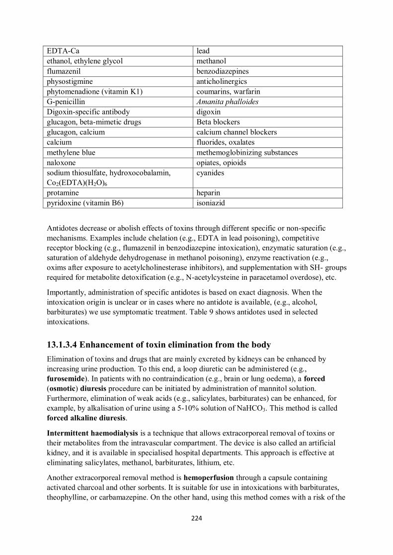
224
EDTA-Ca lead
ethanol, ethylene glycol methanol
flumazenil benzodiazepines
physostigmine anticholinergics
phytomenadione (vitamin K1) coumarins, warfarin
G-penicillin Amanita phalloides
Digoxin-specific antibody digoxin
glucagon, beta-mimetic drugs Beta blockers
glucagon, calcium calcium channel blockers
calcium fluorides, oxalates
methylene blue methemoglobinizing substances
naloxone opiates, opioids
sodium thiosulfate, hydroxocobalamin,
Co2(EDTA)(H2O)6
cyanides
protamine heparin
pyridoxine (vitamin B6) isoniazid
Antidotes decrease or abolish effects of toxins through different specific or non-specific
mechanisms. Examples include chelation (e.g., EDTA in lead poisoning), competitive
receptor blocking (e.g., flumazenil in benzodiazepine intoxication), enzymatic saturation (e.g.,
saturation of aldehyde dehydrogenase in methanol poisoning), enzyme reactivation (e.g.,
oxims after exposure to acetylcholinesterase inhibitors), and supplementation with SH- groups
required for metabolite detoxification (e.g., N-acetylcysteine in paracetamol overdose), etc.
Importantly, administration of specific antidotes is based on exact diagnosis. When the
intoxication origin is unclear or in cases where no antidote is available, (e.g., alcohol,
barbiturates) we use symptomatic treatment. Table 9 shows antidotes used in selected
intoxications.
13.1.3.4 Enhancement of toxin elimination from the body
Elimination of toxins and drugs that are mainly excreted by kidneys can be enhanced by
increasing urine production. To this end, a loop diuretic can be administered (e.g.,
furosemide). In patients with no contraindication (e.g., brain or lung oedema), a forced
(osmotic) diuresis procedure can be initiated by administration of mannitol solution.
Furthermore, elimination of weak acids (e.g., salicylates, barbiturates) can be enhanced, for
example, by alkalisation of urine using a 5-10% solution of NaHCO3. This method is called
forced alkaline diuresis.
Intermittent haemodialysis is a technique that allows extracorporeal removal of toxins or
their metabolites from the intravascular compartment. The device is also called an artificial
kidney, and it is available in specialised hospital departments. This approach is effective at
eliminating salicylates, methanol, barbiturates, lithium, etc.
Another extracorporeal removal method is hemoperfusion through a capsule containing
activated charcoal and other sorbents. It is suitable for use in intoxications with barbiturates,
theophylline, or carbamazepine. On the other hand, using this method comes with a risk of the

225
patient developing thrombocytopenia. Indications for extracorporeal removal techniques are
life-threatening intoxications with substances present in the intravascular compartment or
patients with decreased clearance due to kidney or liver inadequacy. This approach is
generally only suitable for drugs or toxins that are weakly bound to the peripheral tissues or
plasma proteins (low distribution volume), which have a low molecular weight and are
soluble in water.
Peritoneal dialysis is associated with a risk of infection and is generally only used when
forced diuresis or haemodialysis cannot be performed.
Hemofiltration is based on ultrafiltration through a semipermeable membrane (i.e., 10 to
30 l/h) and is only used for patients with the most severe conditions (e.g., in kidney failure).
In drugs or substances with a very low distribution volume a blood transfusion or plasma
transfusion (plasmapheresis) can be used.
Exsanguination transfusion, which is a complete substitution of the patient’s blood with
donor blood, is an exceptional measure. Intravenous administration of lipid microemulsions,
which can adsorb lipid micelles (e.g., soybean oil microemulsion) is a new approach. This
method enhances elimination of lipophilic substances (e.g., in bradycardia induced by local
anaesthetics, calcium channel blockers, β-blockers, or tricyclic antidepressants).
13.1.3.5 Summary
To assure therapeutic benefit for the treatment of intoxications, this algorithm needs to be
followed:
Assure airway patency, respiratory support;
Controlling pulse, blood pressure, adequate amount of intravascular liquid;
Maintaining of acid-base and electrolyte homeostasis;
Treatment of heart rate disorders;
Treatment of seizures;
Maintaining optimal body temperature;
Toxin identification (i.e., sampling for biochemical and toxicological analyses,
consultation with TIS);
Assessment of the ingested dose;
Assessment of time since the ingestion of the intoxicating substance(s); and
Initiation of adequate treatment (i.e., stop further toxin absorption, administration of
activated charcoal at an amount 5 to 10 times higher than the supposed toxin dose,
gastric lavage, enhancement of toxin elimination, and administration of a specific
antidote).
13.2 Therapy of the shock states
Shock reactions, despite their very variable aetiology, are relatively uniform in their
symptoms and in the late stages, also have negative consequences for the organism. All types
of shock are characterised by circulatory failure resulting in a generalised hypoxia. The
damage to the tissues and organs, which occurs in the context of insufficient oxygen

226
saturation, is reversible if treated in the early stages of shock, but irreversible cell damage
occurs in the later stages. Therefore, either late diagnosis or delayed therapy worsens the
clinical outcome.
Treatment should be initiated immediately with the concomitant implementation of the
following measures:
1. Apply an oxygen mask for two to five and a half minutes; an ECG monitor should
be connected to detect heart arrhythmias, and an O2 blood saturation monitor should
be set up.
2. A catheter should be placed into the central vein and measurements of central
venous pressure (CVP) taken.
3. Placement of a catheter into the peripheral artery to monitor blood pressure and ease
sampling of the arterial blood to analyse blood gas and lactic acid should be
completed.
4. Blood sampling should be completed including: Blood type, complete blood count,
platelet count, differential leucocyte count, coagulation, minerals, biochemical
parameters, albumin, blood gases, and lactic acid, and in case of suspected poisoning,
toxicological analysis should be carried out.
5. In the case of sepsis, sampling of biological material (i.e., blood, urine, stool,
sputum, pus, exudate fluids, cerebrospinal fluid, bone marrow, biopsy material) for
microbiological analysis should be done.
6. A urine catheter should be set up for hourly measurement of diuresis. A nasogastric
tube should be placed in the GIT as a preventative measure to stop atonic gastric
content aspiration as well as to measure fluid losses (i.e., gastric contents) and in case
of unconsciousness or intubation, prior to mechanical ventilation.
7. Body temperature should be continuously monitored, preferably by the temperature
sensor on a Swan-Ganz catheter.
The priority of therapy in these situations is to restore mean arterial blood pressure to 75-
80 mmHg. Drugs affecting cardiac output, regulating vascular system volume, or solutions
increasing the volume of circulating fluids can be used either alone or in combination to
achieve this value. In the case of patients with active bleeding and penetrating trauma, the
procedure of so-called "permissive hypotension" is sometimes appropriate. This involves
maintaining a systolic pressure of 80-90 mmHg, subsequent quenching of the cause of blood
loss, and finally replacing the volume of blood lost.
Comprehensive shock therapy includes other drugs, which improve the prognostic or the
course of shock, such as analgesics, anxiolytics, or oxygen. Because of the rate of the onset of
action as well as the variable absorption of drugs due to poorly perfused parts of the drug, a
preferable way of administration is intravascularly into the large veins. At the same time, with
the onset of therapy, it is also essential to discover the origin of the shock reactions; the next
phases of therapy may vary according to the type of shock.
A non-pharmacological, therapeutic approach can be used, which is the intra-aortic balloon
pump counterpulsation. It is used primarily in case of cardiogenic shock resulting from
227
various aetiologies. The mechanical support for circulation results in reduced left ventricular
load and better blood flow to the myocardium, which can ultimately result in increased
contractility and has a beneficial impact on hemodynamics, in general.
13.2.1 The volume therapy
Volume replacement is a tool to increase preload and cardiac output. This therapy is crucial
for the treatment of hypovolemic, anaphylactic, septic, and obstructive shock. In some cases,
this type of cautious volumetric expansion may lead to increased cardiac output even in
cardiogenic shock. However, for severe heart failure, diuretics and vasodilators should be
used. The administration of volume replacement should be rapid as well as a renewal of
cardiac output and perfusion pressure.
In general, this therapy is initiated intravenously by the infusion of a physiological or Ringer's
solution into a subclavian or jugular vein (e.g., 15-20 mL/min) at a dose of 20-40 mL/kg of
body weight. The colloidal solutions can be added later. Continuous bleeding, cardiac
tamponade, or tension pneumothorax should be considered if there is no improvement in the
clinical state despite the large volume replacement.
The amount of the applied substitutions is mostly slightly underestimated due to the estimated
loss, and the replacement amount is mainly managed by the clinical response. After successful
volume resuscitation, heart rate should slow down, tissue perfusion should be improved,
blood pressure increased, and diuresis increased. In case of significant capillary permeability
during septic shock, a combination of volume replacement with inotropic agents and
vasopressors is appropriate. Depending on their chemical nature, the solutions for volume
substitution can be divided into crystalloids, colloids, and blood substitutes.
Crystalloids
The crystalloids, unlike colloids, freely diffuse through the capillary membrane into the
interstitium. It is necessary to give a three- to fourfold higher volume than the actual
circulating fluid volume to achieve compensation for volume losses in the bloodstream. After
the administration of crystalloids, the fluid in the blood stream is increased; concomitantly,
crystalloids expand into the interstitial fluid, and associated complications develop.
Typical complications of crystalloid application are an increase in tissue oedema and body
weight. After application of an overestimated dosage amount of isotonic saline (0.9%, F1/1),
hyperchloremic metabolic acidosis, impaired renal function, and systemic inflammatory
response can occur. Other isotonic solutions can be used, for example Ringer's solution. The
administration of hypertonic solutions (e.g., 7.5% NaCl) leads to a redistribution of
intracellular fluid and transiently increases intravascular volume. This solution is suitable for
the initial phase of volume resuscitation. It is also advantageous to combine it with colloidal
solutions. Glucose solutions are not suitable for volume replacement.
Colloids
Colloidal solutions represent a heterogeneous group of drugs with different compositions and
effects on the volume of circulating fluid. Their disadvantage compared to crystalloids is an

228
increased risk of anaphylactic reactions and worsening blood coagulation abilities; therefore,
it is often preferred to combine crystalloids with colloids.
According to the effect on the amount of circulating fluid, colloids can be divided into:
• Plasma substituents and
• Plasma expanders (i.e., increasing plasma volume over the volume of the administered
solution).
Colloids can be divided into two groups according to their chemical structure:
• Natural, whose representatives are albumin solutions and
• Synthetic, including dextrans, gelatin derivatives, and hydroxyethyl starch.
Albumin
Albumin is currently available in an isotonic (i.e., 5%) or hypertonic (i.e., 20%) solution.
Compared to the synthetic colloids, it is only used marginally, especially in conditions with
manifesting hypoalbuminemia.
Dextrans
Dextrans belong to the glucans – the polysaccharide compounds composed of glucose units.
Solutions of dextran molecules weighing 40 and 70 kDa are used. Nowadays, preparations
with dextrans are considered obsolete due to numerous allergic reactions and the development
of hemocoagulation disorders.
Gelatin derivatives
These are derived from bovine collagen, and the molecules are increased in size and weight to
approximately 30 kDa. Their effect is short, and it can be prolonged by succinylation. Due to
their isotonic properties, they act as plasma substitutes rather than expanders. Like dextrans,
they often cause allergic reactions (i.e., 0.5-10% of the time). Their effect on blood clotting is
insignificant, and rapid renal elimination results in a mild diuretic effect.
Hydroxyethyl starch
This is a widely variable group in terms of substance structure, molecule size, and the
concentration in solutions used. These properties have a fundamental influence on their
pharmacodynamic effects as well as their pharmacokinetic properties. In the body, the
molecules are progressively hydrolysed, and their size gradually decreases until they are
eliminated by the kidney through glomerular filtration. In the Czech Republic, hypertonic and
isotonic solutions with a mean molecular size (i.e., 130 kDa) are used, characterised by a good
safety profile and a minimum duration of action of six hours. Anaphylactic reactions are rare;
there is a minimal influence of hemostasis, and the influence on renal function is comparable
to gelatin derivatives. The application of hydroxyethyl starch leads to an increase in the α
amylase concentrations in plasma, which can be misinterpreted as a pancreatic disease.
13.2.2 Blood substitutes
Blood transfusions or blood derivative administration is indicated for shock states with blood
loss greater than 30%. The blood may be administered whole or as different blood fractions
depending on the desired effect.
229
Erythrocyte concentrates
Erythrocytes are applied in haemorrhagic shock to achieve adequate haemoglobin levels.
The whole conserved blood may be used after a patient's blood type has been previously
determined, or a blood type of O Rh negative can be administered. In vital indications, the
whole blood can also be given. The erythrocyte mass in standard or leukocyte-poor
preparations can be administered in shock as well as anaemic conditions.
Plasma derivatives
Fresh, frozen plasma is indicated for conditions involving deficiency of coagulation factors or
plasma proteins and for supplementation of blood volume and is the agent used most often in
these scenarios.
13.2.3 Inotropics and vasodilatation
If there is no increase in arterial blood pressure and the restoration of diuresis after the use of
volume replacements, application of an inotropic follows. Inotropics may only be given after
a previous volume addition. Otherwise, blood pressure in the central arteries increases, but the
microcirculation in the peripheral tissues deteriorates. It is also necessary to solve hypoxemia,
tachyarrhythmia, and eventual mineral disruption prior to inotropic application. Because of
the different mechanisms of action leading to the different final effects on the cardiovascular
system, inotropics can be divided into sympathomimetics, phosphodiesterase III (PDE III)
inhibitors, and calcium sensitisers.
Sympathomimetics
This therapy is usually started with an infusion of dopamine at a dose of 0.5-1 mg/kg/min (up
to 4 mg/mL/kg). At this dose, dopamine acts positively inotropic and is used to protect the
splanchnic area; due to the stimulation of dopaminergic D1 and D2 receptors, the vasodilation
of renal vessels is induced. In moderate doses (i.e., up to 10 mg/mL/kg), dopamine is
particularly effective at activating the adrenergic β1 receptors increasing the inotropic effect.
Another dose increase leads to vasoconstriction due to activation of the α1 receptors.
If the patient does not respond to dopamine treatment, it can be replaced with noradrenaline
at a dose of 2-4 mg/kg/min or in case of septic shock, adrenaline, at a dose of 0.01-0.02
mg/kg/min can be substituted. Adrenaline at higher doses causes peripheral vasoconstriction,
including decreased kidney perfusion.
In cardiogenic shock and in patients with low cardiac output, a synthetic dopamine analogue
with a non-selective betasympatomimetic effect, dobutamine, given at a dose of 2.5-10
mg/kg/min, is added to the dopamine. It increases myocardial inotropy and induces a mild
arterial dilatation without affecting the heart rate via β2 receptors. A similar substance with
predominant β2 and D1 mimetic effects is dopexamine.
Sympathomimetic therapy should be terminated if it is not accompanied by an increase in
diuresis, a decrease in lactic acid concentration in arterial blood, and the improving mental
state of the patient.

230
Inhibitors of PDE III
By blocking phosphodiesterase, the degradation of cAMP is reduced. An elevated level of
cAMP results in the activation of calcium channels and the input of calcium into the cell.
Milrinone is used for patients with cardiogenic shock resistant to catecholamine therapy. It
has a positive inotropic and vasodilatory effect with minimal influence on heart rate.
Calcium sensitisers
Like the PDE III inhibitors, levosimendan is a drug of choice in case of inadequate patient
response to catecholamines. Its mechanism of action is to increase the sensitivity of
contractile proteins to calcium, specifically the calcium-dependent binding of levosimendan to
troponin C. Besides the positive inotropic effect, levosimendan also acts as a vasodilator.
Vasodilators
Vasodilators are applied similarly to the sympathomimetics after previous circulating fluid
supplementation. They are mainly used for cardiogenic shock, wherein the reduction in
preload and afterload decreases oxygen consumption in the heart. Another target group is
patients whose vasoconstriction persists after correction of blood pressure values. In
particular, nitroglycerin and isosorbide dinitrate are used. For more information on Nitrates,
see Chapter 5.5 Direct-acting vasodilators.
13.3 Pharmacotherapy for pre-hospital care in patients
with polytrauma
Polytrauma is a simultaneous injury of two or more organ systems, at least one of which
directly endangering the patient's life. It is the most common cause of death for those in their
40s, and the most common cause of the trauma is a car accident involving head, chest, and
abdominal injuries. After checking vital functions at the accident site, the transport of injured
patient to hospital follows. Pre-hospital care for a polytraumatised patient in the emergency
phase is performed using standard procedures or so-called "protocols of trauma" (for example,
the Advanced Trauma Life Support (ATLS)).
Shock therapy Shock therapy occurs concurrently with urgent cardiopulmonary resuscitation. Due to the
polytraumatic injury, patients are endangered by the consequences of traumatic,
haemorrhagic, and related hypovolemic shock. In the patients with circulatory stability, the
application of crystalloids at a dose of at least 10 mL/kg/hr is recommended after the
exclusion of cavity injury. A 2000 mL mixture of colloids and crystalloids in a ratio of 1:1 for
blood loss is recommended. For burns, a ratio of 1:2 colloids to crystalloids, given as a rapid
intravenous infusion, is recommended. In case blood pressure responds inadequately to the
administration of volume substitutions, vasopressors are administered.
Analgesia In the case of critical hypotension, ketamine is a drug of choice (slow intravenous bolus 20-
40 mg). It is a dissociated anaesthetic with a strong analgesic effect on somatic, but not
visceral, pain. Unlike opioids, it does not depress the respiratory centre and, in comparison

231
with other anaesthetic drugs, does not adversely affect hemodynamics. The duration of bolus
administration of this dose is approximately 30-45 minutes. Ketamine may also be used for
the analgosedation of paediatric patients. A major adverse effect is the hallucinogenic effect,
which can be prevented through combination with benzodiazepines. Alternatively, ketamine
may be administered transcutaneously or intranasally in a double dose rather than via
intravenous administration to maintain the duration of the effect.
In other cases, when hypotension of the injured patient is non-critical, fractional opioid
analgesics, such as fentanyl (e.g., 0.05-0.1 mg i.v.) or sufentanil (e.g., 0.01 mg i.v.), are
administered.
The onset of action after intravenous administration of fentanyl is three to four minutes and
has an effective duration of approximately 30 minutes. In comparison to other opioids, it is
less hypnotic. A significant complication of fentanyl administration is accumulation of the
drug, potentially causing respiratory centre depression after the repeated administration.
Sufentanil is seven to 10 times more potent than ketamine; it has a faster onset of action, but a
shorter duration of action. The effects of both substances can be antagonised by administering
naloxone.
Sedation Principally, benzodiazepines are used to induce sedation in a polytrauma patient (see Section
3.1 Psychopharmacology). They are applied by titration intravenously or intramuscularly.
Their effect is not only sedative/hypnotic, but also, they have anticonvulsant, anxiolytic, and
slightly myorelaxant effects.
Diazepam is given at the dose of 2.5 to 20 mg, which can be given rectally to paediatric
patients or adults in case venous access is not possible. For short-term sedation, before
intubation, or as an introduction to general anaesthesia, midazolam (DORMICUM), at 2.5-10
mg can be used. It has alternative routes of administration like ketamine, which are
transbuccal and intranasal.
Corticoids Concentrated single dose or short-term, high-dose glucocorticoid therapy is indicated in shock
states not responding to standard therapy, acid aspiration, or for inhalation trauma. Due to
their immunosuppressive effects, glucocorticoids are used to prevent the development of
anaphylactic and toxic shock. For single use, methylprednisolone (SOLU-MEDROL) is
administered at a dose of 30 mg/kg as a short intravenous infusion.
Antioxidants Development of an ischemic state is associated with shock. Further, ischemic-reperfusion
injury or systemic inflammatory response are conditions that lead to the production of free
radicals and increased consumption of endogenous antioxidants in polytraumatic patients.
Therefore, it is appropriate to simultaneously administer drugs that are important for the
maintenance of basic vital functions as well as antioxidant substances to the injured patients,
which can relieve the damage that develops due to the polytrauma and improve the prognosis
of the injured patient. Most frequently, ascorbic acid is administered intravenously at a dose
of 1500 mg.

232
233
14 Radiopharmaceuticals and radiocontrast agents
14.1 Radiopharmaceuticals
Radiopharmaceuticals (or medicinal radiocompounds) are unique medicinal formulations
containing radioisotopes used in major clinical areas for diagnosis and/or therapy. The main
methodological principle of nuclear medicine is the detection of ionizing radiation originating
from radioactive conversion of radionuclides using specialised detection devices.
Radiopharmaceuticals consist of a radionuclide (i.e., the source of radiation itself) and a
suitable vehicle selected for its pharmacokinetic properties. Radionuclides are artificially
prepared, most having been synthesised in radionuclide generators, accelerators, or in nuclear
reactors.
14.1.1 Classification of radiopharmaceuticals
When radiopharmaceuticals are used for therapeutic application, the carrier compound
provides transportation to the desired site, and the radionuclide provides target tissue
exposure to radiation. Generally, the purpose is to destroy cancerous tissue (e.g., application
of iodine-131 for the thyroid gland, which is applied orally and absorbed specifically by the
thyroid cells) or relieve pain (e.g., Strontium-89 and Samarium-153 in patients with bone
metastases).
Diagnostic radiopharmaceuticals (also called radiotracers) are used for non-invasive in vivo
monitoring of biological phenomena. Radioactive labelling for drug distribution
visualisation, positron emission tomography (PET), or single photon emission computerised
tomography (SPECT) are common practices. The radionuclide (as a radiotracer) is bound to a
carrier via a chelating agent. Properties of the carrier guarantee transport to the target site
where the radiotracer is supposed to act. A lyophilised mixture of a vehicle and additives
(often prepared in the form of a kit) is mixed with the radionuclide solution to form the final
radiopharmaceutical in the dosage form suitable for application.
The route of administration is also important. The parenteral route is most commonly used
(i.e., intravenous, subcutaneous, or intraluminal). It is possible to administer these
preparations orally in the form of an aqueous solution or gelatine capsules. If required,
inhalation of gas or mist is an option, as well. Locally applied radiopharmaceuticals are
preferred for targeted tumour therapy.
According to the labelling and production method, the following radiopharmaceuticals are
enumerated:
Long-term nuclides: Samarium-153 (153
Sm), Iodine-131 (131
I), Strontioum-89 (89
Sr);
Medium-term nuclides: Rubidium-82 (82
Rb), Gallium-68 (68
Ga); and
Short-term nuclides: Flourine-18 (18
F), Carbon-11 (11
C), Nitrogen-13 (13
N), Oxygen-
15 (15
O).

234
14.1.2 Diagnostic radionuclides (radiotracers)
Technetium-99m (99m
Tc) is typically used for scintillation assays. The letter, “m”, refers to
the metastable state of the nucleus, which, at a higher energy level, emits γ photons during de-
excitation. The half-life of this nuclide is approximately six hours. This relatively short
radiation exposure after application is one of the main benefits for diagnostic practice (and
also the very limitation for its application therapeutically). The short half-life also allows
administration of a “stronger” dose with higher activity in order to achieve better exam
results.
Gallium-67 (67
Ga), as a citrate, is used for tumour testing and to localise areas of
inflammation (i.e., so-called gallium scan). Another advantage is particularly high uptake by
hepatocellular carcinoma, lymphomas, malignant melanoma, and soft-tissue sarcomas.
Iodine-131 (131
I), despite being the longest used isotope in practice, is being gradually
displaced by technetium (99m
Tc). Radioiodine is a mixed β and γ emitter, which is still widely
applied for the treatment of differentiated thyroid carcinoma (due to the high iodine uptake
capacity of the thyroid) and thrombus identification. A dosimetrically detectable γ radiation
component serves as a tracking signal in the therapy of thyroid disease and metastases
location. However, for diagnostic examinations, only iodine-123 (123
I) and iodine-125 (125
I)
isotopes (for a better ratio of β and γ radiation) are preferred. Likewise, these isotopes are
preferable for the labelling of monoclonal antibodies (125
I-MoAb).
Flourine-18 (18
F) is a positron (β) emitter. However, as a stand-alone drug, its great
disadvantages are a short half-life (i.e., approximately 110 min), high energy annihilation
radiation, and high requirements for protection from radiation. Instead, fludeoxyglucose
(18
FDG) is the primary radiopharmaceutical used in positron emission tomography (PET). 18
FDG is a glucose molecule in which one oxygen atom is replaced with an isotope of
fluorine-18 and is transported and phosphorylated as classical glucose. Its advantage is high
uptake by metabolically active tissues, especially pathological tissues.
The overview of the most commonly used radiopharmaceuticals is summarised in Table 10.
Table 10: Radiopharmaceuticals
Radiopharmaceuticals in SPECT 99m
Tc Technetium broad utility spectrum 201
Tl Thallium myocardial imaging (i.e., perfusion) 131
I Iodine thyroid carcinoma (i.e., radiation destruction of tissue) 67
Ga Gallium inflammation and tumours 111
In Indium leukocytes – infection/inflammation imaging
Radiopharmaceuticals in PET 18
FDG 18F-fludeoxyglucose primary PET radiopharmaceutical 18
FLT 18F-fluoro-L-
thymidine
proliferation marker
visualisation of organs poorly visible otherwise by FDG
(brain)
tumour severity assessment 18
FCH 18F-fluorocholine proliferation marker
brain, lung, prostatic, and oesophageal tumours

235
18F
FET
18F-
fluoroethylthyrosin
brain tumour detection (more sensitive than FDG)
11C
MET
11C-methionine brain tumours
11C Ac 11C-acetate renal, pancreatic, prostatic tumours
18F
NaF
18F-sodium fluoride localisation of primary bone tumours and metastases
Other radionuclides 57
Co Cobalt vitamin B12 resorption screening 59
Fe Iron haematology 51
Cr Chromium survivability of erythrocytes
human plasma albumin labelling 42
K, 22
Na, 24
Na, 82
Br
Potassium, Sodium,
Bromine
plasma ions
85Sr Strontium palliative care in bone malignancies
11C,
13C
Carbon V/Q lung scan (i.e., ventilation/perfusion scintigraphic
lung scan)
urea breath test 45
Ca Calcium bone metabolism screening 32
P Phosphorus radiolabelled DNA and RNA probes for Northern and
Southern blots
14.2 Radiocontrast agents
Contrast agents (i.e., media), used for X-rays, are substances that increase imaging contrast,
thereby improving image quality and resolution. Positive contrast media, due to their high
atomic number, increase radiation absorption, so they appear in the image as more radiopaque
(i.e., "dark spots"). Negative contrast media, of low density, decreases radiation absorption, so
their appearance in the image is more radiolucent (i.e., "bright spots").
There are various possibilities for routes of administration: administration via physiological
orifices (e.g., GIT, bile ducts, peritoneal cavity), pathological orifices (e.g., fistula), systemic
administration (e.g., intravenous, intra-arterial), or into the spinal canal.
14.2.1 Positive contrast media
The basic positive contrast agents include barium compounds (i.e., barium sulfate (BaSO4)).
The white suspension of barium sulphate is most commonly used to visualise the digestive
tract. Due to the very nature of the suspensions, specifically their tendency to sediment, the
preparation should be thoroughly stirred and homogenised prior to application (even RMP).
However, this insoluble salt can cause many complications, such as constipation, diarrhoea,
236
abdominal pain, and rarely, appendicitis. The possible occurrence of barium stones may result
in complete bowel obstruction.
14.2.1.1 Water-insoluble
Iodine contrast agents in a carrier oil have proven to be useful for lymphography and
chemoembolisation. Ethyl-iodised poppy oil is also available as an RMP (e.g., Lipiodol Ultra-
Fluide).
14.2.1.2 Water-soluble
Aqueous contrast media can be divided according to the route of excretion. Hepatotropic
agents are excreted via the liver (i.e., with bile); thus, they are convenient for use in
cholangiography and are given intravenously to reduce the risk of side effects. Nephrotropic
agents are excreted via kidneys. An ideal nephrotropic contrast medium would not affect
physiological functions, could be rapidly renally eliminated, and allow for intensive contrast.
They are classified as high-osmolar (e.g., ioxitalamic acid) and low-osmolar (e.g., iohexol,
iopamidol, iopromide, ioversol, iodixanol, iomeprol, iobitridol). They are mainly used for
angiography, urography, and as a CT contrast agent. Of the side effects, nausea, vomiting, and
feeling overheated are most common.
14.2.2 Negative contrast media
Negative media that reduce radiation absorption are all gases (e.g., oxygen, air, carbon
dioxide, nitrous oxide). Often, they are applied for colon distention, and their administration
does not induce significant complications. Methylcellulose and mannitol are administered
per os and are not absorbed in the digestive tract. To the contrary, they absorb what water
remains in the intestine, so transient diarrhoea after administration is enabled.
14.2.3 Contraindications and side effects
Contrast agents are not suitable for children under 15 years of age. There is also a risk of
iodine allergy (for iodine contrast agents), asthma, eczema, and drug allergy. In 2% of
patients, delayed response (e.g., fever, nausea, joint pain) 1-7 hours after application can
occur and then spontaneously subside. For pregnant women, consideration should be given
with regard to the risks/benefits of application as contrast agents are able to pass into foetal
circulation. Nursing women are advised to avoid breastfeeding until at least 24 hours after
administration. Contraindications also include renal and cardiac insufficiency and severe DM
(e.g., caution must be taken especially when given perorally in conjunction with the
antidiabetic metformin!).
The most serious complication is contrast medium-induced nephropathy (CMIN), which
can cause acute renal failure necessitating hospitalisation and haemodialysis. The kidneys
may be safely protected by nephroprotective agents, such as acetylcysteine or a sodium
bicarbonate infusion.

237
14.2.4 Application of radiocontrast agents
Prior to administration, thorough education of the patient is necessary. The patient should not
eat at least four hours before the procedure, but adequate water intake is required. Informed
consent is needed, and allergic anamnesis, urea, and creatinine levels should be closely
monitored.
After administration, the patient is observed for 30 minutes with vein access kept in place in
case of emergency. Steady, sufficient hydration should be secured for 24 hours (i.e., at least
half a litre of water before the procedure and half litre after or eventually an infusion should
be administered) following the procedure.

238
15 Phytopharmacology
15.1 Introduction to phytopharmacology
The use of herbs, animals, their by-products, and minerals is as old as mankind. The first
written evidence of healing with natural products can be found in almost all ancient
civilisations including Mesopotamia, Egypt, India, Greece, Rome, and China. Well-known
Ebers papyrus, dated to approximately 1600 BC, contains hundreds of natural remedies
including instructions for their preparation. Ancient natural healing was strongly related to
religion; therefore, signs of shamanism (i.e., magical ceremonies and formulas) can be found
in the description of remedies.
Natural healing by laymen (i.e., not provided by priests) developed primarily in Greece, and
around 400 BC, Hippocrates became the most well-known layperson representative. In about
300 BC, the author of first herbariums and famous Greek botanist, Theophrastus, lived.
Galen, a follower of Hippocrates, lived in ancient Rome 200 AD. He was the first to use the
pharmacological terms, agonist and antagonist, which are still used today. Moreover, he
designed the framework for quality management systems for remedial preparation purposes.
In the same era, Dioscorides wrote De materia medica, a fundamental pharmacopoeial work
summarising remedies prepared from herbs, animals, and minerals, which was used for the
preparation of remedies until 17th century.
The knowledge of ancient Arabic healers and alchemists enriched European
phytopharmacology with new herbs and novel laboratory and preparation techniques. In the
middle ages, knowledge and healing services returned to the hands of church. That led to the
deceleration of research in the field of pharmacology as the church strictly followed the works
of ancient Greek medicine men.
In the Renaissance, pharmacological research is revived. First, transatlantic navigations
discover new herbs for European pharmacology, and there is a development of alchemy with
an increase in the number of chemical compounds. One significant person of this period is
alchemist and medicine man, Paracelsus. The work of Dioscorides was reworked by Pietro
Andrea Gregorio Mattioli in Italy, and Mattioli’s herbal, which includes the summary of the
medical properties of herbs, has been translated in many European countries.
Efforts to critically evaluate the effects of medicinal herbs and to identify compounds
responsible for the biological effects prevailed over the empirical and layman’s use of herbs
in the 19th
century. Medical herbs were gradually replaced by their active constituents and
more often, with chemical drugs. This practice still endures today; however, the research of
medicinal uses of herbs continues.
Ethnopharmacology is focused on, aside from other aspects of plants, herbs used as different
ethnic traditional medicines. Researchers cooperate with local shamans and try to discover
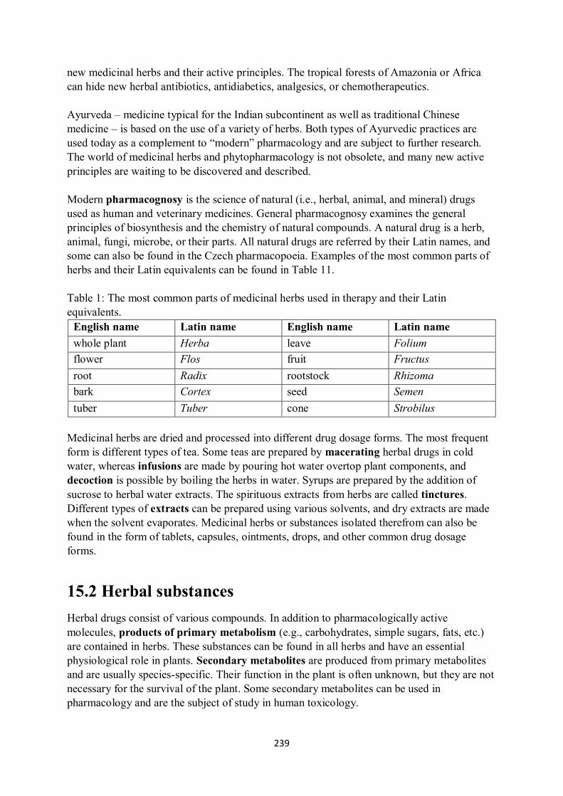
239
new medicinal herbs and their active principles. The tropical forests of Amazonia or Africa
can hide new herbal antibiotics, antidiabetics, analgesics, or chemotherapeutics.
Ayurveda – medicine typical for the Indian subcontinent as well as traditional Chinese
medicine – is based on the use of a variety of herbs. Both types of Ayurvedic practices are
used today as a complement to “modern” pharmacology and are subject to further research.
The world of medicinal herbs and phytopharmacology is not obsolete, and many new active
principles are waiting to be discovered and described.
Modern pharmacognosy is the science of natural (i.e., herbal, animal, and mineral) drugs
used as human and veterinary medicines. General pharmacognosy examines the general
principles of biosynthesis and the chemistry of natural compounds. A natural drug is a herb,
animal, fungi, microbe, or their parts. All natural drugs are referred by their Latin names, and
some can also be found in the Czech pharmacopoeia. Examples of the most common parts of
herbs and their Latin equivalents can be found in Table 11.
Table 1: The most common parts of medicinal herbs used in therapy and their Latin
equivalents.
English name Latin name English name Latin name
whole plant Herba leave Folium
flower Flos fruit Fructus
root Radix rootstock Rhizoma
bark Cortex seed Semen
tuber Tuber cone Strobilus
Medicinal herbs are dried and processed into different drug dosage forms. The most frequent
form is different types of tea. Some teas are prepared by macerating herbal drugs in cold
water, whereas infusions are made by pouring hot water overtop plant components, and
decoction is possible by boiling the herbs in water. Syrups are prepared by the addition of
sucrose to herbal water extracts. The spirituous extracts from herbs are called tinctures.
Different types of extracts can be prepared using various solvents, and dry extracts are made
when the solvent evaporates. Medicinal herbs or substances isolated therefrom can also be
found in the form of tablets, capsules, ointments, drops, and other common drug dosage
forms.
15.2 Herbal substances
Herbal drugs consist of various compounds. In addition to pharmacologically active
molecules, products of primary metabolism (e.g., carbohydrates, simple sugars, fats, etc.)
are contained in herbs. These substances can be found in all herbs and have an essential
physiological role in plants. Secondary metabolites are produced from primary metabolites
and are usually species-specific. Their function in the plant is often unknown, but they are not
necessary for the survival of the plant. Some secondary metabolites can be used in
pharmacology and are the subject of study in human toxicology.

240
Pharmacologically active herbal substances are named herbal active principles. Either a
single substance or a mixture of active principles can be related to the effects of herbal drugs.
The content of active principles in herbal drugs varies depending on different factors, such as
geographical location, growing season, process of drying, grade of disintegration, storage
conditions, etc.
Herbal substances can be classified according to their biological origin into the following
classes:
Carbohydrates,
Polyketides,
Alkaloids,
Shikimates, and
Mevalonates (terpenoids).
15.2.1 Carbohydrates
Different carbohydrates are produced in primary versus secondary pathway metabolism in
plants. The most common monosaccharides, such as glucose, fructose, mannose, and
galactose, are primary metabolites and can represent the sugar moiety of secondary metabolite
glycosides. Examples of specific monosaccharides synthesised in plants
are digitalose, digitoxose, and oleandrose. These saccharides contain an
acetyl, methyl, or amino group in their molecule and are part of glycosides.
Plants can produce various polysaccharides (e.g., inulin, pectins, gums, and
mucilages) beyond the most common cellulose and starches. Pectins are formed by α-D-
galacturonic acid monomers. Pectins are used in the food industry (e.g., as emulsifiers,
thickeners, etc.) and can be used therapeutically for constipation and diarrhoea. They create a
hydrated layer on intestinal mucosa through a nonspecific mode of action, which prevents
contact of the mucosa with intestinal content. This mechanism lubricates and protects the
intestinal mucosa.
Mucilages are polysaccharides similar in their osmotic properties to pectins. They can also
form protective hydrated layers over mucosal surfaces. Drugs with mucilaginous content are
plantain leaves (Plantago lanceolata), flax seed (linen seed, Linum usitatissimum), and
hollyhock root (Althaea officinalis). These can be used as expectorants, antitussives, laxatives,
and to protect the inflamed mucosa of the oral cavity and nasopharynx.
15.2.2 Polyketides
Polyketides are compounds whose biosynthesis is based on acetyl coenzyme A. Fatty acids
present in seeds (e.g., plant oils) and on the surface of fruits (e.g., waxes) are some of them.
Other examples of polyketides are anthraquinones, dianthrones, and anthracyclines, from
which many molecules are used therapeutically as laxatives or chemotherapeutics.
digitoxose

241
Macrolides and tetracyclines are frequently used antibiotics, which have a polyketide
structure produced by moulds.
Cannabinoids, some of the active substances in cannabis (Cannabis sativa L.), are
polyketides, too. Tetrahydrocannabinol (THC) is a psychoactive cannabinoid with muscle-
relaxing properties and cannabidiol (CBD) acts as an anticonvulsant.
Other examples of polyketides are lichen acids (e.g., cetraric acid, protocetraric acid, ursolic
acid, etc.). They possess antibacterial activity, and the extract from Iceland moss (Cetraria
islandica) is a part of preparations against oral cavity infections.
15.2.3 Alkaloids
The name, alkaloids, describes the character of these compounds – all are natural compounds
of alkaline nature. Most also possess a bitter taste. True alkaloids are natural compounds
derived from amino acids, which contain a basic nitrogen in the form of heterocyclic ring.
Other molecules with a basic nitrogen in the molecule, but which are not derived from amino
acids, are called pseudoalkaloids. Conversely, those with the nitrogen from an amino acid,
but not in the typical heterocyclic ring formation, are termed protoalkaloids. Alkaloids can
be further subclassified with respect to their chemical structure into molecules with tropane,
isoquinoline, indole, pyridine, purine, steroid, or terpenic structures. More than 15,000
alkaloids have been identified to date. Therefore, the following review gives only some
examples of alkaloids with significant toxicological or pharmacological importance.
Tropane alkaloids
These molecules are derived from ornithine and are present mainly
in herbs of the Solanaceae (nightshades) and Erythroxylaceae (coca)
families. Parasympatholytic agents, atropine, hyoscyamine, and
scopolamine, are alkaloids from deadly nightshade (Atropa
belladonna), black henbane (Hyoscyamus niger), and jimsonweed (Datura stramonium).
Cocaine, found in leaves of the coca plant (Erythroxylum coca), was used as a local
anaesthetic agent, for example, in ophthalmology. Today, it is no longer used therapeutically,
and it is a subject of abuse.
Isoquinoline alkaloids
sennosid (dianthrone, active
compound of the laxative, senna)
cannabidiol
doxorubicin
(anthracycline chemotherapeutic)

242
These alkaloids are derived from phenylalanine and tyrosine. Well-known isoquinoline
alkaloids are opioids from opium, such as morphine and codeine. Morphine is a strong
analgesic, whereas codeine has weaker analgesic effects and antitussive activity. Both
substances cause constipation and are drugs that are abused. Papaverine, with musculotropic
and antispasmodic effects, is a constituent of opium, too.
Another isoquinoline alkaloid is tubocurarine, a dimer compound
isolated from Amazonian Indian arrow poison. The poison is prepared
from the climbing shrub, Strychnos toxifera, and used for hunting.
Tubocurarine has strong muscle relaxant properties.
Emetine from the root of ipecacuanha (Cephaelis ipecacuanha) is a strong irritant of the
vagus nerve and induces emesis. Formerly, it was used as an expectorant and appetite
stimulator in low doses.
Indole alkaloids
Alkaloids derived from tryptophan are called indole alkaloids.
Physostigmine, with a parasympathomimetic effect, is used for the
therapy of glaucoma, and its analogues (e.g., pyridostigmine, distigmine,
and others) are used to treat myasthenia gravis. It is isolated from the
climbing herb, the Calabar bean (Physostigma venenosum).
Strychnine, a well-known poison from the books of Agatha Christie, is isolated from the
strychnine tree (Strychnos nux-vomica). It evokes strong muscle contractions and victims die
from suffocation.
Important pharmacological activity is known to occur with the use of Vinca alkaloids, which
are isolated from lesser periwinkle (Vinca minor). Vincristine and vinblastine are effective
chemotherapeutics used for the chemotherapy of solid and haematologic tumours.
Vinpocetine is alkaloid with vasodilating activity mostly in the CNS. It is used to increase
cerebral perfusion and metabolism after head injuries, stroke, or in neurovascular diseases.
Ergot alkaloids are a special group among other indole alkaloids. They are isolated from
ergot fungus (Claviceps purpurea), parasitic fungi which grow on different cereals.
Intoxications with symptoms of gangrene due to vasoconstriction or hallucinations with
convulsions have been described from past use. In the Middle Ages, intoxication was known
as “Saint Anthony’s fire”. It was thought that some of the affected persons might have been
bewitched. It is postulated that due to this popular notion, epidemics of ergotism may have
been the reason for witch trials in the past. The research of ergot alkaloids led to the
development of many drugs including: methylergometrine, used as an uterotonic agent in
gynaecology, ergotamine, dihydroergotoxine, used for the
therapy of migraines, and other dihydroergotamine derivatives
with vasodilating properties.
Diethylamide of lysergic acid (LSD) and other derivatives of
lysergic acid are hallucinogens that are misused.
strychnine
ergometrine
morphine
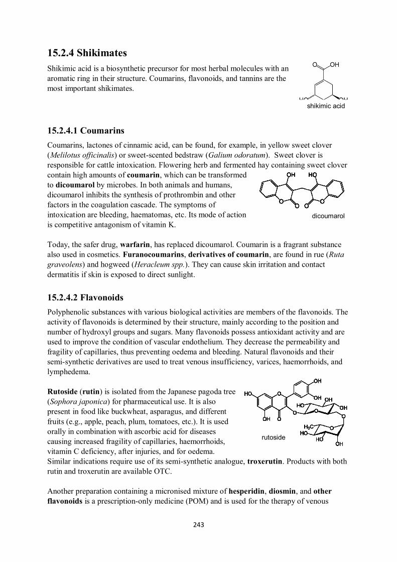
243
15.2.4 Shikimates
Shikimic acid is a biosynthetic precursor for most herbal molecules with an
aromatic ring in their structure. Coumarins, flavonoids, and tannins are the
most important shikimates.
15.2.4.1 Coumarins
Coumarins, lactones of cinnamic acid, can be found, for example, in yellow sweet clover
(Melilotus officinalis) or sweet-scented bedstraw (Galium odoratum). Sweet clover is
responsible for cattle intoxication. Flowering herb and fermented hay containing sweet clover
contain high amounts of coumarin, which can be transformed
to dicoumarol by microbes. In both animals and humans,
dicoumarol inhibits the synthesis of prothrombin and other
factors in the coagulation cascade. The symptoms of
intoxication are bleeding, haematomas, etc. Its mode of action
is competitive antagonism of vitamin K.
Today, the safer drug, warfarin, has replaced dicoumarol. Coumarin is a fragrant substance
also used in cosmetics. Furanocoumarins, derivatives of coumarin, are found in rue (Ruta
graveolens) and hogweed (Heracleum spp.). They can cause skin irritation and contact
dermatitis if skin is exposed to direct sunlight.
15.2.4.2 Flavonoids
Polyphenolic substances with various biological activities are members of the flavonoids. The
activity of flavonoids is determined by their structure, mainly according to the position and
number of hydroxyl groups and sugars. Many flavonoids possess antioxidant activity and are
used to improve the condition of vascular endothelium. They decrease the permeability and
fragility of capillaries, thus preventing oedema and bleeding. Natural flavonoids and their
semi-synthetic derivatives are used to treat venous insufficiency, varices, haemorrhoids, and
lymphedema.
Rutoside (rutin) is isolated from the Japanese pagoda tree
(Sophora japonica) for pharmaceutical use. It is also
present in food like buckwheat, asparagus, and different
fruits (e.g., apple, peach, plum, tomatoes, etc.). It is used
orally in combination with ascorbic acid for diseases
causing increased fragility of capillaries, haemorrhoids,
vitamin C deficiency, after injuries, and for oedema.
Similar indications require use of its semi-synthetic analogue, troxerutin. Products with both
rutin and troxerutin are available OTC.
Another preparation containing a micronised mixture of hesperidin, diosmin, and other
flavonoids is a prescription-only medicine (POM) and is used for the therapy of venous
shikimic acid
rutoside
dicoumarol

244
insufficiency and lymphedema. There are many herbs rich in flavonoid content (e.g.,
quercetin, quercetin, naringenin, and luteolin) including birch leaves (Betula pendula), haw
(Crataegus spp.), the flowers and fruits of elder (Sambucus nigra), and flowers of the lime
tree (Tilia cordata). Citrus fruit, grapes, tea, and other foods can contain significant amounts
of flavonoids, too.
15.2.4.3 Tannins
Tannins are polyphenolic compounds comprised of gallic acid subunits,
which are derived from flavones (i.e., subgroup of flavonoids). They all
possess astringent activity, which means that they coagulate proteins.
They are mostly contained in the roots, bark, and leaves and are an herbal
barrier against infections.
Tannins are used therapeutically as local astringents and hemostyptics to treat
haemorrhoids, skin injuries, and lesions of the oral mucosa (e.g., aphthous stomatitis). Some
substances can be administered orally for anti-diarrhoeal purposes. The mode of their anti-
diarrhoeal effect is through coagulation of the protein layer on the surface of intestinal
mucosa to block contact between chyme and the intestinal wall.
Tinctures prepared from oak apples (galla), herb of agrimony (Agrimonia eupatoria), or
cinquefoils (Potentilla spp.), and oak bark (Quercus spp.) are used topically for their
astringent effect. These preparations contain mostly catechins.
Leaves of blackberry (Rubus fruticosus), raspberry (Rubus idaeus), and strawberry
(Fragaria vesca) are for oral use, and the leaves and dried fruits of blueberry (Vaccinium
myrtillus), which contain hydrolysable tannins, have weaker effect. Black tea, which is rich in
tannins, has an anti-diarrhoeal effect, too.
15.2.5 Mevalonates
Derivatives of the mevalonic acid are called mevalonates or terpenes. Five-carbon chains
comprise the basic subunits that create the foundation for the structure of these molecules. An
overview of terpenes regarding the number of C5 units they contain is shown in Table 2.
Table 2: Classification of terpenoids
Name Number of C5 Example
Hemiterpenes 1 Ergot alkaloids, humulone, and lupulone
Monoterpenes 2 Essential oils (e.g., menthol, camphor, geraniol, etc.)
Sesquiterpenes 3 Essential oils (e.g., azulenes, bisabolene, etc.) and
bittering agents
Diterpenes 4 Essential oils, resins, vitamin A, E, aconitine, and
chlorophyll
Triterpenes 6 Phytosterols, cardioactive glycosides, and saponins
Tetraterpenes 8 Carotenoids
Polyterpenes n Natural rubber
gallic acid
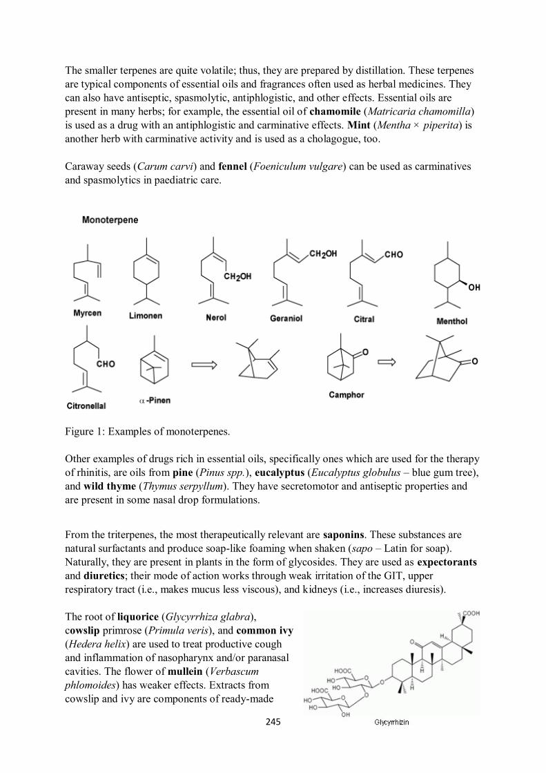
245
The smaller terpenes are quite volatile; thus, they are prepared by distillation. These terpenes
are typical components of essential oils and fragrances often used as herbal medicines. They
can also have antiseptic, spasmolytic, antiphlogistic, and other effects. Essential oils are
present in many herbs; for example, the essential oil of chamomile (Matricaria chamomilla)
is used as a drug with an antiphlogistic and carminative effects. Mint (Mentha × piperita) is
another herb with carminative activity and is used as a cholagogue, too.
Caraway seeds (Carum carvi) and fennel (Foeniculum vulgare) can be used as carminatives
and spasmolytics in paediatric care.
Figure 1: Examples of monoterpenes.
Other examples of drugs rich in essential oils, specifically ones which are used for the therapy
of rhinitis, are oils from pine (Pinus spp.), eucalyptus (Eucalyptus globulus – blue gum tree),
and wild thyme (Thymus serpyllum). They have secretomotor and antiseptic properties and
are present in some nasal drop formulations.
From the triterpenes, the most therapeutically relevant are saponins. These substances are
natural surfactants and produce soap-like foaming when shaken (sapo – Latin for soap).
Naturally, they are present in plants in the form of glycosides. They are used as expectorants
and diuretics; their mode of action works through weak irritation of the GIT, upper
respiratory tract (i.e., makes mucus less viscous), and kidneys (i.e., increases diuresis).
The root of liquorice (Glycyrrhiza glabra),
cowslip primrose (Primula veris), and common ivy
(Hedera helix) are used to treat productive cough
and inflammation of nasopharynx and/or paranasal
cavities. The flower of mullein (Verbascum
phlomoides) has weaker effects. Extracts from
cowslip and ivy are components of ready-made

246
medical preparations (e.g., tablets, syrups, and oral drops).
Other important triterpenes are cardioactive glycosides. Their structure includes a
cyclopentanoperhydrophenanthrene (sterane) nucleus with a lactone ring with
monosaccharide substitutions. Those compounds possess cardiotonic activity by blocking
Na+/K
+-ATPase activity in myocardial cells. Dysregulation of intracellular sodium levels
leads to an increase in intracellular calcium, and the power of contraction increases, too.
Today, most of the herbs containing cardioactive glycosides are only of toxicological
significance. Lily of the valley (Convallaria majalis), pheasant’s eye (Adonis vernalis),
oleander (Nerium oleander), and foxglove (Digitalis purpurea) are examples of such herbs.
Woolly foxglove (Digitalis lanata) is the only herb used as a source of lanatoside C, a
substrate used for the synthesis of digoxin.
15.3 Self-treatment of selected diseases with phytotherapy
Many of common illnesses can be treated by patients themselves through self-treatment. The
use of phytotherapy for self-treatment has been increasing recently. Information about the use
of herbs for the therapy is tied to many myths and fables, and there are many confusing and
sometimes false informational resources regarding herbal therapeutic use found on freely
available databases and sources. The most significant trend in laic patients is the rejection of
“synthetic” drugs. Medicinal plants contain many substances considered “synthetic” drug
xenobiotics with regard to the human body, as has been described previously. At this juncture,
it is not possible to determine whether phytotherapy is superior to the use of “classical”,
“synthetic” drugs. Likewise, it cannot be concluded that use of herbal drugs is related to a
decreased risk of adverse reactions. Furthermore, the risk of interactions with co-administered
drugs should be carefully evaluated.
Therefore, herbal medicines are neither harmless nor omnipotent, and their use should
follow the same rules as apply to any other type of self-therapy:
lanatoside C

247
The herbal medicine should be selected with respect to the knowledge of active
constituents or upon recommendation from a specialist,
the patient informational leaflet should be read carefully and instructions followed for
use of the preparation, and
the patient’s physician or pharmacist should be contacted in case of any problems
occurring.
A review of medicinal herbs used for self-treatment and their active constituents are given in
Table 3.
Table 3: Summary of medicinal herbs for self-therapy and their main active constituents
Symptoms Drug Medicinal herb Components Note
Mild
psychiatric
distress
(nervousnes
s, short-
term
insomnia)
Melissae herba
Melissa
Melissa officinalis
Lemon balm
Essential oils:
citronellal, neral,
geraniol, linalool,
caryophyllene, etc.
Passiflorae herba
Passion flower
Passiflora
incarnata L.
Purple
passionflower
Alkaloids:
harmane, harmine;
flavonoids
Humuli strobilus
Hop cone
Humulus lupulus
Common hop
Essential oils:
myrcene,
farnesene; bitter
acids: humulone,
lupulone
Hyperici herba
St. John’s wort
Hypericum
perforatum
St John’s wort
Naftodianthrone
hypericin;
hyperforin,
flavonoids
CYP3A4
induction
Valerianae radix
Valerian root
Valeriana
officinalis
Garden valerian
Essential oils
“valepotriates”:
valtrate,
isovaltrate;
GABA, flavonoids
Smell attracts cats
Dry cough,
xerostomy
Althaeae radix
Marshmallow
root
Althaea officinalis
Common
marshmallow
Mucilages, pectin Leaf can be
collected
Farfarae folium
Coltsfoot leaf
Tussilago farfara
Coltsfoot
Mucilages,
flavonoids,
triterpenic
substances
Flower can be
collected
Plantaginis
folium
Ribwort plantain
Plantago
lanceolata
English plantain
Mucilages,
aucubin
Has antiphlogistic
activity
Malvae flos Malva spp. Mucilages,

248
Mallow flower g. Mallow anthocyanins
Productive
cough, cold,
sinusitis
Liquiritiae radix
Liquorice root
Glycyrrhiza
glabra L.
Liquorice
Saponins,
flavonoids
Also, used as a
sweetener
Primulae radix
Primula root
Primula veris
Common cowslip
Saponins
Verbasci flos
Mullein flower
Verbascum
phlomoides L.
Woolly mullein
Saponins,
mucilages,
flavonoids,
aucubin
Hederae herba
Ivy leaf
Hedera helix
Common ivy
Saponins
Thymi herba
Thyme
Thymus vulgaris
Common thyme
Essential oils:
thymol, carvacrol,
cineol, etc.
Serpylli herba
Wild thyme
Thymus serpyllum
Wild thyme
Essential oils:
thymol, carvacrol,
citral, linalool, etc.
Eucalypti
aetheroleum
Eucalyptus oil
Eucalyptus
globulus
Tasmanian
bluegum
Essential oils:
eucalyptol,
pinenes,
phellandrene,
cymene, etc.
Has antiseptic
activity
Pini aetheroleum
Pine silvetris oil
Pinus spp.
g. Pine
Essential oils:
bornyl acetate,
pinenes,
phellandrene, etc.
Has antiseptic
activity
Urinary
tract
infections,
dysuria
Urticae herba
Nettle leaf
Urtica dioica
Common nettle
Minerals,
flavonoids,
organic acids,
acetylcholine,
tannins, silicic
acid
Diuretic effect
Betulae folium
Birch leaf
Betula pendula
Silver birch
Saponins,
flavonoids
Diuretic effect
Ononidis radix
Restharrow root
Ononis spinosa
Spiny restharrow
Saponins,
flavonoids,
essential oils
Diuretic and
antiseptic effects
Equiseti herba
Equisetum stem
Equisetum arvense
Field horsetail
Saponins,
flavonoids, silicic
acid
Diuretic effect
Uvae ursi folium
Bearberry leaf
Arctostaphylos
uva-ursi
Kinnikinnick/Bear
berry
Arbutin,
triterpenic acids
Antiseptic effect
Vaccinium vitis- Vaccinium vitis- Proanthocyanidins Blocks bacterial
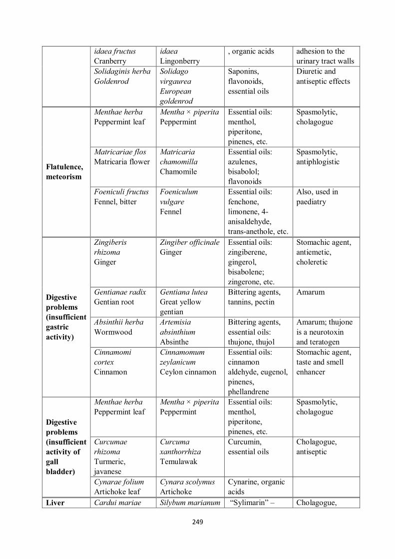
249
idaea fructus
Cranberry
idaea
Lingonberry
, organic acids adhesion to the
urinary tract walls
Solidaginis herba
Goldenrod
Solidago
virgaurea
European
goldenrod
Saponins,
flavonoids,
essential oils
Diuretic and
antiseptic effects
Flatulence,
meteorism
Menthae herba
Peppermint leaf
Mentha × piperita
Peppermint
Essential oils:
menthol,
piperitone,
pinenes, etc.
Spasmolytic,
cholagogue
Matricariae flos
Matricaria flower
Matricaria
chamomilla
Chamomile
Essential oils:
azulenes,
bisabolol;
flavonoids
Spasmolytic,
antiphlogistic
Foeniculi fructus
Fennel, bitter
Foeniculum
vulgare
Fennel
Essential oils:
fenchone,
limonene, 4-
anisaldehyde,
trans-anethole, etc.
Also, used in
paediatry
Digestive
problems
(insufficient
gastric
activity)
Zingiberis
rhizoma
Ginger
Zingiber officinale
Ginger
Essential oils:
zingiberene,
gingerol,
bisabolene;
zingerone, etc.
Stomachic agent,
antiemetic,
choleretic
Gentianae radix
Gentian root
Gentiana lutea
Great yellow
gentian
Bittering agents,
tannins, pectin
Amarum
Absinthii herba
Wormwood
Artemisia
absinthium
Absinthe
Bittering agents,
essential oils:
thujone, thujol
Amarum; thujone
is a neurotoxin
and teratogen
Cinnamomi
cortex
Cinnamon
Cinnamomum
zeylanicum
Ceylon cinnamon
Essential oils:
cinnamon
aldehyde, eugenol,
pinenes,
phellandrene
Stomachic agent,
taste and smell
enhancer
Digestive
problems
(insufficient
activity of
gall
bladder)
Menthae herba
Peppermint leaf
Mentha × piperita
Peppermint
Essential oils:
menthol,
piperitone,
pinenes, etc.
Spasmolytic,
cholagogue
Curcumae
rhizoma
Turmeric,
javanese
Curcuma
xanthorrhiza
Temulawak
Curcumin,
essential oils
Cholagogue,
antiseptic
Cynarae folium
Artichoke leaf
Cynara scolymus
Artichoke
Cynarine, organic
acids
Liver Cardui mariae Silybum marianum “Sylimarin” – Cholagogue,
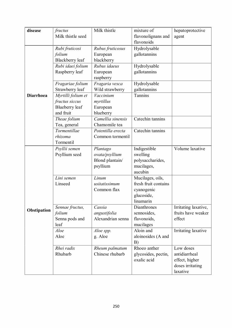
250
disease fructus
Milk thistle seed
Milk thistle mixture of
flavonolignans and
flavonoids
hepatoprotective
agent
Diarrhoea
Rubi fruticosi
folium
Blackberry leaf
Rubus fruticosus
European
blackberry
Hydrolysable
gallotannins
Rubi idaei folium
Raspberry leaf
Rubus idaeus
European
raspberry
Hydrolysable
gallotannins
Fragariae folium
Strawberry leaf
Fragaria vesca
Wild strawberry
Hydrolysable
gallotannins
Myrtilli folium et
fructus siccus
Blueberry leaf
and fruit
Vaccinium
myrtillus
European
blueberry
Tannins
Theae folium
Tea, general
Camellia sinensis
Chamomile tea
Catechin tannins
Tormentillae
rhizoma
Tormentil
Potentilla erecta
Common tormentil
Catechin tannins
Obstipation
Psyllii semen
Psyllium seed
Plantago
ovata/psyllium
Blond plantain/
psyllium
Indigestible
swelling
polysaccharides,
mucilages,
aucubin
Volume laxative
Lini semen
Linseed
Linum
usitatissimum
Common flax
Mucilages, oils,
fresh fruit contains
cyanogenic
glucoside,
linamarin
Sennae fructus,
folium
Senna pods and
leaf
Cassia
angustifolia
Alexandrian senna
Dianthrones
sennosides,
flavonoids,
mucilages
Irritating laxative,
fruits have weaker
effect
Aloe
Aloe
Aloe spp.
g. Aloe
Aloin and
aloinosides (A and
B)
Irritating laxative
Rhei radix
Rhubarb
Rheum palmatum
Chinese rhubarb
Rhoeo anther
glycosides, pectin,
oxalic acid
Low doses
antidiarrheal
effect, higher
doses irritating
laxative

251
15.4 Interactions of phytotherapeutics with co-
administered drugs
Active components of herbal drugs are subject to the same biological processes as other
xenobiotics. They are absorbed, distributed, biotransformed, and excreted. Pharmacokinetic
interactions with co-administered drugs are possible during all these steps. Simultaneously,
herbal drugs possess their own biological activity; pharmacodynamic interactions of their
active components with co-administered drugs are known, too. The most problematic herbs,
in the terms of drug-drug interactions, are St. John’s wort, ginkgo, and garlic.
St. John’s wort (Hyperici herba, see Table 3) is used for its mild hypnosedative, anxiolytic,
and antidepressant effects. One mode of action of St. John’s wort is increasing serotonin
levels. Therefore, many of its pharmacodynamic interactions are based on the potentiation
of the serotonergic drugs as well as increasing levels or effects of serotonin at synapses. Its
combination with SSRIs, MAOIs, and other antidepressants can evoke serotonin syndrome
(e.g., headache, agitation, confusion, hallucinations, tremor, sweating, hypertension,
tachycardia, and vomiting); this adverse reaction is classified as serious and can be lethal.
Active components of St. John’s wort, hypericin and hyperforin, are inducers of the liver
enzyme, CYP3A4. Therefore, those substances can decrease plasma levels of co-administered
drugs, which are substrates of this enzyme.
Ginkgo leaves (Ginkgo folium) are used for their vasodilating effect induced by a complex of
active constituents, namely flavonoids and ginkgolides. Likewise, they are used for cerebral
and extremity perfusion disorders, in vestibular disorders and tinnitus, and frequently, in
geriatrics. Ginkgolides are strong inhibitors of PAF (platelet activating factor) and can
potentiate the effect of anticoagulants and antiplatelet drugs. Cases of serious bleeding were
documented when combined with warfarin.
Garlic (powder prepared from cloves, Allii sativi bulbus pulveratum) is used for its
immunostimulant, antimicrobial, and anthelmintic effects in the GIT. It is recommended to
treat hypertension and atherosclerosis in traditional medicine. It was proven that garlic mildly
decreases LDL cholesterol, blood pressure, and activates NO synthase. Its active constituents
are essential oils, allicin and ajoene, which influence thrombocyte aggregation, the binding of
fibrinogen on activated thrombocytes, and the production of the prothrombotic, thromboxane
A2. Use of garlic carries a risk of potentiating anticoagulant and antiplatelet drug effects.
Ginseng (Panax ginseng) is another example of an herb interacting with co-administered
drugs. Its roots are a component of preparations used in geriatrics. The active components are
triterpene ginsenosides, which act as mild immunostimulants and improve cognition. Cases
of a serious decrease in the plasma levels of warfarin and oral antidiabetics were
documented when combined with ginseng. The mechanisms of these interactions have been
the subject of recent research.
Eastern purple coneflower (Echinacea purpurea) is an herb used as an immunostimulator.
It contains a combination of sesquiterpenic alcohols, tannins, and essential oils, which are
responsible for its effects. From a pharmacodynamic point of view, it should not be combined

252
with immunosuppressives due to possible antagonism. High doses are hepatotoxic and
should not be used chronically or in combination with other hepatotoxic drugs. Coneflower
extract inhibits CYP3A4; therefore, it can increase the plasma levels of its substrates.
Herbal drugs with mucilage content, which are used as antitussives and expectorants (e.g.,
marshmallow root, mallow flower), produce a hydrated layer over the mucosa of the GIT,
which can decrease absorption of co-administered drugs. These medicines should neither be
used at high doses nor chronically.

253

254
16 Drugs used in dentistry
16.1 Drugs used in prevention and treatment of diseases in
dental hard tissues
16.1.1 Enhancement of enamel and dental resistance
Tooth decay is an infectious process that damages hard tooth tissue, which can lead to pulpitis
or periodontitis. Its pathogenesis involves several factors: the condition of hard dental tissues,
the presence of cariogenic bacteria, the consumption of carbohydrates and acidic foods, the
level of oral hygiene, etc. In the Czech Republic, this dental condition is particularly serious
in children; under five years of age, at least one tooth decay has occurred in more than 60% of
children, but the adult dental condition is not otherwise satisfactory.
The consumption of acidic foods and the activity of the oral microflora reduce the pH in the
mouth, and the gradual demineralisation of the enamel occurs when the critical pH value is
exceeded. Proper diet and dental cleaning techniques can reduce the rate of demineralisation.
Hypomineralisation of enamel is generally accompanied by a lack of Ca, P, Mg, and
vitamins D, C, and A, which can be expected under the following circumstances: inadequate
diet, GIT disease, some endocrine diseases, some medications, etc.
The most effective means of enamel remineralisation is fluoride intake/application (F-):
• From diet and water,
• Systemically using drugs, and
• Locally using dental hygiene and special dentistry techniques.
Fluorides have an affinity for the hydroxyapatite of hard tooth tissues forming
fluorohydroxyapatite, which is more resistant in low pH conditions. Historically, the
fluoridation of water or milk has made a significant impact regarding healthy teeth in
populations that have been closely studied. For example, in the 1950s, drinking water
fluoridation was introduced in the town of Tábor, and the incidence of tooth decay was
reduced by 80% as compared to the control city. Only fluoridated salt is available in the
Czech Republic, fluoride water and milk are not widely used.
Sodium fluoride tablets containing 0.25 mg of F- are used for systemic supplementation.
These products are currently available OTC. The tablets dissolve in the mouth (in this case,
the local effect is also significant), or they are ingested with plenty of water. Fluorides are
absorbed in the duodenum and distributed to, among other places, the saliva, which then has
the local effect of promoting enamel remineralisation. Fluorides are excreted via the kidneys.

255
Systemic tablet supplementation is only indicated individually due to the consideration of
fluoride intake via other routes (e.g., drinking water, toothpaste swallowing in children, etc.).
The optimal preventative dose of F- is 0.04-0.07 mg/kg/day. High intake for a long time can
lead to fluorosis of the enamel, which is the appearance of differently stained areas on the
surface of teeth, but this is merely a cosmetic effect. These stains are highly resistant to tooth
decay. Typically, higher fluoride intake can cause bone fluorosis; this involves the
remodelling of bone structure, a worsening of the mineralisation of bone mass, and an
increased the risk of fractures, for instance, at the femoral neck. Although fluorine
supplementation initially activates osteoblasts and reduces the ability of osteoclasts to
decompose bone mass, the pathological conversion and manifestations of osteomalacia
gradually occur. Therefore, the dose of fluoride administration should always be considered.
Fluoridated toothpastes appeared for the first time in the 1970s, and their use has rapidly
expanded. They contain sodium fluoride, stannous fluoride, sodium
monofluorophosphate, or amine fluoride. The concentration in toothpaste for children (i.e.,
from two years old and up) and adults is different. Fluoridated toothpaste is not recommended
for use on first-cut teeth in infancy or for toddlers. For pre-school and elementary school-aged
children, the necessity of adult supervision of dental care is always required to prevent
toothpaste swallowing and to demonstrate correct cleaning techniques.
Stannous fluoride reduces short-term proliferation of oral microflora, making it suitable for
use in periodontal disease. Some mouthwashes are also fluoridated. Applications for highly
concentrated fluoride products exist primarily for use in special dentistry treatments or dental
hygiene procedures (e.g., remineralising gels, lacquers, etc.).
16.1.2 Dental desensitisation
By grinding the enamel and revealing the dentin layer, intraoral stimuli such as changes in
temperature, pH, etc. may cause significant pain in patients. The status of dentin
hypersensitivity is generally poorly therapeutically viable. Sometimes it helps to remineralise
the enamel by local application of fluorides.
If a cement layer in the teeth is removed and such an action induces significant pain,
adhesives or fillers suitable for the dental cervix (neck) can be applied. The protein content of
dentin tubules can be coagulated through topical application of solutions of zinc chloride or
tannin.
There is also a product containing arginine and calcium carbonate, which, by means of the
binding of arginine to dentin, helps to transport calcium carbonate into dentin tubules to form
a stuffing. The efficacy of commonly available potassium-containing toothpastes (generally
referred to as toothpaste for sensitive teeth) remains controversial.
Laser dentine treatment is a modern method that requires some time for testing and
optimisation. The laser dries the dentine layer and produces a non-porous layer. However, it is
known that unwanted microsprays can be generated in the dentin layer through the action of a
laser with an improperly high energy or pulse duration.

256
16.1.3 Treatment of dental pulp and root canals
Extensive carious lesions endanger the pulp of the affected tooth. If the lesions do not
penetrate the tooth, the drilled tooth is treated with calcium hydroxide, which has antiseptic
effects as well as dentinoplastic effects; meaning, it promotes formation of the tertiary dentin
layer and remineralisation of the site. After that, the filler is made. In case of major affection
and pulp inflammation, the procedure is performed with local anaesthesia, during which the
pulp is amputated and extirpated.
An alternative procedure (e.g., for paediatric dentistry, acute emergency treatment) is the
chemical devitalisation of the pulp by local application of formaldehyde, local anaesthetics,
and Cayenne silica (contains antiseptically and locally aesthetically active eugenol). After 14
days, a new treatment with devitalised pulp extirpation is recommended.
Long-term pulp inflammation may result in its devitalisation. Generally, the cause of the
infection originates as a carious lesion that continues penetration inward into the tooth.
Infections can spread through the coronary and root parts and affect the surrounding bone.
Often, an inflammatory vesicle is formed around the affected tooth, which can also be a
source of systemic infection. The aim of the so-called endodontic treatment is to maintain the
devitalised tooth, to remove infectious matter and necrotic tissue, and to close the root with
filler material.
Powerful antiseptics are used to reliably kill microorganisms present in dental channels (e.g.,
chloramine, sodium hypochlorite, peracetic acid, hydrogen peroxide, and calcium
hydroxide). For more details, see Chapter 9.1 Disinfectants and antiseptics. Local ozone
applications can also be used. The advantage of these treatments is the necrotic tissue
(proteins) coagulates, which allows for it to be suctioned off.
Mechanical fillings (e.g., gutta-percha pins with sealers aka sealants, epoxy resins, and glass
ionomer cements), combined with antiseptics (e.g., zinc oxide, eugenol, and calcium
hydroxide) or anti-inflammatory substances (e.g., salicylates), are used to fill the cleaned
channels.
16.2 Drugs used to treat oral mucosal and
periodontal disease
Depending on the type of disease, anti-inflammatory drugs, astringents, local anaesthetics,
antibiotics, antiviral drugs, antifungals, or immunomodulators are used to treat lesions of the
oral cavity and periodontium. The use of herbal medicine (see below) is also popular and
effective. Formulations suitable for oral cavity treatment include mouthwash solutions, oral
sprays, ointments, adhesive pastes, and gels.
An alternative to, for example, antifungal drugs, is the use of a vaginal tablet that adheres to
the mucous membrane of the mouth (i.e., this use is off-label and outside the approved
indication).

257
Adhesive dosage forms are very often gels - colloidal systems formed by gel-forming agents,
such as cellulose derivatives (e.g., carboxymethylcellulose) or polyacrylate.
Protein-depleted blood plasma extract with low-molecular peptides, amino acids, and
nucleic acids also have adhesive properties; they form a long-lasting coating on the mucous
membrane of the mouth.
Table 14 presents an overview of the pharmacotherapy of selected diseases of the oral cavity
and periodontium.
Table 14: Pharmacotherapy of selected diseases of the oral cavity and periodontium
Disease Drug Class Drugs Drug Dosage
Forms Note
Chronic
gingivitis
Antiseptics
Chlorhexidine,
benzydamine and
others
Sprays, solutions
for rinsing and
spreading, lozenges
(pastilles)
Astringents Aluminium acetate-
tartrate, tannin
Aqueous or
glycerol solutions
for rinsing and
spreading
Periodonti
tis
Antiseptics Chlorhexidine and
others
Sprays, solutions
for rinsing and
spreading, lozenges
Antibiotics and
chemotherapeuti
cs
Metronidazole,
spiramycin,
amoxicillin
Oral tablets,
capsules
To manage acute
exacerbation
Acute
necrotisin
g
ulcerative
gingivitis
Antiseptics
Hydrogen peroxide,
hexetidine,
chlorhexidine,
iodophors
Sprays, solutions
for rinsing and
manual spreading,
lozenges
Antibiotics and
chemotherapeuti
cs
Metronidazole,
clindamycin,
doxycycline,
macrolides
Oral tablets,
capsules
As a more
difficult
treatment course
to support local
therapy
Recurrent
aphthous
stomatitis
(mouth
sores)
Antiseptics
Chlorhexidine,
benzydamine,
hexetidine
Sprays, solutions
for rinsing and
manual spreading,
lozenges
Gentian violet Solution for
manual spreading
Used in
paediatrics, but
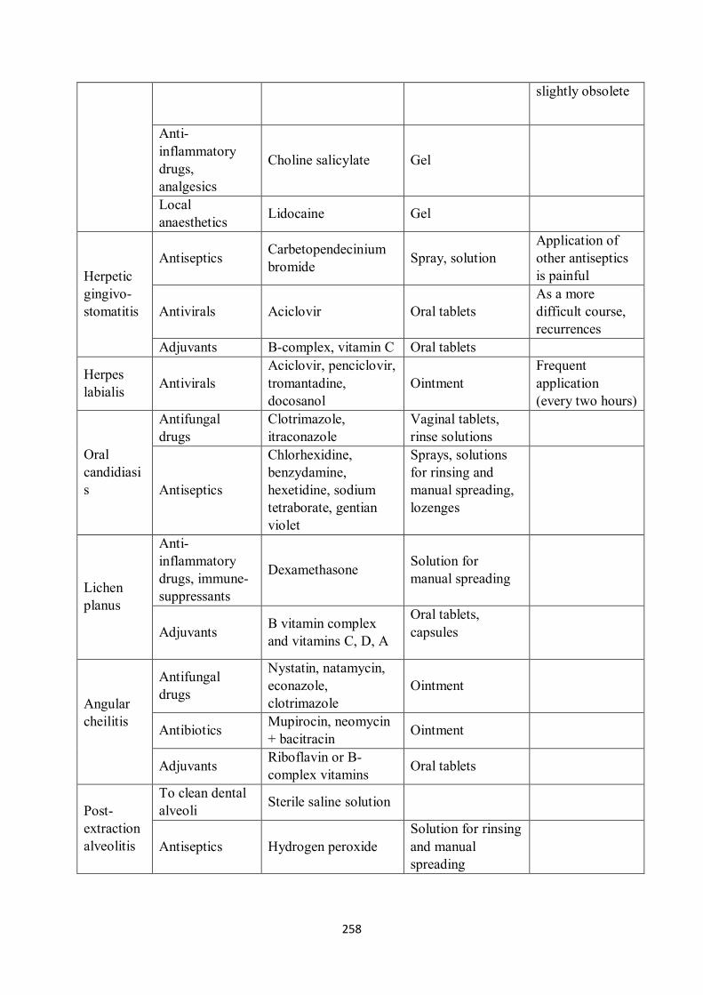
258
slightly obsolete
Anti-
inflammatory
drugs,
analgesics
Choline salicylate Gel
Local
anaesthetics Lidocaine Gel
Herpetic
gingivo-
stomatitis
Antiseptics Carbetopendecinium
bromide Spray, solution
Application of
other antiseptics
is painful
Antivirals Aciclovir Oral tablets
As a more
difficult course,
recurrences
Adjuvants B-complex, vitamin C Oral tablets
Herpes
labialis Antivirals
Aciclovir, penciclovir,
tromantadine,
docosanol
Ointment
Frequent
application
(every two hours)
Oral
candidiasi
s
Antifungal
drugs
Clotrimazole,
itraconazole
Vaginal tablets,
rinse solutions
Antiseptics
Chlorhexidine,
benzydamine,
hexetidine, sodium
tetraborate, gentian
violet
Sprays, solutions
for rinsing and
manual spreading,
lozenges
Lichen
planus
Anti-
inflammatory
drugs, immune-
suppressants
Dexamethasone Solution for
manual spreading
Adjuvants B vitamin complex
and vitamins C, D, A
Oral tablets,
capsules
Angular
cheilitis
Antifungal
drugs
Nystatin, natamycin,
econazole,
clotrimazole
Ointment
Antibiotics Mupirocin, neomycin
+ bacitracin Ointment
Adjuvants Riboflavin or B-
complex vitamins Oral tablets
Post-
extraction
alveolitis
To clean dental
alveoli Sterile saline solution
Antiseptics Hydrogen peroxide
Solution for rinsing
and manual
spreading
259
16.3 Drugs used in salivary disorders
Saliva within the oral cavity serves an indispensable function; it aids in digesting and grinding
food, buffering the oral cavity, remineralising the enamel, repairing soft tissues in the mouth,
maintaining the balance of oral microflora, and fulfilling an immune function (e.g., it contains
lysozyme, cytokines, defensins, etc.).
Increased saliva secretion
Hypersalivation (ptyalism) is present during mechanical irritation of the oral cavity during
dental examination and treatment. In addition, it can accompany oral cavity injury, some CNS
diseases, and occurs concurrently with heavy metal poisoning. Problems with swallowing
saliva are seen in patients with mobility disorders, such as myasthenia gravis or Parkinson's
disease. Increased secretion may also be caused by certain drugs, such as
parasympathomimetic pilocarpine, acetylcholinesterase inhibitors, iodides, etc.
Ptyalism can be treated by oral administration of the parasympatholytic, atropine, which
suppresses gland secretion.
Decreased saliva secretion
Hyposalivation is the cause of xerostomia. Dry mucosa and tongue are susceptible to
infections, injuries, increased risk of caries, and food intake is also more difficult.
Xerostomia often occurs in seniors due to dehydration, metabolic diseases (e.g., diabetes,
hypothyroidism), and certain vitamin deficiencies; it is also typical in Sjörgen's autoimmune
syndrome.
Iatrogenic hyposalivation develops after therapeutic head and neck irradiation in oncology or
from drug use (e.g., tricyclic antidepressants, parasympatholytics, sympathomimetics, first
generation H1 antihistamines, diuretics, sedatives, antipsychotics, opioid analgesics, etc.).
Therapeutic solutions include the use of toothpastes, mouthwashes, sprays, gels, and chewing
gums containing salivary enzymes (e.g., lysozyme, lactoperoxidase), immunoglobulins, and
peptides, and mucosal moisturising aids.
Saliva substitutes, like artificial saliva, are formed through the combination of a salt solution
(e.g., KCl, NaCl, MgCl2, CaCl2, Na2HPO4, NaF) with carboxymethylcellulose (i.e., a gel-
forming agent) and are used several times a day to rinse the mouth. The possibility of
systemic administration of parasympathomimetics, such as pilocarpine or cevimeline, should
also be considered. Cevimeline is registered abroad for use in patients with Sjörgen's
syndrome. The undesirable effects of parasympathomimetics are sweating and acceleration of
GIT peristalsis with diarrhoea and nausea.

260
16.4 Herbal therapy in dentistry
In dentistry, medicinal plants are mainly used for their anti-inflammatory, antimicrobial,
analgesic, astringent, and locally anaesthetic effects. They can be used to rinse the oral cavity,
for mucosal lesions, and to treat root canals.
Drug dosage forms
Vegetable-based drugs may be used in dentistry either unprocessed (less common) or
formulated into drug dosage forms. The most common herbs used are tea blends, which are
fragmented drugs designed to prepare macerations, decoctions, and creams - see Chapter 15.1
Introduction to phytopharmacology. In addition, alcoholic tinctures and extracts are often
used; these can be formulated into aerosols (i.e., sprays for the oral mucosa), gels used to
massage into gums, or ointments.2
Plant extracts are often added to toothpastes and mouthwashes to inhibit the formation of
dental plaque, reduce gingivitis, prevent gum bleeding, and reduce tartar production.
The effective substances
The most important components of herbal drugs used in dentistry are flavonoids, essential
oils, tannins, coumarins, iridoid glycosides, triterpenes, carotenoids, vitamins, and minerals.
For more details on these groups, see Chapter 15.2. Herbal substances.
Some flavonoids may act as antibiotics (i.e., by blocking bacterial cell division), anti-
inflammatory agents, or anti-oedematous compounds.
Essential oils are effective mucosal and skin antiseptics; they act against viruses, bacteria,
yeasts, fungi, and protozoa. Their significant feature is that, unlike synthetic antibiotics,
microorganisms cannot create resistance. Eugenol, carvacrol, and thymol have the most
powerful effects in this regard. Eugenol and thymol, for their strong antibacterial and slightly
anaesthetic action, are used to treat dental cancers during the treatment of dental necrosis.
Tannins can form insoluble compounds with proteins, and they have an astringent effect;
hence, they are able to accelerate wound healing as well as act in a haemostyptic manner (i.e.,
stop minor bleeding). This can be used, for example, to accelerate healing of mouth sores or
lesions on the gums. For more detailed information on herbal pharmaceuticals in dentistry, see
Table 15.
2 In some Third World countries (e.g., Kenya, Tanzania), the local inhabitants chew extracts called Miswaki.
This natural "toothbrush" is the only form of dental hygiene in many regions. It has been shown that some of the
ingredients in these Chewing sticks (Miswak in English) have the ability to prevent gingivitis and tooth decay.

261
Table 15: Medicinal plants and their use in dentistry
Medicinal plant
and its part
used
Active
substances Effects of plant Use in dentistry
Chamomile
flower
(Matricaria
chamomilla)
Essential oils,
flavonoids Anti-inflammatory
Oral cavity rinse for wound
healing, inflammation
treatment, chemically irritated
or burned areas
Arnica montana
(flower)
Essential oils,
flavonoids,
sesquiterpenic
lactones
Anti-inflammatory,
analgesic, increases
blood supply to
tissues, antiseptic
Oral cavity rinse, relieves
inflammation, improves
wound healing (not to be used
internally!)
Salvia officinalis
(leaf)
Essential oils,
antibacterial
flavonoid,
carnosol, tannins
Anti-inflammatory,
antiseptic,
astringent
Infusion for rinsing of oral
cavity and throat, gargle,
extracts (Florsalmin®) for
manual spreading, or after
dilution for rinsing
Galls on
Quercus robur
L.
Tannins Astringent
Tincture for rinsing and
manually spreading on gums
(see above)
Krameria root
(K. argentea +
K. lappacea)
Tannins Astringent
Tincture for rinsing and
spreading on gums (see
above)
Myroxylon
(Peruvian balm)
Phenolic acids,
terpenes
Antiseptic,
granulation
Treatment of complications
after tooth extraction (rare)
Calendula
officinalis,
marigold
(flower)
Essential oils,
flavonoids,
carotenoids,
tannins
Anti-inflammatory
Rinsing of the oral cavity,
improved healing after
surgery, and inflammation
Garlic (onion) Essential oils
(alliin) Antiseptic
Chewing of raw garlic for
five minutes sterilises the oral
cavity; garlic tincture can be
given for infections of the
oral cavity
Gum arabic
(Acacia senegal,
syn. Senegalia
senegal, resin)
Salts (e.g., Ca+,
Mg2+, K
+) of
polysaccharides,
cyanogenic
glycosides,
enzymes
Remineralising,
antiseptic
Enhancing enamel
remineralisation (i.e., due to
high Ca2+
salt of
polysaccharides) and
inhibition of dental plaque
formation
Melaleuca
alternifolia (tea
tree oil)
Essential oils
(cineol) Antiseptic
Spreading on infectious
lesions in the oral cavity, one
of the strongest natural

262
antiseptics and disinfectants
Commiphora
myrrha (gum
myrrh)
Essential oils
(eugenol)
Anti-inflammatory,
antiseptic, locally
anaesthetic
Tincture for manual
spreading or rinsing (after
dilution)
Syzygium
aromaticum -
cloves (flower
bud)
Essential oils
(eugenol)
Anti-inflammatory,
antiseptic, locally
anaesthetic
Tincture for rinsing and
spreading manually on gums
263
17 Ophthalmologic drugs3
17.1 Antimicrobial substances
Infectious and non-infectious inflammations may attack different parts of the eye, hence their
classification to conjunctivitis (conjunctiva inflammation), iridocyclitis (inflammation of the
iris and the ciliated column), blepharitis (inflammation of eyelids), keratitis (cornea
inflammation), otitis (iris inflammation), vitritis (vitreous body inflammation), chorioretinitis
(inflammation of the chorioid and the retina), neuroretinitis (ocular nerve inflammation) etc.
Substances used in eye infection therapy can be divided to antibiotic and chemotherapeutic
categories, virostatics, antimycotics and non-specific antiseptics. In vast majority of cases
local administration in the form of eye drops, ointments, gels, eye inserts or sub-conjunctiva
injections is preferred to systemic administration, especially for the sake of achievement of
high local concentration of the drug with minimised adverse effects.
A particular medicament selection depends on the infection originator; the originator is often
typical of the infection localisation – for example keratitis is mostly caused by Staphylococcus
aureus et epidermidis, Streptococcus pneumonieae, Pseudomonas aeruginosa and Proteus, or
adenoviruses and Herpes simplex virus. In lighter cases the drug is chosen empirically on the
basis of clinical image of the infection. In more severe cases a swab is analysed and targeted
drug selection is performed.
Antibiotics and chemotherapeutics
Antibiotics used for bacterial infection therapy include aminoglycoside antibiotics and
fluorinated 2nd generation quinolones. For severe cases more antibiotics are combined in a
preparation for spectrum extension. In the case of inflammations with allergic component
antibiotics are sometimes also combined with glucocorticoids.
The most often used aminoglycoside drugs include tobramycin, gentamycin, kanamycin
and neomycin (the latter in combination with bacitracin), or fluorinated quinolones
ofloxacin, levofloxacin and mofloxacin. Sulfacetamide of sulfonamide group or
chloramphenicol are used less often. Dexamethasone or betamethasone are used in
combination with glucocorticoids (in combination with aminoglycoside drugs or
chloramphenicol).
3 Note: This chapter summarises specific of pharmacotherapy in ophthalmology; most of the drugs are also used for other indications, though. Mechanisms of action, adverse effects and other specifics are summarised in the relevant chapters (antibiotics, chemotherapy drugs, glucocorticoids, non-steroid antiphlogistics, local anaesthetics).

264
Virostatics
Viral infections are most often caused by adenoviruses and herpetic viruses. Virostatics used
most frequently for their treatment are administered locally (aciclovir, ganciclovir), or for
immune reaction and cornea vascularisation attenuation corticoids are sometimes added after
antivirotic therapy; however, premature corticoid administration may damage the cornea. A
new approach is represented by application of vaccines against HSV 1 and 2, which prevent
irreversible changes, although they cannot prevent the infection.
Antimycotics
Mycotic infections of the eye are mostly caused by Aspergullus and Candida. The
administered antimycotics include older amphotericin B, nystatin, natamycin and newer
azole antimycotics such as ketoconazole, clotrimazole, fluconazole, itraconazole, or
voriconazole).
Antiparasitics
Toxoplasama gondii is the most common eye tissue parasite. Therapies include some products
not authorised for use in the Czech Republic (ILPL production in pharmacies or individual
import) – propamidine isethionate or antiseptic chlorhexidine in combination with systemic
itraconazole therapy.
Antiseptics
These substances are intended for eye infection prevention rather than therapy (for example
after contact with a foreign object, swimming in dirty water etc.). The reason is that antiseptic
preparations show insufficient bactericidal effect and in the case of already progressing
(bacterial, viral or parasitic) infection only delay effective therapy. The substances used
include 1.7% boric acid, benzododecinium bromide and carbethopendecinium bromide.
17.2 Anti-inflammatory, anti-allergic and
immunosuppressive drugs
Anti-inflammatory therapy in ophthalmology is used for addressing non-infectious
inflammations (most often uveitis), allergic inflammations (conjunctivities, blepharitis) and in
some case of infectious inflammations for suppression of pathologic immune reactions,
prevention of synechia, or neovascularisation of eye tissues.
Anti-inflammatory and at the same time immunosuppressive therapy is represented by local
administration of corticoids in the form of eye drops and ointments – the drugs used include
dexamethasone, hydrocortisone, cortisone, prednisolone. Where anti-inflammatory
corticoid therapy is not responded to there the next options include systemic glucocorticoids
followed by systemic immunosuppressive therapy (cyclosporin, azathioprin, methotrexate).
265
Non-steroid antiphlogistics (indomethacin, nepafenac, diclofenac) are used in
ophthalmology for attenuation of post-operative pain and prevention of inflammations and
inflammatory complications and also for peri-operative suppression of miosis.
Allergic inflammation mostly results in bilateral reddening, burning or itching sensation,
sometimes even in the wider eye area. Typical treatment of allergic inflammations includes
local antihistaminic drugs - antazoline, ketotifen, sometimes in combination with
sympathomimetics – tetryzoline.
Antihistaminic drugs used in ophthalmology include for example levocabastine, antazoline,
epinastine as a substance with combined effect on H1 receptors (antagonist) and α1 receptors
(agonist).
Mastocyte stabilisers - cromoglycate (Cromohexal) or olopatadine – substances with
antihistamine (H1) effect at the same time acting as mastocyte stabilisers – may be
administered against seasonal – for example pollen – allergies.
17.3 Substances for treatment of ocular vascular disorders
Targeted therapy has penetrated into many areas, including ophthalmology, in the past
decade. In ophthalmology targeted therapy is mainly applied against age-related macular
degeneration (AMD) and recently also against other eye disorders resulting from
neovascularisation, such as in the case of diabetic retinopathy, diabetic macular oedema,
occlusion of the central retina vein and neovascularisation glaucoma. Uncontrolled
neovascularisation related to these diseases may irreversibly damage the eye and result in
blindness.
Age-related macular degeneration (AMD) is the main cause of vision loss in senior population
in developed countries.
Dry form of AMD os characterised by continuous loss of central acuity with
peripheral vision preserved.
Wet form of AMD represented by mere 10 % of AMD cases is responsible for up to
90 % cases of vision loss. This form is connected with new vessel overgrowing into
the space between the pigment epithelium of the retina and the light-sensing uvulas or
scotopic cells, followed by sub-retinal bleeding, swell and scarring in the macular area
(the acuity area of the retina).
Effective pharmacological treatment of the wet form of AMD did not exist before
introduction of biological therapy. Substantial improvement was only brought by medicines
attenuating vascular proliferation, i.e. substances acting against the vascular endothelial
growth factor (VEGF), supporting uncontrolled neovascularisation connected with this
disease. This factor takes four main forms, A, B, C and D. The ophthalmology-relevant
VEGF-A exists in four isoforms with different numbers of amino acids: VEGF121,
VEGF165, VEGF189 and VEGF206.
266
Pathological growth of new vascular formations is mainly caused by VEGF165. VEGF165
selectively stimulates endothelial cell growth and vascular leak. Intravitreal administration of
drugs against VEGF stabilises the quickly progressing disease and even improves acuity. This
disease would progress rapidly without this therapy and would lead to vision loss.
Drugs used:
Aflibercept (Eylea) is a fully human receptor fusion protein, composed of parts of VEGF-
receptors 1 and 2, and human FcIgG, up-taking VEGF-A and placental growth factor (PIGF).
Ranibizumab (Lucentis) is a fragment of humanised monoclonal antibody against VEGF-A.
Pegaptanib (Macugen) is an oligonucleotide binding to and inactivating VEGF165.
Bevacizumab (Avastin) – a VEGF ligand preventing its binding to its receptors and new
vessel generation. Bevacizumab is not currently authorised for AMD indication (only
authorised for treatment of colorectal and breast carcinomas) and thus is used in the "off-
label" manner for AMD treatment.
Vortepofin (Visudyne), administered by systemic infusion, damages biological structures
within the scope of the diffusion area, following laser activation, leading to local vessel
occlusion, cell damage and necrosis under certain conditions. In the Czech Republic this
photodynamic therapy is not available at the moment.
Wider use of drugs against VEGF is so far prevented by their high price.
17.4 Anti-glaucoma and miotic drugs
Glaucoma is an ocular disease with unclear etiology, more often affecting women (66 %) than
men, usually after 40th year of life. The common feature of most glaucoma types is increased
intraocular pressure. This symptom is not present in all glaucoma types, though, but
represents one of the major risk factors. Intraocular pressure is the cause of eye damage in the
case of congenital glaucoma, chronic closed angle glaucoma and other secondary glaucoma
types. Normal tension glaucoma (NTG) is the cause of 1/3-1/2 open angle glaucoma cases.
Glaucoma is the second most frequent cause of vision loss globally.
Glaucoma therapy is above all aimed at prevention of ocular nerve damage, reduction of
intraocular pressure and minimisation of complications and side effects of the therapy. Acute
glaucoma attacks require immediate assistance of an ophthalmologist.
Anti-glaucoma drugs, with the only exception of acetazolamide, are only administered locally
in the form of eye drops, or rarely ointments. According to the principal therapeutic effect
they may be categorised as follows:
Substances reducing production of intraocular fluid

267
Substances affecting outflow of intraocular fluid
Substances with osmotic effect – hyperosmotics
17.4.1. Substances reducing production of intraocular fluid
Carboanhydrase inhibitors
Ophthalmology uses as active agent acetazolamide (systemically!) and further locally
administered derivates not used elsewhere such as dorzolamide and brinzolamide.
Beta adrenerg receptor blockers – beta blockers
Substances used in this area include above all timolol (non-selective without ISA), betaxolol
(beta 1 selective, without ISA), carteolol (non-selective with ISA). These substances (above all
timolol) are often combined with active agents of other groups – most often with
carboanhydrase inhibitors or prostaglandins.
Non-selective and selective sympathomimetics
Out of this rather broad spectrum of medicaments glaucoma therapy uses brimonidine.
Therapy of neovascular glaucoma also uses a stabilised form of adrenalin in drops prepared
rarely as an IPLP compound.
17.4.2 Substances affecting outflow of intraocular fluid
Parasympathomimetics (miotics)
Miotic drugs, substances formerly used for open angle glaucoma treatment, reduce intraocular
pressure by narrowing the pupil and thus improving intraocular fluid outflow through the
trabecular tissue. They are substances of parasympathomimetic group. Parasympathomimetics
reduce intraocular pressure well, but on the other side reduce acuity and worsen vision in
darkness (by narrowing the pupil). Patients taking Miotic drugs should not drive. They are not
often used for glaucoma treatment and the main reason for their administration is miosis
before intraocular surgery. Parasympathomimetics available in the Czech Republic include
carbachol or pilocarpine as HVLP product, and further IPLP products with pilocarpine, or
less often with physostigmine. Effect of pilocarpine usually manifests itself in 15 minutes
and only lasts for 3 – 4 hours; pilocarpine eye gel is applied at bedtime with the effect lasting
for up to 24 hours post application.
Selective sympathomimetice
Clonidine and brimonidine are α2 selective agonists. Their mechanism of action is based on
reduction of intraocular fluid formation in combination with increased uveoscleral outflow.
268
Prostaglandins and their derivates
Prostaglandin derivates, analogues to prostaglandin F2α, are selective agonists of prostanoid
receptors reducing intraocular pressure by increased outflow of intraocular fluid form the
anterior eye segment. Authorised substances include latanoprost, bimatoprost, travoprost,
tafuprost. An interesting and anecdotic adverse effect related to their long-term use is eyelash
lengthening.
17.4.3 Substances with osmotic effect – hyperosmotics
Hyperosmotic substances increase plasma osmolarity, thus inducing water molecule transfer
from the eye to the vessels, which significantly reduces intraocular pressure. Representatives
worth mentioning include mannitol (for peri-operative i.v. administration) and glycerol
(administered p.o.). These substances need systemic administration. They burden the
cardiovascular system and that is why they are not often used in glaucoma therapy.
17.5 Mydriatics and cyclophlegics
These substances widen the pupil and induce transient cyclophlegia, i.e. paralysis of the
ciliary muscle, preventing pupil accommodation. This is used for the purpose of the ocular
fundus examination. Mydriatics and cyclophlegics may also be used for treatment of
intraocular inflammations. Accommodation suppression is also beneficial in the case of
iridocyclitis where accommodation is painful.
On the basis of their application these substances are classified as follows:
Diagnostic mydriatics – with short effect and suitability for diagnostic interventions.
Therapeutic mydriatics – with long-term effect and suitability for treatment of
iridocyclitis for long-term pupil extension leading to prevention of iris-lens symphysis
(fundus synechia) and for iris inflammation mitigation.
Anticholinerg drugs
Dominant drug class in this indication:
Therapeutic mydriatics - atropine (HVLP) and scopolamine (magistraliter
preparation) with 7-14 day effect
Diagnostic mydriatics – those with short-term action include tropicamide (4-6 hr
effect), cyclopentolate (1 day, both HVLP) and homatropine with up to 3-day effect
(magistraliter preparation)
Anticholinerg drugs reduce tear production and may cause dry eye sensation. Other logical
adverse effects include increased intraocular pressure and blurred vision. Where mydriasis
induction together with cyclophlegia is not the aim sympathomimetics are used instead of
anticholinerg drugs.

269
Sympathomimetics
Sympathomimetics applied in the eye reduce hyperaemia and induce mydriasis, not affecting
intraocular pressure too much. They are mainly used for fundus examination, in eye surgery,
as vasoconstringent preparations for differential diagnostics of conjunctivitis and iridocyclitis,
or for disturbance of synechia in uveitis.
The ones usable include phenylephrine, ephedrine and ibopamine.
17.6 Other ophthalmologic drugs
17.6.1 Decongestion drugs
Calming of hyperaemic conjunctiva is done with some sympathomimetics (the same as in the
case of nasal drops) – naphazoline, tetryzoline, oxymetazoline, oxedrine. Some eye drops
containing these substances are over-the-counter drugs, however, their inappropriate
application may worsen the primary cause (“red eye”); the cause must always be considered
and sympathomimetics applies in justified cases only.
The effects of sympathomimetics are sometimes supported by other drugs depending on the
cause of the hyperaemia – for example esculin reducing capillary break rate or antazoline –
antihistaminic with anti-allergic effect.
17.6.2 Local anaesthetics
Local anaesthetics are used in ophthalmology for desensitisation before surgery or various
diagnostic interferences. They may be applied locally in the form of drops (topical
anaesthesia) - gels, ointments or injections (infiltration anaesthesia – retrobulbar – behind the
bulb, peribulbar – with a shorter needle, or intracameral – in the anterior chamber).
Retrobulbar anaesthesia is rarely used for the risk of the bulb and the ocular nerve injury.
Recent about 2 decades have witnessed return towards topical anaesthesia at the expense of
injection anaesthesia.
Local anaesthetics used in ophthalmology include esters oxybuprocaine, tetracaine, and
amides bupivacaine, lidocaine. Other local anaesthetics used abroad include articaine.
17.6.3 Diagnostics
Fluorescein sodium salt – Fluoresceinum natricum 1–2% - HVLP and IPLP and the
unofficial Bengali red (Roseum bengalense natricum 2%) are used in diagnostic procedures
such as for examination of cornea disintegration with epithelium damage. Bengali red is
suitable in the case of conjunctiva epithelium damage. Both substances are prescribed n the
form of IPLP eye drops.
To prevent microbial contamination of aqueous solutions of the colours there are the
diagnostic filtration strips – they are pieces of dry filtration paper saturated with diagnostic
270
colour (fluorescein, Bengali red, lissamine green - unofficial). The strip is then inserted in the
conjunctiva and the colour is diluted by tears.
17.6.4 Tear substitution in case of dry eye syndrome
Dry eye syndrome is a complex pathophysiological term demoting one of the most common
eye disorders. Dry eye syndrome means hyperosmolar or unstable tear film. Dry eye
syndrome can have several causes (age-related, hormonal, immunological, pharmacological,
hypo-nutritional, post-traumatic, inflammatory etc.). Accompanying symptoms include
burning or tingling sensation, painful twinkling etc.
Therapy is based on non-irritant biologically compatible polymers such as polyvinylalcohol
(PVA), polyvinylpyrrolidone (PVP), hydroxypropylmethylcellulose (HPMC), carbomer,
hyaluronic acid, xyloglucane, dexpanthenol or their combinations.
17.6.5 Other ophthalmologic drugs
Cornea damage of various etiology is addressed by auxiliary medicinal products supporting
metabolic and resorption processes in the eye. Intraocular resorption (resorption of exudate,
minor bleeding etc.) is accelerated for example by isotonic solutions of inorganic iodine salts
(such as the pharmacopoeia eye drops with potassium iodide - Kalii iodidi oculoguttae).
Mechanism of action is apparently based on histamine release causing vasodilation. Stronger
anabolic effect on cornea cells is manifested by androgens or synthetic anabolic drugs
(HVLP).
Note: This chapter summarises specific of pharmacotherapy in ophthalmology; most of the
drugs are also used for other indications, though. Mechanisms of action, adverse effects and
other specifics are summarised in the relevant chapters (antibiotics, chemotherapy drugs,
glucocorticoids, non-steroid antiphlogistics, local anaesthetics).

271
272
References
Adam Z, Fojtík Z. Neopioidní analgetika - farmakologie a léčebné využití. Postgraduální Medicína.
2006;8(3):277–86.
Adam Z, Pour L, Vorlíček J, Hájek R, Koptíková J, Šmardová J. Cílená léčba v onkologii. Remedia
Farmakoter Dvouměsíčník Lékaře Farm. 2005;15(4-5):390–404.
Andělová K. Diabetes a těhotenství. Sanquis. 2008; 59:71-73.
Bednaříková D, Kocák I. Hand-foot syndrom po podání inhibitorů tyrozinkinázové aktivity. Klin Onkol
Časopis Čes Slov Onkol Spol. 2010;23(5):300–5.
Binder F. Farmakoterapie v graviditě. Interní medicína pro praxi. 2006;10:447-450.
Bortlík M. Spasmolytika v gastroenterologii. Remedia. 2002;12(1):41–9.
Broukal Z, Merglová V, Janda J, Cabrnochová H, Gojišová E, Pekárek J, et al. Prevence zubního kazu u
dětí a mládeže. LKS Časopis Čes Stomatol Komory. 2011;21(2):34–41.
Bystroň J. Alergenová imunoterapie. Remedia. 2012;22(4):264–8.
Bystroň J. Antihistaminika v léčbě alergického zánětu. Remedia. 2014;24(4):272–8.
Cífková R. Léčba hypertenze v těhotenství. Remedia. 2007;17(3):258-262.
Cvachovec K. Problematika tekutinové resuscitace - koloidy a krystaloidy. Lékařské listy [Internet].
2010; (12). Dostupné z: http://zdravi.e15.cz/clanek/priloha-lekarske-listy/problematika-tekutinove-
resuscitace-koloidy-a-krystaloidy-452593.
Čepický P, Líbalová Z. Vulvovaginitidy pro farmaceuty. Praha: Levret a.s.; 2009.
Černý V. Oběhové selhání a šokové stavy. Postgraduální medicína [Internet]. 2012; (5). Dostupné z:
http://zdravi.e15.cz/clanek/postgradualni-medicina/obehove-selhani-a-sokove-stavy-464718.
Červený P, Koblihová H. Inhibitory angiotenzin konvertujícího enzymu: přehled a porovnání
základních údajů. Remedia. 2009;19(3):215-219.
Červený P. Inhibitory protonové pumpy: přehled a porovnání základních údajů. Remedia.
2009;19(6):432–7.
Češka R, Kvasilová M, Procházková R, Šmelková G, Vrablík M. Hyperlipoproteinemie a
dyslipoproteinemie I. Klasifikace, diagnostika, kardiovaskulární, kardiometabolické a reziduální riziko.
Vnitřní lékařství. 2010;56(6):526-531.
Doležal T, Hakl M, Kozák J, Kršiak M, Lejčko J, Skála B, et al. Metodické pokyny pro farmakoterapii
nádorové bolesti. Klinická onkologie. 2004;17(3):106-113.
Dortová E, Dort J. Kožní a slizniční kandidóza u novorozence a kojence. Pediatr Praxi. 2012;13(3):153–
4.
Doseděl M, Malý J, Rudolf K. OTC léčiva a samoléčení průjmu a zácpy. Prakt Lékárenství.
2010;6(6):306–11.

273
Dostálová Z, Gerychová R. Vulvovaginitidy - záněty vulvy a pochvy. Interní Medicína. 2011;13(6):262-
4.
Dušek L, Mužík J, Malúšková D, Májek O, Pavlík T, Koptíková J, et al. Cancer Incidence and Mortality in
the Czech Republic. Klin Onkol Časopis Čes Slov Onkol Spol. 2014;27(6):406–23.
Ehler E. Současná terapie spasticity se zaměřením na lokální aplikaci botulotoxinu. Neurol Praxi.
2001;2(3):128–32.
Fojtů H. Kašel – diferenciální diagnostika a terapie. Remedia. 2013;23(3):180–5.
Frič P. Probiotika a prebiotika - renesance terapeutického principu : I. Teorie a experimentální
doklady. Postgraduální Medicína. 2005;7(5):472–7.
Fugh-Berman A. Herb-drug interactions. The Lancet. 2000 Jan 8;355(9198):134–8.
Groppo FC, Bergamaschi C de C, Cogo K, Franz-Montan M, Motta RHL, de Andrade ED. Use of
phytotherapy in dentistry. Phytother Res PTR. 2008;22(8):993–8.
Hájek Z. Farmakoterapie předčasného porodu - tokolytika, kortikosteroidy. Klinická Farmakologie a
Farmacie. 2008;22(4):142-4.
Hakl M, Ševčík P. Opioidní analgetika v léčbě chronické bolesti. Remedia. 2012;22(6):408-411.
Hrdina V, Hrdina R, Jahodář L, Martinec Z, Měrka V. Přírodní toxiny a jedy. Praha: Galén; 2004.
Hubková V. K úloze fluoridů v prevenci zubního kazu. Pediatr Praxi. 2001;2(4):180–2.
Husa P. Současné možnosti léčby virových hepatitid. Interní Medicína Praxi. 2005;7(7-8):342–5.
Chrapek O. Současné možnosti léčby vlhké formy věkem podmíněné makulární degenerace. Remedia
2007; 17 (6) 610-616.
InfoPharm. Mikroverze AISLP. 2013.
Isidoro, L., P. Pires, L. Rito, a G. Cordeiro. 2014. „Progressive Multifocal Leukoencephalopathy in a
Patient with Chronic Lymphocytic Leukaemia Treated with Alemtuzumab". Case Reports 2014 (jan08
1): bcr2013201781-bcr2013201781. doi:10.1136/bcr-2013-201781.
Jandík J. K nejčastější poruše vedoucí k šoku patří hypovolemie. Lékařské listy [Internet]. 2000; (20).
Dostupné z: http://zdravi.e15.cz/clanek/priloha-lekarske-listy/k-nejcastejsi-poruse-vedouci-k-soku-
patri-hypovolemie-126649.
Jeřábek J, Kalitová P. Současné možnosti léčby závratí. Neurol Praxi. 2011;12(5):340–3.
Jirkova B., Chrapek O. Červené oko v ambulanci praktického lékaře. Medicína pro praxi 2009; 6(suppl.
G), 14-23.
Jirsová E. Bezpečnost léčiv během kojení. Medical Tribune. 2012;13:C1-C2.
Jirsová E. Farmakoterapie při kojení. Interní medicína pro praxi. 2006;4:198-200.
Juřica J. Moderní terapie kašle. Pediatr Praxi. 2013;14(1):30–8.
Katzung B., Masters S., Trevor A. Basic and Clinical Pharmacology 12th Ed. New York: McGraw Hill
Professional. 2012.

274
Klempíř J, Roth J. Diferenciální diagnostika a léčba polékových extrapyramidových syndromů.
Lékařské Listy Prevence - Diagn - Ter - Péče - Teor - Praxe Příloha Zdr Novin. 2008;57(18):26–9.
Klener P. Epoetin theta. Remedia. 2011;21(3):212–4.
Koberová Ivančaková R, Slezák R. Infekce ústní sliznice. Medicína Praxi Časopis Prakt Lékařů.
2006;3(6):288–90.
Kozák J. Nové názory na dlouhodobou léčbu opioidy. Neurologie pro praxi. 2008;9(1):42-46.
Kriška M, et al. Memorix klinickej farmakológie. Bratislava: Slovak Academic Press; 2002.
Król E, Rych\lowska M, Szewczyk B. Antivirals-current trends in fighting influenza. Acta Biochim Pol.
2014;61(3):495–504.
Kršiak M. Pokroky ve farmakoterapii bolesti. Interní medicína. 2008;10(6):298-304.
Kubiczková L, Matějíková J, Sedlaříková L, Kryukov F, Hájek R, Ševčíková S. Inhibitory proteazomu v
léčbě mnohočetného myelomu. Klin Onkol Časopis Čes Slov Onkol Spol. 2013;26(1):11–8.
Kuchyňka P. Oční lékařství. Praha.: Grada Publishing a.s., 2007.
Kvapil M. Premixované inzuliny v léčbě diabetu. Remedia Farmakoter Dvouměsíčník Lékaře Farm.
2013;23(5):345–53.
Lapčík O, Sosvorová L. Fytoestrogeny a jejich využití v menopauze. Interní Medicína Praxi.
2011;13(1):38–42.
Látalová K. Tardivní dyskineze vyvolaná atypickými antipsychotiky u rizikových skupin pacientů.
Psychiatr Praxi. 2005;6(5):246–51.
Lejčko J. Přehled opioidních analgetik. Praktické lékárenství. 2009;5(4):172-175.
Lejsek J. Terapie šokových stavů [citováno 2015 2.6.]. Dostupné z:
http://stary.lf2.cuni.cz/Projekty/mua/fm/f3y5.htm.
Leštianský B, Hakl M. Strategie opioidní léčby. Neurologie pro praxi. 2010;11(1):50-54.
Lincová D, Farghali H. Základní a aplikovaná farmakologie. Praha: Galén; 2007.
Lukáš K, Žák A. Gastroenterologie a hepatologie. Praha: Grada; 2007.
Lüllmann K, Mohr K, Weihling M. Farmakologie a toxikologie. Praha: Grada Publishing 2004.
Marambio-Jones C, Hoek EMV. A review of the antibacterial effects of silver nanomaterials and
potential implications for human health and the environment. J Nanoparticle Res. 2010 Mar
23;12(5):1531–51.
Marek J. Farmakoterapie vnitřních nemocí - 4. zcela přepracované a doplněné vydání. Grada
Publishing a.s.; 2010.
Marek J. Léčba kortikoidy. ČSL JEP. Doporučené postupy pro praktické lékaře, 2002.
Martínková J., Farmakologie pro studenty zdravotnických oborů. Praha: Grada Publishing a.s., 2007.
Masteiková R. Aplikační systémy pro léčení onemocnění vnitřního oka Praktické lékárenství 2005; 2.
100-102.

275
Mohr P, Hnídek D, Hanka J, Čermák J, Bravermanová A, Melicher T. Psychofarmaka v těhotenství a
laktaci. Postgraduální medicína. 2012:14(1):17-23.
Moravcová M. Farmakoterapie v těhotenství. Practicus. 2009;1:23-26.
Muchová M. Botulotoxin A v léčbě dětské mozkové obrny. Pediatr Praxi. 2011;12(3):194–8.
Murray RK. Harperova biochemie. Jinočany: H H, 2002.
Nováková J, Ondráčková B, Šulcová A. Základy receptury léčivých přípravků [Internet]. 2010 [cited
2015 Jun 1]. Available from: http://portal.med.muni.cz/clanek-553-zaklady-receptury-lecivych-
pripravku.html
Nožinová E. Doporučený postup ČLK: Léky v těhotenství a při kojení. [Internet]. 2010. verze 1.1
[citováno 2014 2.12.]. Dostupné na http://www.lekarnici.cz/getattachment/Pro-
verejnost/PORADENSTVI---KONZULTACE/PORADENSTVI---KONZULTACE/Doporuceny-postup---Leky-
v-tehotenstvi-a-pri-kojen/DP_tehotenstvi_V1-1.pdf.aspx.
Onishi T, Umemura S, Yanagawa M, Matsumura M, Sasaki Y, Ogasawara T, et al. Remineralization
effects of gum arabic on caries-like enamel lesions. Arch Oral Biol. 2008;53(3):257–60.
Opavský J. Stručný přehled analgeticky účinných léčiv pro klinickou praxi. Remedia. 2002;12(6):409–
23.
Opichalová D, Horák P, Vavrdová V, Tichý M. Kostní fluoróza. Interní Medicína Praxi. 2004;6(4):214–7.
Papežová H. Farmakoterapie poruch příjmu potravy. Remedia Farmakoter Dvouměsíčník Lékaře
Farm. 2007;17(6):557–9.
Penka M, Buliková A. Neonkologická hematologie. Praha: Grada; 2009.
Pernicova I, Korbonits M. Metformin - mode of action and clinical implications for diabetes and
cancer. Nat Rev Endocrinol. 2014 Mar;10(3):143–56.
Piťha J, Bednařík J, Zapletalová O, Schützner J, Zámečník J, Ambler Z, et al. Klinický standard pro
diagnostiku a léčbu myasthenia gravis. Čes Slov Neurol Neurochir Časopis Čes Slov Neurol Neurochir.
2012;75(2):244–52.
Piťhová P. Inzulinové režimy z klinického pohledu. Interní Medicína Praxi. 2010;12(11):531–4.
Polák J. Farmakoterapie obezity. Interní Medicína Praxi. 2006;8(2):54–7.
Rahman K. Effects of garlic on platelet biochemistry and physiology. Mol Nutr Food Res. 2007 Nov
1;51(11):1335–44.
Rang HP. Rang and Dale’s Pharmacology, Churchill Livingstone. Elsevier publications; 2007.
Ritter J. A Textbook of Clinical Pharmacology. Arnold publishers; 1999.
Roth J, Klempíř J. Choreatické dyskineze. Neurol Praxi. 2011;12(1):16–7.
Roth J, Klempíř J. Terapie choreatických dyskinezí. Neurol Praxi. 2009;10(6):353–5.
Říhová E., Šišková A. Léčba neinfekční uveitidy, Remedia 2002; 12 (3):179–184.
276
Salajka F. Asthma bronchiale. Doporučený diagnostický a léčebný postup pro všeobecné praktické
lékaře. Centrum doporučených postupů pro praktické lékaře. Novelizace, 2013. Dostupné z:
http://www.svl.cz/files/files/Doporucene-postupy-od-2013/Astma_Bronchiale.pdf
Sobotková M, Bartůňková J. Monoklonální protilátky a další biologická léčiva užívaná v
imunosupresivní léčbě. Remedia Farmakoter Dvouměsíčník Lékaře Farm. 2008;18(5):356–64.
Soška V, Vaverková H, Vrablík M. Stanovisko výboru ČSAT k doporučením ESC/EAS pro diagnostiku a
léčbu dyslipidemií z roku 2011. Interní Medicína Praxi. 2013; 15(10): 322-326.
Souček M, Kára T. Klinická patofyziologie hypertenze. Grada Pub.; 2002.
Souček M, Špinar J, Vorlíček J. Vnitřní lékařství. Grada; 2011.
Soukoulis S, Hirsch R. The effects of a tea tree oil-containing gel on plaque and chronic gingivitis. Aust
Dent J. 2004;49(2):78–83.
SPC léčivých přípravků dostupná z: www.sukl.cz.
Staňková M, Skokanová V. Historie a perspektivy antiretrovirové terapie infekce HIV/AIDS. Remedia
Farmakoter Dvouměsíčník Lékaře Farm. 2007;17(2):175–80.
Suchý V. Farmakognosie: Část speciální 1. Univerzita Komenského; 1993.
Suchý V. Farmakognosie: Část všeobecná. Univerzita Komenského; 1994.
SÚKL. Farmakoterapeutické informace 2009 (2) 1-4. Praha: Státní ústav pro kontrolu léčiv.
SÚKL. Farmakoterapeutické informace 2014 (5) 1-4. Praha: Státní ústav pro kontrolu léčiv.
Svačina Š. Farmakoterapie obezity. Remedia Farmakoter Dvouměsíčník Lékaře Farm. 2002;12(4):247–
51.
Svačina Š. Potenciální nová antidiabetika pro příští desetiletí. Vnitř Lékařství Orgán Českoslov Spol
Vnitř Lékařství Sekce Českoslov Lékařské Spol J E Purkyně. 2009;55(4):429–33.
Svačina Š. Současný stav farmakoterapie obezity – úspěchy, zklamání i naděje. Kapitoly Z Kardiologie
Prakt Lékaře. 2009;1(3):82–5.
Svobodová M., Palos M. Diagnostika a léčba červeného oka. Interní medicína pro praxi 2013; 15(2)
69-74.
Svozílková P., Říhová E., Heissigerová J., Brichová M., Jeníčková D., Kalvodová B. Farmakoterapie
uveitid. Klinická Farmakologie a Farmacie 2009; 23(3): 115–119.
Synek S. Biologická léčba v oftalmologii. Acta Medicinae 2014;(1):75-76.
Szyszkowska A, Koper J, Szczerba J, Pulawska M, Zajdel D. The use of medicinal plants in dental
treatment. Herba Pol. 2010;56(1):10.
Škorňák O, Škorňáková A. Analgosedace pro posádky RZP. IV Konference KZ ZZS ČR; Olomouc, 2012.
Šrámek V, Pavlík M, Ševčík P. Polytraumata [citováno 2015 3.4.]. Dostupné z:
http://www.med.muni.cz/Traumatologie/ark_sv_Anna/Trauma.htm.
Štejfa M. Kardiologie - 3., přepracované a doplněné vydání. Grada Publishing a.s.; 2007.
Švestka T. Funkční poruchy trávicího traktu: dráždivý tračník. Remedia. 2007;17(4):362–6.

277
Taheri JB, Azimi S, Rafieian N, Zanjani HA. Herbs in dentistry. Int Dent J. 2011;61(6):287–96.
Thompson R g., Gottlieb A, Organ K, Koda J, Kisicki J, Kolterman O g. Pramlintide: A Human Amylin
Analogue Reduced Postprandial Plasma Glucose, Insulin, and C-peptide Concentrations in Patients
with Type 2 Diabetes. Diabet Med. 1997 Jul 1;14(7):547–55.
Tomíška M. Aprepitant. Remedia. 2009;19(1):3–8.
Tomko J, Kresánek J, Hubík J, Suchý V, Felklová M, Sikyta B, et al. Farmakognózia: Učebnica pre
farmaceutické fakulty. Osveta; 1989.
Uchálová Vrcholová D. Současné možnosti v substituční terapii syndromu suchého oka. Remedia
2006; 16 (5), 503-508.
Valha P. Vulvovaginitidy. Posgraduální medicína. 2010;12(2):222-4.
Virk MS, Arttamangkul S, Birdsong WT, Williams JT. Buprenorphine is a weak partial agonist that
inhibits opioid receptor desensitization. The Journal of Neuroscience. 2009; 29(22):7341-7348.
Vrablík M, Šnejdrlová M. Farmakoterapie dyslipidemií. Medicína pro praxi. 2010;7(1):6-8.
Vyhnánková L. Kašel při onemocnění horních cest dýchacích. Pediatr Praxi. 2006;7(1):8–12.
Vyzula R. Modrá kniha České onkologické společnosti. Masarykův onkologický ústav; 18th ed., 2015,
302 p.
Zazula R, Rakovcová H. Současné trendy v léčbě intoxikací. Interní medicína pro praxi. 2004;9:454-
458.
Zazula R. Akutní intoxikace a jejich léčba. Praktické lékárenství, 2006;4:176-178.
Zimandlová D, Bureš J. Současné možnosti léčby peptických vředů. Interní Medicína Praxi.
2012;14(2):51–4.
Živný J. Stárnutí ženy a hormonální substituční terapie. Interní Medicína Praxi. 2004;6(8):403–9.