Prices and Distributive Antagonism in the Work of Smith and Kalecki
Br. J. Pharmac. (1985), 86, 19-25
Pharmacological characterization ofD-aminophosphonovaleric acid antagonism ofaminoacid and synaptically evoked excitations on frogmotoneurones in vitro: an intracellular studyR. Corradetti1, Anne E. King, A. Nistri2, Catherine Rovira3 & Lucia Sivilotti
Department ofPharmacology, St Bartholomew's Hospital Medical College, Charterhouse Square, LondonEC1M 6BQ
1 The effect of D-aminophosphonovaleric acid (D-APV) on the depolarizations induced by N-methyl-D-aspartate (NMDA), glutamate, aspartate or quisqualate was studied with intracellularrecordings from frog motoneurones in vitro.2 D-APV (0.5-10 1M) produced a slight hyperpolarization of the motoneuronal membrane withoutsignificant changes in input conductance.3 In control and tetrodotoxin-containing solutions the depolarizations induced by NMDA were
strongly reduced by D-APV while quisqualate depolarizations were unaffected. Responses toglutamate and aspartate were antagonized to an intermediate level. The relatively small conductanceincreases evoked by excitatory amino acids were unaltered in solutions containing D-APV.4 The amplitude of monosynaptic excitatory postsynaptic potentials (e.p.s.ps) was stronglydepressed by D-APV. The amplitude of polysynaptic e.p.s.ps was little changed but their decay timewas reduced.5 It is suggested that D-APV is a powerful and selective NMDA receptor antagonist and that an
endogenous amino acid acting via NMDA receptors may be the transmitter of monosynaptic e.p.s.pson frog motoneurones.
Introduction
The pharmacological classification of excitatoryamino acid receptors has been greatly aided by usingselective antagonists to distinguish various receptorclasses (Watkins & Evans, 1981). Among severalphosphonic derivatives of amino acids apparentlydisplaying specific antagonism to the excitant N-methyl-D-aspartate (NMDA), the D isomer of2-amin-ophosphonovaleric acid (D-APV) seems to be verypotent on spinal cord preparations (Evans et al.,1982). Other studies (reviewed by McLennan, 1983)have found a similar pharmacology at a variety ofcentral nervous system sites, although most authorshave used the racemic mixture of 2-aminophosphon-' Present address: Department of Pharmacology, Universityof Florence, Viale Morgagni 65, 50134 Florence, Italy.2 Author for correspondence.3Present address: Department of Pharmacology, Universityof North Carolina School of Medicine, Chapel Hill, NC27514, U.S.A.
ovaleric acid rather than its D form (the L form isdescribed as virtually inactive; Evans et al., 1982;McLennan, 1982). In the original work by Evans et al.(1982) extracellular recordings from the in vitro spinalcord of the neonatal rat and the adult frog wereemployed so that detailed information on the neuronalmembrane mechanisms underlying the observedeffects was not available. Many other investigatorshave likewise based their experiments on extracellularrecording techniques (McLennan, 1983).We therefore attempted to study the phar-
macological actions of D-APV, using intracellularrecordings from frog spinal motoneurones, as anapproach to a direct demonstration of its phar-macological selectivity towards exogenous excitantsand excitatory postsynaptic potentials (e.p.s.ps). Weused the adult frog as the experimental animal ratherthan the neonatal rat since we wished to record at lowtemperature to reduce amino acid uptake (Davidoff&
C) The Macmillan Press Ltd 1985
20 R. CORRADETTI et al.
Adair, 1975), and also to avoid the possibility ofimmaturity of the amino acid receptor systems (Saitoet al., 1982; Seno et al., 1984).
Methods
Experiments were carried out on lumbar moton-eurones of the frog (R. temporaria) spinal cord slicepreparation as previously described (Nistri & Aren-son, 1983). Animals were kept in an aquarium at6-7 C before use. After decerebration and removal ofthe spinal cord, a parasagittal spinal slice was preparedand placed in a 0.2 ml bath continuously superfusedwith oxygenated Ringer solution at about 10 ml min-lfor 1- 3 days. The composition of the Ringer solutionwas as follows (mM): NaClI 1, KCl 2.5, NaHCO3 17,NaH2PO4 .2H20 0.1, CaCl2 2, glucose 4, and thesolution was gassed with 95% 02 and 5% CO2. Thebath temperature was maintained at 7TC and mon-itored with an electronic thermometer via a miniatureprobe located near the slice.Motoneurones were impaled with microelectrodes
filled with 3 M KCl (50-80M d.c. resistance) andfunctionally identified on the basis of their characteris-tic short latency, all-or-none antidromic spike elicitedby ventral root stimulation. Two dorsal and twoventral lumbar roots contained in sealed bath sidechambers were connected to suction electrodes (filledwith Ringer solution) which provided rectangularelectrical pulses (0.20-0.25 Hz; 0.1Ims; variable inten-sity) from isolated stimulators. A Ag/AgCl pellet wasused as ground electrode and placed downstream fromthe slice. Intracellularly recorded responses wereamplified through a WPI M-707 electrometer withfacilities for bridge balancing, current injection andcapacity neutralization. Responses were displayed ona storage oscilloscope, recorded on line on a twochannel pen recorder and also stored on FM magnetictape (frequency response d.c. - 2.5 kHz) for furtheranalysis. Retrieval and playback analysis were aidedby a Tektronix 5D10 waveform digitizer. Restingmembrane potential was measured, taking as zero thepotential recorded after electrode withdrawal from thecell. Input conductance was calculated from electro-tonic hyperpolarizing potentials regularly elicited byintracellularly-applied current pulses (600 ms) andfrom the slope of current/voltage plots usually con-structed within ± 20 mV from resting membranepotential. All drugs were applied to the bath throughpre-cooled individual flowlines.The following compounds were used: sodium L-
glutamate (BDH), sodium L-aspartate (Sigma),NMDA (Cambridge Research Biochemicals),quisqualic acid (kindly donated by Dr H. Shinozaki orpurchased from Cambridge Research Biochemicals),D-APV (Cambridge Research Biochemicals),
tetrodotoxin (Sigma). Results are expressed as mean+ s.e.mean.
Results
Control responses to excitatory amino acids
Control data for this study were obtained from 53motoneurones with stable resting potential(- 63 ± I mV) and input conductance (96 ± 7 nS) andwith antidromic action potentials overshooting thezero reference line by 15-20 mV. Control applicationsofglutamate (2 mM; 26 cells),NMDA (30 SAM; 20 cells),aspartate (2 mM; 6 cells) or quisqualate (30 gM; 16cells) produced matched amplitude depolarizations of10±1, 10±2, 10±1 and 13±2mV respectively.These responses were accompanied by an increasedvoltage noise, as shown by the thicker baseline duringthe glutamate amino acid application (see Figure 3 topleft). In about 30% of these cells the depolarizationswere preceded by a small hyperpolarizing componentacociated with a conductance increase (cf. Nistri et al.,1985). The percentage apparent input conductanceincreases at the peak of the depolarizing responses tothese amino acids were 21 ± 8, 22 ± 6, 39 ± 17 and27 ± 20, respectively. For each cell the experimentalprotocol consisted of initially testing dorsal root-evoked excitatory synaptic transmission as well asresponses to at least two amino acids during super-fusion with normal Ringer solution and subsequentlyretesting synaptic potentials and amino acids effects inthe presence of D-APV (minimum exposure 15 min):successful runs were completed with 31 cells.
Finally, five motoneurones were first tested for theirresponsiveness to glutamate and later superfused withtetrodotoxin (0.6-1.2 gM) which, after about 30 min,fully blocked regenerative electrical activity includingthat due to interneurones. The resting potential andconductance values for these cells in tetrodotoxinsolution were - 79 ± 9mV and 46 ± 9 nS respective-ly. Peak depolarizations to glutamate, NMDA andquisqualate were 10 ± 2, 7 ± 1 and 9 ± 3 mV respec-tively (cf. top tracings of Figure 3). Their correspond-ing percentage actual conductance increases were22 ± 8, 3 ± 1 and 39 ± 24, respectively. These cells,too, were subsequently bathed in a solution containingD-APV and tetrodotoxin.
Effects of D-APV on motoneurones and theirresponsiveness to amino acids
On 31 motoneurones D-APV was applied in concen-trations ranging from 0.5 to 1OjM. In the presence ofD-APV motoneurones displayed a slight hyper-polarizations which reached an average maximum of3.1 ± 1.4 mV after approximately 15 min. Applica-
AMINO-ACID ANTAGONISM IN THE SPINAL CORD
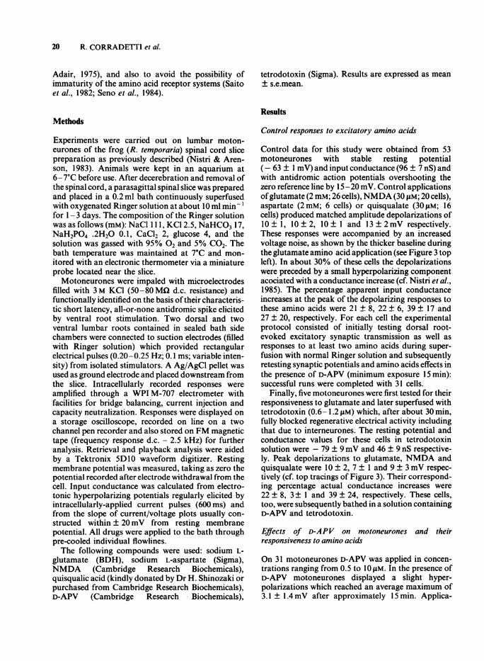
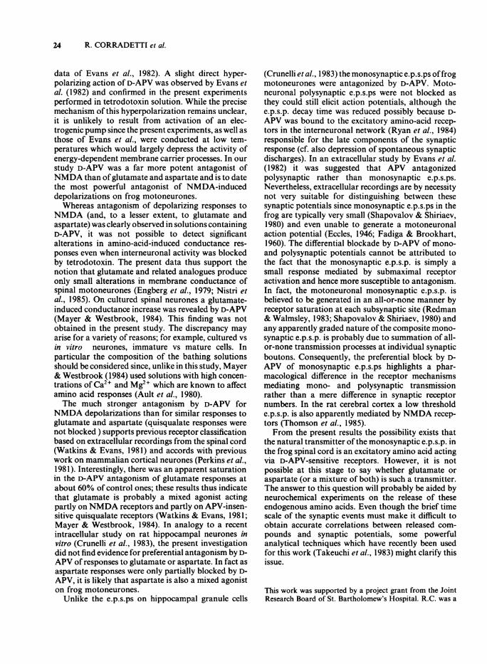
tions ofD-APV produced no change in the slope of thecurrent/voltage line, as shown in Figure 1 (only in onecell was there a modest increase in the slope), or in theamplitude of electrotonic hyperpolarizing potentials
Current (nA)-0.6 -0.4 -0.2 0 0.2
-40r-
-50F-
0.4 0.6
.
a
.9*
120r
100c0
N 80
0.
0a)4) 60'a
- 40
E 20
.0
) _
_
) _
-60h
xU
xX e.
-70k
mV
Figure 1 Current/voltage relation for a single frogmotoneurone in control Ringer (0), in the presence ofIO "M D-aminophosphonovaleric acid (D-APV) (U) and30 min after D-APV washout (x). The relation, apparentlylinear within the values of membrane potentials mon-itored, is typical of that found for the majority ofmotoneurones (Schwindt, 1976). Resting potentials incontrol and D-APV Ringer were -57 mV and -58 mVrespectively.
elicited by a constant current pulse; both tests thusindicated a lack of significant input conductancechanges following D-APV application. No alterationin the configuration of the motoneuronal actionpotential was observed in the presence of D-APV.Changes in the amplitude of excitatory amino acid-
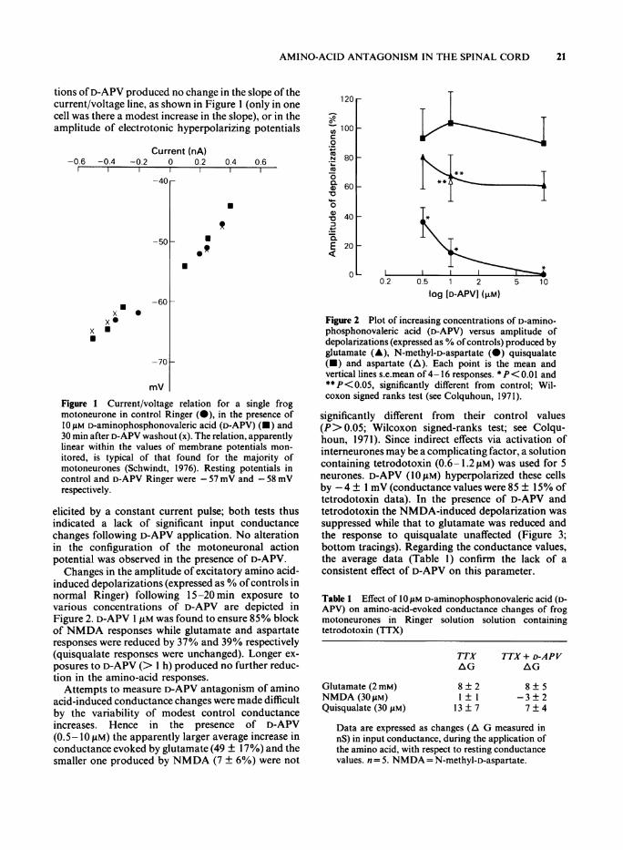
induced depolarizations (expressed as % ofcontrols innormal Ringer) following 15-20 min exposure tovarious concentrations of D-APV are depicted inFigure 2. D-APV I JiM was found to ensure 85% blockof NMDA responses while glutamate and aspartateresponses were reduced by 37% and 39% respectively(quisqualate responses were unchanged). Longer ex-posures to D-APV (> I h) produced no further reduc-tion in the amino-acid responses.
Attempts to measure D-APV antagonism of aminoacid-induced conductance changes were made difficultby the variability of modest control conductanceincreases. Hence in the presence of D-APV(0.5-10IM) the apparently larger average increase inconductance evoked by glutamate (49 ± 17%) and thesmaller one produced by NMDA (7 ± 6%) were not
II0.2 0.5 1 2 5 10
log [D-APVJ (I±M)
Figure 2 Plot of increasing concentrations of D-amino-phosphonovaleric acid (D-APV) versus amplitude ofdepolarizations (expressed as % ofcontrols) produced byglutamate (A), N-methyl-D-aspartate (0) quisqualate(-) and aspartate (A). Each point is the mean andvertical lines s.e.mean of 4-16 responses. *P< 0.01 and**P< 0.05, significantly different from control; Wil-coxon signed ranks test (see Colquhoun, 1971).
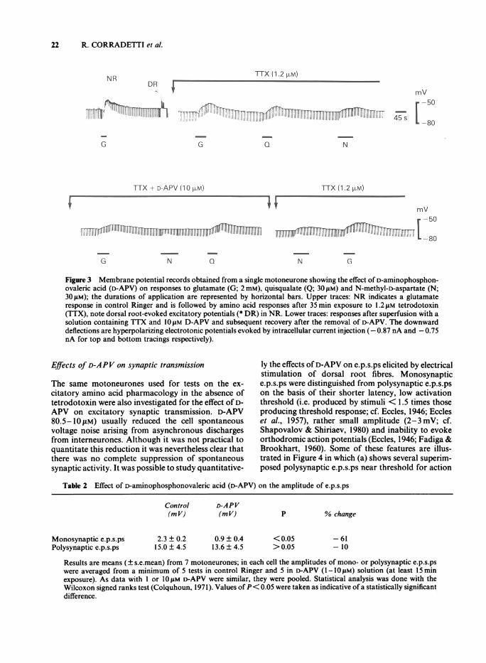
significantly different from their control values(P> 0.05; Wilcoxon signed-ranks test; see Colqu-houn, 1971). Since indirect effects via activation ofinterneurones may be a complicating factor, a solutioncontaining tetrodotoxin (0.6-1.2fM) was used for 5neurones. D-APV (10 M) hyperpolarized these cellsby -4± I mV (conductance values were 85 ± 15% oftetrodotoxin data). In the presence of D-APV andtetrodotoxin the NMDA-induced depolarization wassuppressed while that to glutamate was reduced andthe response to quisqualate unaffected (Figure 3;bottom tracings). Regarding the conductance values,the average data (Table 1) confirm the lack of aconsistent effect of D-APV on this parameter.
Table 1 Effect of 10 pM D-aminophosphonovaleric acid (D-APV) on amino-acid-evoked conductance changes of frogmotoneurones in Ringer solution solution containingtetrodotoxin (TTX)
TTX TTX+ D-APVAG AG
Glutamate (2 mM)NMDA (30 pM)Quisqualate (30 pM)
8 ± 21 ± 1
13 ± 7
8 ± 5-3 ± 27±4
Data are expressed as changes (A G measured innS) in input conductance, during the application ofthe amino acid, with respect to resting conductancevalues. n= 5. NMDA = N-methyl-D-aspartate.
21
22 R. CORRADETTI et al.
TTX (1.2 PLM)DNfNRDR I -
PlillI1111111IRNIg -"m'm 11 olw-.i
G G Q
TTX + D-APV (10 pM)
55""""iIII~II I'lliiuii
N a N
mV
r -50
G
Figure 3 Membrane potential records obtained from a single motoneurone showing the effect of D-aminophosphon-ovaleric acid (D-APV) on responses to glutamate (G; 2 mM), quisqualate (Q; 301sM) and N-methyl-D-aspartate (N;30gpM); the durations of application are represented by horizontal bars. Upper traces: NR indicates a glutamateresponse in control Ringer and is followed by amino acid responses after 35 min exposure to 1.2 tM tetrodotoxin(TTX), note dorsal root-evoked excitatory potentials (* DR) in NR. Lower traces: responses after superfusion with asolution containing TTX and 1O!M D-APV and subsequent recovery after the removal of D-APV. The downwarddeflections are hyperpolarizing electrotonic potentials evoked by intracellular current injection (- 0.87 nA and - 0.75nA for top and bottom tracings respectively).
Effects of D-APV on synaptic transmission
The same motoneurones used for tests on the ex-citatory amino acid pharmacology in the absence oftetrodotoxin were also investigated for the effect of D-APV on excitatory synaptic transmission. D-APV80.5-1OELM) usually reduced the cell spontaneousvoltage noise arising from asynchronous dischargesfrom interneurones. Although it was not practical toquantitate this reduction it was nevertheless clear thatthere was no complete suppression of spontaneoussynaptic activity. It was possible to study quantitative-
ly the effects ofD-APV on e.p.s.ps elicited by electricalstimulation of dorsal root fibres. Monosynaptice.p.s.ps were distinguished from polysynaptic e.p.s.pson the basis of their shorter latency, low activationthreshold (i.e. produced by stimuli < 1.5 times thoseproducing threshold response; cf. Eccles, 1946; Eccleset al., 1957), rather small amplitude (2-3 mV; cf.Shapovalov & Shiriaev, 1980) and inability to evokeorthodromic action potentials (Eccles, 1946; Fadiga &Brookhart, 1960). Some of these features are illus-trated in Figure 4 in which (a) shows several superim-posed polysynaptic e.p.s.ps near threshold for action
Table 2 Effect of D-aminophosphonovaleric acid (D-APV) on the amplitude of e.p.s.ps
Monosynaptic e.p.s.psPolysynaptic e.p.s.ps
Results are means (± s.e.mean) from 7 motoneurones; in each cell the amplitudes of mono- or polysynaptic e.p.s.pswere averaged from a minimum of 5 tests in control Ringer and 5 in D-APV (1-1OI1M) solution (at least 15 minexposure). As data with 1 or 1O gM D-APV were similar, they were pooled. Statistical analysis was done with theWilcoxon signed ranks test (Colquhoun, 1971). Values ofP< 0.05 were taken as indicative ofa statistically significantdifference.
mV-50
45 s -80
5
N
TTX (1.2 FM)
G
Control(m V)
D-APV(mV)
0.9 ± 0.413.6 ± 4.5
2.3 ± 0.215.0 ± 4.5
p
< 0.05> 0.05
% change
-61- 10
111111011111111011
AMINO-ACID ANTAGONISM IN THE SPINAL CORD
D-APV 0.5 ALM
NR
a
I 10 mV
100 ms
b
2 mV
30 ms
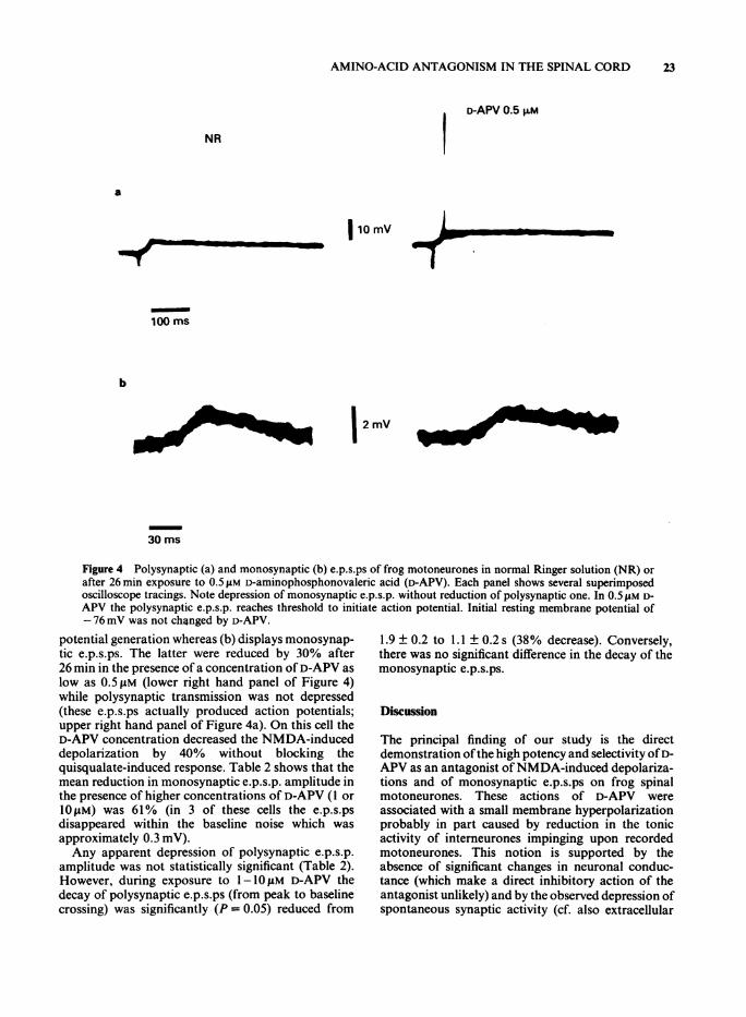
Figure 4 Polysynaptic (a) and monosynaptic (b) e.p.s.ps of frog motoneurones in normal Ringer solution (NR) orafter 26 min exposure to 0.5 AM D-aminophosphonovaleric acid (D-APV). Each panel shows several superimposedoscilloscope tracings. Note depression of monosynaptic e.p.s.p. without reduction of polysynaptic one. In 0.5 AM D-APV the polysynaptic e.p.s.p. reaches threshold to initiate action potential. Initial resting membrane potential of-76 mV was not changed by D-APV.
potential generation whereas (b) displays monosynap-tic e.p.s.ps. The latter were reduced by 30% after26 min in the presence of a concentration of D-APV aslow as 0.5 pM (lower right hand panel of Figure 4)while polysynaptic transmission was not depressed(these e.p.s.ps actually produced action potentials;upper right hand panel of Figure 4a). On this cell theD-APV concentration decreased the NMDA-induceddepolarization by 40% without blocking thequisqualate-induced response. Table 2 shows that themean reduction in monosynaptic e.p.s.p. amplitude inthe presence of higher concentrations of D-APV (1 or1OfiM) was 61% (in 3 of these cells the e.p.s.psdisappeared within the baseline noise which wasapproximately 0.3 mV).Any apparent depression of polysynaptic e.p.s.p.
amplitude was not statistically significant (Table 2).However, during exposure to 1-10 M D-APV thedecay of polysynaptic e.p.s.ps (from peak to baselinecrossing) was significantly (P= 0.05) reduced from
1.9 ± 0.2 to 1.1 ± 0.2 s (38% decrease). Conversely,there was no significant difference in the decay of themonosynaptic e.p.s.ps.
Discussion
The principal finding of our study is the directdemonstration ofthe high potency and selectivity ofD-APV as an antagonist ofNMDA-induced depolariza-tions and of monosynaptic e.p.s.ps on frog spinalmotoneurones. These actions of D-APV wereassociated with a small membrane hyperpolarizationprobably in part caused by reduction in the tonicactivity of interneurones impinging upon recordedmotoneurones. This notion is supported by theabsence of significant changes in neuronal conduc-tance (which make a direct inhibitory action of theantagonist unlikely) and by the observed depression ofspontaneous synaptic activity (cf. also extracellular
Iff
23
I
24 R. CORRADETTI et al.
data of Evans et al., 1982). A slight direct hyper-polarizing action of D-APV was observed by Evans etal. (1982) and confirmed in the present experimentsperformed in tetrodotoxin solution. While the precisemechanism of this hyperpolarization remains unclear,it is unlikely to result from activation of an elec-trogenic pump since the present experiments, as well asthose of Evans et al., were conducted at low tem-peratures which would largely depress the activity ofenergy-dependent membrane carrier processes. In ourstudy D-APV was a far more potent antagonist ofNMDA than ofglutamate and aspartate and is to datethe most powerful antagonist of NMDA-induceddepolarizations on frog motoneurones.Whereas antagonism of depolarizing responses to
NMDA (and, to a lesser extent, to glutamate andaspartate) was clearly observed in solutions containingD-APV, it was not possible to detect significantalterations in amino-acid-induced conductance res-ponses even when interneuronal activity was blockedby tetrodotoxin. The present data thus support thenotion that glutamate and related analogues produceonly small alterations in membrane conductance ofspinal motoneurones (Engberg et al., 1979; Nistri etal., 1985). On cultured spinal neurones a glutamate-induced conductance increase was revealed by D-APV(Mayer & Westbrook, 1984). This finding was notobtained in the present study. The discrepancy mayarise for a variety of reasons; for example, cultured vsin vitro neurones, immature vs mature cells. Inparticular the composition of the bathing solutionsshould be considered since, unlike in this study, Mayer& Westbrook (1984) used solutions with high concen-trations of Ca2" and Mg2" which are known to affectamino acid responses (Ault et al., 1980).The much stronger antagonism by D-APV for
NMDA depolarizations than for similar responses toglutamate and aspartate (quisqualate responses werenot blocked ) supports previous receptor classificationbased on extracellular recordings from the spinal cord(Watkins & Evans, 1981) and accords with previouswork on mammalian cortical neurones (Perkins et al.,1981). Interestingly, there was an apparent saturationin the D-APV antagonism of glutamate responses atabout 60% of control ones; these results thus indicatethat glutamate is probably a mixed agonist actingpartly on NMDA receptors and partly on APV-insen-sitive quisqualate receptors (Watkins & Evans, 1981;Mayer & Westbrook, 1984). In analogy to a recentintracellular study on rat hippocampal neurones invitro (Crunelli et al., 1983), the present investigationdid not find evidence for preferential antagonism by D-APV of responses to glutamate or aspartate. In fact asaspartate responses were only partially blocked by D-APV, it is likely that aspartate is also a mixed agoniston frog motoneurones.
Unlike the e.p.s.ps on hippocampal granule cells
(Crunelli et al., 1983) the monosynaptic e.p.s.ps offrogmotoneurones were antagonized by D-APV. Moto-neuronal polysynaptic e.p.s.ps were not blocked asthey could still elicit action potentials, although thee.p.s.p. decay time was reduced possibly because D-APV was bound to the excitatory amino-acid recep-tors in the interneuronal network (Ryan et al., 1984)responsible for the late components of the synapticresponse (cf. also depression of spontaneous synapticdischarges). In an extracellular study by Evans et al.(1982) it was suggested that APV antagonizedpolysynaptic rather than monosynaptic e.p.s.ps.Nevertheless, extracellular recordings are by necessitynot very suitable for distinguishing between thesesynaptic potentials since monosynaptic e.p.s.ps in thefrog are typically very small (Shapovalov & Shiriaev,1980) and even unable to generate a motoneuronalaction potential (Eccles, 1946; Fadiga & Brookhart,1960). The differential blockade by D-APV of mono-and polysynaptic potentials cannot be attributed tothe fact that the monosynaptic e.p.s.p. is simply asmall response mediated by submaximal receptoractivation and hence more susceptible to antagonism.In fact, the motoneuronal monosynaptic e.p.s.p. isbelieved to be generated in an all-or-none manner byreceptor saturation at each subsynaptic site (Redman& Walmsley, 1983; Shapovalov & Shiriaev, 1980) andany apparently graded nature of the composite mono-synaptic e.p.s.p. is probably due to summation of all-or-none transmission processes at individual synapticboutons. Consequently, the preferential block by D-APV of monosynaptic e.p.s.ps highlights a phar-macological difference in the receptor mechanismsmediating mono- and polysynaptic transmissionrather than a mere difference in synaptic receptornumbers. In the rat cerebral cortex a low thresholde.p.s.p. is also apparently mediated by NMDA recep-tors (Thomson et al., 1985).From the present results the possibility exists that
the natural transmitter of the monosynaptic e.p.s.p. inthe frog spinal cord is an excitatory amino acid actingvia D-APV-sensitive receptors. However, it is notpossible at this stage to say whether glutamate oraspartate (or a mixture of both) is such a transmitter.The answer to this question will probably be aided byneurochemical experiments on the release of theseendogenous amino acids. Even though the brief timescale of the synaptic events must make it difficult toobtain accurate correlations between released com-pounds and synaptic potentials, some powerfulanalytical techniques which have recently been usedfor this work (Takeuchi et al., 1983) might clarify thisissue.
This work was supported by a project grant from the JointResearch Board of St. Bartholomew's Hospital. R.C. was a
AMINO-ACID ANTAGONISM IN THE SPINAL CORD 25
Fellow of the European Molecular Biology Organization(EMBO). A.E.K. is a Postdoctoral Fellow of the MedicalResearch Council; C.R. was a Fellow of the EuropeanScience Foundation. L.S. held a Fellowship from the Royal
Society under their exchange programme with theAccademia dei Lincei, Rome. We wish to thank Miss CarolBrown for typing this manuscript and Mr G. Davis forphotography.
References
AULT, B., EVANS, R.H., FRANCIS, A.A., OAKES, D.J. &WATKINS, J.C. (1980). Selective depression of excitatoryamino acid-induced depolarizations by magnesium ionsin isolated spinal cord preparations. J. Physiol., 307,413-428.
COLQUHOUN, D. (1971). Lectures on Biostatistics, p. 425Oxford: Oxford University Press.
CRUNELLI, V., FORDA, S. & KELLY, J.S. (1983). Blockade ofamino acid-induced depolarizations and inhibitions ofexcitatory post-synaptic potentials in rat dentate gyrus. J.Physiol., 341, 627-640.
DAVIDOFF, R.A. & ADAIR, R. (1975). High affinity aminoacid transport by frog spinal cord slices. J. Neurochem.,24, 545-552.
ECCLES, J.C. (1946). Synaptic potentials ofmotoneurones. J.Neurophysiol., 9, 87-120.
ECCLES, J.C., ECCLES, R.M. & LUNDBERG, A. (1957).Synaptic actions on motoneurones in relation to the twocomponents of the group I muscle afferent volley. J.Physiol., 136, 527-546.
ENGBERG, I., FLATMAN, J.A. & LAMBERT, J.D.C. (1979).The actions of excitatory amino acids on motoneuronesin the feline spinal cord. J. Physiol., 288, 227-262.
EVANS, R.H., FRANCIS, A.A., JONES, A.W., SMITH, D.A.S. &WATKINS, J.C. (1982). The effects of a series of w-phosphonic a-carboxylic amino acids on electricallyevoked and excitant amino acid-induced responses inisolated spinal cord preparations. Br. J. Pharmac., 75,65-75.
FADIGA, E. & BROOKHART, J.M. (1960). Mono-synapticactivation of different portions of the motor neuronmembrane. Am. J. Physiol., 198, 693-703.
MAYER, M. & WESTBROOK, G.L. (1984). Mixed-agonistaction of excitatory amino acids on mouse spinal cordneurones under voltage clamp. J. Physiol., 354, 29- 53.
McLENNAN, H. (1982). The isomers of2-amino-5-phosphon-ovalerate as excitatory amino acid antagonists - areappraisal. Eur. J. Pharmac., 79, 135-137.
McLENNAN, H. (1983). Receptors for the excitatory aminoacids in the mammalian central nervous system. Prog.Neurobiol., 20, 251-271.
NISTRI, A. & ARENSON, M.S. (1983). Differential sensitivity
of spinal neurones to amino acids: an intracellular studyon the frog spinal cord. Neuroscience, 8, 115-122.
NISTRI, A., ARENSON, M.S. & KING, A. (1985). Excitatoryamino acid-induced responses of frog motoneuronesbathed in low Na' media: intracellular study. Neuro-science, 14, 921-927.
PERKINS, M.N., STONE, T.W., COLLINS, J.F. & CURRY, K.(1981). Phosphonate analogues of carboxylic acids asamino acid antagonists on rat cortical neurones.Neurosci. Lett., 23, 333-336.
REDMAN, S. & WALMSLEY, B. (1983). Amplitude fluctua-tions in synaptic potentials evoked in cat spinal moto-neurones at identified group Ia synapses. J. Physiol., 343,135-145.
RYAN, G.P., HACKMAN, J.C., WOHLBERG, C.J. &DAVIDOFF, R.A. (1984). Spontaneous dorsal root poten-tials arise from interneuronal activity in the isolated frogspinal cord. Brain Res., 301, 331-341.
SAITO, K., GOTO, M. & FUKUDA, H. (1982). Postnataldevelopment of the GABA system in the rat spinal cord.Jap. J. Pharmac., 32, 1-7.
SCHWINDT, P.C. (1976). Electrical properties of moto-neurons. In Frog Neurobiology, ed. LlinAs, R. & Precht,W. pp. 750-764. Berlin: Springer.
SENO, N., ITO, S. & OHGA, A. (1984). The development ofresponsiveness to substance P and glutamate in the spinalmotoneurons of rat fetuses. Brain Res., 298, 366-369.
SHAPOVALOV, A.l. & SHIRIAEV, B.I. (1980). Dual mode ofjunctional transmission at synapses between singleprimary afferent fibres and motoneurones in theamphibian. J. Physiol., 306, 1-15.
TAKEUCHI, A., ONODERA, K. & KAWAGOE, R. (1983). Theeffects of dorsal root stimulation on the release ofendogenous glutamate from the frog spinal cord. Proc.Jap. Acad. B., 59, 88-92.
THOMSON, A.M., WEST, D.C. & LODGE, D. (1985). An N-methylaspartate receptor-mediated synapse in ratcerebral cortex: a site of action of ketamine? Nature, 313,479-481.
WATKINS, J.C. & EVANS, R.H. (1981). Excitatory amino acidtransmitters. A. Rev. Pharmac. Tox., 21, 165-204.
(Received October 11, 1984.Revised April 24, 1985.
Accepted May 24, 1985.)
Copyright © 2022 FDOKUMEN