Peruvian horse sickness virus and Yunnan orbivirus, isolated from vertebrates and mosquitoes in Peru...
13
Peruvian horse sickness virus and Yunnan orbivirus, isolated from vertebrates and mosquitoes in Peru and Australia Houssam Attoui a, ⁎ ,1 , Maria Rosario Mendez-lopez b, ⁎ ,1 , Shujing Rao c,1 , Ana Hurtado-Alendes b,1 , Frank Lizaraso-Caparo b , Fauziah Mohd Jaafar a , Alan R. Samuel a , Mourad Belhouchet a , Lindsay I. Pritchard d , Lorna Melville e , Richard P. Weir e , Alex D. Hyatt d , Steven S. Davis e , Ross Lunt d , Charles H. Calisher f , Robert B. Tesh g , Ricardo Fujita b , Peter P.C. Mertens a a Department of Vector Borne Diseases, Institute for Animal Health, Pirbright, Woking, Surrey, GU24 0NF, UK b Research Institute and Institute of Genetics and Molecular Biology, Universidad San Martín de Porres Medical School, Lima, Perú c Clemson University, 114 Long Hall, Clemson, SC 29634-0315, USA d Australian Animal Health Laboratory, CSIRO, Geelong, Victoria, Australia e Northern Territory Department of Primary Industries, Fisheries and Mines, Berrimah Veterinary Laboratories, Berrimah, Northern Territory 0801, Australia f Department of Microbiology, Immunology and Pathology, College of Veterinary Medicine and Biomedical Sciences, Colorado State University, Fort Collins, CO 80523, USA g Department of Pathology, University of Texas Medical Branch, 301 University Boulevard, Galveston, TX 77555-0609, USA abstract article info Article history: Received 11 June 2009 Returned to author for revision 21 July 2009 Accepted 21 August 2009 Available online 18 September 2009 Keywords: Peruvian horse sickness Yunnan orbivirus Orbivirus Reoviridae Peru Australia China Neurological signs Emerging viruses During 1997, two new viruses were isolated from outbreaks of disease that occurred in horses, donkeys, cattle and sheep in Peru. Genome characterization showed that the virus isolated from horses (with neurological disorders, 78% fatality) belongs to a new species the Peruvian horse sickness virus (PHSV), within the genus Orbivirus, family Reoviridae. This represents the first isolation of PHSV, which was subsequently also isolated during 1999, from diseased horses in the Northern Territory of Australia (Elsey virus, ELSV). Serological and molecular studies showed that PHSV and ELSV are very similar in the serotype-determining protein (99%, same serotype). The second virus (Rioja virus, RIOV) was associated with neurological signs in donkeys, cattle, sheep and dogs and was shown to be a member of the species Yunnan orbivirus (YUOV). RIOV and YUOV are also almost identical (97% amino acid identity) in the serotype-determining protein. YUOV was originally isolated from mosquitoes in China. © 2009 Elsevier Inc. All rights reserved. Introduction The genus Orbivirus, which is 1 of 15 genera within the family Reoviridae, contains 22 distinct virus species recognized by the International Committee for the Taxonomy of Viruses (ICTV). Like the other orbiviruses, the “type” species of the genus, bluetongue virus, contains a genome composed of ten segments of linear dsRNA. The orbiviruses replicate in and are transmitted between their vertebrate hosts by blood-feeding anopheline or culicine mosquitoes, Culicoides midges, ticks or phlebotomine flies. They are therefore recognized as arboviruses. Bluetongue virus (BTV), African horse sickness virus (AHSV) and epizootic hemorrhagic disease virus (EHDV) are all transmitted by adult Culicoides and are regarded as the economically most important orbiviruses (Mertens, 1999; Mertens et al. 2005). Bluetongue virus has been studied in greater detail than the other orbiviruses, providing a paradigm for their structure, protein function and replication (Grimes et al. 1998; Mertens, 2004). Although sequence data are available for several of the insect-borne orbiviruses, members of only two of the tick-borne orbivirus species have been analyzed: Broadhaven virus (BRDV) (Great Island virus species; Moss et al., 1992) and St. Croix River virus (St Croix River virus species; Attoui et al. 2001). Comparisons of sequences from homologous proteins have revealed greater genetic divergence between insect-borne and tick- borne orbiviruses (with 23–38% amino acid (aa) identity) than within either of these groups (Attoui et al. 2001). Virology 394 (2009) 298–310 ⁎ Corresponding authors. H. Attoui is to be contacted at Department of Arbovirology, Institute for Animal Health, Pirbright, Woking, Surrey, GU24 0NF, UK. Fax: +44 1483232448. M.R. Méndez-López, Research Institute and Institute of Genetics and Molecular Biology, Universidad San Martín de Porras Medical School, Lima, Perú. Fax: +511 3650487. E-mail addresses: [email protected] (H. Attoui), [email protected] (M.R. Mendez-lopez). 1 Authors contributed equally to this work. 0042-6822/$ – see front matter © 2009 Elsevier Inc. All rights reserved. doi:10.1016/j.virol.2009.08.032 Contents lists available at ScienceDirect Virology journal homepage: www.elsevier.com/locate/yviro
Transcript of Peruvian horse sickness virus and Yunnan orbivirus, isolated from vertebrates and mosquitoes in Peru...
Virology 394 (2009) 298–310
Contents lists available at ScienceDirect
Virology
j ourna l homepage: www.e lsev ie r.com/ locate /yv i ro
Peruvian horse sickness virus and Yunnan orbivirus, isolated from vertebrates andmosquitoes in Peru and Australia
Houssam Attoui a,⁎,1, Maria Rosario Mendez-lopez b,⁎,1, Shujing Rao c,1, Ana Hurtado-Alendes b,1,Frank Lizaraso-Caparo b, Fauziah Mohd Jaafar a, Alan R. Samuel a, Mourad Belhouchet a, Lindsay I. Pritchard d,Lorna Melville e, Richard P. Weir e, Alex D. Hyatt d, Steven S. Davis e, Ross Lunt d, Charles H. Calisher f,Robert B. Tesh g, Ricardo Fujita b, Peter P.C. Mertens a
a Department of Vector Borne Diseases, Institute for Animal Health, Pirbright, Woking, Surrey, GU24 0NF, UKb Research Institute and Institute of Genetics and Molecular Biology, Universidad San Martín de Porres Medical School, Lima, Perúc Clemson University, 114 Long Hall, Clemson, SC 29634-0315, USAd Australian Animal Health Laboratory, CSIRO, Geelong, Victoria, Australiae Northern Territory Department of Primary Industries, Fisheries and Mines, Berrimah Veterinary Laboratories, Berrimah, Northern Territory 0801, Australiaf Department of Microbiology, Immunology and Pathology, College of Veterinary Medicine and Biomedical Sciences, Colorado State University, Fort Collins, CO 80523, USAg Department of Pathology, University of Texas Medical Branch, 301 University Boulevard, Galveston, TX 77555-0609, USA
⁎ Corresponding authors. H. Attoui is to be contacted aInstitute for Animal Health, Pirbright, Woking, Surre1483232448. M.R. Méndez-López, Research InstituteMolecular Biology, Universidad San Martín de PorrasFax: +511 3650487.
E-mail addresses: [email protected] (H. [email protected] (M.R. Mendez-lopez).
1 Authors contributed equally to this work.
0042-6822/$ – see front matter © 2009 Elsevier Inc. Adoi:10.1016/j.virol.2009.08.032
a b s t r a c t
a r t i c l e i n f oArticle history:Received 11 June 2009Returned to author for revision 21 July 2009Accepted 21 August 2009Available online 18 September 2009
Keywords:Peruvian horse sicknessYunnan orbivirusOrbivirusReoviridaePeruAustraliaChinaNeurological signsEmerging viruses
During 1997, two new viruses were isolated from outbreaks of disease that occurred in horses, donkeys,cattle and sheep in Peru. Genome characterization showed that the virus isolated from horses (withneurological disorders, 78% fatality) belongs to a new species the Peruvian horse sickness virus (PHSV), withinthe genus Orbivirus, family Reoviridae. This represents the first isolation of PHSV, which was subsequentlyalso isolated during 1999, from diseased horses in the Northern Territory of Australia (Elsey virus, ELSV).Serological and molecular studies showed that PHSV and ELSV are very similar in the serotype-determiningprotein (99%, same serotype). The second virus (Rioja virus, RIOV) was associated with neurological signs indonkeys, cattle, sheep and dogs and was shown to be a member of the species Yunnan orbivirus (YUOV).RIOV and YUOV are also almost identical (97% amino acid identity) in the serotype-determining protein.YUOV was originally isolated from mosquitoes in China.
© 2009 Elsevier Inc. All rights reserved.
Introduction
The genus Orbivirus, which is 1 of 15 genera within the familyReoviridae, contains 22 distinct virus species recognized by theInternational Committee for the Taxonomy of Viruses (ICTV). Likethe other orbiviruses, the “type” species of the genus, bluetongue virus,contains a genome composed of ten segments of linear dsRNA. Theorbiviruses replicate in and are transmitted between their vertebratehosts by blood-feeding anopheline or culicine mosquitoes, Culicoides
t Department of Arbovirology,y, GU24 0NF, UK. Fax: +44and Institute of Genetics andMedical School, Lima, Perú.
ttoui),
ll rights reserved.
midges, ticks or phlebotomine flies. They are therefore recognized asarboviruses.
Bluetongue virus (BTV), African horse sickness virus (AHSV) andepizootic hemorrhagic disease virus (EHDV) are all transmitted byadult Culicoides and are regarded as the economically most importantorbiviruses (Mertens, 1999; Mertens et al. 2005). Bluetongue virushas been studied in greater detail than the other orbiviruses,providing a paradigm for their structure, protein function andreplication (Grimes et al. 1998; Mertens, 2004). Although sequencedata are available for several of the insect-borne orbiviruses, membersof only two of the tick-borne orbivirus species have been analyzed:Broadhaven virus (BRDV) (Great Island virus species; Moss et al.,1992) and St. Croix River virus (St Croix River virus species; Attoui et al.2001). Comparisons of sequences from homologous proteins haverevealed greater genetic divergence between insect-borne and tick-borne orbiviruses (with 23–38% amino acid (aa) identity) than withineither of these groups (Attoui et al. 2001).

299H. Attoui et al. / Virology 394 (2009) 298–310
Outbreaks of bluetongue disease can affect sheep, some species ofdeer and to a lesser extent cattle and goats, with case-fatality rates innaive sheep populations that can sometimes exceed 70% (Gambles,1949; Elbers et al. 2008). There are 24 distinct serotypes of BTV (witha putative 25th serotype; Hofmann et al. 2008), the identity of whichis primarily determined by outer capsid proteins VP2 and to a lesserextent by VP5 (Maan et al. 2007a, 2007b; Singh et al. 2005). Certainother orbiviruses (including AHSV and the equine encephalosisviruses (EEV)), are responsible for equine diseases that can causevery high levels of mortality in horses (AHS fatality rates can exceed90%; Mellor and Hamblin, 2004). African horse sickness was firstrecognized as early as 1780, during the early days of the Europeancolonization and importation of horses into southern Africa, resultingin epizootics with high levels of mortality in infected animals. AHSV isusually only considered to be enzootic in sub-Saharan Africa, althoughhistorically major epizootics have also occurred in the Middle East,the Indian sub-Continent, North Africa and the Iberian Peninsula.There are nine recognized AHSV serotypes (AHSV-1 to 9), the identityof which is determined by outer capsid proteins VP2 and VP5(particularly VP2) (Bremer et al. 1990).
Equine encephalosis virus was first identified in 1967, associatedwith horses that had died from a previously unrecognized peracuteillness in southern Africa (Viljoen and Huismans, 1989). Sevendifferent EEV serotypes were subsequently identified in southernAfrica, frequently associated with subclinical infection of horses(Howell et al. 2008). Serological investigations during the summer of1967 showed that antibodies to (EEV) were widespread in SouthAfrica but had not occurred to any appreciable extent in the preceding10 years. In some of the years after 1967, the disease took on epidemicproportions, corresponding with the seasonal abundance of adultCulicoides. Outbreaks of EEV infection have been associated withabortions during the first 5 to 6 months of gestation, and it has beensuggested that infection could be misdiagnosed as infertility (Viljoenand Huismans, 1989).
Epizootic and epidemic outbreaks of equine encephalitis, withvarious degrees of neurological severity, have also been reported inthe Americas. These were initially attributed to infections byalphaviruses (Togaviridae), including eastern equine encephalitis(EEEV), western equine encephalitis (WEEV) and particularly Vene-zuelan equine encephalitis (VEEV) (Griffin, 1986; 2001). In 1984,approximately 3000 horses were affected (30 of which died) in theDepartment of San Martin, in the subtropical upper jungle of Peru.This was the first time that an outbreak of equine disease had occurredin the region, with clinical signs of neurological disease that arecompatible with alphavirus infections. However, attempts to isolatean alphavirus were unsuccessful (Méndez et al. 1982; Scherer et al.1979). In subsequent years (1985 and 1986), 19 horses died in thesame region. Again, the virus isolates that were obtained from thedead animals did not match any known alphaviruses. In 1997, a moresevere epizootic, although with similar clinical signs, took place in theDepartment of San Martin. Out of about 8000 animals surveyed, 132horses showed neurological symptoms and 104 of them died as aconsequence.
This paper describes the characterization of two dsRNA virusesisolated from fatalities in equids (particularly horses), cattle, sheepand dogs, which were also detected in wild-caught mosquitoes in SanMartin. One of these viruses, isolated during 1997 from equids thatdied as a consequence of neurological syndrome, was named“Peruvian horse sickness virus” (PHSV). Sequencing and phylogeneticanalysis of the full genome of this PHSV isolate indicate that itrepresents a new Orbivirus species (now recognized by ICTV), whichis also named as Peruvian horse sickness virus (PHSV).
During April and May 1999, two horses, from different locations inthe Northern Territory of Australia (Katherine and Elsey Station,Mataranka), showed signs of neurologic disease. The animals wereeuthanized on humanitarian grounds within 72 h of the onset of
illness. Elsey virus (ELSV), which was subsequently isolated fromaffected horses at both sites, was characterized and its RNAsequenced. ELSV was shown to be almost identical to the Peruvianisolate of PHSV (97–100% identity in individual genes), indicating thatthey both belong to PHSV serotype 1 (PHSV-1).
The second virus from San Martin, which also has a 10 segmenteddsRNA genome, was isolated in 1997 from diseased cattle and sheepand a dog and named Rioja virus (RIOV). The genome segments ofRIOV and PHSV gave a different migration pattern (electropherotype)after agarose gel electrophoresis, indicating that they belong todifferent virus species, as defined by species demarcation criteria setby ICTV (Mertens et al. 2005). However, the RIOV electropherotype issimilar to that of Yunnan orbivirus (YUOV), which was isolated fromwild-caught mosquitoes collected in Yunnan Province, China, during1997 (Attoui et al. 2005a). Sequence analyses of RIOV confirmed thatit belongs to Yunnan orbivirus serotype 1 (YUOV-1).
Results
A total of 43 orbivirus isolates were obtained during the diseaseoutbreaks in SanMartin during 1997: 37 of which were from horses, 1from a donkey, 2 from cattle, 1 from a sheep, 1 from a dog and 1 from apool of mosquitoes (Tables 1 and 2).
Electropherotyping of RNA of viruses from the Department ofSan Martin, Peru
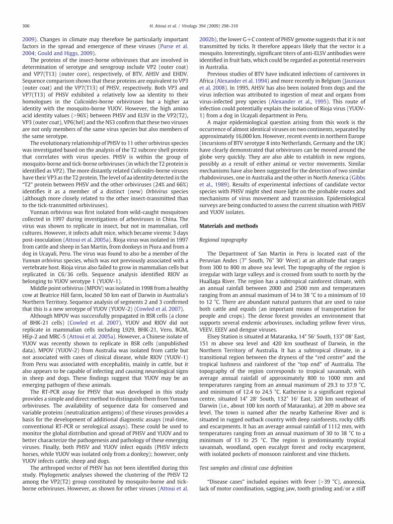
The dsRNA genome segments of viruses isolated in San Martinduring 1997 were analysed by agarose gel electrophoresis (AGE),showing two distinct migration patterns (electropherotypes) (Fig. 1).Electropherotype (AGE) is one of the parameters recognized byICTV for identification of individual virus species within the familyReoviridae (Mertens et al. 2005). One of the two patterns observedwas obtained from isolates subsequently identified as “Peruvian horsesickness virus” (PHSV).
A second electropherotype was obtained from Rioja virus (RIOV)(isolated from sick cattle in Rioja), which is clearly distinct from PHSV.The second electropherotype was also obtained frommultiple furtherisolates from Ochlerotatus scapularis mosquitoes and from cattle,sheep, a dog and a donkey showing clinical signs of disease. Thesecond electropherotype was also identical to that obtained fromviruses belonging to the Yunnan orbivirus species, previously isolatedfrom mosquitoes from both urban and rural cattle-raising areas inChina (Fig. 1; Attoui et al. 2005a). These data indicate that RIOV andYUOV belong to the same Orbivirus species.
Cases and virus isolation
PeruPeruvian horse sickness (PHS; caused by PHSV) was initially
recognized in the Department of San Martin after the unexpecteddeaths of horses during the rainy season. This coincided with thecoffee harvest, when equids are used as means of transportation.Between January and July 1997, 104 horses died with a mortality rateof ∼1.25% and a case-fatality rate of ∼78% (there are ∼8000 horses inthe Department of San Martin; see Table 5). The epizootic affectedeight provinces of the Department, lasting 7 months and reaching itspeak during March to April of 1997, which coincided with periods ofheavy rain.
During the 1997 outbreak, seven equids, one dog, two cows andone sheep showed signs of neurological disease. The virus isolatesfrom blood and/or brain of the sick (neurological disease) cattle,donkey and sheep or from mosquitoes all had a similar electropher-otype to members of the species YUOV and were therefore identifiedas RIOV. One isolate was made from the spleen of one of the sickhorses (Table 1) and had an identical electropherotype to that of
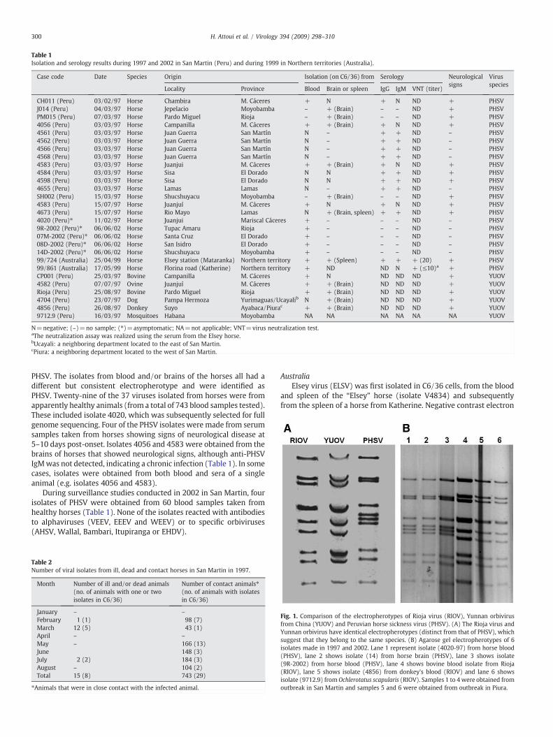
Table 1Isolation and serology results during 1997 and 2002 in San Martin (Peru) and during 1999 in Northern territories (Australia).
Case code Date Species Origin Isolation (on C6/36) from Serology Neurologicalsigns
Virusspecies
Locality Province Blood Brain or spleen IgG IgM VNT (titer)
CH011 (Peru) 03/02/97 Horse Chambira M. Cáceres + N + N ND + PHSVJ014 (Peru) 04/03/97 Horse Jepelacio Moyobamba – + (Brain) – – ND + PHSVPM015 (Peru) 07/03/97 Horse Pardo Miguel Rioja – + (Brain) – – ND + PHSV4056 (Peru) 03/03/97 Horse Campanilla M. Cáceres + + (Brain) + N ND + PHSV4561 (Peru) 03/03/97 Horse Juan Guerra San Martín N – + + ND – PHSV4562 (Peru) 03/03/97 Horse Juan Guerra San Martín N – + + ND – PHSV4566 (Peru) 03/03/97 Horse Juan Guerra San Martín N – + + ND – PHSV4568 (Peru) 03/03/97 Horse Juan Guerra San Martín N – + + ND – PHSV4583 (Peru) 03/03/97 Horse Juanjui M. Cáceres + + (Brain) + N ND + PHSV4584 (Peru) 03/03/97 Horse Sisa El Dorado N N + + ND + PHSV4598 (Peru) 03/03/97 Horse Sisa El Dorado N N + + ND + PHSV4655 (Peru) 03/03/97 Horse Lamas Lamas N – + + ND – PHSVSH002 (Peru) 15/03/97 Horse Shucshuyacu Moyobamba – + (Brain) – – ND + PHSV4583 (Peru) 15/07/97 Horse Juanjuí M. Cáceres + N + N ND + PHSV4673 (Peru) 15/07/97 Horse Rio Mayo Lamas N + (Brain, spleen) + + ND + PHSV4020 (Peru)⁎ 11/02/97 Horse Juanjui Mariscal Cáceres + – – – ND – PHSV9R-2002 (Peru)⁎ 06/06/02 Horse Tupac Amaru Rioja + – – – ND – PHSV07M-2002 (Peru)⁎ 06/06/02 Horse Santa Cruz El Dorado + – – – ND – PHSV08D-2002 (Peru)⁎ 06/06/02 Horse San Isidro El Dorado + – – – ND – PHSV14D-2002 (Peru)⁎ 06/06/02 Horse Shucshuyacu Moyobamba + – – – ND – PHSV99/724 (Australia) 25/04/99 Horse Elsey station (Mataranka) Northern territory + + (Spleen) + + + (20) + PHSV99/861 (Australia) 17/05/99 Horse Florina road (Katherine) Northern territory + ND ND N + (≤10)a + PHSVCP001 (Peru) 25/03/97 Bovine Campanilla M. Cáceres + N ND ND ND + YUOV4582 (Peru) 07/07/97 Ovine Juanjuí M. Cáceres + + (Brain) ND ND ND + YUOVRioja (Peru) 25/08/97 Bovine Pardo Miguel Rioja + + (Brain) ND ND ND + YUOV4704 (Peru) 23/07/97 Dog Pampa Hermoza Yurimaguas/Ucayalib N + (Brain) ND ND ND + YUOV4856 (Peru) 26/08/97 Donkey Suyo Ayabaca/Piurac + + (Brain) ND ND ND + YUOV9712.9 (Peru) 16/03/97 Mosquitoes Habana Moyobamba NA NA NA NA NA NA YUOV
N=negative; (–)=no sample; (⁎)=asymptomatic; NA=not applicable; VNT=virus neutralization test.aThe neutralization assay was realized using the serum from the Elsey horse.bUcayali: a neighboring department located to the east of San Martin.cPiura: a neighboring department located to the west of San Martin.
300 H. Attoui et al. / Virology 394 (2009) 298–310
PHSV. The isolates from blood and/or brains of the horses all had adifferent but consistent electropherotype and were identified asPHSV. Twenty-nine of the 37 viruses isolated from horses were fromapparently healthy animals (from a total of 743 blood samples tested).These included isolate 4020, which was subsequently selected for fullgenome sequencing. Four of the PHSV isolates were made from serumsamples taken from horses showing signs of neurological disease at5–10 days post-onset. Isolates 4056 and 4583 were obtained from thebrains of horses that showed neurological signs, although anti-PHSVIgMwas not detected, indicating a chronic infection (Table 1). In somecases, isolates were obtained from both blood and sera of a singleanimal (e.g. isolates 4056 and 4583).
During surveillance studies conducted in 2002 in San Martin, fourisolates of PHSV were obtained from 60 blood samples taken fromhealthy horses (Table 1). None of the isolates reacted with antibodiesto alphaviruses (VEEV, EEEV and WEEV) or to specific orbiviruses(AHSV, Wallal, Bambari, Itupiranga or EHDV).
Table 2Number of viral isolates from ill, dead and contact horses in San Martin in 1997.
Month Number of ill and/or dead animals(no. of animals with one or twoisolates in C6/36)
Number of contact animals⁎(no. of animals with isolatesin C6/36)
January – –
February 1 (1) 98 (7)March 12 (5) 43 (1)April – –
May – 166 (13)June 148 (3)July 2 (2) 184 (3)August – 104 (2)Total 15 (8) 743 (29)
⁎Animals that were in close contact with the infected animal.
AustraliaElsey virus (ELSV) was first isolated in C6/36 cells, from the blood
and spleen of the “Elsey” horse (isolate V4834) and subsequentlyfrom the spleen of a horse from Katherine. Negative contrast electron
Fig. 1. Comparison of the electropherotypes of Rioja virus (RIOV), Yunnan orbivirusfrom China (YUOV) and Peruvian horse sickness virus (PHSV). (A) The Rioja virus andYunnan orbivirus have identical electropherotypes (distinct from that of PHSV), whichsuggest that they belong to the same species. (B) Agarose gel electropherotypes of 6isolates made in 1997 and 2002. Lane 1 represent isolate (4020-97) from horse blood(PHSV), lane 2 shows isolate (14) from horse brain (PHSV), lane 3 shows isolate(9R-2002) from horse blood (PHSV), lane 4 shows bovine blood isolate from Rioja(RIOV), lane 5 shows isolate (4856) from donkey's blood (RIOV) and lane 6 showsisolate (9712.9) from Ochlerotatus scapularis (RIOV). Samples 1 to 4 were obtained fromoutbreak in San Martin and samples 5 and 6 were obtained from outbreak in Piura.
301H. Attoui et al. / Virology 394 (2009) 298–310
microscopy showed icosahedral virus particles (approx 60 nm)withinthe cytoplasm, budding from plasma membranes, egressing from theplasma membrane and associated with cytoplasmic viral inclusionbodies (VIB) in infected cells. The ultrastructure of the VIB was similarto that described for other orbiviruses, with cores and subcorespresent within a fibrillar matrix. Tubules (approximately 20 nm indiameter) were also present within the cytoplasm. Orbivirusserogrouping ELISA showed no detectable cross-reactions usingantisera against AHSV, BTV, CORV, EHDV, EUBV, EEV, PALV, WALV,WARV and WGRV orbiviruses.
Bloods collected since 1998 throughout the Northern Territory,from both sick and healthy horses, showed approximately 11%seroprevalence for antibodies to ELSV by IgG ELISA. Surveys of serafrom kangaroos (macropods, family Macropodidae) also detectedsome weak positive reactions by indirect IgG ELISA, in approximately11% of tested animals. The causative agent of kangaroo blindness(Wallal virus) has previously been identified as an orbivirus (Hooper,1999; Hooper et al. 1999). However, Elsey virus showed no significantserological relationship to Wallal virus, as demonstrated by anabsence of reactions with specific antibodies by indirect ELISA.
Sera collected in the Northern Territory of Australia from 113 fruitbats, between 1996 and 2003, were also tested for antibodies to ELSVby IgG ELISA and 39 were shown to be positive, with titers higher thanthose observed in horses (titers up to 160). However, no detectableELSV-specific antibodies were found in serum samples from 102 ratscollected in the Northern Territory of Australia, although a lowprevalence of antibody was found in cattle and pigs.
Virus replication, electron microscopy and physicochemical properties
Blood samples andmosquito homogenates (fromPeru)didnot causeCPE in mammalian cell cultures. Agarose gel electrophoresis (AGE) ofmammalian cell culture extracts did not show a poly-segmented dsRNAprofile (characteristic of the reoviruses). However, C6/36 cells inocu-lated with the Peruvian or Australian virus isolates assumed a fusiformmorphology, followed by clumping and detachment from the surface ofthe container. Extracts of the infected C6/36 cells showed a segmenteddsRNAprofile (byAGE), indicating thepresence of a reovirus (amemberof the family Reoviridae). Sucklingmice, adult hamsters and 3-week-oldchickens that had been inoculated with isolate 4020 showed no visiblesigns of infection. However, chickens remained viremic for 2 monthsafter inoculation, as shown by virus isolations.
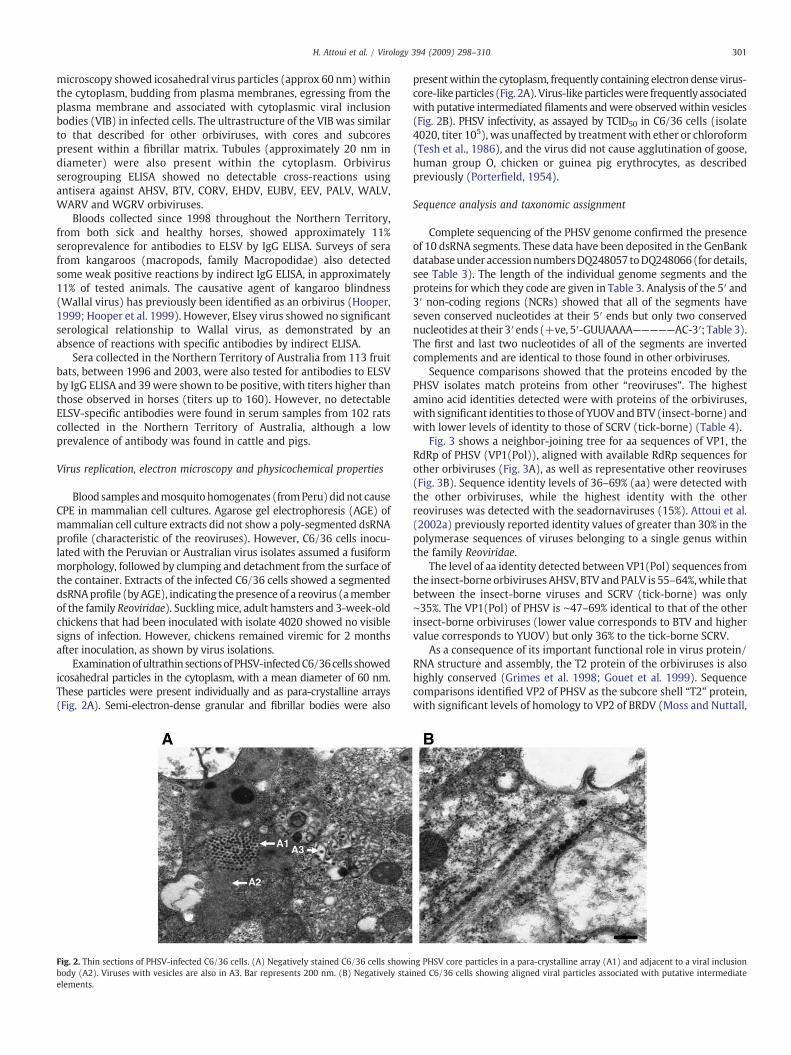
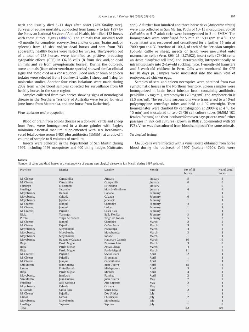
Examinationofultrathin sectionsofPHSV-infectedC6/36cells showedicosahedral particles in the cytoplasm, with a mean diameter of 60 nm.These particles were present individually and as para-crystalline arrays(Fig. 2A). Semi-electron-dense granular and fibrillar bodies were also
Fig. 2. Thin sections of PHSV-infected C6/36 cells. (A) Negatively stained C6/36 cells showibody (A2). Viruses with vesicles are also in A3. Bar represents 200 nm. (B) Negatively staielements.
presentwithin the cytoplasm, frequently containing electron dense virus-core-likeparticles (Fig. 2A). Virus-likeparticleswere frequently associatedwith putative intermediated filaments andwere observedwithin vesicles(Fig. 2B). PHSV infectivity, as assayed by TCID50 in C6/36 cells (isolate4020, titer 105), was unaffected by treatmentwith ether or chloroform(Tesh et al., 1986), and the virus did not cause agglutination of goose,human group O, chicken or guinea pig erythrocytes, as describedpreviously (Porterfield, 1954).
Sequence analysis and taxonomic assignment
Complete sequencing of the PHSV genome confirmed the presenceof 10 dsRNA segments. These data have been deposited in the GenBankdatabaseunder accessionnumbersDQ248057 toDQ248066(for details,see Table 3). The length of the individual genome segments and theproteins for which they code are given in Table 3. Analysis of the 5′ and3′ non-coding regions (NCRs) showed that all of the segments haveseven conserved nucleotides at their 5′ ends but only two conservednucleotides at their 3′ ends (+ve, 5′-GUUAAAA—————AC-3′; Table 3).The first and last two nucleotides of all of the segments are invertedcomplements and are identical to those found in other orbiviruses.
Sequence comparisons showed that the proteins encoded by thePHSV isolates match proteins from other “reoviruses”. The highestamino acid identities detected were with proteins of the orbiviruses,with significant identities to those of YUOV andBTV (insect-borne) andwith lower levels of identity to those of SCRV (tick-borne) (Table 4).
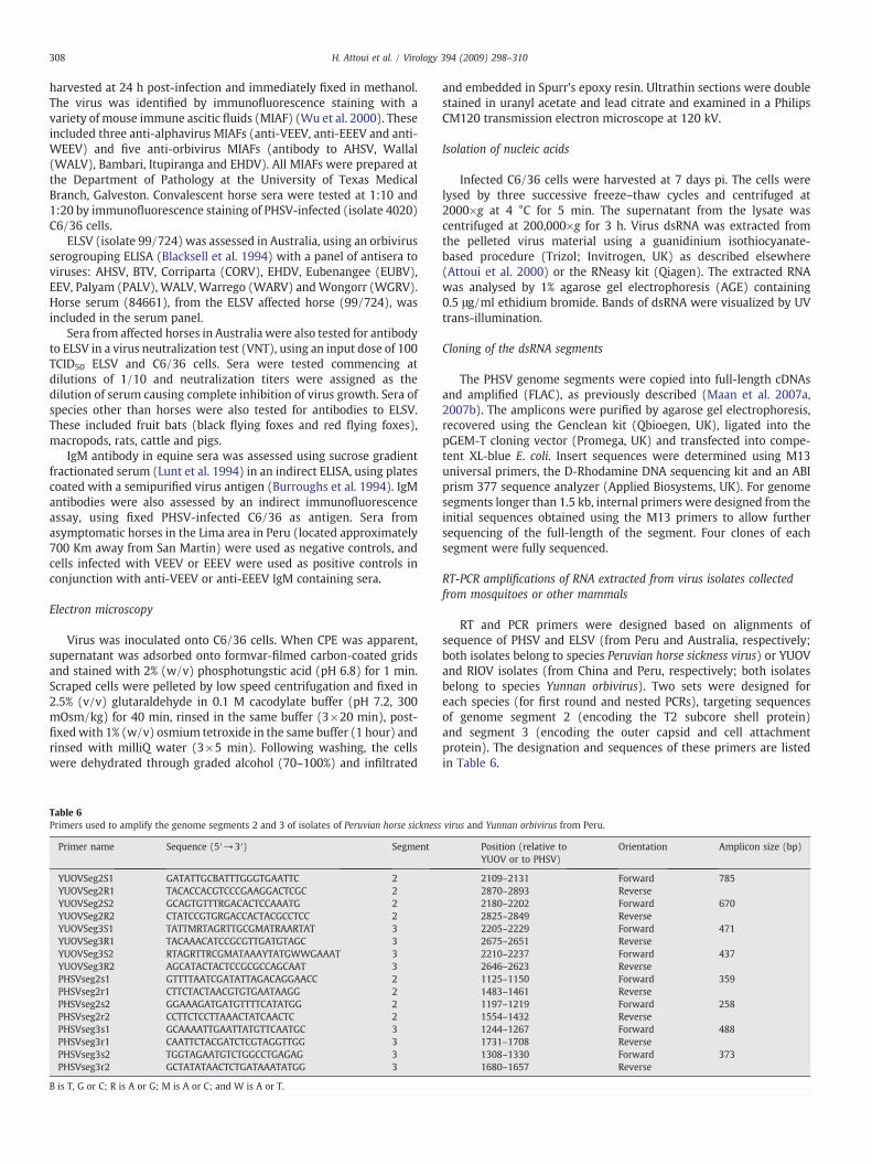
Fig. 3 shows a neighbor-joining tree for aa sequences of VP1, theRdRp of PHSV (VP1(Pol)), aligned with available RdRp sequences forother orbiviruses (Fig. 3A), as well as representative other reoviruses(Fig. 3B). Sequence identity levels of 36–69% (aa) were detected withthe other orbiviruses, while the highest identity with the otherreoviruses was detected with the seadornaviruses (15%). Attoui et al.(2002a) previously reported identity values of greater than 30% in thepolymerase sequences of viruses belonging to a single genus withinthe family Reoviridae.
The level of aa identity detected between VP1(Pol) sequences fromthe insect-borne orbiviruses AHSV, BTV andPALV is 55–64%,while thatbetween the insect-borne viruses and SCRV (tick-borne) was only∼35%. The VP1(Pol) of PHSV is ∼47–69% identical to that of the otherinsect-borne orbiviruses (lower value corresponds to BTV and highervalue corresponds to YUOV) but only 36% to the tick-borne SCRV.
As a consequence of its important functional role in virus protein/RNA structure and assembly, the T2 protein of the orbiviruses is alsohighly conserved (Grimes et al. 1998; Gouet et al. 1999). Sequencecomparisons identified VP2 of PHSV as the subcore shell “T2” protein,with significant levels of homology to VP2 of BRDV (Moss and Nuttall,
ng PHSV core particles in a para-crystalline array (A1) and adjacent to a viral inclusionned C6/36 cells showing aligned viral particles associated with putative intermediate
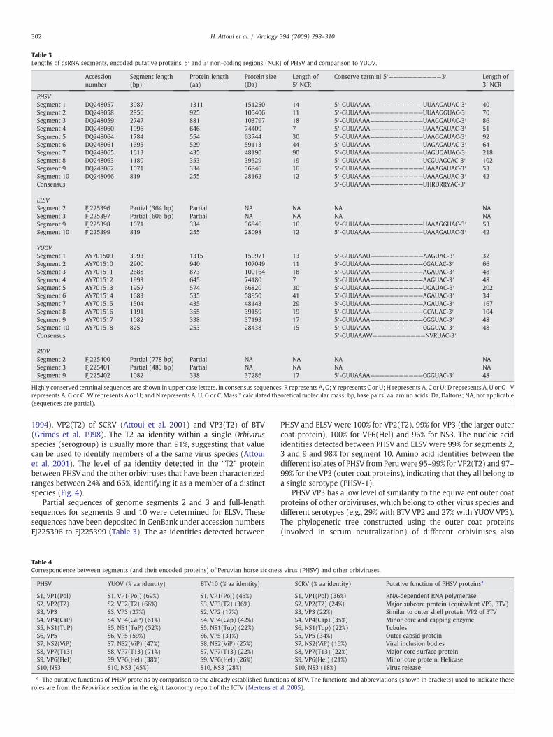
Table 3Lengths of dsRNA segments, encoded putative proteins, 5′ and 3′ non-coding regions (NCR) of PHSV and comparison to YUOV.
Accessionnumber
Segment length(bp)
Protein length(aa)
Protein size(Da)
Length of5′ NCR
Conserve termini 5′———————————3′ Length of3′ NCR
PHSVSegment 1 DQ248057 3987 1311 151250 14 5′-GUUAAAA———————————UUAAGAUAC-3′ 40Segment 2 DQ248058 2856 925 105406 11 5′-GUUAAAA———————————UUAAGGUAC-3′ 70Segment 3 DQ248059 2747 881 103797 18 5′-GUUAAAA———————————UAAGGAUAC-3′ 86Segment 4 DQ248060 1996 646 74409 7 5′-GUUAAAA———————————UAAAGAUAC-3′ 51Segment 5 DQ248064 1784 554 63744 30 5′-GUUAAAA———————————UAAGGAUAC-3′ 92Segment 6 DQ248061 1695 529 59113 44 5′-GUUAAAA———————————UAGAGAUAC-3′ 64Segment 7 DQ248065 1613 435 48190 90 5′-GUUAAAA———————————UAGUGAUAC-3′ 218Segment 8 DQ248063 1180 353 39529 19 5′-GUUAAAA———————————UCGUAGCAC-3′ 102Segment 9 DQ248062 1071 334 36846 16 5′-GUUAAAA———————————UAAAGAUAC-3′ 53Segment 10 DQ248066 819 255 28162 12 5′-GUUAAAA———————————UAAAGAUAC-3′ 42Consensus 5′-GUUAAAA———————————UHRDRRYAC-3′
ELSVSegment 2 FJ225396 Partial (364 bp) Partial NA NA NA NASegment 3 FJ225397 Partial (606 bp) Partial NA NA NA NASegment 9 FJ225398 1071 334 36846 16 5′-GUUAAAA———————————UAAAGGUAC-3′ 53Segment 10 FJ225399 819 255 28098 12 5′-GUUAAAA———————————UAAAGAUAC-3′ 42
YUOVSegment 1 AY701509 3993 1315 150971 13 5′-GUUAAAU———————————AAGUAC-3′ 32Segment 2 AY701510 2900 940 107049 11 5′-GUUAAAA———————————CGAUAC-3′ 66Segment 3 AY701511 2688 873 100164 18 5′-GUUAAAA———————————AGAUAC-3′ 48Segment 4 AY701512 1993 645 74180 7 5′-GUUAAAA———————————AAGUAC-3′ 48Segment 5 AY701513 1957 574 66820 30 5′-GUUAAAA———————————UGAUAC-3′ 202Segment 6 AY701514 1683 535 58950 41 5′-GUUAAAA———————————AGAUAC-3′ 34Segment 7 AY701515 1504 435 48143 29 5′-GUUAAAA———————————AGAUAC-3′ 167Segment 8 AY701516 1191 355 39159 19 5′-GUUAAAA———————————GCAUAC-3′ 104Segment 9 AY701517 1082 338 37193 17 5′-GUUAAAA———————————CGGUAC-3′ 48Segment 10 AY701518 825 253 28438 15 5′-GUUAAAA———————————CGGUAC-3′ 48Consensus 5′-GUUAAAW———————————NVRUAC-3′
RIOVSegment 2 FJ225400 Partial (778 bp) Partial NA NA NA NASegment 3 FJ225401 Partial (483 bp) Partial NA NA NA NASegment 9 FJ225402 1082 338 37286 17 5′-GUUAAAA———————————CGGUAC-3′ 48
Highly conserved terminal sequences are shown in upper case letters. In consensus sequences, R represents A, G; Y represents C or U; H represents A, C or U; D represents A, U or G ; Vrepresents A, G or C;W represents A or U; and N represents A, U, G or C. Mass,⁎ calculated theoretical molecular mass; bp, base pairs; aa, amino acids; Da, Daltons; NA, not applicable(sequences are partial).
302 H. Attoui et al. / Virology 394 (2009) 298–310
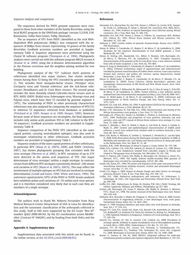
1994), VP2(T2) of SCRV (Attoui et al. 2001) and VP3(T2) of BTV(Grimes et al. 1998). The T2 aa identity within a single Orbivirusspecies (serogroup) is usually more than 91%, suggesting that valuecan be used to identify members of a the same virus species (Attouiet al. 2001). The level of aa identity detected in the “T2” proteinbetween PHSV and the other orbiviruses that have been characterizedranges between 24% and 66%, identifying it as a member of a distinctspecies (Fig. 4).
Partial sequences of genome segments 2 and 3 and full-lengthsequences for segments 9 and 10 were determined for ELSV. Thesesequences have been deposited in GenBank under accession numbersFJ225396 to FJ225399 (Table 3). The aa identities detected between
Table 4Correspondence between segments (and their encoded proteins) of Peruvian horse sicknes
PHSV YUOV (% aa identity) BTV10 (% aa identity)
S1, VP1(Pol) S1, VP1(Pol) (69%) S1, VP1(Pol) (45%)S2, VP2(T2) S2, VP2(T2) (66%) S3, VP3(T2) (36%)S3, VP3 S3, VP3 (27%) S2, VP2 (17%)S4, VP4(CaP) S4, VP4(CaP) (61%) S4, VP4(Cap) (42%)S5, NS1(TuP) S5, NS1(TuP) (52%) S5, NS1(Tup) (22%)S6, VP5 S6, VP5 (59%) S6, VP5 (31%)S7, NS2(ViP) S7, NS2(ViP) (47%) S8, NS2(ViP) (25%)S8, VP7(T13) S8, VP7(T13) (71%) S7, VP7(T13) (22%)S9, VP6(Hel) S9, VP6(Hel) (38%) S9, VP6(Hel) (26%)S10, NS3 S10, NS3 (45%) S10, NS3 (28%)
a The putative functions of PHSV proteins by comparison to the already established functiroles are from the Reoviridae section in the eight taxonomy report of the ICTV (Mertens et
PHSV and ELSV were 100% for VP2(T2), 99% for VP3 (the larger outercoat protein), 100% for VP6(Hel) and 96% for NS3. The nucleic acididentities detected between PHSV and ELSV were 99% for segments 2,3 and 9 and 98% for segment 10. Amino acid identities between thedifferent isolates of PHSV from Peruwere 95–99% for VP2(T2) and 97–99% for the VP3 (outer coat proteins), indicating that they all belong toa single serotype (PHSV-1).
PHSV VP3 has a low level of similarity to the equivalent outer coatproteins of other orbiviruses, which belong to other virus species anddifferent serotypes (e.g., 29% with BTV VP2 and 27% with YUOV VP3).The phylogenetic tree constructed using the outer coat proteins(involved in serum neutralization) of different orbiviruses also
s virus (PHSV) and other orbiviruses.
SCRV (% aa identity) Putative function of PHSV proteinsa
S1, VP1(Pol) (36%) RNA-dependent RNA polymeraseS2, VP2(T2) (24%) Major subcore protein (equivalent VP3, BTV)S3, VP3 (22%) Similar to outer shell protein VP2 of BTVS4, VP4(Cap) (35%) Minor core and capping enzymeS6, NS1(Tup) (22%) TubulesS5, VP5 (34%) Outer capsid proteinS7, NS2(ViP) (16%) Viral inclusion bodiesS8, VP7(T13) (22%) Major core surface proteinS9, VP6(Hel) (21%) Minor core protein, HelicaseS10, NS3 (18%) Virus release
ons of BTV. The functions and abbreviations (shown in brackets) used to indicate theseal. 2005).
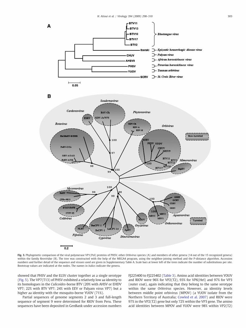
Fig. 3. Phylogenetic comparison of the viral polymerase VP1(Pol) proteins of PHSV, other Orbivirus species (A) and members of other genera (14 out of the 15 recognized genera)within the family Reoviridae (B). The tree was constructed with the help of the MEGA4 program, using the neighbor-joining method and the P-distance algorithm. Accessionnumbers and further detail of the sequence and viruses used are given in Supplementary Table A. Scale bars at lower left of the trees indicate the number of substitutions per site.Bootstrap values are indicated at the nodes. The names in italics indicate the genera.
303H. Attoui et al. / Virology 394 (2009) 298–310
showed that PHSV and the ELSV cluster together as a single serotype(Fig. 5). The VP7(T13) of PHSV exhibited a relatively low aa identity toits homologues in the Culicoides-borne BTV (20% with AHSV or EHDVVP7; 22% with BTV VP7; 24% with EEV or Palyam virus VP7) but ahigher aa identity with the mosquito-borne YUOV (71%).
Partial sequences of genome segments 2 and 3 and full-lengthsequence of segment 9 were determined for RIOV from Peru. Thesesequences have been deposited in GenBank under accession numbers
FJ225400 to FJ225402 (Table 3). Amino acid identities between YOUVand RIOV were 96% for VP2(T2), 93% for VP6(Hel) and 97% for VP3(outer coat), again indicating that they belong to the same serotypewithin the same Orbivirus species. However, aa identity levelsbetween middle point orbivirus (MPOV) (a YUOV isolate from theNorthern Territory of Australia; Cowled et al. 2007) and RIOV were97% in the VP2(T2) gene but only 72% within the VP3 gene. The aminoacid identities between MPOV and YUOV were 98% within VP2(T2)
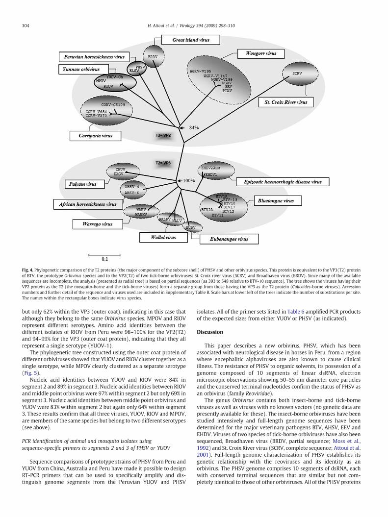
Fig. 4. Phylogenetic comparison of the T2 proteins (the major component of the subcore shell) of PHSV and other orbivirus species. This protein is equivalent to the VP3(T2) proteinof BTV, the prototype Orbivirus species and to the VP2(T2) of two tick-borne orbiviruses: St. Croix river virus (SCRV) and Broadhaven virus (BRDV). Since many of the availablesequences are incomplete, the analysis (presented as radial tree) is based on partial sequences (aa 393 to 548 relative to BTV-10 sequence). The tree shows the viruses having theirVP2 protein as the T2 (the mosquito-borne and the tick-borne viruses) form a separate group from those having the VP3 as the T2 protein (Culicoides-borne viruses). Accessionnumbers and further detail of the sequence and viruses used are included in Supplementary Table B. Scale bars at lower left of the trees indicate the number of substitutions per site.The names within the rectangular boxes indicate virus species.
304 H. Attoui et al. / Virology 394 (2009) 298–310
but only 62% within the VP3 (outer coat), indicating in this case thatalthough they belong to the same Orbivirus species, MPOV and RIOVrepresent different serotypes. Amino acid identities between thedifferent isolates of RIOV from Peru were 98–100% for the VP2(T2)and 94–99% for the VP3 (outer coat protein), indicating that they allrepresent a single serotype (YUOV-1).
The phylogenetic tree constructed using the outer coat protein ofdifferent orbiviruses showed that YUOV and RIOV cluster together as asingle serotype, while MPOV clearly clustered as a separate serotype(Fig. 5).
Nucleic acid identities between YUOV and RIOV were 84% insegment 2 and 89% in segment 3. Nucleic acid identities between RIOVandmiddle point orbivirus were 97%within segment 2 but only 69% insegment 3. Nucleic acid identities betweenmiddle point orbivirus andYUOV were 83% within segment 2 but again only 64% within segment3. These results confirm that all three viruses, YUOV, RIOV and MPOV,aremembers of the same species but belong to two different serotypes(see above).
PCR identification of animal and mosquito isolates usingsequence-specific primers to segments 2 and 3 of PHSV or YUOV
Sequence comparisons of prototype strains of PHSV from Peru andYUOV from China, Australia and Peru have made it possible to designRT-PCR primers that can be used to specifically amplify and dis-tinguish genome segments from the Peruvian YUOV and PHSV
isolates. All of the primer sets listed in Table 6 amplified PCR productsof the expected sizes from either YUOV or PHSV (as indicated).
Discussion
This paper describes a new orbivirus, PHSV, which has beenassociated with neurological disease in horses in Peru, from a regionwhere encephalitic alphaviruses are also known to cause clinicalillness. The resistance of PHSV to organic solvents, its possession of agenome composed of 10 segments of linear dsRNA, electronmicroscopic observations showing 50–55 nm diameter core particlesand the conserved terminal nucleotides confirm the status of PHSV asan orbivirus (family Reoviridae).
The genus Orbivirus contains both insect-borne and tick-borneviruses as well as viruses with no known vectors (no genetic data arepresently available for these). The insect-borne orbiviruses have beenstudied intensively and full-length genome sequences have beendetermined for the major veterinary pathogens BTV, AHSV, EEV andEHDV. Viruses of two species of tick-borne orbiviruses have also beensequenced, Broadhaven virus (BRDV, partial sequence; Moss et al.,1992) and St. Croix River virus (SCRV, complete sequence; Attoui et al.2001). Full-length genome characterization of PHSV establishes itsgenetic relationship with the reoviruses and its identity as anorbivirus. The PHSV genome comprises 10 segments of dsRNA, eachwith conserved terminal sequences that are similar but not com-pletely identical to those of other orbiviruses. All of the PHSV proteins
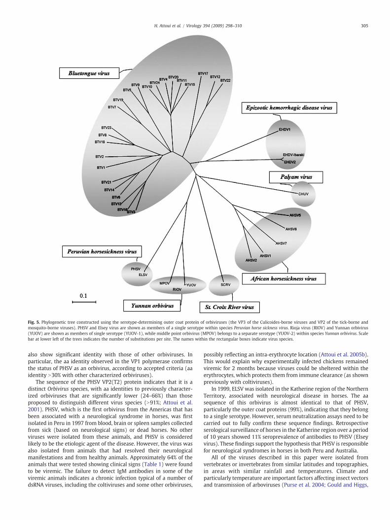
Fig. 5. Phylogenetic tree constructed using the serotype-determining outer coat protein of orbiviruses (the VP3 of the Culicoides-borne viruses and VP2 of the tick-borne andmosquito-borne viruses). PHSV and Elsey virus are shown as members of a single serotype within species Peruvian horse sickness virus. Rioja virus (RIOV) and Yunnan orbivirus(YUOV) are shown as members of single serotype (YUOV-1), while middle point orbivirus (MPOV) belongs to a separate serotype (YUOV-2) within species Yunnan orbivirus. Scalebar at lower left of the trees indicates the number of substitutions per site. The names within the rectangular boxes indicate virus species.
305H. Attoui et al. / Virology 394 (2009) 298–310
also show significant identity with those of other orbiviruses. Inparticular, the aa identity observed in the VP1 polymerase confirmsthe status of PHSV as an orbivirus, according to accepted criteria (aaidentity N30% with other characterized orbiviruses).
The sequence of the PHSV VP2(T2) protein indicates that it is adistinct Orbivirus species, with aa identities to previously character-ized orbiviruses that are significantly lower (24–66%) than thoseproposed to distinguish different virus species (N91%; Attoui et al.2001). PHSV, which is the first orbivirus from the Americas that hasbeen associated with a neurological syndrome in horses, was firstisolated in Peru in 1997 from blood, brain or spleen samples collectedfrom sick (based on neurological signs) or dead horses. No otherviruses were isolated from these animals, and PHSV is consideredlikely to be the etiologic agent of the disease. However, the virus wasalso isolated from animals that had resolved their neurologicalmanifestations and from healthy animals. Approximately 64% of theanimals that were tested showing clinical signs (Table 1) were foundto be viremic. The failure to detect IgM antibodies in some of theviremic animals indicates a chronic infection typical of a number ofdsRNA viruses, including the coltiviruses and some other orbiviruses,
possibly reflecting an intra-erythrocyte location (Attoui et al. 2005b).This would explain why experimentally infected chickens remainedviremic for 2 months because viruses could be sheltered within theerythrocytes, which protects them from immune clearance (as shownpreviously with coltiviruses).
In 1999, ELSV was isolated in the Katherine region of the NorthernTerritory, associated with neurological disease in horses. The aasequence of this orbivirus is almost identical to that of PHSV,particularly the outer coat proteins (99%), indicating that they belongto a single serotype. However, serum neutralization assays need to becarried out to fully confirm these sequence findings. Retrospectiveserological surveillance of horses in the Katherine region over a periodof 10 years showed 11% seroprevalence of antibodies to PHSV (Elseyvirus). These findings support the hypothesis that PHSV is responsiblefor neurological syndromes in horses in both Peru and Australia.
All of the viruses described in this paper were isolated fromvertebrates or invertebrates from similar latitudes and topographies,in areas with similar rainfall and temperatures. Climate andparticularly temperature are important factors affecting insect vectorsand transmission of arboviruses (Purse et al. 2004; Gould and Higgs,
306 H. Attoui et al. / Virology 394 (2009) 298–310
2009). Changes in climate may therefore be particularly importantfactors in the spread and emergence of these viruses (Purse et al.2004; Gould and Higgs, 2009).
The proteins of the insect-borne orbiviruses that are involved indetermination of serotype and serogroup include VP2 (outer coat)and VP7(T13) (outer core), respectively, of BTV, AHSV and EHDV.Sequence comparison shows that these proteins are equivalent to VP3(outer coat) and the VP7(T13) of PHSV, respectively. Both VP3 andVP7(T13) of PHSV exhibited a relatively low aa identity to theirhomologues in the Culicoides-borne orbiviruses but a higher aaidentity with the mosquito-borne YUOV. However, the high aminoacid identity values (N96%) between PHSV and ELSV in the VP2(T2),VP3 (outer coat), VP6(hel) and the NS3 confirm that these two virusesare not only members of the same virus species but also members ofthe same serotype.
The evolutionary relationship of PHSV to 11 other orbivirus specieswas investigated based on the analysis of the T2 subcore shell proteinthat correlates with virus species. PHSV is within the group ofmosquito-borne and tick-borne orbiviruses (inwhich the T2 protein isidentified as VP2). The more distantly related Culicoides-borne viruseshave their VP3 as the T2 protein. The level of aa identity detected in the“T2” protein between PHSV and the other orbiviruses (24% and 66%)identifies it as a member of a distinct (new) Orbivirus species(although more closely related to the other insect-transmitted thanto the tick-transmitted orbiviruses).
Yunnan orbivirus was first isolated from wild-caught mosquitoescollected in 1997 during investigations of arboviruses in China. Thevirus was shown to replicate in insect, but not in mammalian, cellcultures. However, it infects adult mice, which became viremic 3 dayspost-inoculation (Attoui et al. 2005a). Rioja virus was isolated in 1997from cattle and sheep in SanMartin, from donkeys in Piura and from adog in Ucayali, Peru. The virus was found to also be a member of theYunnan orbivirus species, which was not previously associated with avertebrate host. Rioja virus also failed to grow in mammalian cells butreplicated in C6/36 cells. Sequence analysis identified RIOV asbelonging to YUOV serotype 1 (YUOV-1).
Middle point orbivirus (MPOV)was isolated in 1998 from a healthycow at Beatrice Hill farm, located 50 km east of Darwin in Australia'sNorthern Territory. Sequence analysis of segments 2 and 3 confirmedthat this is a new serotype of YUOV (YUOV-2) (Cowled et al. 2007).
Although MPOV was successfully propagated in BSR cells (a cloneof BHK-21 cells) (Cowled et al. 2007), YUOV and RIOV did notreplicate in mammalian cells including L929, BHK-21, Vero, BGM,HEp-2 and MRC-5 (Attoui et al. 2005a). However, a Chinese isolate ofYUOV was recently shown to replicate in BSR cells (unpublisheddata). MPOV (YUOV-2) from Australia was isolated from cattle butnot associated with cases of clinical disease, while RIOV (YUOV-1)from Peru was associated with encephalitis, mainly in cattle, but italso appears to be capable of infecting and causing neurological signsin sheep and dogs. These findings suggest that YUOV may be anemerging pathogen of these animals.
The RT-PCR assay for PHSV that was developed in this studyprovides a simple and directmethod to distinguish them fromYunnanorbiviruses. The availability of sequence data for conserved andvariable proteins (neutralization antigens) of these viruses provides abasis for the development of additional diagnostic assays (real-time,conventional RT-PCR or serological assays). These could be used tomonitor the global distribution and spread of PHSV and YUOV and tobetter characterize the pathogenesis and pathology of these emergingviruses. Finally, both PHSV and YUOV infect equids (PHSV infectshorses, while YUOV was isolated only from a donkey); however, onlyYUOV infects cattle, sheep and dogs.
The arthropod vector of PHSV has not been identified during thisstudy. Phylogenetic analyses showed the clustering of the PHSV T2among the VP2(T2) group constituted by mosquito-borne and tick-borne orbiviruses. However, as shown for other viruses (Attoui et al.
2002b), the lower G+C content of PHSV genome suggests that it is nottransmitted by ticks. It therefore appears likely that the vector is amosquito. Interestingly, significant titers of anti-ELSV antibodies wereidentified in fruit bats, which could be regarded as potential reservoirsin Australia.
Previous studies of BTV have indicated infections of carnivores inAfrica (Alexander et al. 1994) and more recently in Belgium (Jauniauxet al. 2008). In 1995, AHSV has also been isolated from dogs and thevirus infection was attributed to ingestion of meat and organs fromvirus-infected prey species (Alexander et al., 1995). This route ofinfection could potentially explain the isolation of Rioja virus (YUOV-1) from a dog in Ucayali department in Peru.
A major epidemiological question arising from this work is theoccurrence of almost identical viruses on two continents, separated byapproximately 16,000 km. However, recent events in northern Europe(incursions of BTV serotype 8 into Netherlands, Germany and the UK)have clearly demonstrated that orbiviruses can be moved around theglobe very quickly. They are also able to establish in new regions,possibly as a result of either animal or vector movements. Similarmechanisms have also been suggested for the detection of two similarrhabdoviruses, one in Australia and the other in North America (Gibbset al., 1989). Results of experimental infections of candidate vectorspecies with PHSV might shed more light on the probable routes andmechanisms of virus movement and transmission. Epidemiologicalsurveys are being conducted to assess the current situation with PHSVand YUOV isolates.
Materials and methods
Regional topography
The Department of San Martin in Peru is located east of thePeruvian Andes (7° South, 76° 30′ West) at an altitude that rangesfrom 300 to 800 m above sea level. The topography of the region isirregular with large valleys and is crossed from south to north by theHuallaga River. The region has a subtropical rainforest climate, withan annual rainfall between 2000 and 2500 mm and temperaturesranging from an annual maximum of 34 to 38 °C to a minimum of 10to 12 °C. There are abundant natural pastures that are used to raiseboth cattle and equids (an important means of transportation forpeople and crops). The dense forest provides an environment thatsupports several endemic arboviruses, including yellow fever virus,VEEV, EEEV and dengue viruses.
Elsey Station is situated at Mataranka, 14° 56′ South, 133° 08′ East,151 m above sea level and 420 km southeast of Darwin, in theNorthern Territory of Australia. It has a subtropical climate, in atransitional region between the dryness of the “red centre” and thetropical lushness and rainforest of the “top end” of Australia. Thetopography of the region corresponds to tropical savannah, withaverage annual rainfall of approximately 800 to 1000 mm andtemperatures ranging from an annual maximum of 29.3 to 37.9 °C,and minimum of 12.4 to 24.5 °C. Katherine is a significant regionalcentre, situated 14° 28′ South, 132° 16′ East, 320 km southeast ofDarwin (i.e., about 100 km north of Mataranka), at 209 m above sealevel. The town is named after the nearby Katherine River and issituated in rugged outback country with deep rainforests, rocky cliffsand escarpments. It has an average annual rainfall of 1112 mm, withtemperatures ranging from an annual maximum of 30 to 38 °C to aminimum of 13 to 25 °C. The region is predominantly tropicalsavannah, woodland, open eucalypt forest and rocky escarpment,with isolated pockets of monsoon rainforest and vine thickets.
Test samples and clinical case definition
“Disease cases” included equines with fever (N39 °C), anorexia,lack of motor coordination, sagging jaw, tooth grinding and/or a stiff
307H. Attoui et al. / Virology 394 (2009) 298–310
neck and usually died 8–11 days after onset (78% fatality rate).Surveys of equine mortality, conducted from January to July 1997 bythe Peruvian National Service of Animal Health, identified 132 horseswith these clinical signs (Table 5). The animals that survived took∼3 months for complete recovery. Sera and/or organs (brains and/orspleens) from 15 sick and/or dead horses and sera from 743apparently healthy horses were tested for viruses. Thirty-seven outof a total of 758 horses, were identified as positive, producingcytopathic effects (CPE) in C6/36 cells (8 from sick and/or deadanimals and 29 from asymptomatic horses). During the outbreak,some animals (from other vertebrate species) showed similar clinicalsigns and some died as a consequence. Blood and/or brain or spleenisolates were selected from 1 donkey, 2 cattle, 1 sheep and 1 dog formolecular studies. Another four virus isolations were made during2002 from whole blood samples collected for surveillance from 60healthy horses in the same region.
Samples collected from two horses showing signs of neurologicaldisease in the Northern Territory of Australia were tested for virus(one horse from Mataranka, and one horse from Katherine).
Virus isolation and propagation
Blood or brain from equids (horses or a donkey), cattle and sheepfrom Peru, were homogenized in a tissue grinder with Eagle'sminimum essential medium, supplemented with 10% heat-inacti-vated fetal bovine serum (FBS) plus antibiotics (EMEM), at a ratio of 1volume of sample to 5 volumes of medium.
Insects were collected in the Department of San Martin during1997, including 1193 mosquitoes and 400 biting midges (Culicoides
Table 5Number of cases and dead horses as a consequence of equine neurological disease in San M
Province District Locality
M. Cáceres Campanilla AmpatoM. Cáceres Campanilla CampanillaHuallaga El Eslabón El EslabónHuallaga Sacanche Moscú-MirafloresMoyobamba Habana HabanaMoyobamba Calzada CalzadaMoyobamba Jepelacio JepelacioM. Cáceres Juanjuí ChambiraM. Cáceres Juanjuí LedoyM. Cáceres Pajarillo Costa RicaRioja Yorongos Bella FloridaPicota Tingo de Ponaza Tingo de PonazaM. Cáceres Juanjuí ChambiraM. Cáceres Pajarillo CuñumbuzaMoyobamba Moyobamba PacayzapaMoyobamba Moyobamba MoyobambaMoyobamba Moyobamba IndañeMoyobamba Habana y Calzada Habana y CalzadaRioja Pardo Miguel Pioneros AltoRioja Pardo Miguel Aguas ClarasRioja Pardo Miguel Pardo MiguelM. Cáceres Pajarillo Sector ClaraM. Cáceres Pajarillo ShumanzaM. Cáceres Juanjuí CunchihuilloSan Martín Juan Guerra Juan GuerraLamas Pinto Recodo MishquiyacuRioja Pardo Miguel MiradorMoyobamba Jepelacio RamirezSan Martín Juan Guerra Juan GuerraHuallaga Alto Saposoa Alto SaposoaMoyobamba Calzada CalzadaEl Dorado Santa Rosa Santa RosaM. Cáceres Pajarillo Dos UnidosLamas Lamas ChurucayuMoyobamba Moyobamba MoyobambaHuallaga Saposoa SaposoaTotal
spp.). A further four hundred and three horse ticks (Anocentor nitens)were also collected in San Martin. Pools of 10–15 mosquitoes, 30–50Culicoides or 5–7 adult ticks were homogenized in 3 ml EMEM. Thehomogenates were centrifuged for 5 min at 1500 rpm at 4 °C. Thesupernatant was recovered and centrifuged for a further 15 min at7000 rpm at 4 °C. Fractions of 100 μl, of each of the Peruvian samples(Equids, cattle or sheep, insects or ticks) were inoculated ontomammalian cells (Vero, BHK-21, LLCMK2), insect cells (C6/36 cells;an Aedes albopictus cell line) and intracranially, intraperitoneally orintramuscularly into 2-day-old suckling mice, 1-month-old hamstersand 3-week-old chickens in Peru. Cells were monitored for CPEfor 10 days pi. Samples were inoculated into the main vein ofembryonated chicken eggs.
Samples of sera and spleen necropsies were obtained from twosymptomatic horses in the Northern Territory. Spleen samples werehomogenized in brain heart infusion broth containing antibioticspenicillin (6 mg/ml), streptomycin (20 mg/ml) and amphotericin B(2.5 mg/ml). The resulting suspensions were transferred to a 10-mlpolypropylene centrifuge tubes and held at 4 °C overnight. Thenhomogenates were clarified by centrifugation at 2000×g at 4 °C for15 min) and inoculated to two C6/36 cell culture tubes (EMEM 10%fetal calf serum) and then incubated for seven days prior to two furtherpassages in BSR cell cultures (grown in BME supplemented with 5%FCS). Virus was also cultured from blood samples of the same animals.
Serological testing
C6/36 cells were infected with a virus isolate obtained from horseblood during the outbreak of 1997 (isolate 4020). Cells were
artin during 1997 epizootic.
Month No. of illhorses
No. of deadhorses
January 5 5January 6 6January 1 0January 4 4February 1 1February 6 4February 1 1February 3 2February 1 1February 1 1February 3 3February 5 2March 2 2March 3 3March 4 4March 3 3March 1 1March 16 16March 3 0March 4 4March 11 3April 2 1April 1 1April 1 1April 18 13April 3 3April 4 4April 2 2May 7 7May 2 1May 1 1June 2 1July 1 1July 2 1July 1 1July 1 0
132 104
308 H. Attoui et al. / Virology 394 (2009) 298–310
harvested at 24 h post-infection and immediately fixed in methanol.The virus was identified by immunofluorescence staining with avariety of mouse immune ascitic fluids (MIAF) (Wu et al. 2000). Theseincluded three anti-alphavirus MIAFs (anti-VEEV, anti-EEEV and anti-WEEV) and five anti-orbivirus MIAFs (antibody to AHSV, Wallal(WALV), Bambari, Itupiranga and EHDV). All MIAFs were prepared atthe Department of Pathology at the University of Texas MedicalBranch, Galveston. Convalescent horse sera were tested at 1:10 and1:20 by immunofluorescence staining of PHSV-infected (isolate 4020)C6/36 cells.
ELSV (isolate 99/724) was assessed in Australia, using an orbivirusserogrouping ELISA (Blacksell et al. 1994) with a panel of antisera toviruses: AHSV, BTV, Corriparta (CORV), EHDV, Eubenangee (EUBV),EEV, Palyam (PALV), WALV, Warrego (WARV) andWongorr (WGRV).Horse serum (84661), from the ELSV affected horse (99/724), wasincluded in the serum panel.
Sera from affected horses in Australia were also tested for antibodyto ELSV in a virus neutralization test (VNT), using an input dose of 100TCID50 ELSV and C6/36 cells. Sera were tested commencing atdilutions of 1/10 and neutralization titers were assigned as thedilution of serum causing complete inhibition of virus growth. Sera ofspecies other than horses were also tested for antibodies to ELSV.These included fruit bats (black flying foxes and red flying foxes),macropods, rats, cattle and pigs.
IgM antibody in equine sera was assessed using sucrose gradientfractionated serum (Lunt et al. 1994) in an indirect ELISA, using platescoated with a semipurified virus antigen (Burroughs et al. 1994). IgMantibodies were also assessed by an indirect immunofluorescenceassay, using fixed PHSV-infected C6/36 as antigen. Sera fromasymptomatic horses in the Lima area in Peru (located approximately700 Km away from San Martin) were used as negative controls, andcells infected with VEEV or EEEV were used as positive controls inconjunction with anti-VEEV or anti-EEEV IgM containing sera.
Electron microscopy
Virus was inoculated onto C6/36 cells. When CPE was apparent,supernatant was adsorbed onto formvar-filmed carbon-coated gridsand stained with 2% (w/v) phosphotungstic acid (pH 6.8) for 1 min.Scraped cells were pelleted by low speed centrifugation and fixed in2.5% (v/v) glutaraldehyde in 0.1 M cacodylate buffer (pH 7.2, 300mOsm/kg) for 40 min, rinsed in the same buffer (3×20 min), post-fixedwith 1% (w/v) osmium tetroxide in the same buffer (1 hour) andrinsed with milliQ water (3×5 min). Following washing, the cellswere dehydrated through graded alcohol (70–100%) and infiltrated
Table 6Primers used to amplify the genome segments 2 and 3 of isolates of Peruvian horse sickness
Primer name Sequence (5′→3′) Segment
YUOVSeg2S1 GATATTGCBATTTGGGTGAATTC 2YUOVSeg2R1 TACACCACGTCCCGAAGGACTCGC 2YUOVSeg2S2 GCAGTGTTTRGACACTCCAAATG 2YUOVSeg2R2 CTATCCGTGRGACCACTACGCCTCC 2YUOVSeg3S1 TATTMRTAGRTTGCGMATRAARTAT 3YUOVSeg3R1 TACAAACATCCGCGTTGATGTAGC 3YUOVSeg3S2 RTAGRTTRCGMATAAAYTATGWWGAAAT 3YUOVSeg3R2 AGCATACTACTCCGCGCCAGCAAT 3PHSVseg2s1 GTTTTAATCGATATTAGACAGGAACC 2PHSVseg2r1 CTTCTACTAACGTGTGAATAAGG 2PHSVseg2s2 GGAAAGATGATGTTTTCATATGG 2PHSVseg2r2 CCTTCTCCTTAAACTATCAACTC 2PHSVseg3s1 GCAAAATTGAATTATGTTCAATGC 3PHSVseg3r1 CAATTCTACGATCTCGTAGGTTGG 3PHSVseg3s2 TGGTAGAATGTCTGGCCTGAGAG 3PHSVseg3r2 GCTATATAACTCTGATAAATATGG 3
B is T, G or C; R is A or G; M is A or C; and W is A or T.
and embedded in Spurr's epoxy resin. Ultrathin sections were doublestained in uranyl acetate and lead citrate and examined in a PhilipsCM120 transmission electron microscope at 120 kV.
Isolation of nucleic acids
Infected C6/36 cells were harvested at 7 days pi. The cells werelysed by three successive freeze–thaw cycles and centrifuged at2000×g at 4 °C for 5 min. The supernatant from the lysate wascentrifuged at 200,000×g for 3 h. Virus dsRNA was extracted fromthe pelleted virus material using a guanidinium isothiocyanate-based procedure (Trizol; Invitrogen, UK) as described elsewhere(Attoui et al. 2000) or the RNeasy kit (Qiagen). The extracted RNAwas analysed by 1% agarose gel electrophoresis (AGE) containing0.5 μg/ml ethidium bromide. Bands of dsRNA were visualized by UVtrans-illumination.
Cloning of the dsRNA segments
The PHSV genome segments were copied into full-length cDNAsand amplified (FLAC), as previously described (Maan et al. 2007a,2007b). The amplicons were purified by agarose gel electrophoresis,recovered using the Genclean kit (Qbioegen, UK), ligated into thepGEM-T cloning vector (Promega, UK) and transfected into compe-tent XL-blue E. coli. Insert sequences were determined using M13universal primers, the D-Rhodamine DNA sequencing kit and an ABIprism 377 sequence analyzer (Applied Biosystems, UK). For genomesegments longer than 1.5 kb, internal primers were designed from theinitial sequences obtained using the M13 primers to allow furthersequencing of the full-length of the segment. Four clones of eachsegment were fully sequenced.
RT-PCR amplifications of RNA extracted from virus isolates collectedfrom mosquitoes or other mammals
RT and PCR primers were designed based on alignments ofsequence of PHSV and ELSV (from Peru and Australia, respectively;both isolates belong to species Peruvian horse sickness virus) or YUOVand RIOV isolates (from China and Peru, respectively; both isolatesbelong to species Yunnan orbivirus). Two sets were designed foreach species (for first round and nested PCRs), targeting sequencesof genome segment 2 (encoding the T2 subcore shell protein)and segment 3 (encoding the outer capsid and cell attachmentprotein). The designation and sequences of these primers are listedin Table 6.
virus and Yunnan orbivirus from Peru.
Position (relative toYUOV or to PHSV)
Orientation Amplicon size (bp)
2109–2131 Forward 7852870–2893 Reverse2180–2202 Forward 6702825–2849 Reverse2205–2229 Forward 4712675–2651 Reverse2210–2237 Forward 4372646–2623 Reverse1125–1150 Forward 3591483–1461 Reverse1197–1219 Forward 2581554–1432 Reverse1244–1267 Forward 4881731–1708 Reverse1308–1330 Forward 3731680–1657 Reverse
309H. Attoui et al. / Virology 394 (2009) 298–310
Sequence analysis and comparisons
The sequences derived for PHSV genome segments were com-pared to those from other members of the family Reoviridae, using thelocal BLAST program in the DNATools package (version 5.2.018, S.W.Rasmussen; Valby Data Center, Valby, Denmark).
The aa sequence of VP1 from PHSV (identified as the viral RNA-dependent RNA polymerase (RdRp)) was compared with the se-quences of RdRps from viruses representing 14 genera of the familyReoviridae. GenBank accession numbers are provided in Supple-mentary Table A. Sequence alignments were performed using theClustal W software program (Thompson et al. 1994). Phylogeneticanalyses were carried out with the software programMEGA version 4(Kumar et al. 2004) using the p-distance determination algorithmor the Poisson correction and the neighbor-joining method for treebuilding.
Phylogenetic analysis of the “T2” (subcore shell) proteins oforbiviruses identified two major clusters. One cluster includesviruses having their T2 being the VP2 (encoded by genome segment2). This includes three mosquito-borne viruses (Wongorr virus,Corriparta virus and Yunnan orbivirus) and the tick-borne orbi-viruses (Broadhaven and St. Croix River viruses). The second groupincludes the more distantly related Culicoides-borne viruses such asBTV, AHSV, EHDV, Wallal virus, Eubenangee virus, Warrego virus andPalyam virus, which have their T2 encoded by genome segment 3(VP3). The relationship of PHSV to other previously characterizedorbiviruses was also analysed by comparing the sequence of VP2(T2)to orbivirus T2 sequences retrieved from databases or published(Pritchard et al. 1995; Hooper et al. 1999; Hooper, 1999). However,because some of these sequences are incomplete, the final alignmentincludes only amino acids positions 393 to 548 (relative to the BTV-10 sequence). GenBank accession numbers are provided in Supple-mentary Table B.
Sequence comparison of the PHSV VP3 (identified as the outercapsid protein, carrying neutralization epitopes) was also used toinvestigate relationship to other orbiviruses. GenBank accessionnumbers are provided in Supplementary Table C.
Sequence analysis of the outer capsid proteins of other orbiviruses,in particular BTV (Maan et al. 2007b, 2008) and EHDV (Anthony,2007), has shown phylogenetic grouping that correlates with thevirus serotype (Mertens et al. 2005). In BTV, variations of up to 27%were detected in the amino acid sequences of VP2 (the majordeterminant of virus serotype) within a single serotype. In contrast,viruses from different BTV serotypes consistently showed N24% aminoacid variation in VP2 (Maan et al. 2007a, 2007b). This may reflect theinvolvement of multiple epitopes on VP2 that are involved in serotypedetermination (Gould and Eaton, 1990; White and Eaton, 1990). Theoutermost capsid protein (VP3) of the PHSV or YUOV strains analysedhere exhibited amino acid variations of b3% within each virus species,and it is therefore considered very likely that in each case they aremembers of a single serotype.
Acknowledgments
The authors wish to thank Mr. Roberto Fernandez from NavyMedical Research Center Detachment of USA in Lima for identifica-tion and the taxonomic classification of the arthropods collected inthis study. Staff at IAH were supported by the EU Grant “Reo ID”number QLK2-2000-00143, by the EU coordination action MedRe-oNet (Contract N° 044285) and by funding from both Defra and theBBSRC.
Appendix A. Supplementary data
Supplementary data associated with this article can be found, inthe online version, at doi:10.1016/j.virol.2009.08.032.
References
Alexander, K.A., MacLachlan, N.J., Kat, P.W., House, C., O'Brien, S.J., Lerche, N.W., Sawyer,M., Frank, L.G., Holekamp, K., Smale, L., Mcnutf, J.W., Laurenson, M.K., Mills, M.O.L.,Osburn, A.I., 1994. Evidence of natural bluetongue virus infection among Africancarnivores. Am. J. Trop. Med. Hyg. 51, 568–576.
Alexander, K.A., Kat, P.W., House, J., House, C., O'Brien, S.J., Laurenson, M.K., McNutt,J.W., Osburn, B.I., 1995. African horse sickness and African carnivores. Vet.Microbiol. 47, 133–140.
Anthony, S., 2007. Genetic studies of epizootic heaemorrhagic disease virus (EHDV).PhD dissertation.
Attoui, H., Billoir, F., Cantaloube, J.F., Biagini, P., de Micco, P., de Lamballerie, X., 2000.Strategies for the sequence determination of viral dsRNA genomes. J. Virol.Methods 89, 147–158.
Attoui, H., Stirling, J.M., Munderloh, U.G., Billoir, F., Brookes, S.M., Burroughs, J.N.,de Micco, P., Mertens, P.P., de Lamballerie, X., 2001. Complete sequencecharacterization of the genome of the St Croix River virus, a new orbivirus isolatedfrom cells of Ixodes scapularis. J. Gen. Virol. 82, 795–804.
Attoui, H., Fang, Q.,Mohd Jaafar, F., Cantaloube, J.F., Biagini, P., deMicco, P., de Lamballerie,X., 2002a. Common evolutionary origin of aquareoviruses and orthoreovirusesrevealed by genome characterization of Golden shiner reovirus, Grass carp reovirus,Striped bass reovirus and golden ide reovirus (genus Aquareovirus, familyReoviridae). J. Gen, Virol. 83, 1941–1951.
Attoui, H., Mohd Jaafar, F., Biagini, P., Cantaloube, J.F, de Micco, P., Murphy, F.A., deLamballerie, X., 2002b. Genus Coltivirus (family Reoviridae): genomic andmorphologic characterization of Old World and New World viruses. Arch. Virol.147, 533–561.
Attoui,H,Mohd Jaafar, F., Belhouchet,M.,Aldrovandi,N., Tao, S., Chen,B., Liang,G., Tesh,R.B.,de Micco, P., de Lamballerie, X., 2005a. Yunnan orbivirus, a new orbivirus speciesisolated fromCulex tritaeniorhynchusmosquitoes in China. J. Gen. Virol. 86, 3409–3417.
Attoui, H., Mohd Jaafar, F., de Micco, P., de Lamballerie, X., 2005b. Coltiviruses andseadornaviruses in North America, Europe, and Asia. Emerg. Infect. Dis. 11,1673–1679.
Blacksell, S.D., Lunt, R.A., White, J.R., 1994. A rapid indirect ELISA for the serogrouping ofAustralian orbiviruses. J. Virol. Methods 49, 67–78.
Bremer, C.W., Huismans, H., Van Dijk, A.A., 1990. Characterization and cloning of theAfrican horsesickness virus genome. J. Gen. Virol. 71, 793–799.
Burroughs, J.N., O'Hara, R.S., Smale, C.J., Hamblin, C., Walton, A., Armstrong, R., Mertens,P.P.C., 1994. Purification and properties of virus particles, infectious sub viralparticles, cores and VP7 crystals of African Horse sickness virus serotype 9. J. Gen.Virol. 75, 1849–1857.
Cowled, C., Melville, L., Weir, R., Walsh, S., Hyatt, A., Van Driel, R., Davis, S., Gubala, A.,Boyle, D., 2007. Genetic and epidemiological characterization of middle pointorbivirus, a novel virus isolated from sentinel cattle in northern Australia. J. Gen.Virol. 88, 3413–3422.
Elbers, A.R., Backx, A., Mintiens, K., Gerbier, G., Staubach, C., Hendrickx, G., van der Spek,A., 2008. Field observations during the bluetongue serotype 8 epidemic in 2006. II.Morbidity and mortality rate, case fatality and clinical recovery in sheep and cattlein the Netherlands. Prev. Vet. Med. 87, 31–40.
Gambles, R.M., 1949. Bluetongue of sheep in Cyprus. J. Comp. Pathol. 59, 176–190.Gibbs, E.P.J., Calisher, C.H., Tesh, R.B., Labuick, J.S., Bowen, R., Greiner, E.C., 1989. Bivens
Arm virus: a new rhabdovirus isolated from Culicoides insignis in Florida andrelated to Tibrogargan virus of Australia. Vet. Mircobiol. 19, 141–150.
Gouet, P., Diprose, J.M., Grimes, J.M., Malby, R., Burroughs, J.N., Zientara, S., Stuart, D.I.,Mertens, P.P.C., 1999. The highly ordered double-stranded RNA genome ofbluetongue virus revealed by crystallography. Cell 97, 481–490.
Gould, A.R., Eaton, B.T., 1990. The amino acid sequence of the outer coat protein VP2 ofneutralizing monoclonal antibody-resistant, virulent and attenuated bluetongueviruses. Virus Res. 17, 161–172.
Gould, E.A., Higgs, S., 2009. Impact of climate change and other factors on emergingarbovirus diseases. Trans. R. Soc. Trop. Med. Hyg. 103, 109–121.
Griffin, D.E., 1986. Alphavirus pathogenesis and immunity. In: Schlesinger, S.,Schlesinger, M. (Eds.), The Togaviridae and Flaviviridae. Plenum Press, New York,pp. 209–249.
Griffin, D.E., 2001. Alphaviruses, In: Knipe, M., Howley, P.M. (Eds.), Fields' Virology, 4thedition. Lippincott, Williams and Wilkins, Philadelphia, pp. 917–962.
Grimes, J.M., Burroughs, J.N., Gouet, P., Diprose, J.M., Malby, R., Zientara, S., Mertens,P.P.C., Stuart, D.I., 1998. The atomic structure of the bluetongue virus core. Nature395, 470–478.
Hofmann, M.A., Renzullo, S., Mader, M., Chaignat, V., Worwa, G., Thuer, B., 2008. Geneticcharacterization of toggenburg orbivirus, a new bluetongue virus, from goats.Switzerland. Emerg. Infect. Dis. 14, 1855–1861.
Hooper, P., 1999. Kangaroo blindness and some other new viral diseases in Australia.Aust. Vet. J. 77, 514–515.
Hooper, P.T., Lunt, R.A., Gould, A.R., Hyatt, A.D., Russell, G.M., Kattenbelt, J.A., Blacksell,S.D., Reddacliff, L.A., Kirkland, P.D., Davis, R.J., Durham, P.J., Bishop, A.L.,Waddington,J., 1999. Epidemic blindness in kangaroos—evidence of a viral aetiology. Aust. Vet. J.77, 529–536.
Howell, P.G., Nurton, J.P., Nel, D., Lourens, C.W., Guthrie, A.J., 2008. Prevalence ofserotype specific antibody to equine encephalosis virus in thoroughbred yearlingsin South Africa (1999–2004). Onderstepoort J. Vet. Res. 75, 153–161.
Jauniaux, T.P., De Clercq, K.E., Cassart, D.E., Kennedy, S., Vandenbussche, F.E.,Vandemeulebroucke, E.L., Vanbinst, T.M., Verheyden, B.I., Goris, N.E., Coignoul,F.L., 2008. Bluetongue in Eurasian lynx. Emerg. Infect. Dis. 14, 1496–1498.
Kumar, S., Tamura, K., Nei, M., 2004. MEGA3: integrated software for molecularevolutionary genetics analysis and sequence alignment. Brief. Bioinform. 5, 150–163.
310 H. Attoui et al. / Virology 394 (2009) 298–310
Lunt, R.A., Blacksell, S.D., Newberry, K.M., 1994. The use of an indirect ELISA withprotein G-peroxidase conjugate and a blocking ELISA to demonstrate recentbluetongue infection in sheep. J. Virol. Methods 48, 53–63.
Maan, S., Maan, N.S., Samuel, A.R., Rao, S., Attoui, H., Mertens, P.P.C., 2007a. Analysis andphylogenetic comparisons of full-length VP2 genes of the 24 bluetongue virusserotypes. J. Gen. Virol. 88, 621–630.
Maan, S., Rao, S., Maan, N.S., Anthony, S.J., Attoui, H., Samuel, A.R., Mertens, P.P., 2007b.Rapid cDNA synthesis and sequencing techniques for the genetic study ofbluetongue and other dsRNA viruses. J. Virol. Methods 143, 132–139.
Maan, S., Maan, N.S., Ross-smith, N., Batten, C.A., Shaw, A.E., Anthony, S.J., Samuel, A.R.,Darpel, K.E., Veronesi, E., Oura, C.A., Singh, K.P., Nomikou, K., Potgieter, A.C., Attoui,H., van Rooij, E., van Rijn, P., De Clercq, K., Vandenbussche, F., Zientara, S., Bréard, E.,Sailleau, C., Beer, M., Hoffman, B., Mellor, P.S., Mertens, P.P.C., 2008. Sequenceanalysis of bluetongue virus serotype 8 from the Netherlands 2006 and comparisonto other European strains. Virology 377, 308–318.
Mellor, P.S., Hamblin, C., 2004. African horse sickness. Vet. Res. 35, 445–466.Méndez, M.R., Bullón, F., Calderón, G., Sánchez, S., Sipán, F., 1982. Brote de Encefalitis
equina del Este en el Departamento de San Martín—Perú 1979. Bol. del. Inst.Nacional de Salud 3, 108–112.
Mertens, P., 2004. The dsRNA viruses. Virus Res. 101, 3–13.Mertens, P.P.C., 1999. Orbiviruses and coltiviruses – general features. In: Webster,
R.G., Granoff, A. (Eds.), Encyclopedia of Virology, 2nd ed. Academic Press,London, pp. 1043–1061.
Mertens, P.P.C., Attoui, H., Duncan, R., Dermody, T.S., 2005. Reoviridae. In: Fauquet, C.M.,Mayo, M.A., Maniloff, J., Desselberger, U., Ball, L.A. (Eds.), Virus Taxonomy. EighthReport of the International Committee on Taxonomy of Viruses. Elsevier/AcademicPress, London, pp. 447–454.
Moss, S.R., Nuttall, P.A., 1994. Subcore-and core-like particles of Broadhaven virus(BRDV), a tick-borne orbivirus, synthesized from baculovirus expressed VP2 andVP7, the major core proteins of BRDV. Virus Res. 32, 401–407.
Moss, S.R., Jones, L.D., Nuttal, P.A., 1992. Comparison of the major structural coreproteins of tick-borne and Culicoides-borne orbiviruses. Journal of GeneralVirology 22, 232–233.
Porterfield, J.S., 1954. The haemagglutination–inhibition test in the diagnosis of yellowfever in man. Trans. R. Soc. Trop. Med. Hyg. 48, 261–266.
Pritchard, L.I., Gould, A.R., Mertens, P.P.C., Wade-Evans, A., Wilson, W.C., Thompson, I.,1995. Complete nucleotide sequence of RNA segment 3 of bluetongue virusserotype 2 (ONA-A). Phylogenetic analyses reveal the probable origins of this virus.Virus Res. 35, 247–261.
Purse, B.V., Baylis, M., Tatem, A.J., Rogers, D.J., Mellor, P.S., Van Ham, M., Chizov-Ginzburg, A., Braverman, Y., 2004. Predicting the risk of bluetongue through time:climate models of temporal patterns of outbreaks in Israel. Rev. Sci. Tech. 23,761–775.
Scherer, W.F., Madalengoitia, J., Menesis, O., Acosta, M., 1979. Study of VE virus andisolation of SLE, EE, group C, and GUAMA group arboviruses in the Amazon regionof Peru, 1975. Bull. Pan. Am. Health Organ. 13, 272–284.
Singh, K.P., Maan, S., Samuel, A.R., Rao, S., Meyer, A., Mertens, P.P.C., 2005. Phylogeneticanalysis of bluetongue virus genome segment 6 (encoding VP5) from differentserotypes. Vet. Ital. 40, 479–483.
Tesh, R.B., Peleg, J., Samina, I., Margalit, J., Bodkin, D.K., Shope, R.E., Knudson, D., 1986.Biological and antigenic characterization of Netivot virus, an unusual neworbivirus recovered from mosquitoes in Israel. Amer. J. Trop. Med. Hyg. 35,418–428.
Thompson, J.D., Higgins, D.G., Gibson, T.J., 1994. CLUSTAL W: improving thesensitivity of progressive multiple sequence alignment through sequenceweighting, position-specific gap penalties and weight matrix choice. Nucleic.Acids Res. 22, 4673–4680.
Viljoen, G.J., Huismans, H., 1989. The characterization of equine encephalosis virus andthe development of genomic probes. J. Gen. Virol. 70, 2007–2015.
White, J.R., Eaton, B.T., 1990. Conformation of the VP2 protein of bluetongue virus (BTV)determines the involvement in virus neutralization of highly conserved epitopeswithin the BTV serogroup. J. Gen. Virol. 71, 1325–1332.
Wu, S.J., Grouard-Vogel, G., Sun, W., Mascola, J.R., Brachtel, E., Putvatana, R., Louder,M.K., Filgueira, L., Marovich, M.A., Wong, H.K., Blauvelt, A., Murphy, G.S., Robb, M.L.,Innes, B.L., Birx, D.L., Hayes, C.G., Frankel, S.S., 2000. Human skin Langerhans cellsare targets of dengue virus infection. Nat. Med. 6, 816–820.