Personalized Medicine in Mitochondrial Health and Disease
49
Citation: Tragni, V.; Primiano, G.; Tummolo, A.; Cafferati Beltrame, L.; La Piana, G.; Sgobba, M.N.; Cavalluzzi, M.M.; Paterno, G.; Gorgoglione, R.; Volpicella, M.; et al. Personalized Medicine in Mitochondrial Health and Disease: Molecular Basis of Therapeutic Approaches Based on Nutritional Supplements and Their Analogs. Molecules 2022, 27, 3494. https:// doi.org/10.3390/molecules27113494 Academic Editors: Kamelija Zarkovic and Neven Zarkovic Received: 8 May 2022 Accepted: 26 May 2022 Published: 29 May 2022 Publisher’s Note: MDPI stays neutral with regard to jurisdictional claims in published maps and institutional affil- iations. Copyright: © 2022 by the authors. Licensee MDPI, Basel, Switzerland. This article is an open access article distributed under the terms and conditions of the Creative Commons Attribution (CC BY) license (https:// creativecommons.org/licenses/by/ 4.0/). molecules Review Personalized Medicine in Mitochondrial Health and Disease: Molecular Basis of Therapeutic Approaches Based on Nutritional Supplements and Their Analogs Vincenzo Tragni 1,2,† , Guido Primiano 3,4,† , Albina Tummolo 5,† , Lucas Cafferati Beltrame 1,† , Gianluigi La Piana 1 , Maria Noemi Sgobba 1 , Maria Maddalena Cavalluzzi 6 , Giulia Paterno 5 , Ruggiero Gorgoglione 1 , Mariateresa Volpicella 1 , Lorenzo Guerra 1 , Domenico Marzulli 2 , Serenella Servidei 3,4 , Anna De Grassi 1 , Giuseppe Petrosillo 2, * , Giovanni Lentini 6, * and Ciro Leonardo Pierri 1, * 1 Department of Biosciences, Biotechnologies, Biopharmaceutics, University of Bari Aldo Moro, Via E. Orabona, 4, 70125 Bari, Italy; [email protected] (V.T.); [email protected] (L.C.B.); [email protected] (G.L.P.); [email protected] (M.N.S.); [email protected] (R.G.); [email protected] (M.V.); [email protected] (L.G.); [email protected] (A.D.G.) 2 Institute of Biomembranes, Bioenergetics and Molecular Biotechnologies (IBIOM), National Research Council (CNR), 70126 Bari, Italy; [email protected] 3 Fondazione Policlinico Universitario A. Gemelli IRCCS, 00168 Rome, Italy; [email protected] (G.P.); [email protected] (S.S.) 4 Dipartimento Universitario di Neuroscienze, Università Cattolica del Sacro Cuore, 00168 Rome, Italy 5 Department of Metabolic Diseases, Clinical Genetics and Diabetology, Giovanni XXIII Children Hospital, Azienda Ospedaliero-Universitaria Consorziale, Via Amendola 207, 70126 Bari, Italy; [email protected] (A.T.); [email protected] (G.P.) 6 Department of Pharmacy—Pharmaceutical Sciences, University of Bari Aldo Moro, Via E. Orabona 4, 70125 Bari, Italy; [email protected] * Correspondence: [email protected] (G.P.); [email protected] (G.L.); [email protected] (C.L.P.); Tel.: +39-080-5443614 (C.L.P.) † These authors contributed equally to this work. Abstract: Mitochondrial diseases (MDs) may result from mutations affecting nuclear or mitochon- drial genes, encoding mitochondrial proteins, or non-protein-coding mitochondrial RNA. Despite the great variability of affected genes, in the most severe cases, a neuromuscular and neurodegenerative phenotype is observed, and no specific therapy exists for a complete recovery from the disease. The most used treatments are symptomatic and based on the administration of antioxidant cocktails combined with antiepileptic/antipsychotic drugs and supportive therapy for multiorgan involve- ment. Nevertheless, the real utility of antioxidant cocktail treatments for patients affected by MDs still needs to be scientifically demonstrated. Unfortunately, clinical trials for antioxidant therapies using α-tocopherol, ascorbate, glutathione, riboflavin, niacin, acetyl-carnitine and coenzyme Q have met a limited success. Indeed, it would be expected that the employed antioxidants can only be effective if they are able to target the specific mechanism, i.e., involving the central and peripheral nervous system, responsible for the clinical manifestations of the disease. Noteworthily, very often the phenotypes characterizing MD patients are associated with mutations in proteins whose function does not depend on specific cofactors. Conversely, the administration of the antioxidant cocktails might determine the suppression of endogenous oxidants resulting in deleterious effects on cell viability and/or toxicity for patients. In order to avoid toxicity effects and before administering the antioxidant therapy, it might be useful to ascertain the blood serum levels of antioxidants and cofactors to be administered in MD patients. It would be also worthwhile to check the localization of mutations affecting proteins whose function should depend (less or more directly) on the cofac- tors to be administered, for estimating the real need and predicting the success of the proposed cofactor/antioxidant-based therapy. Keywords: mitochondrial impairment; mitochondrial dysfunction; mitochondrial diseases; vitamins; cofactors; dietary supplement; aminoacyl tRNA synthetase; phospholipids; peptide-based treatments; CRAT deficiency; SLC25A10 and DIC deficiency; MERRF; MELAS; Leigh syndrome; Leigh-like Molecules 2022, 27, 3494. https://doi.org/10.3390/molecules27113494 https://www.mdpi.com/journal/molecules
-
Upload
khangminh22 -
Category
Documents
-
view
2 -
download
0
Transcript of Personalized Medicine in Mitochondrial Health and Disease
Citation: Tragni, V.; Primiano, G.;
Tummolo, A.; Cafferati Beltrame, L.;
La Piana, G.; Sgobba, M.N.;
Cavalluzzi, M.M.; Paterno, G.;
Gorgoglione, R.; Volpicella, M.; et al.
Personalized Medicine in
Mitochondrial Health and Disease:
Molecular Basis of Therapeutic
Approaches Based on Nutritional
Supplements and Their Analogs.
Molecules 2022, 27, 3494. https://
doi.org/10.3390/molecules27113494
Academic Editors: Kamelija Zarkovic
and Neven Zarkovic
Received: 8 May 2022
Accepted: 26 May 2022
Published: 29 May 2022
Publisher’s Note: MDPI stays neutral
with regard to jurisdictional claims in
published maps and institutional affil-
iations.
Copyright: © 2022 by the authors.
Licensee MDPI, Basel, Switzerland.
This article is an open access article
distributed under the terms and
conditions of the Creative Commons
Attribution (CC BY) license (https://
creativecommons.org/licenses/by/
4.0/).
molecules
Review
Personalized Medicine in Mitochondrial Health and Disease:Molecular Basis of Therapeutic Approaches Based onNutritional Supplements and Their AnalogsVincenzo Tragni 1,2,† , Guido Primiano 3,4,† , Albina Tummolo 5,†, Lucas Cafferati Beltrame 1,†,Gianluigi La Piana 1 , Maria Noemi Sgobba 1, Maria Maddalena Cavalluzzi 6 , Giulia Paterno 5,Ruggiero Gorgoglione 1, Mariateresa Volpicella 1 , Lorenzo Guerra 1, Domenico Marzulli 2, Serenella Servidei 3,4,Anna De Grassi 1 , Giuseppe Petrosillo 2,* , Giovanni Lentini 6,* and Ciro Leonardo Pierri 1,*
1 Department of Biosciences, Biotechnologies, Biopharmaceutics, University of Bari Aldo Moro, Via E. Orabona,4, 70125 Bari, Italy; [email protected] (V.T.); [email protected] (L.C.B.);[email protected] (G.L.P.); [email protected] (M.N.S.); [email protected] (R.G.);[email protected] (M.V.); [email protected] (L.G.); [email protected] (A.D.G.)
2 Institute of Biomembranes, Bioenergetics and Molecular Biotechnologies (IBIOM), National ResearchCouncil (CNR), 70126 Bari, Italy; [email protected]
3 Fondazione Policlinico Universitario A. Gemelli IRCCS, 00168 Rome, Italy;[email protected] (G.P.); [email protected] (S.S.)
4 Dipartimento Universitario di Neuroscienze, Università Cattolica del Sacro Cuore, 00168 Rome, Italy5 Department of Metabolic Diseases, Clinical Genetics and Diabetology, Giovanni XXIII Children Hospital,
Azienda Ospedaliero-Universitaria Consorziale, Via Amendola 207, 70126 Bari, Italy;[email protected] (A.T.); [email protected] (G.P.)
6 Department of Pharmacy—Pharmaceutical Sciences, University of Bari Aldo Moro, Via E. Orabona 4,70125 Bari, Italy; [email protected]
* Correspondence: [email protected] (G.P.); [email protected] (G.L.); [email protected] (C.L.P.);Tel.: +39-080-5443614 (C.L.P.)
† These authors contributed equally to this work.
Abstract: Mitochondrial diseases (MDs) may result from mutations affecting nuclear or mitochon-drial genes, encoding mitochondrial proteins, or non-protein-coding mitochondrial RNA. Despite thegreat variability of affected genes, in the most severe cases, a neuromuscular and neurodegenerativephenotype is observed, and no specific therapy exists for a complete recovery from the disease. Themost used treatments are symptomatic and based on the administration of antioxidant cocktailscombined with antiepileptic/antipsychotic drugs and supportive therapy for multiorgan involve-ment. Nevertheless, the real utility of antioxidant cocktail treatments for patients affected by MDsstill needs to be scientifically demonstrated. Unfortunately, clinical trials for antioxidant therapiesusing α-tocopherol, ascorbate, glutathione, riboflavin, niacin, acetyl-carnitine and coenzyme Q havemet a limited success. Indeed, it would be expected that the employed antioxidants can only beeffective if they are able to target the specific mechanism, i.e., involving the central and peripheralnervous system, responsible for the clinical manifestations of the disease. Noteworthily, very oftenthe phenotypes characterizing MD patients are associated with mutations in proteins whose functiondoes not depend on specific cofactors. Conversely, the administration of the antioxidant cocktailsmight determine the suppression of endogenous oxidants resulting in deleterious effects on cellviability and/or toxicity for patients. In order to avoid toxicity effects and before administeringthe antioxidant therapy, it might be useful to ascertain the blood serum levels of antioxidants andcofactors to be administered in MD patients. It would be also worthwhile to check the localizationof mutations affecting proteins whose function should depend (less or more directly) on the cofac-tors to be administered, for estimating the real need and predicting the success of the proposedcofactor/antioxidant-based therapy.
Keywords: mitochondrial impairment; mitochondrial dysfunction; mitochondrial diseases; vitamins;cofactors; dietary supplement; aminoacyl tRNA synthetase; phospholipids; peptide-based treatments;CRAT deficiency; SLC25A10 and DIC deficiency; MERRF; MELAS; Leigh syndrome; Leigh-like
Molecules 2022, 27, 3494. https://doi.org/10.3390/molecules27113494 https://www.mdpi.com/journal/molecules

Molecules 2022, 27, 3494 2 of 49
syndromes; MEGDEL; encephalomyopathies; antioxidant cocktails; mitochondrial carriers; complexI; type I NADH dehydrogenase
Contents1. Introduction······························································································································································· 22. Different Molecular Mechanisms Associated with the Same Phenotype············································································· 23. Symptomatic Treatments Based on Antioxidant Cocktails································································································34. Small Molecules, Vitamins and Cofactors Administered as Single Molecules or as a Cocktail to Patients Affected by MD·········3
4.1. Vitamin B1—Thiamine········································································································································ 174.2. Vitamin B2—Riboflavin·······································································································································194.3. Vitamin B3—Niacin············································································································································ 204.4. Vitamin B5—Pantothenic Acid····························································································································· 214.5. Vitamin B6—Pyridoxine······································································································································214.6. Vitamin B7—Biotin············································································································································· 224.7. Vitamin B9—Folic Acid······································································································································· 224.8. Vitamin B12—Cobalamin···································································································································· 224.9. Vitamin C—L-Ascorbic Acid································································································································234.10. Lipoic Acid······················································································································································ 234.11. Vitamin A—Retinol···········································································································································244.12. Vitamin D—Calciferol········································································································································244.13. Vitamin E—α-Tocopherol···································································································································244.14. Vitamin K—Phylloquinone·································································································································254.15. Coenzyme Q10 (Reduced, as Ubiquinol; Oxidized, as Ubiquinone)··········································································254.16. L-Carnitine/Acetyl-L-Carnitine···························································································································26
5. Molecular Mechanisms of ROS Production and Mitochondrial Damage Observed in Cells from Mitochondrial Patient Tissues····276. The Need for a Structural Analysis for Evaluating the Real Need for Vitamin Supplementation·········································· 277. Missense Mutations Responsible for Severe Encephalomyopathies: The Case of MT-ND5, NDUFAF6 and SERAC1··············· 298. The Case of Carnitine O-Acetyltransferase (CRAT) Deficiency························································································ 319. SLC25A10 Dicarboxylate Carrier (DIC) Deficiency········································································································ 3210. MDs Depending on AA-tRNA Mutations or AA-tRNA Synthetase Mutations·································································3211. CoQ Analogs and Mitochondrial Delivery Systems: Organic/Inorganic Chemicals for Stimulating Mitochondrial Activity···3712. Conclusions····························································································································································38Abbreviations·······························································································································································39References····································································································································································40
1. Introduction
Mitochondria are organelles known for being the powerhouse of the cell, and theyare present in almost all human cells except for erythrocytes. The mitochondrial oxidativephosphorylation (OXPHOS) process plays a pivotal role in cellular energy production bycoupling the respiratory chain electron transfer to oxygen with the production of ATP. Theseorganelles are also involved in various cellular metabolic pathways and play a critical rolein apoptosis in phylogenetically distant eukaryotic organisms [1–5]. Thus, mitochondrialdysfunction may adversely affect cell physiology, and the abilities of mitochondria toprovide the cell with the proper amount of ATP and to make correct life-or-death decisionsare vital for supporting a healthy life. Mitochondrial diseases (MDs) are, in the vast majority,clinically devastating human disorders that can occur at any age with a wide range ofclinical symptoms that generally involve tissues highly dependent on aerobic metabolism.
2. Different Molecular Mechanisms Associated with the Same Phenotype
MDs are long-term, genetic disorders characterized by alterations in mitochondrialfunction that may result from mutations affecting nuclear or mitochondrial genes, encodingmitochondrial proteins or non-protein-coding RNA [6,7].
Different clinical manifestations of severe encephalomyopathies characterize Leigh orLeigh-like syndromes (https://www.omim.org/entry/256000; accessed on 8 April 2022),the commonest syndromic presentations of pediatric MD, mitochondrial encephalopa-thy, lactic acidosis, stroke-like episodes (MELAS, https://www.omim.org/entry/540000,
Molecules 2022, 27, 3494 3 of 49
accessed on 8 April 2022) and myoclonic epilepsy with ragged-red fibers (MERRF; https://www.omim.org/entry/545000 accessed on 8 April 2022) syndromes, more commonlyassociated with late-onset pattern [8–19].
At the molecular level, Leigh syndrome can be caused by numerous mutations, bothin nuclear and mitochondrial genes. Mutations involving complex I subunits or muta-tions affecting other proteins localized within mitochondria were held responsible forLeigh [9,13–15,20] or a Leigh-like syndrome with severe encephalomyopathy [20–23]. Sev-eral epileptic encephalopathies share phenotypical traits with the Leigh syndrome, with mu-tations affecting both coding and non-coding mitochondrial or nuclear DNA [12,19,21,24–26].
MELAS and MERRF syndromes are instead caused by mutations in mitochondrial DNAcoding for tRNAs. In particular, MELAS syndrome is associated in more than 80% of cases withthe m.3243A>G (pathogenic variant within MT-TL1, encoding mt-tRNALeu(UUR)) with theconsequent impairment of the folding of different respiratory chain complexes [25]. Conversely,MERRF syndrome is commonly related to m.8344A>G mutation (within MT-TK, encoding mt-tRNALys) [25] and a predominant involvement of respiratory chain complex IV [13,18,19,25,27].
3. Symptomatic Treatments Based on Antioxidant Cocktails
Even if new molecular therapeutic strategies are available [28,29] and recent develop-ments in the reproductive options for patients with mitochondrial myopathies provide apossibility for preventing the transmission of the mutation to the next generation [30], thereare currently no effective treatments approved and available for the majority of patientswith MDs. In fact, the currently employed therapeutic options focus on the symptomaticmanagement of disease manifestations, aiming to improve the patients’ quality of life.
Regular aerobic exercise, when possible, is suggested and was shown to enhanceexercise tolerance and improve the fatigue MD common condition [31,32]. Similarly,surgical interventions for eyelid ptosis, another common manifestation in patients affectedby MDs [33], can significantly improve patients’ quality of life.
From a purely pharmacological point of view, while few and selected drugs (e.g., valproic acid)should be avoided or used with caution in this group of neurogenetic diseases [34,35], mostMD patients are treated with antioxidant cocktails (mito-cocktails) or the related dietarysupplements, only based on the concept of a primary defect in oxidative phosphorylation(OXPHOS), a common pathogenic condition for all MDs.
Nevertheless, the real usefulness of treatments based on the administration of antioxi-dant cocktails (consisting of a-tocopherol, ascorbate, glutathione, riboflavin, niacin, vitaminE, acetyl-carnitine and coenzyme Q) to patients affected by MDs still needs to be scientifi-cally and clinically demonstrated [36–38]. In general, antioxidant cocktails are frequentlyadministered in combination with symptomatic therapy based on antiepileptic drugs andother chemicals aiming at alleviating the systemic manifestations of these diseases [37–39].
The explanation of the limited success of these therapeutic approaches could be relatedto the fact that the employed antioxidants can only be effective if the specific mechanismcausing the clinical manifestations depends on the failed interactions between the administeredcofactor/vitamin and its binding site in the mutated protein or on the scavenging abilities ofthe employed small molecules to reduce the reactive species produced by the mutated protein.Thus, it might be useful to perform a structural analysis of the mutated proteins for establishingstructural/functional relationships between the localization of a pathological mutation and thereal need to administer a specific small molecule/cofactor/vitamin to a patient.
4. Small Molecules, Vitamins and Cofactors Administered as Single Molecules or as aCocktail to Patients Affected by MD
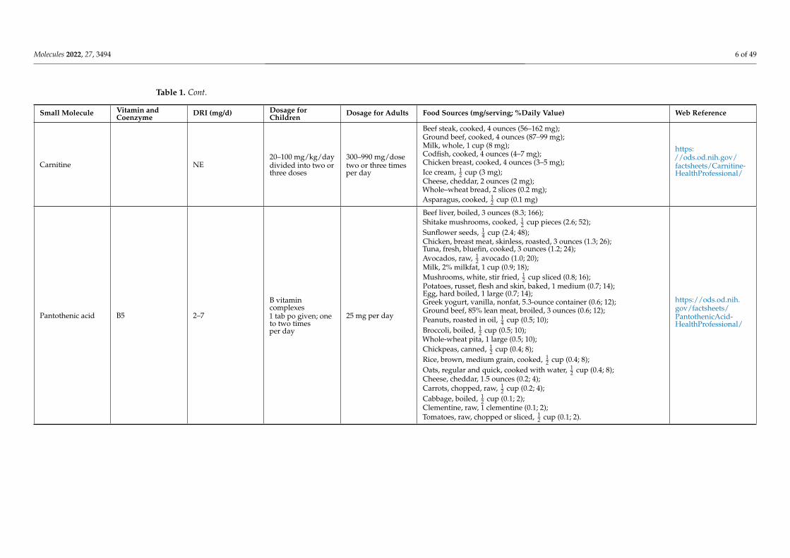
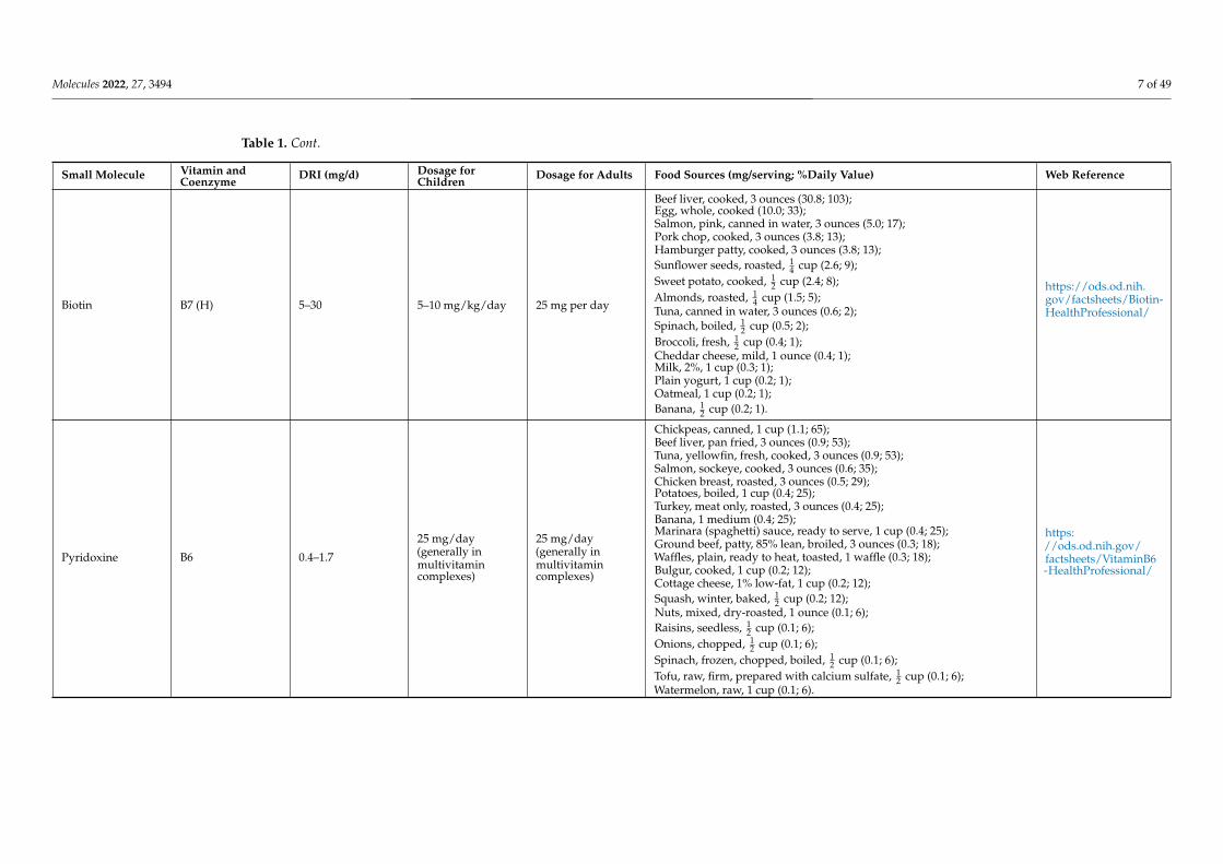
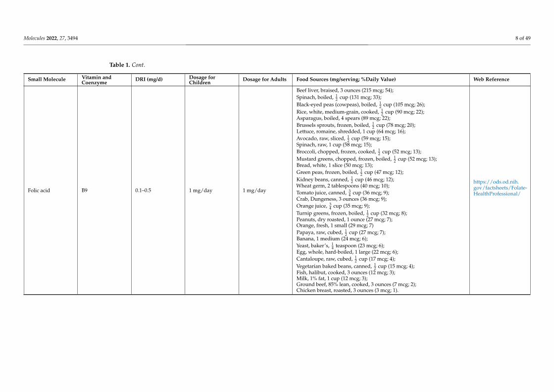
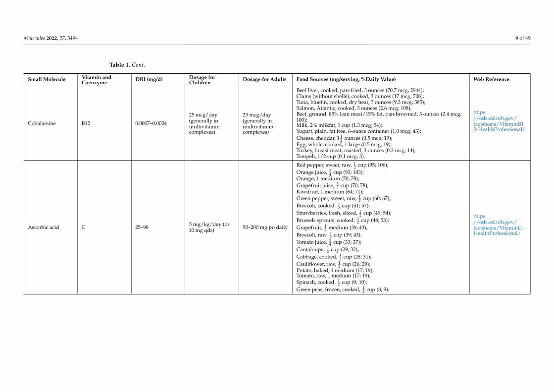
Tables 1 and 2 list the most common vitamins used in patients affected by MD as singlemolecules or as an antioxidant cocktail, with dietary reference intakes (DRIs, as reportedin https://www.ncbi.nlm.nih.gov/books/NBK56068/table/summarytables.t7/?report=objectonly; accessed on 8 April 2022), whereas Tables 3 and 4 list vitamin-related cofactorsand the known associated protein targets.

Molecules 2022, 27, 3494 4 of 49
Table 1. Hydrosoluble vitamin DRIs are reported according to https://www.ncbi.nlm.nih.gov/books/NBK222881/ and https://sinu.it/. An estimation of dailynutrient recommendations based on the DRI can be obtained by completing the linked form: https://www.nal.usda.gov/legacy/fnic/dri-calculator/index.php.Dietary supplement dosages for children and adults are coherent with those reported in https://ods.od.nih.gov/factsheets/PrimaryMitochondrialDisorders-HealthProfessional/. All the indicated web-links were accessed on 8 April 2022 for a final check.
Small Molecule Vitamin andCoenzyme
DRI (mg/d) Dosage forChildren
Dosage for Adults Food Sources (mg/serving; %Daily Value) Web Reference
Thiamine B1 0.4–1.3 10 mg/kg/day 100–1000 mg/day
Rice, white, long grain, enriched, parboiled, 12 cup (1.4; 117);
Pork chop, bone-in, broiled, 3 ounces (0.4; 33);Trout, cooked, dry heat, 3 ounces (0.4; 33);Black beans, boiled, 1
2 cup (0.4; 33);Mussels, blue, cooked, moist heat, 3 ounces (0.3; 25);Tuna, Bluefin, cooked, dry heat, 3 ounces (0.2; 17);Macaroni, whole wheat, cooked, 1 cup (0.2; 17);Acorn squash, cubed, baked, 1
2 cup (0.2; 17);Rice, brown, long grain, not enriched, cooked, 1
2 cup (0.1; 8);Bread, whole wheat, 1 slice (0.1; 8);Orange juice, prepared from concentrate, 1 cup (0.1; 8);Sunflower seeds, toasted, 1 ounce (0.1; 8);Beef steak, bottom round, trimmed of fat, braised, 3 ounces (0.1; 8);Yogurt, plain, low fat, 1 cup (0.1; 8);Oatmeal, regular and quick, unenriched, cooked with water, 1
2 cup (0.1; 8);Corn, yellow, boiled, 1 medium ear (0.1; 8);Milk, 2%, 1 cup (0.1; 8);Barley, pearled, cooked, 1 cup (0.1; 8).
https://ods.od.nih.gov/factsheets/Thiamin-HealthProfessional/
Riboflavin B2 50–400 mg po daily 50–400 mg po daily50–400 mg/day(divided in2–3 doses)
Beef liver, pan fried, 3 ounces (2.9; 223);Yogurt, plain, fat free, 1 cup (0.6; 46);Milk, 2% fat, 1 cup (0.5; 38);Beef, tenderloin steak, boneless, trimmed of fat, grilled, 3 ounces (0.4; 31);Clams, mixed species, cooked, moist heat, 3 ounces (0.4; 31);Mushrooms, portabella, sliced, grilled, 1
2 cup (0.3; 23);Almonds, dry roasted, 1 ounce (0.3; 23);Cheese, Swiss, 3 ounces (0.3; 23);Rotisserie chicken, breast meat only, 3 ounces (0.2; 15);Egg, whole, scrambled, 1 large (0.2; 15);Quinoa, cooked, 1 cup (0.2; 15);Salmon, pink, canned, 3 ounces (0.2; 15);Spinach, raw, 1 cup (0.1; 8);Apple, with skin, 1 large (0.1; 8);Kidney beans, canned, 1 cup (0.1; 8);Macaroni, elbow shaped, whole wheat, cooked, 1 cup (0.1; 8);Bread, whole wheat, 1 slice (0.1; 8);Cod, Atlantic, cooked, dry heat, 3 ounces (0.1; 8);Sunflower seeds, toasted, 1 ounce (0.1; 8);Tomatoes, crushed, canned, 1
2 cup (0.1; 8).
https://ods.od.nih.gov/factsheets/Riboflavin-HealthProfessional/

Molecules 2022, 27, 3494 5 of 49
Table 1. Cont.
Small Molecule Vitamin andCoenzyme
DRI (mg/d) Dosage forChildren
Dosage for Adults Food Sources (mg/serving; %Daily Value) Web Reference
Niacin B3 (PP) 5–14 25–250 mg/day 250 mg/day up to1 g/day
Beef liver, pan fried, 3 ounces (14.9; 93);Chicken breast, meat only, grilled, 3 ounces (10.3; 64);Marinara (spaghetti) sauce, ready to serve, 1 cup (10.3; 64);Turkey breast, meat only, roasted, 3 ounces (10;0; 63);Salmon, sockeye, cooked, 3 ounces (8.6; 54);Tuna, light, canned in water, drained, 3 ounces (8.6; 54);Pork, tenderloin, roasted, 3 ounces (6.3; 39);Beef, ground, 90% lean, pan-browned, 3 ounces (5.8; 36);Rice, brown, cooked, 1 cup (5.2; 33);Peanuts, dry roasted, 1 ounce (4.2; 26);Potato (russet), baked, 1 medium (2.3; 14);Sunflower seeds, dry roasted, 1 ounce (2.0; 13);Bread, whole wheat, 1 slice (1.4; 9);Pumpkin seeds, dry roasted, 1 ounce (1.3; 8);Soymilk, unfortified, 1 cup (1.3; 8);Lentils, boiled and drained, 1
2 cup (1.0; 6);Bulgur, cooked, 1 cup (0.9; 6);Banana, 1 medium (0.8; 5);Edamame, frozen, prepared, 1
2 cup (0.7; 4);Raisins, 1
2 cup (0.6; 4);Tomatoes, cherry, 1
2 cup (0.5; 3);Broccoli, boiled, drained, chopped, 1
2 cup (0.4; 3);Cashews, dry roasted, 1 ounce (0.4; 3);Yogurt, plain, low fat, 1 cup (0.3; 2);Apple, 1 medium (0.2; 1);Chickpeas, canned, drained, 1 cup (0.2; 1);Milk, 1% milkfat, 1 cup (0.2; 1);Spinach, frozen, chopped, boiled, 1
2 cup (0.2; 1);Tofu, raw, firm, 1
2 cup (0.2; 1);Onions, chopped, 1
2 cup (0.1; 1).
https://ods.od.nih.gov/factsheets/Niacin-HealthProfessional/
Coenzyme Q10(reduced, i.e., asubiquinol, oroxidized, i.e., asubiquinone)
3–6
CoQ10 as ubiquinol(preferred)2–8 mg/kg po dailydivided in twodoses CoQ10 asubiquinone10–30 mg/kg podaily divided in2 doses
CoQ10 as ubiquinol(preferred)50–600 mg po dailyCoQ10 asubiquinone300–2400 mg podaily divided2–3 times a day
Sardines, salmon, trout, mackerel, 3 ounces (2.3–4/up to 50)).Chicken, beef, pork, 3 ounces (1.4–2.6/up to 50).Spinach, broccoli, cauliflower, 1
2 cup, chopped (0.4–0.5/up to 20).Fruits: strawberries, oranges, 1
2 cup (0.1–0.3/up to 20).Soybean and canola oils, 1 tablespoon (1.0–1.3/up to 30).Soybeans, lentils, peanuts, 1 ounce (0.6/up to 20).Pistachio, sesame seeds, 1 ounce (3.6, up to 80).Egg boiled, 1 medium (0.1/up to 3)
https://lpi.oregonstate.edu/mic/dietary-factors/coenzyme-Q10
Molecules 2022, 27, 3494 6 of 49
Table 1. Cont.
Small Molecule Vitamin andCoenzyme
DRI (mg/d) Dosage forChildren
Dosage for Adults Food Sources (mg/serving; %Daily Value) Web Reference
Carnitine NE20–100 mg/kg/daydivided into two orthree doses
300–990 mg/dosetwo or three timesper day
Beef steak, cooked, 4 ounces (56–162 mg);Ground beef, cooked, 4 ounces (87–99 mg);Milk, whole, 1 cup (8 mg);Codfish, cooked, 4 ounces (4–7 mg);Chicken breast, cooked, 4 ounces (3–5 mg);Ice cream, 1
2 cup (3 mg);Cheese, cheddar, 2 ounces (2 mg);Whole–wheat bread, 2 slices (0.2 mg);Asparagus, cooked, 1
2 cup (0.1 mg)
https://ods.od.nih.gov/factsheets/Carnitine-HealthProfessional/
Pantothenic acid B5 2–7
B vitamincomplexes1 tab po given; oneto two timesper day
25 mg per day
Beef liver, boiled, 3 ounces (8.3; 166);Shitake mushrooms, cooked, 1
2 cup pieces (2.6; 52);Sunflower seeds, 1
4 cup (2.4; 48);Chicken, breast meat, skinless, roasted, 3 ounces (1.3; 26);Tuna, fresh, bluefin, cooked, 3 ounces (1.2; 24);Avocados, raw, 1
2 avocado (1.0; 20);Milk, 2% milkfat, 1 cup (0.9; 18);Mushrooms, white, stir fried, 1
2 cup sliced (0.8; 16);Potatoes, russet, flesh and skin, baked, 1 medium (0.7; 14);Egg, hard boiled, 1 large (0.7; 14);Greek yogurt, vanilla, nonfat, 5.3-ounce container (0.6; 12);Ground beef, 85% lean meat, broiled, 3 ounces (0.6; 12);Peanuts, roasted in oil, 1
4 cup (0.5; 10);Broccoli, boiled, 1
2 cup (0.5; 10);Whole-wheat pita, 1 large (0.5; 10);Chickpeas, canned, 1
2 cup (0.4; 8);Rice, brown, medium grain, cooked, 1
2 cup (0.4; 8);Oats, regular and quick, cooked with water, 1
2 cup (0.4; 8);Cheese, cheddar, 1.5 ounces (0.2; 4);Carrots, chopped, raw, 1
2 cup (0.2; 4);Cabbage, boiled, 1
2 cup (0.1; 2);Clementine, raw, 1 clementine (0.1; 2);Tomatoes, raw, chopped or sliced, 1
2 cup (0.1; 2).
https://ods.od.nih.gov/factsheets/PantothenicAcid-HealthProfessional/
Molecules 2022, 27, 3494 7 of 49
Table 1. Cont.
Small Molecule Vitamin andCoenzyme
DRI (mg/d) Dosage forChildren
Dosage for Adults Food Sources (mg/serving; %Daily Value) Web Reference
Biotin B7 (H) 5–30 5–10 mg/kg/day 25 mg per day
Beef liver, cooked, 3 ounces (30.8; 103);Egg, whole, cooked (10.0; 33);Salmon, pink, canned in water, 3 ounces (5.0; 17);Pork chop, cooked, 3 ounces (3.8; 13);Hamburger patty, cooked, 3 ounces (3.8; 13);Sunflower seeds, roasted, 1
4 cup (2.6; 9);Sweet potato, cooked, 1
2 cup (2.4; 8);Almonds, roasted, 1
4 cup (1.5; 5);Tuna, canned in water, 3 ounces (0.6; 2);Spinach, boiled, 1
2 cup (0.5; 2);Broccoli, fresh, 1
2 cup (0.4; 1);Cheddar cheese, mild, 1 ounce (0.4; 1);Milk, 2%, 1 cup (0.3; 1);Plain yogurt, 1 cup (0.2; 1);Oatmeal, 1 cup (0.2; 1);Banana, 1
2 cup (0.2; 1).
https://ods.od.nih.gov/factsheets/Biotin-HealthProfessional/
Pyridoxine B6 0.4–1.7
25 mg/day(generally inmultivitamincomplexes)
25 mg/day(generally inmultivitamincomplexes)
Chickpeas, canned, 1 cup (1.1; 65);Beef liver, pan fried, 3 ounces (0.9; 53);Tuna, yellowfin, fresh, cooked, 3 ounces (0.9; 53);Salmon, sockeye, cooked, 3 ounces (0.6; 35);Chicken breast, roasted, 3 ounces (0.5; 29);Potatoes, boiled, 1 cup (0.4; 25);Turkey, meat only, roasted, 3 ounces (0.4; 25);Banana, 1 medium (0.4; 25);Marinara (spaghetti) sauce, ready to serve, 1 cup (0.4; 25);Ground beef, patty, 85% lean, broiled, 3 ounces (0.3; 18);Waffles, plain, ready to heat, toasted, 1 waffle (0.3; 18);Bulgur, cooked, 1 cup (0.2; 12);Cottage cheese, 1% low-fat, 1 cup (0.2; 12);Squash, winter, baked, 1
2 cup (0.2; 12);Nuts, mixed, dry-roasted, 1 ounce (0.1; 6);Raisins, seedless, 1
2 cup (0.1; 6);Onions, chopped, 1
2 cup (0.1; 6);Spinach, frozen, chopped, boiled, 1
2 cup (0.1; 6);Tofu, raw, firm, prepared with calcium sulfate, 1
2 cup (0.1; 6);Watermelon, raw, 1 cup (0.1; 6).
https://ods.od.nih.gov/factsheets/VitaminB6-HealthProfessional/
Molecules 2022, 27, 3494 8 of 49
Table 1. Cont.
Small Molecule Vitamin andCoenzyme
DRI (mg/d) Dosage forChildren
Dosage for Adults Food Sources (mg/serving; %Daily Value) Web Reference
Folic acid B9 0.1–0.5 1 mg/day 1 mg/day
Beef liver, braised, 3 ounces (215 mcg; 54);Spinach, boiled, 1
2 cup (131 mcg; 33);Black-eyed peas (cowpeas), boiled, 1
2 cup (105 mcg; 26);Rice, white, medium-grain, cooked, 1
2 cup (90 mcg; 22);Asparagus, boiled, 4 spears (89 mcg; 22);Brussels sprouts, frozen, boiled, 1
2 cup (78 mcg; 20);Lettuce, romaine, shredded, 1 cup (64 mcg; 16);Avocado, raw, sliced, 1
2 cup (59 mcg; 15);Spinach, raw, 1 cup (58 mcg; 15);Broccoli, chopped, frozen, cooked, 1
2 cup (52 mcg; 13);Mustard greens, chopped, frozen, boiled, 1
2 cup (52 mcg; 13);Bread, white, 1 slice (50 mcg; 13);Green peas, frozen, boiled, 1
2 cup (47 mcg; 12);Kidney beans, canned, 1
2 cup (46 mcg; 12);Wheat germ, 2 tablespoons (40 mcg; 10);Tomato juice, canned, 3
4 cup (36 mcg; 9);Crab, Dungeness, 3 ounces (36 mcg; 9);Orange juice, 3
4 cup (35 mcg; 9);Turnip greens, frozen, boiled, 1
2 cup (32 mcg; 8);Peanuts, dry roasted, 1 ounce (27 mcg; 7);Orange, fresh, 1 small (29 mcg; 7)Papaya, raw, cubed, 1
2 cup (27 mcg; 7);Banana, 1 medium (24 mcg; 6);Yeast, baker’s, 1
4 teaspoon (23 mcg; 6);Egg, whole, hard-boiled, 1 large (22 mcg; 6);Cantaloupe, raw, cubed, 1
2 cup (17 mcg; 4);Vegetarian baked beans, canned, 1
2 cup (15 mcg; 4);Fish, halibut, cooked, 3 ounces (12 mcg; 3);Milk, 1% fat, 1 cup (12 mcg; 3);Ground beef, 85% lean, cooked, 3 ounces (7 mcg; 2);Chicken breast, roasted, 3 ounces (3 mcg; 1).
https://ods.od.nih.gov/factsheets/Folate-HealthProfessional/
Molecules 2022, 27, 3494 9 of 49
Table 1. Cont.
Small Molecule Vitamin andCoenzyme
DRI (mg/d) Dosage forChildren
Dosage for Adults Food Sources (mg/serving; %Daily Value) Web Reference
Cobalamine B12 0.0007–0.0024
25 mcg/day(generally inmultivitamincomplexes)
25 mcg/day(generally inmultivitamincomplexes)
Beef liver, cooked, pan-fried, 3 ounces (70.7 mcg; 2944);Clams (without shells), cooked, 3 ounces (17 mcg; 708);Tuna, bluefin, cooked, dry heat, 3 ounces (9.3 mcg; 385);Salmon, Atlantic, cooked, 3 ounces (2.6 mcg; 108);Beef, ground, 85% lean meat/15% fat, pan-browned, 3 ounces (2.4 mcg;100);Milk, 2% milkfat, 1 cup (1.3 mcg; 54);Yogurt, plain, fat free, 6-ounce container (1.0 mcg; 43);Cheese, cheddar, 1 1
2 ounces (0.5 mcg; 19);Egg, whole, cooked, 1 large (0.5 mcg; 19);Turkey, breast meat, roasted, 3 ounces (0.3 mcg; 14);Tempeh, 1/2 cup (0.1 mcg; 3).
https://ods.od.nih.gov/factsheets/VitaminB12-HealthProfessional/
Ascorbic acid C 25–90 5 mg/kg/day (or10 mg qds) 50–200 mg po daily
Red pepper, sweet, raw, 12 cup (95; 106);
Orange juice, 34 cup (93; 103);
Orange, 1 medium (70; 78);Grapefruit juice, 3
4 cup (70; 78);Kiwifruit, 1 medium (64; 71);Green pepper, sweet, raw, 1
2 cup (60; 67);Broccoli, cooked, 1
2 cup (51; 57);Strawberries, fresh, sliced, 1
2 cup (49; 54);Brussels sprouts, cooked, 1
2 cup (48; 53);Grapefruit, 1
2 medium (39; 43);Broccoli, raw, 1
2 cup (39; 43);Tomato juice, 3
4 cup (33; 37);Cantaloupe, 1
2 cup (29; 32);Cabbage, cooked, 1
2 cup (28; 31);Cauliflower, raw, 1
2 cup (26; 29);Potato, baked, 1 medium (17; 19);Tomato, raw, 1 medium (17; 19);Spinach, cooked, 1
2 cup (9; 10);Green peas, frozen, cooked, 1
2 cup (8; 9).
https://ods.od.nih.gov/factsheets/VitaminC-HealthProfessional/

Molecules 2022, 27, 3494 10 of 49
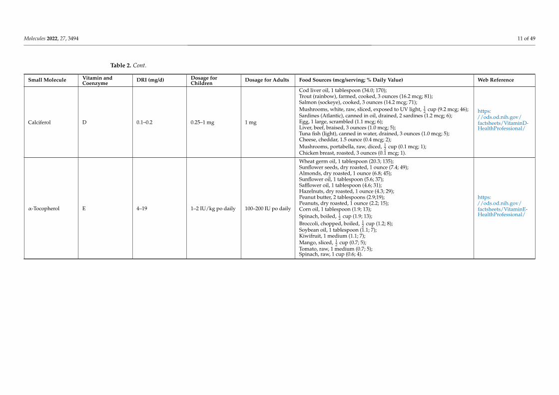
Table 2. Liposoluble (hydrophobic) vitamins: DRIs are reported according to https://www.ncbi.nlm.nih.gov/books/NBK222881/ and https://sinu.it/. Anestimation of daily nutrient recommendations based on the DRI can be obtained by completing the linked form: https://www.nal.usda.gov/legacy/fnic/dri-calculator/index.php; https://ods.od.nih.gov/factsheets/VitaminE-HealthProfessional/. Dietary supplement dosages for children and adults are coherent withthose reported in the cited references within paragraphs dedicated to each vitamin (see below) and/or with those reported in https://ods.od.nih.gov/factsheets/PrimaryMitochondrialDisorders-HealthProfessional/. All the indicated web-links were accessed on 8 April 2022 for a final check.
Small Molecule Vitamin andCoenzyme
DRI (mg/d) Dosage forChildren
Dosage for Adults Food Sources (mcg/serving; % Daily Value) Web Reference
Lipoic acid 50–600 50–200 mg/day 50–200 mg/dayBeef kidney, heart and liver (1–3 mcg/g dry wt) as lipoyllysine);Spinach and broccoli (1–3 mcg/g dry wt, as lipoyllysine);Tomatoes, peas and brussels sprouts (0.5 mcg/g dry wt, as lip).
https://lpi.oregonstate.edu/mic/dietary-factors/lipoic-acid
Retinol A 0.7–1.30.3–0.9 mg per day(depending onthe age)
Up to 3 mg per day
Beef liver, pan fried, 3 ounces (6582 mcg; 731);Sweet potato, baked in skin, 1 whole (1403 mcg; 156);Spinach, frozen, boiled, 1
2 cup (573 mcg; 64);Pumpkin pie, commercially prepared, 1 piece (488 mcg; 54);Carrots, raw, 1
2 cup (459 mcg; 51);Ice cream, French vanilla, soft serve, 1 cup (278 mcg; 31);Cheese, ricotta, part skim, 1 cup (263 mcg; 29);Herring, Atlantic, pickled, 3 ounces (219 mcg; 24);Cantaloupe, raw, 1
2 cup (135 mcg; 15);Peppers, sweet, red, raw, 1
2 cup (117 mcg; 13);Mangos, raw, 1 whole (112 mcg; 12);Egg, hard boiled, 1 large (75 mcg; 8);Black-eyed peas (cowpeas), boiled, 1 cup (66 mcg; 7);Apricots, dried, sulfured, 5 apricots (63 mcg; 7);Broccoli, boiled, 1
2 cup (60 mcg; 7);Salmon, sockeye, cooked, 3 ounces (59 mcg; 7);Tomato juice, canned, 3
4 cup (42 mcg; 5);Yogurt, plain, low fat, 1 cup (32 mcg; 4);Tuna, light, canned in oil, drained solids, 3 ounces (20 mcg; 2);Baked beans, canned, plain or vegetarian, 1 cup (13 mcg; 1);Summer squash, all varieties, boiled, 1
2 cup (10 mcg; 1);Chicken, breast meat and skin, roasted, 1
2 breast (5 mcg; 1);Pistachio nuts, dry roasted, 1 ounce (4 mcg; 0).
https://ods.od.nih.gov/factsheets/VitaminA-HealthProfessional/
Molecules 2022, 27, 3494 11 of 49
Table 2. Cont.
Small Molecule Vitamin andCoenzyme
DRI (mg/d) Dosage forChildren
Dosage for Adults Food Sources (mcg/serving; % Daily Value) Web Reference
Calciferol D 0.1–0.2 0.25–1 mg 1 mg
Cod liver oil, 1 tablespoon (34.0; 170);Trout (rainbow), farmed, cooked, 3 ounces (16.2 mcg; 81);Salmon (sockeye), cooked, 3 ounces (14.2 mcg; 71);Mushrooms, white, raw, sliced, exposed to UV light, 1
2 cup (9.2 mcg; 46);Sardines (Atlantic), canned in oil, drained, 2 sardines (1.2 mcg; 6);Egg, 1 large, scrambled (1.1 mcg; 6);Liver, beef, braised, 3 ounces (1.0 mcg; 5);Tuna fish (light), canned in water, drained, 3 ounces (1.0 mcg; 5);Cheese, cheddar, 1.5 ounce (0.4 mcg; 2);Mushrooms, portabella, raw, diced, 1
2 cup (0.1 mcg; 1);Chicken breast, roasted, 3 ounces (0.1 mcg; 1).
https://ods.od.nih.gov/factsheets/VitaminD-HealthProfessional/
α-Tocopherol E 4–19 1–2 IU/kg po daily 100–200 IU po daily
Wheat germ oil, 1 tablespoon (20.3; 135);Sunflower seeds, dry roasted, 1 ounce (7.4; 49);Almonds, dry roasted, 1 ounce (6.8; 45);Sunflower oil, 1 tablespoon (5.6; 37);Safflower oil, 1 tablespoon (4.6; 31);Hazelnuts, dry roasted, 1 ounce (4.3; 29);Peanut butter, 2 tablespoons (2.9;19);Peanuts, dry roasted, 1 ounce (2.2; 15);Corn oil, 1 tablespoon (1.9; 13);Spinach, boiled, 1
2 cup (1.9; 13);Broccoli, chopped, boiled, 1
2 cup (1.2; 8);Soybean oil, 1 tablespoon (1.1; 7);Kiwifruit, 1 medium (1.1; 7);Mango, sliced, 1
2 cup (0.7; 5);Tomato, raw, 1 medium (0.7; 5);Spinach, raw, 1 cup (0.6; 4).
https://ods.od.nih.gov/factsheets/VitaminE-HealthProfessional/

Molecules 2022, 27, 3494 12 of 49
Table 2. Cont.
Small Molecule Vitamin andCoenzyme
DRI (mg/d) Dosage forChildren
Dosage for Adults Food Sources (mcg/serving; % Daily Value) Web Reference
Phylloquinone K 0.09–0.12 2–75 mcg 90–120 mcg
Natto, 3 ounces (as MK-7; 850 mcg; 708);Collards, frozen, boiled, 1
2 cup (530 mcg; 442);Turnip greens, frozen, boiled 1
2 cup (426 mcg; 355);Spinach, raw, 1 cup (145 mcg; 121);Kale, raw, 1 cup (113 mcg; 94);Broccoli, chopped, boiled, 1
2 cup (110 mcg; 92);Soybeans, roasted, 1
2 cup (43 mcg; 36);Carrot juice, 3
4 cup (28 mcg; 23);Soybean oil, 1 tablespoon (25 mcg; 21);Edamame, frozen, prepared, 1
2 cup (21 mcg; 18);Pumpkin, canned, 1
2 cup (20 mcg; 17);Pomegranate juice, 3
4 cup (19 mcg; 16);Okra, raw, 1
2 cup (16 mcg; 13);Salad dressing, Caesar, 1 tablespoon (15 mcg; 13);Pine nuts, dried, 1 ounce (15 mcg; 13);Blueberries, raw, 1
2 cup (14 mcg; 12);Iceberg lettuce, raw, 1 cup (14 mcg; 12);Chicken, breast, rotisserie, 3 ounces (as MK-4; 13 mcg; 11);Grapes, 1
2 cup (11 mcg; 9);Vegetable juice cocktail, 3
4 cup (10 mcg; 8);Canola oil, 1 tablespoon cup (10 mcg; 8);Cashews, dry roasted, 1 ounce cup (10 mcg; 8);Carrots, raw, 1 medium (8 mcg; 7);Olive oil, 1 tablespoon (8 mcg; 7);Ground beef, broiled, 3 ounces (as MK-4; 6 mcg; 5);Figs, dried, 1
4 cup (6 mcg; 5);Chicken liver, braised, 3 ounces (as MK-4; 6 mcg; 5);Ham, roasted or pan-broiled, 3 ounces (as MK-4; 4 mcg; 3);Cheddar cheese, 1 1
2 ounces (as MK-4; 4 mcg; 3);Mixed nuts, dry roasted, 1 ounce (4 mcg; 3);Egg, hard boiled, 1 large (as MK-4; 4 mcg; 3);Mozzarella cheese, 1 1
2 ounces (as MK-4; 2 mcg; 2);Milk, 2%, 1 cup (as MK-4; 1 mcg; 1);Salmon, sockeye, cooked, 3 ounces (as MK-4; 0.3 mcg; 0);Shrimp, cooked, 3 ounces (as MK-4; 0.3 mcg; 0).
https://ods.od.nih.gov/factsheets/vitaminK-HealthProfessional/
Molecules 2022, 27, 3494 13 of 49
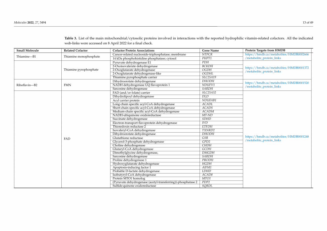
Table 3. List of the main mitochondrial/cytosolic proteins involved in interactions with the reported hydrophilic vitamin-related cofactors. All the indicatedweb-links were accessed on 8 April 2022 for a final check.
Small Molecule Related Cofactor Cofactor Protein Associations Gene Name Protein Targets from HMDB
Thiamine—B1 Thiamine monophosphateCancer-related nucleoside-triphosphatase; membrane NTPCR https://hmdb.ca/metabolites/HMDB0002666
/metabolite_protein_links14 kDa phosphohistidine phosphatase; cytosol PHPT1
Thiamine pyrophosphate
Pyruvate dehydrogenase E1 PDH
https://hmdb.ca/metabolites/HMDB0001372/metabolite_protein_links
2-Oxoisovalerate dehydrogenase BCKDH2-Oxoglutarate dehydrogenase OGDH2-Oxoglutarate dehydrogenase-like OGDHLThiamine pyrophosphate carrier SLC25A19
Riboflavin—B2 FMNDihydroorotate dehydrogenase DHODH https://hmdb.ca/metabolites/HMDB0001520
/metabolite_protein_linksNADH dehydrogenase UQ flavoprotein 1 NDUFV1Sarcosine dehydrogenase SARDH
FAD
FAD (and/or folate) carrier SLC25A32
https://hmdb.ca/metabolites/HMDB0001248/metabolite_protein_links
Dihydrolipoyl dehydrogenase DLDAcyl carrier protein NDUFAB1Long-chain specific acyl-CoA dehydrogenase ACADLShort-chain specific acyl-CoA dehydrogenase ACADSMedium-chain specific acyl-CoA dehydrogenase ACADMNADH-ubiquinone oxidoreductase MT-NDSuccinate dehydrogenase SDHDElectron-transport flavoprotein dehydrogenase IVDThioredoxin reductase 2 ETFDHIsovaleryl-CoA dehydrogenase TXNRD2Dihydroorotate dehydrogenase DHODHGlutathione reductase GSRGlycerol-3-phosphate dehydrogenase GPD2Choline dehydrogenase CHDHGlutaryl-CoA dehydrogenase GCDHDimethylglycine dehydrogenase, DMGDHSarcosine dehydrogenase SARDHProline dehydrogenase 1 PRODHHydroxyglutarate dehydrogenase HGDHApoptosis-inducing factor 1 AIFM1Probable D-lactate dehydrogenase LDHDIsobutyryl-CoA dehydrogenase ACAD8Protein MTO1 homolog MTO1(Pyruvate dehydrogenase (acetyl-transferring))-phosphatase 2 PDP2Sulfide:quinone oxidoreductase SQRDL
Molecules 2022, 27, 3494 14 of 49
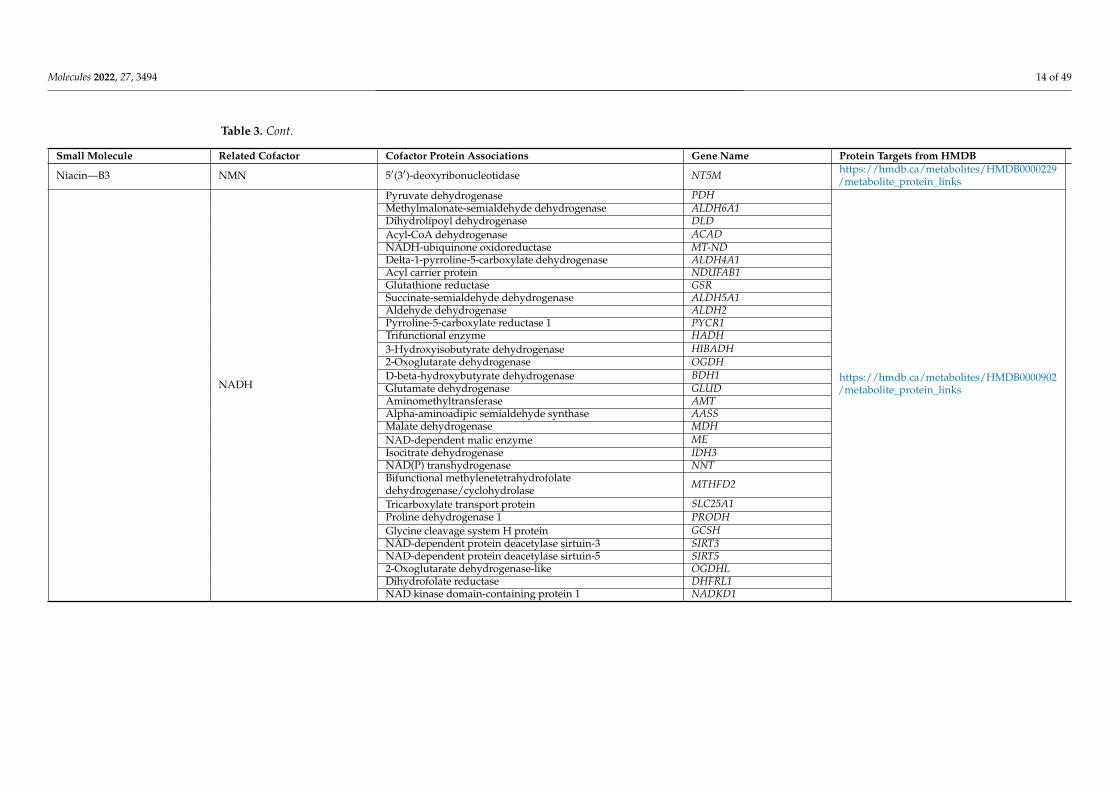
Table 3. Cont.
Small Molecule Related Cofactor Cofactor Protein Associations Gene Name Protein Targets from HMDB
Niacin—B3 NMN 5′(3′)-deoxyribonucleotidase NT5M https://hmdb.ca/metabolites/HMDB0000229/metabolite_protein_links
NADH
Pyruvate dehydrogenase PDH
https://hmdb.ca/metabolites/HMDB0000902/metabolite_protein_links
Methylmalonate-semialdehyde dehydrogenase ALDH6A1Dihydrolipoyl dehydrogenase DLDAcyl-CoA dehydrogenase ACADNADH-ubiquinone oxidoreductase MT-NDDelta-1-pyrroline-5-carboxylate dehydrogenase ALDH4A1Acyl carrier protein NDUFAB1Glutathione reductase GSRSuccinate-semialdehyde dehydrogenase ALDH5A1Aldehyde dehydrogenase ALDH2Pyrroline-5-carboxylate reductase 1 PYCR1Trifunctional enzyme HADH3-Hydroxyisobutyrate dehydrogenase HIBADH2-Oxoglutarate dehydrogenase OGDHD-beta-hydroxybutyrate dehydrogenase BDH1Glutamate dehydrogenase GLUDAminomethyltransferase AMTAlpha-aminoadipic semialdehyde synthase AASSMalate dehydrogenase MDHNAD-dependent malic enzyme MEIsocitrate dehydrogenase IDH3NAD(P) transhydrogenase NNTBifunctional methylenetetrahydrofolatedehydrogenase/cyclohydrolase MTHFD2
Tricarboxylate transport protein SLC25A1Proline dehydrogenase 1 PRODHGlycine cleavage system H protein GCSHNAD-dependent protein deacetylase sirtuin-3 SIRT3NAD-dependent protein deacetylase sirtuin-5 SIRT52-Oxoglutarate dehydrogenase-like OGDHLDihydrofolate reductase DHFRL1NAD kinase domain-containing protein 1 NADKD1
Molecules 2022, 27, 3494 15 of 49
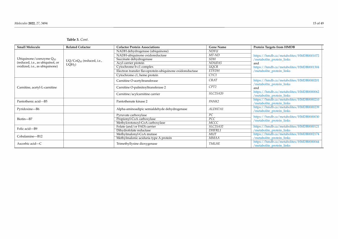
Table 3. Cont.
Small Molecule Related Cofactor Cofactor Protein Associations Gene Name Protein Targets from HMDB
Ubiquinone/coenzyme Q10(reduced, i.e., as ubiquinol, oroxidized, i.e., as ubiquinone)
UQ/CoQ10 (reduced, i.e.,UQH2)
NADH dehydrogenase (ubiquinone) NDFUhttps://hmdb.ca/metabolites/HMDB0001072/metabolite_protein_linksandhttps://hmdb.ca/metabolites/HMDB0001304/metabolite_protein_links
NADH-ubiquinone oxidoreductase MT-NDSuccinate dehydrogenase SDHAcyl carrier protein NDUFA5Cytochrome b-c1 complex UQCRElectron transfer flavoprotein-ubiquinone oxidoreductase ETFDHCytochrome c1, heme protein CYC1
Carnitine, acetyl-L-carnitine
Carnitine O-acetyltransferase CRAT https://hmdb.ca/metabolites/HMDB0000201/metabolite_protein_linksandhttps://hmdb.ca/metabolites/HMDB0000062/metabolite_protein_links
Carnitine O-palmitoyltransferase 2 CPT2
Carnitine/acylcarnitine carrier SLC25A20
Pantothenic acid—B5 Pantothenate kinase 2 PANK2 https://hmdb.ca/metabolites/HMDB0000210/metabolite_protein_links
Pyridoxine—B6 Alpha-aminoadipic semialdehyde dehydrogenase ALDH7A1 https://hmdb.ca/metabolites/HMDB0000239/metabolite_protein_links
Biotin—B7Pyruvate carboxylase PC https://hmdb.ca/metabolites/HMDB0000030
/metabolite_protein_linksPropionyl-CoA carboxylase PCCMethylcrotonoyl-CoA carboxylase MCCC
Folic acid—B9Folate (and/or FAD) carrier SLC25A32 https://hmdb.ca/metabolites/HMDB0000121
/metabolite_protein_linksDihydrofolate reductase DHFRL1
Cobalamine—B12Methylmalonyl-CoA mutase MUT https://hmdb.ca/metabolites/HMDB0002174
/metabolite_protein_linksMethylmalonic aciduria type A protein MMAA
Ascorbic acid—C Trimethyllysine dioxygenase TMLHE https://hmdb.ca/metabolites/HMDB0000044/metabolite_protein_links
Molecules 2022, 27, 3494 16 of 49
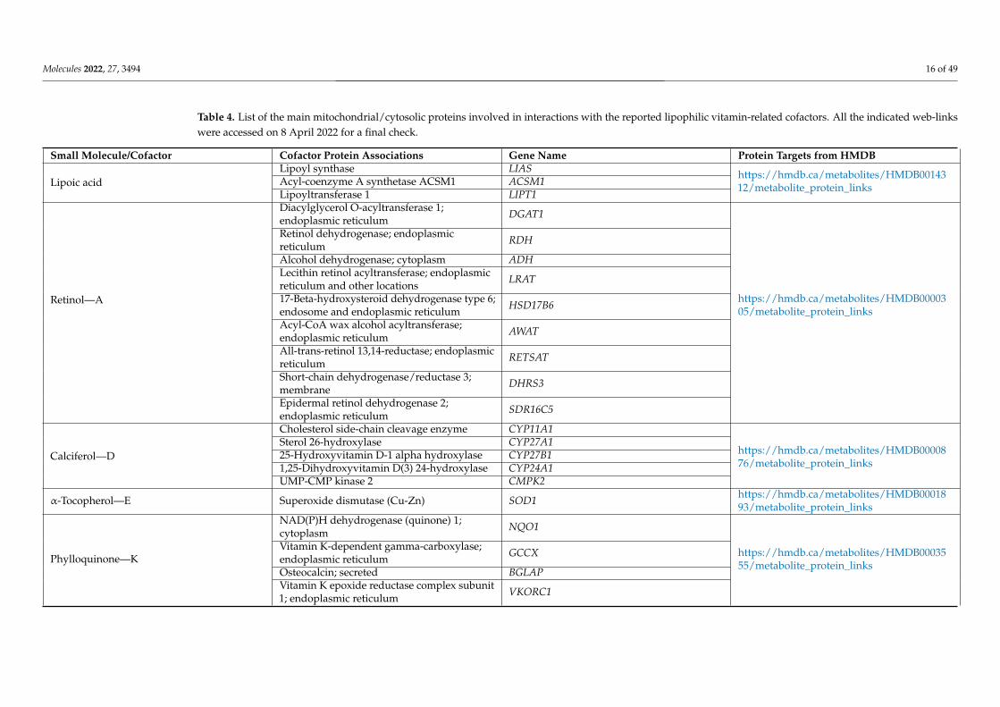
Table 4. List of the main mitochondrial/cytosolic proteins involved in interactions with the reported lipophilic vitamin-related cofactors. All the indicated web-linkswere accessed on 8 April 2022 for a final check.
Small Molecule/Cofactor Cofactor Protein Associations Gene Name Protein Targets from HMDB
Lipoic acidLipoyl synthase LIAS https://hmdb.ca/metabolites/HMDB00143
12/metabolite_protein_linksAcyl-coenzyme A synthetase ACSM1 ACSM1Lipoyltransferase 1 LIPT1
Retinol—A
Diacylglycerol O-acyltransferase 1;endoplasmic reticulum DGAT1
https://hmdb.ca/metabolites/HMDB0000305/metabolite_protein_links
Retinol dehydrogenase; endoplasmicreticulum RDH
Alcohol dehydrogenase; cytoplasm ADHLecithin retinol acyltransferase; endoplasmicreticulum and other locations LRAT
17-Beta-hydroxysteroid dehydrogenase type 6;endosome and endoplasmic reticulum HSD17B6
Acyl-CoA wax alcohol acyltransferase;endoplasmic reticulum AWAT
All-trans-retinol 13,14-reductase; endoplasmicreticulum RETSAT
Short-chain dehydrogenase/reductase 3;membrane DHRS3
Epidermal retinol dehydrogenase 2;endoplasmic reticulum SDR16C5
Calciferol—D
Cholesterol side-chain cleavage enzyme CYP11A1
https://hmdb.ca/metabolites/HMDB0000876/metabolite_protein_links
Sterol 26-hydroxylase CYP27A125-Hydroxyvitamin D-1 alpha hydroxylase CYP27B11,25-Dihydroxyvitamin D(3) 24-hydroxylase CYP24A1UMP-CMP kinase 2 CMPK2
α-Tocopherol—E Superoxide dismutase (Cu-Zn) SOD1 https://hmdb.ca/metabolites/HMDB0001893/metabolite_protein_links
Phylloquinone—K
NAD(P)H dehydrogenase (quinone) 1;cytoplasm NQO1
https://hmdb.ca/metabolites/HMDB0003555/metabolite_protein_links
Vitamin K-dependent gamma-carboxylase;endoplasmic reticulum GCCX
Osteocalcin; secreted BGLAPVitamin K epoxide reductase complex subunit1; endoplasmic reticulum VKORC1
Molecules 2022, 27, 3494 17 of 49
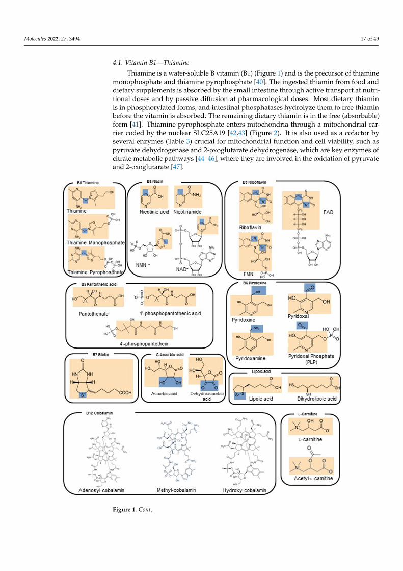
4.1. Vitamin B1—Thiamine
Thiamine is a water-soluble B vitamin (B1) (Figure 1) and is the precursor of thiaminemonophosphate and thiamine pyrophosphate [40]. The ingested thiamin from food anddietary supplements is absorbed by the small intestine through active transport at nutri-tional doses and by passive diffusion at pharmacological doses. Most dietary thiaminis in phosphorylated forms, and intestinal phosphatases hydrolyze them to free thiaminbefore the vitamin is absorbed. The remaining dietary thiamin is in the free (absorbable)form [41]. Thiamine pyrophosphate enters mitochondria through a mitochondrial car-rier coded by the nuclear SLC25A19 [42,43] (Figure 2). It is also used as a cofactor byseveral enzymes (Table 3) crucial for mitochondrial function and cell viability, such aspyruvate dehydrogenase and 2-oxoglutarate dehydrogenase, which are key enzymes ofcitrate metabolic pathways [44–46], where they are involved in the oxidation of pyruvateand 2-oxoglutarate [47].
Molecules 2022, 27, 3494 24 of 60
Figure 1. Cont.

Molecules 2022, 27, 3494 18 of 49Molecules 2022, 27, 3494 25 of 60
Figure 1. D structures of vitamins and derived enzyme cofactors. Orange squares indicate the vita-min portion of each reported molecule. Blue squares indicate the reactive centers involved in elec-tron transfer and/or redox changes.
Figure 1. 2D structures of vitamins and derived enzyme cofactors. Orange squares indicate thevitamin portion of each reported molecule. Blue squares indicate the reactive centers involved inelectron transfer and/or redox changes.
Thiamine has been used in mitochondrial diseases as a single molecule or in combina-tion with other antioxidants and drugs [39,40]. It is reported that thiamine administrationto patients affected by MELAS syndrome and/or thiamine deficiency improves lacticacidosis and myopathy [48]. The administration of thiamine, in combination with coen-zyme Q10 (CoQ10), carnitine and vitamins C and E, produced a clinical improvement ina Leigh patient with a severe encephalopathy [39,49] and in a recently described caseof MEGDEL syndrome [50]. Mutations in the SLC19A3 gene (encoding a ubiquitouslyexpressed transmembrane thiamine transporter) are responsible for thiamine deficiency,which can manifest with encephalopathy, features of Leigh syndrome on neuroimagingand lactic acidosis. Similarly, mutations in the SLC25A19 gene coding for a mitochondrialthiamine-pyrophosphate transporter of the inner mitochondrial membrane are responsiblefor a rare disease characterized by microcephaly and bilateral striatal necrosis [42,43,51].The doses of thiamine for adults and children are reported in Table 1 [52]. Higher doses ofthiamine (20 mg/kg/day) are administered to patients affected by SLC19A3 and SLC25A19deficiencies to limit neurological and biochemical abnormalities [53].

Molecules 2022, 27, 3494 19 of 49Molecules 2022, 27, 3494 26 of 60
Figure 2. Scheme of a mitochondrion with a set of representative proteins, pathways and cycles. Respiratory chain complexes, mitochondrial transporters and other proteins are reported in surf representation and labeled. ATP synthase (CV) is reported in blue. Mitochondrial carriers are re-ported in cyan. VDAC is reported in pink. Bax and Bak/Bcl-2 are reported in dark grey and firebrick, respectively. MPC is reported in black. Complex I (CI), complex II (CII), complex III (CIII) and com-plex IV (CIV) are reported in green, yellow, magenta and grey, respectively. Black circular arrows indicate cyclic pathways. Red arrows indicate impaired pathways or reactions. Black solid/dashed lines indicate the possible direction of the reported reactions. Abbreviations: MIM: mitochondrial inner membrane; MOM, mitochondrial outer membrane; IMS, intermembrane space; AAC, ADP/ATP carrier, coded in H. sapiens by SLC25A4, SLC25A5, SLC25A6, SLC25A31; TPC, thiamine pyrophosphate carrier, coded by SLC25A19; CAC, carnitine/acyl-carnitine carrier, coded by SLC25A20; ORC, ornithine carrier, coded by SLC25A15 (or SLC25A2); AGC, aspartate/glutamate carrier, coded by SLC25A12 and SLC25A13; DIC, dicarboxylate carrier, coded by SLC25A10; NDT, assumed to be the NAD+ carrier, coded by SLC25A51; MFT, assumed to be the FAD (folate/ribofla-vin) carrier, coded by SLC25A32; OGC, malate/2-oxoglutarate carrier, coded by SLC25A11; CIC, citrate carrier, coded by SLC25A1; PiC, phosphate carrier, coded by SLC25A3; MAS, malate/aspar-tate shuttle; TCA, tricarboxylic acid cycle; Bax, Bcl-2 associated X protein; Bak, Bcl-2 antago-nist/killer-1; Bcl-2, B-cell lymphoma-2; MPC, mitochondrial pyruvate carrier; UCP, uncoupling pro-tein, coded by SLC25A7, SLC25A8, SLC25A9, SLC25A14, SLC25A27 and SLC25A30; CypD, cyclo-philin D; CytC, cytochrome C; VDAC, voltage-dependent anion channel; AIF, apoptosis-inducing factor; PNC, pyrimidine nucleotide carrier, coded in H. sapiens by SLC25A33 and SLC25A36.
4.2. Vitamin B2—Riboflavin Riboflavin is a water-soluble vitamin also known as vitamin B2 and represents the
precursor of flavin mononucleotide (FMN) and flavin adenine dinucleotide (FAD) (Figure 1). More than 90% of dietary riboflavin is in the form of FAD or FMN; the remaining 10% is made up of the free form and glycosides or esters. Most riboflavin is absorbed in the proximal small intestine. The body absorbs little riboflavin from single doses beyond 27 mg and stores only small amounts of riboflavin in the liver, heart, and kidneys. When excess amounts are consumed, they are either not absorbed or excreted in the urine.
Bacteria in the large intestine produce free riboflavin that can be absorbed by the large intestine in amounts depending on the diet. More riboflavin is produced after
Figure 2. Scheme of a mitochondrion with a set of representative proteins, pathways and cycles.Respiratory chain complexes, mitochondrial transporters and other proteins are reported in surfrepresentation and labeled. ATP synthase (CV) is reported in blue. Mitochondrial carriers arereported in cyan. VDAC is reported in pink. Bax and Bak/Bcl-2 are reported in dark grey andfirebrick, respectively. MPC is reported in black. Complex I (CI), complex II (CII), complex III(CIII) and complex IV (CIV) are reported in green, yellow, magenta and grey, respectively. Blackcircular arrows indicate cyclic pathways. Red arrows indicate impaired pathways or reactions. Blacksolid/dashed lines indicate the possible direction of the reported reactions. Abbreviations: MIM:mitochondrial inner membrane; MOM, mitochondrial outer membrane; IMS, intermembrane space;AAC, ADP/ATP carrier, coded in H. sapiens by SLC25A4, SLC25A5, SLC25A6, SLC25A31; TPC,thiamine pyrophosphate carrier, coded by SLC25A19; CAC, carnitine/acyl-carnitine carrier, coded bySLC25A20; ORC, ornithine carrier, coded by SLC25A15 (or SLC25A2); AGC, aspartate/glutamatecarrier, coded by SLC25A12 and SLC25A13; DIC, dicarboxylate carrier, coded by SLC25A10; NDT,assumed to be the NAD+ carrier, coded by SLC25A51; MFT, assumed to be the FAD (folate/riboflavin)carrier, coded by SLC25A32; OGC, malate/2-oxoglutarate carrier, coded by SLC25A11; CIC, citratecarrier, coded by SLC25A1; PiC, phosphate carrier, coded by SLC25A3; MAS, malate/aspartateshuttle; TCA, tricarboxylic acid cycle; Bax, Bcl-2 associated X protein; Bak, Bcl-2 antagonist/killer-1;Bcl-2, B-cell lymphoma-2; MPC, mitochondrial pyruvate carrier; UCP, uncoupling protein, codedby SLC25A7, SLC25A8, SLC25A9, SLC25A14, SLC25A27 and SLC25A30; CypD, cyclophilin D;CytC, cytochrome C; VDAC, voltage-dependent anion channel; AIF, apoptosis-inducing factor; PNC,pyrimidine nucleotide carrier, coded in H. sapiens by SLC25A33 and SLC25A36.
4.2. Vitamin B2—Riboflavin
Riboflavin is a water-soluble vitamin also known as vitamin B2 and represents theprecursor of flavin mononucleotide (FMN) and flavin adenine dinucleotide (FAD) (Figure 1).More than 90% of dietary riboflavin is in the form of FAD or FMN; the remaining 10%is made up of the free form and glycosides or esters. Most riboflavin is absorbed in theproximal small intestine. The body absorbs little riboflavin from single doses beyond 27 mgand stores only small amounts of riboflavin in the liver, heart, and kidneys. When excessamounts are consumed, they are either not absorbed or excreted in the urine.
Bacteria in the large intestine produce free riboflavin that can be absorbed by thelarge intestine in amounts depending on the diet. More riboflavin is produced afteringestion of vegetable-based than meat-based foods. It is known that riboflavin and/orFAD can enter mitochondria through a mitochondrial carrier coded by the nuclear geneSLC25A32 [42,43] (Figure 2). FMN is an important cofactor in complex I whereas FAD
Molecules 2022, 27, 3494 20 of 49
is a key cofactor of complex II, FAD synthase, dihydrolipoamide dehydrogenase, andpyruvate dehydrogenase, playing a crucial role in mitochondrial respiratory chain andKrebs cycle [44,54–58]. Riboflavin derivatives serve as a cofactor in several other keyenzymatic reactions (Tables 1 and 3) involving fatty acid oxidation [59] and pyrimidinebiosynthesis [60]. FAD plays also a crucial role in the apoptosis-inducing factor, thioredoxinreductase and glutathione reductase [54].
Riboflavin has been used in mitochondrial diseases as a single molecule or in combi-nation with other antioxidants and drugs [39,40,52,55,61–63]. It is reported that riboflavinsupplementation to patients affected by MDs, with specific references to those causedby complex I and/or complex II mutations, improves lactic acidosis, myopathy andseizures [55,61,62]. The administration of riboflavin produced the amelioration of symp-toms in patients affected by multiple acyl CoA dehydrogenase deficiency (MADD) causedby mutations at the electron-transport flavoprotein dehydrogenase (ETFDH) or in theelectron transfer flavoprotein (ETF) consisting of α and β subunits encoded by ETFA andETFB, respectively [64–66].
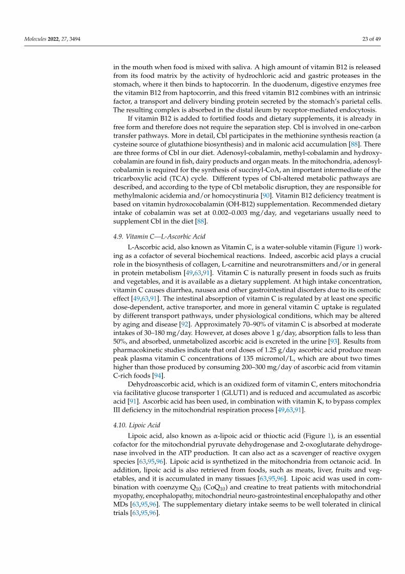
4.3. Vitamin B3—Niacin
Niacin or nicotinamide is a water-soluble vitamin also known as vitamin B3, andit represents the precursor of nicotinamide mononucleotide (NMN) and nicotinamideadenine dinucleotide (NAD+) (Figure 1). The ingested niacin is absorbed primarily in thesmall intestine, but some is absorbed in the stomach. All tissues in the body convert theabsorbed niacin into its main metabolically active form, the coenzyme NAD+, which in turnis converted into another active form, the coenzyme nicotinamide adenine dinucleotidephosphate (NADP+), in all tissues except skeletal muscle. Most dietary niacin is in the formof nicotinic acid and nicotinamide, but some foods contain small amounts of NAD+ andNADP+. Cells are also able to convert tryptophan to NAD+, so tryptophan is considereda dietary source of niacin. When NAD+ and NADP+ are consumed in foods, they areconverted to nicotinamide in the gut and then absorbed.
It is known that reducing equivalents cross the inner mitochondrial membrane byusing the malate/aspartate shuttle [67–70] to participate in the oxidation of the cytosolicNADH (Figure 2). In addition, it was recently proposed that SLC25A51 is responsible forthe NAD+ uptake into mitochondria [71–73], although a biochemical assay with the purifiedSLC25A51 reconstituted into proteoliposomes, as done for other mitochondrial carriers [74],is still lacking and is required to determine kinetic parameters of mitochondrial NAD+
uptake. NMN is an important cofactor in mitochondrial 5′-(3′)-deoxyribonucleotidase,whereas NAD+ is a key cofactor of complex I, dihydrolipoamide dehydrogenase and pyru-vate dehydrogenase, playing a crucial role in the mitochondrial respiratory chain and citratemetabolism [44,55,75]. NAD+ serves as a cofactor in several other key enzymatic reactions(Tables 1 and 3) involving fatty acid oxidation [59] and pyrimidine biosynthesis [60]. NAD+
is also a crucial cofactor for the apoptosis-inducing factor (AIF), glutathione reductaseand thioredoxin reductase protein [54], as well as for sirtuin deacetylases, which play animportant role in neurodegeneration [76].
Niacin and niacin derivatives have been used in the treatment of mitochondrial dis-eases as single molecules or in combination with other antioxidants and drugs [39,49].Niacin supplementation in patients affected by MDs, with specific references to thosecaused by complex I mutations, has been reported to improve lactic acidosis, myopathyand seizures [77], although previous papers have described niacin toxicity [78]. The ad-ministration of niacin is supposed to produce the amelioration of symptoms in patientsaffected by Leigh syndrome associated with mutations of nuclear [12,21,22] and mitochon-drial genes [15,77]. Niacin is available in multivitamin–mineral products, in supplementscontaining other B-complex vitamins and in supplements containing niacin only. Nicotinicacid and nicotinamide are the two most common forms of niacin in supplements. Someniacin-only supplements contain 500 mg or more per serving, which is much higher than

Molecules 2022, 27, 3494 21 of 49
the recommended dietary allowance (RDA) for this nutrient. The doses of niacin for adultsand children are reported in Table 1 [49,52].
4.4. Vitamin B5—Pantothenic Acid
Pantothenic acid, also known as vitamin B5 (Figure 1), is a water-soluble vitamin,widely distributed in foodstuffs and ubiquitous in nature [79]. About 85% of dietarypantothenic acid is in the form of CoA or phosphopantetheine. These forms are convertedto pantothenic acid by digestive enzymes in the intestinal lumen and intestinal cells.Pantothenic acid is absorbed in the intestine and delivered directly into the bloodstreamby active transport (and possibly simple diffusion at higher doses). Pantetheine, thedephosphorylated form of phosphopantetheine, however, is first taken up by intestinalcells and converted to pantothenic acid before being delivered into the bloodstream. Theintestinal flora also produces pantothenic acid, but its contribution to the total amount ofpantothenic acid that the body absorbs is not known. Red blood cells carry pantothenicacid throughout the body.
Within the cytosol, pantothenic acid is phosphorylated into 4′-phosphopantothenicacid by a pantothenate kinase (PANK). A derivative of the 4′-phosphopantothenic acid,4′-phosphopantetheine, is transported into the mitochondria for the synthesis of CoA.Pantothenate is the key precursor for the biosynthesis of CoA.
Noteworthily, the acyl-carrier protein (ACP) requires 4-phosphopantothenic acidfor its activity as a prosthetic group [79]. As a component of ACP and in the formof CoA, pantothenic acid is essential for several biochemical pathways involving themetabolism of lipids, proteins and carbohydrates and for mitochondrial energy productionand respiration.
Evidence from limited studies suggests that pantothenic acid might improve theheading process of skin wounds. Little or no toxicity has been associated with dietary orsupplemental pantothenic acid intake.
Pantothenic acid has been used in patients with Leber’s hereditary optic neuropathy(LHON), chronic progressive external ophthalmoplegia (CPEO), MELAS, neurogenic weak-ness, ataxia, retinitis pigmentosa or cytochrome c oxidase deficiency in combination withcarnitine, CoQ10 and other vitamins. The cocktail resulted in increased ATP productionand slower progression of clinical symptoms [39,80].
4.5. Vitamin B6—Pyridoxine
Pyridoxine (Vitamin B6) is a water-soluble vitamin (Figure 1) commonly found infruits, vegetables and grains. Substantial proportions of pyridoxine from foods exist inglycosylated forms that exhibit reduced bioavailability. It must be obtained from the dietbecause the body cannot synthesize it [81].
The human body absorbs vitamin B6 in the jejunum. Phosphorylated forms of the vita-min are dephosphorylated, and the pool of free vitamin B6 is absorbed by passive diffusion.
The absorption of vitamin B6 from supplements is similar to that from food sourcesand does not differ substantially among the various forms of supplements. Althoughthe body absorbs large pharmacological doses of vitamin B6 well, it quickly eliminatesmost of the vitamin in the urine. Pyridoxine can permeate the mitochondrial membranesby simple diffusion; however, the limited availability of pyridoxine and the lack of apyridoxine-processing salvage enzyme in the mitochondrial matrix makes the pyridoxinemitochondrial uptake unlikely to be biologically important. Pyridoxine is metabolized topyridoxal phosphate (PLP), essential for the synthesis of serotonin, norepinephrine, aminoacids, aminolevulinic acid, lipids and carbohydrates. In contrast to pyridoxine, the PLPuptake is biologically important. PLP enters the mitochondrial matrix in a concentrativeprocess that is insensitive to inhibitors and uncouplers of oxidative phosphorylation. PLP isa cofactor of several mitochondrial enzymes, such as synthases, ligases, aminotransferases,hydroxymethyltransferases and desulfurases [82]. Pyridoxine was used in combinationwith other vitamins of complex B, carnitine, CoQ10, vitamin C and vitamin K1 to treat

Molecules 2022, 27, 3494 22 of 49
patients with LHON, CPEO, MELAS, neuropathy, ataxia and retinitis pigmentosa (NARP)or cytochrome c oxidase deficiency [39,49,63].
4.6. Vitamin B7—Biotin
Biotin, also known as B7 vitamin (or vitamin H), is a water-soluble vitamin (Figure 1)naturally present in some foods and works as a covalently linked prosthetic group inmetabolic enzymes [83]. Most biotin in foods is bound to protein, although some dietarybiotin is in the free form. Gastrointestinal proteases and peptidases break down the protein-bound forms of ingested biotin into biocytin and biotin-oligopeptides, which undergofurther processing by the enzyme biotinidase in the intestinal lumen able to release freebiotin. The latter is then absorbed in the small intestine, and most biotin is stored inthe liver.
The absorption rate of oral, free biotin is 100%, even when people consume pharmaco-logical doses of biotin, up to 20 mg/day.
Biotin is a cofactor of five biotin-dependent carboxylases (BDCs), four of which arelocated primarily in the mitochondria, and plays a key role in the metabolism of fatty acids,glucose and amino acids [84]. BDCs supply intermediates for the tricarboxylic acid cycle,which are regularly removed for the synthesis of key metabolites such as heme or aminoacids [85].
It is important to note that biotin-thiamine-responsive basal ganglia disease (BTBGD),an autosomal recessive disorder that results in severe neurological impairment, can mimicLeigh Syndrome and responds to biotin and thiamine supplementation [83,84,86,87]. Biotinhas also been used in cocktails with other B vitamins [83,84,86].
4.7. Vitamin B9—Folic Acid
Folic acid (also known as vitamin B9) is a hydrophilic molecule (Figure 1) and is thegeneric term for a family of compounds that act as coenzymes in the folate cycle, whichplays an important role in creatine synthesis, homocysteine re-methylation and DNA andRNA synthesis [83]. Food folates are hydrolyzed to the monoglutamate form in the gutbefore absorption by active transport across the intestinal mucosa.
Folic acid is commonly present in foods and in dietary supplements. Passive diffusionalso occurs when pharmacological doses of folic acid are consumed. Before folic acid entersthe bloodstream, the enzyme dihydrofolate reductase reduces the monoglutamate form totetrahydrofolate (THF) and converts it to either methyl or formyl forms. The main form offolate in plasma is 5-methyl-THF (5-MTHF).
Vitamin B9 can be found in different forms such as folinic acid and folic acid. Thoughfolinic acid is chemically different from folic acid, they act in a similar way. Folic acidis reduced by a cascade of enzymatic reactions into its active form 5-MTHF. 5-MTHF isbelieved to be one of the central methyl donors required for mitochondrial protein andnucleotide and/or purine biosynthesis [88].
In contrast to folic acid, folinic acid is an immediate precursor of 5-MTHF. It is essentialfor the treatment of patients with primary or secondary cerebral folate deficiency (CFD),and its deficiency is associated with several primary mitochondrial disorders. Folinicacid determines the direct increase in brain 5-MTHF levels, believed to reduce whitematter demyelination in patients with Kearns–Sayre syndrome (KSS). Limited evidencesuggests that folinic acid might reverse the pathological condition associated with folicacid deficiency as in KSS. No adverse reactions were observed in patients with KSS afterfolinic acid supplementation, according to the dosage indicated in Table 1. However,hypersensitivity to folinic acid includes urticarial and anaphylactoid reactions [89].
4.8. Vitamin B12—Cobalamin
Cobalamin (Cbl), also known as vitamin B12, is a water-soluble vitamin (Figure 1)involved in DNA synthesis and fatty acid and amino acid metabolism. Vitamin B12 isbound to proteins in food and must be released before being absorbed. The process starts
Molecules 2022, 27, 3494 23 of 49
in the mouth when food is mixed with saliva. A high amount of vitamin B12 is releasedfrom its food matrix by the activity of hydrochloric acid and gastric proteases in thestomach, where it then binds to haptocorrin. In the duodenum, digestive enzymes freethe vitamin B12 from haptocorrin, and this freed vitamin B12 combines with an intrinsicfactor, a transport and delivery binding protein secreted by the stomach’s parietal cells.The resulting complex is absorbed in the distal ileum by receptor-mediated endocytosis.
If vitamin B12 is added to fortified foods and dietary supplements, it is already infree form and therefore does not require the separation step. Cbl is involved in one-carbontransfer pathways. More in detail, Cbl participates in the methionine synthesis reaction (acysteine source of glutathione biosynthesis) and in malonic acid accumulation [88]. Thereare three forms of Cbl in our diet. Adenosyl-cobalamin, methyl-cobalamin and hydroxy-cobalamin are found in fish, dairy products and organ meats. In the mitochondria, adenosyl-cobalamin is required for the synthesis of succinyl-CoA, an important intermediate of thetricarboxylic acid (TCA) cycle. Different types of Cbl-altered metabolic pathways aredescribed, and according to the type of Cbl metabolic disruption, they are responsible formethylmalonic acidemia and/or homocystinuria [90]. Vitamin B12 deficiency treatment isbased on vitamin hydroxocobalamin (OH-B12) supplementation. Recommended dietaryintake of cobalamin was set at 0.002–0.003 mg/day, and vegetarians usually need tosupplement Cbl in the diet [88].
4.9. Vitamin C—L-Ascorbic Acid
L-Ascorbic acid, also known as Vitamin C, is a water-soluble vitamin (Figure 1) work-ing as a cofactor of several biochemical reactions. Indeed, ascorbic acid plays a crucialrole in the biosynthesis of collagen, L-carnitine and neurotransmitters and/or in generalin protein metabolism [49,63,91]. Vitamin C is naturally present in foods such as fruitsand vegetables, and it is available as a dietary supplement. At high intake concentration,vitamin C causes diarrhea, nausea and other gastrointestinal disorders due to its osmoticeffect [49,63,91]. The intestinal absorption of vitamin C is regulated by at least one specificdose-dependent, active transporter, and more in general vitamin C uptake is regulatedby different transport pathways, under physiological conditions, which may be alteredby aging and disease [92]. Approximately 70–90% of vitamin C is absorbed at moderateintakes of 30–180 mg/day. However, at doses above 1 g/day, absorption falls to less than50%, and absorbed, unmetabolized ascorbic acid is excreted in the urine [93]. Results frompharmacokinetic studies indicate that oral doses of 1.25 g/day ascorbic acid produce meanpeak plasma vitamin C concentrations of 135 micromol/L, which are about two timeshigher than those produced by consuming 200–300 mg/day of ascorbic acid from vitaminC-rich foods [94].
Dehydroascorbic acid, which is an oxidized form of vitamin C, enters mitochondriavia facilitative glucose transporter 1 (GLUT1) and is reduced and accumulated as ascorbicacid [91]. Ascorbic acid has been used, in combination with vitamin K, to bypass complexIII deficiency in the mitochondrial respiration process [49,63,91].
4.10. Lipoic Acid
Lipoic acid, also known as α-lipoic acid or thioctic acid (Figure 1), is an essentialcofactor for the mitochondrial pyruvate dehydrogenase and 2-oxoglutarate dehydroge-nase involved in the ATP production. It can also act as a scavenger of reactive oxygenspecies [63,95,96]. Lipoic acid is synthetized in the mitochondria from octanoic acid. Inaddition, lipoic acid is also retrieved from foods, such as meats, liver, fruits and veg-etables, and it is accumulated in many tissues [63,95,96]. Lipoic acid was used in com-bination with coenzyme Q10 (CoQ10) and creatine to treat patients with mitochondrialmyopathy, encephalopathy, mitochondrial neuro-gastrointestinal encephalopathy and otherMDs [63,95,96]. The supplementary dietary intake seems to be well tolerated in clinicaltrials [63,95,96].

Molecules 2022, 27, 3494 24 of 49
4.11. Vitamin A—Retinol
Vitamin A is a fat-soluble vitamin (Figure 1) essential for several physiological pro-cesses [97]. The human diet contains two sources of vitamin A: preformed vitamin Ain foods from animal sources (retinol and retinyl esters) and provitamin A carotenoidsin foods from vegetable sources (beta-carotene, alpha-carotene and beta-cryptoxanthin).The various forms of vitamin A are solubilized into micelles in the intestinal lumen andabsorbed by duodenal mucosal cells. Retinyl esters and provitamin A carotenoids areconverted to retinol after uptake into the lumen (for retinyl esters) or absorption (forprovitamin A carotenoids). The absorption of preformed vitamin A esters from dietarysupplements is 70–90%, and that of beta-carotene ranges from 8.7% to 65%. The deprivationof vitamin A determines an increase in reactive oxygen species (ROS) production, whichcauses mitochondrial membrane depolarization and rapid loss of plasma membrane in-tegrity. The term vitamin A includes provitamin A carotenoids that are dietary precursorsof retinol [98]. The RDA for men and women is 900 and 700 µg retinol activity equivalentper day, respectively. The tolerable upper intake level for adults is set at 3000 µg/dayof preformed vitamin A [98]. The vitamin A deprivation causes the activation of poly-(ADP-ribose) polymerase 1 and catalyzes the NAD+ dependent synthesis and attachmentof ADP-ribose units to gamma-carboxyl groups of glutamine residues of acceptor proteins.Chronic vitamin A intake causes a reduction in bone mineral density, teratogenicity andliver abnormalities [98]. More in detail, vitamin A intake induces cell fusion, hemolysis,swelling of mitochondria and lipid peroxidation in vitro [99]. It is known that retinol caninduce cytochrome c release and an increase in the production of superoxide radical anion,although the right amount of retinol is essential for cell signaling [97–99].
4.12. Vitamin D—Calciferol
Vitamin D is a fat-soluble vitamin (Figure 1) that provides calcium and phosphoruslevels necessary for the mineralization of bone tissue [100]. Vitamin D obtained from sunexposure, foods and supplements is biologically inert and must undergo two hydroxylationevents in the body for activation [101]. In foods and dietary supplements, vitamin Dhas two main forms, D2 (ergocalciferol) and D3 (cholecalciferol), that differ chemicallyonly in their side-chain structures. Both forms are well absorbed in the small intestine.Absorption occurs by simple passive diffusion and by a mechanism that involves intestinalmembrane carrier proteins. The concurrent presence of fat in the gut enhances vitamin Dabsorption, but some vitamin D is absorbed even without dietary fat. Neither aging norobesity alters vitamin D absorption from the gut. The 1,25-dihydroxyvitamin D, the activeform of vitamin D3, is essential for several physiological functions, based on the correctmitochondrial respiratory function, including the control of the systemic inflammation andimmune response [100,102,103]. Consequently, hypovitaminosis D impairs mitochondrialfunctions and enhances systemic inflammation and oxidative stress [100]. Vitamin Dhas antioxidant properties contributing to mitochondrial redox homeostasis. Thus, itis expected that vitamin D3 supplementation should allow keeping many degenerativedisease processes under control [100,102,103].
4.13. Vitamin E—α-Tocopherol
Vitamin E, also known as α-tocopherol, is a major lipid-soluble chain-breaking antiox-idant (Figure 1), scavenging lipid peroxyl radicals in a lipid milieu, and it is expected toexert an antioxidant effect when taken as a supplement [63,104]. One of the most importanttargets of vitamin E is the mitochondria [104,105]. The form of α-tocopherol present in foodis RRR-α-tocopherol. The 2R-stereoisomeric forms of α-tocopherol (RRR-, RRS-, RSR- andRSS-α-tocopherol) are found in supplements. All forms are absorbed in the small intestine(passive diffusion). Vitamin E has been used as a supplement in the diet of patients affectedby MDs, in combination with other antioxidants [49,63,104,105]. Although vitamin E isgenerally not considered toxic and its supplementation was even associated with a reducedrisk of amyotrophic lateral sclerosis [106], the continued administration of this vitamin

Molecules 2022, 27, 3494 25 of 49
was associated with an increased risk of hemorrhage, particularly in anticoagulant-treatedpatients [107].
4.14. Vitamin K—Phylloquinone
Phylloquinone, also known as Vitamin K, is a fat-liposoluble vitamin (Figure 1) withantioxidant properties, used as a cofactor for enzymes involved in blood clotting and bonemetabolism [108]. Phylloquinone is present primarily in green leafy vegetables and isthe main dietary form of vitamin K. Menaquinones are also produced by bacteria in thehuman gut [109]. Vitamin K is incorporated into mixed micelles via the action of bile andpancreatic enzymes, and it is absorbed by enterocytes of the small intestine. The absorptionrate of phylloquinone in its free form is approximately 80%, but its absorption rate fromfoods is significantly lower. Several forms of vitamin K are used in dietary supplements,including vitamin K1 as phylloquinone or phytonadione (a synthetic form of vitamin K1)and vitamin K2 as MK-4 or MK-7. Few data are available on the relative bioavailability ofthe various forms of vitamin K supplements. One study found that both phytonadione andMK-7 supplements are well absorbed, but MK-7 has a longer half-life.
Menadione, which is sometimes called vitamin K3, is another synthetic form of vitaminK. It was shown to damage hepatic cells, so it is no longer used in dietary supplementsor fortified foods. Vitamin K shows very low toxicity levels also in the case of long-termsupplementations [110]. Phylloquinone was used in combination with other vitamins ordrugs for the treatment of primary mitochondrial disorders [63,111–113], although it cannotreplace coenzyme Q10 in the mitochondrial respiratory chain of mammalian cells [114],despite what was observed in drosophila models [115].
4.15. Coenzyme Q10 (Reduced, as Ubiquinol; Oxidized, as Ubiquinone)
CoQ10 is a cofactor (Figure 1) endogenously synthesized within mammalian mi-tochondria and is an electron transporter crucial for the function of the mitochondrialrespiratory chain (Figure 3), shuttling electrons from complex I, complex II and glycerol-3P-dehydrogenase and from several other enzymes, such as those involved in beta-oxidationor citrate metabolic pathways [39,52,63,75,116,117].
CoQ10 also appears to play an important role in the function of other dehydrogenases(i.e., AIF, glutathione reductase and thioredoxin reductase [54,118,119]). CoQ10 plays animportant intracellular signaling role and serves as both an antioxidant and pro-oxidantmolecule, being able to serve as an oxygen radical scavenger [39,52,116,117]. CoQ10 mod-ulates the mitochondrial permeability transition pore in the regulation of mitochondrialapoptosis [39,52,116,117].
CoQ10 supplementation is used in the treatment of patients affected by mitochondrialdiseases [39,52,116,117] depending on defects of the mitochondrial respiratory chain in-dependently from the phenotype and is specifically employed in primary coenzyme Q10deficiency, manifesting with different phenotypes, such as encephalomyopathy (Leigh orLeigh-like syndrome), cerebellar ataxia, severe infantile multisystemic disease, nephropathyand myopathy [12,21,39,63,120–122].
Patients with primary CoQ10 deficiency and the myopathic variant have clinicalmanifestations affecting both the peripheral and central nervous systems: severe exerciseintolerance, peripheral neuropathy, seizures, ataxia, pyramidal signs and mental retarda-tion. Muscle biopsies show abundant RRF and increased lipids. Muscle CoQ10 level isreduced. Primary CoQ deficiencies may respond to exogenous CoQ10 administration witha general amelioration of the patient clinical conditions [39,63,86,116,123–127]. CoQ10 sup-plementation is particularly effective in improving symptoms in the myopathies associatedwith pathogenic variants in ETFDH [64–66].

Molecules 2022, 27, 3494 26 of 49Molecules 2022, 27, 3494 33 of 60
Figure 3. Mitochondrial respiratory chain and sites of superoxide production within mitochondria. Incomplete reduction of O2 at complex I and complex III during mitochondrial respiration leads to the formation of the superoxide anion. The superoxide anion produced at the level of complex I is released into the mitochondrial matrix. The superoxide anion produced at the level of complex III can be released either to the matrix space or to the intermembrane space. MIM and MOM stand for mitochondrial inner and outer membrane, respectively. IMS stands for intermembrane space.
Patients with primary CoQ10 deficiency and the myopathic variant have clinical man-ifestations affecting both the peripheral and central nervous systems: severe exercise in-tolerance, peripheral neuropathy, seizures, ataxia, pyramidal signs and mental retarda-tion. Muscle biopsies show abundant RRF and increased lipids. Muscle CoQ10 level is re-duced. Primary CoQ deficiencies may respond to exogenous CoQ10 administration with a general amelioration of the patient clinical conditions [39,63,86,116,123–127]. CoQ10 sup-plementation is particularly effective in improving symptoms in the myopathies associ-ated with pathogenic variants in ETFDH [64–66].
CoQ10 is insoluble in water, and its powder formulations are very poorly absorbed from the intestine [128]. The conjugation of CoQ10 with nanoparticles in suspension has improved CoQ10 bioavailability [129]. The reduced CoQ10 as ubiquinol is 3 to 5 times better absorbed than its oxidized counterpart in the form of ubiquinone [39,49,63,130]. The doses of ubiquinone or ubiquinol for adults and children are reported in Table 1. High-dose oral CoQ10 supplementation (300–1500 mg daily) appears to be beneficial in all the investigated patients.
Figure 3. Mitochondrial respiratory chain and sites of superoxide production within mitochondria.Incomplete reduction of O2 at complex I and complex III during mitochondrial respiration leads tothe formation of the superoxide anion. The superoxide anion produced at the level of complex I isreleased into the mitochondrial matrix. The superoxide anion produced at the level of complex IIIcan be released either to the matrix space or to the intermembrane space. MIM and MOM stand formitochondrial inner and outer membrane, respectively. IMS stands for intermembrane space.
CoQ10 is insoluble in water, and its powder formulations are very poorly absorbedfrom the intestine [128]. The conjugation of CoQ10 with nanoparticles in suspension hasimproved CoQ10 bioavailability [129]. The reduced CoQ10 as ubiquinol is 3 to 5 timesbetter absorbed than its oxidized counterpart in the form of ubiquinone [39,49,63,130]. Thedoses of ubiquinone or ubiquinol for adults and children are reported in Table 1. High-dose oral CoQ10 supplementation (300–1500 mg daily) appears to be beneficial in all theinvestigated patients.
4.16. L-Carnitine/Acetyl-L-Carnitine
L-Carnitine is a small molecule structurally related to glutamate (Figure 1), playinga critical role in the process of mitochondrial β-oxidation of fatty acids and in the es-terification of free fatty acids that may otherwise be sequestered by coenzyme A (CoA).Carnitine transfers long-chain fatty acids across the mitochondrial inner membrane asacylcarnitine esters through the carnitine/acyl-carnitine antiporter coded by SLC25A20(Figure 2) [42,43,131]. These esters are oxidized to acetyl CoA, which enters the Krebs cycleand results in the subsequent generation of ATP via oxidative phosphorylation. Certaintissues, such as skeletal muscle, heart and liver, largely depend on β-oxidation for ATPproduction. Carnitine may prevent CoA depletion and remove excess, potentially toxic,acyl compounds. Diet is the source of 75% of carnitine, and 25% is synthesized in the body,predominantly within muscle, liver and kidneys. Skeletal muscles contain 90% of totalbody carnitine [131,132]. The plasma concentration of carnitine is regulated by its activereabsorption in the proximal renal tubules. Most (54–86%) dietary carnitine is absorbed in

Molecules 2022, 27, 3494 27 of 49
the small intestine and enters the bloodstream. The kidneys efficiently conserve carnitine.Rather than being metabolized, excess carnitine is excreted in the urine to maintain stableblood concentrations.
Primary carnitine deficiency due to defective carnitine synthesis or transport is nota typical feature of MDs. However, patients with respiratory chain defects tend to havelower than average free carnitine levels and increased esterified carnitine levels in plasma.This shift may reflect a partial β-oxidation impairment [117]. L-Carnitine supplementationfor MDs is a common practice aimed at restoring free carnitine levels and removingaccumulating toxic acyl compounds. It has been found that L-carnitine or acetyl-L-carnitinesupplementation may stimulate mitochondrial biogenesis [133–136]. In MDs, carnitine isgenerally administered in combination with other vitamins [133,137], although a real benefitof the administration of carnitine in patients affected by MDs remains to be demonstratedwith dedicated studies.
5. Molecular Mechanisms of ROS Production and Mitochondrial Damage Observed inCells from Mitochondrial Patient Tissues
To support respiration, mitochondria consume 80–90% of the cell’s oxygen. It hasbeen reported that approximately 0.2–2% of the oxygen taken up by the cell is transformedinto ROS by mitochondrial respiration [138]. The main source of mitochondrial ROSproduction is the electron transport chain (ETC) at the level of complex I and complexIII (Figure 3) [139,140]. The mitochondrial complex I can produce a superoxide anion,mainly when the matrix NADH/NAD+ ratio is high and when mitochondria are notproducing ATP in the presence of a high proton-motive force and a reduced coenzymeQ pool [139]. Superoxide production at mitochondrial complex III is probably due to theunstable ubisemiquinone molecules [139,141] or cytochrome b [142].
The primary ROS produced by mitochondrial ETC is superoxide anion (O2•−) [139],
which can be subsequently converted to hydrogen peroxide (H2O2) by spontaneous dismu-tation or by the action of the superoxide dismutase enzyme. Subsequently, hydrogen perox-ide, in turn, can be converted into water by the glutathione peroxidase or catalase; otherwise,in the presence of divalent cations such as iron, H2O2 can undergo the Fenton reaction toproduce hydroxyl radical (•OH), which can be more harmful to cellular components.
ROS production is counteracted by an intricate antioxidant defense system. Thebalance between ROS production and antioxidant defenses determines the level of oxidativestress [143–146]. The consequences of this stress may include alterations of proteins, DNAand membrane phospholipids, mainly in the immediate cellular surroundings where thesereactive species are produced.
Among mitochondrial membrane phospholipids, cardiolipin is particularly susceptibleto ROS-induced oxidation, either due to its high content of unsaturated fatty acids or dueto its location near the site of ROS production. It is well accepted that cardiolipin playsa pivotal role in several mitochondrial bioenergetic processes. In fact, cardiolipin hasbeen reported to interact with several proteins and enzymes of the inner mitochondrialmembrane involved in OXPHOS [12,42,43,46,68,69,147–150]. It has been proposed that insome physio-pathological conditions, ROS-induced oxidative damage to mitochondrialcardiolipin can increase electron leakage from respiratory chain complexes, producingmore superoxide anions and triggering a cycle of ROS-induced mitochondrial membranedamage, which ultimately leads to alterations in mitochondrial bioenergetics [151,152].
6. The Need for a Structural Analysis for Evaluating the Real Need forVitamin Supplementation
Vitamins are administered to patients affected by MDs with the expectation that thesemolecules will act as ROS scavengers and/or counteract ROS formation, also after targetingan impaired metabolic pathway or a specific protein complex or enzyme (Figure 2). Ifthe damaged protein complex or the mutated enzymes are involved in oxidative reac-tions, the provided supplementation should rescue (at least partially) the affected proteinfunction [39,49,63]. That is the case of respiratory chain dysfunction causing Leigh syn-
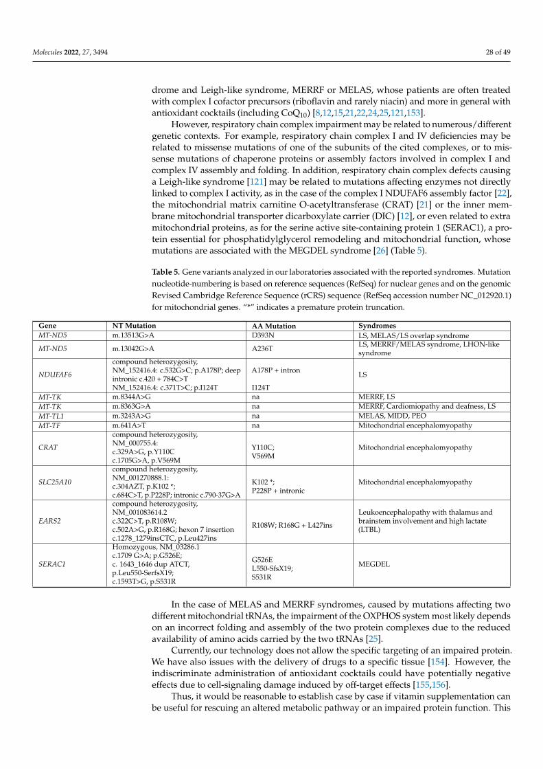
Molecules 2022, 27, 3494 28 of 49
drome and Leigh-like syndrome, MERRF or MELAS, whose patients are often treatedwith complex I cofactor precursors (riboflavin and rarely niacin) and more in general withantioxidant cocktails (including CoQ10) [8,12,15,21,22,24,25,121,153].
However, respiratory chain complex impairment may be related to numerous/differentgenetic contexts. For example, respiratory chain complex I and IV deficiencies may berelated to missense mutations of one of the subunits of the cited complexes, or to mis-sense mutations of chaperone proteins or assembly factors involved in complex I andcomplex IV assembly and folding. In addition, respiratory chain complex defects causinga Leigh-like syndrome [121] may be related to mutations affecting enzymes not directlylinked to complex I activity, as in the case of the complex I NDUFAF6 assembly factor [22],the mitochondrial matrix carnitine O-acetyltransferase (CRAT) [21] or the inner mem-brane mitochondrial transporter dicarboxylate carrier (DIC) [12], or even related to extramitochondrial proteins, as for the serine active site-containing protein 1 (SERAC1), a pro-tein essential for phosphatidylglycerol remodeling and mitochondrial function, whosemutations are associated with the MEGDEL syndrome [26] (Table 5).
Table 5. Gene variants analyzed in our laboratories associated with the reported syndromes. Mutationnucleotide-numbering is based on reference sequences (RefSeq) for nuclear genes and on the genomicRevised Cambridge Reference Sequence (rCRS) sequence (RefSeq accession number NC_012920.1)for mitochondrial genes. “*” indicates a premature protein truncation.
Gene NT Mutation AA Mutation SyndromesMT-ND5 m.13513G>A D393N LS, MELAS/LS overlap syndrome
MT-ND5 m.13042G>A A236T LS, MERRF/MELAS syndrome, LHON-likesyndrome
NDUFAF6
compound heterozygosity,NM_152416.4: c.532G>C; p.A178P; deepintronic c.420 + 784C>TNM_152416.4: c.371T>C; p.I124T
A178P + intron
I124T
LS
MT-TK m.8344A>G na MERRF, LSMT-TK m.8363G>A na MERRF, Cardiomiopathy and deafness, LSMT-TL1 m.3243A>G na MELAS, MIDD, PEOMT-TF m.641A>T na Mitochondrial encephalomyopathy
CRAT
compound heterozygosity,NM_000755.4:c.329A>G, p.Y110Cc.1705G>A, p.V569M
Y110C;V569M
Mitochondrial encephalomyopathy
SLC25A10
compound heterozygosity,NM_001270888.1:c.304AZT, p.K102 *;c.684C>T, p.P228P; intronic c.790-37G>A
K102 *;P228P + intronic
Mitochondrial encephalomyopathy
EARS2
compound heterozygosity,NM_001083614.2c.322C>T, p.R108W;c.502A>G, p.R168G; hexon 7 insertionc.1278_1279insCTC, p.Leu427ins
R108W; R168G + L427ins
Leukoencephalopathy with thalamus andbrainstem involvement and high lactate(LTBL)
SERAC1
Homozygous, NM_03286.1c.1709 G>A; p.G526E;c. 1643_1646 dup ATCT,p.Leu550-SerfsX19;c.1593T>G, p.S531R
G526EL550-SfsX19;S531R
MEGDEL
In the case of MELAS and MERRF syndromes, caused by mutations affecting twodifferent mitochondrial tRNAs, the impairment of the OXPHOS system most likely dependson an incorrect folding and assembly of the two protein complexes due to the reducedavailability of amino acids carried by the two tRNAs [25].
Currently, our technology does not allow the specific targeting of an impaired protein.We have also issues with the delivery of drugs to a specific tissue [154]. However, theindiscriminate administration of antioxidant cocktails could have potentially negativeeffects due to cell-signaling damage induced by off-target effects [155,156].
Thus, it would be reasonable to establish case by case if vitamin supplementation canbe useful for rescuing an altered metabolic pathway or an impaired protein function. This

Molecules 2022, 27, 3494 29 of 49
approach would allow a more focused treatment, according to the affected protein and tothe localization of the investigated specific mutation in the structure of the affected protein,eventually supported by biochemical investigations that prove a deficiency of the vitaminsand cofactors to be administered.
7. Missense Mutations Responsible for Severe Encephalomyopathies: The Case ofMT-ND5, NDUFAF6 and SERAC1
The mitochondrial DNA mutations m.13513G>A and m.13042G>A are among themost studied mitochondrial DNA mutations affecting complex I [157–159]. These muta-tions cause the amino acid replacement D393N and A236T, respectively, at the MT-ND5subunit, determining a similar neurodegenerative phenotype [160]. For slowing downthe neurodegeneration often observed in patients showing mutations at ND5, patients aretreated with an antioxidant cocktail including thiamine derivatives, carnitine, ascorbate,pyridoxal phosphate, cobalamin, biotin, beyond niacin, riboflavin and CoQ10, known thelatter three for being involved in direct interactions with complex I [157] (Figure 4). Itappears that complex I does not bind thiamine derivatives, carnitine, ascorbate, pyridoxalphosphate, cobalamin and biotin analogs. Thus, it is questionable if the administration ofthose ligands can directly improve mitochondrial function or counteract ROS productionthrough interactions with complex I. Notably, occasional ameliorative effects might beascribed to a generic scavenger effect played by those molecules. Conversely, it shouldbe stressed that the cited cofactors can target other proteins that support and/or dependon mitochondrial function; thus, it cannot be excluded that the administration of thosemolecules can even produce an overload of work for complex I, whose function is alreadypartially damaged due to ND5 mutations.
According to the solved structure of complex I, the ND5 subunit is located at the endof the complex I membrane arm (Figure 4). It is known that the two investigated mutationscan cause an alteration in the local secondary structure packing and an important change inthe number and type of both short/medium interactions and long-range interactions thatmay affect the flexibility and packing of the local secondary structure elements [161,162].In fact, the replacement of aspartic acid with an asparagine causes the loss of local ionicinteractions and impairs the local net charge, whereas the replacement of the hydrophobicalanine with the polar threonine causes an increase in the number of local hydrophilicinteractions and impairment in the local hydrophobic interaction network [161,162], whichcan cause problems in ND5 protein folding.
Notably, the investigated mutations at ND5 are very far from NADH or FMN bind-ing regions (Figure 3). Thus, the administration of FMN (i.e., riboflavin) and NADH(i.e., niacin) precursors to patients carrying the m.13513G>A (D393N) or m.13042G>A(A236T) mutations also remains questionable, because these molecules could not help tomake complex I more efficient, due to the fact that they interact with subunits far from ND5.
Conversely, the administration of ubiquinol (CoQH2)/ubiquinone (CoQ) or α-tocopherolto a patient with the above-cited mutations could be more useful due to the similarityof the three molecules with the native coenzyme Q, which moves across the membranearm of complex I and can somewhat participate in the proton translocation through ND5subunit [49,63,117].
Notably, the administration of CoQH2 (i.e., the reduced form of coenzyme Q) couldhelp in “overcoming” complex I inactivation by bypassing it [49,63,117].
It is instead expected that the administration of niacin (and NADH structurally relatedligands) and riboflavin (and FMN or FAD structurally related ligands) can produce impor-tant results in patients carrying mutations in those complex subunits directly involved ininteractions with NADH and FMN cofactors [157].

Molecules 2022, 27, 3494 30 of 49
Molecules 2022, 27, 3494 37 of 60
stressed that the cited cofactors can target other proteins that support and/or depend on mitochondrial function; thus, it cannot be excluded that the administration of those mol-ecules can even produce an overload of work for complex I, whose function is already partially damaged due to ND5 mutations.
According to the solved structure of complex I, the ND5 subunit is located at the end of the complex I membrane arm (Figure 4). It is known that the two investigated mutations can cause an alteration in the local secondary structure packing and an important change in the number and type of both short/medium interactions and long-range interactions that may affect the flexibility and packing of the local secondary structure elements [161,162]. In fact, the replacement of aspartic acid with an asparagine causes the loss of local ionic interactions and impairs the local net charge, whereas the replacement of the hydrophobic alanine with the polar threonine causes an increase in the number of local hydrophilic interactions and impairment in the local hydrophobic interaction network [161,162], which can cause problems in ND5 protein folding.
Figure 4. The sus complex I structure (7v2r.pdb, [157]) is reported in colored cartoon representation as a representative complex I structure from mammalian species. The same color scheme is used for the labels indicating each reported subunit. The subunits NUDFA7, NDUFC1 and NDUFB1 are not viewable in this side view. FMN and NAD+ are reported in orange and yellow sticks, as shown in the zoomed view, at the level of subunit NDUFV1. UQ is reported in black sticks as shown in the zoomed view, at the level of MT-ND1, NDUFS2, NDUFS7 and NDUFA9. The distance between the center of mass of ND5 and the center of mass of the closest ubiquinone molecule is represented by the black solid line, here used as a reference distance.
Figure 4. The sus complex I structure (7v2r.pdb, [157]) is reported in colored cartoon representationas a representative complex I structure from mammalian species. The same color scheme is used forthe labels indicating each reported subunit. The subunits NUDFA7, NDUFC1 and NDUFB1 are notviewable in this side view. FMN and NAD+ are reported in orange and yellow sticks, as shown inthe zoomed view, at the level of subunit NDUFV1. UQ is reported in black sticks as shown in thezoomed view, at the level of MT-ND1, NDUFS2, NDUFS7 and NDUFA9. The distance between thecenter of mass of ND5 and the center of mass of the closest ubiquinone molecule is represented bythe black solid line, here used as a reference distance.
At variance with missense mutations directly affecting the activity of crucial complex Isubunits, other proteins involved in the folding of respiratory chain complexes or participat-ing in the same metabolic pathways can affect the activity of respiratory chain complexes,in case of mutations. As an example of proteins responsible for the impairment of mitochon-drial respiration, we had the possibility to analyze in our laboratories the case of NDUFAF6,which plays a crucial role in the assembly of complex I [23] and SERAC1 responsiblefor the MEGDEL syndrome (3-methylglutaconic acidemia, deafness, encephalopathy andLeigh-like syndrome; https://www.omim.org/entry/614739, accessed on 8 April 2022),which shares with Leigh syndromes several phenotypic traits [163,164]. Notably, SERAC1plays a crucial role in phospholipid remodeling at the interface between the endoplasmicreticulum and mitochondria. SERAC1 mutations impair mitochondrial respiration andintracellular cholesterol trafficking causing dystonia and deafness [164]. Conversely, mu-tations occurring at the NDUFAF6 assembly factor (e.g., I124T, A178P [22,165]) cause aneurodegenerative phenotype [23,166] similar to the one observed in NDUFAF1 or ND-UFAF5 mutations characterized by severe degenerative encephalomyopathies [167,168].The administration of antioxidant cocktails to patients affected by mutations at complex Iassembly factors or by mutations affecting SERAC1 should be decided according to thedemonstrated biochemical need of the cofactors to be administered. In the case of SERAC1mutations, instead of antioxidant cocktails, one might consider the possibility of administer-ing a supplement containing phosphatidylglycerol-34:1 and/or phosphatidylglycerol-36:1,both precursors of cardiolipin, important for the correct folding of mitochondrial membraneproteins. Indeed, the phosphatidylglycerol-34:1/phosphatidylglycerol-36:1 ratio appearsto be unbalanced as a consequence of SERAC1 mutations [164]. Thus, it is expected that
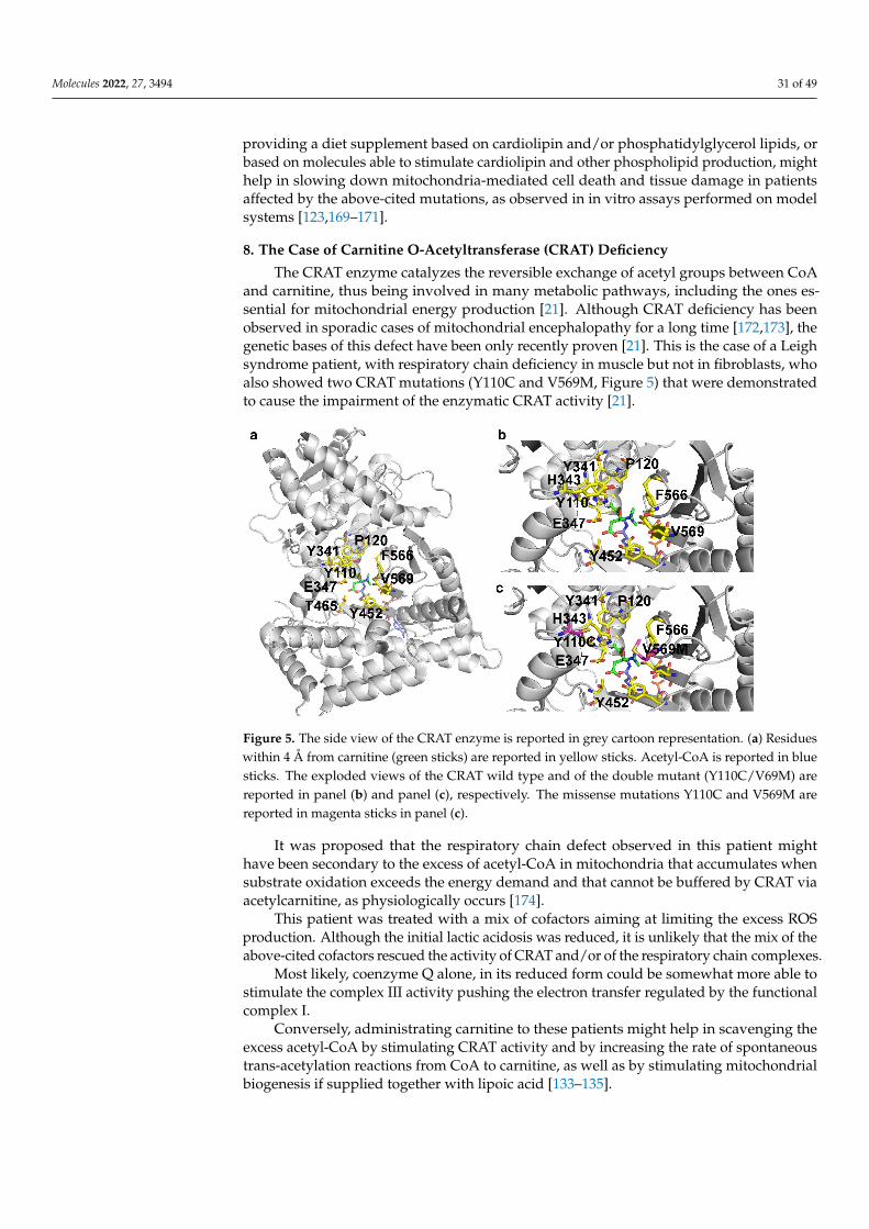
Molecules 2022, 27, 3494 31 of 49
providing a diet supplement based on cardiolipin and/or phosphatidylglycerol lipids, orbased on molecules able to stimulate cardiolipin and other phospholipid production, mighthelp in slowing down mitochondria-mediated cell death and tissue damage in patientsaffected by the above-cited mutations, as observed in in vitro assays performed on modelsystems [123,169–171].
8. The Case of Carnitine O-Acetyltransferase (CRAT) Deficiency
The CRAT enzyme catalyzes the reversible exchange of acetyl groups between CoAand carnitine, thus being involved in many metabolic pathways, including the ones es-sential for mitochondrial energy production [21]. Although CRAT deficiency has beenobserved in sporadic cases of mitochondrial encephalopathy for a long time [172,173], thegenetic bases of this defect have been only recently proven [21]. This is the case of a Leighsyndrome patient, with respiratory chain deficiency in muscle but not in fibroblasts, whoalso showed two CRAT mutations (Y110C and V569M, Figure 5) that were demonstratedto cause the impairment of the enzymatic CRAT activity [21].
Molecules 2022, 27, 3494 39 of 60
It was proposed that the respiratory chain defect observed in this patient might have been secondary to the excess of acetyl-CoA in mitochondria that accumulates when sub-strate oxidation exceeds the energy demand and that cannot be buffered by CRAT via acetylcarnitine, as physiologically occurs [174].
This patient was treated with a mix of cofactors aiming at limiting the excess ROS production. Although the initial lactic acidosis was reduced, it is unlikely that the mix of the above-cited cofactors rescued the activity of CRAT and/or of the respiratory chain complexes.
Most likely, coenzyme Q alone, in its reduced form could be somewhat more able to stimulate the complex III activity pushing the electron transfer regulated by the functional complex I.
Conversely, administrating carnitine to these patients might help in scavenging the excess acetyl-CoA by stimulating CRAT activity and by increasing the rate of spontaneous trans-acetylation reactions from CoA to carnitine, as well as by stimulating mitochondrial biogenesis if supplied together with lipoic acid [133–135].
Figure 5. The side view of the CRAT enzyme is reported in grey cartoon representation. (a) Residues within 4 Å from carnitine (green sticks) are reported in yellow sticks. Acetyl-CoA is reported in blue sticks. The exploded views of the CRAT wild type and of the double mutant (Y110C/V69M) are reported in panel (b) and panel (c), respectively. The missense mutations Y110C and V569M are reported in magenta sticks in panel (c).
9. SLC25A10 Dicarboxylate Carrier (DIC) Deficiency Similarly to the patient affected by CRAT deficiency, another patient, affected by
SLC25A10 deficiency, showed impaired activity of the respiratory complex I in muscle biopsy but not in fibroblasts, in addition to a multidrug-resistant degenerative myopathy and to a severe epileptic and progressive encephalopathy [12].
The quantity and the activity of SLC25A10, which transports dicarboxylates (malate and succinate) and phosphate across the inner mitochondrial membrane, were almost completely absent in this patient due to mutations that caused both aberrant RNA splicing and the insertion of a premature stop codon abolishing the translation of 70% of the orig-inal protein sequence (Figure 6) [12].
Figure 5. The side view of the CRAT enzyme is reported in grey cartoon representation. (a) Residueswithin 4 Å from carnitine (green sticks) are reported in yellow sticks. Acetyl-CoA is reported in bluesticks. The exploded views of the CRAT wild type and of the double mutant (Y110C/V69M) arereported in panel (b) and panel (c), respectively. The missense mutations Y110C and V569M arereported in magenta sticks in panel (c).
It was proposed that the respiratory chain defect observed in this patient mighthave been secondary to the excess of acetyl-CoA in mitochondria that accumulates whensubstrate oxidation exceeds the energy demand and that cannot be buffered by CRAT viaacetylcarnitine, as physiologically occurs [174].
This patient was treated with a mix of cofactors aiming at limiting the excess ROSproduction. Although the initial lactic acidosis was reduced, it is unlikely that the mix of theabove-cited cofactors rescued the activity of CRAT and/or of the respiratory chain complexes.
Most likely, coenzyme Q alone, in its reduced form could be somewhat more able tostimulate the complex III activity pushing the electron transfer regulated by the functionalcomplex I.
Conversely, administrating carnitine to these patients might help in scavenging theexcess acetyl-CoA by stimulating CRAT activity and by increasing the rate of spontaneoustrans-acetylation reactions from CoA to carnitine, as well as by stimulating mitochondrialbiogenesis if supplied together with lipoic acid [133–135].
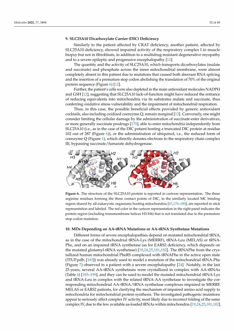
Molecules 2022, 27, 3494 32 of 49
9. SLC25A10 Dicarboxylate Carrier (DIC) Deficiency
Similarly to the patient affected by CRAT deficiency, another patient, affected bySLC25A10 deficiency, showed impaired activity of the respiratory complex I in musclebiopsy but not in fibroblasts, in addition to a multidrug-resistant degenerative myopathyand to a severe epileptic and progressive encephalopathy [12].
The quantity and the activity of SLC25A10, which transports dicarboxylates (malateand succinate) and phosphate across the inner mitochondrial membrane, were almostcompletely absent in this patient due to mutations that caused both aberrant RNA splicingand the insertion of a premature stop codon abolishing the translation of 70% of the originalprotein sequence (Figure 6) [12].
Further, the patient’s cells were also depleted in the main antioxidant molecules NADPHand GSH [12], suggesting that SLC25A10 lack-of-function might have reduced the entranceof reducing equivalents into mitochondria via its substrates malate and succinate, thusconferring oxidative stress vulnerability and the impairment of mitochondrial respiration.
Thus, in this case, the possible beneficial effects provided by generic antioxidantcocktails, also including oxidized coenzyme Q, remain marginal [12]. Conversely, one mightconsider limiting the cellular damage by the administration of succinate-ester derivatives,or more generally succinate prodrugs [175], able to enter mitochondria independently fromSLC25A10 (i.e., as in the case of the DIC patient hosting a truncated DIC protein at residue102 out of 287 (Figure 6)), or the administration of ubiquinol, i.e., the reduced form ofcoenzyme Q (Figure 1), which directly donates electrons to the respiratory chain complexIII, bypassing succinate/fumarate dehydrogenase.
Molecules 2022, 27, 3494 40 of 60
Further, the patient’s cells were also depleted in the main antioxidant molecules NADPH and GSH [12], suggesting that SLC25A10 lack-of-function might have reduced the entrance of reducing equivalents into mitochondria via its substrates malate and suc-cinate, thus conferring oxidative stress vulnerability and the impairment of mitochondrial respiration.
Thus, in this case, the possible beneficial effects provided by generic antioxidant cocktails, also including oxidized coenzyme Q, remain marginal [12]. Conversely, one might consider limiting the cellular damage by the administration of succinate-ester de-rivatives, or more generally succinate prodrugs [175], able to enter mitochondria inde-pendently from SLC25A10 (i.e., as in the case of the DIC patient hosting a truncated DIC protein at residue 102 out of 287 (Figure 6)), or the administration of ubiquinol, i.e., the reduced form of coenzyme Q (Figure 1), which directly donates electrons to the respira-tory chain complex III, bypassing succinate/fumarate dehydrogenase.
Figure 6. The structure of the SLC25A10 protein is reported in cartoon representation. The three arginine residues forming the three contact points of DIC, in the similarly located MC binding re-gion shared by all eukaryotic organisms hosting mitochondria [43,176–180], are reported in stick representation and labeled. The red color in the cartoon representation in the right panel indicates the protein region (including transmembrane helices H3-H6) that is not translated due to the prem-ature stop codon mutation.
10. MDs Depending on AA-tRNA Mutations or AA-tRNA Synthetase Mutations Different forms of severe encephalopathies depend on mutated mitochondrial tRNA,
as in the case of the mitochondrial tRNA-Lys (MERRF), tRNA-Leu (MELAS) or tRNA-Phe, and on an impaired tRNA synthetase (as for EARS2 deficiency, which depends on the mutated glutamyl-tRNA synthetase) [19,24,25,181,182]. The tRNAPhe from the crys-tallized human mitochondrial PheRS complexed with tRNAPhe in the active open state (3TUP.pdb, [183]) was already used to model a mutation of the mitochondrial tRNA-Phe (Figure 7) observed in a patient with a severe encephalopathy [24]. Notably, in the last 25 years, several AA-tRNA synthetases were crystallized in complex with AA-tRNAs (Table 6) [183–199], and they can be used to model the mutated mitochondrial tRNA-Lys and tRNA-Leu in complex with the related tRNA-AA synthetase to investigate the corre-sponding mitochondrial AA-tRNA/tRNA synthetase complexes impaired in MERRF, ME-LAS or EARS2 patients, for clarifying the mechanism of impaired amino acid supply to mitochondria for mitochondrial protein synthesis. The investigated pathogenic mutations appear to seriously affect complex IV activity, most likely due to incorrect folding of the same complex IV, due to the few available aa-loaded tRNAs within mitochondria [19,24,25,181,182].
Given that complex IV inactivation most likely depends on an unfolding problem, due to the missing AA-loaded-tRNA, patients affected by mitochondrial diseases caused by tRNA mutations or by mutations impairing the tRNA-AA synthetase might be treated with amino acids, or amino acids precursors, whose availability may be altered in specific tissues hosting the mutated tRNA or the corresponding synthetase, after having
Figure 6. The structure of the SLC25A10 protein is reported in cartoon representation. The threearginine residues forming the three contact points of DIC, in the similarly located MC bindingregion shared by all eukaryotic organisms hosting mitochondria [43,176–180], are reported in stickrepresentation and labeled. The red color in the cartoon representation in the right panel indicates theprotein region (including transmembrane helices H3-H6) that is not translated due to the prematurestop codon mutation.
10. MDs Depending on AA-tRNA Mutations or AA-tRNA Synthetase Mutations
Different forms of severe encephalopathies depend on mutated mitochondrial tRNA,as in the case of the mitochondrial tRNA-Lys (MERRF), tRNA-Leu (MELAS) or tRNA-Phe, and on an impaired tRNA synthetase (as for EARS2 deficiency, which depends onthe mutated glutamyl-tRNA synthetase) [19,24,25,181,182]. The tRNAPhe from the crys-tallized human mitochondrial PheRS complexed with tRNAPhe in the active open state(3TUP.pdb, [183]) was already used to model a mutation of the mitochondrial tRNA-Phe(Figure 7) observed in a patient with a severe encephalopathy [24]. Notably, in the last25 years, several AA-tRNA synthetases were crystallized in complex with AA-tRNAs(Table 6) [183–199], and they can be used to model the mutated mitochondrial tRNA-Lysand tRNA-Leu in complex with the related tRNA-AA synthetase to investigate the cor-responding mitochondrial AA-tRNA/tRNA synthetase complexes impaired in MERRF,MELAS or EARS2 patients, for clarifying the mechanism of impaired amino acid supply tomitochondria for mitochondrial protein synthesis. The investigated pathogenic mutationsappear to seriously affect complex IV activity, most likely due to incorrect folding of the samecomplex IV, due to the few available aa-loaded tRNAs within mitochondria [19,24,25,181,182].
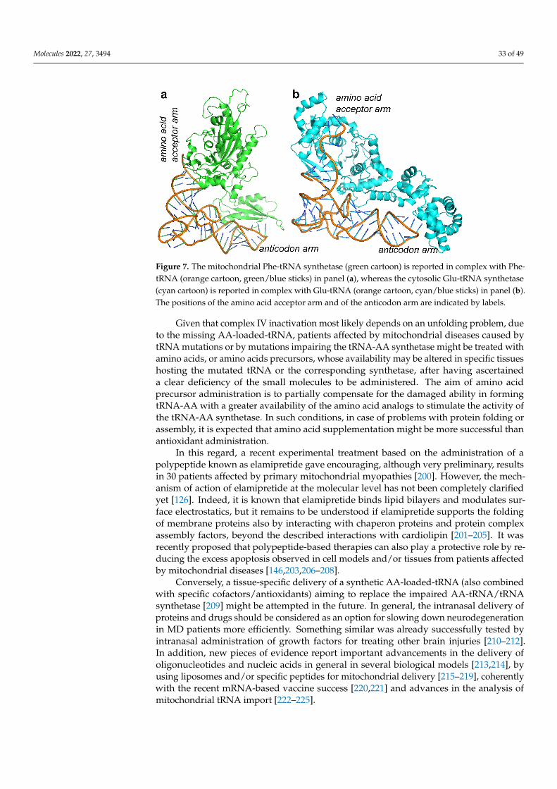
Molecules 2022, 27, 3494 33 of 49
Molecules 2022, 27, 3494 41 of 60
ascertained a clear deficiency of the small molecules to be administered. The aim of amino acid precursor administration is to partially compensate for the damaged ability in form-ing tRNA-AA with a greater availability of the amino acid analogs to stimulate the activity of the tRNA-AA synthetase. In such conditions, in case of problems with protein folding or assembly, it is expected that amino acid supplementation might be more successful than antioxidant administration.
In this regard, a recent experimental treatment based on the administration of a pol-ypeptide known as elamipretide gave encouraging, although very preliminary, results in 30 patients affected by primary mitochondrial myopathies [200]. However, the mecha-nism of action of elamipretide at the molecular level has not been completely clarified yet [126]. Indeed, it is known that elamipretide binds lipid bilayers and modulates surface electrostatics, but it remains to be understood if elamipretide supports the folding of mem-brane proteins also by interacting with chaperon proteins and protein complex assembly factors, beyond the described interactions with cardiolipin [201–205]. It was recently pro-posed that polypeptide-based therapies can also play a protective role by reducing the excess apoptosis observed in cell models and/or tissues from patients affected by mito-chondrial diseases [146,203,206–208].
Conversely, a tissue-specific delivery of a synthetic AA-loaded-tRNA (also combined with specific cofactors/antioxidants) aiming to replace the impaired AA-tRNA/tRNA syn-thetase [209] might be attempted in the future. In general, the intranasal delivery of pro-teins and drugs should be considered as an option for slowing down neurodegeneration in MD patients more efficiently. Something similar was already successfully tested by in-tranasal administration of growth factors for treating other brain injuries [210–212]. In ad-dition, new pieces of evidence report important advancements in the delivery of oligonu-cleotides and nucleic acids in general in several biological models [213,214], by using lip-osomes and/or specific peptides for mitochondrial delivery [215–219], coherently with the recent mRNA-based vaccine success [220,221] and advances in the analysis of mitochon-drial tRNA import [222–225].
Figure 7. The mitochondrial Phe-tRNA synthetase (green cartoon) is reported in complex with Phe-tRNA (orange cartoon, green/blue sticks) in panel (a), whereas the cytosolic Glu-tRNA synthetase (cyan cartoon) is reported in complex with Glu-tRNA (orange cartoon, cyan/blue sticks) in panel (b). The positions of the amino acid acceptor arm and of the anticodon arm are indicated by labels.
Figure 7. The mitochondrial Phe-tRNA synthetase (green cartoon) is reported in complex with Phe-tRNA (orange cartoon, green/blue sticks) in panel (a), whereas the cytosolic Glu-tRNA synthetase(cyan cartoon) is reported in complex with Glu-tRNA (orange cartoon, cyan/blue sticks) in panel (b).The positions of the amino acid acceptor arm and of the anticodon arm are indicated by labels.
Given that complex IV inactivation most likely depends on an unfolding problem, dueto the missing AA-loaded-tRNA, patients affected by mitochondrial diseases caused bytRNA mutations or by mutations impairing the tRNA-AA synthetase might be treated withamino acids, or amino acids precursors, whose availability may be altered in specific tissueshosting the mutated tRNA or the corresponding synthetase, after having ascertaineda clear deficiency of the small molecules to be administered. The aim of amino acidprecursor administration is to partially compensate for the damaged ability in formingtRNA-AA with a greater availability of the amino acid analogs to stimulate the activity ofthe tRNA-AA synthetase. In such conditions, in case of problems with protein folding orassembly, it is expected that amino acid supplementation might be more successful thanantioxidant administration.
In this regard, a recent experimental treatment based on the administration of apolypeptide known as elamipretide gave encouraging, although very preliminary, resultsin 30 patients affected by primary mitochondrial myopathies [200]. However, the mech-anism of action of elamipretide at the molecular level has not been completely clarifiedyet [126]. Indeed, it is known that elamipretide binds lipid bilayers and modulates sur-face electrostatics, but it remains to be understood if elamipretide supports the foldingof membrane proteins also by interacting with chaperon proteins and protein complexassembly factors, beyond the described interactions with cardiolipin [201–205]. It wasrecently proposed that polypeptide-based therapies can also play a protective role by re-ducing the excess apoptosis observed in cell models and/or tissues from patients affectedby mitochondrial diseases [146,203,206–208].
Conversely, a tissue-specific delivery of a synthetic AA-loaded-tRNA (also combinedwith specific cofactors/antioxidants) aiming to replace the impaired AA-tRNA/tRNAsynthetase [209] might be attempted in the future. In general, the intranasal delivery ofproteins and drugs should be considered as an option for slowing down neurodegenerationin MD patients more efficiently. Something similar was already successfully tested byintranasal administration of growth factors for treating other brain injuries [210–212].In addition, new pieces of evidence report important advancements in the delivery ofoligonucleotides and nucleic acids in general in several biological models [213,214], byusing liposomes and/or specific peptides for mitochondrial delivery [215–219], coherentlywith the recent mRNA-based vaccine success [220,221] and advances in the analysis ofmitochondrial tRNA import [222–225].

Molecules 2022, 27, 3494 34 of 49
Table 6. List of the aminoacyl tRNA synthetases crystallized in complex with a transported amino acid.
PDB_ID Aminoacyl tRNASynthetase Name Number of Residues Aminoacyl tRNA Number of
Nucleotides Organism Cell Compartment Resolution
1f7u (1f7v) Arginyl-tRNA synthetase 608 (607) Arg tRNA 76 S. cerevisiae cytoplasmic/mitochondrion 2.20 Å (2.90 Å)2zue (2zuf) Arginyl-tRNA synthetase 629 Arg tRNA 78 P. horikoshii cytoplasmic 2.00 Å (2.30 Å)
3kfu
Glutamyl-tRNAamidotransferase subunit A,
aspartyl/Glutamyl-tRNAamidotrasferase subunit B,Glutamyl amidotrasferase
subunit C-tRNA
471, 466, 92 Asn tRNA 76 T. thermophilus HB8 cytoplasmic 3.00 Å
1c0a Aspartyl-tRNA synthetase 585 Asp tRNA 77 E. coli cytoplasmic 2.40 Å1efw Aspartyl-tRNA synthetase 580 Asp tRNA 73 T. thermpohilus/E. coli cytoplasmic 3.00 Å1il2 Aspartyl-tRNA synthetase 590 Asp tRNA 75 E. coli/S. cerevisiae cytoplasmic/mitochondrion 2.60 Å
1u0b Cysteinyl-tRNA synthetase 461 Cys tRNA 74 E. coli cytoplasmic 2.30 Å
1exd Glutaminyl-tRNAsynthetase 548 Gln Aptamer tRNA 73 E. coli cytoplasmic 2.70 Å
3aL0 Glutamyl-tRNAamidotransferase subunit A 475 Gln tRNA 74 T. maritima MSB8 cytoplasmic 3.37 Å
1euy (1euq) Glutaminyl-tRNAsynthetase 548 Gln tRNA 74 (72) E. coli cytoplasmic 2.60 Å (3.10 Å)
1gsg Glutaminyl-tRNAsynthetase 553 Gln tRNA 75 E. coli cytoplasmic 2.80 Å
1gts Glutaminyl-tRNA(E.C.6.1.1.18) synthetase 553 Gln tRNA 74 E. coli cytoplasmic 2.80 Å
1o0b (1o0c) Glutaminyl-tRNAsynthetase 554 Gln tRNA 75 E. coli cytoplasmic 2.70 Å (2.70 Å)
1zjw Glutaminyl-tRNAsynthetase 553 Gln tRNA 75 E. coli cytoplasmic 2.50 Å
2rd2 Glutaminyl-tRNAsynthetase 556 Gln tRNA 75 E. coli K-12 (E. coli) cytoplasmic 2.60 Å
3akz Glutamyl-tRNA synthetase2 487 Gln tRNA T. maritima MSB8 cytoplasmic 2.90 Å
1qrs (1qrt, 1qru) Glutaminyl-tRNA(E.C.6.1.1.18) synthetase 553 Gln2 tRNA 75 E. coli cytoplasmic 2.60 Å (2.70 Å, 3.00 Å
1qtq Glutaminyl-tRNAsynthetase 553 Gln2 tRNA 75 E. coli cytoplasmic 2.25 Å
1g59 Glutamyl-tRNA synthetase 648 Glu tRNA 75 T. thermophilus cytoplasmic 2.40 Å1n78 (1n77, 1j09,
1n78) Glutamyl-tRNA synthetase 468 Glu tRNA (Glu tRNA,/, /) 75 (75, /, /) T. thermophilus cytoplasmic 2.10 Å (2.40 Å, 1.80 Å,
1.90 Å)4kr2 (4kr3) Glycine-tRNA synthetase 637 Gly tRNA-ccc 74 H. sapiens cytoplasmic/mitochondria 3.29 Å (3.31 Å)
5e6m Glycine-tRNA synthetase 693 Gly tRNA-ccc 74 H. sapiens cytoplasmic/mitochondria 2.93 Å4qei (4kqe) Glycine-tRNA synthetase 630 Gly tRNA-ccc-2-2 (/) 69 (/) H. sapiens cytoplasmic/mitochondria 2.88 Å
4rdx Histidine-tRNA synthetase 423 His tRNA 78 T. thermophilus HB27 cytoplasmic 2.55 Å
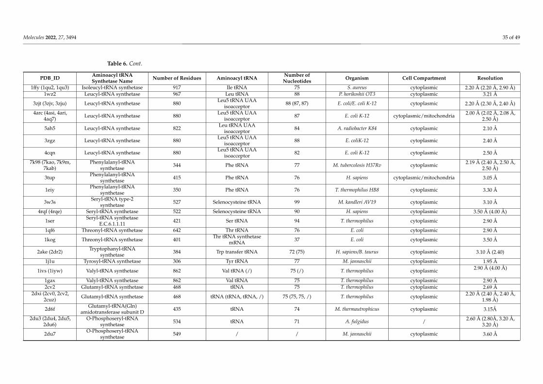
Molecules 2022, 27, 3494 35 of 49
Table 6. Cont.
PDB_ID Aminoacyl tRNASynthetase Name Number of Residues Aminoacyl tRNA Number of
Nucleotides Organism Cell Compartment Resolution
1ffy (1qu2, 1qu3) Isoleucyl-tRNA synthetase 917 Ile tRNA 75 S. aureus cytoplasmic 2.20 Å (2.20 Å, 2.90 Å)1wz2 Leucyl-tRNA synthetase 967 Leu tRNA 88 P. horikoshii OT3 cytoplasmic 3.21 Å
3zjt (3zjv, 3zju) Leucyl-tRNA synthetase 880 Leu5 tRNA UAAisoacceptor 88 (87, 87) E. coli/E. coli K-12 cytoplasmic 2.20 Å (2.30 Å, 2.40 Å)
4arc (4asi, 4ari,4aq7) Leucyl-tRNA synthetase 880 Leu5 tRNA UAA
isoacceptor 87 E. coli K-12 cytoplasmic/mitochondria 2.00 Å (2.02 Å, 2.08 Å,2.50 Å)
5ah5 Leucyl-tRNA synthetase 822 Leu tRNA UAAisoacceptor 84 A. radiobacter K84 cytoplasmic 2.10 Å
3zgz Leucyl-tRNA synthetase 880 Leu5 tRNA UAAisoacceptor 88 E. coliK-12 cytoplasmic 2.40 Å
4cqn Leucyl-tRNA synthetase 880 Leu5 tRNA UAAisoacceptor 82 E. coli K-12 cytoplasmic 2.50 Å
7k98 (7kao, 7k9m,7kab)
Phenylalanyl-tRNAsynthetase 344 Phe tRNA 77 M. tubercolosis H37Rv cytoplasmic 2.19 Å (2.40 Å, 2.50 Å,
2.50 Å)
3tup Phenylalanyl-tRNAsynthetase 415 Phe tRNA 76 H. sapiens cytoplasmic/mitochondria 3.05 Å
1eiy Phenylalanyl-tRNAsynthetase 350 Phe tRNA 76 T. thermophilus HB8 cytoplasmic 3.30 Å
3w3s Seryl-tRNA type-2synthetase 527 Selenocysteine tRNA 99 M. kandleri AV19 cytoplasmic 3.10 Å
4rqf (4rqe) Seryl-tRNA synthetase 522 Selenocysteine tRNA 90 H. sapiens cytoplasmic 3.50 Å (4.00 Å)
1ser Seryl-tRNA synthetaseE.C.6.1.1.11 421 Ser tRNA 94 T. thermophilus cytoplasmic 2.90 Å
1qf6 Threonyl-tRNA synthetase 642 Thr tRNA 76 E. coli cytoplasmic 2.90 Å
1kog Threonyl-tRNA synthetase 401 Thr tRNA synthetasemRNA 37 E. coli cytoplasmic 3.50 Å
2ake (2dr2) Tryptophanyl-tRNAsynthetase 384 Trp transfer tRNA 72 (75) H. sapiens/B. taurus cytoplasmic 3.10 Å (2.40)
1j1u Tyrosyl-tRNA synthetase 306 Tyr tRNA 77 M. jannaschii cytoplasmic 1.95 Å
1ivs (1iyw) Valyl-tRNA synthetase 862 Val tRNA (/) 75 (/) T. thermophilus cytoplasmic 2.90 Å (4.00 Å)
1gax Valyl-tRNA synthetase 862 Val tRNA 75 T. thermophilus cytoplasmic 2.90 Å2cv2 Glutamyl-tRNA synthetase 468 tRNA 75 T. thermophilus cytoplasmic 2.69 Å
2dxi (2cv0, 2cv2,2cuz) Glutamyl-tRNA synthetase 468 tRNA (tRNA, tRNA, /) 75 (75, 75, /) T. thermophilus cytoplasmic 2.20 Å (2.40 Å, 2.40 Å,
1.98 Å)
2d6f Glutamyl-tRNA(Gln)amidotransferase subunit D 435 tRNA 74 M. thermautrophicus cytoplasmic 3.15Å
2du3 (2du4, 2du5,2du6)
O-Phosphoseryl-tRNAsynthetase 534 tRNA 71 A. fulgidus / 2.60 Å (2.80Å, 3.20 Å,
3.20 Å)
2du7 O-Phosphoseryl-tRNAsynthetase 549 / / M. jannaschii cytoplasmic 3.60 Å

Molecules 2022, 27, 3494 36 of 49
Table 6. Cont.
PDB_ID Aminoacyl tRNASynthetase Name Number of Residues Aminoacyl tRNA Number of
Nucleotides Organism Cell Compartment Resolution
4yye Threonyl-tRNA synthetase 460 tRNA 76 S. cerevisiae S288c cytoplasmic/mitochondria 2.30 Å5yyn Arginyl-tRNA synthetase 586 tRNA 77 E. coli K-12 cytoplasmic 3.00Å2zni Pyrrolysyl-tRNA synthetase 308 Bacterial tRNA 72 D. hefniense cytoplasmic 3.10 Å3a2k Lysidine-tRNA synthase 464 Bacterial tRNA G. kaustophilus cytoplasmic 3.65 Å
2hrk (2hsn, 2hsm) Glutamyl-tRNA synthetase 207 / / S. cerevisiae cytoplasmic/mitochondria 2.05 Å (2.20 Å, 3.00 Å)2rkj Tyrosyl-tRNA synthetase 392 238-MER RNA 246 N. crassa cytoplasmic/mitochondria 4.50 Å
5ud5 (5v6x) Pyrrolysyl-tRNA synthetase 109 70-MER RNA 72 M.mazei Go1 cytoplasmic 2.35 Å (2.76 Å)
4jxx (4jxz, 4jyzx) Glutaminyl-tRNAsynthetase 553 71-MER RNA (71-MER
RNA, 72-MER RNA) 75 E.coli K-12 cytoplasmic 2.30 Å (2.40Å, 2.50 Å)
2azx Tryptophanyl-tRNAsynthetase 477 72-MER 75 H. sapiens cytoplasmic 2.80 Å
2csx (2ct8) Methionyl-tRNA synthetase 497 75-MER RNA (74-MERRNA) 75 (74) A. aeolicus cytoplasmic 2.70 Å (2.70 Å)
3wqy (3wqz) Alenine-tRNA synthetase 906 75-MER RNA 75 A. filgidus DSM 4304 cytoplasmic 3.30 Å (3.49 Å)
4wj3 (4wj4) Glutamyl-tRNA(Gln)amidotransferase subunit A 484 (599) 76-MER tRNA 76
P.aeruginosa
PAO1cytoplasmic 3.71 Å (3.29 Å)

Molecules 2022, 27, 3494 37 of 49
11. CoQ Analogs and Mitochondrial Delivery Systems: Organic/Inorganic Chemicalsfor Stimulating Mitochondrial Activity
Numerous small molecules have been proposed as redox-active drugs, possibly usefulas antioxidants, electron conductors, and/or pro-oxidants [39]. Most of those experimentaldrugs present a (hydro)quinone scaffold (general structure A, Figure 8), thus being closelyrelated to CoQ10. On the other hand, the redox-active compounds reported so far displayevident similarities to the antioxidant vitamins E (α-tocopherol) and K (phylloquinone—K1;Figure 8B). Generally, the ring scaffold is decorated with electron-donating substituents(R1 and R3; Figure 8C) while an alkyl chain (R2) is mainly responsible for ADME andtargeting issues [226]. KH176 was developed as a Trolox analog and may be considered abioprecursor of the active metabolite KH176m (Figure 8D) [227]. As a notable exception,SHP622 is not related to CoQ10 (Figure 8E). Interestingly, it may act as an electron donorgenerating the corresponding oxindole derivative. The latter cannot be directly reducedback to the parent compound. This is why SHP622 lacks pro-oxidant activity and, inprinciple, should not be considered a redox-active compound.
Idebenone, EPI-743, KH176 and more recently all the other cited small moleculeshave been used in preclinical studies and then employed in clinics for counteracting themitochondrial respiratory chain defects [86,123–127]. EPI-743, already used in clinicaltrials for the treatment of Leigh syndrome [228], is now being investigated in clinical trialsfor its potential usage in the treatment of visual and neurological complications due to asecondary altered redox state in Cbl C deficiency [229] and in Friedreich ataxia [230].
However, it is still not clear whether these molecules, beyond their ROS scavengerproperties, exert their antioxidant activity by targeting a specific protein or a specific groupof proteins. Both surface plasmon resonance interaction studies and in silico modelingindicated peroxiredoxin as a possible target of KH176m [227]. On the other hand, whileCoQ analogs can stimulate mitochondrial respiratory chain activity, it is also possiblethat those small molecules can target other mitochondrial/cytosolic enzymes (using CoQanalogs as cofactors) and be involved in oxidative reactions [54,227,231,232].
The undesired targeting of other proteins can cause damage to the endogenous ROSsignaling (off-target effects) that may result, in case of prolonged administration, in tissuedamage [233–235].
According to these findings and observations, the administration of the above-reportedmolecules to MD-affected patients should be carefully evaluated case by case. The mainscope of the administration of those molecules is indeed to counteract ROS increase (actingas ROS scavengers) and improve the activity of the impaired respiratory chain complexesto provide a valid therapeutic strategy for dealing with severe MDs. Conversely, the greatersuccess of those molecules should be related to their possible direct involvement in thefunction of the affected protein. Thus, a therapeutical approach based on idebenone, EPI-743 and KH176 will be more successful if the treated patients carry a missense mutation ina protein or in a protein subunit whose function directly depends on the availability of theCoQ cofactor.
According to the above-reported comments, the limited clinical success of the citedmolecules could be related to the administration of the investigated molecules to largeand genetically heterogeneous cohorts of patients without taking into consideration thedistance between the localization of the missense mutation responsible for the pathol-ogy and the expected site of action of the investigated CoQ analog. Furthermore, if theinvestigated molecules have to be administered to subjects affected by MDs for counter-acting mitochondrial ROS production, the administration of the small molecules shouldbe organelle-specific in order to reduce off-target effects, although some of those off-targeteffects may be apparently beneficial.
To increase the delivery specificity of the cited molecules and cofactors to mitochondria,the same small molecules might be conjugated to triphenylphosphonium (TPP) salts [236]or can be entrapped in ad hoc functionalized liposomes [237]. Both systems should ensurethe proper targeting of the small molecules to mitochondria, as already observed with
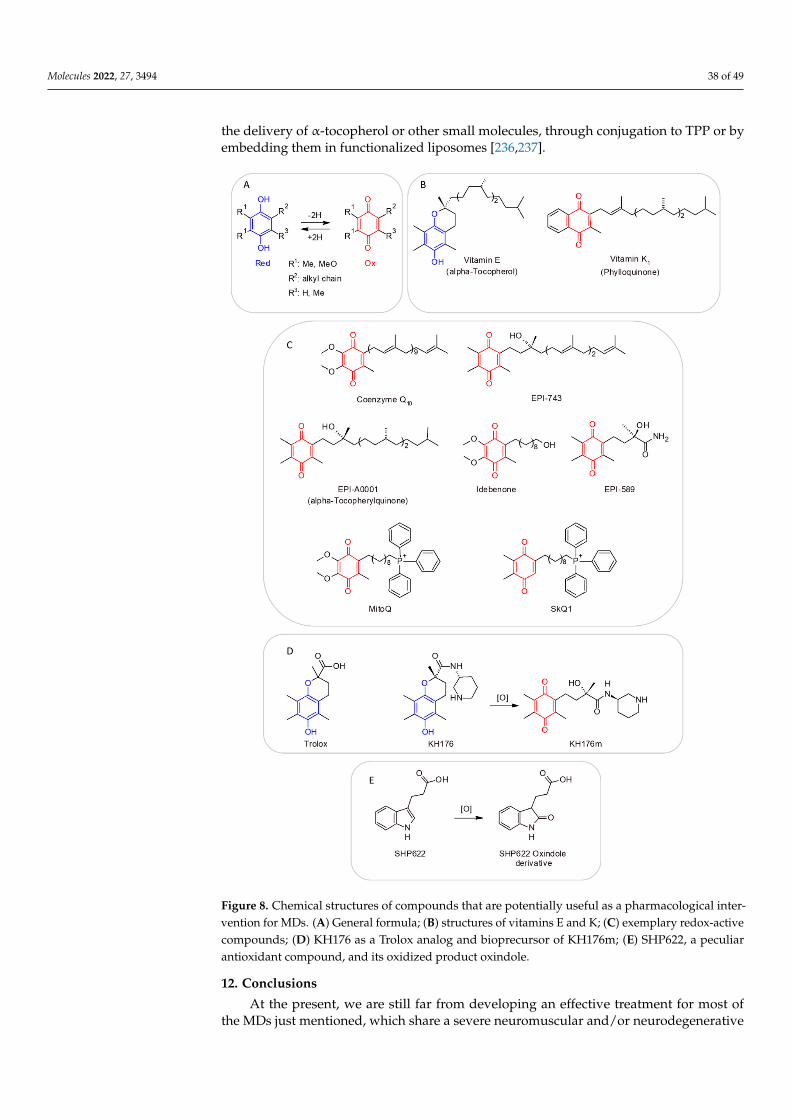
Molecules 2022, 27, 3494 38 of 49
the delivery of α-tocopherol or other small molecules, through conjugation to TPP or byembedding them in functionalized liposomes [236,237].
Molecules 2022, 27, 3494 47 of 60
Figure 8. Chemical structures of compounds that are potentially useful as a pharmacological inter-vention for MDs. (A) General formula; (B) structures of vitamins E and K; (C) exemplary redox-active compounds; (D) KH176 as a Trolox analog and bioprecursor of KH176m; (E) SHP622, a pe-culiar antioxidant compound, and its oxidized product oxindole.
Figure 8. Chemical structures of compounds that are potentially useful as a pharmacological inter-vention for MDs. (A) General formula; (B) structures of vitamins E and K; (C) exemplary redox-activecompounds; (D) KH176 as a Trolox analog and bioprecursor of KH176m; (E) SHP622, a peculiarantioxidant compound, and its oxidized product oxindole.
12. Conclusions
At the present, we are still far from developing an effective treatment for most ofthe MDs just mentioned, which share a severe neuromuscular and/or neurodegenerative
Molecules 2022, 27, 3494 39 of 49
phenotype and are characterized by a severe multisystem involvement still leading to ahigh mortality rate. Indeed, no real dedicated MD cure is available to date, although symp-tomatic therapies are established aiming to mitigate oxidative damage in tissues (i.e., brain,skeletal muscle, heart, liver, kidney) with primary mitochondrial dysfunction [49,63].
Within the vast group of inherited metabolic disorders, new strategies based ongene delivery and enzyme replacement approaches are under investigation [238,239], andtherapeutic molecules that have been made available over the last few decades havesignificantly improved the prognosis quoad vitam and quoad valetudinem of affectedpatients [240].
Most of the employed symptomatic therapies are administered in combination withantioxidant cocktails. Conversely, according to what many authors have reported, the effi-ciency of antioxidant cocktails in patients affected by MDs has not been proven yet [36,37].Furthermore, putative cytotoxic side-effects of therapies based on antioxidant adminis-tration need to be monitored for avoiding toxicity [36,37,78]. Indeed, the hypothesis thatthe suppression of endogenous oxidants could be deleterious and trigger local apopto-sis/necrosis events cannot be excluded [36,37,78].
In order to evaluate and predict the possible advantages in administering specificcofactors, the most updated computational approaches based on molecular dynamics andenergy stabilization calculations should be used [24,43,54,176–180,241–247], after havingascertained a real deficiency of these small molecules in the plasma and/or biopsy samplesfrom patients. Indeed, while biopsy and serum blood levels of a specific cofactor can helpin quantifying the real need for the antioxidant administration, computational approachescan help to understand if and how missense mutations affect the binding of a cofactor and,thus, to what extent the administered cofactor can successfully rescue the damage causedby a missense mutation in the investigated protein–cofactor binding area.
Author Contributions: V.T. and L.C.B. contributed to the structural biology analysis; G.P. (GuidoPrimiano), A.T. and S.S. contributed to the clinical aspects of the manuscript; G.L.P., D.M. and G.P.(Giuseppe Petrosillo) provided the analysis about ROS production molecular mechanisms; M.N.S.,R.G., G.P. (Giulia Paterno), L.G. and M.V. contributed to data curation; M.M.C. and G.L. provided themolecular analysis about CoQ analogs; A.D.G. provided the analysis about SLC25A10 deficiency andCRAT deficiency; C.L.P. contributed to the structural biology analysis and supervised the assemblyof the manuscript. All authors contributed to writing and editing the manuscript. All authors haveread and agreed to the published version of the manuscript.
Funding: The research activities of several papers were funded by the University of Bari with theprojects “ProgettoCompetitivo 2018”, “FFABR 2017–2018” and “Fondi Ateneo ex-60% 2016” andMIUR with the project “Health, Diet and wealth”: identification of a set of biomarkers of the apoptosisfor an innovative industrial Ph.D. course—PON RI 2014–2020, CUP H92H18000160006.
Institutional Review Board Statement: Not applicable.
Informed Consent Statement: Not applicable.
Data Availability Statement: Not applicable.
Acknowledgments: The authors would also like to thank the Italian Association for MitochondrialResearch (www.mitoairm.it, accessed on 8 april 2022) and Mitocon (www.mitocon.it, accessed on 8april 2022) for supporting the realization of research projects focused on the topic of this manuscript.G.P. (Guido Primiano) and S.S. are members of the European Reference Network for NeuromuscularDiseases—Project ID No. 870177.
Conflicts of Interest: The authors declare no conflict of interest.
Abbreviations
AAC, ADP/ATP carrier; ACP, acyl-carrier protein; AGC, aspartate/glutamate carrier; AIF,apoptosis-inducing factor; Bak, Bcl-2 antagonist/killer-1; Bax, Bcl-2 associated X protein; Bcl-2, B-celllymphoma-2; BDC, biotin-dependent carboxylases; BTBGD, biotin-thiamine-responsive basal gangliadisease; CAC, carnitine/acyl-carnitine carrier; Cbl, Cobalamin; CFD, cerebral folate deficiency; CI,
Molecules 2022, 27, 3494 40 of 49
complex I; CIC, citrate carrier; CII, complex II; CIII, complex III; CIV, complex IV; CV, complex V;CoA, coenzyme A; CoQ, ubiquinone; CoQ10, coenzyme Q10; CoQH2, ubiquinol; CPEO, chronicprogressive external ophthalmoplegia; CRAT, carnitine acetyltransferase; CypD, cyclophilin D; CytC,cytochrome C; DIC, dicarboxylate carrier; DRI, dietary reference intake; ETC, electron transportchain; ETF, electron transfer flavoprotein; ETFDH, electron-transport flavoprotein dehydrogenase;FAD, flavin adenine dinucleotide; FMN, flavin mononucleotide; GLUT1, glucose transporter 1; IMS,intermembrane space; KSS, Kearns–Sayre syndrome; LHON, Leber’s hereditary optic neuropathy;MADD, multiple acyl CoA dehydrogenase deficiency; MAS, malate/aspartate shuttle; MEGDEL syn-drome, 3-methylglutaconic acidemia, deafness, encephalopathy and Leigh-like syndrome; MELAS,stroke-like episodes; MERRF, myoclonic epilepsy with ragged-red fibers; 5-MTHF, 5-methyl-THF;MDs, mitochondrial diseases; MFT, FAD (folate/riboflavin/FAD) carrier; MIM, mitochondrial innermembrane; MOM, mitochondrial outer membrane; MPC, mitochondrial pyruvate carrier; MPC,mitochondrial pyruvate carrier; NAD+, nicotinamide adenine dinucleotide; NADP+, nicotinamideadenine dinucleotide phosphate; NARP, neuropathy, ataxia and retinitis pigmentosa; NDT, NAD+
carrier; NMN, nicotinamide mononucleotide; OGC, malate/2-oxoglutarate carrier; ORC, ornithinecarrier; OXPHOS, mitochondrial oxidative phosphorylation; PANK, pantothenate kinase; PiC, phos-phate carrier; PLP, pyridoxal phosphate; PNC, pyrimidine nucleotide carrier; RDA, recommendeddietary allowance; ROS, reactive oxygen species; SERAC1, serine active site-containing protein 1;TCA, tricarboxylic acid cycle; THF, tetrahydrofolate; TPC, thiamine pyrophosphate carrier; TPP,triphenylphosphonium; UCP, uncoupling protein; VDAC, voltage-dependent anion channel.
References1. Trisolini, L.; Laera, L.; Favia, M.; Muscella, A.; Castegna, A.; Pesce, V.; Guerra, L.; De Grassi, A.; Volpicella, M.; Pierri, C.L. Differ-
ential Expression of ADP/ATP Carriers as a Biomarker of Metabolic Remodeling and Survival in Kidney Cancers. Biomolecules2020, 11, 38. [CrossRef] [PubMed]
2. Halestrap, A.P.; McStay, G.P.; Clarke, S.J. The permeability transition pore complex: Another view. Biochimie 2002, 84, 153–166.[CrossRef]
3. Giorgio, V.; von Stockum, S.; Antoniel, M.; Fabbro, A.; Fogolari, F.; Forte, M.; Glick, G.D.; Petronilli, V.; Zoratti, M.; Szabó, I.; et al.Dimers of mitochondrial ATP synthase form the permeability transition pore. Proc. Natl. Acad. Sci. USA 2013, 110, 5887–5892.[CrossRef] [PubMed]
4. Saccone, C.; Lanave, C.; De Grassi, A. Metazoan OXPHOS gene families: Evolutionary forces at the level of mitochondrial andnuclear genomes. Biochim. Biophys. Acta 2006, 1757, 1171–1178. [CrossRef] [PubMed]
5. De Grassi, A.; Lanave, C.; Saccone, C. Evolution of ATP synthase subunit c and cytochrome c gene families in selected Metazoanclasses. Gene 2006, 371, 224–233. [CrossRef]
6. Zeviani, M.; Viscomi, C. Mitochondrial Neurodegeneration. Cells 2022, 11, 637. [CrossRef]7. Hughes, K.T. Non-coding. In Brenner’s Encyclopedia of Genetics, 2nd ed.; Maloy, S., Hughes, K., Eds.; Academic Press: San Diego,
CA, USA, 2013; p. 83. ISBN 978-0-08-096156-9.8. Di Mauro, S. Mitochondrial encephalomyopathies: Where next? Acta Myol. 1996, 19, 489–503.9. Di Mauro, S.; Servidei, S.; Zeviani, M.; DiRocco, M.; DeVivo, D.C.; DiDonato, S.; Uziel, G.; Berry, K.; Hoganson, G.;
Johnsen, S.D.; et al. Cytochromec oxidase deficiency in leigh syndrome. Ann. Neurol. 1987, 22, 498–506. [CrossRef]10. Mancuso, M.; Orsucci, D.; Angelini, C.; Bertini, E.; Carelli, V.; Comi, G.P.; Minetti, C.; Moggio, M.; Mongini, T.; Servidei, S.; et al.
Phenotypic heterogeneity of the 8344A>G mtDNA “MERRF” mutation. Neurology 2013, 80, 2049–2054. [CrossRef]11. Mancuso, M.; Orsucci, D.; Angelini, C.; Bertini, E.; Carelli, V.; Comi, G.P.; Donati, A.; Minetti, C.; Moggio, M.; Mongini, T.; et al.
The m.3243A>G mitochondrial DNA mutation and related phenotypes. A matter of gender? J. Neurol. 2014, 261, 504–510.[CrossRef]
12. Punzi, G.; Porcelli, V.; Ruggiu, M.; Hossain, F.; Menga, A.; Scarcia, P.; Castegna, A.; Gorgoglione, R.; Pierri, C.L.; Laera, L.; et al.SLC25A10 biallelic mutations in intractable epileptic encephalopathy with complex I deficiency. Hum. Mol. Genet. 2017,27, 499–504. [CrossRef] [PubMed]
13. Fang, H.; Shi, H.; Li, X.; Sun, D.; Li, F.; Li, B.; Ding, Y.; Ma, Y.; Liu, Y.; Zhang, Y.; et al. Exercise intolerance and developmentaldelay associated with a novel mitochondrial ND5 mutation. Sci. Rep. 2015, 5, 10480. [CrossRef] [PubMed]
14. Ma, H.; Folmes, C.; Wu, J.; Morey, R.; Mora-Castilla, S.; Ocampo, A.; Ma, L.; Poulton, J.; Wang, X.; Ahmed, R.; et al. Metabolicrescue in pluripotent cells from patients with mtDNA disease. Nature 2015, 524, 234–238. [CrossRef]
15. Wang, S.-B.; Weng, W.-C.; Lee, N.-C.; Hwu, W.-L.; Fan, P.-C.; Lee, W.-T. Mutation of Mitochondrial DNA G13513A Presentingwith Leigh Syndrome, Wolff-Parkinson-White Syndrome and Cardiomyopathy. Pediatr. Neonatol. 2008, 49, 145–149. [CrossRef]
Molecules 2022, 27, 3494 41 of 49
16. Ardissone, A.; Bruno, C.; Diodato, D.; Donati, A.; Ghezzi, D.; Lamantea, E.; Lamperti, C.; Mancuso, M.; Martinelli, D.;Primiano, G.; et al. Clinical, imaging, biochemical and molecular features in Leigh syndrome: A study from the Italian networkof mitochondrial diseases. Orphanet J. Rare Dis. 2021, 16, 1–12. [CrossRef] [PubMed]
17. Lim, A.Z.; Ng, Y.S.; Blain, A.; Jiminez-Moreno, C.; Alston, C.L.; Nesbitt, V.; Simmons, L.; Santra, S.; Wassmer, E.; Blakely, E.L.; et al.Natural History of Leigh Syndrome: A Study of Disease Burden and Progression. Ann. Neurol. 2021, 91, 117–130. [CrossRef]
18. Chuang, Y.-C.; Liou, C.-W.; Chen, S.-D.; Wang, P.-W.; Chuang, J.-H.; Tiao, M.-M.; Hsu, T.-Y.; Lin, H.-Y.; Lin, T.-K. MitochondrialTransfer from Wharton’s Jelly Mesenchymal Stem Cell to MERRF Cybrid Reduces Oxidative Stress and Improves MitochondrialBioenergetics. Oxidative Med. Cell. Longev. 2017, 2017, 1–22. [CrossRef]
19. Fornuskova, D.; Brantova, O.; Tesarova, M.; Stiburek, L.; Honzik, T.; Wenchich, L.; Tietzeova, E.; Hansikova, H.; Zeman, J. Theimpact of mitochondrial tRNA mutations on the amount of ATP synthase differs in the brain compared to other tissues. Biochim.Biophys. Acta BBA Mol. Basis Dis. 2008, 1782, 317–325. [CrossRef]
20. Lake, N.J.; Compton, A.G.; Rahman, S.; Thorburn, D.R. Leigh syndrome: One disorder, more than 75 monogenic causes. Ann.Neurol. 2016, 79, 190–203. [CrossRef]
21. Laera, L.; Punzi, G.; Porcelli, V.; Gambacorta, N.; Trisolini, L.; Pierri, C.L.; De Grassi, A. CRAT missense variants cause abnormalcarnitine acetyltransferase function in an early-onset case of Leigh syndrome. Hum. Mutat. 2019, 41, 110–114. [CrossRef]
22. Baide-Mairena, H.; Gaudó, P.; Marti-Sánchez, L.; Emperador, S.; Sánchez-Montanez, A.; Alonso-Luengo, O.; Correa, M.;Grau, A.M.; Ortigoza-Escobar, J.D.; Artuch, R.; et al. Mutations in the mitochondrial complex I assembly factor NDUFAF6 causeisolated bilateral striatal necrosis and progressive dystonia in childhood. Mol. Genet. Metab. 2019, 126, 250–258. [CrossRef][PubMed]
23. Bianciardi, L.; Imperatore, V.; Fernandez-Vizarra, E.; Lopomo, A.; Falabella, M.; Furini, S.; Galluzzi, P.; Grosso, S.; Zeviani, M.;Renieri, A.; et al. Exome sequencing coupled with mRNA analysis identifies NDUFAF6 as a Leigh gene. Mol. Genet. Metab. 2016,119, 214–222. [CrossRef] [PubMed]
24. Itkis, Y.; Krylova, T.; Pechatnikova, N.L.; De Grassi, A.; Tabakov, V.Y.; Pierri, C.L.; Aleshin, V.; Boyko, A.; Bunik, V.I.;Zakharova, E.Y. A novel variant m.641A>T in the mitochondrial MT-TF gene is associated with epileptic encephalopathyin adolescent. Mitochondrion 2019, 47, 10–17. [CrossRef] [PubMed]
25. Richter, U.; McFarland, R.; Taylor, R.W.; Pickett, S.J. The molecular pathology of pathogenic mitochondrial tRNA variants. FEBSLett. 2021, 595, 1003–1024. [CrossRef]
26. Finsterer, J.; Scorza, F.A.; Fiorini, A.C.; Scorza, C.A. MEGDEL Syndrome. Pediatr. Neurol. 2020, 110, 25–29. [CrossRef]27. Catteruccia, M.; Sauchelli, D.; Della Marca, G.; Primiano, G.; Cuccagna, C.; Bernardo, D.; Leo, M.; Camporeale, A.; Sanna, T.;
Cianfoni, A.; et al. “Myo-cardiomyopathy” is commonly associated with the A8344G “MERRF” mutation. J. Neurol. 2015,262, 701–710. [CrossRef]
28. Viscomi, C.; Bottani, E.; Zeviani, M. Emerging concepts in the therapy of mitochondrial disease. Biochim. Biophys. ActaBBA-Bioenerg. 2015, 1847, 544–557. [CrossRef]
29. Hirano, M.; Emmanuele, V.; Quinzii, C.M. Emerging therapies for mitochondrial diseases. Essays Biochem. 2018, 62, 467–481.[CrossRef]
30. Ng, Y.S.; A Bindoff, L.; Gorman, G.S.; Klopstock, T.; Kornblum, C.; Mancuso, M.; McFarland, R.; Sue, C.M.; Suomalainen, A.;Taylor, R.W.; et al. Mitochondrial disease in adults: Recent advances and future promise. Lancet Neurol. 2021, 20, 573–584.[CrossRef]
31. Jeppesen, T.D. Aerobic Exercise Training in Patients With mtDNA-Related Mitochondrial Myopathy. Front. Physiol. 2020, 11, 349.[CrossRef]
32. Stefanetti, R.J.; Blain, A.; Jimenez-Moreno, C.; Errington, L.; Ng, Y.S.; McFarland, R.; Turnbull, D.M.; Newman, J.; Gorman, G.S.Measuring the effects of exercise in neuromuscular disorders: A systematic review and meta-analyses. SSRN Electron. J. 2020,5, 84. [CrossRef] [PubMed]
33. Hamedani, A.G.; Gold, D.R. Eyelid Dysfunction in Neurodegenerative, Neurogenetic, and Neurometabolic Disease. Front. Neurol.2017, 8, 329. [CrossRef] [PubMed]
34. Primiano, G.; Servidei, S. A severe linezolid-induced rhabdomyolysis and lactic acidosis in Leigh syndrome. J. Inherit. Metab. Dis.2020, 44, 4–5. [CrossRef] [PubMed]
35. De Vries, M.C.; Brown, D.A.; Allen, M.E.; Bindoff, L.; Gorman, G.S.; Karaa, A.; Keshavan, N.; Lamperti, C.; McFarland, R.;Ng, Y.S.; et al. Safety of drug use in patients with a primary mitochondrial disease: An international Delphi-based consensus. J.Inherit. Metab. Dis. 2020, 43, 800–818. [CrossRef] [PubMed]
36. Giordano, S.; Darley-Usmar, V.; Zhang, J. Autophagy as an essential cellular antioxidant pathway in neurodegenerative disease.Redox Biol. 2013, 2, 82–90. [CrossRef]
37. Kuszak, A.; Espey, M.G.; Falk, M.J.; Holmbeck, M.A.; Manfredi, G.; Shadel, G.S.; Vernon, H.J.; Zolkipli-Cunningham, Z.Nutritional Interventions for Mitochondrial OXPHOS Deficiencies: Mechanisms and Model Systems. Annu. Rev. Pathol. Mech.Dis. 2018, 13, 163–191. [CrossRef]
38. Lightowlers, R.N.; Taylor, R.W.; Turnbull, D.M. Mutations causing mitochondrial disease: What is new and what challengesremain? Science 2015, 349, 1494–1499. [CrossRef]
39. Weissig, V. Drug Development for the Therapy of Mitochondrial Diseases. Trends Mol. Med. 2019, 26, 40–57. [CrossRef]

Molecules 2022, 27, 3494 42 of 49
40. El-Hattab, A.W.; Zarante, A.M.; Almannai, M.; Scaglia, F. Therapies for mitochondrial diseases and current clinical trials. Mol.Genet. Metab. 2017, 122, 1–9. [CrossRef]
41. Sriram, K.; Manzanares, W.; Joseph, K. Thiamine in nutrition therapy. Nutr. Clin. Pract. 2012, 27, 41–50. [CrossRef]42. Palmieri, F.; Pierri, C.L. Mitochondrial metabolite transport. Essays Biochem. 2010, 47, 37–52. [CrossRef] [PubMed]43. Pierri, C.L.; Palmieri, F.; De Grassi, A. Single-nucleotide evolution quantifies the importance of each site along the structure of
mitochondrial carriers. Cell. Mol. Life Sci. 2013, 71, 349–364. [CrossRef] [PubMed]44. Infantino, V.; Pierri, C.L.; Iacobazzi, V. Metabolic Routes in Inflammation: The Citrate Pathway and its Potential as Therapeutic
Target. Curr. Med. Chem. 2020, 26, 7104–7116. [CrossRef] [PubMed]45. Santarsiero, A.; Convertini, P.; Todisco, S.; Pierri, C.L.; De Grassi, A.; Williams, N.C.; Iacobazzi, D.; De Stefano, G.; O’Neill, L.A.J.;
Infantino, V. ACLY Nuclear Translocation in Human Macrophages Drives Proinflammatory Gene Expression by NF-κB Acetyla-tion. Cells 2021, 10, 2962. [CrossRef] [PubMed]
46. Palmieri, E.M.; Spera, I.; Menga, A.; Infantino, V.; Porcelli, V.; Iacobazzi, V.; Pierri, C.L.; Hooper, D.C.; Palmieri, F.; Castegna, A.Acetylation of human mitochondrial citrate carrier modulates mitochondrial citrate/malate exchange activity to sustain NADPHproduction during macrophage activation. Biochim. Et Biophys. Acta 2015, 1847, 729–738. [CrossRef] [PubMed]
47. Berardo, A.; DiMauro, S.; Hirano, M. A Diagnostic Algorithm for Metabolic Myopathies. Curr. Neurol. Neurosci. Rep. 2010,10, 118–126. [CrossRef]
48. Sato, Y.; Nakagawa, M.; Higuchi, I.; Osame, M.; Naito, E.; Oizumi, K. Mitochondrial myopathy and familial thiamine deficiency.Muscle Nerve 2000, 23, 1069–1075. [CrossRef]
49. Mermigkis, C.; Bouloukaki, I.; Mastorodemos, V.; Plaitakis, A.; Alogdianakis, V.; Siafakas, N.; Schiza, S. Medical treatment withthiamine, coenzyme Q, vitamins E and C, and carnitine improved obstructive sleep apnea in an adult case of Leigh disease. SleepBreath. 2013, 17, 1129–1135. [CrossRef]
50. Zhang, X.; Li, D.; Lyu, N.; Yang, J.; Yang, C.; Zhang, X.; Ma, W.; Li, D. Clinical and molecular genetic analysis of a case of MEGDELsyndrome. Zhonghua Yi Xue Yi Chuan Xue Za Zhi 2021, 38, 271–274. [CrossRef]
51. Lindhurst, M.J.; Fiermonte, G.; Song, S.; Struys, E.; De Leonardis, F.; Schwartzberg, P.L.; Chen, A.; Castegna, A.; Verhoeven, N.;Mathews, C.K.; et al. Knockout of Slc25a19 causes mitochondrial thiamine pyrophosphate depletion, embryonic lethality, CNSmalformations, and anemia. Proc. Natl. Acad. Sci. USA 2006, 103, 15927–15932. [CrossRef]
52. Alfadhel, M.; Al-Thihli, K.; Moubayed, H.; Eyaid, W.; Al-Jeraisy, M. Drug treatment of inborn errors of metabolism: A systematicreview. Arch. Dis. Child. 2013, 98, 454–461. [CrossRef] [PubMed]
53. Pérez-Dueñas, B.; Serrano, M.; Rebollo, M.; Muchart, J.; Gargallo, E.; Dupuits, C.; Artuch, R. Reversible Lactic Acidosis in aNewborn with Thiamine Transporter-2 Deficiency. Pediatrics 2013, 131, e1670–e1675. [CrossRef] [PubMed]
54. Trisolini, L.; Gambacorta, N.; Gorgoglione, R.; Montaruli, M.; Laera, L.; Colella, F.; Volpicella, M.; De Grassi, A.; Pierri, C.L.FAD/NADH Dependent Oxidoreductases: From Different Amino Acid Sequences to Similar Protein Shapes for Playing anAncient Function. J. Clin. Med. 2019, 8, 2117. [CrossRef] [PubMed]
55. Carrozzo, R.; Torraco, A.; Fiermonte, G.; Martinelli, D.; Di Nottia, M.; Rizza, T.; Vozza, A.; Verrigni, D.; Diodato, D.; Parisi, G.; et al.Riboflavin responsive mitochondrial myopathy is a new phenotype of dihydrolipoamide dehydrogenase deficiency. Thechaperon-like effect of vitamin B2. Mitochondrion 2014, 18, 49–57. [CrossRef]
56. Leone, P.; Galluccio, M.; Quarta, S.; Anoz-Carbonell, E.; Medina, M.; Indiveri, C.; Barile, M. Mutation of Aspartate 238 in FADSynthase Isoform 6 Increases the Specific Activity by Weakening the FAD Binding. Int. J. Mol. Sci. 2019, 20, 6203. [CrossRef]
57. Giancaspero, T.A.; Wait, R.; Boles, E.; Barile, M. Succinate dehydrogenase flavoprotein subunit expression in Saccharomycescerevisiae- involvement of the mitochondrial FAD transporter, Flx1p. FEBS J. 2008, 275, 1103–1117. [CrossRef]
58. Nesci, S.; Trombetti, F.; Pagliarani, A.; Ventrella, V.; Algieri, C.; Tioli, G.; Lenaz, G. Molecular and Supramolecular Structure of theMitochondrial Oxidative Phosphorylation System: Implications for Pathology. Life 2021, 11, 242. [CrossRef]
59. Russell, A.P.; Schrauwen, P.; Somm, E.; Gastaldi, G.; Hesselink, M.K.C.; Schaart, G.; Kornips, E.; Lo, S.K.; Bufano, D.;Giacobino, J.-P.; et al. Decreased Fatty Acid β-Oxidation in Riboflavin-Responsive, Multiple Acylcoenzyme A Dehydrogenase-Deficient Patients Is Associated with an Increase in Uncoupling Protein-3. J. Clin. Endocrinol. Metab. 2003, 88, 5921–5926.[CrossRef]
60. Sigoillot, F.D.; Berkowski, J.A.; Sigoillot, S.M.; Kotsis, D.H.; Guy, H.I. Cell Cycle-dependent Regulation of Pyrimidine Biosynthesis.J. Biol. Chem. 2003, 278, 3403–3409. [CrossRef]
61. Bugiani, M.; Lamantea, E.; Invernizzi, F.; Moroni, I.; Bizzi, A.; Zeviani, M.; Uziel, G. Effects of riboflavin in children with complexII deficiency. Brain Dev. 2006, 28, 576–581. [CrossRef]
62. Bernsen, P.; Gabreëls, F.; Ruitenbeek, W.; Hamburger, H. Treatment of complex I deficiency with riboflavin. J. Neurol. Sci. 1993,118, 181–187. [CrossRef]
63. Parikh, S.; Saneto, R.; Falk, M.J.; Anselm, I.; Cohen, B.H.; Haas, R. A modern approach to the treatment of mitochondrial disease.Curr. Treat. Options Neurol. 2009, 11, 414–430. [CrossRef] [PubMed]
64. Olsen, R.K.J.; Olpin, S.E.; Andresen, B.S.; Miedzybrodzka, Z.H.; Pourfarzam, M.; Merinero, B.; Frerman, F.E.; Beresford, M.W.;Dean, J.C.S.; Cornelius, N.; et al. ETFDH mutations as a major cause of riboflavin-responsive multiple acyl-CoA dehydrogenationdeficiency. Brain 2007, 130, 2045–2054. [CrossRef] [PubMed]
Molecules 2022, 27, 3494 43 of 49
65. Gempel, K.; Topaloglu, H.; Talim, B.; Schneiderat, P.; Schoser, B.; Hans, V.H.; Pálmafy, B.; Kale, G.; Tokatli, A.; Quinzii, C.; et al.The myopathic form of coenzyme Q10 deficiency is caused by mutations in the electron-transferring-flavoprotein dehydrogenase(ETFDH) gene. Brain 2007, 130, 2037–2044. [CrossRef]
66. Macchione, F.; Salviati, L.; Bordugo, A.; Vincenzi, M.; Camilot, M.; Teofoli, F.; Pancheri, E.; Zordan, R.; Bertolin, C.; Rossi, S.; et al.Multiple acyl-COA dehydrogenase deficiency in elderly carriers. J. Neurol. 2020, 267, 1414–1419. [CrossRef]
67. Amoedo, N.D.; Punzi, G.; Obre, E.; Lacombe, D.; De Grassi, A.; Pierri, C.L.; Rossignol, R. AGC1/2, the mitochondrial aspartate-glutamate carriers. Biochim. Biophys. Acta 2016, 1863, 2394–2412. [CrossRef]
68. Wibom, R.; Lasorsa, F.; Töhönen, V.; Barbaro, M.; Sterky, F.H.; Kucinski, T.; Naess, K.; Jonsson, M.; Pierri, C.L.; Palmieri, F.; et al.AGC1 Deficiency Associated with Global Cerebral Hypomyelination. N. Engl. J. Med. 2009, 361, 489–495. [CrossRef]
69. Fiermonte, G.; Parisi, G.; Martinelli, D.; De Leonardis, F.; Torre, G.; Pierri, C.L.; Saccari, A.; Lasorsa, F.M.; Vozza, A.;Palmieri, F.; et al. A new Caucasian case of neonatal intrahepatic cholestasis caused by citrin deficiency (NICCD): A clinical,molecular, and functional study. Mol. Genet. Metab. 2011, 104, 501–506. [CrossRef]
70. Saheki, T.; Kobayashi, K. Mitochondrial aspartate glutamate carrier (citrin) deficiency as the cause of adult-onset type IIcitrullinemia (CTLN2) and idiopathic neonatal hepatitis (NICCD). J. Hum. Genet. 2002, 47, 333–341. [CrossRef]
71. Kory, N.; de Bos, J.U.; van der Rijt, S.; Jankovic, N.; Güra, M.; Arp, N.; Pena, I.A.; Prakash, G.; Chan, S.H.; Kunchok, T.; et al.MCART1/SLC25A51 is required for mitochondrial NAD transport. Sci. Adv. 2020, 6, eabe5310. [CrossRef]
72. Girardi, E.; Agrimi, G.; Goldmann, U.; Fiume, G.; Lindinger, S.; Sedlyarov, V.; Srndic, I.; Gürtl, B.; Agerer, B.; Kartnig, F.; et al.Epistasis-driven identification of SLC25A51 as a regulator of human mitochondrial NAD import. Nat. Commun. 2020, 11, 6145.[CrossRef] [PubMed]
73. Luongo, T.S.; Eller, J.M.; Lu, M.-J.; Niere, M.; Raith, F.; Perry, C.; Bornstein, M.R.; Oliphint, P.; Wang, L.; McReynolds, M.R.; et al.SLC25A51 is a mammalian mitochondrial NAD+ transporter. Nature 2020, 588, 174–179. [CrossRef] [PubMed]
74. Todisco, S.; Di Noia, M.A.; Onofrio, A.; Parisi, G.; Punzi, G.; Redavid, G.; De Grassi, A.; Pierri, C.L. Identification of new highlyselective inhibitors of the human ADP/ATP carriers by molecular docking and in vitro transport assays. Biochem. Pharmacol.2015, 100, 112–132. [CrossRef] [PubMed]
75. Gnoni, A.; Giudetti, A.M. Dietary long-chain unsaturated fatty acids acutely and differently reduce the activities of lipogenicenzymes and of citrate carrier in rat liver. J. Physiol. Biochem. 2016, 72, 485–494. [CrossRef]
76. Pinti, M.; Gibellini, L.; Beretti, F.; Carnevale, G.; De Biasi, S.; Pierri, C.L.; Cossarizza, A. Sirtuin 3 interacts with Lon protease andregulates its acetylation state. Biochim. Et Biophys. Acta 2014, 1837, e75. [CrossRef]
77. Pirinen, E.; Auranen, M.; Khan, N.A.; Brilhante, V.; Urho, N.; Pessia, A.; Hakkarainen, A.; Kuula, J.; Heinonen, U.;Schmidt, M.S.; et al. Niacin Cures Systemic NAD+ Deficiency and Improves Muscle Performance in Adult-Onset MitochondrialMyopathy. Cell Metab. 2020, 31, 1078–1090.e5. [CrossRef]
78. Earthman, T.P.; Odom, L.; Mullins, C.A. Lactic Acidosis Associated with High-Dose Niacin Therapy. S. Med. J. 1991, 84, 496–497.[CrossRef]
79. Traber, M.G.; Vitamin, E. Pantothenic Acid. In Present Knowledge in Nutrition, 10th ed.; Erdman, J.W., Jr., Macdonald, I.A.,Zeisel, S.H., Eds.; ILSI Press: Washington, DC, USA, 2012; pp. 214–229. ISBN 978-0-470-95917-6.
80. Bottani, E.; Lamperti, C.; Prigione, A.; Tiranti, V.; Persico, N.; Brunetti, D. Therapeutic Approaches to Treat MitochondrialDiseases: “One-Size-Fits-All” and “Precision Medicine” Strategies. Pharmaceutics 2020, 12, 1083. [CrossRef]
81. Drouin, G.; Godin, J.-R.; Page, B. The Genetics of Vitamin C Loss in Vertebrates. Curr. Genom. 2011, 12, 371–378. [CrossRef]82. Whittaker, J.W. Intracellular trafficking of the pyridoxal cofactor. Implications for health and metabolic disease. Arch. Biochem.
Biophys. 2015, 592, 20–26. [CrossRef]83. Tarnopolsky, M. The mitochondrial cocktail: Rationale for combined nutraceutical therapy in mitochondrial cytopathies. Adv.
Drug Deliv. Rev. 2008, 60, 1561–1567. [CrossRef] [PubMed]84. Lohr, K.M.; Frost, B.; Scherzer, C.; Feany, M.B. Biotin rescues mitochondrial dysfunction and neurotoxicity in a tauopathy model.
Proc. Natl. Acad. Sci. USA 2020, 117, 33608–33618. [CrossRef] [PubMed]85. Atamna, H.; Newberry, J.; Erlitzki, R.; Schultz, C.S.; Ames, B.N. Biotin Deficiency Inhibits Heme Synthesis and Impairs
Mitochondria in Human Lung Fibroblasts. J. Nutr. 2007, 137, 25–30. [CrossRef] [PubMed]86. Distelmaier, F.; Haack, T.B.; Wortmann, S.B.; Mayr, J.; Prokisch, H. Treatable mitochondrial diseases: Cofactor metabolism and
beyond. Brain 2016, 140, e11. [CrossRef]87. Battini, R.; Olivieri, G.; Milone, R.; Mazio, F.; Scalise, R.; Verdolotti, T.; Primiano, G.; Genovese, O.; Mercuri, E.; Servidei, S. Spinal
cord demyelination in children: A diagnostic challenge in neuropaediatrics for a good outcome. Brain Dev. 2020, 42, 457–461.[CrossRef]
88. Depeint, F.; Bruce, W.R.; Shangari, N.; Mehta, R.; O’Brien, P.J. Mitochondrial function and toxicity: Role of B vitamins on theone-carbon transfer pathways. Chem. Interact. 2006, 163, 113–132. [CrossRef]
89. Florit-Sureda, M.; Conde-Estévez, D.; Vidal, J.; Montagut, C. Hypersensitivity reaction caused by folinic acid administration: Acase report and literature review. J. Chemother. 2016, 28, 500–505. [CrossRef]
90. Green, R.; Miller, J.W. Chapter Fifteen—Vitamin B12 deficiency. In Vitamin B12; Litwack, G., Ed.; Academic Press: Cambridge,MA, USA, 2022; Volume 119, pp. 405–439. ISBN 0083-6729.
91. Kc, S.; Càrcamo, J.M.; Golde, D.W. Vitamin C enters mitochondria via facilitative glucose transporter 1 (Gluti) and confersmitochondrial protection against oxidative injury. FASEB J. 2005, 19, 1657–1667. [CrossRef]

Molecules 2022, 27, 3494 44 of 49
92. Wilson, J.X. Regulation of Vitamin C Transport. Annu. Rev. Nutr. 2005, 25, 105–125. [CrossRef]93. Jacob, R.A.; Sotoudeh, G. Vitamin C Function and Status in Chronic Disease. Nutr. Clin. Care 2002, 5, 66–74. [CrossRef]94. Pfeffer, G.; Majamaa, K.; Turnbull, D.M.; Thorburn, D.; Chinnery, P.F. Treatment for mitochondrial disorders. Cochrane Database
Syst. Rev. 2012, 2012, CD004426. [CrossRef] [PubMed]95. Shay, K.P.; Moreau, R.F.; Smith, E.J.; Smith, A.R.; Hagen, T.M. Alpha-lipoic acid as a dietary supplement: Molecular mechanisms
and therapeutic potential. Biochim. Biophys. Acta BBA-Gen. Subj. 2009, 1790, 1149–1160. [CrossRef] [PubMed]96. Anderson, P.M.; Pinault, D.; O’Brien, T.J.; Jones, N.C.; Andreasen, N.; Flaum, M.; Angeles, L.; Van Horn, J.D.; Balla, A.;
Nattini, M.E.; et al. Alpha-lipoic acid as a dietary supplement: Molecular mechanisms and therapeutic potential. Schizophr. Res.2013, 45, 107–116. [CrossRef]
97. Chiu, H.; Fischman, D.A.; Hammerling, U. Vitamin A depletion causes oxidative stress, mitochondrial dysfunction, andPARP-1-dependent energy deprivation. FASEB J. 2008, 22, 3878–3887. [CrossRef] [PubMed]
98. Shenkin, A. Dietary reference values for vitamin A, vitamin K, arsenic, boron, chromium, copper, iodine, iron, manganese,molybdenum, nickel, silicon, vanadium and zinc. J. Hum. Nutr. Diet. 2003, 16, 199–200. [CrossRef]
99. de Oliveira, M.R. Vitamin A and Retinoids as Mitochondrial Toxicants. Oxidative Med. Cell. Longev. 2015, 2015, 1–13. [CrossRef]100. Wimalawansa, S.J. Vitamin D Deficiency: Effects on Oxidative Stress, Epigenetics, Gene Regulation, and Aging. Biology 2019,
8, 30. [CrossRef]101. Jones, G.; Prosser, D.E.; Kaufmann, M. The Activating Enzymes of Vitamin D Metabolism (25- and 1α-Hydroxylases). In Vitamin
D, 4th ed.; Feldman, D., Pike, W.J., Bouillon, R., Giovannucci, E., Goltzman, D., Hewison, M., Eds.; Academic Press: Cambridge,MA, USA, 2018; ISBN 9780128099667.
102. Denlinger, L.C.; King, T.S.; Cardet, J.C.; Craig, T.; Holguin, F.; Jackson, D.J.; Kraft, M.; Peters, S.P.; Ross, K.; Sumino, K.; et al.Vitamin D Supplementation and the Risk of Colds in Patients with Asthma. Am. J. Respir. Crit. Care Med. 2016, 193, 634–641.[CrossRef]
103. Boga, A. The Effect Of Vıtamın D Levels On The Frequency Of Upper Respıratory Infectıon In Chıldren. Haydarpasa NumuneTrain. Res. Hosp. Med. J. 2022, 62, 93–96. [CrossRef]
104. Lauridsen, C.; Jensen, S.K. α-Tocopherol incorporation in mitochondria and microsomes upon supranutritional vitamin Esupplementation. Genes Nutr. 2012, 7, 475–482. [CrossRef]
105. Majima, H.J.; Indo, H.P.; Suenaga, S.; Matsui, H.; Yen, H.-C.; Ozawa, T. Mitochondria as Possible Pharmaceutical Targets for theEffects of Vitamin E and its Homologues in Oxidative Stress-Related Diseases. Curr. Pharm. Des. 2011, 17, 2190–2195. [CrossRef][PubMed]
106. Ascherio, A.; Weisskopf, M.G.; O’Reilly, E.J.; Jacobs, E.J.; McCullough, M.L.; Calle, E.E.; Cudkowicz, M.; Thun, M.J. Vitamin Eintake and risk of amyotrophic lateral sclerosis. Ann. Neurol. 2004, 57, 104–110. [CrossRef] [PubMed]
107. Pastori, D.; Carnevale, R.; Cangemi, R.; Saliola, M.; Nocella, C.; Bartimoccia, S.; Vicario, T.; Farcomeni, A.; Violi, F.; Pignatelli, P.Vitamin E Serum Levels and Bleeding Risk in Patients Receiving Oral Anticoagulant Therapy: A Retrospective Cohort Study. J.Am. Hear. Assoc. 2013, 2, e000364. [CrossRef] [PubMed]
108. Hayes, A.; Hennessy, Á.; Walton, J.; A McNulty, B.; Lucey, A.; Kiely, M.; Flynn, A.; Cashman, K.D. Phylloquinone Intakesand Food Sources and Vitamin K Status in a Nationally Representative Sample of Irish Adults. J. Nutr. 2016, 146, 2274–2280.[CrossRef]
109. Booth, S.L. Vitamin K: Food composition and dietary intakes. Food Nutr. Res. 2012, 56, 5505. [CrossRef]110. Mladenka, P.; Macáková, K.; Kujovská Krcmová, L.; Javorská, L.; Mrštná, K.; Carazo, A.; Protti, M.; Remião, F.; Nováková, L.
Vitamin K—Sources, physiological role, kinetics, deficiency, detection, therapeutic use, and toxicity. Nutr. Rev. 2021, 80, 677–698.[CrossRef]
111. Ivanova, D.; Zhelev, Z.; Getsov, P.; Nikolova, B.; Aoki, I.; Higashi, T.; Bakalova, R. Vitamin K: Redox-modulation, prevention ofmitochondrial dysfunction and anticancer effect. Redox Biol. 2018, 16, 352–358. [CrossRef]
112. Prasuhn, J.; Kasten, M.; Vos, M.; König, I.R.; Schmid, S.M.; Wilms, B.; Klein, C.; Brüggemann, N. The Use of Vitamin K2 in Patientswith Parkinson’s Disease and Mitochondrial Dysfunction (PD-K2): A Theranostic Pilot Study in a Placebo-Controlled ParallelGroup Design. Front. Neurol. 2021, 11, 592104. [CrossRef]
113. Guerra, F.; Girolimetti, G.; Beli, R.; Mitruccio, M.; Pacelli, C.; Ferretta, A.; Gasparre, G.; Cocco, T.; Bucci, C. Synergistic Effect ofMitochondrial and Lysosomal Dysfunction in Parkinson’s Disease. Cells 2019, 8, 452. [CrossRef]
114. Bernardini, C.; Algieri, C.; La Mantia, D.; Trombetti, F.; Pagliarani, A.; Forni, M.; Nesci, S. Vitamin K Vitamers Differently AffectEnergy Metabolism in IPEC-J2 Cells. Front. Mol. Biosci. 2021, 8, 682191. [CrossRef]
115. Cerqua, C.; Casarin, A.; Pierrel, F.; Fonseca, L.V.; Viola, G.; Salviati, L.; Trevisson, E. Vitamin K2 cannot substitute Coenzyme Q10as electron carrier in the mitochondrial respiratory chain of mammalian cells. Sci. Rep. 2019, 9, 6553. [CrossRef] [PubMed]
116. Haas, R.H. The evidence basis for coenzyme Q therapy in oxidative phosphorylation disease. Mitochondrion 2007, 7, S136–S145.[CrossRef] [PubMed]
117. DiMauro, S.; Hirano, M.; Schon, E.A. Approaches to the treatment of mitochondrial diseases. Muscle Nerve 2006, 34, 265–283.[CrossRef] [PubMed]
118. Miseviciene, L.; Anusevicius, Ž.; Šarlauskas, J.; Sevrioukova, I.F.; Cenas, N. Redox reactions of the FAD-containing apoptosis-inducing factor (AIF) with quinoidal xenobiotics: A mechanistic study. Arch. Biochem. Biophys. 2011, 512, 183–189. [CrossRef]
Molecules 2022, 27, 3494 45 of 49
119. Sousa, J.S.; Calisto, F.; Langer, J.D.; Mills, D.J.; Refojo, P.N.; Teixeira, M.; Kühlbrandt, W.; Vonck, J.; Pereira, M.M. Structural basisfor energy transduction by respiratory alternative complex III. Nat. Commun. 2018, 9, 1728. [CrossRef]
120. Quinzii, C.; Naini, A.; Salviati, L.; Trevisson, E.; Navas, P.; DiMauro, S.; Hirano, M. A Mutation in Para-Hydroxybenzoate-Polyprenyl Transferase (COQ2) Causes Primary Coenzyme Q10 Deficiency. Am. J. Hum. Genet. 2006, 78, 345–349. [CrossRef]
121. Finsterer, J. Leigh and Leigh-Like Syndrome in Children and Adults. Pediatr. Neurol. 2008, 39, 223–235. [CrossRef]122. López, L.C.; Schuelke, M.; Quinzii, C.M.; Kanki, T.; Rodenburg, R.J.; Naini, A.; DiMauro, S.; Hirano, M. Leigh Syndrome with
Nephropathy and CoQ10 Deficiency Due to decaprenyl diphosphate synthase subunit 2 (PDSS2) Mutations. Am. J. Hum. Genet.2006, 79, 1125–1129. [CrossRef]
123. Almannai, M.; El-Hattab, A.W.; Ali, M.; Soler-Alfonso, C.; Scaglia, F. Clinical trials in mitochondrial disorders, an update. Mol.Genet. Metab. 2020, 131, 1–13. [CrossRef]
124. Singh, A.; Faccenda, D.; Campanella, M. Pharmacological advances in mitochondrial therapy. EBioMedicine 2021, 65, 103244.[CrossRef]
125. Suárez-Rivero, J.M.; Pastor-Maldonado, C.J.; Povea-Cabello, S.; Álvarez-Córdoba, M.; Villalón-García, I.; Munuera-Cabeza, M.;Suárez-Carrillo, A.; Talaverón-Rey, M.; Sánchez-Alcázar, J.A. Coenzyme q10 analogues: Benefits and challenges for therapeutics.Antioxidants 2021, 10, 236. [CrossRef] [PubMed]
126. de Barcelos, I.P.; Emmanuele, V.; Hirano, M. Advances in primary mitochondrial myopathies. Curr. Opin. Neurol. 2019,32, 715–721. [CrossRef] [PubMed]
127. Marriage, B.; Clandinin, M.; Glerum, D. Nutritional cofactor treatment in mitochondrial disorders. J. Am. Diet. Assoc. 2003,103, 1029–1038. [CrossRef]
128. Shults, C.W.; Beal, M.F.; Song, D.; Fontaine, D. Pilot trial of high dosages of coenzyme Q10 in patients with Parkinson’s disease.Exp. Neurol. 2004, 188, 491–494. [CrossRef]
129. Bhagavan, H.N.; Chopra, R.K. Plasma coenzyme Q10 response to oral ingestion of coenzyme Q10 formulations. Mitochondrion2007, 7, S78–S88. [CrossRef]
130. Mantle, D.; Dybring, A. Bioavailability of Coenzyme Q10: An Overview of the Absorption Process and Subsequent Metabolism.Antioxidants 2020, 9, 386. [CrossRef]
131. Indiveri, C.; Iacobazzi, V.; Tonazzi, A.; Giangregorio, N.; Infantino, V.; Convertini, P.; Console, L.; Palmieri, F. The mitochondrialcarnitine/acylcarnitine carrier: Function, structure and physiopathology. Mol. Asp. Med. 2011, 32, 223–233. [CrossRef]
132. Tein, I. Carnitine transport: Pathophysiology and metabolism of known molecular defects. J. Inherit. Metab. Dis. 2003, 26, 147–169.[CrossRef]
133. Nicassio, L.; Fracasso, F.; Sirago, G.; Musicco, C.; Picca, A.; Marzetti, E.; Calvani, R.; Cantatore, P.; Gadaleta, M.N.; Pesce, V.Dietary supplementation with acetyl-l-carnitine counteracts age-related alterations of mitochondrial biogenesis, dynamics andantioxidant defenses in brain of old rats. Exp. Gerontol. 2017, 98, 99–109. [CrossRef]
134. Pesce, V.; Fracasso, F.; Cassano, P.; Lezza, A.M.S.; Cantatore, P.; Gadaleta, M.N. Acetyl-l-Carnitine Supplementation to OldRats Partially Reverts the Age-Related Mitochondrial Decay of Soleus Muscle by Activating Peroxisome Proliferator-ActivatedReceptor γ Coactivator-1α–Dependent Mitochondrial Biogenesis. Rejuvenation Res. 2010, 13, 148–151. [CrossRef]
135. Tanaka, Y.; Sasaki, R.; Fukui, F.; Waki, H.; Kawabata, T.; Okazaki, M.; Hasegawa, K.; Ando, S. Acetyl-l-carnitine supplementationrestores decreased tissue carnitine levels and impaired lipid metabolism in aged rats. J. Lipid Res. 2004, 45, 729–735. [CrossRef][PubMed]
136. Gnoni, A.; Longo, S.; Gnoni, G.V.; Giudetti, A.M. Carnitine in human muscle bioenergetics: Can carnitine supplementationimprove physical exercise? Molecules 2020, 25, 182. [CrossRef] [PubMed]
137. Marriage, B.J.; Clandinin, M.; Macdonald, I.M.; Glerum, D. Cofactor treatment improves ATP synthetic capacity in patients withoxidative phosphorylation disorders. Mol. Genet. Metab. 2004, 81, 263–272. [CrossRef] [PubMed]
138. Boveris, A.; Chance, B. The mitochondrial generation of hydrogen peroxide. General properties and effect of hyperbaric oxygen.Biochem. J. 1973, 134, 707–716. [CrossRef]
139. Murphy, M.P. How mitochondria produce reactive oxygen species. Biochem. J. 2009, 417, 1–13. [CrossRef]140. Cocco, T.M.; Di, M.; Papa, P.; Lorusso, M. Arachidonic acid interaction with the mitochondrial electron transport chain promotes
reactive oxygen species generation. Free Radic. Biol. Med. 1999, 27, 51–59. [CrossRef]141. Turrens, J.F.; Alexandre, A.; Lehninger, A.L. Ubisemiquinone is the electron donor for superoxide formation by complex III of
heart mitochondria. Arch. Biochem. Biophys. 1985, 237, 408–414. [CrossRef]142. Nohl, H.; Stolze, K. Ubisemiquinones of the Mitochondrial Respiratory Chain Do not Interact with Molecular Oxygen. Free Radic.
Res. Commun. 1992, 16, 409–419. [CrossRef]143. Poljsak, B.; Šuput, D.; Milisav, I. Achieving the Balance between ROS and Antioxidants: When to Use the Synthetic Antioxidants.
Oxidative Med. Cell. Longev. 2013, 2013, 1–11. [CrossRef]144. De Gaetano, A.; Gibellini, L.; Zanini, G.; Nasi, M.; Cossarizza, A.; Pinti, M. Mitophagy and Oxidative Stress: The Role of Aging.
Antioxidants 2021, 10, 794. [CrossRef]145. Signorile, A.; Ferretta, A.; Ruggieri, M.; Paolicelli, D.; Lattanzio, P.; Trojano, M.; De Rasmo, D. Mitochondria, Oxidative Stress,
cAMP Signalling and Apoptosis: A Crossroads in Lymphocytes of Multiple Sclerosis, A Possible Role of Nutraceutics. Antioxidants2020, 10, 21. [CrossRef] [PubMed]
Molecules 2022, 27, 3494 46 of 49
146. Atlante, A.; Bobba, A.; Calissano, P.; Passarella, S.; Marra, E. The apoptosis/necrosis transition in cerebellar granule cells dependson the mutual relationship of the antioxidant and the proteolytic systems which regulate ROS production and cytochrome crelease en route to death. J. Neurochem. 2003, 84, 960–971. [CrossRef] [PubMed]
147. Ren, M.; Phoon, C.K.; Schlame, M. Metabolism and function of mitochondrial cardiolipin. Prog. Lipid Res. 2014, 55, 1–16.[CrossRef] [PubMed]
148. Paradies, G.; Paradies, V.; De Benedictis, V.; Ruggiero, F.M.; Petrosillo, G. Functional role of cardiolipin in mitochondrialbioenergetics. Biochim. Biophys. Acta 2014, 1837, 408–417. [CrossRef] [PubMed]
149. Klingenberg, M. Cardiolipin and mitochondrial carriers. Biochim. Biophys. Acta BBA Biomembr. 2009, 1788, 2048–2058. [CrossRef][PubMed]
150. Musatov, A.; Sedlák, E. Role of cardiolipin in stability of integral membrane proteins. Biochimie 2017, 142, 102–111. [CrossRef]151. Paradies, G.; Paradies, V.; Ruggiero, F.M.; Petrosillo, G. Mitochondrial bioenergetics and cardiolipin alterations in myocar-
dial ischemia-reperfusion injury: Implications for pharmacological cardioprotection. Am. J. Physiol. Circ. Physiol. 2018,315, H1341–H1352. [CrossRef]
152. Paradies, G.; Paradies, V.; Ruggiero, F.M.; Petrosillo, G. Role of Cardiolipin in Mitochondrial Function and Dynamics in Healthand Disease: Molecular and Pharmacological Aspects. Cells 2019, 8, 728. [CrossRef]
153. Kirby, D.M.; McFarland, R.; Ohtake, A.; Dunning, C.; Ryan, M.T.; Wilson, C.; Ketteridge, D.; Turnbull, D.M.; Thorburn, D.R.;Taylor, R.W. Mutations of the mitochondrial ND1 gene as a cause of MELAS. J. Med. Genet. 2004, 41, 784–789. [CrossRef]
154. Vargason, A.M.; Anselmo, A.C.; Mitragotri, S. The evolution of commercial drug delivery technologies. Nat. Biomed. Eng. 2021,5, 951–967. [CrossRef]
155. Salehi, B.; Martorell, M.; Arbiser, J.L.; Sureda, A.; Martins, N.; Maurya, P.K.; Sharifi-Rad, M.; Kumar, P.; Sharifi-Rad, J. Antioxidants:Positive or negative actors? Biomolecules 2018, 8, 124. [CrossRef] [PubMed]
156. Auten, R.L.; Davis, J.M. Oxygen Toxicity and Reactive Oxygen Species: The Devil Is in the Details. Pediatr. Res. 2009, 66, 121–127.[CrossRef] [PubMed]
157. Gu, J.; Liu, T.; Guo, R.; Zhang, L.; Yang, M. The coupling mechanism of mammalian mitochondrial complex I. Nat. Struct. Mol.Biol. 2022, 29, 172–182. [CrossRef]
158. Corona, P.; Antozzi, C.; Carrara, F.; D’Incerti, L.; Lamantea, E.; Tiranti, V.; Zeviani, M. A novel mtDNA mutation in the ND5subunit of complex I in two MELAS patients. Ann. Neurol. 2001, 49, 106–110. [CrossRef]
159. Papa, S.; Petruzzella, V.; Scacco, S.; Sardanelli, A.M.; Iuso, A.; Panelli, D.; Vitale, R.; Trentadue, R.; De Rasmo, D.;Capitanio, N.; et al. Pathogenetic mechanisms in hereditary dysfunctions of complex I of the respiratory chain in neuro-logical diseases. Biochim. Biophys. Acta 2009, 1787, 502–517. [CrossRef]
160. Naini, A.B.; Lu, J.; Kaufmann, P.; Bernstein, R.A.; Mancuso, M.; Bonilla, E.; Hirano, M.; DiMauro, S. Novel Mitochondrial DNAND5 Mutation in a Patient with Clinical Features of MELAS and MERRF. Arch. Neurol. 2005, 62, 473–476. [CrossRef]
161. Pierri, C.L.; De Grassi, A.; Turi, A. Lattices for ab initio protein structure prediction. Proteins: Struct. Funct. Bioinform. 2008,73, 351–361. [CrossRef]
162. Onofrio, A.; Parisi, G.; Punzi, G.; Todisco, S.; Di Noia, M.A.; Bossis, F.; Turi, A.; De Grassi, A.; Pierri, C.L. Distance-dependenthydrophobic–hydrophobic contacts in protein folding simulations. Phys. Chem. Chem. Phys. 2014, 16, 18907–18917. [CrossRef]
163. Devi, A.R.R.; Lingappa, L. Novel mutations in SERAC1 gene in two Indian patients presenting with dystonia and intellectualdisability. Eur. J. Med Genet. 2018, 61, 100–103. [CrossRef]
164. Wortmann, S.B.; Vaz, F.M.; Gardeitchik, T.; Vissers, L.E.L.M.; Renkema, G.H.; Schuurs-Hoeijmakers, J.H.M.; Kulik, W.;Lammens, M.; Christin, C.; Kluijtmans, L.A.J.; et al. Mutations in the phospholipid remodeling gene SERAC1 impair mi-tochondrial function and intracellular cholesterol trafficking and cause dystonia and deafness. Nat. Genet. 2012, 44, 797–802.[CrossRef]
165. Catania, A.; Ardissone, A.; Verrigni, D.; Legati, A.; Reyes, A.; Lamantea, E.; Diodato, D.; Tonduti, D.; Imperatore, V.;Pinto, A.M.; et al. Compound heterozygous missense and deep intronic variants in NDUFAF6 unraveled by exome sequencingand mRNA analysis. J. Hum. Genet. 2018, 63, 563–568. [CrossRef] [PubMed]
166. Pagliarini, D.J.; Calvo, S.E.; Chang, B.; Sheth, S.A.; Vafai, S.B.; Ong, S.-E.; Walford, G.A.; Sugiana, C.; Boneh, A.; Chen, W.K.; et al.A Mitochondrial Protein Compendium Elucidates Complex I Disease Biology. Cell 2008, 134, 112–123. [CrossRef] [PubMed]
167. Fassone, E.; Taanman, J.-W.; Hargreaves, I.P.; Sebire, N.J.; Cleary, M.A.; Burch, M.; Rahman, S. Mutations in the mitochondrialcomplex I assembly factor NDUFAF1 cause fatal infantile hypertrophic cardiomyopathy. J. Med. Genet. 2011, 48, 691–697.[CrossRef] [PubMed]
168. Wen, Y.; Lu, G.; Qiao, L.; Li, Y. A Leigh syndrome caused by compound heterozygous mutations on NDUFAF5 induce earlyinfant death: A case report. Mol. Genet. Genom. Med. 2021, 10, e1852. [CrossRef]
169. Chen, W.-W.; Chao, Y.-J.; Chang, W.-H.; Chan, J.-F.; Hsu, Y.-H.H. Phosphatidylglycerol Incorporates into Cardiolipin to ImproveMitochondrial Activity and Inhibits Inflammation. Sci. Rep. 2018, 8, 4919. [CrossRef]
170. Khalifat, N.; Rahimi, M.; Bitbol, A.-F.; Seigneuret, M.; Fournier, J.-B.; Puff, N.; Arroyo, M.; Angelova, M.I. Interplay of Packingand Flip-flop in Local Bilayer Deformation. How Phosphatidylglycerol Could Rescue Mitochondrial Function in a Cardiolipin-deficient Yeast Mutant. Biophys. J. 2014, 107, 879–890. [CrossRef]

Molecules 2022, 27, 3494 47 of 49
171. Dabner, L.; E Pieles, G.; Steward, C.G.; Hamilton-Shield, J.P.; Ness, A.R.; A Rogers, C.; Bucciarelli-Ducci, C.; Greenwood, R.;Ellis, L.; Sheehan, K.; et al. Treatment of Barth Syndrome by Cardiolipin Manipulation (CARDIOMAN) with Bezafibrate: Protocolfor a Randomized Placebo-Controlled Pilot Trial Conducted in the Nationally Commissioned Barth Syndrome Service. JMIR Res.Protoc. 2021, 10, e22533. [CrossRef]
172. DiDonato, S.; Rimoldi, M.; Moise, A.; Bertagnoglio, B.; Uziel, G. Fatal ataxic encephalopathy and carnitine acetyltransferasedeficiency: A functional defect of pyruvate oxidation? Neurology 1979, 29, 1578–1583. [CrossRef]
173. Przyrembel, H. Therapy of mitochondrial disorders. J. Inherit. Metab. Dis. 1987, 10, 129–146. [CrossRef]174. Seiler, S.E.; Martin, O.J.; Noland, R.C.; Slentz, D.H.; DeBalsi, K.L.; Ilkayeva, O.R.; An, J.; Newgard, C.B.; Koves, T.; Muoio, D.M.
Obesity and lipid stress inhibit carnitine acetyltransferase activity. J. Lipid Res. 2014, 55, 635–644. [CrossRef]175. Ehinger, J.K.; Piel, S.; Ford, R.; Karlsson, M.; Sjövall, F.; Frostner, E.; Morota, S.; Taylor, R.W.; Turnbull, D.; Cornell, C.; et al.
Cell-permeable succinate prodrugs bypass mitochondrial complex I deficiency. Nat. Commun. 2016, 7, 12317. [CrossRef] [PubMed]176. Lunetti, P.; Cappello, A.R.; Marsano, R.M.; Pierri, C.L.; Carrisi, C.; Martello, E.; Caggese, C.; Dolce, V.; Capobianco, L. Mitochon-
drial glutamate carriers from Drosophila melanogaster: Biochemical, evolutionary and modeling studies. Biochim. Biophys. Acta2013, 1827, 1245–1255. [CrossRef] [PubMed]
177. Vozza, A.; De Leonardis, F.; Paradies, E.; De Grassi, A.; Pierri, C.L.; Parisi, G.; Marobbio, C.M.T.; Lasorsa, F.M.; Muto, L.;Capobianco, L.; et al. Biochemical characterization of a new mitochondrial transporter of dephosphocoenzyme A in Drosophilamelanogaster. Biochim. Biophys. Acta 2016, 1858, 137–146. [CrossRef] [PubMed]
178. Pietropaolo, A.; Pierri, C.L.; Palmieri, F.; Klingenberg, M. The switching mechanism of the mitochondrial ADP/ATP carrierexplored by free-energy landscapes. Biochim. Biophys. Acta 2016, 1857, 772–781. [CrossRef] [PubMed]
179. Tragni, V.; Cotugno, P.; De Grassi, A.; Massari, F.; Di Ronzo, F.; Aresta, A.M.; Zambonin, C.; Sanzani, S.M.; Ippolito, A.; Pierri, C.L.Targeting mitochondrial metabolite transporters in Penicillium expansum for reducing patulin production. Plant Physiol. Biochem.2020, 158, 158–181. [CrossRef]
180. Curcio, R.; Muto, L.; Pierri, C.L.; Montalto, A.; Lauria, G.; Onofrio, A.; Fiorillo, M.; Fiermonte, G.; Lunetti, P.; Vozza, A.; et al. Newinsights about the structural rearrangements required for substrate translocation in the bovine mitochondrial oxoglutarate carrier.Biochim. Et Biophys. Acta BBA Proteins Proteom. 2016, 1864, 1473–1480. [CrossRef]
181. Biancheri, R.; Lamantea, E.; Severino, M.; Diodato, D.; Pedemonte, M.; Cassandrini, D.; Ploederl, A.; Trucco, F.; Fiorillo, C.;Minetti, C.; et al. Expanding the Clinical and Magnetic Resonance Spectrum of Leukoencephalopathy with Thalamus andBrainstem Involvement and High Lactate (LTBL) in a Patient Harboring a Novel EARS2 Mutation. JIMD Rep. 2015, 23, 85–89.[CrossRef]
182. Goto, Y. MERRF. Nippon rinsho. Jpn. J. Clin. Med. 2002, 60, 296–297.183. Klipcan, L.; Moor, N.; Finarov, I.; Kessler, N.; Sukhanova, M.; Safro, M.G. Crystal Structure of Human Mitochondrial PheRS
Complexed with tRNAPhe in the Active “Open” State. J. Mol. Biol. 2012, 415, 527–537. [CrossRef]184. Guo, M.; Ignatov, M.; Musier-Forsyth, K.; Schimmel, P.; Yang, X.-L. Crystal structure of tetrameric form of human lysyl-tRNA
synthetase: Implications for multisynthetase complex formation. Proc. Natl. Acad. Sci. USA 2008, 105, 2331–2336. [CrossRef]185. Qin, X.; Hao, Z.; Tian, Q.; Zhang, Z.; Zhou, C.; Xie, W. Cocrystal Structures of Glycyl-tRNA Synthetase in Complex with tRNA
Suggest Multiple Conformational States in Glycylation. J. Biol. Chem. 2014, 289, 20359–20369. [CrossRef] [PubMed]186. Sekine, S.-I.; Nureki, O.; Shimada, A.; Vassylyev, D.G.; Yokoyama, S. Structural basis for anticodon recognition by discriminating
glutamyl-tRNA synthetase. Nat. Genet. 2001, 8, 203–206. [CrossRef]187. Tian, Q.; Wang, C.; Liu, Y.; Xie, W. Structural basis for recognition of G-1-containing tRNA by histidyl-tRNA synthetase. Nucleic
Acids Res. 2015, 43, 2980–2990. [CrossRef]188. Ruff, M.; Krishnaswamy, S.; Boeglin, M.; Poterszman, A.; Mitschler, A.; Podjarny, A.; Rees, B.; Thierry, J.C.; Moras, D. Class II
Aminoacyl Transfer Rna Synthetases: Crystal Structure of Yeast Aspartyl-trna Synthetase Complexed with tRNA Asp. Science1991, 252, 1682–1689. [CrossRef] [PubMed]
189. Kobayashi, T.; Nureki, O.; Ishitani, R.; Yaremchuk, A.; Tukalo, M.; Cusack, S.; Sakamoto, K.; Yokoyama, S. Structural basis fororthogonal tRNA specificities of tyrosyl-tRNA synthetases for genetic code expansion. Nat. Struct. Mol. Biol. 2003, 10, 425–432.[CrossRef]
190. Yaremchuk, A.; Kriklivyi, I.; Tukalo, M.; Cusack, S. Class I tyrosyl-tRNA synthetase has a class II mode of cognate tRNArecognition. EMBO J. 2002, 21, 3829–3840. [CrossRef] [PubMed]
191. Holman, K.M.; Wu, J.; Ling, J.; Simonovic, M. The crystal structure of yeast mitochondrial ThrRS in complex with the canonicalthreonine tRNA. Nucleic Acids Res. 2016, 44, 1428–1439. [CrossRef]
192. Shen, N.; Guo, L.; Yang, B.; Jin, Y.; Ding, J. Structure of human tryptophanyl-tRNA synthetase in complex with tRNATrp revealsthe molecular basis of tRNA recognition and specificity. Nucleic Acids Res. 2006, 34, 3246–3258. [CrossRef]
193. Wang, C.; Guo, Y.; Tian, Q.; Jia, Q.; Gao, Y.; Zhang, Q.; Zhou, C.; Xie, W. SerRS-tRNASec complex structures reveal mechanism ofthe first step in selenocysteine biosynthesis. Nucleic Acids Res. 2015, 43, 10534–10545. [CrossRef]
194. Kim, S.; Yoon, I.; Son, J.; Park, J.; Kim, K.; Lee, J.-H.; Park, S.-Y.; Kang, B.S.; Han, J.M.; Hwang, K.Y.; et al. Leucine-sensingmechanism of leucyl-tRNA synthetase 1 for mTORC1 activation. Cell Rep. 2021, 35, 109031. [CrossRef]
195. Fukai, S.; Nureki, O.; Sekine, S.-I.; Shimada, A.; Tao, J.; Vassylyev, D.G.; Yokoyama, S. Structural Basis for Double-SieveDiscrimination of L-Valine from L-Isoleucine and L-Threonine by the Complex of tRNAVal and Valyl-tRNA Synthetase. Cell 2000,103, 793–803. [CrossRef]

Molecules 2022, 27, 3494 48 of 49
196. Yaremchuk, A.; Tukalo, M.; Grøtli, M.; Cusack, S. A succession of substrate induced conformational changes ensures the aminoacid specificity of Thermus thermophilus prolyl-tRNA synthetase: Comparison with histidyl-tRNA synthetase. J. Mol. Biol. 2001,309, 989–1002. [CrossRef] [PubMed]
197. Biou, V.; Yaremchuk, A.; Tukalo, M.; Cusack, S. The 2.9 Å Crystal Structure of T. thermophilus Seryl-tRNA Synthetase Complexedwith tRNA Ser. Science 1994, 263, 1404–1410. [CrossRef] [PubMed]
198. Sankaranarayanan, R.; Dock-Bregeon, A.-C.; Romby, P.; Caillet, J.; Springer, M.; Rees, B.; Ehresmann, C.; Ehresmann, B.; Moras, D.The Structure of Threonyl-tRNA Synthetase-tRNAThr Complex Enlightens Its Repressor Activity and Reveals an Essential ZincIon in the Active Site. Cell 1999, 97, 371–381. [CrossRef]
199. Rould, M.A.; Perona, J.J.; Steitz, T.A. Structural basis of anticodon loop recognition by glutaminyl-tRNA synthetase. Nature 1991,352, 213–218. [CrossRef] [PubMed]
200. Karaa, A.; Haas, R.; Goldstein, A.; Vockley, J.; Cohen, B.H. A randomized crossover trial of elamipretide in adults with primarymitochondrial myopathy. J. Cachex Sarcopenia Muscle 2020, 11, 909–918. [CrossRef]
201. Mitchell, W.; Ng, E.A.; Tamucci, J.D.; Boyd, K.J.; Sathappa, M.; Coscia, A.; Pan, M.; Han, X.; Eddy, N.A.; May, E.R.; et al.The mitochondria-targeted peptide SS-31 binds lipid bilayers and modulates surface electrostatics as a key component of itsmechanism of action. J. Biol. Chem. 2020, 295, 7452–7469. [CrossRef]
202. Yamada, Y.; Satrialdi; Hibino, M.; Sasaki, D.; Abe, J.; Harashima, H. Power of mitochondrial drug delivery systems to produceinnovative nanomedicines. Adv. Drug Deliv. Rev. 2020, 154–155, 187–209. [CrossRef]
203. Colella, F.; Scillitani, G.; Pierri, C.L. Sweet as honey, bitter as bile: Mitochondriotoxic peptides and other therapeutic proteinsisolated from animal tissues, for dealing with mitochondrial apoptosis. Toxicology 2020, 447, 152612. [CrossRef]
204. Chitwood, P.; Hegde, R.S. An intramembrane chaperone complex facilitates membrane protein biogenesis. Nature 2020,584, 630–634. [CrossRef]
205. Machiraju, P.; Wang, X.; Sabouny, R.; Huang, J.; Zhao, T.; Iqbal, F.; King, M.; Prasher, D.; Lodha, A.; Jimenez-Tellez, N.; et al.SS-31 Peptide Reverses the Mitochondrial Fragmentation Present in Fibroblasts from Patients with DCMA, a MitochondrialCardiomyopathy. Front. Cardiovasc. Med. 2019, 6, 167. [CrossRef] [PubMed]
206. Mirabella, M.; Di Giovanni, S.; Silvestri, G.; Tonali, P.; Servidei, S. Apoptosis in mitochondrial encephalomyopathies withmitochondrial DNA mutations: A potential pathogenic mechanism. Brain 2000, 123, 93–104. [CrossRef] [PubMed]
207. Di Giovanni, S.; Mirabella, M.; D’Amico, A.; Tonali, P.; Servidei, S. Apoptotic features accompany acute quadriplegic myopathy.Neurology 2000, 55, 854–858. [CrossRef]
208. Green, D.R.; Kroemer, G. The Pathophysiology of Mitochondrial Cell Death. Science 2004, 305, 626–629. [CrossRef] [PubMed]209. Francklyn, C.S.; Mullen, P. Progress and challenges in aminoacyl-tRNA synthetase-based therapeutics. J. Biol. Chem. 2019,
294, 5365–5385. [CrossRef]210. Chiaretti, A.; Eftimiadi, G.; Buonsenso, D.; Rendeli, C.; Staccioli, S.; Conti, G. Intranasal nerve growth factor administration
improves neurological outcome after GBS meningitis. Child’s Nerv. Syst. 2020, 36, 2083–2088. [CrossRef]211. Chiaretti, A.; Conti, G.; Falsini, B.; Buonsenso, D.; Crasti, M.; Manni, L.; Soligo, M.; Fantacci, C.; Genovese, O.; Calcagni, M.L.; et al.
Intranasal Nerve Growth Factor administration improves cerebral functions in a child with severe traumatic brain injury: A casereport. Brain Inj. 2017, 31, 1538–1547. [CrossRef]
212. Hanson, L.R.; Frey, W.H. Intranasal delivery bypasses the blood-brain barrier to target therapeutic agents to the central nervoussystem and treat neurodegenerative disease. BMC Neurosci. 2008, 9, S5. [CrossRef]
213. Mahata, B.; Mukherjee, S.; Mishra, S.; Bandyopadhyay, A.; Adhya, S. Functional Delivery of a Cytosolic tRNA into MutantMitochondria of Human Cells. Science 2006, 314, 471–474. [CrossRef]
214. Dolgin, E. tRNA therapeutics burst onto startup scene. Nat. Biotechnol. 2022, 40, 283–286. [CrossRef]215. Templeton, N.S. Liposomal Delivery of Nucleic AcidsIn Vivo. DNA Cell Biol. 2002, 21, 857–867. [CrossRef] [PubMed]216. Barba, A.A.; Bochicchio, S.; Dalmoro, A.; Lamberti, G. Lipid Delivery Systems for Nucleic-Acid-Based-Drugs: From Production to
Clinical Applications. Pharmaceutics 2019, 11, 360. [CrossRef] [PubMed]217. Weissig, V.; Boddapati, S.V.; Cheng, S.-M.; D’Souza, G.G.M. Liposomes and Liposome-like Vesicles for Drug and DNA Delivery
to Mitochondria. J. Liposome Res. 2006, 16, 249–264. [CrossRef] [PubMed]218. Weissig, V.; Cheng, S.-M.; D’Souza, G.G. Mitochondrial pharmaceutics. Mitochondrion 2004, 3, 229–244. [CrossRef] [PubMed]219. Weissig, V. Mitochondrial Delivery of Biologically Active Molecules. Pharm. Res. 2011, 28, 2633–2638. [CrossRef] [PubMed]220. Polack, F.P.; Thomas, S.J.; Kitchin, N.; Absalon, J.; Gurtman, A.; Lockhart, S.; Perez, J.L.; Pérez Marc, G.; Moreira, E.D.;
Zerbini, C.; et al. Safety and efficacy of the BNT162b2 mRNA COVID-19 vaccine. N. Engl. J. Med. 2020, 383, 2603–2615. [CrossRef][PubMed]
221. Baden, L.R.; El Sahly, H.M.; Essink, B.; Kotloff, K.; Frey, S.; Novak, R.; Diemert, D.; Spector, S.A.; Rouphael, N.; Creech, C.B.; et al.Efficacy and Safety of the mRNA-1273 SARS-CoV-2 Vaccine. N. Engl. J. Med. 2021, 384, 403–416. [CrossRef]
222. Schneider, A. Mitochondrial tRNA Import and Its Consequences for Mitochondrial Translation. Annu. Rev. Biochem. 2011,80, 1033–1053. [CrossRef]
223. Baleva, M.; Gowher, A.; Kamenski, P.; Tarassov, I.; Entelis, N.; Masquida, B. A Moonlighting Human Protein Is Involved inMitochondrial Import of tRNA. Int. J. Mol. Sci. 2015, 16, 9354–9367. [CrossRef]
224. Suzuki, T.; Yashiro, Y.; Kikuchi, I.; Ishigami, Y.; Saito, H.; Matsuzawa, I.; Okada, S.; Mito, M.; Iwasaki, S.; Ma, D.; et al. Completechemical structures of human mitochondrial tRNAs. Nat. Commun. 2020, 11, 4269. [CrossRef]
Molecules 2022, 27, 3494 49 of 49
225. Kamenski, P.A.; Krasheninnikov, I.A.; Tarassov, I. 40 Years of Studying RNA Import into Mitochondria: From Basic Mechanismsto Gene Therapy Strategies. Mol. Biol. 2019, 53, 813–819. [CrossRef]
226. Murphy, M.P.; Smith, R.A. Targeting Antioxidants to Mitochondria by Conjugation to Lipophilic Cations. Annu. Rev. Pharmacol.Toxicol. 2007, 47, 629–656. [CrossRef] [PubMed]
227. Beyrath, J.; Pellegrini, M.; Renkema, H.; Houben, L.; Pecheritsyna, S.; Van Zandvoort, P.; Broek, P.V.D.; Bekel, A.; Eftekhari, P.;Smeitink, J.A.M. KH176 Safeguards Mitochondrial Diseased Cells from Redox Stress-Induced Cell Death by Interacting with theThioredoxin System/Peroxiredoxin Enzyme Machinery. Sci. Rep. 2018, 8, 6577. [CrossRef] [PubMed]
228. Tiet, M.Y.; Lin, Z.; Gao, F.; Jennings, M.J.; Horvath, R. Targeted Therapies for Leigh Syndrome: Systematic Review and StepsTowards a ‘Treatabolome’. J. Neuromuscul. Dis. 2021, 8, 885–897. [CrossRef] [PubMed]
229. Martinelli, D.; Diodato, D.; Ricci, D.; Falsini, B.; Iarossi, G.; Olivieri, G.; Ceravolo, F.; Gambardella, M.L.; Ferrantini, G.;Mercuri, E.; et al. Effect of epi-743 on the clinical course of visual damage in Cobalamin c patients: A prospective randomizeddouble-blind phase ii clinical trial. J. Inborn Errors Metab. Screen. In Proceedings of the III Congress Annual SIMMESN, Roma,Italy, 29 November–1 December 2017.
230. Zesiewicz, T.; Salemi, J.; Perlman, S.; Sullivan, K.L.; Shaw, J.D.; Huang, Y.; Isaacs, C.; Gooch, C.; Lynch, D.R.; Klein, M.B.Double-blind, randomized and controlled trial of EPI-743 in Friedreich’s ataxia. Neurodegener. Dis. Manag. 2018, 8, 233–242.[CrossRef] [PubMed]
231. Enns, G.M.; Cohen, B.H. Clinical Trials in Mitochondrial Disease. J. Inborn Errors Metab. Screen. 2017, 5. [CrossRef]232. Jaber, S.M.; Ge, S.X.; Milstein, J.L.; VanRyzin, J.W.; Waddell, J.; Polster, B.M. Idebenone Has Distinct Effects on Mitochondrial Res-
piration in Cortical Astrocytes Compared to Cortical Neurons Due to Differential NQO1 Activity. J. Neurosci. 2020, 40, 4609–4619.[CrossRef]
233. Ronis, M.J.J.; Pedersen, K.B.; Watt, J. Adverse Effects of Nutraceuticals and Dietary Supplements. Annu. Rev. Pharmacol. Toxicol.2018, 58, 583–601. [CrossRef]
234. Kris-Etherton, P.M.; Lichtenstein, A.H.; Howard, B.V.; Steinberg, D.; Witztum, J.L. Antioxidant Vitamin Supplements andCardiovascular Disease. Circulation 2004, 110, 637–641. [CrossRef]
235. Angebault, C.; Gueguen, N.; Desquiret-Dumas, V.; Chevrollier, A.; Guillet, V.; Verny, C.; Cassereau, J.; Ferre, M.; Milea,D.; Amati-Bonneau, P.; et al. Idebenone increases mitochondrial complex I activity in fibroblasts from LHON patients whileproducing contradictory effects on respiration. BMC Res. Notes 2011, 4, 557. [CrossRef]
236. James, A.M.; Sharpley, M.S.; Manas, A.-R.B.; Frerman, F.E.; Hirst, J.; Smith, R.A.J.; Murphy, M.P. Interaction of the Mitochondria-targeted Antioxidant MitoQ with Phospholipid Bilayers and Ubiquinone Oxidoreductases. J. Biol. Chem. 2007, 282, 14708–14718.[CrossRef] [PubMed]
237. Yamada, Y.; Akita, H.; Kamiya, H.; Kogure, K.; Yamamoto, T.; Shinohara, Y.; Yamashita, K.; Kobayashi, H.; Kikuchi, H.;Harashima, H. MITO-Porter: A liposome-based carrier system for delivery of macromolecules into mitochondria via membranefusion. Biochim. Biophys. Acta BBA Biomembr. 2008, 1778, 423–432. [CrossRef] [PubMed]
238. Slone, J.; Huang, T. The special considerations of gene therapy for mitochondrial diseases. NPJ Genom. Med. 2020, 5, 1–7.[CrossRef] [PubMed]
239. Rapoport, M.; Salman, L.; Sabag, O.; Patel, M.S.; Lorberboum-Galski, H. Successful TAT-mediated enzyme replacement therapyin a mouse model of mitochondrial E3 deficiency. J. Mol. Med. 2010, 89, 161–170. [CrossRef]
240. van Konijnenburg, E.M.M.H.; Wortmann, S.B.; Koelewijn, M.J.; Tseng, L.A.; Houben, R.; Stöckler-Ipsiroglu, S.; Ferreira, C.R.;van Karnebeek, C.D.M. Treatable inherited metabolic disorders causing intellectual disability: 2021 review and digital app.Orphanet J. Rare Dis. 2021, 16, 1–35. [CrossRef]
241. Gustafson, M.A.; Perera, L.; Shi, M.; Copeland, W.C. Mechanisms of SSBP1 variants in mitochondrial disease: Molecular dynamicssimulations reveal stable tetramers with altered DNA binding surfaces. DNA Repair 2021, 107, 103212. [CrossRef]
242. Euro, L.; Haapanen, O.; Róg, T.; Vattulainen, I.; Suomalainen-Wartiovaara, A.; Sharma, V. Atomistic Molecular Dynamics Simula-tions of Mitochondrial DNA Polymerase γ: Novel Mechanisms of Function and Pathogenesis. Biochemistry 2017, 56, 1227–1238.[CrossRef]
243. Kubo, S.; Niina, T.; Takada, S. Molecular dynamics simulation of proton-transfer coupled rotations in ATP synthase FO motor. Sci.Rep. 2020, 10, 8225. [CrossRef]
244. Pierri, C.L.; Parisi, G.; Porcelli, V. Computational approaches for protein function prediction: A combined strategy frommultiple sequence alignment to molecular docking-based virtual screening. Biochim. Biophys. Acta BBA Proteins Proteom. 2010,1804, 1695–1712. [CrossRef]
245. Palese, L.L. Random Matrix Theory in molecular dynamics analysis. Biophys. Chem. 2015, 196, 1–9. [CrossRef]246. Bossis, F.; De Grassi, A.; Palese, L.L.; Pierri, C.L. Prediction of high- and low-affinity quinol-analogue-binding sites in the aa3 and
bo3 terminal oxidases from Bacillus subtilis and Escherichia coli. Biochem. J. 2014, 461, 305–314. [CrossRef] [PubMed]247. Amoedo, N.D.; Dard, L.; Sarlak, S.; Mahfouf, W.; Blanchard, W.; Rousseau, B.; Izotte, J.; Claverol, S.; Lacombe, D.;
Rezvani, H.R.; et al. Targeting Human Lung Adenocarcinoma with a Suppressor of Mitochondrial Superoxide Production.Antioxid. Redox Signal. 2020, 33, 883–902. [CrossRef] [PubMed]