Persistent Hijacking of Brain Proteasomes in HIV-Associated Dementia
10
Neurobiology Persistent Hijacking of Brain Proteasomes in HIV-Associated Dementia Trung P. Nguyen,* Vicki M. Soukup, † and Benjamin B. Gelman* From the Departments of Pathology * and Neurology, † the University of Texas Medical Branch, Galveston, Texas Immunoproteasome induction sustains class 1 antigen presentation and immunological vigilance against HIV-1 in the brain. Investigation of HIV-1-associated alter- ations in brain protein turnover by the ubiquitin- proteasome system was performed by (1) determining proteasome subunit changes associated with persistent brain inflammation due to HIV-1; (2) determining whether these changes are related to HIV-1 neurocogni- tive disturbances , encephalitis , and viral loads; and (3) localizing proteasome subunits in brain cells and syn- apses. On the basis of neurocognitive performance , vi- rological , and immunological measurements obtained within 6 months before death , 153 autopsy cases were selected. Semiquantitative immunoblot analysis per- formed in the dorsolateral prefrontal cortex revealed up to threefold induction of immunoproteasome sub- units LMP7 and PA28 in HIV-1-infected subjects and was strongly related to diagnoses of neuropsychological impairment and HIV encephalitis. Low performance on neurocognitive tests specific for dorsolateral prefrontal cortex functioning domains was selectively correlated with immunoproteasome induction. Immunohisto- chemistry and laser confocal microscopy were then used to localize immunoproteasome subunits to glial and neuronal elements including perikarya , dystrophic axons , and synapses. In addition , HIV loads in brain tissue , cerebrospinal fluid , and blood plasma were ro- bustly correlated to immunoproteasome levels. This persistent “hijacking” of the proteasome by HIV-1- mediated inflammatory response and immunoprotea- some induction in the brain is hypothesized to impede turnover of folded proteins in brain cells. This would disrupt neuronal and synaptic protein dynamics , con- tributing to HIV-1 neurocognitive disturbances. (Am J Pathol 2010, 176:893–902; DOI: 10.2353/ajpath.2010.090390) People infected with HIV-1 are vulnerable to syndromes of neurocognitive impairment at a relatively young age, including HIV-associated dementia (HAD) and mild cog- nitive and motor disturbance (MCMD). Highly active an- tiretroviral therapy suppresses HIV-1 replication, pre- vents dementia, and prolongs survival, but does not eradicate HIV-1 infection. 1 Inflammation is the putative driving force behind MCMD and HAD. 2,3 HIV-1 enters the central nervous system (CNS) via infected macrophages and triggers inflammatory changes including the release of cytokines, neurotoxins, and toxic viral proteins. HIV-1 produces inflammatory changes neuropathologically that are known as HIV encephalitis (HIVE). 4 HIVE and HAD are correlated with each other, which supports a proin- flammatory mechanism for the pathophysiology of de- mentia in many, but not all cases. 5 Inflammation has an influence on protein turnover through the ubiquitin proteasome system (UPS). 6–8 The proteasome is a multicatalytic proteinase that is the main route of cellular protein degradation and turnover. 9 In- flammatory mediators including interferon- (IFN-) and tumor necrosis factor modify expression of proteasome subunits to promote the synthesis of the immunoprotea- some complex (IPS). 6 – 8,10 –15 This causes switching from the synthesis of “standard” constitutive proteasome com- plexes (CPS), which process folded proteins through the UPS, to IPS complexes, which are specialized for pro- cessing unfolded polypeptides for class 1 antigen pre- sentation in viral defense. 10,15 The “borrowing” of the UPS by IPS induction is not pathological to cells because it subsides quickly after an infected host eradicates the pathogen. 7 Eradication of HIV-1 in the CNS, however, is not achieved and a vigilant immune defense must be maintained. 15–17 This persistent inflammatory drive in HIV/AIDS could exert a potentially harmful slowing of protein turnover through the UPS. That in turn could have a profound influence in the CNS because impairment of protein turnover interferes with synaptic function and im- pairs learning and memory formation. 18,19 A persistent slowing of protein turnover via the UPS probably leads to Supported by grants R01 MH69200 and U01-MH083507 from the National Institutes of Health. Accepted for publication October 20, 2009. Address reprint requests to Benjamin B. Gelman, M.D., Ph.D., the Univer- sity of Texas Medical Branch, Department of Pathology, 301 University Blvd, Route 0609, Galveston, TX 77555-0609. E-mail: [email protected]. The American Journal of Pathology, Vol. 176, No. 2, February 2010 Copyright © American Society for Investigative Pathology DOI: 10.2353/ajpath.2010.090390 893
-
Upload
independent -
Category
Documents
-
view
0 -
download
0
Transcript of Persistent Hijacking of Brain Proteasomes in HIV-Associated Dementia

Neurobiology
Persistent Hijacking of Brain Proteasomes inHIV-Associated Dementia
Trung P. Nguyen,* Vicki M. Soukup,†
and Benjamin B. Gelman*From the Departments of Pathology * and Neurology, † the
University of Texas Medical Branch, Galveston, Texas
Immunoproteasome induction sustains class 1 antigenpresentation and immunological vigilance against HIV-1in the brain. Investigation of HIV-1-associated alter-ations in brain protein turnover by the ubiquitin-proteasome system was performed by (1) determiningproteasome subunit changes associated with persistentbrain inflammation due to HIV-1; (2) determiningwhether these changes are related to HIV-1 neurocogni-tive disturbances, encephalitis, and viral loads; and (3)localizing proteasome subunits in brain cells and syn-apses. On the basis of neurocognitive performance, vi-rological, and immunological measurements obtainedwithin 6 months before death, 153 autopsy cases wereselected. Semiquantitative immunoblot analysis per-formed in the dorsolateral prefrontal cortex revealedup to threefold induction of immunoproteasome sub-units LMP7 and PA28� in HIV-1-infected subjects andwas strongly related to diagnoses of neuropsychologicalimpairment and HIV encephalitis. Low performance onneurocognitive tests specific for dorsolateral prefrontalcortex functioning domains was selectively correlatedwith immunoproteasome induction. Immunohisto-chemistry and laser confocal microscopy were thenused to localize immunoproteasome subunits to glialand neuronal elements including perikarya, dystrophicaxons, and synapses. In addition, HIV loads in braintissue, cerebrospinal fluid, and blood plasma were ro-bustly correlated to immunoproteasome levels. Thispersistent “hijacking” of the proteasome by HIV-1-mediated inflammatory response and immunoprotea-some induction in the brain is hypothesized to impedeturnover of folded proteins in brain cells. This woulddisrupt neuronal and synaptic protein dynamics, con-tributing to HIV-1 neurocognitive disturbances. (Am JPathol 2010, 176:893–902; DOI: 10.2353/ajpath.2010.090390)
People infected with HIV-1 are vulnerable to syndromesof neurocognitive impairment at a relatively young age,
including HIV-associated dementia (HAD) and mild cog-nitive and motor disturbance (MCMD). Highly active an-tiretroviral therapy suppresses HIV-1 replication, pre-vents dementia, and prolongs survival, but does noteradicate HIV-1 infection.1 Inflammation is the putativedriving force behind MCMD and HAD.2,3 HIV-1 enters thecentral nervous system (CNS) via infected macrophagesand triggers inflammatory changes including the releaseof cytokines, neurotoxins, and toxic viral proteins. HIV-1produces inflammatory changes neuropathologically thatare known as HIV encephalitis (HIVE).4 HIVE and HADare correlated with each other, which supports a proin-flammatory mechanism for the pathophysiology of de-mentia in many, but not all cases.5
Inflammation has an influence on protein turnoverthrough the ubiquitin proteasome system (UPS).6–8 Theproteasome is a multicatalytic proteinase that is the mainroute of cellular protein degradation and turnover.9 In-flammatory mediators including interferon-� (IFN-�) andtumor necrosis factor � modify expression of proteasomesubunits to promote the synthesis of the immunoprotea-some complex (IPS).6–8,10–15 This causes switching fromthe synthesis of “standard” constitutive proteasome com-plexes (CPS), which process folded proteins through theUPS, to IPS complexes, which are specialized for pro-cessing unfolded polypeptides for class 1 antigen pre-sentation in viral defense.10,15 The “borrowing” of theUPS by IPS induction is not pathological to cells becauseit subsides quickly after an infected host eradicates thepathogen.7 Eradication of HIV-1 in the CNS, however, isnot achieved and a vigilant immune defense must bemaintained.15–17 This persistent inflammatory drive inHIV/AIDS could exert a potentially harmful slowing ofprotein turnover through the UPS. That in turn could havea profound influence in the CNS because impairment ofprotein turnover interferes with synaptic function and im-pairs learning and memory formation.18,19 A persistentslowing of protein turnover via the UPS probably leads to
Supported by grants R01 MH69200 and U01-MH083507 from theNational Institutes of Health.
Accepted for publication October 20, 2009.
Address reprint requests to Benjamin B. Gelman, M.D., Ph.D., the Univer-sity of Texas Medical Branch, Department of Pathology, 301 University Blvd,Route 0609, Galveston, TX 77555-0609. E-mail: [email protected].
The American Journal of Pathology, Vol. 176, No. 2, February 2010
Copyright © American Society for Investigative Pathology
DOI: 10.2353/ajpath.2010.090390
893

accumulation of misfolded ubiquitinylated proteins inpathological aging, which is a hallmark neuropathologi-cal change in neurodegenerative diseases.20–27 An in-crease in ubiquitin-protein conjugates was reported inHIV/AIDS brains that was associated with inflammationand altered synaptic protein content.28 Here we reportthat HIV-1 infection exerts a strong influence on brainUPS that is associated with neurocognitive impairmentand neuropathological changes.
Materials and Methods
Study Subjects
Eighty-eight HIV-positive (HIV�) subjects were selectedfrom the National NeuroAIDS Tissue Consortium29 and/orthe Texas NeuroAIDS Research Center. Forty-sevenHIV� subjects had neuropsychological impairment (NPI),including 23 subjects with HAD and 24 subjects withMCMD. Eleven HIV� subjects did not have syndromicimpairment. Twenty HIV� subjects had NPI combinedwith other conditions (NPI-O), which precluded a diag-nosis of HAD or MCMD. Ten HIV� decedents were in-cluded that did not have neurocognitive diagnoses.Twenty subjects had HIVE. All HIV� patients were treatedwith antiretroviral therapy. Sixty-five HIV-negative (HIV�)subjects of comparable age, gender, and race with nosignificant neuropathological findings were included. Theprotection of human subjects was approved by the insti-tutional review board of the University of Texas MedicalBranch at Galveston under protocol 98-402.
Brain Tissue Preparation and Western Blots
Samples from the dorsolateral prefrontal cortex (DLPFC)and frontal white matter (WM) from fresh-frozen brainslices stored at �80°C were homogenized by silica beadbeating and sonication in 10 mmol/L Tris-HCl, 0.5 mmol/L
Dithiothreitol, 0.03% Triton X-100, 5 mmol/L MgCl2, andpH 7.8. Homogenates (10 to 30 �g total protein) wereadded to 2X Laemmli Sample Buffer (Bio-Rad Laborato-ries, Hercules, CA) with 5% �-mercaptoethanol, boiled,and loaded into Criterion Precast Tris-HCL gels (Bio-RadLaboratories) for SDS-polyacrylamide gel electrophore-sis. Protein was transferred to polyvinylidene difluoridemembranes. The membranes were then blocked with 5%nonfat dry milk. Primary antibodies from Biomol Interna-tional, Inc. (Plymouth Meeting, PA) and Affinity Biore-agents (Golden, CO) (Table 1), anti-rabbit or anti-mousesecondary antibodies and Enhanced Chemilumines-cence Detection Reagent (Amersham Biosciences, Pis-cataway, NJ), were applied. Exposed X-ray film banddensities were quantified with One-Dscan (BD Bio-sciences Bioimaging, Rockville, MD).
Neurocognitive Testing
A battery of tests was designed by the National Neu-roAIDS Tissue Consortium to evaluate domains of cogni-tive functioning in MCMD and HAD.29,30 The WisconsinCard Sorting Test-64 (WCST-64) assesses abstract andexecutive functioning driven primarily by frontal lobe cir-cuitry. The Wechsler Adult Intelligence Scale III subtests,Digit Symbol and Symbol Search, provide an index ofinformation processing speed. Also included were theHopkins Verbal Learning Test Revised, the Brief Visuo-spatial Memory Test Revised, the Paced Auditory SerialAddition Test, the F-A-S Test, and the Wide Range As-sessment Test. A neuropsychologist rendered the neuro-psychological diagnosis.
Viral Load
Laboratory values of plasma CD4� lymphocyte countand viral loads (VL) in plasma and cerebrospinal fluid(CSF) were obtained within 6 months of death. Plasma
Table 1. Immunoblotting Primary Antibodies
Antibody (clone) Source ID Type Dilution
20S �-type�4 (MCP79) Biomol PW9140 Mouse mAb 1:1000�6 (MCP20) Biomol PW8100 Mouse mAb 1:1000�7 (MCP72) Biomol PW8110 Mouse mAb 1:1000
20S �-typeX/�5 Affinity Bioreagents PA1-977 Rabbit pAb 1:1000Y/�1 Affinity Bioreagents PA1-978 Rabbit pAb 1:1000Z/�2 Affinity Bioreagents PA1-979 Rabbit pAb 1:1000LMP7/�5i (LMP7-1) Biomol PW8845 Mouse mAb 1:1000LMP2/�1i (LMP2-13) Biomol PW8840 Mouse mAb 1:1000MECL-1/�2i Biomol PW8150 Rabbit pAb 1:1000
19S regulator ATPaseRpt1 Biomol PW8315 Rabbit pAb 1:1000Rpt5 Affinity Bioreagents PA1-967 Rabbit pAb 1:500
19S regulator non-ATPaseRpn2 Affinity Bioreagents PA1-973 Rabbit pAb 1:1000Rpn8 Affinity Bioreagents PA1-1963 Rabbit pAb 1:1000
11S regulatorPA28� Biomol PW8185 Rabbit pAb 1:1000
mAb, monoclonal antibody; pAb, polyclonal antibody.
894 Nguyen et alAJP February 2010, Vol. 176, No. 2
and CSF were analyzed by using the Roche AMPLICORHIV-1 Monitor Test (Branchburg, NJ). Brain tissue RNA wasextracted by using the RNeasy Lipid Tissue Mini Kit (Qia-gen, Valencia, CA) for HIV-1 RNA single copy detection asdescribed by Palmer et al.31 One microgram of brain RNAand 1 �mol/L of antisense primer 84R were used in 20 �lreaction (iScript cDNA Synthesis Kit, Bio-Rad Laboratories,Hercules, CA). Four microliters of cDNA was used for 25 �lreal-time PCR by using JumpStart Taq ReadyMix for Quan-titative PCR (Sigma Aldrich, St. Louis, MO) and SmartCycler(Cepheid, Sunnyvale, CA). Results were standardizedagainst a known brain secondary standard.
Microscopy
Immunoperoxidase histochemistry was performed on for-malin-fixed paraffin-embedded tissue sections that werequenched with 3% hydrogen peroxide in methanol, irra-diated with microwaves in sodium citrate buffer (10mmol/L sodium citrate, 0.05% Tween-20, pH 6.0), andblocked with 0.1% nonfat dry milk and 1% normal goatserum. Rabbit polyclonal anti-LMP7 (1:1000) or anti-PA28� (1:1000) (BIOMOL), secondary antibody, andVectastain avidin-biotin complex and Peroxidase sub-strate diaminobenzidine kits (Vector Laboratories, Burlin-game, CA) were applied. Immunohistochemistry forCD8� lymphocytes was similarly performed. Antigen re-trieval was performed in 1 mmol/L EDTA, pH 8.0. Mouseanti-CD8 (clone 4B11, 1:40) (Novocastra Labs, New-castle on Tyne, UK) was applied, followed by the labeledstreptavidin-biotin method and diaminobenzidine stain-ing (Dako, Glostrup, Denmark). Slides were then coun-terstained with Harris Hematoxylin (Fisher Scientific, Pitts-burgh, PA). For immunofluorescence, sections weresteamed in sodium citrate buffer and blocked with Im-age-iT FX signal enhancer (Invitrogen Molecular Probes,Eugene, OR), 5% bovine serum albumin, and 5% normalgoat serum. Primary antibodies (Table 2) and Alexa-Fluor fluorochrome-conjugated secondary antibodies(Invitrogen Molecular Probes) were applied. Autofluores-cence was quenched with 1% Sudan Black B (SigmaAldrich) in 70% ethanol. Coverslips were mounted withSlow Fade Gold with 4�,6-diamidino-2-phenylindole (In-vitrogen Molecular Probes). Confocal images of single
optical sections of 500-nm thickness were acquired witha Zeiss LSM 510 UV META laser scanning confocal mi-croscope consisting of an Axiovert 200M Inverted Micro-scope equipped with Argon, dual HeNe, and UV lasers,fluorescence filters set for DAPI, fluorescein isothiocya-nate, and tetramethylrhodamine isothicyanate, a scan-ning module with visible and UV acousto optical tunablefilters, two independent photomultiplier tubes (PMT) array(Carl Zeiss MicroImaging, Inc, Thornwood, NY).
Statistics
SAS/STAT 9.1.3 PROC REG (SAS Institute, Inc, Cary,NC), Microsoft Excel 2003 (Microsoft Corporation, Red-mond, WA), and GraphPad InStat 3.06 (GraphPad Soft-ware, Inc, La Jolla, CA) were used. Analysis of variancewas used for comparisons between more than twogroups. Data with normal distribution were analyzed byusing one-way analysis of variance followed by theTukey-Kramer Multiple Comparisons Test, or Student’s ttest. Otherwise, the Kruskal-Wallis Test followed byDunn’s Multiple Comparison Test was used. Error barsrepresent SEM. Regression analysis was used to deter-mine effects of age and HIV status.
Results
Proteasome and Immunoproteasome Subunits
Initial immunoblot screening compared eight HIV� sub-jects and eight HIV� subjects with HIVE and/or neuro-cognitive impairment to identify proteasome subunits withaltered protein levels for further analysis. Screening re-sults showed that levels of IPS 20S � subunits LMP2,LMP7, and MECL-1 were increased substantially inDLPFC and WM of HIV� subjects compared with thebasal IPS levels in HIV� subjects (Figure 1B). PA28�, aninducible subunit of the 11S regulatory complex, was alsoincreased in HIV� subjects (Figure 1D). CPS 20S � (Fig-ure 1A) and � (Figure 1E) subunits were not changed.The majority of CPS 19S subunits screened were un-changed (Figure 1C). However, one delta 19S non-ATPase subunit known as Rpn2, a likely chaperonin, wasdecreased in DLPFC of HIV� subjects.
Table 2. Immunofluorescence Primary Antibodies
Antibody (Clone) Source ID Type Dilution
ImmunoproteasomeLMP2/�1i Abcam Cambridge, MA ab3328 Rabbit pAb 1:1000PA28� Biomol PW8185 Rabbit pAb 1:1000
NeuronsNeuN (A60) Millipore Billerica, MA MAB377 Mouse mAb 1:100Neurofilament (SPM204) Abcam ab17832 Mouse mAb 1:50Synaptophysin (SVP-38) Sigma Aldrich S5768 Mouse mAb 1:200
AstrocytesGFAP (GA5) Millipore MAB360 Mouse mAb 1:400OligodendrocytesOMG (4A9) Lifespan Seattle, WA LS-C27282 Mouse mAb 1:300
Macrophage/microgliaCD68 (KP1) Dako M0814 Mouse mAb 1:100
mAb, monoclonal antibody; pAb, polyclonal antibody.
Immunoproteasomes in HIV-Infected Brains 895AJP February 2010, Vol. 176, No. 2

CD8� Lymphocytes
In our screening panel, we explored whether CD8� lym-phocytes, which can produce IFN-�, might be more prev-alent with IPS induction. Histologically stained CD8� lym-phocytes were unexpectedly less prevalent in subjectswith IPS induction compared with those without IPS in-duction (13.73 � 12.7 cells/cm2 versus 35.9 � 20.8 cells/cm2, df � 15, P � 0.025, not illustrated).
Association with Neuropathology andNeuropsychological Diagnoses
LMP7 (Figure 2, A and B) and PA28� (Figure 2, C and D)were measured in DLPFC and WM samples from allsubjects by using serial Western blotting with loadingcontrols and densitometrical analysis. The average levelsof these inducible subunits in HIV� subjects were signif-icantly increased approximately twofold to threefold com-pared with HIV� subjects.
LMP7 (Figure 3, A and B) and PA28� (Figure 3, C andD) were significantly increased in those subjects withHIVE in DLPFC and WM. The average increase in HIVEwas three- to fivefold compared with HIV� subjects. InHIV� subjects without HIVE, a more modest yet signifi-cant increase was present that was approximately two-fold higher than HIV� subjects.
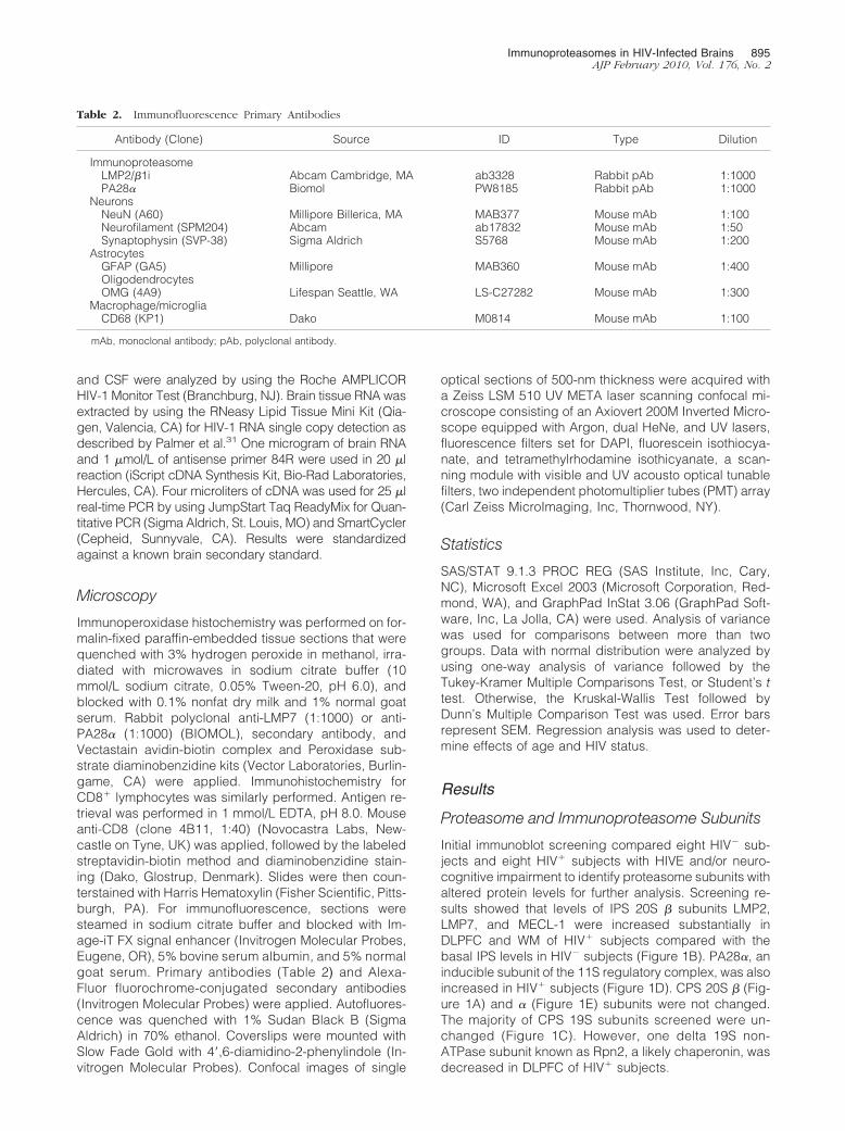
Subjects with NPI had significantly increased LMP7(Figure 4, A and B) and PA28� (Figure 4, C and D) inDLPFC and WM. Average increases were approximatelytwo- to fourfold, with the most pronounced affect seenwith WM PA28� measurements. HIV� subjects withoutNPI had little or no increase in either inducible subunit.HIV� NPI-O subjects also had significantly increased IPSsubunits, but the increase was less pronounced thanthose with NPI.
The decrease in DLPFC constitutive 19S regulatorycomplex subunit Rpn2 seen in proteasome subunitscreening was investigated. A slight decrease in HIV�
subjects was not significant (not shown). Further analysis
Figure 1. Western blots illustrate altered proteasome subunit concentrationsin DLPFC and WM from eight HIV� subjects with HIVE and/or neurocogni-tive impairment compared with eight HIV� subjects. A: Constitutive 20Sproteasome subunits X(�5), Y(�1), and Z(�2) were not changed in HIV�
subjects in DLPFC or WM. B: Three inducible 20S immunoproteasome �subunits were increased in DLPFC and WM of HIV� subjects. C: The majorityof constitutively expressed 19S proteasome regulatory complex subunitsremained unchanged, except for Rpn2, which showed a decrease in theDLPFC of HIV� subjects. D: The immunoproteasome 11S regulatory subunitPA28� was increased in DLPFC and WM of HIV� subjects. E: 20S � subunitswere not changed.
Figure 2. Immunoproteasome subunit proteins in 88 HIV� and 65 HIV� sub-jects were quantified by using densitometry of calibrated Western blots withloading controls. Averaged LMP7 was increased 99% in DLPFC (A) and 184% inWM (B). PA28� was increased by 204% in DLPFC (C) and 233% in WM (D).Statistics: Student’s t test; *P � 10�7; **P � 10�10. OD � optical density.
Figure 3. Significantly increased immunoproteasome subunit LMP7 andPA28� concentration in HIV� subjects with and without HIVE. A: DLPFCLMP7 levels in HIV� subjects with and without HIVE were increased 173%and 81%, respectively, compared with HIV� subjects. B: WM LMP7 in HIVEwas increased 414% and 114% compared with HIV� subjects and HIV�
subjects without HIVE, respectively. LMP7 levels of HIV� subjects withoutHIVE were 140% greater than HIV� subjects. C: DLPFC PA28� levels of HIV�
subjects with and without HIVE were 349% and 166% higher than HIV�
subjects, respectively. D: In WM, PA28� was increased 443% and 185% inHIV� subjects with and without HIVE, respectively, compared with HIV�
subjects. Statistics: one-way analysis of variance with Tukey-Kramer MultipleComparisons Test; *P � 0.05; ***P � 0.001. OD � optical density.
896 Nguyen et alAJP February 2010, Vol. 176, No. 2
revealed mild decreases in Rpn2 associated with NPIand HIVE (not shown) that may account for the decreaseseen in screening, but with the accompanying data ofcomparable 19S subunits, provided only minimal evi-dence for alteration in the constitutively expressed 19Sregulatory complex.
Effects of Age
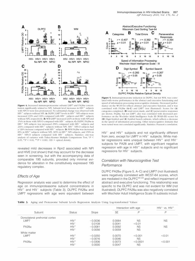
Regression analysis was used to determine the effect ofage on immunoproteasome subunit concentrations inHIV� and HIV� subjects (Table 3). DLPFC PA28� andLMP7 regressions with age were equivalent between
HIV� and HIV� subjects and not significantly differentfrom zero, except for LMP7 in HIV� subjects. White mat-ter regressions were unequal between HIV� and HIV�
subjects for PA28 and LMP7, with significant negativeregression with age in HIV� subjects and no significantregressions for HIV� subjects.
Correlation with Neurocognitive TestPerformance
DLPFC PA28� (Figure 5, A–C) and LMP7 (not illustrated)were negatively correlated with WCST-64 scores, whichare mediated in the DLPFC32,33 and reflect impairment ofabstract and executive functioning. This relationship wasspecific to the DLPFC and was not evident for WM (notillustrated). DLPFC PA28� was also negatively correlatedwith Wechsler Adult Intelligence Scale III subtests includ-
Figure 4. Increased immunoproteasome subunit LMP7 and PA28� concen-tration significantly related to NPI. Subunit level increases in HIV� subjectswith NPI-O were less pronounced. No substantial change was observed withHIV� subjects without NPI. A: DLPFC LMP7 levels in HIV� NPI subjects wereincreased 133% and 110% compared with HIV� subjects and HIV� subjectswithout NPI, respectively. B: WM LMP7 increased 243% in those with NPI and154% in those with NPI-O compared with HIV� subjects. C: DLPFC PA28� inHIV� NPI subjects was increased 259% compared with HIV� subjects and216% compared with HIV� subjects without NPI. HIV� NPI-O subjects hada 126% increase compared with HIV� subjects. D: WM PA28� was increased85% in HIV� subjects without NPI, 305% in HIV� NPI subjects, and 159% inHIV� NPI-O subjects compared with HIV� subjects. Statistics: one-wayanalysis of variance with Tukey-Kramer Multiple Comparisons Test; *P �0.05; **P � 0.01; ***P � 0.001. OD � optical density.
Table 3. Aging and Proteasome Subunit Levels Regression Analysis Using Log-transformed Values
Interaction with age HIV� vs. HIV�
Subunit Status Slope SE P P
Dorsolateral prefrontal cortexLMP7 HIV� �0.0036 0.0064 NS NS
HIV� 0.0108 0.0041 �0.01PA28� HIV� �0.0081 0.0092 NS NS
HIV� 0.0056 0.0059 NSWhite matter
LMP7 HIV� �0.0250 0.0070 �0.001 �0.01HIV� �0.0016 0.0046 NS
PA28� HIV� �0.0306 0.0073 �0.0001 �0.001HIV� 0.0000 0.0047 NS
Figure 5. Immunoproteasome induction in DLPFC, but not WM, was corre-lated with worse performance in the abstract and executive functioning andspeed of information processing neurocognitive domains. Decreased perfor-mance on the WCST-64 reflects abstract and executive function, and it wascorrelated with PA28� (A–C) and LMP7 (not illustrated). Scores for (A)categories completed, (B) total errors made, and (C) perseverative responsesare shown. PA28�, but not LMP7, also was correlated with decreased per-formance on the Wechsler Adult Intelligence Scale III (WAIS-III) scores for(D) Digit-Symbol and (E) Symbol Search subtests, which reflects a decreasein the speed of information processing. Other neurocognitive domains thatwere tested were not correlated significantly. Statistics: Pearson’s correlation.
Immunoproteasomes in HIV-Infected Brains 897AJP February 2010, Vol. 176, No. 2

ing Digit Symbol (Figure 5D) and Symbol Search (Figure5E), which reflect a decrease in the speed of informationprocessing. The same relationship was not present inWM (not illustrated). No correlations between IPS levelsand other functional domains evaluated by the NationalNeuroAIDS Tissue Consortium battery were observed(not illustrated).
Correlation with Clinical Virology andImmunology
Correlations between IPS subunit levels and VL in braintissue, CSF, and plasma, and plasma CD4� T-cell countwere assessed. DLPFC PA28� levels were robustly andpositively correlated with VL in all tissue compartmentsmeasured, and were negatively correlated with CD4�
T-cell counts (Figure 6, A–D). WM PA28� levels alsocorrelated with VL (Figure 6, E–G) but were not correlatedwith CD4� T-cell count (Figure 6H). Analysis with LMP7levels produced results equivalent to PA28� (not illus-trated). Assessment of IFN-� levels in brain tissue byenzyme-linked immunosorbent assay showed an in-creasing trend with HIV� subjects, but marked variabilityprecluded further analysis (not shown).
Localization in Neurons
Glial and neuronal elements were positively stained forIPS subunits in HIVE. In general, immunoreactivity inwhite matter was more intense than in the cortex. Patho-logical structures characteristically present in HIVE wereheavily stained. Microglia, macrophages, and oligoden-drocyte nuclei were stained in microglial nodules anddiffusely (Figure 7A). Neuronal cell bodies in neocorticallaminae III and IV had positive staining for IPS subunits(Figure 7, B and D). WM axons often contained IPS
subunits (Figure 7C). HIV� sections of DLPFC (Figure 7E)and WM (Figure 7F) lacked focal immunostaining. Colo-calization with neuronal markers (NeuN and neurofila-ment) was present in HIVE (Figure 8, A and B) but notHIV� sections (Figure 8 inserts). LMP2 was present pri-marily in perikaryal cytoplasm, whereas PA28� was moreevident in neuronal nuclei. LMP2 and PA28� localizedwith neurofilament protein in WM axons. Dystrophic swol-len axons, which are a neuropathological anomaly inHIVE,34–37 contained IPS (Figure 8B). Synaptic morphol-ogy is disturbed in HIVE,38 and these structures alsocontained IPS. Presynaptic boutons stained for synapto-physin showed the typical punctate staining pattern ofsynapses. Numerous synaptic boutons contained IPS(Figure 9A). Complete overlapping of IPS and synapto-physin suggested localization in presynaptic boutons(Figure 9B). Incomplete overlapping (Figure 9, C and D)suggested that IPS might be present in postsynapticdensities or synaptic astrocyte foot processes. Markersfor glial cells including glial fibrillary acidic protein forhypertrophic astrocytes, CD68 for microglia/macro-phages, and oligodendrocyte and myelin glycoproteinconfirmed that IPS subunits were present in those cells assuggested in Figure 7 (not illustrated).
Discussion
Immunoproteasomes are induced widely in the brains ofHIV� people compared with baseline levels in HIV� sub-
Figure 6. Immunoproteasome induction in DLPFC (A–D) and WM (E–H)was correlated significantly with virological and immunological status ofHIV-infected subjects. HIV-1 RNA concentrations in brain (A and E), CSF(B and F), and blood plasma (C and G) were positively correlated with PA28�levels. CD4� lymphocyte counts in blood plasma were negatively correlatedwith PA28� levels in DLPFC (D), but not WM (H). Equivalent results for LMP7were obtained (not illustrated). Statistics: Pearson’s correlation.
Figure 7. Immunoperoxidase histochemistry illustrates immunoproteasomesubunit staining for PA28� in glial and neuronal elements that are patholog-ical in HIV encephalitis. Microglia, macrophages, and oligodendrocyte nucleiare stained in a microglial nodule (A). Neuronal cell bodies in neocorticalneurons were stained in focal pathology (B) and were evident in laminae IIIand IV generally, though admittedly lighter than focal pathology (D). Whitematter axons often contained immunoproteasome subunits (C). Low immu-noreactivity was observed in the (E) cortex and (F) white matter of HIV�
subjects.
898 Nguyen et alAJP February 2010, Vol. 176, No. 2

jects, and this abnormality is clinically and pathologicallyrelevant. Induction of IPS with switching of 20S catalyticsubunits and 11S regulatory subunits of the CPS waspresent in about half of the HIV� decedents and pro-
duced a three- to fourfold increase in IPS subunit con-centration. IPS synthesis was relevant neuropathologi-cally because it was significantly more prevalent inpeople with a postmortem diagnosis of HIVE, althoughHIVE was not required. The high prevalence of IPS in-duction in people with HIVE indicates that the neuro-pathologically evident inflammatory response to replicat-ing HIV-1 in the brain is important, although it was not thesole risk factor. HIV-1 VL in brain tissue and CSF werecorrelated, as was HIV-1 VL in the plasma compartment.The latter observation agrees with studies that show link-age between HAD and abnormalities in the vascularcompartment, including anemia and altered monocytepopulations.1,39,40 A relationship between IPS inductionand weakened systemic immunity was implied by thelower plasma CD4� lymphocyte counts in affected sub-jects, and suggests that having advanced HIV/AIDS is arisk factor.1 The broad clinical-pathological impressionthat emerges from these results is that inflammatory de-fense against virus,15 systemically and in the brain, areinvolved in IPS induction. Nevertheless, HIVE was notrequired for IPS induction in the brain; other risk factorsare involved.
IPS synthesis was significantly more prevalent in peo-ple with MCMD or HAD. IPS synthesis was present in theDLPFC and WM, although significant correlation withneurocognitive impairment was most evident in DLPFC.Indeed, IPS induction measured in the DLPFC was cor-related specifically with performance on WCST-64 andWechsler Adult Intelligence Scale III processing speedsubtests, which are driven by circuits involved with ex-ecutive function and speed of information processinglocated in Brodmann areas 9 and 46 of the DLPFC.32,33
IPS changes in WM were not correlated with these func-tional domains even though they were significantly differentin infected people. The correlation between increased IPSin DLPFC and specific functional impairments driven by theDLPFC circuitry illustrates that region- and circuit-specificchanges drive specific facets of the overall syndrome ofHIV-associated neurocognitive decline (HAND). “Task-specific” changes that correlate selectively with a change ingray matter versus white matter have been reported inHIV-1 infected people, and suggest that neocortical andsubcortical pathologies both contribute to HAND.41
Authors of several reports have observed changes inconstitutive proteasomes and immunoproteasomes asso-ciated with advanced age.21,42,43 Potential influence ofage on immunoproteasome induction associated withHIV infection was investigated by performing regressionanalysis with HIV status and age regressed on the quan-tified immunoproteasome subunits. The results indicatedthat DLPFC immunoproteasome subunit concentrationsin HIV� subjects were not significantly influenced by age.WM subunit concentrations were associated with a slightdecrease with increasing age of approximately 3% peryear. Thus, advanced age does not cause or contributeto the immunoproteasome induction, the three- to fourfoldincrease in immunoproteasome subunits, or the correla-tion between DLPFC immunoproteasome subunit con-centrations and neurocognitive performance in HIV�
subjects.
Figure 8. Immunoproteasome subunits expressed in neurons in HIV enceph-alitis. Dual indirect immunofluorescence staining for LMP2 (A) or PA28� (B)shows colocalization with the neuronal markers NeuN and neurofilament insingle optical sections from confocal microscopy. LMP2 is present in granulardeposits in the parikaryon (A). PA28� is more prominent in the nucleus (B). Apathologically swollen axon in white matter contains PA28� (arrows in B), asdo several other neurofilament-containing processes. Immunofluorescencestainings of HIV� sections (insets) reveal an absence of LMP2 and PA28�. Scalebar � 10 �m.
Figure 9. Immunofluorescence and laser confocal microscopy reveal that im-munoproteasome subunit LMP2 is localized to some neocortical synapses in HIVencephalitis. A: LMP2 was colocalized within punctate deposits of synaptophy-sin, which is an established cell marker of presynaptic boutons (arrows). About12% of labeled synapses in the figure contain immunoproteasome antigenicity.Complete overlapping of these two antigens was evident in some synapses andsuggests that immunoproteasomes often are present in presynaptic boutons (B).Some synapses had incomplete overlapping (C and D), which suggests thatimmunoproteasomes may be present in adjacent structures, including thepostsynaptic density or in synaptic astrocyte foot processes. Scale bar � 10 �m.
Immunoproteasomes in HIV-Infected Brains 899AJP February 2010, Vol. 176, No. 2

IFN-� mediated IPS induction in inflammatory cells andglia is consistent with a role in antigen presentation andhost defense against virus infection.10,15 Our carefullydocumented observation that IPS synthesis occurs inneuronal perikarya, dystrophic axons, and synapses isemphasized because it is quite novel. The ability ofneurons to undergo IPS induction is not widely appre-ciated10,11,44 and could suggest that neurons may par-ticipate in self antigen presentation or are passively in-volved in a potentially harmful metabolic shift. NeuronalIPS induction has been observed in only two other neu-rodegenerative diseases, Huntington’s disease and Alz-heimer’s disease.43,44 Neurons express IFN-� receptorsand can synthesize Class I histocompatibility complexesin response to IFN-�,43,44 which probably requires IPSinduction. As well, exposure of neurons to IFN-� pro-duces distinct neurophysiological changes in synaptictransmission pertaining to learning and memory.45 It isnotable that correlation was present between HAND andIPS induction in brain cortex (predominantly neuronal),but not white matter (mostly glial). Thus, IPS synthesis inneurons could be particularly important for neocorticaldysfunction due to HIV-1 infection. For example, syn-apses and dendrites are abnormal morphologically38
and biochemically28,46 in HIVE and are potentially impor-tant targets for therapeutic intervention.47 Synapses anddendrites contain proteasomes and undergo localizedprotein turnover.18,19,48,49 Polyribosomes, mRNA, andthe UPS all are present locally in synaptic and dendriticcompartments, and they play a pivotal role in regulatingsynaptic function.19,49,50 Experimental clogging of pro-tein turnover in these compartments can be especiallydevastating because proteins involved in memory forma-tion in synapses and dendrites turn over with astonishingspeed (minutes to hours).19 Local protein turnover in thesynaptic compartment is necessary because synaptictransmission and memory formation require rapid localadjustments that require changes in protein synthesisand degradation.18 Blocking protein turnover at the levelof synthesis in dendritic polyribosomes, or degradationvia the UPS, alters the electrophysiological changes thatdrive learning and memory and impairs performance ontests of learning and memory.26,51
The accumulation of ubiquitinylated pathological pro-tein is a virtual “signature” of pathological brain aging,reflecting unsuccessful removal of abnormal protein viathe UPS.23–25,27 The slowing down of the UPS in normaland pathological brain aging is potentially damaging be-cause effete and oxidatively damaged cellular proteinsdepend on UPS for efficient removal.9,21 Inherited de-fects of the UPS lead to Parkinson’s disease and An-gelman’s syndrome.52,53 UPS protein processing is de-creased sharply in Alzheimer’s disease and during brainaging.20,21,54 As well, experimental disruption producessynaptic dysfunction and deficits in learning and mem-ory.18–27,48,49 Likewise, the finding of increased accumu-lation of ubiquitin-stained deposits and high molecularweight ubiquitin conjugates in HIV/AIDS brains suggestUPS dysfunction.28 Neuronal autophagy, which aids inubiquitin-protein aggregate degradation, is also reducedin HIV infection and may further exacerbate the accumu-
lation of ubiquitinated protein conjugates due to UPSdysfunction.55,56
HAND represents a novel example of an acquireddefect in the UPS linked to synaptic dysfunction.38,46
Though the proteasome subunit analysis does not exam-ine the structure or quantity of fully assembled protea-some complexes, the HIV-associated increases in IPSand 11S subunits implicate increasing integration ofthese subunits into newly formed IPS complexes at theexpense of constitutively expressed counterparts, whichare assembled four times slower than IPS.6,57,58 The re-placement of constitutively expressed 19S regulatorcomplexes on the proteasome core by the increasedpresence of 11S activator complexes, which are integralto IPS formation and antigen processing,59,60 is importantbecause 19S processes many thousands of polyubiquiti-nylated protein species during routine turnover and cel-lular maintenance. After a folded protein has beentagged for destruction with polyubiquitin, it binds to asubunit of 19S. The 19S complex contains ATPases,chaperonins, unfoldases, and isopeptidases and capsthe 20S core complex to form the 26S proteasome com-plex. The substrate is energized by ATPases, polyubiq-uitin is removed and recycled, and the polypeptide isunfolded and chaperoned “single file” into the 20S corefor hydrolysis. Virtually all folded proteins turn over by the26S proteasome in that manner. The 11S activator, how-ever, does not contain ATPases or isopeptidases. Thus, itcan neither process folded polyubiquitinylated proteinnor recycle ubiquitin monomers, which are absolutelyessential for normal operation of the UPS.61–63 IPS induc-
Figure 10. Persistent “hijacking” of brain proteasomes in HIV-1-infectedpeople may lead to neuronal dysfunction. Normally, proteasome complexesrapidly turn over ubiquitinylated proteins. Persistent infection with HIV-1produces inflammatory cytokines such as IFN-� that induce a temporary shiftto immunoprotesome synthesis. In turn, the protein substrate repertoire istemporarily shifted toward the processing of unfolded peptides for class Iantigen presentation. “Borrowing” the proteasome apparatus for heightenedantigen presentation persists until the pathogen is eradicated, after whichnormal brain protein turnover should resume. Since HIV-1 infection is noteradicated in the brain, there is a persistent “hijacking” of the proteasome.Normal turnover of folded proteins is disrupted chronically. This leadseventually to the accumulation of pathologically misfolded proteins, neuro-nal dysfunction, and dementia.
900 Nguyen et alAJP February 2010, Vol. 176, No. 2
tion also coincides with destabilization and decrease inconstitutive 26S proteasome complexes.12,64 The persis-tent replacement of 19S complexes on 20S complexes byincreasing 11S subunits due to HIV-1 infection is poten-tially devastating because thousands of cellular compo-nents can be perturbed by the changes in substraterepertoire.9,12–14
Pervasive anomalies of the UPS in HAND suggest anovel concept regarding neuroinflammatory modulationof brain function (Figure 10). As 11S increasingly dis-places 19S regulatory complexes, the neuronal protea-some population shifts to accommodate newly synthe-sized 11S/IPS complexes at the expense of the 26Sproteasomes. The IPS complexes cannot perform “rou-tine” turnover of polyubiquitinylated protein, which is theprimary route for the turning over of most folded pro-teins.9 Instead, the IPS is specifically synthesized to pro-cess unfolded protein and small, unconjugated polypep-tides with high efficiency. The best known function of theIPS is processing unfolded peptides for antigen presen-tation in Class I histocompatibility complexes.10,11 IFN-�-induced increases of IPS synthesis reflect heightenedimmune surveillance in response to the presence of for-eign antigen.15 A temporary “borrowing” of the UPS byIPS is tolerable physiologically because pathogens arenormally eradicated in due time, and the IPS disappearsquickly due to its inherent structural instability.7 The shiftto IPS synthesis, however, is not transient in the HIV-1infected brain because the CNS is a reservoir of persis-tently replicating HIV-1.16,17 HIV-1 provokes a long-last-ing sustained cytokine response that includes tumor ne-crosis factor � and IFN-� expression,65,66 synthesis ofinflammatory response protein28,67 and heightened ex-pression of several IFN-� inducible genes.68 Instead ofthe typical “borrowing” of the UPS for immune defense,7
a persistent “hijacking” of the UPS characterizes aptly thenature of the changes in HIV-1-infected brain tissue.
References
1. McArthur JC, Brew BJ, Nath A: Neurological complications of HIVinfection. Lancet Neurol 2005, 4:543–555
2. Kaul M, Garden GA, Lipton SA: Pathways to neuronal injury andapoptosis in HIV-associated dementia. Nature 2001, 410:988–994
3. Williams KC, Hickey WF: Central nervous system damage, monocytesand macrophages, and neurological disorders in AIDS. Annu RevNeurosci 2002, 25:537–562
4. Budka H: Neuropathology of human immunodeficiency virus infec-tion. Brain Pathol 1991, 1:163–175
5. Wiley CA, Achim C: Human immunodeficiency virus encephalitis isthe pathological correlate of dementia in acquired immunodeficiencysyndrome. Ann Neurol 1994, 36:673–676
6. Aki M, Shimbara N, Takashina M, Akiyama K, Kagawa S, Tamura T,Tanahashi N, Yoshimura T, Tanaka K, Ichihara A: Interferon-gammainduces different subunit organizations and functional diversity ofproteasomes. J Biochem 1994, 115:257–269
7. Heink S, Ludwig D, Kloetzel PM, Kruger E: IFN-gamma-inducedimmune adaptation of the proteasome system is an accelerated andtransient response. Proc Natl Acad Sci USA 2005, 102:9241–9246
8. Raasi S, Schmidtke G, de Giuli R, Groettrup M: A ubiquitin-like proteinwhich is synergistically inducible by interferon-gamma and tumornecrosis factor-alpha. Eur J Immunol 1999, 29:4030–4036
9. Glickman MH, Ciechanover A: The ubiquitin-proteasome proteolytic
pathway: destruction for the sake of construction. Physiol Rev 2002,82:373–428
10. Kloetzel PM: Generation of major histocompatibility complex class Iantigens: functional interplay between proteasomes and TPPII. NatImmunol 2004, 5:661–669
11. Kloetzel PM, Ossendorp F: Proteasome and peptidase function inMHC-class-I-mediated antigen presentation. Curr Opin Immunol2004, 16:76–81
12. Bose S, Brooks P, Mason GG, Rivett AJ: Gamma-interferon de-creases the level of 26 S proteasomes and changes the pattern ofphosphorylation. Biochem J 2001, 353:291–297
13. Gaczynska M, Rock KL, Goldberg AL: Gamma-interferon and expres-sion of MHC genes regulate peptide hydrolysis by proteasomes.Nature 1993, 365:264–267
14. Gaczynska M, Rock KL, Spies T, Goldberg AL: Peptidase activities ofproteasomes are differentially regulated by the major histocompati-bility complex-encoded genes for LMP2 and LMP7. Proc Natl AcadSci USA 1994, 91:9213–9217
15. Tishon A, Lewicki H, Rall G, Von Herrath M, Oldstone MB: An essen-tial role for type 1 interferon-gamma in terminating persistent viralinfection. Virology 1995, 212:244–250
16. Langford D, Marquie-Beck J, de Almeida S, Lazzaretto D, Letendre S,Grant I, McCutchan JA, Masliah E, Ellis RJ: Relationship of antiretro-viral treatment to postmortem brain tissue viral load in human immu-nodeficiency virus-infected patients. J Neurovirol 2006, 12:100–107
17. Kulkosky J, Bray S: HAART-persistent HIV-1 latent reservoirs: theirorigin, mechanisms of stability and potential strategies for eradica-tion. Curr HIV Res 2006, 4:199–208
18. Hegde AN: Ubiquitin-proteasome-mediated local protein degrada-tion and synaptic plasticity. Prog Neurobiol 2004, 73:311–357
19. Ehlers MD: Activity level controls postsynaptic composition and sig-naling via the ubiquitin-proteasome system. Nat Neurosci 2003,6:231–242
20. Keller JN, Hanni KB, Markesbery WR: Impaired proteasome functionin Alzheimer’s disease. J Neurochem 2000, 75:436–439
21. Keller JN, Gee J, Ding Q: The proteasome in brain aging. Ageing ResRev 2002, 1:279–293
22. Ma J, Wollmann R, Lindquist S: Neurotoxicity and neurodegenerationwhen PrP accumulates in the cytosol. Science 2002, 298:1781–1785
23. Tsuji T, Shimohama S: Protein degradation in Alzheimer’s diseaseand aging of the brain. Prog Mol Subcell Biol 2002, 29:43–60
24. Chondrogianni N, Gonos ES: Proteasome dysfunction in mammalianaging: steps and factors involved. Exp Gerontol 2005, 40:931–938
25. Ciechanover A, Brundin P: The ubiquitin proteasome system in neu-rodegenerative diseases: sometimes the chicken, sometimes theegg. Neuron 2003, 40:427–446
26. Ding Q, Dimayuga E, Markesbery WR, Keller JN: Proteasome inhibi-tion induces reversible impairments in protein synthesis. FASEB J2006, 20:1055–1063
27. Layfield R, Cavey JR, Lowe J: Role of ubiquitin-mediated proteolysisin the pathogenesis of neurodegenerative disorders. Ageing Res Rev2003, 2:343–356
28. Gelman BB, Schuenke K: Brain aging in acquired immunodeficiencysyndrome: increased ubiquitin-protein conjugate is correlated withdecreased synaptic protein but not amyloid plaque accumulation.J Neurovirol 2004, 10:98–108
29. Morgello S, Gelman BB, Kozlowski PB, Vinters HV, Masliah E, CornfordM, Cavert W, Marra C, Grant I, Singer EJ: The National NeuroAIDSTissue Consortium: a new paradigm in brain banking with an emphasison infectious disease. Neuropathol Appl Neurobiol 2001, 27:326–335
30. Woods SP, Rippeth JD, Frol AB, Levy JK, Ryan E, Soukup VM, HinkinCH, Lazzaretto D, Cherner M, Marcotte TD, Gelman BB, Morgello S,Singer EJ, Grant I, Heaton RK: Interrater reliability of clinical ratingsand neurocognitive diagnoses in HIV. J Clin Exp Neuropsychol 2004,26:759–778
31. Palmer S, Wiegand AP, Maldarelli F, Bazmi H, Mican JM, Polis M,Dewar RL, Planta A, Liu S, Metcalf JA, Mellors JW, Coffin JM: Newreal-time reverse transcriptase-initiated PCR assay with single-copysensitivity for human immunodeficiency virus type 1 RNA in plasma.J Clin Microbiol 2003, 41:4531–4536
32. Anderson SW, Damasio H, Jones RD, Tranel D: Wisconsin CardSorting Test performance as a measure of frontal lobe damage. J ClinExp Neuropsychol 1991, 13:909–922
33. Monchi O, Petrides M, Petre V, Worsley K, Dagher A: Wisconsin Card
Immunoproteasomes in HIV-Infected Brains 901AJP February 2010, Vol. 176, No. 2

Sorting revisited: distinct neural circuits participating in differentstages of the task identified by event-related functional magneticresonance imaging. J Neurosci 2001, 21:7733–7741
34. An SF, Giometto B, Groves M, Miller RF, Beckett AA, Gray F, TavolatoB, Scaravilli F: Axonal damage revealed by accumulation of beta-APPin HIV-positive individuals without AIDS. J Neuropathol Exp Neurol1997, 56:1262–1268
35. Giometto B, An SF, Groves M, Scaravilli T, Geddes JF, Miller R,Tavolato B, Beckett AA, Scaravilli F: Accumulation of beta-amyloidprecursor protein in HIV encephalitis: relationship with neuropsycho-logical abnormalities. Ann Neurol 1997, 42:34–40
36. Raja F, Sherriff FE, Morris CS, Bridges LR, Esiri MM: Cerebral whitematter damage in HIV infection demonstrated using beta-amyloid pre-cursor protein immunoreactivity. Acta Neuropathol 1997, 93:184–189
37. Adle-Biassette H, Chretien F, Wingertsmann L, Hery C, Ereau T,Scaravilli F, Tardieu M, Gray F: Neuronal apoptosis does not correlatewith dementia in HIV infection but is related to microglial activationand axonal damage. Neuropathol Appl Neurobiol 1999, 25:123–133
38. Masliah E, Heaton RK, Marcotte TD, Ellis RJ, Wiley CA, Mallory M,Achim CL, McCutchan JA, Nelson JA, Atkinson JH, Grant I: Dendriticinjury is a pathological substrate for human immunodeficiency virus-related cognitive disorders. HNRC Group the HIV NeurobehavioralResearch Center. Ann Neurol 1997, 42:963–972
39. Gartner S: HIV infection and dementia. Science 2000, 287:602–60440. Fischer-Smith T, Croul S, Sverstiuk AE, Capini C, L’Heureux D, Regu-
lier EG, Richardson MW, Amini S, Morgello S, Khalili K, Rappaport J:CNS invasion by CD14�/CD16� peripheral blood-derived monocytesin HIV dementia: perivascular accumulation and reservoir of HIVinfection. J Neurovirol 2001, 7:528–541
41. Gelman BB, Soukup VM, Holzer CE, 3rd, Fabian RH, Schuenke KW,Keherly MJ, Richey FJ, Lahart CJ: Potential role for white matterlysosome expansion in HIV-associated dementia. J Acquir ImmuneDefic Syndr 2005, 39:422–425
42. Keller JN, Huang FF, Markesbery WR: Decreased levels of protea-some activity and proteasome expression in aging spinal cord. Neu-roscience 2000, 98:149–156
43. Mishto M, Bellavista E, Santoro A, Stolzing A, Ligorio C, Nacmias B,Spazzafumo L, Chiappelli M, Licastro F, Sorbi S, Pession A, Ohm T,Grune T, Franceschi C: Immunoproteasome and LMP2 polymorphismin aged and Alzheimer’s disease brains. Neurobiol Aging 2006,27:54–66
44. Diaz-Hernandez M, Hernandez F, Martin-Aparicio E, Gomez-RamosP, Moran MA, Castano JG, Ferrer I, Avila J, Lucas JJ: Neuronalinduction of the immunoproteasome in Huntington’s disease. J Neu-rosci 2003, 23:11653–11661
45. Vikman KS, Owe-Larsson B, Brask J, Kristensson KS, Hill RH: Inter-feron-gamma-induced changes in synaptic activity and AMPA recep-tor clustering in hippocampal cultures. Brain Res 2001, 896:18–29
46. Gelman BB, Spencer JA, Holzer CE, 3rd, Soukup VM: Abnormalstriatal dopaminergic synapses in National NeuroAIDS Tissue Con-sortium subjects with HIV encephalitis. J Neuroimmune Pharmacol2006, 1:410–420
47. Bellizzi MJ, Lu SM, Gelbard HA: Protecting the synapse: evidence fora rational strategy to treat HIV-1 associated neurologic disease.J Neuroimmune Pharmacol 2006, 1:20–31
48. Bingol B, Schuman EM: Synaptic protein degradation by the ubiquitinproteasome system. Curr Opin Neurobiol 2005, 15:536–541
49. Yi JJ, Ehlers MD: Ubiquitin and protein turnover in synapse function.Neuron 2005, 47:629–632
50. Eberwine J, Belt B, Kacharmina JE, Miyashiro K: Analysis of subcel-
lularly localized mRNAs using in situ hybridization, mRNA amplifica-tion, and expression profiling. Neurochem Res 2002, 27:1065–1077
51. Eisenberg M, Kobilo T, Berman DE, Dudai Y: Stability of retrievedmemory: inverse correlation with trace dominance. Science 2003,301:1102–1104
52. DiAntonio A, Haghighi AP, Portman SL, Lee JD, Amaranto AM,Goodman CS: Ubiquitination-dependent mechanisms regulate synapticgrowth and function. Nature 2001, 412:449–452
53. Chung KK, Dawson VL, Dawson TM: The role of the ubiquitin-protea-somal pathway in Parkinson’s disease and other neurodegenerativedisorders. Trends Neurosci 2001, 24:S7–S14
54. Lam YA, Pickart CM, Alban A, Landon M, Jamieson C, Ramage R,Mayer RJ, Layfield R: Inhibition of the ubiquitin-proteasome system inAlzheimer’s disease. Proc Natl Acad Sci USA 2000, 97:9902–9906
55. Alirezaei M, Kiosses WB, Flynn CT, Brady NR, Fox HS: Disruption ofneuronal autophagy by infected microglia results in neurodegenera-tion. PLoS One 2008, 3:e2906
56. Zhu Y, Vergote D, Pardo C, Noorbakhsh F, McArthur JC, HollenbergMD, Overall CM, Power C: CXCR3 activation by lentivirus infectionsuppresses neuronal autophagy: neuroprotective effects of antiretro-viral therapy. FASEB J 2009, 23:2928–2941
57. Yewdell JW: Immunoproteasomes: regulating the regulator. Proc NatlAcad Sci USA 2005, 102:9089–9090
58. Griffin TA, Nandi D, Cruz M, Fehling HJ, Kaer LV, Monaco JJ, ColbertRA: Immunoproteasome assembly: cooperative incorporation of in-terferon gamma (IFN-gamma)-inducible subunits. J Exp Med 1998,187:97–104
59. Groettrup M, Soza A, Eggers M, Kuehn L, Dick TP, Schild H,Rammensee HG, Koszinowski UH, Kloetzel PM: A role for the protea-some regulator PA28alpha in antigen presentation. Nature 1996, 381:166–168
60. Preckel T, Fung-Leung WP, Cai Z, Vitiello A, Salter-Cid L, Winqvist O,Wolfe TG, Von Herrath M, Angulo A, Ghazal P, Lee JD, Fourie AM, WuY, Pang J, Ngo K, Peterson PA, Fruh K, Yang Y: Impaired immuno-proteasome assembly and immune responses in PA28�/� mice. Sci-ence 1999, 286:2162–2165
61. Dubiel W, Pratt G, Ferrell K, Rechsteiner M: Purification of an 11 Sregulator of the multicatalytic protease. J Biol Chem 1992, 267:22369–22377
62. Hershko A, Ciechanover A: The ubiquitin system. Annu Rev Biochem1998, 67:425–479
63. Ma CP, Slaughter CA, DeMartino GN: Identification, purification, andcharacterization of a protein activator (PA28) of the 20 S proteasome(macropain). J Biol Chem 1992, 267:10515–10523
64. Bose S, Stratford FL, Broadfoot KI, Mason GG, Rivett AJ: Phosphor-ylation of 20S proteasome alpha subunit C8 (alpha7) stabilizes the26S proteasome and plays a role in the regulation of proteasomecomplexes by gamma-interferon. Biochem J 2004, 378:177–184
65. Shapshak P, Duncan R, Minagar A, Rodriguez de la Vega P, StewartRV, Goodkin K: Elevated expression of IFN-gamma in the HIV-1infected brain. Front Biosci 2004, 9:1073–1081
66. Griffin DE: Cytokines in the brain during viral infection: clues toHIV-associated dementia. J Clin Invest 1997, 100:2948–2951
67. Achim CL, Morey MK, Wiley CA: Expression of major histocompati-bility complex and HIV antigens within the brains of AIDS patients.Aids 1991, 5:535–541
68. Masliah E, Roberts ES, Langford D, Everall I, Crews L, Adame A,Rockenstein E, Fox HS: Patterns of gene dysregulation in the frontalcortex of patients with HIV encephalitis. J Neuroimmunol 2004,157:163–175
902 Nguyen et alAJP February 2010, Vol. 176, No. 2