Paediatric Over-the-Counter (OTC) Oral Liquids Can Soften ...
Upload
eastangliaCategory
view
0download
0
Performance of urine lipoarabinomannanassays for paediatric tuberculosis inTanzania
Inge Kroidl1,3,11, Petra Clowes3,11, Klaus Reither4,5, Bariki Mtafya3,Gabriel Rojas-Ponce3, Elias N Ntinginya3, Mariam Kalomo6, Lilian T. Minja3,4,5,7,Dickens Kowuor3, Elmar Saathoff1,2, Arne Kroidl1,2,3, Norbert Heinrich1,2,Leonard Maboko3, Matthew Bates8,9,10, Justin O’Grady8,9,10,Alimuddin Zumla8,9,10, Michael Hoelscher1,2,3 and Andrea Rachow1,2,3
Affiliations: 1Division of Infectious Diseases and Tropical Medicine, Medical Centre of the University of Munich(LMU), Germany. 2German Centre for Infection Research (DZIF), partner site Munich, Germany. 3NationalInstitute for Medical Research–Mbeya Medical Research Centre, Mbeya, Tanzania. 4Medical Services andDiagnostic, Swiss Tropical and Public Health Institute, Basel, Switzerland. 5Medical Services and Diagnostic(Swiss TPH), University of Basel, Basel, Switzerland. 6Dept for Paediatrics and Child Health, Mbeya ReferralHospital, Mbeya, Tanzania. 7Ifakara Health Institute, Bagamoyo, Tanzania. 8University of Zambia-UniversityCollege London Medical School Research and Training Project, University Teaching Hospital, Lusaka, Zambia.9Division of Infection and Immunity, University College London, London, UK. 10NIHR Biomedical ResearchCentre, University College London Hospitals, London, UK. 11Both authors contributed equally.
Correspondence: Inge Kroidl, Division of Infectious Diseases and Tropical Medicine, Medical Centre of theUniversity of Munich (LMU), Germany. E-mail: [email protected]
ABSTRACT We evaluated the diagnostic performance of two tests based on the release oflipoarabinomannan (LAM) into the urine, the MTB-LAM-ELISA assay and the Determine TB-LAM-stripassay, in children with suspected tuberculosis (TB) in a high TB/HIV-prevalence setting.
In a prospective study, 132 children with suspected active TB were assigned to diagnostic subgroups.Urine samples were subjected to testing by both assays to ascertain sensitivity and specificity. Host factorsassociated with positive LAM results were investigated and LAM excretion monitored after antituberculoustreatment initiation.
18 (13.6%) children had culture-confirmed pulmonary TB. The assays’ sensitivity was higher in HIV-positive versus HIV-negative children: 70% (95% confidence interval 35–93%) versus 13% (0–53%) forMTB-LAM-ELISA and 50% (19–81%) versus 0% (0–37%) for Determine TB-LAM. In 35 (27%) childrenwith excluded active TB, both assays showed a specificity of 97.1% (85–100%). Proteinuria and low bodymass index were independently associated with LAM positivity. In most patients, LAM excretion declinedto zero during or at conclusion of antituberculous treatment.
HIV/TB co-infected children might benefit from LAM-based tests to aid early TB diagnosis andsubsequent positive impact on morbidity and mortality. Using LAM as a rule-in and treatment-monitoringtool may also show further potential.
@ERSpublicationsUrine lipoarabinomannan assays show reasonable sensitivity in HIV+ but not HIV− TB-infected children http://ow.ly/N56aG
Copyright ©ERS 2015
This article has supplementary data available from erj.ersjournals.com
Received: Jan 09 2015 | Accepted after revision: April 09 2015
Conflict of interest: None declared.
Eur Respir J 2015; in press | DOI: 10.1183/09031936.00003315 1
ORIGINAL ARTICLEIN PRESS | CORRECTED PROOF

IntroductionThe diagnosis of tuberculosis (TB) is usually established by detecting mycobacteria, either through smearmicroscopy or culturing methods, which is considered the current gold standard. However, due to thepaucibacillary nature of TB in children, the microbiological diagnosis is extremely difficult. Smearmicroscopy is positive in <15% [1–3] and mycobacterial culture is positive in 20–80% of children withpresumed TB [1, 2, 4]. Paediatric TB diagnosis is therefore mainly based on clinical assessments, scoringsystems and radiological findings, which can be erroneous. In an autopsy study from Zambia, TBaccounted for 18% of deaths in HIV co-infected and 26% of deaths in HIV-negative children withrespiratory illnesses [5]. In contrast, during a recent review of the global burden of disease in children [6,7], TB was not listed among the most common causes of paediatric deaths, demonstrating the low numberof microbiologically confirmed cases.
For TB cases in clinical settings, the inconsistency between pathological and epidemiological datahighlights the need for new and more accurate methods to diagnose TB in children, using non-invasiveclinical samples. Lipoarabinomannan (LAM) detection in urine for TB diagnosis was first investigated inthe late 1990s [8–10]. LAM is a 19 kD (±8.5 kD) lipopolysaccharide, specific to the cell wall of members ofthe Mycobacterium genus and is released from metabolically active or degrading bacterial cells [11, 12]. Itcan subsequently be detected in urine and other body fluids. Advantages of urine LAM diagnosis includethe ease of specimen collection, short bench-time, low cost and relatively low training and set uprequirements [13, 14]. Ideally the LAM-strip assay can be even performed as a point-of-care test in remotesettings. The reported sensitivity of the different LAM diagnostic tests in adults ranges between 13% [15]and 67% [16], with best performance in patients with advanced HIV disease [15–18]. Data on children arescarce with only one study published previously on performance of LAM diagnostic tests in children [19].
The two diagnostic assays evaluated here, the MTB-LAM-ELISA (Chemogen, Portland, OR, USA) and theDetermine TB-LAM (Alere, Waltham, MA, USA), have been studied recently in South African children,for whom they performed poorly [19]. We evaluated the diagnostic performance of both assays inTanzanian children with presumed TB and a high HIV co-infection rate. We also investigated host factorsthat might be related to LAM performance, as well as the change in LAM excretion during the course ofanti-TB treatment.
MethodsStudy design and settingWe undertook a prospective observational study in children presenting with suspected TB at theoutpatient department of the Mbeya Zonal Referral Hospital (Mbeya, Tanzania). The study wascoordinated by the National Institute for Medical Research (NIMR) Mbeya Medical Research Centre(MMRC), in close collaboration with the Mbeya Zonal Referral Hospital. The study was approved by theethics committee of the Tanzania National Institute for Medical Research and the local Mbeya MedicalResearch and Ethics Committee. Written informed consent for all children was obtained from anaccompanying parent or a legal guardian. In addition, children aged ⩾9 years signed an assent form.
Clinical study proceduresFrom May 2008 till November 2010, we approached all children with suspected TB attending the outpatientclinic and invited them to take part in the study. Inclusion criteria were 6 weeks to 14 years of age, and atleast one of the following symptoms: persistent unremitting cough for >21 days; repeated episodes of feverwithin the last 21 days; weight loss or failure to thrive within the previous 3 months. Children who hadreceived antituberculous treatment within the last 3 months were excluded from the study. Recruitmentprocedures, baseline diagnostics, physical assessment and clinical treatment of this cohort have previouslybeen described [20]. Follow-up visits were scheduled at 1, 3, 6 and 12 months after enrolment or afterantituberculous treatment initiation. For this evaluation, children were retrospectively assigned to distinctdiagnostic subgroups, based on the recently proposed classification by GRAHAM et al. [21] (fig. 1).
A decision on antituberculous treatment initiation was made in liaison with the paediatric department ofthe Mbeya Zonal Referral Hospital and the District TB and Leprosy Coordinators and was based onmicrobiological and clinical findings (including tuberculin skin test and chest radiography), and previousmedical history. Antituberculous treatment was administered following Tanzanian National Guidelines andpatients diagnosed with HIV infection were referred for further staging and treatment to the relevant HIVCare and Treatment Centres.
Sample collection and laboratory proceduresUp to three induced sputum samples were collected from each child at baseline and processed for smearmicroscopy and Mycobacterium tuberculosis (MTB) culture [20]. Additionally, the Xpert MTB/rifampicin
2 DOI: 10.1183/09031936.00003315
TUBERCULOSIS | I. KROIDL ET AL.

(RIF) assay (Cepheid, Sunnyvale, CA, USA) was performed on stored, frozen sputum samples as reportedpreviously [20]. The results were not included in the diagnostic algorithm as all children were enrolled beforeXpert endorsement by the World Health Organization (WHO). At each study visit, a urine sample wascollected for LAM testing, either midstream urine when possible or with a collection bag in younger children.Within 8 h of collection, all urine was boiled at 95–100°C for 30 min, centrifuged and the supernatants frozenat −20°C. For the execution of the MTB-LAM-ELISA assay, the thawed urine was processed in duplicate,according to the manufacturer’s instructions as described previously [18]. For the Determine TB-LAM assay,60 µL of pre-treated urine was applied onto the sample pad of the LAM strip. After 25 min, the test bandcolour intensity was compared to the colour intensity of a series of bands on a paper reference card suppliedby the manufacturer. Grade 1 colour intensity and above were defined as positive results, consistent with themanufacturer’s instructions at the time of the study [22]. According to the manufacturer (personalcommunication) and our own data, results for the Determine TB-LAM assay do not differ betweenpre-treated (boiled and centrifuged) or native urine samples. All lab staff performing LAM tests was blindedto culture results and clinical data. Furthermore, all urine samples underwent testing with urine dipsticks(Combur-Test; Roche, Basel, Switzerland) for detection of protein, glucose, leukocytes and erythrocytes.
All participants were screened for HIV, using the HIV1/2 STAT-PAK RDT (Chembio Diagnostics Systems,Medford, NY, USA) and following the manufacturer’s instructions. RDT results were confirmed with athird generation ELISA (Biorad Laboratories, Redmond WA, USA) and, in case of discordance, retested byWestern Blot (MPD HIV Blot 2.2, MP Biomedicals, Geneva, Switzerland). For children below the age of2 years a PCR (Roche Amplicor) was performed instead of ELISA. In HIV-positive participants CD4count and HIV viral load were determined by flow cytometry and PCR (Amplicor; Roche). HIV-positivechildren were classified according to the WHO classification of HIV-associated immunodeficiency ininfants and children (see the online supplementary data) [23].
Statistical analysisIn contrast to the classification in the original study, all children were re-classified into five differentdiagnostic groups in order to comply with the recently published proposed consensus of paediatric clinical
No response to
TB treatment
and
no documented
exposure
and
no immunological
evidence of TB
Resolution of
symptoms
without TB
treatment
and
immunological
evidence of TB
Response to
TB treatment
and
documented
exposure
and
immunological
evidence of TB
No response to
TB treatment
and
no documented
exposure
and
no immunological
evidence of TB
Response to
TB treatment
or
documented
exposure
or
immunological
evidence of TB
Consistent with TB Not consistent with TB
Possible TBMycobaterium
tuberculosis infection
"latent" TB
Probable TBTB unlikely/
not TB
X-ray
Microbiological
confirmationTB suspectedConfirmed TB Yes
No
FIGURE 1 Algorithm for diagnostic classification. Diagnostic classification for children presenting with suspectedpulmonary/intrathoracic tuberculosis (TB). In total, 379 sputum samples were analysed, 122 children gave three sputa,six children only two and three children only one sputum sample. One child classified as “No TB”, gave no sputumsample. In five children with signs of extrapulmonary TB, one pleural fluid sample, one ascites sample and three lymphnode aspirates were analysed. Mycobacterium tuberculosis was confirmed in the pleural fluid and in one lymph nodeaspirate. Both children were additionally diagnosed with sputum culture confirmed TB. All 132 children received chestradiography examination which was assessed by a radiologist and two clinical study investigators who were blinded toclinical and microbiological data. In case of discrepant readings a consensus radiography diagnosis was made afterdiscussion among the investigators. Clinical follow-up of all analysed children allowed assessment of treatment responsein those who received antituberculous treatment.
DOI: 10.1183/09031936.00003315 3
TUBERCULOSIS | I. KROIDL ET AL.

case definition by GRAHAM et al. [21]. Statistical analyses were performed using Stata statistics software(version 12; Stata Corp., College Station, TX, USA). The sensitivity, specificity and their respectiveconfidence intervals were calculated using the “diagt” command in Stata. Pearson’s Chi-squared test wasused to compare binominal variables between groups (confirmed TB versus no-TB or HIV-positive versusHIV-negative) and the non-parametric Wilcoxon rank sum test was used to compare selected baselinecharacteristics of continuous variables, since none of the continuous variables was normally distributed.The correlation of optical density values and grading of MTB-LAM ELISA and Determine TB-LAM assayswere compared by Spearman rank correlation. Univariable and multivariable log link binomial regressionanalyses, using robust variance estimates, were performed to examine the influence of potentiallyimportant factors on LAM positivity.
ResultsBetween May 2008 and November 2010, 180 children with presumed intrathoracic TB were enrolled intothe study. Due to an incomplete data set, 48 study subjects were excluded from this analysis. In themajority of excluded children, 37 (77%) out of 48, no urine sample was collected at baseline. As depictedin figure 2, all diagnostic groups were equally affected by this exclusion criterion. The remaining 132children were assigned to one of five distinct diagnostic classification groups in line with the definitions byGRAHAM et al. [21] (figs. 1 and 2). Antituberculous treatment was offered to all children with confirmedand probable TB and to more than half of the children with possible TB, depending on their clinical andradiological presentation. Five children initially received antituberculous treatment, but a differentdiagnosis was later established. 14 (10.5%) children who demonstrated immunological evidence of TB atbaseline, but improved without antituberculous treatment, were classified with MTB infection. Overall, adecision for antituberculous treatment was made for 80 (61%) of the 132 children. 69 of all recruited
Confirmed TB
18
ELISA + 8 (44%)
Strip + 5 (28%)
132 children with LAM result and clinical
classification for analysis
Probable TB
36
ELISA + 6 (17%)
Strip + 2 (6%)
Possible TB
43
ELISA + 0
Strip + 1 (2%)
180 enrolled children with suspected tuberculosis
48 children were excluded
37 children without urine sample (10 of them died)
7 (19%) with "comfirmed TB"
5 (14%) with "probable TB"
10 (27%) with "possible TB"
2 (5%) with Mycobacterium tuberculosis infection
4 (11%) with "no TB"
9 (24%) not classifiable
9 children not classifiable
1 died early in the study, 8 were lost
2 children withdrew from study
noTB
21
ELISA + 0
Strip + 0
Mycobacterium tuberculosis infection
14 ("latent")
ELISA + 1 (7%)
Strip + 1 (7%)
FIGURE 2 Study flow chart with diagnostic classification of participants. The number of Mycobacteriumtuberculosis-lipoarabinomannan (LAM)-ELISA positive urines (ELISA +) and the number of Determine TB LAM-Strippositive urines (STRIP +) is given for each clinical diagnostic group. Confirmed TB: 18 children had microbiologicalconfirmation of M. tuberculosis infection. Probable TB: tuberculosis could not be microbiologically confirmed, but washighly probable in 36 children and antituberculous treatment was offered to all of them. Possible TB: tuberculosis couldnot be reliably excluded in 43 children, but they did not fulfil criteria for any other group 24 (56%) of the children withpossible tuberculosis received tuberculosis treatment. MTB-infection: 14 children demonstrated immunologic evidence ofTB, but improved without antituberculous treatment. None of them died. No TB: In 21 children, tuberculosis wasretrospectively reliably excluded. Five children initially received antituberculous treatment, but subsequently an alternativediagnosis was established. The tuberculin skin test was non-reactive in all children, all recovered during the follow-up.
4 DOI: 10.1183/09031936.00003315
TUBERCULOSIS | I. KROIDL ET AL.

children were hospitalised at or during enrolment, 42 of whom were included in this analysis.Demographical, radiological and clinical data of all children and differences in parameters per group at thetime of enrolment are displayed in table 1. Apart from the clinical and radiological data presented in table1, no further data on extra-thoracic TB disease spectrum were systematically collected.
Out of the group of culture-confirmed TB, eight TB-cases were identified by the MTB-LAM-ELISA and fiveby the Determine TB-LAM-strip assay, resulting in a sensitivity of 44% (95% CI 22–69%) and 28% (95% CI10–54%), respectively (table 2). Among the children with a strong clinical suspicion of TB, six and two(additional) children were flagged by the MTB-LAM-ELISA and the Determine TB-LAM-strip assay,respectively. In a combined approach, including children of both groups, the sensitivity was 26% (95% CI 15–40%) and 13% (95% CI 5–25%), respectively. Concerning specificity, none of the LAM-assays was positive inthe group of children where TB was reliably excluded, resulting in a specificity of 100% (95% CI 84–100%) atbaseline evaluation. One child with MTB infection had positive LAM diagnostic tests. If this diagnostic groupis included in the calculation, the overall specificity was 97.1% (95% CI 85–100%) for both tests. In the directcomparison of both LAM-assays, the MTB-LAM-ELISA detected 14 of the 54 children with confirmed orprobable TB, whereas the Determine TB-LAM-strip assay only identified 7 of those children.
The overall HIV prevalence in our study cohort was 51%. In children with confirmed TB, the sensitivity ofboth LAM diagnostic tests was significantly higher in HIV-positive compared with HIV-negative children:70% (95% CI 35–93%) versus 13% (95% CI 0–53%) were detected by the MTB-LAM-ELISA and 50%(95% CI 19–81%) versus 0% (95% CI 0–37%) by the Determine TB-LAM assay (table 2). The comparisonof the performance of both LAM-assays in children with advanced or severe immunosuppression versusthose with mild or no immunosuppression does however not suggest a higher sensitivity of LAMdiagnostics in those with advanced HIV infection.
Employing binominal regression analysis, we found an independent and strong association betweenproteinuria and LAM positivity for the MTB-LAM-ELISA (table 3). No other urine-associated factors suchas haematuria, leukocyturia, specific weight or glycosuria could be linked with a positive LAM result (datanot shown). Testing the influence of additional host factors, we found that a positive MTB-LAM-ELISA wasindependently associated with a low body mass index (BMI) for age and with higher mortality (table 3).
Similarly, a significant association of the Determine TB-LAM with low BMI was found. Trends for increasedrisk were demonstrated for concomitant HIV infection and proteinuria, but did not reach significance(table 4). No significant influence of age or sex on test positivity could be demonstrated for either assay.
In the per-sample analysis of the 16 urine samples identified as positive by one of the assays at baseline,the quantitative readouts of the Determine TB-LAM (grade 1–5) and the LAM ELISA optical densitymeasurements showed a good correlation, reflected by a Spearman rank correlation of rho=0.79, p=0.0003(data not shown). Seven out of eight positive Determine TB-LAM tests were graded 2 or above, theremaining test was grade 1, which was a positive result at the time of testing.
In 13 out of 14 children with confirmed or probable TB and a positive LAM result at baseline, both LAMassays were performed at follow up visits. Figure 3 shows the general decline of signal positivity for theMTB-LAM-ELISA at different time points after starting antituberculous treatment. Overall, signal intensityreached zero after 3 months of antituberculous treatment in six participants with MTB-LAM-ELISA-positiveurine samples at baseline. Only one child excreted measurable LAM more than 7 month afterantituberculous treatment started. Clinically, all children responded well to treatment and were consideredcured after 6 months of therapy. The same trend towards a major decline of signal positivity duringantituberculous treatment was seen for the Determine TB LAM assay (data not shown).
In the HIV-infected subgroup with confirmed TB diagnosis, both LAM-diagnostic tests demonstrated a bettersensitivity than smear microscopy, which detected only 30% of all HIV-positive confirmed TB-cases (fig. 4).The combination of smear microscopy and Determine TB LAM strip led to a combined sensitivity of 60%(fig. 4) and smear microscopy plus MTB-LAM-ELISA amounted to a sensitivity of 80%. Combining theXpert MTB/RIF assay and any of the LAM assays led to an overall sensitivity of 90% amongst these children,as both LAM-tests detected one confirmed TB case which was negative in the Xpert MTB/RIF-assay (fig. 4).
DiscussionWe evaluated the diagnostic performance of the MTB-LAM-ELISA and the new and easier-to-use DetermineTB-LAM strip test in a paediatric cohort from a resource limited setting in Tanzania with high TB and HIVburden. In line with studies on adults [15, 16, 18, 24], both assays showed a poor sensitivity when comparedto MTB culture, which increased significantly in children with HIV co-infection. However, contrary to reportson adults [15, 16, 25], a positive correlation between advanced immunosuppression and increased sensitivityof the LAM-tests could not be confirmed by our paediatric data. Interestingly, LAWN et al. [26] reported that
DOI: 10.1183/09031936.00003315 5
TUBERCULOSIS | I. KROIDL ET AL.

TABLE 1 Baseline characteristics of children in different diagnostic classes
Included children Excluded children# Confirmed TB Probable TB Possible TB MTB infection No TB p-value¶
Subjects n 132 45 18 36 43 14 21Sex male+ 70 (53) 22 (50) 7 (39) 19 (53) 22 (51) 8 (57) 14 (67) 0.083Age years 6.8 (3.9–9.5) 2.1 (0.7–5.5) 7.3 (4.8–11.5) 6.8 (3.9–9.4) 7.2 (3.9–10.0) 5.2 (2.6–8–8) 5.9 (3.9–10.1) 0.383BMI for age z-score −0.44 (−1.5–0.4) −1.35 (−3.1–0.1) −1.21 (−2.6–0.1) −0.11 (−1.2–0.6) −0.96 (−2.0–0.2) 0.56 (−0.3–1.1) −0.49 (−1.2–0.4) 0.105TST reactive 44 (36) 11 (31) 10 (59) 18 (55) 5 (12) 11 (92) 0 <0.001Proteinuria >30 mg§ 13 (10) 3 (20) 4 (22) 4 (11) 4 (10) 0 1 (5) 0.104Mortality 11 (8) 11 (24) 0 3 (8) 8 (19) 0 0Days to treatment 21 (7–58) 15 (5–40) 9 (6–25) 18 (7–44) 31 (13–76) N/A 40 (6–61) 0.307HIV-positive 67 (51) 19 (53) 10 (56) 18 (50) 22 (51) 4 (29) 13 (62) 0.688In HIV-positive children:ART at baseline 17 (25) 3 (16) 1 (10) 5 (28) 7 (32) 0 4 (31) 0.231no significant immunosuppressionƒ 18 (27) 1 (6) 1 (10) 5 (28) 5 (24) 1 (25) 6 (46) 0.062Mild immunosuppressionƒ 5 (8) 1 (6) 2 (20) 0 2 (10) 0 1 (8) 0.385advanced immunosuppressionƒ 8 (12) 0 3 (30) 2 (11) 1 (5%) 1 (25) 1 (8) 0.162severe immunosuppressionƒ 35 (53) 15 (88) 4 (40) 11 (61) 13 (62) 2 (50) 5 (38) 0.940
Data are presented as n (%) or median (interquartile range), unless otherwise stated. Site of tuberculosis for children with confirmed tuberculosis (TB): two perihilar infiltrate, nine hilarlymphadenopathy, three tuberculous bronchopneumonia, four tuberculous pleural effusion. Site of tuberculosis for children with probable TB: nine perihilar infiltrate, nine hilarlymphadenopathy, seven tuberculous bronchopneumonia, three tuberculous pleural effusion, three military TB, one cavitating pulmonary TB. MTB: Mycobacterium tuberculosis; BMI: bodymass index; TST, tuberculin skin test; ART: antiretroviral therapy. #: for three of the 48 excluded children no further clinical data were available; ¶: p-value for comparison betweenchildren with confirmed TB and no TB using Pearson’s Chi-squared test for binary and the Wilcoxon rank-sum test for continuous variables; +: for one (excluded) child no sex informationwas available; §: for two included children no urine dipstick result was available; ƒ: for one included child no CD4 count was measured and the level of immunosuppression could not becalculated.
6DOI:10.1183/09031936.00003315
TUBER
CULO
SIS|I.K
ROIDLET
AL.

TABLE 2 Diagnostic performance of both lipoarabinomannan (LAM) assays
Sensitivity# Specificity¶
Confirmed TB Probable TB Combined TB diagnosis TB excluded Combined TB excluded and TB infection
All childrenELISA 8/18 (44) 6/36 (17) 14/54 (26) 21/21 (100) 34/35 (97)Determine strip 5/18 (28) 2/36 (6) 7/54 (13) 21/21 (100) 34/35 (97)HIV-negative childrenELISA 1/8 (13) 2/18 (11) 3/26 (12) 8/8 (100) 18/18 (100)
Determine-Strip 0/8 (0) 1/18 (6) 1/26 (4) 8/8 (100) 18/18 (100)HIV-positive childrenELISA 7/10 (70) 4/18 (22) 11/28 (39) 13/13 (100) 16/17 (94)Determine strip 5/10 (50) 1/18 (6) 6/28 (21) 13/13 (100) 16/17 (94)
ImmunosuppressionMild or not significantELISA 3/3 (100) 2/5 (40) 5/8 (63) 7/7 (100) 8/8 (100)Determine Strip 2/3 (67) 1/5 (20) 3/8 (38) 7/7 (100) 8/8 (100)
Advanced or severeELISA 4/7 (57) 2/13 (15) 6/20 (30) 6/6 (100) 8/9 (89)Determine Strip 3/7 (43) 0/13 (0) 3/20 (15) 6/6 (100) 8/9 (89)
The diagnostic performance of both LAM assays is shown for HIV-negative and HIV-positive children. For HIV-positive children, two subgroupswere analysed. The children with no significant or mild immune suppression were compared with thechildren with advanced or severe immunesuppression.The Pearson’s Chi-squared test was used to compare LAM positivity between HIV-positive versus HIV-negative children. Thedifference ofMTB-LAM positivity in confirmed and probable TB HIV-negative versus HIV-positive was p=0.020. The difference of Determine TB LAMstrip positivity in confirmed and probable TB HIV-negative versus HIV-positive was p=0.055. #: data presented astest positive/total n children indiagnostic class (%);¶:date presented as test negative/total n TB negative children (%)
TABLE 3 Association of host factors with MTB-LAM-ELISA positivity in confirmed and probableTB cases
Subjects LAM RR (95% CI) p-value
Age 54 14 (26) 1.07 (0.93–1.22) 0.371SexFemale 28 8 (29) 1Male 26 6 (23) 0.81 (0.32–2.03) 0.650
HIV statusNegative 26 3 (12) 1Positive 28 11 (39) 3.40 (1.06–11.0) 0.040
ImmunosuppressionNone or mild 8 5 (63) 1Advanced or severe 20 6 (30) 0.48 (0.20–1.15) 0.100
ProteinuriaNo 39 7 (18) 1Yes 14 7 (50) 2.79 (1.18–6.58) 0.019
BMI for ageZ score 54 14 (26) 0.76 (0.60–0.96) 0.020
DiedNo 51 12 (24) 1Yes 3 2 (67) 2.83 (1.09–7.32) 0.032
TB classificationConfirmed TB 18 8 (44) 1Probable TB 36 6 (17) 0.38 (0.15–0.93) 0.033
Data are presented as n or n (%), unless otherwise specified. Results from separate univariable binomiallog link regression models for each of the above variables. A significantly increased risk for a positiveMycobacterium tuberculosis (MTB)- lipoarabinomannan (LAM)-ELISA result was found in participants withconfirmed tuberculosis (TB) diagnosis, concomitant HIV infection, proteinuria, low body mass index (BMI)and participants who died during the course of the trial. In a multivariable model, which only included HIV,BMI and proteinuria, the risk ratios (RRs) and p-values remained similar, demonstrating an independentassociation of these variables with LAM-positivity.
DOI: 10.1183/09031936.00003315 7
TUBERCULOSIS | I. KROIDL ET AL.
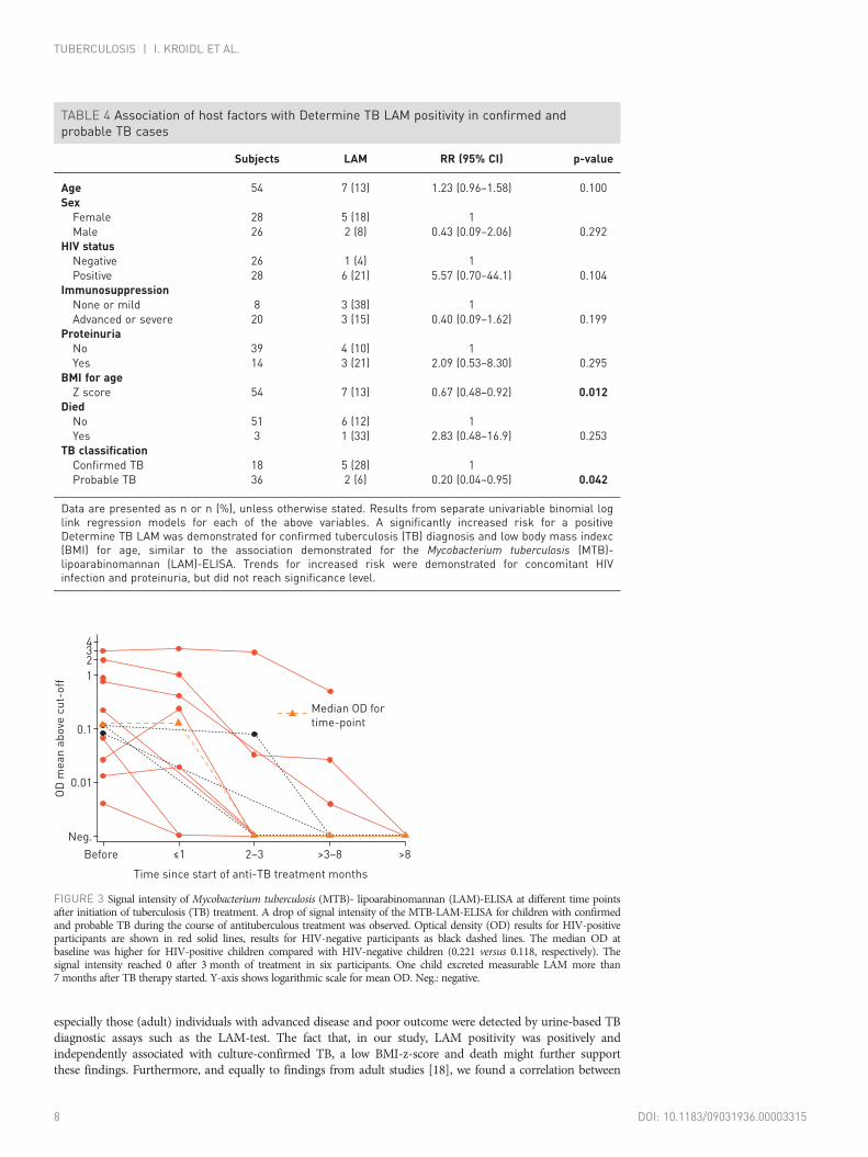
especially those (adult) individuals with advanced disease and poor outcome were detected by urine-based TBdiagnostic assays such as the LAM-test. The fact that, in our study, LAM positivity was positively andindependently associated with culture-confirmed TB, a low BMI-z-score and death might further supportthese findings. Furthermore, and equally to findings from adult studies [18], we found a correlation between
TABLE 4 Association of host factors with Determine TB LAM positivity in confirmed andprobable TB cases
Subjects LAM RR (95% CI) p-value
Age 54 7 (13) 1.23 (0.96–1.58) 0.100SexFemale 28 5 (18) 1Male 26 2 (8) 0.43 (0.09–2.06) 0.292
HIV statusNegative 26 1 (4) 1Positive 28 6 (21) 5.57 (0.70–44.1) 0.104
ImmunosuppressionNone or mild 8 3 (38) 1Advanced or severe 20 3 (15) 0.40 (0.09–1.62) 0.199
ProteinuriaNo 39 4 (10) 1Yes 14 3 (21) 2.09 (0.53–8.30) 0.295
BMI for ageZ score 54 7 (13) 0.67 (0.48–0.92) 0.012
DiedNo 51 6 (12) 1Yes 3 1 (33) 2.83 (0.48–16.9) 0.253
TB classificationConfirmed TB 18 5 (28) 1Probable TB 36 2 (6) 0.20 (0.04–0.95) 0.042
Data are presented as n or n (%), unless otherwise stated. Results from separate univariable binomial loglink regression models for each of the above variables. A significantly increased risk for a positiveDetermine TB LAM was demonstrated for confirmed tuberculosis (TB) diagnosis and low body mass indexc(BMI) for age, similar to the association demonstrated for the Mycobacterium tuberculosis (MTB)-lipoarabinomannan (LAM)-ELISA. Trends for increased risk were demonstrated for concomitant HIVinfection and proteinuria, but did not reach significance level.
234
1
0.1
0.01
Neg.
OD
me
an
ab
ove
cu
t-o
ff
Time since start of anti-TB treatment months
Before ≤1 2–3 >3–8 >8
Median OD for
time-point
FIGURE 3 Signal intensity of Mycobacterium tuberculosis (MTB)- lipoarabinomannan (LAM)-ELISA at different time pointsafter initiation of tuberculosis (TB) treatment. A drop of signal intensity of the MTB-LAM-ELISA for children with confirmedand probable TB during the course of antituberculous treatment was observed. Optical density (OD) results for HIV-positiveparticipants are shown in red solid lines, results for HIV-negative participants as black dashed lines. The median OD atbaseline was higher for HIV-positive children compared with HIV-negative children (0.221 versus 0.118, respectively). Thesignal intensity reached 0 after 3 month of treatment in six participants. One child excreted measurable LAM more than7 months after TB therapy started. Y-axis shows logarithmic scale for mean OD. Neg.: negative.
8 DOI: 10.1183/09031936.00003315
TUBERCULOSIS | I. KROIDL ET AL.

proteinuria and LAM-positivity, indicating that the excretion of LAM might also depend on the condition ofthe kidney membrane. This and other risk factors for LAM-positivity require further investigation in order tobetter define the potential paediatric target group for LAM-based diagnostics in the future.
Our data regarding the overall sensitivity of the Determine TB-LAM strip test, were comparable with thosepreviously published for a paediatric cohort from Cape Town [19]. However, the Cape Town study wasunable to demonstrate an improved LAM sensitivity in TB/HIV co-infected children. Unfortunately, anin-depth comparison of our findings with those of NICOL et al. [19] is hampered by the extremely lowsensitivity of both LAM tests, as well as the poor correlation between the MTB-LAM-ELISA and theDetermine TB-LAM strip test in that study.
Contrary to our previously published data from the same setting in Tanzania [18, 27] and the datapublished by NICOL et al. [19], the specificity of both LAM assays was high in our cohort. Depending onthe hygienic standards and the procedures during sample collection, false-positive LAM results had beenobserved previously, most likely due to contamination of the sample with environmental mycobacteria orother bacteria [27]. In this study, we collected urine samples with great precautions, including washinginstructions and the use of clean containers, in order to avoid false positive results. However, especially inHIV-positive children response to antituberculous treatment should be closely monitored as LAM-basedurine tests cross react with other pathogenic mycobacteria (e.g. Mycobacterium avium complex) and mightinfluence results. Although the sensitivity of both LAM assays was unsatisfactory, and the requirements forcorrect sample collection are high, our data indicate that the use of urine LAM-based tests as rule-in testcould still be advantageous for children in certain settings where sophisticated TB diagnostics are notavailable but strict urine collection criteria can be adhered to.
Furthermore, the fact that a decline of LAM-excretion could be measured during treatment may open up apossibility to monitor antituberculous treatment success in LAM positive children. Although LAM-basedTB diagnosis has the disadvantage that it does not include information on drug resistance, it could behypothesised that ongoing excretion of urine LAM during treatment might provide information oninsufficiently treated drug resistant TB. Larger studies with a long clinical follow up are needed to furtherscrutinise this hypothesis.
One weakness of our study is the relatively low number of 18 confirmed TB cases, which prevented adefinite conclusion of the performance of LAM assays in certain subgroups, such as HIV-negative childrenor in children with HIV co-infection and different levels of immunosuppression. Furthermore, theexclusion of 37 children from the analysis because no urine sample or LAM result was available atbaseline, may have introduced a selection bias. Urine collection was more cumbersome especially inyounger and sicker children as it requires both the child’s and the caregiver’s cooperation and may beaffected by medical causes such as dehydration. However, gathering a urine specimen was not a prioritywhen the main study was designed, and we are confident that the proportion of children with an available
90
100
80
60
70
50
40
20
30
10
0
Se
nsit
ivit
y %
Xp
ert
+ a
ny
LA
M a
ssa
y
Xp
ert
LA
M E
LIS
A
Sm
ea
r +
LA
M s
trip
LA
M s
trip
Sm
ea
r
Any LAM
LAM ELISA
Xpert
LAM strip
Smear
FIGURE 4 Sensitivity of single and combined tuberculosis (TB) diagnostic tests in HIV-infected children. This graph showsthe sensitivity of each diagnostic test alone and the additional gain when combining lipoarabinomannan (LAM) diagnostictests with either smear microscopy or Xpert MTB/RIF-assay in HIV-postive individuals with confirmed TB. LAM-Strip:Determine-TB-LAM; LAM ELISA: MTB LAM-ELISA; Xpert: GeneXpert.
DOI: 10.1183/09031936.00003315 9
TUBERCULOSIS | I. KROIDL ET AL.

sample would be higher if staff could be trained accordingly. We hypothesise that the exclusion of youngchildren with advanced disease might have led rather to an underestimation of LAM-sensitivity, as datafrom our analysis indicate.
In conclusion, both LAM tests demonstrated a reasonable sensitivity in HIV-positive TB-infected children,whereas for HIV-negative children the sensitivity was extremely poor. The combination of LAM tests withother rapid TB diagnostics could substantially improve the detection of TB in HIV co-infected children.This holds promise for earlier TB-diagnosis in children, which might in turn have an impact on childhoodmorbidity and mortality associated with TB. Additionally, clean sample collection methods to achieve ahigh specificity have to be defined in more detail.
AcknowledgementsWe thank the staff at the Active Detection of Active Tuberculosis (ADAT) paediatric TB clinic, the laboratories atNIMR-MMRC (Mbeya, Tanzania) and staff from the paediatric wards at the Mbeya Zonal Referral Hospital for theirdedicated work, as well as the children and their parents or guardians who agreed to participate in this study. Also wewant to thank Alere (Waltham, MA, USA) for providing the Determine TB-LAM Strips and some MTB-LAM-ELISA.
References1 Marais BJ, Pai M. Recent advances in the diagnosis of childhood tuberculosis. Arch Dis Child 2007; 92: 446–452.2 Starke JR. Pediatric tuberculosis: time for a new approach. Tuberculosis (Edinb) 2003; 83: 208–212.3 Zar HJ, Hanslo D, Apolles P, et al. Induced sputum versus gastric lavage for microbiological confirmation of
pulmonary tuberculosis in infants and young children: a prospective study. Lancet 2005; 365: 130–134.4 Marais BJ, Hesseling AC, Gie RP, et al. The bacteriologic yield in children with intrathoracic tuberculosis. Clin
Infect Dis 2006; 42: e69–e71.5 Chintu C, Mudenda V, Lucas S, et al. Lung diseases at necropsy in African children dying from respiratory
illnesses: a descriptive necropsy study. Lancet 2002; 360: 985–990.6 Liu L, Johnson HL, Cousens S, et al. Global, regional, and national causes of child mortality: an updated
systematic analysis for 2010 with time trends since 2000. Lancet 2012; 379: 2151–2161.7 Liu L, Oza S, Hogan D, et al. Global, regional, and national causes of child mortality in 2000-13, with projections
to inform post-2015 priorities: an updated systematic analysis. Lancet 2014.8 Hamasur B, Bruchfeld J, Haile M, et al. Rapid diagnosis of tuberculosis by detection of mycobacterial
lipoarabinomannan in urine. J Microbiol Methods 2001; 45: 41–52.9 Hamasur B, Kallenius G, Svenson SB. A new rapid and simple method for large-scale purification of
mycobacterial lipoarabinomannan. FEMS Immunol Med Microbiol 1999; 24: 11–17.10 Tessema TA, Hamasur B, Bjun G, et al. Diagnostic evaluation of urinary lipoarabinomannan at an Ethiopian
tuberculosis centre. Scand J Infect Dis 2001; 33: 279–284.11 Boehme C, Molokova E, Minja F, et al. Detection of mycobacterial lipoarabinomannan with an antigen-capture
ELISA in unprocessed urine of Tanzanian patients with suspected tuberculosis. Trans R Soc Trop Med Hyg 2005;99: 893–900.
12 Chan J, Fan XD, Hunter SW, et al. Lipoarabinomannan, a possible virulence factor involved in persistence ofMycobacterium tuberculosis within macrophages. Infect Immun 1991; 59: 1755–1761.
13 Peter JG, Cashmore TJ, Meldau R, et al. Diagnostic accuracy of induced sputum LAM ELISA for tuberculosisdiagnosis in sputum-scarce patients. Int J Tuberc Lung Dis 2012; 16: 1108–1112.
14 Peter JG, Theron G, Dheda K. Can Point-of-Care Urine LAM Strip Testing for Tuberculosis Add Value to ClinicalDecision Making in Hospitalised HIV-Infected Persons? PLoS One 2013; 8: e54875.
15 Dheda K, Davids V, Lenders L, et al. Clinical utility of a commercial LAM-ELISA assay for TB diagnosis inHIV-infected patients using urine and sputum samples. PLoS One 2010; 5: e9848.
16 Shah M, Variava E, Holmes CB, et al. Diagnostic accuracy of a urine lipoarabinomannan test for tuberculosis inhospitalized patients in a High HIV prevalence setting. J Acquir Immune Defic Syndr 2009; 52: 145–151.
17 Mutetwa R, Boehme C, Dimairo M, et al. Diagnostic accuracy of commercial urinary lipoarabinomannandetection in African tuberculosis suspects and patients. Int J Tuberc Lung Dis 2009; 13: 1253–1259.
18 Reither K, Saathoff E, Jung J, et al. Low sensitivity of a urine LAM-ELISA in the diagnosis of pulmonarytuberculosis. BMC Infect Dis 2009; 9: 141.
19 Nicol MP, Allen V, Workman L, et al. Urine lipoarabinomannan testing for diagnosis of pulmonary tuberculosisin children: a prospective study. The lancet global health 2014; 2: e278–e284.
20 Rachow A, Clowes P, Saathoff E, et al. Increased and expedited case detection by Xpert MTB/RIF assay inchildhood tuberculosis: a prospective cohort study. Clin Infect Dis 2012; 54: 1388–1396.
21 Graham SM, Ahmed T, Amanullah F, et al. Evaluation of tuberculosis diagnostics in children: 1. Proposed clinicalcase definitions for classification of intrathoracic tuberculosis disease. Consensus from an expert panel. J Infect Dis2012; 205:Suppl. 2, S199–208.
22 Alere. http://alerehiv.com/hiv-comorbidities/procedure/ Date last updated: 2012. Date last accessed: Jan 9, 2015.23 WHO. WHO Immunological Classification of HIV-related diseases in adults and children. http://wwwwhoint/hiv/
pub/guidelines/HIVstaging150307pdf Date last updated: 2007. Date last accessed: Jan 15, 2015.24 Wood R, Racow K, Bekker LG, et al. Lipoarabinomannan in urine during tuberculosis treatment: association with
host and pathogen factors and mycobacteriuria. BMC Infect Dis 2012; 12: 47.25 Lawn SD, Kerkhoff AD, Vogt M, et al. Diagnostic accuracy of a low-cost, urine antigen, point-of-care screening
assay for HIV-associated pulmonary tuberculosis before antiretroviral therapy: a descriptive study. Lancet InfectDis 2012; 12: 201–209.
26 Lawn SD, Kerkhoff AD, Vogt M, et al. HIV-associated tuberculosis: relationship between disease severity and thesensitivity of new sputum-based and urine-based diagnostic assays. BMC Med 2013; 11: 231.
27 Kroidl I, Clowes P, Mwakyelu J, et al. Reasons for false-positive lipoarabinomannan ELISA results in a Tanzanianpopulation. Scand J Infect Dis 2014; 46: 144–148.
10 DOI: 10.1183/09031936.00003315
TUBERCULOSIS | I. KROIDL ET AL.
Copyright © 2022 FDOKUMEN