Neurological and neurourological complications of electrical ...
Upload
independentCategory
view
4download
0
B R A I N R E S E A R C H 1 2 6 0 ( 2 0 0 9 ) 5 5 – 6 4
ava i l ab l e a t www.sc i enced i r ec t . com
www.e l sev i e r. com/ l oca te /b ra in res
Research Report
Pentoxifylline treatment improves neurological andneurochemical deficits in rats subjected to transientbrain ischemia
Renata de Barros Brunoa, Tiago Fontenele Marquesa, Tiago Melo Teixeira Batistaa,Júlio César Sá Silveira Limaa, Karla Gleice de Arrudaa, Priscili Fortaleza Souza Fiúza Limaa,Nilton da Silva Santosa, Geanne M.A. Cunhab,Helio Vitorino Nobre Vitorb, Glauce Socorro de Barros Vianaa,b,⁎aDepartment of Biophysiology and Pharmacology, Faculty of Medicine of Juazeiro do Norte—FMJ, Av. Tenente Raimundo Rocha s/n,Juazeiro do Norte, 63040-000, BrazilbDepartment of Physiology and Pharmacology, Federal University of Ceará—UFC, Rua Coronel Nunes de Melo, 1127, Fortaleza,60430-270, Brazil
A R T I C L E I N F O
⁎ Corresponding author. Rua Barbosa de FreitE-mail address: [email protected] (G.S.B.
0006-8993/$ – see front matter © 2009 Elsevidoi:10.1016/j.brainres.2008.12.064
A B S T R A C T
Article history:Accepted 17 December 2008Available online 7 January 2009
The possible neuroprotector effects of pentoxifylline (P), a methylxanthine derivative andphosphodiesterase inhibitor, were studied on male Wistar rats subjected to a model oftransient brain ischemia. One groupwas treated, 1 h before ischemia (ISC) and 1 h after, withpentoxifylline (P), 50 and 100 mg/kg, i.p. (ISC+P). The other groups were ischemic, ISC; shamoperated, SO; P100; SO+P100; and normal controls, N. One week after ischemia, the animalswere submitted to the open field, passive avoidance, and water maze tests for evaluatinglocomotor activity and cognitive deficits, as well as for scintillographic (SPECT)measurements. After these procedures, they were sacrificed and their brain dissectedunder ice, for histological studies as well as striatal dopamine (DA) and DOPACmeasurements by HPLC. In the open field test, ischemia increased locomotor activity, ascompared to N, SO, P100 and SO+P100. This parameter was not significantly reversed after Ptreatment (ISC+P50 or ISC+P100). Grooming behavior was also increased in the ISC group, ascompared to all other ones, although only statistically different as compared to the P100group. The P treatment (ISC+P50 and ISC+P100) brought these values close to normality. Inthe passive avoidance test, ischemia significantly impaired the short term memory, ascompared to the N group, as well as scopolamine (SCP) used as a positive control. In the longterm memory, similar effects were observed in the ISC and SCP groups that significantlyimpairedmemory consolidation. P treatment completely reversed the effects observed afterischemia (ISC+P50 and ISC+P100) on the short as well as on the long term memory, andvalues were similar to those of the P100 group (which increased both parameters, as relatedto the N group). In the water maze, the ISC group was not different from the N, SO and SO+P100 groups. However, ISC+P50, ISC+P100 and P100 significantly improved the spatialmemory, as related to all other groups. SPECT data showed that ischemia produced a lower
Keywords:PentoxifyllineBrain ischemiaCognitive deficitNeuroprotectionStriatal monoamine
as, 130 apt. 1100 Fortaleza, 60170-020, Brazil. Fax: +55 88 2101 900.Viana).
er B.V. All rights reserved.

56 B R A I N R E S E A R C H 1 2 6 0 ( 2 0 0 9 ) 5 5 – 6 4
but significant decrease in brain optical densities, and P brought values close to those seenin the N group. Furthermore, P treatment of the ischemic group (ISC+P50) increased thenumber of viable hippocampal CA3 neurons, as compared to the ISC group. While ischemiasignificantly decreased DA levels, the P treatment of ischemic animals brought those valuesclose to normality. In conclusion, P counteracted neurological and neurochemical changesseen after brain ischemia, in rats. The TNF-α inhibition and anti-inflammatory actions of Pcould be responsible, at least in part, for the neuroprotection afforded by the drug.
© 2009 Elsevier B.V. All rights reserved.
Table 1 – Scintillographic data (SPECT) from brain areas ofnormal control (N), ischemic (ISC), and pentoxifylline-treated ischemic (ISC+P100) rats
Group Integrated area optical density
Normal control (N) (9) 6.63±0.096Ischemic (ISC) (8) 5.90±0.229a
ISC+P100 (10) 6.54±0.129b
Values are means±SEM of the number of animals in parentheses.Animals were treated with P (100 mg/kg, i.p.), 1 h before and 1 hafter ischemia. Scintillography was performed 4 to 6 h after ischemia.a vs. N, q=4.596, p<0.01;b vs. ISC, q=4.153, p<0.01.
1. Introduction
Pentoxifylline (P) is a methylxanthine derivative and anonspecific type 4 phosphodiesterase inhibitor, clinicallyused in the treatment of lower extremity claudication. Themechanisms underlying its beneficial effects seem to berelated to alterations in cellular functions and to theimprovement of microcirculatory perfusion in both peripheraland cerebral vascular beds (Seiffge, 1997; Windmeier andGressner, 1997). P is termed a hemorrheologic modifier for itseffects decreasing the deformability of red blood cells. In vitroas well as in vivo experiments indicated an additional the-rapeutic potential for P as an anti-inflammatory and immu-nomodulator (Teixeira et al., 1997; Laurat et al., 2001; Haddadet al., 2002).
Ischemia results in rapid loss of high-energy phosphatecompounds and generalized depolarization, which inducesrelease of glutamate and opening of both voltage-dependentand glutamate regulated calcium channels. The brain isparticularly vulnerable to ischemia. Brain ischemia andreperfusion induce an inflammatory response which mayexacerbate initial levels of tissue injury. Elevation of TNF-alpha and IL-1beta occurs as early as 1 h after ischemiainduction (Lee et al., 2000).
Furthermore, brain injury results in the uncontrolledrelease of many neurotransmitters, including glutamate anddopamine. Both are neurotoxic in high concentrations, andthemechanism of action is possibly due to the oxidative stresscaused by reactive oxygen species generation (Hoyt et al.,1997). There is evidence indicating that brain ischemiasignificantly increased locomotor activity which seems to berelated to abnormalities in dopaminergic functions, particu-larly the dopamine D2 receptor (Araki et al., 1999). In addition,the impairment of spatial cognitive performance in rats withchronic focal cerebral ischemia is, in part, associated withdamage to the parietal cortex, as well as to damage to thecaudate-putamen region (Yonemori et al., 1999).
The P anti-inflammatory properties include the inhibitionof TNF-alpha production (Gutierrez et al., 2006) that seems tobe due to reduced TNF protein levels by the inhibition of TNFmRNA transcription (Lin et al., 2004). TNF-alpha is expressedin the ischemic brain (Navashiro et al., 1997), and is known torapidly upregulate in the brain after injury (Barone et al., 1997).This last study demonstrated that the exogenous TNF-alphaexacerbates focal ischemic injury, and the blockade of theendogenous TNF-alpha is neuroprotective. Furthermore, TNF-alpha inhibition may represent a novel pharmacologicalstrategy for the treatment of ischemic stroke.
Previous studies reported that P has a neuroprotectiveeffect in experimental models of global as well as focal
cerebral ischemia. Thus, P treatment has been shown toimprove recovery of the cerebral electrical function in dogs,after transient cerebral global ischemia, by a mechanism thatdoes not involve improvement of cerebral blood flow or globaloxygen consumption (Toung et al., 1994). Furthermore, thepretreatment with P decreased the incidence and severity ofhypoxic-ischemic injury in immature rat brain, by attenuatingthe expression of IL-1beta and TNF-alpha genes (Kim et al.,1999). P also afforded neuroprotection in a rat model ofcerebral ischemia, such as occlusion of the ipsilateral commoncarotid and middle cerebral arteries (Evans et al., 1999).
The objectives of the present work were to investigate thepossible neuroprotective effects of pentoxifylline on a modelof global transient ischemia, by evaluating the animal'slocomotor activity and cognitive functions (acquisition andlearning processes, and spatial memory). Since it is largelyaccepted that brain ischemia causes striatal damage withsubsequent deterioration of sensorimotor and cognitive func-tions that may be mediated by the dopaminergic system(Borlongan et al., 1995), striatal DA and DOPACmeasurementswere performed. Besides, it is recognized that the in vivo SPECTimaging is a powerful technique for studying cerebral bloodflow deficits in brain ischemia (Seo et al., 2007; Lappalainenet al., 2008), what led us to carry out scintillographic andhistological studies on ischemic groups, treated or untreatedwith P.
2. Results
2.1. Scintillography data
Our results showed that brain ischemia (ISC) produced a lowerbut significant (F2,26=3.462) decrease (11%) in the integratedbrain area expressed as optical densities (Table 1), as

Fig. 1 – Brain ischemia effects in the absence (ISC) andpresence of pentoxifylline treatment (ISC+P), as evaluated bythe open field test in rats. Brain ischemia was performed bythe bilateral occlusion of both common carotid arteries, asdescribed in Experimental procedures. (A) Locomotor activity(LA), as evaluated by the number of squares crossed; (B)Rearing behavior (R), and C. Grooming behavior (G). Valuesare means±SEM of 10 to 25 animals from (N), sham-operated(SO), SO+P100, ischemic (ISC), ISC+P50 and ISC+P100 groups.LA-ISC: a. vs. N, p<0.05; b. vs. SO, p<0.01; c. vs. SO+P100,p<0.001; d. vs. P100, p<0.001; ISC+P50: e. vs. N, p<0.05; f.vs. SO, p<0.05; g. vs. SO+P100, p<0.001; h. vs. P100, p<0.01;ISC+P100: i. vs. SO+P100, p<0.01; j. vs. P100, p<0.01. G-ISC:a. vs. N, p<0.01; b. vs. SO, p<0.001; c. vs. SO+P100, p<0.01;d. vs. P100, p<0.001; e. vs. ISC+P50, p<0.01; f. vs. ISC+P100,p<0.001.
Fig. 2 – Pentoxifylline (P, 50 or 100 mg/kg, i.p.) reversed theeffects of ischemia on memory acquisition (short termmemory, latency at 15 min) and consolidation (long termmemory, latency at 24 h), as evaluated by the passiveavoidance test, in rats. Values are means±SEM from 12 to 27animals from normal (N), sham-operated (SO), SO+P100,ischemic (ISC), scopolamine (SCP), ISC+P50 and. ISC+P100groups. Brain ischemia was performed by the bilateralocclusion of both common carotid arteries, as described inExperimental procedures. (A) Short termmemory—ISC: a. vs.N, p<0.05; b. vs. P100, p<0.01; c. vs. ISC+P50, p<0.01; d. vs.ISC+P100, p<0.01; SCP: a. vs. N, p<0.001; b. vs. SO, p<0.001;c. vs. SO+P100, p<0.001; d. vs. P100, p<0.001; e. vs. ISC+P50,p<0.001; f. vs. ISC+P100, p<0.001. (B) Long termmemory—ISC: a. vs. N, p<0.05; b. vs. SO, p<0.01; c. vs. SO+P100, p<0.01; d. vs. P100, p<0.05; e. vs. ISC+P50, p<0.01;f. vs. ISC+P100, p<0.01; SCP: a. vs. N, p<0.01; b. vs. SO,p<0.01; c. vs. SO+P100, p<0.01; d. vs. P100, p<0.01; e. vs. ISC+P50, p<0.01; f. vs. ISC+P100, p<0.001.
57B R A I N R E S E A R C H 1 2 6 0 ( 2 0 0 9 ) 5 5 – 6 4
compared to values observed in normal control (N) animals(ISC: 5.90±0.229, q=4.596, p<0.01; N: 6.63±0.096). On the otherhand, P (100mg/kg, i.p.) administered to ischemic rats broughtvalues close to those shown in normal animals, suggesting analmost total recovery of the pathological events occurringduring brain ischemia and reperfusion (ISC+P100: 6.54±0.129,q=4.153, p<0.01).

58 B R A I N R E S E A R C H 1 2 6 0 ( 2 0 0 9 ) 5 5 – 6 4
2.2. Behavioral tests
The open field test is designed tomeasure behavioral responses,such as locomotor activity, hyperactivity, and exploratorybehaviors. Our results showed that brain ischemia (ISC) sig-nificantly increased in 62 (p<0.05), 44 (p<0.01), 83 (p<0.01) and77% (p<0.001) the locomotor activity, expressed as number ofsquares crossed, as compared to normal controls (N), sham-operated (SO), SO+P100 and P100 groups, respectively. The Ptreatment of ischemic animals did not reverse the ISC effect.Grooming (G) but not rearing was also significantly increased in124 (p<0.01), 93 (p<0.01), 98 (p<0.01), and 146% (p<0.001) afterischemia, as compared to the N, SO, SO+P100 and P100 groups,respectively. However, the P treatment of the ISC group broughtgrooming behavior to values close and not statistically differentfrom normality. Except for a tendency for decreasing in rearingbehavior in theP100group,nosignificantalterationwasobservedin this parameter in any other group studied (Figs. 1 A–C).
In the passive avoidance test, largely used for the screeningof drugs affecting learning and memory, ischemia signifi-cantly impaired the short term memory by decreasing thelatency time in 31 (p<0.05), 38 (p<0.01), 37 (p<0.01) and 38%(p<0.01), as compared to the N, P100, ISC+P50 and ISC+ P100groups, respectively. Ischemia also impaired in 41, 52, 51 and46% the long termmemory as compared to N, SO, SO+P100 andP100 groups respectively. Similarly, scopolamine (SCP) used asa positive control also significantly impaired both short (62, 60,59 and 66% decreases in latency times) and long termmemory(35, 47, 46 and 40% decreases in latency times), as compared tothe N, SO, SO+P100 and P100 groups, respectively. The treat-ment of the ischemic groups with P completely reversed these
Fig. 3 – Pentoxifylline effects in the absence (P100) andpresence (ISC+P50 or ISC+P100) of brain ischemia on spatialmemory, as evaluated by the water maze task in rats. Brainischemia was performed by the bilateral occlusion of bothcommon carotid arteries, as described in Experimentalprocedures. Values are means±SEM from 10 to 28 animalsfrom normal (N), sham-operated (SO), SO+P100, ischemic(ISC, ISC+P50 and. ISC+P100) groups. P100: a. vs. N, p<0.01; b.vs. SO, p<0.001; c. vs. ISC, p<0.001. SO+P100: a. vs. N, p<0.05;b. vs. SO, p<0.01; c. vs. ISC, p<0.01. ISC+P50: a. vs. N, p<0.01;b. vs. SO, p<0.001; c. vs. ISC, p<0.001. ISC+P100: a. vs. N,p<0.01; b. vs. SO, p<0.001; c. vs. ISC, p<0.001.
Fig. 4 – Pentoxifylline (50 mg/kg, i.p.) effects, in the presence(ISC+P50) and absence (P50) of brain ischemia (ISC) in rats, onthe number of viable cells in (A) dentate gyrus (DG), (B) CA1hippocampus and (C) Parietal cortex in rats, as evaluated bythe Nissl stain (cresyl violet). Animals from all groups weresacrificed 1 week after ischemia, for brain dissection underice. Data from 3 to 4 animals were used in each case. A (DG):*ISC vs. SO and vs. P50, p<0.05; B (CA1): *ISC vs. SO and vs.P50, p<0.001 and vs. ISC+P50, p<0.05; and C (Parietal cortex):*ISC vs. P50 and vs. SO, p<0.001.
effects in both short and long term memories. Interestingly,the P treatment had an effect of its own, and latency times forboth learning and memory consolidation were even higherthan those observed in N and SO groups (Figs. 2 A, B).
In the water maze task (Fig. 3), used to evaluate spatialmemory expressed as time (s) to find the hidden platform,data from ischemic rats (ISC) were not significantly differentfrom those of N and SO groups. The SO+P100 group

59B R A I N R E S E A R C H 1 2 6 0 ( 2 0 0 9 ) 5 5 – 6 4
significantly improved the spatial memory and effects weresignificant different from those of N, SO and ISC groups.Furthermore, P treatment (P100 group) significantly decreasedfrom 67 to 72% the time to find the platform, as compared to N,SO and ISC groups, indicating that pentoxifylline had an effectof its own. Furthermore, P treatment of ischemic animalssignificantly improved spatial memory, as observed by thedecreased time to find the platform, and the values werebrought towards those of the P100 group. These resultssuggest that brain mechanisms of spatial orientation weresignificantly and drastically improved in ischemic rats, after Ptreatment.
Fig. 5 – Pentoxifylline (50 mg/kg, i.p.) effects, in the presence (ISCnumber of viable cells. (A) A representativemicrophotography of tevaluated by the Nissl stain (cresyl violet). Animals from all grouunder ice. Data from 3 to 4 animals were used in each case. *ISC
2.3. Histological studies
Neuronal cell counts from brain sections stained with cresylviolet revealed that brain ischemia resulted in moderate butsignificant damage to the cortex, and CA1 and dentate gyrus(DG) regions of the hippocampus (34% cell loss) (Figs. 4 A, B).The pentoxifylline treatment (P50) of ischemic animals (ISC)reduced brain injury relatively to controls (10% cell loss) in theCA3 region, but not in the cortex and DG regions. An increasein the number of viable cells was also observed in thehippocampus CA1 region, as related to the ISC group. Fig. 5shows the neuroprotection (indicated by cell counts and
+P50) and absence (P50) of brain ischemia (ISC) in rats, on thehe CA3 region; (B) Number of viable cells in the CA3 region, asps were sacrificed 1 week after ischemia, for brain dissectionvs. SO, vs. P50 and vs. ISC+P50, p<0.001.
60 B R A I N R E S E A R C H 1 2 6 0 ( 2 0 0 9 ) 5 5 – 6 4
representative microphotographies) of the hippocampal CA3region in ischemic animals, after P treatment (ISC+P50), ascompared to ISC, P50 and SO groups.
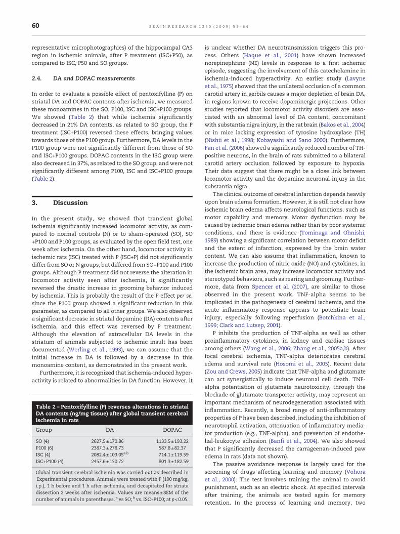
2.4. DA and DOPAC measurements
In order to evaluate a possible effect of pentoxifylline (P) onstriatal DA and DOPAC contents after ischemia, we measuredthese monoamines in the SO, P100, ISC and ISC+P100 groups.We showed (Table 2) that while ischemia significantlydecreased in 21% DA contents, as related to SO group, the Ptreatment (ISC+P100) reversed these effects, bringing valuestowards those of the P100 group. Furthermore, DA levels in theP100 group were not significantly different from those of SOand ISC+P100 groups. DOPAC contents in the ISC group werealso decreased in 37%, as related to the SO group, andwere notsignificantly different among P100, ISC and ISC+P100 groups(Table 2).
3. Discussion
In the present study, we showed that transient globalischemia significantly increased locomotor activity, as com-pared to normal controls (N) or to sham-operated (SO), SO+P100 and P100 groups, as evaluated by the open field test, oneweek after ischemia. On the other hand, locomotor activity inischemic rats (ISC) treated with P (ISC+P) did not significantlydiffer from SO or N groups, but differed from SO+P100 and P100groups. Although P treatment did not reverse the alteration inlocomotor activity seen after ischemia, it significantlyreversed the drastic increase in grooming behavior inducedby ischemia. This is probably the result of the P effect per se,since the P100 group showed a significant reduction in thisparameter, as compared to all other groups. We also observeda significant decrease in striatal dopamine (DA) contents afterischemia, and this effect was reversed by P treatment.Although the elevation of extracellular DA levels in thestriatum of animals subjected to ischemic insult has beendocumented (Werling et al., 1993), we can assume that theinitial increase in DA is followed by a decrease in thismonoamine content, as demonstrated in the present work.
Furthermore, it is recognized that ischemia-induced hyper-activity is related to abnormalities in DA function. However, it
Table 2 – Pentoxifylline (P) reverses alterations in striatalDA contents (ng/mg tissue) after global transient cerebralischemia in rats
Group DA DOPAC
SO (4) 2627.5±170.86 1133.5±193.22P100 (6) 2387.3±278.73 587.8±82.37ISC (4) 2082.4±103.05a,b 714.1±119.59ISC+P100 (4) 2457.6±130.72 801.3±182.59
Global transient cerebral ischemia was carried out as described inExperimental procedures. Animals were treated with P (100 mg/kg,i.p.), 1 h before and 1 h after ischemia, and decapitated for striatadissection 2 weeks after ischemia. Values are means±SEM of thenumber of animals in parentheses. a vs SO; b vs. ISC+P100; at p<0.05.
is unclear whether DA neurotransmission triggers this pro-cess. Others (Haque et al., 2001) have shown increasednorepinephrine (NE) levels in response to a first ischemicepisode, suggesting the involvement of this catecholamine inischemia-induced hyperactivity. An earlier study (Lavyneet al., 1975) showed that the unilateral occlusion of a commoncarotid artery in gerbils causes a major depletion of brain DA,in regions known to receive dopaminergic projections. Otherstudies reported that locomotor activity disorders are asso-ciated with an abnormal level of DA content, concomitantwith substantia nigra injury, in the rat brain (Bakos et al., 2004)or in mice lacking expression of tyrosine hydroxylase (TH)(Nishii et al., 1998; Kobayashi and Sano 2000). Furthermore,Fan et al. (2006) showed a significantly reduced number of TH-positive neurons, in the brain of rats submitted to a bilateralcarotid artery occlusion followed by exposure to hypoxia.Their data suggest that there might be a close link betweenlocomotor activity and the dopamine neuronal injury in thesubstantia nigra.
The clinical outcome of cerebral infarction depends heavilyupon brain edema formation. However, it is still not clear howischemic brain edema affects neurological functions, such asmotor capability and memory. Motor dysfunction may becaused by ischemic brain edema rather than by poor systemicconditions, and there is evidence (Tominaga and Ohnishi,1989) showing a significant correlation between motor deficitand the extent of infarction, expressed by the brain watercontent. We can also assume that inflammation, known toincrease the production of nitric oxide (NO) and cytokines, inthe ischemic brain area, may increase locomotor activity andstereotyped behaviors, such as rearing and grooming. Further-more, data from Spencer et al. (2007), are similar to thoseobserved in the present work. TNF-alpha seems to beimplicated in the pathogenesis of cerebral ischemia, and theacute inflammatory response appears to potentiate braininjury, especially following reperfusion (Botchkina et al.,1999; Clark and Lutsep, 2001).
P inhibits the production of TNF-alpha as well as otherproinflammatory cytokines, in kidney and cardiac tissuesamong others (Wang et al., 2006; Zhang et al., 2005a,b). Afterfocal cerebral ischemia, TNF-alpha deteriorates cerebraledema and survival rate (Hosomi et al., 2005). Recent data(Zou and Crews, 2005) indicate that TNF-alpha and glutamatecan act synergistically to induce neuronal cell death. TNF-alpha potentiation of glutamate neurotoxicity, through theblockade of glutamate transporter activity, may represent animportant mechanism of neurodegeneration associated withinflammation. Recently, a broad range of anti-inflammatoryproperties of P have been described, including the inhibition ofneurotrophil activation, attenuation of inflammatory media-tor production (e.g., TNF-alpha), and prevention of endothe-lial-leukocyte adhesion (Banfi et al., 2004). We also showedthat P significantly decreased the carrageenan-induced pawedema in rats (data not shown).
The passive avoidance response is largely used for thescreening of drugs affecting learning and memory (Vohoraet al., 2000). The test involves training the animal to avoidpunishment, such as an electric shock. At specified intervalsafter training, the animals are tested again for memoryretention. In the process of learning and memory, two

61B R A I N R E S E A R C H 1 2 6 0 ( 2 0 0 9 ) 5 5 – 6 4
important stages are considered, i.e., acquisition and con-solidation, expressed in the test as short and long termmemories (Das et al., 2003). As expected, we observed that, inthe passive avoidance test, the ISC group showed a significantimpairment in short as well as long term memories, ascompared to ISC+P groups, indicating that P treatmentcompletely reversed the ischemia effect. In both parameters,P presented an effect of its own, improving learning andmemory consolidation processes.
It is known that the transient middle cerebral arteryocclusion (MCAO) model in rats induces cognitive deficitsthat appear to remain fairly stable (Hattori et al., 2000). Thepassive avoidance task has often and consistently been usedto reveal MCAO-induced cognitive deficits (Hirakawa et al.,1994; Yonemori et al., 1999). In the present study deficits in theISC groupwere observed in both short termmemory (learning)as well as in long term memory (retention), and much betterresults were demonstrated in the water maze task in ISC+Pgroups. In this test, similar values (time to find the platform)were observed in N, SO and ISC groups. However, a substantialtime reductionwas seen in ISC+P rats, suggesting an improvedtest performance and a neuroprotective effect of P.
Effects of P, examined on the cerebral blood flow of patientswith cerebrovascular disease, showed that it significantlyincreased the cerebral blood flow, 2 h after the administrationof 400 and 800 mg (Bowton et al., 1989). Previously, wedemonstrated (Cunha et al., 2000) that P (50, 100 and 200 mg/kg, i.p.) significantly improved learning and memory, in ratswith hippocampal lesions induced by glutamate, as evaluatedby the T-maze, passive avoidance, and water maze tasks, ascompared to the glutamate-lesioned group without P treat-ment. In that protocol, rats were submitted to stereotaxicsurgery to induce hippocampal glutamate lesions and, imme-diately after surgery, the animals were treated with P for6 days. Furthermore, a recent report (Vakili and Zahedi, 2007)indicated that P administration, at least 3 h post-transientfocal cerebral ischemia, reduces cortical brain damage in rats.
An earlier study (Matsumoto et al., 1984) showed signifi-cant decrease of monoamine concentration in several brainareas, after 1 h bilateral carotid occlusion. On the other hand,others (Kuhmonen et al., 1997) have shown that cerebralischemia, induced by bilateral carotid occlusion for 5 min,leads to a massive release of norepinephrine, associated withneuronal brain death. Long term brain ischemia, induced byboth sides occlusion of common carotid arteries, has beendemonstrated to result in 55% mortality of experimental rats,and those that survived were characterized by partial sup-pression of orientation in the T-maze test (Gallant et al., 2000).Rogers et al. (1997), demonstrated that volumes of infarction inthe cerebral hemisphere, cerebral cortex, and striatum, as wellas brain swelling, are highly correlated with functionaldeficits, as evaluated by the rota-rod and grid walking tests,and by neurological score, 24 h after MCAO.
We also showed that brain ischemia significantlydecreased brain optical density, as measured by scintillogra-phy (SPECT), and that the P treatment (ISC+P) brought valuesclose to those seen in normal animals. SPECT brain perfusionstudies have been playing an important role in understandingthe pathophysiology involving brain injury. It is highlysensitive for detecting regional cerebral blood flow distur-
bances in acute brain injury. In conclusion, we showed that Pcounteracts some of the neurological changes seen after brainischemia, such as locomotor activity and stereotyped beha-vior, as well as cognitive deficits, as evaluated in the model oftransient global ischemia, in rats. The role of TNF-alpha as animmune mediator is largely accepted, but its function in thebrain is still unclear. It has been shown that the elevation ofTNF-alpha is a hallmark of acute and chronic neuroinflamma-tion, including ischemic stroke, Alzheimer's disease andParkinson's disease, among other neurodegenerative condi-tions (McCoy and Tansey, 2008). Recently (Bermpohl et al.,2007), increased levels of TNF-alpha were detected aftertraumatic brain injury, and mice deficient in TNF-alpha hadimproved motor performance and spatial memory acquisi-tion, and decreased brain lesion size, as assessed by amodel ofcontrolled cortical impact.
Our data also agree with another study suggesting that Preduces cerebral injury and preserves neurologic functions intransient global ischemia, in rats (Sirin et al., 1998). Inflam-matory mediators are implicated in the pathogenesis of brainischemic injury. P is a phosphodiesterase inhibitor known toinhibit TNF-alpha and PAF, and phosphodiesterase inhibitionhas been proposed as an effective strategy to decrease theseverity of neonatal hypoxic-ischemic brain injury (Eun et al.,2000). Besides, a report indicated that the P treatment dose-dependently prevents the occurrence of spontaneous braindamage, by reducing inflammatory events (Banfi et al., 2004)what might be the case in the present study. P is known toreduce the activation of NF-Kappa B and the production ofTNF-alpha (Ji et al., 2004; Zhang et al., 2005a, 2005 b). Theseanti-inflammatory related events, associated with a possibleaction of P on oxidative damage (Davila-Esqueda and Marti-nez-Morales, 2004; Davila-Esqueda et al., 2005, Radfar et al.,2005), could be responsible for the neuroprotection afforded bythis drug, leading to a decrease in neurologic deficits and animprovement in dopaminergic neurotransmission as seen inthe present study.
4. Experimental procedures
4.1. Drugs
Pentoxifylline, P (400 mg/ml Trental ampoules produced byAventis Pharmaceutical, USA) was purchased from Hoescht,Brazil; sodium thiopental, from Abbott, Brazil. Scopolaminesulfate, dopamine, 3, 4 dihydroxyphenylacetic acid (DOPAC),octanesulfonic acid, acetonitrile and tetrahydrofuran werepurchased from Sigma Chemical Co. (USA). All other drugswere of analytical grade. P was administered as such (directlyfrom the ampoule).
4.2. Animals
Experimental protocol: Male Wistar rats (250 g) from theAnimal House of the Faculty of Medicine of Juazeiro do Norte(FMJ)wereused. Animalswereanesthetizedwithpentobarbital(40mg/kg, i.p.) and submitted to transient global ischemia thatconsisted in clamping bilateral common carotid arteries for60 min. After this time, clips were removed and the flow

62 B R A I N R E S E A R C H 1 2 6 0 ( 2 0 0 9 ) 5 5 – 6 4
reestablished. Animals breathed room air spontaneously, andtheir rectal temperaturewasmaintained at about 36.7 °Cwith aheat lamp, during the experiment. Ischemic groups (ISC) weretreated with distilled water or with P (50 or 100 mg/kg, i.p.), 1 hbefore and1hafter the ischemia (P50+ISC; P100+ISC), accordingto previous studies (Cunha et al., 2000, Vakili andZahedi, 2007).Sham-operated (SO) rats were subjected to the same surgicalprocedure except for arterial clamping. Controls (NC) receiveddistilled water. At the 2nd day after ischemia, the training forthe water maze test started until the next day. The test itselfwas performed, at the 6th day after ischemia, followed by thepassive avoidance test at the 7th day. In the case of behavioralstudies, the following groups were used: normal controls (N);P100 (animals treatedwith two doses of pentoxifylline, 100mg/kg, i.p., administered2h apart, in order tomimic the time spentwith ISC groups); SO; SO+P100; ISC; ISC+P50 and ISC+P100, andthe number of animals ranged from 10 to 30 per group. For thescintillographic studies and in order to see early brainalterations, the experiments were performed 4 to 6 h afterischemia, while in the case of the histological studies animalsfrom each group were sacrificed after the behavioral tests(1 week after ischemia). In these two experiments (scintillo-graphic and histological studies) other groups of animals wereused. Experiments were carried out according to the Guide forthe Care andUse of Laboratory Animals of theU.S. Departmentof Health and Human Services, Washington, DC (1985).
4.3. Scintillographic measurements by SPECT(single photon emission computerized tomography)
Four to 6 hours after the ischemic procedure, rats wereanesthetized with thiopental (40 mg/kg, i.p.) and immediatelyinjected with 3 mCi of the radionuclide ECD-99m Tc (ethylene-dicysteine diethyl ester). The animals were then submitted toscanning in a gamma camera, in order to obtain the cerebralspectra. Axial sections images of brain areas studied werethen analyzed. These analyses consisted in limiting an areafrom each axial section and reading it, with the imageprocessing software (Scion Image or Image-J, USA). Resultswere expressed as means of optical densities of integratedbrain areas. Values of the groups studied (untreated ischemicor ischemic treated with pentoxifylline, P) were comparedwith sham-operated (SO) and normal control (NC) rats.
4.4. Behavioral testing
Open field test: The open field area was made of wood(50×50 cm) with 20 cm height walls, and the floor divided intofour squares of equal areas. The open field was used toevaluate the exploratory activity of the animal for 5 min. Theobserved parameters were: number of squares crossed (withthe four paws) and numbers of grooming (stereotypedbehavior) and rearing (vertical exploratory activity).
Passive avoidance test: The apparatus (48×22×22 cm),from Ugo Basili, Italy, was made of acrylic and divided into alight and a dark compartment. The two compartments wereseparated by a sliding door. Themethodwas that described byImanishi et al., 1997. The rat was initially placed into the lightcompartment for 1 min (habituation). After that, the animalwas removed from the apparatus, and 30 s latter replaced in
the light compartment with the sliding door raised. Once therat moved completely into the dark compartment (all fourpaws on the shock grid floor), the sliding door was lowereddown and a foot shock (1 mA, 1 s) was delivered through thegrid floor. Then, the rat was immediately removed from thedark compartment. The procedure was repeated 15 min latterto register the time each animal took to enter the darkcompartment, in order to receive a shock again. Thisevaluated the 15 min passive avoidance response or acquisi-tion. After 24 h, the procedure was repeated for a second time,for the evaluation of memory consolidation (24 h passiveavoidance response). The cut-off time was established in300 s. Scopolamine (0.5 mg/kg, i.p.) was used as a positivecontrol.
Water maze test: The water maze consisted of a blackcircular pool (1.7 m diameter and 1 m height), with water(0.59 m deep) at 22 °C, inside a controlled temperature roomwith visual cues on the walls, at one of four equally spacedlocations: North (N), East (E), South (S) and West (W). The poolwas divided into 4 quadrants: NW. NE, SE and SW. After thetreatment, each animal was submitted to two trials in thepool, for the next three days. In these trials, the animals wereplaced into the pool, facing thewall, and had amaximum timeof 54 s to find the submersed platform. The animal stayed onthe platform for 10 s. Animals that did not find the platform atthe end of 54 s were placed on it for 10 s. After this time, theanimal was removed from the pool for 30 s, and the procedurewas repeated (6 times for each animal). The procedure wasrepeated again 24 h later (pre-test). After 48 h of the pre-test,the procedure was repeated oncemore for evaluating the timeneeded by the animal to find the platform.
4.5. Histological examination
Mounted slides were stained with 1% Cresyl Violet. Compar-isonsof levels of neuronal viable cellweredetermined fromthenumber of Nissl stained cells and the number of non stainedcells in the hippocampal CA1, CA3, cortex and dentate gyrus(DG) regions. The numbers of cells in each groupwere countedseparately in hippocampal areas per section. An area wasdefined as 50 μm (width)×100 μm (length), with the neuronalcell layer of the hippocampal CA1, CA2, CA3, and DG region setup parallel to the major axis. Mounted slides were examinedunder a light microscope (Nikon Microscope ECLIPSE E600W,Tokyo, Japan) and microphotographed using a digital camera(Microscope Digital Camera DP70, Tokyo, Japan). The micro-photographies (×40 magnifications) were analyzed by a blindobserver to different groups.
4.6. Determination of dopamine (DA) and3,4 dihydroxyphenylacetic acid (DOPAC) contents
For the measurement of DA and DOPAC contents, the striatalbrain tissue was identified and dissected on ice, according tothe Paxinos and Watson's (1986) stereotaxic atlas of the ratbrain, one week after ischemia, and used for preparing 10%homogenates. Homogenateswere sonicated in 0.1MHClO4, for30 s, centrifuged at 4 °C for 15 min at 15,000 rpm, and thesupernatantwas filtered (0.2 μm,Millipore). A 20 μl samplewasthen injected into a high-performance liquid chromatograph

63B R A I N R E S E A R C H 1 2 6 0 ( 2 0 0 9 ) 5 5 – 6 4
(HPLC) column. The mobile phase was 0.163 M citric acid, pH3.0, containing 0.02 mM EDTA, with 0.69 mM sodium octane-sulfonic acid (SOS), as ion pairing reagent, 4% v/v acetonitrileand 1.7% v/v tetrahydrofuran. NE, DA, 5-HT and theirmetabolites were electrochemically detected, using anamperometric detector (Shimadzu, Japan), by oxidation on aglassy carbon electrode at 0.85 V relative to the Ag–AgClreference electrode. The amount of monoamines was deter-mined by comparison with standards injected into the HPLCcolumn at the day of experiment, and their concentrationswere expressed as ng/mg tissue.
4.7. Statistical analysis
Data are presented as means±SEM and analyzed by Analysisof Variance (One-Way ANOVA) followed by Student–New-man–Keuls as the test post-hoc.
Acknowledgments
The work had the financial support of the Research Founda-tion of the State of Ceará (FUNCAP) and the Brazilian NationalResearch Council (CNPq). The authors thank Prof. M.O.L.Viana, for the orthographic revision of the manuscript, andthe technical assistance of Ms. Xenia M. L. Sousa and Ms. IvnaA. A. Fernandes.
R E F E R E N C E S
Araki, T., Tanji, H., Kato, H., Mizugaki, M., Itoyama, Y., 1999.Alterations of second messenger systems in the rat brain after6-hydroxydopamine lesions of the medial forebrain bundle.Eur. J. Pharm. Sci. 8, 261–267.
Bakos, J., Duncko, R., Makatsori, A., Pirnik, Z., Kiss, A., Jezova, D.,2004. Prenatal immune challenge affects growth, behavior,and brain dopamine in offspring. Ann. N.Y. Acad. Sci. 1018,281–287.
Banfi, C., Sironi, L., De Simoni, G., Gelosa, P., Barcella, S., Perego, C.,Gianazza, E., Guerrini, U., Tremodi, E., Mussoni, L., 2004.Pentoxifylline prevents spontaneous brain ischemia instroke-prone rats. J. Pharmacol. Exp. Ther. 310, 890–895.
Barone, F.C., Arvin, B.,White, R.F., Miller, A.,Webb, C.L.,Willette, R.N., Lysko, P.G., Feuerstein, G.Z., 1997. Tumor necrosis factor-alpha. A mediator of focal ischemic brain injury. Stroke 28,1233–1244.
Bermpohl, D., You, Z., Lo, E.H., Kim, H.H., Whalen, M.J., 2007. TNFalpha and Fas mediate tissue damage and functional outcomeafter traumatic brain injury inmice. J. Cereb. Blood FlowMetab.27, 1806–1818.
Borlongan, C.V.,Martinez, R., Shytle, R.D., Freeman,T.B., Cahill, D.W.,Sanberg, P.R., 1995. Striatal dopamine-mediated motor behavioris altered following occlusion of the middle cerebral artery.Pharmacol. Biochem. Behav. 52, 225–229.
Botchkina, G.I., Geimonen, E., Bilof, M.L., Villarreal, O., Tracey, K.J.,1999. Loss of NF-kappaB activity during cerebral ischemia andTNF cytotoxicity. Mol. Med. 5, 372–381.
Bowton, D.L., Stump, D.A., Prough, D.S., Toole, J.F., Lefkowitz, D.S.,Coker, L., 1989. Pentoxifylline increases cerebral blood flow inpatients with cerebrovascular disease. Stroke 20, 1062–1066.
Clark, W.M., Lutsep, H.L., 2001. Potential of anticytokine therapiesin central nervous system ischemia. Expert Opin. Biol. Ther. 1,227–237.
Cunha, G.M.A., Bezerra, P.J.P., Saldanda, M.D.D., Cavalcante, M.C.,Bruin, V.M.S., Viana, G.S.B., 2000. Pentoxifylline improveslearning and memory in glutamate-lesioned rats. Pharmacol.Biochem. Behav. 66, 687–694.
Das, A., Dikshit, M., Singh, H.K., Nath, C., 2003. Evaluation of effectof scopolamine on stages of active avoidance learning in rats.Indian J. Pharmacol. 35, 47–50.
Davila-Esqueda, M.E., Martinez-Morales, F., 2004. Pentoxifyllinediminishes the oxidative damage to renal tissue induced bystreptozotocin in the rat. Exp. Diabesity Res. 5, 245–251.
Davila-Esqueda, M.E., Vertiz-Hernandez, A.A., Martinez-Morales,F., 2005. Comparative analysis of the renoprotective effects ofpentoxifylline and vitamin E on streptozotocin-induced dia-betes mellitus. Ren. Fail. 27, 115–122.
Evans, S.M., Pinto Pereira, L.M., Addae, J.I., 1999. Neuroprotectionby caffeine and pentoxifylline during cerebral ischaemia. WestIndian Med. J. 48, 23–25.
Eun, B.L., Liu, X.H., Barks, J.D., 2000. Pentoxifylline attenuateshypoxic-ischemic brain injury in immature rats. Pediatr. Res.47, 73–78.
Fan, L.W., Lin, S., Pang, Y., Rhodes, P.G., Cai, Z., 2006. Minocyclineattenuates hypoxia-ischemia-induced neurologicaldysfunction and brain injury in the juvenile rat. Eur. J.Neurosci. 24, 341–350.
Gallant, S., Kukley, M., Stvolinsky, S., Bulygina, E., Boldyrev, A.,2000. Effect of carnosine on rats under experimental brainischemia. Tohoku J. Exp. Med. 191, 85–99.
Gutierrez, M., Diez Tejedor, E., Alonso de Leciñana, M., Fuentes,B., Carceller, F., Roda, J.M., 2006. Thrombolysis andneuroprotection in cerebral ischemia. Cerebrovasc. Dis.Suppl. 2, 118–126.
Haddad, J.J., Land, S.C., Tarnow-Mordi, W.O., Zembala, M.,Kowalczyk, D., Lauterbach, R., 2002. Immunopharmacologicalpotential of selective phosphodiesterase inhibition. I.Differential regulation of lipopolysaccharide-mediatedproinflammatory cytokine (interleukin-6 and tumor necrosisfactor-alpha) biosynthesis in alveolar epithelial cells.J. Pharmacol. Exp. Ther. 300, 559–566.
Haque, M.E., Tanaka, K.-i., Ogawa, N., 2001. Relationship betweenlocomotor activity and monoamines following single anddouble transient forebrain ischemia in gerbils. Neurochem.Res. 26, 401–406.
Hattori, K., Lee, H., Hurn, P.D., Crain, B., Traystman, R.J., DeVries,C., 2000. Cognitive deficits after cerebral ischemia in mice.Stroke 31, 1939–1944.
Hirakawa, M., Tamura, A., Nagashima, H., Nakayama, H., Sano, K.,1994. Disturbance of retention of memory after focal cerebralischemia in rats. Stroke 25, 2471–2475.
Hosomi, N., Ban, C.R., Naya, T., Takahashi, T., Guo, P., Song, X.Y.,Kohno, M., 2005. Tumor necrosis factor-alpha neutralizationreduced cerebral edema through inhibition of matrixmetalloproteinase production after transient focal cerebralischemia. J. Cereb. Blood Flow Metab. 25, 959–967.
Hoyt, K.R., Gallagher, A.J., Hastings, T.G., Reynolds, I.J., 1997.Characterization of hydrogen peroxide toxicity in cultured ratforebrain neurons. Neurochem. Res. 22, 333–340.
Imanishi, T., Sawa, A., Ichimaru, Y., Miyashiro, M., Kato, S.,Yamamoto, T., Ueki, S., 1997. Ameliorating effects of rolipramon experimentally induced impairments of learning andmemory. Eur. J. Pharmacol. 321, 273–278.
Ji, Q., Zhang, L., Jia, H., Xu, J., 2004. Pentoxifylline inhibitsendotoxin-induced NF-Kappa B activation and associatedproduction of proinflammatory cytokines. Ann. Clin. Lab. Sci.34, 427–436.
Kim, K.B., Jeon, G.H., Kim, Y.R., Lee, J.H., Lee, K.H., Eun, B.L., Sim,S.K., 1999. The effect of pentoxifylline on IL-1 beta andTNF-alpha mRNA gene expression in hypoxic-ischemic braininjury of immature rat. J. Korean Child. Neurol. Soc. 7,181–187.

64 B R A I N R E S E A R C H 1 2 6 0 ( 2 0 0 9 ) 5 5 – 6 4
Kobayashi, K., Sano, H., 2000. Dopamine deficiency in mice. BrainDev. 22 (Suppl. 1), S54–S60.
Kuhmonen, J., Pokorny, J., Miettinen, R., Haapalinna, A., Jolkkonen,J., Riekkinen, P., Sivenius, J., 1997. Neuroprotective effects ofdexmedetomide in the gerbil hippocampus after transientglobal ischemia. Anesthesiology 87, 371–377.
Lappalainen, R.S., Narkilahti, S., Huhtala, T., Liimatainen, T.,Suuronen, T., Närvänen, A., Suuronen, R., Hovatta, O.,Jolkkonen, J., 2008. The SPECT imaging shows theaccumulation of neural progenitor cells into internal organsafter systemic administration in middle cerebral arteryocclusion rats. Neurosci. Lett. 440, 246–250.
Lavyne, M.H., Moskowitz, M.A., Larin, F., Zervas, N.T., Wurtman,R.J., 1975. Brain H3 -catecholaminemetabolism in experimentalcerebral ischemia. Neurology 25, 483–485.
Laurat, E., Poirier, B., Tupin, E., Caligiuri, G., Hansson, G.K., Bariety,J., Nicoletti, A., 2001. In vivo downregulation of T helper cell 1immune responses reduces atherogenesis in apolipoproteinE-knockout mice. Circulation 194, 197–202.
Lee, J.M., Grabb, M.C., Zipfel, G.J., Choi, D.W., 2000. Brain tissueresponses to ischemia. J. Clin. Invest. 106, 723–731.
Lin, S.L., Chiang, W.C., Chen, Y.M., Lai, C.F., Tsai, T.J., Hsieh, B.S.,2004. The renoprotective potential of pentoxifylline in chronickidney disease. J. Chin. Med. Assoc. 68, 99–105.
Matsumoto, M., Kimura, K., Fujisawa, A., Matsuyama, R.,Fukunaga, R., Yoneda, S., Wada, H., Abe, H., 1984. Differentialeffect of cerebral ischemia on monoamine content of discretebrain regions of the Mongolian gerbil (Meriones unguiculatus).J. Neurochem. 42, 647–651.
McCoy, M.K., Tansey, M.G., 2008. TNF signaling inhibition in theCNS: implications for normal brain function andneurodegenerative disease. J. Neuroinflamm. 5, 45–57.
Navashiro, H., Martin, D., Hallenbeck, J.M., 1997. Inhibition oftumor necrosis factor and amelioration of brain infarction inmice. J. Cereb. Blood Flow Metab. 17, 229–232.
Nishii, K., Matsushita, N., Sawada, H., Sano, H., Noda, Y., Mamiya,T., Nabeshima, T., Nagatsu, I., Hata, T., Kiuchi, K., Yoshizato, H.,Nakashima, K., Nagatsu, T., Kobayashi, K., 1998. Motor andlearning dysfunction during postnatal development in micedefective in dopamine neuronal transmission. J. Neurosci. Res.54, 450–464.
Paxinos, G., Watson, C., 1986. The rat brain in stereotaxiccoordinates, 2nd ed., Sydney, Academic Press.
Radfar, M., Larijani, B., Hadjibabaie, M., Rajabipour, B., Mojtahedi,A., Abdollahi, M., 2005. Effects of pentoxifylline on oxidativestress and levels of EGF and NO in blood of diabetic type-2patients: a randomized, double-blind placebo-controlledclinical trial. Biomed. Pharmacother. 59, 302–306.
Rogers, D.C., Campbell, C.A., Stretton, J.L., Mackay, K.B., 1997.Correlation between motor impairment and infarct volumeafter permanent and transientmiddle cerebral artery occlusionin the rat. Stroke 28, 2060–2066.
Seiffge, D., 1997. Pentoxifylline: its influence on interaction ofblood cells with the vessel wall. Atherosclerosis 131, 27–28.
Seo, Y., Gao, D.W., Hasegawa, B.H., Dae, M.W., Franc, B.L., 2007.Rodent brain imagingwith SPECT/CT.Med. Phys. 34, 1217–1220.
Sirin, B.H., Yilik, L., Coskun, E., Ortac, R., Sirin, H., 1998.Pentoxifylline reduces injury of the brain in transient ischemia.Acta Cardiol. 53, 89–95.
Spencer, S.J., Mouihate, A., Pittman, Q.J., 2007. Peripheralinflammation exacerbates damage after global ischemiaindependently of temperature and acute brain inflammation.Stroke 38, 1570–1577.
Teixeira, M.M., Gristwood, R.W., Cooper, N., Hellewell, P.G., 1997.Phosphodiesterase (PDE)4 inhibitors: anti-inflammatory drugsfor the future? Trends Pharmacol. Sci. 18, 164–170.
Tominaga, T., Ohnishi, T., 1989. Interrelationship of brain edema,motor deficits, and memory impairment in rats exposed tofocal ischemia. Stroke 20, 513–518.
Toung, T.J., Kirsch, J.R., Maruki, Y., Traystman, R.J., 1994. Effects ofpentoxifylline on cerebral blood flow, metabolism, and evokedresponse after total cerebral ischemia in dogs. Crit. Care Med22, 273–281.
Vakili, A., Zahedi, k.M., 2007. POst-ischemic treatment ofpentoxifyline reduces cortical not striatal infarct volume intransient model of focal cerebral ischemia in rat. Brain Res.1144, 186–191.
Vohora, D., Pal, S.N., Pillai, K.K., 2000. Effect of locomotor activityon the passive avoidance test for the evaluation of cognitivefunction. Indian J. Pharmacol. 32, 242–245.
Yonemori, F., Yamaguchi, T., Yamada, H., Tamura, A., 1999. Spatialcognitive performance after chronic focal cerebral ischemia inrats. J. Cereb. Blood Flow Metab. 19, 483–494.
Wang,W., Zolty, E., Falk, S., Basava, V., Reznikov, L., Schrier, R., 2006.Pentoxifylline protects against endotoxin-induced acute renalfailure in mice. Am. J. Physiol., Renal. Physiol. 291, F1090–1095.
Werling, L.L., Jacocks, H.M., Rosenthal, R.E., Fiskum, G., 1993.Dopamine release from canine striatum following globalcerebral ischemia/reperfusion. Brain Res. 606, 99–105.
Windmeier, C., Gressner, A.M., 1997. Pharmacological aspects ofpentoxifylline with emphasis on its inhibitory actions onhepatic fibrogenesis. Gen. Pharmacol. 29, 181–196.
Zhang, M., Xu, Y.J., Saini, H.K., Turan, B., Liu, P.P., Dhalla, N.S.,2005a. Pentoxifylline attenuates cardiac dysfunction andreduces TNF-alpha level in ischemic-reperfused heart. Am. J.Physiol, Heart Circ. Physiol. 289, H832–839.
Zhang, M., Xu, Y.J., Saini, H.K., Turan, B., Liu, P.P., Dhalla, N.S.,2005b. TNF-alpha as a potential mediator of cardiacdysfunction due to intracellular Ca2+-overload. Biochem.Biophys. Res. Commun. 327, 57–63.
Zou, J.Y., Crews, F.T., 2005. TNF-alpha potentiates glutamateneurotoxicity by inhibiting glutamate uptake in organotypicbrain slice cultures: neuroprotection by NF kappa B inhibition.Brain Res. 1034, 11–24.
Copyright © 2022 FDOKUMEN